Protein array technology to detect HER2 (erbB-2)-induced ‘cytokine signature’ in breast cancer
Upload
benthamscienceCategory
view
7download
0
Current Medicinal Chemistry, 2012, 19, 1065-1075 1065
0929-8673/12 $58.00+.00 © 2012 Bentham Science Publishers
Pharmacological Strategies to Overcome HER2 Cross-Talk and Trastuzumab Resistance
R. Nahta*,1,2,3,4
Departments of 1Pharmacology and 2Hematology & Medical Oncology, Emory University School of Medicine; 3Molecular & Systems Pharmacology Program, Graduate Division of Biomedical and Biological Sciences, Emory University; 4Winship Cancer Institute of Emory University, Suite 5001, 1510 Clifton Rd, USA
Abstract: Approximately 20-30% of breast cancers show increased expression of the HER2 receptor tyrosine kinase. Trastuzumab (Herceptin) is a clinically approved anti-HER2 monoclonal antibody. Many patients with HER2-overexpressing metastatic breast cancer respond to trastuzumab; however, a subset display primary drug resistance. In addition, many patients who initially respond to trastuzumab ultimately develop disease progression. Multiple molecular mechanisms contributing to trastuzumab resistance have been proposed in the literature. These mechanisms include cross-signaling from related HER/erbB receptors and compensatory signaling from receptors outside of the HER/erbB family, including receptors for insulin-like growth factor-I, vascular endothelial growth factor, and transforming growth factor beta. The major downstream signaling pathway activated by HER2 cross-talk is PI3K/mTOR, and a potential integrator of receptor cross-talk is Src-focal adhesion kinase (FAK) signaling. PI3K, Src, and FAK have independently been implicated in trastuzumab resistance. In this review, we will discuss pharmacological inhibition of HER2 cross-talk as a strategy to treattrastuzumab-refractory HER2-overexpresssing breast cancer.
Keywords: Breast cancer, erbB2, Her2, Herceptin, resistance, Trastuzumab, cross-talk, lapatinib, pertuzumab, IGF-IR, VEGF, TGF beta, FAK.
INTRODUCTION
Over-expression of the receptor tyrosine kinase HER2 occurs in approximately 20-30% of metastatic breast cancers, and is associated with reduced overall survival, particularly prior to the introduction of HER2-targeted therapies [1]. The introduction of trastuzumab has significantly improved progression-free survival of patients with HER2-overexpressing metastatic disease [2-5]. Implementation of this molecularly targeted therapy was a major shift in the treatment of cancer, as previous approaches relied primarily on non-specific cytotoxic chemotherapies and estrogen receptor-directed therapies. The initial clinical trials that were conducted tested trastuzumab as a single agent in patients with HER2-over-expressing metastatic breast cancer, and demonstrated overall response rates ranging from 11% to 21% for a median duration of less than one year [2, 3]. The fact that many patients did not respond to trastuzumab monotherapy, and the relatively short duration of response, indicate that primary (intrinsic) and acquired drug resistance is a major concern in the treatment of HER2-positive breast cancer. Current treatment regimens for metastatic HER2-positive breast cancers combine trastuzumab with chemotherapy, resulting in significantly increased response rates ranging from 50% to 81% [5-7]. However, median time to progression remained less than one year when trastuzumab was combined with chemotherapy [5, 6], indicating that acquired trastuzumab resistance develops rapidly even in the current clinical setting of trastuzumab-chemotherapy combination. Receptor cross-talk to HER2 (i.e. trans-phosphorylation of HER2 by other growth factor receptors) is an important mechanism by which HER2 can remain activated even in the presence of HER2-targeted therapy (Fig. 1). In this review, we will examine recent data supporting the concept of HER2 cross-talk as a mechanism of trastuzumab resistance, and will discuss pharmacological approaches to inhibit HER2 signaling cross-talk as a novel strategy for treating patients with trastuzumab-refractory HER2-overexpressing breast cancer. While multiple mechanisms of trastuzumab resistance exist, we have restricted this review to mechanisms of compensatory kinase signaling that abrogate response to HER2 inhibition.
*Address correspondence to this author at the Suite 5001, 1510 Clifton Rd, USA; Tel: 404-778-3097; Fax: 404-778-5530; E-mail: [email protected]
DISRUPTION OF HER2/HER3 DIMERIZATION AS A STRATEGY TO IMPROVE RESPONSE TO TRASTUZ-UMAB
Trans-phosphorylation to and from HER2 (i.e. HER2 receptor cross-talk) maintains HER2 signaling and promotes HER2-dependent tumorigenesis [8]. Multiple members of the erbB/HER receptor family (EGFR, HER2, and HER3) are generally co-expressed in breast cancer cells. HER2 serves as a critical heterodimerization partner to its related erbB family members [9]. Cross-signaling from EGFR activates HER2 and could theoretically limit response to HER2 inhibition. Indeed, increased expression of EGFR ligands EGF, TGF (transforming growth factor)-alpha, and betacellulin, and HER3 ligand heregulin (HRG) reduced trastuzumab-mediated growth inhibition in HER2-overexpressing breast cancer cells [10-12]. In addition, cells that had acquired trastuzumab resistance showed higher levels of EGFR/HER2 heterodimerization, increased phosphorylated EGFR, and increased transcripts for EGF, TGF-alpha, heparin-binding EGF, and HRG [12]. The relative contributions of EGFR and HER3 to HER2-dependent breast cancer have been examined [13]. The results suggested that EGFR may be dispensable for HER2-driven breast cancer. In contrast, HER3 knockdown inhibited proliferation and 3-dimensional culture growth, and promoted in vivo tumor regression of HER2-positive breast cancers. The ability of HER2 to transform cells has been also been shown to be increased when HER3 is co-expressed [14], whereas loss of HER3 prevented proliferation of HER2-overexpressing breast cancer cells [15]. Thus, HER3 appears to play a critical role in HER2-dependent breast cancer progression. The HER2/HER3 heterodimer is thought to be such a potent signaling complex due to the direct recruitment and activation of the PI3K catalytic subunit by HER3. Despite lacking intrinsic kinase activity, HER3 possesses six consensus binding sites in its cytoplasmic tail for the p85 catalytic subunit of PI3K [16], linking HER3 to potent mitogenic, proliferative, and anti-apoptotic pathways. In vivo, total and phosphorylated HER3 levels were increased in transgenic HER2 mouse models [17], and in human tumor tissue samples of HER2-amplified breast cancer [13]. Thus, HER2-HER3 interaction and trans-activation of HER3 by HER2 appears to be an important mechanism driving HER2-overexpressing breast cancer.
Pharmacologic strategies to block HER2-HER3 signaling include antibodies that directly target HER3, or HER2 monoclonal

1066 Current Medicinal Chemistry, 2012 Vol. 19, No. 7 R. Nahta et al.
Fig. (1). Schematic of HER2 Cross-Signaling and Trastuzumab Resistance. Interactions and cross-talk between HER2 and HER3, IGF-IR, and TGF beta receptor are reported to reduce response to trastuzumab. Pharmacologic inhibition of IGF-IR via antibody blockade (MK0646) or kinase inhibition (AG538, NVP-AEW541, or BMS-536924) may block IGF-IR cross-activation of HER2 and improve sensitivity to trastuzumab. The HER2 monoclonal antibody pertuzumab disrupts HER2-HER3 interaction. TGF beta and the structurally similar GDF15 have been shown to be potentially involved in trastuzumab resistance. Strategies to block TGF beta or GDF15 may improve sensitivity to trastuzumab. Src-FAK and PI3K/mTOR signaling are central to all of the proposed compensatory signaling mechanisms. Src inhibitors include the commercially available PP2 compound and the clinically applicable dasatinib and saracatinib, whereas the dual FAK/IGF-IR inhibitor TAE226 should be tested in preclinical models of trastuzumab resistance. Finally, PI3K inhibition has been tested in preclinical models using LY294002 and its analogue SF1126 and the Akt inhibitor triciribine amongst others. Inhibition of mTOR has shown the most promise clinically, with the rapamycin analogues everolimus, temsirolimus, and ridaforolimus showing response in trastuzumab-refractory populations.
antibodies that sterically hinder receptor dimerization. An anti-HER3 monoclonal antibody (mAb) blocked HRG binding, and prevented proliferation and migration of breast cancer cells that co-express HER2 and HER3 [18]. In addition, this HER3 mAb reduced HER2 and HER3 phosphorylation, leading to inhibition of downstream signaling through Shc, Grb2, and PI3K. Pertuzumab (Table 1) is a HER2 mAb that binds to dimerization subdomain II of HER2, blocking HER2 interaction with other erbB receptors. In contrast, trastuzumab binds subdomain IV of HER2 and does not prevent ligand-induced heterodimerization. Recent work by Ghosh et al. [19] demonstrated that high levels of HER2 homodimerization predict favorably for response to trastuzumab, whereas HER2 heterodimerization was associated with lower response to trastuzumab. HER2 homodimerization activated signaling through Erk1/2, which was overcome by trastuzumab. In contrast, HER2 heterodimerization activated PI3K signaling, which was not inhibited by trastuzumab, but was reduced by pertuzumab and the dual EGFR/HER2 kinase inhibitor lapatinib (Fig. 2) [19]. Binding of pertuzumab was not impaired by the presence of trastuzumab; both antibodies were capable of binding HER2 simultaneously [20]. In addition, both antibodies stimulated antibody-dependent cellular cytotoxicity (ADCC) of HER2-positive cells [20]. The combination of pertuzumab plus trastuzumab has previously been shown to be synergistic in trastuzumab-naïve HER2-overexpressing breast cancer cell lines [21]. In addition, combination pertuzumab plus trastuzumab was more effective than either agent alone in mediating tumor regression and blocking tumor re-growth or metastasis in xenograft models of HER2-overexpressing breast cancer [13, 20]. However, this synergy does not appear to be due to enhanced ADCC, but may instead be a reflection of two differing mechanisms, possibly due to the ability
of trastuzumab to block p95HER2 formation and of pertuzumab to disrupt heterodimerization [20]. Since p95HER2 is constitutively active, the ability of trastuzumab to block cleavage of full-length HER2 into p95HER2 may help limit HER2 kinase activity. Similarly, lapatinib can block p95HER2 activity [22], accounting at least in part for the synergy between trastuzumab and lapatinib. In addition, combined use of HER2 antibodies appears to more effectively block downstream PI3K signaling than either drug alone [21, 23]. Interestingly, pertuzumab may also affect interactions between HER2 and non-erbB receptors such as IGF-IR [24], suggesting that pertuzumab may significantly limit cross-talk to HER2. Thus, combining 2 different HER2 antibodies offers more complete blockade of HER2 signaling due in part to abrogated cross-talk from other receptors.
A phase II clinical trial testing trastuzumab plus pertuzumab was performed in patients with advanced HER2-positive breast cancer in whom disease progression had occurred during prior trastuzumab-based therapy [25]. Amongst 66 patients, the objective response rate was 24.2%, and the clinical benefit rate was 50%. Complete response was reported in five patients (7.6%); partial response in 11 patients (16.7%); and 17 patients (25.8%) experienced stable disease for at least 6 months. Median progression-free survival was 5.5 months. Hence, combined trastuzumab plus pertuzumab is an effective strategy for patients with HER2-overexpressing breast cancer that has progressed on prior trastuzumab treatment. Now, the combination is being tested in the first-line setting in the CLEOPATRA phase III trial, which is a randomized, double blind, placebo-controlled multicenter trial of pertuzumab + trastuzumab + docetaxel vs. placebo + trastuzumab + docetaxel in previously untreated HER2-positive metastatic breast cancer [26]. The hope is that this HER2 antibody combination will
GDF15GDF15trastuzumab
HER3
AG538NVP-AEW541BMS-536924
TGFb-RI
GDF15GDF15TGF b
trastuzumabpertuzumabMK0646
IGF-IRHER2
HER2
TK TK
IRS-1p85 PI3K
FAKFAK
TGFb RII
SrcSrcTK
cell membrane
PP
TAE226
p85p110PI3K
Akt pAktlapatinib
LYSB431542
TGFb-RII
PP2dasatinibsaracatinib
PP
pAkt
triciribine mTOR
rapamycineverolimusridaforolimustemsirolimus

HER2 Cross-Signaling and Resistance Current Medicinal Chemistry, 2012 Vol. 19, No. 7 1067
Table 1. Pharmacological Strategies to Overcome HER2 Cross-Talk and Trastuzumab Resistance
Strategy Compounds Molecular target Rationale for use in HER2+ Breast Cancer
References
Inhibit dimerization of HER2 with other receptors
Pertuzumab Monoclonal antibody against HER2
Disrupts HER2-HER3 heterodimer, which is not disrupted by trastuzumab; synergy with trastuzumab has been shown in drug-naïve and drug-resistant cells; potentially disrupts HER2-IGF-IR interactions in trastuzumab-resistant cells
[13, 19-21, 25, 26]
Block IGF-IR kinase BMS-536924; NVP-AEW541 IGF-IR tyrosine kinase inhibitors Increased expression of IGF-IR and IGF-IR cross-talk to HER2 have been shown to reduce response to trastuzumab
[24, 99]
Block VEGF signaling Sunitinib; sorafenib; bevacizumab; tivozanib
VEGF antibody, VEGFR kinase inhibition
Clinical correlation between VEGF and HER2 expression; synergy between trastuzumab and VEGF blockade
[38-40, 42-49]
Block TGF beta cross-talk to HER2; inhibit GDF15
SB431542 TGF beta receptor type II kinase inhibitor
TGF beta promotes metastasis and progression of HER2+ breast cancer; GDF15 induces phosphorylation of HER2 and GDF15 over-expression reduces response to trastuzumab
[52-59, 65-67, 85]
PI3K / mTOR inhibition LY294002; SF1126; triciribine; rapamycin; everolimus; ridaforolimus
PI3K / Akt / mTOR serine-threonine kinase inhibitors
Cross-talk to HER2 from the receptors discussed here almost always activates and sustains PI3K / mTOR signaling; mTOR inhibition has been shown to improve response to trastuzumab
[68-72, 74-77]
Block downstream FAK-Src signaling
FAK inhibitor; Src inhibitor FAK-Src appear to integrate cross-talk between HER2 and other growth factor receptors
[65, 69, 82-95]
show increased efficacy in a trastuzumab-naïve population and will delay development of resistance to HER2-targeted therapy.
HER2/IGF-IR CROSS-TALK AS A POTENTIAL MECHA-NISM OF TRASTUZUMAB RESISTANCE
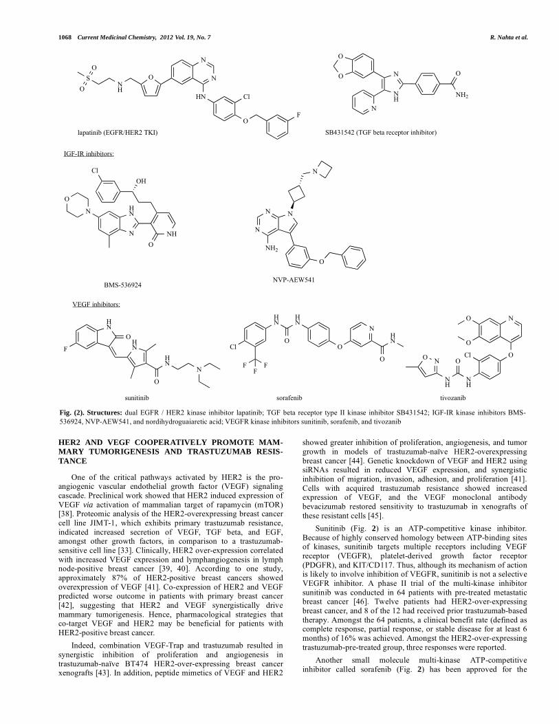
We [24] and others [27] have previously described a unique interaction between IGF-IR and HER2 in HER2-overexpressing breast cancer cells that have acquired trastuzumab resistance [24]. This interaction facilitated cross-talk from IGF-IR to HER2, such that IGF-I stimulation induced phosphorylation of HER2, whereas inhibition of IGF-IR with neutralizing antibody alpha IR3 or IGF-IR tyrosine kinase inhibitor I-OMe-AG538 blocked phosphoryl-ation of HER2 [24]. Resistant cells exhibited more rapid stimulation of PI3K/Akt and MAPK pathways in response to IGF-I relative to parental cells [24]. Subsequently, this receptor cross-talk was shown to also include HER3 in a unique IGF-IR/HER2/HER3 heterocomplex [27]. Thus, cross-phosphorylation from IGF-IR to HER2 and HER3 is a potential novel mechanism driving development of trastuzumab resistance. We previously showed that the dual EGFR/HER2 inhibitor lapatinib inhibits IGF-I signaling and induces apoptosis in cells with acquired trastuzumab resistance [28]. Thus, combination lapatinib plus trastuzumab may be an effective approach in HER2-positive cells that show elevated IGF-IR signaling.
The translational significance of IGF-IR cross-talk to HER2 and its role in trastuzumab resistance remains controversial. Breast tumor tissues of the ER-positive, triple-negative, and HER2-overexpressing subtypes stained positive for phosphorylated IGF-IR / insulin receptor (IR) in 48.1%, 41.9%, and 64.3% of cases, respectively [29], indicating that IGF-IR is activated in a majority of HER2-positive breast cancers. Activation of IGF-IR/IR in these tissues was associated with activation of the mTOR substrate S6 [29], which has been proposed as a marker of trastuzumab
resistance. In contrast, two independent studies [30, 31] suggested a lack of association between IGF-IR expression alone and trastuzumab response. However, combined analysis of increased IGF-IR expression with increased mTOR signaling correlated with reduced response to trastuzumab [30]. In addition, HER2-over-expressing breast cancer cells isolated from a patient to create the JIMT-1 cell line, which shows primary trastuzumab resistance [32], also showed constitutive hyper-activation of IGF-1R [33]. Finally, a clinical correlative study showed that high IGF-IR expression correlated with reduced response to pre-operative trastuzumab plus chemotherapy in patients with HER2-positive breast cancer [34]. Thus, substantial rationale exists for studying IGF-IR inhibition as an approach for overcoming trastuzumab resistance.
Preclinical studies showed that the ATP-competitive IGF-IR tyrosine kinase inhibitor (TKI) BMS-536924 (Fig. 2) blocked growth of HER2-overexpressing breast cancer cells [29]. BMS-536924 blocks the ATP-binding site of IGF-IR and structurally similar IR. The kinase domains of IGF-IR and IR share 84% identity [35], making it difficult to develop selective IGF-IR inhibitors that do not also block insulin receptor activity. The inherent risk then of an IGF-IR kinase inhibitor is cross-inhibition of IR and interference with glucose/insulin metabolism. The IGF-IR TKI NVP-AEW541 (Fig. 2) showed 27-fold higher selectivity for native IGF-IR versus IR because NVP-AEW541 is believed to target conformational differences in the kinase domains of IGF-IR and IR [35]. NVP-AEW541 combined with trastuzumab synergistically blocked PI3K signaling, induced p27kip1 expression, and inhibited proliferation of HER2-positive breast cancer cells [36]. Importantly, pharmacological inhibition of IGF-IR by antibody blockade or kinase inhibition restored trastuzumab sensitivity in models of acquired trastuzumab resistance [24, 37]. These studies demonstrate the potential importance of IGF-IR as a therapeutic target in HER2-overexpressing breast cancers, including those that have progressed on prior trastuzumab treatment.
1068 Current Medicinal Chemistry, 2012 Vol. 19, No. 7 R. Nahta et al.
HER2 AND VEGF COOPERATIVELY PROMOTE MAM-MARY TUMORIGENESIS AND TRASTUZUMAB RESIS-TANCE
One of the critical pathways activated by HER2 is the pro-angiogenic vascular endothelial growth factor (VEGF) signaling cascade. Preclinical work showed that HER2 induced expression of VEGF via activation of mammalian target of rapamycin (mTOR) [38]. Proteomic analysis of the HER2-overexpressing breast cancer cell line JIMT-1, which exhibits primary trastuzumab resistance, indicated increased secretion of VEGF, TGF beta, and EGF, amongst other growth factors, in comparison to a trastuzumab-sensitive cell line [33]. Clinically, HER2 over-expression correlated with increased VEGF expression and lymphangiogenesis in lymph node-positive breast cancer [39, 40]. According to one study, approximately 87% of HER2-positive breast cancers showed overexpression of VEGF [41]. Co-expression of HER2 and VEGF predicted worse outcome in patients with primary breast cancer [42], suggesting that HER2 and VEGF synergistically drive mammary tumorigenesis. Hence, pharmacological strategies that co-target VEGF and HER2 may be beneficial for patients with HER2-positive breast cancer.
Indeed, combination VEGF-Trap and trastuzumab resulted in synergistic inhibition of proliferation and angiogenesis in trastuzumab-naïve BT474 HER2-over-expressing breast cancer xenografts [43]. In addition, peptide mimetics of VEGF and HER2
showed greater inhibition of proliferation, angiogenesis, and tumor growth in models of trastuzumab-naïve HER2-overexpressing breast cancer [44]. Genetic knockdown of VEGF and HER2 using siRNAs resulted in reduced VEGF expression, and synergistic inhibition of migration, invasion, adhesion, and proliferation [41]. Cells with acquired trastuzumab resistance showed increased expression of VEGF, and the VEGF monoclonal antibody bevacizumab restored sensitivity to trastuzumab in xenografts of these resistant cells [45].
Sunitinib (Fig. 2) is an ATP-competitive kinase inhibitor. Because of highly conserved homology between ATP-binding sites of kinases, sunitinib targets multiple receptors including VEGF receptor (VEGFR), platelet-derived growth factor receptor (PDGFR), and KIT/CD117. Thus, although its mechanism of action is likely to involve inhibition of VEGFR, sunitinib is not a selective VEGFR inhibitor. A phase II trial of the multi-kinase inhibitor sunitinib was conducted in 64 patients with pre-treated metastatic breast cancer [46]. Twelve patients had HER2-over-expressing breast cancer, and 8 of the 12 had received prior trastuzumab-based therapy. Amongst the 64 patients, a clinical benefit rate (defined as complete response, partial response, or stable disease for at least 6 months) of 16% was achieved. Amongst the HER2-over-expressing trastuzumab-pre-treated group, three responses were reported.
Another small molecule multi-kinase ATP-competitive inhibitor called sorafenib (Fig. 2) has been approved for the
N
N
HN
ONH
SO
O
OF
Cl
lapatinib (EGFR/HER2 TKI)
NHO
HN
N
O
N
OHCl
BMS-536924
N
N
N
NH2
O
N
NVP-AEW541
IGF-IR inhibitors:
VEGF inhibitors:
NH
O
NH
O NO
NO
O
Cl
HN
F
OHN
HN
O
N
sunitinib sorafenib tivozanib
Cl
F FF
HN
HN
OO
NHN
O
NH
N
NH2
O
N
O
O
SB431542 (TGF beta receptor inhibitor)
Fig. (2). Structures: dual EGFR / HER2 kinase inhibitor lapatinib; TGF beta receptor type II kinase inhibitor SB431542; IGF-IR kinase inhibitors BMS-536924, NVP-AEW541, and nordihydroguaiaretic acid; VEGFR kinase inhibitors sunitinib, sorafenib, and tivozanib
HER2 Cross-Signaling and Resistance Current Medicinal Chemistry, 2012 Vol. 19, No. 7 1069
treatment of primary renal cell carcinoma and advanced primary hepatocellular carcinoma. Sorafenib blocks VEGFR, PDGFR, and Raf-MAPK activity, and thus, similar to sunitinib, is not specific for VEGFR, but its mechanism of action likely relies in part upon inhibition of angiogenesis. Because of the strong correlation between HER2 and angiogenesis, sorafenib has been studied within the context of HER2-positive breast cancer. Sorafenib blocked growth of cells that showed primary resistance to trastuzumab (JIMT-1 cell line) and cells that had acquired resistance to trastuzumab [47]. The mechanism of action appeared to involve sorafenib-mediated down-regulation of anti-apoptotic proteins Mcl-1 and survivin. The effects of sorafenib on VEGFR activity and angiogenesis were not examined in this study, but could potentially contribute to sorafenib-mediated regression of JIMT-1 tumors, which was observed in xenograft models.
The phase III AVEREL trial evaluated bevacizumab combined with trastuzumab plus docetaxel as first-line therapy in HER2-positive locally recurrent metastatic breast cancer. Results from 60 centers including 424 patients randomized to trastuzumab, bevacizumab, and paclitaxel versus the standard trastuzumab plus docetaxel showed increased progression free survival (PFS) amongst patients receiving bevacizumab [san Antonio 2011]. At a median follow up of 26 months, patients treated with the bevacizumab combination experienced a median PFS of 16.8 months versus 13.9 months for those treated with trastuzumab and docetaxel alone, equivalent to a 28% reduction in the risk of disease progression (hazard ratio [HR], 0.72; P = 0.0162). Future analysis will include examining whether circulating VEGF levels serve as a biomarker of response to more individually tailor therapy. A phase I study is currently being conducted in which bevacizumab is combined with one of the following: sunitinib, sorafenib, combination erlotinib and cetuximab, or combination trastuzumab and lapatinib. All eligible patients were refractory to standard treatments including trastuzumab for HER2-positive patients. Early results were reported for 145 patients. Amongst the HER2-positive, trastuzumab-refractory group, one complete response and four partial responses were reported [48]. This early data provides compelling evidence that anti-angiogenic therapy, and specifically VEGF-targeted therapy, may improve response to HER2-targeted therapies in patients with trastuzumab-refractory breast cancer. Thus, VEGFR kinase inhibition is a potentially effective pharmacological strategy for trastuzumab-refractory HER2-positive breast cancer. Tivozanib (AV-951; KRN-951; AVEO Pharmaceuticals Inc) (Fig. 2) is a novel quinoline-urea derivative that acts as a selective ATP-competitive pan-VEGFR kinase inhibitor [49]. In contrast to the previously discussed ATP-competitive inhibitors, tivozanib showed selectivity to VEGFR versus other kinase receptors. Importantly, tivozanib displayed strong in vivo anti-tumor activity in multiple mouse models of solid tumors, including breast cancer [49]. Given the association between HER2 and VEGF expression, and the preliminary evidence that anti-angiogenic agents improve response to trastuzumab, rationale exists for testing this new VEGFR inhibitor as well as other selective VEGFR inhibitors against HER2-positive disease, particularly in the trastuzumab-refractory setting.
EVIDENCE THAT NOTCH SIGNALING DRIVES TRASTU-ZUMAB RESISTANCE
HER2 inhibition by trastuzumab or a dual EGFR/HER2 TKI has previously been shown to activate Notch signaling in HER2-positive breast cancer cell lines [50]. Trastuzumab-resistant cells showed up-regulation of Notch-1 and its targets. Gamma secretase inhibition of Notch signaling or Notch siRNA overcame trastuzumab resistance and induced apoptosis. Notch-1 knockdown decreased cell growth by 30% in trastuzumab-sensitive cells, and by more than 50% in trastuzumab-resistant cells. Growth of both trastuzumab-sensitive and -resistant cells was completely inhibited
by combining trastuzumab plus Notch-1 siRNA. Treatment of orthotopic xenografts of HER2-positive breast cancer with a gamma secretase inhibitor significantly reduced breast tumour recurrence after trastuzumab treatment in sensitive tumors [51]. Combining lapatinib with a gamma secretase inhibitor also showed significant reduction of tumor growth. Importantly, gamma secretase inhibition partially reversed trastuzumab resistance in xenografts of acquired trastuzumab resistance. Further testing and development of gamma secretase inhibitors as a potential new therapy in the setting of trastuzumab-refractory breast cancer is warranted based on this strong preclinical data.
EVIDENCE FOR CROSS-TALK BETWEEN HER2 AND TRANSFORMING GROWTH FACTOR (TGF) BETA SIGNALING
Another signaling family that appears to enhance progression of HER2-driven breast cancers is the TGF beta family of cytokines. Mammary gland-specific overexpression of TGF beta I in MMTV-neu/erbB2 mice accelerated metastasis of Neu-dependent breast cancer in vivo [52, 53] , although primary tumor development was reduced [53]. A genetic screen that was performed to identify genes that cooperate with HER2 to promote migration showed that TGF beta enhanced HER2-mediated migration and invasion through an Erk-dependent mechanism [54]. Consistent with these data, mice with mammary-specific expression of soluble TGF beta receptor II, which acts an antagonist of TGF beta signaling, showed reduced metastases from Neu-induced mammary tumors [55]. These data argue for possible functional cross-talk from TGF beta signaling to HER2 signaling. In fact, evidence suggests that co-expression of oncogenes such as HER2 may convert TGF beta function from a tumor suppressor to a growth promoter with invasive and metastatic potential [56]. For example, TGF beta has been shown to induce binding of Smad transcription factors to specific gene promoters in MCF10A/HER2 stable cells and not in MCF10A control vector cells that do not overexpress HER2 [57]. In addition, TGF beta promoted migration of MCF10A/HER2 but not MCF10A control vector cells [58]. TGF beta-HER2 cross-talk may be mediated in part by TGF beta-stimulated phosphorylation and membrane relocalization of TACE/ADAM17 sheddase, resulting in increased secretion of erbB ligands TGF alpha, amphiregulin, and HRG [59].
However, controversy exists with respect to the role that TGF beta plays in HER2-driven breast cancer. As already mentioned, TGF beta appears to adapt pro-tumorigenic behavior in the presence of an oncogenic background. However, tumor suppressive effects of TGF beta are well-documented, even in the context of HER2-over-expressing breast cancer. For example, one study showed that preneoplastic tissue from MMTV-neu mice showed down-regulation of TGF beta signaling with loss of TGF beta receptor type I relative to normal wild-type mammary tissues [60]. Further, mammary epithelial cells (MECs) transformed by stable expression of Ras or HER2 were growth inhibited by TGF beta I to the same extent as were non-transformed MECs or MCF10A cells [61]. HER2 has also been shown to induce expression of the TGF beta signaling inhibitor Smad 7 [62], further suggesting that HER2 over-expression may suppress TGF beta function. HER2 was recently shown to activate expression of an inhibitory isoform of the transcription factor C/EBP� (LIP), which ultimately blocks TGF beta-mediated growth suppression [63]. Importantly, increased expression of LIP versus the activating isoforms of C/EBP� was associated with clinical trastuzumab resistance [63]. Thus, one body of literature suggests cooperativity and cross-talk between TGF beta and HER2 while another set of studies suggests that EHR2 abrogates TGF beta function.
A possible explanation for the discrepancy was offered by Wislon et al. [64], who previously showed that response to TGF beta in HER2-over-expressing breast cancer cells differs depending

1070 Current Medicinal Chemistry, 2012 Vol. 19, No. 7 R. Nahta et al.
on whether cells are luminal or mesenchymal. Luminal HER2-over-expressing cancer cells lost the TGF beta-induced transcriptional response observed in parental luminal cells that did not over-express HER2. In contrast, mesenchymal cells engineered to over-express HER2 showed increased expression of pro-invasive and metastatic genes relative to non-HER2-over-expressing parental cells. Another possibility is that TGF beta does not affect primary tumor growth but instead directly promotes epithelial-mesenchymal transition, invasion and metastasis of HER2-over-expressing breast cancer cells. Thus, differing results would be obtained regarding the effects of TGF beta on HER2-positive cancer depending on the stage of tumor progression that was being studied. Indeed, one study suggested that while TGF beta suppresses growth of the primary HER2-overexpressing tumor, invasion and metastasis are stimulated [53].
Another secreted cytokine, growth differentiation factor 15 (GDF15), is structurally similar to TGF beta, and has been reported to stimulate phosphorylation of HER2 [65-67]. We recently showed that GDF15-mediated phosphorylation of HER2 reduced sensitivity of HER2-overexpressing breast cancer cells to trastuzumab. Cells with primary or acquired resistance to trastuzumab expressed increased levels of GDF15 versus sensitive cells [65]. Trastuzumab-mediated growth inhibition of sensitive cells was blocked by purified recombinant human GDF15 or by stable transfection of a GDF15 expression plasmid. The HER2 tyrosine kinase inhibitor lapatinib (Fig. 2) abrogated GDF15-mediated Akt and Erk1/2 phosphorylation and blocked GDF15-mediated trastuzumab resistance, indicating that HER2 signaling is critical for GDF15-driven trastuzumab resistance (Fig. 3). We also showed that GDF15 induced phosphorylation of Src. Pharmacologic inhibition of TGF beta receptor by SB431542 (Fig. 2) blocked GDF15-mediated Src phosphorylation. In addition, SB431542 or Src inhibitor PP2 (Fig. 4) blocked GDF15-mediated trastuzumab resistance. Thus, TGF beta receptor and Src play important roles in GDF15-mediated resistance. Finally, we showed that GDF15 knockdown using lentiviral GDF15 shRNA increased trastuzumab sensitivity in cells with acquired or primary trastuzumab resistance. These results support GDF15-mediated activation of TGF beta receptor-Src-HER2 signaling crosstalk as a novel mechanism of trastuzumab resistance [65].
Thus, given that TGF beta appears to develop pro-invasive and metastatic behavior in the presence of an oncogene such as HER2, and given the data supporting cooperativity between TGF beta and HER2 signaling, pharmacologic inhibition of TGF beta is a rational approach for blocking progression of HER2-overexpressing breast cancers. Further, trastuzumab-resistant cancers that secrete higher levels of the TGF beta-related cytokine GDF15 may also benefit from pharmacologic inhibition of TGF beta signaling.
PI3K SIGNALING, A MAJOR DOWNSTREAM MEDIATOR OF HER2 CROSS-TALK
All of the cross-signaling pathways discussed in this review converge to activate downstream PI3K signaling (Fig. 1). Hyperactive PI3K signaling has been proposed as a critical mechanism limiting response to trastuzumab [68, 69]. Growth inhibition of trastuzumab-resistant cancer cells and tumor regression in associated xenograft models was achieved by pharmacological PI3K inhibitors triciribine (Fig. 4) and SF1126 (Fig. 4), an analogue of the pan-PI3K inhibitor LY294002 (Fig. 4)[70, 71]. LY294002 is not useful clinically due to poor water solubility and short half-life. SF1126 is one example of a modified version of LY294002, which has an RGDS peptide–linked integrin-targeted (�v�3/�5�1 targeted) group conjugated to the original LY backbone [72]. SF1126 inhibited proliferation and induced apoptosis of trastuzumab-naïve and trastuzumab-resistant HER2-over-expressing cells. In addition, the combination of SF1126 and trastuzumab showed synergistic anti-proliferative activity in trastuzumab-resistant cells [71]. Increased PI3K signaling may promote resistance in part due to deregulation of cell survival pathways. Trastuzumab-resistant cells showed increased expression of the anti-apoptotic regulator Bcl-2 [73]. Up-regulation of Bcl-2 appeared to be due to increased PI3K signaling and increased inhibitor of NF-kappa B (IKK) signaling, as inhibitors of these kinases reduced Bcl-2 levels and increased sensitivity to trastuzumab. Further, resistant cells showed increased sensitivity to the Bcl-2 inhibitor ABT-737, suggesting a potential new therapeutic approach that should be examined in pre-clinical xenograft models.
Clinical trial data using specific pan-PI3K or PI3K isoform-specific inhibitors is not yet available. However, mTOR inhibitors
Fig. (3). Schematic of GDF15-HER2 cross talk as a mechanism of trastuzumab resistance. GDF15 was up-regulated in models of trastuzumab resistance, and activated TGF beta receptor signaling and downstream Smad and p38MAPK. TGF beta receptor signaling cross-talk to HER2 via Src induced HER2 phosphorylation and activation of downstream PI3K/mTOR and MAPK signaling. HER2 inhibition by lapatinib overcame GDF15-mediated HER2 phosphorylation, suggesting that GDF15-overexpressing trastuzumab-resistant cells retain sensitivity to lapatinib.
GDF15
Trastuzumab
PP2GDF15
GDF15GDF15
GDF15GDF15
GDF15GDF15 GDF15GDF15
cell membrane TGFb-RI
GDF15HER2
LapatinibSB431542
PP2
TGFb-RI
GDF15HER2
TGFb-RII
P Src PTGFb-RII
P Src P
Smad2P Erk1/2
PSmad2
P Erk1/2P
Pp38 Akt
mTOR
PP
SB203580mTOR
rapamycininvasion

HER2 Cross-Signaling and Resistance Current Medicinal Chemistry, 2012 Vol. 19, No. 7 1071
are being actively pursued in the clinical setting of trastuzumab-refractory HER2-overexpressing metastatic breast cancer. Response rates greater than 40% and disease control rates of more than 70% were achieved in metastatic HER2-overexpressing breast cancers resistant to trastuzumab and chemotherapy when treated with trastuzumab, paclitaxel and the mTOR inhibitor and rapamycin (Fig. 4) analogue everolimus (Fig. 4) [74]. Phase II study of the mTOR inhibitor ridaforolimus (Fig. 4) plus trastuzumab in patients with HER2-positive trastuzumab-refractory metastatic breast cancer also showed evidence of anti-cancer activity with 2 partial responses in 22 patients [75]. A retrospective analysis of two phase I trials was performed to determine the safety and efficacy of everolimus in combination with trastuzumab-based chemotherapy in patients who had progressed on prior treatment with trastuzumab [76, 77]. The overall response rate was higher in patients who had not previously received lapatinib (31%) compared to those who had previously received lapatinib (18%). Time to progression was shorter in patients who had received lapatinib (29 weeks) versus those who did not receive lapatinib (41 weeks). However, the
clinical benefit was similar in both groups: 89% in the lapatinib pre-treated group and 84% in the lapatinib-free group. Thus, patients whose disease has progressed on trastuzumab or lapatinib may still gain benefit from mTOR inhibitors. Two phase I/II trials of trastuzumab plus everolimus were also performed in patients who had progressed on trastuzumab. Retrospective analysis of these trials showed partial responses in 15% and stable disease in 19% of patients, resulting in a clinical benefit rate of 34% [78, 79], providing evidence that mTOR inhibition improves response to trastuzumab in trastuzumab-refractory disease.
These results suggest that patients who have progressed on trastuzumab and lapatinib in the metastatic setting may derive clinical benefit from combination trastuzumab plus mTOR inhibitor. These results are being confirmed in ongoing phase 3 trials in first-line and trastuzumab-resistant settings. Thus, PI3K/mTOR inhibition is an attractive and effective strategy for improving response to HER2-targeted therapies. Given that many receptor-driven mechanisms of trastuzumab resistance ultimately
O
H OH
O
O
O
O
O
O
OO
OH
O
H
O
N
H
O OH
OH
rapamycin everolimus ridaforolimus
O
OH
H
O
O
NH
O
O
HO
OH
OH
OO
OP
O
O
N
OO
O
O
O
O
OOH
O
OO
OH
HOH
H
H
H
O
PI3K inhibitors:
O
O
N
O O
OH OH
HO N
N
NN
N
H2N
LY294002 triciribine SF1126
O
O
N+
O
O
OHN
O
NH
O
NH
HN
O
NH
O
O
OH
OH
O
OH
NH2
NH
O
O-
mTOR inhibitors:
NN
N
NCl
H2N N N
N
NHO
HN
N
SNH
OCl
H2O
Src inhibitors:
PP2 dasatinib
Fig. (4). Structures: PI3K inhibitor LY294002, its analogue SF1126, and Akt inhibitor triciribine; mTOR inhibitors rapamycin and analogues everolimus, ridaforolimus, and temsirolimus; Src inhibitors PP2 and dasatinib.
1072 Current Medicinal Chemistry, 2012 Vol. 19, No. 7 R. Nahta et al.
activate PI3K/mTOR, drugs that block this downstream pathway should effectively block compensatory signaling and restore sensitivity to trastuzumab. In contrast to trastzuumab resistance, PTEN loss and PI3KCA activating mutations do not appear to predict for lapatinib resistance [80]. However, sensitivity to lapatinib may still gain benefit from combination with mTOR inhibitors. For example, cells with primary trastuzumab resistance and poor response to lapatinib showed increased lapatinib sensitivity when co-treated with rapamycin or ridaforolimus [81]. This synergy was maintained in vivo in a xenograft model of primary trastuzumab resistance that showed complete suppression of tumor growth upon co-treatment with lapatinib plus ridaforolimus. Thus, mTOR inhibition is an important therapeutic strategy for sensitizing HER2-positive breast cancers to HER2-targeted therapies.
FOCAL ADHESION KINASE (FAK) AND SRC: CENTRAL INTEGRATORS OF HER2 SIGNALING CROSS-TALK AND TRASTUZUMAB RESISTANCE?
In addition to converging downstream to activate a common signaling pathway (PI3K), evidence suggests that compensatory cross-talk activity to HER2 commonly occurs via activation of Src-FAK activity. The HER3 ligand heregulin (HRG) has been shown to differentially affect phosphorylation of FAK depending on the concentration of HRG [82]. Sub-physiological doses induced FAK phosphorylation on Y577 and Y925, and promoted interaction of FAK with HER2, while increasing adhesion of cultured breast cancer cells. In contrast, 1 nM HRG induced interaction of Shp2 phosphatase with HER2, leading to dephosphorylation of FAK Y577 and Y925. HRG stimulation or HER2 stable over-expression was associated with increased phosphorylation of Src on Y215, which in turn induced phosphorylation of Y861on FAK, a site believed to be critical to FAK-mediated cell migration [83]. In clinical samples, increased FAK expression correlated with HER2 over-expression, phosphorylated Src Y215, and phosphorylated Akt [84]. Specifically, HER2 over-expression was associated with increased phosphorylation of FAK Y861 [84], consistent with reported in vitro results [83].
In addition to HRG, TGF beta has been shown to induce phosphorylation of Src-FAK in MCF10A/HER2 stable cells in association with increased cell motility [57]. TGF beta-stimulated Src-FAK phosphorylation resulted in FAK-dependent clustering of HER2 and integrins on HER2-over-expressing cells [85]. TGF beta-mediated trastuzumab resistance was overcome by Src-FAK inhibition [85]. In fact, resistance to either trastuzumab or lapatinib was associated with integrin-FAK signaling [86]. Trastuzumab-resistant cells have been shown to be sensitive to blockade of either integrins or FAK [86, 87], suggesting a novel therapeutic strategy for breast cancers that have progressed on trastuzumab. TAE226 (Novartis, Inc., Switzerland) is a novel bis-anilino pyrimidine ATP-competitive FAK inhibitor with an IC50 of 5.5 nM for FAK and 8-fold higher IC50 for IR and 25-fold higher for IGF-IR [88]. TAE226 blocked FAK activity and induced apoptosis in BT474 EGFR- and HER2-over-expressing breast cancer cells in contrast to MCF10A non-transformed mammary epithelial cells [89]. Further, TAE226 blocked mTOR signaling in models of esophageal cancer [90]. Because of the strong evidence supporting mTOR inhibition as a strategy in trastuzumab-resistant breast cancer, and because of the potential roles of FAK and IGF-IR signaling as mechanisms of trastuzumab resistance, TAE226 may hold therapeutic value in the setting of HER2-over-expressing breast cancer. The combination of TAE226 and trastuzumab should be examined pre-clinically in models of trastuzumab-naïve and trastuzumab-refractory HER2-over-expressing breast cancer.
In addition to FAK inhibition, Src inhibition is a novel strategy for treating trastuzumab-resistant cancers. Src kinase activity has
been reported to mediate multiple mechanisms of trastuzumab resistance, and ultimately activates FAK and PI3K/mTOR signaling. Normally, trastuzumab blocks Src Y416 phosphorylation [69], resulting in reduced activity of downstream targets. One example is that trastuzumab blocked interaction of Src with HER2 and reduced PTEN tyrosine phosphorylation while increasing PTEN membrane localization and activity. While multiple mechanisms of trastuzumab resistance have been demonstrated to exist, almost all share in common activation of Src and downstream PI3K. These mechanisms of trastuzumab resistance include PTEN down-regulation [69], IGF-IR over-expression [69], exposure to recombinant human erythropoietin [91], increased ephA2 kinase activity [92], and GDF15 over-expression [65], all of which have been shown to activate Src activity and PI3K signaling. Targeting SRC by genetic knockdown or pharmacologic inhibitor saracatinib (AZD0530) sensitized multiple trastuzumab-resistant cell lines to trastuzumab and blocked growth of trastuzumab-resistant tumors in vivo [93]. A recent phase I trial of saracatinib in 81 patients with advanced solid tumors including 13 metastatic breast cancers showed reduced Src activity using p-FAK as a marker [94]. Phase II study of single-agent dasatinib showed limited activity in trastuzumab-refractory HER2-amplified (HER2+) or endocrine therapy-refractory hormone receptor-positive (HR+) breast cancers, with overall response rate of 4% [95]. The disease control rate (defined as partial response or stable disease) was 8% and 16% in the HER2+ and HR+ groups, respectively. Stronger activity was observed in the HR+ group which included HR+ HER2+ cancers, with poorer activity observed in the HER2-amplified HR- group. Combining Src inhibition with targeted HER2 therapies (trastuzumab, lapatinib, and/or pertuzumab) is likely to produce greater benefit in patients with HER2-positive breast cancer.
CONCLUSIONS
Trastuzumab has revolutionized the treatment and prognosis of HER2-over-expressing breast cancer. The next necessary step is to understand the limitations of trastuzumab therapy by elucidating the molecular mechanisms that drive therapeutic resistance. Extensive effort has been made over the past decade to develop models of trastuzumab resistance. These preclinical models have identified multiple kinases that cross-activate and sustain HER2 activity in the presence of trastuzumab. Central to these compensatory signaling mechanisms of trastuzumab resistance are activation of Src and PI3K, inhibition of which will likely improve survival rates of patients with trastuzumab-refractory HER2-over-expressing breast cancer. In particular, current clinical trials show promising results with combined mTOR inhibition plus trastuzumab in trastuzumab-refractory populations. A central issue brought to light in breast cancer clinical trials, however, is toxicity related to treatment with inhibitors of downstream tyrosine kinases. For example, in early stage breast cancer, treatment with sorafenib plus chemotherapy showed unfavorable toxicity in 47% of patients [96]. Similarly, in endocrine-resistant breast cancers, frequent toxicities associated with sorafenib plus aromatase inhibitor treatment resulted in a high rate of discontinuation from the trial study [97]. A recent phase 2 trial of Src inhibitor dasatinib in HER2-positive or hormone receptor-positive breast cancer also showed grade 3-4 toxicity in 37% of patients [98]. Thus, inhibition of central signaling nodes, such as Src and PI3K, may result in undue toxicity due to blockade of multiple pathways that converge on these signaling molecules. Strategies that combine multiple HER2-directed therapies may delay development of resistance if used in the front-line setting. Indeed, combination trastuzumab plus lapatinib and trastuzumab plus pertuzumab are being tested for use in patients with trastuzumab-naïve HER2-positive breast cancer. Future studies will also need to carefully assess the role of estrogen receptor (ER) signaling in trastuzumab-resistant HER2-positive ER-positive disease. While resistance to ER-targeted therapy has been shown to

HER2 Cross-Signaling and Resistance Current Medicinal Chemistry, 2012 Vol. 19, No. 7 1073
rely in part upon increased HER2 expression and signaling, the converse is still unknown, i.e. does increased ER signaling limit response to trastuzumab? Differential response to trastuzumab in ER-positive versus ER-negative disease may suggest differential courses of treatment in these two subtypes. Ultimately, identification of relevant predictors of response will be critical for rational patient selection, allowing individualized therapy and limited adverse effects to those who are unlikely to benefit. One strategy is to develop a so-called gene signature of trastuzumab response or trastuzumab resistance, which would likely involve many of the kinases discussed in this review, such as PIK3CAmutations that activate PI3K. However, the most useful assays may be those that are serum-based, as these assays offer a largely non-invasive, easily standardized approach to predict which patients will receive clinical benefit from trastuzumab. Such assays would measure secreted proteins such as erbB ligands, TGF beta, GDF15, and VEGF as markers of trastuzumab resistance. Development and implementation of gene- and protein-based assays that measure the molecular markers of resistance described above will allow further personalization of HER2-targeted therapy beyond HER2 expression status, and should ultimately improve patient survival rates.
ACKNOWLEDGEMENTS
R.N. gratefully acknowledges funding from the National Cancer Institute (K01CA118174 and 3K01CA118174-5S1), The Mary Kay Foundation, and the Georgia Cancer Coalition (Distinguished Cancer Scholar Award).
REFERENCES
[1] Slamon, D.J.; Clark, G.M.; Wong, S.G.; Levin, W.J.; Ullrich, A.; McGuire, W.L. Human breast cancer: correlation of relapse and survival with amplification of the HER-2/neu oncogene. Science, 1987, 235(4785), 177-182.
[2] Baselga, J.; Tripathy, D.; Mendelsohn, J.; Baughman, S.; Benz, C.C.; Dantis, L.; Sklarin, N.T.; Seidman, A.D.; Hudis, C.A.; Moore, J.; Rosen, P.P.; Twaddell, T.; Henderson, I.C.; Norton, L. Phase II study of weekly intravenous recombinant humanized anti-p185HER2 monoclonal antibody in patients with HER2/neu-overexpressing metastatic breast cancer. J Clin Oncol, 1996, 14(3), 737-744.
[3] Cobleigh, M.A.; Vogel, C.L.; Tripathy, D.; Robert, N.J.; Scholl, S.; Fehrenbacher, L.; Wolter, J.M.; Paton, V.; Shak, S.; Lieberman, G.; Slamon, D.J. Multinational study of the efficacy and safety of humanized anti-HER2 monoclonal antibody in women who have HER2-overexpressing metastatic breast cancer that has progressed after chemotherapy for metastatic disease. JClin Oncol, 1999, 17(9), 2639-2648.
[4] Geyer, C.E.; Forster, J.; Lindquist, D.; Chan, S.; Romieu, C.G.; Pienkowski, T.; Jagiello-Gruszfeld, A.; Crown, J.; Chan, A.; Kaufman, B.; Skarlos, D.; Campone, M.; Davidson, N.; Berger, M.; Oliva, C.; Rubin, S.D.; Stein, S.; Cameron, D. Lapatinib plus capecitabine for HER2-positive advanced breast cancer. N Engl J Med, 2006, 355(26), 2733-2743.
[5] Slamon, D.J.; Leyland-Jones, B.; Shak, S.; Fuchs, H.; Paton, V.; Bajamonde, A.; Fleming, T.; Eiermann, W.; Wolter, J.; Pegram, M.; Baselga, J.; Norton, L. Use of chemotherapy plus a monoclonal antibody against HER2 for metastatic breast cancer that overexpresses HER2. N Engl J Med, 2001,344(11), 783-792.
[6] Esteva, F.J.; Valero, V.; Booser, D.; Guerra, L.T.; Murray, J.L.; Pusztai, L.; Cristofanilli, M.; Arun, B.; Esmaeli, B.; Fritsche, H.A.; Sneige, N.; Smith, T.L.; Hortobagyi, G.N. Phase II study of weekly docetaxel and trastuzumab for patients with HER-2-overexpressing metastatic breast cancer. J Clin Oncol, 2002, 20(7), 1800-1808.
[7] Seidman, A.D.; Fornier, M.N.; Esteva, F.J.; Tan, L.; Kaptain, S.; Bach, A.; Panageas, K.S.; Arroyo, C.; Valero, V.; Currie, V.; Gilewski, T.; Theodoulou, M.; Moynahan, M.E.; Moasser, M.; Sklarin, N.; Dickler, M.; D'Andrea, G.; Cristofanilli, M.; Rivera, E.; Hortobagyi, G.N.; Norton, L.; Hudis, C.A. Weekly trastuzumab and paclitaxel therapy for metastatic breast cancer with analysis of efficacy by HER2 immunophenotype and gene amplification. J Clin Oncol, 2001, 19(10), 2587-2595.
[8] Bender, L.M.; Nahta, R. Her2 cross talk and therapeutic resistance in breast cancer. Front Biosci, 2008, 13, 3906-3912.
[9] Graus-Porta, D.; Beerli, R.R.; Daly, J.M.; Hynes, N.E. ErbB-2, the preferred heterodimerization partner of all ErbB receptors, is a mediator of lateral signaling. EMBO J, 1997, 16(7), 1647-1655.
[10] Motoyama, A.B.; Hynes, N.E.; Lane, H.A. The efficacy of ErbB receptor-targeted anticancer therapeutics is influenced by the availability of epidermal growth factor-related peptides. Cancer Res, 2002, 62(11), 3151-3158.
[11] Moulder, S.L.; Yakes, F.M.; Muthuswamy, S.K.; Bianco, R.; Simpson, J.F.; Arteaga, C.L. Epidermal growth factor receptor (HER1) tyrosine kinase inhibitor ZD1839 (Iressa) inhibits HER2/neu (erbB2)-overexpressing breast cancer cells in vitro and in vivo. Cancer Res, 2001, 61(24), 8887-8895.
[12] Ritter, C.A.; Perez-Torres, M.; Rinehart, C.; Guix, M.; Dugger, T.; Engelman, J.A.; Arteaga, C.L. Human breast cancer cells selected for resistance to trastuzumab in vivo overexpress epidermal growth factor receptor and ErbB ligands and remain dependent on the ErbB receptor network. Clin Cancer Res, 2007, 13(16), 4909-4919.
[13] Lee-Hoeflich, S.T.; Crocker, L.; Yao, E.; Pham, T.; Munroe, X.; Hoeflich, K.P.; Sliwkowski, M.X.; Stern, H.M. A central role for HER3 in HER2-amplified breast cancer: implications for targeted therapy. Cancer Res, 2008,68(14), 5878-5887.
[14] Alimandi, M.; Romano, A.; Curia, M.C.; Muraro, R.; Fedi, P.; Aaronson, S.A.; Di Fiore, P.P.; Kraus, M.H. Cooperative signaling of ErbB3 and ErbB2 in neoplastic transformation and human mammary carcinomas. Oncogene,1995, 10(9), 1813-1821.
[15] Holbro, T.; Beerli, R.R.; Maurer, F.; Koziczak, M.; Barbas, C.F., 3rd; Hynes, N.E. The ErbB2/ErbB3 heterodimer functions as an oncogenic unit: ErbB2 requires ErbB3 to drive breast tumor cell proliferation. Proc Natl Acad Sci U S A, 2003, 100(15), 8933-8938.
[16] Hellyer, N.J.; Cheng, K.; Koland, J.G. ErbB3 (HER3) interaction with the p85 regulatory subunit of phosphoinositide 3-kinase. Biochem J, 1998, 333 ( Pt 3), 757-763.
[17] Siegel, P.M.; Ryan, E.D.; Cardiff, R.D.; Muller, W.J. Elevated expression of activated forms of Neu/ErbB-2 and ErbB-3 are involved in the induction of mammary tumors in transgenic mice: implications for human breast cancer. EMBO J, 1999, 18(8), 2149-2164.
[18] van der Horst, E.H.; Murgia, M.; Treder, M.; Ullrich, A. Anti-HER-3 MAbs inhibit HER-3-mediated signaling in breast cancer cell lines resistant to anti-HER-2 antibodies. Int J Cancer, 2005, 115(4), 519-527.
[19] Ghosh, R.; Narasanna, A.; Wang, S.E.; Liu, S.; Chakrabarty, A.; Balko, J.M.; Gonzalez-Angulo, A.M.; Mills, G.B.; Penuel, E.; Winslow, J.; Sperinde, J.; Dua, R.; Pidaparthi, S.; Mukherjee, A.; Leitzel, K.; Kostler, W.J.; Lipton, A.; Bates, M.; Arteaga, C.L. Trastuzumab has preferential activity against breast cancers driven by HER2 homodimers. Cancer Res, 2011, 71(5), 1871-1882.
[20] Scheuer, W.; Friess, T.; Burtscher, H.; Bossenmaier, B.; Endl, J.; Hasmann, M. Strongly enhanced antitumor activity of trastuzumab and pertuzumab combination treatment on HER2-positive human xenograft tumor models. Cancer Res, 2009, 69(24), 9330-9336.
[21] Nahta, R.; Hung, M.C.; Esteva, F.J. The HER-2-targeting antibodies trastuzumab and pertuzumab synergistically inhibit the survival of breast cancer cells. Cancer Res, 2004, 64(7), 2343-2346.
[22] Xia, W.; Liu, L.H.; Ho, P.; Spector, N.L. Truncated ErbB2 receptor (p95ErbB2) is regulated by heregulin through heterodimer formation with ErbB3 yet remains sensitive to the dual EGFR/ErbB2 kinase inhibitor GW572016. Oncogene, 2004, 23(3), 646-653.
[23] Yao, E.; Zhou, W.; Lee-Hoeflich, S.T.; Truong, T.; Haverty, P.M.; Eastham-Anderson, J.; Lewin-Koh, N.; Gunter, B.; Belvin, M.; Murray, L.J.; Friedman, L.S.; Sliwkowski, M.X.; Hoeflich, K.P. Suppression of HER2/HER3-mediated growth of breast cancer cells with combinations of GDC-0941 PI3K inhibitor, trastuzumab, and pertuzumab. Clin Cancer Res,2009, 15(12), 4147-4156.
[24] Nahta, R.; Yuan, L.X.; Zhang, B.; Kobayashi, R.; Esteva, F.J. Insulin-like growth factor-I receptor/human epidermal growth factor receptor 2 heterodimerization contributes to trastuzumab resistance of breast cancer cells. Cancer Res, 2005, 65(23), 11118-11128.
[25] Baselga, J.; Gelmon, K.A.; Verma, S.; Wardley, A.; Conte, P.; Miles, D.; Bianchi, G.; Cortes, J.; McNally, V.A.; Ross, G.A.; Fumoleau, P.; Gianni, L. Phase II trial of pertuzumab and trastuzumab in patients with human epidermal growth factor receptor 2-positive metastatic breast cancer that progressed during prior trastuzumab therapy. J Clin Oncol, 2010, 28(7), 1138-1144.
[26] Baselga, J.; Swain, S.M. CLEOPATRA: a phase III evaluation of pertuzumab and trastuzumab for HER2-positive metastatic breast cancer. Clin Breast Cancer, 2010, 10(6), 489-491.
[27] Huang, X.; Gao, L.; Wang, S.; McManaman, J.L.; Thor, A.D.; Yang, X.; Esteva, F.J.; Liu, B. Heterotrimerization of the growth factor receptors erbB2, erbB3, and insulin-like growth factor-i receptor in breast cancer cells resistant to herceptin. Cancer Res, 2010, 70(3), 1204-1214.
[28] Nahta, R.; Yuan, L.X.; Du, Y.; Esteva, F.J. Lapatinib induces apoptosis in trastuzumab-resistant breast cancer cells: effects on insulin-like growth factor I signaling. Mol Cancer Ther, 2007, 6(2), 667-674.
[29] Law, J.H.; Habibi, G.; Hu, K.; Masoudi, H.; Wang, M.Y.; Stratford, A.L.; Park, E.; Gee, J.M.; Finlay, P.; Jones, H.E.; Nicholson, R.I.; Carboni, J.; Gottardis, M.; Pollak, M.; Dunn, S.E. Phosphorylated insulin-like growth factor-i/insulin receptor is present in all breast cancer subtypes and is related to poor survival. Cancer Res, 2008, 68(24), 10238-10246.
[30] Smith, B.L.; Chin, D.; Maltzman, W.; Crosby, K.; Hortobagyi, G.N.; Bacus, S.S. The efficacy of Herceptin therapies is influenced by the expression of other erbB receptors, their ligands and the activation of downstream signalling proteins. Br J Cancer, 2004, 91(6), 1190-1194.
[31] Kostler, W.J.; Hudelist, G.; Rabitsch, W.; Czerwenka, K.; Muller, R.; Singer, C.F.; Zielinski, C.C. Insulin-like growth factor-1 receptor (IGF-1R) expression does not predict for resistance to trastuzumab-based treatment in

1074 Current Medicinal Chemistry, 2012 Vol. 19, No. 7 R. Nahta et al.
patients with Her-2/neu overexpressing metastatic breast cancer. J Cancer Res Clin Oncol, 2006, 132(1), 9-18.
[32] Tanner, M.; Kapanen, A.I.; Junttila, T.; Raheem, O.; Grenman, S.; Elo, J.; Elenius, K.; Isola, J. Characterization of a novel cell line established from a patient with Herceptin-resistant breast cancer. Mol Cancer Ther, 2004, 3(12), 1585-1592.
[33] Oliveras-Ferraros, C.; Vazquez-Martin, A.; Martin-Castillo, B.; Perez-Martinez, M.C.; Cufi, S.; Del Barco, S.; Bernado, L.; Brunet, J.; Lopez-Bonet, E.; Menendez, J.A. Pathway-focused proteomic signatures in HER2-overexpressing breast cancer with a basal-like phenotype: new insights into de novo resistance to trastuzumab (Herceptin). Int J Oncol, 2010, 37(3), 669-678.
[34] Harris, L.N.; You, F.; Schnitt, S.J.; Witkiewicz, A.; Lu, X.; Sgroi, D.; Ryan, P.D.; Come, S.E.; Burstein, H.J.; Lesnikoski, B.A.; Kamma, M.; Friedman, P.N.; Gelman, R.; Iglehart, J.D.; Winer, E.P. Predictors of resistance to preoperative trastuzumab and vinorelbine for HER2-positive early breast cancer. Clin Cancer Res, 2007, 13(4), 1198-1207.
[35] Garcia-Echeverria, C.; Pearson, M.A.; Marti, A.; Meyer, T.; Mestan, J.; Zimmermann, J.; Gao, J.; Brueggen, J.; Capraro, H.G.; Cozens, R.; Evans, D.B.; Fabbro, D.; Furet, P.; Porta, D.G.; Liebetanz, J.; Martiny-Baron, G.; Ruetz, S.; Hofmann, F. In vivo antitumor activity of NVP-AEW541-A novel, potent, and selective inhibitor of the IGF-IR kinase. Cancer Cell, 2004, 5(3), 231-239.
[36] Esparis-Ogando, A.; Ocana, A.; Rodriguez-Barrueco, R.; Ferreira, L.; Borges, J.; Pandiella, A. Synergic antitumoral effect of an IGF-IR inhibitor and trastuzumab on HER2-overexpressing breast cancer cells. Ann Oncol,2008, 19(11), 1860-1869.
[37] Rowe, D.L.; Ozbay, T.; Bender, L.M.; Nahta, R. Nordihydroguaiaretic acid, a cytotoxic insulin-like growth factor-I receptor/HER2 inhibitor in trastuzumab-resistant breast cancer. Mol Cancer Ther, 2008, 7(7), 1900-1908.
[38] Klos, K.S.; Wyszomierski, S.L.; Sun, M.; Tan, M.; Zhou, X.; Li, P.; Yang, W.; Yin, G.; Hittelman, W.N.; Yu, D. ErbB2 increases vascular endothelial growth factor protein synthesis via activation of mammalian target of rapamycin/p70S6K leading to increased angiogenesis and spontaneous metastasis of human breast cancer cells. Cancer Res, 2006, 66(4), 2028-2037.
[39] Schoppmann, S.F.; Tamandl, D.; Roberts, L.; Jomrich, G.; Schoppmann, A.; Zwrtek, R.; Dubsky, P.; Gnant, M.; Jakesz, R.; Birner, P. HER2/neu expression correlates with vascular endothelial growth factor-C and lymphangiogenesis in lymph node-positive breast cancer. Ann Oncol, 2010,21(5), 955-960.
[40] Yang, W.; Klos, K.; Yang, Y.; Smith, T.L.; Shi, D.; Yu, D. ErbB2 overexpression correlates with increased expression of vascular endothelial growth factors A, C, and D in human breast carcinoma. Cancer, 2002,94(11), 2855-2861.
[41] Tai, W.; Qin, B.; Cheng, K. Inhibition of breast cancer cell growth and invasiveness by dual silencing of HER-2 and VEGF. Molecular pharmaceutics, 2010, 7(2), 543-556.
[42] Konecny, G.E.; Meng, Y.G.; Untch, M.; Wang, H.J.; Bauerfeind, I.; Epstein, M.; Stieber, P.; Vernes, J.M.; Gutierrez, J.; Hong, K.; Beryt, M.; Hepp, H.; Slamon, D.J.; Pegram, M.D. Association between HER-2/neu and vascular endothelial growth factor expression predicts clinical outcome in primary breast cancer patients. Clin Cancer Res, 2004, 10(5), 1706-1716.
[43] Le, X.F.; Mao, W.; Lu, C.; Thornton, A.; Heymach, J.V.; Sood, A.K.; Bast, R.C., Jr. Specific blockade of VEGF and HER2 pathways results in greater growth inhibition of breast cancer xenografts that overexpress HER2. Cell Cycle, 2008, 7(23), 3747-3758.
[44] Foy, K.C.; Liu, Z.; Phillips, G.; Miller, M.; Kaumaya, P.T. Combination treatment with HER-2 and VEGF peptide mimics induces potent anti-tumor and anti-angiogenic responses in vitro and in vivo. J Biol Chem, 2011,286(15), 13626-13637.
[45] du Manoir, J.M.; Francia, G.; Man, S.; Mossoba, M.; Medin, J.A.; Viloria-Petit, A.; Hicklin, D.J.; Emmenegger, U.; Kerbel, R.S. Strategies for delaying or treating in vivo acquired resistance to trastuzumab in human breast cancer xenografts. Clin Cancer Res, 2006, 12(3 Pt 1), 904-916.
[46] Burstein, H.J.; Elias, A.D.; Rugo, H.S.; Cobleigh, M.A.; Wolff, A.C.; Eisenberg, P.D.; Lehman, M.; Adams, B.J.; Bello, C.L.; DePrimo, S.E.; Baum, C.M.; Miller, K.D. Phase II study of sunitinib malate, an oral multitargeted tyrosine kinase inhibitor, in patients with metastatic breast cancer previously treated with an anthracycline and a taxane. J Clin Oncol,2008, 26(11), 1810-1816.
[47] Valabrega, G.; Capellero, S.; Cavalloni, G.; Zaccarello, G.; Petrelli, A.; Migliardi, G.; Milani, A.; Peraldo-Neia, C.; Gammaitoni, L.; Sapino, A.; Pecchioni, C.; Moggio, A.; Giordano, S.; Aglietta, M.; Montemurro, F. HER2-positive breast cancer cells resistant to trastuzumab and lapatinib lose reliance upon HER2 and are sensitive to the multitargeted kinase inhibitor sorafenib. Breast Cancer Res Treat, 2011, 130(1), 29-40.
[48] Falchook, G.S.W., J. J.; Naing, A.; Hong, D. S.; Moulder, S. L.; Piha-Paul, S. A.; Ng, C. S.; Jackson, E.; Kurzrock, R. In J Clin Oncol, 2010 ASCO Annual Meeting, A phase I study of bevaizumab in combination with sunitinib, sorafenib, and erlotinib plus cetuximab, and trastuzumab plus lapatinib: 2010; p 15s.
[49] Nakamura, K.; Taguchi, E.; Miura, T.; Yamamoto, A.; Takahashi, K.; Bichat, F.; Guilbaud, N.; Hasegawa, K.; Kubo, K.; Fujiwara, Y.; Suzuki, R.;
Shibuya, M.; Isae, T. KRN951, a highly potent inhibitor of vascular endothelial growth factor receptor tyrosine kinases, has antitumor activities and affects functional vascular properties. Cancer Res, 2006, 66(18), 9134-9142.
[50] Osipo, C.; Patel, P.; Rizzo, P.; Clementz, A.G.; Hao, L.; Golde, T.E.; Miele, L. ErbB-2 inhibition activates Notch-1 and sensitizes breast cancer cells to a gamma-secretase inhibitor. Oncogene, 2008, 27(37), 5019-5032.
[51] Pandya, K.; Meeke, K.; Clementz, A.G.; Rogowski, A.; Roberts, J.; Miele, L.; Albain, K.S.; Osipo, C. Targeting both Notch and ErbB-2 signalling pathways is required for prevention of ErbB-2-positive breast tumour recurrence. Br J Cancer, 2011, 105(6), 796-806.
[52] Muraoka, R.S.; Koh, Y.; Roebuck, L.R.; Sanders, M.E.; Brantley-Sieders, D.; Gorska, A.E.; Moses, H.L.; Arteaga, C.L. Increased malignancy of Neu-induced mammary tumors overexpressing active transforming growth factor beta1. Mol Cell Biol, 2003, 23(23), 8691-8703.
[53] Siegel, P.M.; Shu, W.; Cardiff, R.D.; Muller, W.J.; Massague, J. Transforming growth factor beta signaling impairs Neu-induced mammary tumorigenesis while promoting pulmonary metastasis. Proc Natl Acad Sci U S A, 2003, 100(14), 8430-8435.
[54] Seton-Rogers, S.E.; Lu, Y.; Hines, L.M.; Koundinya, M.; LaBaer, J.; Muthuswamy, S.K.; Brugge, J.S. Cooperation of the ErbB2 receptor and transforming growth factor beta in induction of migration and invasion in mammary epithelial cells. Proc Natl Acad Sci U S A, 2004, 101(5), 1257-1262.
[55] Bandyopadhyay, A.; Lopez-Casillas, F.; Malik, S.N.; Montiel, J.L.; Mendoza, V.; Yang, J.; Sun, L.Z. Antitumor activity of a recombinant soluble betaglycan in human breast cancer xenograft. Cancer Res, 2002,62(16), 4690-4695.
[56] Wang, S.E. The Functional Crosstalk between HER2 Tyrosine Kinase and TGF-beta Signaling in Breast Cancer Malignancy. J Signal Transduct, 2011,2011, 804236.
[57] Wang, S.E.; Wu, F.Y.; Shin, I.; Qu, S.; Arteaga, C.L. Transforming growth factor {beta} (TGF-{beta})-Smad target gene protein tyrosine phosphatase receptor type kappa is required for TGF-{beta} function. Mol Cell Biol,2005, 25(11), 4703-4715.
[58] Ueda, Y.; Wang, S.; Dumont, N.; Yi, J.Y.; Koh, Y.; Arteaga, C.L. Overexpression of HER2 (erbB2) in human breast epithelial cells unmasks transforming growth factor beta-induced cell motility. J Biol Chem, 2004,279(23), 24505-24513.
[59] Wang, S.E.; Xiang, B.; Guix, M.; Olivares, M.G.; Parker, J.; Chung, C.H.; Pandiella, A.; Arteaga, C.L. Transforming growth factor beta engages TACE and ErbB3 to activate phosphatidylinositol-3 kinase/Akt in ErbB2-overexpressing breast cancer and desensitizes cells to trastuzumab. Mol Cell Biol, 2008, 28(18), 5605-5620.
[60] Landis, M.D.; Seachrist, D.D.; Montanez-Wiscovich, M.E.; Danielpour, D.; Keri, R.A. Gene expression profiling of cancer progression reveals intrinsic regulation of transforming growth factor-beta signaling in ErbB2/Neu-induced tumors from transgenic mice. Oncogene, 2005, 24(33), 5173-5190.
[61] Basolo, F.; Fiore, L.; Ciardiello, F.; Calvo, S.; Fontanini, G.; Conaldi, P.G.; Toniolo, A. Response of normal and oncogene-transformed human mammary epithelial cells to transforming growth factor beta 1 (TGF-beta 1): lack of growth-inhibitory effect on cells expressing the simian virus 40 large-T antigen. Int J Cancer, 1994, 56(5), 736-742.
[62] Dowdy, S.C.; Mariani, A.; Janknecht, R. HER2/Neu- and TAK1-mediated up-regulation of the transforming growth factor beta inhibitor Smad7 via the ETS protein ER81. J Biol Chem, 2003, 278(45), 44377-44384.
[63] Arnal-Estape, A.; Tarragona, M.; Morales, M.; Guiu, M.; Nadal, C.; Massague, J.; Gomis, R.R. HER2 silences tumor suppression in breast cancer cells by switching expression of C/EBPss isoforms. Cancer Res, 2010,70(23), 9927-9936.
[64] Wilson, C.A.; Cajulis, E.E.; Green, J.L.; Olsen, T.M.; Chung, Y.A.; Damore, M.A.; Dering, J.; Calzone, F.J.; Slamon, D.J. HER-2 overexpression differentially alters transforming growth factor-beta responses in luminal versus mesenchymal human breast cancer cells. Breast Cancer Res, 2005,7(6), R1058-1079.
[65] Joshi, J.P.; Brown, N.E.; Griner, S.E.; Nahta, R. Growth differentiation factor 15 (GDF15)-mediated HER2 phosphorylation reduces trastuzumab sensitivity of HER2-overexpressing breast cancer cells. Biochem Pharmacol,2011, 82(9), 1090-1099.
[66] Kim, K.K.; Lee, J.J.; Yang, Y.; You, K.H.; Lee, J.H. Macrophage inhibitory cytokine-1 activates AKT and ERK-1/2 via the transactivation of ErbB2 in human breast and gastric cancer cells. Carcinogenesis, 2008, 29(4), 704-712.
[67] Park, Y.J.; Lee, H.; Lee, J.H. Macrophage inhibitory cytokine-1 transactivates ErbB family receptors via the activation of Src in SK-BR-3 human breast cancer cells. BMB Rep, 2010, 43(2), 91-96.
[68] Berns, K.; Horlings, H.M.; Hennessy, B.T.; Madiredjo, M.; Hijmans, E.M.; Beelen, K.; Linn, S.C.; Gonzalez-Angulo, A.M.; Stemke-Hale, K.; Hauptmann, M.; Beijersbergen, R.L.; Mills, G.B.; van de Vijver, M.J.; Bernards, R. A functional genetic approach identifies the PI3K pathway as a major determinant of trastuzumab resistance in breast cancer. Cancer Cell,2007, 12(4), 395-402.
[69] Nagata, Y.; Lan, K.H.; Zhou, X.; Tan, M.; Esteva, F.J.; Sahin, A.A.; Klos, K.S.; Li, P.; Monia, B.P.; Nguyen, N.T.; Hortobagyi, G.N.; Hung, M.C.; Yu, D. PTEN activation contributes to tumor inhibition by trastuzumab, and loss
HER2 Cross-Signaling and Resistance Current Medicinal Chemistry, 2012 Vol. 19, No. 7 1075
of PTEN predicts trastuzumab resistance in patients. Cancer Cell, 2004, 6(2), 117-127.
[70] Lu, C.H.; Wyszomierski, S.L.; Tseng, L.M.; Sun, M.H.; Lan, K.H.; Neal, C.L.; Mills, G.B.; Hortobagyi, G.N.; Esteva, F.J.; Yu, D. Preclinical testing of clinically applicable strategies for overcoming trastuzumab resistance caused by PTEN deficiency. Clin Cancer Res, 2007, 13(19), 5883-5888.
[71] Ozbay, T.; Durden, D.L.; Liu, T.; O'Regan, R.M.; Nahta, R. In vitroevaluation of pan-PI3-kinase inhibitor SF1126 in trastuzumab-sensitive and trastuzumab-resistant HER2-over-expressing breast cancer cells. Cancer Chemother Pharmacol, 2010, 65(4), 697-706.
[72] Garlich, J.R.; De, P.; Dey, N.; Su, J.D.; Peng, X.; Miller, A.; Murali, R.; Lu, Y.; Mills, G.B.; Kundra, V.; Shu, H.K.; Peng, Q.; Durden, D.L. A vascular targeted pan phosphoinositide 3-kinase inhibitor prodrug, SF1126, with antitumor and antiangiogenic activity. Cancer Res, 2008, 68(1), 206-215.
[73] Crawford, A.; Nahta, R. Targeting Bcl-2 in Herceptin-Resistant Breast Cancer Cell Lines. Current pharmacogenomics and personalized medicine,2011, 9(3), 184-190.
[74] Andre, F.; Campone, M.; O'Regan, R.; Manlius, C.; Massacesi, C.; Sahmoud, T.; Mukhopadhyay, P.; Soria, J.C.; Naughton, M.; Hurvitz, S.A. Phase I study of everolimus plus weekly paclitaxel and trastuzumab in patients with metastatic breast cancer pretreated with trastuzumab. J Clin Oncol, 2010,28(34), 5110-5115.
[75] Yardley, D.S., M.; Ray-Coquard, I.; Melichar, B.; Hart, L.; Dieras, V.; Barve, M.; Melnyk, A.; Dorer, D.; Turner, C.; Dodion, P. In Cancer Res,Proceedings of the 32nd Annual CTRC-AACR San Antonio Breast Cancer Symposium, Ridaforolimus (AP23573; MK-8669) in Combination with Trastuzumab for Patients with HER2-Positive Trastuzumab-Refractory Metastatic Breast Cancer: A Multicenter Phase 2 Clinical Trial.: 2009; p 3091.
[76] Baselga, J.B., I.; Eidtmann, H.; Di Cosimo, S.; Aura, C.; De Azambuja, E.; Gomez, H.; Dinh, P.; Fauria, K.; Van Dooren, V.; Paoletti, P.; Goldhirsch, A.; Chang, T-W.; Lang, I.; Untch, M.; Gelber, R. D.; Piccart-Gebhart, M.; on Behalf of the NeoALTTO Study Team. In Cancer Res, Proceedings of the Thirty-Third Annual CTRC-AACR San Antonio Breast Cancer Symposium, First Results of the NeoALTTO Trial (BIG 01-06/EGF 106903): A Phase III, Randomized, Open Label, Neoadjuvant Study of Lapatinib, Trastuzumab, and Their Combination Plus Paclitaxel in Women with HER2-Positive Primary Breast Cancer.: 2010; pp S3-3.
[77] Jerusalem, G.; Fasolo, A.; Dieras, V.; Cardoso, F.; Bergh, J.; Vittori, L.; Zhang, Y.; Massacesi, C.; Sahmoud, T.; Gianni, L. Phase I trial of oral mTOR inhibitor everolimus in combination with trastuzumab and vinorelbine in pre-treated patients with HER2-overexpressing metastatic breast cancer. Breast Cancer Res Treat, 2011, 125(2), 447-455.
[78] Morrow, P.K.; Wulf, G.M.; Ensor, J.; Booser, D.J.; Moore, J.A.; Flores, P.R.; Xiong, Y.; Zhang, S.; Krop, I.E.; Winer, E.P.; Kindelberger, D.W.; Coviello, J.; Sahin, A.A.; Nunez, R.; Hortobagyi, G.N.; Yu, D.; Esteva, F.J. Phase I/II study of trastuzumab in combination with everolimus (RAD001) in patients with HER2-overexpressing metastatic breast cancer who progressed on trastuzumab-based therapy. J Clin Oncol, 2011, 29(23), 3126-3132.
[79] Nahta, R.; O'Regan, R.M. Evolving strategies for overcoming resistance to HER2-directed therapy: targeting the PI3K/Akt/mTOR pathway. Clin Breast Cancer, 2010, 10 Suppl 3, S72-78.
[80] Xia, W.; Husain, I.; Liu, L.; Bacus, S.; Saini, S.; Spohn, J.; Pry, K.; Westlund, R.; Stein, S.H.; Spector, N.L. Lapatinib antitumor activity is not dependent upon phosphatase and tensin homologue deleted on chromosome 10 in ErbB2-overexpressing breast cancers. Cancer Res, 2007, 67(3), 1170-1175.
[81] Gayle, S.S.; Arnold, S.L.; O'Regan, R.M.; Nahta, R. Pharmacologic Inhibition of mTOR Improves Lapatinib Sensitivity in HER2-Overexpressing Breast Cancer Cells with Primary Trastuzumab Resistance. Anti-cancer agents in medicinal chemistry, 2011,
[82] Vadlamudi, R.K.; Adam, L.; Nguyen, D.; Santos, M.; Kumar, R. Differential regulation of components of the focal adhesion complex by heregulin: role of phosphatase SHP-2. J Cell Physiol, 2002, 190(2), 189-199.
[83] Vadlamudi, R.K.; Sahin, A.A.; Adam, L.; Wang, R.A.; Kumar, R. Heregulin and HER2 signaling selectively activates c-Src phosphorylation at tyrosine 215. FEBS Lett, 2003, 543(1-3), 76-80.
[84] Schmitz, K.J.; Grabellus, F.; Callies, R.; Otterbach, F.; Wohlschlaeger, J.; Levkau, B.; Kimmig, R.; Schmid, K.W.; Baba, H.A. High expression of
focal adhesion kinase (p125FAK) in node-negative breast cancer is related to overexpression of HER-2/neu and activated Akt kinase but does not predict outcome. Breast Cancer Res, 2005, 7(2), R194-203.
[85] Wang, S.E.; Xiang, B.; Zent, R.; Quaranta, V.; Pozzi, A.; Arteaga, C.L. Transforming growth factor beta induces clustering of HER2 and integrins by activating Src-focal adhesion kinase and receptor association to the cytoskeleton. Cancer Res, 2009, 69(2), 475-482.
[86] Yang, X.H.; Flores, L.M.; Li, Q.; Zhou, P.; Xu, F.; Krop, I.E.; Hemler, M.E. Disruption of laminin-integrin-CD151-focal adhesion kinase axis sensitizes breast cancer cells to ErbB2 antagonists. Cancer Res, 2010, 70(6), 2256-2263.
[87] Huang, C.; Park, C.C.; Hilsenbeck, S.G.; Ward, R.; Rimawi, M.F.; Wang, Y.C.; Shou, J.; Bissell, M.J.; Osborne, C.K.; Schiff, R. beta1 integrin mediates an alternative survival pathway in breast cancer cells resistant to lapatinib. Breast Cancer Res, 2011, 13(4), R84.
[88] Shi, Q.; Hjelmeland, A.B.; Keir, S.T.; Song, L.; Wickman, S.; Jackson, D.; Ohmori, O.; Bigner, D.D.; Friedman, H.S.; Rich, J.N. A novel low-molecular weight inhibitor of focal adhesion kinase, TAE226, inhibits glioma growth. Mol Carcinog, 2007, 46(6), 488-496.
[89] Golubovskaya, V.M.; Virnig, C.; Cance, W.G. TAE226-induced apoptosis in breast cancer cells with overexpressed Src or EGFR. Mol Carcinog, 2008,47(3), 222-234.
[90] Wang, Z.G.; Fukazawa, T.; Nishikawa, T.; Watanabe, N.; Sakurama, K.; Motoki, T.; Takaoka, M.; Hatakeyama, S.; Omori, O.; Ohara, T.; Tanabe, S.; Fujiwara, Y.; Shirakawa, Y.; Yamatsuji, T.; Tanaka, N.; Naomoto, Y. TAE226, a dual inhibitor for FAK and IGF-IR, has inhibitory effects on mTOR signaling in esophageal cancer cells. Oncol Rep, 2008, 20(6), 1473-1477.
[91] Liang, K.; Esteva, F.J.; Albarracin, C.; Stemke-Hale, K.; Lu, Y.; Bianchini, G.; Yang, C.Y.; Li, Y.; Li, X.; Chen, C.T.; Mills, G.B.; Hortobagyi, G.N.; Mendelsohn, J.; Hung, M.C.; Fan, Z. Recombinant human erythropoietin antagonizes trastuzumab treatment of breast cancer cells via Jak2-mediated Src activation and PTEN inactivation. Cancer Cell, 2010, 18(5), 423-435.
[92] Zhuang, G.; Brantley-Sieders, D.M.; Vaught, D.; Yu, J.; Xie, L.; Wells, S.; Jackson, D.; Muraoka-Cook, R.; Arteaga, C.; Chen, J. Elevation of receptor tyrosine kinase EphA2 mediates resistance to trastuzumab therapy. Cancer Res, 2010, 70(1), 299-308.
[93] Zhang, S.; Huang, W.C.; Li, P.; Guo, H.; Poh, S.B.; Brady, S.W.; Xiong, Y.; Tseng, L.M.; Li, S.H.; Ding, Z.; Sahin, A.A.; Esteva, F.J.; Hortobagyi, G.N.; Yu, D. Combating trastuzumab resistance by targeting SRC, a common node downstream of multiple resistance pathways. Nat Med, 2011, 17(4), 461-469.
[94] Baselga, J.; Cervantes, A.; Martinelli, E.; Chirivella, I.; Hoekman, K.; Hurwitz, H.I.; Jodrell, D.I.; Hamberg, P.; Casado, E.; Elvin, P.; Swaisland, A.; Iacona, R.; Tabernero, J. Phase I safety, pharmacokinetics, and inhibition of SRC activity study of saracatinib in patients with solid tumors. Clin Cancer Res, 2010, 16(19), 4876-4883.
[95] Mayer, E.; Baurain, J.F.; Sparano, J.A.; Strauss, L.C.; Campone, M.; Fumoleau, P.; Rugo, H.; Awada, A.; Sy, O.; Llombart-Cussac, A. A phase 2 trial of dasatinib in patients with advanced HER2-positive and/or hormone receptor-positive breast cancer. Clin Cancer Res, 2011,
[96] Spigel, D.R.; Hainsworth, J.D.; Burris, H.A., 3rd; Molthrop, D.C.; Peacock, N.; Kommor, M.; Vazquez, E.R.; Greco, F.A.; Yardley, D.A. A pilot study of adjuvant doxorubicin and cyclophosphamide followed by paclitaxel and sorafenib in women with node-positive or high-risk early-stage breast cancer. Clinical advances in hematology & oncology : H&O, 2011, 9(4), 280-286.
[97] Isaacs, C.; Herbolsheimer, P.; Liu, M.C.; Wilkinson, M.; Ottaviano, Y.; Chung, G.G.; Warren, R.; Eng-Wong, J.; Cohen, P.; Smith, K.L.; Creswell, K.; Novielli, A.; Slack, R. Phase I/II study of sorafenib with anastrozole in patients with hormone receptor positive aromatase inhibitor resistant metastatic breast cancer. Breast Cancer Res Treat, 2011, 125(1), 137-143.
[98] Mayer, E.L.; Baurain, J.F.; Sparano, J.; Strauss, L.; Campone, M.; Fumoleau, P.; Rugo, H.; Awada, A.; Sy, O.; Llombart-Cussac, A. A phase 2 trial of dasatinib in patients with advanced HER2-positive and/or hormone receptor-positive breast cancer. Clin Cancer Res, 2011, 17(21), 6897-6904.
[99] Lu, Y.; Zi, X.; Zhao, Y.; Mascarenhas, D.; Pollak, M. Insulin-like growth factor-I receptor signaling and resistance to trastuzumab (Herceptin). J Natl Cancer Inst, 2001, 93(24), 1852-1857.
Received: October 19, 2011 Revised: December 28, 2011 Accepted: December 29, 2011
Copyright © 2022 FDOKUMEN