CCR2-V64I genetic polymorphism: a possible involvement in HER2+ breast cancer
Upload
independentCategory
view
1download
0
Cardiovascular SystemISSN 2052-4358
Research Open Access
Trastuzumab and lapatinib differ in effects on calcium cycling and HER2 expression in human embryonic stem cell-derived cardiomyocytes
Shenjun Zhu, Sharon M. Cawley, Kenneth D. Bloch and Paul L. Huang*
*Correspondence: [email protected] Research Center, Massachusetts General Hospital, 149 Thirteenth Street, Charlestown, MA 02129, USA.
Abstract Background: Human epidermal growth factor receptor-2 (HER2) related signaling pathways are important to the survival of breast cancer cells, making HER2 an effective target for the treatment of certain breast cancers. However, HER2 is also important in cardiac development and cardiomyocyte survival, proliferation and stress adaption. Cardiotoxicity is a serious side effect of drugs that target HER2, including trastuzumab and lapatinib. In this study, we systematically compared the effects of these two drugs on human embryonic stem cell (hESC)-derived cardiomyocytes. Methods: Cardiomyocytes were generated from hESC by stimulation with Activin A, Wnt3a and Bone Morphogenetic Protein 4 pathways. Matrigel was used to recoat and trap the differentiating cells to augment the yield of cardiomyocytes. We studied the effects of trastuzumab and lapatinib on cell injury, apoptosis, mitochondrial function, DNA synthesis, calcium handling and HER2 surface expression and phosphorylation in hESC-derived cardiomyocytes. Results: Both trastuzumab and lapatinib induce apoptosis, impair mitochondrial membrane potential and block neuregulin-1 stimulated cardiomyocyte proliferation. At clinically relevant concentrations, trastuzumab displayed more pronounced effects on calcium handling. Trastuzumab also decreased HER2 protein levels and increased HER2 mRNA levels, while lapatinib did not. These differences may explain clinical differences between the two agents in their cardiac effects. Conclusions: While trastuzumab and lapatinib have similar dose-dependent effects on many readouts of cellular physiology, they differ remarkably in calcium handling and HER2 expression. These results point to calcium handling and HER2 expression as possible mechanisms for the clinical differences in cardiotoxicity between these two agents. Combined with new technologies in generating induced pluripotent stem cells (iPSC), these results suggest a new approach to screen patient-specific iPSC-CM for cardiotoxicity to prospectively predict drug response and toxicity, and optimize drug selection in individual patients.Keywords: Cardiac differentiation, cardiotoxicity, calcium cycling, trastuzumab, lapatinib, HER2, chemotherapy
© 2013 Huang et al; licensee Herbert Publications Ltd. This is an Open Access article distributed under the terms of Creative Commons Attribution License (http://creativecommons.org/licenses/by/3.0). This permits unrestricted use, distribution, and reproduction in any medium, provided the original work is properly cited.
IntroductionTrastuzumab (Herceptin), a humanized monoclonal antibody to HER2, and lapatinib (Tykerb), a small molecule tyrosine kinase inhibitor, both target the HER2 receptor which is highly expressed in 20-30% of breast cancers and other tumors. These agents have revolutionized breast cancer treatment [1,2]. Addition of trastuzumab to anthracycline- or taxane-based chemotherapy significantly improves response rate, time to progression, and survival in HER2-positive metastatic disease. Recently, trastuzumab conjugated with emtansine (T-DM1) has been shown to be highly effective against metastatic HER2-positive disease [3,4]. Lapatinib is used in metastatic or advanced HER2-positive breast cancer in patients previously treated with regimens including trastuzumab. Cardiotoxicity is a significant side effect of HER2-directed chemotherapy, ranging from 4-7% for trastuzumab alone, to 27% for trastuzumab in combination with anthracyclines [5,6]. Lapatinib is also associated with cardiotoxicity, but much less frequently. Cardiotoxicity is characterized primarily by reduction in systolic contractility and decreased ejection fraction, although prolonged QT interval
and increased susceptibility to ventricular arrhythmias have also been reported [7,8]. These effects are likely related to the roles of HER2-related signaling pathways in cardiomyocyte development, survival, proliferation, and stress adaption [9-11]. Inhibition of these pathways, especially in combination with anthracycline exposure, may adversely affect cardiac function and response to stress, leading to cardiotoxicity.
To date, studies on the cardiotoxicity from drugs that target HER2 have relied primarily on rodent models. However, these approaches are limited by the fact that trastuzumab is a huma-nized monoclonal antibody, and by the important differences between human and rodent cardiomyocytes. Furthermore, it is not well understood why some patients, but not others, develop cardiotoxicity. Advances in stem cell research allow large-scale generation of highly purified cardiomyocytes from human embryonic stem cells (hESC), which demonstrate prolonged self-renewal and pluripotency. The hESC-derived cardiomyocytes (hESC-CMs) are structurally and functionally comparable to endogenous human embryonic or young cardiomyocytes. These cells have been widely used in regenerative medicine, and for
Zhu et al. Cardiovascular System 2013, http://www.hoajonline.com/journals/pdf/2052-4358-1-10.pdf
2
doi: 10.7243/2052-4358-1-10
studies of disease mechanism and drug safety testing [12-17]. In this report, we generated hESC-CMs, and systematically
tested the dose-dependent effects of trastuzumab and lapatinib on various aspects of hESC-CM growth and function. To explore the mechanisms involved in cardiotoxicity, we assessed cell injury, apoptosis, mitochondrial function, DNA synthesis, calcium handling and HER2 surface expression and phosphorylation. Our major findings are: 1) neither trastuzumab nor lapatinib cause significant cell apoptosis and necrosis on hESC-CMs at clinically relevant concentrations; 2) both trastuzumab and lapatinib inhibit mitochondrial function and cardiomyocyte proliferation; 3) trastuzumab, as compared to lapatinib, shows greater effects on calcium handling in hESC-CMs; 4) trastuzumab, but not lapatinib, decreased HER2 protein expression while it increased its mRNA level. These results show that trastuzumab and lapatinib have similar dose-dependent effects on some aspects of cardiomyocyte function, but they differ in effects on calcium handling and HER2 expression. Our results also suggest the feasibility of using induced pluripotent stem cell (iPSC)-derived CM to predict susceptibility to cardiotoxicity.
MethodshESC culture and cardiac directed differentiationWe used a modified protocol using Activin A and BMP4 to induce cardiac lineage differentiation in serum-free cultures
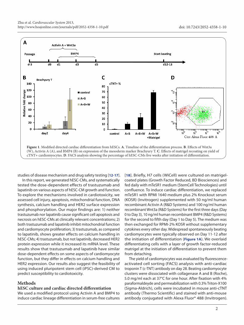
[18]. Briefly, H7 cells (WiCell) were cultured on matrigel-coated plates (Growth Factor Reduced, BD Biosciences) and fed daily with mTeSR1 medium (StemCell Technologies) until confluence. To induce cardiac differentiation, we replaced mTeSR1 with RPMI 1640 medium plus 2% Knockout serum (KOSR) (Invitrogen) supplemented with 50 ng/ml human recombinant Activin A (R&D Systems) and 100 ng/ml human recombinant Wnt3a (R&D Systems) for the first three days (Day 0 to Day 3), 10 ng/ml human recombinant BMP4 (R&D Systems) for the second to fifth day (Day 1 to Day 5). The medium was then exchanged for RPMI-2% KOSR without supplementary cytokines every other day. Widespread spontaneously beating cardiomyocytes were typically observed on Day 11-12 after the initiation of differentiation (Figure 1A). We overlaid differentiating cells with a layer of growth factor-reduced matrigel at the initiation of differentiation to prevent them from detaching.
The yield of cardiomyocytes was evaluated by fluorescence-activated cell sorting (FACS) analysis with anti-cardiac troponin T (c-TNT) antibody on day 28. Beating cardiomyocyte clusters were dissociated with collagenase A and B (Roche), 5.0 mg/ml each at 37°C for one hour. After fixation with 4% paraformaldeyde and permeabilization with 0.3% Triton-X100 (Sigma-Aldrich), cells were incubated in mouse anti-cTNT antibody (Thermo Scientific) and stained with anti-mouse antibody conjugated with Alexa Fluor® 488 (Invitrogen).
Figure 1. Modified directed cardiac differentiation from hESCs. A. Timeline of the differentiation process. B. Effects of Wnt3a (W), Activin A (A), and BMP4 (B) on expression of the mesoderm marker Brachyury T. C. Effects of matrigel recoating on yield of cTNT+ cardiomyocytes. D. FACS analysis showing the percentage of hESC-CMs five weeks after initiation of differentiation.
Zhu et al. Cardiovascular System 2013, http://www.hoajonline.com/journals/pdf/2052-4358-1-10.pdf
3
doi: 10.7243/2052-4358-1-10
Cardiomyocyte percentage was quantified by FACS.
General design for drug toxicity testsDay 28 beating hESC-CMs were dissociated with collagenase A and B (Roche) and preplated twice for 1 hour each. Nonadherent cells were plated onto 12-well plates at 5 X 10⁴ cells per wells. Plates were pre-coated with fibronectin (Sigma-Aldrich) and RPMI medium with 20% FBS. After three days, hESC-CMs were treated with trastuzumab or lapatinib. Clinically relevant concentration ranges were 50-100 ug/ml fpr trastuzumab, and 1.0-2.0 uM for lapatinib. Serial 2-fold dilutions were done to span the dose ranges for trastuzumab (0, 12.5 µg/ml, 25 µg/ml, 50 µg/ml, 100 µg/ml and 200 µg/ml) and lapatinib (0, 0.25 µM, 0.50 µM, 1.0 µM, 2.0 µM and 4.0 µM). All endpoints were evaluated after 48 hours drug treatment, unless otherwise specified.
Mitochondrial membrane potential and intracellular ATP/ADP assessment TMRM, a cell-permeant, cationic, red-orange fluorescent dye readily sequestered by active mitochondria, is a sensitive marker for mitochondrial membrane potential (Δψm) in newly differentiated cardiomyocytes [19,20]. After trastuzumab or lapatinib treatment for 48 hours, cells were washed with PBS and loaded with 25 nM TMRM for 20 min at 37°C, and the cell nuclei were counterstained with Hoechst 33342. TMRM measurements were made using PerkinElmer 2030 Explorer; TMRM was excited using the 543 nm laser line and fluorescence measured using a 560 nm filter. Hoechst 33342 was excited at 405 nm and emission fluorescence detected via a 463/39 nm bandpass filter. The fluorescence intensity of TMRM was standardized by the measurement of Hoechst 33342 (as an indicator of cell number). All data presented were obtained from 5 wells and two to three different cell preparations.
To further investigate whether the alternations in mitochondrial integrity were sufficient to interfere with cellular energetics, intracellular ATP and ADP/ATP ratios were measured using the bioluminescent ADP/ATP ratio assay kit from Abcam. hESC-CM aliquots were cultured in 24-well plates (104 cells/well) for 3 days and then treated with lapatinib or trastuzumab for 48 hours. Cells were lysed with 100 µl nucleotide releasing buffer. The ATP level in cell lysate was proportional to the luminescence and was evaluated with luminometer, while the ADP level was measured by its conversion to ATP with ADP-converting enzyme. Data were normalized for protein content.
BrdU incorporation assayhESC-CMs were exposed to increasing concentrations of lapatinib or trastuzumab and a fixed concentration (100 ng/ml) of Nrg-1 (epidermal growth factor domain, R&D systems) for 7 days. 5-bromo-2’-deoxyuridine (BrdU) was added in the medium at a final concentration of 30 µM. BrdU incorporation was measured by FACS and immunostaining. For FACS,
hESC-CMs were dissociated with trypsin and fixed with 4% paraformaldehyde. After permeabilization with 0.3% Triton X-100 and incubation in 2 N HCl for 20 min at 37°C, the cells were incubated in rat anti-BrdU antibody (Abcam) and mouse anti-cTNT antibody, followed by anti-rat IgG (conjugated with Alexa Fluor® Cy5) and anti-mouse IgG (conjugated with Alexa Fluor® 488) antibodies. The percentage of BrdU positive cardiomyocytes were quantified as a ratio of BrdU+cTNT+/cTNT+ with FACS. For immunostaining, the cells were treated and stained same as above, but without dissociation. Calcium imaging hESC-CMs were replated on fibronectin-coated coverslips and treated with trastuzumab or lapatinib for 48 hours. Intracellular Ca2+ (ΔCai, measured as the difference between the peak systolic-to-diastolic fura 2 ratios) was measured by loading hESC-CMs with fura 2-AM (Molecular Probes) at 1 mM for 25 min followed by superfusion with a physiological solution containing 137 mM NaCl, 5.4 mM KCl, 1.2 mM Ca2+, 0.5 mM MgCl2, 10 mM HEPES, pH 7.4, and 0.5mM probenecid to improve fura2-AM retention. Cells were externally paced at 1 Hz by a MyoPacer field stimulator (IonOptix, Milton, MA) at 37°C. Intracellular calcium ratio data were recorded and analyzed using IonWizard software (IonOptix) on an acquisition system that includes a Nikon TS100 inverted microscope, a MyoCam-S digital charge-coupled device video camera with 500-Hz acquisition rate, a HyperSwitch dual-excitation light source, and a photomultiplier tube. The 340-to-380 ratio was calculated after subtracting background fluorescence, and the maximal rates of [Ca2+]I development (Dep Vmax) and return (Ret Vmax) were calculated and averaged over 10 transients. For statistical analysis, data were compared using an unpaired Student’s t-test.
Analysis of HER2 expression and its phosphorylationTo assess effects on HER2 expression and phosphorylation, hESC-CMs were cultured in RPMI-2% KOSR for 48 hours, in the presence of trastuzumab or lapatinib. Before harvesting, hESC-CMs were treated with Nrg-1 at 100 ng/ml for 30 min at 37°C. Cells were quickly rinsed with ice-cold PBS, homogenized in 100 ul regular lysis buffer supplemented with 1X protease inhibitor cocktail and phosphatase inhibitor cocktail (Roche) and microcentrifuged for 20 min at 13,000 rpm. Supernatant proteins were fractionated on 10% SDS polyacrylamide gels and transferred to polyvinylidene difluoride membranes. Membranes were blocked with 5% bovine serum albumin and incubated overnight with primary antibodies against anti-HER2, anti-phosphorylated HER2 (Tyr877) and β-actin. Bound antibodies were detected with horseradish peroxidase (HRP)-conjugated secondary antibody and chemiluminescence detection kit (GE Healthcare). To detect the HER2 mRNA expression level, total RNA was isolated using TRIzol reagent (Invitrogen) after 48 hours drug treatment and reverse transcribed into cDNA using SuperScript II (Invitrogen). The

Zhu et al. Cardiovascular System 2013, http://www.hoajonline.com/journals/pdf/2052-4358-1-10.pdf
4
doi: 10.7243/2052-4358-1-10
mRNA level of HER2 was measured by quantitative polymerase chain reaction (qPCR) with SYBR Green PCR master mix (Applied Biosystems), normalized with α-MHC. The primers used in qPCR were: HER2: Forward–ACAGAGACTCAGACCCTG GC; Reverse–CTTCATGTCTGTGCCGGTG; α-MHC: Forward- TCTTCGTTACAACCTCATGGTG; Reverse–TCCCAGGTCTTAG-CTCCCAG.
ResultshESC cardiac differentiation To induce the differentiation of cardiomyocytes from hESC, we compared several protocols including embryoid body formation, co-culture with endodermal cells, and directed differentiation. In our hands, directed differentiation with Activin A, Wnt3a and Bone Morphogenetic Protein 4 (BMP4) gave the most consistent and the highest yield of cardiomyocytes. We tried several combinations of growth factors and timing. As shown in Figure 1A, the addition of Wnt3a on first 3 days of differentiation resulted in a marked increase in the expression of the mesodermal marker Brachyury T on day 4 (91.6±34.2 vs. 24.9±12.8 fold, P<0.05), and in the final yield of cTNT-positive cardiomyocyte (22.6±7.2% vs. 10.6±2.8%, P<0.05), as compared with Activin
A and BMP4 only. Furthermore, matrigel re-coating trapped migrating cells during differentiation and gave a supportive extracellular matrix environment, increasing the cTNT-positive cardiomyocytes to as high as 69.1±3.7%. hESC-CMs showed sarcomeric structure when stained with cTNT antibodies, and calcium cycling and action potentials similar to those observed in isolated ventricular myocytes.
Mitochondrial function assay and intracellular ATP concentrationWe used the mitochondrial potential sensor tetramethylrho-damine methyl ester (TMRM), to evaluate the mitochondrial membrane potential. Both trastuzumab and lapatinib induced loss of mitochondrial membrane potential in a dose-dependent manner. The ratio of fluorescence intensity of TMRM to Hoechst 33342 decreased with increasing concentrations of both trastuzumab and lapatinib. The hESC-CMs exhibited a significant decline in the Δψm after 48 hour incubation with either trastuzumab or lapatinib. The ratio of fluorescence intensity of TMRM to Hoechst 33342 decreased from 0.62±0.01 to 0.50±0.02 under trastuzumab treatment, and 0.61±0.02 to 0.50±0.03 under lapatinib treatment. The dose-response curves of lapatinib and trastuzumab were similar (Figure 2B).
Figure 2. Effects of trastuzumab (Hcp) and lapatinib (Lap) on cellular energetics. A. hESC-CMs stained with TMRM. B. Dose response curves of TMRM staining intensity for trastuzumab or lapatinib. There was no significant difference between the two curves over the dose range examined. C. Dose response curves of trastuzumab or lapatinib on ATP intracellular concentrations, reflected by bioluminescence intensity. D.Dose response curves of effects on ADP/ATP ratio. The gray bars indicate the clinically relevant concentration ranges of both two drugs.
Zhu et al. Cardiovascular System 2013, http://www.hoajonline.com/journals/pdf/2052-4358-1-10.pdf
5
doi: 10.7243/2052-4358-1-10
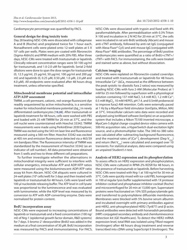
However, ATP levels in hESC-CMs were severely reduced after high dose trastuzumab, and the ADP/ATP ratio was greater (Figure 2C). In contrast, there was no significant change in ATP level or ADP/ATP radio with lapatinib exposure (Figure 2D). These results show that energy metabolism is altered by high doses of trastuzumab, but not lapatinib. BrdU incorporation assayDuring the process of embryogenesis and in vitro stem cell differentiation, cardiac progenitor cells gradually lose their proliferative capacity and withdraw from the cell cycle. Neuregulin-1 (Nrg-1), a paracrine growth factor produced by cardiac microvascular endothelial cells, binds to HER2 and induces cardiomyocytes to reenter the cell cycle. In this study, we observed around half of 4-5 week-old hESC-CMs were still undergoing DNA synthesis. After stimulation of Nrg-1 (100 ng/ml) for 7 days, there was a 1.8 fold increase of the percentage of BrdU positive cells (71.7±16.2% vs. 40.5±4.56%, P<0.05). The co-administration of trastuzumab or lapatinib blocked the stimulation of Nrg-1 in a dose-dependent manner (Figure 3). At clinically relevant concentrations, there was no significant difference between the effects of trastuzumab and lapatinib.
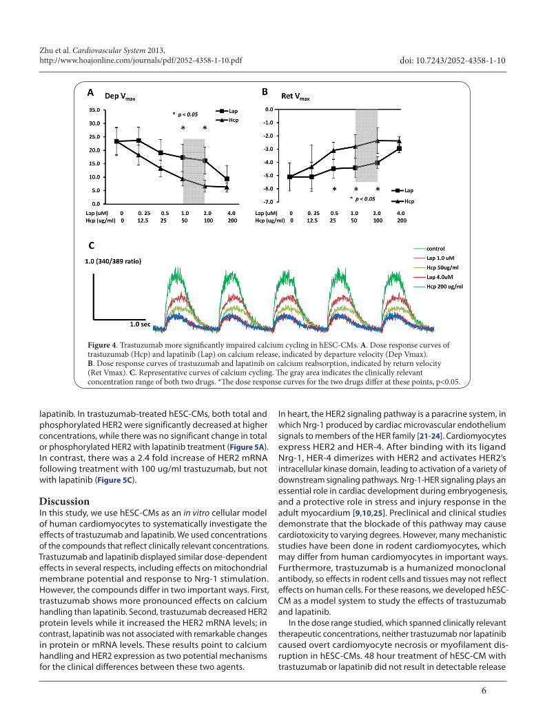
Calcium imagingCalcium handling is crucial to excitation-contraction coupling in cardiomyocytes and defects in calcium handling are a
common endpoint in cardiotoxicity. In recorded calcium imaging, the maximal departure velocity (Dep Vmax) represents the efficacy of calcium release from sarcoplasmic reticulum (SR) to cytoplasm in each beating cycle, and the return velocity (Ret Vmax) indicates the calcium reabsorption ability. Consistent with the mitochondrial integrity and BrdU incorporation assays, both trastuzumab and lapatinib impaired calcium handling in a dose-dependent manner. Unlike the former two endpoints, there were differences between trastuzumab and lapatinib within the clinically relevant concentration ranges. Trastuzumab, as compared to lapatinib, more severely reduces the Dep Vmax (6.7±2.3 to 9.4±2.7 vs.16.2±4.3 to 17.3±3.3, P<0.05, respectively) and Ret Vmax (-2.4±0.45 to -2.8±0.75 vs. -4.0±0.96 to -4.4±0.89, P<0.05 respectively) of calcium transients (Figure 4A and 4B). These results indicate that trastuzumab has more pronounced effects on calcium handling than lapatinib.
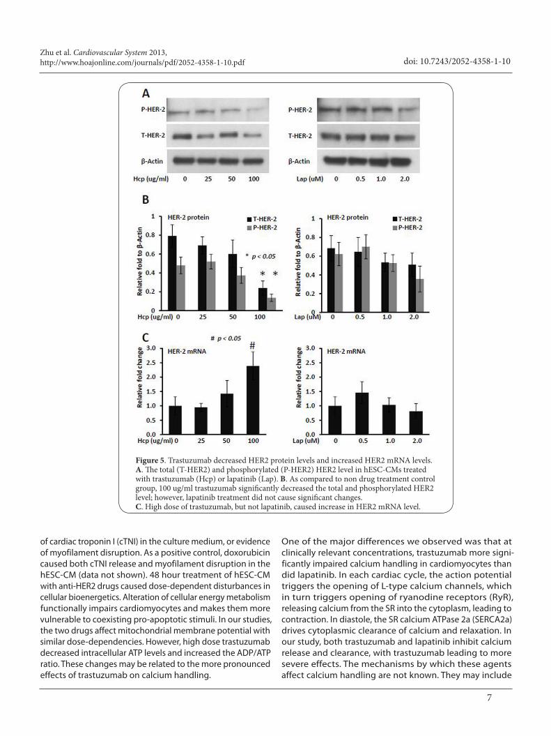
HER2 protein and mRNA levelsWe assessed dose-dependent effects of trastuzumab and lapatinib on the expression and phosphorylation of HER2. After 48 hours, no differences in β-actin levels were noted. Treatment with either drug did not alter the total cell number at 48 hours. We measured expression of HER2 protein and its phosphorylation status following stimulation with 100 ng/ml Nrg-1 after 48 hours treatment with trastuzumab or
Figure 3. Effects of trastuzumab (Hcp) and lapatinib (Lap) on Nrg-1 stimulated cardiomyocyte proliferation. A. Immunofluorescence staining and FACS assay of BrdU incorporation. hESC-CMs were stained with cTNT (red), BrdU (green) and DAPI for nuclei (blue). B. Dose response curves of trastuzumab and lapatinib on inhibition of Nrg-1 stimulated DNA synthesis. Gray bars indicate clinically relevant concentration ranges.
Zhu et al. Cardiovascular System 2013, http://www.hoajonline.com/journals/pdf/2052-4358-1-10.pdf
6
doi: 10.7243/2052-4358-1-10
Figure 4. Trastuzumab more significantly impaired calcium cycling in hESC-CMs. A. Dose response curves of trastuzumab (Hcp) and lapatinib (Lap) on calcium release, indicated by departure velocity (Dep Vmax). B. Dose response curves of trastuzumab and lapatinib on calcium reabsorption, indicated by return velocity (Ret Vmax). C. Representative curves of calcium cycling. The gray area indicates the clinically relevant concentration range of both two drugs. *The dose response curves for the two drugs differ at these points, p<0.05.
lapatinib. In trastuzumab-treated hESC-CMs, both total and phosphorylated HER2 were significantly decreased at higher concentrations, while there was no significant change in total or phosphorylated HER2 with lapatinib treatment (Figure 5A).In contrast, there was a 2.4 fold increase of HER2 mRNA following treatment with 100 ug/ml trastuzumab, but not with lapatinib (Figure 5C).
DiscussionIn this study, we use hESC-CMs as an in vitro cellular model of human cardiomyocytes to systematically investigate the effects of trastuzumab and lapatinib. We used concentrations of the compounds that reflect clinically relevant concentrations. Trastuzumab and lapatinib displayed similar dose-dependent effects in several respects, including effects on mitochondrial membrane potential and response to Nrg-1 stimulation. However, the compounds differ in two important ways. First, trastuzumab shows more pronounced effects on calcium handling than lapatinib. Second, trastuzumab decreased HER2 protein levels while it increased the HER2 mRNA levels; in contrast, lapatinib was not associated with remarkable changes in protein or mRNA levels. These results point to calcium handling and HER2 expression as two potential mechanisms for the clinical differences between these two agents.
In heart, the HER2 signaling pathway is a paracrine system, in which Nrg-1 produced by cardiac microvascular endothelium signals to members of the HER family [21-24]. Cardiomyocytes express HER2 and HER-4. After binding with its ligand Nrg-1, HER-4 dimerizes with HER2 and activates HER2’s intracellular kinase domain, leading to activation of a variety of downstream signaling pathways. Nrg-1-HER signaling plays an essential role in cardiac development during embryogenesis, and a protective role in stress and injury response in the adult myocardium [9,10,25]. Preclinical and clinical studies demonstrate that the blockade of this pathway may cause cardiotoxicity to varying degrees. However, many mechanistic studies have been done in rodent cardiomyocytes, which may differ from human cardiomyocytes in important ways. Furthermore, trastuzumab is a humanized monoclonal antibody, so effects in rodent cells and tissues may not reflect effects on human cells. For these reasons, we developed hESC-CM as a model system to study the effects of trastuzumab and lapatinib.
In the dose range studied, which spanned clinically relevant therapeutic concentrations, neither trastuzumab nor lapatinib caused overt cardiomyocyte necrosis or myofilament dis-ruption in hESC-CMs. 48 hour treatment of hESC-CM with trastuzumab or lapatinib did not result in detectable release
Zhu et al. Cardiovascular System 2013, http://www.hoajonline.com/journals/pdf/2052-4358-1-10.pdf
7
doi: 10.7243/2052-4358-1-10
Figure 5. Trastuzumab decreased HER2 protein levels and increased HER2 mRNA levels. A. The total (T-HER2) and phosphorylated (P-HER2) HER2 level in hESC-CMs treated with trastuzumab (Hcp) or lapatinib (Lap). B. As compared to non drug treatment control group, 100 ug/ml trastuzumab significantly decreased the total and phosphorylated HER2 level; however, lapatinib treatment did not cause significant changes. C. High dose of trastuzumab, but not lapatinib, caused increase in HER2 mRNA level.
of cardiac troponin I (cTNI) in the culture medium, or evidence of myofilament disruption. As a positive control, doxorubicin caused both cTNI release and myofilament disruption in the hESC-CM (data not shown). 48 hour treatment of hESC-CM with anti-HER2 drugs caused dose-dependent disturbances in cellular bioenergetics. Alteration of cellular energy metabolism functionally impairs cardiomyocytes and makes them more vulnerable to coexisting pro-apoptotic stimuli. In our studies, the two drugs affect mitochondrial membrane potential with similar dose-dependencies. However, high dose trastuzumab decreased intracellular ATP levels and increased the ADP/ATP ratio. These changes may be related to the more pronounced effects of trastuzumab on calcium handling.
One of the major differences we observed was that at clinically relevant concentrations, trastuzumab more signi-ficantly impaired calcium handling in cardiomyocytes than did lapatinib. In each cardiac cycle, the action potential triggers the opening of L-type calcium channels, which in turn triggers opening of ryanodine receptors (RyR), releasing calcium from the SR into the cytoplasm, leading to contraction. In diastole, the SR calcium ATPase 2a (SERCA2a) drives cytoplasmic clearance of calcium and relaxation. In our study, both trastuzumab and lapatinib inhibit calcium release and clearance, with trastuzumab leading to more severe effects. The mechanisms by which these agents affect calcium handling are not known. They may include
Zhu et al. Cardiovascular System 2013, http://www.hoajonline.com/journals/pdf/2052-4358-1-10.pdf
8
doi: 10.7243/2052-4358-1-10
interference with downstream effector pathways, including PI3K/Akt, Ras/MEK/ERK and Src/FAK [1,2,7,22], and effects on function or expression of proteins involved in calcium release and uptake, such as RyR or SERCA2a.
The other major difference we observed was in HER2 expression levels. We found that trastuzumab decreased HER2 protein levels, while it increased HER2 mRNA levels. Lapatinib did not alter the HER2 protein level and mRNA expression. Trastuzumab may be able to block HER2-related signaling pathways more completely because affects not only receptor dimerization and signaling, but also endocytosis and degradation. Upregulation of HER2 mRNA level after trastuzumab exposure may reflect a compensatory response to maintain HER2 protein levels.
We should point out that the calcium handling of hESC-derived CM, which are structurally and functionally comparable to human embryonic or young cardiomyocytes, may differ from that of adult cardiomyocytes in the relative importance of transmembrane flux, release from the SR via the RyR, and IP3-mediated SR Ca2+ release. In addition, we assessed changes of trastuzumab and lapatinib after 48 hours treatment, and we have not characterized the changes that occur more acutely with shorter treatment.
Recent advances in stem cell and iPSC technology have led to the ability to establish in vitro patient-specific disease models [12-17]. In this setting, our work suggests a new strategy of testing patient-specific iPS-derived CM to evaluate susceptibility to cardiotoxicity. By assessing response of calcium handling, HER2 gene expression, and other endpoints, we may be able to screen patient-specific iPS-derived CM for toxicity from trastuzumab, lapatnib, and other agents in culture, before patients are exposed to potentially toxic medications.
ConclusionsIn conclusion, our results are important for the following reasons. First, we used hESC-CM as an in vitro model of human cardiomyocytes to study cardiotoxicity of trastuzumab and lapatinib. Such studies in rodent cells are limited by the differences between rodent and human cells, including different expression patterns of HER receptor proteins. Furthermore, trastuzumab is a humanized monoclonal antibody, so its effects on human cells would not be replicated in rodent cells. Second, we found that while trastuzumab and lapatinib have similar dose-dependent effects on many readouts of cellular physiology, they differ remarkably in calcium handling and HER2 expression. These results point to calcium handling and HER2 expression as possible mechanisms for the clinical differences in cardiotoxicity between these two agents. Third, our results demonstrate the utility of hESC-CM for mechanistic studies of cardiotoxicity. The same approach could theoretically be used with iPSC-CM.
List of abbreviationsHER2: human epidermal growth factor receptor-2
EGF: epidermal growth factor hESC: human embryonic stem cell iPSC: induced pluripotent stem cell BMP4: Bone Morphogenetic Protein 4 SR: sarcoplasmic reticulum cTNI: cardiac troponin I cTNT: cardiac troponin TTMRM: tetramethylrhodamine methyl esterAMPK: AMP-activated protein kinase RyR: ryanodine receptor SERCA2a: sarcoplasmic reticulum calcium ATPase 2a
Competing interestsThe authors declare that they have no competing interests.
Authors’ contributions
Acknowledgement and fundingThis study is funded by GlaxoSmithKline, Inc (GSK). GSK played no role in study design, in data collection, analysis and interpretation of data, in the writing of the report, and or the decision to submit the article for publication.
Publication historyEIC: William Clifford Roberts, Baylor University Medical Center, USA.Received: 09-Aug-2013 Revised: 23-Aug-2013Accepted: 17-Sep-2013 Published: 25-Sep-2013
Authors’ contributions SZ SMC KDB PLH
Research concept and design √ -- √ √
Collection and/or assembly of data √ √ -- --
Data analysis and interpretation √ √ √ √
Writing the article √ -- -- √
Critical revision of the article -- -- √ √
Final approval of article √ √ √ √
Statistical analysis √ √ -- √
References
1. Albini A, Cesana E, Donatelli F, Cammarota R, Bucci EO, Baravelli M, Anza C and Noonan DM. Cardio-oncology in targeting the HER receptor family: the puzzle of different cardiotoxicities of HER2 inhibitors. Future Cardiol. 2011; 7:693-704. | Article | PubMed
2. Zambelli A, Della Porta MG, Eleuteri E, De Giuli L, Catalano O, Tondini C and Riccardi A. Predicting and preventing cardiotoxicity in the era of breast cancer targeted therapies. Novel molecular tools for clinical issues. Breast. 2011; 20:176-83. | Article | PubMed
3. Beeram M, Krop IE, Burris HA, Girish SR, Yu W, Lu MW, Holden SN and Modi S. A phase 1 study of weekly dosing of trastuzumab emtansine (T-DM1) in patients with advanced human epidermal growth factor 2-positive breast cancer. Cancer. 2012; 118:5733-40. | Article | PubMed
4. Burris HA, 3rd, Tibbitts J, Holden SN, Sliwkowski MX and Lewis Phillips GD. Trastuzumab emtansine (T-DM1): a novel agent for targeting HER2+ breast cancer. Clin Breast Cancer. 2011; 11:275-82. | Article | PubMed
5. Seidman A, Hudis C, Pierri MK, Shak S, Paton V, Ashby M, Murphy M, Stewart SJ and Keefe D. Cardiac dysfunction in the trastuzumab clinical trials experience. J Clin Oncol. 2002; 20:1215-21. | Article | PubMed
6. Slamon DJ, Leyland-Jones B, Shak S, Fuchs H, Paton V, Bajamonde A, Fleming T, Eiermann W, Wolter J, Pegram M, Baselga J and Norton L. Use of chemotherapy plus a monoclonal antibody against HER2 for metastatic breast cancer that overexpresses HER2. N Engl J Med. 2001; 344:783-92. | Article | PubMed
7. Force T, Krause DS and Van Etten RA. Molecular mechanisms of cardiotoxicity of tyrosine kinase inhibition. Nat Rev Cancer. 2007; 7:332-44. | Article | PubMed
Zhu et al. Cardiovascular System 2013, http://www.hoajonline.com/journals/pdf/2052-4358-1-10.pdf
9
doi: 10.7243/2052-4358-1-10
8. Orphanos GS, Ioannidis GN and Ardavanis AG. Cardiotoxicity induced by tyrosine kinase inhibitors. Acta Oncol. 2009; 48:964-70. | Article | PubMed
9. Crone SA, Zhao YY, Fan L, Gu Y, Minamisawa S, Liu Y, Peterson KL, Chen J, Kahn R, Condorelli G, Ross J, Jr., Chien KR and Lee KF. ErbB2 is essential in the prevention of dilated cardiomyopathy. Nat Med. 2002; 8:459-65. | Article | PubMed
10. Ozcelik C, Erdmann B, Pilz B, Wettschureck N, Britsch S, Hubner N, Chien KR, Birchmeier C and Garratt AN. Conditional mutation of the ErbB2 (HER2) receptor in cardiomyocytes leads to dilated cardiomyopathy. Proc Natl Acad Sci U S A. 2002; 99:8880-5. | Article | PubMed Abstract | PubMed Full Text
11. Grazette LP, Boecker W, Matsui T, Semigran M, Force TL, Hajjar RJ and Rosenzweig A. Inhibition of ErbB2 causes mitochondrial dysfunction in cardiomyocytes: implications for herceptin-induced cardiomyopathy. J Am Coll Cardiol. 2004; 44:2231-8. | Article | PubMed
12. Carvajal-Vergara X, Sevilla A, D’Souza SL, Ang YS, Schaniel C, Lee DF, Yang L, Kaplan AD, Adler ED, Rozov R, Ge Y, Cohen N, Edelmann LJ, Chang B, Waghray A, Su J, Pardo S, Lichtenbelt KD, Tartaglia M, Gelb BD and Lemischka IR. Patient-specific induced pluripotent stem-cell-derived models of LEOPARD syndrome. Nature. 2010; 465:808-12. | Article | PubMed Abstract | PubMed Full Text
13. Itzhaki I, Maizels L, Huber I, Zwi-Dantsis L, Caspi O, Winterstern A, Feldman O, Gepstein A, Arbel G, Hammerman H, Boulos M and Gepstein L. Modelling the long QT syndrome with induced pluripotent stem cells. Nature. 2011; 471:225-9. | Article | PubMed
14. Moretti A, Bellin M, Welling A, Jung CB, Lam JT, Bott-Flugel L, Dorn T, Goedel A, Hohnke C, Hofmann F, Seyfarth M, Sinnecker D, Schomig A and Laugwitz KL. Patient-specific induced pluripotent stem-cell models for long-QT syndrome. N Engl J Med. 2010; 363:1397-409. | Article | PubMed
15. Sun N, Yazawa M, Liu J, Han L, Sanchez-Freire V, Abilez OJ, Navarrete EG, Hu S, Wang L, Lee A, Pavlovic A, Lin S, Chen R, Hajjar RJ, Snyder MP, Dolmetsch RE, Butte MJ, Ashley EA, Longaker MT, Robbins RC and Wu JC. Patient-specific induced pluripotent stem cells as a model for familial dilated cardiomyopathy. Sci Transl Med. 2012; 4:130ra47. | Article | PubMed Abstract | PubMed Full Text
16. Tiscornia G, Vivas EL, and Belmonte JC. Diseases in a dish: modeling human genetic disorders using induced pluripotent cells. Nat Med 2011; 17:1570-6. | Article
17. Wu SM and Hochedlinger K. Harnessing the potential of induced pluripotent stem cells for regenerative medicine. Nat Cell Biol. 2011; 13:497-505. | Article | PubMed Abstract | PubMed Full Text
18. Laflamme MA, Chen KY, Naumova AV, Muskheli V, Fugate JA, Dupras SK, Reinecke H, Xu C, Hassanipour M, Police S, O’Sullivan C, Collins L, Chen Y, Minami E, Gill EA, Ueno S, Yuan C, Gold J and Murry CE. Cardiomyocytes derived from human embryonic stem cells in pro-survival factors enhance function of infarcted rat hearts. Nat Biotechnol. 2007; 25:1015-24. | Article | PubMed
19. French KJ, Coatney RW, Renninger JP, Hu CX, Gales TL, Zhao S, Storck LM, Davis CB, McSurdy-Freed J, Chen E and Frazier KS. Differences in effects on myocardium and mitochondria by angiogenic inhibitors suggest separate mechanisms of cardiotoxicity. Toxicol Pathol. 2010; 38:691-702. | Article | PubMed
20. Hattori F, Chen H, Yamashita H, Tohyama S, Satoh YS, Yuasa S, Li W, Yamakawa H, Tanaka T, Onitsuka T, Shimoji K, Ohno Y, Egashira T, Kaneda R, Murata M, Hidaka K, Morisaki T, Sasaki E, Suzuki T, Sano M, Makino S, Oikawa S and Fukuda K. Nongenetic method for purifying stem cell-derived cardiomyocytes. Nat Methods. 2010; 7:61-6. | Article | PubMed
21. Arteaga CL, Sliwkowski MX, Osborne CK, Perez EA, Puglisi F and Gianni L. Treatment of HER2-positive breast cancer: current status and future perspectives. Nat Rev Clin Oncol. 2011; 9:16-32. | Article | PubMed
22. Pentassuglia L, Graf M, Lane H, Kuramochi Y, Cote G, Timolati F, Sawyer DB, Zuppinger C and Suter TM. Inhibition of ErbB2 by receptor tyrosine kinase inhibitors causes myofibrillar structural damage without cell death in adult rat cardiomyocytes. Exp Cell Res. 2009; 315:1302-12. | Article | PubMed
23. Pentassuglia L and Sawyer DB. The role of Neuregulin-1beta/ErbB signaling in the heart. Exp Cell Res. 2009; 315:627-37. | Article | PubMed Abstract | PubMed Full Text
24. Yarden Y and Sliwkowski MX. Untangling the ErbB signalling network. Nat Rev Mol Cell Biol. 2001; 2:127-37. | Article | PubMed
25. Meyer D and Birchmeier C. Multiple essential functions of neuregulin in development. Nature. 1995; 378:386-90. | Article | PubMed
Citation:Zhu S, Cawley SM, Bloch KD and Huang PL. Trastuzumab and lapatinib differ in effects on calcium cycling and HER2 expression in human embryonic stem cell-derived cardiomyocytes. Cardio Vasc Syst. 2013; 1:10. http://dx.doi.org/10.7243/2052-4358-1-10
Copyright © 2022 FDOKUMEN