Differentiation of human embryonic stem cells to cardiomyocytes on microcarrier cultures
17
23 Differentiation of Human Embryonic Stem Cells Toward the Chondrogenic Lineage Wei Seong Toh, Zheng Yang, Boon Chin Heng, and Tong Cao Summary Human embryonic stem cells (hESCs) have the ability to self-replicate and differentiate into cells from all three embryonic germ layers, thereby holding great promise for tissue regener- ation applications. However, controlling the differentiation of hESCs and obtaining homogenous differentiated cell populations still remain a challenge. We present a highly efficient and repro- ducible experimental system that mimics the three-dimensional (3-D) environment of in vivo chondrogenesis and that supports the directed differentiation of human embryoid body (EB)- derived cells toward the chondrogenic lineage under serum-free chondrogenic culture conditions in the presence of bone morphogenetic protein-2 (BMP-2). Key Words: BMP2; Chondrogenic; Differentiation; Embryonic stem cells; Human. 1. Introduction Development of the vertebrate skeleton is a complex multi-step process that involves lineage commitment of mesenchymal cells, migration of these cells to the site of skeletogenesis, mesenchymal–epithelial interactions that result in cellular condensation, and differentiation of chondroblasts and/or osteoblasts (1,2). Although many factors that have important function during this process are known, there is limited knowledge about the commitment of mesenchymal cells to differentiate into cartilage or bone (3). Embryonic stem cells (ESCs), derived from the inner cell mass of the blastocyst, represent a promising cell source for transplantation because of their From: Methods in Molecular Biology, vol. 407: Stem Cell Assays Edited by: M. C. Vemuri © Humana Press, Totowa, NJ 333
-
Upload
independent -
Category
Documents
-
view
3 -
download
0
Transcript of Differentiation of human embryonic stem cells to cardiomyocytes on microcarrier cultures
23
Differentiation of Human Embryonic Stem Cells Towardthe Chondrogenic Lineage
Wei Seong Toh, Zheng Yang, Boon Chin Heng, and Tong Cao
Summary
Human embryonic stem cells (hESCs) have the ability to self-replicate and differentiate intocells from all three embryonic germ layers, thereby holding great promise for tissue regener-ation applications. However, controlling the differentiation of hESCs and obtaining homogenousdifferentiated cell populations still remain a challenge. We present a highly efficient and repro-ducible experimental system that mimics the three-dimensional (3-D) environment of in vivochondrogenesis and that supports the directed differentiation of human embryoid body (EB)-derived cells toward the chondrogenic lineage under serum-free chondrogenic culture conditionsin the presence of bone morphogenetic protein-2 (BMP-2).
Key Words: BMP2; Chondrogenic; Differentiation; Embryonic stem cells; Human.
1. IntroductionDevelopment of the vertebrate skeleton is a complex multi-step process that
involves lineage commitment of mesenchymal cells, migration of these cellsto the site of skeletogenesis, mesenchymal–epithelial interactions that result incellular condensation, and differentiation of chondroblasts and/or osteoblasts(1,2). Although many factors that have important function during this processare known, there is limited knowledge about the commitment of mesenchymalcells to differentiate into cartilage or bone (3).
Embryonic stem cells (ESCs), derived from the inner cell mass of theblastocyst, represent a promising cell source for transplantation because of their
From: Methods in Molecular Biology, vol. 407: Stem Cell AssaysEdited by: M. C. Vemuri © Humana Press, Totowa, NJ
333
334 Toh et al.
capacity for unlimited self-renewal and ability to differentiate into all somaticcell lineages (4,5). Recent studies in mouse ESCs suggested that ESC differenti-ation through EBs parallels early embryonic development, and biochemical cuesprovided directly by growth factors can induce differentiation of ESC-derivedembryoid bodies (EBs) toward the chondrogenic lineage (6,7,8). Recent tissueengineering studies have also demonstrated potential chondrogenic differen-tiation of ESCs using three dimensional (3-D) scaffold systems incorporatedwith appropriate growth factors (9,10). We have established a reproduciblemodel system to study full-span chondrogenesis of human EB-derived cells andalso demonstrated the temporal involvement of bone morphogenetic protein-2(BMP-2) in early and late phase chondrogenesis (11,12).
This chapter will focus on the effects of BMP-2 on chondrogenic differen-tiation of human EB-derived cells, culture systems for chondrogenic differen-tiation as well as characterization of human EB-derived chondrogenic cells.
2. Materials2.1. Cell Culture
2.1.1. Embryoid Body Culture (see Note 1)
1. Dulbecco’s Modified Eagle’s Medium (DMEM)/F-12 media (cat. no. #11330-032,Gibco/BRL, Gaithersburg, MD).
2. Knockout™ Serum Replacement (KSR) (cat. no. #10828-028, Gibco/BRL)3. MEM Non-Essential Amino Acids (NEAA) Solution, 10 mM; 100× (cat. no.
#11140-050, Gibco/BRL)4. Collagenase Type IV, Lyophilized (cat. no. #17104-019, Gibco/BRL). Prepare the
collagenase IV splitting medium (1 mg/mL) with DMEM/F-12 media. Filter-sterilizebefore use.
5. EB differentiation medium: DMEM/F-12 supplemented with 20% KSR, 1%(v/v) NEAA, 1 mM l-glutamine (cat. no. #21051-016, Gibco/BRL), and 0.1 mM2-mercaptoethanol (cat. no. #M7522, Sigma, St Louis, MO). EB media should beprotected from light and stored in 4 �C.
6. 6-Well Ultra Low Attachment Microplates (cat. no. #3471, Costar® Corning, NagogPark Acton, MA).
2.1.2. Culture Media for Chondrogenic Differentiation
1. DMEM high-glucose (4.5g/L; cat. no. #D1152, Sigma)2. Fetal Bovine Serum (FBS) (cat. no. #CH30160.03, Hyclone, Logan, UT, USA).3. Serum substitutes: KSR (cat. no. #10828-028, Gibco/BRL).4. ITS+1 (6�25 �g/mL insulin, 6�25 �g/mL transferrin, 6.25 ng/mL selenium,
1.25 mg/mL bovine serum albumin (BSA), 5�35 �g/mL linoleic acid) (cat. no.#354352, BD Bioscience, Franklin Lakes, NJ).

Chondrogenic Differentiation of hESCs 335
5. MEM NEAA Solution, 10 mM; 100× (cat. no. #11140-050, Gibco/BRL).6. Recombinant human BMP-2 (cat. no. #355-BM, R&D Systems, Minneapolis, MN)
(13). Prepare stock solution of 100 �g/mL by reconstituting in filter-sterilized4 mM HCl containing 0.1% (w/v) BSA, follow by freezing in aliquots of workingvolume.
7. BSA (cat. no. #A9418, Sigma).8. Gelatin, Type A, from Porcine Skin, approximately 300 Bloom (cat. no. #G1890,
Sigma). 0.1% (w/v) gelatin coated 12-well tissue culture plates. Weigh out 0.1 ggelatin in an autoclavable bottle and add 100 mL of distilled water �dH2O�.Autoclave with cap tightened loosely, allow cooling to room temperature (RT),and pipet into the culture plates (1 mL/well of 12-well plate). Coat the platesovernight by incubating at 37 �C until use.
9. Dulbecco’s Ca2+ and Mg2+ free phosphate-buffered saline (PBS).10. 0.25% Trypsin/1 mM ethylenediaminetetraacetic acid (EDTA) solution (cat. no.
#25200-072, Gibco/BRL).11. Basic medium : DMEM high-glucose supplemented with 10% FBS, 10% KSR,
and 100U/100 �g Penicillin/Streptomycin (P/S).12. Basic serum-free chondrogenic medium: DMEM high-glucose supplemented with
1% ITS+1 (6�25 �g/mL insulin, 6�25 �g/mL transferrin, 6.25 ng/mL selenium,1.25 mg/mL BSA, 5�35 �g/mL linoleic acid), 1% KSR (see Note 2), 2 mMl-glutamine (cat. no. #21051-016, Gibco/BRL), 40 �g/mL l-proline (cat. no.#P5607, Sigma), 50 �g/mL ascorbic acid 2-phosphate (AA2P) (cat. no. #A8960,Sigma), 1% sodium pyruvate (cat. no. #11360-070, Gibco/BRL), 1% NEAA,10−7 M dexamethasone (cat. no. #D2915, Sigma), and 100U/100 �g P/S (cat.no. #15140-122, Gibco/BRL). Preparation of the basic serum-free chondrogenicdifferentiation media, the media components, and their storage conditions aresummarized in Table 1.
13. 40-�m nylon cell strainer (Falcon cat. no. #352340, BD Biosciences).14. 10-mL syringes (Becton Dickinson, Franklin Lakes, NJ, USA).15. 22-G needles (Sterican B Braun).
2.2. Analysis of mRNA
1. Total RNA extraction performed using RNeasy® Mini Kit (cat. no. #74106,Qiagen, Chatsworth, CA, USA).
2. QIAshredder spin column (cat. no. #79654, Qiagen).3. cDNA synthesis performed using iScript™ cDNA synthesis kit (cat. no. #170-
8891, Bio-Rad, Hercules, CA).4. Real-time reverse transcriptase–polymerase chain reaction (RT–PCR) using
SYBR® Green PCR Master Mix System (cat. no. #204143, Qiagen).5. Sterile RNase-free reagents, polypropylene tubes, tips, and other materials.6. Nanodrop ND-1000 spectrophotometer (NanoDrop Technologies, Wilmingon,
DE).7. PCR thermal cycler (Bio-Rad).

336 Toh et al.
Table 1Preparation of Basic Serum-Free Chondrogenic Differentiation Media(see Note 9)
Media components Stock concentration/storage(see Note 10)
100 mLPreparation
Final concentration
DMEM-highglucose
NA; 4 �C ∼ 92 mL NA
l-Glutamine 200 mM �100×�� −20 �C 1 mL 2 mM �1×�l-Proline 5 mg/mL; −20 �C 0.8 mL 40 �g/mLITS 100×� 4 �C 1 mL 1%KSR 100×� −20 �C 1 mL 1%NEAA 10 mM �100×�� 4 �C 1 mL 0.1 mMP/S 10� 000U/10� 000�g �100×��
−20 �C1 mL 100U/100�g �1×�
Dexamethasone 10−4 M �1000×�� −20 �C 100 �L 10−7MAA2P 5 mg/mL �100×�� −20 �C 1 mL 50 �g/mLSodium pyruvate 100 mM �100×�� 4 �C 1 mL 1 mM �1×�
NA, not applicable.AA2P, ascorbic acid 2-phosphate; DMEM, Dulbecco’s Modified Eagle’s Medium; KSR,
Knockout™ Serum Replacement; NA, not applicable; NEAA, non-essential amino acids; P/S,Penicillin/Streptomycin.
8. RT–PCR thermocycler (MX3000P; Stratagene, La Jolla, CA).9. Gel electrophoresis and standard apparatus (Bio-Rad).
10. Gel Doc EQ Imaging System (Bio-Rad).
2.3. Analysis of Matrix Protein Synthesis
2.3.1. Collagen Synthesis
2.3.1.1. Immunofluorescence Staining
1. Fixative: Methanol: Acetone (7:3).2. Goat serum (cat. no. #S-1000, Vector Laboratories, Burlingame, CA, USA).3. 2-Chamber tissue culture-treated glass slides (cat. no. #354102, Becton Dickinson).4. Vetashield mounting medium with 4’,6-diamidino-2-phenylindole (DAPI) for
nuclear counterstaining (cat. no. #H-1200, Vector Laboratories).
2.3.1.2. Immunohistochemistry
1. UltraVision HRP Detection System (cat. no. #TM-125-HL, Lab Vision, Fremont,CA, USA).

Chondrogenic Differentiation of hESCs 337
2. Pepsin (cat. no. #AP-9007-005, Lab Vision).3. Hydrogen peroxide block (cat. no. #TA-125-HP, Lab Vision).
2.3.1.3. Antibodies
1. Monoclonal antibody II-II6B3 (Chemicon, Temecuela, CA, USA).2. Qdot 655 goat anti-mouse immunoglobulin G (IgG) antibody (Quantum Dot,
Hayward, CA, USA).3. Monoclonal antibody clone X-53 (Quartett Immunodiagnostika GmBH, Berlin,
Germany).4. Control mouse IgG isotype (Zymed Laboratories, San Francisco, CA, USA).
2.3.2. Proteoglycan Synthesis
2.3.2.1. Sulfated Glycosaminoglycan and DNA Quantitation
1. Papain digestion buffer: Dissolve 125 �g/mL papain (cat. no. #P4762, Sigma) insterile PBS with 5 mM cysteine hydrochloride (cat. no. #C6852, Sigma) and 5 mMNa2EDTA (see Note 3).
2. Blyscan Sulfated Glycosaminoglycan (s-GAG) Assay kit (Biocolor,Newtownabbey, Ireland).
3. 10× TNE buffer: Dissolve 12.11 g Tris base, 3�72 g Na2EDTA, and 116.89 g NaClin 900 mL of dH2O. Adjust the pH to 7.4 with concentrated HCl and top up to1000 mL. Stir well to dissolve and filter to remove particles. 1× TNE: Dilute 10 mL10× TNE with 90 mL of dH2O.
4. 1× TE buffer: Dissolve 1.211 g Tris base and 0�372 g Na2EDTA in 900 mL ofdH2O. Adjust the pH to 7.4 with concentrated HCl and top up to 1000 mL. Stirwell to dissolve and filter to remove particles.
5. Hoechst 33258 dye (cat. no. #B2883, Sigma): Dilute 1 mL Hoechst 33258(10 mg/mL solution) with 9 mL of dH2O to make up the Hoechst 33258 stock dyesolution (1 mg/mL). Protect from light and store at 4 �C for up to 6 months. Dilute20 �L Hoechst 33258 stock solution (1 mg/mL) with 100 mL1× TNE to obtain0�2 �g/mL �2×� assay solution (see Note 4).
6. Calf thymus DNA (Cat No. #D1501, Sigma): Prepare 1 mg/mL stock solution ofCalf Thymus DNA in 1× TE.
7. Fluorescence plate reader (Safire; Tecan GmbH, Salzburg, Austria).8. Multichannel pipetter (Eppendorf AG, Hamburg, Germany).
2.3.2.2. Alcian Blue Staining
1. 0.05% (w/v) Alcian Blue staining solution: Dissolve 4.5 g NaCl and 6�4 g MgCl2
in 400 ml 3% acetic acid, add 0.25 g Alcian blue 8GX (cat. no. #A3157, Sigma),adjust to pH 1.5 and the volume to 500 mL. Stir for 2–3 h and clear by filtration.
2. 10% (v/v) neutral buffered formalin (NBF): To make 1 L of NBF, weigh out anddissolve 4 g NaH2PO4 and 6�5 g Na2HPO4 in 900 mL of dH20 and then add 100 mLof 37% formaldehyde stock solution.

338 Toh et al.
3. MethodsThe methods pertaining to the (i) formation of EBs from hESCs, (ii) culture
systems for chondrogenic differentiation of EBs, (iii) analysis of the mRNAexpression, and (iv) synthesis of cartilage-specific matrix proteins are describedbelow:
3.1. Formation of Embryoid Bodies
In vitro differentiation can be induced by culturing hESCs (see Note 1) insuspension to form EBs.
1. Aspirate medium from hESCs and add 1 mL/well of collagenase IV splitting mediuminto each well of the 6-well plate.
2. After incubation at 37 �C in incubator for 5 min, scrape the hESC colonies from theplate with a 5 mL pipet and transfer cells to a 15-mL sterile falcon tube.
3. Add 1 mL of EB differentiation medium (DMEM/F-12 supplemented with 20%KSR, 1% (v/v) NEAA, 1 mM l-glutamine, and 0.1 mM 2-mercaptoethanol) intoeach well. Scrape off the remaining cells in the well with a cell scraper and poolthe cells together in the 15-mL falcon tube. Gently pipette the cells up and down afew times in the tube to further break up the colonies.
4. Pellet cells by centrifugation at 200 g for 5 min. Aspirate the supernatant and thenre-suspend the cell pellet with EB differentiation medium to give a final volumeof 3 mL per well of 6-well ultra low attachment microplates. The splitting ratio isset at 1:1, where one 6-well plate of hESCs is split to one 6-well plate of EBs,maintaining at approximately 300 EBs per well.
5. After overnight culture in suspension, hESCs form floating aggregates known asEBs. Culture medium is changed every 2–3 days. Transfer EBs into 15-mL falcontube and let the aggregates settle for 5 min. Aspirate the supernatant, replace withfresh EB differentiation medium (3 mL/well), and transfer back to the 6-well ultralow attachment microplates for further culture.
6. During the first few days, the EBs are small with irregular outline; they increasein size by day 4. At day 5, EBs are harvested for induction of chondrogenicdifferentiation.
3.2. Culture Systems for Chondrogenic Differentiation of EBs
3.2.1. EB-Outgrowth Culture
1. Transfer the 5“d” EBs into a sterile 50-mL falcon tube and allow the EBs to settlefor 5 min. Aspirate the supernatant and wash once with PBS (see Note 5). Removethe PBS after EBs have settled and replace with pre-warmed basic medium (DMEMhigh-glucose supplemented with 10% FBS and 10% KSR and 100U/100 �g P/S).
2. For direct plating of EBs, split 1 well of EBs to 10 wells of the 12-well plate,maintaining at approximately 30 EBs per well (1:10 splitting ratio). The EB cultureswere incubated at 37 �C for 24 h to enable cell attachment, before induction ofdifferentiation.
Chondrogenic Differentiation of hESCs 339
3.2.2. EB-High Density Micromass Culture
1. 5“d” EBs are dissociated into single cells by trypsinization using 0.25%trypsin/EDTA for 5 min at 37 �C, followed by passing the cell suspension througha 22-G needle and then a 40-�m cell strainer to obtain single cells suspended inpre-warmed basic medium (DMEM high-glucose supplemented with 10% FBS and10% KSR and 100U/100 �g P/S) (see Note 6). The medium inactivates the trypsin.
2. Wash twice with the same medium by spinning down the cells at 250 g for 5 minand re-suspending in medium. At the second spin, resuspend the cells in a lowervolume of medium for cell count.
3. Culture cells at a high density of 3 × 10⁵ cells per 15 �L spot in a 12-well platepre-coated with 0.1% gelatin (see Note 7). After incubation for at least 1 h, 1 mLof the same medium was carefully added to each well. High-density micromass(HDMM) cultures were incubated at 37 �C for 24 h to enable cell attachment, beforeinduction of differentiation the next day.
3.2.3. Chondrogenic Induction
1. Chondrogenic differentiation of EB-derived cells plated directly as EB outgrowthor as HDMM is induced with basic serum-free chondrogenic medium. Preparationof the basic serum-free chondrogenic differentiation media, the media components,and their storage conditions are summarized in Table 1.
2. Following incubation for 24 h to allow cell attachment, replace the serum-containingmedium with serum-free chondrogenic medium with or without 100 ng/mL BMP-2(day 1 of differentiation) for a period of 21 days with medium change every alternateday. Cultures in the basic serum-free chondrogenic medium without growth factorsupplementation will serve as the control.
3.3. Analysis of Cartilage-Specific Genes
3.3.1. Total RNA Extraction and cDNA Synthesis
1. Remove the differentiation medium from the cultures and wash once with PBS.Extract total RNA from the plated EBs or HDMM using the RNeasy Mini Kit,following the manufacturer’s instructions. Scrape the cells with 350 �L of RNeasyLysis Buffer per well of a 12-well plate.
2. Homogenize the cell suspension by passing through the QIAshredder column andspin down to keep the homogenate. The homogenate can be frozen at −80 �C tostore the samples for RNA extraction.
3. Continue the RNA extraction protocol according to the manufacturer’s instructionsand elute the sample RNA with RNase-free water provided in the kit.
4. Determine the RNA quantity and quality by measuring the absorbance at 260 and280 nm for each sample using the Nanodrop ND-1000 spectrophotometer. Calculatethe RNA concentration of the sample: 1 unit at 260 nm corresponds to 40 �g ofRNA per mL. The final preparation should give yields of range between 5–8 �gof RNA per sample by day 21 of differentiation with appropriate A260:A280 ratio

340 Toh et al.
of approximately 2.0. At earlier time points of differentiation, the amount of RNAyield may be as low as 500 ng. Store the RNA samples at −80 �C if cDNA synthesisis not carried out on the same day.
5. RT reaction is performed using a PCR thermal cycler. cDNA synthesis is performedusing 500 ng of total RNA per 20 �L reaction volume over a 30 min incubationtime at 42 �C, with the addition of 5× iScript reaction mix and iScript reversetranscriptase, followed by enzyme inactivation at 85 �C for 5 min, following themanufacturer’s instructions.
3.3.2. Semi-Quantitative RT–PCR
1. Perform PCR amplifications using 1 �L of the resulting cDNA samples by firstdenaturing at 95 �C for 5 min, followed by 35 cycles of 95 �C for 30 s, annealingat 58 �C for 45 s, and extending at 72 �C for 60 s, followed by a final extension at72 �C for 5 min. PCR primers are summarized in Table 2 (14).
2. Analyze the PCR amplified products on 2% (w/v) agarose gel-electrophoresis. Theresults are evaluated by ultraviolet detection of the ethidium bromide-stained gel.Images are taken using the Biorad Light Imaging System.
Table 2Primers Used for RT–PCR
Gene Primer sequence Productsize (bp)
Sox 9 Sox9F:5′-GAACGCACATCAAGACGGAG-3′ 631Sox9R:5′-TCTCGTTGATTTCGCTGCTC-3′
Col 2a1 Col2F:5′-TTCAGCTATGGAGATGACAATC-3′ 472Col2R:5′-AGAGTCCTAGAGTGACTGAG-3′
Link protein LPF:5′-CCTATGATGAAGCGGTGC-3′ 618LPR:5′-TTGTGCTTGTGGAACCTG-3′
�-Actin -ActinF: 5′-CCAAGGCCAACCGCGAGAAGATGAC-3′ 587-ActinR: 5′-AGGGTACATGGTGGTGCCGCCAGAC-3′
Col 2a Col2F: 5′-GGCAATAGCAGGTTCACGTACA-3′ 79Col2R: 5′-CGATAACAGTCTTGCCCCACTT-3′
GAPDHa GAPDHF: 5′-ATGGGGAAGGTGAAGGTCG-3′ 119GAPDHR: 5′-TAAAAGCAGCCCTGGTGACC-3′
RT–PCR, reverse transcriptase-polymerase chain reaction.PCR conditions are as follows: 95 �C/5 min, followed by 35 cycles of 30 s denaturation at 95 �C,45 s annealing at 58 �C, and 1 min elongation at 72 �C, and final extension for 5 min at 72 �C.
a Real-time RT–PCR conditions are as follows: 95 �C for 15 min followed by 40 cycles of15 s denaturation at 94 �C, 30 s annealing at 55 �C, and 30 s elongation at 72 �C.
Chondrogenic Differentiation of hESCs 341
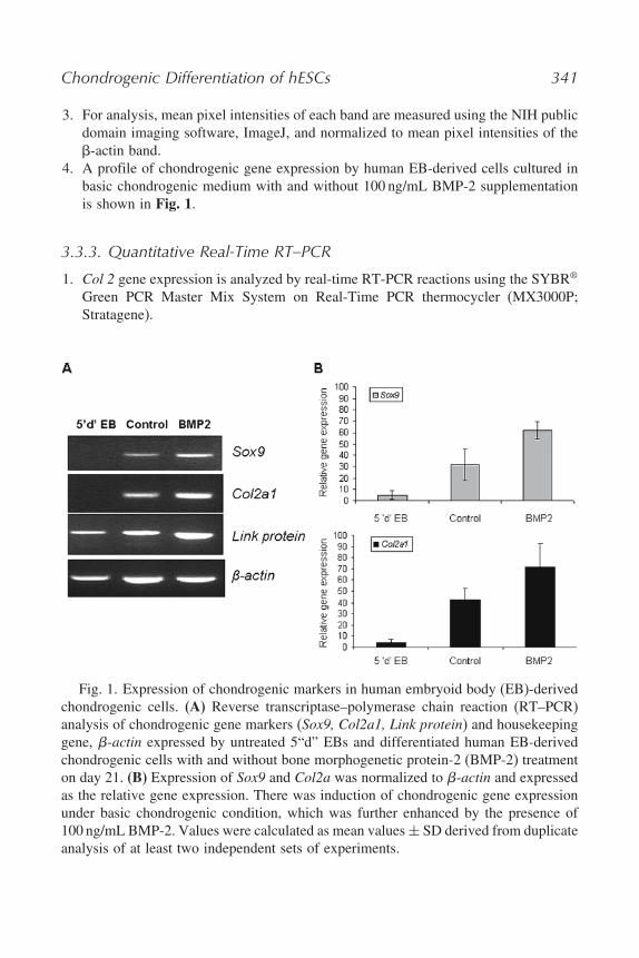
3. For analysis, mean pixel intensities of each band are measured using the NIH publicdomain imaging software, ImageJ, and normalized to mean pixel intensities of the-actin band.
4. A profile of chondrogenic gene expression by human EB-derived cells cultured inbasic chondrogenic medium with and without 100 ng/mL BMP-2 supplementationis shown in Fig. 1.
3.3.3. Quantitative Real-Time RT–PCR
1. Col 2 gene expression is analyzed by real-time RT-PCR reactions using the SYBR®
Green PCR Master Mix System on Real-Time PCR thermocycler (MX3000P;Stratagene).
Fig. 1. Expression of chondrogenic markers in human embryoid body (EB)-derivedchondrogenic cells. (A) Reverse transcriptase–polymerase chain reaction (RT–PCR)analysis of chondrogenic gene markers (Sox9, Col2a1, Link protein) and housekeepinggene, �-actin expressed by untreated 5“d” EBs and differentiated human EB-derivedchondrogenic cells with and without bone morphogenetic protein-2 (BMP-2) treatmenton day 21. (B) Expression of Sox9 and Col2a was normalized to �-actin and expressedas the relative gene expression. There was induction of chondrogenic gene expressionunder basic chondrogenic condition, which was further enhanced by the presence of100 ng/mL BMP-2. Values were calculated as mean values ± SD derived from duplicateanalysis of at least two independent sets of experiments.

342 Toh et al.
2. cDNA samples (1 �L for a total volume of 25 �L per reaction) were analyzed forgene of interest normalized to reference gene glyceraldehydes-3-phosphate dehydro-genase (GAPDH). The level of expression of each target gene is then calculated as2−Ct, as previously described (15).
3. Real-time RT–PCR is performed at 95 �C for 15 min followed by 40 cycles of 15 sdenaturation at 94 �C, 30 s annealing at 55 �C, and 30 s elongation at 72 �C. PCRprimers are summarized in Table 2 (16).
4. Human EB-derived cells cultured as EB outgrowth and HDMM in the presence ofBMP-2 to induce chondrogenesis and processed for real-time RT–PCR for a periodup to day 21, as shown in Fig. 3A.
3.4. Analysis of Matrix Protein Synthesis
3.4.1. Collagen II Immunofluorescence Staining
1. Rinse the EBs cultivated on chamber slides with PBS, fix with methanol : acetone(7:3) at −20 �C for 5 min, rinse three times in PBS, and incubate for 15 min in 10%goat serum for blocking at room temperature.
2. For detection of collagen II, incubate with the monoclonal antibody (II-II6B3),diluted 1:40 with PBS for 1 h at 37 �C in a humidified chamber.
3. Following incubation, rinse the specimens three times with PBS and incubate for2 h at room temperature with Qdot 655 goat anti-mouse IgG antibody diluted 1:200in PBS.
4. Slides are then washed three times in PBS and mounted with Vetashield mountingmedium with DAPI for nuclear counterstaining. For negative control, primaryantibody is omitted.
5. Single chondrogenic cells and nodules were observed in BMP-2-treated EB cultures,as shown in Fig. 2.
3.4.2. Collagen II Immunohistochemistry
1. Remove the culture medium and rinse the cultures twice with PBS. Fix with 10%NBF for 30 min before rinsing twice with PBS.
2. To facilitate antigen retrieval and antibody access, incubate cultures with pepsin at37 �C for 20 min.
3. Rinse once with PBS, then block with hydrogen peroxide block for 15 min at roomtemperature to quench any endogenous peroxidase activity.
4. Wash four times with PBS, block with Ultra V Block before 1 h incubation ofthe cultures at room temperature with anti-collagen II monoclonal antibody, MabII-II6B3 diluted 1:500 in PBS.
5. At the end of primary antibody incubation, rinse the cultures four times with PBS,before adding the pre-diluted biotin-conjugated goat derived anti-mouse secondaryantibody and incubate at room temperature for 30 min.

Chondrogenic Differentiation of hESCs 343
Fig. 2. Bone morphogenetic protein-2 BMP-2 induces chondrogenic differentiationof human EB-derived cells. Indirect immunofluorescence micrographs of control (A, C,and E) and BMP-2-treated (B, D, and F) EB cells 21 days after treatment. Expressionof cartilage-specific collagen II protein is analyzed by immunofluorescence staining.Representative areas of the EB and its outgrowth are shown. BMP-2 induces chondro-genic differentiation of EB-derived cells with intense staining of collagen II-producingcells at the boundary of the EB (B) and outgrowth of chondrogenic cells that couldexist as single cells in monolayer (D) or in three-dimensional nodular aggregates (F).Bar = 100 �m.
6. At the end of secondary antibody incubation, rinse the cultures four times withPBS, before incubating with streptavidin-conjugated horseradish peroxidase at roomtemperature for 45 min.
7. Wash four times with PBS before adding the diaminobenzidine (DAB)chromogen/substrate to visualize the antibody-antigen reaction.
8. Control mouse isotype is included as a negative control (see Note 8).

344 Toh et al.
Fig. 3. Real-time polymerase chain reaction (PCR) analysis of Col 2 expressedby human embryoid body (EB)-derived chondrogenic cells cultured as EB outgrowthand high density micromass (HDMM) along the course of differentiation. (A) Bonemorphogenetic protein-2 BMP-2 stimulation of chondrogenic differentiation in EBoutgrowth displays a slow and steady increase in Col 2 mRNA levels up to day 14,

Chondrogenic Differentiation of hESCs 345
9. Human EB-derived cells cultured as EB outgrowth or HDMM in the presence ofBMP-2 to induce chondrogenesis and processed for collagen II immunohistochem-istry at the end of 21 days, as shown in Fig. 3B.
3.4.3. s-GAG and DNA Quantitation
1. Remove the culture medium from the cultures and wash once with PBS. Removethe PBS completely and scrape the cells with 500 �L papain digestion buffer perwell of a 12-well plate.
2. Collect the samples using sterile 1.5-mL eppendorf tubes and incubate the samplesin water bath at 60 �C for 18 h.
3. The digests are collected and spun down at 16� 000 g for 5 min and sample super-natants can be assayed immediately or frozen down at −20 �C for subsequentanalyses.
4. s-GAG content is measured spectrophotometrically at 630 nm using the BlyscanSulfated Glycosaminoglycan Assay kit (17,18), and normalized to the DNAcontent, measured fluorometrically using the Hoechst 33258 method (19).
5. Add 50 �L of samples to 250 �L of dimethyl-methylene blue (DMMB) dye reagentin 1.5 mL eppendorf assay tubes and incubate at room temperature for 30 min.
6. After incubation, spin the sample at 16� 000 g for 10 min. Discard the supernatant,but retain the dye-bound pellet, which is then dissociated with 200 �L dissociationagent (supplied in the Blyscan Sulfated Glycosaminoglycan Assay kit) per sampleto release the color.
7. Transfer the entire contents from the assay tubes to the wells of a 96-well microwellplate for spectrophotometric measurement at 630 nm. The dissociation agent isused as the blank. Standard curve of s-GAG is constructed using different concen-trations of bovine trachea chondroitin sulfate (provided in the kit), according tothe manufacturer’s instructions.
8. Dilute 10 �L of papain digests 10 times with PBS to a final sample volume of100 �L before adding to the wells containing 100 �L of prepared Hoechst 33258dye assay solution �0�2 �g/mL�.
�Fig. 3. before it decreases and plateaus, whereas in HDMM culture system, BMP-2
causes acute induction of Col 2 by day 7 of chondrogenic differentiation in HDMMculture. Values are calculated as means ± SD from duplicate analysis of at leasttwo independent experiments. (B) Collagen II immunohistochemistry indicates markedenhancement in collagen II synthesis in HDMM culture (b) at the end of day 21differentiation. Appropriate isotype control is carried out in both EB outgrowth (c) andHDMM (d) cultures in parallel.

346 Toh et al.
Fig. 4. (A) Analysis of sulfated glycosaminoglycan (s-GAG) synthesis byhuman embryoid body (EB)-derived chondrogenic cells cultured in EB outgrowthand high-density micromass (HDMM) along the course of differentiation.Bone morphogenetic protein-2 BMP-2 stimulation of chondrogenic differenti-ation in EB outgrowth culture system displays acute decrease in ratio ofs-GAG/DNA from day 7 onward, whereas in HDMM culture system, BMP-2 inducestime-dependent increase in ratio of s-GAG/DNA up to day 14, before it decreases, there-after indicating hypertrophic maturation. Values are calculated as means ± SD fromduplicate analysis of at least two independent experiments. (B) Alcian blue staining alsoindicates marked enhancement in s-GAG deposition in the HDMM culture, particularlyin the regions of nodular growth.

Chondrogenic Differentiation of hESCs 347
9. The fluorescence measurement of Hoechst 33258 dye is performed using a fluores-cence plate reader at 360 nm excitation and 460 nm emission. Calf thymus DNAis used for the construction of the standard curve for DNA.
10. The reagent blank (100 �L of dye +100 �L dH2O) fluorescence value is subtractedfrom the sample data before analysis.
11. Human EB-derived cells are cultured as EB outgrowth or HDMM in the presenceof BMP-2 to induce chondrogenesis and processed for s-GAG and DNA quantifi-cation along the course of differentiation, as shown in Fig. 4A.
3.4.4. Alcian Blue Staining
1. Remove the medium from the cultures and wash once with PBS. Fix the cell cultureswith 10% (v/v) neutral buffered formalin for 30 min, followed by washing threetimes with PBS and rinsing in dH2O.
2. Fixed cells are incubated with 0.05% (w/v) alcian blue solution overnight.3. Remove excess stain by washing at least two times with PBS, followed by a further
rinse in 5% acetic acid to remove non-specific staining and then a last rinse with PBSagain. Store cultures in PBS for subsequent examination by light photomicrography.
4. Human EB-derived cells cultured as EB outgrowth or HDMM in the presence ofBMP-2 to induce chondrogenesis and processed for alcian blue staining at the endof 21 days, as shown in Fig. 4B.
4. Notes1. Culture of hESCs (H1 and H9; NIH stem cell registry: http://stemcells.nih.gov/
research/registry, also see refs 4 and 5) follows exactly as recommended by theWicell protocol. Refer to Web site http://www.wicell.org/ for protocols describingthe expansion and propagation of hESCs on murine embryonic feeder cells.
2. 1% KSR instead of the strictly serum-free medium was used in this study, based onreports that mesodermal differentiation of ESCs can be inhibited under serum-freeconditions (20).
3. Always prepare the papain-digestion buffer fresh when needed. Allow the papainto completely dissolve and filter-sterilize before use.
4. Hoechst 33258 is a possible carcinogen and possible mutagen. Wear gloves and amask and work under a fume hood. Keep 2× assay solution at room temperature.Prepare fresh solution whenever needed. Do not filter once dye has been added.Protect from light. 10× TNE stock buffer solution used in Hoechst dye DNAassay can be stored at 4 �C for up to 3 months. Working solution is prepared freshwhenever needed.
5. Allow the EBs to settle down during the wash with PBS. Usually, it takes lessthan 3 min for all the EBs to settle. Avoid pipetting too many times or foamingbecause the cells should be collected in clumps.

348 Toh et al.
6. Trypsinize the EBs into single cells for no more than 5 min and pass the cellsuspension once through a 22-G needle and then 40-�m cell strainer. Prolong orrepeated trypsinization is detrimental to cell viability.
7. The method for HDMM culture has been adapted from previously publishedmethods (21,22). After removal of 0.1% gelatin from the wells, ensure the plates arecompletely dry before use. This is to prevent dispersion of cells when dispensingthe cells in a single drop onto the well in HDMM culture.
8. Isotype antibody control is necessary to ensure antibody specificity, to rule outany non-specific staining and to give better interpretation of the results.
9. Use chondrogenic differentiation media for not more than 2 weeks after prepa-ration, as components such as l-glutamine and AA2P degrade over time. Storethe chondrogenic differentiation media at 4 �C and discard unused media after2 weeks.
10. Prepare l-Proline, AA2P, dexamethasone in PBS/0.1% BSA, aliquot and freezethem at −20 �C in working volumes till use. Also aliquot P/S, l-glutamine, andKSR in working volumes and freeze them at −20 �C. Avoid repeated freeze-thawof working solutions.
References1. Hall BK, Miyake T. (2000) All for one and one for all: condensations and the
initiation of skeletal development. Bioessays 22, 138–147.2. DeLise AM, Fischer L, Tuan RS. (2000) Cellular interactions and signaling in
cartilage development. Osteoarthr Cartilage 8, 309–334.3. Heng BC, Cao T, Lee EH. (2004) Directing stem cell differentiation into the
chondrogenic lineage in vitro. Stem Cells 22, 1152–1167.4. Thomson JA, Itskovitz-Eldor J, Shapiro SS, Waknitz MA, Swiergiel JJ, Marshall
VS, Jones JM. (1998) Embryonic stem cell lines derived from human blastocysts.Science 282, 1145–1147.
5. Reubinoff BE, Pera MF, Fong CY, Trounson A, Bongso A. (2000) Embryonic stemcell lines from human blastocysts: somatic differentiation in vitro. Nat. Biotechnol.18, 399–404.
6. Kramer J, Hegert C, Guan K, Wobus AM, Muller PK, Rohwedel J. (2000)Embryonic stem cell-derived chondrogenic differentiation in vitro: activation byBMP-2 and BMP-4. Mech. Dev. 92, 193–205.
7. Hegert C, Kramer J, Hargus G, Muller J, Guan K, Wobus AM, Muller PK,Rohwedel J. (2002) Differentiation plasticity of chondrocytes derived from mouseembryonic stem cells. J. Cell. Sci. 115, 4617–4628.
8. Kawaguchi J, Mee PJ, Smith AG. (2005) Osteogenic and chondrogenic differen-tiation of embryonic stem cells in response to specific growth factors. Bone 36,758–769.

Chondrogenic Differentiation of hESCs 349
9. Hwang NS, Kim MS, Sampattavanich S, Baek JH, Zhang Z, Elisseeff J. (2006)The effects of three dimensional culture and growth factors on the chondrogenicdifferentiation of murine embryonic stem cells. Stem Cells 24, 284–291.
10. Tanaka H, Murphy CL, Murphy C, Kimura M, Kawai S, Polak JM. (2004)Chondrogenic differentiation of murine embryonic stem cells: effects of cultureconditions and dexamethasone. J. Cell. Biochem. 93, 454–462.
11. Toh WS, Yang Z, Heng BC, Cao T. (2006) New perspectives in chondro-genic differentiation of stem cells for cartilage repair. ScientificWorldJournal 6,361–364.
12. Toh WS, Yang Z, Liu H, Heng BC, Lee EH, Cao T. (2007) Effects of cultureconditions and bone morphogenetic protein 2 on extent of chondrogenesis fromhuman embryonic. Stem Cells 25(4): 950–960.
13. Wozney JM, Rosen V, Celeste AJ, Mitsock LM, Whitters MJ, Kriz RW, HewickRM, Wang EA. (1988) Novel regulators of bone formation: molecular clones andactivities. Science 242, 1528–1534.
14. Sekiya I, Vuoristo JT, Larson BL, Prockop DJ. (2002) In vitro cartilage formationby human adult stem cells from bone marrow stroma defines the sequence ofcellular and molecular events during chondrogenesis. Proc. Natl. Acad. Sci. U.S.A.99, 4397–4402.
15. Livak KJ, Schmittgen TD. (2001) Analysis of relative gene expression data usingreal-time quantitative PCR and the 2(-Delta Delta C(T)) method. Methods 25,402–408.
16. Martin I, Jakob M, Schafer D, Dick W, Spagnoli G, Heberer M. (2001) Quanti-tative analysis of gene expression in human articular cartilage from normal andosteoarthritic joints. Osteoarthr Cartilage 9, 112–118.
17. Toh WS, Liu H, Heng BC, Rufaihah AJ, Ye CP, Cao T. (2005) Combined effectsof TGF1 and BMP2 in serum-free chondrogenic differentiation of mesenchymalstem cells induced hyaline-like cartilage formation. Growth Factors 23, 313–321.
18. Farndale RW, Buttle DJ, Barrett AJ. (1986) Improved quantitation and discrim-ination of sulphated glycosaminoglycans by use of dimethylmethylene blue.Biochim. Biophys. Acta 883, 173–177.
19. Kim YJ, Sah RL, Doong JY, Grodzinsky AJ. (1988) Fluorometric assay of DNAin cartilage explants using Hoechst 33258. Anal. Biochem. 174, 168–176.
20. Wiles MV, Johansson BM. (1999) Embryonic stem cell development in a chemi-cally defined medium. Exp. Cell. Res. 247, 241–248.
21. Ahrens PB, Solursh M, Reiter RS. (1977) Stage-related capacity for limb chondro-genesis in cell culture. Dev. Biol. 60, 69–82.
22. Mello MA, Tuan RS. (1999) High density micromass cultures of embryonic limbbud mesenchymal cells: an in vitro model of endochondral skeletal development.In Vitro Cell Dev. Biol. Anim. 35, 262–269.


![[Composite Cultures] - CORE](https://static.fdokumen.com/doc/165x107/6325e67de491bcb36c0a86c0/composite-cultures-core.jpg)