Neural progenitors from human embryonic stem cells
7
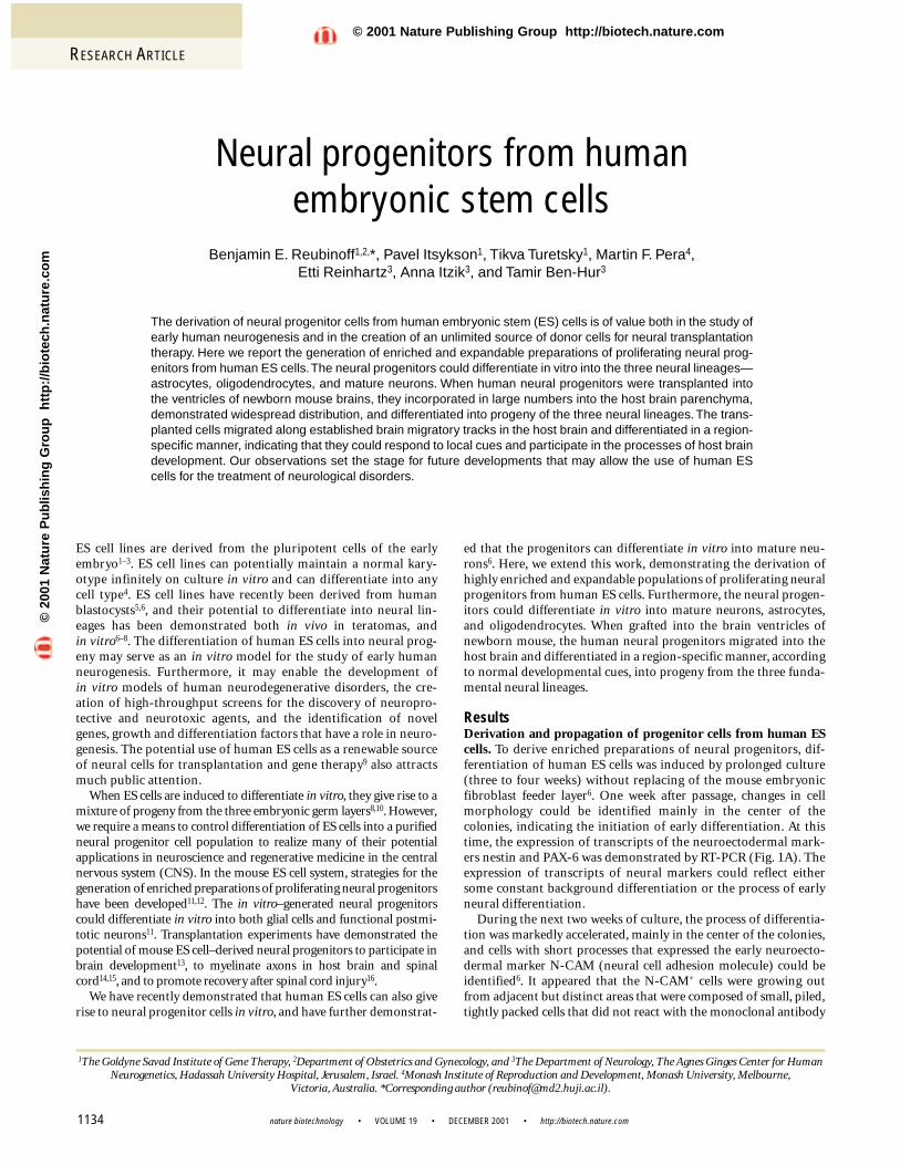
nature biotechnology • VOLUME 19 • DECEMBER 2001 • http://biotech.nature.com RESEARCH ARTICLE 1134 Neural progenitors from human embryonic stem cells Benjamin E. Reubinoff 1,2, *, Pavel Itsykson 1 , Tikva Turetsky 1 , Martin F. Pera 4 , Etti Reinhartz 3 , Anna Itzik 3 , and Tamir Ben-Hur 3 The derivation of neural progenitor cells from human embryonic stem (ES) cells is of value both in the study of early human neurogenesis and in the creation of an unlimited source of donor cells for neural transplantation therapy. Here we report the generation of enriched and expandable preparations of proliferating neural prog- enitors from human ES cells.The neural progenitors could differentiate in vitro into the three neural lineages— astrocytes, oligodendrocytes, and mature neurons. When human neural progenitors were transplanted into the ventricles of newborn mouse brains, they incorporated in large numbers into the host brain parenchyma, demonstrated widespread distribution, and differentiated into progeny of the three neural lineages. The trans- planted cells migrated along established brain migratory tracks in the host brain and differentiated in a region- specific manner, indicating that they could respond to local cues and participate in the processes of host brain development. Our observations set the stage for future developments that may allow the use of human ES cells for the treatment of neurological disorders. ES cell lines are derived from the pluripotent cells of the early embryo 1–3 . ES cell lines can potentially maintain a normal kary- otype infinitely on culture in vitro and can differentiate into any cell type 4 . ES cell lines have recently been derived from human blastocysts 5,6 , and their potential to differentiate into neural lin- eages has been demonstrated both in vivo in teratomas, and in vitro 6–8 . The differentiation of human ES cells into neural prog- eny may serve as an in vitro model for the study of early human neurogenesis. Furthermore, it may enable the development of in vitro models of human neurodegenerative disorders, the cre- ation of high-throughput screens for the discovery of neuropro- tective and neurotoxic agents, and the identification of novel genes, growth and differentiation factors that have a role in neuro- genesis. The potential use of human ES cells as a renewable source of neural cells for transplantation and gene therapy 9 also attracts much public attention. When ES cells are induced to differentiate in vitro, they give rise to a mixture of progeny from the three embryonic germ layers 8,10 . However, we require a means to control differentiation of ES cells into a purified neural progenitor cell population to realize many of their potential applications in neuroscience and regenerative medicine in the central nervous system (CNS). In the mouse ES cell system, strategies for the generation of enriched preparations of proliferating neural progenitors have been developed 11,12 . The in vitro–generated neural progenitors could differentiate in vitro into both glial cells and functional postmi- totic neurons 11 . Transplantation experiments have demonstrated the potential of mouse ES cell–derived neural progenitors to participate in brain development 13 , to myelinate axons in host brain and spinal cord 14,15 , and to promote recovery after spinal cord injury 16 . We have recently demonstrated that human ES cells can also give rise to neural progenitor cells in vitro, and have further demonstrat- ed that the progenitors can differentiate in vitro into mature neu- rons 6 . Here, we extend this work, demonstrating the derivation of highly enriched and expandable populations of proliferating neural progenitors from human ES cells. Furthermore, the neural progen- itors could differentiate in vitro into mature neurons, astrocytes, and oligodendrocytes. When grafted into the brain ventricles of newborn mouse, the human neural progenitors migrated into the host brain and differentiated in a region-specific manner, according to normal developmental cues, into progeny from the three funda- mental neural lineages. Results Derivation and propagation of progenitor cells from human ES cells. To derive enriched preparations of neural progenitors, dif- ferentiation of human ES cells was induced by prolonged culture (three to four weeks) without replacing of the mouse embryonic fibroblast feeder layer 6 . One week after passage, changes in cell morphology could be identified mainly in the center of the colonies, indicating the initiation of early differentiation. At this time, the expression of transcripts of the neuroectodermal mark- ers nestin and PAX-6 was demonstrated by RT-PCR (Fig. 1A). The expression of transcripts of neural markers could reflect either some constant background differentiation or the process of early neural differentiation. During the next two weeks of culture, the process of differentia- tion was markedly accelerated, mainly in the center of the colonies, and cells with short processes that expressed the early neuroecto- dermal marker N-CAM (neural cell adhesion molecule) could be identified 6 . It appeared that the N-CAM + cells were growing out from adjacent but distinct areas that were composed of small, piled, tightly packed cells that did not react with the monoclonal antibody 1 The Goldyne Savad Institute of Gene Therapy, 2 Department of Obstetrics and Gynecology, and 3 The Department of Neurology, The Agnes Ginges Center for Human Neurogenetics, Hadassah University Hospital, Jerusalem, Israel. 4 Monash Institute of Reproduction and Development, Monash University, Melbourne, Victoria, Australia. *Corresponding author ([email protected]). © 2001 Nature Publishing Group http://biotech.nature.com © 2001 Nature Publishing Group http://biotech.nature.com
-
Upload
independent -
Category
Documents
-
view
1 -
download
0
Transcript of Neural progenitors from human embryonic stem cells
nature biotechnology • VOLUME 19 • DECEMBER 2001 • http://biotech.nature.com
RESEARCH ARTICLE
1134
Neural progenitors from human embryonic stem cells
Benjamin E. Reubinoff1,2,*, Pavel Itsykson1, Tikva Turetsky1, Martin F. Pera4, Etti Reinhartz3, Anna Itzik3, and Tamir Ben-Hur3
The derivation of neural progenitor cells from human embryonic stem (ES) cells is of value both in the study ofearly human neurogenesis and in the creation of an unlimited source of donor cells for neural transplantationtherapy. Here we report the generation of enriched and expandable preparations of proliferating neural prog-enitors from human ES cells.The neural progenitors could differentiate in vitro into the three neural lineages—astrocytes, oligodendrocytes, and mature neurons. When human neural progenitors were transplanted intothe ventricles of newborn mouse brains, they incorporated in large numbers into the host brain parenchyma,demonstrated widespread distribution, and differentiated into progeny of the three neural lineages. The trans-planted cells migrated along established brain migratory tracks in the host brain and differentiated in a region-specific manner, indicating that they could respond to local cues and participate in the processes of host braindevelopment. Our observations set the stage for future developments that may allow the use of human EScells for the treatment of neurological disorders.
ES cell lines are derived from the pluripotent cells of the earlyembryo1–3. ES cell lines can potentially maintain a normal kary-otype infinitely on culture in vitro and can differentiate into anycell type4. ES cell lines have recently been derived from humanblastocysts5,6, and their potential to differentiate into neural lin-eages has been demonstrated both in vivo in teratomas, and in vitro6–8. The differentiation of human ES cells into neural prog-eny may serve as an in vitro model for the study of early humanneurogenesis. Furthermore, it may enable the development ofin vitro models of human neurodegenerative disorders, the cre-ation of high-throughput screens for the discovery of neuropro-tective and neurotoxic agents, and the identification of novelgenes, growth and differentiation factors that have a role in neuro-genesis. The potential use of human ES cells as a renewable sourceof neural cells for transplantation and gene therapy9 also attractsmuch public attention.
When ES cells are induced to differentiate in vitro, they give rise to amixture of progeny from the three embryonic germ layers8,10. However,we require a means to control differentiation of ES cells into a purifiedneural progenitor cell population to realize many of their potentialapplications in neuroscience and regenerative medicine in the centralnervous system (CNS). In the mouse ES cell system, strategies for thegeneration of enriched preparations of proliferating neural progenitorshave been developed11,12. The in vitro–generated neural progenitorscould differentiate in vitro into both glial cells and functional postmi-totic neurons11. Transplantation experiments have demonstrated thepotential of mouse ES cell–derived neural progenitors to participate inbrain development13, to myelinate axons in host brain and spinalcord14,15, and to promote recovery after spinal cord injury16.
We have recently demonstrated that human ES cells can also giverise to neural progenitor cells in vitro, and have further demonstrat-
ed that the progenitors can differentiate in vitro into mature neu-rons6. Here, we extend this work, demonstrating the derivation ofhighly enriched and expandable populations of proliferating neuralprogenitors from human ES cells. Furthermore, the neural progen-itors could differentiate in vitro into mature neurons, astrocytes,and oligodendrocytes. When grafted into the brain ventricles ofnewborn mouse, the human neural progenitors migrated into thehost brain and differentiated in a region-specific manner, accordingto normal developmental cues, into progeny from the three funda-mental neural lineages.
ResultsDerivation and propagation of progenitor cells from human EScells. To derive enriched preparations of neural progenitors, dif-ferentiation of human ES cells was induced by prolonged culture(three to four weeks) without replacing of the mouse embryonicfibroblast feeder layer6. One week after passage, changes in cellmorphology could be identified mainly in the center of thecolonies, indicating the initiation of early differentiation. At thistime, the expression of transcripts of the neuroectodermal mark-ers nestin and PAX-6 was demonstrated by RT-PCR (Fig. 1A). Theexpression of transcripts of neural markers could reflect eithersome constant background differentiation or the process of earlyneural differentiation.
During the next two weeks of culture, the process of differentia-tion was markedly accelerated, mainly in the center of the colonies,and cells with short processes that expressed the early neuroecto-dermal marker N-CAM (neural cell adhesion molecule) could beidentified6. It appeared that the N-CAM+ cells were growing outfrom adjacent but distinct areas that were composed of small, piled,tightly packed cells that did not react with the monoclonal antibody
1The Goldyne Savad Institute of Gene Therapy, 2Department of Obstetrics and Gynecology, and 3The Department of Neurology, The Agnes Ginges Center for HumanNeurogenetics, Hadassah University Hospital, Jerusalem, Israel. 4Monash Institute of Reproduction and Development, Monash University, Melbourne,
Victoria, Australia. *Corresponding author ([email protected]).
©20
01 N
atu
re P
ub
lish
ing
Gro
up
h
ttp
://b
iote
ch.n
atu
re.c
om
© 2001 Nature Publishing Group http://biotech.nature.com

RESEARCH ARTICLE
http://biotech.nature.com • DECEMBER 2001 • VOLUME 19 • nature biotechnology 1135
GCTM-2, which identifies undifferentiated ES cells6, and did notexpress the early neuroectodermal marker N-CAM (data notshown). These distinct areas had a uniformly white–gray andopaque appearance under dark-field stereomicroscopy (Fig. 2A),and could be identified in 54% of the colonies (67/124). They weresurrounded by cells with diverse morphologies expressing a largearray of somatic and extraembryonic markers, including muscleactin and desmin6, α-fetoprotein, hepatocyte nuclear factor (HNF)-α, cardiac actin, and kallikrein (RT-PCR; not shown).
Assuming that the cells in the distinct areas gave rise to the adja-cent N-CAM+ cells, clumps of about 150 cells were mechanicallyisolated from these areas and replated in serum-free medium17.Under these culture conditions, the clumps formed free-floatingspherical structures within 24 h.
Supplementing the medium with basic fibroblast growth factor(bFGF) and epidermal growth factor (EGF), a growth factor combi-nation that is known to be effective for the propagation of humanfetal- and adult-derived neuroepithelial progenitors17–20, facilitated
sequential propagation and expansion of the sphere cultures.During the first two weeks in culture, some cell death was observedand the spheres gradually acquired a uniform round morphology(Fig. 2B). A detailed analysis of marker expression and the growthand differentiation potential of the cells within the spheres wasconducted in three preparations that were separately derived andpropagated.
The level of proliferation of the cells within the spheres wasmonitored indirectly by measuring the increase in the volume ofthe spheres over time. Most of the cells within the spheres wereviable as demonstrated by Trypan Blue staining (94 ± 3.2%, n = 47spheres). A positive correlation between the volume of the spheresand the number of cells within the spheres (Fig. 3B) was documented at various passage levels (5–15 weeks after deriva-tion), indicating that an increment in sphere volume could beused as an indirect indication of cell proliferation. The spheresgrew over an 18- to 22-week period, after which time the volumeof the spheres was stable or declined. A relatively rapid growth ratewas observed during the first five to six weeks after derivation,with a population doubling time of ∼ 4.7 days. It was followed by a10- to 16-week period of slow and stable cell growth with a popu-lation doubling time of ∼ 2.5 weeks. This proliferative capabilitycould potentially allow a significant expansion of the progenitorcell cultures (Fig. 3A).
Figure 1. RT-PCR analysis of the expression of markers in human ES cellcolonies, ES-derived spheres, and in differentiated cells originating fromthe spheres. (A) Oct-4, nestin, and PAX-6 in human ES cell colonies atone week after plating and in neural progenitor (NP) spheres. (B) Theexpression of non-neural marker genes in human ES cell–derivedspheres. (C) Neuronal and glial markers in differentiated cells originatingfrom human ES cell–derived neural progenitor spheres. All panels show2% agarose gels stained with ethidium bromide. The symbols + and –indicate whether the PCR reaction was done with or without the additionof reverse transcriptase. A 1 kb plus DNA ladder was used in all panels.Oct-4 band is 320 bp, nestin 208 bp, PAX-6 274 bp, β-actin 291 bp, keratin780 bp, Flk-1 199 bp, CD34 200 bp, AC133 200 bp, transferrin 367 bp,amylase 490 bp, α1-antitrypsin 360 bp, plp and dm-20 are 354 bp and249 bp, respectively, MBP is 379 bp, GFAP is 383 bp, NSE is 254 bp, andNF-M is 430 bp.
Figure 2. Analysis of morphology and marker expression in human ES-derived progenitor cells. (A) Dark-field stereomicroscopic photographof a differentiating ES cell colony, four weeks after plating, with areas ofcells (arrows) that are destined to give rise to neural progenitors.(B) Phase contrast micrograph of a sphere cultured in serum-freemedium. (C–F) Indirect immunofluorescence staining of progenitor cells,4–12 h after disaggregating of spheres and plating on adhesive substrate,for N-CAM, vimentin, nestin, and A2B5, respectively. Bars =1.6 mm (A), 100 µm (B), 25 µm (C, E, F), 35 µm (D).
A
B
C
A B
C D
E F
©20
01 N
atu
re P
ub
lish
ing
Gro
up
h
ttp
://b
iote
ch.n
atu
re.c
om
© 2001 Nature Publishing Group http://biotech.nature.com

RESEARCH ARTICLE
nature biotechnology • VOLUME 19 • DECEMBER 2001 • http://biotech.nature.com1136
Characterization of the progenitor cells within the spheres. Cellsin the spheres expressed markers of neural progenitor cells, such asN-CAM (ref. 21; Fig. 2C), the intermediate-filament proteinnestin22 (immunostaining, Fig 2E; RT-PCR, Fig. 1A), A2B5 (ref. 23;Fig. 2F), vimentin24 (Fig. 2D), and the transcription factor PAX-6(Fig. 1A). The expression of these markers was maintained withprolonged cultivation in vitro (18 weeks).
To evaluate the proportion of neural progenitors in the cultures,spheres were disaggregated into single cells that were plated, fixed,and analyzed for the expression of the early neural markers (Fig. 2C–F). A high proportion of the cells expressed N-CAM (99 ± 1.6%, n = 11 experiments), nestin (97 ± 2.3%, n = 10 experi-ments), and A2B5 (90.5 ± 1.1%, n = 6). A lower proportion of cellswere immunoreactive to the vimentin-specific antibody (67 ± 16.8%, n = 9 experiments). These proportions were stableduring cultivation of the spheres (up to 18 weeks).
Oct-4 is a member of the POU-domain transcription factor fam-ily whose expression is limited in the mouse to pluripotent cells andis downregulated upon differentiation25. We have previouslydemonstrated a similar pattern of expression in human ES cells6.Oct-4 was not expressed by cells in the neural progenitor spheres,indicating that undifferentiated human ES cells were not presentwithin the spheres (Fig. 1A).
To determine whether cells that had acquired markers of othertissues or lineages were present within the spheres, the expression ofmarkers representing derivatives of mesoderm, endoderm, and epi-dermis were examined. Cells within the spheres expressed tran-scripts of markers of hematopoietic/endothelial progenitors(CD34, AC-133, Flk-1), endoderm (α1-antitrypsin, transferring,and amylase) and epidermis (keratin), as demonstrated by RT-PCR(Fig. 1B). Markers of extraembryonic endoderm were not expressedby the progenitors (α-fetoprotein and HNF-α, RT-PCR; notshown) or their differentiated progeny (low-molecular-weightcytokeratin and laminin immunostaining; not shown). The expres-
sion of transcripts of non-neural markers was evident after pro-longed cultivation of the spheres. It could represent contaminationby a small number of non-neural cells generated during the deriva-tion of our cultures. Alternatively, it could represent plasticity ofprimitive neural progenitors that expressed markers, or gave rise tocells from other lineages26,27. Whatever the source, additional selec-tion either on the basis of cell-surface markers18 or on the expres-sion of lineage-specific genes12 may be needed to generate pureneural cultures.
In vitro neural differentiation. The neural progenitors in thespheres could differentiate in vitro into derivatives of the three fun-damental neural lineages. In general, differentiation was induced byplating whole spheres on an appropriate substrate in the absence ofgrowth factors. Under these conditions the spheres attached rapid-ly, and cells migrated out to form a monolayer of differentiated cells(Fig. 4A).
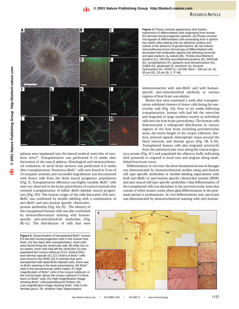
For neuronal differentiation studies, spheres were plated onpoly-D-lysine and laminin-coated dishes. After two to three weeks,cells that migrated out and formed a monolayer both displayed themorphology and also expressed the structural markers that arecharacteristic of immature neurons, such as βIII-tubulin (Fig. 4B),the 70 kDa neurofilament proteins (Fig. 4C), and neuron-specificenolase (NSE; Fig. 1C). Moreover, the differentiated cells expressedmarkers of mature neurons such as the 160 kDa neurofilament pro-teins (NF-M, Fig. 4D; RT-PCR, Fig. 1C), MAP-2ab (Fig. 4E), andsynaptophysin (Fig. 4F). Furthermore, the cultures contained cellsthat synthesized glutamate, expressed glutamic acid decarboxylase(GAD; the rate-limiting enzyme in GABA biosynthesis), synthe-sized GABA and serotonin, and expressed tyrosine hydroxylase(TH; Fig. 4G–K). Neurons that synthesized GABA and glutamatewere relatively abundant, comprising 35% and 15% of the neuronalpopulation, respectively. TH- and serotonin-producing cells wererelatively rare (<1%).
For glial differentiation studies, spheres were plated on poly-D-lysine- and fibronectin-coated dishes and were cultured first in thepresence of EGF, bFGF, and platelet-derived growth factor-AA(PDGF-AA), followed by culture in the presence of tri-iodothyro-nine (T3). The combination of bFGF and PDGF-AA is known topromote the proliferation of glial precursor cells14, whereas T3 hasbeen shown to enhance differentiation of oligodendrocyte lineagecells from human embryonic neural spheres28.
Differentiation into astrocytes was demonstrated by the presence ofcells that expressed glial fibrillary acidic protein (GFAP) (Fig. 4L;RT-PCR, Fig. 1C). Oligodendrocyte lineage cells were infrequent underour culture conditions and few cells were immunoreactive to O4, anantibody recognizing oligodendrocyte-specific glycolipids29 (Fig. 4M).Differentiation to the oligodendrocyte lineage was further confirmedby demonstrating the expression of RNA transcripts of both myelinbasic protein (MBP) and the plp gene (Fig. 1C). The plp gene encodesthe proteolipid protein and its alternatively spliced product DM-20,which are major proteins of brain myelin21.
To evaluate the proportion of neurons versus glial cells followinginduction of differentiation, spheres that were propagated 10 weekswere disaggregated and plated on poly-D-lysine- and laminin-coated dishes and cultured in the absence of mitogens for five days.Fifty-seven percent of the cells were immunoreactive to anti-β III-tubulin (a marker characteristic of immature neurons) and26% to anti-GFAP. Therefore, at least 83% of the cells took on aneural fate. The potential of the neural progenitors to give rise toboth neurons and glial cells in vitro was maintained for the dura-tion of the 22 weeks of propagation.
Integration and differentiation in host brain. To explore thedevelopmental potential of the human ES-derived neural progeni-tors in vivo, disaggregated bromodeoxyuridine (BrdU)-labeled
Figure 3. Cumulative growth curve for human ES-derived progenitor cells.(A) Continuous growth is evident during an 18- to 22-week period. Theincrement in the volume of the spheres was continuously monitored as anindirect measure of the increase in cell numbers. A linear positivecorrelation between the volume of the spheres and the number of cellswithin the spheres (B, insert) was maintained during cultivation.
A
B
©20
01 N
atu
re P
ub
lish
ing
Gro
up
h
ttp
://b
iote
ch.n
atu
re.c
om
© 2001 Nature Publishing Group http://biotech.nature.com
RESEARCH ARTICLE
http://biotech.nature.com • DECEMBER 2001 • VOLUME 19 • nature biotechnology 1137
spheres were implanted into the lateral cerebral ventricles of new-born mice19. Transplantation was performed 9–15 weeks afterderivation of the neural spheres. Histological and immunochemi-cal evaluation of serial brain sections was performed 4–6 weeksafter transplantation. Numerous BrdU+ cells were found in 9 out of14 recipient animals, and successful engraftment was documentedwith donor cells from the three neural progenitor populations (Fig. 5). Transplantation efficiency was highly variable. BrdU+ cellswere not observed in the brain parenchyma of control animals thatreceived transplantation of killed, BrdU-labeled, neural progeni-tors (Fig. 5D). The human origin of the cells decorated with anti-BrdU was confirmed by double labeling with a combination ofanti-BrdU and anti-human specific ribonucleo-protein antibodies (Fig. 6A–D). The identity ofthe transplanted human cells was also confirmedby immunofluorescent staining with human-specific anti-mitochondrial antibodies (Fig.6E–G). The distribution of cells that were
immunoreactive with anti-BrdU and with human-specific anti-mitochondrial antibody in variousregions of host brain was similar.
Brains that were examined a week after transplan-tation exhibited clusters of donor cells lining the ven-tricular wall (Fig. 5A). Four to six weeks followingtransplantation, human cells had left the ventriclesand migrated in large numbers mainly as individualcells into the host brain parenchyma. The human cellsdemonstrated a widespread distribution in variousregions of the host brain including periventricularareas, the entire length of the corpus callosum, fim-bria, internal capsule, diencephalic tissue around thethird ventricle, and dentate gyrus (Fig. 5B, E–H).Transplanted human cells also migrated anteriorilyfrom the subventricular zone along the rostral migra-
tory stream (Fig. 5C) and populated the olfactory bulb, indicatingtheir potential to respond to local cues and migrate along estab-lished host brain tracts.
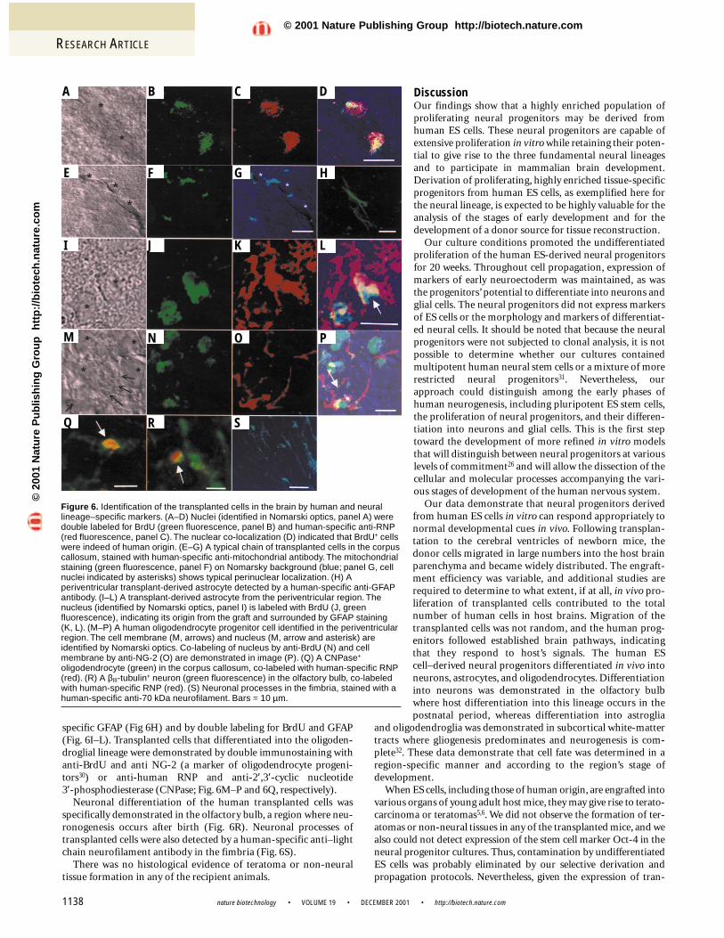
Differentiation in vivo into the three fundamental neural lineageswas demonstrated by immunochemical studies using anti-humancell type–specific antibodies or double-labeling experiments withboth anti-BrdU or anti-human specific ribonuclear protein (RNP)and anti-neural cell type–specific antibodies. Glial differentiation ofthe transplanted cells was abundant in the periventricular areas thatconsist of white-matter tracks where glial differentiation in the post-natal period is predominant. In vivo differentiation into astrocyteswas demonstrated by immunochemical staining with anti-human-
Figure 4. Phase contrast appearance and markerexpression of differentiated cells originating from humanES-derived neural progenitor spheres. (A) Phase contrastmicrograph of differentiated cells emanating from a spheretwo weeks after plating onto an adhesive surface andculture in the absence of growth factors. (B–M) Indirectimmunofluorescence microscopy of differentiated cellsdecorated with antibodies against the following neuronaland glial markers: βIII-tubulin (B), 70 kDa neurofilamentproteins (C), 160 kDa neurofilament proteins (D), MAP2ab(E), synaptophysin (F), glutamic acid decarboxylase (G),GABA (H), glutamate (I), serotonin (J), tyrosinehydroxylase (K), GFAP (L), O4 (M). Bars = 200 µm (A, D),50 µm (E), 20 µm (B, C, F–M).
A B C
F
D E
G H I
J K L M
Figure 5. Dissemination of transplanted BrdU+ humanES-derived neural progenitor cells in the mouse hostbrain. (A) Two days after transplantation, most cellswere found lining the ventricular wall. (B) After four tosix weeks, most cells had left the ventricles (V) andpopulated the corpus callosum (CC), fimbria (fim),and internal capsule (ic). (C) Chains of BrdU+ cellswere found in the RMS. (D) In animals that weretransplanted with dead BrdU-labeled cells, there wasno BrdU staining in the brain parenchyma. (E) BrdU+
cells in the periventricular white matter. (F) Highmagnification of BrdU+ cells in the corpus callosum; inthe cortical layer above the corpus callosum (*) therewere no BrdU+ cells. (G) High-magnification imageshowing BrdU+ cells populating the fimbria. (H) Low-magnification image showing BrdU+ cells in thedentate gyrus. Str, striatum; hipp, hippocampus.
©20
01 N
atu
re P
ub
lish
ing
Gro
up
h
ttp
://b
iote
ch.n
atu
re.c
om
© 2001 Nature Publishing Group http://biotech.nature.com
RESEARCH ARTICLE
nature biotechnology • VOLUME 19 • DECEMBER 2001 • http://biotech.nature.com1138
specific GFAP (Fig 6H) and by double labeling for BrdU and GFAP(Fig. 6I–L). Transplanted cells that differentiated into the oligoden-droglial lineage were demonstrated by double immunostaining withanti-BrdU and anti NG-2 (a marker of oligodendrocyte progeni-tors30) or anti-human RNP and anti-2′,3′-cyclic nucleotide 3′-phosphodiesterase (CNPase; Fig. 6M–P and 6Q, respectively).
Neuronal differentiation of the human transplanted cells wasspecifically demonstrated in the olfactory bulb, a region where neu-ronogenesis occurs after birth (Fig. 6R). Neuronal processes oftransplanted cells were also detected by a human-specific anti–lightchain neurofilament antibody in the fimbria (Fig. 6S).
There was no histological evidence of teratoma or non-neuraltissue formation in any of the recipient animals.
DiscussionOur findings show that a highly enriched population ofproliferating neural progenitors may be derived fromhuman ES cells. These neural progenitors are capable ofextensive proliferation in vitro while retaining their poten-tial to give rise to the three fundamental neural lineagesand to participate in mammalian brain development.Derivation of proliferating, highly enriched tissue-specificprogenitors from human ES cells, as exemplified here forthe neural lineage, is expected to be highly valuable for theanalysis of the stages of early development and for thedevelopment of a donor source for tissue reconstruction.
Our culture conditions promoted the undifferentiatedproliferation of the human ES-derived neural progenitorsfor 20 weeks. Throughout cell propagation, expression ofmarkers of early neuroectoderm was maintained, as wasthe progenitors’ potential to differentiate into neurons andglial cells. The neural progenitors did not express markersof ES cells or the morphology and markers of differentiat-ed neural cells. It should be noted that because the neuralprogenitors were not subjected to clonal analysis, it is notpossible to determine whether our cultures containedmultipotent human neural stem cells or a mixture of morerestricted neural progenitors31. Nevertheless, ourapproach could distinguish among the early phases ofhuman neurogenesis, including pluripotent ES stem cells,the proliferation of neural progenitors, and their differen-tiation into neurons and glial cells. This is the first steptoward the development of more refined in vitro modelsthat will distinguish between neural progenitors at variouslevels of commitment26 and will allow the dissection of thecellular and molecular processes accompanying the vari-ous stages of development of the human nervous system.
Our data demonstrate that neural progenitors derivedfrom human ES cells in vitro can respond appropriately tonormal developmental cues in vivo. Following transplan-tation to the cerebral ventricles of newborn mice, thedonor cells migrated in large numbers into the host brainparenchyma and became widely distributed. The engraft-ment efficiency was variable, and additional studies arerequired to determine to what extent, if at all, in vivo pro-liferation of transplanted cells contributed to the totalnumber of human cells in host brains. Migration of thetransplanted cells was not random, and the human prog-enitors followed established brain pathways, indicatingthat they respond to host’s signals. The human EScell–derived neural progenitors differentiated in vivo intoneurons, astrocytes, and oligodendrocytes. Differentiationinto neurons was demonstrated in the olfactory bulbwhere host differentiation into this lineage occurs in thepostnatal period, whereas differentiation into astroglia
and oligodendroglia was demonstrated in subcortical white-mattertracts where gliogenesis predominates and neurogenesis is com-plete32. These data demonstrate that cell fate was determined in aregion-specific manner and according to the region’s stage ofdevelopment.
When ES cells, including those of human origin, are engrafted intovarious organs of young adult host mice, they may give rise to terato-carcinoma or teratomas5,6. We did not observe the formation of ter-atomas or non-neural tissues in any of the transplanted mice, and wealso could not detect expression of the stem cell marker Oct-4 in theneural progenitor cultures. Thus, contamination by undifferentiatedES cells was probably eliminated by our selective derivation andpropagation protocols. Nevertheless, given the expression of tran-
Figure 6. Identification of the transplanted cells in the brain by human and neurallineage–specific markers. (A–D) Nuclei (identified in Nomarski optics, panel A) weredouble labeled for BrdU (green fluorescence, panel B) and human-specific anti-RNP(red fluorescence, panel C). The nuclear co-localization (D) indicated that BrdU+ cellswere indeed of human origin. (E–G) A typical chain of transplanted cells in the corpuscallosum, stained with human-specific anti-mitochondrial antibody. The mitochondrialstaining (green fluorescence, panel F) on Nomarsky background (blue; panel G, cellnuclei indicated by asterisks) shows typical perinuclear localization. (H) Aperiventricular transplant-derived astrocyte detected by a human-specific anti-GFAPantibody. (I–L) A transplant-derived astrocyte from the periventricular region. Thenucleus (identified by Nomarski optics, panel I) is labeled with BrdU (J, greenfluorescence), indicating its origin from the graft and surrounded by GFAP staining(K, L). (M–P) A human oligodendrocyte progenitor cell identified in the periventricularregion. The cell membrane (M, arrows) and nucleus (M, arrow and asterisk) areidentified by Nomarski optics. Co-labeling of nucleus by anti-BrdU (N) and cellmembrane by anti-NG-2 (O) are demonstrated in image (P). (Q) A CNPase+
oligodendrocyte (green) in the corpus callosum, co-labeled with human-specific RNP(red). (R) A βIII-tubulin+ neuron (green fluorescence) in the olfactory bulb, co-labeledwith human-specific RNP (red). (S) Neuronal processes in the fimbria, stained with ahuman-specific anti-70 kDa neurofilament. Bars = 10 µm.
A B C D
E F G H
I J K L
M N O P
Q R S
©20
01 N
atu
re P
ub
lish
ing
Gro
up
h
ttp
://b
iote
ch.n
atu
re.c
om
© 2001 Nature Publishing Group http://biotech.nature.com
RESEARCH ARTICLE
http://biotech.nature.com • DECEMBER 2001 • VOLUME 19 • nature biotechnology 1139
scripts of markers of non-neural lineages by the cells in our cultures,additional immunohistochemical and molecular studies are neededto determine whether non-neural human cells are generated in thehost brain parenchyma. Thorough long-term studies are required todetermine the safety of the transplantation of human ES cell–derivedneural progeny, and to rule out potential hazards such as tumor for-mation or the development of cells from other lineages.
Data are accumulating rapidly regarding the signals and factorsthat govern the proliferation of neural progenitors and determinetheir fate during CNS development31. In this study we have usedthis knowledge to generate an enriched, expandable population ofdevelopmentally competent neural progenitors from human EScells. This work serves as a platform for further manipulations withgrowth and differentiating factors that may eventually enable thederivation of specific neural cells33, and may facilitate the use ofhuman ES cells as a useful tool in basic neuroscience research andregenerative medicine.
Experimental protocolDerivation and culture of progenitor cells. Human ES cells (HES-1 cell line6)with a stable normal (46XX) karyotype were cultured on mitomycin C mitot-ically inactivated mouse embryonic fibroblast feeder layer in gelatin-coatedtissue culture dishes as described6. After three weeks of continuous culture,patches containing ∼ 150 cells each were cut out from distinct areas within thedifferentiating ES colonies using the razor-sharp edge of a micro-glasspipette. Contamination by other cell types was avoided by paying carefulattention to cut well within the distinct areas. The clusters of cells were transferred to plastic tissue culture dishes containing growth medium thatconsisted of Dulbecco’s minimal essential medium (DMEM)/F12 (1:1), B27supplementation (1:50), glutamine 2 mM, penicillin 50 units/ml, and strep-tomycin 50 µg/ml (Gibco, Gaithersburg, MD), and supplemented with 20 ng/ml human recombinant EGF and 20 ng/ml bFGF (R&D Systems, Inc.,Minneapolis, MN). The clusters of cells developed into round spheres thatwere subcultured by dissection into quarters (by two no. 20 surgical blades;Swann-Morton, Sheffield, UK), every 7–21 days when their diameter exceed-ed 0.5 mm. Fifty percent of the medium was replaced every three to four days.
Analysis of growth. The increment in the volume of 24 spheres was monitoredweekly starting from the first passage (one week after derivation). A stereomi-croscope was used to measure the diameter of individual spheres, and their vol-ume was calculated using the equation for the volume of a ball. The sphereswere passaged every 7–21 days when the diameter of at least six spheres exceed-ed 0.5 mm. At each passage, six spheres (diameter >0.5 mm) were sectionedinto quarters that were plated individually in a 24-well tissue culture dish.When growth was evaluated a week after passage, the sum of volumes of thedaughter spheres was compared to the sum of volumes of the mother spheres.
Immunohistochemistry studies. Immunostaining of ES cell colonies toevaluate the expression of GCTM-2 and N-CAM was performed asdescribed6. Standard protocols were used for the immunophenotyping ofspheres, disaggregated progenitor cells, and differentiated cells. Fixationwith 4% paraformaldehyde was used unless otherwise specified. Primaryantibody localization was done by using swine anti-rabbit and goat anti-mouse immunoglobulins conjugated to fluorescein isothiocyanate (FITC1:20; Dako, Carpinteria, CA), and goat anti-mouse IgM conjugated to TexasRed (1:50; Jackson Laboratories, West Grove, PA). Proper controls for pri-mary and secondary antibodies revealed neither nonspecific staining norantibody cross-reactivity.
To characterize the immunophenotype of cells within the aggregates,spheres that were cultivated 6–18 weeks were disaggregated and the singlecells were plated on poly-D-lysine (30–70 kDa, 10 µg/ml; Sigma, St. Louis,MO) and laminin (4 µg/ml; Sigma), fixed after 4–12 h, and examined for theexpression of N-CAM (acetone fixation, 1:10; Dako), nestin (rabbit anti-serum, a gift of Dr. Ron McKay; 1:25), A2B5 (1:20; American Type CultureCollection, ATCC, Manassas, VA), and vimentin (methanol fixation, 1:20;Roche Diagnostics Australia, Castle Hill, NSW). Two hundred cells werescored within random fields (at 400×) for the expression of each of thesemarkers, and the experiments were repeated at least three times.
For the study of the expression of extraembryonic endodermal markers,
whole spheres were plated on poly-D-lysine and fibronectin (5 µg/ml;Sigma), cultured four weeks in growth medium without growth factors, andexamined for the expression of low-molecular-weight (LMW) cytokeratin(Beckton Dickinson, San Jose, CA) and laminin (1:500; Sigma).
Neuronal differentiation was induced by culturing the spheres on poly-D-lysine and laminin in growth medium without supplementation ofgrowth factors for two to three weeks. In some of the experiments, startingfrom the sixth day after plating, the medium was supplemented with all-trans retinoic acid (10-6 M; Sigma). Differentiated cells were analyzed forthe expression of 160 kDa neurofilament protein (methanol fixation, 1:50;Chemicon, Temecula, CA), 70 kDa neurofilament protein (1:100;Chemicon), MAP2ab (1:100; Neomarkers, Union City, CA), glutamate(1:1,000; 1% (wt/vol) paraformaldehyde–1% (vol/vol) glutaraldehyde fix-ation; Sigma), synaptophysin (1:50; Dako), TH (Sigma), serotonin(1:1,000; Sigma), GAD (1:200, 1% (wt/vol) paraformaldehyde–1%(vol/vol) glutaraldehyde fixation; Chemicon; 1:200), GABA (1:1,000;Sigma), and βIII-tubulin (1:150; Sigma). To determine the proportion ofneurons that synthesized the various neurotransmitters, at least 100 cellswere scored within random fields of the outgrowth from differentiatingspheres (at 400×) for the expression of βIII-tubulin and each of the neuro-transmitters, and the experiments were repeated at least three times.
To enhance the differentiation toward the glial lineages, spheres were plat-ed on poly-D-lysine and fibronectin, cultured two weeks in growth mediumsupplemented with recombinant human PDGF-AA (20 ng/ml), bFGF (20 ng/ml), and EGF (20 ng/ml), followed by two weeks in the presence ofT3 (30 nM; Sigma) only. Differentiated cells were analyzed for the expressionof GFAP (1:50; Dako) and O4 (1:10; Chemicon).
Reverse transcription (RT)-PCR analysis. Total RNA was collected fromhuman ES cell colonies (one week after passage), from free-floating spheres,and from differentiated cells growing from spheres that were induced to dif-ferentiate to the neuronal or glial lineages as detailed above. Total RNA wasisolated using RNA STAT-60 solution (TEL-TEST, Inc., Friendswood, TX)and was reverse-transcribed into complementary DNA (cDNA) withSuperScript First Strand Synthesis System (Gibco) using oligo (dT) as aprimer according to the manufacturer’s instructions. PCR was carried outusing standard protocols with Taq DNA Polymerase (Gibco) or Tf1 DNAPolymerase (Promega, Madison, WI). Primer sequences (forward andreverse) and the length of amplified products were as follows: Oct-4(primers34); nestin, PAX-6, NSE, NF-M, plp (primers35); keratin, amylase,α1-antitrypsin (primers8); flk-1, CD34, AC133 (primers36); GFAP, MBP(primers20); transferrin: 5′-CTGACCTCACCTGGGACAAT-3′, 5′-CCAT-CAAGGCACAGC-3′ (367 bp); α-fetoprotein: 5′-CCATGTACATGAG-CACTGTTG-3′, 5′-CTCCAATAACTCCTGGTATCC-3′ (338 bp); HNF-α:5′-GAGTTTACAGGCTTGTGGCA-3′, 5′-GAGGGCAATTCCTGAGGATT-3′ (390 bp). As a control for messenger RNA (mRNA) quality, β-actin tran-scripts were assayed using the same RT-PCR and the following primers:5′-TCACCACCACGGCCGAGCG-3′, 5′-TCTCCTTCTGCATCCTGTCG-3′(291 bp). Amplification conditions were as follows: 94°C for 4 min followedby 40 cycles of 94°C for 15 s, 55°C for 30 s, 72°C for 45 s, and extension at72ºC for 7 min. Products were analyzed on a 2% agarose gel and visualizedby ethidium bromide staining.
Transplantation to the developing brain. Spheres were cultured in the pres-ence of BrdU (50 µM; Sigma) for 10 days. Fifty percent of the medium wasreplaced every three to four days with fresh medium containing fresh BrdU.The spheres were then disaggregated; 86% of the cells were viable as deter-mined by Trypan Blue staining, and 40% were decorated by anti-BrdU.Approximately 50,000–100,000 cells (in 2 µl PBS) were injected into the lat-eral ventricles of newborn (P1) mice (Sabra mice; Harlan, Jerusalem, Israel)by using a glass micropipette (300 µm outer diameter) connected to amicro-injector (Narishigi Inc., Tokyo, Japan). Transplantation of dead,BrdU-labeled, human ES cell–derived neural progenitors served as controlexperiments. The neural progenitors underwent three cycles of freezing byplunging into liquid nitrogen and thawing in room temperature just beforetransplantation. At one to six weeks of age, recipients were anesthetized andperfused with 4% paraformaldehyde in PBS.
Detection and characterization of donor human neural progenitors invivo. Serial 7-µm frozen sections were examined by immunostaining afterpostfixation with acetone or with 4% paraformaldehyde. The transplantedcells were detected by immunostaining with antibodies for BrdU
©20
01 N
atu
re P
ub
lish
ing
Gro
up
h
ttp
://b
iote
ch.n
atu
re.c
om
© 2001 Nature Publishing Group http://biotech.nature.com
(1:20; Dako), anti-human specific RNP antibody (1:20; Chemicon), andanti-human specific mitochondrial antibody (1:20; Chemicon). BrdUantibody was detected by using the peroxidase-conjugated Vectastain kit(Vector Laboratories, Burlingame, CA), developed with diaminobenzidine(DAB), or by using goat anti-mouse IgG conjugated to Alexa 488 (1:100;Jackson). Anti-RNP and mitochondrial antibodies were detected with goatanti-mouse IgM conjugated to Cy5 and goat anti-mouse IgG conjugated toAlexa488, respectively (1:100; Jackson). Transplanted astrocytes were iden-tified by double staining for BrdU and GFAP (1:100; Dako) or by anti-human specific GFAP (1:100; Sternberger Monoclonals Inc., Lutherville,MD). Anti-CNPase (1:100; Sigma) and anti NG2 (1:100; Chemicon) wereused for the oligodendrocyte lineage. Neurons were detected by immunos-taining with human-specific anti–neurofilament light chain (1:100;Chemicon) and anti-βIII-tubulin (antibody as detailed above; 1:100). Goatanti-rabbit conjugated to Cy5 (1:100; Jackson) and goat anti-mouse IgG
conjugated to Alexa488 (1:100; Jackson) were used for detection of prima-ry antibodies. Images were taken with a confocal microscope (Zeiss). Alldouble-stain immunofluorescence signals were analyzed at multiple con-secutive planes to ensure the co-localization of nuclear and cytoplasmic ormembranal signals to the same cell.
AcknowledgmentsWe gratefully acknowledge Eithan Galun for critically reviewing this manu-script, Neri Laufer for his generous support, and Orna Singer for assistance incell culture. Many thanks to Mark Tarshish for his help in obtaining confocalimages. The study was supported by a grant (No. 2005-1-99) from the IsraeliMinistry of Science, a grant from Embryonic Stem Cells International (ESI) Pte Ltd., and by The Hilda Katz Blaustein Fund.
Received 31 July 2001; accepted 25 October 2001
RESEARCH ARTICLE
nature biotechnology • VOLUME 19 • DECEMBER 2001 • http://biotech.nature.com1140
1. Martin, G.R. Isolation of a pluripotent cell line from early mouse embryos culturedin medium conditioned by teratocarcinoma stem cells. Proc. Natl. Acad. Sci. USA78, 7634–7638 (1981).
2. Evans, M.J. & Kaufman, M.H. Establishment in culture of pluripotential cells frommouse embryos. Nature 292, 154–156 (1981).
3. Brook, F.A. & Gardner, R.L. The origin and efficient derivation of embryonic stemcells in the mouse. Proc. Natl. Acad. Sci. USA 94, 5709–5712 (1997).
4. Robertson, E.J. Embryo derived stem cell lines. In Teratocarcinomas and embry-onic stem cells: a practical approach. (ed. Robertson, E.J.) 71–112 (IRL Press,Oxford, UK; 1987).
5. Thomson, J.A. et al. Embryonic stem cell lines derived from human blastocysts.Science 282, 1145–1147 (1998).
6. Reubinoff, B.E., Pera, M.F., Fong, C-Y., Trounson, A. & Bongso, A. Embryonicstem cell lines from human blastocysts: somatic differentiation in vitro. Nat.Biotechnol. 18, 399–405 (2000).
7. Itskovitz-Eldor, J. et al. Differentiation of human embryonic stem cells into embryoidbodies compromising the three embryonic germ layers. Mol. Med. 6, 88–95 (2000).
8. Schuldiner, M., Yanuka, O., Itskovitz-Eldor, J., Melton, D.A. & Benvenisty, N.Effects of eight growth factors on the differentiation of cells derived from humanembryonic stem cells. Proc. Natl. Acad. Sci. USA 97, 11307–11312 (2000).
9. Svendsen, C.N. & Smith, A.G. New prospects for human stem-cell therapy in thenervous system. Trends Neurosci. 22, 357–364 (1999).
10. Doetschman, T.C., Eistetter, H., Katz, M., Schmidt, W. & Kemler, R. The in vitrodevelopment of blastocyst-derived embryonic stem cell lines: formation of viscer-al yolk sac, blood islands, and myocardium. J. Embryol. Exp. Morphol. 87, 27–45(1985).
11. Okabe, S., Forsberg-Nilsson, K., Spiro, A.C., Segal, M. & McKay, R.D.G.Development of neuronal precursor cells and functional postmitotic neurons fromembryonic stem cells in vitro. Mech. Dev. 59, 89–102 (1996).
12. Li, M., Pevny, L., Lovell-Badge, R. & Smith, A. Generation of purified neural pre-cursors from embryonic stem cells by lineage selection. Curr. Biol. 8, 971–974(1998).
13. Brustle, O. et al. In vitro–generated neural precursors participate in mammalianbrain development. Proc. Natl. Acad. Sci. USA 94, 14809–14814 (1997).
14. Brustle, O. et al. Embryonic stem cell–derived glial precursors: a source of myeli-nating transplants. Science 285, 754–756 (1999).
15. Liu, S. et al. Embryonic stem cells differentiate into oligodendrocytes and myeli-nate in culture and after spinal cord transplantation. Proc. Natl. Acad. Sci. USA 97,6126–6131 (2000).
16. McDonald, J.W. et al. Transplanted embryonic stem cells survive, differentiate andpromote recovery in injured rat spinal cord. Nat. Med. 5, 1410–1412 (1999).
17. Svendsen, C.N. et al. A new method for the rapid and long term growth of humanneural precursor cells. J. Neurosci. Methods 85, 141–152 (1998).
18. Uchida, N. et al. Direct isolation of human central nervous system stem cells.Proc. Natl. Acad. Sci. USA 97, 14720–14725 (2000).
19. Flax, J.D. et al. Engraftable human neural stem cells respond to developmentalcues, replace neurons, and express foreign genes. Nat. Biotechnol. 16,1033–1039 (1998).
20. Vescovi, A.L. et al. Isolation and cloning of multipotential stem cells from theembryonic human CNS and establishment of transplantable human neural stemcell lines by epigenetic stimulation. Exp. Neurol. 156, 71–83 (1999).
21. Wolpert, L. et al. Principles of development. (Oxford University Press, New York;1998).
22. Lendhal, U., Zimmerman, L.B. & McKay, R.D.G. CNS stem cells express a newclass of intermediate filament protein. Cell 60, 585–595 (1990).
23. Mujtaba, T. et al. Lineage-restricted neural precursors can be isolated from boththe mouse neural tube and cultured ES cells. Dev. Biol. 214, 113–127 (1999).
24. Kilpatrick, T. & Bartlett, P.E. Cloning and growth of multipotential neural precur-sors: requirements for proliferation and differentiation. Neuron 10, 255–265(1993).
25. Niwa, H., Miyazaki, J. & Smith, A.G. Quantitative expression of Oct-3/4 defines dif-ferentiation, dedifferentiation or self-renewal of ES cells. Nat. Genet. 24, 372–376(2000).
26. Tropepe, V. et al. Direct neural fate specification from embryonic stem cells: aprimitive mammalian neural stem cell stage acquired through a default mecha-nism. Neuron 30, 65–78 (2001).
27. Clarke, D.L. et al. Generalized potential of adult neural stem cells.Science 288,1660–1663 (2000).
28. Murray, K. & Dubois-Dalcq, M. Emergence of oligodendrocytes from human neur-al spheres. J. Neurosci. Res. 50, 146–156 (1997).
29. Sommer, I. & Schachner, M. Monoclonal antibodies (O1–O4) to oligodendrocytecell surface: an immunocytological study in the central nervous system. Dev. Biol.83, 311–327 (1981).
30. Dawson, M.R.I., Levine, J.M. & Reynolds, R. NG2 expressing cells in the centralnervous system: are they oligodendroglial progenitors. J. Neurosci. Res. 61,471–479 (2000).
31. McKay, R. Stem cells in the central nervous system. Science 276, 66–70 (1997).32. Goldman, S.A. & Luskin, M.B. Strategies utilized by migrating neurons of the post
natal vertebrate forebrain. Trends Neurosci. 21, 107–114 (1998).33. Lee, S.H., Lumelsky, N., Studer, L., Auerbach, J.M. & Mckay, R.D. Efficient gener-
ation of midbrain and hindbrain neurons from mouse embryonic stem cells. Nat.Biotechnol. 18, 675–679 (2000).
34. van Eijk, M.J.T. et al. Molecular cloning, genetic mapping and developmentalexpression of bovine POU5F1. Biol. Reprod. 60, 1093–1103 (1999).
35. Kukekov, V.G. et al. Multipotent stem/progenitor cells with similar properties arisefrom two neurogenic regions of adult human brain. Exp. Neurol. 156, 333–344(1999).
36. Shamblott, M.J. et al. Human embryonic germ cell derivatives express a broadrange of developmentally distinct markers and proliferate extensively in vitro.Proc. Natl. Acad. Sci. USA 98, 113–118 (2001).
©20
01 N
atu
re P
ub
lish
ing
Gro
up
h
ttp
://b
iote
ch.n
atu
re.c
om
© 2001 Nature Publishing Group http://biotech.nature.com