Medulloblastoma Can Be Initiated by Deletion of Patched in Lineage-Restricted Progenitors or Stem...
23
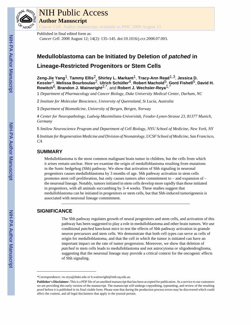
Medulloblastoma can be Initiated by Deletion of patched in Lineage-Restricted Progenitors or Stem Cells Zeng-Jie Yang 1 , Tammy Ellis 2 , Shirley L. Markant 1 , Tracy-Ann Read 1,3 , Jessica D. Kessler 1 , Melissa Bourboulas 2 , Ulrich Schüller 4 , Robert Machold 5 , Gord Fishell 5 , David H. Rowitch 6 , Brandon J. Wainwright 2,* , and Robert J. Wechsler-Reya 1,* 1 Department of Pharmacology and Cancer Biology, Duke University Medical Center, Durham, NC 2 Institute for Molecular Bioscience, University of Queensland, St Lucia, Australia 3 Department of Biomedicine, University of Bergen, Bergen, Norway 4 Center for Neuropathology, Ludwig-Maximilians-Universität, Feodor-Lynen-Strasse 23, 81377 Munich, Germany 5 Smilow Neuroscience Program and Department of Cell Biology, NYU School of Medicine, New York, NY 6 Institute for Regeneration Medicine and Division of Neonatology, UCSF School of Medicine, San Francisco, CA SUMMARY Medulloblastoma is the most common malignant brain tumor in children, but the cells from which it arises remain unclear. Here we examine the origin of medulloblastoma resulting from mutations in the Sonic hedgehog (Shh) pathway. We show that activation of Shh signaling in neuronal progenitors causes medulloblastoma by 3 months of age. Shh pathway activation in stem cells promotes stem cell proliferation, but only causes tumors after commitment to – and expansion of – the neuronal lineage. Notably, tumors initiated in stem cells develop more rapidly than those initiated in progenitors, with all animals succumbing by 3–4 weeks. These studies suggest that medulloblastoma can be initiated in progenitors or stem cells, but that Shh-induced tumorigenesis is associated with neuronal lineage commitment. SIGNIFICANCE The Shh pathway regulates growth of neural progenitors and stem cells, and activation of this pathway has been suggested to play a role in medulloblastoma and other brain tumors. We use conditional patched knockout mice to test the effects of Shh pathway activation in granule neuron precursors and stem cells. We demonstrate that both cell types can serve as cells of origin for medulloblastoma, and that the cell in which the tumor is initiated can have an important impact on the rate of tumor progression. Moreover, we show that deletion of patched in stem cells leads to medulloblastoma and not astrocytoma or oligodendroglioma, suggesting that the neuronal lineage may provide a critical context for the oncogenic effects of Shh signaling. *Correspondence: [email protected] or [email protected]. Publisher's Disclaimer: This is a PDF file of an unedited manuscript that has been accepted for publication. As a service to our customers we are providing this early version of the manuscript. The manuscript will undergo copyediting, typesetting, and review of the resulting proof before it is published in its final citable form. Please note that during the production process errors may be discovered which could affect the content, and all legal disclaimers that apply to the journal pertain. NIH Public Access Author Manuscript Cancer Cell. Author manuscript; available in PMC 2009 August 12. Published in final edited form as: Cancer Cell. 2008 August 12; 14(2): 135–145. doi:10.1016/j.ccr.2008.07.003. NIH-PA Author Manuscript NIH-PA Author Manuscript NIH-PA Author Manuscript
-
Upload
independent -
Category
Documents
-
view
4 -
download
0
Transcript of Medulloblastoma Can Be Initiated by Deletion of Patched in Lineage-Restricted Progenitors or Stem...
Medulloblastoma can be Initiated by Deletion of patched inLineage-Restricted Progenitors or Stem Cells
Zeng-Jie Yang1, Tammy Ellis2, Shirley L. Markant1, Tracy-Ann Read1,3, Jessica D.Kessler1, Melissa Bourboulas2, Ulrich Schüller4, Robert Machold5, Gord Fishell5, David H.Rowitch6, Brandon J. Wainwright2,*, and Robert J. Wechsler-Reya1,*
1 Department of Pharmacology and Cancer Biology, Duke University Medical Center, Durham, NC
2 Institute for Molecular Bioscience, University of Queensland, St Lucia, Australia
3 Department of Biomedicine, University of Bergen, Bergen, Norway
4 Center for Neuropathology, Ludwig-Maximilians-Universität, Feodor-Lynen-Strasse 23, 81377 Munich,Germany
5 Smilow Neuroscience Program and Department of Cell Biology, NYU School of Medicine, New York, NY
6 Institute for Regeneration Medicine and Division of Neonatology, UCSF School of Medicine, San Francisco,CA
SUMMARYMedulloblastoma is the most common malignant brain tumor in children, but the cells from whichit arises remain unclear. Here we examine the origin of medulloblastoma resulting from mutationsin the Sonic hedgehog (Shh) pathway. We show that activation of Shh signaling in neuronalprogenitors causes medulloblastoma by 3 months of age. Shh pathway activation in stem cellspromotes stem cell proliferation, but only causes tumors after commitment to – and expansion of –the neuronal lineage. Notably, tumors initiated in stem cells develop more rapidly than those initiatedin progenitors, with all animals succumbing by 3–4 weeks. These studies suggest thatmedulloblastoma can be initiated in progenitors or stem cells, but that Shh-induced tumorigenesis isassociated with neuronal lineage commitment.
SIGNIFICANCEThe Shh pathway regulates growth of neural progenitors and stem cells, and activation of thispathway has been suggested to play a role in medulloblastoma and other brain tumors. We useconditional patched knockout mice to test the effects of Shh pathway activation in granuleneuron precursors and stem cells. We demonstrate that both cell types can serve as cells oforigin for medulloblastoma, and that the cell in which the tumor is initiated can have animportant impact on the rate of tumor progression. Moreover, we show that deletion ofpatched in stem cells leads to medulloblastoma and not astrocytoma or oligodendroglioma,suggesting that the neuronal lineage may provide a critical context for the oncogenic effectsof Shh signaling.
*Correspondence: [email protected] or [email protected]'s Disclaimer: This is a PDF file of an unedited manuscript that has been accepted for publication. As a service to our customerswe are providing this early version of the manuscript. The manuscript will undergo copyediting, typesetting, and review of the resultingproof before it is published in its final citable form. Please note that during the production process errors may be discovered which couldaffect the content, and all legal disclaimers that apply to the journal pertain.
NIH Public AccessAuthor ManuscriptCancer Cell. Author manuscript; available in PMC 2009 August 12.
Published in final edited form as:Cancer Cell. 2008 August 12; 14(2): 135–145. doi:10.1016/j.ccr.2008.07.003.
NIH
-PA Author Manuscript
NIH
-PA Author Manuscript
NIH
-PA Author Manuscript
INTRODUCTIONThe cell of origin for most types of cancer remains unknown. Identifying the normal cell thatgives rise to a tumor is important because it allows studies of the normal cell to be used as asource of insight into the behavior of the tumor. Moreover, it allows for direct comparisonsbetween tumor cells and their normal counterparts (e.g. using genomic or proteomicapproaches), so that key differences and vulnerabilities of tumor cells can be identified. Finally,recent studies suggest that cells resembling the cell of origin may persist in mature tumors, andmay be critical for propagating these tumors in vivo. If so, identifying the cell of origin mayfacilitate development of more effective approaches to therapy.
Our studies have focused on the cell of origin for medulloblastoma, a highly malignant tumorof the cerebellum. Medulloblastoma occurs most frequently in children between the ages of 5and 10, but may occur in adults as well (Ellison, 2002; Packer et al., 1999). Despite importantprogress in treatment of the disease, the prognosis for many medulloblastoma patients remainsbleak: almost half the patients who develop the disease die from it, and those who survive oftensuffer severe side effects from the treatment, including cognitive deficits, endocrine disordersand increased susceptibility to secondary tumors later in life (Heikens et al., 1998; Mulhern etal., 2005; Packer et al., 1999). Improved approaches for treating medulloblastoma are likelyto come from a deeper understanding of its molecular and cellular origins.
The cell of origin for medulloblastoma has been the subject of debate for many years (Eberhart,2007; Read et al., 2006). The morphology of tumor cells and their location on the surface ofthe cerebellum have led to speculation that the tumors arise from granule neuron precursors(GNPs), restricted progenitors that give rise only to granule neurons. In support of this view,immunohistochemical studies have demonstrated that medulloblastoma cells express markerscommonly associated with GNPs, such as p75NTR, TrkC, Zic1 and Math1 (Buhren et al.,2000; Pomeroy et al., 1997; Salsano et al., 2004; Yokota et al., 1996). On the other hand, avariety of studies have shown that medulloblastomas can express stem cell markers and candifferentiate into both neurons and glia (Hemmati et al., 2003; Singh et al., 2003), raising thepossibility that these tumors may arise from multipotent neural stem cells (NSCs). Morerecently, it has been suggested that some subtypes of medulloblastoma may arise from stemcells and others from GNPs (Gilbertson and Ellison, 2008); however, definitive evidence forthe cell of origin of any particular subtype of medulloblastoma remains elusive.
Although analysis of markers expressed by human medulloblastoma cells can provide clues tothe cell of origin, such studies have significant limitations. In part, this is because the earlystages of tumorigenesis are usually not detectable or accessible in human patients, so inferencesabout the cell of origin must be made retrospectively, based on the phenotype of cells fromfully formed tumors. Since these cells may have changed dramatically during the course oftumorigenesis, expression of a particular marker in a tumor cell may not reflect the expressionof this marker during development. In these respects, mouse models of medulloblastoma offersignificant advantages for studying the origin of the disease.
One of the most powerful and widely studied models of medulloblastoma is the patched(ptc) mutant mouse (Goodrich et al., 1997; Oliver et al., 2005). ptc is an antagonist of the Shhsignaling pathway, which functions as a critical regulator of both stem cells and progenitorsin the CNS (Ahn and Joyner, 2005; Balordi and Fishell, 2007; Wechsler-Reya and Scott,1999). Homozygous ptc knockouts have multiple defects in the neural tube, the heart and othertissues, and die early in embryogenesis (Goodrich et al., 1997). Heterozygotes from this strainsurvive, and approximately 15% of them develop cerebellar tumors that resemble humanmedulloblastoma (Goodrich et al., 1997; Oliver et al., 2005). Since ptc mutations have alsobeen observed in many human medulloblastomas (Hahn et al., 1996; Johnson et al., 1996;
Yang et al. Page 2
Cancer Cell. Author manuscript; available in PMC 2009 August 12.
NIH
-PA Author Manuscript
NIH
-PA Author Manuscript
NIH
-PA Author Manuscript
Raffel et al., 1997), these animals have become an important model for the disease. Studies ofptc mutant mice have provided insight into the early stages of tumorigenesis (Oliver et al.,2005), interactions between ptc and other tumor suppressor genes (Hahn et al., 2000; Wetmoreet al., 2001; Zindy et al., 2007) and the utility of hedgehog pathway inhibitors as therapeuticagents for medulloblastoma (Romer et al., 2004; Sanchez and Ruiz i Altaba, 2005). However,because ptc is mutated in all cells in these animals (including NSCs and GCPs), they cannotbe readily used to study the cell of origin.
To identify the cell of origin for ptc-associated medulloblastoma, we have taken advantage ofa conditional allele of ptc (Adolphe et al., 2006; Ellis et al., 2003) that allows inactivation ofthe gene in either GNPs or NSCs. We show that deletion of ptc in GNPs results in a markedexpansion of the EGL where granule cells develop. Although many ptc-deficient GNPseventually stop dividing and differentiate into neurons, in each animal a cohort of GNPscontinues to proliferate, and by 3 months of age, all mice develop tumors. Deletion of ptc inmultipotent stem cells leads to expansion of the stem cell population, but only stem cells thatcommit to the granule lineage continue to divide and go on to form tumors. The increasedproduction of GNPs (from the expanded stem cell pool) and the continued growth of these cellsduring postnatal development leads to rapid tumor formation, with 100% of animalssuccumbing to medulloblastoma by 3–4 weeks of age. These studies demonstrate that bothprogenitors and stem cells can respond to Shh signaling and can serve as cells of origin formedulloblastoma.
RESULTSMath1-Cre/PtcC/C mice allow deletion of ptc in GNPs
Mice heterozygous for mutations in ptc develop cerebellar tumors that resemble humanmedulloblastoma (Goodrich et al., 1997; Oliver et al., 2005). In these mice, ptc is inactivatedin all cells (including GNPs and NSCs), so definitive conclusions about the cell of origin arenot possible. To determine whether loss of ptc in GNPs can lead to medulloblastoma, we soughtto create GNP-specific ptc knockout mice. To this end, we crossed two strains of mice: Math1-Cre transgenic mice (Schuller et al., 2007), which express Cre recombinase specifically inGNPs, and conditional ptc knockout (PtcC/C) mice (Adolphe et al., 2006; Ellis et al., 2003),which have loxP recombinase recognition sites flanking a portion of the ptc gene.
Math1-Cre mice were generated using a construct containing the Cre coding sequencedownstream of a 1.4 kb Math1 enhancer element that has been used previously to expresstransgenes in GNPs (Machold and Fishell, 2005). To examine expression of Cre protein inthese mice, we stained embryonic and postnatal tissues with anti-Cre antibodies (Supp Fig. 1).Consistent with previous studies (Machold and Fishell, 2005; Wang et al., 2005), we foundthat the Cre transgene was first expressed by rhombic lip-derived cells that are destined to leavethe cerebellum and give rise to neurons of the deep cerebellar nuclei (DCN) and brainstem(Supp. Fig 1A) (Machold and Fishell, 2005). By embryonic day 14.5 (E14.5), Cre wasexpressed by GNPs in the EGL (Supp. Figs. 1B–D). Cre expression persisted in the EGL forthe first 2–3 weeks after birth (Supp. Fig. 1E); by adulthood, when all GNPs had differentiatedinto granule neurons and migrated into the internal granule layer (IGL), Cre expression wasno longer detectable in the cerebellum (Supp. Fig 1F).
To determine whether Cre activity correlated with Cre protein expression, we crossed Math1-Cre mice with ROSA26 reporter (R26R) mice expressing green fluorescent protein (GFP)preceded by a loxP-flanked stop sequence (Mao et al., 2001). As shown in Figure 1A, neonatalMath1-Cre/R26R-GFP mice expressed high levels of GFP in the cerebellum. Analysis ofpostnatal cerebellar sections (Figure 1B) revealed GFP expression only in proliferating GNPsin the outer EGL (see Ki67+ cells in Figure 1C) and in post-mitotic granule cells in the inner
Yang et al. Page 3
Cancer Cell. Author manuscript; available in PMC 2009 August 12.
NIH
-PA Author Manuscript
NIH
-PA Author Manuscript
NIH
-PA Author Manuscript
EGL and IGL (NeuN+ cells in Figure 1D). GFP could also be detected in rhombic lip-derivedcells that had left the cerebellum and settled in the DCN and brain stem (Supp. Fig. 2). No GFPwas seen in other cell types in the cerebellar cortex, including Purkinje neurons, basket andstellate neurons, Bergmann glia and oligodendrocytes (Figure 1E–H). By postnatal day 21(P21), greater than 95% of NeuN positive cells expressed GFP (Supp. Fig 3A), indicating thatthe majority of granule cells had been exposed to Cre during development. Thus, Math1-Cremice allow deletion of loxP-flanked genes in GNPs.
To test the effects of loss of ptc on GNPs, we isolated cells from the cerebellum of neonatalPtcC/C mice, infected them with Cre-encoding retroviruses and analyzed expression of ptc andhedgehog target genes by RT-PCR. As shown in Figure 1I, cells infected with Cre virusesshowed no detectable ptc expression. Since Ptc is a negative regulator of the hedgehog pathway(Goodrich et al., 1996), its loss should result in Shh pathway activation. Consistent with this,Cre retrovirus-mediated deletion of ptc resulted in increased expression of gli1, cyclin D1 andN-myc, all previously identified as Shh target genes in GNPs (Kenney et al., 2003; Oliver etal., 2003) (Figure 1J). Finally, Shh pathway activation has been demonstrated to promoteproliferation of GNPs (Wechsler-Reya and Scott, 1999); Figure 1K shows that cells infectedwith Cre viruses show extensive proliferation compared to cells infected with control viruses.Thus, PtcC/C mice allow deletion of ptc and activation of the Shh pathway in GNPs.
Deletion of ptc in GNPs promotes proliferation but does not prevent differentiationWe next tested the effects of GNP-specific ptc deletion in vivo, by crossing PtcC/C mice withMath1-Cre mice. The efficiency of ptc deletion in these mice was examined by laser-capturingEGL cells from Math1-Cre/PtcC/C cerebella and analyzing their DNA by quantitative PCR;this analysis revealed a >90% reduction in ptc compared to EGL from wild-type (WT) mice(Supp Fig. 3C–D). We then examined the structure of the cerebellum in these animals at variousstages of development. At E15, Math1-Cre/PtcC/C mice were indistinguishable from WTlittermates, and showed no significant changes in cerebellar architecture (data not shown).However, after birth (P1–P8) Math1-Cre/PtcC/C animals exhibited a dramatic thickening ofthe EGL (Figure 2E–F) compared to WT littermates (Figure 2A–B). By P21, all GNPs in WTmice had differentiated and migrated into the IGL, and there was no EGL remaining (Figure2D). At this stage, the cerebellum of Math1-Cre/PtcC/C mice was significantly larger than thatof WT littermates (Figure 2C,G), and still contained a thick EGL (Figure 2H). These dataindicate that deletion of ptc in GNPs leads to severe cerebellar hyperplasia.
Animals with defects in granule cell development often display ataxia or other behavioralabnormalities (Hamre and Goldowitz, 1997; Surmeier et al., 1996). However, at 4–6 weeks ofage most Math1-Cre/PtcC/C mice displayed normal behavior, suggesting that at least somenormal granule neurons were generated in these animals. To determine whether this was thecase, we analyzed the cerebellum of Math1-Cre/PtcC/C mice at 6-weeks. In contrast to thecerebellum of a WT mouse (Figure 3A, D), the Math1-Cre/PtcC/C cerebellum still containeda thick layer of proliferating GNPs on its surface (Figure 3B, E). However, beneath thisproliferative zone the normal organization of the cerebellum was maintained, with a distinctPurkinje cell layer surrounding a layer of mature granule neurons that expressed normaldifferentiation markers (Figure 3E and Supp. Fig. 4). Deletion of ptc in mature granule cellswas confirmed by RT-PCR analysis after laser-capture microdissection (Figure 3G–I). Someregions of the Math1-Cre/PtcC/C cerebellum were more disrupted, showing a nodular structurerather than discrete layers of cells (Figure 3C, F). However, even in these regions there was aremarkable conservation of granule cell organization and differentiation, with each nodulecontaining a core of proliferating GNPs surrounded by a molecular layer, a Purkinje cell layerand a ring of differentiated granule cells that resembled an IGL (Figure 3F and Supp. Fig. 4B).
Yang et al. Page 4
Cancer Cell. Author manuscript; available in PMC 2009 August 12.
NIH
-PA Author Manuscript
NIH
-PA Author Manuscript
NIH
-PA Author Manuscript
These observations suggest that loss of ptc in GNPs results in prolonged proliferation, but thatmany ptc-deficient cells are still able to exit the cell cycle and differentiate into mature neurons.
Deletion of ptc in GNPs results in medulloblastomaAlthough Math1-Cre/PtcC/C mice were asymptomatic in early adulthood, by 8 weeks of agemany animals began to display signs of illness, including domed head, hunched back, abnormalgait or decreased movement. By 10–11 weeks, all Math1-Cre/PtcC/C mice became severely illand had to be sacrificed (Figure 4F). Upon gross examination, the cerebellum from theseanimals appeared enlarged and covered with blood vessels (Figure 4A). Histological analysisrevealed massive tumors that pervaded the cerebellum and obliterated the normal architecturethat was present earlier in development (Figure 4B). The tumors could be propagated bytransplantation into immunodeficient hosts, indicating that they represented fully transformedtumor cells (Figure 4C). These data suggest that deletion of ptc in Math1-expressing cells –while not sufficient to transform every cell – is sufficient to cause tumors in every mouse.
Although Math1 is specific to GNPs in the postnatal cerebellum, during embryonicdevelopment the Math1 enhancer is also expressed in progenitors that end up leaving thecerebellar cortex and migrating to the DCN and brainstem (Machold and Fishell, 2005; Wanget al., 2005) and Supp. Fig. 2). To determine whether the tumors in Math1-Cre/PtcC/C micecould have originated from these progenitors, we crossed PtcC/C mice with Math1-CreERtransgenic mice (Machold and Fishell, 2005), which express a Cre recombinase-estrogenreceptor fusion protein that can be activated by tamoxifen. When Math1-CreER/PtcC/C micewere treated with tamoxifen at E10.5 – a stage when Math1 is only expressed by DCN andbrainstem progenitors -- none of the animals (0/8 mice) developed tumors (Supp. Table 1). Incontrast, when mice were treated with tamoxifen at E14.5, when Math1 is expressed in GNPs,100% of animals (7/7) developed tumors. These data suggest that tumors in Math1-Cre/PtcC/C are derived from GNPs.
GNPs are present in mice until 2–3 weeks after birth. To determine whether loss of ptc couldtrigger medulloblastoma in postnatal GNPs, we treated Math1-CreER/PtcC/C mice withtamoxifen at P4, and then examined mice at several time points for changes in cerebellarstructure. As with embryonic deletion, postnatal deletion of ptc resulted in increased GNPproliferation and expansion of the EGL by P21 (Supp Fig 5A). GNP proliferation persistedinto adulthood, but the majority of ptc-deficient GNPs differentiated and migrated into theIGL, leaving only discrete foci of proliferating cells on the surface of the cerebellum (SuppFig 5B). By 12 weeks of age some Math1-CreER/PtcC/C animals began to exhibit symptomsof intracranial pressure, and by 19 weeks, all of these mice developed cerebellar tumors (Figure4D–F). Tumors were also observed in mice treated with tamoxifen at P8 and P10, but not inanimals treated at P12 or later (Supp. Table 1). These data indicate that both embryonic andpostnatal GNPs can serve as cells of origin for medulloblastoma.
GFAP-Cre/PtcC/C mice allow deletion of ptc in neural stem cellsHaving observed that deletion of ptc in GNPs could cause medulloblastoma, we wonderedwhether deletion in NSCs would have the same effect. On one hand, since NSCs give rise toGNPs, loss of ptc in these cells might also be expected to result in medulloblastoma. On theother hand, NSCs can also give rise to other types of neurons and glia, so loss of ptc in thesecells could potentially lead to other tumor types, such as astrocytoma or oligodendroglioma.To delete ptc in NSCs, we used GFAP-Cre mice, in which expression of Cre is controlled bythe human glial fibrillary acidic protein promoter (Zhuo et al., 2001). Whereas the Math1enhancer drives expression in progenitors after they have left the ventricular zone (VZ) andcommitted to the granule cell lineage (or destined for the DCN or brainstem), the GFAPpromoter turns on prior to lineage commitment, in VZ progenitors that give rise to granule
Yang et al. Page 5
Cancer Cell. Author manuscript; available in PMC 2009 August 12.
NIH
-PA Author Manuscript
NIH
-PA Author Manuscript
NIH
-PA Author Manuscript
neurons as well as most other types of neurons and glia in the cerebellar cortex (Casper andMcCarthy, 2006; Zhuo et al., 2001).
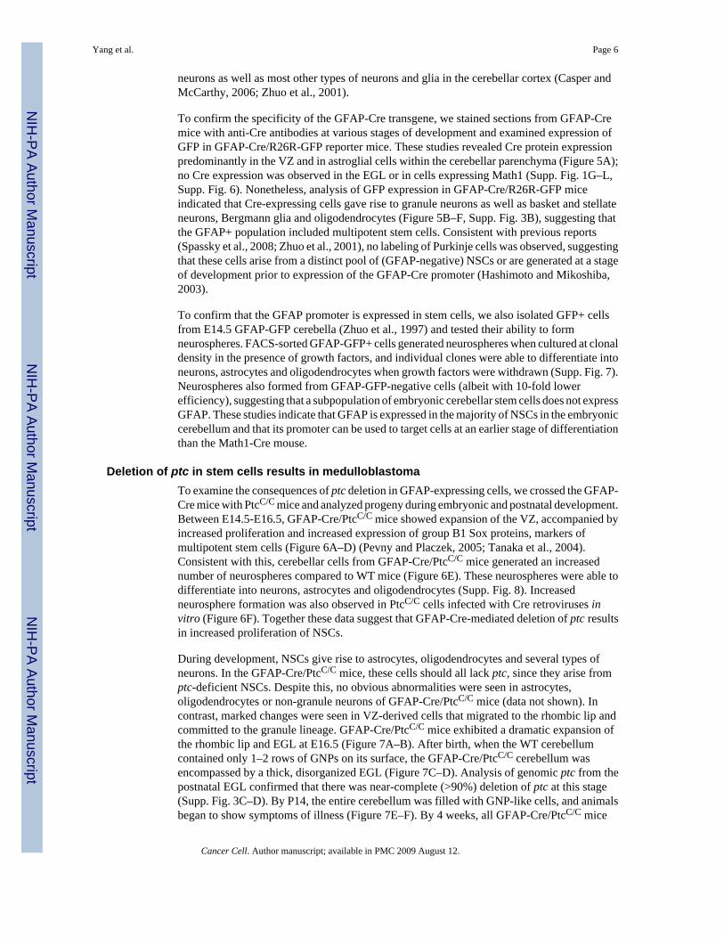
To confirm the specificity of the GFAP-Cre transgene, we stained sections from GFAP-Cremice with anti-Cre antibodies at various stages of development and examined expression ofGFP in GFAP-Cre/R26R-GFP reporter mice. These studies revealed Cre protein expressionpredominantly in the VZ and in astroglial cells within the cerebellar parenchyma (Figure 5A);no Cre expression was observed in the EGL or in cells expressing Math1 (Supp. Fig. 1G–L,Supp. Fig. 6). Nonetheless, analysis of GFP expression in GFAP-Cre/R26R-GFP miceindicated that Cre-expressing cells gave rise to granule neurons as well as basket and stellateneurons, Bergmann glia and oligodendrocytes (Figure 5B–F, Supp. Fig. 3B), suggesting thatthe GFAP+ population included multipotent stem cells. Consistent with previous reports(Spassky et al., 2008; Zhuo et al., 2001), no labeling of Purkinje cells was observed, suggestingthat these cells arise from a distinct pool of (GFAP-negative) NSCs or are generated at a stageof development prior to expression of the GFAP-Cre promoter (Hashimoto and Mikoshiba,2003).
To confirm that the GFAP promoter is expressed in stem cells, we also isolated GFP+ cellsfrom E14.5 GFAP-GFP cerebella (Zhuo et al., 1997) and tested their ability to formneurospheres. FACS-sorted GFAP-GFP+ cells generated neurospheres when cultured at clonaldensity in the presence of growth factors, and individual clones were able to differentiate intoneurons, astrocytes and oligodendrocytes when growth factors were withdrawn (Supp. Fig. 7).Neurospheres also formed from GFAP-GFP-negative cells (albeit with 10-fold lowerefficiency), suggesting that a subpopulation of embryonic cerebellar stem cells does not expressGFAP. These studies indicate that GFAP is expressed in the majority of NSCs in the embryoniccerebellum and that its promoter can be used to target cells at an earlier stage of differentiationthan the Math1-Cre mouse.
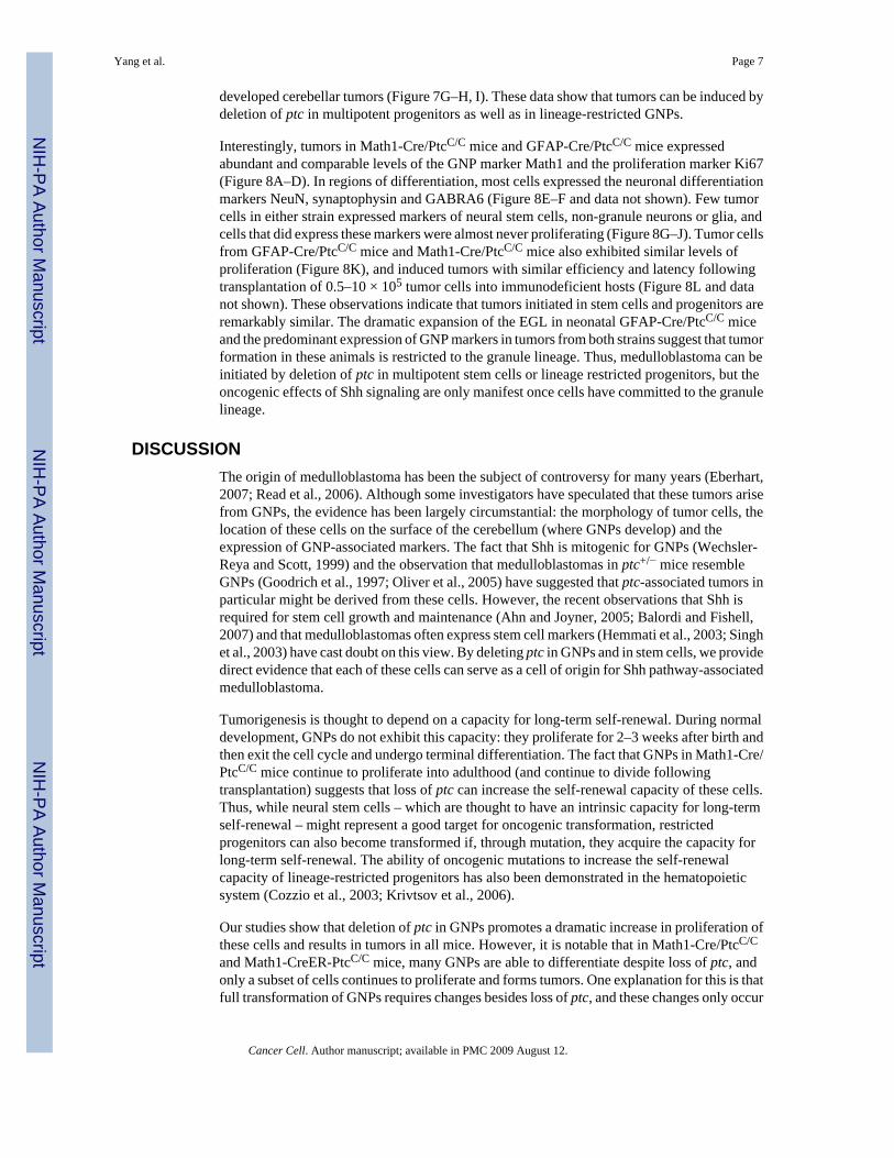
Deletion of ptc in stem cells results in medulloblastomaTo examine the consequences of ptc deletion in GFAP-expressing cells, we crossed the GFAP-Cre mice with PtcC/C mice and analyzed progeny during embryonic and postnatal development.Between E14.5-E16.5, GFAP-Cre/PtcC/C mice showed expansion of the VZ, accompanied byincreased proliferation and increased expression of group B1 Sox proteins, markers ofmultipotent stem cells (Figure 6A–D) (Pevny and Placzek, 2005; Tanaka et al., 2004).Consistent with this, cerebellar cells from GFAP-Cre/PtcC/C mice generated an increasednumber of neurospheres compared to WT mice (Figure 6E). These neurospheres were able todifferentiate into neurons, astrocytes and oligodendrocytes (Supp. Fig. 8). Increasedneurosphere formation was also observed in PtcC/C cells infected with Cre retroviruses invitro (Figure 6F). Together these data suggest that GFAP-Cre-mediated deletion of ptc resultsin increased proliferation of NSCs.
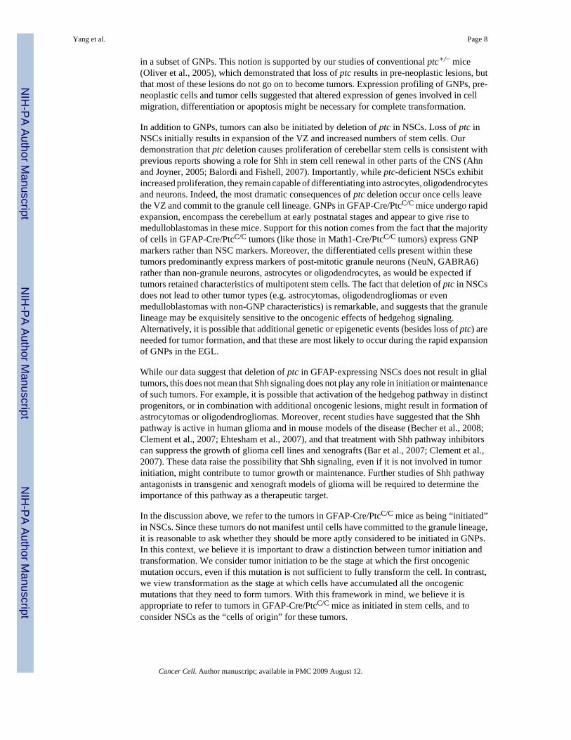
During development, NSCs give rise to astrocytes, oligodendrocytes and several types ofneurons. In the GFAP-Cre/PtcC/C mice, these cells should all lack ptc, since they arise fromptc-deficient NSCs. Despite this, no obvious abnormalities were seen in astrocytes,oligodendrocytes or non-granule neurons of GFAP-Cre/PtcC/C mice (data not shown). Incontrast, marked changes were seen in VZ-derived cells that migrated to the rhombic lip andcommitted to the granule lineage. GFAP-Cre/PtcC/C mice exhibited a dramatic expansion ofthe rhombic lip and EGL at E16.5 (Figure 7A–B). After birth, when the WT cerebellumcontained only 1–2 rows of GNPs on its surface, the GFAP-Cre/PtcC/C cerebellum wasencompassed by a thick, disorganized EGL (Figure 7C–D). Analysis of genomic ptc from thepostnatal EGL confirmed that there was near-complete (>90%) deletion of ptc at this stage(Supp. Fig. 3C–D). By P14, the entire cerebellum was filled with GNP-like cells, and animalsbegan to show symptoms of illness (Figure 7E–F). By 4 weeks, all GFAP-Cre/PtcC/C mice
Yang et al. Page 6
Cancer Cell. Author manuscript; available in PMC 2009 August 12.
NIH
-PA Author Manuscript
NIH
-PA Author Manuscript
NIH
-PA Author Manuscript
developed cerebellar tumors (Figure 7G–H, I). These data show that tumors can be induced bydeletion of ptc in multipotent progenitors as well as in lineage-restricted GNPs.
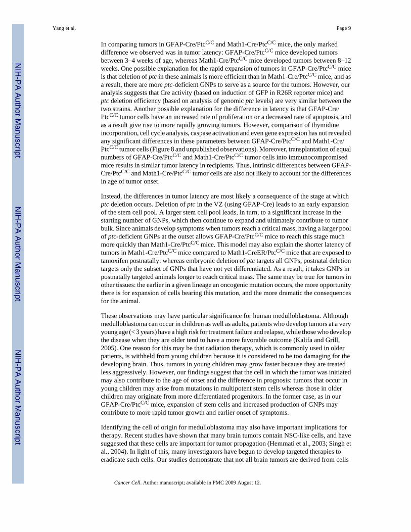
Interestingly, tumors in Math1-Cre/PtcC/C mice and GFAP-Cre/PtcC/C mice expressedabundant and comparable levels of the GNP marker Math1 and the proliferation marker Ki67(Figure 8A–D). In regions of differentiation, most cells expressed the neuronal differentiationmarkers NeuN, synaptophysin and GABRA6 (Figure 8E–F and data not shown). Few tumorcells in either strain expressed markers of neural stem cells, non-granule neurons or glia, andcells that did express these markers were almost never proliferating (Figure 8G–J). Tumor cellsfrom GFAP-Cre/PtcC/C mice and Math1-Cre/PtcC/C mice also exhibited similar levels ofproliferation (Figure 8K), and induced tumors with similar efficiency and latency followingtransplantation of 0.5–10 × 105 tumor cells into immunodeficient hosts (Figure 8L and datanot shown). These observations indicate that tumors initiated in stem cells and progenitors areremarkably similar. The dramatic expansion of the EGL in neonatal GFAP-Cre/PtcC/C miceand the predominant expression of GNP markers in tumors from both strains suggest that tumorformation in these animals is restricted to the granule lineage. Thus, medulloblastoma can beinitiated by deletion of ptc in multipotent stem cells or lineage restricted progenitors, but theoncogenic effects of Shh signaling are only manifest once cells have committed to the granulelineage.
DISCUSSIONThe origin of medulloblastoma has been the subject of controversy for many years (Eberhart,2007; Read et al., 2006). Although some investigators have speculated that these tumors arisefrom GNPs, the evidence has been largely circumstantial: the morphology of tumor cells, thelocation of these cells on the surface of the cerebellum (where GNPs develop) and theexpression of GNP-associated markers. The fact that Shh is mitogenic for GNPs (Wechsler-Reya and Scott, 1999) and the observation that medulloblastomas in ptc+/− mice resembleGNPs (Goodrich et al., 1997; Oliver et al., 2005) have suggested that ptc-associated tumors inparticular might be derived from these cells. However, the recent observations that Shh isrequired for stem cell growth and maintenance (Ahn and Joyner, 2005; Balordi and Fishell,2007) and that medulloblastomas often express stem cell markers (Hemmati et al., 2003; Singhet al., 2003) have cast doubt on this view. By deleting ptc in GNPs and in stem cells, we providedirect evidence that each of these cells can serve as a cell of origin for Shh pathway-associatedmedulloblastoma.
Tumorigenesis is thought to depend on a capacity for long-term self-renewal. During normaldevelopment, GNPs do not exhibit this capacity: they proliferate for 2–3 weeks after birth andthen exit the cell cycle and undergo terminal differentiation. The fact that GNPs in Math1-Cre/PtcC/C mice continue to proliferate into adulthood (and continue to divide followingtransplantation) suggests that loss of ptc can increase the self-renewal capacity of these cells.Thus, while neural stem cells – which are thought to have an intrinsic capacity for long-termself-renewal – might represent a good target for oncogenic transformation, restrictedprogenitors can also become transformed if, through mutation, they acquire the capacity forlong-term self-renewal. The ability of oncogenic mutations to increase the self-renewalcapacity of lineage-restricted progenitors has also been demonstrated in the hematopoieticsystem (Cozzio et al., 2003; Krivtsov et al., 2006).
Our studies show that deletion of ptc in GNPs promotes a dramatic increase in proliferation ofthese cells and results in tumors in all mice. However, it is notable that in Math1-Cre/PtcC/C
and Math1-CreER-PtcC/C mice, many GNPs are able to differentiate despite loss of ptc, andonly a subset of cells continues to proliferate and forms tumors. One explanation for this is thatfull transformation of GNPs requires changes besides loss of ptc, and these changes only occur
Yang et al. Page 7
Cancer Cell. Author manuscript; available in PMC 2009 August 12.
NIH
-PA Author Manuscript
NIH
-PA Author Manuscript
NIH
-PA Author Manuscript
in a subset of GNPs. This notion is supported by our studies of conventional ptc+/− mice(Oliver et al., 2005), which demonstrated that loss of ptc results in pre-neoplastic lesions, butthat most of these lesions do not go on to become tumors. Expression profiling of GNPs, pre-neoplastic cells and tumor cells suggested that altered expression of genes involved in cellmigration, differentiation or apoptosis might be necessary for complete transformation.
In addition to GNPs, tumors can also be initiated by deletion of ptc in NSCs. Loss of ptc inNSCs initially results in expansion of the VZ and increased numbers of stem cells. Ourdemonstration that ptc deletion causes proliferation of cerebellar stem cells is consistent withprevious reports showing a role for Shh in stem cell renewal in other parts of the CNS (Ahnand Joyner, 2005; Balordi and Fishell, 2007). Importantly, while ptc-deficient NSCs exhibitincreased proliferation, they remain capable of differentiating into astrocytes, oligodendrocytesand neurons. Indeed, the most dramatic consequences of ptc deletion occur once cells leavethe VZ and commit to the granule cell lineage. GNPs in GFAP-Cre/PtcC/C mice undergo rapidexpansion, encompass the cerebellum at early postnatal stages and appear to give rise tomedulloblastomas in these mice. Support for this notion comes from the fact that the majorityof cells in GFAP-Cre/PtcC/C tumors (like those in Math1-Cre/PtcC/C tumors) express GNPmarkers rather than NSC markers. Moreover, the differentiated cells present within thesetumors predominantly express markers of post-mitotic granule neurons (NeuN, GABRA6)rather than non-granule neurons, astrocytes or oligodendrocytes, as would be expected iftumors retained characteristics of multipotent stem cells. The fact that deletion of ptc in NSCsdoes not lead to other tumor types (e.g. astrocytomas, oligodendrogliomas or evenmedulloblastomas with non-GNP characteristics) is remarkable, and suggests that the granulelineage may be exquisitely sensitive to the oncogenic effects of hedgehog signaling.Alternatively, it is possible that additional genetic or epigenetic events (besides loss of ptc) areneeded for tumor formation, and that these are most likely to occur during the rapid expansionof GNPs in the EGL.
While our data suggest that deletion of ptc in GFAP-expressing NSCs does not result in glialtumors, this does not mean that Shh signaling does not play any role in initiation or maintenanceof such tumors. For example, it is possible that activation of the hedgehog pathway in distinctprogenitors, or in combination with additional oncogenic lesions, might result in formation ofastrocytomas or oligodendrogliomas. Moreover, recent studies have suggested that the Shhpathway is active in human glioma and in mouse models of the disease (Becher et al., 2008;Clement et al., 2007; Ehtesham et al., 2007), and that treatment with Shh pathway inhibitorscan suppress the growth of glioma cell lines and xenografts (Bar et al., 2007; Clement et al.,2007). These data raise the possibility that Shh signaling, even if it is not involved in tumorinitiation, might contribute to tumor growth or maintenance. Further studies of Shh pathwayantagonists in transgenic and xenograft models of glioma will be required to determine theimportance of this pathway as a therapeutic target.
In the discussion above, we refer to the tumors in GFAP-Cre/PtcC/C mice as being “initiated”in NSCs. Since these tumors do not manifest until cells have committed to the granule lineage,it is reasonable to ask whether they should be more aptly considered to be initiated in GNPs.In this context, we believe it is important to draw a distinction between tumor initiation andtransformation. We consider tumor initiation to be the stage at which the first oncogenicmutation occurs, even if this mutation is not sufficient to fully transform the cell. In contrast,we view transformation as the stage at which cells have accumulated all the oncogenicmutations that they need to form tumors. With this framework in mind, we believe it isappropriate to refer to tumors in GFAP-Cre/PtcC/C mice as initiated in stem cells, and toconsider NSCs as the “cells of origin” for these tumors.
Yang et al. Page 8
Cancer Cell. Author manuscript; available in PMC 2009 August 12.
NIH
-PA Author Manuscript
NIH
-PA Author Manuscript
NIH
-PA Author Manuscript
In comparing tumors in GFAP-Cre/PtcC/C and Math1-Cre/PtcC/C mice, the only markeddifference we observed was in tumor latency: GFAP-Cre/PtcC/C mice developed tumorsbetween 3–4 weeks of age, whereas Math1-Cre/PtcC/C mice developed tumors between 8–12weeks. One possible explanation for the rapid expansion of tumors in GFAP-Cre/PtcC/C miceis that deletion of ptc in these animals is more efficient than in Math1-Cre/PtcC/C mice, and asa result, there are more ptc-deficient GNPs to serve as a source for the tumors. However, ouranalysis suggests that Cre activity (based on induction of GFP in R26R reporter mice) andptc deletion efficiency (based on analysis of genomic ptc levels) are very similar between thetwo strains. Another possible explanation for the difference in latency is that GFAP-Cre/PtcC/C tumor cells have an increased rate of proliferation or a decreased rate of apoptosis, andas a result give rise to more rapidly growing tumors. However, comparison of thymidineincorporation, cell cycle analysis, caspase activation and even gene expression has not revealedany significant differences in these parameters between GFAP-Cre/PtcC/C and Math1-Cre/PtcC/C tumor cells (Figure 8 and unpublished observations). Moreover, transplantation of equalnumbers of GFAP-Cre/PtcC/C and Math1-Cre/PtcC/C tumor cells into immunocompromisedmice results in similar tumor latency in recipients. Thus, intrinsic differences between GFAP-Cre/PtcC/C and Math1-Cre/PtcC/C tumor cells are also not likely to account for the differencesin age of tumor onset.
Instead, the differences in tumor latency are most likely a consequence of the stage at whichptc deletion occurs. Deletion of ptc in the VZ (using GFAP-Cre) leads to an early expansionof the stem cell pool. A larger stem cell pool leads, in turn, to a significant increase in thestarting number of GNPs, which then continue to expand and ultimately contribute to tumorbulk. Since animals develop symptoms when tumors reach a critical mass, having a larger poolof ptc-deficient GNPs at the outset allows GFAP-Cre/PtcC/C mice to reach this stage muchmore quickly than Math1-Cre/PtcC/C mice. This model may also explain the shorter latency oftumors in Math1-Cre/PtcC/C mice compared to Math1-CreER/PtcC/C mice that are exposed totamoxifen postnatally: whereas embryonic deletion of ptc targets all GNPs, postnatal deletiontargets only the subset of GNPs that have not yet differentiated. As a result, it takes GNPs inpostnatally targeted animals longer to reach critical mass. The same may be true for tumors inother tissues: the earlier in a given lineage an oncogenic mutation occurs, the more opportunitythere is for expansion of cells bearing this mutation, and the more dramatic the consequencesfor the animal.
These observations may have particular significance for human medulloblastoma. Althoughmedulloblastoma can occur in children as well as adults, patients who develop tumors at a veryyoung age (< 3 years) have a high risk for treatment failure and relapse, while those who developthe disease when they are older tend to have a more favorable outcome (Kalifa and Grill,2005). One reason for this may be that radiation therapy, which is commonly used in olderpatients, is withheld from young children because it is considered to be too damaging for thedeveloping brain. Thus, tumors in young children may grow faster because they are treatedless aggressively. However, our findings suggest that the cell in which the tumor was initiatedmay also contribute to the age of onset and the difference in prognosis: tumors that occur inyoung children may arise from mutations in multipotent stem cells whereas those in olderchildren may originate from more differentiated progenitors. In the former case, as in ourGFAP-Cre/PtcC/C mice, expansion of stem cells and increased production of GNPs maycontribute to more rapid tumor growth and earlier onset of symptoms.
Identifying the cell of origin for medulloblastoma may also have important implications fortherapy. Recent studies have shown that many brain tumors contain NSC-like cells, and havesuggested that these cells are important for tumor propagation (Hemmati et al., 2003; Singh etal., 2004). In light of this, many investigators have begun to develop targeted therapies toeradicate such cells. Our studies demonstrate that not all brain tumors are derived from cells
Yang et al. Page 9
Cancer Cell. Author manuscript; available in PMC 2009 August 12.
NIH
-PA Author Manuscript
NIH
-PA Author Manuscript
NIH
-PA Author Manuscript
with a stem cell phenotype. In particular, we show that Shh pathway-associatedmedulloblastomas may be initiated in either progenitors or stem cells, and ultimately adopt aphenotype that resembles GNPs rather than NSCs. Whether the same is true for other subtypesof medulloblastoma remains to be investigated. Determining whether a tumor is derived fromlineage-restricted progenitors or stem cells, finding markers specific for these cells andidentifying signals that control their growth and differentiation may be important prerequisitesfor effective therapy.
EXPERIMENTAL PROCEDURESMice
PtcC/C mice and Math1-Cre-ER mice have been described previously (Ellis et al., 2003;Machold and Fishell, 2005). GFAP-GFP (Zhuo et al., 1997), GFAP-Cre mice (Zhuo et al.,2001) and R26R-GFP reporter mice (Mao et al., 2001) were from Jackson Labs (Bar Harbor,ME). SCID-beige mice were from Charles River (Wilmington, MA). Animals were maintainedin the Cancer Center Isolation Facility at Duke University, and all experiments were performedin accordance with procedures approved by the Duke University Animal Care and UseCommittee.
Histology and immunohistochemistryFor histological analysis, animals were perfused with PBS followed by 4% paraformaldehyde(PFA). Cerebella were removed, fixed in 4% PFA overnight, transferred to 70% ethanol andparaffin-embedded. 5-μM sections were stained with hematoxylin and eosin (H&E, Sigma, St.Louis, MO) or with anti-BrdU or anti-Group B1 Sox antibodies (Tanaka et al., 2004).
For immunohistochemistry, cerebella from PFA-perfused animals were fixed overnight in 4%PFA, cryoprotected in 30% sucrose, frozen in Tissue Tek-OCT (Sakura Finetek, Torrance,CA) and cut into 10–12um saggital sections. Sections were blocked and permeabilized for 2hours with PBS containing 0.1% Triton X-100 and 1% normal goat serum, stained with primaryantibodies overnight at 4°C, and incubated with secondary antibodies for 2h at roomtemperature. Sections were counterstained with DAPI and mounted with Fluoromount G(Southern Biotechnology, Birmingham, AL) before being visualized using a Nikon TE-200microscope. Antibodies used for immunostaining are listed in Supplementary Methods.
Cell isolation and Proliferation assaysCerebella from P6 mice were digested with 10U/ml papain (Worthington, Lakewood, NJ),200ug/ml L-cysteine and 250 U/ml DNase (Sigma), triturated to obtain a single cell suspensionand then centrifuged through a 35%–65% Percoll gradient (Sigma). Cells from the 35–65%interface were suspended in NB-B27 (Neurobasal with 1mM sodium pyruvate, 2mM L-glutamine, Pen/Strep and B27 supplement, all from Invitrogen) and transferred to Poly-D-lysine-coated 96-well plates at a density of 2 × 10⁵ cells per well. After 48h, cells were pulsedwith methyl-3H thymidine (Amersham/GE Healthcare, Piscataway, NJ) and cultured for anadditional 16–18 h. Cells were harvested onto filters using a Mach IIIM Manual Harvester 96(Tomtec, Hamden, CT) and incorporated radioactivity was quantified by liquid scintillationspectrophotometry on a Wallac MicroBeta scintillation counter (Perkin Elmer, Wellesley,MA).
Laser capture microdissection and real-time RT-PCRCerebella were removed immediately and frozen in OCT. 5-μM sagittal sections were mountedonto glass slides, fixed in 75% ethanol for 30 sec, and stained with Cresyl Violet (Ambion,Austin, TX). After 5 min of air-drying, microdissection was carried out using a PixCell IIe
Yang et al. Page 10
Cancer Cell. Author manuscript; available in PMC 2009 August 12.
NIH
-PA Author Manuscript
NIH
-PA Author Manuscript
NIH
-PA Author Manuscript
LCM system (Arcturus Bioscience, Sunnyvale, CA). RNA was extracted from LCM caps usingan RNA isolation kit (Arcturus). For real time RT-PCR, first strand cDNA was synthesizedfrom equal amounts of RNA (0.1–1 μg) using Superscript III Reverse Transcriptase(Invitrogen). Triplicate reactions were prepared using a 25-μl mixture containing iQ SYBRGreen Supermix (Biorad, Hercules, CA). Real-time quantification was performed on a BIO-RAD iCycler iQ system (BioRad). Serial 10-fold dilutions of cDNA were used as referencesfor the standard curve. Raw data were normalized based on expression of actin. Primers usedfor gli1, Nmyc, cyclin D1 and ptc are listed in Supplementary Methods.
Intracranial transplantationSCID-beige mice (6–8 weeks old) were anesthetized with a xylazine (25 mg/kg, LloydLaboratories, Shenandoah, IA) and ketamine (125 mg/kg, Fort Dodge Animal Health, FortDodge, IA), and placed in a stereotaxic apparatus (Kopf, Tujunga, CA). After exposing theskull with a scalpel, a 1 mm diameter hole was drilled in the skull over the cerebellum usingan 18G needle. A cell suspension (1 × 10⁶ cells in 5-μl NB-B27) was slowly injected into thecerebellum at a depth of 1mm, using a 5-μl Hamilton Syringe with an unbevelled 24G needle(Hamilton, Reno, NV). After injection, the incision was sutured using catgut sutures (Johnson& Johnson, Piscataway, NJ).
Tamoxifen treatmentTamoxifen (T-5648, Sigma) was prepared as a 20 mg/ml stock solution in corn oil (Sigma).Tamoxifen was administered by oral gavage using 24-G gavaging needles (Fine Science Tools,Foster City, CA). Tamoxifen doses were: 0.6 mg/30 μl at P4, 1 mg/50 μl at P8, 1.4 mg/70 μlat P10, 1.6 mg/80 μl at P12 and 4.0 mg/200μl for treatment of pregnant females.
Neurosphere culturesTo generate primary neurospheres, cells from E14.5 embryonic cerebella were plated at 2 ×10⁵ cells/ml in NSC proliferation medium (NeuroCult Basal Medium with ProliferationSupplement, Stem Cell Technologies, Seattle, WA) plus 10 ng/ml basic fibroblast growthfactor and 20 ng/ml epidermal growth factor (Peprotech, Rocky Hill, NJ). Neurospheres werecounted or harvested for immunostaining after 7 days in culture. To passage, neurosphereswere mechanically dissociated and re-plated in fresh proliferation medium at 2 × 10³ cells/ml.For differentiation, neurospheres were plated on Poly-D-lysine coated coverslips (BDBiosciences) in NSC differentiation medium (NeuroCult Basal Medium with DifferentiationSupplement). Seven days after plating, cultures were fixed with 4% PFA for immunostaining.
Supplementary MaterialRefer to Web version on PubMed Central for supplementary material.
AcknowledgementsThe authors thank Susan Su, Pate Skene and the NIH Neuroscience Microarray Consortium for help with laser captureand microarray analysis, Simon Lin at Northwestern University for assistance with bioinformatics, Beth Harvat andMike Cook for flow cytometry, and Jack Dutton, Mark Johnson and Zhen Zhao for assistance with animal colonymaintenance. Tammy Ellis is a John Trivett Senior Research Fellow. This work was supported by funds from theNational Health and Medical Research Council of Australia (B.J.W.), the ARC Special Research Centre for Functionaland Applied Genomics (B.J.W.), Queensland Cancer Fund (B.J.W.), the Hope Street Kids Foundation (Z.J.Y.), theKislak-Sussman Fund (R.W.R.), the Children’s Brain Tumor Foundation (R.W.R), the Pediatric Brain TumorFoundation (R.W.R.), the McDonnell Foundation (R.W.R.) and NINDS grant number NS052323-01 (R.W.R.).
Yang et al. Page 11
Cancer Cell. Author manuscript; available in PMC 2009 August 12.
NIH
-PA Author Manuscript
NIH
-PA Author Manuscript
NIH
-PA Author Manuscript
AbbreviationsGNP
granule neuron precursor
NSC neural stem cell
DCN deep cerebellar nuclei
Shh Sonic hedgehog
EGL external germinal layer
IGL internal granule layer
VZ ventricular zone
ReferencesAdolphe C, Hetherington R, Ellis T, Wainwright B. Patched1 functions as a gatekeeper by promoting
cell cycle progression. Cancer research 2006;66:2081–2088. [PubMed: 16489008]Ahn S, Joyner AL. In vivo analysis of quiescent adult neural stem cells responding to Sonic hedgehog.
Nature 2005;437:894–897. [PubMed: 16208373]Balordi F, Fishell G. Hedgehog signaling in the subventricular zone is required for both the maintenance
of stem cells and the migration of newborn neurons. J Neurosci 2007;27:5936–5947. [PubMed:17537964]
Bar EE, Chaudhry A, Lin A, Fan X, Schreck K, Matsui W, Piccirillo S, Vescovi AL, DiMeco F, OliviA, Eberhart CG. Cyclopamine-mediated hedgehog pathway inhibition depletes stem-like cancer cellsin glioblastoma. Stem Cells 2007;25:2524–2533. [PubMed: 17628016]
Becher OJ, Hambardzumyan D, Fomchenko EI, Momota H, Mainwaring L, Bleau AM, Katz AM, EdgarM, Kenney AM, Cordon-Cardo C, et al. Gli activity correlates with tumor grade in platelet-derivedgrowth factor-induced gliomas. Cancer research 2008;68:2241–2249. [PubMed: 18381430]
Buhren J, Christoph AH, Buslei R, Albrecht S, Wiestler OD, Pietsch T. Expression of the neurotrophinreceptor p75NTR in medulloblastomas is correlated with distinct histological and clinical features:evidence for a medulloblastoma subtype derived from the external granule cell layer. Journal ofneuropathology and experimental neurology 2000;59:229–240. [PubMed: 10744061]
Casper KB, McCarthy KD. GFAP-positive progenitor cells produce neurons and oligodendrocytesthroughout the CNS. Mol Cell Neurosci 2006;31:676–684. [PubMed: 16458536]
Clement V, Sanchez P, de Tribolet N, Radovanovic I, Ruiz i Altaba A. HEDGEHOG-GLI1 signalingregulates human glioma growth, cancer stem cell self-renewal, and tumorigenicity. Curr Biol2007;17:165–172. [PubMed: 17196391]
Cozzio A, Passegue E, Ayton PM, Karsunky H, Cleary ML, Weissman IL. Similar MLL-associatedleukemias arising from self-renewing stem cells and short-lived myeloid progenitors. Genes &development 2003;17:3029–3035. [PubMed: 14701873]
Eberhart CG. In search of the medulloblast: neural stem cells and embryonal brain tumors. NeurosurgClin N Am 2007;18:59–69. viii–ix. [PubMed: 17244554]
Ehtesham M, Sarangi A, Valadez JG, Chanthaphaychith S, Becher MW, Abel TW, Thompson RC,Cooper MK. Ligand-dependent activation of the hedgehog pathway in glioma progenitor cells.Oncogene 2007;26:5752–5761. [PubMed: 17353902]
Yang et al. Page 12
Cancer Cell. Author manuscript; available in PMC 2009 August 12.
NIH
-PA Author Manuscript
NIH
-PA Author Manuscript
NIH
-PA Author Manuscript
Ellis T, Smyth I, Riley E, Graham S, Elliot K, Narang M, Kay GF, Wicking C, Wainwright B. Patched1 conditional null allele in mice. Genesis 2003;36:158–161. [PubMed: 12872247]
Ellison D. Classifying the medulloblastoma: insights from morphology and molecular genetics.Neuropathol Appl Neurobiol 2002;28:257–282. [PubMed: 12175339]
Gilbertson RJ, Ellison DW. The origins of medulloblastoma subtypes. Annual review of pathology2008;3:341–365.
Goodrich LV, Johnson RL, Milenkovic L, McMahon JA, Scott MP. Conservation of the hedgehog/patched signaling pathway from flies to mice: induction of a mouse patched gene by Hedgehog.Genes & development 1996;10:301–312. [PubMed: 8595881]
Goodrich LV, Milenkovic L, Higgins KM, Scott MP. Altered neural cell fates and medulloblastoma inmouse patched mutants. Science (New York, NY 1997;277:1109–1113.
Hahn H, Wicking C, Zaphiropoulous PG, Gailani MR, Shanley S, Chidambaram A, Vorechovsky I,Holmberg E, Unden AB, Gillies S, et al. Mutations of the human homolog of Drosophila patched inthe nevoid basal cell carcinoma syndrome. Cell 1996;85:841–851. [PubMed: 8681379]
Hahn H, Wojnowski L, Specht K, Kappler R, Calzada-Wack J, Potter D, Zimmer A, Muller U, SamsonE, Quintanilla-Martinez L. Patched target Igf2 is indispensable for the formation of medulloblastomaand rhabdomyosarcoma. The Journal of biological chemistry 2000;275:28341–28344. [PubMed:10884376]
Hamre KM, Goldowitz D. meander tail acts intrinsic to granule cell precursors to disrupt cerebellardevelopment: analysis of meander tail chimeric mice. Development 1997;124:4201–4212. [PubMed:9334269]
Hashimoto M, Mikoshiba K. Mediolateral compartmentalization of the cerebellum is determined on the“birth date” of Purkinje cells. J Neurosci 2003;23:11342–11351. [PubMed: 14672998]
Heikens J, Michiels EM, Behrendt H, Endert E, Bakker PJ, Fliers E. Long-term neuro-endocrine sequelaeafter treatment for childhood medulloblastoma. Eur J Cancer 1998;34:1592–1597. [PubMed:9893634]
Hemmati HD, Nakano I, Lazareff JA, Masterman-Smith M, Geschwind DH, Bronner-Fraser M,Kornblum HI. Cancerous stem cells can arise from pediatric brain tumors. Proceedings of the NationalAcademy of Sciences of the United States of America 2003;100:15178–15183. [PubMed: 14645703]
Johnson RL, Rothman AL, Xie J, Goodrich LV, Bare JW, Bonifas JM, Quinn AG, Myers RM, Cox DR,Epstein EH Jr, Scott MP. Human homolog of patched, a candidate gene for the basal cell nevussyndrome. Science (New York, NY 1996;272:1668–1671.
Kalifa C, Grill J. The therapy of infantile malignant brain tumors: current status? J Neurooncol2005;75:279–285. [PubMed: 16195802]
Kenney AM, Cole MD, Rowitch DH. Nmyc upregulation by sonic hedgehog signaling promotesproliferation in developing cerebellar granule neuron precursors. Development 2003;130:15–28.[PubMed: 12441288]
Krivtsov AV, Twomey D, Feng Z, Stubbs MC, Wang Y, Faber J, Levine JE, Wang J, Hahn WC, GillilandDG, et al. Transformation from committed progenitor to leukaemia stem cell initiated by MLL-AF9.Nature 2006;442:818–822. [PubMed: 16862118]
Machold R, Fishell G. Math1 is expressed in temporally discrete pools of cerebellar rhombic-lip neuralprogenitors. Neuron 2005;48:17–24. [PubMed: 16202705]
Mao X, Fujiwara Y, Chapdelaine A, Yang H, Orkin SH. Activation of EGFP expression by Cre-mediatedexcision in a new ROSA26 reporter mouse strain. Blood 2001;97:324–326. [PubMed: 11133778]
Mulhern RK, Palmer SL, Merchant TE, Wallace D, Kocak M, Brouwers P, Krull K, Chintagumpala M,Stargatt R, Ashley DM, et al. Neurocognitive consequences of risk-adapted therapy for childhoodmedulloblastoma. J Clin Oncol 2005;23:5511–5519. [PubMed: 16110011]
Oliver TG, Grasfeder LL, Carroll AL, Kaiser C, Gillingham CL, Lin SM, Wickramasinghe R, Scott MP,Wechsler-Reya RJ. Transcriptional profiling of the Sonic hedgehog response: a critical role for N-myc in proliferation of neuronal precursors. Proceedings of the National Academy of Sciences of theUnited States of America 2003;100:7331–7336. [PubMed: 12777630]
Oliver TG, Read TA, Kessler JD, Mehmeti A, Wells JF, Huynh TT, Lin SM, Wechsler-Reya RJ. Lossof patched and disruption of granule cell development in a pre-neoplastic stage of medulloblastoma.Development 2005;132:2425–2439. [PubMed: 15843415]
Yang et al. Page 13
Cancer Cell. Author manuscript; available in PMC 2009 August 12.
NIH
-PA Author Manuscript
NIH
-PA Author Manuscript
NIH
-PA Author Manuscript
Packer RJ, Cogen P, Vezina G, Rorke LB. Medulloblastoma: clinical and biologic aspects. Neuro-oncol1999;1:232–250. [PubMed: 11550316]
Pevny L, Placzek M. SOX genes and neural progenitor identity. Curr Opin Neurobiol 2005;15:7–13.[PubMed: 15721738]
Pomeroy SL, Sutton ME, Goumnerova LC, Segal RA. Neurotrophins in cerebellar granule celldevelopment and medulloblastoma. J Neurooncol 1997;35:347–352. [PubMed: 9440031]
Raffel C, Jenkins RB, Frederick L, Hebrink D, Alderete B, Fults DW, James CD. Sporadicmedulloblastomas contain PTCH mutations. Cancer research 1997;57:842–845. [PubMed: 9041183]
Read TA, Hegedus B, Wechsler-Reya R, Gutmann DH. The neurobiology of neurooncology. Ann Neurol2006;60:3–11. [PubMed: 16802285]
Romer JT, Kimura H, Magdaleno S, Sasai K, Fuller C, Baines H, Connelly M, Stewart CF, Gould S,Rubin LL, Curran T. Suppression of the Shh pathway using a small molecule inhibitor eliminatesmedulloblastoma in Ptc1(+/−)p53(−/−) mice. Cancer cell 2004;6:229–240. [PubMed: 15380514]
Salsano E, Pollo B, Eoli M, Giordana MT, Finocchiaro G. Expression of MATH1, a marker of cerebellargranule cell progenitors, identifies different medulloblastoma sub-types. Neurosci Lett2004;370:180–185. [PubMed: 15488319]
Sanchez P, Ruiz i Altaba A. In vivo inhibition of endogenous brain tumors through systemic interferenceof Hedgehog signaling in mice. Mech Dev 2005;122:223–230. [PubMed: 15652709]
Schuller U, Zhao Q, Godinho SA, Heine VM, Medema RH, Pellman D, Rowitch DH. Forkheadtranscription factor FoxM1 regulates mitotic entry and prevents spindle defects in cerebellar granuleneuron precursors. Molecular and cellular biology 2007;27:8259–8270. [PubMed: 17893320]
Singh SK, Clarke ID, Terasaki M, Bonn VE, Hawkins C, Squire J, Dirks PB. Identification of a cancerstem cell in human brain tumors. Cancer research 2003;63:5821–5828. [PubMed: 14522905]
Singh SK, Hawkins C, Clarke ID, Squire JA, Bayani J, Hide T, Henkelman RM, Cusimano MD, DirksPB. Identification of human brain tumour initiating cells. Nature 2004;432:396–401. [PubMed:15549107]
Spassky N, Han YG, Aguilar A, Strehl L, Besse L, Laclef C, Romaguera Ros M, Garcia-Verdugo JM,Alvarez-Buylla A. Primary cilia are required for cerebellar development and Shh-dependentexpansion of progenitor pool. Developmental biology. 2008
Surmeier DJ, Mermelstein PG, Goldowitz D. The weaver mutation of GIRK2 results in a loss of inwardlyrectifying K+ current in cerebellar granule cells. Proceedings of the National Academy of Sciencesof the United States of America 1996;93:11191–11195. [PubMed: 8855331]
Tanaka S, Kamachi Y, Tanouchi A, Hamada H, Jing N, Kondoh H. Interplay of SOX and POU factorsin regulation of the Nestin gene in neural primordial cells. Molecular and cellular biology2004;24:8834–8846. [PubMed: 15456859]
Wang VY, Rose MF, Zoghbi HY. Math1 expression redefines the rhombic lip derivatives and revealsnovel lineages within the brainstem and cerebellum. Neuron 2005;48:31–43. [PubMed: 16202707]
Wechsler-Reya RJ, Scott MP. Control of neuronal precursor proliferation in the cerebellum by SonicHedgehog. Neuron 1999;22:103–114. [PubMed: 10027293]
Wetmore C, Eberhart DE, Curran T. Loss of p53 but not ARF accelerates medulloblastoma in miceheterozygous for patched. Cancer research 2001;61:513–516. [PubMed: 11212243]
Yokota N, Aruga J, Takai S, Yamada K, Hamazaki M, Iwase T, Sugimura H, Mikoshiba K. Predominantexpression of human zic in cerebellar granule cell lineage and medulloblastoma. Cancer research1996;56:377–383. [PubMed: 8542595]
Zhuo L, Sun B, Zhang CL, Fine A, Chiu SY, Messing A. Live astrocytes visualized by green fluorescentprotein in transgenic mice. Developmental biology 1997;187:36–42. [PubMed: 9224672]
Zhuo L, Theis M, Alvarez-Maya I, Brenner M, Willecke K, Messing A. hGFAP-cre transgenic mice formanipulation of glial and neuronal function in vivo. Genesis 2001;31:85–94. [PubMed: 11668683]
Zindy F, Uziel T, Ayrault O, Calabrese C, Valentine M, Rehg JE, Gilbertson RJ, Sherr CJ, Roussel MF.Genetic Alterations in Mouse Medulloblastomas and Generation of Tumors De novo from PrimaryCerebellar Granule Neuron Precursors. Cancer research 2007;67:2676–2684. [PubMed: 17363588]
Yang et al. Page 14
Cancer Cell. Author manuscript; available in PMC 2009 August 12.
NIH
-PA Author Manuscript
NIH
-PA Author Manuscript
NIH
-PA Author Manuscript
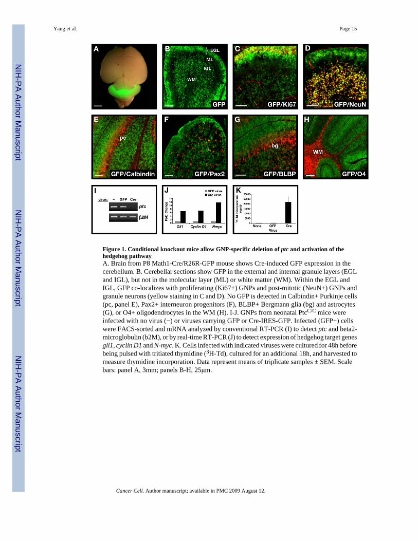
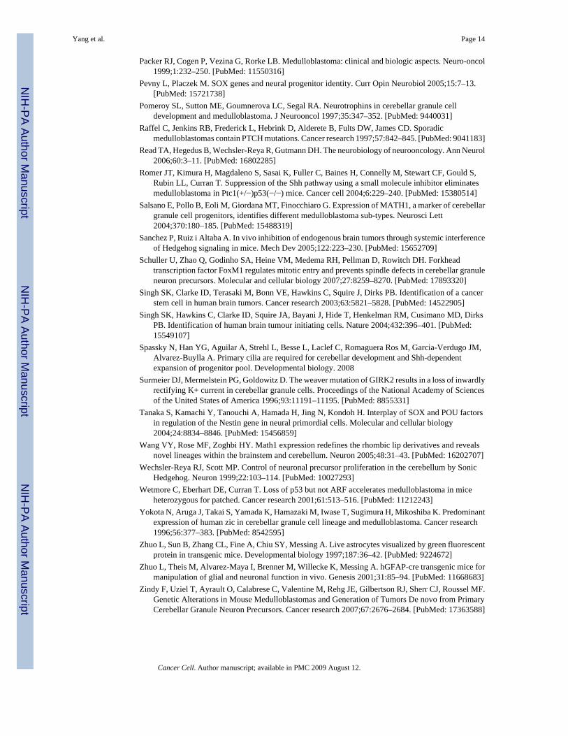
Figure 1. Conditional knockout mice allow GNP-specific deletion of ptc and activation of thehedgehog pathwayA. Brain from P8 Math1-Cre/R26R-GFP mouse shows Cre-induced GFP expression in thecerebellum. B. Cerebellar sections show GFP in the external and internal granule layers (EGLand IGL), but not in the molecular layer (ML) or white matter (WM). Within the EGL andIGL, GFP co-localizes with proliferating (Ki67+) GNPs and post-mitotic (NeuN+) GNPs andgranule neurons (yellow staining in C and D). No GFP is detected in Calbindin+ Purkinje cells(pc, panel E), Pax2+ interneuron progenitors (F), BLBP+ Bergmann glia (bg) and astrocytes(G), or O4+ oligodendrocytes in the WM (H). I-J. GNPs from neonatal PtcC/C mice wereinfected with no virus (−) or viruses carrying GFP or Cre-IRES-GFP. Infected (GFP+) cellswere FACS-sorted and mRNA analyzed by conventional RT-PCR (I) to detect ptc and beta2-microglobulin (b2M), or by real-time RT-PCR (J) to detect expression of hedgehog target genesgli1, cyclin D1 and N-myc. K. Cells infected with indicated viruses were cultured for 48h beforebeing pulsed with tritiated thymidine (3H-Td), cultured for an additional 18h, and harvested tomeasure thymidine incorporation. Data represent means of triplicate samples ± SEM. Scalebars: panel A, 3mm; panels B-H, 25μm.
Yang et al. Page 15
Cancer Cell. Author manuscript; available in PMC 2009 August 12.
NIH
-PA Author Manuscript
NIH
-PA Author Manuscript
NIH
-PA Author Manuscript

Figure 2. Deletion of ptc in GNPs leads to severe hyperplasiaMath1-Cre mice were crossed with PtcC/C mice, and brains of WT or mutant progeny wereharvested at indicated ages. Brains were fixed and photographed intact (C, G) or sectioned andstained with H&E (A, B, D, E, F, H). Note the enlargement of the cerebellum and severehyperplasia on the surface at P8 and P21. Scale bars: 30 μm (A, B, E and F); 2.5mm (C andG); 400 μm (D and H).
Yang et al. Page 16
Cancer Cell. Author manuscript; available in PMC 2009 August 12.
NIH
-PA Author Manuscript
NIH
-PA Author Manuscript
NIH
-PA Author Manuscript
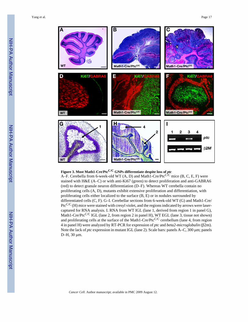
Figure 3. Most Math1-Cre/PtcC/C GNPs differentiate despite loss of ptcA–F. Cerebella from 6-week-old WT (A, D) and Math1-Cre/PtcC/C mice (B, C, E, F) werestained with H&E (A–C) or with anti-Ki67 (green) to detect proliferation and anti-GABRA6(red) to detect granule neuron differentiation (D–F). Whereas WT cerebella contain noproliferating cells (A, D), mutants exhibit extensive proliferation and differentiation, withproliferating cells either localized to the surface (B, E) or in nodules surrounded bydifferentiated cells (C, F). G–I. Cerebellar sections from 6-week-old WT (G) and Math1-Cre/PtcC/C (H) mice were stained with cresyl violet, and the regions indicated by arrows were laser-captured for RNA analysis. I. RNA from WT IGL (lane 1, derived from region 1 in panel G),Math1-Cre/PtcC/C IGL (lane 2, from region 2 in panel H), WT EGL (lane 3, tissue not shown)and proliferating cells at the surface of the Math1-Cre/PtcC/C cerebellum (lane 4, from region4 in panel H) were analyzed by RT-PCR for expression of ptc and beta2-microglobulin (β2m).Note the lack of ptc expression in mutant IGL (lane 2). Scale bars: panels A–C, 300 μm; panelsD–H, 30 μm.
Yang et al. Page 17
Cancer Cell. Author manuscript; available in PMC 2009 August 12.
NIH
-PA Author Manuscript
NIH
-PA Author Manuscript
NIH
-PA Author Manuscript
Figure 4. ptc deletion in GNPs leads to medulloblastomaA–C. By 10 weeks of age, most Math1-Cre/PtcC/C mice develop tumors. A. Cerebellumcontaining tumor. B. H&E-stained section of tumor. C. H&E-stained section of secondarytumor, induced by transplantation of Math1-Cre/PtcC/C tumor cells into cerebellum of adultSCID-beige mouse. D–E. Postnatal deletion of ptc in GNPs results in medulloblastoma. Math1-CreER/PtcC/C mice were treated with tamoxifen at P4 and sacrificed when symptomsdeveloped (in this case at 17 weeks). Cerebella were photographed intact (D) or sectioned andstained with H&E (E). F. Survival curves for Math1-Cre/PtcC/C mice (red) and Math1-CreER/PtcC/C mice treated with tamoxifen at P4 (blue). Scale bars: 3mm (A, B, D and E); 400 μm(C).
Yang et al. Page 18
Cancer Cell. Author manuscript; available in PMC 2009 August 12.
NIH
-PA Author Manuscript
NIH
-PA Author Manuscript
NIH
-PA Author Manuscript
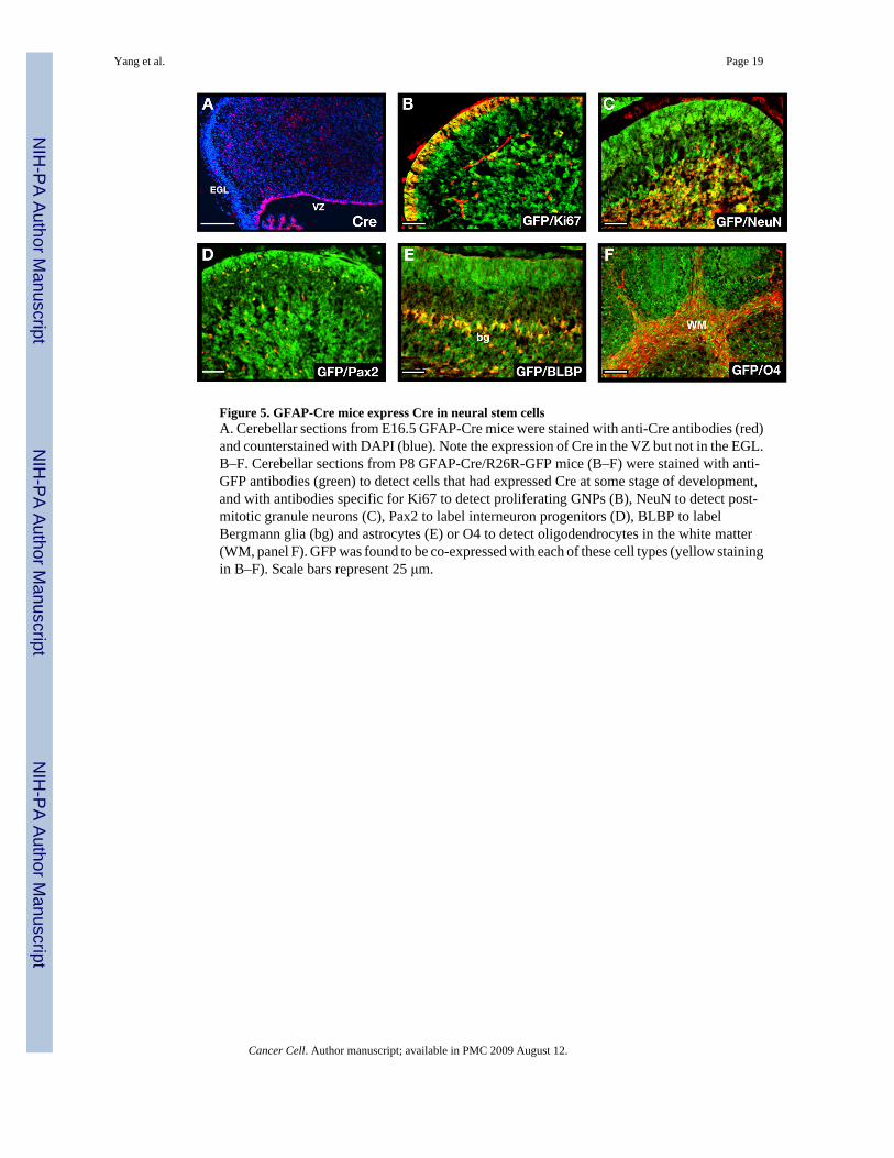
Figure 5. GFAP-Cre mice express Cre in neural stem cellsA. Cerebellar sections from E16.5 GFAP-Cre mice were stained with anti-Cre antibodies (red)and counterstained with DAPI (blue). Note the expression of Cre in the VZ but not in the EGL.B–F. Cerebellar sections from P8 GFAP-Cre/R26R-GFP mice (B–F) were stained with anti-GFP antibodies (green) to detect cells that had expressed Cre at some stage of development,and with antibodies specific for Ki67 to detect proliferating GNPs (B), NeuN to detect post-mitotic granule neurons (C), Pax2 to label interneuron progenitors (D), BLBP to labelBergmann glia (bg) and astrocytes (E) or O4 to detect oligodendrocytes in the white matter(WM, panel F). GFP was found to be co-expressed with each of these cell types (yellow stainingin B–F). Scale bars represent 25 μm.
Yang et al. Page 19
Cancer Cell. Author manuscript; available in PMC 2009 August 12.
NIH
-PA Author Manuscript
NIH
-PA Author Manuscript
NIH
-PA Author Manuscript
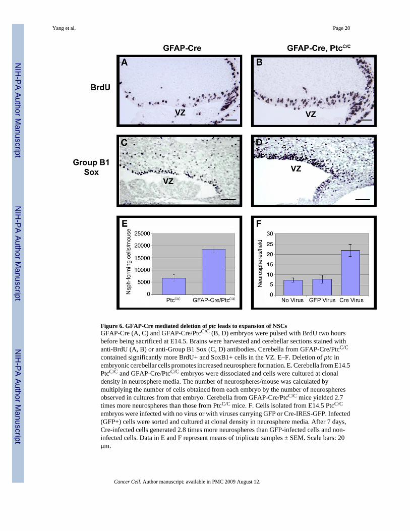
Figure 6. GFAP-Cre mediated deletion of ptc leads to expansion of NSCsGFAP-Cre (A, C) and GFAP-Cre/PtcC/C (B, D) embryos were pulsed with BrdU two hoursbefore being sacrificed at E14.5. Brains were harvested and cerebellar sections stained withanti-BrdU (A, B) or anti-Group B1 Sox (C, D) antibodies. Cerebella from GFAP-Cre/PtcC/C
contained significantly more BrdU+ and SoxB1+ cells in the VZ. E–F. Deletion of ptc inembryonic cerebellar cells promotes increased neurosphere formation. E. Cerebella from E14.5PtcC/C and GFAP-Cre/PtcC/C embryos were dissociated and cells were cultured at clonaldensity in neurosphere media. The number of neurospheres/mouse was calculated bymultiplying the number of cells obtained from each embryo by the number of neurospheresobserved in cultures from that embryo. Cerebella from GFAP-Cre/PtcC/C mice yielded 2.7times more neurospheres than those from PtcC/C mice. F. Cells isolated from E14.5 PtcC/C
embryos were infected with no virus or with viruses carrying GFP or Cre-IRES-GFP. Infected(GFP+) cells were sorted and cultured at clonal density in neurosphere media. After 7 days,Cre-infected cells generated 2.8 times more neurospheres than GFP-infected cells and non-infected cells. Data in E and F represent means of triplicate samples ± SEM. Scale bars: 20μm.
Yang et al. Page 20
Cancer Cell. Author manuscript; available in PMC 2009 August 12.
NIH
-PA Author Manuscript
NIH
-PA Author Manuscript
NIH
-PA Author Manuscript
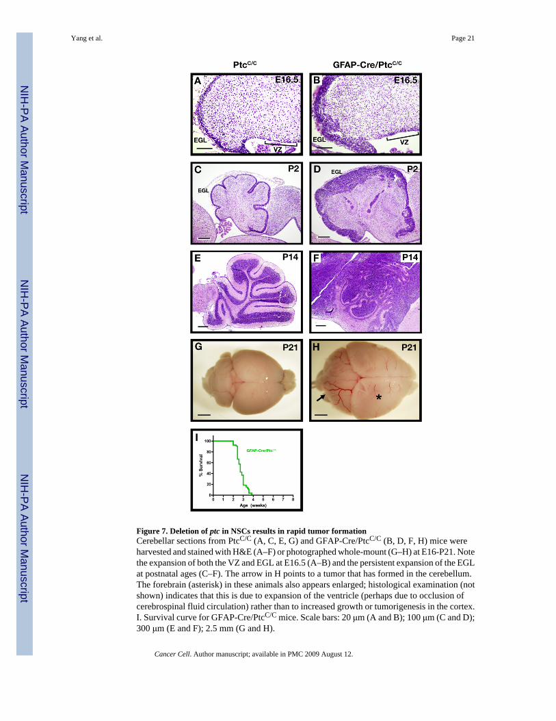
Figure 7. Deletion of ptc in NSCs results in rapid tumor formationCerebellar sections from PtcC/C (A, C, E, G) and GFAP-Cre/PtcC/C (B, D, F, H) mice wereharvested and stained with H&E (A–F) or photographed whole-mount (G–H) at E16-P21. Notethe expansion of both the VZ and EGL at E16.5 (A–B) and the persistent expansion of the EGLat postnatal ages (C–F). The arrow in H points to a tumor that has formed in the cerebellum.The forebrain (asterisk) in these animals also appears enlarged; histological examination (notshown) indicates that this is due to expansion of the ventricle (perhaps due to occlusion ofcerebrospinal fluid circulation) rather than to increased growth or tumorigenesis in the cortex.I. Survival curve for GFAP-Cre/PtcC/C mice. Scale bars: 20 μm (A and B); 100 μm (C and D);300 μm (E and F); 2.5 mm (G and H).
Yang et al. Page 21
Cancer Cell. Author manuscript; available in PMC 2009 August 12.
NIH
-PA Author Manuscript
NIH
-PA Author Manuscript
NIH
-PA Author Manuscript
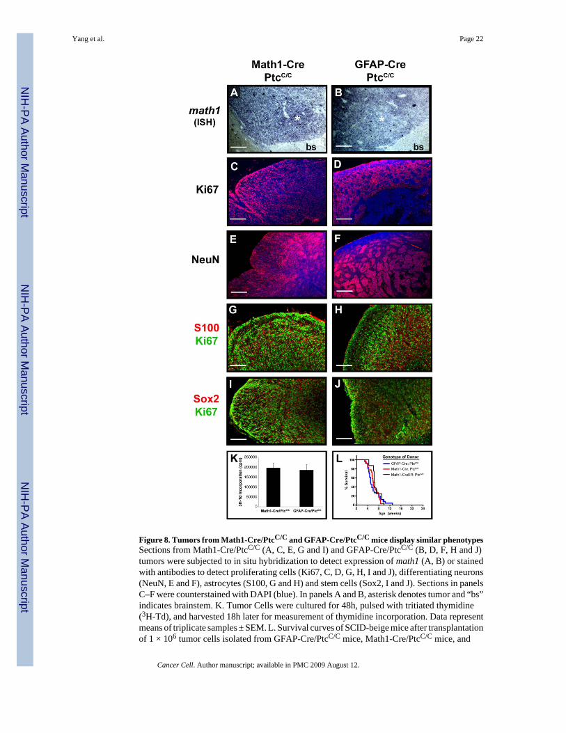
Figure 8. Tumors from Math1-Cre/PtcC/C and GFAP-Cre/PtcC/C mice display similar phenotypesSections from Math1-Cre/PtcC/C (A, C, E, G and I) and GFAP-Cre/PtcC/C (B, D, F, H and J)tumors were subjected to in situ hybridization to detect expression of math1 (A, B) or stainedwith antibodies to detect proliferating cells (Ki67, C, D, G, H, I and J), differentiating neurons(NeuN, E and F), astrocytes (S100, G and H) and stem cells (Sox2, I and J). Sections in panelsC–F were counterstained with DAPI (blue). In panels A and B, asterisk denotes tumor and “bs”indicates brainstem. K. Tumor Cells were cultured for 48h, pulsed with tritiated thymidine(3H-Td), and harvested 18h later for measurement of thymidine incorporation. Data representmeans of triplicate samples ± SEM. L. Survival curves of SCID-beige mice after transplantationof 1 × 10⁶ tumor cells isolated from GFAP-Cre/PtcC/C mice, Math1-Cre/PtcC/C mice, and
Yang et al. Page 22
Cancer Cell. Author manuscript; available in PMC 2009 August 12.
NIH
-PA Author Manuscript
NIH
-PA Author Manuscript
NIH
-PA Author Manuscript
Math1-CreER/PtcC/C mice that had been treated with tamoxifen at P4. Scale bars represent 25μm.
Yang et al. Page 23
Cancer Cell. Author manuscript; available in PMC 2009 August 12.
NIH
-PA Author Manuscript
NIH
-PA Author Manuscript
NIH
-PA Author Manuscript