Transcriptional diversity during lineage commitment of human blood progenitors
Upload
buckinstituteCategory
view
1download
0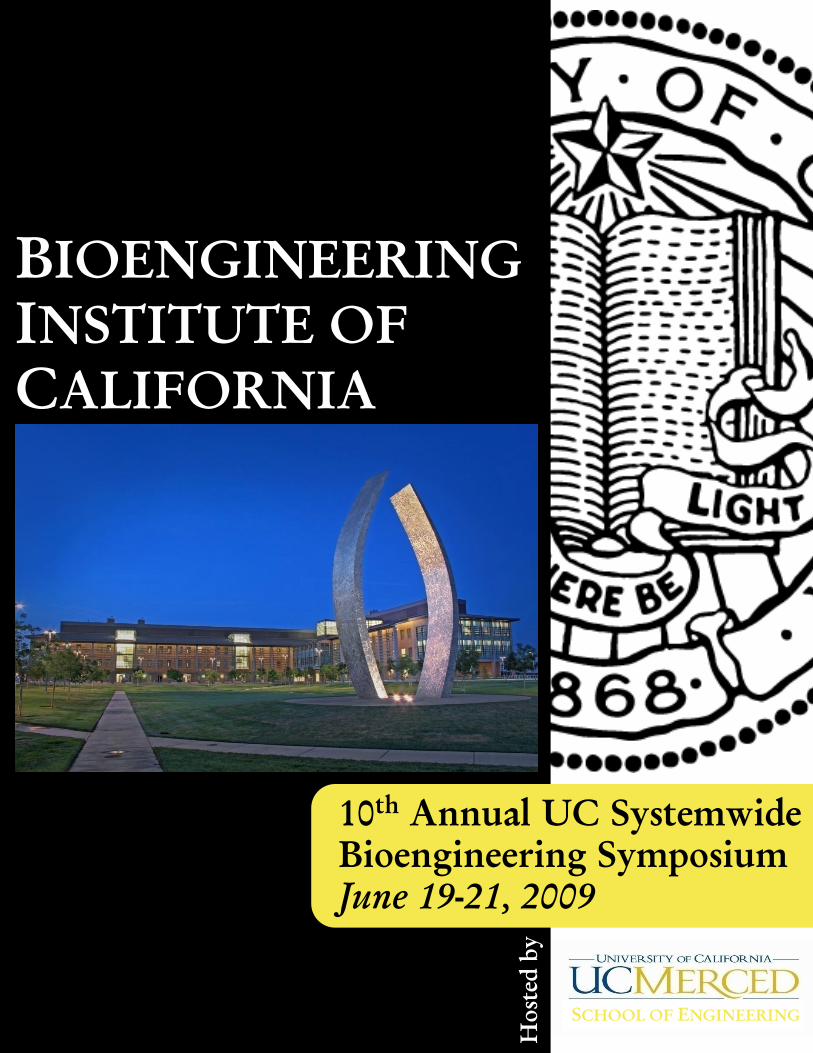
SCHOOL OF ENGINEERING
BIOENGINEERING
INSTITUTE OF
CALIFORNIA
10th
Annual UC Systemwide
Bioengineering Symposium
June 19-21, 2009
Hosted
b
y
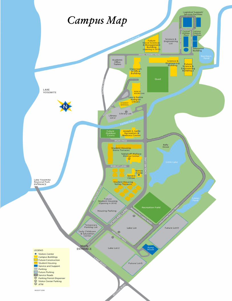
Campus Map

Welcome to
Merced!

In October 2003, the Bioengineering Institute of California
(BIC) was approved as a Multicampus Research Unit. Under
the MRU, all 10 UC campuses will establish a modern
information infrastructure with facilities and staffing for
broadband inter-campus transmission, thus forming a network
for research and teaching system-wide. It will make possible
the sharing of database, broadcasting of teaching materials,
teleoperation of specialized instruments, video conferencing,
and telecommunication. The MRU will provide seed funds
for inter-campus collaboration for high-risk, high payoff
research, establish graduate student fellowships and facilitate
intercampus joint training, set up a Traveling Seminar
Program, and attract the participation of large industrial
companies to facilitate academia-industry collaboration and
technology transfer. The MRU will establish state-of-the-art
research core facilities for shared usage by the participating
campuses. The activities of the MRU will synergize with
other units in the UC system, including the three new
California Institutes for Science and Innovation.
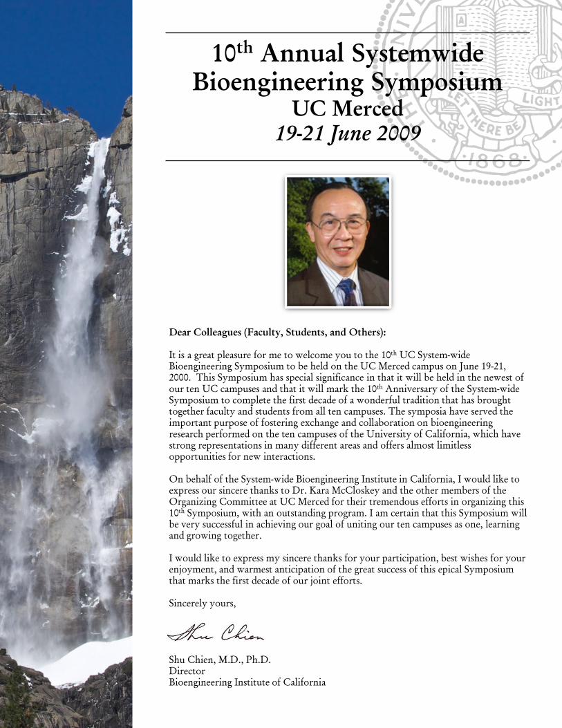
10th
Annual Systemwide
Bioengineering Symposium
UC Merced
19-21 June 2009
Dear Colleagues (Faculty, Students, and Others):
It is a great pleasure for me to welcome you to the 10th
UC System-wide
Bioengineering Symposium to be held on the UC Merced campus on June 19-21,
2000. This Symposium has special significance in that it will be held in the newest of
our ten UC campuses and that it will mark the 10th
Anniversary of the System-wide
Symposium to complete the first decade of a wonderful tradition that has brought
together faculty and students from all ten campuses. The symposia have served the
important purpose of fostering exchange and collaboration on bioengineering
research performed on the ten campuses of the University of California, which have
strong representations in many different areas and offers almost limitless
opportunities for new interactions.
On behalf of the System-wide Bioengineering Institute in California, I would like to
express our sincere thanks to Dr. Kara McCloskey and the other members of the
Organizing Committee at UC Merced for their tremendous efforts in organizing this
10th
Symposium, with an outstanding program. I am certain that this Symposium will
be very successful in achieving our goal of uniting our ten campuses as one, learning
and growing together.
I would like to express my sincere thanks for your participation, best wishes for your
enjoyment, and warmest anticipation of the great success of this epical Symposium
that marks the first decade of our joint efforts.
Sincerely yours,
Shu Chien, M.D., Ph.D.
Director
Bioengineering Institute of California

Partners & Sponsers

Program Organizing & Logistics Chair
Kara McCloskey
Scientific Program Chair
Michelle Khine
Fundraising Director
Ron Durbin
Financial Director
Christina Christensen
Organizing & Logistics Subcommittee
Jonathan Pegan
Maureen Long
Drew Glaser
Booklet Photography
Anthony Grimes
Special Thanks to BIC Staff
Shu Chien, Director
Jennifer Griffin
Rowella Garcia
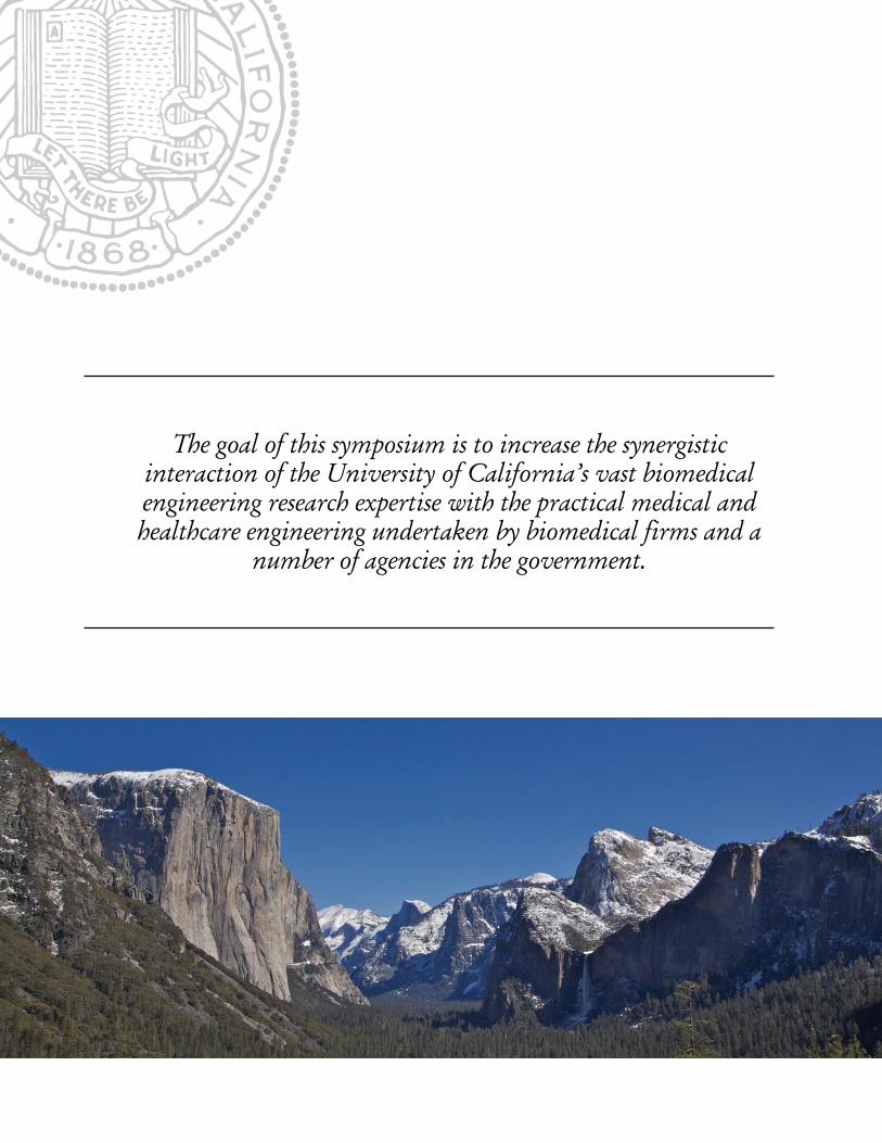
The goal of this symposium is to increase the synergistic
interaction of the University of California’s vast biomedical
engineering research expertise with the practical medical and
healthcare engineering undertaken by biomedical firms and a
number of agencies in the government.
Table of Contents
WELCOME .................................................................... P. 1-2
ACKNOWLEDGEMENTS ............................................ P. 3-4
SYMPOSIUM INFORMATION ................................. P. 7-12
KEYNOTE SPEAKERS ................................................................. P. 7-8
TATRC PARTNER ........................................................................... P. 9
WELCOME MAP .......................................................................... P. 10
PROGRAM SCHEDULE ............................................................ P. 11-12
SCIENTIFIC SESSIONS ........................................... P. 13–33
SATURDAY, JUNE 20TH
.......................................................... P. 13–25
Podium Session I ...................................................................... P. 13–17
Poster Session ............................................................................ P. 30–33
Podium Session II ...................................................................... P. 18-21
Podium Session III .................................................................... P. 22-25
SUNDAY, JUNE 21ST
................................................................ P. 26-29
Podium Session IV .................................................................... P. 26-29
PROGRAM ABSTRACTS ....................................... P. 24-140
ORAL ABSTRACTS .................................................................P. 24-123
POSTER ABSTRACTS ............................................................ P. 124-140
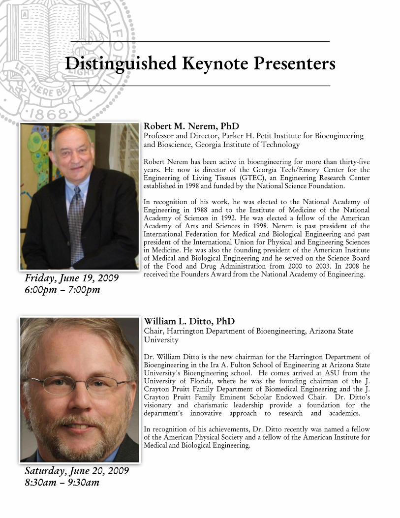
Robert M. Nerem, PhD
Professor and Director, Parker H. Petit Institute for Bioengineering
and Bioscience, Georgia Institute of Technology
Robert Nerem has been active in bioengineering for more than thirty-five
years. He now is director of the Georgia Tech/Emory Center for the
Engineering of Living Tissues (GTEC), an Engineering Research Center
established in 1998 and funded by the National Science Foundation.
In recognition of his work, he was elected to the National Academy of
Engineering in 1988 and to the Institute of Medicine of the National
Academy of Sciences in 1992. He was elected a fellow of the American
Academy of Arts and Sciences in 1998. Nerem is past president of the
International Federation for Medical and Biological Engineering and past
president of the International Union for Physical and Engineering Sciences
in Medicine. He was also the founding president of the American Institute
of Medical and Biological Engineering and he served on the Science Board
of the Food and Drug Administration from 2000 to 2003. In 2008 he
received the Founders Award from the National Academy of Engineering.
William L. Ditto, PhD
Chair, Harrington Department of Bioengineering, Arizona State
University
Dr. William Ditto is the new chairman for the Harrington Department of
Bioengineering in the Ira A. Fulton School of Engineering at Arizona State
University's Bioengineering school. He comes arrived at ASU from the
University of Florida, where he was the founding chairman of the J.
Crayton Pruitt Family Department of Biomedical Engineering and the J.
Crayton Pruitt Family Eminent Scholar Endowed Chair. Dr. Ditto's
visionary and charismatic leadership provide a foundation for the
department's innovative approach to research and academics.
In recognition of his achievements, Dr. Ditto recently was named a fellow
of the American Physical Society and a fellow of the American Institute for
Medical and Biological Engineering.
Distinguished Keynote Presenters
Friday, June 19, 2009
6:00pm – 7:00pm
Saturday, June 20, 2009
8:30am – 9:30am
Distinguished Keynote Presenters
Buddy Ratner, PhD
University of Washington Engineered Biomaterials (UWEB)
Professor of Bioengineering and Chemical Engineering, University
of Washington
Buddy Ratner became director of UWEB in 1996. He is also the Darland
Endowed Chair in Technology Commercialization and professor of
bioengineering and chemical engineering at the University of Washington.
He has launched two companies and three others have resulted from his
research. His research interests include biomaterials, tissue engineering,
polymers, biocompatibility, surface analysis of organic materials, self
assembly, nanobiotechnology, and RF-plasma thin film deposition. He has
written more than 400 scholarly works and holds 17 patents.
Buddy Ratner was elected a member of the National Academy of
Engineering (USA) in 2002 and president of the Tissue Engineering Society
of North America in 2003. He is on the council of the Tissue Engineering
and Regenerative Medicine International Society and is an associate editor
of the Journal of Biomedical Materials Research. He is on the advisory
board of the journal Biointerphases, and is on the editorial boards of 10
other journals.
Jack Lloyd
Founder, Alere Medical and Nellcor Inc.
Jack Lloyd has been a founder, officer, and director of many medical and
high technology companies since 1970, including Nellcor, Aradigm, and
Alere Medical. From 1974 to 1981, Mr. Lloyd was founder and President of
Humphrey Instruments (acquired by Carl Zeiss Meditec). Mr. Lloyd served
as founder, president and CEO of Nellcor from 1981-1990, during which
time he built Nellcor from a start-up, based on his development of the Pulse
Oximeter, to a company with $150 million in annual sales, now owned by
Covidian Medical. He was also Chairman and President of Aradigm, a
developer of aerosol drug delivery systems to deliver insulin; he is a founder
of Alere Medical, and developed a system to monitor patients with
congestive heart failure at home. Alere was sold to Inverness Medical in
2007. Mr. Lloyd holds 29 U.S. Patents.
Jack Lloyd currently serves on the boards of directors of several medical
companies. Mr. Lloyd earned his bachelor's degree in mechanical
engineering from the University of California, Berkeley and now serves on
the Engineering Advisory Board of the UC Berkeley School of Engineering
and is a trustee of the U.C. Berkeley Foundation.
Saturday, June 20, 2009
6:00pm – 7:00pm
Sunday, June 21, 2009
8:30am – 9:30am

TATRC Sponsor
Chuck Peterson, MD
Telemedicine and Advanced Technology Research Center
Saturday, June 20, 2009, 11:30am – 12:00pm
The Telemedicine and Advanced Technology Research Center (TATRC), a subordinate element of
the U.S. Army and Medical Research and Materiel Command, has as its mission to explore science
and engineering technologies ahead of programmed research, leveraging other programs to
maximize benefits to military medicine. Specifically, TATRC is charged with managing core
research, development, test and evaluation (RDT&E) and congressionally mandated projects in
telemedicine and advanced medical technologies. To support its research and development effort,
TATRC maintains a productive mix of partnerships with federal, academic, and commercial
organizations. TATRC also provides short-duration technical support to federal and defense
agencies, and develops, evaluates, and demonstrates new technologies and concepts. In addition,
TATRC conducts market surveillance with a focus on leveraging emerging technologies in
healthcare and ancillary services. Ultimately, TATRC aims to be the government model of
opportunity-driven research agility. TATRC strives to make medical care and services more
accessible to military personnel, and to reduce costs, thereby enhancing the overall quality of
military healthcare.
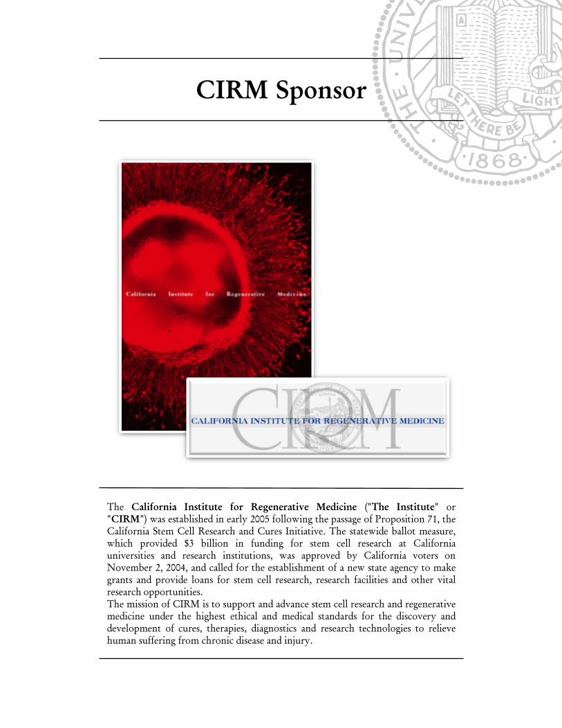
CIRM Sponsor
The California Institute for Regenerative Medicine ("The Institute" or
"CIRM") was established in early 2005 following the passage of Proposition 71, the
California Stem Cell Research and Cures Initiative. The statewide ballot measure,
which provided $3 billion in funding for stem cell research at California
universities and research institutions, was approved by California voters on
November 2, 2004, and called for the establishment of a new state agency to make
grants and provide loans for stem cell research, research facilities and other vital
research opportunities.
The mission of CIRM is to support and advance stem cell research and regenerative
medicine under the highest ethical and medical standards for the discovery and
development of cures, therapies, diagnostics and research technologies to relieve
human suffering from chronic disease and injury.

11 | 10th
Annual UC Systemwide Bioengineering Symposium | Bioengineering Institute of California
Symposium Schedule
Time and Date
Event
Friday, June 19, 2009
4:00pm – 6:00pm Check–In and Registration (Housing)
6:00pm – 9:00pm Keynote: Dr. Robert Nerem followed by Dinner
Keynote: COB 105
Dinner: Cat Quad between Housing and Dining
Saturday, June 20, 2009
7:15 am – 8:30am Bioengineering Institute of California Steering Committee Meeting
COB 263
7:30am – 8:30am Breakfast in COB lobby(2nd
floor)
7:30am – 8:30am Check–In and Registration (COB lobby)
Exhibitor Set-Up (Lantern)
7:30am – 8:30am All Posters Set-Up (Lantern)
8:30am – 9:30am Welcome Address and
Plenary speaker: Dr. William Ditto
COB 102
9:30am – 11:15am Podium Session I (2nd
floor COB)
Track 1 Track 2 Track 3
Bioinformatics &
Biosystems Modeling
Biomedical Imaging I Biomaterials
11:15am – 11:30am Coffee Break and Poster Review
11:30am – 12:00pm Plenary Speaker: Dr. Charles Peterson, TATRC
COB 102
12:00pm – 2:00pm Lunch wih Poster Session - Lantern
2:00pm – 3:45pm Podium Session II (2nd
floor COB)
Track 4 Track 5 Track 6
Computation, in Silico Tissue Engineering Nanotechnology &
BioMEMS
3:45pm – 4:00pm Coffee Break and Poster Review

10th
Annual UC Systemwide Bioengineering Symposium | Bioengineering Institute of California | 12
4:00pm – 5:45pm Podium Session III (2nd
floor COB)
Track 7 Track 8 Track 9
Stem Cells Drug Delivery &
Targeting
Biomechanics
5:45pm – 6:00pm Poster Removal
6:00pm – 7:00 pm Reception with Keynote: Dr. Buddy Ratner
7:00pm – 10:30pm 10th
Annual Gala Celebration Dinner
Sunday, June 21, 2009
7:30am – 8:30am Breakfast in COB lobby (2nd
floor)
8:30am - 9:30am Plenary Speaker: John Lloyd
COB 102
9:30am - 9:45am Coffee Break
9:45am – 11:30am Podium Session IV (2nd
floor COB)
Track 10 Track 11 Track 12
Molecular & Cellular
Engineering
Biophysics
New Frontiers
11:30 am – 12:00 pm Judges Meeting COB 263/Check-out in Housing
12:00 pm – 2:00pm Lunch and Award Ceremony
Dining Commons
13 | 10th
Annual UC Systemwide Bioengineering Symposium | Bioengineering Institute of California
Podium Session I
Saturday, June 20, 2009
9:30am – 11:15am
Track 1
Bioinformatics and
Genomics
COB 279
Session Chair
J. Liao, UCR
Track 2
Biomaterials
COB 263
Session Chair
K. Leach, UCD
Track 3
Biomedical Imaging
COB 267
Session Chairs
X. Li, UCSF
X. Zhang, UCSF
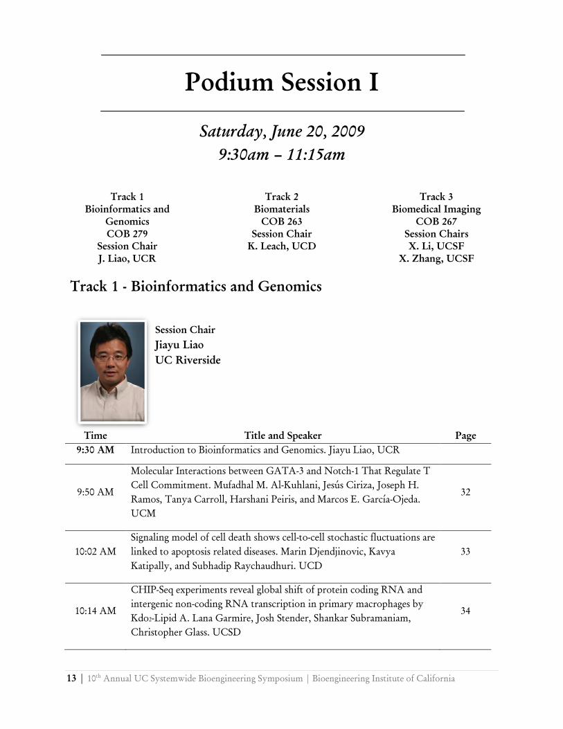
Track 1 - Bioinformatics and Genomics
Time Title and Speaker Page
9:30 AM Introduction to Bioinformatics and Genomics. Jiayu Liao, UCR
9:50 AM
Molecular Interactions between GATA-3 and Notch-1 That Regulate T
Cell Commitment. Mufadhal M. Al-Kuhlani, Jesús Ciriza, Joseph H.
Ramos, Tanya Carroll, Harshani Peiris, and Marcos E. García-Ojeda.
UCM
32
10:02 AM
Signaling model of cell death shows cell-to-cell stochastic fluctuations are
linked to apoptosis related diseases. Marin Djendjinovic, Kavya
Katipally, and Subhadip Raychaudhuri. UCD
33
10:14 AM
CHIP-Seq experiments reveal global shift of protein coding RNA and
intergenic non-coding RNA transcription in primary macrophages by
Kdo2-Lipid A. Lana Garmire, Josh Stender, Shankar Subramaniam,
Christopher Glass. UCSD
34
Session Chair
Jiayu Liao
UC Riverside
10th
Annual UC Systemwide Bioengineering Symposium | Bioengineering Institute of California | 14
10:26 AM
Selective PPARγ Ligand Modulation of Metabolic Pathways in Obese
Zucker fa/fa Rats. Gene Hsiao, Dr. Shankar Subramaniam, and Dr.
Dorothy D. Sears. UCSD
35
10:38 AM Energy based Monte Carlo Simulation of B-cell Receptor Clustering. A.
Srinivas Reddy, Sandeep Chilukuri, and Subhadip Raychaudhuri. UCD
36
10:50 AM Computational Analysis of Feedback Regulation in Signaling Networks.
Sean Kim, Arnold Kim, Jian Qiao Sun, and Henry Foreman. UCM
37
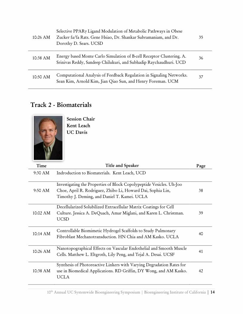
Track 2 - Biomaterials
Time Title and Speaker Page
9:30 AM Indroduction to Biomaterials. Kent Leach, UCD
9:50 AM
Investigating the Properties of Block Copolypeptide Vesicles. Uh-Joo
Choe, April R. Rodriguez, Zhibo Li, Howard Dai, Sophia Lin,
Timothy J. Deming, and Daniel T. Kamei. UCLA
38
10:02 AM
Decellularized Solubilized Extracellular Matrix Coatings for Cell
Culture. Jessica A. DeQuach, Amar Miglani, and Karen L. Christman.
UCSD
39
10:14 AM
Controllable Biomimetic Hydrogel Scaffolds to Study Pulmonary
Fibroblast Mechanotransduction. HN Chia and AM Kasko. UCLA
40
10:26 AM
Nanotopographical Effects on Vascular Endothelial and Smooth Muscle
Cells. Matthew L. Eltgroth, Lily Peng, and Tejal A. Desai. UCSF
41
10:38 AM
Synthesis of Photoreactive Linkers with Varying Degradation Rates for
use in Biomedical Applications. RD Griffin, DY Wong, and AM Kasko.
UCLA
42
Session Chair
Kent Leach
UC Davis
15 | 10th
Annual UC Systemwide Bioengineering Symposium | Bioengineering Institute of California
10:50 AM
Capsule Thickness Surrounding Titanium Oxide Nanotube Implants.
Garrett C. Smith, Seunghan Oh, Linda Fauxius, Kristian Kolind, Adam
Bohr, Sungho Jin, and Lars M. Bjursten. UCSD
43
11:02 AM
Towards methodology to characterize fibrillar collagen assembled in
vitro under different initial parameters.Yu-Jer Hwang, and Julia
Lyubovitsky. UCR
44
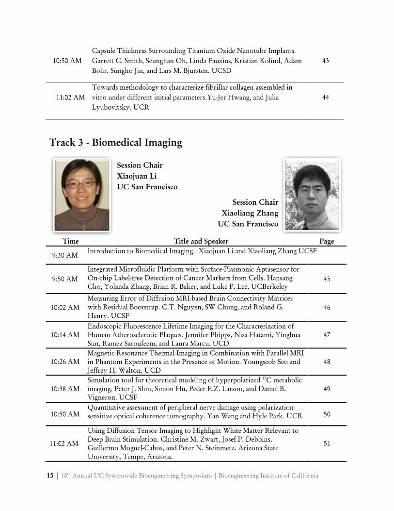
Track 3 - Biomedical Imaging
Time Title and Speaker Page
9:30 AM Introduction to Biomedical Imaging. Xiaojuan Li and Xiaoliang Zhang UCSF
9:50 AM
Integrated Microfluidic Platform with Surface-Plasmonic Aptasensor for
On-chip Label-free Detection of Cancer Markers from Cells. Hansang
Cho, Yolanda Zhang, Brian R. Baker, and Luke P. Lee. UCBerkeley
45
10:02 AM
Measuring Error of Diffusion MRI-based Brain Connectivity Matrices
with Residual Bootstrap. C.T. Nguyen, SW Chung, and Roland G.
Henry. UCSF
46
10:14 AM
Endoscopic Fluorescence Lifetime Imaging for the Characterization of
Human Atherosclerotic Plaques. Jennifer Phipps, Nisa Hatami, Yinghua
Sun, Ramez Saroufeem, and Laura Marcu. UCD
47
10:26 AM
Magnetic Resonance Thermal Imaging in Combination with Parallel MRI
in Phantom Experiments in the Presence of Motion. Youngseob Seo and
Jeffrey H. Walton. UCD
48
10:38 AM
Simulation tool for theoretical modeling of hyperpolarized 13
C metabolic
imaging. Peter J. Shin, Simon Hu, Peder E.Z. Larson, and Daniel B.
Vigneron. UCSF
49
10:50 AM
Quantitative assessment of peripheral nerve damage using polarization-
sensitive optical coherence tomography. Yan Wang and Hyle Park. UCR 50
11:02 AM
Using Diffusion Tensor Imaging to Highlight White Matter Relevant to
Deep Brain Stimulation. Christine M. Zwart, Josef P. Debbins,
Guillermo Moguel-Cabos, and Peter N. Steinmetz. Arizona State
University, Tempe, Arizona.
51
Session Chair
Xiaojuan Li
UC San Francisco
Session Chair
Xiaoliang Zhang
UC San Francisco
10th
Annual UC Systemwide Bioengineering Symposium | Bioengineering Institute of California | 16
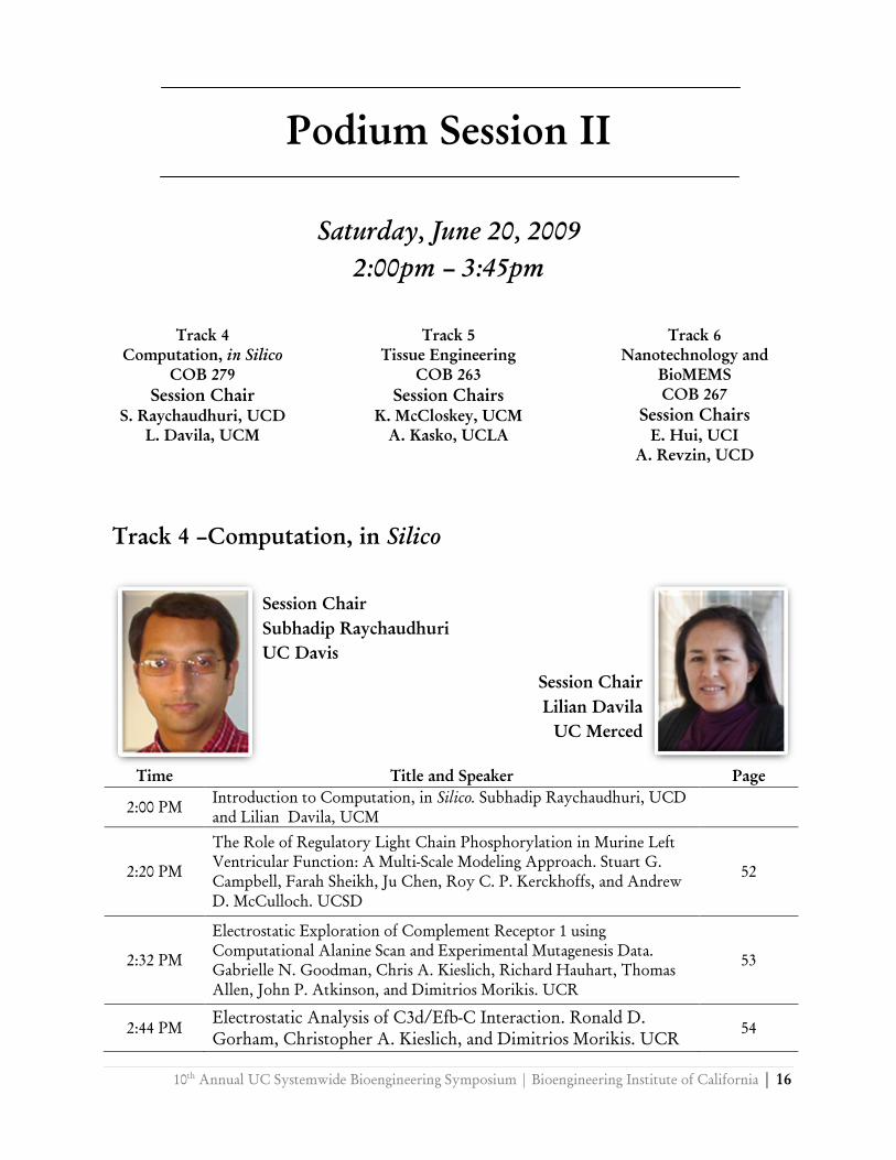
Session Chair
Subhadip Raychaudhuri
UC Davis
Session Chair
Lilian Davila
UC Merced
Podium Session II
Saturday, June 20, 2009
2:00pm – 3:45pm
Track 4
Computation, in Silico
COB 279
Session Chair
S. Raychaudhuri, UCD
L. Davila, UCM
Track 5
Tissue Engineering
COB 263
Session Chairs
K. McCloskey, UCM
A. Kasko, UCLA
Track 6
Nanotechnology and
BioMEMS
COB 267
Session Chairs
E. Hui, UCI
A. Revzin, UCD
Track 4 –Computation, in Silico
Time Title and Speaker Page
2:00 PM Introduction to Computation, in Silico. Subhadip Raychaudhuri, UCD
and Lilian Davila, UCM
2:20 PM
The Role of Regulatory Light Chain Phosphorylation in Murine Left
Ventricular Function: A Multi-Scale Modeling Approach. Stuart G.
Campbell, Farah Sheikh, Ju Chen, Roy C. P. Kerckhoffs, and Andrew
D. McCulloch. UCSD
52
2:32 PM
Electrostatic Exploration of Complement Receptor 1 using
Computational Alanine Scan and Experimental Mutagenesis Data.
Gabrielle N. Goodman, Chris A. Kieslich, Richard Hauhart, Thomas
Allen, John P. Atkinson, and Dimitrios Morikis. UCR
53
2:44 PM Electrostatic Analysis of C3d/Efb-C Interaction. Ronald D.
Gorham, Christopher A. Kieslich, and Dimitrios Morikis. UCR
54
17 | 10th
Annual UC Systemwide Bioengineering Symposium | Bioengineering Institute of California
2:56 PM
Lipid peroxidation in living cells promotes membrane
electropermeabilization. Zachary A. Levine, Yu-Hsuan Wu, Matthew J.
Ziegler, Martin A. Gundersen, D. Peter Tieleman, and P. Thomas
Vernier. University of Southern California, Los Angeles, California
55
3:08 PM
Clustering of Sequences and Electrostatic Potentials of HIV-1 Subtypes.
Aliana López De Victoria, Chris A. Kieslich, and Dimitrios Morikis.
UCR
56
3:20 PM
Computational Modeling of Immunological Synapse Formation Shows
That Cytoskeletal Transport of Receptor Molecules Is a Potential
Formation Mechanism. Philippos K. Tsourkas, and Subhadip
Raychaudhuri. UCD
57
3:32 PM The Impact of Mass Transfer of AMPK Signaling Pathways. Prashanthi
Vandrangi, John Shyy, and V. G. J. Rodgers. UCR 58
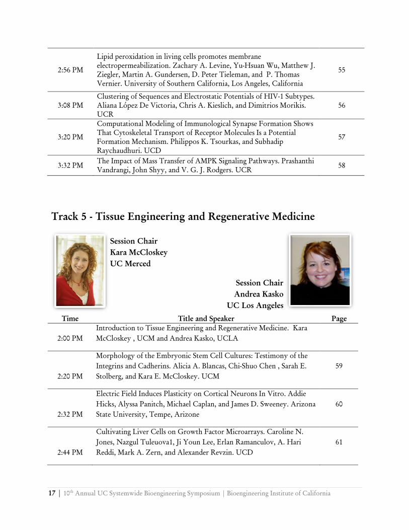
Track 5 - Tissue Engineering and Regenerative Medicine
Time Title and Speaker Page
2:00 PM
Introduction to Tissue Engineering and Regenerative Medicine. Kara
McCloskey , UCM and Andrea Kasko, UCLA
2:20 PM
Morphology of the Embryonic Stem Cell Cultures: Testimony of the
Integrins and Cadherins. Alicia A. Blancas, Chi-Shuo Chen , Sarah E.
Stolberg, and Kara E. McCloskey. UCM
59
2:32 PM
Electric Field Induces Plasticity on Cortical Neurons In Vitro. Addie
Hicks, Alyssa Panitch, Michael Caplan, and James D. Sweeney. Arizona
State University, Tempe, Arizone
60
2:44 PM
Cultivating Liver Cells on Growth Factor Microarrays. Caroline N.
Jones, Nazgul Tuleuova1, Ji Youn Lee, Erlan Ramanculov, A. Hari
Reddi, Mark A. Zern, and Alexander Revzin. UCD
61
Session Chair
Andrea Kasko
UC Los Angeles
Session Chair
Kara McCloskey
UC Merced
10th
Annual UC Systemwide Bioengineering Symposium | Bioengineering Institute of California | 18
2:56 PM
Wrinkled microtopography to induce cell alignment and maintain
contractibility of cardiac myocytes. Jesus Isaac Luna, Jesus Ciriza,
Marcos E. García-Ojeda, and Michelle Khine. UCM
62
3:08 PM
Contribution of Bioceramic Towards Osteogenic Response and
Mechanical Properties of Composite Scaffolds. Diana G. Morales and J.
Kent Leach. UCD
63
3:20 PM
Cartilage Regeneration: A Macrodesigned, Acellular Scaffold Promotin
Endogenous Cell Influx and Chondrogenesis. Stephanie Reed, Dr. Bill
Tawil, and Dr. Benjamin Wu. UCLA
64
3:32 PM
Injectable myocardial matrix for cardiac tissue engineering. Jennifer M.
Singelyn, Jessica A. DeQuach, Sonya B. Seif-Naraghi, Robert B.
Littlefield, Pamela J. Schup-Magoffin, Karen L. Christman. UCSD
65
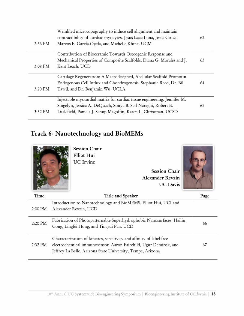
Track 6- Nanotechnology and BioMEMs
Time Title and Speaker Page
2:00 PM
Introduction to Nanotechnology and BioMEMS. Elliot Hui, UCI and
Alexander Revzin, UCD
2:20 PM
Fabrication of Photopatternable Superhydrophobic Nanosurfaces. Hailin
Cong, Lingfei Hong, and Tingrui Pan. UCD
66
2:32 PM
Characterization of kinetics, sensitivity and affinity of label-free
electrochemical immunosensor. Aaron Fairchild, Ugur Demirok, and
Jeffrey La Belle. Arizona State University, Tempe, Arizona
67
Session Chair
Elliot Hui
UC Irvine
Session Chair
Alexander Revzin
UC Davis
19 | 10th
Annual UC Systemwide Bioengineering Symposium | Bioengineering Institute of California
2:44 PM
Metal nanowrinkles and nanopetals for surface enhanced sensing in
microfluidic devices. Chi-Cheng Fu, Maureen Long, Anthony Grimes,
Christopher G.L. Ferri, Brent D. Rich, Somnath Ghosh, Ajay
Gopinathan, Sayantini Ghosh, Luke P. Lee, and Michelle Khine. UCM
68
2:56 PM
Engineering dynamic surfaces of single molecule DNA structures. Eric
Josephs, Jingru Shao, Janice Lianne Cosio, Tao Ye. UCM
69
3:08 PM
On-Cue Detachment of Cell-Containing Heparin Hydrogels from a
Conductive Substrate. Mihye Kim, Ji Youn Lee, Sunny Shah, Alexander
Revzin, and Giyoong Tae. UCD
70
3:20 PM
Frequency Domain Analysis of an Artificial Reflex Device Derived from
Response Characteristics of a Wireless Accelerometer Reflex Quantification
System. R. C. Lemoyne, C. Coroian, T. Mastroianni, and W. S. Grundfest.
UCLA
71
3:32 PM
Do-It-Yourself Three-Dimensional Microfabrication: Direct Projection-
Lithography On Dry-Film Photoresist. Siwei Zhao, Hailin Cong, and
Tingrui Pan. UCD
72
10th
Annual UC Systemwide Bioengineering Symposium | Bioengineering Institute of California | 20
Podium Session III
Saturday, June 20, 2009
4:00pm – 5:45pm
Track 7
Stem Cells
COB 263
Session Chairs
K. McCloskey, UCM
S. Simon, UCD
Track 8
Drug Delivery and
Targeting
COB 267
Session Chairs
J. Lu, UCM
V. Rogers, UCR
Track 9
Biomechanics
COB 279
Session Chairs
C. Viney, UCM
T. Pan, UCD
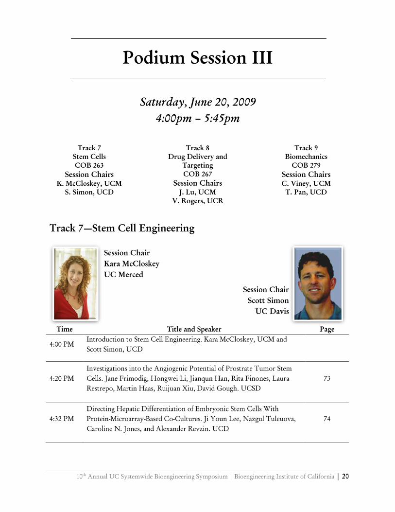
Track 7—Stem Cell Engineering
Time Title and Speaker Page
4:00 PM
Introduction to Stem Cell Engineering. Kara McCloskey, UCM and
Scott Simon, UCD
4:20 PM
Investigations into the Angiogenic Potential of Prostrate Tumor Stem
Cells. Jane Frimodig, Hongwei Li, Jianqun Han, Rita Finones, Laura
Restrepo, Martin Haas, Ruijuan Xiu, David Gough. UCSD
73
4:32 PM
Directing Hepatic Differentiation of Embryonic Stem Cells With
Protein-Microarray-Based Co-Cultures. Ji Youn Lee, Nazgul Tuleuova,
Caroline N. Jones, and Alexander Revzin. UCD
74
Session Chair
Kara McCloskey
UC Merced
Session Chair
Scott Simon
UC Davis
21 | 10th
Annual UC Systemwide Bioengineering Symposium | Bioengineering Institute of California
4:44 PM
Synergistic Effects of Biomineralization and Inductive Signals on
Osteogenic Differentiation of Human Mesenchymal Stem Cells. S.
Lauren Miller, Erin M. Case, Hillary E. Davis, J. Kent Leach. UCD
75
4:56 PM
Age related dynamics of committed T cell progenitors in mice. T.
Harshani Peiris, Jesús Ciriza, Mufadhal Al-Kulhani, Tanya Carroll, and
Marcos E. García-Ojeda. UCM
76
5:08 PM
Honeywell Microchip for Efficient and Controlled Generation of
Embryoid Bodies for Cardiomyocyte Differentiation. Silin Sa, Diep
Nuyenen, Michelle Khine, and Kara McCloskey. UCM
77
5:20 PM
Adhesion Molecules Direct Hematopoietic and Endothelial
Commitment of Murine Embryonic Stem Cells. Basha Stankovich,
Esmeralda Aguayo, Fatima Barragan, Aniket Sharma, and Maria
Pallavicini. UCM
78
5:32 PM
The in vitro response of human adipose-derived stem cells to biomimetic
apatite microstructure. Eric Tsang, Chris Arakawa, Benjamin Wu, and
Patricia Zuk. UCLA
79
Track 8—Drug Delivery and Targetting
Time Title and Speaker Page
4:00 PM
Introduction to Drug Delivery and Targetting. Jennifer Liu, UCM and
Victor Rogers, UCR
4:20 PM
Imaging of regulable expression of matriptase, a marker for cancer
progression in a mouse model for human breast cancer with PET. Julia
C. Choi, Sven H. Hausner, M. Karen J. Gagnon, David L. Kukis,
Chen-Yong Lin, Michael D. Johnson, and Julie L. Sutcliffe. UCD
80
Session Chair
Jennifer Lu
UC Merced
Session Chair
Victor Rogers
UC Riverside
10th
Annual UC Systemwide Bioengineering Symposium | Bioengineering Institute of California | 22
4:32 PM
Honeycomb Microwell Assay Platform for Generation and Culture of
Embryoid Bodies from Human Embryonic Stem Cells. Diep Nguyen,
Guangxin Xiang, Jon Pegan, Jason S. Park, Kenta Nakamura, Jennifer
Manilay, Bruce R. Conklin, and Michelle Khine. UCM
81
4:44 PM
Steric Stabilization of Liposomes for Drug Delivery: Impact Membrane
Fluidity and Diffusion.Raquel Orozco-Alcaraz and Tonya Kuhl. UC
Davis
82
4:56 PM
Controlling supramolecular architecture of poly(glutamyl-glutamate)
Paclitaxel nanoparticles by selective hydrophilic/hydrophobic
patterning: A coarse-grained modeling study. Lili X. Peng, Anthony
Ivetac, Sang Van, Lei Yu, J. Andrew McCammon, and David A. Gough.
UCSD
83
5:08 PM
Microfluidic Solvent Extraction Method for poly(lactide-co-glycolide)
Particle Formation. Shia-Yen Teh, Lung-Hsin Hung, and Abraham P.
Lee. UCI
84
5:20 PM
Engineering transferrin-diphtheria toxin conjugates for the treatment of
glioblastoma multiforme.Dennis J. Yoon, Byron H. Kwan, Felix C.
Chao,
Anne B. Mason, and Daniel T. Kamei. UCLA
85
5:32 PM
Microneedle Drug Delivery System for Skin Diseases. Kevin Zhang,
Benjamin Wu. UCLA
86
23 | 10th
Annual UC Systemwide Bioengineering Symposium | Bioengineering Institute of California
Track 9 – Biomechanics and Biodevices
Time Title and Speaker Page
4:00 PM
Introduction to Biomechanics and Biodevices. Christophery Viney, UCM
and Tangrui Pan, UCD
4:20 PM
The Effects of Coil Packing Density on Cerebral Aneurysm Inflow: In
Vitro Assessment with Particle Image Velocimetry. Haithem Babiker, L.
Fernando Gonzalez, Arius Elvikis, Dan Collins, Felipe Albuquerque,
andDavid Frakes. Arizona State University, Tempe, Arizona
87
4:32 PM
Open-Surface Microfluidics Using Photosensitive Superhydrophobic
Nanocomposite. Lingfei Hong, Hailin Cong, and Tingrui Pan. UCD
88
4:44 PM
Carbon Nanotubes lead to early Onset of Electrical Activity in Developing
Hippocampal Neurons Cultured on Silicon Microelectrodes. Massoud L
Khraiche, Nathan Jackson, and Jit Muthuswamy. Arizona State University,
Tempe, Arizona
89
4:56 PM
Long-Term Oxygen Sensor Implantation in the Porcine Subcutaneous
Environment. L.S. Kumosa, J. Lin, T. Routh, J. Lucisano, and D.A. Gough.
UC San Diego
90
5:08 PM
A simple three-dimensional vortex micromixer. Maureen Long, Michael A.
Sprague. Anthony A. Grimes, Brent D. Rich, and Michelle Khine. UCM
91
5:20 PM
Immobilization of Lactate Oxidase for Stability and High Loading in a
Lactate Sensor. Adam Strobl, Henry Tse, and David Gough. UCSF
92
5:32 PM
A Smart Contact-Lens Sensor for Dynamic Measurement of Intraocular
Flow Resistance. Chaoqi Zhang and Tingrui Pan. UCD
93
Session Chair
Christopher Viney
UC Merced
Session Chair
Tangrui Pan
UC Davis
10th
Annual UC Systemwide Bioengineering Symposium | Bioengineering Institute of California | 24
Podium Session IV
Sunday, June 21, 2009
9:45am – 11:30am
Track 10
Molecular and Cellular
Engineering
COB 263
Session Chairs
S. Li, UCB
D. Kamei, UCLA
Track 11
Biophysics
COB 267
Session Chairs
Y. Seo, UCSF
Jane P. Bearinger, LLNL
Track 12
New Frontiers
COB 279
Session Chairs
D. DiCarlo, UCLA
M. Khine, UCM
Track 10-Molecular and Cellular Engineering
Time Title and Speaker Page
9:45 AM
Introduction to Molecular and Cellular Engineering. Song Li, UCB and
Dan Kamei, UCLA
10:05 AM
Postprandial up-regulation of monocyte integrin CD11c/CD18 increases
firm arrest to vascular cell adhesion molecule-1. R Michael Gower, Anne A
Knowlton, and Scott I Simon. UCD
94
10:17 AM
Engineered proteolytic antibody fragments as therapeutics for Alzheimer’s
disease. Srinath Kasturirangan, and Michael Sierks. Arizona State
University, Tempe, Arizona
95
Session Chair
Song Li
UC Berkeley
Session Chair
Dan Kamei
UC Los Angeles
25 | 10th
Annual UC Systemwide Bioengineering Symposium | Bioengineering Institute of California
10:29 AM
Sudden death from gut ischemia may result from a neurogenic shock
mechanism. Alexander Hayes Penn, and Geert W. Schmid-Schönbein.
UCSD
96
10:41 AM
Development of FRET-based high-throughput screening to discover small
chemical inhibitors targeting protein-protein interaction in the
SUMOylation network. Yang Song, Vipul Madahar, Yan Liu, and Jiayu
Liao. UCR
97
10:53 AM
Cellular Uptake of Polyarginine-Polyleucine Block Copolymer Vesicles.
Victor Z. Sun, Zhibo Li, Timothy J. Deming, and Daniel T. Kamei. UCLA
98
11:05 AM
Design of an Aptamer Beacon for Real-Time Detection of Interferon-
Gamma. Nazgul Tuleuova, Caroline N. Jones, Jun Yan, and Erlan
Ramanculov,
Alexander Revzin. UCD
99
11:17 AM
Solid-Phase Peptide Synthesis of Bioinspired Electrets Based on Non-
Traditional Amino Acids: Synthesizing oligo-ortho-anthranilic acids for
improved Charge-Transfer properties in photovoltaic cells. Srigokul
Upadhyayula, Duoduo Bao, David Bui, and Valentine I. Vullev. UCR
100
Track 11—Biophysics
Time Title and Speaker Page
9:45 AM
Introducton Biophysics. Youngho Seo, UCSF and Jane P. Bearinger,
LLNL.
10:05 AM
The Rehm-Weller Equation in View of Bioengineering. Duoduo Bao,
Antonio Contreras, and Valentine I. Vullev. UCR
101
Session Chair
Youngho Seo
UC San Francisco
Session Chair
Jane P. Bearinger
Lawrence Livermore National Laboratories

10th
Annual UC Systemwide Bioengineering Symposium | Bioengineering Institute of California | 26
10:17 AM
Ca2+
depletion of sarcoplasmic reticulum during reperfusion after ischemia.
Marcela Ferreiro, Dmytro Kornyeyev, Carlos A. Valverde, Alicia
Mattiazzi, and Ariel L. Escobar. UCM
102
10:29 AM
Mapping the Position of DNA Polymerase-Bound DNA Templates in a
Nanopore at 5Å Resolution. Daniel R. Garalde, Brett Gyarfas, Felix
Olasagasti, Seico Benner, William Dunbar, Kate R. Lieberman, and Mark
Akeson. UCSC
103
10:41 AM
Concentrating DNA Using Two-Phase Aqueous Micellar Systems. Foad
Mashayekhi, Aaron S. Meyer, Stacey A. Shiigi, and Daniel T. Kamei.
UCLA
104
10:53 AM
Fluorescence Lifetime Imaging Microscopy (FLIM) for Cancer
Demarcation during Medical Surgery. Yinghua Sun, Jennifer Phipps,
Daniel S. Elson, Jeremy Meier, Nisa Hatami, Frank S. Chuang, Rudolph J.
Schrot, D. Gregory Farwell, and Laura Marcu. UC Davis
105
11:05 AM
Multi-photon optical microscopy of actin filaments and mitochondrial
bioenergetics of ACBT human grade IV glioblastoma cells migrating within
3-D collagen-based hydrogels. Miso Yang, Yu-Jer Hwang, Edgar Sanchez,
Chung-ho Sun, Tatiana B. Krasieva, Bruce J. Tromberg, and Julia G.
Lyubovitsky. UCR
106
11:17 AM
Optical Model of Human Skin for Biomedical Reflectance and
Fluorescence Spectroscopy. Dmitry Yudovsky and Laurent Pilon. UCLA
107
27 | 10th
Annual UC Systemwide Bioengineering Symposium | Bioengineering Institute of California
Track 12 - New Frontiers in Bioengineering
Time Title and Speaker Page
9:45 AM Introduction to New Frontiers in Bioengineering. Dino DiCarlo,
UCLA and Michelle Khine, UCM
10:05 AM
Bench Scale Electroenzymatic Biosensor for the Rapid Detection of
Pyruvate. Lorenzo D’Amico, Andrew Basilio, Si Luo, Justin Yeap, and
Dale A. Baker. UCSD
108
10:17 AM
Effects of coating material on cellular uptake of nanocapsules
loaded with indocyanine green.Bongsu Jung and Bahman Anvari.
UCR
109
10:29 AM
Novel Dielectrophoretic Device for Cancer Cell, Stem Cell and DNA
Biomarker Isolation and Detection. Rajaram Krishnan, Joaquim
Teixeira, Jennifer Y. Marciniak, Mark Mercola, Sadik C. Esener, and
Michael J. Heller. UCSD
110
10:41 AM
Detection of Enzymatic Biomarkers Directly in Whole Blood for Point-
Of-Care Diagnostics. Roy B. Lefkowitz, Jennifer Y. Marciniak, Che-
Ming Hu, Geert W. Schmid-Schönbein, and Michael J. Heller. UCSD
111
10:53 AM
The Deposition and Fate of Ultra-fine Pollutants in Normal and
Asthmatic Mice using Positron Emission Tomography. Heather A.
Palko and Angelique Y. Louie. UCD
112
11:05 AM Dual-Beam Optical Fiber Trapping Platform for Biophotonics
Applications. Tessa Piñón and Jay Sharping. UCM 113
Session Chair
Dino DiCarlo
UC Los Angeles
Session Chair
Michelle Khine
UC Merced
10th
Annual UC Systemwide Bioengineering Symposium | Bioengineering Institute of California | 28
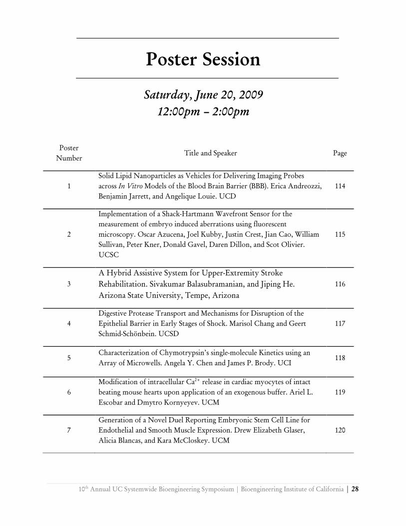
Poster Session
Saturday, June 20, 2009
12:00pm – 2:00pm
Poster
Number
Title and Speaker Page
1
Solid Lipid Nanoparticles as Vehicles for Delivering Imaging Probes
across In Vitro Models of the Blood Brain Barrier (BBB). Erica Andreozzi,
Benjamin Jarrett, and Angelique Louie. UCD
114
2
Implementation of a Shack-Hartmann Wavefront Sensor for the
measurement of embryo induced aberrations using fluorescent
microscopy. Oscar Azucena, Joel Kubby, Justin Crest, Jian Cao, William
Sullivan, Peter Kner, Donald Gavel, Daren Dillon, and Scot Olivier.
UCSC
115
3
A Hybrid Assistive System for Upper-Extremity Stroke
Rehabilitation. Sivakumar Balasubramanian, and Jiping He.
Arizona State University, Tempe, Arizona
116
4
Digestive Protease Transport and Mechanisms for Disruption of the
Epithelial Barrier in Early Stages of Shock. Marisol Chang and Geert
Schmid-Schönbein. UCSD
117
5
Characterization of Chymotrypsin’s single-molecule Kinetics using an
Array of Microwells. Angela Y. Chen and James P. Brody. UCI
118
6
Modification of intracellular Ca2+
release in cardiac myocytes of intact
beating mouse hearts upon application of an exogenous buffer. Ariel L.
Escobar and Dmytro Kornyeyev. UCM
119
7
Generation of a Novel Duel Reporting Embryonic Stem Cell Line for
Endothelial and Smooth Muscle Expression. Drew Elizabeth Glaser,
Alicia Blancas, and Kara McCloskey. UCM
120
29 | 10th
Annual UC Systemwide Bioengineering Symposium | Bioengineering Institute of California
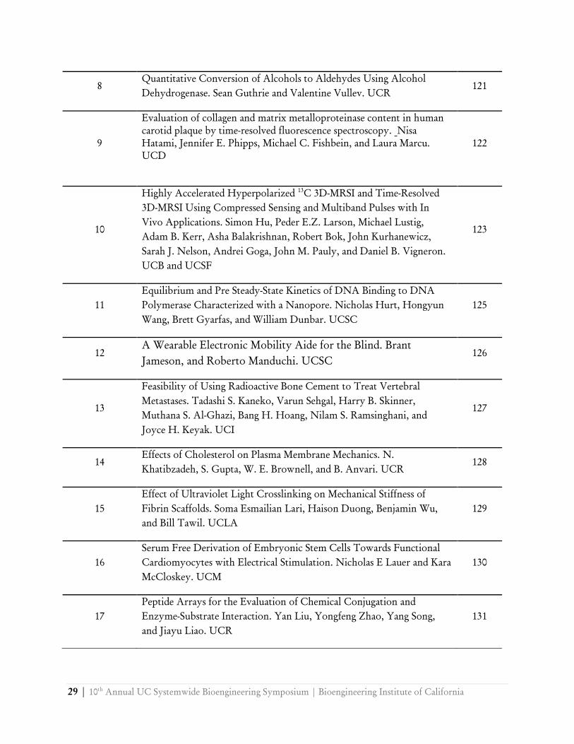
8
Quantitative Conversion of Alcohols to Aldehydes Using Alcohol
Dehydrogenase. Sean Guthrie and Valentine Vullev. UCR
121
9
Evaluation of collagen and matrix metalloproteinase content in human
carotid plaque by time-resolved fluorescence spectroscopy. Nisa
Hatami, Jennifer E. Phipps, Michael C. Fishbein, and Laura Marcu.
UCD
122
10
Highly Accelerated Hyperpolarized 13
C 3D-MRSI and Time-Resolved
3D-MRSI Using Compressed Sensing and Multiband Pulses with In
Vivo Applications. Simon Hu, Peder E.Z. Larson, Michael Lustig,
Adam B. Kerr, Asha Balakrishnan, Robert Bok, John Kurhanewicz,
Sarah J. Nelson, Andrei Goga, John M. Pauly, and Daniel B. Vigneron.
UCB and UCSF
123
11
Equilibrium and Pre Steady-State Kinetics of DNA Binding to DNA
Polymerase Characterized with a Nanopore. Nicholas Hurt, Hongyun
Wang, Brett Gyarfas, and William Dunbar. UCSC
125
12
A Wearable Electronic Mobility Aide for the Blind. Brant
Jameson, and Roberto Manduchi. UCSC
126
13
Feasibility of Using Radioactive Bone Cement to Treat Vertebral
Metastases. Tadashi S. Kaneko, Varun Sehgal, Harry B. Skinner,
Muthana S. Al-Ghazi, Bang H. Hoang, Nilam S. Ramsinghani, and
Joyce H. Keyak. UCI
127
14
Effects of Cholesterol on Plasma Membrane Mechanics. N.
Khatibzadeh, S. Gupta, W. E. Brownell, and B. Anvari. UCR
128
15
Effect of Ultraviolet Light Crosslinking on Mechanical Stiffness of
Fibrin Scaffolds. Soma Esmailian Lari, Haison Duong, Benjamin Wu,
and Bill Tawil. UCLA
129
16
Serum Free Derivation of Embryonic Stem Cells Towards Functional
Cardiomyocytes with Electrical Stimulation. Nicholas E Lauer and Kara
McCloskey. UCM
130
17
Peptide Arrays for the Evaluation of Chemical Conjugation and
Enzyme-Substrate Interaction. Yan Liu, Yongfeng Zhao, Yang Song,
and Jiayu Liao. UCR
131

10th
Annual UC Systemwide Bioengineering Symposium | Bioengineering Institute of California | 30
18
Ariadne’s Thread: A Wayfinding Tool for the Visually Impaired Based
on Camera Cell Phones. Roberto Manduchi and James Coughlan.
UCSC
132
19
Theoretical Significance of Ion Binding on Observed Non-idealities in
Osmotic Pressure in Crowded Macromolecular Environments. Devin
W. McBride and Victor G. J. Rodgers. UCR
133
20
In vivo Optical Microscopy of Axonal Myelination of a Multiple
Sclerosis Disease Model with Polarization Sensitive-Optical Coherence
Tomography. Christian Oh and Hyle Park. UCR
134
21
Cascaded Microconcentration Cells. Oxana S. Pantchenko, Javad
Shavani, Mona Zebrajadi, Howard Young, Mehrdad Mahomoodi,
Michail Isaacson, Ali Shakouri. UCSC
135
22
Inhibition of the sodium/calcium exchanger by lithium in intact mouse
hearts modifies cardiac alternans. Azadé Petrosky, Dmytro Kornyeyev,
and Ariel L. Escobar. UCM
136
23
On Calibrating the Power of a Microwave Oven. Emily J. Reed and
Christopher Viney. UCM
137
24 Optimizing qNano: Characterizing a resizable nanopore. Jessie Rucker,
Asma Uz-Zaman, David Deamer, and William Dunbar. UCSC
138
25
In Vitro Culturing of the Ovarian Follicle: Alginate Encapsulation and
Evaluation of the Nutrients Environment. Noriko Sausman, P. Talbot,
and V. G. J. Rodgers. UCR
139
26
A Microdevice for Detecting Cytokine Production from Individual
Immune Cells. Jaime Silangcruz, Gulnaz Stybayeva, He Zhu, and
Alexander Revzin. UCD
140
27
Kinetics of Staining: Flourescence Enhancement Induced By Escerichia
Coli. Marlon S. Thomas, Elizabeth T. Zielins, Duoduo Bao, Baharak
Bahmni, Vicente Numez and Valentine I. Vullev. UCR
141
28
Fluorescence Enhancement of Warfarin Induced by Interaction with β-
Cyclodextrin. Jacob M. Vasquez, Andrew Vu, Jerome S. Schultz, and
Valentine I. Vullev. UCR
142
29
Luminal Ca2+
Regulation of Single RyR2 Channels by Cardiac
Calsequestrin. Patricio Vélez, Dmytro Kornyeyev, Marcia Cortés-
Gutiérrez, Björn C. Knollmann, and Ariel L. Escobar. UCM
143
31 | 10th
Annual UC Systemwide Bioengineering Symposium | Bioengineering Institute of California
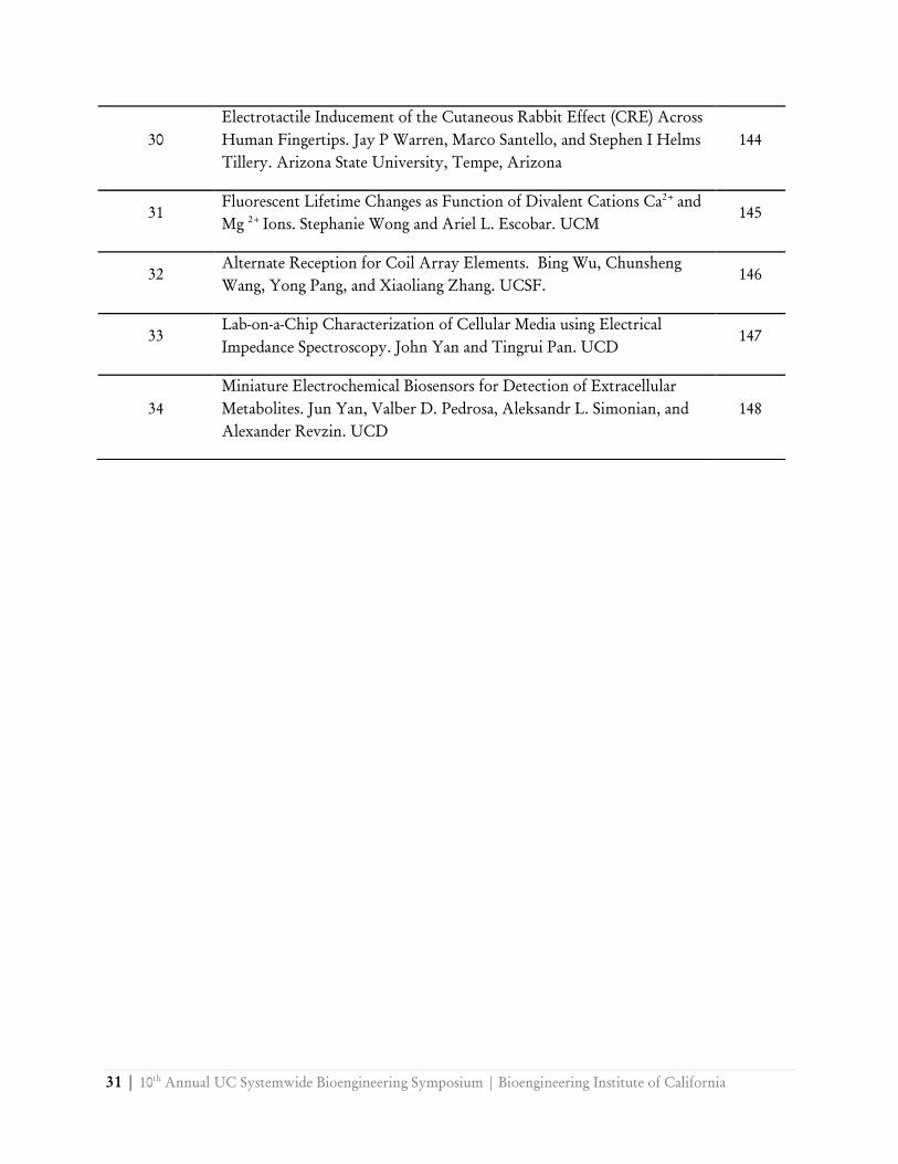
30
Electrotactile Inducement of the Cutaneous Rabbit Effect (CRE) Across
Human Fingertips. Jay P Warren, Marco Santello, and Stephen I Helms
Tillery. Arizona State University, Tempe, Arizona
144
31
Fluorescent Lifetime Changes as Function of Divalent Cations Ca2+
and
Mg 2+
Ions. Stephanie Wong and Ariel L. Escobar. UCM
145
32
Alternate Reception for Coil Array Elements. Bing Wu, Chunsheng
Wang, Yong Pang, and Xiaoliang Zhang. UCSF.
146
33
Lab-on-a-Chip Characterization of Cellular Media using Electrical
Impedance Spectroscopy. John Yan and Tingrui Pan. UCD
147
34
Miniature Electrochemical Biosensors for Detection of Extracellular
Metabolites. Jun Yan, Valber D. Pedrosa, Aleksandr L. Simonian, and
Alexander Revzin. UCD
148

10th
Annual UC Systemwide Bioengineering Symposium | Bioengineering Institute of California | 32
Molecular Interactions between GATA-3 and Notch-1 That Regulate T Cell Commitment
Mufadhal M. Al-Kuhlani, Jesús Ciriza, Joseph H. Ramos, Tanya Carroll, Harshani Peiris, and
Marcos E. García-Ojeda
School of Natural Sciences, University of California, Merced
Hematopoietic stem cells (HSC) differentiate into all mature blood cells, including lymphoid progenitors (LPs) that can give rise to NK, dendritic, B and T cells. The final fate of these LPs depends on the signals and growth factors received during development. The transmembrane receptor Notch-1 and the transcription factor GATA-3 are two of the signals that regulate the commitment of LPs towards the T cell lineage. Notch-1 instructs lymphocyte progenitors to differentiate into T cells but not B cells. However, the role that GATA-3 plays in early T cell development in relation to Notch-1 is poorly characterized. Our studies show that GATA-3 deficient progenitors initiate a normal T cell development program, but become arrested at an early CD44+CD25+double negative (DN2) stage, and generate aberrantly B cells in the presence of Notch-1 signals. The gene expression profile of GATA-3 deficient DN2 cells showed elevated expression of Deltex-1, a Notch-1 regulator known to induce B cell development while inhibiting T cell differentiation. To elucidate the molecular mechanism exerted by GATA-3 on Notch-1 signaling, we will transduce fetal liver HSC with GATA-3-GFP shRNA retrovirus followed by a co-culture on OP9DL-1 stroma, which expresses the Notch-1 ligand Delta-like-1. After two weeks of culture, the transduced cells are sorted on the basis of GFP expression. Molecular analysis for the expression of three of Notch-1 regulators, Deltex-1, Mint and Numb, is evaluated via q-PCR. Understanding the mechanisms of interaction between GATA-3 and Notch-1, as well as other genes involved in T cell commitment is very crucial. Such understanding will allow us to reveal how early lymphocyte precursors commit to the T cell fate, and lead to the development of new stem cell-based therapeutic approaches to treat diseases related to impaired T cells.
33 | 10th
Annual UC Systemwide Bioengineering Symposium | Bioengineering Institute of California
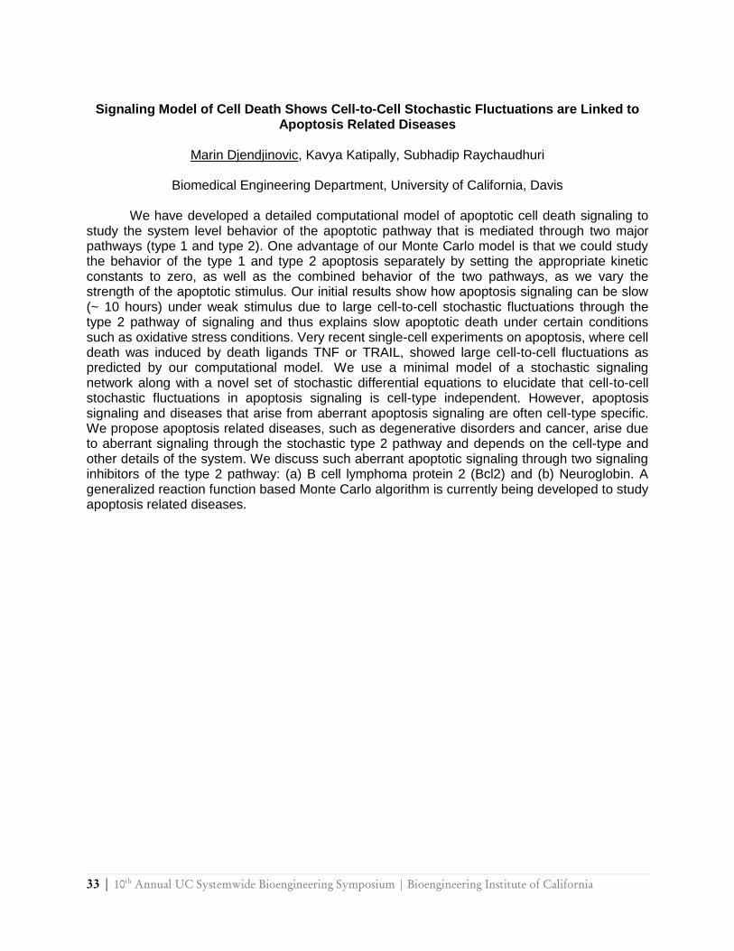
Signaling Model of Cell Death Shows Cell-to-Cell Stochastic Fluctuations are Linked to
Apoptosis Related Diseases
Marin Djendjinovic, Kavya Katipally, Subhadip Raychaudhuri
Biomedical Engineering Department, University of California, Davis
We have developed a detailed computational model of apoptotic cell death signaling to study the system level behavior of the apoptotic pathway that is mediated through two major pathways (type 1 and type 2). One advantage of our Monte Carlo model is that we could study the behavior of the type 1 and type 2 apoptosis separately by setting the appropriate kinetic constants to zero, as well as the combined behavior of the two pathways, as we vary the strength of the apoptotic stimulus. Our initial results show how apoptosis signaling can be slow (~ 10 hours) under weak stimulus due to large cell-to-cell stochastic fluctuations through the type 2 pathway of signaling and thus explains slow apoptotic death under certain conditions such as oxidative stress conditions. Very recent single-cell experiments on apoptosis, where cell death was induced by death ligands TNF or TRAIL, showed large cell-to-cell fluctuations as predicted by our computational model. We use a minimal model of a stochastic signaling network along with a novel set of stochastic differential equations to elucidate that cell-to-cell stochastic fluctuations in apoptosis signaling is cell-type independent. However, apoptosis signaling and diseases that arise from aberrant apoptosis signaling are often cell-type specific. We propose apoptosis related diseases, such as degenerative disorders and cancer, arise due to aberrant signaling through the stochastic type 2 pathway and depends on the cell-type and other details of the system. We discuss such aberrant apoptotic signaling through two signaling inhibitors of the type 2 pathway: (a) B cell lymphoma protein 2 (Bcl2) and (b) Neuroglobin. A generalized reaction function based Monte Carlo algorithm is currently being developed to study apoptosis related diseases.
10th
Annual UC Systemwide Bioengineering Symposium | Bioengineering Institute of California | 34
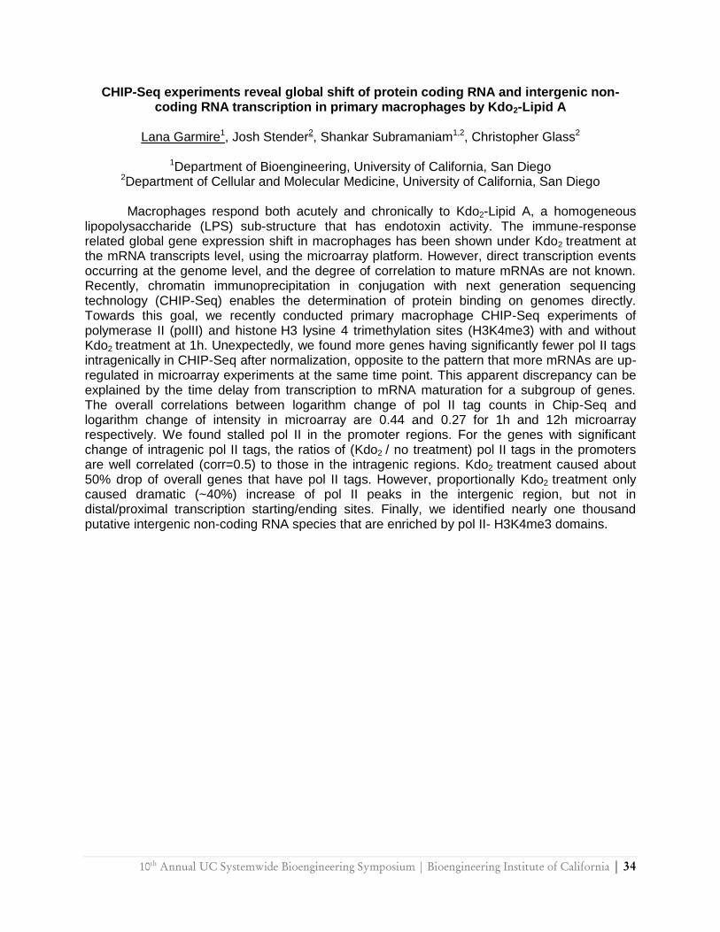
CHIP-Seq experiments reveal global shift of protein coding RNA and intergenic non-coding RNA transcription in primary macrophages by Kdo2-Lipid A
Lana Garmire1, Josh Stender2, Shankar Subramaniam1,2, Christopher Glass2
1Department of Bioengineering, University of California, San Diego
2Department of Cellular and Molecular Medicine, University of California, San Diego
Macrophages respond both acutely and chronically to Kdo2-Lipid A, a homogeneous lipopolysaccharide (LPS) sub-structure that has endotoxin activity. The immune-response related global gene expression shift in macrophages has been shown under Kdo2 treatment at the mRNA transcripts level, using the microarray platform. However, direct transcription events occurring at the genome level, and the degree of correlation to mature mRNAs are not known. Recently, chromatin immunoprecipitation in conjugation with next generation sequencing technology (CHIP-Seq) enables the determination of protein binding on genomes directly. Towards this goal, we recently conducted primary macrophage CHIP-Seq experiments of polymerase II (polII) and histone H3 lysine 4 trimethylation sites (H3K4me3) with and without Kdo2 treatment at 1h. Unexpectedly, we found more genes having significantly fewer pol II tags intragenically in CHIP-Seq after normalization, opposite to the pattern that more mRNAs are up-regulated in microarray experiments at the same time point. This apparent discrepancy can be explained by the time delay from transcription to mRNA maturation for a subgroup of genes. The overall correlations between logarithm change of pol II tag counts in Chip-Seq and logarithm change of intensity in microarray are 0.44 and 0.27 for 1h and 12h microarray respectively. We found stalled pol II in the promoter regions. For the genes with significant change of intragenic pol II tags, the ratios of (Kdo2 / no treatment) pol II tags in the promoters are well correlated (corr=0.5) to those in the intragenic regions. Kdo2 treatment caused about 50% drop of overall genes that have pol II tags. However, proportionally Kdo2 treatment only caused dramatic (~40%) increase of pol II peaks in the intergenic region, but not in distal/proximal transcription starting/ending sites. Finally, we identified nearly one thousand putative intergenic non-coding RNA species that are enriched by pol II- H3K4me3 domains.
35 | 10th
Annual UC Systemwide Bioengineering Symposium | Bioengineering Institute of California
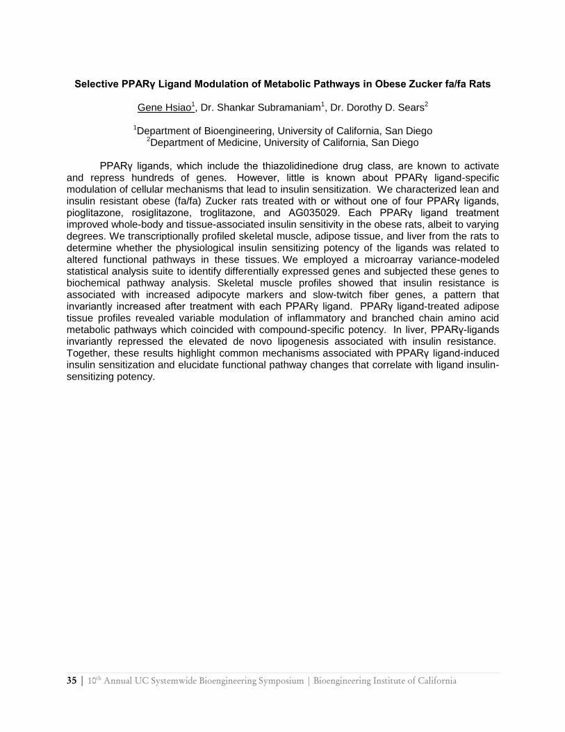
Selective PPARγ Ligand Modulation of Metabolic Pathways in Obese Zucker fa/fa Rats
Gene Hsiao1, Dr. Shankar Subramaniam1, Dr. Dorothy D. Sears2
1Department of Bioengineering, University of California, San Diego
2Department of Medicine, University of California, San Diego
PPARγ ligands, which include the thiazolidinedione drug class, are known to activate and repress hundreds of genes. However, little is known about PPARγ ligand-specific modulation of cellular mechanisms that lead to insulin sensitization. We characterized lean and insulin resistant obese (fa/fa) Zucker rats treated with or without one of four PPARγ ligands, pioglitazone, rosiglitazone, troglitazone, and AG035029. Each PPARγ ligand treatment improved whole-body and tissue-associated insulin sensitivity in the obese rats, albeit to varying degrees. We transcriptionally profiled skeletal muscle, adipose tissue, and liver from the rats to determine whether the physiological insulin sensitizing potency of the ligands was related to altered functional pathways in these tissues. We employed a microarray variance-modeled statistical analysis suite to identify differentially expressed genes and subjected these genes to biochemical pathway analysis. Skeletal muscle profiles showed that insulin resistance is associated with increased adipocyte markers and slow-twitch fiber genes, a pattern that invariantly increased after treatment with each PPARγ ligand. PPARγ ligand-treated adipose tissue profiles revealed variable modulation of inflammatory and branched chain amino acid metabolic pathways which coincided with compound-specific potency. In liver, PPARγ-ligands invariantly repressed the elevated de novo lipogenesis associated with insulin resistance. Together, these results highlight common mechanisms associated with PPARγ ligand-induced insulin sensitization and elucidate functional pathway changes that correlate with ligand insulin-sensitizing potency.
10th
Annual UC Systemwide Bioengineering Symposium | Bioengineering Institute of California | 36
Energy based Monte Carlo Simulation of B-cell Receptor Clustering
A. Srinivas Reddy1, Sandeep Chilukuri 2,1 Subhadip Raychaudhuri 1
1Department of Biomedical Engineering, University of California-Davis, Davis, California
2Department of Biotechnology, Indian Institute of Technology Madras, Chennai, India
Antigens in solution are known to trigger B-cell receptor (BCR) clustering which in turn leads to B-cell activation. Such clustering is thought to be organized as a two-step process: (a) early-time micro-clustering where a number of micro-clusters form that typically contain a few BCR (~10) molecules, (b) followed by a large macroscopic clustering of B cell receptors. Little is known about the molecular mechanics that prompt the BCR clustering and how such clustering leads to B-cell activation. We develop an energy based Monte Carlo model to elucidate the mechanism of B-cell receptor clustering. We propose a model of B cell receptor clustering due to intrinsic attractions among the receptor molecules. Such mutual attractions may also arise indirectly due to cross-linking by soluble antigens among other possibilities. At the outset, micro-clusters of receptor molecules are formed due to mutual BCR-BCR attractions, however, such mutual interactions are not enough to create a large macro-cluster at a later time. A simple model of biased diffusion where BCR molecules experience a biased directed motion towards the largest cluster is then applied, resulting in a single macro cluster of receptor molecules. The various types of clusters are analyzed using network-based metrics such as the average distance between any pairs of receptors.
37 | 10th
Annual UC Systemwide Bioengineering Symposium | Bioengineering Institute of California
Computational Analysis of Feedback Regulation in Signaling Networks
Sean Kim1, *, Arnold Kim2, Jian Qiao Sun3, Henry Forman1, *
1 Quantitative and Systems Biology, University of California, Merced 2 Applied Mathematics, University of California, Merced
3 Mechanical Engineering, University of California, Merced * Atmospheric Aerosol and Health Lead Program. UC TSR&TP
Signal transduction networks contain complex interconnections that regulate their
properties over the spatiotemporal domain. One general class of such interconnections is feedback regulation that can confers positive and/or negative effects on the components of signaling networks. Based on Michaelis-Menten kinetic formalism, feedback regulation can be represented as nonlinear ordinary differential equations (ODEs) that allow emergence of a rich class of behaviors such as hysteresis, bifurcation, oscillations, and robustness, some of which have been verified experimentally in the literature. (1)
In this work, we apply various ways to study the feedback regulation such as autonomous linear ODEs, controlled nonlinear ODEs, and non-autonomous nonlinear ODEs to test if the systems can produce expected behaviors in stable and robust manner. When feedback systems are represented as a system of linear ODEs, we can capture the main expected behaviors such as oscillation and exponential growth for negative and positive feedback, respectively; however, it lacks complexity necessary to confer behaviors like bifurcation seen in nonlinear systems. We also show that positive feedback, on its own, is uncontrollable and unstabilizable; whereas, interlinked positive and negative feedback regulations form a control circuit, and systems become controllable and stabilizable as expected. (2) We also present the signaling network as a non-autonomous system that has explicit time-dependent parameters and study the stability and the robustness. We also show that the behavior of the signaling networks is perturbed by electrophilic aldehydes generated during normal signaling by reactive oxygen species. 1. J. J. Tyson, K. C. Chen, B. Novak, Curr. Opin. Cell Biol. 15, 221 (2003) 2. T. Y. Tsai, Y. S. Choi, W. Ma, J.R. Pomerening, C. Tang, J.E. Ferrell Jr. Science Vol. 321.
no. 5885, 126 (2008)
10th
Annual UC Systemwide Bioengineering Symposium | Bioengineering Institute of California | 38
Investigating the Properties of Block Copolypeptide Vesicles
Uh-Joo Choe, April R. Rodriguez, Zhibo Li, Howard Dai, Sophia Lin,
Timothy J. Deming, and Daniel T. Kamei
Department of Bioengineering, University of California, Los Angeles Investigation of polymeric vesicles as novel drug delivery vehicles is an emerging area of research and shows great promise. With respect to this field, our focus has been on developing amino acid-based nanomaterials for drug delivery. We previously developed vesicles composed of lysine-leucine (poly(L-lysine)60-block-poly(L-leucine)20, K60L20), glutamate-leucine (poly(L-glutamic acid)60-block-poly(L-leucine)20, E60L20), and arginine-leucine (poly(L-arginine)60-block-poly(L-leucine)20, R60L20) block copolypeptides. These block copolypeptides formed vesicles in aqueous solutions that were stable up to 80°C, could encapsulate polar molecules with negligible leakage, and could be prepared with diameters ranging from 50 nm to 1 μm. In the case of the R60L20 vesicles, they were also shown to be able to deliver hydrophilic cargo into both endothelial and epithelial cells. We recently performed studies to further characterize and optimize the vesicles as potential drug delivery vehicles. The vesicle extrusion process was optimized to control the size distribution of the vesicles. The toxicity of K60L22 vesicles extruded to different sizes was investigated in HeLa cells with the MTS assay. To study the leakage properties of the vesicles, fluroescein was encapsulated in R60L20 vesicles. These characterization studies are currently being extended to polypeptides with varying lengths.
39 | 10th
Annual UC Systemwide Bioengineering Symposium | Bioengineering Institute of California
Decellularized Solubilized Extracellular Matrix Coatings for Cell Culture
Jessica A. DeQuach, Amar Miglani, and Karen L. Christman
Department of Bioengineering, University of California, San Diego
The use of biomaterials in conjunction with cells is important for many bioengineering applications. For these cellular-based therapies, the extracellular matrix (ECM) used plays an important role as it has been shown that the matrix composition and matrix mechanical properties can affect cell behavior and function. It is hypothesized that the best growth environment for cells would be the same ECM as found in vivo. The aim of this present study is to test whether native ECM would demonstrate better properties for cell culture when compared to conventional coatings. To test this hypothesis, various porcine tissue was decellularized, and then solubilized using a pepsin digestion to be used as cell culture coatings. These coatings were characterized using SDS-PAGE, and were found to be more complex than collagen I and laminin, as indicated by a mixture of different sized peptides. Glycosaminoglycan content was quantified using a Blyscan assay, where frontal lobe coating had the highest content of GAG at 35.95±1.45 ug/mg ECM, followed by cardiac 27.77±1.20 ug/mg ECM, cortex 12.80±0.43 ug/mg ECM, and skeletal muscle at 2.99±0.11 μg/mg ECM. GAG content was thought to be important as several other decellularization techniques have not shown to retain the GAG content. C2C12 cells plated on the skeletal muscle matrix at day 3 were shown to have increased myotube width
and percent differentiation when compared to collagen coating (myotube 17.6±1.8 m vs.
14.2±1.7 m; p~0.0001) (differentiation 16.3±0.2 vs. 13.1±0.1; p=0.018). The study demonstrates that the ECM component for biomaterials has an important effect on cell behavior, and consideration should be taken when selecting an ECM coating for any biomaterial application. The more solubilized, decellularized ECM demonstrated an increase on differentiation and structure for C2C12 myoblasts, and following studies are being performed on the other tissue coatings.
10th
Annual UC Systemwide Bioengineering Symposium | Bioengineering Institute of California | 40
Controllable Biomimetic Hydrogel Scaffolds to Study Pulmonary Fibroblast Mechanotransduction
Chia HN, Kasko AM
Department of Bioengineeirng, UCLA, Los Angeles, CA
Idiopathic pulmonary fibrosis (IPF) is a progressive, fatal disease. While the cause is not
well-understood, IPF is thought to be the result of an abnormal wound-healing process in the lungs. Many chemical cues have been identified as effectors in fibrosis, but the effect of mechanical environment in the development of IPF is poorly understood. Fibroblast differentiation into the myofibroblast phenotype is a critical event in the wound healing process, and when this differentiation is not ―turned off‖, fibrosis occurs. We are developing a two- and three-dimensional hydrogel cell scaffolds with varying stiffness and biochemical composition that will enable us to answer important questions about the effects of cell-matrix interactions on the differentiation of pulmonary fibroblasts. Poly(ethylene glycol) macromers are copolymerized with peptide-based crosslinking agents that mimic extracellular matrix adhesive fragments to produce hydrogels where the chemistry and gel structure can be independently controlled. Human pulmonary fibroblasts show good adhesion to hydrogels containing the adhesion peptide sequence CRGDSC (cysteines are reactive with the PEG macromers), and little adhesion on hydrogels without RGDS. Increasing the crosslinking density of the hydrogel (via decreasing macromer length) increases the compressive modulus of elasticity. Pulmonary fibroblasts seeded on to soft hydrogels (E= 67 kPa, [RGDS]=2 mM) express no alpha-smooth
muscle actin (-SMA), a marker of the myofibroblast phenotype. On more rigid hydrogels
(E=585 kPa, [RGDS]=2 mM) cells express significant amount of -SMA, indicating the myofibroblast phenotype. Pulmonary fibroblasts encapsulated into a 3D hydrogel construct are viable after 72 hours, but persist in a non-native rounded morphology. We are currently optimizing 3D culture conditions in this biomimetic matrix to allow fibroblasts to adopt their native morphology. This synthetic, biomimetic cell culture system allows the chemistry of the cellular environment to be decoupled from the mechanical environment, and provides a universal synthetic platform for cell culture.
41 | 10th
Annual UC Systemwide Bioengineering Symposium | Bioengineering Institute of California
Nanotopographical Effects on Vascular Endothelial and Smooth Muscle Cells
Matthew L. Eltgroth, Lily Peng, Tejal A. Desai
Department of Bioengineering and Therapeutic Sciences and Department of Physiology, University of California, San Francisco
The nanotopographical cues provided by biomaterial surfaces are known to have an
important influence on the behavior of cells they interact with. In addition, specific cell types have been shown to exhibit different responses depending on nanotube diameter. This study was aimed at investigating the effects that nanotubular titania (TiO2) surfaces have on vascular endothelial and vascular smooth muscle cells. Using a variety of microscopic techniques and biochemical assays, we showed that nanotubular TiO2 surfaces promoted vascular endothelial cell proliferation and production of anti-thrombotic factors, while inhibiting proliferation of vascular smooth muscle cells and causing them to assume a more differentiated phenotype. Additional work has been performed using gene expression analysis to lend further support to these observations. These results support prior findings that nanotubular TiO2 surfaces may promote cellular responses that would make them favorable for use in endovascular applications, such as stents.

10th
Annual UC Systemwide Bioengineering Symposium | Bioengineering Institute of California | 42
Synthesis of Photoreactive Linkers with Varying Degradation Rates for use in Biomedical Applications
Griffin DR1, Wong DY2, Kasko AM1,2
1Biomedical Engineering Interdepartmental Program, UCLA, Los Angeles, CA 2Department of Bioengineering, UCLA, Los Angeles, CA
A critical aspect of designing biomaterial carriers for cells and drug delivery is tuning and
controlling the material‘s degradation behavior. Most synthetic biomaterials degrade via hydrolysis or enzymolysis. The rate of hydrolysis is pre-engineered and cannot be modified after the scaffold is fabricated under physiologically relevant conditions. The rate of enzymatic degradation is cell-mediated and is normally limited to local degradation. In the last decade, there has been considerable interest in using photochemistry to produce biomaterials because of the ability to form scaffolds in situ under physiological conditions1. Integrating photochemistry as a degradation mechanism should be equally biocompatible, affording spatial and temporal control over the chemical, mechanical, and physical properties of the biomaterial, and allowing for the controlled and triggerable release of therapeutic agents. We have designed a series of five photodegradable linkers based on nitrobenzylether to provide a range of degradation rates upon exposure to long-wave UV light2 (365 nm, biocompatible1). We developed three separate synthesis schemes, each using high yield reactions and inexpensive starting materials. We followed the photodegradation of the linkers by 1H NMR and found that the rate of degradation increases as the number of aryl ether groups decreases. Additionally, compounds with a secondary benzyl ether group degrade at a faster rate than those with a primary benzyl ether. We incorporated each linker into PEG macromers with bifunctional acrylate groups and created photodegradable hydrogels by combining the macromers with a multi-functional thiol using a pseudo-Michael addition. Through this approach, we have developed a sophisticated material platform suitable for cell encapsulation and drug delivery with real-time external control.
43 | 10th
Annual UC Systemwide Bioengineering Symposium | Bioengineering Institute of California
Capsule Thickness Surrounding Titanium Oxide Nanotube Implants
Garrett C. Smith,1* Seunghan Oh,2 Linda Fauxius,3 Kristian Kolind,4 Adam Bohr,4 Sungho Jin2
and Lars M. Bjursten1, 3
1Department of Bioengineering, University of California, San Diego 2Materials Science and Engineering Program, Department of Aerospace and Mechanical
Engineering, University of California, San Diego 3Clinical Sciences, Lund University, Malmö, Sweden
4Department of Biomedical Engineering, Technical University of Denmark, Copenhagen, Denmark
Titanium is a widely used biomaterial for oral implants, although it is susceptible to intra-
oral bacteria and inflammatory reactions.1 In this project we explored the soft tissue response of titanium dioxide (TiO2) nanotubes which represent new possibilities to influence tissue response, and compared with a mesoscale structured surface. Vertically-aligned nanotubes with a ~70nm inner diameter and ~250nm height were fabricated by electrochemical anodization on Ti disks (5mm Ø by 1.5mm height). Gritblasted implants with a typical roughness depth of ~2um were used as reference. Twenty rats received each implant type in the abdominal wall as previously described.2 Tissues were removed en bloc after one or six weeks of healing. Histological evaluation showed that foreign body capsule thickness was significantly lower for the nanotube surface at one week (p=0.002) and six weeks (p=0.046) compared to gritblasted surface. Higher amounts of ED1 positive macrophages were observed at one week compared with six weeks for both implant types. Significantly lower NO activity, measured by presence of nitrotyrosine, (p=0.05) was found on the nanotube surface at one week. The reduced numbers of recruited macrophages, and less developed fibrotic capsule suggests that the nanotube-modified surface is beneficial for implants in contact with soft tissues. This may be due to the NO scavenging properties of TiO surfaces3 that is greatly increased by the nanotube structure. These findings may be significant for the interaction between titanium implants in soft tissue as well as bone tissue4 and provide a mechanism to improve future clinical implants. 1. Myshin, J. of Prosthetic Dentistry 94, 5, 440-444 2. Rosengren Biomaterials. 18(14):979-87 3. Sahlin, H, J Biomed Mater Res A.;77(1):43-9 4. Bjursten, J. Biomed. Mater Res. In press 2009
10th
Annual UC Systemwide Bioengineering Symposium | Bioengineering Institute of California | 44
Towards methodology to characterize fibrillar collagen assembled in vitro under different initial parameters
Yu-Jer Hwang1, Julia Lyubovitsky2
1Cell, Molecular, and Developmental Biology Graduate Program, University of California,
Riverside 2Department of Bioengineering, University of California, Riverside
We developed a combination of methods to systematically identify the relationship
between the collagen nanostructures formed and their scattering properties on the micrometer scale when sample are assembled under different initial protein concentrations and incubation temperatures. From the turbidity curves measured at 450 nm, we obtained the kinetic parameters of fibrillogenesis which indicated faster polymerization rate at 37°C, however, higher final optical density at 27°C. Transmission electron microscopy (TEM) had revealed fibrillar morphologies. For example, incubated at both 27°C and 37°C, spindle-shaped fibrils appeared at the concentrations of 1, 2, 2.5 and 4 g/l and the spiral-shaped fibrils prevailed at the concentrations of 2 and 2.5 g/l. The reflectance multiphoton optical microscopy (MPM) evaluations demonstrated a nonlinear increase in scattering from structures formed from higher initial collagen concentrations. The approach and the knowledge obtained can be applied in the future developments to tissue engineer extracellular matrices that use collagen as a substrate material.
45 | 10th
Annual UC Systemwide Bioengineering Symposium | Bioengineering Institute of California
Integrated Microfluidic Platform with Surface-Plasmonic Aptasensor for On-chip Label-free Detection of Cancer Markers from Cells
Hansang Cho1,2, Yolanda Zhang1, Brian R. Baker2, and Luke P. Lee1
1Biomolecular Nanotechnology Center, Berkeley Sensor & Actuator Center
Department of Bioengineering, University of California, Berkeley 2BioSecurity and NanoSciences Laboratory, Lawrence Livermore National Laboratory
Although there are many hypotheses from clinical and laboratory data on the mechanism
of growth factors in angiogenesis, there is not yet any definitive and quantitative evidence for their efficacy. In this paper, we propose an integrated nanoplasmonic aptasensor within a microfluidic device for on-chip and label-free detection of secreted growth factor under the spatial and temporal control of a simulated tumor microenvironment. The sensor is applicable to culturing conditions owing to the stability of the aptamer at 37°C for a week. The integrated platform achieved the label-free detection of vascular endothelial growth factor (VEGF) down to 1 nM in buffer solution and also VEGF secreted from MCF-7 (human breast cancer) cells upon continuous stimulation with 0.1 mM estrodiole for 37 hrs. Additionally, there was no discernible signal change in the absence of VEGF in buffer or in the absence of the estrodiole stimulus in cells.
In the absence of targets, Cy3-conjugated VEGF binding aptamer is immobilized on gold nanoparticle (GNP) surfaces by an electrostatic force and baseline intensity is observed as local surface plasmon resonance (LSPR) induces surface enhanced fluorescence of Cy3. Secreted VEGF, induced by estrodiole, interacts with the aptamer resulting in displacement of the aptamer from the GNP surface and a subsequent decrease in fluorescence intensity by displacing Cy3 from the LSPR region. The signal decrease reached a saturation level within 20 min at 100 nM VEGF. Furthermore the integrated platform could monitor VEGF present in culturing media containing 10% FBS and detect additional VEGF secreted from MCF-7 cells stimulated by estrodiole at 0.1 mM after culturing for 37 hrs.
The integrated microfluidic platform may be useful for future studies on angiogenesis under chemical stimulus and for high-throughput screening of drug candidates that inhibit VEGF secretion.
10th
Annual UC Systemwide Bioengineering Symposium | Bioengineering Institute of California | 46
Measuring Error of Diffusion MRI-based Brain Connectivity Matrices with Residual
Bootstrap
C.T. Nguyen1,2, SW Chung1,3, Roland G. Henry1,3
1Center for Molecular and Functional Imaging, Department of Radiology and Biomedical Imaging, University of California, San Francisco
2Department of Bioengineering, University of California, Berkeley 3Graduate Group in Bioengineering, University of California, San Francisco
Brain connectivity matrices are highly resourceful representations of network
connectivity relating specific regions of the brain to function. Recently, noninvasive techniques using diffusion MRI were used to construct in-vivo connectivity matrices and networks. However with the existence of numerous fiber tracking techniques, using diffusion MRI to construct connectivity networks leads to ambiguity in interpreting results. We propose a method to measure error in obtaining connectivity matrices allowing for greater confidence in interpreting results. We constructed a connectivity matrix that examines the number of streamlines between pairs of 63 Brodmann regions of interest (ROIs) of a normal control subject. We chose to use a simple FACT algorithm but any fiber tracking algorithm can be used. Residual bootstrap, an empirical non-parametric statistical resampling technique, was performed to yield standard errors of the number of tracks between the pairs of ROIs. We present the connectivity matrix of number of streamlines and a useful fractional error (FE) matrix in which we normalize the standard error with the number of streamlines. The fractional error matrix reveals that for the majority of pair of ROIs there exists large error. Observing a particular pair of ROIs, the cuneus (occipital lobe) and superior parietal, the FE matrix shows high variability in the number of streamlines. Our results illustrate the error that may exist in constructing connectivity matrices and the need to quantify variability that may result in choosing a particular fiber tracking algorithm. In choosing FACT, we found significant variability (FE>0.05) in most connections between the pairs of ROIs. This is consistent with previous literature in which FACT fails to resolve crossing fibers that may exist between these ROIs. As a result of quantifying this variability, connectivity matrices constructed with different fiber tracking algorithms can be compared and interpreted.
47 | 10th
Annual UC Systemwide Bioengineering Symposium | Bioengineering Institute of California
Endoscopic Fluorescence Lifetime Imaging for the Characterization of Human
Atherosclerotic Plaques
Jennifer Phipps1, Nisa Hatami1, Yinghua Sun1, 2, Ramez Saroufeem3, and Laura Marcu1,2
1Department of Biomedical Engineering, University of California, Davis
2NSF Center for Biophotonics, University of California, Davis 3Department of Medical Pathology and Laboratory Medicine, University of California, Davis
Atherosclerotic plaque composition has been associated with plaque instability and
rupture. This study investigates the use of a fluorescence lifetime imaging microscopy (FLIM) system for assessing features of plaque vulnerability, particularly lipid accumulation, macrophage infiltration, and collagen degradation. Me asurements were conducted in atherosclerotic human aortic and carotid plaque samples using a new endoscopic FLIM system (spatial resolution of 35 µm; temporal resolution 200 ps) developed in our lab. Chemical composition of the samples is mapped within a volume of 4 mm diameter x 250 µm depth with each pixel in the images representing a corresponding fluorescence lifetime value. Fluorescence images are formed through a flexible 0.6 mm imaging bundle (10,000 coherent fibers with a GRIN objective lens and 90 degree prism for side-viewing) which allows for further intravascular applications. Based on previously recorded spectra of human atherosclerotic plaque, fluorescence emission was collected through three filters: F1: 377/50, F2: 460/60 and F3: 510/84 nm (center wavelength/bandwidth), which together provides optimum discrimination of intrinsic fluorophores related to plaque vulnerability. The lifetime images were retrieved using a Laguerre expansion deconvolution technique and correlated with the histopathological analysis of the plaque.
Current average fluorescence lifetime results demonstrate discrimination between compositional features of plaque. For example, discrimination is seen between elastin-rich (2.14 +/- 0.07 ns) and collagen-rich (2.51 +/- 0.11 ns) regions when using F1 and between collagen-rich (2.21 +/- 0.06 ns) and lipid-rich (1.94 +/- 0.07 ns) regions when using F2. These results correlate with earlier reported data from human atherosclerotic plaques obtained using time-resolved fluorescence-spectroscopy measurements and demonstrate the potential of this system for implementation as an intravascular diagnostic modality.
10th
Annual UC Systemwide Bioengineering Symposium | Bioengineering Institute of California | 48
Magnetic Resonance Thermal Imaging in Combination with Parallel MRI in Phantom Experiments in the Presence of Motion
Youngseob Seo1 and Jeffrey H. Walton2
1Department of Biomedical Engineering, University of California, Davis
2Department of Biomedical Engineering Graduate Group, University of California, Davis
MR thermal imaging has been used to generate real-time in vivo temperature maps for monitoring thermal therapies such as radiofrequency (RF) ablation, laser ablation, focused ultrasound, and cryotherapy. In this way, the treatment can be tailored to meet very specific endpoints for individual patients with immediate feedback-ensuring the coagulation of tumor margins and protecting adjacent normal tissue.
MR temperature maps, however, are easily corrupted by motion, meaning these techniques are limited to static organs. Previous methods of reducing motion artifacts have drawbacks that prevent them from being optimal for the application of thermal MRI. We want to apply recently developed motion detection and correction technique based on parallel MRI concepts. The combination of thermal ablation and parallel MRI will enable monitoring and controlling the heat distribution and temperature change in the dynamic tumor tissue under thermal therapies, and will be a very important tool for cancer treatment in mobile organs. The main purpose of this research is to develop and optimize an MR thermometry method combined with parallel MRI techniques in phantom experiments with respect to detecting and correcting motion artifacts from a phantom. We simulated an in vivo liver motion as a simple, linear harmonic motion using a motor system and the phantom was heated by hot water at 60°C. MR thermal imaging based on the modified proton resonance frequency (PRF) shift method combined with SMASH navigator technique resulted in accurate temperature maps of the heated phantom in the presence of motion

49 | 10th
Annual UC Systemwide Bioengineering Symposium | Bioengineering Institute of California
Simulation tool for theoretical modeling of hyperpolarized 13C metabolic imaging
Peter J. Shin1, Simon Hu1,2, Peder E.Z. Larson2, Daniel B. Vigneron1,2
1Joint Graduate Group in Bioengineering, University of California at San Francisco & Berkeley 2Department of Radiology, University of California at San Francisco
Hyperpolarized 13C magnetic resonance spectroscopy and imaging (MRSI) has recently
emerged as a means for real-time monitoring of metabolic processes in vivo. In this method, 13C labeled metabolic substrates (such as pyruvate) are highly polarized through dynamic nuclear polarization (DNP) and injected in vivo for time resolved spectroscopic imaging of the substrate and its metabolites. Careful data acquisition schemes must be used as the magnetization achieved through hyperpolarization undergoes T1 relaxation to thermal equilibrium within the time window of 1-2 minutes. In addition, each RF excitation irreversibly destroys some portion of this non-thermal magnetization. The goal of this project is to develop a simulation package for theoretical modeling of hyperpolarized 13C MRSI. Users could simulate various data acquisition schemes including different phase-encoding trajectories and arbitrary RF excitation flip angles. Evaluation on signal-to-noise ratio (SNR) and blurring effect in data can provide useful insight and optimization before the users do actual experiments in vivo. The simulation tool is developed with MATLAB (The Mathworks, Natick, MA, USA). A flexible software object model is created to simulate physical phantom objects used in 3D-MRSI. The users will be able to specify different 13C compounds in different locations and test their pulse sequence schemes. With physical and chemical conditions taken into account, 4D data set composed of object‘s spectral-spatial information will be calculated and, in turn, reconstructed (Fourier transform) into user interpretable information. So far, point-spread-function (PSF) analysis of variable flip angle (VFA) RF excitation with different phase encoding schemes has been tested. From the result, non-uniform weighting in excitation k-space has been observed. This is due to the signal loss in the later stages of data acquisition which reflects the T1 relaxation of the longitudinal magnetization. As a consequence, severe reduction in SNR and blurring could occur in reconstructed data unless the acquisition parameters are optimized. Initial results have been very encouraging and more functionality for the tool and new RF excitation with T1 compensated VFA is under development. 1Klaes Golman, et al., PNAS 2006, 103, 11270-11275. 2Peder E.Z. Larson, et al., J Magn Reson 2008, 194, 121-127
10th
Annual UC Systemwide Bioengineering Symposium | Bioengineering Institute of California | 50
Quantitative assessment of peripheral nerve damage using polarization-sensitive optical coherence tomography
Yan Wang, Hyle Park
Department of Bioengineering, University of California, Riverside
The necessity for surgical intervention for peripheral nerve damage often depends on the
severity of the injury. Neurapraxic and axonotmesic injuries preserve enough surrounding connective tissue for axonal regeneration, whereas the disruption of these tissues in neurotmesis requires surgery to re-establish continuity of the nerve. The overall length of the repaired nerve is critical, and so such surgeries often involve differentiation of viable from damaged nerve by observation of scar formation, intraoperative electrophysiology, or imaging modalities such as computed tomography and magnetic resonance myelography. While these well-established techniques have led to great improvements in the field of acute nerve repair, they are not without flaw; waiting for scar formation introduces a delay before surgical intervention, intraoperative electrophysiology yields only bulk conduction properties of a nerve bundle with no cross-sectional differentiation, and CT and MR myelography can image only gross morphologies. Polarization-sensitive optical coherence tomography (PS-OCT) can provide rapid volumetric imaging of nerve microstructure with a resolution on the order of 2-5 microns. We present results of an animal study using data acquired from rat sciatic nerve that demonstrate the ability to non-destructively assess the viability of the peripheral nervous system through examination of structural features and quantitative assessment of nerve myelination.
51 | 10th
Annual UC Systemwide Bioengineering Symposium | Bioengineering Institute of California
Using Diffusion Tensor Imaging to Highlight White Matter Relevant to Deep Brain Stimulation
Christine M. Zwart1,2, Josef P. Debbins2, Guillermo Moguel-Cabos2, Peter N. Steinmetz2
1Arizona State University,Tempe, AZ
2Barrow Neurological Institute (BNI), St. Joseph's Hospital, Phoenix, AZ Studies of optimum placements for deep brain stimulators (DBS) typically rely on atlases for defining the regional anatomy. As a result, implantation decisions are made with distant landmarks that are defined in structural images and templates registered to patient anatomy. We are developing imaging based techniques to highlight patient anatomy in the region of potential stimulation during DBS of the subthalamic nucleus (STN). The zona incerta (ZI), fields of Forel (FF), and the internal capsule (IC) are highly anisotropic structures within this region that we aim to identify with diffusion tensor imaging and tractography. Our method relies on the T2 hypointensity of the STN, RN, and the lentiform nucleus. By seeding tractography on the horizontal plane between the STN and the RN (roughly the ZI), a stereotypical looping of the white matter through FF and into the lentiform nucleus can be observed leaving a hollow region containing the IC, (integration with the IC occurs in higher slices). To summarize our approach: the FMRIB Software Library (FSL) is used for preliminary processing, the ZI is identified based on the gradient of the intensity of a thresholded version of the T2 weighted image using custom MATLAB code, tractography is performed using FSL's probabilistic tractography package, and the subregions representing the relevant structures are isolated within the results based on a weighted consideration of the tract probability and collinearity using custom MATLAB code. Initial results (five patients) with clinical protocols (GE Signa 3T, 24 cm field of view, 128 x 128 matrix, 2.6 cm slices spaced 1.5 cm apart) show identification of the ZI and tractography results that include the desired structures in all patients. Some cases yielded IC connections in the same plane as connections through the lentiform nucleus suggesting the inter-slice spaces in the imaging protocol should be reduced.
10th
Annual UC Systemwide Bioengineering Symposium | Bioengineering Institute of California | 52
The Role of Regulatory Light Chain Phosphorylation in Murine Left Ventricular Function: A Multi-Scale Modeling Approach
Stuart G. Campbell1, Farah Sheikh2, Ju Chen2, Roy C. P. Kerckhoffs1, and Andrew D.
McCulloch1
1Department of Bioengineering, University of California, San Diego 2Department of Medicine, University of California, San Diego
We have combined computational models at several biological scales to study the effects of region-dependent protein phosphorylation on heart function. Several mutations altering phosphorylation of myosin regulatory light chain (RLC) have been found in patients with familial hypertrophic cardiomyopathy (FHC), however the precise role of RLC phosphorylation in the heart is not well understood. Spatial gradients of RLC phosphorylation have been reported in the murine ventricular wall, suggesting that its role is region-dependent. We proposed that altered myosin kinetics induced by RLC phosphorylation act in concert with ventricular gradients in phosphorylation to create a physiological pattern of left ventricular torsion. Additionally, we hypothesized that the abolishment of the gradient and molecular-level effects of phosphorylation, as is caused by certain FHC mutations, results in substantial losses to cardiac output. We tested these hypotheses by representing molecular-scale effects of phosphorylation in a model of myofilament force generation. This model was integrated within a finite-element model of left ventricular tissue mechanics, which was in turn coupled to a lumped-parameter model of the circulatory system. Simulations performed with the combined model revealed that the transmural gradient in phosphorylation contributed substantially to ventricular torsion during ejection when compared with a model in which phosphorylation was uniform. Simulations in which phosphorylation was completely eliminated displayed a further decrease in peak systolic torsion as well as reduced ejection fraction. These results suggest an important role for both RLC phosphorylation and its spatial distribution in normal ventricular function.
53 | 10th
Annual UC Systemwide Bioengineering Symposium | Bioengineering Institute of California
Electrostatic Exploration of Complement Receptor 1 using Computational Alanine Scan and Experimental Mutagenesis Data
Gabrielle N. Goodman1, Chris A. Kieslich1, Richard Hauhart2, Thomas Allen2, John P. Atkinson2,
Dimitrios Morikis1
1Department of Bioengineering, University of California, Riverside
2Division of Rheumatology, Department of Medicine, Washington University School of Medicine
Complement Receptor 1 (CR1/CD35) is approximately a 220kDa single chain
glycoprotein consisting of 30 complement control protein (CCP) modules and exists within the human immune system as both a complement immune adherence receptor and membrane-bound regulator. Whether as a receptor or regulator, CR1 functions mainly by binding complement proteins C3b and C4b. Without proper regulation via CR1, multiple autoimmune diseases may develop as a result. Our goal is to contribute to the physicochemical understanding of the binding interaction that occurs between Binding Site 2 of CR1 (CCP modules 15-17) and C3b/C4b at atomic resolution. In previous studies, we have determined that electrostatic properties, derived from intrinsic protein charges, mediate the processes of recognition and binding between excessively-charged regulators of the complement system and their target proteins. In this study, we altered the electrostatic properties of CR1 Site 2 using a high-throughput computational protocol to generate three datasets of theoretical mutants. In two datasets, mutants were generated based on our computational alanine scan method in which we mutated all charged residues within CR1 Site 2 to alanine. The rationale behind the alanine scan is to quantify the relative contribution of each charged residue to the overall spatial distribution of the electrostatic potential and, according to our model, to recognition and binding. In the third dataset, mutants were generated based on previously determined experimental mutagenesis data. In all cases, we present correlations between the similarities of the electrostatic potentials, binding properties, and related immunological activities for mutants of CR1 Site 2. Our goal is to understand the underlying electrostatic properties of existing experimental data and to provide an integrated database of mutants which may serve as a predictive set to guide future experimental mutagenesis studies. These studies may form the basis for development of regulators of the immune system with tailored properties.
10th
Annual UC Systemwide Bioengineering Symposium | Bioengineering Institute of California | 54
Electrostatic Analysis of C3d/Efb-C Interaction
Ronald D. Gorham, Christopher A. Kieslich, Dimitrios Morikis
Department of Bioengineering, University of California, Riverside
The complement system serves as a link between innate and adaptive immunities, participating in removal of pathogens from the body. The C-terminal fragment of the Staphylococcus aureus extracellular fibrinogen binding protein (Efb-C) is a potent inhibitor of complement system activation. Efb-C is excessively charged, possessing a net charge of +7e. The protein binds to its complement target C3d at an acidic interface. Within a 2-step model of association, which separates recognition from binding, the excess charge in Efb-C is expected to play a significant role in recognition with C3d. In addition, charge localization at the interface of Efb-C with C3d implies that electrostatic interactions also contribute to binding. Using a high-throughput computational approach, the C3d/Efb-C complex was theoretically mutated through an alanine scan of ionizable residues, and electrostatic potentials were calculated using Poisson-Boltzmann electrostatics. Based on these calculations, similarity indices were utilized to cluster mutants according to their respective electrostatic properties. Additionally, free energies of association and solvation were calculated for each mutant. The results showed that electrostatic free energies of association are strongly correlated to the clustering analysis, indicating the importance of clustering in determining which mutants have similar characteristics. Mutations that significantly reduced the electrostatic free energy of association were mostly located within the binding interface, although several mutations located >10 Å from the interface affected binding as well. This indicates that not only are electrostatic interactions at the interface are crucial for binding, but the global electrostatic contributions are important as well. This study serves as the foundation for future work in two areas: (1) development of an inhibitor of the C3d/Efb-C interaction to combat bacterial evasion of the complement system and (2) design of a therapeutic inhibitor of complement activation using Efb-C as a model, which may serve as a treatment for certain autoimmune diseases.
55 | 10th
Annual UC Systemwide Bioengineering Symposium | Bioengineering Institute of California
Lipid peroxidation in living cells promotes membrane electropermeabilization
Zachary A. Levine1,2, Yu-Hsuan Wu3, Matthew J. Ziegler1,3, Martin A. Gundersen2,3,4, D. Peter Tieleman5, P. Thomas Vernier1,4
1MOSIS, Information Sciences Institute, University of Southern California 2Department of Physics and Astronomy, University of Southern California
3Mork Family Department of Chemical Engineering and Materials Science, University of Southern California
4Ming Hsieh Department of Electrical Engineering, University of Southern California 5Department of Biological Sciences, University of Calgary
Nanosecond, megavolt-per-meter, pulsed electric field (nanoelectropulse) technology —
a low-energy, nondestructive means for perturbing the intracellular environment and for transiently electropermeabilizing cell membranes — is used in cancer therapy, genetic engineering, and cell biology. In order to optimize pulsing protocols, a better understanding of the mechanisms of membrane permeabilization and of the factors affecting the susceptibility of cells to nanoelectropulse exposure is needed. Molecular dynamics (MD) studies have shown that oxidized lipids increase the frequency of water defects in phospholipid bilayers and suggest that the presence of oxidized lipids in a bilayer will also increase the sensitivity of the bilayer to electropermeabilization. To investigate this possibility we applied external electric fields during MD simulations of lipid bilayers using varying concentrations of oxidized lipids. Systems with higher concentrations of oxidized lipids form hydrophilic electropores in significantly shorter times than do systems with lower oxidized lipid concentrations, and at lower electric fields. Sites of water defect formation and subsequent electroporation appear to coincide with local clustering of oxidized lipids in the bilayer. In large-area simulations containing localized high oxidized lipid concentrations, pores formed preferentially in these oxidized regions. The presence of aldehyde and hydroperoxy oxygens on an otherwise nonpolar lipid tail appears to facilitate the penetration of water into the bilayer interior. To validate these simulations, in vitro cell experiments were carried out using human T lymphoblasts. After peroxidation, cell suspensions were exposed to pulsed electric fields in a medium containing YO-PRO-1, a membrane-impermeant dye that fluoresces only when the cell membrane becomes permeabilized. The results confirm the simulation results — peroxidation significantly increases pulse-induced membrane permeabilization.
10th
Annual UC Systemwide Bioengineering Symposium | Bioengineering Institute of California | 56
Clustering of Sequences and Electrostatic Potentials of HIV-1 Subtypes
Aliana López De Victoria, Chris A. Kieslich, Dimitrios Morikis
Department of Bioengineering, University of California, Riverside
HIV infection involves binding of its envelope glycoprotein gp120 to CD4 receptor and
coreceptors CCR5 or CXCR4 in the host cell. The third variable region of gp120 forms a loop, called the V3-loop, which is composed of 31-39 residues. Previous studies have demonstrated that the V3-loop interacts with the N-terminal extra-cellular domain of CCR5 (CCR5-Nt) and that electrostatics plays the dominant role in this interaction. The electrostatic attraction involves a highly positive V3-loop and a highly negative CCR5-Nt. The V3-loop is responsible for determining HIV tropism and plays an important role in viral entry by selecting the appropriate coreceptor. HIV-1 is divided into three distinct genetic groups: M, N, and O; with the M group being responsible for the majority of the infected population. This group is further divided based on the sequence variability of its env and gag genes into 10 subtypes. A consensus sequence for the V3-loop of several subtypes was obtained from Los Alamos National Laboratory.1 Modeling of the V3-loop for each subtype was performed using the two available crystal structures with an intact V3-loop as templates. We have applied our high-throughput computational methodology to cluster the various subtypes according to similarities or dissimilarities of the spatial distributions of electrostatic potential generated by the V3-loop charges. The resulting clusters have been analyzed to determine correlations between the electrostatic potentials and net charge, global prevalence, geographical distribution, and coreceptor selection. This work is important for design of potential therapeutics targeting the electrostatic properties of the interaction between the V3-loop of gp120 with CCR5. 1Myers G, Kober B, Foley B, Jeang KT, Mellers JW, Wain-Hubson S. (1996). Human retroviruses and AIDS 1996: A Compilation and Analysis of Nucleic Acid and Amino Acid Sequences. Los Alamos National Laboratory. Los Alamos, New Mexico.

57 | 10th
Annual UC Systemwide Bioengineering Symposium | Bioengineering Institute of California
Computational Modeling of Immunological Synapse Formation Shows That Cytoskeletal Transport of Receptor Molecules Is a Potential Formation Mechanism
Philippos K. Tsourkas*, Subhadip Raychaudhuri*†‡§
*Dept. of Biomedical Engineering, †Biophysics Graduate Group,
‡Graduate Group in Immunology, §Graduate Group in Applied Mathematics University of California, Davis
The formation of the protein segregation structure known as the ―immunological synapse‖ in the contact region between B cells and antigen presenting cells (APCs) appears to precede antigen (Ag) uptake by B cells. The mature B cell synapse consists of a central cluster of B cell receptor/Antigen (BCR/Ag) complexes surrounded by a ring of LFA-1/ICAM-1 complexes. In this study, we developed an in silico model to investigate whether cytoskeletally-driven transport of molecules towards the center of the contact zone is a potential mechanism of immunological synapse formation in B cells. We modeled cytoskeletally-driven transport in an effective manner, by biasing the diffusion of molecules towards the center of the contact zone. Our results clearly show that biased diffusion of BCR/Ag complexes on the B cell surface is sufficient to produce patterns similar to experimentally observed immunological synapses. This is true even in the presence of significant membrane deformation as a result of receptor-ligand binding, which in previous work we showed has a detrimental effect on synapse formation for high affinity antigens. Comparison of our model‘s results to those of experiments shows that our model produces synapses for realistic length, time, and affinity scales. Our results also show that strong biased diffusion of integrin molecules has a negative effect on synapse formation by excluding BCR/Ag complexes from the center of the contact zone. However, synapses may still form provided the bias in diffusion of integrins is an order-of-magnitude weaker than that of BCR/Ag complexes. We also show how diffusion trajectories obtained from recent single-molecule tracking experiments can generate insight into the cytoskeletally-driven transport mechanism of synapse formation.
10th
Annual UC Systemwide Bioengineering Symposium | Bioengineering Institute of California | 58
The Impact of Mass Transfer of AMPK Signaling Pathways
Prashanthi Vandrangi1, John Shyy2, and V. G. J. Rodgers1
1Department of Bioengineering, University of California Riverside 2Department of Biomedical Sciences, University of California Riverside
AMP-activated protein kinase (AMPK) plays an important role in regulating the metabolism of the body. AMPK signalling pathway is correlated to a myriad of processes such as fatty acid oxidation, synthesis and transport of glucose, glycolysis, apoptosis, protein synthesis, vascular shear stress, ischemia, and activation of ion channels.[1] At the vascular bed, AMPK phosphorylation triggers endothelial nitric oxide synthase (eNOS) and the production of nitric oxide (NO). In fatty acid synthesis, AMPK phosphorylation inhibits the rate-limiting step of cholesterol and the assembly of low density lipoproteins (LDL). In this study, we explore the role of NO and LDL mass transfer at the arterial vasculature in response to altering blood flow parameters. Physiologically, endothelial cells, the inner most layer of the vascular system, experience blood flow. The mechanism by which vascular blood flow elicits AMPK signalling in the endothelial cells remains vague. Mechanotransduction of hemodynamic forces into AMPK cascading has been extensively studied during the past decade.[2] Along with hemodynamic forces such as shear stress, endothelial cells might be triggered by a biochemical response such as mass transfer. However, vascular shear stress might modify the gradients of molecules such as NO and LDL at the endothelium surface. The subsequent role of mass transfer in arteriosclerosis, arrhythmias, and other diseased states has, however, received relatively little attention.[3]
We mathematically simulate a 2-D parallel flow chamber experiment to simulate the biochemical or mass transfer response of the vascular endothelium to varying blood flow parameters. The corresponding effect of amplitude and frequency of pulsatile and oscillatory blood flow on mass flux is studied. Our research also investigates the impact of coupling viscosity and diffusivity to shear field gradients. We also investigate the significance of mass flux of molecules, such as NO and LDL, decoupled from mechanical shear stress in the phosphorylation of AMPK and evaluate their effects. [1] D., Carling, 'The AMP-activated protein kinase cascade – a unifying system for energy control', Trends in Biochemical Sciences, Vol. 29(1):18-24, 2004. [2] Y Zhang, T. S., Lee, E. M., Kolb, K. Sun, X. Lu, F. M., Sladek, G. S., Kassab, T. Jr., Graland, J. Y., Shyy, 'AMP-activated protein kinase is involved in endothelial NO synthase activation in response to shear stress', Arteriosclerosis, Thrombosis, and Vascular Biology, Vol. 26(6):1281-7, 2006. [3] M. U., Nollert, S. L., Diamond, and L. V., McIntire, "Hydrodynamic Shear Stress and Mass Transport Modulation of Endothelial Cell Metabolism," Biotechnology and Bioengineering, 38:588-602, 1991.
59 | 10th
Annual UC Systemwide Bioengineering Symposium | Bioengineering Institute of California
Morphology of the Embryonic Stem Cell Cultures: Testimony of the Integrins and Cadherins
Alicia A. Blancas1, Chi-Shuo Chen 2, Sarah E. Stolberg1, Kara E. McCloskey1,2,3
1Graduate Group in Quantitative and Systems Biology, University of California, Merced 2Graduate Group in Biological Engineering and Small Scale Technology, University of
California, Merced 3School of Engineering, University of California, Merced
In vitro studies employing mouse and/or human embyronic stem cells (ESC) are
becoming widely popular due to their ability to differentiate into all tissue-specific cell types. The pluripotent property of these cells, coupled with their proliferative capabilities, makes them an attractive in vitro system for studying cell fate and applications in regenerative medicine. Compared with many tissue cell culture systems that classically grow as contact-inhibited monolayers, the ESC form tightly packed three-dimensional (3-D) colonies and readily form cell aggregates, called embryoid bodies (EB) that partially mimic the spatio- and temporal processes of the developing embryo. The cells‘ adhesion strength to two-dimensional (2-D) extracellular matrix (ECM), ESC-to-ECM, or mouse embryonic fibroblasts (MEF)-coated tissue culture dishes, ESC-to-MEF, is precariously balanced by their adhesion to one another, ESC-to-ESC, forming a spherical-like structure. Here we model and estimate the adhesive forces of cell-cell and cell-ECM of ESC which, at least in part, supports the observed spherical 3-D nature of ESC colonies.
10th
Annual UC Systemwide Bioengineering Symposium | Bioengineering Institute of California | 60
Electric Field Induces Plasticity on Cortical Neurons In Vitro
Addie Hicks1, Alyssa Panitch2, Michael Caplan1, James D. Sweeney3*
1Harrington Department of Bioengineering, Arizona State University 2Weldon School of Biomedical Engineering, Purdue University
3U.A. Whitaker School of Engineering, Florida Gulf Coast University
Endogenous electric fields (naturally occurring) are present during development and regeneration of tissue. Studies suggest that they provide a signal for cell migration, affect cellular alignment, and enhance growth. They may also play a role in neuronal pathfinding of target tissue. Our study seeks to determine the responses of existing fibers of mammalian cortical neurons to an applied DC electric field (EF) in vitro, and the feasibility of using electrical stimulation as a therapeutic mechanism after spinal cord injury. Cells with in tact fibers were obtained from partially dissociated embryonic (day 18) rat cortical neurons. The cells were cultured for 12 to 20 hours prior to applying electric fields ranging from 37.75 to 226 mV/mm. Neurite length, alignment, and response variations to the anode and cathode were observed for stimulated and control cultures. Our results suggest that an imposed electric field can induce the fibers of cortical neurons to realign and appears to also enhance their growth.
61 | 10th
Annual UC Systemwide Bioengineering Symposium | Bioengineering Institute of California
Cultivating Liver Cells on Growth Factor Microarrays Caroline N. Jones1, Nazgul Tuleuova1†, Ji Youn Lee1, Erlan Ramanculov†, A. Hari Reddi2, Mark
A. Zern‡ and Alexander Revzin1
1Department of Biomedical Engineering, University of California. Davis
2Department Orthopaedic Surgery, Center for Tissue Regeneration and Repair, University of California, Davis
‡ Department of Medicine, Transplant Research Institute, University of California, Davis † National Center for Biotechnology, Astana, Republic of Kazakhstan
Growth factors are commonly present in soluble form during in vitro cell cultivation
experiments in order to provide signals for cellular proliferation or differentiation. This study investigates solid-phase presentation of hepatocyte growth factor (HGF), a protein important in liver development and regeneration, on microarrays of extracellular matrix (ECM) proteins. In our experiments, HGF was mixed in solution with ECM proteins (collagen (I), (IV) or laminin) and robotically printed onto silane-modified glass slides. Primary rat hepatocytes were seeded onto HGF/ECM protein microarrays and formed cellular clusters that corresponded in size to the
dimensions of individual protein spots (500 m diameter). Analysis of liver specific products,
albumin and 1-antitrypsin, revealed several fold higher levels of expression of these proteins in hepatocytes cultured on HGF/ECM microarrays compared to cells cultivated on ECM proteins alone. In addition, cultivation of hepatocytes on HGF/ECM protein spots led to spontaneous reorganization of cellular clusters from a monolayer into three-dimensional spheroids. We also investigated the effects of surface-tethered HGF on hepatocytes co-cultivated with stromal cells such as hepatic stellate cells and observed a significantly higher level of albumin in co-cultures where hepatocytes were stimulated by HGF/ECM spots compared to co-cultures created on ECM protein islands without the growth factor. In summary, our study suggests that incorporation of HGF into ECM protein microarrays has a profound and long-lasting effect on the morphology and phenotype of primary hepatocytes. Current studies are focused on using the platform developed in this study to investigate the therapeutic potential of multiple growth factors during acute alcohol injury.
10th
Annual UC Systemwide Bioengineering Symposium | Bioengineering Institute of California | 62
Wrinkled microtopography to induce cell alignment and maintain contractibility of
cardiac myocytes
Jesus Isaac Luna1, Jesus Ciriza2, Marcos E. García-Ojeda2, and Michelle Khine1
1School of Engineering, University of California, Merced 2School of Natural Sciences, University of California, Merced
Ventricular cardiac myocytes in the native tissue are arranged in such a way that allows
them to electrically and mechanically synchronize. Commonly used cardiac myocyte culture methods do not provide the proper environmental factors that allow these cells to physiologically nor morphologically respond as they do in the native tissue. Here we present a rapid method to tunably fabricate a bio-mimetic substrate patterned with wrinkled microtopographies to align and culture murine neonatal cardiac myocytes (NNCM). Notably, this wrinkled substrate resembles more the fibrous environment provided by the cardiac tissue. The generated metal wrinkles serve as a soft lithography mold to fabricate polydimethylsiloxane (PDMS) substrate. Coating the PDMS wrinkled substrate with a combination of laminin and fibronectin at high concentrations induces NNCM to align within days and contract for weeks. This protein matrix provides anchorage where cells establish focal adhesions that allow them to align with respect to the wrinkles direction and to beat. Cell alignment was examined by staining cytoskeletal elements. We demonstrated that our microtextured substrates induce cells to synchronize and form cardiac tissue. In addition, gap junction and cell adhesion proteins were examined by immunostaining to analyze cell to cell interactions. Finally, we observed sarcomere development over time which indicates cardiac myocyte maturation on our substrate.
63 | 10th
Annual UC Systemwide Bioengineering Symposium | Bioengineering Institute of California
Contribution of Bioceramic Towards Osteogenic Response and Mechanical Properties of Composite Scaffolds
Diana G. Morales and J. Kent Leach
Department of Biomedical Engineering, University of California, Davis
Bone defects resulting from trauma or degenerative bone diseases are a significant clinical problem, and implants formed of polymers and bioceramics are under investigation to fill these defects. Synthetic polymers such as poly(lactide-co-glycolide) [PLG] provide space for cells to reside but lack sufficient mechanical properties for treating many bone defects. Alternatively, implants formed of bioceramics (e.g., hydroxyapatite [HA] and bioactive glass [BG]) exhibit increased strength and osteoconductivity and can promote osteogenic differentiation in vivo. However, these lack appropriate porosity to support cell viability within the substrate. Composite biomaterials represent a promising alternative to bone grafts and other materials and capitalize on the beneficial aspects of individual components. We hypothesized that the mechanical properties and osteogenic potential of composite scaffolds is dependent upon biomineral selection. We fabricated 3-dimensional composite scaffolds at a 2.5:1 mass ratio of bioceramic (either HA or BG) to polymer (PLG) using the gas foaming/particulate leaching method. MC3T3-E1 preosteoblasts were seeded on composite scaffolds or scaffolds lacking mineral and cultured for 21 days in media containing osteogenic supplements. Mechanical properties were determined by measuring the compressive moduli. The osteogenic response of cells to each material was assessed by quantifying intracellular alkaline phosphatase activity (ALP), while metabolic activity was monitored with an AlamarBlue assay. Compared to control scaffolds (1.4+0.156 MPa), HA containing composite scaffolds possessed significantly increased mechanical strength exhibiting higher compressive moduli (12.2+3.4 MPa) than BG-containing scaffolds (0.482+0.25 MPa). We observed significant increases in ALP for MC3T3s on control scaffolds compared to composite scaffolds at both 7 and 21 days, with no significant differences detected for cells on composite substrates. However, cells cultured on composite scaffolds possessed greater metabolic activity compared to control scaffolds. Our results demonstrate that the inclusion of various biominerals in composite scaffolds enables enhanced control over mechanical properties while maintaining a comparable osteogenic response.
10th
Annual UC Systemwide Bioengineering Symposium | Bioengineering Institute of California | 64
Cartilage Regeneration: A Macrodesigned, Acellular Scaffold Promotin Endogenous Cell Influx and Chondrogenesis
Stephanie Reed1, Dr. Bill Tawil1, Dr. Benjamin Wu1,2
1Biomedical Engineering, University of California, Los Angeles
2Division of Advanced Prosthodontics, Biomaterials, and Hospital Dentistry, University of California, Los Angeles
While many tissue-engineered constructs aimed to treat cartilage defects are being
researched, most involve chondrocytes or stem cells seeded on a scaffold and provided with proper signaling molecules to maintain phenotype and function. Various cell-loaded scaffolds in conjunction with delivered growth factors are being studied both in vitro and in vivo in animals, but the clinical application of these techniques is limited due to the cost of maintaining cellular constructs on the shelf, the potential immune response to allogeneic cell lines, and autologous cell sources requiring biopsy from already diseased, scarce tissue. Thus an acellular scaffold that can induce the endogenous influx of native cells holds great promise for cartilage regeneration. An ideal scaffold would encourage migration of surrounding chondrocytes and also effortlessly allow access to the underlying bone marrow for mesenchymal stem cell influx. If the subchondral bone is exposed during cartilage resurfacing, a tissue engineered construct that quickly absorbs blood and its contained stem cells could accelerate regeneration without the need for exogenously seeded cells. Chitosan-alginate scaffolds are highly wetting because of their porous structure and the hydrophilicity of chitosan. However, incorporating macroscopic channels into the microscopic porous scaffold architecture drastically increases the wetting speed. This system of capillary action and porous absorption allows for a much greater uptake flow rate than porous absorption alone. Wetting speed in chitosan-alginate scaffolds with channels was three fold faster than scaffolds without channels. Further, swelling ratio and equilibrium water uptake were also increased in scaffolds with channels compared to those without, and these parameters reached their peaks within 1 minute of wetting. Finally, channeled chitosan-alginate scaffolds imbibing a concentrated cell solution demonstrated homogeneously distributed chondrocytes throughout the entire scaffold height and diameter. All of these results indicate that macrodesigned chitosan-alginate scaffolds are good candidates for endogenous cell-based cartilage regeneration.

65 | 10th
Annual UC Systemwide Bioengineering Symposium | Bioengineering Institute of California
Injectable myocardial matrix for cardiac tissue engineering
Jennifer M. Singelyn1, Jessica A. DeQuach1, Sonya B. Seif-Naraghi1, Robert B. Littlefield2, Pamela J. Schup-Magoffin1, Karen L. Christman1
1Department of Bioengineering, University of California San Diego, La Jolla, CA, USA 2Department of Biomedical Engineering, Bucknell University, Lewisburg, PA, USA.
Heart failure remains one of the leading causes of death in the United States, and is only
curable by a total heart transplant. Cardiac tissue engineering has thus emerged as an important field to prevent heart failure following a myocardial infarction. Several materials have been examined as injectable scaffolds; however none have been specifically designed to mimic the natural myocardial extracellular environment. Injectable materials offer the unique advantage of minimally invasive delivery. Herein, we evaluate in vitro properties and in vivo feasibility of a naturally derived myocardial matrix. Decellularized and solubilized porcine
myocardial tissue was able to gel at 37C in vitro, as well as in vivo, when injected into the left ventricular (LV) free wall in a rat model. Characterization of the matrix demonstrated its
complexity, including nanofibers 100-150 nm in diameter, and 23.2 ± 4.63 g of glycosaminoglycans per mg of matrix. An in vitro assay revealed migration of human coronary artery endothelial cells and rat aortic smooth muscle cells towards the myocardial matrix. Migration of endothelial and smooth muscle cells into the myocardial matrix was also seen in vivo, at 11 days post injection into the LV myocardium, with a significant increase in arteriole formation. To determine the potential of the myocardial matrix to be delivered minimally invasively, it was pushed through a small gauge catheter, currently used for cellular delivery into the myocardium. In this study, we have demonstrated that the myocardial matrix self-assembles to form a nanofibrous scaffold both in vitro and in situ, promotes cell infiltration and neovascularization, and is able to be pushed through a small gauge catheter for minimally invasive delivery into the myocardium. Thus, we have demonstrated the potential of this material, developed specifically as a myocardial mimic, to be used as an injectable scaffold for cardiac tissue engineering.
10th
Annual UC Systemwide Bioengineering Symposium | Bioengineering Institute of California | 66
Fabrication of Photopatternable Superhydrophobic Nanosurfaces
Hailin Cong1, Lingfei Hong2, Tingrui Pan1
1Department of Biomedical Engineering, University of California, Davis 2Department of Instrument Science and Optoelectronic Engineering, Beihang University
Micro-nanoengineered surfaces with desired superhydrophobicity have attracted
considerable interest in recent years. To define the selected superhydrophoic patterns on the surface, a number of approaches have been demonstrated, including microcontact printing, chemical vapor deposition, layer by layer self-assembly, TiO2 based photochemistry, mechanical manipulation, laser etching, plasma chemical patterning and so on. However, all the aforementioned techniques either involve lengthy preparation/generation of superhydrophobic surfaces, or require special microfabrication equipment or dedicated molding templates. In this paper, we first report on direct fabrication of photodefinable superhydrophobic surfaces by mixing and spray coating nanocomposite methods, combining the superhydrophobicity of polytetrafluoroethylene (PTFE) nanoparticles and photopatternability of SU-8 photoresist. By the mixing nanocomposite method, the superhydrophobic PTFE/SU-8 nanocomposite surface can be reliably achieved on transparent substrates (e.g., glass) with a 150° contact angle of water and minimal feature resolution of 50 µm, the transparency of the formed nanocomposites by this method is about 20 %. While by the spray coating nanocomposite method, thermally immobilized PTFE nanosurface on SU-8 can be well controlled from 1 µm to 14 µm in thickness with corresponding transparency from 91% to 43 %, the superhydrophobic nanocomposite surface can be reliably achieved on various substrates (e.g., glass, silicon, and plastics) with a more than 165° contact angle of water and minimal feature resolution of 10 µm. Additionally, the superhydrophobic nanosurface made by both methods exhibits excellent adherence to the underlining substrates under prolonged high-pressure water and air streams. The novel PTFE/SU-8 nanocomposite surface provides unique combination of superhydrophobicity, optical transparency, and photopatternability along with excellent adaptability and simple processability, which offers great extension to the rapid-evolving micro-nanoengineering applications including micro-nanofluidics, self-cleaning coatings and textiles, non-fogging surfaces, oil-water separation, friction-drag reduction, molecular assembly and biochemical manipulation, as well as biological and medical investigations.
67 | 10th
Annual UC Systemwide Bioengineering Symposium | Bioengineering Institute of California
Characterization of kinetics, sensitivity and affinity of label-free electrochemical immunosensor.
Aaron Fairchild1,2, Ugur Demirok2,3, Jeffrey La Belle1,2
1Harrington Department of Bioengineering, Arizona State University
2Biodesign Institute, Arizona State University 3Chemical Engineering Department, Arizona State University
A printed circuit board (PCB) based Electrochemical Impedance Spectroscopy (EIS)
immunosensor quantifying the cytokine IL-12 in physiologically relevant ranges has been described previously (La Belle et al 2007). Here, the sensor is deployed for multiplexing, multimarker detection of cytokines. These markers could be useful in research such as the development of point of care technologies for disease including cancer, Multiple Sclerosis and pathogenic infection. This arrangement is characterized for individualized optimal detection frequency for each cytokine with an aim towards time-based EIS on a sensor array. On-sensor antibody-antigen affinity is determined via fluorescent imaging then correlate to the impedance over time measurement. Finally, the limit of detection for each cytokine is determined and compared with commercial immunoassays. The goal of this work is to develop a rapid, reproducible and scalable sensor platform for screening of proteomic markers of disease.

10th
Annual UC Systemwide Bioengineering Symposium | Bioengineering Institute of California | 68
Metal nanowrinkles and nanopetals for surface enhanced sensing in microfluidic devices
Chi-Cheng Fu,1 Maureen Long, 1 Anthony Grimes, 1 Christopher G.L. Ferri, 2 Brent D. Rich, 1 Somnath Ghosh, 2 Ajay Gopinathan, Sayantini Ghosh, 2 Luke P. Lee 3 and Michelle Khine1
1School of Engineering, University of California, Merced
2School of Natural Sciences, University of California, Merced 3Department of Bioengineering, University of California, Berkeley
A simple two-step (metal deposition and subsequent heating) approach to fabricate
controllable biaxial and uniaxial nanowrinkles and nanopetals based on shape memory polymer (pre-stressed polystyrene) sheets is presented. Wavelengths of wrinkles and size of petals can be controlled by varying thickness of deposited metal. We demonstrate utility of these nanostructures with ready integration into microchannels and effectiveness in surface enhanced sensing.
Metal wrinkles, thin films of metal on polymer substrates, have promise for a variety of applications. Previous demonstrations of metal wrinkles exhibited relatively large wrinkle wavelengths, and the process requires a microfabricated mold and several hours to create. Here, we present a novel fabrication based on Shrinky-Dinks technique. For biaxial wrinkles, after sputtering a nanometer thickness of gold on a Shrinky-Dinks sheet, heating induces the substrate to retract and causes the stiffer, non-shrinkable metal film to buckle. For uniaxial wrinkles, before heating two short-edges of a gold-coated sheet are clamped by clips to make it only can retract in the other direction. Both bi- and uni-axial wrinkles can be controllably tuned from 300nm to 800nm by adjusting the thickness of deposited gold. To create nanopetals, bimetallic wrinkles (with 40nm thickness of silver and gold films) are fabricated. Wrinkling induced cracks make it possible to fabricate both bi-and uni- axial nanopetals.
To widen applications, these metal nanostructure are integrated into microchannel and demonstrated as substrates for surface enhanced sensing. A significant enhancement on fluorescence intensity, together with the high throughput of lab-on-chip technique, makes these novel materials promising low-cost substrates for ultra-sensitive and -fast detection for biomedical applications. In addition, such nanoscale features, along with ease of surface functionalization, render the gold-wrinkle a potential useful substrate for studying cell membrane dynamics
69 | 10th
Annual UC Systemwide Bioengineering Symposium | Bioengineering Institute of California
Engineering dynamic surfaces of single molecule DNA structures
Eric Josephs1, Jingru Shao2, Janice Lianne Cosio2, Tao Ye2
1School of Engineering, University of California, Merced 2School of Natural Sciences, University of California, Merced
DNA is generally thought of solely as a biological information carrier, but precise control
of its topology has allowed for the engineering of DNA assemblies as structurally well-defined tools for cell biology. Chemical patterning of these nanotechnologies would integrate them as active components on surfaces and allow for the creation of dynamic substrates with unprecedented chemical, spatial, and temporal control of the cell-surface interface. Here we report our nanometer-resolution patterning, observation, and manipulation of single DNA molecules on a chemically well-defined surface by combining electrochemical atomic force microscopy (EC-AFM) and nanolithography. We can control DNA location and density over micrometer areas by replacing select nanometer-scale regions in a neutral surface monolayer with a layer of single, negatively-charged DNA molecules that are covalently linked to the gold substrate and diluted with surface-bound, positively-charged molecules. We demonstrate electrochemical switching of the strong resulting surface confinement of the chemisorbed DNA. The versatility and availability of DNA and its ability to hybridize on surfaces have allowed for our patterning of a variety of DNA forms, from flexible single-stranded DNA to catalytic DNAzymes to longer strands suitable for in situ folding of large-scale, ‗origami‘ structures. These results suggest that we can combine lithographic techniques with compatible ‗bottom-up' molecular self-assembly to create complex and dynamic biotechnological surfaces with control over multiple, biologically-relevant length-scales.
10th
Annual UC Systemwide Bioengineering Symposium | Bioengineering Institute of California | 70
On-Cue Detachment of Cell-Containing Heparin Hydrogels from a Conductive Substrate
Mihye Kim1, Ji Youn Lee2, Sunny Shah2, Alexander Revzin2 and Giyoong Tae1
1Research Center for Biomolecular Nanotechnology and Department of Materials Science and Engineering, Gwangju Institute of Science and Technology (GIST)
2Department of Biomedical Engineering, University of California, Davis
We developed a novel method of controlling the detachment of a bioactive, heparin-based hydrogels from optically transparent indium tin oxide (ITO) electrode surfaces. The hydrogel elements, patterned on individually addressable ITO electrodes, could be detached with both spatial and temporal control by applying reductive potential to the desired electrode. First, micropattern of ITO electrodes was fabricated on glass substrates by using photolithography, and the surface of ITO electrodes was further modified with vinyl- functionalized silane. Then, heparin-based hydrogels, which are crosslinked by a Michael-type addition reaction between thiolated heparin and diacrylated poly (ethylene glycol), were formed on the micropatterned ITO using a PDMS stencil. Vinyl groups on the ITO pattern enabled a stable binding of the hydrogel on ITO surface. Application of a reductive potential (-1.8 V) resulted in detaching heparin-based hydrogels from the microelectrodes. Thus, the specific location and the time point of detachment of hydrogel from micropattern could be modulated by an electrical signal. Effective and complete removal of hydrogel from ITO electrodes was verified by optical microscopy and scanning electron microscopy (SEM), as well as cyclic voltammetry. In addition, this electrochemical release method was used to detach heparin hydrogels with encapsulated cells and was found to cause minimal damage to the cells. Therefore, the present approach of controlling detachment of cell-containing hydrogel structures represents a novel means of sampling and sorting of cells from a complex cell culture substrate.
71 | 10th
Annual UC Systemwide Bioengineering Symposium | Bioengineering Institute of California
Frequency Domain Analysis of an Artificial Reflex Device Derived from Response
Characteristics of a Wireless Accelerometer Reflex Quantification System
R. C. Lemoyne1, C. Coroian2, T. Mastroianni3, W. S. Grundfest1
1Department of Biomedical Engineering IDP,
2David Geffen School of Medicine, UCLA, Los Angeles, CA; 3Cognition Engineering, Pittsburgh, PA
The deep tendon reflex is a fundamental aspect of the neurological examination. Reflex
response is a particular parameter for the deep tendon evaluation, which can augment the classification of recovery from central nervous system trauma. For instance, the recovery status for traumatic brain injury and spinal cord trauma can be assessed with tendon reflex evaluation. Other neurodegenerative diseases, such as Parkinson‘s disease and Multiple Sclerosis, can be assessed with respect to tendon reflex examination. Essential to the association between central nervous system trauma and tendon reflex is the descending supraspinal influence to the modulation of the reflex, which subsequent to central nervous system trauma can be perturbed from nominal functionality. Response can be quantified through the incorporation of ordinal scales. The NINDS Myotatic Reflex Scale is widely used. The capacity of such ordinal scales to provide accurate reflex quantification is a subject of dispute. The ordinal scales lack the capacity to characterize temporal data of the reflex response. A device incorporating a potential energy derived reflex input, with a wireless MEMS accelerometer to quantify reflex response is tested and evaluated as an advance relative to qualitative evaluation based on the existing ordinal scales. Testing and evaluation of the wireless reflex quantification system is conducted using an artificial reflex device, which lacks the inherent variability of the neurological reflex loop. Previous test and evaluation of the quantified wireless reflex system has demonstrated the capacity to evaluate the extreme bounds of the reflex response acceleration waveform. Further testing and evaluation is extended into the evaluation of the frequency domain of the reflex response acceleration waveform. Initial evaluation indicates that the analysis of the frequency domain could provide further insight and classification as to the status of a trauma to the central nervous system.
10th
Annual UC Systemwide Bioengineering Symposium | Bioengineering Institute of California | 72
Do-It-Yourself Three-Dimensional Microfabrication: Direct Projection-Lithography On
Dry-Film Photoresist
Siwei Zhao, Hailin Cong, Tingrui Pan
Department of Biomedical Engineering, University of California, Davis, CA
Benefiting from modern microfabrication techniques, the emerging miniature lab-on-a-chip systems for biological and chemical applications have drawn increasing attention over the past years 1, 2. Recently, numerous rapid prototyping techniques for fabricating lab-on-a-chip devices have been explored 3, 4, among which soft lithography demonstrates the superior potential to reduce multiple complications of the cleanroom-based fabrication 5. Although these techniques greatly simplify the existing microfabrication procedures, none of them offers a combinational solution of flexible pattern design and transfer, high feature resolution, multilayer alignment and packaging in a self-sustaining process. Therefore, in this abstract, we present the first completely out-of-cleanroom micromachining technique of three-dimensional multilayer microstructures with a 10 µm resolution, named as Direct Projection on Dry-film Photoresist (DP2) 6. The photolithography setup employs a DLP projector, a camera lens and a digital microscope. The projector serves as both the mask-generation tool and photoexposure unit. The camera lens is in place of the original projector lens, which provides a tunable magnification from the DLP chip to projection plane. An easy-processing dry-film photoresist, a negative-tone photosensitive film for PCB, is utilized as both the photopatternable layer as well as the structural film to construct three-dimensional microfluidic chips because of its superb mechanical and biological properties 7, 8. Moreover, the alignment between patterned dry-film layers has been successfully addressed by a novel software alignment scheme with 5 µm precision. The digital microscope is to oversee the projected image and then its position is adjusted according to the alignment mark in the layout software directly instead of using any mechanical motion stage. The plasma-assisted thermal bonding between different layers of dry film offers an easy implementation for suspended multilayer microstructures. Development of a complex microfluidic chip from computer layout can thus be accomplished within an hour in a regular chemical or biological lab environment using this approach. 1. G. M. Whitesides, Nature, 2006, 442, 368-373. 2. D. J. Beebe, G. A. Mensing and G. M. Walker, Annual Review of Biomedical
Engineering, 2002, 4, 261-286. 3. H. C. W. Wang, Z. Qiu, S. Zhao, H. Zhu, A. Revzin, and T. Pan, in Hilton Head 2008,
130 Shipyard Drive, Hilton Head Island, SC 29928, 2008. 4. W. Wang, S. Zhao and T. Pan, Lab on a Chip, 2009, DOI: 10.1039/b816287e. 5. Y. N. Xia and G. M. Whitesides, Angewandte Chemie-International Edition, 1998, 37,
551-575. 6. S. Zhao, H. Cong and T. Pan, Lab on a Chip, 2009, DOI: 10.1039/b817925e. 7. P. Vulto, N. Glade, L. Altomare, J. Bablet, L. Del Tin, G. Medoro, I. Chartier, N.
Manaresi, M. Tartagni and R. Guerrieri, Lab on a Chip, 2005, 5, 158-162. 8. L. C. Adarsh D. Radadia, Hae-Kwon Jeong, Mark A. Shannon, and Richard I. Masel, in
IEEE 20th International Conference on Micro Electro Mechanical Systems, Kobe, Japan, 2007.

73 | 10th
Annual UC Systemwide Bioengineering Symposium | Bioengineering Institute of California
Investigations into the Angiogenic Potential of Prostrate Tumor Stem Cells
Jane Frimodig1, Hongwei Li2, Jianqun Han2, Rita Finones1, Laura Restrepo4, Martin Haas3,
Ruijuan Xiu2, David Gough5
1 Department of Materials Science and Engineering, University of California San Diego
2 Institute of Microcirculation, Chinese Academy of Medical Sciences & Peking Union Medical College
3 Department of Biology, University of California San Diego 4 Department of Bioengineering, Antioquia School of Engineering, Columbia
5 Department of Bioengineering, University of California San Diego
One in every six men in the United States will get prostate cancer in his lifetime; one in every 34 men will die of metastatic prostate cancer (American Cancer Society). There is currently no successful therapy available for prostate cancer that has progressed to the androgen-deprivation-resistant metastatic stage. It is believed that after androgen-deprivation ablation of the bulk of differentiated cells that make up the tumor, the hormone-deprivation resistant tumor regrows from a subpopulation of prostate epithelial cells that do not have androgen receptors. The role of these prostate tumor stem cells (PrTuSC) in neovascularization is of primary interest, due to the direct correlation of angiogenesis with metastasis. However, the cells are difficult to study because they senesce in culture. To overcome this problem, PrTuSC isolated from early human prostate tumors were immortalized. In vitro analyses examined the angiogenic potential of the PrTuSC cells. These included an angiogenic antibody array assay of medium conditioned by the cells to discover which angiogenic proteins they expressed. Proteins expressed at high levels in vitro were Tissue Inhibitor of Metalloproteinases (TIMP-1) and Interleukin 8 (Il-8). Next, the functional angiogenic properties of these proteins were verified by a migration assay. ELISA was done to quantify protein levels. Since hypoxia is known to promote angiogenesis in tumors, cells were cultured in hypoxia and normoxia and the expression of TIMP-1 and Il-8 under these conditions was compared. Development of an in vivo methodology to study paracrine effects of PrTuSC on neovascularization has been undertaken. This methodology entails encapsulation of the PrTuSC in alginate disks, followed by implantation in the dorsal skin-fold window chamber of nude mice.
10th
Annual UC Systemwide Bioengineering Symposium | Bioengineering Institute of California | 74
Directing Hepatic Differentiation of Embryonic Stem Cells With Protein-Microarray-Based Co-Cultures
Ji Youn Lee, Nazgul Tuleuova, Caroline N. Jones, Alexander Revzin
Department of Biomedical Engineering, University of California, Davis Embryonic stem cells hold considerable promise in tissue engineering and regenerative medicine as a source of tissue-specific cells. However, realizing this promise requires novel methods for guiding lineage-specific differentiation of stem cells. In this study, we developed a micropatterned co-culture platform for stimulating hepatic differentiation of mouse embryonic stem cells (mESCs). Studies of mESC and hepatic cell adhesion preferences revealed that mESC required fibronectin for attachment while hepatic cells (HepG2) preferred collagen (I) substrate and did not adhere to fibronectin. Printing columns of collagen (I) and fibronectin spots (300 or 500 μm diameter) followed by sequential seeding of the two cell types allowed to position clusters of mESCs adjacent to groups of hepatic cells within the same microarray. These micropatterned co-cultures were maintained for up to 2 weeks in differentiation media supplemented with insulin and dexamethasone. To examine the differentiation, mESCs were selectively extracted from the co-culture using laser microdissection and analyzed using real-time RT-PCR. These analyses revealed that mESCs co-cultured with HepG2 cells showed a decrease in pluripotency gene expression concomitant with upregulation of endodermal genes. In addition, the co-culture format induced a significant increase in the expression of liver genes compared to mESCs cultured alone. In conclusion, micropatterned co-cultures of mESCs and hepatic cells showed a significant promise in driving stem cell differentiation towards hepatic phenotype. In the future, this cell culture platform will be further enhanced to enable efficient conversion of mouse and human ESCs to hepatocytes.

75 | 10th
Annual UC Systemwide Bioengineering Symposium | Bioengineering Institute of California
Synergistic Effects of Biomineralization and Inductive Signals on Osteogenic Differentiation of Human Mesenchymal Stem Cells
S. Lauren Miller1, Erin M. Case1, Hillary E. Davis1,2, J. Kent Leach1
1Department of Biomedical Engineering, University of California, Davis 2School of Medicine, University of California, Davis
The repair of slow-healing fractures and non-unions requires biomaterials-based
interventions to facilitate bone regeneration. The addition of bone-like mineral (apatite) can increase the osteoconductivity of synthetic polymers, yet previous data by our group and others suggest this mineral can impair osteogenic differentiation. Growth factors, such as bone morphogenic proteins (BMPs), induce differentiation of progenitor cells. However, the effects of BMP-2 in combination with apatite-coated scaffolds on progenitor differentiation are unknown. Three-dimensional poly(lactide-co-glycolide) scaffolds were fabricated using a gas foaming/particulate leaching method. Apatite coating was accomplished by soaking the scaffolds in modified simulated body fluid (mSBF). Nonmineralized scaffolds served as our control material. Scaffolds were then seeded with human mesenchymal stem cells (hMSC) and cultured in media containing osteogenic supplements in the presence or absence of BMP-2 (100 ng/mL) for 4 weeks. Osteogenic differentiation was determined by quantifying alkaline phosphatase (ALP) activity, DNA content, and cell-secreted calcium. As expected, hMSC on nonmineralized scaffolds yielded increased ALP activity in the presence of BMP-2, yet cells on mineralized scaffolds exhibited similar ALP activity in the presence or absence of the growth factor. Furthermore, calcium secretion was impaired for hMSC on mineralized scaffolds with BMP-2 compared to osteogenic media alone. These results demonstrate that the presence of multiple signals, both substrate and chemically-mediated, can have varied effects on osteogenic differentiation of human progenitor cell populations. These data necessitate the need for further investigation of the role of apatite in current bone regeneration strategies.

10th
Annual UC Systemwide Bioengineering Symposium | Bioengineering Institute of California | 76
Age Related Dynamics of Committed T cell Progenitors in Mice
T. Harshani Peiris, Jesús Ciriza, Mufadhal Al-Kulhani, Tanya Carroll, Marcos E. García-Ojeda.
School of Natural Sciences, University of California, Merced
Immunosenescence is the reduced ability of the body to mediate an adaptive immune response. Multiple factors contribute to immunosenescence, including age-related changes in hematopoietic stem cell (HSC) phenotype, its niche, and the tissues responsible for lymphocyte development, such as the bone marrow and the thymus. Therefore, understanding lymphocyte development in the aged is fundamental to enhance the immune response capacity of the aging immune system. Here, we investigate the age-related dynamics of committed T cell progenitors (CTP), an HSC-derived bone marrow population capable of generating functional T cells via both thymic and extrathymic pathways. Specifically, we will examine the CTP‘s frequency and absolute numbers in the bone marrow of young (1 month) and old (12 and 18 months) C57BL/6 mice using flow cytometry. Moreover, we will compare the capability of young and aged CTP to generate T cells in vitro and in vivo following transplantation into irradiated young and old hosts. Understanding the biology of CTP in relationship to aging could have important clinical applications in the treatment of diseases related to immune deficiencies and cancer by hematopoietic stem cell transplantation, as CTP derived T cells protect against viral infections without inducing graft vs. host disease. This work could pave the way to studies related to human CTP populations and their therapeutic potential.
77 | 10th
Annual UC Systemwide Bioengineering Symposium | Bioengineering Institute of California
Honeywell Microchip for Efficient and Controlled Generation of Embryoid Bodies for Cardiomyocyte Differentiation
Silin Sa1,2, Diep Nuyenen1,2, Michelle Khine1,2, and Kara McCloskey1,2
1 School of Engineering, University of California, Merced
2Graduate Program in Biological Engineering and Small-scale Technologies, University of California, Merced
Many cell types have been shown to engraft in areas of myocardial damage and partially
repair cardiac function, suggesting cardiac/stem cell transplantation to be a promising treatment for cardiomyopathy. However, full cardiac tissue regeneration will only become possible when technologies allow the generation of a sustainable, purified source of mature functional cardiac progenitors. Human embryonic stem cells (hESC) are an exciting potential cell source for cardiac repair due their proliferative potential, pluripotency, and scale-up capabilities.
Conventional EB formation approaches for initial induction of hESC use hanging drops that often results in inhomogeneous environments that result in diverse EB sizes. In order to address this problem, we employ a new micro-technology that captures hESC into microwells for controlling the EB sizes. In these chips, our hESC generate embryoid bodies up to 300 um in diameter. After plating on gelatin, approximately 60% of these EB will express beta myosin heavy chain by day 13 increasing to 85% by day 15, and 30% will begin to spontaneously beat by day 15.
10th
Annual UC Systemwide Bioengineering Symposium | Bioengineering Institute of California | 78
Adhesion Molecules Direct Hematopoietic and Endothelial Commitment of Murine Embryonic Stem Cells
Basha Stankovich, Esmeralda Aguayo, Fatima Barragan, Aniket Sharma, Maria Pallavicini
School of Natural Sciences, University of California, Merced
Embryonic stem cells (ESC) have therapeutic potential due to their ability to differentiate
into any cell type. Understanding the mechanism driving differentiation along a specific lineage is important in a tissue engineering platform. Mouse ESC collect information from their environment and make cell fate decisions based on intrinsic and extrinsic factors, including cell-cell contact or cell-environment interactions. However, the molecular mechanisms underlying modulation of ESC fate decisions by cell-environmental interactions are incompletely understood. Adhesion molecules influence proliferation and differentiation in multiple developing and adult tissues. We hypothesize that adhesion molecule interactions have a critical role in guiding ESC commitment to hematopoietic and endothelial lineages. Quantitative RT-PCR was used to establish the relative levels of adhesion molecule expression during EB formation and early hematopoietic differentiation. Adhesion molecules differentially expressed under these conditions were primarily representative of adherens junction, tight junction and gap junction pathways. Differential regulation of molecules in the junction pathways during hematopoietic and endothelial development supports the hypothesis that cell-cell interactions are important for ESC fate decisions. Stable ESC lines constitutively knocking down expression of E-cadherin, Connexin-43, Claudin-4, ZO-1 and ZO-2 were generated using lentiviral transductions with shRNA constructs. Expression of CD45, an extracellular protein found on most hematopoietic cells, is decreased in cell lines with decreased expression in each of these genes. A parallel increase in endothelial differentiation is observed in these knockdown lines as indicated by VE-cadherin expression and functional assays of endothelial sprouting EB. Functional and molecular assays are ongoing to determine the consequence of manipulation of adhesion molecule expression levels on transitional states of hematopoietic and endothelial differentiation.

79 | 10th
Annual UC Systemwide Bioengineering Symposium | Bioengineering Institute of California
The in vitro response of human adipose-derived stem cells to biomimetic apatite microstructure
Eric Tsang1, Chris Arakawa1, Benjamin Wu1, Patricia Zuk2
1Department of Bioengineering, University of California, Los Angeles
2Department of Surgery, David Geffen School of Medicine, University of California, Los Angeles
Biomimetic apatites and other calcium phosphate materials have been shown to play an important role in bone tissue engineering. Coating substrates with apatite by immersion in solutions that have similar ionic composition as blood plasma, or simulated body fluid (SBF), may promote osteogenic activity by improving the in vivo bone-bonding ability of non-bioactive materials, as well as by enhancing the proliferation and differentiation of human osteoblasts, murine osteoblasts, and mesenchymal stem cells. The effect of apatite coatings on human adipose-derived stem cells (hASCs), however, is less well understood. Here, various biomimetic apatite microstructure coatings were created by altering simulated body fluid formulation to evaluate in vitro the effect of apatite architecture on the viability, proliferation, and differentiation of hASCs. Furthermore, the ability of apatite to manipulate cell signal transduction pathways for promoting osteogenesis in hASCs, such as the MAPK signaling pathway, was investigated. Tissue culture treated polystyrene culture dishes were immersed in various SBF solutions to create uniform coatings of amorphous/nano-crystalline precursor apatite spheres, polycrystalline small plate-like apatite, or single crystalline large plate-like apatite. Human ASCs were cultured on the apatite-coated substrates and were examined for osteogenic differentiation after 1, 3, 7, 14, 21, 28 and 35 days by real-time PCR, ELISA, Western blot analysis and by assessment of alkaline phosphatase activity. Similar methods were used to determine the ability of apatite to influence activity of ERK signaling in the MAPK transduction pathway. Preliminary data suggests that ALP activity is augmented in hASCs cultured on large plate-like apatite coatings compared to non-coated TCPS controls. Additionally, examination of ERK pathway activity shows trends of decreasing ERK and MEK phosphorylation in hASCs cultured on apatite in osteogenic media versus hASCs cultured on non-coated TCPS by 21 days in culture.
10th
Annual UC Systemwide Bioengineering Symposium | Bioengineering Institute of California | 80
Imaging of regulable expression of matriptase, a marker for cancer progression in a mouse model for human breast cancer with PET
Julia C. Choi1, Sven H. Hausner1, M. Karen J. Gagnon1, David L. Kukis2,
Chen-Yong Lin3, Michael D. Johnson4, Julie L. Sutcliffe1,3
1Department of Biomedical Engineering, University of California, Davis 2Center for Molecular and Genomic Imaging, University of California, Davis
3School of Medicine, University of Maryland 4Department of Oncology, Lombardi Comprehensive Cancer Center, Georgetown University
The serine protease matriptase has been implicated in many epithelial cancers. We
propose to develop radioimmunoconjugates to image in vivo expression of both the activated and total states of matriptase using microPET. The radioimmunoconjugates 64Cu-TETA-M69 (against activated matriptase) and 64Cu-TETA-M32 (against total matriptase) were synthesized and evaluated in vivo. Briefly, female nude mice were implanted with a tetracycline (dox)-regulable cell line (3E6 cells), and fed dox chow for matriptase-positive expressing tumors or standard chow for control tumors. Mice were injected with 64Cu-TETA-M69 or 64Cu-TETA-M32 (50-150 μCi, 20 μg) and imaged using microPET at 24, 48, 72 and 96h; corresponding biodistribution studies were also performed. Biodistribution and image values were evaluated for statistical significance with general linear model testing. 64Cu-labeled radioimmunoconjugates were >95% radiochemically pure and immunoreactive. PET images showed specific accumulation of both immunoconjugates in target positive tumors. Biodistribution revealed a two-fold increase in tumor activity from dox-fed mice over those fed normal chow for 64Cu-TETA-M32 at 96 h, with a more modest uptake for 64Cu-TETA-M69. We have developed two radioimmunoconjugates for imaging activated matriptase in vivo. We have demonstrated that tet-regulable matriptase expression can be monitored in vivo using PET. While faster tumor targeting was observed for the total state, activated matriptase is the more relevant target for future clinical development. We observed an upward trend of specific tumor uptake at 96h; longer-lived isotopes may be required for improved detection of activated matriptase. This approach has the potential for imaging metastasis, the primary cause of mortality in breast cancer patients.
81 | 10th
Annual UC Systemwide Bioengineering Symposium | Bioengineering Institute of California
Honeycomb Microwell Assay Platform for Generation and Culture of Embryoid Bodies
from Human Embryonic Stem Cells
Diep Nguyen1, Guangxin Xiang1, Jon Pegan2, Jason S. Park3, Kenta Nakamura3, Jennifer Manilay4, Bruce R. Conklin3 Michelle Khine1, 2
1Biological Engineering & Small-scale Technologies, School of Engineering, University of California, Merced
2School of Engineering, University of California, Merced, 3Department of Medicine, University of California, San Francisco,
4School of Natural Sciences, University of California, Merced.
Embryoid body (EB) formation and lineage commitment closely recapitulates early embryonic development and is greatly affected by cell-cell interactions. Studies have shown the preferential commitment of EBs towards specific lineages based on the initial size of cell aggregates [1]. Thus the ability to control the initial number of cell aggregates in addition to providing a hydrophobic substrate are crucial parameters for inducing uniform EB formation [2]. Here we report and ultra-rapid fabrication method utilizing a laser-jet printer to generate closely arrayed microwells (honeycomb microwells) of tunable sizes for the induction of uniform embryoid body from single cell suspensions of human embryonic stem cells. Through heat-induced shrinking of this pre-stressed polymer, high aspect micromolds are generated [3]. By staggering the well design and spacing the wells such as to minimize free surface area, upon heat-induced shrinking, the wells are pulled together forming closely packed honeycomb shapes. To produce the microwells, polydimethylsiloxane (PDMS) can then be molded on to the micromolds. Notably, we achieve rounded bottom wells not easily achievable with standard microfabrication methods but critical to achieve spherical EBs. Through the curvature of the microwells, single cell hESCs are forced into cell aggregates, facilitating the rapid formation of uniform EBs. In addition, the microwell arrayed system can be adapted to most standard well plates and is easily integrated into commercial liquid handling systems, thus potentially providing an inexpensive high throughput drug screening platform.
10th
Annual UC Systemwide Bioengineering Symposium | Bioengineering Institute of California | 82
Steric Stabilization of Liposomes for Drug Delivery: Impact Membrane Fluidity and Diffusion
Raquel Orozco-Alcaraz and Tonya Kuhl
Department of Chemical Engineering and Materials Science, University of California, Davis
The interaction between bilayers coated with PEG polymers is important for drug
delivery applications as PEG functionalized bilayers are commonly used to extend the circulation time of liposomes. Here we investigate the impact of liposome phase state on the interaction forces using the Surface Force Apparatus. The force-distance profiles show the presence of electrostatic and steric repulsion due to the PEG chains and negatively charged PEG-lipid. Similar behavior has been observed with solid phase bilayers containing PEG-lipid. The much greater lateral diffusivity in the fluid phase relative to gel phase, allows exclusion of the PEG-lipid upon compression and may be used to achieve better liposome targeting. However a quantitative comparison between fluid and gel phases demonstrates a reduced rate of diffusion for PEG functionalized lipids. These findings suggest that the reduced diffusion of PEG-lipids results from lateral friction and entanglements between the polymer chains in restricted geometries and provides new information to better tailor drug delivery vehicles.
83 | 10th
Annual UC Systemwide Bioengineering Symposium | Bioengineering Institute of California
Controlling supramolecular architecture of poly(glutamyl-glutamate) Paclitaxel
nanoparticles by selective hydrophilic/hydrophobic patterning: A coarse-grained modeling study
Lili X. Peng1, Anthony Ivetac2, Sang Van3, Lei Yu3, J. Andrew McCammon2, and David A.
Gough1
1Department of Bioengineering, University of California, San Diego 2Department of Chemistry and Biochemistry, University of California, San Diego
3Nitto Denko Technical Corporation (Oceanside, CA)
Polymers are of particular interest as drug carriers due to their ability of targeting drugs to tumors while simultaneously decreasing drug exposure to normal tissues. The classical method of designing polymer-drug conjugates invokes trial-and-error testing of chemical substances on animals and subsequently matching apparent effects to treatments. While effective, this procedure can be time-consuming and expensive. In our study, we use an ab initio approach to elucidate physicochemical properties of polymer-drug therapeutics that cannot as readily determined by traditional experimental methods: bottom-up atomistic-to-mesoscale modeling.
Our polymeric nanoparticle is poly-L-glutamyl-glutamate (PGG) covalently bound to Paclitaxel, a widely-used yet hydrophobic anticancer therapeutic. Physicochemical properties of polymer-drug conjugates that have been shown to potentially affect the delivery and targeting of drugs to tumors are particle size and shape. The size and shape of polymer-drug conjugates have been shown to affect their abilities of adhering to tumor endothelium, being taken up by tumor cells, and diffusing through fenestrations of tumor vasculature. We hypothesize that an intricate balance of hydrophobic vs. hydrophilic molecules of an amphiphilic nanoparticle ultimately determines its supramolecular morphology. Thus, we have developed all-atom models of PGG Paclitaxel varying in the mass fraction of Paclitaxels vs. PGG (fphob = 0.18, 0.24, and 0.37) and spatial positioning of Paclitaxels on PGG (even, random, clusters, middle, side, ends). Atomistic MD simulations were run on the PGG Paclitaxel systems until they reached equilibrium. Coarse-grained parameterization was accomplished by using extracting the target observables, intramolecular bond distances and angles, according to the MARTINI force field and employing the Boltzmann inversion method. Using GROMACS, simulations were then run in explicit water in 310 K until self-assembly of PGG Paclitaxels into micelles were observed. Finally, to validate our model, we use dynamic light scattering and transmission electron microscopy to confirm the size and morphology of PGG Paclitaxel.
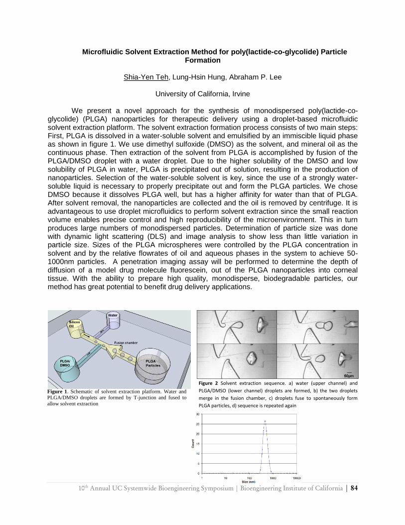
10th
Annual UC Systemwide Bioengineering Symposium | Bioengineering Institute of California | 84
Microfluidic Solvent Extraction Method for poly(lactide-co-glycolide) Particle Formation
Shia-Yen Teh, Lung-Hsin Hung, Abraham P. Lee
University of California, Irvine
We present a novel approach for the synthesis of monodispersed poly(lactide-co-
glycolide) (PLGA) nanoparticles for therapeutic delivery using a droplet-based microfluidic solvent extraction platform. The solvent extraction formation process consists of two main steps: First, PLGA is dissolved in a water-soluble solvent and emulsified by an immiscible liquid phase as shown in figure 1. We use dimethyl sulfoxide (DMSO) as the solvent, and mineral oil as the continuous phase. Then extraction of the solvent from PLGA is accomplished by fusion of the PLGA/DMSO droplet with a water droplet. Due to the higher solubility of the DMSO and low solubility of PLGA in water, PLGA is precipitated out of solution, resulting in the production of nanoparticles. Selection of the water-soluble solvent is key, since the use of a strongly water-soluble liquid is necessary to properly precipitate out and form the PLGA particles. We chose DMSO because it dissolves PLGA well, but has a higher affinity for water than that of PLGA. After solvent removal, the nanoparticles are collected and the oil is removed by centrifuge. It is advantageous to use droplet microfluidics to perform solvent extraction since the small reaction volume enables precise control and high reproducibility of the microenvironment. This in turn produces large numbers of monodispersed particles. Determination of particle size was done with dynamic light scattering (DLS) and image analysis to show less than little variation in particle size. Sizes of the PLGA microspheres were controlled by the PLGA concentration in solvent and by the relative flowrates of oil and aqueous phases in the system to achieve 50-1000nm particles. A penetration imaging assay will be performed to determine the depth of diffusion of a model drug molecule fluorescein, out of the PLGA nanoparticles into corneal tissue. With the ability to prepare high quality, monodisperse, biodegradable particles, our method has great potential to benefit drug delivery applications.
Figure 1. Schematic of solvent extraction platform. Water and PLGA/DMSO droplets are formed by T-junction and fused to
allow solvent extraction
Figure 2 Solvent extraction sequence. a) water (upper channel) and
PLGA/DMSO (lower channel) droplets are formed, b) the two droplets
merge in the fusion chamber, c) droplets fuse to spontaneously form
PLGA particles, d) sequence is repeated again

85 | 10th
Annual UC Systemwide Bioengineering Symposium | Bioengineering Institute of California
Engineering Transferrin-Diphtheria Toxin Conjugates for the Treatment of Glioblastoma Multiforme
Dennis J. Yoon1, Byron H. Kwan1, Felix C. Chao1,
Anne B. Mason2 and Daniel T. Kamei1
1Department of Bioengineering, University of California, Los Angeles 2Department of Biochemistry, University of Vermont College of Medicine
Glioblastoma multiforme (GBM) is a highly aggressive form of primary brain tumors with
a median survival rate less than a year post-diagnosis. To develop more effective treatments for GBM, ligands, such as transferrin (Tf), for receptors naturally overexpressed in cancer cells have been utilized as targeting moieties for cancer therapeutics, such as diphtheria toxin (DT). Though Tf-DT conjugates have shown promise, the short association time of Tf with cancer cells can limit its drug-carrier efficacy. Therefore, we have developed Tf mutants using mathematical modeling and site-directed mutagenesis with greater cellular association times and have shown that DT conjugates of these Tf mutants exhibit a greater cytotoxicity against HeLa cells than native Tf-DT. Additionally, we have applied our mutant Tf-DT conjugates to the treatment of GBM by performing in vitro cytotoxicity experiments on U87 and U251 human glioma cells. Results have shown a significant increase in cytotoxicity with our mutant Tf-DT conjugates compared to the native counterpart. We also investigated the mechanism of our Tf-DT conjugate‘s mode of action by performing a protein synthesis inhibition assay.
10th
Annual UC Systemwide Bioengineering Symposium | Bioengineering Institute of California | 86
Microneedle Drug Delivery System for Skin Diseases
Kevin Zhang, Benjamin Wu.
Biomedical Engineering IDP, University of California, Los Angeles
Current methods for treating many skin diseases rely on passive diffusion of topically
applied drugs on the skin surface. The stratum corneum layer, however, provides an efficient physical barrier to such technique due to the lack of vasculatures and innervations to carry drugs across the skin into the blood stream. As a result, drugs often are required to be applied multiple times at high dosage to set up high concentration gradient for effective drug diffusion. More importantly, drug molecules must be small and lipophilic so that diffusion of such species is more favorable. For many skin conditions, larger drug molecules, for instance growth factors and enzymes, provide more effective treatments for diseases such as scleroderma and skin burn, but the diffusion of these drugs is significantly limited. A patch of microneedles loaded with desired drug molecules can be utilized to overcome the stratum corneum barrier and increase efficiency of drug delivery. In this study, microneedles, which are fabricated from low-molecular-weight carboxymethycellulose (CMC), can be rapidly dissolved in the presence of water in the skin and release drug-encapsulated polycarprolactone (PCL) microspheres and nano-silver particles. PCL micospheres allow sustained release of drugs to maximize drug effectiveness while minimizing the required doses and the number of drug applications, which is highly desirable and convenient. The presence of nano-silver particles provides anti-microbial property. CMC microneedles have shown to possess high Young‘s Modulus and can successfully puncture the stratum corneum layer without distortion. Because of the micro-scale size of these microneedles, the microneedles only reach to the least innervated region of the skin, causing minimal or no pain. The materials used to construct the delivery system are chemically inert, dissolvable, and biodegradable. Therefore, such an innovative system shows potentials to become low cost, minimally invasive, and highly efficient drug delivery method to target a variety of skin conditions

87 | 10th
Annual UC Systemwide Bioengineering Symposium | Bioengineering Institute of California
The Effects of Coil Packing Density on Cerebral Aneurysm Inflow: In Vitro Assessment with Particle Image Velocimetry
Haithem Babiker1, L. Fernando Gonzalez2, Arius Elvikis3, Dan Collins3, Felipe Albuquerque2,
David Frakes1,4
1Harrington Department of Bioengineering, Arizona State University 2Barrow Neurological Institute, St. Joseph‘s Hospital and Medical Center 3Partnership for Research in Spatial Modeling, Arizona State University
4Department of Electrical Engineering, Arizona State University
Ruptured cerebral aneurysms cause an estimated 80% of fatal subarachnoid hemorrhages. Over the past decade, coil embolization has emerged as the endovascular treatment of choice for cerebral aneurysms. Nevertheless, current understanding of post-treatment aneurysm inflow and recurrence is both limited and highly qualitative. We present an in vitro study that quantifies the relationship between coil packing density and aneurysm inflow. Idealized models of basilar tip aneurysms with variable neck sizes were constructed from transparent silicon elastomer. A blood analog solution was circulated through the models at steady flow rates spanning a range of physiologic conditions. Aneurysms were embolized with bare platinum coils. Volumetric 3D flow velocity data were acquired at the aneurysm necks before and after each stage of multi-coil deployments using particle image velocimetry. Results showed a strong correlation between increased packing density and decreased aneurysm inflow. Specifically, aneurysm inflow in the narrowest neck model was reduced by 41.7% at a packing density of 36.5% for a parent vessel flow rate of 3 ml/s. Under the same parent vessel flow conditions, aneurysm inflow in the widest neck model was reduced by 65.8% at a packing density of 28.4%. To assess inter-deployment inflow variation, one of the aneurysm models was coiled repeatedly with different coils of the same model and size. At identical packing density, an average inflow discrepancy of 6.75% was observed over 6 repeat data acquisitions. Overall, the relative effects of coiling on aneurysm inflow were found to be more significant at lower parent vessel flow rates and for wider neck aneurysms. Marginal decreases in aneurysm inflow were also found to be greater at higher packing densities. Our in vitro study provides novel quantitative evidence to support clinical findings that high packing density contributes to decreased aneurysm recurrence rates through more complete embolization.

10th
Annual UC Systemwide Bioengineering Symposium | Bioengineering Institute of California | 88
Open-Surface Microfluidics Using Photosensitive Superhydrophobic Nanocomposite
Lingfei Hong1, 2, Hailin Cong1, Tingrui Pan1
1Department of Biomedical Engineering, University of California, Davis, USA 2School of Instrument Science and Optoelectronic Engineering, Beihang University, Beijing,
China
Open-Surface microfluidics present unique opportunities to address the insurmountable challenges in the conventional microfluidic systems, including hydrodynamic cavitation and optical obstruction. In this abstract, we present the recently developed photosensitive superhydrophobic nanocomposite combining superhydrophobicity of polytetrafluoroethylene (PTFE) nanoparticles and photopatternability and transparency of SU-8 photoresist for microfabrication of the open-surface microfluidic devices. General adaptability and simple processability of the superhydrophobic nanocomposite enable its excellent applicability in open-surface microfluidic devices for chemical and biomedical applications. In order to power the device two simple passive pumping mechanics have been investigated. In the first design, we utilize the Laplace pressure gradient generated by the surface tension on the different curvatures of the inlet and outlet droplets. [1-3] Therefore, the flow rates primarily dependent on the curvatures/shapes of the droplets, surface tension of the fluid, as well as the geometry and the resistance of the photo-defined open-surface microchannels [4-5]. Alternatively, an absorption pumping is constructed based on absorption and swelling property of the microfabricated structure (i.e., hydrogel) at the outlet. In comparison with the previous surface tension pump, the absorption pump offers more maneuver over the pump rate and volume by control of the geometry and diffusion of the absorption media, and thereby, high flow rates and high pump volume can be implemented in the miniature device.
89 | 10th
Annual UC Systemwide Bioengineering Symposium | Bioengineering Institute of California
Carbon Nanotubes lead to early Onset of Electrical Activity in Developing Hippocampal Neurons Cultured on Silicon Microelectrodes
Massoud L Khraiche1, Nathan Jackson1, Jit Muthuswamy1
1Harintgton‘s Department of Bioengineering, Arizona State University
In this study, we test the hypothesis that increased surface roughness resulting from
carbon nanotubes (CNTs) modification enhances neuronal adhesion and consequently electrical excitability of single neurons. Primary hippocampal neurons (embryonic day 18) are grown on CNT modified silicon microelectrode arrays (MEAs). Electrophysiology revealed initiation of multi-unit activity as early as 4 days after seeding compared to 7 days in control cultures grown on microelectrodes without CNTs. Progressive maturation of single neuronal action potentials as manifested by increasing peak-to-peak amplitudes and firing rate, were observed days 4-7 on microelectrodes with immobilized CNTs. The results overall, demonstrate earlier onset and significantly higher level of electrical activity in neurons seeded on CNT modified MEAs compared to non-modified control MEAs. We conclude that CNTs on microelectrodes enhance electrical excitability of single neurons in culture.
10th
Annual UC Systemwide Bioengineering Symposium | Bioengineering Institute of California | 90
Long-Term Oxygen Sensor Implantation in the Porcine Subcutaneous Environment
L.S. Kumosa†, J. Lin‡, T. Routh‡, J. Lucisano‡, D.A. Gough†‡
†Biosensors Lab, Department of Bioengineering, UCSD
‡Glysens, Inc., San Diego, CA
The effect of long-term implantation on metabolically active devices is of utmost importance for not only the success of implanted glucose sensors used in diabetic therapy, but also for the development of artificial tissues and encapsulated cell devices. Such devices are also dependent on the constant, predictable supply of metabolites from the local vasculature. Long-term implantation leads to the formation of a foreign body capsule whose purpose is the protection of the host and isolation of the foreign material from local tissue resources. While this encapsulation is successful in protecting tissues from invading agents, metabolite flow continues, albeit at greatly reduced levels. The understanding of this encapsulation process is critical for the design and successful implementation of active implants dependent on metabolite supply. The project goal is to utilize implantable wireless telemeters designed and manufactured by Glysens, Inc. to understand changes in oxygen levels of the surrounding subcutaneous tissues over the course of implantation in pigs. This is performed in three distinct, yet interrelated parts, namely: the analysis of oxygen signals collected from the long-term implantation of telemeters, the histological analysis of serial tissue samples collected from regions adjacent to the telemeters over the course of implantation, and the development of an accurate model for the dynamics of metabolite supply to implanted devices over the long-term. The successful completion of this project will lead to better understanding of the tissue changes that occur during the foreign body reaction and the improved design and implementation of devices that require constant tissue-supplied metabolites.

91 | 10th
Annual UC Systemwide Bioengineering Symposium | Bioengineering Institute of California
A simple three-dimensional vortex micromixer
Maureen Long1, Michael A. Sprague2, Anthony A. Grimes1, Brent D. Rich1, and Michelle Khine1
1School of Engineering, University of California, Merced
2School of Natural Sciences, University of California, Merced
We demonstrate rapid homogenous micromixing at low Reynolds numbers in an easily fabricated and geometrically simple three-dimensional polystyrene vortex micromixer. Micromixing is critically important for miniaturized analysis systems. However, rapid and
effective mixing at these small scales remains a persistent challenge. We compare our micromixer's performance against a two-dimensional square-wave design by examining its effectiveness in mixing solutions of dissimilar concentration as well as suspension solutions comprised of microparticles. Numerical simulations confirm our experimental observations and provide insights on the self-rotational mixing dynamics achieved with our simple geometry at low Reynolds numbers. This rapid, robust, and easily fabricated micromixer is amenable readily to large scale integration.
10th
Annual UC Systemwide Bioengineering Symposium | Bioengineering Institute of California | 92
Immobilization of Lactate Oxidase for Stability and High Loading in a Lactate Sensor
Adam Strobl, Henry Tse, David Gough
Department of Bioengineering, University of California, San Diego
Knowledge of metabolic state is the cornerstone of treatment for diabetic patients. The
current standard of care relies on infrequent discrete measurements of one metabolite, glucose. Knowledge of other important variables such as lactic acid would help form a more complete picture of the patient‘s metabolism. In addition, continuous sensors allow the gathering of dynamic information so that all metabolic excursions can be tracked and treated. Other patients suffering from pulmonary or cardiac impairments would also benefit from continuous lactate information. In comparison with continuous glucose sensors in development, lactate sensors have a short lifespan due to the instability of the enzyme lactate oxidase (LOx). Past results show a lifespan on the order of three weeks for continuous lactate sensors; this is unacceptable in a subcutaneous implant since frequent surgeries would be necessitated.
Enzyme immobilization is necessary for the construction of a successful implantable sensor. We have characterized LOx in immobilized form in order to understand how immobilization affects sensor lifespan. In particular, the effect of process parameters on the resulting activity and stability of LOx in an enzyme-loaded membrane have been studied. A chemical immobilization procedure utilizing glutaraldehyde was developed and optimized to give maximum enzymatic yield and stability at physiological conditions. Results were analyzed through a reaction/diffusion model to calculate the effects of diffusion on the spectrophotometric assay utilized. In addition, we are developing methods for immobilization based on electrostatic interaction. There is evidence that this processing technique can lead to higher activity yields, and literature also indicates the possibility of increased stability. Finally, the loading levels and stabilization achieved in vitro are used to estimate the lifetime of an in vivo sensor based on a previously developed model. Using parameter results achieved in the lab, sensor lifetime is expected to be four to five months.
93 | 10th
Annual UC Systemwide Bioengineering Symposium | Bioengineering Institute of California
A Smart Contact-Lens Sensor for Dynamic Measurement of Intraocular Flow Resistance
Chaoqi Zhang, Tingrui Pan
Department of Biomedical Engineering, University of California, Davis
Worldwide more than 60 million people suffer from glaucoma, a leading cause of the irreversible blindness due to the optic nerve damage. [1-4] Although the tonometry-based intraocular pressure (IOP) measurement has been used as the gold standard to monitor the glaucoma for over a century, a new diagnostic device for early glaucoma detection is highly in demand, due to the indirect correlation, imprecise measurement principle, operation-sensitive assessment of the current tonometry. In this abstract, we proposed a new dynamic measurement principle to assess intraocular flow resistance, a direct indication of ocular flow circulation. Analogous to the circuit analysis, ocular volume is altered through a coupled contact-lens device while the IOP change is continuously tracked. Such a smart contact-lens measurement device has been fabricated using soft lithography on polymer materials. In addition, an artificial ocular model is constructed for evaluation and calibration of the dynamic measurement system.
10th
Annual UC Systemwide Bioengineering Symposium | Bioengineering Institute of California | 94
Postprandial up-regulation of monocyte integrin CD11c/CD18 increases firm arrest to vascular cell adhesion molecule-1
R Michael Gower1, Anne A Knowlton2, Scott I Simon1
1Biomedical Engineering Department,
2Molecular & Cellular Cardiology and Cardiovascular Division, Department of Medicine, 3Department of Medical Pharmacology, University of California, Davis
Monocyte activation and migration into the arterial wall are key events in atherogenesis. Monocyte integrin CD11c/CD18 mediates adhesion to vascular cell adhesion molecule (VCAM)-1 and knocking out CD11c in hypercholesterolemic mice lowers the incidence of atherosclerosis. Both suggest CD11c may be an attractive therapeutic target for cardiovascular disease (CVD). We demonstrate that monocyte CD11c expression and function can be efficiently screened in whole blood. Flow cytometric analysis of fasting and postprandial blood samples from 37 healthy human donors revealed that a high fat meal induces a 10% increase in monocyte CD11c expression. Plasma triglyceride concentration significantly correlated with the increase in CD11c (P < 0.0001; Pearson r = 0.6419; 95% CI: 0.4013 to 0.7996). When donors were grouped by plasma triglyceride, those above 160 mg/dL experienced a 30% increase in CD11c postprandial, while those below 90 mg/dL exhibited no increase. Monocyte inflammatory potential following the meal was measured by recruitment to VCAM-1 in shear flow. Cover slips coated with recombinant VCAM-1 were incorporated into a microfluidic device and blood was perfused over these cover slips at a shear force of 4 dynes/cm2. Firmly arrested monocytes were enumerated by cover slip staining. The fraction of monocytes among total leukocytes firmly arrested on VCAM-1 increased 50% postprandial. Firm arrest could be decreased 40% with an antibody that inhibits CD11c or increased 55% with a CD11c agonist. We conclude that following a high fat meal the systemic circulation exerts pro-inflammatory effects on monocytes and activation can be sensitively monitored by changes in CD11c expression. Thus CD11c is a functional biomarker that correlates with known risk factors of CVD and is important for monocyte adhesion to VCAM-1.
95 | 10th
Annual UC Systemwide Bioengineering Symposium | Bioengineering Institute of California
Engineered proteolytic antibody fragments as therapeutics for Alzheimer’s disease
Srinath Kasturirangan1, Michael Sierks2
1Harrington Department of Bioengineering, Arizona State University 2Department of Chemical Engineering, Arizona State University
Deposition of beta-amyloid (Aβ) is considered an important early event in the
pathogenesis of Alzheimer's Disease (AD) and reduction of Aβ levels in the brain can be a viable therapeutic approach. A potentially non-inflammatory approach to facilitate clearance and reduce toxicity is to hydrolyze Aβ at its -secretase site using single chain antibody fragments (scFvs). We have previously identified antibody light chain mk18 having -secretase-like catalytic activity producing the 1-16 and 17-40 amino acid fragments of Aβ40. To improve the specific activity of the recombinant antibody by affinity maturation, we constructed a yeast surface displayed scFv library by mutating the CDR3 heavy chain region responsible for antigen recognition. A biotinylated covalently reactive analog mimicking -secretase site cleavage site was synthesized, immobilized on streptavidin beads, and used to select scFvs with increased specificity for Aβ. Following bio-panning, a fluorescently labeled A substrate was used to further screen isolated clones for improved activity. Two clones which showed the highest increase in proteolytic activity compared to the parent mk18 were selected for further analysis. Kinetic analyses using purified soluble scFvs showed a 3- and 6-fold increase in catalytic activity (kcat/KM) toward the synthetic Aβ substrate compared to the original scFv primarily due to an expected decrease in KM rather than an increase in kcat. Affinity maturation strategy resulted in highly stable scFv with improved affinity and activity towards A. These clones prevented aggregation of Ain-vitro as determined by atomic force microscopy (AFM) and reduced A induced cytotoxicity towards SHSY5Y neuroblastoma cells. The ability of the proteolytic scFv to cleave pre-formed Aoligomers, the predominant toxic species, as well as their capacity to cleave the amyloid precursor protein (APP), producing the neuroprotective sAPP fragment, are also being studied. These proteolytic scFvs could have potential therapeutic applications for AD by decreasing soluble Aβ levels in vivo.
10th
Annual UC Systemwide Bioengineering Symposium | Bioengineering Institute of California | 96
Sudden death from gut ischemia may result from a neurogenic shock mechanism
Alexander Hayes Penn, Geert W. Schmid-Schönbein
Department of Bioengineering, University of California, San Diego
Hemorrhagic shock is a life-threatening event. Splanchnic arterial occlusion (SAO)
followed by reperfusion is an established model for physiologic shock. Even without reperfusion, SAO is highly lethal. We show here that death is either associated with gradual drops in mean arterial blood pressure (MABP) over <1 to 3 hours following onset of ischemia or with a severe rapid drop in MABP (often > 80 mmHg) over 1 to 6 minutes. The rapid drop occurs after ~ 75-150 min of ischemia. We observed that a 10% glucose solution added to the lumen of the small intestine significantly decreases the onset of the fatal drop and significantly increases its rate of occurrence (from 29% to 63%). As gut ischemia stimulates the sympathetic nervous system and afferent neurons in the gut can detect hyperglycemia, our objective was to determine whether a total subdiaphragmatic vagotomy (TSV), could prevent the rapid drop in MABP by interfering with the autonomic nervous system. We found that survival time was increased by TSV (P<0.05). TSV significantly reduced the instances of rapid drops in MABP (P<0.008), though gradual pressure drops still occurred. Addition of a broad-spectrum digestive enzyme inhibitor, previously shown to improve the outcome of SAO followed by reperfusion, did not prevent the fatal rapid drops in blood pressure, supporting the hypothesis that these deaths are due to a separate mechanism from that of typical physiologic shock. These results suggest that the autonomic nervous system can cause rapid death with gut ischemia, especially after meals high in glucose. Supported in part by HL 67825, 76180 and an unrestricted educational gift from Leading Ventures.
97 | 10th
Annual UC Systemwide Bioengineering Symposium | Bioengineering Institute of California
Development of FRET-Based High-Throughput Screening to Discover Small Chemical Inhibitors Targeting Protein-Protein Interaction in the SUMOylation Network
Yang Song, Vipul Madahar, Yan Liu, Jiayu Liao
Department of Bioengineering at University of California, Riverside
Covalent modifications of proteins are general mechanisms to alter protein functions in
most cells, especially in eukaryotic cells. SUMO (Small Ubiquitin-like MOdifier) is a well-known small protein family which is attached to proteins involved in diverse cellular processes by the catalytic enzyme cascade, E1, E2 and E3. However, the investigations of SUMOylation in different cellular processes are limited by the difficulties in determining the interaction between SUMO, SUMO E1-E3 enzymes and the substrates as well as the lethality of gene knockout studies given the importance roles of SUMO in vivo. Using sensitive fluorescence proteins by genetic labeling techniques, our lab is currently developing FRET-based methods to test the in vivo interactions between different proteins involved in the SUMOylation network. We are also developing these methods into high-throughput screening assays for novel small chemical inhibitor screening, which can disrupt the protein-protein interactions between SUMO and its E1, E2 and E3 enzymes. Once discovered, these small chemical compounds will not only serve as unique tools for the functional studies of SUMOylation, but also can have great potentials in the treatment of diseases related to the dysregulation of SUMOylation network.
10th
Annual UC Systemwide Bioengineering Symposium | Bioengineering Institute of California | 98
Cellular Uptake of Polyarginine-Polyleucine Block Copolymer Vesicles
Victor Z. Sun, Zhibo Li, Timothy J. Deming, and Daniel T. Kamei
Department of Bioengineering, University of California, Los Angeles
We previously reported the preparation of vesicles composed of lysine-leucine (K60L20) block copolypeptides. These vesicles were stable in an aqueous environment, and could be processed to different sizes. They could also be prepared in large quantities, and were found to be able to encapsulate water-soluble contents. These vesicles, however, were not able to cross the plasma membrane of cells for intracellular delivery of a payload. To engineer this added feature, we investigated the literature, and found that the arginine-rich HIV-1 Tat peptide (GRKKRRQRRRAP59) and oligomers of arginine can transport molecules into cells. We therefore reasoned that the lysine residues in our copolypeptides could be replaced with arginine without disrupting their ability to form vesicles while also enabling them to enter cells. Subsequent experiments confirmed our hypothesis, as R60L20 block copolypeptides could form vesicles with similar properties as the K60L20 vesicles, while additionally being able to deliver water-soluble Texas-Red dextran into various mammalian cell lines. In the current study, we examined the endocytosis and intracellular trafficking pathways of the R60L20 vesicles. Specifically, we investigated the mechanism by which the R60L20 vesicles could enter cells by preparing 100 nm diameter FITC-labeled vesicles and incubating them with cells in the presence of different drugs that inhibit various endocytosis pathways. To investigate the fate of the R60L20 polypeptide vesicles inside the cell, we performed immunofluorescence labeling of the endosomes and lysosomes in cells loaded with FITC-labeled R60L20 vesicles. The ability of these vesicles to deliver bioactive therapeutic agents is also currently being tested.
99 | 10th
Annual UC Systemwide Bioengineering Symposium | Bioengineering Institute of California
Design of an Aptamer Beacon for Real-Time Detection of Interferon-Gamma
Nazgul Tuleuova1,2, Caroline N. Jones1, Jun Yan1, Erlan Ramanculov,2 Alexander Revzin1
1Department of Biomedical Engineering, University of California, Davis 2National Center for Biotechnology, Astana, Kazakhstan
Aptamers are nucleic acid ligands, either RNA or DNA, that have been selected to bind a
huge variety of targets ranging from whole cells to metal ions. The aptamers nucleic acid structure ensures chemical and thermal stability of these molecules making them real rivals to antibodies and also allows to develop novel sensing strategies. Aptamer beacons represent one such sensing strategy where is an aptamer and shorter complimentary oligonucleotide conjugated with fluorophore-quencher pairs and where fluorescence signal changes as the function of hybridization/dehybridization due to the analyte binding. The goal of the present
study was to develop an aptamer beacon for detection of interferon-gamma (IFN-) – an important inflammatory cytokine commonly monitored in clinical setting and basic research. For testing we immobilized biotinylated aptamers with different orientation (immobilization via 3‘-end or 5‘-end) and distance to the surface (incorporation of a spacer at the ends) onto avidin-coated surfaces and were examined using surface plasmon resonance (SPR) and fluorescence
microscopy. SPR studies of IFN- - aptamer interactions revealed the equilibrium constant for the best scenario was 3.44 nM for immobilization via 3‘-end. In addition, SPR was employed to
characterize interaction IFN- a double-stranded DNA construct comprised of fluorophore-labeled aptamer and a complementary sequence containing a quencher. These SPR studies revealed that cytokine binding occurred rapidly within 30 mins – pointing to the possibility of
dynamic replacement of quencher-complementary strand with IFN- molecules. Importantly, these results where corroborated by fluorescence microscopy experiments where a shift in fluorescence intensity was observed from low fluorescence due to the quencher-complementary
strand hybridized with fluorophore-aptamer to high fluorescence after introduction of IFN- into
the system. This change in signal was observed in real-time and was dependent on IFN- concentration. This aptamer beacon had a detection limit of 10 nM which is sufficient to monitor physiological levels of this cytokine. In conclusion, we designed an aptamer beacon for real-
time monitoring of IFN-. In the future, this sensing strategy will be employed to monitor in real-time cytokine production by the immune cells.
10th
Annual UC Systemwide Bioengineering Symposium | Bioengineering Institute of California | 100
Solid-Phase Peptide Synthesis of Bioinspired Electrets Based on Non-Traditional Amino
Acids: Synthesizing oligo-ortho-anthranilic Acids for Improved Charge-Transfer Properties in Photovoltaic Cells
Srigokul Upadhyayula1,2, Duoduo Bao1, David Bui1, Valentine I. Vullev1 1Department of Bioengineering, University of California, Riverside
2Department of Biochemistry, University of California, Riverside
As of 2008, fossil fuels account for 77.88% of energy production; while only 0.02% is generated using renewable energy sources (Science 2007, 315, 798-801). The cost of solar energy conversion technology is the main drawback in the primary widespread use of photovoltaic devices (Nature Materials 2006, 5, 161-164; PNAS 2006, 103, 5251-5255). Conversion of fossil fuels to electricity costs approximately 0.05 kWh-1, whereas converting solar energy cost approximately 5 times higher (Monthly Energy Review, DOE/EIA-0035, Janary 2009). The gap between energy generated via fossil fuels vs. photovoltaics exists solely due to price and can be bridged either by an increase in the efficiency of solar cells made of inexpensive materials, or by lowering the cost of the materials used for the fabrication of highly efficient photovoltaic devices. Utilizing de novo macromolecular design and engineering, we aim at bioinspired electrets that can mediate efficiently long-range charge transfer and at the same time, suppress the undesired charge recombination that is responsible for energy loss. We demonstrated that, similar to protein -helices, oligo-anthranilamides possess considerable intrinsic dipole moments making them highly promising macromolecular electrets (Ashraf et al. Biotech. Progress 2009, in press). Although others and we currently prepare oligoanthranilamides using conventional solution-phase coupling, it is highly inconvenient for exploring a broad range of such conjugates. Moreover, others and we have observed that traditional solid-phase synthetic procedures (which can be automated and conducted in parallel) do not yield coupling between anthranilic residues, and hence, solid-phase synthesis has never been used for making such conjugates. Our experimental analysis indicated that the steric hindrance was the principal reason for the observed failure of the ―traditional‖ approaches. Utilizing small-size active acid intermediates allowed us for the first time to prepare large-molecular weight oligoanthranilamides using automated solid-phase synthesis. We explored in situ and ex situ activation. We believe that these procedures for preparation of bioinspired macromolecular electrets can be immensely beneficial for the facile automated parallel synthesis of a large range of biomaterials of non-traditional amino acids.
101 | 10th
Annual UC Systemwide Bioengineering Symposium | Bioengineering Institute of California
The Rehm-Weller Equation in View of Bioengineering
Duoduo Bao, Antonio Contreras, and Valentine I. Vullev
Bioengineering Department, University of California, Riverside
Charge transfer is a key process for all redox functionalities in biological systems. The Rehm-Weller equation allows for etimation of the driving force of photoinduced charge-transfer processes. A term, based on the Born equation for solvation energy of ions, is introduced in the Rehm-Weller equation to correct for the differences measuring media. The Born correction term, however, requires a prior knowledge of the dielectric constants of the electrolyte solutions used for the redox measurements and the radii of the donor and acceptor molecules. Because of limited information for such dielectrics, the values for the dielectric constants of electrolyte solutions are approximated to the values of the dielectric constants of the corresponding neat solvents. We determined that this approximation can lead to significant errors in the calculated values of photoinduced charge transfer [1], as well as to discrepancies between the measured and estimated charge-separation kinetics [2]. We demonstrated the use of non-linear extrapolation for elimination of the dielectric-constant approximation [1]. The other key component for estimation the kinetics and thermodynamics of charge transfer is the ionic radii of the donor and the acceptor. Usually, the ionic radii are estimated from to the van der Waals dimensions of the redox species. This estimation assumes that the charges are delocalized over the whole molecule, which for a range of biologically active redox species is wrong. Recently, we reported a discrepancy that implied that the ionic radius of acridinium is smaller than its molecular radius [3]. Extending our extrapolation cyclic-voltammetry methodology to non-polar media allowed us to determine the ionic radii of electron donors and acceptors, which are not necessarily their van der Waals radii. We believe that our advances will allow for improved characterization of biological redox process and of redox species with biological activity. 1Bao et al. J. Phys. Chem. A 2009, 113, 1259-1267 2Wan et al., J. Photochem. Biotobiol. A: Chem. 2008, 197, 364-374 3Hu et al., J. Phys. Chem. A 2009, 113; 3096–3107
10th
Annual UC Systemwide Bioengineering Symposium | Bioengineering Institute of California | 102
Ca2+ Depletion of Sarcoplasmic Reticulum during Reperfusion after Ischemia
Marcela Ferreiro1, Dmytro Kornyeyev1, Carlos A. Valverde2, Alicia Mattiazzi2 and Ariel L. Escobar2
1School of Engineering, University of California, Merced
2School of Medicine, Universidad Nacional de La Plata, Argentina
One of the negative consequences of myocardial infarction is cytosolic Ca2+ overload occurring shortly after the restoration of the blood flow through coronary arteries. In order to investigate this phenomenon we conducted ischemia-reperfusion experiments on intact mouse hearts. Our hypothesis was that the cytosolic Ca2+ overload is associated with the redistribution of intracellular Ca2+ from sarcoplasmic reticulum (SR) to cytosol. To address this issue, we performed 12 min of global no-flow ischemia followed by reperfusion in the isovolumic Langendorff perfused mouse heart positioned on a Pulsed Local Field Fluorescence microscope and loaded with fluorescent dyes (Rhod-2 or Mag-Fluo-4 to assess cytosolic or SR Ca2+, respectively) or membrane potential (di-8-ANNEPS). The results indicated there is an initial increase in diastolic Ca2+ during early reperfusion that gradually returned to pre-ischemic levels. This increase was associated with a decrease in SR Ca2+ content that recovered within 10 min, as a mirror image of the diastolic Ca2+ profile. Additionally, experiments in which caffeine pulses (20 mM) were applied, confirmed that SR Ca2+ content was greatly diminished at the onset of reperfusion. Moreover, both Ca2+ release and Ca2+ reuptake slowed down significantly during the ischemia and restored during reperfusion. Action potentials recorded during ischemia were shorter (no normal spike-and-dome morphology) probably due to the inhibition of Ca2+ release. This is in agreement with the decrease in Ca2+ content in SR during reperfusion. Interestingly, similar changes occurred when a pulse of caffeine was applied. The present findings indicate that the increase in diastolic Ca2+ that occurs upon reperfusion is due to a SR Ca2+ release and not just because of the Ca2+ entry through the reverse NCX mode, as has been previously thought. Supported NIH R01-HL-084487 to AE.
103 | 10th
Annual UC Systemwide Bioengineering Symposium | Bioengineering Institute of California
Mapping the Position of DNA Polymerase-Bound DNA Templates in a Nanopore at 5Å Resolution
Daniel R. Garalde1, Brett Gyarfas1, Felix Olasagasti2, Seico Benner2, William Dunbar1, Kate R.
Lieberman2, Mark Akeson2
1Department of Computer Engineering, Baskin School of Engineering, University of California, Santa Cruz
2Department of Biomolecular Engineering, Baskin School of Engineering, University of California, Santa Cruz
Nanopores have emerged as a method for single molecule analysis of DNA, RNA, and
protein complexes formed with these polymers. In this study, we used the α-hemolysin nanopore to study complexes of DNA and the Klenow fragment of Escherichia coli DNA polymerase I (KF) or T7 DNA polymerase (T7 DNA pol). Ionic current flows through the nanopore as a result of an applied electrical potential and the current is obstructed when DNA is drawn into the nanopore by electrophoresis. A block of abasic (1‘,2‘-dideoxy) residues within a DNA template can be detected if the abasic residues are positioned within a restricted region of the nanopore because they cause less obstruction of ion flow than standard nucleotides. We mapped the sensitivity of the α-hemolysin nanopore to abasic residue position by designing a series of DNA template-primer hybrids, each with a block of three abasic residues placed at a different position along the template DNA. We measured ionic current blockades during nanopore capture of these DNA hybrids bound in complexes with either KF or T7 DNA pol. A single nucleotide displacement of the abasic block position could be clearly distinguished. Maps of ionic current as a function of the position of abasic residues for templates bound to KF or T7 DNA pol were nearly identical, indicating with single nucleotide resolution that when captured atop the nanopore, both enzymes hold the DNA in a similar position
10th
Annual UC Systemwide Bioengineering Symposium | Bioengineering Institute of California | 104
Concentrating DNA Using Two-Phase Aqueous Micellar Systems
Foad Mashayekhi, Aaron S. Meyer, Stacey A. Shiigi, Daniel T. Kamei
Department of Bioengineering, University of California, Los Angeles
The concentration of cancer biomarkers, such as DNA, prior to a subsequent detection step may facilitate the early detection of cancer, which could significantly increase chances for survival. In this study, the partitioning behavior of mammalian genomic DNA fragments, as well as oligonucleotides, were experimentally and theoretically investigated in a two-phase aqueous micellar system. The micellar system was generated using the nonionic surfactant Triton X-114 and phosphate-buffered saline. Partition coefficients were measured under a variety of conditions and compared with our theoretical predictions, demonstrating that the partitioning behavior of DNA fragments in this system is primarily driven by repulsive, steric, excluded-volume interactions that operate between the micelles and the DNA fragments, but is limited by the entrainment of micelle-poor, DNA-rich domains in the macroscopic micelle-rich phase. Furthermore, the volume ratio, that is, the volume of the top, micelle-poor phase divided by that of the bottom, micelle-rich phase, was manipulated to concentrate DNA fragments in the top phase. Specifically, by decreasing the volume ratio from 1 to 1/10, we demonstrated proof-of-principle that the concentration of DNA fragments in the top phase could be increased 2- to 9-fold in a predictive manner.
105 | 10th
Annual UC Systemwide Bioengineering Symposium | Bioengineering Institute of California
Fluorescence Lifetime Imaging Microscopy (FLIM) for Cancer Demarcation during Medical Surgery
Yinghua Sun1,2, Jennifer Phipps1, Daniel S. Elson3, Jeremy Meier4, Nisa Hatami1, Frank S.
Chuang2, Rudolph J. Schrot5, D. Gregory Farwell4, and Laura Marcu1,2
1Department of Biomedical Engineering, University of California, Davis
2NSF Center for Biophotonics Science & Technology, University of California, Davis 3Imperial College London, London, U. K.
4Department of Otolaryngology, University of California, Davis 5Department of Neurological Surgery, University of California, Davis
A compact endoscopic fluorescence lifetime imaging microscopy (FLIM) system was
developed for intraoperative disease diagnosis in clinics. By extracting multiple parameters including fluorescence intensity, spectrum, and lifetime, FLIM has the inherent advantages for in vivo application because the time-resolved analysis is minimally affected by the irregular tissue surface, the non-uniform illumination, and the presence of endogenous absorbers. The endoscopic design in this system allowed for the remote image collection via a flexible fiber imaging bundle (2 m long, 0.6 mm outer diameter, 10,000 optical fibers) cemented with a gradient index lens (0.5 mm diameter) as the objective. Tissue autofluorescence was induced with 337 nm, 700 ps pulsed laser. Fluorescence images were recorded using a compact gated ICCD camera with the temporal resolution up to 200 ps. The spatial resolution of the imaging system was examined as 35 μm in the field of view with a 4 mm diameter. Pertinent emission wavelengths were selected by bandpass filters in a motorized filter wheel. The system performance and imaging processing were calibrated with standard fluorophores. A hamster model was used to validate the system in vivo for carcinoma characterization. A significant drop of fluorescence lifetime from 2.5±0.36 ns to 1.77±0.26 ns at 450/65 nm and an 81% intensity decrease at 390 nm were observed in tumor regions compared with normal healthy tissue. Finally the portable FLIM system was installed on a mobile cart for clinical applications in operating room. 10 patients were studied for tumor delineation during head and neck surgery and 4 patients were examined for glioblastoma. This work demonstrates the potential of FLIM as a non-invasive intraoperative diagnostic technique.
10th
Annual UC Systemwide Bioengineering Symposium | Bioengineering Institute of California | 106
Multi-photon Optical Microscopy of Actin Filaments and Mitochondrial Bioenergetics of ACBT Human Frade IV Flioblastoma Cells Migrating within 3-D Collagen-based Hydrogels
Miso Yang1, Yu-Jer Hwang1, Edgar Sanchez2, Chung-ho Sun2, Tatiana B. Krasieva2,
Bruce J. Tromberg2, 3, Julia G. Lyubovitsky1
1Bioengineering Department, University of California, Riverside 2Laser Microbeam and Medical Program, Beckman Laser Institute, University of California,
Irvine 3Biomedical Engineering, University of California, Irvine
Multi-photon optical microscopy (MPM) that combines second harmonic generation
(SHG) and two-photon fluorescence (TPF) signals to allow many opportunities to non-destructive probing of molecular processes in tissue with high spatial resolution and contrast in real time. In this study, we created 3-D mitochondial metabolic state maps of ACBT human grade IV glioblastoma cells migrating within 3-D collagen-based hydrogels using the multi-photon ratiometric redox fluorometry method. The average mitochondrial metabolic state number was 0.9, which indicates that for the majority of mitochondria, the fluorescence was equally distributed between reduced nicotinamide adenine dinucleotide (NADH) and oxidized flavin adenine dinucleotide (FAD). Apoptosis and aging are strongly associated with the interactions of mitochondria with actin cytoskeleton. Therefore, we established protocols to probe the distribution of F-actin filaments in ACBT glioblastoma cells that migrate within 3-D collagen-based hydrogels. Specifically, we both stained and transiently transfected ACBT cells with green fluorescent protein (GFP)-actin to follow actin reorganization during cell migration in real time.

107 | 10th
Annual UC Systemwide Bioengineering Symposium | Bioengineering Institute of California
Optical Model of Human Skin for Biomedical Reflectance and Fluorescence Spectroscopy
Dmitry Yudovsky and Laurent Pilon
Inter-Departmental Biomedical Engineering Program, University of California, Los Angeles
Biomedical fluorescence spectroscopy has been used in many applications, including
detection of malignant tissue, feedback during ablative laser therapy, non-invasive monitoring of drug delivery, and real-time non-invasive glucose monitoring. Human skin is the largest and most accessible organ of the body. Furthermore, changes in its reflective and fluorescence properties are known to correlate with pathological and normal bodily function. Therefore, there is a great interest in developing optical methods and algorithms for analyzing the reflectance and fluorescence of skin for non-invasive health-monitoring.
Analysis of light transfer through biological tissue is complicated by tissue heterogeneity. Skin is made of several layers which absorb and scatter incident and fluorescent light. The top layer (the epidermis) is characterized by strong absorption due to melanin and fluorescence by tryptophan, keratin, and tyrosine. Beneath the epidermis is the dermis whose optical properties are affected by the presence of blood, collagen, NADH, pepsin, and tryptophan. Blood has drastically different absorptive and scattering characteristics that vary with oxygen saturation.
Spectroscopic analysis of skin has been based on approximate models of light transfer such as Beer-Lambert‘s law or the diffusion approximation. Then, skin is treated as a semi-infinite and homogenous medium. Unfortunately, these methods provide an incomplete and often inaccurate assessment of light transfer through biological tissue. More complex, multilayered Monte Carlo models have been developed, but are too slow and computer intensive to use in real-time spectroscopy or in inverse method for non-invasive monitoring of skin.
In this study, a model of light transfer through skin, treated as a two-layer medium, is developed that is both realistic and computationally efficient. Skin reflectance and fluorescence as well as the distribution of excitation and fluorescence irradiances through the tissue are modeled taking into account optical and geometric properties of the epidermis and dermis.
10th
Annual UC Systemwide Bioengineering Symposium | Bioengineering Institute of California | 108
Bench Scale Electroenzymatic Biosensor for the Rapid Detection of Pyruvate
Lorenzo D‘Amico, Andrew Basilio, Si Luo, Justin Yeap, and Dale A. Baker, Ph.D.
Department of Bioengineering, University of California, San Diego
A bench-scale electrochemical biosensor was developed for the rapid detection of pyruvate. The design is based on the enzyme, pyruvate oxidase, which is entrapped in a membrane in contact with a Clark-type oxygen sensor. During the reaction catalyzed by pyruvate oxidase, pyruvate and oxygen are consumed in a one-to-one ratio. Utilizing this ratio, the concentration of pyruvate can be related to the amount of oxygen that is consumed while a sample of pyruvate diffuses across the enzyme membrane. The sensor has been shown to respond to a step in pyruvate within 5 minutes and to pyruvate concentrations, between 50 and 1500 μM, which covers a wide physiological range. This amperometric sensor was operated at 37oC to assess its potential for an implantable sensor design. The bench-top sensor was build to be an alternative to the standard spectrophotometric method for determining pyruvate from blood samples, a method which usually requires perchloric acid treatment for deproteinizing the samples, and thus, centrifugation as well. So to avoid this pre-treatment inconvenience, we have developed a simple, inexpensive and reliable electrochemical method for determining pyruvate. The concentration level of lactate to pyruvate or L/P ratio is used to assess ischemia, circulatory shock and hypoxia. The L/P ratio has at times been shown to be a better indicator than just the lactate concentration in some forms of ischemia, hypoxia and concomitant cellular dysfunction .A person at rest typically has a blood L/P ratio of 10-15:1. When experiencing hypo-perfusion, ischemia or hypoxia, the L/P ratio increases, which indicate an abnormality. Elevated levels are considered to be L/P ratio greater than 20. The severity of the abnormality as indicated by the ratio depends on the specific ischemic, hypoxic or shock situation. Pyruvate levels are also helpful in the evaluation of novel oxygen therapeutics and their role in resuscitation.
109 | 10th
Annual UC Systemwide Bioengineering Symposium | Bioengineering Institute of California
Effects of Coating Material on Cellular Uptake of Nanocapsules Loaded with Indocyanine Green
Bongsu Jung, Bahman Anvari
Department of Bioengineering, University of California Riverside
Indocyanine green (ICG) is an FDA-approved near-infrared (NIR) fluorescent dye used
in ophthalmic angiography and assessment of liver function angiography. However, clinical applications of ICG remain very limited due to its rapid clearance from general circulation and unstable optical properties. To overcome these limitations, we have encapsulated ICG within nanoscale biopolymers. Nanoencapsulation of ICG can be potentially applied for tumor-targeting purposes by further surface coating and or functionalization of the capsule surface. ICG containing nanocapsules offer a dual ability for optical imaging and optical therapy in that the same nanocapsule system can be irradiated with laser light to elicit a thermal or chemical response that will lead to destruction of the targeted structures. Our preliminary results demonstrate that ICG containing nanocapsules coated with the composite magnetite and polyethylene glycol (PEG) material deposit in greater amounts within the lungs of healthy mice than nanocapsules coated with polylysine or dextran. To understand the effects of coating materials on the cellular distribution of the nanocapsules, we measure the uptake of ICG containing nanocapsules coated with various materials by different cells including peripheral blood monocytes, and normal and cancerous bronchial epithelial cells. Results of these studies will provide important information for subsequent applications of ICG containing nanocapsules that can be used for targeted optical vascular and tissue imaging as well laser therapy.
10th
Annual UC Systemwide Bioengineering Symposium | Bioengineering Institute of California | 110
Novel Dielectrophoretic Device for Cancer Cell, Stem Cell and DNA Biomarker Isolation and Detection
Rajaram Krishnan1, Joaquim Teixeira2, Jennifer Y. Marciniak1, Mark Mercola2, Sadik C. Esener3
and Michael J. Heller1,3
1Department of Bioengeering, University of California, San Diego
2Burnham Institute for Medical Research, San Diego 3Department of Nanoengeering, University of California, San Diego
The goal for our NCI NanoTumor Center project is the development of new
dielectrophoretic (DEP) technology that allows cancer cells, high molecular weight (hmw) DNA nanoparticulates and other cancer biomarkers to be isolated and detected directly in whole blood or plasma. Achieving this goal required overcoming the basic limitation of DEP to low conductance (ionic strength) samples. Last year, we demonstrated the separation and detection of DNA nanoparticles in high-conductance solutions (Electrophoresis 2008). Now, we have been able to demonstrate that DEP can be used for the separation of nanoparticles and hmw DNA biomarkers directly from whole blood. Both fluorescent nanoparticles and fluorescent-stained hmw DNA in undiluted whole blood samples were separated and held in DEP high field regions and then detected after the blood cells were removed by a fluidic wash. In buffy coat blood, 40nm nanoparticles concentrated into the DEP high field regions while the blood cells concentrated into the DEP low field regions. A fluidic wash then selectively removed the cells while the nanoparticles remained trapped. We further showed that unlabeled hmw DNA could first be separated by DEP, and then stained with a fluorescent dye for subsequent detection. This was an important result that demonstrated both the intrinsic advantage of DEP for separating unlabeled analytes and the potential for using the technology in a seamless sample to answer process. In the area of stem cells, we have been able to use DEP to carry out the separation of cardiomyocytes from fibroblasts and endothelial cells. Currently, we are constructing a new generation of diagnostic systems, where specific analytes are rapidly concentrated from a complex sample onto microscopic locations and subsequently detected. Such DEP systems will allow biological samples such as blood, plasma etc. to be rapidly analyzed for a variety of disease related biomarkers.

111 | 10th
Annual UC Systemwide Bioengineering Symposium | Bioengineering Institute of California
Detection of Enzymatic Biomarkers Directly in Whole Blood for Point-Of-Care Diagnostics
Roy B. Lefkowitz1, Jennifer Y. Marciniak1, Che-Ming Hu1, Geert W. Schmid-Schönbein1, and
Michael J. Heller1,2
1Bioengineering Department, University of California San Diego 2Nanoengineering Department, University of California San Diego
Rapidly progressing high-mortality diseases and medical conditions such as
physiological shock (52% hospital mortality) and pancreatic cancer (1-year survival rate of 24%) require rapid early-stage Point-of-Care (POC) diagnostics in order to facilitate immediate intervention and maximize patient survival. Several studies have shown that that degradative enzymes (e.g. pancreatic proteases) are potential biomarkers for these and several other major diseases. Unfortunately, POC testing of these enzymes is often limited by the requirement for costly time-consuming sample preparation. To overcome this fundamental limitation, we need to detect these disease biomarkers directly in unprocessed plasma and whole blood. Our goal is to facilitate the development of POC diagnostics by creating technology that can rapidly measure degradative enzymes directly in plasma and whole blood. Toward this end, we have developed charge-changing substrates that produce positively charged fluorescent cleavage products upon cleavage by the target enzyme. These products can readily be separated from the predominantly negatively charged components of whole blood by electrophoresis and then quantified with a fluorescent detector. We developed a charge changing substrate for detection of pancreatic α-chymotrypsin and trypsin activity. After a 1-hour reaction of substrate and enzyme and then 30 minutes of agarose gel electrophoresis, the detection limit for both enzymes is 2 nM in human plasma and 20 nM in whole rat blood. The Michaelis Menten kinetics parameter, kcat/Km, is 2000 s-1 M-1 for α-chymotrypsin and 800 s-1 M-1 for trypsin. The substrate is specific toward α-chymotrypsin and trypsin, with no significant cross-reactivity with trypsin-like protease thrombin, plasmin, and kallikrein. We have also developed substrates specific to α-chymotrypsin, trypsin, elastase, MMP-2, and MMP-9. This simple assay overcomes a major limitation in developing viable, cost effective POC diagnostics because it can measure disease biomarkers directly in clinical samples, without sample preparation.
10th
Annual UC Systemwide Bioengineering Symposium | Bioengineering Institute of California | 112
The Deposition and Fate of Ultra-fine Pollutants in Normal and Asthmatic Mice using Positron Emission Tomography
Heather A. Palko1 and Angelique Y. Louie2
1Department of Chemistry, University of California, Davis, CA 95616 2Department of Biomedical Engineering, University of California, Davis, CA 95616
Air pollution of all types is now understood to be detrimental to human health. It has been shown
that there is a connection between air pollution and serious health effects. These effects include problems with lung function and cardiovascular complications. There is great interest in studying the bio-distribution of particulate matter (PM) after delivery to the lung to look for correlation between sites of particle accumulation and abnormal conditions known to be associated with PM exposure. The aim of this work is to use Positron Emission Tomography (PET) to track the deposition and fate of model ultra-fine particles in vivo after delivery to the lungs in both normal and asthmatic mice. Amine terminated polystyrene nanoparticles conjugated to DOTA-64Cu were used as a model of ultra-fine particles. In these studies it has been shown that the polystyrene nanoparticles are capable of translocating out of the lungs in both mouse models. Future work will focus on how surface charge affects translocation out of the lung into other target organs.

113 | 10th
Annual UC Systemwide Bioengineering Symposium | Bioengineering Institute of California
Dual-Beam Optical Fiber Trapping Platform for Biophotonics Applications
Tessa Piñón, Jay Sharping
Biological Engineering and Small-Scale Technologies, University of California, Merced
Optical fiber trapping is a technique utilized for manipulating micron-sized dielectric
particles such as microspheres and biological cells. In this submission, we describe the fabrication and particle trapping performance of a compact and inexpensive optical trapping system. The system is constructed using one or more pairs of single-mode optical fibers arranged in a counter-propagating configuration with a small space between the cleaved fiber ends. Particles are trapped by a combination of optical scattering and optical gradient forces from a 980 nm laser, where proper alignment of the fibers is essential for stable performance. Uniform fiber alignment channels are burned into cast acrylic ―plexiglass‖ using resistive wire. The optical fibers are introduced from the sides, stabilized using alignment rods, and glued into place. Proper alignment is verified by observing the system through a microscope and monitoring laser light coupled through the trapping space. The resulting system can be scaled to support numerous independent optical traps, and it is small enough to fit into a standard microscope for trapped particle observation.
We investigate the light force dynamics acting on polystyrene microspheres in such a counter-propagating beam trap. Polystyrene particle measurements are a necessary foundation for future studies involving optical cell manipulation and sorting within a microlaboratory environment. To characterize the trapping forces of the system, we modulate the optical power in the trap in order to displace the microsphere from its equilibrium position. The subsequent motion of the particle allows us to calculate the effective spring constant of the trap from which trapping forces can be estimated. We observe spring constants over the range of 100-500 nN/m. As an extension of this work, we plan to scale the system into a multi-fiber array of traps. This project sets the stage for simple and consumable microfluidic optical trapping technology.
10th
Annual UC Systemwide Bioengineering Symposium | Bioengineering Institute of California | 114
Solid Lipid Nanoparticles as Vehicles for Delivering Imaging Probes across In Vitro Models of the Blood Brain Barrier (BBB)
Erica Andreozzi1, Benjamin Jarrett2, Angelique Louie1
Department of Biomedical Engineering, University of California, Davis
Activated microglia, macrophage-lineage cells located in the central nervous system (CNS), are known to play an important role in the neuroinflammatory response that is associated with progression of Alzheimer‘s Disease (AD).1 Although the role of microglia in the pathogenesis of AD is still unknown, regions with high microglia content tend to be correlated with greater neurotoxicity.2 We propose to develop novel imaging probes that are targeted to activated microglia through a type of scavenger receptor, SRA, that are highly expressed by microglia in AD but not by normal microglia. We propose to deliver these imaging probes across the blood brain barrier (BBB) with nanoparticle transport systems such as solid lipid nanoparticles (SLNs). SLNs constitute an attractive colloidal drug carrier system that has previously shown effective transport across the BBB, resulting in increased drug loading efficiencies to the brain.4, 5, 6 To validate our proposed delivery methods, we have encapsulated fluorescently-labeled bovine serum albumin (i.e. TAMRA-BSA) probes into SLNs and employed these encapsulated probes in cell studies of BBB transport. Using established in vitro BBB models comprised of human primary brain endothelial cells (HPBECs) and human primary astrocytes (HPAs) in a Transwell co-culture system, the transport efficiency of SLN-encapsulated TAMRA-BSA was evaluated using optical methods. SLN were introduced to the upper chamber of the Transwell system and transport of probes across the HPBEC monolayer was assessed by collecting media from the lower chamber at various time-points and measuring fluorescence of TAMRA. Unencapsulated probes (i.e. free TAMRA-BSA) were tested in parallel wells as a control. Our preliminary data from these in vitro BBB model studies lend strong support that our imaging probes can be encapsulated in SLN carriers for effective BBB transport in animal models, and that therefore high resolution in vivo imaging is possible using these encapsulated contrast agents.
1. D. Giulian, "Microglia and the immune pathology of Alzheimer disease", American
Journal of Human Genetics 65 (1), 13 (1999),
2. H. L. Weiner and D. Frenkel, "Immunology and immunotherapy of Alzheimer's disease.‖ Nature Reviews Imm 6 (6), 490 (2006)
3. P. Blasi, S. Glovagnoli, A. Schoubben et al., "Solid lipid nanoparticles for targeted brain drug delivery", Advanced Drug Delivery Reviews 59 (6), 454 (2007).
4. RH Muller, "Lipid nanoparticles: recent advances", Adv. Drug. Delivery Reviews 59, 375 (2007).
5. I Kaur, R Bhandari, S Bhandari et al., "Potential of solid lipid nanoparicles in brain targeting", J Controlled Release 127 (2), 97 (2008)
115 | 10th
Annual UC Systemwide Bioengineering Symposium | Bioengineering Institute of California
Implementation of a Shack-Hartmann Wavefront Sensor for the measurement of embryo induced aberrations using fluorescent microscopy
Oscar Azucena,1 Joel Kubby,1 Justin Crest,2 Jian Cao,2 William Sullivan,2 Peter Kner,3 Donald Gavel,4 Daren Dillon,4 Scot Olivier5
1Jack Baskin School of Engineering, University of California, Santa Cruz 2Molecular, Cell, and Developmental Biology, University of California, Santa Cruz
3 Department of Biochemistry and Biophysics, University of California, San Francisco 4Laboratory for Adaptive Optics, University of California, Santa Cruz
5Physics and Advanced Technologies, Lawrence Livermore National Laboratory
Adaptive optics (AO) improves the quality of astronomical imaging systems by using real time measurement of the turbulent medium in the optical path using a guide star (natural or artificial) as a point source reference beacon. AO has also been applied to vision science to improve the current view of the human eye. This paper will address our current research focused on the improvement of fluorescent microscopy for biological imaging utilizing current AO technology. A Shack-Hartmann wavefront sensor (SHWS) was used to measure the aberration introduced by a Drosophila Melanogaster embryo with an implanted 1 micron fluorescent bead that serves as a point source reference beacon. The measurements show an average peak-to-valley and root-mean-square (RMS) wavefront error of 0.77 micrometers and 0.15 micrometers, respectively. The Zernike coefficients have been measured for these aberrations which indicate that the correction of the first 14 Zernike coefficients should be sufficient to correct the aberrations we have obtained. These results support the utilization of SHWS for biological imaging applications and that a MEMS deformable mirror with 1 micron of stroke and 100 actuators will be sufficient to correct these aberrations. The design, assembly and initial results for the use of a MEMS deformable mirror, SHWS and implanted fluorescent reference beacon for wavefront correction will also be discussed.

10th
Annual UC Systemwide Bioengineering Symposium | Bioengineering Institute of California | 116
A Hybrid Assistive System for Upper-Extremity Stroke Rehabilitation
Sivakumar Balasubramanian1, Jiping He1
1Harrington Department of Bioengineering, Arizona State University, Tempe, Arizona
Recent developments in the stroke rehabilitation arena with the use of robotic devices and functional electrical therapy (FET) for delivering of intense task-oriented therapy for the upper-extremity (UE) demonstrate potential for targeted functional recovery. However, several challenges remain to be overcome. Present-day robotic and FET devices mainly focus on the isolated training of the proximal and distal sections of the UE, respectively. Although these isolated trainings of different sections are useful, most of the UE movements in day-to-day activities involve the use of the entire UE. A recent preliminary study showed that training the whole UE can result in better outcomes than training parts of the UE in isolation. And, this approach of training the entire UE as single entity might be a good approach even from a task-oriented therapy point-of-view.
In our current study, we propose the development of a rehabilitation device that combines a rehabilitation robot for the assisting the arm movements, and a simple surface FES system for assisting hand movements. Such a device that combines a robotic component with an electrical stimulation component is defined as a hybrid assistive system (HAS). The HAS is mainly used for training whole UE tasks such as a reach-and-grasp task, which is an important task for performing activities-of-daily living.
The proposed HAS uses RUPERT III (Robotic UPper-Extremity Repetitive Trainer, Version III), a wearable exoskeleton robot developed by our group for assisting arm movements, and a computer controller 4-channel surface electrical stimulator (OctoStim, Freiler Corp) for assisting hand movements. The device also uses a system consisting of bend sensors for sensing finger movements; these sensors along with the ones in RUPERT III will allow the HAS to operate in an active-assist therapy mode that takes into account volitional effort from the patient. Additionally, a simple biofeedback environment that can be used for training whole UE tasks has also been developed.
:
117 | 10th
Annual UC Systemwide Bioengineering Symposium | Bioengineering Institute of California
Digestive Protease Transport and Mechanisms for Disruption of the Epithelial Barrier in Early Stages of Shock
Marisol Chang and Geert Schmid-Schönbein
Department of Bioengineering, Institute for Engineering in Medicine, University of California San Diego
Physiological shock is an important clinical problem that is associated with high mortality. During early states of shock the integrity of the intestinal mucosa becomes irreversibly compromised, making epithelial cells and the multiple layers of the intestinal wall accessible to digestive enzymes in the lumen of the intestine including those of the family of serine proteases. Increased permeability of the intestinal mucosal layer results also in the generation and leakage of inflammatory mediators causing cellular dysfunction, multiple organ failure and death due to autodigestion. However, the mechanism by which these events take place is not understood. We hypothesize that pancreatic digestive proteases disrupt the epithelial barrier at early stages of shock by enzymatic cleavage of epithelial adhesion molecules, e.g. E-cadherin. In this study we occlude the superior mesentery artery as a model to study the pathophysiologic breakdown of the intestinal barrier. We use tissue zymography for quantification and visualization of the serine proteases activity along the intestinal wall and immunohistochemistry to determine the extent to which both the cellular and mechanical barriers are disrupted during early stages of intestinal ischemia. The results show the entry of chymotrypsin within minutes of ischemia from the lumen of the intestine across the epithelium into the intestinal mucosa; in addition to an early loss of the extracellular domain of E-cadherin, blocking the ability for tight junction formation. These results indicate that the powerful pancreatic digestive proteases disrupt the epithelial barrier as soon as intestinal ischemia starts to take place. Supported by HL 76180.

10th
Annual UC Systemwide Bioengineering Symposium | Bioengineering Institute of California | 118
Characterization of Chymotrypsin’s Single-Molecule Kinetics Using an Array of Microwells.
Angela Y. Chen and James P. Brody
Department of Biomedical Engineering, University of California, Irvine
The isolation of a population of enzymes into individual molecules is a key step in observing single-enzyme activity. To uncover the single-molecule kinetics hidden by the ensemble average, we describe a simple method that allows simultaneous observation of hundreds of individual chymotrypsin molecules. Each enzyme molecule is enclosed in a cylindrical well that is 2µm in diameter and 1.35µm in height. An array of these identical wells comprises the reaction chambers whose intensity we track via fluorescence imaging. We fabricate the wells by using electron-beam lithography, metal evaporation, and deep reactive ion etching (DRIE). Fluorescence images of the microwells containing single enzymes are captured every 3min for a total duration of 1 hour by using a charge-coupled device (CCD). We calibrate and analyze the images by using the software ImageJ. The enzyme to substrate ratio for a given well enclosing a single enzyme is 1:1,000. We use orange fluorescent microspheres (540/560nm) as a reference standard. In addition, the photobleaching rate of the BODIPY Texas Red dye conjugated to the substrate is determined by exposing the dye to the mercury lamp and recording fluorescence changes over time. Our results show that the product formation rate of individual molecules of chymotrypsin not only differs from one another, but they are also stable over time, confirming static inhomogeneity.
119 | 10th
Annual UC Systemwide Bioengineering Symposium | Bioengineering Institute of California
Modification of intracellular Ca2+ release in cardiac myocytes of intact beating mouse hearts upon application of an exogenous buffer
Ariel L. Escobar and Dmytro Kornyeyev
School of Engineering, University of California, Merced
The investigation of the mechanism of Ca2+-induced Ca2+ release (CICR) is vital to understand normal the function of ventricular cardiac myocytes as well as some pathological conditions. Two likely major factors contributing to the regulation of CICR are the free Ca2+ concentration in cytosol and the Ca2+ content of the sarcoplasmic reticulum (SR). We explored the possibility of regulating CICR in the myocytes through Ca2+ buffering with EGTA AM, a high-affinity slow association rate Ca2+ chelator. Pulsed Local-Field Fluorescence microscopy was employed to measure both cytosolic and the SR luminal Ca2+ signals from the intact mouse hearts using Ca2+-sensitive fluorescent dyes rhod-2 AM and mag-fluo-4 AM at different temperatures. Analysis of the Ca2+ transients recorded at two different temperatures 21 and 37oC implies that the presence of EGTA shortens the refractory period and accelerates the restitution of Ca2+ release. These changes were not induced by alterations in the refractoriness of the epicardial action potentials or the Ca2+ influx through L-Type channels. Additionally, the frequency dependence of alternans in Ca2+ release (cyclic beat-to-beat variations in the amplitude of Ca2+ release) was shifted toward higher frequency in response to the treatment with EGTA AM. These results are consistent with the hypotheses that the addition of a high affinity exogenous Ca2+ buffers can modify the luminal Ca2+ content by decreasing the uptake through the SERCa pump and that Ca2+ release from the SR is a rate limiting step defining the refractoriness of ventricular contractility. Supported NIH R01-HL-084487 to AE.
10th
Annual UC Systemwide Bioengineering Symposium | Bioengineering Institute of California | 120
Generation of a Novel Duel Reporting Embryonic Stem Cell Line for Endothelial and Smooth Muscle Expression
Drew E. Glaser1, Alicia A. Blancas2 and Kara E. McCloskey1,2*
1Biological Engineering and Small-scale Technologies, University of California, Merced
2Graduate Program in Quantitative and Systems Biology, University of California, Merced
Embryonic stem cells offer a vast range of possible therapeutic applications for a multitude of diseases. In order to further understand the differentiation process, fluorescent reporters (i.e. green fluorescent protein, GFP) linked with specific promoters are often used to visualize and quantify the expression of various proteins. Using this technology, we are generating a novel murine embryonic stem cell line that will express GFP when Tie-2, an endothelial gene, is expressed and red fluorescent protein (RFP) when α-smooth muscle actin is expressed as the cells differentiate into smooth muscle cells. In order to generate this cell line, a mouse carrying a Tie-2 GFP reporter has been crossed with a mouse with α-smooth muscle actin RFP reporter. Delayed blastocysts are recovered from the pregnant mouse between days 6-8 and cultivated on a mouse embryonic fibroblast feeder layer for 4 days. The embryonic inner cell mass (i.e. embryonic stem cells) will then be dissected from each embryo and transferred to separate dishes. For verification of the cell line with both reporters, the cells will be allowed to differentiate into embryoid bodies for 7 days and observed via fluorescence microscopy.

121 | 10th
Annual UC Systemwide Bioengineering Symposium | Bioengineering Institute of California
Quantitative Conversion of Alcohols to Aldehydes Using Alcohol Dehydrogenase
Sean Guthrie and Valentine Vullev
Department of Bioengineering, University of California, Riverside
Due to their reactivity, aliphatic aldehydes are important precursors for a range of coupling reactions broadly used in organic and bioorganic synthetic procedures. We demonstrated utilization of surface-anchored aldehydes for the engineering of non-fouling bioactive interfaces (Wan et al., Annals of Biomedical Engineering 2009, in press). Polyethylene glycol is a versatile material for preparation of bioinert (i.e., non-fouling) interfaces. Thus, we utilize PEG for assurance that the proteins, covalently attached to substrates surfaces, preserve their activity. Aldehydes, essential for such surface-engineering procedures, are usually prepared by selective chemical oxidation of the corresponding alcohol derivatives. Such chemical procedures, however, can readily lead to overoxidation and formation of carboxylic acids, decreasing the yield of aldehyde production. Furthermore, PEGs are susceptible to oxidation, placing high demands on the types of reaction used for their chemical transformations. Enzyme catalysis, on the other hand, allows for highly selective control of chemical transformation under mild conditions in aqueous media. We utilized alcohol dehydrogenase (AD) for the development of homogeneous and heterogeneous procedures for quantitative transformation of aliphatic alcohols into the corresponding aldehydes. We demonstrated the AD-catalyzed conversion of hydroxyl-terminated polyethylene glycol (PEG) into PEG-aldehyde conjugates. This conversion is supported by enzyme kinetic measurements and characterization of the enzymatic reaction progress using high performance liquid chromatography. The selective oxidation of this family of hydroxyl-terminated polymers conveys the utility of our enzyme-catalyzed conversion methodology.
10th
Annual UC Systemwide Bioengineering Symposium | Bioengineering Institute of California | 122
Evaluation of collagen and matrix metalloproteinase content in human carotid plaque by time-resolved fluorescence spectroscopy
Nisa Hatami1, Jennifer E. Phipps1, Michael C. Fishbein2, Laura Marcu1
1Department of Biomedical Engineering, University of California Davis 2Department of Pathology and Laboratory Medicine, David Geffen School of Medicine,
University of California Los Angeles
Matrix metalloproteinases (MMPs) play a major role in the pathogenesis of atherosclerosis, degrading the collagen cross-links within the plaque and disrupting the fibrous cap. The goal of this study is to design an algorithm to quantify collagen content from picrosirius red (PSR) stained slides and show the correlation between collagen and MMP content. In addition we will determine whether using time-resolved laser-induced fluorescence spectroscopy (TR-LIFS), a technique that is sensitive to the fluorescence of collagen and collagen cross-links, can be used to detect levels of MMP-2 and -9 in human carotid plaques. TR-LIFS measurements were obtained from 78 carotid plaque samples (29 patients undergoing carotid endarterectomy) using a 337 nm pulsed nitrogen laser as an excitation source. Spectroscopic parameters, such as average fluorescence lifetime, were analyzed and correlated with the specific cellular and biological content of plaque. An algorithm was designed to use digitized images of the PSR slides (viewed under circularly polarized light) to quantify collagen content by segmenting the image into three layers (hue, saturation and value), thresholding for known hues of thick and thin collagen fibers, and counting the number of pixels of each type. Using ANOVA, a significant correlation was found between the algorithm quantification of collagen content and the pathologist assessment from the trichrome stain, thus verifying the accuracy of the algorithm. Also, we found that as levels of MMP-2 and -9 increased, the average lifetime at 550 nm significantly decreased from 1.62 ns to 1.44 ns, and from 1.65 ns to 1.47 ns, respectively. In summary, we successfully developed an algorithm to quantify collagen content from PSR stained slides and correlated collagen and MMP levels with spectroscopic parameters. This further supports the potential of this method as a novel tool for evaluating the chemical composition of vulnerable plaques.

123 | 10th
Annual UC Systemwide Bioengineering Symposium | Bioengineering Institute of California
Highly Accelerated Hyperpolarized 13C 3D-MRSI and Time-Resolved 3D-MRSI Using Compressed Sensing and Multiband Pulses with
In Vivo Applications
Simon Hu1,2, Peder E.Z. Larson1, Michael Lustig3, Adam B. Kerr3, Asha Balakrishnan4, Robert Bok1, John Kurhanewicz1,2, Sarah J. Nelson1,2,
Andrei Goga4, John M. Pauly3, Daniel B. Vigneron1,2
1 Dept. of Radiology and Biomedical Imaging, University of California, San Francisco, CA 2 UCSF & UCB Joint Graduate Group in Bioengineering
3 Dept. of Electrical Engineering, Stanford University, Stanford, CA 4 Dept. of Medicine, Division of Hematology/Oncology, University of California, San Francisco, CA
High polarization of nuclear spins in liquid state through hyperpolarized technology utilizing DNP has enabled the direct monitoring of 13C metabolites in vivo at very high SNR [1]. Acquisition time limitations due to T1 decay of the hyperpolarized signal make accelerated imaging methods, such as compressed sensing, very important. Compressed sensing is an approach that combines random undersampling with a non-linear reconstruction [2-3]. Previously, we developed a compressed sensing scheme applied in one dimension to achieve 2-fold acceleration to obtain 2 times better spatial resolution without increasing scan time [4]. In this project, we developed new techniques and applications. We developed a 7.5-fold accelerated sequence for 3D spectroscopic imaging, which we validated with simulations, phantom testing, and in vivo experiments. In addition, we combined compressed sensing with a multiband excitation approach [5] to achieve a time-resolved 3D spectroscopic imaging sequence. Finally, we acquired in vivo compressed sensing data from transgenic liver cancer mice [6-7], a new animal model for hyperpolarized studies. Our new accelerated pulse sequences extend our previous designs by employing both x and y gradient blips (Figure 1) to achieve random undersampling in the spectral dimension and two spatial dimensions. In the case of 3D-MRSI, this meant undersampling in kf-kx-ky with kz fully sampled. In time-resolved 3D-MRSI, multiple 3D-MRSI datasets were acquired over time with each having a different kf-kx-ky undersampling pattern. In addition, for time-resolved 3D-MRSI, spectrally selective multiband excitation pulses were used instead of standard SLR pulses in order to place a smaller flip angle on pyruvate, the injected substrate, which has a much higher concentration and signal intensity, and larger flip angles on the metabolic products lactate and alanine, with the metabolically inactive pyruvate-hydrate not excited at all. This allowed for efficient use and preservation of the hyperpolarized magnetization over the many flips required for time-resolved 3D-MRSI. The reconstruction iteratively filled in missing k-space data using a non-linear conjugate gradient implementation [8] that included a wavelet transform in the spectral dimension and a total variation penalty. Animal imaging of transgenic mice was performed on a GE 3T system using the double spin-echo sequence [4-5] shown in Figure 1. Acquisition parameters were: 1) phantom 3D-MRSI: 10 degree flip angle, TE = 140 ms, TR = 2 s, FOV = 8x8 cm, and 5x5x10 mm spatial resolution 2) animal 3D-MRSI: variable flip angle, centric phase encoding order, TE = 140 ms, TR = 215 ms, FOV = 4x4 cm, 2.5x2.5x5.4 mm spatial resolution 3) animal time-resolved 3D-MRSI: TE = 160ms, TR = 250ms, FOV = 6x6 cm, 5x5x5.4 mm spatial resolution, and 6 s time resolution.

10th
Annual UC Systemwide Bioengineering Symposium | Bioengineering Institute of California | 124
Figure 2 shows simulation and phantom validation of a x7.53 accelerated 3D-MRSI sequence. The simulations, in which peaks were perfect lorentzians, showed that as expected the x7.53 undersampled compressed sensing reconstructed data set matched the fully sampled data set
extremely well. The error in the magnitude reconstruction was uniformly 1/50 of the original signal throughout all the slices. The phantom experiments also showed excellent agreement between the accelerated and normal acquisitions. Finally, the in vivo data in Figure 2, which show a factor-of-4 resolution enhancement in approximately half the acquisition time, demonstrated good spectral quality and the preservation of small peaks. Figure 3 shows 3D-MRSI compressed sensing data acquired from normal and liver cancer mice in which a x3.37 accelerated sequence was used to achieve a factor-of-4 resolution enhancement without increasing scan time. These spectra and color overlays demonstrate the feasibility of performing hyperpolarized liver cancer studies in vivo, showing the elevated lactate in tumor compared with normal tissue that is characteristic of hyperpolarized cancer studies. Also of prominence was elevation of the alanine biomarker in tumors, which is linked to unique ALT activity in the liver and thus has not been observed in other tumor models. Finally, Figure 4 shows metabolite color overlays generated from a x6 accelerated time-resolved 3D-MRSI acquisition of a prostate cancer mouse. Figure 4 demonstrates that dynamic images can be acquired in 3 spatial dimensions (liver, kidney, and tumor slices are shown in Figure 4).
Figure 1: Blipped compressed sensing
pulse sequence. The multiband pulse was
used for time-resolved 3D-MRSI. Blip
areas were multiples of the area in a
phase encode step.
Figure 2: Slices from simulation/phantom validation of a x7.53
sequence and an in vivo example from a prostate cancer
mouse. The simulated and phantom grids show the central
portions from the full 16x16. Figure 3: Representative hyperpolarized
spectroscopic data from normal and liver
cancer mice. Individual spectra as well as
metabolite color overlays are shown.
Figure 4: Selected slices and time points from a full 3D
dynamic acquisition showing metabolites and their
kinetics.
125 | 10th
Annual UC Systemwide Bioengineering Symposium | Bioengineering Institute of California
Equilibrium and Pre Steady-State Kinetics of DNA Binding to DNA Polymerase Characterized with a Nanopore
Nicholas Hurt1, Hongyun Wang2, Brett Gyarfas3, William Dunbar3,
1Department of Chemistry and Biochemistry, University of Calfornia, Santa Cruz
2Department of Mathematics and Applied Statistics, University of California, Santa Cruz
3Department of Biomolecular Engineering, University of California, Santa Cruz
The nanopore is emerging as an important tool to study single molecule biophysical interactions. Under an applied voltage, charged biomolecules can be captured from solution in an alpha-hemolysin nanopore. This results in a characteristic current blockade signature which contains information about the identity and state of the captured molecule. In this study, we exploit the capability of the pore to distinguish between single molecules of Klenow Fragment (KF) bound and unbound primer/template DNA to directly observe binding kinetics. Equilibrium binding probabilities were determined under varying concentrations of Klenow Fragment. A 1:1 equilibrium binding model fit to the data gives us a Kd of 66.56nM. Saturation of this binding curve at 73.8% enzyme bound events at high KF concentration suggests that there are one or more binding modes of KF to DNA that are not observed in the nanopore, but which inhibit the observed binding state. Pre-steady state binding kinetics were examined using feedback control and a technique called ‗fishing‘, in which the accessibility of the primer-template junction to free Klenow Fragment can be precisely temporally voltage-controlled. From this method, we determined the kon for KF binding to DNA to be 13s-1uM-1.
10th
Annual UC Systemwide Bioengineering Symposium | Bioengineering Institute of California | 126
A Wearable Electronic Mobility Aide for the Blind
Brant Jameson, Roberto Manduchi
University of California at Santa Cruz
It is estimated that over 250,000 Americans are blind or have only some light perception, many of whom rely on guide dogs or long canes to ambulate autonomously. Guide dogs and long canes, while providing excellent protection at ground level, do not provide adequate coverage of one's torso and head. Head level collisions are known to occur during independent excursions, causing injury and possibly reducing one's confidence in their travel skills. While many Electronic Travel Aids (ETAs) have been designed for the blind, few have been considered successful. It is speculated that previous ETAs have not gained widespread acceptance due to poor performance, cosmetics and difficulty of use.
We propose a new ETA, a miniaturized, wearable ultrasonic system that complements the user's primary mobility device. This device relies on ultrasonic multi-lateration to locate, discriminate and track hazards at head level. An accelerometer is used to detect the user's movements, thereby reducing the occurrence of annoying and uninformative alarms. Novel signal processing schemes enable a form-factor and power consumption that was unachievable in previous systems. Excellent performance in terms of detection accuracy and range resolution is demonstrated by experimental results.
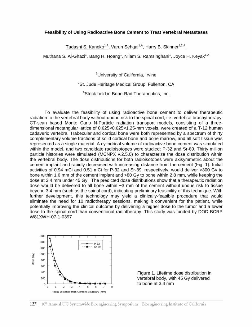
127 | 10th
Annual UC Systemwide Bioengineering Symposium | Bioengineering Institute of California
Feasibility of Using Radioactive Bone Cement to Treat Vertebral Metastases
Tadashi S. Kaneko1,A, Varun Sehgal1,A, Harry B. Skinner1,2,A,
Muthana S. Al-Ghazi1, Bang H. Hoang1, Nilam S. Ramsinghani1, Joyce H. Keyak1,A
1University of California, Irvine
2St. Jude Heritage Medical Group, Fullerton, CA
AStock held in Bone-Rad Therapeutics, Inc.
To evaluate the feasibility of using radioactive bone cement to deliver therapeutic radiation to the vertebral body without undue risk to the spinal cord, i.e. vertebral brachytherapy. CT-scan based Monte Carlo N-Particle radiation transport models, consisting of a three-dimensional rectangular lattice of 0.625×0.625×1.25-mm voxels, were created of a T-12 human cadaveric vertebra. Trabecular and cortical bone were both represented by a spectrum of thirty complementary volume fractions of solid cortical bone and bone marrow, and all soft tissue was represented as a single material. A cylindrical volume of radioactive bone cement was simulated within the model, and two candidate radioisotopes were studied: P-32 and Sr-89. Thirty million particle histories were simulated (MCNPX v.2.5.0) to characterize the dose distribution within the vertebral body. The dose distributions for both radioisotopes were axisymmetric about the cement implant and rapidly decreased with increasing distance from the cement (Fig. 1). Initial activities of 0.94 mCi and 0.51 mCi for P-32 and Sr-89, respectively, would deliver >300 Gy to bone within 1.6 mm of the cement implant and >80 Gy to bone within 2.8 mm, while keeping the dose at 3.4 mm under 45 Gy. The predicted dose distributions show that a therapeutic radiation dose would be delivered to all bone within ~3 mm of the cement without undue risk to tissue beyond 3.4 mm (such as the spinal cord), indicating preliminary feasibility of this technique. With further development, this technology may yield a clinically-feasible procedure that would eliminate the need for 10 radiotherapy sessions, making it convenient for the patient, while potentially improving the clinical outcome by delivering a higher dose to the tumor and a lower dose to the spinal cord than conventional radiotherapy. This study was funded by DOD BCRP W81XWH-07-1-0397
0
200
400
600
800
1000
1200
1400
1600
0 1 2 3 4 5 6 7 8
P-32Sr-89
Do
se (
Gy)
Radial Distance from Cement Boundary (mm)
Figure 1. Lifetime dose distribution in vertebral body, with 45 Gy delivered to bone at 3.4 mm
10th
Annual UC Systemwide Bioengineering Symposium | Bioengineering Institute of California | 128
Effects of Cholesterol on Plasma Membrane Mechanics
N. Khatibzadeh1, S. Gupta2, W. E. Brownell3, and B. Anvari2
1 Department of Mechanical Engineering, University of California, Riverside
2 Department of Bioengineering, University of California, Riverside
3 Bobby R. Alford Department of Otolaryngology-Head and Neck Surgery, Baylor College of Medicine, Houston, TX
Cholesterol is an important factor in determining the biophysical properties of the cell membrane, and plays an important role in the regulation of the membrane proteins. In this study, effects of cholesterol on viscoelastic properties of the plasma membrane are investigated. We use optical tweezers to extract nanotubes (tethers) under various pulling rates from the plasma membrane of human embryonic kidney (HEK) cells under cholesterol depleted and cholesterol enriched conditions. Threshold tether formation force, and the elastic and viscous parameters are quantified and correlated to the changes in the membrane cholesterol level.

129 | 10th
Annual UC Systemwide Bioengineering Symposium | Bioengineering Institute of California
Effect of Ultraviolet Light Crosslinking on Mechanical Stiffness of Fibrin Scaffolds
Soma Esmailian Lari, Haison Duong, Benjamin Wu, Bill Tawil
University of California, Los Angeles, Department of Bioengineering
Fibrin has been recognized as a good biomaterial because it is biocompatible, bioresorbable, and biodegradable. It has been shown that the mechanical and structural properties of fibrin scaffolds are determined by its compositions; namely, fibrinogen and thrombin. The interactions between fibrin monomers and thrombin form a stable network comprising of noncovalent bonding between the monomers .This bonding process defines the mechanical stiffness of the fibrin network. Fibrin have been extensively used in tissue engineered applications: from sealants to hydrogels and tissue constructs; however, the downside of using fibrin is that it has low mechanical stiffness and it rapidly degrades in vivo which is not sufficient for many tissue engineered structures. Thus, in order to optimize fibrin‘s potential as a bioscaffold, its mechanical stiffness and resistance to degradation should be improved. The purpose of this study is to enhance the overall mechanical stiffness as well as to modify the rate of degradation by thermal treatment and UV- irradiation of the fibrin network. The results show that upon heat treatment the mechanical stiffness of fibrin matrices increased, and this increase is most significant in the lower fibrin concentration .Upon UV-irradiation with 4000 µW/cm2 intensity the stiffness was increased by as much as 150% for all the fibrin matrices with different fibrinogen and thrombin concentration. Similarly the crosslinked fibrin matrices showed a slower rate of degradation (as indicated by a decrease in the modulus) when incubated in either serum or serum free medium.
10th
Annual UC Systemwide Bioengineering Symposium | Bioengineering Institute of California | 130
Serum Free Derivation of Embryonic Stem Cells Towards Functional Cardiomyocytes with Electrical Stimulation
Nicholas E Lauer1 , Kara McCloskey1
1 Graduate Program in Biological Engineering and Small-scale Technologies, University of California, Merced
In 2005 coronary heart disease killed more than 7.6 million people worldwide. Many forms of heart disease leads to the progressive apoptosis of cardiomyocytes (CM) within the heart from myocardial infarctions, ischemia, heart failures, and other cardiomyopathies. Human embryonic stem cells (hESC) could potentially serve as a vast source for a variety of therapeutic applications, including heart tissue repair. The differentiation and purification of hESC into functional CM is a first step towards using hESC for heart repair. Our laboratory is exploring the physical, electrical, and biochemical cues required for the differentiation of hESC towards CM. Using a C-Pace EP multi-channel stimulation device, electrical stimulation of primary isolated CM has shown to help maintain consistent contractility for 6-7 days [1, 2]. Pacing hESC at different stages of CM differentiation may increase the percentage of contractile cells, and is expected to coordinate pacing frequency. The stages of differentiation studied include: undifferentiated hESC in monolayer cultures, embryoid bodies (EBs) in suspension, and beating CM progenitors manually selected from EB outgrowths. Characterization includes cardiac specific protein markers cardiac troponin-T, GATA4, Nkx2.5, and brachyury, and stem cell marker, SSEA4, to test for undifferentiated hESC. 1. Shimizu, T., et al., Electrically communicating three-dimensional cardiac tissue mimic
fabricated by layered cultured cardiomyocyte sheets. J Biomed Mater Res, 2002. 60(1): p. 110-7.
2. Shimizu, T., et al., Fabrication of pulsatile cardiac tissue grafts using a novel 3-dimensional cell sheet manipulation technique and temperature-responsive cell culture surfaces. Circ Res, 2002. 90(3): p. e40.
131 | 10th
Annual UC Systemwide Bioengineering Symposium | Bioengineering Institute of California
Peptide Arrays for the Evaluation of Chemical Conjugation and Enzyme-Substrate Interation
Yan Liu, Yongfeng Zhao, Yang Song, Jiayu Liao
Department of Bioengineering, University of California, Riverside
Microarrays have become a widely used tool for biological research in last decade. One crucial step in microarrays fabrication is the covalently immobilization of biological samples on a solid surface. We have successfully developed two chemical strategies to immobilize peptides onto glass surface using either amine- or keto- reactive groups. An engineered peptide with a specific cleavage site for SENP2 (SUMO1 specific peptidase 2) and fluorofores at the end of peptide was immobilized on glass surface by chemical conjugation. The peptide arrays can be utilized to characterize the efficiencies of different chemical conjugations methods. Moreover, measuring the changes of emitted fluorescent intensity can show the cleavage efficiency of SENP2. This technology can be developed into high-throughput peptide array which can be used to studying enzyme catalytic activity, including characterizing inhibition effect of small compounds or molecules, which can be used in drug screening in the future.
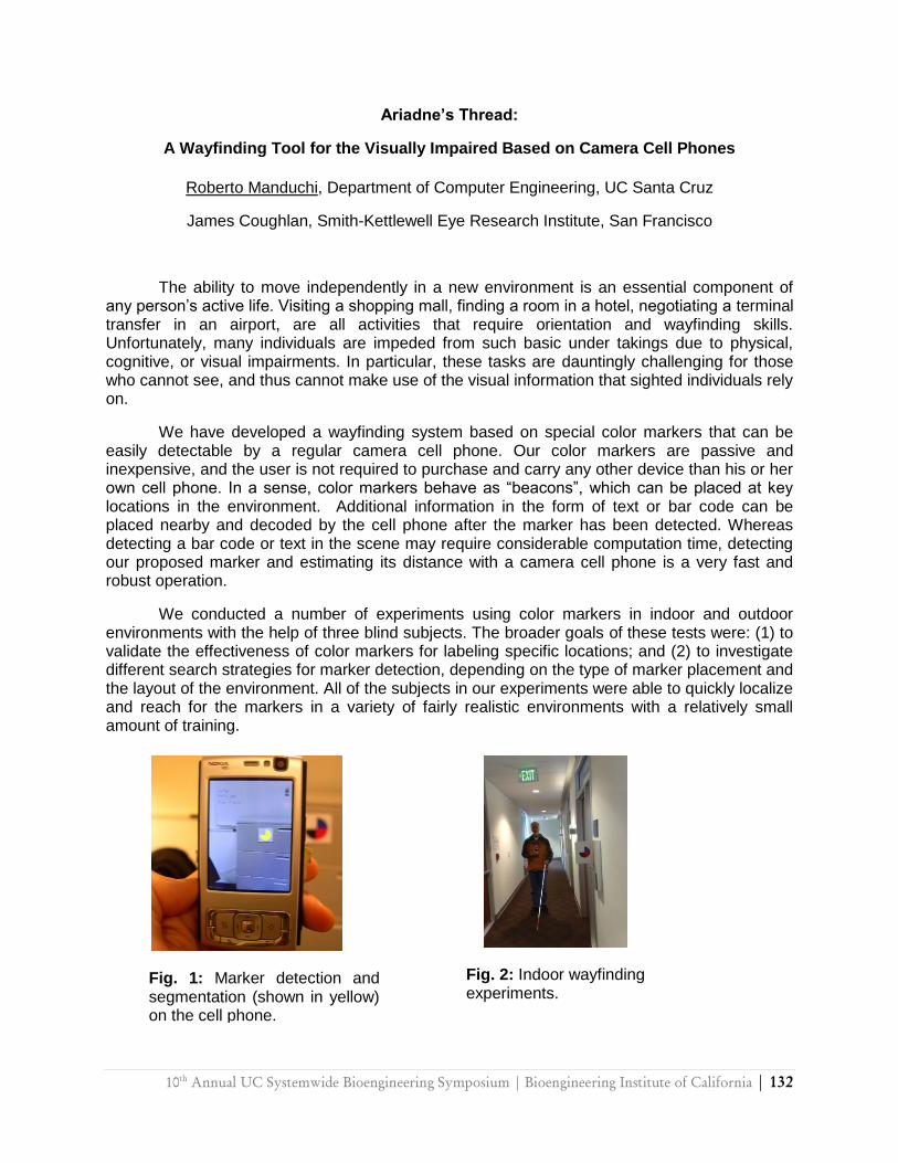
10th
Annual UC Systemwide Bioengineering Symposium | Bioengineering Institute of California | 132
Ariadne’s Thread:
A Wayfinding Tool for the Visually Impaired Based on Camera Cell Phones
Roberto Manduchi, Department of Computer Engineering, UC Santa Cruz
James Coughlan, Smith-Kettlewell Eye Research Institute, San Francisco
The ability to move independently in a new environment is an essential component of any person‘s active life. Visiting a shopping mall, finding a room in a hotel, negotiating a terminal transfer in an airport, are all activities that require orientation and wayfinding skills. Unfortunately, many individuals are impeded from such basic under takings due to physical, cognitive, or visual impairments. In particular, these tasks are dauntingly challenging for those who cannot see, and thus cannot make use of the visual information that sighted individuals rely on.
We have developed a wayfinding system based on special color markers that can be easily detectable by a regular camera cell phone. Our color markers are passive and inexpensive, and the user is not required to purchase and carry any other device than his or her own cell phone. In a sense, color markers behave as ―beacons‖, which can be placed at key locations in the environment. Additional information in the form of text or bar code can be placed nearby and decoded by the cell phone after the marker has been detected. Whereas detecting a bar code or text in the scene may require considerable computation time, detecting our proposed marker and estimating its distance with a camera cell phone is a very fast and robust operation.
We conducted a number of experiments using color markers in indoor and outdoor environments with the help of three blind subjects. The broader goals of these tests were: (1) to validate the effectiveness of color markers for labeling specific locations; and (2) to investigate different search strategies for marker detection, depending on the type of marker placement and the layout of the environment. All of the subjects in our experiments were able to quickly localize and reach for the markers in a variety of fairly realistic environments with a relatively small amount of training.
Fig. 1: Marker detection and segmentation (shown in yellow) on the cell phone.
Fig. 2: Indoor wayfinding experiments.
133 | 10th
Annual UC Systemwide Bioengineering Symposium | Bioengineering Institute of California
Theoretical Significance of Ion Binding on Observed Non-idealities in Osmotic Pressure in Crowded Macromolecular Environments
Devin W. McBride and Victor G. J. Rodgers
Department of Bioengineering, University of California, Riverside
Previous studies have shown that, in crowded macromolecular environments, protein-ion binding is a significant factor for the observed non-idealities in osmotic pressure and can be captured by a free-solvent model1. This work will address the specifics for protein-ion binding and protein hydration using in vitro and in silico studies; the equilibrium dialysis method will be used in the in vitro studies, while an electrostatic potential map of the surface will be used in the in silico studies. The binding of NaCl to Bovine Serum Albumin (BSA), for varying ionic strengths and pH, will be used as a model system; ionic strengths ranged from 0 M to 0.15 M and pH ranged from 4.5 to 10. Human Serum Albumin (HSA) will be compared to determine the specifics for the slight differences in ion binding observed. For the in silico studies, a homology model was developed for BSA from Human Serum Albumin (PDB code: 1BM0). Protein-ion binding and protein hydration was determined for BSA and HSA; ions and water were added to the proteins using Visual Molecular Dynamics (VMD); Molecular Dynamics (MD) simulations were run to determine the number of bound ions and water. In addition to the MD simulations, the electrostatic potential maps for BSA and HSA, under each condition, were compared and the sites of ion binding were determined; the regions in which electrostatic potential changes were observed were near those residues in which charge was altered, such as become negatively charged (aspartate, glutamate, cysteine) or uncharged (histidine), in the pH range studied. The in silico protein-ion binding and protein hydration, by BSA and HSA, were compared to the experimental data and the predicted ion-binding results of the free-solvent model.

10th
Annual UC Systemwide Bioengineering Symposium | Bioengineering Institute of California | 134
In vivo Optical Microscopy of Axonal Myelination of a Multiple Sclerosis Disease Model with Polarization Sensitive-Optical Coherence Tomography
Christian Oh1 and Hyle Park1
1Bourns College of Engineering, Bioengineering Department, University of California, Riverside
Encephalomyelitis disseminata, commonly known as multiple sclerosis, is an incurable autoimmune condition in which the immune system attacks the central nervous system. This causes demyelination of nerves and progressive levels of physical and cognitive disability. Early detection of demyelination may provide preventable measures for the affected population. Myelin exhibits birefringence, and thus, using polarization-sensitive optical coherence tomography (PS-OCT), we are capable of imaging nerves and quantitatively assessing the degree of myelination. Preliminary results indicate a direct correlation of histological parameters with birefringence values obtained from PS-OCT images which allows for a statistically significant method of quantifying axonal myelination. We will apply this approach to imaging rats in which multiple sclerosis has been induced. By means of direct histological correlation, the ability of PS-OCT to evaluate and quantify axonal myelination/demyelination in a non-destructive, in vivo manner will be assessed for this disease model.

135 | 10th
Annual UC Systemwide Bioengineering Symposium | Bioengineering Institute of California
Cascaded Micro Concentration Cells
Oxana S. Pantchenko1, Javad Shavani1, Mona Zebarjadi1, Howard Young1, Mehrdad Mahmoodi1, Michael Isaacson1, Ali Shakouri1
1Department of Electrical Engineering, University of California, Santa Cruz
For generations, electric fish, electrophorus electricushas, have been a subject of interest for studying the property of the membrane of electrogenic cell, electrocyte, etc. A few have designed mathematical models for mimicking such a complex system while others have focused their attention on studying individual types of ionic pumps. We report on the cascading of micro concentration cells, the scaling factor, as well as, the resulting power dissipation of such system.
10th
Annual UC Systemwide Bioengineering Symposium | Bioengineering Institute of California | 136
Inhibition of the sodium/calcium exchanger by lithium in intact mouse hearts modifies cardiac alternans
Azadé Petrosky, Dmytro Kornyeyev, Ariel L. Escobar
School of Engineering, University of California, Merced
The Na/Ca2+ exchanger (NCX) plays an important role in the regulation of the cardiac function. The aim of our research was to study the effect of impairing NCX with various concentrations of LiCl (by replacing Na+ with Li+ in normal Tyrode solution). We hypothesized that the inhibition of the exchanger would lead to the accumulation of Ca2+ in the cytosol and consequently to the enhancement of Ca2+ release from the sacroplasmic reticulum. Elevation of the amplitude of the release can affect the amplitude of cardiac alternans, a beat-to-beat oscillation in the various functional characteristics of the heart. The measurements were conducted using the Pulsed Local-Field Fluorescence microscopy on Langendorff-perfused mouse hearts that were loaded with fluorescent dyes sensitive to Ca2+ (rhod-2, AM) and membrane potential (DI-8ANEPPS). The presence of LiCl was found to slow down the restitution of Ca2+ release as well as to augment Ca2+ alternans that occur at high stimulation frequency (14 Hz, 37˚C). Additionally, LiCl induce a dramatic increase in the diastolic Ca2+ levels. Action potentials were measured to verify the effects of LiCl on the NCX. The maximum rate of phase 0 depolarization (dV/dt) of the action potentials and the duration of phase 2 were reduced with increasing concentration of LiCl in Tyrode solution. Similar to Ca2+ alternans, action potential alternans was more pronounced in the presence of LiCl. These results suggest that in the epicardial layer of the ventricular wall the repolarization of the action potential is partially defined by the influx of Na+ through NCX. Supported NIH R01-HL-084487 to AE.
137 | 10th
Annual UC Systemwide Bioengineering Symposium | Bioengineering Institute of California
On Calibrating the Power of a Microwave Oven
Emily J. Reed1, Christopher Viney1
1School of Engineering, University of California at Merced
Commercially available microwave ovens are a convenient source of electromagnetic waves that are useful not only for heating food but also as an aid in materials processing. In both contexts, it becomes necessary to consider the power output of the microwave source, in order to determine the appropriate microwave exposure time for a given application. We use beakers of water to investigate how sample volume affects the apparent power as determined by a calibration procedure. As a general rule, we find that smaller samples do not accumulate energy as effectively as larger samples do, leading to an erroneously low measure of microwave power (in some cases, 600 watts too low). We also observe points of deviation from this general relationship; these points are considered in view of previously published observations on microwave heating of small samples. In addition, marshmallows are used to map out the distribution of ‗hot spots‘ in the microwave oven, verifying that sample placement in the chamber is important for obtaining consistent results in experiments. Our results demonstrate relationships of practical consequence, and suggest directions for further study.
10th
Annual UC Systemwide Bioengineering Symposium | Bioengineering Institute of California | 138
Optimizing qNano: Characterizing a resizable nanopore
Jessie Rucker, Asma Uz-Zaman, David Deamer, William Dunbar
Department of Computer Engineering , University of California, Santa Cruz
Our goal is to realize the potential of the qNano instrument (developed by IZON) for accurate
pore characterization and particle analysis. The ability to detect, count and identify nanoscale
particles in real time would significantly advance
on-site particle analysis platforms critical to a wide range of quality control and technical
development applications such as bio-detection in research, pharmaceutical, and hospital
laboratories. The portable and inexpensive qNano instrument is ideally suited to meet these
unique particle detection requirements.
The qNano is a resizable nanopore, electrical characterization is attained by means of
electrophoresis2 of charged polystyrene beads3. We will model the ionic current through the
pore as a function of ion concentration, voltage, and pore size. The qNano allows real-time
control of the nanopore size and applied voltage. Modeling the characteristics of the pore using
MATLAB, we conclude that although we can detect and count translocation events,
identification of particle size, concentration, and composition is not possible with only the
amplifier built into the qNano. We propose for future research interfacing the qNano with an
AXIOPATCH amplifier to compensate for amplifier deficiencies in qNano. IZON is currently
designing a custom chip to replace the existing amplifier. Our studies will help identify optimal
pore geometries and derive the conditions necessary for detection and analysis of particles
ranging is size from 40 to 800nm.
2 (movement of suspended particles carrying a charge through an applied electric field)
3 (charged with carboxyl groups, 4 atoms present in carboxylic acid, and DNA)

139 | 10th
Annual UC Systemwide Bioengineering Symposium | Bioengineering Institute of California
In Vitro Culturing of the Ovarian Follicle: Alginate Encapsulation and Evaluation of the Nutrients Environment
Noriko Sausman1, P. Talbot2, V. G. J. Rodgers1
1Department of Bioengineering, University of California, Riverside
2Stem Cell Center, University of California, Riverside
Traditional in vitro culturing methods carried out in a 2-D culture plate do not maintain the structural integrity of ovarian follicles, often resulting in the degradation of follicles and limited success rate of the in vitro maturation (IVM). To improve culturing methods, it is necessary to determine growth conditions that simulate the in vivo environment so that the follicles retain their complex 3-D structure and have natural growth and maturation. Here, both experimental and computational techniques were used to elucidate the suitable culturing condition as well as provide a quantitative understanding of nutrient transport to the follicle for improved IVM techniques.
Experimentally, we propose to use biocompatible alginate hydrogels to encapsulate follicles to provide uniform structural supports. To analyze particle preparation parameters, maintain encapsulation consistency, we have developed an adjustable concentric needle device, a coaxial gas flow extrusion device. The inner needle (18 gauge) carries the alginate solution and the outer needle (13 gauge) conducts nitrogen gas, which shears off the extruding alginate solution into a droplet size. The adjustable inner needle is varied to alter the gas and liquid exit distance. Alginate beads prepared by this concentric needle device were quantified and analyzed. In summary, the gas pressure and relative tip locations significantly affected overall microparticle morphology or development of particles. The gas pressure was most significant for the size and shape of microparticles when formed.
In order to understand the nutrient environments in which follicles are matured, the nutrient transport, mainly glucose, across the alginate layer into the follicle is computationally analyzed in COMSOL®. An asymmetrical 3-D model that reflects the shape and size of the alginate bead prepared by the concentric needle is evaluated.
10th
Annual UC Systemwide Bioengineering Symposium | Bioengineering Institute of California | 140
A Microdevice for Detecting Cytokine Production from Individual Immune Cells
Jaime Silangcruz, Gulnaz Stybayeva, He Zhu, and Alexander Revzin
Department of Biomedical Engineering, University of California, Davis, CA
The high throughput analysis of immune cells at the single cell level is crucial in understanding cellular function in response to viral and bacterial infection. In particular, the analysis of cytokines produced by immune cells in the presence of pathogens provides significant diagnostic value. The present study describes the development of a microfabricated cytometry platform used to determine the interferon (IFN)-γ cytokine production of individual CD4+ T-lymphocytes (T-cells) isolated from human blood. A mixture of cell and cytokine-specific antibodies was co-printed onto a poly(ethylene glycol) (PEG) hydrogel coated glass slide and superimposed with an array of PEG microwells via photolithography. This engineered surface was then enclosed in a microfluidic device to minimize blood volume requirement. Introduction of red blood cell (RBC) depleted whole human blood into the microfluidic chamber followed by washing at a pre-defined shear stress resulted in the isolation of individual CD4+ T-cells in PEG microwells. Mitogenic activation of the captured T-cells followed by immunofluorescent staining in the microfluidic chamber revealed a localized IFN-γ cytokine signal around individual cells. Significantly, IFN-γ secretion profiles for hundreds of CD4 T-cells could be characterized at single cell resolution. The device and process presented herein marks the first step towards functional analysis of immune cells organized into high density single cell arrays.
141 | 10th
Annual UC Systemwide Bioengineering Symposium | Bioengineering Institute of California
Fluorescence Enhancement of Warfarin Induced by Interaction with β-Cyclodextrin Jacob M. Vasquez†, ‡, Andrew Vu†, Jerome S. Schultz†, Valentine I. Vullev†,*
Department of Bioengineering and Department of Biochemistry and Molecular Biology, University of California, Riverside
Warfarin is the most common agent used for control and prevention of venous as well as arterial thromboembolism (blood clots). In aqueous media, warfarin forms inclusion complexes with a family of cyclic oligosaccharides, α, β, γ-cyclodextrins (CD). The formation of these complexes results in enhancement of the fluorescence of warfarin. Such spectroscopic changes offer a venue for the development of bioanalytical methodologies for warfarin quantification in biological liquids. We characterized the photophysical properties of warfarin in solvents with varying polarity and viscosity. The fluorescence quantum yield of warfarin correlated: (1) strongly with the solvent viscosity (R=0.979) and (2) weakly with the solvent polarity (R=0.118). These findings indicate that it is the change of the viscosity, rather than polarity, of the microenvironment that causes the fluorescence enhancement of warfarin upon binding to β-CD. Utilizing the observed fluorescence enhancement in fluorescence titration measurements, the
binding constants of warfarin to β-CD were obtained (2.6102 M–1-3.7102 M–1). Using multivariable linear analysis, we extracted the stoichiometry of warfarin-b-CD interaction (1:1).
10th
Annual UC Systemwide Bioengineering Symposium | Bioengineering Institute of California | 142
Kinetics of Staining: Fluorescence Enhancement Induced by Escherichia Coli
Marlon S. Thomas, Elizabeth R. Zielins, Duoduo Bao, Baharak Bahmni, Vicente Nunez and Valentine I. Vullev
Department of Bioengineering, University of California, Riverside
For more than a century, colorimetric and fluorescence staining has been the foundation
of a broad range of key bioanalytical techniques and methodologies. The dynamics of such staining processes, however, still remains largely unexplored. We investigated the kinetics of fluorescence staining of a Gram-negative bacterium, Escherichia coli (E. coli) TOP10, with a cyanine dye, 3,3‘-diethylthiacyanine iodide (THIA). The observed fluorescence staining resulted from an orders-of-magnitude increase in the emission quantum yield of THIA upon binding to the bacterial cells. The kinetics of fluorescence enhancement did not manifest statistically significant dependence neither on the dye concentration, nor on the cell density (within the investigated concentration ranges). We utilized ANOVA analysis to demonstrate that within a 95% confidence, the time constants for E. coli are not random. Our findings suggest that this assay can provide a very rapid, species specific, fingerprint for bacterial cells.
143 | 10th
Annual UC Systemwide Bioengineering Symposium | Bioengineering Institute of California
Luminal Ca2+ Regulation of Single RyR2 Channels by Cardiac Calsequestrin
Patricio Vélez1,2, Dmytro Kornyeyev1, Marcia Cortés-Gutiérrez1, Björn C. Knollmann3, Ariel L. Escobar1
1School of Engineering, University of California, Merced
2Faculty of Sciences, University of Valparaiso, Chile
3Department of Medicine, Vanderbilt University Medical Center, Nashville
Intracellular Ca2+ measurements in whole hearts show that ablation of Calsequestrin (Casq2) causes faster relaxation of intra-SR and cytosolic Ca2+ transients, accelerated recovery of Ca2+ release and less pronounced Ca2+ alternans. These changes may be related to Ca2+ buffering by Casq2 but also to luminal control of RyR2 gating by Casq2. We examined this later hypothesis combining single channel electrophysiology and lanthanide resonance energy transfer. Under steady-state, open probability (Po) of RyR2 of cardiac SR fractions from wild-type mice is modulated by luminal [Ca2+]. Po increased from 0.1 to 0.8 when luminal [Ca2+] was increased from 6 µM to 8 mM at a fixed cytosolic [Ca2+] of 2 µM. The apparent KD for this luminal Ca2+ modulation was ~1 mM. This effect on RyR2 appears to be mediated by luminal sites since it was observed under conditions that precluded Ca2+ feed-through on cytosolic sites. This effect of luminal Ca2+ was not observed in RyR2 obtained from Casq2 KO mice indicating that luminal Ca2+ regulation of RyR2 requires Casq2. To gain mechanistic insights on the Casq2-mediated luminal regulation, we used Tb+3 as a sensitive probe. Fluorescence of purified dog SR Casq2 (6 nM) increased in the presence of Tb+3. This fluorescence was reduced as [Ca2+] in the solution increased suggesting that Ca2+ binds to purified Casq2 (apparent KD ~ 800 µM) by displacing tightly bound Tb+3 from a common binding site. At fixed cytosolic [Ca2+] of 2 µM, Po of single RyR2 from dog microsomes increased as a function of luminal [Tb+3], approaching saturating Po when [Tb+3] reached 1 µM (KD ~ 500 nM, Hill coeff. ~5.7). These results are consistent with Casq2 acting as luminal sensor detecting intra-SR [Ca2+] and translating it into changes in RyR2 gating. Supported by NIH R01-HL-084487 to AE. Supported NIH R01-HL-084487 to AE.
10th
Annual UC Systemwide Bioengineering Symposium | Bioengineering Institute of California | 144
Electrotactile Inducement of the Cutaneous Rabbit Effect (CRE) Across Human Fingertips
Jay P Warren1, Marco Santello1,2, Stephen I Helms Tillery1,2
1Harrington Department of Bioengineering, Arizona State University
2Department of Kinesiology, Arizona State University
A new electrotactile stimulus (ETS) paradigm was developed to determine if saltatory stimuli spanning the fingertips could elicit the CRE. Consistent with work by other groups, our previous research indicates that two ETS timing sequences can elicit the CRE on the forearm. Both timing sequences consist of two 5 ms electrical pulses delivered to a stimulation site. In the first ETS timing sequence, two electrical stimuli are delivered to the same stimulus site, here a fingertip, with an inter-stimulus interval (ISI) of 10 ms and are delivered to the next stimulation site, the next fingertip, with an inter-digit interval (IDI) of 150 ms. The second ETS timing sequence consists of 25 ms ISI and 100 ms IDI. We designed a train of stimuli, the order in which the fingertips were stimulated, to induce the CRE across the fingertips. We then compared this ―illusory train‖ to three distinctive control trains; tap, motion bias, and a negative control. Subjects reported that their middle fingertip (the location of the CRE/illusion) had been stimulated in 90 % of tap trials, 40 % of illusory trials, 25 % of motion bias trials, and 15 % of negative control trials. In most cases, the presence of stimuli on the middle fingertip was reported significantly more often under illusory trains compared to motion bias trains. Alternately subjects were asked to, ‗Indicate the order of the preceding stimulus train,‘ the order their fingertips were stimulated, and their responses were analyzed for reports of middle fingertip stimulation. The data here were very similar to the previous question type. These results suggest that saltatory stimuli delivered across the fingertips can generate a cutaneous rabbit effect. This is the first time that the CRE has been shown to ‗jump‘ across non-continuous body segments that can independently assume different spatial configurations.
145 | 10th
Annual UC Systemwide Bioengineering Symposium | Bioengineering Institute of California
Fluorescent Lifetime Changes as Function of Divalent Cations Ca2+ and Mg 2+ Ions
Stephanie Wong1, Ariel L. Escobar1
1School of Engineering, University of California, Merced
The measurement of intracellular Ca2+ or Mg2+ concentrations using non-ratiometric fluorescent dyes in living cells is a challenging process due to possible experimental artifacts. Changes in the measured fluorescence can be attributed not only to changes in the divalent concentration, but also in dye concentration, excitation light intensity and illumination/detection volumes. These fluorescent indicators showed dramatic changes in the dye‘s quantum efficiency between the free and bound form of the dye. These changes in quantum efficiency are associated with the time the dye stayed in the excited state before returning to the ground state. The development of fluorescence lifetime measurements has proven to be a useful method in analyzing the dynamics of fluorescent dye relaxations as functions for Ca2+ or Mg2+. Here, we evaluated the steady state and dynamic behavior of three different dyes as functions for Ca2+or Mg2+. Mag-Fluo 4 showed a large change in the quantum efficiency as function of the free divalent concentration, and no change in lifetime. Interestingly, Magnesium-Green and OGB-5N both displayed a quantum efficiency change and a large fluorescence lifetime increase. This approach can be used to measure divalent concentrations in intracellular organelles as the sarcoplasmic reticulum of intact beating mouse hearts. Supported NIH R01-HL-084487 to AE.

10th
Annual UC Systemwide Bioengineering Symposium | Bioengineering Institute of California | 146
FIG.1 FIG.1 FIG.1 FIG.21
Alternate Reception for Coil Array Elements
Bing Wu1, Chunsheng Wang2, Yong Pang1, Xiaoliang Zhang1,2
1Department of Radiology and Biomedical Imaging, University of California San Francisco
2UCSF/UC Berkeley Joint Graduate Group in Bioengineering, San Francisco & Berkeley, CA, United States
Phased array coils with densely placed coil elements have been used for magnetic resonance imaging to pursue high signal-to-noise ratio or fast imaging acquisitions. Recently many companies and research groups reported their array coils with more than 64 channels for human torso, head and other applications. However, the coil array with large number of coil elements raises a high demand for MRI system in which the large number of receive channels is necessary. In this work, we study the possibility to apply array coils to MRI system with less receive channels. In such manner, the coil with high number of elements is applicable for MRI systems equipped with any receive channels. A general multiple reception technique by using alternate reception of coil elements is discussed. A 16-ch head array was then applied to a MRI system with 8 receive channels. The result shows that the decoupling between coil elements was optimized. Noise correlation is much improved.
This principle of the alternative reception method is straight-forward and is demonstrated in Fig.1. 16 coil elements were divided into two groups, and two continuous receptions were conducted for those groups. Images were then combined with sum-of-square method. Compared with 8-ch array with NEX2, the image qualities have no much difference. Fig.2 illustrates the improvements of noise correlation matrix with this alternative reception method. In Fig2a, noise correlation is calculated from 16-ch array with 16 receive channels. After grouping, noise coefficients were relocated in the matrix (Fig.2b), and after twice receptions (Fig.2c), the final noise matrix in Fig.2d has much smaller mean value than the case shown in Fig.2a, which indicates the improvements of coil isolations.
A general method is applied for array coil with less receive channels by using alternative reception. Thus the number of array elements and receive channels can be variance.
147 | 10th
Annual UC Systemwide Bioengineering Symposium | Bioengineering Institute of California
Lab-on-a-Chip Characterization of Cellular Media using Electrical Impedance Spectroscopy
John Yan1, Tingrui Pan2
1Department of Electrical and Computer Engineering, University of California, Davis
2Department of Biomedical Engineering, University of California, Davis
Recent advances in the lab-on-a-chip technologies offer new opportunities for biological investigations, which typically require minimal analytes to facilitate the biochemical reactions and thereby to improve sensitivity and precision [1]. Several electrical detection strategies, including electrical impedance spectroscopy (EIS) techniques, have been demonstrated in the lab-on-a-chip systems for high-throughput cellular and biomolecular interrogation recently [2,3]. In this abstract, we reported a lab-on-a-chip system incorporating miniature label-free detection electrodes into a PDMS-based microfluidics for cellular media characterization. A variety of cellular contents, including bacterial and mammalian cell media, have been fully characterized in the system. Furthermore, an electrical circuit model with noise considerations has been developed to interpret the biological findings from these electrical measurements.
[1] J. El-Ali, P. K. Sorger and K. F. Jensen, Nature, 2006, 442, 403–411.
[2] K. Cheung, S. Gawad and P. Renaud, Cytometry, Part A, 2005, 65, 124–132.
[3] K. Ahn, C. Kerbage, T. P. Hunt, R. M. Westervelt, D. R. Link and D. A. Weitz, Appl. Phys. Lett., 2006, 88(2), 024104.
10th
Annual UC Systemwide Bioengineering Symposium | Bioengineering Institute of California | 148
Miniature Electrochemical Biosensors for Detection of Extracellular Metabolites
Jun Yan1, Valber D. Pedrosa2, Aleksandr L. Simonian2, Alexander Revzin1
1Department of Biomedical Engineering, University of California, Davis
2Materials Research and Education Center, Department of Chemical Engineering, Auburn University, Auburn
Seamless integration of biological and electrochemical sensing components is critical for successful development of BioMEMS. In this presentation, we will propose a platform that integrates small group of cancer cells with miniature glucose electrochemical biosensors in a microfabricated device where cellular microenvironment can be precisely defined and easily modulated. The juxtaposing of miniature sensing elements with cells is achieved through photolithographic patterning of glucose oxidase (GOX)-containing poly (ethylene glycol) (PEG) hydrogel microstructures on silane modified glass substrates. Gold microelectrode array is fabricated onto regular glass slide using standard semiconductor process of photolithography. The ability to deposit enzyme-containing hydrogel microstructures onto gold microelectrodes is very critical for the development of electrochemical bisosensors. PEG patterning process is very similar to the traditional photoresist lithography so that enzyme containing PEG can be fabricated on top of existing gold microelectrodes. Enzyme GOX is entrapped inside PEG hydrogel. To facilitate electron transfer through PEG hydrogel and wire GOX, redox polymer vinylferrocene is also added to PGE precursor solution and covalently bound to PEG polymer during UV exposure. Importantly, enzyme-entrapping hydrogel micropatterns do not support protein or cell deposition and allowed to guide attachment of small group of cells next to the sensing elements. The determination of glucose generation by stimulated cells is accomplished using three-electrode electrochemical system. In addition, the PEG hydrogel lithography also allows us to deposit second enzyme such as lactate oxidase (LOX) next to GOX using the same method, realizing simultaneous detection of two analytes. A microfluidic device is developed to enclose cells and sensing hydrogel elements inside a confined volume of ~ 2 µl, thus increasing sensitivity of the biosensor and allowing to dynamically change the extracellular microenvironment. This novel electrochemical dual biosensor integrated with cell cultivation element is envisioned as an in vitro model of metabolism study of cancer cells.

Celebrating 10 years
of excellence and
achievement
Across 10 united
Universities of
California
Held at UC Merced,
the 10th
and newest
campus
Copyright © 2022 FDOKUMEN