Impairment of circulating endothelial progenitors in Down syndrome
13
RESEARCH ARTICLE Open Access Impairment of circulating endothelial progenitors in Down syndrome Valerio Costa 1 , Linda Sommese 2 , Amelia Casamassimi 3 , Roberta Colicchio 4 , Claudia Angelini 5 , Valentina Marchesano 6 , Lara Milone 7 , Bartolomeo Farzati 3 , Alfonso Giovane 7 , Carmela Fiorito 8 , Monica Rienzo 3 , Marco Picardi 9 , Bice Avallone 6 , Massimiliano Marco Corsi 10 , Berardo Sarubbi 11 , Raffaele Calabrò 11 , Paola Salvatore 12 , Alfredo Ciccodicola 1*† , Claudio Napoli 3† Abstract Background: Pathological angiogenesis represents a critical issue in the progression of many diseases. Down syndrome is postulated to be a systemic anti-angiogenesis disease model, possibly due to increased expression of anti-angiogenic regulators on chromosome 21. The aim of our study was to elucidate some features of circulating endothelial progenitor cells in the context of this syndrome. Methods: Circulating endothelial progenitors of Down syndrome affected individuals were isolated, in vitro cultured and analyzed by confocal and transmission electron microscopy. ELISA was performed to measure SDF-1a plasma levels in Down syndrome and euploid individuals. Moreover, qRT-PCR was used to quantify expression levels of CXCL12 gene and of its receptor in progenitor cells. The functional impairment of Down progenitors was evaluated through their susceptibility to hydroperoxide-induced oxidative stress with BODIPY assay and the major vulnerability to the infection with human pathogens. The differential expression of crucial genes in Down progenitor cells was evaluated by microarray analysis. Results: We detected a marked decrease of progenitors’ number in young Down individuals compared to euploid, cell size increase and some major detrimental morphological changes. Moreover, Down syndrome patients also exhibited decreased SDF-1a plasma levels and their progenitors had a reduced expression of SDF-1a encoding gene and of its membrane receptor. We further demonstrated that their progenitor cells are more susceptible to hydroperoxide-induced oxidative stress and infection with Bartonella henselae. Further, we observed that most of the differentially expressed genes belong to angiogenesis, immune response and inflammation pathways, and that infected progenitors with trisomy 21 have a more pronounced perturbation of immune response genes than infected euploid cells. Conclusions: Our data provide evidences for a reduced number and altered morphology of endothelial progenitor cells in Down syndrome, also showing the higher susceptibility to oxidative stress and to pathogen infection compared to euploid cells, thereby confirming the angiogenesis and immune response deficit observed in Down syndrome individuals. Background Down syndrome (DS) is a complex disorder caused by trisomy of the entire or a critical portion of chromo- some 21 (HSA21); it represents the most frequent genetic cause of mental retardation, with a frequency of about 1/1000 new-borns, and is associated with a huge number of congenital heart defects [1]. DS individuals have also an increased risk of early-onset Alzheimer dis- ease [1]. Immunological and autoimmune disturbances, with high rates of infections and malignancies, are recurrent phenomena in DS pathogenesis [2], and infec- tions still represent major cause of death in DS [3,4]. Despite the increased risk of leukaemia, DS patients have a low incidence to develop solid tumors [5,6], and * Correspondence: [email protected] † Contributed equally 1 Institute of Genetics and Biophysics ‘’A. Buzzati-Traverso’’, IGB-CNR, Naples, Italy Full list of author information is available at the end of the article Costa et al. BMC Medical Genomics 2010, 3:40 http://www.biomedcentral.com/1755-8794/3/40 © 2010 Costa et al; licensee BioMed Central Ltd. This is an Open Access article distributed under the terms of the Creative Commons Attribution License (http://creativecommons.org/licenses/by/2.0), which permits unrestricted use, distribution, and reproduction in any medium, provided the original work is properly cited.
Transcript of Impairment of circulating endothelial progenitors in Down syndrome
RESEARCH ARTICLE Open Access
Impairment of circulating endothelial progenitorsin Down syndromeValerio Costa1, Linda Sommese2, Amelia Casamassimi3, Roberta Colicchio4, Claudia Angelini5,Valentina Marchesano6, Lara Milone7, Bartolomeo Farzati3, Alfonso Giovane7, Carmela Fiorito8, Monica Rienzo3,Marco Picardi9, Bice Avallone6, Massimiliano Marco Corsi10, Berardo Sarubbi11, Raffaele Calabrò11, Paola Salvatore12,Alfredo Ciccodicola1*†, Claudio Napoli3†
Abstract
Background: Pathological angiogenesis represents a critical issue in the progression of many diseases. Downsyndrome is postulated to be a systemic anti-angiogenesis disease model, possibly due to increased expression ofanti-angiogenic regulators on chromosome 21. The aim of our study was to elucidate some features of circulatingendothelial progenitor cells in the context of this syndrome.
Methods: Circulating endothelial progenitors of Down syndrome affected individuals were isolated, in vitrocultured and analyzed by confocal and transmission electron microscopy. ELISA was performed to measure SDF-1aplasma levels in Down syndrome and euploid individuals. Moreover, qRT-PCR was used to quantify expressionlevels of CXCL12 gene and of its receptor in progenitor cells. The functional impairment of Down progenitors wasevaluated through their susceptibility to hydroperoxide-induced oxidative stress with BODIPY assay and the majorvulnerability to the infection with human pathogens. The differential expression of crucial genes in Downprogenitor cells was evaluated by microarray analysis.
Results: We detected a marked decrease of progenitors’ number in young Down individuals compared to euploid,cell size increase and some major detrimental morphological changes. Moreover, Down syndrome patients alsoexhibited decreased SDF-1a plasma levels and their progenitors had a reduced expression of SDF-1a encodinggene and of its membrane receptor. We further demonstrated that their progenitor cells are more susceptible tohydroperoxide-induced oxidative stress and infection with Bartonella henselae. Further, we observed that most ofthe differentially expressed genes belong to angiogenesis, immune response and inflammation pathways, and thatinfected progenitors with trisomy 21 have a more pronounced perturbation of immune response genes thaninfected euploid cells.
Conclusions: Our data provide evidences for a reduced number and altered morphology of endothelial progenitorcells in Down syndrome, also showing the higher susceptibility to oxidative stress and to pathogen infectioncompared to euploid cells, thereby confirming the angiogenesis and immune response deficit observed in Downsyndrome individuals.
BackgroundDown syndrome (DS) is a complex disorder caused bytrisomy of the entire or a critical portion of chromo-some 21 (HSA21); it represents the most frequentgenetic cause of mental retardation, with a frequency of
about 1/1000 new-borns, and is associated with a hugenumber of congenital heart defects [1]. DS individualshave also an increased risk of early-onset Alzheimer dis-ease [1]. Immunological and autoimmune disturbances,with high rates of infections and malignancies, arerecurrent phenomena in DS pathogenesis [2], and infec-tions still represent major cause of death in DS [3,4].Despite the increased risk of leukaemia, DS patientshave a low incidence to develop solid tumors [5,6], and
* Correspondence: [email protected]† Contributed equally1Institute of Genetics and Biophysics ‘’A. Buzzati-Traverso’’, IGB-CNR, Naples,ItalyFull list of author information is available at the end of the article
Costa et al. BMC Medical Genomics 2010, 3:40http://www.biomedcentral.com/1755-8794/3/40
© 2010 Costa et al; licensee BioMed Central Ltd. This is an Open Access article distributed under the terms of the Creative CommonsAttribution License (http://creativecommons.org/licenses/by/2.0), which permits unrestricted use, distribution, and reproduction inany medium, provided the original work is properly cited.
a reduced incidence of diabetic retinopathy, suggesting,at least in part, a common angiogenesis’ suppression[5,7]. Impaired endothelial function at a young age, pos-sibly due to increased oxidative stress and yet unknownmechanisms, is a common DS feature [8].DS phenotype results from a dosage imbalance of
HSA21 genes, although expression analyses havereported conflicting results [9,10]. The over-expressionof chromosome 21 genes greatly varies across the triso-mic tissues [11,12], and analyzing specific cell type/tis-sue, in easy-accessible and non-invasive manner, may bemore productive [13,14].Growing interest is emerging on circulating endothe-
lial progenitor cells (EPCs) and their pivotal role in themaintenance of endothelium integrity, repair after injuryand postnatal neovascularization [15-17]. Many studiesare providing encouraging insights into the use of EPCsin the clinical setting [18,19]. Indeed, accumulating evi-dences indicate a reduced availability, and/or impairedEPC function, in cardiovascular and metabolic diseases[17,20,21]. EPCs number was recently shown to beimpaired in DS fetuses and children [22,23] and CD34+haematopoietic progenitors exhibited a marked growthdecrease in Ts65Dn - a DS mouse model - accounting,at least in part, for DS vascular anomalies and defectiveimmune response to pathogens [24].Bacterial toxins may trigger pathogenic events through
the over-production of cytokines and chemokines, lead-ing to the alteration of endothelial function and capillaryleakage [25]. Particularly, we recently demonstrated [26]that Bartonella henselae, a gram-negative intracellularbacteria responsible of vasoproliferative disorders inimmunocompromised individuals [27,28], adheres toand invades EPCs.The present study was designed to pursue the molecular
mechanisms contributing to immune, vascular and haema-topoietic defective DS phenotypes, by investigating thenumber and functions of DS EPCs compared to euploidcells, also focusing on bioinformatics analysis of differen-tially expressed genes. Moreover, by using the previouslydescribed B. henselae model, we investigated the suscept-ibility of DS progenitors to this pathogen infection, alsoperforming a detailed analysis of deregulated genes afterBartonella infection, with particular attention to angiogen-esis and immune response pathways.
MethodsSubjectsDS and euploid donors were recruited at the Institute ofGeneral Pathology, Section of Clinical Pathology, Facultyof Medicine, University of Milan, and at the SecondUniversity of Naples, and an approval statement wasobtained by the ethics’ review boards of both Institu-tions. Informed consent was obtained from all persons
involved in all clinical investigation of this study accord-ing to the principles expressed in the Declaration ofHelsinki.All subjects recruited for EPC isolation were free of
infection, and no individual was taking any medicationknown to affect immune system/response. DS andeuploid individuals were 65% males and 35% females asgender and 28 ± 9 as mean age. The experiments,where not specified, were performed on at least six DSand age-matched euploid individuals.Plasma samples were obtained from 50 DS individuals
and 30 age matched euploids subdivided into three agesubgroups (young 0-20 y.o.; adult 21-40 y.o.; old 41-60y.o.) as described elsewhere [29].
EPC IsolationEPCs were isolated from non-institutionalized indivi-duals with DS and age-matched euploid donors.EPCs were isolated as previously described [30].
Briefly, PBMCs were isolated by density gradient centri-fugation of peripheral blood samples on Histopaque-1077 (Sigma). Cells were washed twice with PBS andcounted. PBMCs were plated on culture dishes pre-coated with gelatin and fibronectin and maintained inendothelial basal medium-2 (EBM2; Cell Systems). Cellswere cultured at 37°C with 5% CO2 in a humidifiedatmosphere. After four days, non-adherent cells wereremoved and adherent cells were used for furtheranalyses.
Bacterial strains and growth conditionsThe B. henselae strain ATCC 49882 (LGC Promochem)was grown on Columbia agar supplemented with 5% defi-brinated sheep blood (Oxoid) in a humidified atmosphereat 37°C and 5% CO2. For production of bacterial stocksuspensions, bacteria were harvested after 7 days of cul-ture until they reached the mid-exponential phase ofgrowth (109 bacteria/ml), resuspended in Tryptone SoyaBroth USP (Oxoid) containing 10% glycerol, and storedat -80°C. The number of viable bacteria in the frozenstocks was determined as previously described [26].
B. henselae infectionFor infection, Bartonella stock solutions were thawed,washed and suspended in antibiotic-free cell culturemedium, and sedimented onto cultured EPCs at differ-ent multiplicity of infection (MOI) of 50, 100, 250, 500and 1000 [26]. The MOI for infections was confirmedby plating serial dilutions of the infection inoculum.Assays were performed three times in triplicate.
Confocal immunofluorescence microscopyEPCs were dual stained with Dil-Ac-LDL and lectinfrom Ulex europaeus and counted both by fluorescence
Costa et al. BMC Medical Genomics 2010, 3:40http://www.biomedcentral.com/1755-8794/3/40
Page 2 of 13
microscopy and flow cytometry as previously described[26,30]. Images were obtained by Zeiss LSM 510 withplan-apochromat X 63 (NA 1.4) oil immersion objective.EPCs images were used to measure cell size with ImageJ(http://rsb.info.nih.gov/ij/).
SDF-1a plasma levelsCommercially available SDF-1a ELISA kit (Quantikine,R&D Systems) was used to determine plasma SDF-1alevels. Tests were carried out at RT on freshly thawedplasma samples of 50 DS individuals and 30 agematched euploids subdivided into three age subgroups(young 0-20 y.o.; adult 21-40 y.o.; old 41-60 y.o.) [29].Concentration was determined by comparison with astandard curve, following manufacturer’s instruction.
Transmission electron microscopyAfter a short incubation with Trypsin/EDTA, DS andeuploid EPCs, both infected and uninfected with B. hen-selae, were harvested, centrifuged and washed in PBS.After centrifugation at a speed of 400 g for 7 min, cellswere fixed in 4% glutaraldehyde as described [31]. Post-fixation, dehydratation of specimen, semithin (2 μm)and ultrathin (80 nm) sections were performed as pre-viously described [26]. Semithin sections were analysedwith a light microscope (Polivar Reichert-Jung). Ultra-thin sections were examined with Leo 912 AB transmis-sion electron microscope operating at 80 kV.
C11-BODIPY581/591 fluorescenceOxidation-sensitive fluorescent probe, C11-BODIPY581/591 (C11-BO, Invitrogen), was loaded (2 μM final con-centration) into the cells 30 min. before oxidative treat-ment. The samples were aliquoted in triplicate wells of a24-well microplate, and fluorescence was determinedwith confocal laser microscopy at different times (0, 1,and 6 hours) from oxidant treatment. Between times,plates were maintained at 37°C in 5% CO2. To determinered fluorescence each microplate was excited at 543 nm(emission at 590 nm); for green fluorescence, microplateswere excited at 488 nm (emission at 526 nm). Blank wellswere also evaluated as well as C11-BO alone.
Microarray analysis and quantitative RT-PCRTotal RNA (10 μg) was isolated as previously described[32] [Additional file 1: Supplementary Methods]. A poolof three samples (a total 15 μg of cRNA) was used foreach hybridization - 2 pools of three infected and threeuninfected euploid and DS - on the Affymetrix U133 2.0probe array cartridge as described elsewhere [26].Microarray data were submitted to Array Express(http://www.ebi.ac.uk/arrayexpress; provisional accessionnumber E-MTAB-312). Results were validated by qRT-PCR and semi-qRT-PCR, performed as described [32],
using primer pairs listed in [Additional file 2: Supple-mental Table S1].
In silico significant pathway identificationAnalysis of over-represented genes was performed usingthe Database for Annotation, Visualization, and Inte-grated Discovery (DAVID) [33,34] and the PANTHER(Protein ANalysis THrough Evolutionary Relationships)Classification System [35 [Additional file 1: Supplemen-tary Methods].
Statistical analysisEPCs number, cell size differences, SDF-1a plasma levels,fluorescence intensity of C11-BO and qRT-PCRs data werereported as mean values, and results analysed by pairedStudent t test. P value < 0.05 was considered statisticallysignificant [Additional file 1: Supplementary Methods].
ResultsEPC number and phenotypeWe established that the number of EPCs isolated fromperipheral blood of young and adult (mean age 28 ± 9)DS was significantly lower than age-matched euploidindividuals (P < 0.0001 vs euploid EPCs; see Figure 1A).Phase contrast fluorescent microscopy [Additional file 3:Supplemental Figure S1A] and FACS analysis (data notshown) were used to identify double-positive cells forDil-Ac-LDL and lectin [26,30]. By confocal microscopyand TEM we also observed early signs of cytoplasmaticdisruption in DS progenitors, and cell size increase com-pared to euploid. In particular, by using ImageJ, wemeasured the cell size of DS EPCs, showing a significantincrease compared to euploid cells (Figures 1B andAdditional file 3: Supplemental Figure S1B]). We alsomeasured cell cycle progression of DS progenitors vseuploid cells and we did not find any significant differ-ence [Additional file 3: Supplemental Figure S1C].Moreover, the ultrastructural examination revealed anincreased number of phagolysosomes and vacuolizationof DS progenitors compared to euploid cells (Figure 1C).
SDF-1a plasma levelsEPC number is known to correlate to chemokines’ plasmalevels, such as SDF-1a (stromal derived factor-1a). Thus,we first measured by ELISA its plasma levels in peripheralblood of 30 euploid and 50 DS individuals, collected inthree age subgroups [29]. Then, comparing mean SDF-1avalues we found a significant decrease of SDF-1a plasmalevels in young and adults DS compared to age-matchedeuploid individuals (P = 0.02) (Figure 1D).Moreover, we measured by quantitative RT-PCR the
expression of SDF-1a encoding gene, CXCL12, and ofits membrane receptor CXCR4. A significant decrease inthe expression of CXCL12 (5-fold; P < 0.05) and CXCR4
Costa et al. BMC Medical Genomics 2010, 3:40http://www.biomedcentral.com/1755-8794/3/40
Page 3 of 13
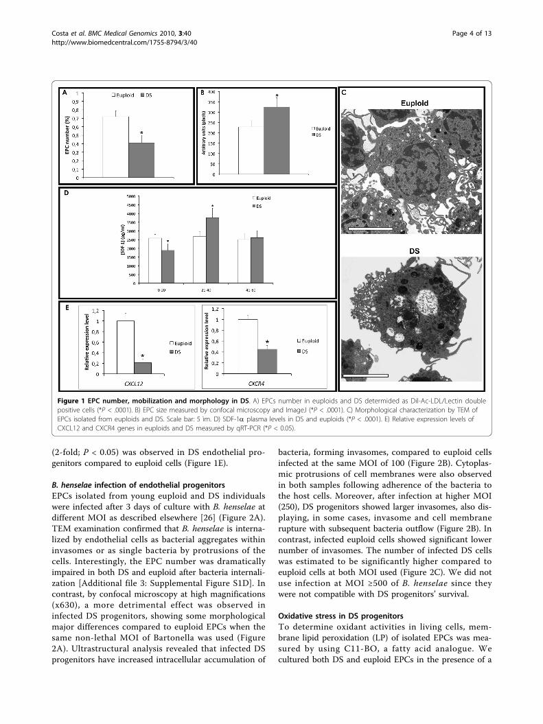
(2-fold; P < 0.05) was observed in DS endothelial pro-genitors compared to euploid cells (Figure 1E).
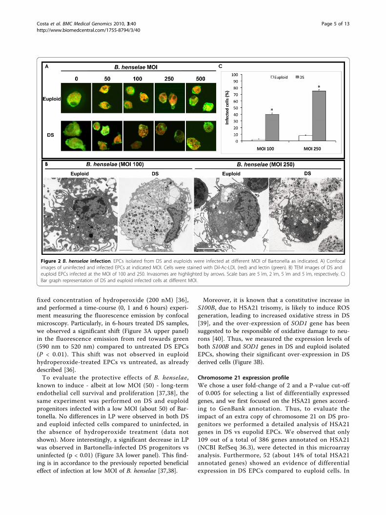
B. henselae infection of endothelial progenitorsEPCs isolated from young euploid and DS individualswere infected after 3 days of culture with B. henselae atdifferent MOI as described elsewhere [26] (Figure 2A).TEM examination confirmed that B. henselae is interna-lized by endothelial cells as bacterial aggregates withininvasomes or as single bacteria by protrusions of thecells. Interestingly, the EPC number was dramaticallyimpaired in both DS and euploid after bacteria internali-zation [Additional file 3: Supplemental Figure S1D]. Incontrast, by confocal microscopy at high magnifications(x630), a more detrimental effect was observed ininfected DS progenitors, showing some morphologicalmajor differences compared to euploid EPCs when thesame non-lethal MOI of Bartonella was used (Figure2A). Ultrastructural analysis revealed that infected DSprogenitors have increased intracellular accumulation of
bacteria, forming invasomes, compared to euploid cellsinfected at the same MOI of 100 (Figure 2B). Cytoplas-mic protrusions of cell membranes were also observedin both samples following adherence of the bacteria tothe host cells. Moreover, after infection at higher MOI(250), DS progenitors showed larger invasomes, also dis-playing, in some cases, invasome and cell membranerupture with subsequent bacteria outflow (Figure 2B). Incontrast, infected euploid cells showed significant lowernumber of invasomes. The number of infected DS cellswas estimated to be significantly higher compared toeuploid cells at both MOI used (Figure 2C). We did notuse infection at MOI ≥500 of B. henselae since theywere not compatible with DS progenitors’ survival.
Oxidative stress in DS progenitorsTo determine oxidant activities in living cells, mem-brane lipid peroxidation (LP) of isolated EPCs was mea-sured by using C11-BO, a fatty acid analogue. Wecultured both DS and euploid EPCs in the presence of a
Figure 1 EPC number, mobilization and morphology in DS. A) EPCs number in euploids and DS determided as Dil-Ac-LDL/Lectin doublepositive cells (*P < .0001). B) EPC size measured by confocal microscopy and ImageJ (*P < .0001). C) Morphological characterization by TEM ofEPCs isolated from euploids and DS. Scale bar: 5 ìm. D) SDF-1a plasma levels in DS and euploids (*P < .0001). E) Relative expression levels ofCXCL12 and CXCR4 genes in euploids and DS measured by qRT-PCR (*P < 0.05).
Costa et al. BMC Medical Genomics 2010, 3:40http://www.biomedcentral.com/1755-8794/3/40
Page 4 of 13
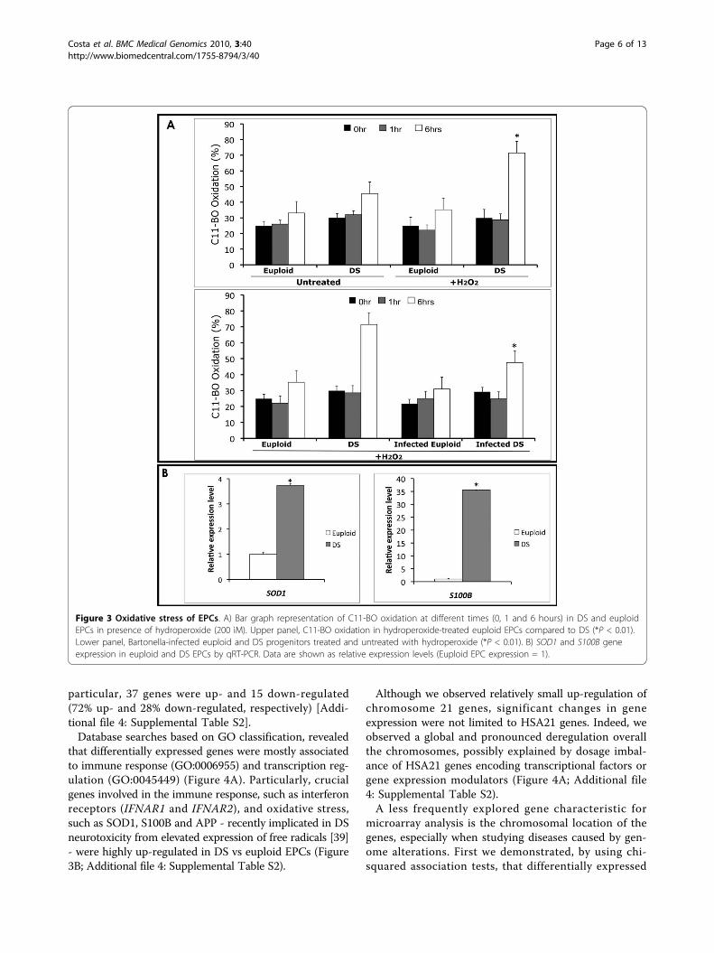
fixed concentration of hydroperoxide (200 nM) [36],and performed a time-course (0, 1 and 6 hours) experi-ment measuring the fluorescence emission by confocalmicroscopy. Particularly, in 6-hours treated DS samples,we observed a significant shift (Figure 3A upper panel)in the fluorescence emission from red towards green(590 nm to 520 nm) compared to untreated DS EPCs(P < 0.01). This shift was not observed in euploidhydroperoxide-treated EPCs vs untreated, as alreadydescribed [36].To evaluate the protective effects of B. henselae,
known to induce - albeit at low MOI (50) - long-termendothelial cell survival and proliferation [37,38], thesame experiment was performed on DS and euploidprogenitors infected with a low MOI (about 50) of Bar-tonella. No differences in LP were observed in both DSand euploid infected cells compared to uninfected, inthe absence of hydroperoxide treatment (data notshown). More interestingly, a significant decrease in LPwas observed in Bartonella-infected DS progenitors vsuninfected (p < 0.01) (Figure 3A lower panel). This find-ing is in accordance to the previously reported beneficialeffect of infection at low MOI of B. henselae [37,38].
Moreover, it is known that a constitutive increase inS100B, due to HSA21 trisomy, is likely to induce ROSgeneration, leading to increased oxidative stress in DS[39], and the over-expression of SOD1 gene has beensuggested to be responsible of oxidative damage to neu-rons [40]. Thus, we measured the expression levels ofboth S100B and SOD1 genes in DS and euploid isolatedEPCs, showing their significant over-expression in DSderived cells (Figure 3B).
Chromosome 21 expression profileWe chose a user fold-change of 2 and a P-value cut-offof 0.005 for selecting a list of differentially expressedgenes, and we first focused on the HSA21 genes accord-ing to GenBank annotation. Thus, to evaluate theimpact of an extra copy of chromosome 21 on DS pro-genitors we performed a detailed analysis of HSA21genes in DS vs eupolid EPCs. We observed that only109 out of a total of 386 genes annotated on HSA21(NCBI RefSeq 36.3), were detected in this microarrayanalysis. Furthermore, 52 (about 14% of total HSA21annotated genes) showed an evidence of differentialexpression in DS EPCs compared to euploid cells. In
Figure 2 B. henselae infection. EPCs isolated from DS and euploids were infected at different MOI of Bartonella as indicated. A) Confocalimages of uninfected and infected EPCs at indicated MOI. Cells were stained with Dil-Ac-LDL (red) and lectin (green). B) TEM images of DS andeuploid EPCs infected at the MOI of 100 and 250. Invasomes are highlighted by arrows. Scale bars are 5 ìm, 2 ìm, 5 ìm and 5 ìm, respectively. C)Bar graph representation of DS and euploid infected cells at different MOI.
Costa et al. BMC Medical Genomics 2010, 3:40http://www.biomedcentral.com/1755-8794/3/40
Page 5 of 13
particular, 37 genes were up- and 15 down-regulated(72% up- and 28% down-regulated, respectively) [Addi-tional file 4: Supplemental Table S2].Database searches based on GO classification, revealed
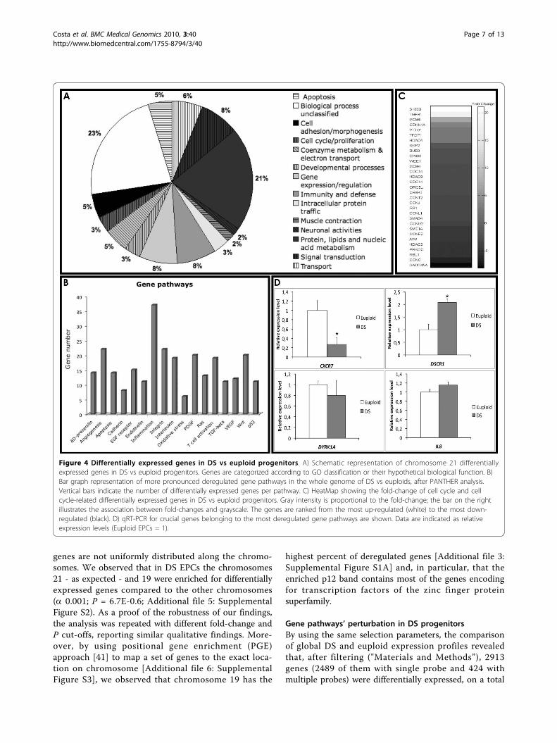
that differentially expressed genes were mostly associatedto immune response (GO:0006955) and transcription reg-ulation (GO:0045449) (Figure 4A). Particularly, crucialgenes involved in the immune response, such as interferonreceptors (IFNAR1 and IFNAR2), and oxidative stress,such as SOD1, S100B and APP - recently implicated in DSneurotoxicity from elevated expression of free radicals [39]- were highly up-regulated in DS vs euploid EPCs (Figure3B; Additional file 4: Supplemental Table S2).
Although we observed relatively small up-regulation ofchromosome 21 genes, significant changes in geneexpression were not limited to HSA21 genes. Indeed, weobserved a global and pronounced deregulation overallthe chromosomes, possibly explained by dosage imbal-ance of HSA21 genes encoding transcriptional factors orgene expression modulators (Figure 4A; Additional file4: Supplemental Table S2).A less frequently explored gene characteristic for
microarray analysis is the chromosomal location of thegenes, especially when studying diseases caused by gen-ome alterations. First we demonstrated, by using chi-squared association tests, that differentially expressed
Figure 3 Oxidative stress of EPCs. A) Bar graph representation of C11-BO oxidation at different times (0, 1 and 6 hours) in DS and euploidEPCs in presence of hydroperoxide (200 ìM). Upper panel, C11-BO oxidation in hydroperoxide-treated euploid EPCs compared to DS (*P < 0.01).Lower panel, Bartonella-infected euploid and DS progenitors treated and untreated with hydroperoxide (*P < 0.01). B) SOD1 and S100B geneexpression in euploid and DS EPCs by qRT-PCR. Data are shown as relative expression levels (Euploid EPC expression = 1).
Costa et al. BMC Medical Genomics 2010, 3:40http://www.biomedcentral.com/1755-8794/3/40
Page 6 of 13
genes are not uniformly distributed along the chromo-somes. We observed that in DS EPCs the chromosomes21 - as expected - and 19 were enriched for differentiallyexpressed genes compared to the other chromosomes(a 0.001; P = 6.7E-0.6; Additional file 5: SupplementalFigure S2). As a proof of the robustness of our findings,the analysis was repeated with different fold-change andP cut-offs, reporting similar qualitative findings. More-over, by using positional gene enrichment (PGE)approach [41] to map a set of genes to the exact loca-tion on chromosome [Additional file 6: SupplementalFigure S3], we observed that chromosome 19 has the
highest percent of deregulated genes [Additional file 3:Supplemental Figure S1A] and, in particular, that theenriched p12 band contains most of the genes encodingfor transcription factors of the zinc finger proteinsuperfamily.
Gene pathways’ perturbation in DS progenitorsBy using the same selection parameters, the comparisonof global DS and euploid expression profiles revealedthat, after filtering ("Materials and Methods”), 2913genes (2489 of them with single probe and 424 withmultiple probes) were differentially expressed, on a total
Figure 4 Differentially expressed genes in DS vs euploid progenitors. A) Schematic representation of chromosome 21 differentiallyexpressed genes in DS vs euploid progenitors. Genes are categorized according to GO classification or their hypothetical biological function. B)Bar graph representation of more pronounced deregulated gene pathways in the whole genome of DS vs euploids, after PANTHER analysis.Vertical bars indicate the number of differentially expressed genes per pathway. C) HeatMap showing the fold-change of cell cycle and cellcycle-related differentially expressed genes in DS vs euploid progenitors. Gray intensity is proportional to the fold-change; the bar on the rightillustrates the association between fold-changes and grayscale. The genes are ranked from the most up-regulated (white) to the most down-regulated (black). D) qRT-PCR for crucial genes belonging to the most deregulated gene pathways are shown. Data are indicated as relativeexpression levels (Euploid EPCs = 1).
Costa et al. BMC Medical Genomics 2010, 3:40http://www.biomedcentral.com/1755-8794/3/40
Page 7 of 13
of 11327 distinct genes considered. By using DAVIDand PANTHER Classification System, differentiallyexpressed genes were categorized according to theirknown or hypothetical biological function, and theenrichment for specific gene pathways was evaluated.The analysis revealed that most of the deregulated geneswere involved in “angiogenesis”, “inflammation mediatedby cytokines and chemokines”, “integrins” and “interleu-kines” signaling pathways (Figure 4B). A particularenrichment was also observed for cell cycle and cellcycle-related genes (Figure 4C).Interestingly, angiogenesis inhibitors encoding genes -
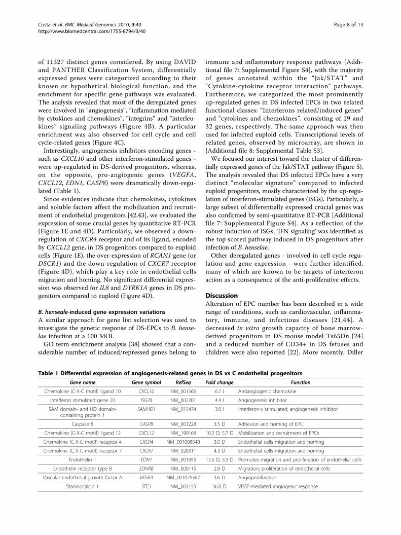
such as CXCL10 and other interferon-stimulated genes -were up-regulated in DS-derived progenitors, whereas,on the opposite, pro-angiogenic genes (VEGFA,CXCL12, EDN1, CASP8) were dramatically down-regu-lated (Table 1).Since evidences indicate that chemokines, cytokines
and soluble factors affect the mobilization and recruit-ment of endothelial progenitors [42,43], we evaluated theexpression of some crucial genes by quantitative RT-PCR(Figure 1E and 4D). Particularly, we observed a down-regulation of CXCR4 receptor and of its ligand, encodedby CXCL12 gene, in DS progenitors compared to euploidcells (Figure 1E), the over-expression of RCAN1 gene (orDSCR1) and the down-regulation of CXCR7 receptor(Figure 4D), which play a key role in endothelial cellsmigration and homing. No significant differential expres-sion was observed for IL8 and DYRK1A genes in DS pro-genitors compared to euploid (Figure 4D).
B. henseale-induced gene expression variationsA similar approach for gene list selection was used toinvestigate the genetic response of DS-EPCs to B. hense-lae infection at a 100 MOI.GO term enrichment analysis [38] showed that a con-
siderable number of induced/repressed genes belong to
immune and inflammatory response pathways [Addi-tional file 7: Supplemental Figure S4], with the majorityof genes annotated within the “Jak/STAT” and“Cytokine-cytokine receptor interaction” pathways.Furthermore, we categorized the most prominentlyup-regulated genes in DS infected EPCs in two relatedfunctional classes: “Interferons related/induced genes”and “cytokines and chemokines”, consisting of 19 and32 genes, respectively. The same approach was thenused for infected euploid cells. Transcriptional levels ofrelated genes, observed by microarray, are shown in[Additional file 8: Supplemental Table S3].We focused our interest toward the cluster of differen-
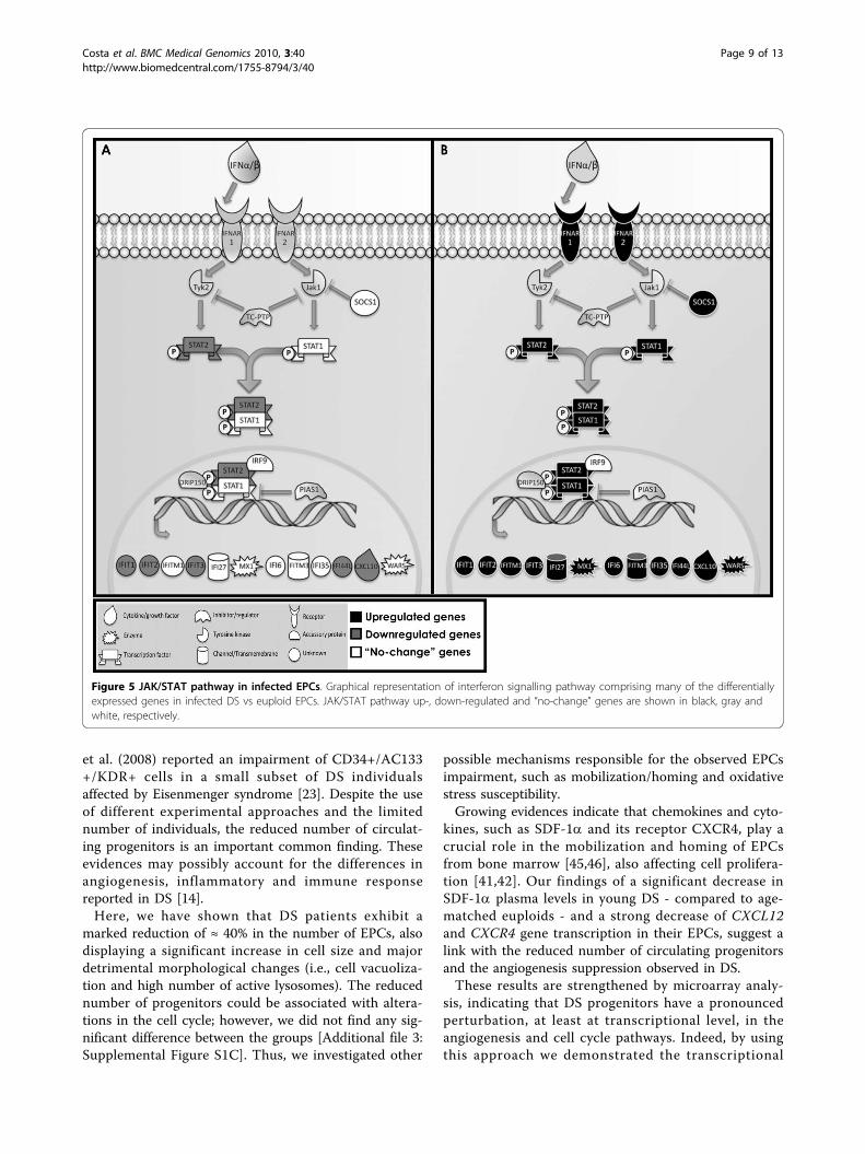
tially expressed genes of the Jak/STAT pathway (Figure 5).The analysis revealed that DS infected EPCs have a verydistinct “molecular signature” compared to infectedeuploid progenitors, mostly characterized by the up-regu-lation of interferon-stimulated genes (ISGs). Particularly, alarge subset of differentially expressed crucial genes wasalso confirmed by semi-quantitative RT-PCR [Additionalfile 7: Supplemental Figure S4]. As a reflection of therobust induction of ISGs, ‘IFN signaling’ was identified asthe top scored pathway induced in DS progenitors afterinfection of B. henselae.Other deregulated genes - involved in cell cycle regu-
lation and gene expression - were further identified,many of which are known to be targets of interferonaction as a consequence of the anti-proliferative effects.
DiscussionAlteration of EPC number has been described in a widerange of conditions, such as cardiovascular, inflamma-tory, immune, and infectious diseases [21,44]. Adecreased in vitro growth capacity of bone marrow-derived progenitors in DS mouse model Ts65Dn [24]and a reduced number of CD34+ in DS fetuses andchildren were also reported [22]. More recently, Diller
Table 1 Differential expression of angiogenesis-related genes in DS vs C endothelial progenitors
Gene name Gene symbol RefSeq Fold change Function
Chemokine (C-X-C motif) ligand 10 CXCL10 NM_001565 6.7 I Antiangiogenic chemokine
Interferon stimulated gene 20 ISG20 NM_002201 4.4 I Angiogenesis inhibitor
SAM domain- and HD domain-containing protein 1
SAMHD1 NM_015474 3.0 I Interferon-g stimulated; angiogenesis inhibitor
Caspase 8 CASP8 NM_001228 3.5 D Adhesion and homing of EPC
Chemokine (C-X-C motif) ligand 12 CXCL12 NM_199168 10.2 D; 5.7 D Mobilization and recruitment of EPCs
Chemokine (C-X-C motif) receptor 4 CXCR4 NM_001008540 3.0 D Endothelial cells migration and homing
Chemokine (C-X-C motif) receptor 7 CXCR7 NM_020311 4.3 D Endothelial cells migration and homing
Endothelin 1 EDN1 NM_001955 13.6 D; 3.5 D Promotes migration and proliferation of endothelial cells
Endothelin receptor type B EDNRB NM_000115 2.8 D Migration, proliferation of endothelial cells
Vascular endothelial growth factor A VEGFA NM_001025367 3.6 D Angioproliferative
Stanniocalcin 1 STC1 NM_003155 56.0 D VEGF-mediated angiogenic response
Costa et al. BMC Medical Genomics 2010, 3:40http://www.biomedcentral.com/1755-8794/3/40
Page 8 of 13
et al. (2008) reported an impairment of CD34+/AC133+/KDR+ cells in a small subset of DS individualsaffected by Eisenmenger syndrome [23]. Despite the useof different experimental approaches and the limitednumber of individuals, the reduced number of circulat-ing progenitors is an important common finding. Theseevidences may possibly account for the differences inangiogenesis, inflammatory and immune responsereported in DS [14].Here, we have shown that DS patients exhibit a
marked reduction of ≈ 40% in the number of EPCs, alsodisplaying a significant increase in cell size and majordetrimental morphological changes (i.e., cell vacuoliza-tion and high number of active lysosomes). The reducednumber of progenitors could be associated with altera-tions in the cell cycle; however, we did not find any sig-nificant difference between the groups [Additional file 3:Supplemental Figure S1C]. Thus, we investigated other
possible mechanisms responsible for the observed EPCsimpairment, such as mobilization/homing and oxidativestress susceptibility.Growing evidences indicate that chemokines and cyto-
kines, such as SDF-1a and its receptor CXCR4, play acrucial role in the mobilization and homing of EPCsfrom bone marrow [45,46], also affecting cell prolifera-tion [41,42]. Our findings of a significant decrease inSDF-1a plasma levels in young DS - compared to age-matched euploids - and a strong decrease of CXCL12and CXCR4 gene transcription in their EPCs, suggest alink with the reduced number of circulating progenitorsand the angiogenesis suppression observed in DS.These results are strengthened by microarray analy-
sis, indicating that DS progenitors have a pronouncedperturbation, at least at transcriptional level, in theangiogenesis and cell cycle pathways. Indeed, by usingthis approach we demonstrated the transcriptional
Figure 5 JAK/STAT pathway in infected EPCs. Graphical representation of interferon signalling pathway comprising many of the differentiallyexpressed genes in infected DS vs euploid EPCs. JAK/STAT pathway up-, down-regulated and “no-change” genes are shown in black, gray andwhite, respectively.
Costa et al. BMC Medical Genomics 2010, 3:40http://www.biomedcentral.com/1755-8794/3/40
Page 9 of 13
deregulation of CXCR7, IL8 and RCAN1 genes, crucialfactors involved in endothelial cells’ migration, homingand angiogenesis [47-49]. Moreover, we found a down-regulation of CASP8, which has been demonstrated tohave a novel apoptosis-unrelated role in proangiogeniccells [50], although this gene was found to be asso-ciated with breast cancer by gene-based associationstudy, and its down-regulation has also been reportedin breast cancer [51].We recently demonstrated the relevance of oxidative
stress on the number and function of progenitor cells[52]. Besides, it is known that oxidative stress is a cru-cial issue in the pathogenesis of DS, especially due tothe high incidence of Alzheimer-like disease at youngage [53]. It has been also suggested that constitutiveexpression of trisomic genes, S100B and SOD1, is likelyto represent a leading cause of ROS generation andincreased oxidation in DS neurons [39,40]. Oxidativestress relevance in DS fetuses was also highlighted bymicroarray analysis of uncultured amniotic fluid [54].Here, we have assessed, by both experimental evi-
dences and microarray analysis, that DS progenitors aremore susceptible to hydroperoxide-induced LP and sig-nificantly over-express S100B and SOD1 genes. Thesefindings strengthen the hypothesis of their involvementin the susceptibility to oxidation observed in DSendothelial progenitors.In our study we have infected DS progenitors with a
human pathogen, B. henselae, responsible of vasoproli-ferative diseases such as bacillary angiomatosis andpeliosis in immunocompromised patients [27,28], pre-viously demonstrated to adhere to and invade EPCs[26]. Particularly, we investigated the effect of infectionon DS progenitors’ number, morphology and oxidativestress response. After hydroperoxide treatment, weobserved a significant LP decrease in Bartonella-infectedDS progenitors compared to uninfected DS cells. Thisfinding confirms the beneficial effect of B. henselaeinfection at low MOI on mature endothelial cells’ survi-val [37,38].In contrast, a reduced cell number in both DS and
euploid groups was observed after Bartonella infectionat higher MOIs; however, detrimental changes were visi-ble only in DS EPCs, displaying a higher number ofinvasomes and infected cells compared to euploid cells.The molecular basis of such Bartonella-induced detri-
mental effect on endothelial progenitors of DS wasinvestigated at a transcriptional level by microarray. Sig-nificant up-regulation of the Jak/STAT pathway wasobserved only within infected DS progenitors, whereas,on the opposite, infected euploid cells displayed a signif-icant down-regulation. These findings strengthen thehypothesis that transcriptional analysis of EPCs is clearly
of major interest in the context of this syndrome.Indeed, the activation of ISGs, involved in the immuneresponse against infections and in tumor surveillance[55], also inhibits angiogenesis by decreasing the pro-duction of angiogenic factors such as VEGF and IL8[56]. Our results clearly show that a similar ISGs activa-tion occurs in infected DS progenitor cells, and, albeit atlower levels, in uninfected DS progenitors (data notshown).Pathologic immune and inflammatory responses are
regulated by the cross-talk between interferons and TNFa[26], as well as deregulation of chemokines/cytokinesgreatly affects the mobilization and recruitment ofendothelial progenitors. The imbalance between ISGs andother molecules might be of great immunological relevanceconcerning the well-known DS haematological defects.
ConclusionsPhysiological angiogenesis plays a central role in theembryogenesis and placental development; on the otherhand, pathological angiogenesis represents a criticalissue in the progression of many diseases, such as solidtumor growth and retinopathy.Individuals with Down syndrome, due to decreased
incidence of angiogenesis-dependent diseases, have beenpostulated to be a systemic anti-angiogenesis model.Indeed, they exhibit a significantly increased anti-angio-genic surveillance, possibly due to increased expressionof anti-angiogenic regulators on chromosome 21 [57].However, it has been shown a complex regulation of
gene expression not only related to gene copy number,with several genes escaping the rule of “increased tran-scription proportional to the gene copy number”[58,59]. This findings suggest that many pathologicaltraits observed in DS may be controlled by other morecomplex and, above all tissue-specific, regulatorymechanisms [59].Our study shows that circulating endothelial progeni-
tors are reduced in patients with DS, possibly correlatingto the low SDF-1a plasma levels, to a reduced expres-sion of its membrane receptor in these cells, and totheir higher oxidative stress and pathogen infection sus-ceptibility compared to euploid cells. A significant per-turbation in the angiogenesis and inflammation genepathways was also observed by microarray analysis,highlighting that gene expression analysis is a crucialissue for the study of common diseases. Endothelial dys-function, angiogenesis’ suppression and infection recur-rence are hallmarks of DS, and the impairment in thenumber and function of circulating progenitors mayaccount for some of their pathological features. Furtherstudies are needed to understand possible therapeuticimplications of circulating EPCs in Down syndrome.
Costa et al. BMC Medical Genomics 2010, 3:40http://www.biomedcentral.com/1755-8794/3/40
Page 10 of 13
Additional material
Additional file 1: Supplementary Methods
Additional file 2: Table S1: Primer pairs used for quantitative andsemi-quantitative RT-PCR
Additional file 3: Figure S1: Impaired EPC number and function. A)Representative photomicrographs of merged double-positive Dil-Ac-LDL/Lectin cells isolated from euploid (left panel) and DS (right panel)subjects (100X magnification). B) Fluorescence micrographs of EPCslabeled for 30 min with C11-BO in euploid and DS subjects. C) EPCnumber expressed as percentage in the different phases of cell cycleobtained by FACS. D) Curves indicate the percentage of EPC numberinfected with B. henselae in euploid and DS individuals. Results arerepresentative of five different experiments in duplicate.
Additional file 4: Table S2: Chromosome 21 genes differentiallyexpressed in DS vs euploids
Additional file 5: Figure S2: Distribution of differentially expressedgenes along the human chromosomes (DS vs euploids). A) Bar graphshowing the empirical frequency distribution of differentially expressedgenes along the autosomes of DS progenitors vs euploids. Asterisksindicate the significantly deregulated chromosomes. B) Representation ofthe robustness of our findings shown in A. The left column shows thedifferent user-defined fold-change. For each á value used in the analysisare shown the relative p-values. C) Bar graph showing the percent ofdifferentially expressed genes along the DS autosomes.
Additional file 6: Figure S3: Positional gene mapping ofdifferentially expressed genes (DS vs euploids). Graphicrepresentation of positional gene enrichment (PGE) approach used tomap differentially expressed genes in DS vs euploids EPCs to the exactlocation on the chromosome.
Additional file 7: Figure S4: B. henseale-induced gene expression inDS EPCs. A) Bar graph showing the top-scored deregulated genepathways after infection in DS progenitors. Ratio indicates the percent ofdifferentially expressed genes within the related pathway. B)Semiquantitative RT-PCR of Jak/STAT genes deregulated after B. hensealeinfection.
Additional file 8: Table S3: Differentially expressed genes after B.henselae infection
AcknowledgementsWe thank the patients and individuals recruited as controls for theirparticipation. This work was supported by Legge 5, Regione Campania 2008to Prof. A. Ciccodicola; PRIN 2006, Foundation Jerome Lejeune, and RegioneCampania 2008 to Prof. C. Napoli; and Progetto di Rilevante InteresseNazionale Ministero Italiano Università e Ricerca 2006 [grant number2006068944_001] “Basi genetiche e molecolari della patogenicità batterica”to Prof. P. Salvatore.
Author details1Institute of Genetics and Biophysics ‘’A. Buzzati-Traverso’’, IGB-CNR, Naples,Italy. 2Section of Microbiology, Department of Experimental Medicine, 1stSchool of Medicine, Second University of Naples, Naples, Italy. 3Departmentof General Pathology and Excellence Research Center on CardiovascularDiseases, 1st School of Medicine, Second University of Naples, Naples, Italy.4IRCCS Fondazione SDN, Naples, Italy. 5Istituto per le Applicazioni delCalcolo, “Mauro Picone”, CNR, Naples, Italy. 6Department of BiologicalScience, University of Naples “Federico II”, Naples, Italy. 7Department ofBiochemistry and Biophysics, Second University of Naples, Naples, Italy.8IRCCS Multimedica, Milan, Italy. 9Department of Biochemistry and MedicalBiotechnology, University of Naples “Federico II”, Naples, Italy. 10Institute ofGeneral Pathology, Section of Clinical Pathology, Faculty of Medicine,University of Milan, Milan, Italy. 11Cardiology Department of SecondUniversity of Naples, “Monaldi Hospital”, Naples, Italy. 12Department ofCellular and Molecular Biology and Pathology “L. Califano” and School ofBiotechnological Sciences, University of Naples “Federico II”, Naples, Italy.
Authors’ contributionsCV, LS, ACa, BA, PS, ACi and CN designed research. CV, LS, ACa, RCo, VM, LM,CF, MR and MP carried out experimental research. VM and BA performedTransmission electron microscopy. CV and MR performed microarray analysisand quantitative RT-PCR. CV and CAn assisted with statistical analysis. BF,MMC, BS and RCa participated in the participant recruitment. CV, LS, ACa,RCo, CAn, VM, LM, BF, AG, CF, MR, MP, BA, MMC, BS, RCa, PS, ACi and CNanalyzed data. CV, LS, ACa, CAn, BA, PS, ACi and CN wrote the paper. Allauthors read and approved the final manuscript.
Competing interestsThe authors declare that they have no competing interests.
Received: 17 February 2010 Accepted: 13 September 2010Published: 13 September 2010
References1. Wiseman FK, Alford KA, Tybulewicz VL, Fisher EM: Down syndrome–recent
progress and future prospects. Hum Mol Genet 2009, 18:75-83.2. Gillespie KM, Dix RJ, Williams AJ, Newton R, Robinson ZF, Bingley PJ,
Gale EA, Shield JP: Islet autoimmunity in children with Down’s syndrome.Diabetes 2006, 55:3185-3188.
3. Ugazio AG, Maccario R, Notarangelo LD, Burgio GR: Immunology of Downsyndrome: a review. Am J Med Genet Suppl 1990, 7:204-212.
4. Garrison MM, Jeffries H, Christakis DA: Risk of death for children withdown syndrome and sepsis. J Pediatr 2005, 147:748-752.
5. Folkman J: Angiogenesis: an organizing principle for drug discovery? NatRev Drug Discov 2007, 6:273-286.
6. Hasle H, Clemmensen IH, Mikkelsen M: Risks of leukaemia and solidtumours in individuals with Down’s syndrome. Lancet 2000, 355:165-169.
7. Fulcher T, Griffin M, Crowley S, Firth R, Acheson R, O’ Meara N: Diabeticretinopathy in Down’s syndrome. Br J Ophthalmol 1998, 82:407-409.
8. Cappelli-Bigazzi M, Santoro G, Battaglia C, Palladino MT, Carrozza M,Russo MG, Pacileo G, Calabrò R: Endothelial cell function in patients withDown’s syndrome. Am J Cardiol 2004, 94:392-395.
9. FitzPatrick DR, Ramsay J, McGill NI, Shade M, Carothers AD, Hastie ND:Transcriptome analysis of human autosomal trisomy. Hum Mol Genet2002, 11:3249-3256.
10. Mao R, Zielke CL, Zielke HR, Pevsner J: Global up-regulation ofchromosome 21 gene expression in the developing Down syndromebrain. Genomics 2003, 81:457-467.
11. Li CM, Guo M, Salas M, Schupf N, Silverman W, Zigman WB, Husain S,Warburton D, Thaker H, Tycko B: Cell type-specific over-expression ofchromosome 21 genes in fibroblasts and fetal hearts with trisomy 21.BMC Med Genet 2006, 7:24.
12. Sommer CA, Pavarino-Bertelli EC, Goloni-Bertollo EM, Henrique-Silva F:Identification of dysregulated genes in lymphocytes from children withDown syndrome. Genome 2008, 51:19-29.
13. Gardiner K: Gene-dosage effects in Down syndrome and trisomic mousemodels. Genome Biol 2004, 5:244.
14. Antonarakis SE, Epstein CJ: The challenge of Down syndrome. Trends MolMed 2006, 12:473-479.
15. Yoder MC, Mead LE, Prater D, Krier TR, Mroueh KN, Li F, Krasich R, Temm CJ,Prchal JT, Ingram DA: Redefining endothelial progenitor cells via clonalanalysis and hematopoietic stem/progenitor cell principals. Blood 2007,109:1801-1809.
16. Hirschi KK, Ingram DA, Yoder MC: Assessing identity, phenotype, and fateof endothelial progenitor cells. Arterioscler Thromb Vasc Biol 2008,28:1584-1595.
17. Zampetaki A, Kirton JP, Xu Q: Vascular Repair by Endothelial ProgenitorCells. Cardiovascular Research 2008, 78:413-421.
18. Krenning G, van Luyn MJ, Harmsen MC: Endothelial progenitor cell-basedneovascularization: implications for therapy. Trends Mol Med 2009,15:180-189.
19. Shantsila E, Watson T, Lip GY: Endothelial progenitor cells incardiovascular disorders. Journal of the American College of Cardiology2007, 49:741-752.
20. Vasa M, Fichtlscherer S, Aicher A, Adler K, Urbich C, Martin H, Zeiher AM,Dimmeler S: Number and migratory activity of circulating andothelialprogenitor cells inversely correlate with risk factors for coronary arterydisease. Circulation Research 2001, 89:e1-7.
Costa et al. BMC Medical Genomics 2010, 3:40http://www.biomedcentral.com/1755-8794/3/40
Page 11 of 13
21. Sabatier F, Camoin-Jau L, Anfosso F, Sampol J, Dignat-George F: Circulatingendothelial cells, microparticles and progenitors: key players towardsthe definition of vascular competence. J Cell Mol Med 2009, 13:454-471.
22. Holmes DK, Bates N, Murray M, Ladusans EJ, Morabito A, Bolton-Maggs PH,Johnston TA, Walkenshaw S, Wynn RF, Bellantuono I: Hematopoieticprogenitor cell deficiency in fetuses and children affected by Down’ssyndrome. Exp Hematol 2006, 34:1611-1615.
23. Diller GP, van Eijl S, Okonko DO, Howard LS, Ali O, Thum T, Wort SJ,Bédard E, Gibbs JS, Bauersachs J, Hobbs AJ, Wilkins MR, Gatzoulis MA,Wharton J: Circulating endothelial progenitor cells in patients withEisenmenger syndrome and idiopathic pulmonary arterial hypertension.Circulation 2008, 117:3020-3030.
24. Jablonska B, Ford D, Trisler D, Pessac B: The growth capacity of bonemarrow CD34 positive cells in culture is drastically reduced in a murinemodel of Down syndrome. C R Biol 2006, 329:726-732.
25. Mantovani A, Sozzani S, Introna M: Endothelial activation by cytokines.Ann N Y Acad Sci 1997, 832:93-116.
26. Salvatore P, Casamassimi A, Sommese L, Fiorito C, Ciccodicola A, Rossiello R,Avallone B, Grimaldi V, Costa V, Rienzo M, Colicchio R, Williams-Ignarro S,Pagliarulo C, Prudente ME, Abbondanza C, Lamberti F, Baroni A,Buommino E, Farzati B, Tufano MA, Ignarro LJ, Napoli C: Detrimentaleffects of Bartonella henselae are counteracted by L-arginine and nitricoxide in human endothelial progenitor cells. Proc Natl Acad Sci USA 2008,105:9427-9432.
27. Dehio C: Bartonella-host-cell interactions and vascular tumour formation.Nat Rev Microbiol 2005, 3:621-631.
28. Kunz S, Oberle K, Sander A, Bogdan C, Schleicher U: Lymphadenopathy ina novel mouse model of Bartonella-induced cat scratch disease resultsfrom lymphocyte immigration and proliferation and is regulated byinterferon-alpha/beta. Am J Pathol 2008, 172:1005-1018.
29. Dogliotti G, Galliera E, Licastro F, Corsi MM: Age-related changes in plasmalevels of BDNF in Down syndrome patients. Immun Ageing 2010, 7:2.
30. Casamassimi A, Balestrieri ML, Fiorito C, Schiano C, Maione C, Rossiello R,Grimaldi V, Del Giudice V, Balestrieri C, Farzati B, Sica V, Napoli C:Comparison between total endothelial progenitor cell isolation versusenriched CD133+ culture. J Biochem 2007, 141:503-511.
31. Avallone B, Porritiello M, Esposito D, Mutone R, Balsamo G, Marmo F:Evidence for hair cell regeneration in the crista ampullaris of the lizardPodarcis sicula. Hear Res 2003, 178:79-88.
32. Costa V, Conte I, Ziviello C, Casamassimi A, Alfano G, Banfi S, Ciccodicola A:Identification and expression analysis of novel Jakmip1 transcripts. Gene2007, 402:1-8.
33. Hosack DA, Dennis G Jr, Sherman BT, Lane HC, Lempicki RA: Identifyingbiological themes within lists of genes with EASE. Genome Biol 2003, 4:R70.
34. Huang da W, Sherman BT, Lempicki RA: Systematic and integrativeanalysis of large gene lists using DAVID bioinformatics resources. NatProtoc 2009, 4:44-57.
35. Thomas PD, Campbell MJ, Kejariwal A, Mi H, Karlak B, Daverman R,Diemer K, Muruganujan A, Narechania A: PANTHER: a library of proteinfamilies and subfamilies indexed by function. Genome Res 2003,13:2129-2141.
36. Dernbach E, Urbich C, Brandes RP, Hofmann WK, Zeiher AM, Dimmller S:Antioxidative stress-associated genes in circulating progenitor cells:evidence for enhanced resistance against oxidative stress. Blood 2004,104:3591-3597.
37. Kirby JE, Nekorchuk DM: Bartonella-associated endothelial proliferationdepends on inhibition of apoptosis. Proc Natl Acad Sci USA 2002,99:4656-4661.
38. Pulliainen AT, Dehio C: Bartonella henselae: subversion of vascularendothelial cell functions by translocated bacterial effector proteins. Int JBiochem Cell Biol 2009, 41:507-510.
39. Esposito G, Imitola J, Lu J, De Filippis D, Scuderi C, Ganesh VS, Folkerth R,Hecht J, Shin S, Iuvone T, Chesnut J, Steardo L, Sheen V: Genomic andfunctional profiling of human Down syndrome neural progenitorsimplicates S100B and aquaporin 4 in cell injury. Hum Mol Genet 2008,17:440-457.
40. Lee M, Hyun D, Jenner P, Halliwell B: Effect of overexpression of wild-typeand mutant Cu/Zn-superoxide dismutases on oxidative damage andantioxidant defences: relevance to Down’s syndrome and familialamyotrophic lateral sclerosis. J Neurochem 2001, 76:957-965.
41. De Preter K, Barriot R, Speleman F, Vandesompele J, Moreau Y: Positionalgene enrichment analysis of gene sets for high-resolution identificationof overrepresented chromosomal regions. Nucleic Acids Res 2008, 36:e43.
42. Hristov M, Wolfgang W, Weber PC: Endothelial progenitor cellsmobilization differentiation and homing. Arterioscler Thromb Vasc Biol2003, 23:1185-1189.
43. Egan CG, Lavery R, Caporali F, Fondelli C, Laghi-Pasini F, Dotta F,Sorrentino V: Generalised reduction of putative endothelial progenitorsand CXCR4-positive peripheral blood cells in type 2 diabetes.Diabetologia 2008, 51:1296-1305.
44. Schmidt-Lucke C, Rössig L, Fichtlscherer S, Vasa M, Britten M, Kamper U,Dimmeler S, Zeiher AM: Reduced number of circulating endothelialprogenitor cells predicts future cardiovascular events: proof of conceptfor the clinical importance of endogenous vascular repair. Circulation2005, 111:2981-2987.
45. Yamaguchi J, Kusano KF, Masuo O, Kawamoto A, Silver M, Murasawa S,Bosch-Marce M, Masuda H, Losordo DW, Isner JM, Asahara T: Stromal cell-derived factor-1 effects on ex vivo expanded endothelial progenitor cellrecruitment for ischemic neovascularization. Circulation 2003,107:1322-1328.
46. Walter DH, Haendeler J, Reinhold J, Rochwalsky U, Seeger F, Honold J,Hoffmann J, Urbich C, Lehmann R, Arenzana-Seisdesdos F, Aicher A,Heeschen C, Fichtlscherer S, Zeiher AM, Dimmeler S: Impaired CXCR4signaling contributes to the reduced neovascularisation capacity ofendothelial progenitor cells from patients with coronary artery disease.Circ Res 2005, 97:1142-1151.
47. Waugh DJ, Wilson C: The interleukin-8 pathway in cancer. Clin Cancer Res2008, 14:6735-6741.
48. Burns JM, Summers BC, Wang Y, Melikian A, Berahovich R, Miao Z,Penfold ME, Sunshine MJ, Littman DR, Kuo CJ, Wei K, McMaster BE,Wright K, Howard MC, Schall TJ: A novel chemokine receptor for SDF-1and I-TAC involved in cell survival, cell adhesion, and tumordevelopment. J Exp Med 2006, 203:2201-2213.
49. Baek KH, Zaslavsky A, Lynch RC, Britt C, Okada Y, Siarey RJ, Lensch MW,Park IH, Yoon SS, Minami T, Korenberg JR, Folkman J, Daley GQ, Aird WC,Galdzicki Z, Ryeom S: Down’s syndrome suppression of tumour growthand the role of the calcineurin inhibitor DSCR1. Nature 2009,459:1126-1130.
50. Scharner D, Rössig L, Carmona G, Chavakis E, Urbich C, Fischer A, Kang TB,Wallach D, Chiang YJ, Deribe YL, Dikic I, Zeiher AM, Dimmeler S: Caspase-8is involved in neovascularization-promoting progenitor cell functions.Arterioscler Thromb Vasc Biol 2009, 29:571-578.
51. Wu Y, Alvarez M, Slamon DJ, Koeffler P, Vadgama JV: Caspase 8 andmaspin are downregulated in breast cancer cells due to CpG sitepromoter methylation. BMC Cancer 2010, 10:32.
52. Fiorito C, Rienzo M, Crimi E, Rossiello R, Balestrieri ML, Casamassimi A,Muto F, Grimaldi V, Giovane A, Farzati B, Mancini FP, Napoli C: Antioxidantsincrease number of progenitor endothelial cells through multiple geneexpression pathways. Free Radic Res 2008, 42:754-762.
53. Zana M, Janka Z, Kálmán J: Oxidative stress: a bridge between Down’ssyndrome and Alzheimer’s disease. Neurobiol Aging 2007, 28:648-676.
54. Slonim DK, Koide K, Johnson KL, Tantravahi U, Cowan JM, Jarrah Z,Bianchi DW: Functional genomic analysis of amniotic fluid cell-freemRNA suggests that oxidative stress is significant in Down syndromefetuses. Proc Natl Acad Sci USA 2009, 106:9425-9429.
55. Borden EC, Sen GC, Uze G, Silverman RH, Ransohoff RM, Foster GR, Stark GR:Interferons at age 50: past, current and future impact on biomedicine.Nat Rev Drug Discov 2007, 6:975-990.
56. Rosewicz S, Detjen K, Scholz A, von Marschall Z: Interferon-alpha:regulatory effects on cell cycle and angiogenesis. Neuroendocrinology2004, 80:85-93.
57. Ryeom S, Folkman J: Role of Endogenous Angiogenesis Inhibitors inDown Syndrome. J Craniofac Surg 2009, 20:595-596.
58. Lyle R, Gehrig C, Neergaard-Henrichsen C, Deutsch S, Antonarakis SE: Geneexpression from the aneuploid chromosome in a trisomy mouse modelof down syndrome. Genome Res 2004, 14:1268-1274.
59. Kahlem P, Sultan M, Herwig R, Steinfath M, Balzereit D, Eppens B, Saran NG,Pletcher MT, South ST, Stetten G, Lehrach H, Reeves RH, Yaspo ML:Transcript level alterations reflect gene dosage effects across multipletissues in a mouse model of down syndrome. Genome Res 2004,14:1258-1267.
Costa et al. BMC Medical Genomics 2010, 3:40http://www.biomedcentral.com/1755-8794/3/40
Page 12 of 13

Pre-publication historyThe pre-publication history for this paper can be accessed here:http://www.biomedcentral.com/1755-8794/3/40/prepub
doi:10.1186/1755-8794-3-40Cite this article as: Costa et al.: Impairment of circulating endothelialprogenitors in Down syndrome. BMC Medical Genomics 2010 3:40.
Submit your next manuscript to BioMed Centraland take full advantage of:
• Convenient online submission
• Thorough peer review
• No space constraints or color figure charges
• Immediate publication on acceptance
• Inclusion in PubMed, CAS, Scopus and Google Scholar
• Research which is freely available for redistribution
Submit your manuscript at www.biomedcentral.com/submit
Costa et al. BMC Medical Genomics 2010, 3:40http://www.biomedcentral.com/1755-8794/3/40
Page 13 of 13