Circulating Mitochondrial DNA and Inter-Organelle Contact ...
22
Citation: Picca, A.; Guerra, F.; Calvani, R.; Romano, R.; Coelho-Junior, H.J.; Damiano, F.P.; Bucci, C.; Marzetti, E. Circulating Mitochondrial DNA and Inter-Organelle Contact Sites in Aging and Associated Conditions. Cells 2022, 11, 675. https:// doi.org/10.3390/cells11040675 Academic Editor: Marcello Pinti Received: 26 January 2022 Accepted: 14 February 2022 Published: 15 February 2022 Publisher’s Note: MDPI stays neutral with regard to jurisdictional claims in published maps and institutional affil- iations. Copyright: © 2022 by the authors. Licensee MDPI, Basel, Switzerland. This article is an open access article distributed under the terms and conditions of the Creative Commons Attribution (CC BY) license (https:// creativecommons.org/licenses/by/ 4.0/). cells Review Circulating Mitochondrial DNA and Inter-Organelle Contact Sites in Aging and Associated Conditions Anna Picca 1,† , Flora Guerra 2,† , Riccardo Calvani 1, * , Roberta Romano 2 ,Hélio José Coelho-Junior 3 , Francesco P. Damiano 1 , Cecilia Bucci 2 and Emanuele Marzetti 1,3 1 Fondazione Policlinico Universitario “Agostino Gemelli” IRCCS, 00168 Rome, Italy; [email protected] (A.P.);[email protected] (F.P.D.); [email protected] (E.M.) 2 Department of Biological and Environmental Sciences and Technologies, Università del Salento, 73100 Lecce, Italy; fl[email protected] (F.G.); [email protected] (R.R.); [email protected] (C.B.) 3 Department of Geriatrics and Orthopedics, Università Cattolica del Sacro Cuore, 00168 Rome, Italy; [email protected] * Correspondence: [email protected]; Tel.: +39-06-3015-5559; Fax: +39-06-3051-911 † These authors contributed equally to this work. Abstract: Mitochondria are primarily involved in cell bioenergetics, regulation of redox homeostasis, and cell death/survival signaling. An immunostimulatory property of mitochondria has also been recognized which is deployed through the extracellular release of entire or portioned organelle and/or mitochondrial DNA (mtDNA) unloading. Dynamic homo- and heterotypic interactions involving mitochondria have been described. Each type of connection has functional implications that eventually optimize mitochondrial activity according to the bioenergetic demands of a specific cell/tissue. Inter-organelle communications may also serve as molecular platforms for the extra- cellular release of mitochondrial components and subsequent ignition of systemic inflammation. Age-related chronic inflammation (inflamm-aging) has been associated with mitochondrial dysfunc- tion and increased extracellular release of mitochondrial components—in particular, cell-free mtDNA. The close relationship between mitochondrial dysfunction and cellular senescence further supports the central role of mitochondria in the aging process and its related conditions. Here, we provide an overview of (1) the mitochondrial genetic system and the potential routes for generating and releasing mtDNA intermediates; (2) the pro-inflammatory pathways elicited by circulating mtDNA; (3) the participation of inter-organelle contacts to mtDNA homeostasis; and (4) the link of these processes with senescence and age-associated conditions. Keywords: exosomes; extracellular vesicles; inflamm-aging; senescence; mitophagy; mitochondrial damage; mitochondrial dynamics; mitochondrial-derived vesicles; mitochondrial-lysosomal axis; oxidative stress 1. Introduction Mitochondria are cytoplasmic double-membrane organelles residing within eukary- otic cells. Mitochondria are vestiges of the incorporation of an anaerobic bacterial ancestor into an unicellular eukaryote which occurred over a billion years ago [1]. The endosym- biotic fusion marked the evolution of eukaryotic cells via a serendipitous switch towards aerobic respiration. From an evolutionary perspective, this metabolic change contributed to eukaryote complexity and the development of multicellular life [2]. While initially envisioned as resident and isolated, mitochondria are increasingly recognized as “social” organelles immersed into the cytoplasmic fluid together with other organelles with which they interact and coordinate a plethora of cellular activities [3,4]. Different from other cytoplasmic organelles, mitochondria are semi-autonomous as they possess their own genome, the mitochondrial DNA (mtDNA) [5]. This nucleic acid Cells 2022, 11, 675. https://doi.org/10.3390/cells11040675 https://www.mdpi.com/journal/cells
-
Upload
khangminh22 -
Category
Documents
-
view
0 -
download
0
Transcript of Circulating Mitochondrial DNA and Inter-Organelle Contact ...

�����������������
Citation: Picca, A.; Guerra, F.;
Calvani, R.; Romano, R.;
Coelho-Junior, H.J.; Damiano, F.P.;
Bucci, C.; Marzetti, E. Circulating
Mitochondrial DNA and
Inter-Organelle Contact Sites in
Aging and Associated Conditions.
Cells 2022, 11, 675. https://
doi.org/10.3390/cells11040675
Academic Editor: Marcello Pinti
Received: 26 January 2022
Accepted: 14 February 2022
Published: 15 February 2022
Publisher’s Note: MDPI stays neutral
with regard to jurisdictional claims in
published maps and institutional affil-
iations.
Copyright: © 2022 by the authors.
Licensee MDPI, Basel, Switzerland.
This article is an open access article
distributed under the terms and
conditions of the Creative Commons
Attribution (CC BY) license (https://
creativecommons.org/licenses/by/
4.0/).
cells
Review
Circulating Mitochondrial DNA and Inter-Organelle ContactSites in Aging and Associated ConditionsAnna Picca 1,† , Flora Guerra 2,† , Riccardo Calvani 1,* , Roberta Romano 2 , Hélio José Coelho-Junior 3 ,Francesco P. Damiano 1, Cecilia Bucci 2 and Emanuele Marzetti 1,3
1 Fondazione Policlinico Universitario “Agostino Gemelli” IRCCS, 00168 Rome, Italy;[email protected] (A.P.); [email protected] (F.P.D.);[email protected] (E.M.)
2 Department of Biological and Environmental Sciences and Technologies, Università del Salento,73100 Lecce, Italy; [email protected] (F.G.); [email protected] (R.R.);[email protected] (C.B.)
3 Department of Geriatrics and Orthopedics, Università Cattolica del Sacro Cuore, 00168 Rome, Italy;[email protected]
* Correspondence: [email protected]; Tel.: +39-06-3015-5559; Fax: +39-06-3051-911† These authors contributed equally to this work.
Abstract: Mitochondria are primarily involved in cell bioenergetics, regulation of redox homeostasis,and cell death/survival signaling. An immunostimulatory property of mitochondria has also beenrecognized which is deployed through the extracellular release of entire or portioned organelleand/or mitochondrial DNA (mtDNA) unloading. Dynamic homo- and heterotypic interactionsinvolving mitochondria have been described. Each type of connection has functional implicationsthat eventually optimize mitochondrial activity according to the bioenergetic demands of a specificcell/tissue. Inter-organelle communications may also serve as molecular platforms for the extra-cellular release of mitochondrial components and subsequent ignition of systemic inflammation.Age-related chronic inflammation (inflamm-aging) has been associated with mitochondrial dysfunc-tion and increased extracellular release of mitochondrial components—in particular, cell-free mtDNA.The close relationship between mitochondrial dysfunction and cellular senescence further supportsthe central role of mitochondria in the aging process and its related conditions. Here, we provide anoverview of (1) the mitochondrial genetic system and the potential routes for generating and releasingmtDNA intermediates; (2) the pro-inflammatory pathways elicited by circulating mtDNA; (3) theparticipation of inter-organelle contacts to mtDNA homeostasis; and (4) the link of these processeswith senescence and age-associated conditions.
Keywords: exosomes; extracellular vesicles; inflamm-aging; senescence; mitophagy; mitochondrialdamage; mitochondrial dynamics; mitochondrial-derived vesicles; mitochondrial-lysosomal axis;oxidative stress
1. Introduction
Mitochondria are cytoplasmic double-membrane organelles residing within eukary-otic cells. Mitochondria are vestiges of the incorporation of an anaerobic bacterial ancestorinto an unicellular eukaryote which occurred over a billion years ago [1]. The endosym-biotic fusion marked the evolution of eukaryotic cells via a serendipitous switch towardsaerobic respiration. From an evolutionary perspective, this metabolic change contributedto eukaryote complexity and the development of multicellular life [2]. While initiallyenvisioned as resident and isolated, mitochondria are increasingly recognized as “social”organelles immersed into the cytoplasmic fluid together with other organelles with whichthey interact and coordinate a plethora of cellular activities [3,4].
Different from other cytoplasmic organelles, mitochondria are semi-autonomous asthey possess their own genome, the mitochondrial DNA (mtDNA) [5]. This nucleic acid
Cells 2022, 11, 675. https://doi.org/10.3390/cells11040675 https://www.mdpi.com/journal/cells

Cells 2022, 11, 675 2 of 22
is a circular double-stranded molecule spanning approximately 16 kb pairs and encodesfor 37 genes (13 messenger RNAs, 22 transfer RNAs, and 2 ribosomal RNAs). MtDNA isresponsible for the synthesis of hydrophobic protein subunits of the electron transport chain(ETC), the apparatus that enables cellular respiration by coupling oxygen consumptionwith the generation of a membrane potential at the inner mitochondrial membrane. Thelatter is the bioenergetic core of the cell via production of adenosine triphosphate (ATP) thatfuels most cellular activities. Mitochondria are involved in a number of other vital activities,including regulation of ionic (calcium and iron) levels, hormone synthesis, antioxidantdetoxification, iron-sulfur cluster and heme biosynthesis, and programmed cell death [6].
MtDNA exists in hundreds to thousands of copies within the cell. This number, whichreflects mitochondria abundance and/or mitochondrial mass, varies according to cellmetabolism and in response to several intrinsic and extrinsic stimuli [7]. The large mtDNAcopy number compared with the amount needed to support oxidative phosphorylationmay reflect the mitochondrial involvement in organelle signaling and/or deployment ofimmune functions [8].
The recognition of an intrinsic mtDNA immunostimulatory property is a feature that hasrevolutionized the mitochondrial outlook in the context of several inflammatory conditions [8].In addition to the pro-inflammatory domains of circulating mtDNA molecules, the formation ofnon-canonical nucleic acid structures during mtDNA transcription and replication holds uniquefeatures that may engage nucleic acid sensors and trigger innate immunity [8].
Although possessing a certain degree of independence, mitochondria are under thecontrol of the nucleus with regard to mtDNA stability and sequence evolution over time [9].This is made possible by their strategic relocation near the nucleus, whereby mitochondriaform contact sites able to generate output signals that can influence the expression ofnuclear genes [10].
Dynamic homo- and heterotypic interactions involving mitochondria have been de-scribed. Inter-organelle communications are enabled by several processes/structures,including fusion, nanotunnel protrusion, and inter-organelle contact sites [11]. Althoughthe exact mechanisms regulating the formation and the activity of these structures arepresently unknown, each type of connection has functional implications that can be reg-ulated to optimize mitochondrial activity to meet the bioenergetic demands of a specificcell/tissue [12].
The analysis of these mitochondrial properties and the molecules involved in such com-munication may help unveil the complex interactions that mitochondria establish to achievetheir own homeostasis. Indeed, mitochondrial metabolism and its alterations have been listedamong the nine pillars of the geroscience paradigm, and are a key target amenable for anti-aginginterventions [13]. In this regard, the close relationship among mitochondrial dysfunction, in-flammation and cellular senescence, and their contribution to aging and age-related conditionsconfer further priority to the understanding of these processes.
Here, we provide an overview of (1) the mitochondrial genetic system and the potentialroutes for generating and releasing mtDNA intermediates; (2) the contribution of circulat-ing mtDNA to inflammation; (3) the participation of inter-organelle contact sites to mtDNAhomeostasis; and (4) the link of these processes with senescence and age-associated conditions.
2. Mitochondrial Genetics: MtDNA Transcription, Replication, and the Generation ofNon-Canonical Structures
The transcription of mtDNA guides the synthesis of a subset of hydrophobic ETCcomplex subunits through mtDNA-encoded ribosomes (12S and 16S) and 22 tRNAs [14].Hydrophyilic ETC subunits and proteins involved in mtDNA transcription, translation,replication, and maintenance are nuclear-encoded and are co- or post-translationally im-ported into the organelle [7,15].
A large non-coding arc of mtDNA, the so-called displacement-loop (D-loop) region,harbors both the heavy- (HSP) and the light-strand promoter (LSP) of mtDNA transcription,

Cells 2022, 11, 675 3 of 22
along with the origin of heavy-strand replication (OH) and conserved cis-acting elements.Hence, the D-loop is a major regulatory site of mtDNA activity and synthesis [16].
The expression of mtDNA genes starts at both promoters and begins with the tran-scription of RNA primary transcripts (for both mtDNA strands) that are almost full-lengthand are processed into mature mRNAs by specific RNases. Almost synchronously, thetranscripts generated by the LSP are used as primers for mtDNA replication of the leadingstrand that follows a replicative asymmetric mode. The termination of transcription and/orspecific RNA processing occurs downstream LSP, and strand extension by the mtDNApolymerase γ (Pol γ) generates the 3′ end of this mtDNA strand. Following Pol γ-guidedDNA synthesis, the enzyme activity becomes stalled or terminated approximately 1 kbpast the LSP. The newly synthesized mtDNA strand remains bound to the template andforms a stable three-stranded D-loop structure. This latter is a hallmark of the mammalianmtDNA replicating structure with uncertain biological relevance [14,16] (Figure 1). MtDNAsynthesis downstream from the 3′ end of the D-loop region is mandatory for mtDNAreplication, which requires priming of the lagging strand at multiple mtDNA sites. Amajor replication starter is the origin of light-strand replication (OL), which is located~12 kb away from the OH and enables asynchronous replication of the leading and laggingstrands. As a result of these events, large stretches of single-stranded DNA (ss-DNA) andRNA–DNA hybrids originate and persist as intermediates of mtDNA synthesis. Othermodes of mtDNA replication have been described and involve a “bootlace” mechanismfollowing the incorporation of processed mtDNA transcripts [17] (Figure 1).
Cells 2022, 11, x FOR PEER REVIEW 3 of 22
Cells 2022, 11, x. https://doi.org/10.3390/xxxxx www.mdpi.com/journal/cells
Hydrophyilic ETC subunits and proteins involved in mtDNA transcription, translation, replication, and maintenance are nuclear-encoded and are co- or post-translationally im-
ported into the organelle [7,15]. A large non-coding arc of mtDNA, the so-called displacement-loop (D-loop) region,
harbors both the heavy- (HSP) and the light-strand promoter (LSP) of mtDNA transcrip-
tion, along with the origin of heavy-strand replication (OH) and conserved cis-acting ele-ments. Hence, the D-loop is a major regulatory site of mtDNA activity and synthesis [16].
The expression of mtDNA genes starts at both promoters and begins with the tran-
scription of RNA primary transcripts (for both mtDNA strands) that are almost full-length and are processed into mature mRNAs by specific RNases. Almost synchronously, the transcripts generated by the LSP are used as primers for mtDNA replication of the leading
strand that follows a replicative asymmetric mode. The termination of transcription and/or specific RNA processing occurs downstream LSP, and strand extension by the
mtDNA polymerase γ (Pol γ) generates the 3′ end of this mtDNA strand. Following Pol γ-guided DNA synthesis, the enzyme activity becomes stalled or terminated approxi-mately 1 kb past the LSP. The newly synthesized mtDNA strand remains bound to the
template and forms a stable three-stranded D-loop structure. This latter is a hallmark of the mammalian mtDNA replicating structure with uncertain biological relevance [14,16] (Figure 1). MtDNA synthesis downstream from the 3′ end of the D-loop region is manda-
tory for mtDNA replication, which requires priming of the lagging strand at multiple mtDNA sites. A major replication starter is the origin of light-strand replication (OL), which is located ~12 kb away from the OH and enables asynchronous replication of the
leading and lagging strands. As a result of these events, large stretches of single-stranded DNA (ss-DNA) and RNA–DNA hybrids originate and persist as intermediates of mtDNA synthesis. Other modes of mtDNA replication have been described and involve a “boot-
lace” mechanism following the incorporation of processed mtDNA transcripts [17] (Fig-ure 1).
Figure 1. Molecular events of mtDNA expression and replication. D-loop, displacement-loop; LSP,
light-strand promoter; OH, origin of heavy-strand replication; OL, origin of light-strand replication; ss-DNA, single-stranded DNA. Created with BioRender.com, accessed on 21 October 2021.
While an intrinsic immunostimulatory property has been recognized in the bacterial-
like hypomethylated CpG motifs of the mtDNA molecule [18], the generation of non-ca-nonical nucleic acids structures during mtDNA transcription and replication has also been suggested to be sensed by and trigger innate immunity by engaging nucleic acid sensors
[8]. In particular, ssDNA, RNA–DNA hybrids, and mtDNA-derived higher-order nucleic
Figure 1. Molecular events of mtDNA expression and replication. D-loop, displacement-loop; LSP,light-strand promoter; OH, origin of heavy-strand replication; OL, origin of light-strand replication;ss-DNA, single-stranded DNA. Created with BioRender.com, accessed on 21 October 2021.
While an intrinsic immunostimulatory property has been recognized in the bacterial-like hypomethylated CpG motifs of the mtDNA molecule [18], the generation of non-canonical nucleic acids structures during mtDNA transcription and replication has alsobeen suggested to be sensed by and trigger innate immunity by engaging nucleic acid sen-sors [8]. In particular, ssDNA, RNA–DNA hybrids, and mtDNA-derived higher-order nu-cleic acid structures, including triplexes, R-loops, and four-way junctions, are recognized bythe innate immunity via the cyclic guanosine monophosphate–adenosine monophosphatesynthase (cGAS) and other pattern-recognition receptors (PRRs) [8]. These supramolecularstructures are able to hijack the repair systems and have gained much attention as mecha-nisms that may bridge the molecular routes regulating mitochondrial biogenesis with thosecontrolling tissue homeostasis [19].

Cells 2022, 11, 675 4 of 22
3. Mitochondrial DNA Mutations and Diseases
A wide range of mtDNA mutations and polymorphisms have been identified in severalpathological conditions (i.e., chronic progressive external ophthalmoplegia, Kearns–Sayresyndrome, Leber hereditary optic neuropathy (LHON), mitochondrial encephalopathy,lactic acidosis, stroke-like episodes, myoclonus, epilepsy, ragged-red fibers, and neurogenicweakness with ataxia and retinitis pigmentosa) (reviewed in [20]). Of note, both mutatedand wild-type mtDNA allele variants can co-exist in the same individual, a conditionreferred to as heteroplasmy, which explains the wide spectrum of disease severity [20].
A high proportion of mutated mtDNA molecules must be harbored by the cell toimpact oxidative phosphorylation and ATP production (threshold effect) [21]. Therefore,individuals inheriting a high proportion of mtDNA heteroplasmy are more prone to severedisease than those with low levels. However, families affected by diseases transmittingonly mutated mtDNA (homoplasmy) (i.e., LHON) show very low penetrance. Hence,factors other than genetics, including physiological and environmental conditions, have animpact on disease etiology and penetrance [22]. Moreover, a specific mtDNA polymorphicvariation (i.e., population haplogroup) and, in particular, the European haplogroup Jhave been identified in several LHON families [23], supporting a role for the geneticbackground in the clinical expression of the disorder [24]. Common haplogroups havealso been associated with the risk of developing neurodegeneration, (i.e., Parkinson’sdisease [25] and Alzheimer’s disease [26]), and other late-onset disorders, including type IIdiabetes [27,28].
Acquired mtDNA mutations have been identified in tissues and organs of peoplewith late-onset disorders [29,30]. In particular, combinations of point mutations and large-scale mtDNA deletions can accumulate at different rates in the cells and undergo clonalexpansion over the life-course until reaching critical levels and affecting cellular bioener-getics. The generation of preclinical models bearing these mutations has allowed for theestablishment of a causal link between the clonal expansion of mtDNA mutations andage-related conditions [31,32], as well as their role as drivers of aging itself. The accumula-tion of mtDNA mutations may contribute to the appearance of aging phenotypes throughmechanisms involved in tumorigenesis and cellular senescence [33,34]. Indeed, mtDNAmutations promote tumor growth via metabolic remodeling which triggers cellular senes-cence as an oncosuppressive response. Over time, the accumulation of mtDNA mutationsincreases the burden of senescent cells in the body and contributes to the development ofaging phenotypes [35].
MtDNA heteroplasmy seems to occur more frequently than previously thought [36,37],and the high mutational load observed in older age may result from life-long clonally expandedmtDNA mutations that were likely inherited at a very low level of heteroplasmy at birth [38].While the exact mechanisms regulating mtDNA mutation inheritance and penetrance arelargely unknown, several lines of evidence indicate that mtDNA manipulation may representa promising route for preventing and treating diseases for which mtDNA mutations play akey role.
The involvement of mtDNA in pathological conditions has largely been investigatedin relation to inherited and/or acquired mutations in resident mtDNA. However, thereis also evidence for a possible role of mtDNA displacement into the circulation in thepathogenesis of several conditions. In particular, a pattern of pro-inflammatory mediatorspertaining to innate immune responses, including tumor necrosis factor alpha (TNF-α)and interferons (IFNs), have also been indicated to link cellular senescence with chroniclow-grade inflammation [39]. In the next sections, the mechanisms of mtDNA release andthe inflammatory signaling pathways elicited by circulating mtDNA molecules duringaging and associated conditions are described.
4. Mitochondrial DNA: A Signaling Molecule beyond Organelle Boundaries
The proximity of the mitochondrial genome to the ETC, a major intracellular sourceof reactive oxygen species (ROS), exposes mtDNA to oxidative damage, thus making
Cells 2022, 11, 675 5 of 22
mtDNA highly polymorphic and subject to a high mutational rate. These mutations caninterfere with ETC assembly and, hence, contribute to mitochondrial dysfunction viaimpairment of ATP production, dispersion of transmembrane potential, and increase ofROS production [40]. These events culminate in oxidative damage to cellular structures.
The possibility that mtDNA, along with other organellar components, may translocate andsignal into the cytosol or at the extracellular level was not fully appreciated until recently. Indeed,mtDNA has been shown to be released into the cytosol in a dose-dependent manner underoxidative stress induced by lipopolysaccharide [41]. To reach out the cytosol, the mitochondrialgenome must cross the inner and the outer mitochondrial membranes. This process may befacilitated by the opening of mitochondrial permeability transition pores [42]. Apoptosis is oneof the mechanisms promoting the formation of these macro-pores for mtDNA escape. Indeed,activation of the pro-apoptotic proteins BAK and BAX form gateway structures for mtDNAherniation and release into the cytosol [43]. In this scenario, the integrity of mtDNA and themorphology of the mitochondrial membrane are preserved as opposed to necrosis that involvesmtDNA rupture [44]. Therefore, intact mtDNA is released from the mitochondrial matrix intothe cytosol during apoptosis, whereas necrosis may release mtDNA fragments outside the cellsas circulating cell-free (ccf)-mtDNA. Indeed, ccf-mtDNA has been detected in the extracellularfluid of necrotic cells in the setting of acute tissue injuries, such as trauma, acute myocardialinfarction, and sepsis [45].
Cells may also enact other forms of mitochondrial outer membrane permeabilization(MOMP) that are characterized by partial depolarization [46]. MOMP is triggered in thesetting of mild stressors that favor the oligomerization of the voltage-dependent anionchannel proteins (VDAC1 and VDAC3) and lead to pore formations that allow mtDNAfragments to reach the cytosol [47]. In this case, inner mitochondrial membrane permeabi-lization occurs through yet unknown mechanisms which may involve the mitochondrialpermeability transition pore [48].
Ccf-mtDNA is not necessarily membrane-free. Indeed, the human plasma also con-tains intact cell-free mitochondria [49]. In addition, ccf-mtDNA has been retrieved withinextracellular vesicles (EVs), a set of small lipid membrane vesicles of ~30–400 nm of di-ameter that are released by several cell types. Different types of EVs have been identifiedaccording to their surface characteristics and biogenesis. The majority of EVs are exosomes,microvesicles, and apoptotic bodies, although current isolation techniques make it diffi-cult to differentiate the various subtypes. Exosomes are released through the fusion ofa multivesicular body with the plasma membrane. Since these specific subtypes of EVsemerge from the endo-lysosomal pathway, they are very informative of the intracellulardegradative routes regulating cellular quality control processes. Microvesicles, instead, areformed through pinching off of the plasma membrane, and apoptotic bodies are releasedduring apoptosis [50,51]. Proteins, lipids, and nucleic acids are delivered to target cells byEVs [52–54] and the pathophysiological status of the originating cell influences the compo-sition of newly-formed EVs [55]. MtDNA fragments have been identified within exosomesreleased by astrocytes and myoblasts [56,57]. EVs derived from mesenchymal stem cellsand astrocytes in response to oxidative stress and containing mitochondrial componentsin addition to mtDNA have also been described [58,59]. Nevertheless, the mechanismsregulating the loading of mitochondrial constituents into EVs and their role/signalingoutside the cells require further investigation.
The whole mitochondrial genome has been identified in circulating EVs isolated frompatients with metastatic breast cancer resistant to hormonal therapy, and a role in cancerresistance has been proposed for this horizontal mtDNA transfer [60]. In particular, theacquisition of mtDNA through EVs was shown to restore oxidative phosphorylation incancer stem-like cells [60]. Moreover, cancer cells producing higher levels of mtDNA-enriched EVs were also able to induce faster metabolic reprogramming in response tooxidative stress and contribute to hormonal therapy resistance [60]. However, few data areavailable on exosome characteristics and signaling in aging and associated conditions. Forinstance, circulating MDVs have been identified in older adults with physical frailty and
Cells 2022, 11, 675 6 of 22
sarcopenia [61,62] and in patients with Parkinson’s disease [63]. The observation that EVsderived from mesenchymal stem cell were able to attenuate mitochondrial damage andinflammation by stabilizing mitochondrial DNA may indicate that these EVs hold signalingroles and may represent pivotal mediators in conditions characterized by dysfunctionalmitochondria [64]. The recognition of mitochondrial disfunction among the pillars ofaging makes the characterization of mitochondrial-derived vesicles (MDVs) a relevantmechanism to be investigated to identify biomarkers that may be informative on aging andrelated phenomena.
Mitochondrial markers were also identified in larger platelet-derived EVs using flowcytometry [65] and visualized by electron microscopy analysis within EVs released by mes-enchymal stem cells as an attempt to outsource mitophagy [58]. Similarly, mitochondrial-derived EVs have been identified among the EVs produced by activated monocytes tostimulate type I IFN and TNF responses in endothelial cells [66]. Finally, EVs can transfermtDNA from T lymphocytes to dendritic cells [67] to trigger an inflammatory response viathe toll-like receptor-9 (TLR-9)−nuclear factor kappa B (NF-κB) pathway in patients withheart failure [56].
5. Circulating Cell-Free MtDNA: A Trigger of Inflammation
Ccf-mtDNA can be present either as double-stranded short (<1 kb) or long (up to21 kb) fragments. Whether these mtDNA fractions have a functional role similar to mtDNAmolecules released by differentiated cell populations is currently unknown [68].
Ccf-mtDNA acts as a damage-associated molecular pattern (DAMP) and is able totrigger inflammation, coagulation, and immunity, as well as to induce cell death andtissue damage [69]. MtDNA is immunogenic because of its bacterial ancestor. AlthoughDNA methyltransferases are present within mitochondria [70,71], mtDNA contains manyunmethylated CpG motifs [72] that can trigger inflammation through the activation ofpattern recognition receptors (PRRs) such as TLR9 [72–75]. These PRRs are differentiallyexpressed in tissues and cell types, and the pro-inflammatory effects of mtDNA can beenhanced by oxidative modifications [76]. Immune cells such as monocytes, macrophages,plasmacytoid dendritic cells, and B lymphocytes [77] express TRL9, as well as other cellssuch as hepatocytes, epithelial cells, and cardiomyocytes [73,78,79].
MtDNA triggers inflammation through activation of TRL9 within the endolysosomalcompartment [72,80]. TRL9, in turn, activates the adaptor myeloid differentiation primaryresponse protein 88 (MyD88)/mitogen-activated protein kinases (MAPKs)/NF-κB or IFNregulatory factor 7 (IRF7) pathway [81,82], with subsequent production pro-inflammatorycytokines and adhesion molecules to enhance leukocyte differentiation and extravasationinto tissues [82]. High levels of ccf-mtDNA have been associated with the development ofcardiovascular disease, including atherosclerosis, hypertension, acute myocardial infarction,and heart failure via triggering the TLR9-dependent inflammatory pathway [41,77,83,84].
The inflammasome, a cytosolic multiprotein machinery mainly expressed in immunecells such as macrophages, is another target of circulating mtDNA. This complex is con-stituted by four receptors, including nucleotide-binding oligomerization domain (NOD),leucine-rich repeat (LRR) receptor kinase, and (NOD)-like receptor family pyrin domaincontaining protein 1 (NLRP1) and NLRP3, NLR family CARD domain-containing protein4 (NLRC4), and absent in melanoma 2 (AIM2) [85]. Intact and oxidized mtDNA can pro-mote inflammation via inflammasome activation by binding to the NLRC4 and NLRP3complexes, respectively [86]. During myocardial ischemia/reperfusion injury, cardiacfibroblasts show upregulation of NLRP3 inflammasome [87]. Moreover, in people withtype 2 diabetes, circulating mtDNA is sensed and recognized by AIM2 inflammasomedetermining an increased production of interleukin (IL)-1β and 18 by macrophages [88,89].
An additional component of the innate immune system is the cGAS-stimulator ofinterferon genes (STING) DNA-sensing pathway. cGAS binds mtDNA and recruits STINGto induce IRF-3 phosphorylation via TANK-binding kinase (TBK). The production of typeI and type III IFNs (b and k1) and IFN-stimulated nuclear gene product are induced by
Cells 2022, 11, 675 7 of 22
IRF-3 [90]. Conversely, the cleavage of cGAS and the downstream transcription factorIRF-3 via apoptotic caspases may act as a non-inflammatory mechanism of cell demise byimpairing cGAS sensing of mtDNA [91–93].
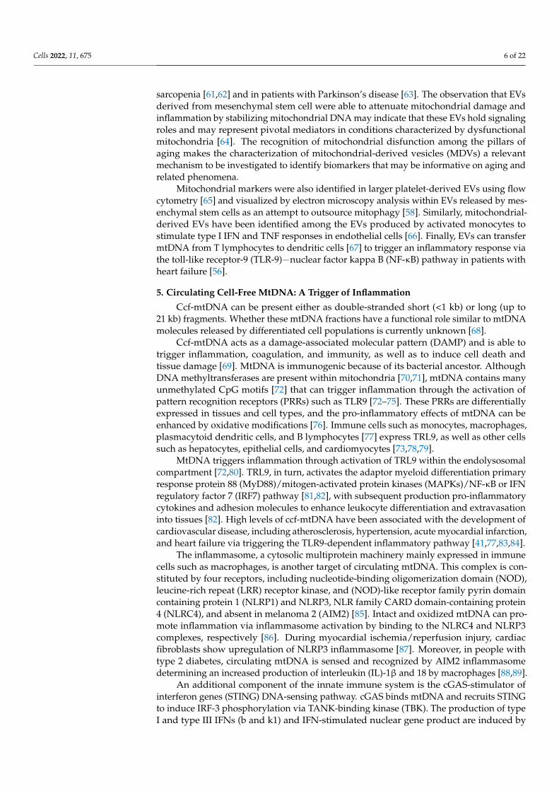
Finally, DNA PRRs are also activated by intermediates of mtDNA replication and tran-scription. Indeed, mtRNA−DNA hybrids that form during transcription, long stretchesof ss-DNA, and R-loops containing RNA−DNA hybrids with a non-template ss-DNAare sensed and recognized by cGAS [73] (Figure 2). The identification of high levels ofpro-inflammatory mediators pertaining to innate immune responses in cellular senescenceand chronic low-grade inflammation lends further support to a role of these mtDNA inter-mediates in triggering PRR-mediated responses [39]. However, their specific involvementin age-related conditions and senescence is currently unknown.
Cells 2022, 11, x FOR PEER REVIEW 7 of 22
Cells 2022, 11, x. https://doi.org/10.3390/xxxxx www.mdpi.com/journal/cells
4 (NLRC4), and absent in melanoma 2 (AIM2) [85]. Intact and oxidized mtDNA can pro-mote inflammation via inflammasome activation by binding to the NLRC4 and NLRP3
complexes, respectively [86]. During myocardial ischemia/reperfusion injury, cardiac fi-broblasts show upregulation of NLRP3 inflammasome [87]. Moreover, in people with type 2 diabetes, circulating mtDNA is sensed and recognized by AIM2 inflammasome
determining an increased production of interleukin (IL)-1β and 18 by macrophages [88,89].
An additional component of the innate immune system is the cGAS-stimulator of
interferon genes (STING) DNA-sensing pathway. cGAS binds mtDNA and recruits STING to induce IRF-3 phosphorylation via TANK-binding kinase (TBK). The production of type I and type III IFNs (b and k1) and IFN-stimulated nuclear gene product are in-
duced by IRF-3 [90]. Conversely, the cleavage of cGAS and the downstream transcription factor IRF-3 via apoptotic caspases may act as a non-inflammatory mechanism of cell de-
mise by impairing cGAS sensing of mtDNA [91–93]. Finally, DNA PRRs are also activated by intermediates of mtDNA replication and
transcription. Indeed, mtRNA−DNA hybrids that form during transcription, long
stretches of ss-DNA, and R-loops containing RNA−DNA hybrids with a non-template ss-DNA are sensed and recognized by cGAS [73] (Figure 2). The identification of high levels of pro-inflammatory mediators pertaining to innate immune responses in cellular senes-
cence and chronic low-grade inflammation lends further support to a role of these mtDNA intermediates in triggering PRR-mediated responses [39]. However, their specific involve-ment in age-related conditions and senescence is currently unknown.
Figure 2. Schematic representation of major signaling pathways through which the displacement of mitochondrial components can trigger inflammation. A decline in the efficiency mitochondrial qual-ity control processes may lead to the intracellular accrual of oxidized components, including mtDNA, that further engulf the mitophagy machinery. These debris can be cleared by the cell along alternative non-degradative routes that release mitochondrial-derived components into the cyto-
plasm or the extracellular compartment. Displaced mitochondrial-derived components can be rec-ognized as damage-associated molecular patters and trigger inflammation by activating three dis-tinct signaling pathways via the interaction with (1) cytosolic cyclic GMP-AMP synthase (cGAS)-stimulator of interferon genes (STING) DNA-sensing system; (2) toll-like receptors (TLRs); (3) nu-
cleotide-binding oligomerization domain (NOD)-like receptor family pyrin domain containing 3 (NLRP3) inflammasome. ATP, Adenosine triphosphate; cGAMP, Cyclic guanosine
Figure 2. Schematic representation of major signaling pathways through which the displacementof mitochondrial components can trigger inflammation. A decline in the efficiency mitochondrialquality control processes may lead to the intracellular accrual of oxidized components, includingmtDNA, that further engulf the mitophagy machinery. These debris can be cleared by the cell alongalternative non-degradative routes that release mitochondrial-derived components into the cytoplasmor the extracellular compartment. Displaced mitochondrial-derived components can be recognizedas damage-associated molecular patters and trigger inflammation by activating three distinct sig-naling pathways via the interaction with (1) cytosolic cyclic GMP-AMP synthase (cGAS)-stimulatorof interferon genes (STING) DNA-sensing system; (2) toll-like receptors (TLRs); (3) nucleotide-binding oligomerization domain (NOD)-like receptor family pyrin domain containing 3 (NLRP3)inflammasome. ATP, Adenosine triphosphate; cGAMP, Cyclic guanosine monophosphate–adenosinemonophosphate; GTP, guanosine triphosphate; IFNs, interferons; IL, interleukin; IRAK, interleukin1 receptor associated kinase; IRF, interferon regulatory factor; mtDNA, mitochondrial DNA; MyD88,Myeloid differentiation primary response 88; TBK1, TANK-binding kinase 1; TFAM, mitochondrialtranscription factor A; TNF-α, tumor necrosis factor-α; TRAF, TNF Receptor Associated Factor 6.Created with BioRender.com, accessed on 18 January 2022.
6. How Mitochondria “Socialize”: Mitochondrial Contact Sites
Cellular organelles are not isolated but establish physical interactions with one an-other. Communications between mitochondria and other cellular components are vital fordeploying their functions and their dynamics change with age [94]. As such, mitochon-
Cells 2022, 11, 675 8 of 22
drial contacts are crucial molecular platforms contributing to age-associated mitochondrialdysfunction and gateways for mtDNA release and signaling.
6.1. Mitochondria−Endoplasmic Reticulum
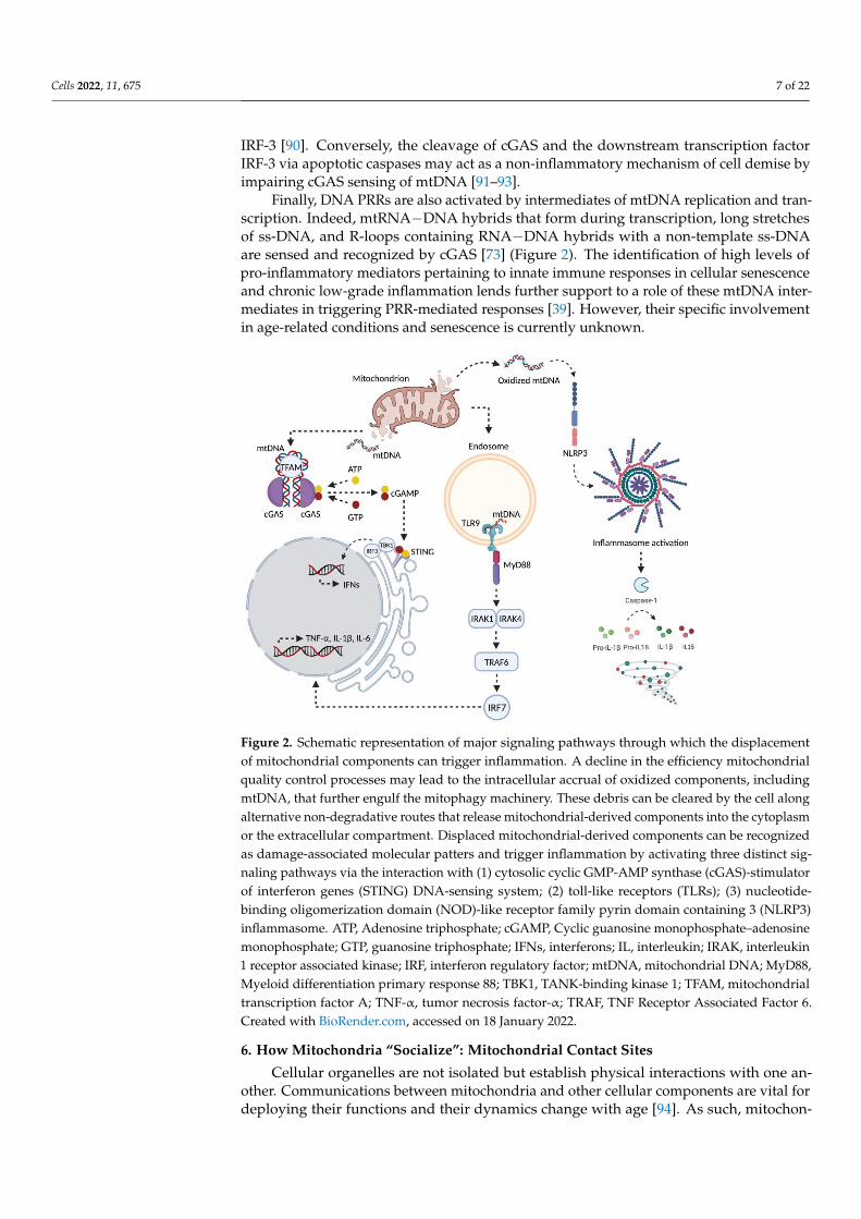
Physical interactions between mitochondria and the endoplasmic reticulum (ER)membranes are referred to as mitochondria−ER contacts (MERCs) (Figure 3). Severalaspects of mitochondrial functions, such as mitochondrial dynamics, calcium homeostasisand mitophagy are influenced by MERCs [95]. Due to their implication in all these activities,and especially in the modulation of redox signaling, altered MERCs have been associatedwith aging and related disorders [96].
Cells 2022, 11, x FOR PEER REVIEW 8 of 22
Cells 2022, 11, x. https://doi.org/10.3390/xxxxx www.mdpi.com/journal/cells
monophosphate–adenosine monophosphate; GTP, guanosine triphosphate; IFNs, interferons; IL, interleukin; IRAK, interleukin 1 receptor associated kinase; IRF, interferon regulatory factor; mtDNA, mitochondrial DNA; MyD88, Myeloid differentiation primary response 88; TBK1, TANK-binding kinase 1; TFAM, mitochondrial transcription factor A; TNF-α, tumor necrosis factor-α;
TRAF, TNF Receptor Associated Factor 6. Created with BioRender.com, accessed on 18 January 2022.
6. How Mitochondria “Socialize”: Mitochondrial Contact Sites
Cellular organelles are not isolated but establish physical interactions with one an-other. Communications between mitochondria and other cellular components are vital for deploying their functions and their dynamics change with age [94]. As such, mitochon-
drial contacts are crucial molecular platforms contributing to age-associated mitochon-drial dysfunction and gateways for mtDNA release and signaling.
6.1. Mitochondria−Endoplasmic Reticulum
Physical interactions between mitochondria and the endoplasmic reticulum (ER) membranes are referred to as mitochondria−ER contacts (MERCs) (Figure 3). Several as-pects of mitochondrial functions, such as mitochondrial dynamics, calcium homeostasis
and mitophagy are influenced by MERCs [95]. Due to their implication in all these activi-ties, and especially in the modulation of redox signaling, altered MERCs have been asso-
ciated with aging and related disorders [96].
Figure 3. Schematic representation of mitochondrial contact sites. To coordinate all the activities and achieve homeostasis, mitochondria use molecular platforms through which establish contacts with the endoplasmic reticulum, lysosome, peroxisome, and lipid droplet. ACSL1, acyl-CoA synthase long chain family member 1; Mfn2, mitofusin 2; MDM34, Mitochondrial distribution and morphol-ogy protein 34; ORP, oxysterol-binding protein-related proteins; PEX, peroxisomal integral mem-
brane protein; PLIN1, peripilin 1; PTPIP51, protein tyrosine phosphatase interacting protein 51; RAB7, Ras-related in brain 7; SNAP23, synaptosomal-associated protein 23; VAMP4, vesicle-associ-ated membrane protein 4; VPS13A, Vacuolar Protein Sorting 13 Homolog A; Created with BioRen-der.com, accessed on 21 January 2022.
Mitochondrial dynamics consist of fission and fusion events that allow shaping mi-tochondrial morphology from fragmented to filamentous organelles. MERCs are rich in proteins involved in fission and fusion. Mitochondrial fission is favored by ER tubules
that interact with mitochondria, defining fission sites and enhancing dynamin-related protein 1 (DRP1) functions [97]. DRP1 is recruited by the mitochondrial fission 1 protein
Figure 3. Schematic representation of mitochondrial contact sites. To coordinate all the activities andachieve homeostasis, mitochondria use molecular platforms through which establish contacts withthe endoplasmic reticulum, lysosome, peroxisome, and lipid droplet. ACSL1, acyl-CoA synthase longchain family member 1; Mfn2, mitofusin 2; MDM34, Mitochondrial distribution and morphologyprotein 34; ORP, oxysterol-binding protein-related proteins; PEX, peroxisomal integral membraneprotein; PLIN1, peripilin 1; PTPIP51, protein tyrosine phosphatase interacting protein 51; RAB7,Ras-related in brain 7; SNAP23, synaptosomal-associated protein 23; VAMP4, vesicle-associatedmembrane protein 4; VPS13A, Vacuolar Protein Sorting 13 Homolog A; Created with BioRender.com,accessed on 21 January 2022.
Mitochondrial dynamics consist of fission and fusion events that allow shaping mi-tochondrial morphology from fragmented to filamentous organelles. MERCs are rich inproteins involved in fission and fusion. Mitochondrial fission is favored by ER tubules thatinteract with mitochondria, defining fission sites and enhancing dynamin-related protein 1(DRP1) functions [97]. DRP1 is recruited by the mitochondrial fission 1 protein (FIS1) andthe mitochondrial fission factor (MFF) that can also localize at the ER, thus representing aplatform for DRP1 oligomerization [98]. Moreover, ER-localized inverted formin 2 (INF2)stimulates actin polymerization at ER-mitochondrial contact sites, increase calcium effluxfrom the ER to the mitochondria, and initiate mitochondrial membrane constriction viamyosin, favoring DRP1 oligomerization and DRP1-mediated fission [99]. Less is knownabout the reason why these proteins are shared by ER and mitochondria. One possibility isthat ER behaves as a regulator of mitochondrial dynamics being responsible for sequestra-tion of these proteins in case of excessive fission or initiation of mitochondrial membraneconstriction if fusion is too low [94].
MERCs are also involved in mitochondrial fusion. Key proteins in this process aremitofusin (Mfn) 1 and 2. Mfn2 is one of the first identified proteins localized at MERCs,

Cells 2022, 11, 675 9 of 22
while Mfn1 does not localize to ER [100,101]. Mechanisms by which Mfn2 regulates MERCsare still unclear as it was demonstrated that MERCs formation can be both promoted andinhibited by Mfn2 [102,103]. Aging is associated with reduced ER-mitochondria contactsites, thus having a negative impact on mitochondrial dynamics and several other processesregulated by the two organelles [104].
Intracellular calcium is stored mostly in the ER and mitochondria, and MERCs repre-sent sites where calcium is exchanged between the two organelles [95]. Calcium can exitfrom the ER through the 1,4,5-trisphosphate receptor (IP3R) and can enter mitochondriavia the mitochondrial calcium uniporter (MCU). Moreover, mitochondrial permeabilitytransition pore and ER sarco/endoplasmic reticulum Ca2+-ATPase (SERCA) enable calciumrelease from mitochondria and uptake by the ER, respectively [105].
Mitochondrial fragmentation observed in aged cells can also be explained by an al-tered calcium exchange between the ER and mitochondria, with increased calcium releasefrom the ER followed by a decreased uptake by mitochondria. Indeed, the mitochondriallocalization of DRP1 is stimulated by calcium derived from the ER [106]. Mitochondrialcalcium uptake is also regulated by oxysterol-binding protein-related protein 5 (ORP5),localized at MERCs [107]. This protein is also involved in regulating mitochondrial func-tion. Indeed, at ER-mitochondria contact sites, ORP5 and ORP8 interact with the proteintyrosine phosphatase interacting protein 51 (PTPIP51), localized on the outer mitochon-drial membrane, and defects in mitochondrial morphology and respiratory functions areobserved following ORP5/ORP8 deletion [107]. The interaction between these proteinsenable the transfer of phosphatidylserine from the ER to mitochondria, where it is con-verted in phosphatidylethanolamine (PE) which is important for mitochondrial structureand function [107]. Therefore, the lipid composition of ER−mitochondria interface can bemodulated by MERCs, which also influences other processes—such as autophagy—thatare stimulated by increased levels of PE [108]. The selective degradation of mitochondriaby mitophagy is influenced by ER−mitochondrial contacts. Indeed, calnexin on the ERmembrane interacts with FUN14 domain-containing 1 (FUNDC1), localized at the mi-tochondria, thereby stimulating the recruitment of DRP1 during mitophagy in order toinitiate mitochondrial fission [109]. Therefore, mitophagy is reduced with age also as aconsequence of a decrease in ER−mitochondria contacts [110].
Mitochondrial morphology is influenced by a process that occurs specifically in theER, known as ER-specific unfolded protein response (ER−UPR) [111]. Unfolded proteinsactivate the protein kinase RNA-like endoplasmic reticulum kinase (PERK) which, inturn, activates eIF2α, that causes a reduction of protein translation [112]. Mitochondrialmorphology is regulated by this mechanism that favors an increased ATP productionto enable the cell to cope with ER−UPR, a process named stress-induced mitochondrialhyperfusion (SIMH) [113].
In aging cells, impaired ER−mitochondria contact sites cause accumulation of mis-folded protein at the ER−mitochondrial interface [104]. Therefore, it is not surprisingthat age-associated disorders are characterized by accumulation of toxic proteins at theER−mitochondria interface, having negative effects on calcium uptake, autophagy, mito-chondrial respiration, and ROS production [114].
6.2. Mitochondria−Lysosomes
The existence of a connection between mitochondria and lysosomes has becomeclear since the observation that alterations in lysosomes, often seen in neurodegenerativediseases, were reflected by impaired mitochondrial networks [115]. The first observationscame from experiments in yeast cells where decreased vacuolar acidification and alteredmitochondrial dynamics were detected [116]. In the same paper, the authors demonstratedthat mitochondrial fragmentation was prevented by the expression of the subunit A of theyeast V-ATPase V1 domain. The treatment with the inhibitor of V-ATPase concanamycin Adid not rescue mitochondrial fragmentation, indicating that this was strictly dependent onV-ATPase activity [116].

Cells 2022, 11, 675 10 of 22
The close relationship between mitochondria and lysosomes is mediated by twotranscription factors, TFAM and transcription factor EB (TFEB). The first is fundamentalfor mtDNA transcription and replication, and, therefore, mitochondrial biogenesis [117].Likewise, TFEB is crucial for lysosomal biogenesis [118]. A recent report showed that TFAM-deficient cells are characterized by high mitochondrial fragmentation and dysfunction. Inthese cells, the nuclear translocation of TFEB activates the mitochondrial master-regulatorperoxisome proliferator-activated receptor-gamma coactivator 1 alpha (PGC-1α) leading toco-regulated lysosomal and mitochondrial biogenesis [119,120].
Mitochondria and lysosomes have also physical interactions (Figure 3), as demon-strated by Wong and co-workers [121]. These contacts are mediated by active GTP-boundlysosomal Ras-related in brain (RAB) 7, while FIS1, through recruiting Tre-2/Bub2/Cdc16member 15 (TBC1D15) to mitochondria, determines the separation of two organelles,stimulating RAB7 GTPase activity [121]. Contacts are important for mitochondrial dynam-ics since the same group demonstrated that mitochondrial fission was decreased by theexpression of a constitutively active mutant of RAB7 (RAB7Q67L), increasing mitochondria-lysosomes contacts [121]. The formation of these contacts is totally independent of metabo-lite transfer, autophagosome biogenesis or mitophagy, as demonstrated by the fact thatmitochondria-lysosomes do not show expression of autophagosomal markers [121], norare they influenced by genetic ablation of autophagy receptors [122].
Mitophagy is not only influenced by the ER−mitochondria, but also by mitochondria−lysosome contacts. The interaction between TBC1D15, microtubule-associated protein1 light chain 3 (LC3), and FIS1 is pivotal for regulating RAB7 activity and the formationof the isolation membrane that engulfs damaged mitochondria. Instead, LC3-taggedphagosomes without cargo orientation appear when TBC1D15 is depleted or its RABGAPactivity is inhibited [123]. These findings indicate that the formation of the isolationmembrane requires RAB7 activation, while the detachment of LC3-positive membranesfrom microtubules needs RAB7 inactivation [124].
During aging, a progressive reduction of mitophagy occurs, thereby leading to theaccumulation of damaged mitochondria with a detrimental effect on cellular homeostasisand with particularly negative effects for long-lived cells, more sensitive to this phe-nomenon [125]. Recently, a new mechanism for the elimination of damaged mitochondriawas identified and described to operate via MDVs [126,127].
The molecular mechanisms that coordinate the formation of MDVs are still poorlyunderstood. One possibility is represented by the accumulation of proteins near mitochon-drial membranes, under oxidative stress conditions. In response to protein aggregation,mitochondrial membrane originate curvatures and produce a vesicle [128]. This lattercan reach the late endosomes/multivesicular bodies (MVBs) [129] or peroxisomes forcargo detoxification [130]. However, MDVs from mildly damaged mitochondria can fusewith MVBs and be secreted as exosomes, which may represent an indirect evidence ofmitochondria/endolysosomal system crosstalk [60,63].
6.3. Mitochondria−Peroxisomes
As mentioned above, mitochondria and peroxisomes are in communication throughMDVs [130]. Interestingly, they also show physical interaction (Figure 3). Indeed, theperoxisome membrane elongation and fission protein peroxisomal integral membraneprotein 11 (PEX11) interacts with mitochondrial distribution and morphology protein34 (MDM34), localized on mitochondria [131]. Moreover, peroxisomes share the fissionmachinery with mitochondria. DRP1 is required for the formation of a constriction ringthat allows peroxisomal fission [132]. DRP1 is recruited at the constriction sites by PEX11βwhich does not have scission activity, but peroxisomes appear enlarged and elongatedfollowing its ablation, thus indicating that it is essential for peroxisomal fission throughmodulation of DRP1 activity [131,133,134].

Cells 2022, 11, 675 11 of 22
The mechanisms of mitochondrial-peroxisomal communication are still under investi-gation, but, given the importance of peroxisomes and mitochondria in lipid metabolism,ROS signaling, and protein exchange, they may have a role in aging [135].
6.4. Mitochondria−Lipid Droplets
Though for long time considered just an inert cytoplasmatic inclusion of fat, in recentyears lipid droplets (LDs) have begun to be considered as full-fledged organelles withseveral functions [136]. Every eukaryotic cell contains LDs and their main function isthe storage of fatty acids in the form of neutral lipids, mostly triacylglycerols and sterolesters, and hydrolysis [137]. To carry out this function, they possess a unique structureencompassing a core of neutral lipids surrounded by a phospholipid monolayer [138].Several proteins, such as perilipins (PLINs), are associated with the monolayer and arefundamental for LD functions and dynamics [139,140].
The size of LDs changes depending on nutrient conditions, with an enlargement inthe case of fatty acid abundance and shrinking during starvation, when fatty acids areused for energy production through β-oxidation [141]. Cellular homeostasis is guaranteedby these organelles, protecting the cells from lipotoxicity caused by excessive fatty acidaccumulation [136]. Indeed, dysregulation of lipid metabolism is associated with severaldiseases, many of which are also characterized by an increase in LD abundance [142]. Inthis context, LDs have gained attention for their potential role in cancer and neurodegen-eration. For instance, a form of Charcot–Marie–Tooth type 2 (CMT2) disease, an axonalneuropathy affecting the peripheral nervous system, is caused by a missense mutation inthe diacylglycerol O-Acyltransferase 2 (DGAT2Y223H), an enzyme involved in the synthe-sis of triglycerides [143]. Moreover, in CMT2 type 2 B (CMT2B), caused by mutations inRAB7 gene, an accumulation of LDs was observed in the cytoplasm, further highlightingthe role of altered LD dynamics in neurodegeneration [144,145].
Originally it was thought that inter-organelle contact sites only concerned subcellularcompartments with a phospholipid bilayer. However, inter-organelle communicationengages also monolayer organelles, such as LDs and RNA granules, or stress granules,which are devoid of membranes [95].
LDs form contact sites with ER, through interaction between acyl-CoA synthetasefatty acid transport protein 1 (FATP1) localized at the ER, and DGAT2, localized at the LDmembrane [146].
LD−mitochondria contact sites are established during nutrient deprivation or cellgrowth, two conditions characterized by a high demand of phospholipids for membranebiosynthesis and lipids for β-oxidation [147,148]. Furthermore, the complex constituted byRAB18 on LDs and the tethering complex NRZ (NAG-RINT1-ZW1) associated with solubleN-ethylmaleimide-sensitive-factor attachment protein receptor (SNAREs) is involved inLD−mitochondria contacts [149].
PLIN5 is localized on the LD membrane, and it was described also as fundamental forLD−mitochondria contact sites for its ability to recruit LDs to mitochondria. Nevertheless,the interacting partner of PLIN5 on the outer mitochondrial membrane is not yet known,and the mechanism of mitochondria recruitment has not been elucidated [150].
Other proteins involved in LD−mitochondria contacts are still under investigation.The interaction between Mfn2 and PLIN1 was identified in LD−mitochondria of brownadipocytes [151]. Moreover, by using the BioID tool that is able to detect candidateprotein−protein interactions in living cells, a set of other interactors were identified [152].For instance, the acyl-CoA synthase long chain family member 1 (ACSL1) on mitochondriainteracts with the synaptosomal-associated protein 23 (SNAP23) and vesicle-associatedmembrane protein 4 (VAMP4) on LDs [153] (Figure 3).
Several studies are ongoing to investigate other proteins involved in LD recruitment onmitochondrial. Moreover, the contacts described so far are highly dynamic, fitting the “kiss andrun” paradigm. However, a more stable interaction, known as anchoring, between LDs andmitochondria bind has been described. This type of contact has been identified in oxidative

Cells 2022, 11, 675 12 of 22
tissues such as brown adipose tissue, skeletal muscle, and the myocardium [154]. Emergingevidence invites questions about the role of LD−mitochondria contacts in immune function.In hepatocytes, lipopolysaccharide induces PLIN 5 downregulation, consequent detachmentof mitochondria from LDs, and decrease of β-oxidation and ketogenesis [155]. In contrast,the increase of PLIN5 in human THP-1 macrophages determines increased LD−mitochondriacontacts and reduced antibacterial function [155]. Notably, the antibacterial proinflammatoryactivity of M1 macrophages is based on glycolysis, while anti-inflammatory M2 macrophagespreferentially use fatty acid oxidation and oxidative phosphorylation, supporting the rationalefor LD−mitochondria detachment in response to M1-polarizing stimuli such as lipopolysaccha-ride [156].
Aging is characterized by several mitochondrial alterations, from the increase of oxida-tive stress to alterations in mitochondria dynamics, such as fragmentation of mitochondrialnetwork, reduced number of mitochondria, mtDNA mutagenesis, and loss of mitochondrialmembrane potential [157,158]. Recently, in yeast as well as in mammalian cell lines, it wasdemonstrated that, under stress conditions, the interaction between LDs and mitochondriaincreased in order to transport toxic proteins from mitochondria to LDs, protecting cellsfrom apoptotic death [159]. Another recent study in yeasts showed that an increase in LDnumber extended cell survival during the stationary phase, paralleled by a progressiveincrease in ROS levels. The results obtained in this study suggest that LD−mitochondriacontact sites are beneficial for cell fitness during yeast aging. Indeed, LDs contribute tomitochondrial “rejuvenation”, eliminating toxic protein, attenuating oxidative stress, and,finally, indicating a role of LD−mitochondria contact sites in longevity [160].
The transport of molecules between mitochondria and LDs is not limited to proteins,but it also involves lipids. Upon stress condition and aging, phosphatidylinositols andergosterols (the yeast sterols) increase in mitochondria, while their content decreasedin LDs [161]. Proteins and lipids, shuttled between mitochondria and LDs, are thendegraded in the lysosome/vacuole during macrolipophagy [162]. Further studies onLD−mitochondria contact sites in mammalian cells are required to confirm the importanceof this inter-organelle communication in longevity.
7. Mitochondrial Dyshomeostasis and Inflammation in Aging and Related Conditions
A global reduction in the capacity to cope with a variety of stressors and a progressiveincrease in pro-inflammatory mediators are major characteristics of the aging process [163].This phenomenon, referred to as “inflamm-aging”, is induced by a continuous antigenicload and stress. Under these conditions, a state of permanent arrest of cell proliferationoccurs and leads to cellular senescence characterized by the acquisition of a senescence-associated secretory phenotype (SASP) [164].
The tight relationship between inflammation and cellular senescence has also beenshown by gene expression studies revealing inflammatory gene expression patterns similarin the two conditions [165]. Senescent human fibroblasts express several inflammation-associated genes, including monocyte chemotactic protein-1, Gro-α, IL-1β, IL-15, TLR4,and intercellular adhesion molecule 1 [166,167]. Inflammatory genes were found to beup-regulated not only in senescent fibroblasts but also in senescent human hepatic stellatecells [168]. However, this pattern was not found in senescent retinal pigment epithelial cellsand vascular endothelial cells, suggesting that inflammatory pathways associated withsenescence might be cell-specific.
A set of pro-inflammatory and pro-fibrotic factors and metalloproteases have beenidentified among SASP molecules [164]. As a consequence of the progressive accumu-lation of senescent cells and chronically released SASP molecules, inflammation, tissuedamage, and fibrosis can eventually ensue, which predispose to age-related conditions (e.g.,metabolic disorders, atherosclerosis, muscle wasting and neurodegeneration) [169–171].Tezze et al. [172] found that age-associated loss of the mitochondrial protein optic atrophy1 (OPA1) was related to muscle atrophy and systemic inflammation. Indeed, OPA1−/−
mice showed an early aging phenotype and high circulating levels of several inflamma-
Cells 2022, 11, 675 13 of 22
tory cytokines (i.e., IL-1α, IL-1β, IL-6, and TNF-α). The accrual of senescent cells duringaging has been described in several animal models [173,174] along with an increase inintracellular damage and reduced senescence immune surveillance [175,176]. A great dealof research is ongoing to evaluate the effects of senolytics/senomorphics at eliminatingsenescent cells and attenuating SASP production. As such, a deeper understanding of themolecular mechanisms underlying cellular senescence and related signaling routes at thesystemic levels is crucial for devising specific anti-aging strategies.
Mitochondrial integrity is preserved by a set of quality control mechanisms of whichautophagy, and more specifically mitophagy, plays a major role [77]. Indeed, autophagylimits the accumulation of pro-inflammatory factors and the dysregulation of this processmay result in increased cytoplasmic mtDNA-driven inflammation [177]. MtDNA canbe degraded by DNAses I contained in the autolysosomes [178], and the mtDNA–TLR9-mediated inflammatory response was found to be increased in the heart of DNase II-deficient mice developing cardiomyopathy [77]. Moreover, sterile inflammation followingmitochondrial dysfunction and the concomitant rise in mtDNA levels can trigger therelease of IL-1β and IFNα via cGAS engagement [179]. A dysregulation in the clearance ofdysfunctional mitochondria has also been hypothesized to determine the escape of oxidizedccf-mtDNA or nucleoids which can trigger inflammation by interacting with PRRs [180].Moreover, similar to the nuclear DNA system, disruption of processes involved in mtDNAhomeostasis, such as mtDNA replication and repair, can trigger cGAS–STING activation.For instance, a defective mtDNA packaging into nucleoids following TFAM depletion is aprominent signal for cGAS activity in diverse cells [181].
Recently, MERCs have also been indicated as molecular platforms contributing toaging and age-related diseases via SASP signaling [182]. Indeed, alterations of MERCsquantity and quality with aging and related diseases have been reported [95,96,104].
Pinti and collaborators [183] described an association between ccf-mtDNA and el-evated levels of proinflammatory cytokines during aging. In particular, they observedthat the number of copies of ccf-mtDNA increased significantly after the fifth decade oflife, peaking past the age of 90 [183]. Interestingly, systemic levels of ccf-mtDNA de-crease in healthy adults after moderate aerobic exercise, a well-known anti-inflammatoryintervention [184–186]. Sliter et al. [187] investigated the effect of exhaustive exerciseon inflammation in Parkin−/− or Pink1−/− mice. The authors observed that exhaustiveexercise caused a striking increase in serum levels of pro-inflammatory IL-6 and IFN-b.Remarkably, deletion of STING or the administration of IFNAR-blocking antibody com-pletely rescued a normal phenotype, suggesting that mtDNA released from damagedmitochondria that are not cleared is responsible for the observed inflammation [187]. Spe-cific alterations of the mitochondrial quality control axis have also been reported in musclebiopsies from old hip-fractured patients with sarcopenia in whom a link between muscularmitochondrial dysfunction and systemic inflammation via the release of mtDNA has beenhypothesized [188].
Inflammation also represents an underlying mechanism of neurodegeneration [189]. Al-tered mitophagy and activation of cGAS–STING signaling have been indicated as a pathogenicmechanism of Parkinson’s disease [187]. Familial forms of Parkinson’s disease carry missensemutations in PINK1 and Parkin proteins whose deletion in mice induces an accumulation ofmtDNA and elevation of systemic cytokine levels [187]. Of note, the latter effect is fully abro-gated by co-deletion of STING [187]. The cGAS–STING-dependent inflammation triggered bymtDNA has also been involved in the neuropathological processes associated with amyotrophiclateral sclerosis and frontotemporal lobar degeneration [48]. This has been mainly ascribedto accrual of misplaced mitochondrial of TDP43, a DNA/RNA-binding protein, that inducesmtDNA release through mitochondrial transition pore opening and leakage via VDAC1. As aconsequence, a release of type I INFs and inflammatory cytokines in a cGAS–STING-dependentmanner occurs in human and mouse cells [48]. Depletion of STING in a preclinical model ofamyotrophic lateral sclerosis overexpressing TDP43 was also able to dampen neuroinflamma-tion and mitigate disease progression. Finally, the elevation of type I INFs following STING

Cells 2022, 11, 675 14 of 22
activation has also been shown in models of Huntington disease [190] or neurodegenerationand astrocytic inflammation guided by protein aggregates deposition [191].
8. Conclusions
Mitochondrial metabolism and its dysfunction have been listed among the nine pillarsof the geroscience paradigm and indicated as key targets amenable for anti-aging interven-tions. The contribution of age-related changes in inter-organelle contacts to mitochondrialdyshomeostasis has recently been investigated. These molecular platforms are implicatedin mitochondrial remodeling and transfer of selected molecules with pro-inflammatoryproperties. A deeper characterization of these inter-organelle interactions and their con-tribution to cellular senescence may be very informative towards the intricate pathwaysregulating cellular decay during aging. The close relationship of mitochondrial signalswith cellular senescence and their contribution to aging and age-related conditions conferfurther priority to investigating these processes.
Author Contributions: Conceptualization, A.P. and E.M.; writing—original draft preparation, A.P.,F.G. and R.R.; writing—review and editing, C.B., F.P.D., H.J.C.-J. and R.C.; supervision, C.B. and E.M.All authors have read and agreed to the published version of the manuscript.
Funding: This work was supported by the Università Cattolica del Sacro Cuore [D1.2020]; theMinistero dell’Istruzione, dell’Università e della Ricerca (MIUR) [DM 587, 08/08/2018; CIB N.112/19]; and the nonprofit research foundation “Centro Studi Achille e Linda Lorenzon” [N/A]. TheAPC was funded by the Università Cattolica del Sacro Cuore [D1.2020].
Institutional Review Board Statement: Not applicable.
Informed Consent Statement: Not applicable.
Data Availability Statement: No data were generated for the present article.
Acknowledgments: All figures were created with BioRender.com.
Conflicts of Interest: The authors declare no conflict of interest.
References1. Margulis, L.; Bermudes, D. Symbiosis as a mechanism of evolution: Status of cell symbiosis theory. Symbiosis 1985, 1, 101–124.
[PubMed]2. Lane, N.; Martin, W. The energetics of genome complexity. Nature 2010, 467, 929–934. [CrossRef] [PubMed]3. Klecker, T.; Böckler, S.; Westermann, B. Making connections: Interorganelle contacts orchestrate mitochondrial behavior. Trends
Cell Biol. 2014, 24, 537–545. [CrossRef] [PubMed]4. Valm, A.M.; Cohen, S.; Legant, W.R.; Melunis, J.; Hershberg, U.; Wait, E.; Cohen, A.R.; Davidson, M.W.; Betzig, E.; Lippincott-
Schwartz, J. Applying systems-level spectral imaging and analysis to reveal the organelle interactome. Nature 2017, 546, 162–167.[CrossRef]
5. Wallace, D.C. Colloquium paper: Bioenergetics, the origins of complexity, and the ascent of man. Proc. Natl. Acad. Sci. USA 2010,107, 8947–8953. [CrossRef]
6. Martínez-Reyes, I.; Chandel, N.S. Mitochondrial TCA cycle metabolites control physiology and disease. Nat. Commun. 2020,11, 102. [CrossRef]
7. Bonawitz, N.D.; Clayton, D.A.; Shadel, G.S. Initiation and beyond: Multiple functions of the human mitochondrial transcriptionmachinery. Mol. Cell 2006, 24, 813–825. [CrossRef]
8. West, A.P.; Shadel, G.S. Mitochondrial DNA in innate immune responses and inflammatory pathology. Nat. Rev. Immunol. 2017,17, 363–375. [CrossRef]
9. Wei, W.; Tuna, S.; Keogh, M.J.; Smith, K.R.; Aitman, T.J.; Beales, P.L.; Bennett, D.L.; Gale, D.P.; Bitner-Glindzicz, M.A.K.;Black, G.C.; et al. Germline selection shapes human mitochondrial DNA diversity. Science 2019, 364, eaau6520. [CrossRef]
10. Picard, M. Mitochondrial synapses: Intracellular communication and signal integration. Trends Neurosci. 2015, 38, 468–474.[CrossRef]
11. Picca, A.; Calvani, R.; Coelho-Junior, H.J.; Landi, F.; Bernabei, R.; Marzetti, E. Inter-organelle membrane contact sites andmitochondrial quality control during aging: A geroscience view. Cells 2020, 9, 598. [CrossRef] [PubMed]
12. Glancy, B.; Kim, Y.; Katti, P.; Willingham, T.B. The functional impact of mitochondrial structure across subcellular scales. Front.Physiol. 2020, 11, 541040. [CrossRef] [PubMed]
13. López-Otín, C.; Blasco, M.A.; Partridge, L.; Serrano, M.; Kroemer, G. The Hallmarks of Aging. Cell 2013, 153, 1194–1217. [CrossRef]

Cells 2022, 11, 675 15 of 22
14. Shadel, G.; Clayton, D. Mitochondrial DNA maintenance in vertebrates. Annu. Rev. Biochem. 1997, 66, 409–436. [CrossRef]15. Calvo, S.; Mootha, V. The mitochondrial proteome and human disease. Annu. Rev. Genom. Hum. Genet. 2010, 11, 25–44. [CrossRef]
[PubMed]16. Nicholls, T.; Minczuk, M. In D-loop: 40 years of mitochondrial 7S DNA. Exp. Gerontol. 2014, 56, 175–181. [CrossRef] [PubMed]17. Reyes, A.; Kazak, L.; Wood, S.; Yasukawa, T.; Jacobs, H.; Holt, I. Mitochondrial DNA replication proceeds via a “bootlace”
mechanism involving the incorporation of processed transcripts. Nucleic Acids Res. 2013, 41, 5837–5850. [CrossRef]18. Collins, L.V.; Hajizadeh, S.; Holme, E.; Jonsson, I.-M.; Tarkowski, A. Endogenously oxidized mitochondrial DNA induces in vivo
and in vitro inflammatory responses. J. Leukoc. Biol. 2004, 75, 995–1000. [CrossRef]19. Chew, K.; Zhao, L. Interactions of mitochondrial transcription factor a with DNA damage: Mechanistic insights and functional
implications. Genes 2021, 12, 1246. [CrossRef]20. Wei, W.; Chinnery, P.F. Inheritance of mitochondrial DNA in humans: Implications for rare and common diseases. J. Intern. Med.
2020, 287, 634–644. [CrossRef]21. Boulet, L.; Karpati, G.; Shoubridge, E.A. Distribution and threshold expression of the tRNA(Lys) mutation in skeletal muscle of
patients with myoclonic epilepsy and ragged-red fibers (MERRF). Am. J. Hum. Genet. 1992, 51, 1187–1200. [PubMed]22. Carelli, V.; Giordano, C.; D’Amati, G. Pathogenic expression of homoplasmic mtDNA mutations needs a complex nuclear-
mitochondrial interaction. Trends Genet. 2003, 19, 257–262. [CrossRef]23. Brown, M.D.; Torroni, A.; Reckord, C.L.; Wallace, D.C. Phylogenetic analysis of Leber’s hereditary optic neuropathy mitochondrial
DNA’s indicates multiple independent occurrences of the common mutations. Hum. Mutat. 1995, 6, 311–325. [CrossRef] [PubMed]24. Hudson, G.; Carelli, V.; Spruijt, L.; Gerards, M.; Mowbray, C.; Achilli, A.; Pyle, A.; Elson, J.; Howell, N.; La Morgia, C.; et al.
Clinical expression of Leber hereditary optic neuropathy is affected by the mitochondrial DNA-haplogroup background. Am. J.Hum. Genet. 2007, 81, 228–233. [CrossRef]
25. Hudson, G.; Nalls, M.; Evans, J.R.; Breen, D.P.; Winder-Rhodes, S.; Morrison, K.E.; Morris, H.R.; Williams-Gray, C.H.; Barker, R.A.;Singleton, A.B.; et al. Two-stage association study and meta-analysis of mitochondrial DNA variants in Parkinson disease.Neurology 2013, 80, 2042–2048. [CrossRef]
26. Santoro, A.; Balbi, V.; Balducci, E.; Pirazzini, C.; Rosini, F.; Tavano, F.; Achilli, A.; Siviero, P.; Minicuci, N.; Bellavista, E.; et al.Evidence for sub-haplogroup h5 of mitochondrial DNA as a risk factor for late onset Alzheimer’s disease. PLoS ONE 2010,5, e12037. [CrossRef]
27. Chinnery, P.F.; Gomez-Duran, A. Oldies but goldies mtDNA population variants and neurodegenerative diseases. Front. Neurosci.2018, 12, 682. [CrossRef]
28. Ye, Z.; Gillson, C.; Sims, M.; Khaw, K.T.; Plotka, M.; Poulton, J.; Langenberg, C.; Wareham, N.J. The association of the mitochondrialDNA OriB variant (16184-16193 polycytosine tract) with type 2 diabetes in Europid populations. Diabetologia 2013, 56, 1907–1913.[CrossRef]
29. Nagley, P.; Mackay, I.R.; Baumer, A.; Maxwell, R.J.; Vaillant, F.; Wang, Z.; Zhang, C.; Linnane, A.W. Mitochondrial DNA mutationassociated with aging and degenerative disease. Ann. N. Y. Acad. Sci. 1992, 673, 92–102. [CrossRef]
30. Bender, A.; Krishnan, K.J.; Morris, C.M.; Taylor, G.A.; Reeve, A.K.; Perry, R.H.; Jaros, E.; Hersheson, J.S.; Betts, J.;Klopstock, T.; et al. High levels of mitochondrial DNA deletions in substantia nigra neurons in aging and Parkinson dis-ease. Nat. Genet. 2006, 38, 515–517. [CrossRef]
31. Trifunovic, A.; Wredenberg, A.; Falkenberg, M.; Spelbrink, J.N.; Rovio, A.T.; Bruder, C.E.; Bohlooly-Y, M.; Gidlöf, S.; Oldfors, A.;Wibom, R.; et al. Premature ageing in mice expressing defective mitochondrial DNA polymerase. Nature 2004, 429, 417–423.[CrossRef] [PubMed]
32. Kujoth, C.C.; Hiona, A.; Pugh, T.D.; Someya, S.; Panzer, K.; Wohlgemuth, S.E.; Hofer, T.; Seo, A.Y.; Sullivan, R.; Jobling, W.A.; et al.Mitochondrial DNA mutations, oxidative stress, and apoptosis in mammalian aging. Science 2005, 309, 481–484. [CrossRef][PubMed]
33. Smith, A.L.M.; Whitehall, J.C.; Bradshaw, C.; Gay, D.; Robertson, F.; Blain, A.P.; Hudson, G.; Pyle, A.; Houghton, D.; Hunt, M.; et al.Age-associated mitochondrial DNA mutations cause metabolic remodelling that contributes to accelerated intestinal tumorigene-sis. Nat. Cancer 2020, 1, 976–989. [CrossRef] [PubMed]
34. Yuan, Y.; Ju, Y.S.; Kim, Y.; Li, J.; Wang, Y.; Yoon, C.J.; Yang, Y.; Martincorena, I.; Creighton, C.J.; Weinstein, J.N.; et al. Comprehensivemolecular characterization of mitochondrial genomes in human cancers. Nat. Genet. 2020, 52, 342–352. [CrossRef]
35. Wolf, A.M. MtDNA mutations and aging-not a closed case after all? Signal Transduct. Target. Ther. 2021, 6, 56. [CrossRef]36. Payne, B.A.I.; Wilson, I.J.; Yu-Wai-Man, P.; Coxhead, J.; Deehan, D.; Horvath, R.; Taylor, R.W.; Samuels, D.C.; Santibanez-Koref, M.;
Chinnery, P.F. Universal heteroplasmy of human mitochondrial DNA. Hum. Mol. Genet. 2013, 22, 384–390. [CrossRef]37. Li, M.; Schönberg, A.; Schaefer, M.; Schroeder, R.; Nasidze, I.; Stoneking, M. Detecting heteroplasmy from high-throughput
sequencing of complete human mitochondrial DNA genomes. Am. J. Hum. Genet. 2010, 87, 237–249. [CrossRef]38. Keogh, M.; Chinnery, P.F. Hereditary mtDNA heteroplasmy: A baseline for aging? Cell Metab. 2013, 18, 463–464. [CrossRef]39. Dabravolski, S.A.; Nikiforov, N.G.; Zhuravlev, A.D.; Orekhov, N.A.; Grechko, A.V.; Orekhov, A.N. Role of the mtDNA mutations
and mitophagy in inflammaging. Int. J. Mol. Sci. 2022, 23, 1323. [CrossRef]40. Judge, S.; Leeuwenburgh, C. Cardiac mitochondrial bioenergetics, oxidative stress, and aging. Am. J. Physiol. Cell Physiol. 2007,
292, C1983–C1992. [CrossRef]

Cells 2022, 11, 675 16 of 22
41. Ding, Z.; Liu, S.; Wang, X.; Dai, Y.; Khaidakov, M.; Deng, X.; Fan, Y.; Xiang, D.; Mehta, J.L. LOX-1, mtDNA damage, andNLRP3 inflammasome activation in macrophages: Implications in atherogenesis. Cardiovasc. Res. 2014, 103, 619–628. [CrossRef][PubMed]
42. Patrushev, M.; Kasymov, V.; Patrusheva, V.; Ushakova, T.; Gogvadze, V.; Gaziev, A. Mitochondrial permeability transition triggersthe release of mtDNA fragments. Cell. Mol. Life Sci. 2004, 61, 3100–3103. [CrossRef] [PubMed]
43. McArthur, K.; Whitehead, L.W.; Heddleston, J.M.; Li, L.; Padman, B.S.; Oorschot, V.; Geoghegan, N.D.; Chappaz, S.; Davidson, S.;Chin, H.S.; et al. BAK/BAX macropores facilitate mitochondrial herniation and mtDNA efflux during apoptosis. Science 2018,359, eaao6047. [CrossRef]
44. Saraste, A.; Pulkki, K. Morphologic and biochemical hallmarks of apoptosis. Cardiovasc. Res. 2000, 45, 528–537. [CrossRef]45. Lindqvist, D.; Wolkowitz, O.M.; Picard, M.; Ohlsson, L.; Bersani, F.S.; Fernström, J.; Westrin, Å.; Hough, C.M.; Lin, J.;
Reus, V.I.; et al. Circulating cell-free mitochondrial DNA, but not leukocyte mitochondrial DNA copy number, is elevatedin major depressive disorder. Neuropsychopharmacology 2018, 43, 1557–1564. [CrossRef]
46. Ichim, G.; Lopez, J.; Ahmed, S.U.; Muthalagu, N.; Giampazolias, E.; Delgado, M.E.; Haller, M.; Riley, J.S.; Mason, S.M.;Athineos, D.; et al. Limited mitochondrial permeabilization causes DNA damage and genomic instability in the absence of celldeath. Mol. Cell 2015, 57, 860–872. [CrossRef]
47. Kim, J.; Gupta, R.; Blanco, L.P.; Yang, S.; Shteinfer-Kuzmine, A.; Wang, K.; Zhu, J.; Yoon, H.E.; Wang, X.; Kerkhofs, M.; et al.VDAC oligomers form mitochondrial pores to release mtDNA fragments and promote lupus-like disease. Science 2019, 366,1531–1536. [CrossRef] [PubMed]
48. Yu, C.H.; Davidson, S.; Harapas, C.R.; Hilton, J.B.; Mlodzianoski, M.J.; Laohamonthonkul, P.; Louis, C.; Low, R.R.J.; Moecking, J.;De Nardo, D.; et al. TDP-43 triggers mitochondrial DNA release via mPTP to activate cGAS/STING in ALS. Cell 2020, 183,636–649.e18. [CrossRef] [PubMed]
49. Al Amir Dache, Z.; Otandault, A.; Tanos, R.; Pastor, B.; Meddeb, R.; Sanchez, C.; Arena, G.; Lasorsa, L.; Bennett, A.; Grange, T.; et al.Blood contains circulating cell-free respiratory competent mitochondria. FASEB J. 2020, 34, 3616–3630. [CrossRef]
50. Verderio, P.; Pandolfi, L.; Mazzucchelli, S.; Marinozzi, M.R.; Vanna, R.; Gramatica, F.; Corsi, F.; Colombo, M.; Morasso, C.;Prosperi, D. Antiproliferative effect of ASC-J9 delivered by PLGA nanoparticles against estrogen-dependent breast cancer cells.Mol. Pharm. 2014, 11, 2864–2875. [CrossRef]
51. Yáñez-Mó, M.; Siljander, P.R.-M.; Andreu, Z.; Zavec, A.B.; Borràs, F.E.; Buzas, E.I.; Buzas, K.; Casal, E.; Cappello, F.;Carvalho, J.; et al. Biological properties of extracellular vesicles and their physiological functions. J. Extracell. Vesicles 2015,4, 27066. [CrossRef]
52. Elzanowska, J.; Semira, C.; Costa-Silva, B. DNA in extracellular vesicles: Biological and clinical aspects. Mol. Oncol. 2021, 15,1701–1714. [CrossRef] [PubMed]
53. Greening, D.W.; Xu, R.; Gopal, S.K.; Rai, A.; Simpson, R.J. Proteomic insights into extracellular vesicle biology—Definingexosomes and shed microvesicles. Expert Rev. Proteom. 2017, 14, 69–95. [CrossRef]
54. Kim, K.M.; Abdelmohsen, K.; Mustapic, M.; Kapogiannis, D.; Gorospe, M. RNA in extracellular vesicles. Wiley Interdiscip. Rev.RNA 2017, 8, e1413. [CrossRef] [PubMed]
55. Théry, C.; Zitvogel, L.; Amigorena, S. Exosomes: Composition, biogenesis and function. Nat. Rev. Immunol. 2002, 2, 569–579.[CrossRef] [PubMed]
56. Guescini, M.; Genedani, S.; Stocchi, V.; Agnati, L.F. Astrocytes and glioblastoma cells release exosomes carrying mtDNA. J. NeuralTransm. 2010, 117, 1–4. [CrossRef]
57. Guescini, M.; Guidolin, D.; Vallorani, L.; Casadei, L.; Gioacchini, A.M.M.; Tibollo, P.; Battistelli, M.; Falcieri, E.; Battistin, L.;Agnati, L.F.F.; et al. C2C12 myoblasts release micro-vesicles containing mtDNA and proteins involved in signal transduction. Exp.Cell Res. 2010, 316, 1977–1984. [CrossRef]
58. Phinney, D.G.; Di Giuseppe, M.; Njah, J.; Sala, E.; Shiva, S.; St Croix, C.M.; Stolz, D.B.; Watkins, S.C.; Di, Y.P.; Leikauf, G.D.; et al.Mesenchymal stem cells use extracellular vesicles to outsource mitophagy and shuttle microRNAs. Nat. Commun. 2015, 6, 8472.[CrossRef]
59. Hayakawa, K.; Esposito, E.; Wang, X.; Terasaki, Y.; Liu, Y.; Xing, C.; Ji, X.; Lo, E.H. Transfer of mitochondria from astrocytes toneurons after stroke. Nature 2016, 535, 551–555. [CrossRef]
60. Sansone, P.; Savini, C.; Kurelac, I.; Chang, Q.; Amato, L.B.; Strillacci, A.; Stepanova, A.; Iommarini, L.; Mastroleo, C.; Daly, L.; et al.Packaging and transfer of mitochondrial DNA via exosomes regulate escape from dormancy in hormonal therapy-resistant breastcancer. Proc. Natl. Acad. Sci. USA 2017, 114, E9066–E9075. [CrossRef]
61. Picca, A.; Beli, R.; Calvani, R.; Coelho-Júnior, H.J.; Landi, F.; Bernabei, R.; Bucci, C.; Guerra, F.; Marzetti, E. Older adults withphysical frailty and sarcopenia show increased levels of circulating small extracellular vesicles with a specific mitochondrialsignature. Cells 2020, 9, 973. [CrossRef] [PubMed]
62. Marzetti, E.; Guerra, F.; Calvani, R.; Marini, F.; Biancolillo, A.; Gervasoni, J.; Primiano, A.; Coelho-Júnior, H.J.; Landi, F.;Bernabei, R.; et al. Circulating mitochondrial-derived vesicles, inflammatory biomarkers and amino acids in older adults withphysical frailty and sarcopenia: A preliminary BIOSPHERE multi-marker study using sequential and orthogonalized covarianceselection—Linear discriminant analysis. Front. Cell Dev. Biol. 2020, 8, 564417. [CrossRef]

Cells 2022, 11, 675 17 of 22
63. Picca, A.; Guerra, F.; Calvani, R.; Marini, F.; Biancolillo, A.; Landi, G.; Beli, R.; Landi, F.; Bernabei, R.; Bentivoglio, A.; et al.Mitochondrial signatures in circulating extracellular vesicles of older adults with Parkinson’s disease: Results from the EXosomesin PArkiNson’s Disease (EXPAND) study. J. Clin. Med. 2020, 9, 504. [CrossRef] [PubMed]
64. Zhao, M.; Liu, S.; Wang, C.; Wang, Y.; Wan, M.; Liu, F.; Gong, M.; Yuan, Y.; Chen, Y.; Cheng, J.; et al. Mesenchymal stemcell-derived extracellular vesicles attenuate mitochondrial damage and inflammation by stabilizing mitochondrial DNA. ACSNano 2021, 15, 1519–1538. [CrossRef] [PubMed]
65. Marcoux, G.; Magron, A.; Sut, C.; Laroche, A.; Laradi, S.; Hamzeh-Cognasse, H.; Allaeys, I.; Cabon, O.; Julien, A.S.;Garraud, O.; et al. Platelet-derived extracellular vesicles convey mitochondrial DAMPs in platelet concentrates and their levelsare associated with adverse reactions. Transfusion 2019, 59, 2403–2414. [CrossRef]
66. Puhm, F.; Afonyushkin, T.; Resch, U.; Obermayer, G.; Rohde, M.; Penz, T.; Schuster, M.; Wagner, G.; Rendeiro, A.F.; Melki, I.; et al.Mitochondria are a subset of extracellular vesicles released by activated monocytes and induce type I IFN and TNF responses inendothelial cells. Circ. Res. 2019, 125, 43–52. [CrossRef]
67. Torralba, D.; Baixauli, F.; Villarroya-Beltri, C.; Fernández-Delgado, I.; Latorre-Pellicer, A.; Acín-Pérez, R.; Martín-Cófreces, N.B.;Jaso-Tamame, Á.L.; Iborra, S.; Jorge, I.; et al. Priming of dendritic cells by DNA-containing extracellular vesicles from activated Tcells through antigen-driven contacts. Nat. Commun. 2018, 9, 2658. [CrossRef]
68. Gahan, P.B.; Anker, P.; Stroun, M. Metabolic DNA as the origin of spontaneously released DNA? Ann. N. Y. Acad. Sci. 2008, 1137,7–17. [CrossRef]
69. Riley, J.; Tait, S. Mitochondrial DNA in inflammation and immunity. EMBO Rep. 2020, 21, e49799. [CrossRef]70. Bellizzi, D.; D’Aquila, P.; Scafone, T.; Giordano, M.; Riso, V.; Riccio, A.; Passarino, G. The control region of mitochondrial DNA
shows an unusual CpG and non-CpG methylation pattern. DNA Res. 2013, 20, 537–547. [CrossRef]71. Shock, L.; Thakkar, P.; Peterson, E.; Moran, R.; Taylor, S. DNA methyltransferase 1, cytosine methylation, and cytosine hydrox-
ymethylation in mammalian mitochondria. Proc. Natl. Acad. Sci. USA 2011, 108, 3630–3635. [CrossRef] [PubMed]72. Hong, E.; Okitsu, C.; Smith, A.; Hsieh, C. Regionally specific and genome-wide analyses conclusively demonstrate the absence of
CpG methylation in human mitochondrial DNA. Mol. Cell. Biol. 2013, 33, 2683–2690. [CrossRef]73. Mankan, A.; Schmidt, T.; Chauhan, D.; Goldeck, M.; Höning, K.; Gaidt, M.; Kubarenko, A.; Andreeva, L.; Hopfner, K.; Hornung, V.
Cytosolic RNA:DNA hybrids activate the cGAS-STING axis. EMBO J. 2014, 33, 2937–2946. [CrossRef] [PubMed]74. Chatterjee, A.; Mambo, E.; Sidransky, D. Mitochondrial DNA mutations in human cancer. Oncogene 2006, 25, 4663–4674.
[CrossRef]75. Jiang, W.; Li, R.; Zhang, Y.; Wang, P.; Wu, T.; Lin, J.; Yu, J.; Gu, M. Mitochondrial DNA mutations associated with type 2 diabetes
mellitus in Chinese Uyghur population. Sci. Rep. 2017, 7, 16989. [CrossRef] [PubMed]76. Shimada, K.; Crother, T.R.; Karlin, J.; Dagvadorj, J.; Chiba, N.; Chen, S.; Ramanujan, V.K.; Wolf, A.J.; Vergnes, L.; Ojcius, D.M.; et al.
Oxidized mitochondrial DNA activates the NLRP3 inflammasome during apoptosis. Immunity 2012, 36, 401–414. [CrossRef]77. Oka, T.; Hikoso, S.; Yamaguchi, O.; Taneike, M.; Takeda, T.; Tamai, T.; Oyabu, J.; Murakawa, T.; Nakayama, H.; Nishida, K.; et al.
Mitochondrial DNA that escapes from autophagy causes inflammation and heart failure. Nature 2012, 485, 251–255. [CrossRef][PubMed]
78. Rothenfusser, S.; Tuma, E.; Endres, S.; Hartmann, G. Plasmacytoid dendritic cells: The key to CpG. Hum. Immunol. 2002, 63,1111–1119. [CrossRef]
79. De Leo, M.G.; Staiano, L.; Vicinanza, M.; Luciani, A.; Carissimo, A.; Mutarelli, M.; Di Campli, A.; Polishchuk, E.; Di Tullio, G.;Morra, V.; et al. Autophagosome-lysosome fusion triggers a lysosomal response mediated by TLR9 and controlled by OCRL. Nat.Cell Biol. 2016, 18, 839–850. [CrossRef]
80. Barbalat, R.; Ewald, S.E.; Mouchess, M.L.; Barton, G.M. Nucleic acid recognition by the innate immune system. Annu. Rev.Immunol. 2011, 29, 185–214. [CrossRef]
81. Ma, C.; Ouyang, Q.; Huang, Z.; Chen, X.; Lin, Y.; Hu, W.; Lin, L. Toll-like receptor 9 inactivation alleviated atheroscleroticprogression and inhibited macrophage polarized to M1 phenotype in ApoE−/− mice. Dis. Markers 2015, 2015, 909572. [CrossRef][PubMed]
82. Kawai, T.; Akira, S. Signaling to NF-kappaB by Toll-like receptors. Trends Mol. Med. 2007, 13, 460–469. [CrossRef]83. Wang, L.; Xie, L.; Zhang, Q.; Cai, X.; Tang, Y.; Wang, L.; Hang, T.; Liu, J.; Gong, J. Plasma nuclear and mitochondrial DNA levels
in acute myocardial infarction patients. Coron. Artery Dis. 2015, 26, 296–300. [CrossRef]84. McCarthy, C.G.; Wenceslau, C.F.; Goulopoulou, S.; Ogbi, S.; Baban, B.; Sullivan, J.C.; Matsumoto, T.; Webb, R.C. Circulating
mitochondrial DNA and Toll-like receptor 9 are associated with vascular dysfunction in spontaneously hypertensive rats.Cardiovasc. Res. 2015, 107, 119–130. [CrossRef] [PubMed]
85. Man, S.M.; Kanneganti, T.D. Converging roles of caspases in inflammasome activation, cell death and innate immunity. Nat. Rev.Immunol. 2016, 16, 7–21. [CrossRef]
86. Tumurkhuu, G.; Shimada, K.; Dagvadorj, J.; Crother, T.R.; Zhang, W.; Luthringer, D.; Gottlieb, R.A.; Chen, S.; Arditi, M.Ogg1-dependent DNA repair regulates NLRP3 inflammasome and prevents atherosclerosis. Circ. Res. 2016, 119, e76–e90.[CrossRef]
87. Sandanger, Ø.; Ranheim, T.; Vinge, L.E.; Bliksøen, M.; Alfsnes, K.; Finsen, A.V.; Dahl, C.P.; Askevold, E.T.; Florholmen, G.;Christensen, G.; et al. The NLRP3 inflammasome is up-regulated in cardiac fibroblasts and mediates myocardial ischaemia-reperfusion injury. Cardiovasc. Res. 2013, 99, 164–174. [CrossRef] [PubMed]

Cells 2022, 11, 675 18 of 22
88. Bae, J.H.; Jo, S.I.; Kim, S.J.; Lee, J.M.; Jeong, J.H.; Kang, J.S.; Cho, N.-J.; Kim, S.S.; Lee, E.Y.; Moon, J.-S. Circulating cell-Free mtDNAcontributes to AIM2 inflammasome-mediated chronic inflammation in patients with type 2 diabetes. Cells 2019, 8, 328. [CrossRef]
89. Cataño Cañizales, Y.G.; Uresti Rivera, E.E.; García Jacobo, R.E.; Portales Perez, D.P.; Yadira, B.; Rodriguez Rivera, J.G.; GonzalezAmaro, R.; Enciso Moreno, J.A.; García Hernández, M.H. Increased levels of AIM2 and circulating mitochondrial DNA in type 2diabetes. Iran. J. Immunol. 2018, 15, 142–155. [CrossRef]
90. Picca, A.; Lezza, A.M.S.; Leeuwenburgh, C.; Pesce, V.; Calvani, R.; Bossola, M.; Manes-Gravina, E.; Landi, F.; Bernabei, R.;Marzetti, E. Circulating mitochondrial DNA at the crossroads of mitochondrial dysfunction and inflammation during aging andmuscle wasting disorders. Rejuvenation Res. 2018, 21, 350–359. [CrossRef]
91. Rongvaux, A.; Jackson, R.; Harman, C.C.D.; Li, T.; West, A.P.; De Zoete, M.R.; Wu, Y.; Yordy, B.; Lakhani, S.A.; Kuan, C.Y.; et al.Apoptotic caspases prevent the induction of type I interferons by mitochondrial DNA. Cell 2014, 159, 1563–1577. [CrossRef][PubMed]
92. White, M.J.; McArthur, K.; Metcalf, D.; Lane, R.M.; Cambier, J.C.; Herold, M.J.; Van Delft, M.F.; Bedoui, S.; Lessene, G.;Ritchie, M.E.; et al. Apoptotic caspases suppress mtDNA-induced STING-mediated type I IFN production. Cell 2014, 159,1549–1562. [CrossRef]
93. Ning, X.; Wang, Y.; Jing, M.; Sha, M.; Lv, M.; Gao, P.; Zhang, R.; Huang, X.; Feng, J.M.; Jiang, Z. Apoptotic caspases suppress typeI interferon production via the cleavage of cGAS, MAVS, and IRF3. Mol. Cell 2019, 74, 19–31.e7. [CrossRef] [PubMed]
94. Sharma, A.; Smith, H.J.; Yao, P.; Mair, W.B. Causal roles of mitochondrial dynamics in longevity and healthy aging. EMBO Rep.2019, 20, e48395. [CrossRef]
95. Petkovic, M.; O’Brien, C.E.; Jan, Y.N. Interorganelle communication, aging, and neurodegeneration. Genes Dev. 2021, 35, 449–469.[CrossRef] [PubMed]
96. Moltedo, O.; Remondelli, P.; Amodio, G. The mitochondria-endoplasmic reticulum contacts and their critical role in aging andage-associated diseases. Front. Cell Dev. Biol. 2019, 7, 172. [CrossRef]
97. Friedman, J.R.; Lackner, L.L.; West, M.; DiBenedetto, J.R.; Nunnari, J.; Voeltz, G.K. ER tubules mark sites of mitochondrial division.Science 2011, 334, 358–362. [CrossRef]
98. Ji, W.K.; Chakrabarti, R.; Fan, X.; Schoenfeld, L.; Strack, S.; Higgs, H.N. Receptor-mediated Drp1 oligomerization on endoplasmicreticulum. J. Cell Biol. 2017, 216, 4123–4139. [CrossRef]
99. Chakrabarti, R.; Ji, W.K.; Stan, R.V.; Sanz, J.d.J.; Ryan, T.A.; Higgs, H.N. INF2-mediated actin polymerization at the ER stimulatesmitochondrial calcium uptake, inner membrane constriction, and division. J. Cell Biol. 2018, 217, 251–268. [CrossRef]
100. De Brito, O.M.; Scorrano, L. Mitofusin 2 tethers endoplasmic reticulum to mitochondria. Nature 2008, 456, 605–610. [CrossRef]101. Sebastián, D.; Hernández-Alvarez, M.I.; Segalés, J.; Sorianello, E.; Muñoz, J.P.; Sala, D.; Waget, A.; Liesa, M.; Paz, J.C.;
Gopalacharyulu, P.; et al. Mitofusin 2 (Mfn2) links mitochondrial and endoplasmic reticulum function with insulin signal-ing and is essential for normal glucose homeostasis. Proc. Natl. Acad. Sci. USA 2012, 109, 5523–5528. [CrossRef]
102. Naon, D.; Zaninello, M.; Giacomello, M.; Varanita, T.; Grespi, F.; Lakshminaranayan, S.; Serafini, A.; Semenzato, M.; Herkenne, S.;Hernández-Alvarez, M.I.; et al. Critical reappraisal confirms that Mitofusin 2 is an endoplasmic reticulum-mitochondria tether.Proc. Natl. Acad. Sci. USA 2016, 113, 11249–11254. [CrossRef] [PubMed]
103. Cosson, P.; Marchetti, A.; Ravazzola, M.; Orci, L. Mitofusin-2 independent juxtaposition of endoplasmic reticulum and mitochon-dria: An ultrastructural study. PLoS ONE 2012, 7, e46293. [CrossRef] [PubMed]
104. Janikiewicz, J.; Szymanski, J.; Malinska, D.; Patalas-Krawczyk, P.; Michalska, B.; Duszynski, J.; Giorgi, C.; Bonora, M.; Dobrzyn,A.; Wieckowski, M.R. Mitochondria-associated membranes in aging and senescence: Structure, function, and dynamics. CellDeath Dis. 2018, 9, 332. [CrossRef] [PubMed]
105. Wacquier, B.; Combettes, L.; Van Nhieu, G.T.; Dupont, G. Interplay between intracellular Ca(2+) oscillations and Ca(2+)-stimulatedmitochondrial metabolism. Sci. Rep. 2016, 6, 19316. [CrossRef] [PubMed]
106. Cribbs, J.T.; Strack, S. Reversible phosphorylation of Drp1 by cyclic AMP-dependent protein kinase and calcineurin regulatesmitochondrial fission and cell death. EMBO Rep. 2007, 8, 939–944. [CrossRef]
107. Galmes, R.; Houcine, A.; Vliet, A.R.; Agostinis, P.; Jackson, C.L.; Giordano, F. ORP5/ORP8 localize to endoplasmic reticulum-mitochondria contacts and are involved in mitochondrial function. EMBO Rep. 2016, 17, 800–810. [CrossRef]
108. Rockenfeller, P.; Koska, M.; Pietrocola, F.; Minois, N.; Knittelfelder, O.; Sica, V.; Franz, J.; Carmona-Gutierrez, D.; Kroemer, G.;Madeo, F. Phosphatidylethanolamine positively regulates autophagy and longevity. Cell Death Differ. 2015, 22, 499–508. [CrossRef]
109. Chen, M.; Chen, Z.; Wang, Y.; Tan, Z.; Zhu, C.; Li, Y.; Han, Z.; Chen, L.; Gao, R.; Liu, L.; et al. Mitophagy receptor FUNDC1regulates mitochondrial dynamics and mitophagy. Autophagy 2016, 12, 689–702. [CrossRef]
110. Palikaras, K.; Lionaki, E.; Tavernarakis, N. Coupling mitogenesis and mitophagy for longevity. Autophagy 2015, 11, 1428–1430.[CrossRef]
111. Bravo, R.; Vicencio, J.M.; Parra, V.; Troncoso, R.; Munoz, J.P.; Bui, M.; Quiroga, C.; Rodriguez, A.E.; Verdejo, H.E.; Ferreira, J.; et al.Increased ER-mitochondrial coupling promotes mitochondrial respiration and bioenergetics during early phases of ER stress. J.Cell Sci. 2011, 124, 2143–2152. [CrossRef] [PubMed]
112. Teske, B.F.; Wek, S.A.; Bunpo, P.; Cundiff, J.K.; McClintick, J.N.; Anthony, T.G.; Wek, R.C. The eIF2 kinase PERK and the integratedstress response facilitate activation of ATF6 during endoplasmic reticulum stress. Mol. Biol. Cell 2011, 22, 4390–4405. [CrossRef]
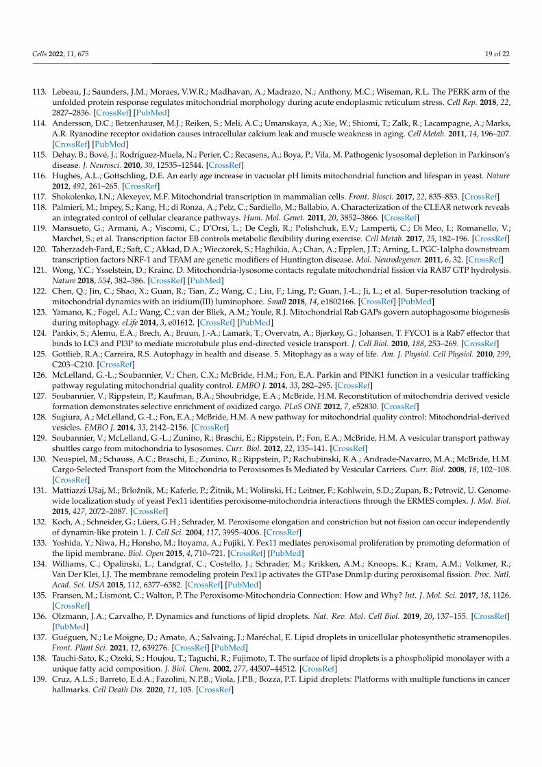
Cells 2022, 11, 675 19 of 22
113. Lebeau, J.; Saunders, J.M.; Moraes, V.W.R.; Madhavan, A.; Madrazo, N.; Anthony, M.C.; Wiseman, R.L. The PERK arm of theunfolded protein response regulates mitochondrial morphology during acute endoplasmic reticulum stress. Cell Rep. 2018, 22,2827–2836. [CrossRef] [PubMed]
114. Andersson, D.C.; Betzenhauser, M.J.; Reiken, S.; Meli, A.C.; Umanskaya, A.; Xie, W.; Shiomi, T.; Zalk, R.; Lacampagne, A.; Marks,A.R. Ryanodine receptor oxidation causes intracellular calcium leak and muscle weakness in aging. Cell Metab. 2011, 14, 196–207.[CrossRef] [PubMed]
115. Dehay, B.; Bové, J.; Rodríguez-Muela, N.; Perier, C.; Recasens, A.; Boya, P.; Vila, M. Pathogenic lysosomal depletion in Parkinson’sdisease. J. Neurosci. 2010, 30, 12535–12544. [CrossRef]
116. Hughes, A.L.; Gottschling, D.E. An early age increase in vacuolar pH limits mitochondrial function and lifespan in yeast. Nature2012, 492, 261–265. [CrossRef]
117. Shokolenko, I.N.; Alexeyev, M.F. Mitochondrial transcription in mammalian cells. Front. Biosci. 2017, 22, 835–853. [CrossRef]118. Palmieri, M.; Impey, S.; Kang, H.; di Ronza, A.; Pelz, C.; Sardiello, M.; Ballabio, A. Characterization of the CLEAR network reveals
an integrated control of cellular clearance pathways. Hum. Mol. Genet. 2011, 20, 3852–3866. [CrossRef]119. Mansueto, G.; Armani, A.; Viscomi, C.; D’Orsi, L.; De Cegli, R.; Polishchuk, E.V.; Lamperti, C.; Di Meo, I.; Romanello, V.;
Marchet, S.; et al. Transcription factor EB controls metabolic flexibility during exercise. Cell Metab. 2017, 25, 182–196. [CrossRef]120. Taherzadeh-Fard, E.; Saft, C.; Akkad, D.A.; Wieczorek, S.; Haghikia, A.; Chan, A.; Epplen, J.T.; Arning, L. PGC-1alpha downstream
transcription factors NRF-1 and TFAM are genetic modifiers of Huntington disease. Mol. Neurodegener. 2011, 6, 32. [CrossRef]121. Wong, Y.C.; Ysselstein, D.; Krainc, D. Mitochondria-lysosome contacts regulate mitochondrial fission via RAB7 GTP hydrolysis.
Nature 2018, 554, 382–386. [CrossRef] [PubMed]122. Chen, Q.; Jin, C.; Shao, X.; Guan, R.; Tian, Z.; Wang, C.; Liu, F.; Ling, P.; Guan, J.-L.; Ji, L.; et al. Super-resolution tracking of
mitochondrial dynamics with an iridium(III) luminophore. Small 2018, 14, e1802166. [CrossRef] [PubMed]123. Yamano, K.; Fogel, A.I.; Wang, C.; van der Bliek, A.M.; Youle, R.J. Mitochondrial Rab GAPs govern autophagosome biogenesis
during mitophagy. eLife 2014, 3, e01612. [CrossRef] [PubMed]124. Pankiv, S.; Alemu, E.A.; Brech, A.; Bruun, J.-A.; Lamark, T.; Overvatn, A.; Bjørkøy, G.; Johansen, T. FYCO1 is a Rab7 effector that
binds to LC3 and PI3P to mediate microtubule plus end-directed vesicle transport. J. Cell Biol. 2010, 188, 253–269. [CrossRef]125. Gottlieb, R.A.; Carreira, R.S. Autophagy in health and disease. 5. Mitophagy as a way of life. Am. J. Physiol. Cell Physiol. 2010, 299,
C203–C210. [CrossRef]126. McLelland, G.-L.; Soubannier, V.; Chen, C.X.; McBride, H.M.; Fon, E.A. Parkin and PINK1 function in a vesicular trafficking
pathway regulating mitochondrial quality control. EMBO J. 2014, 33, 282–295. [CrossRef]127. Soubannier, V.; Rippstein, P.; Kaufman, B.A.; Shoubridge, E.A.; McBride, H.M. Reconstitution of mitochondria derived vesicle
formation demonstrates selective enrichment of oxidized cargo. PLoS ONE 2012, 7, e52830. [CrossRef]128. Sugiura, A.; McLelland, G.-L.; Fon, E.A.; McBride, H.M. A new pathway for mitochondrial quality control: Mitochondrial-derived
vesicles. EMBO J. 2014, 33, 2142–2156. [CrossRef]129. Soubannier, V.; McLelland, G.-L.; Zunino, R.; Braschi, E.; Rippstein, P.; Fon, E.A.; McBride, H.M. A vesicular transport pathway
shuttles cargo from mitochondria to lysosomes. Curr. Biol. 2012, 22, 135–141. [CrossRef]130. Neuspiel, M.; Schauss, A.C.; Braschi, E.; Zunino, R.; Rippstein, P.; Rachubinski, R.A.; Andrade-Navarro, M.A.; McBride, H.M.
Cargo-Selected Transport from the Mitochondria to Peroxisomes Is Mediated by Vesicular Carriers. Curr. Biol. 2008, 18, 102–108.[CrossRef]
131. Mattiazzi Ušaj, M.; Brložnik, M.; Kaferle, P.; Žitnik, M.; Wolinski, H.; Leitner, F.; Kohlwein, S.D.; Zupan, B.; Petrovic, U. Genome-wide localization study of yeast Pex11 identifies peroxisome-mitochondria interactions through the ERMES complex. J. Mol. Biol.2015, 427, 2072–2087. [CrossRef]
132. Koch, A.; Schneider, G.; Lüers, G.H.; Schrader, M. Peroxisome elongation and constriction but not fission can occur independentlyof dynamin-like protein 1. J. Cell Sci. 2004, 117, 3995–4006. [CrossRef]
133. Yoshida, Y.; Niwa, H.; Honsho, M.; Itoyama, A.; Fujiki, Y. Pex11 mediates peroxisomal proliferation by promoting deformation ofthe lipid membrane. Biol. Open 2015, 4, 710–721. [CrossRef] [PubMed]
134. Williams, C.; Opalinski, L.; Landgraf, C.; Costello, J.; Schrader, M.; Krikken, A.M.; Knoops, K.; Kram, A.M.; Volkmer, R.;Van Der Klei, I.J. The membrane remodeling protein Pex11p activates the GTPase Dnm1p during peroxisomal fission. Proc. Natl.Acad. Sci. USA 2015, 112, 6377–6382. [CrossRef] [PubMed]
135. Fransen, M.; Lismont, C.; Walton, P. The Peroxisome-Mitochondria Connection: How and Why? Int. J. Mol. Sci. 2017, 18, 1126.[CrossRef]
136. Olzmann, J.A.; Carvalho, P. Dynamics and functions of lipid droplets. Nat. Rev. Mol. Cell Biol. 2019, 20, 137–155. [CrossRef][PubMed]
137. Guéguen, N.; Le Moigne, D.; Amato, A.; Salvaing, J.; Maréchal, E. Lipid droplets in unicellular photosynthetic stramenopiles.Front. Plant Sci. 2021, 12, 639276. [CrossRef] [PubMed]
138. Tauchi-Sato, K.; Ozeki, S.; Houjou, T.; Taguchi, R.; Fujimoto, T. The surface of lipid droplets is a phospholipid monolayer with aunique fatty acid composition. J. Biol. Chem. 2002, 277, 44507–44512. [CrossRef]
139. Cruz, A.L.S.; Barreto, E.d.A.; Fazolini, N.P.B.; Viola, J.P.B.; Bozza, P.T. Lipid droplets: Platforms with multiple functions in cancerhallmarks. Cell Death Dis. 2020, 11, 105. [CrossRef]
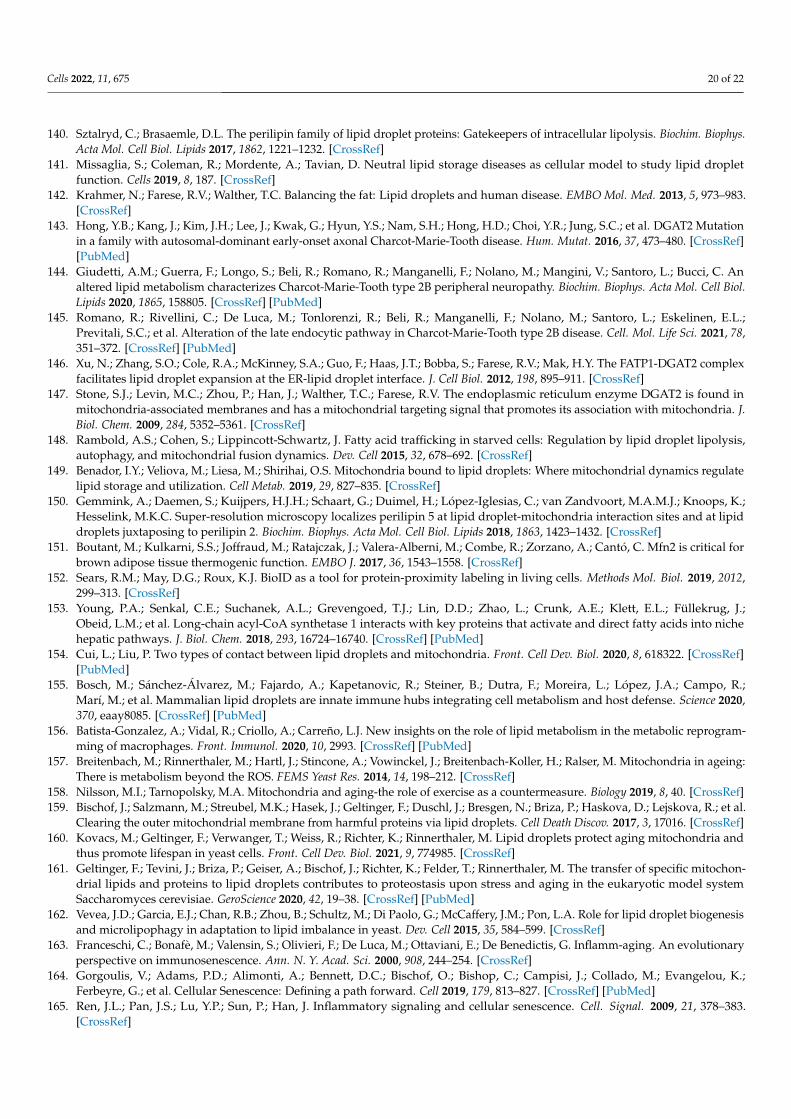
Cells 2022, 11, 675 20 of 22
140. Sztalryd, C.; Brasaemle, D.L. The perilipin family of lipid droplet proteins: Gatekeepers of intracellular lipolysis. Biochim. Biophys.Acta Mol. Cell Biol. Lipids 2017, 1862, 1221–1232. [CrossRef]
141. Missaglia, S.; Coleman, R.; Mordente, A.; Tavian, D. Neutral lipid storage diseases as cellular model to study lipid dropletfunction. Cells 2019, 8, 187. [CrossRef]
142. Krahmer, N.; Farese, R.V.; Walther, T.C. Balancing the fat: Lipid droplets and human disease. EMBO Mol. Med. 2013, 5, 973–983.[CrossRef]
143. Hong, Y.B.; Kang, J.; Kim, J.H.; Lee, J.; Kwak, G.; Hyun, Y.S.; Nam, S.H.; Hong, H.D.; Choi, Y.R.; Jung, S.C.; et al. DGAT2 Mutationin a family with autosomal-dominant early-onset axonal Charcot-Marie-Tooth disease. Hum. Mutat. 2016, 37, 473–480. [CrossRef][PubMed]
144. Giudetti, A.M.; Guerra, F.; Longo, S.; Beli, R.; Romano, R.; Manganelli, F.; Nolano, M.; Mangini, V.; Santoro, L.; Bucci, C. Analtered lipid metabolism characterizes Charcot-Marie-Tooth type 2B peripheral neuropathy. Biochim. Biophys. Acta Mol. Cell Biol.Lipids 2020, 1865, 158805. [CrossRef] [PubMed]
145. Romano, R.; Rivellini, C.; De Luca, M.; Tonlorenzi, R.; Beli, R.; Manganelli, F.; Nolano, M.; Santoro, L.; Eskelinen, E.L.;Previtali, S.C.; et al. Alteration of the late endocytic pathway in Charcot-Marie-Tooth type 2B disease. Cell. Mol. Life Sci. 2021, 78,351–372. [CrossRef] [PubMed]
146. Xu, N.; Zhang, S.O.; Cole, R.A.; McKinney, S.A.; Guo, F.; Haas, J.T.; Bobba, S.; Farese, R.V.; Mak, H.Y. The FATP1-DGAT2 complexfacilitates lipid droplet expansion at the ER-lipid droplet interface. J. Cell Biol. 2012, 198, 895–911. [CrossRef]
147. Stone, S.J.; Levin, M.C.; Zhou, P.; Han, J.; Walther, T.C.; Farese, R.V. The endoplasmic reticulum enzyme DGAT2 is found inmitochondria-associated membranes and has a mitochondrial targeting signal that promotes its association with mitochondria. J.Biol. Chem. 2009, 284, 5352–5361. [CrossRef]
148. Rambold, A.S.; Cohen, S.; Lippincott-Schwartz, J. Fatty acid trafficking in starved cells: Regulation by lipid droplet lipolysis,autophagy, and mitochondrial fusion dynamics. Dev. Cell 2015, 32, 678–692. [CrossRef]
149. Benador, I.Y.; Veliova, M.; Liesa, M.; Shirihai, O.S. Mitochondria bound to lipid droplets: Where mitochondrial dynamics regulatelipid storage and utilization. Cell Metab. 2019, 29, 827–835. [CrossRef]
150. Gemmink, A.; Daemen, S.; Kuijpers, H.J.H.; Schaart, G.; Duimel, H.; López-Iglesias, C.; van Zandvoort, M.A.M.J.; Knoops, K.;Hesselink, M.K.C. Super-resolution microscopy localizes perilipin 5 at lipid droplet-mitochondria interaction sites and at lipiddroplets juxtaposing to perilipin 2. Biochim. Biophys. Acta Mol. Cell Biol. Lipids 2018, 1863, 1423–1432. [CrossRef]
151. Boutant, M.; Kulkarni, S.S.; Joffraud, M.; Ratajczak, J.; Valera-Alberni, M.; Combe, R.; Zorzano, A.; Cantó, C. Mfn2 is critical forbrown adipose tissue thermogenic function. EMBO J. 2017, 36, 1543–1558. [CrossRef]
152. Sears, R.M.; May, D.G.; Roux, K.J. BioID as a tool for protein-proximity labeling in living cells. Methods Mol. Biol. 2019, 2012,299–313. [CrossRef]
153. Young, P.A.; Senkal, C.E.; Suchanek, A.L.; Grevengoed, T.J.; Lin, D.D.; Zhao, L.; Crunk, A.E.; Klett, E.L.; Füllekrug, J.;Obeid, L.M.; et al. Long-chain acyl-CoA synthetase 1 interacts with key proteins that activate and direct fatty acids into nichehepatic pathways. J. Biol. Chem. 2018, 293, 16724–16740. [CrossRef] [PubMed]
154. Cui, L.; Liu, P. Two types of contact between lipid droplets and mitochondria. Front. Cell Dev. Biol. 2020, 8, 618322. [CrossRef][PubMed]
155. Bosch, M.; Sánchez-Álvarez, M.; Fajardo, A.; Kapetanovic, R.; Steiner, B.; Dutra, F.; Moreira, L.; López, J.A.; Campo, R.;Marí, M.; et al. Mammalian lipid droplets are innate immune hubs integrating cell metabolism and host defense. Science 2020,370, eaay8085. [CrossRef] [PubMed]
156. Batista-Gonzalez, A.; Vidal, R.; Criollo, A.; Carreño, L.J. New insights on the role of lipid metabolism in the metabolic reprogram-ming of macrophages. Front. Immunol. 2020, 10, 2993. [CrossRef] [PubMed]
157. Breitenbach, M.; Rinnerthaler, M.; Hartl, J.; Stincone, A.; Vowinckel, J.; Breitenbach-Koller, H.; Ralser, M. Mitochondria in ageing:There is metabolism beyond the ROS. FEMS Yeast Res. 2014, 14, 198–212. [CrossRef]
158. Nilsson, M.I.; Tarnopolsky, M.A. Mitochondria and aging-the role of exercise as a countermeasure. Biology 2019, 8, 40. [CrossRef]159. Bischof, J.; Salzmann, M.; Streubel, M.K.; Hasek, J.; Geltinger, F.; Duschl, J.; Bresgen, N.; Briza, P.; Haskova, D.; Lejskova, R.; et al.
Clearing the outer mitochondrial membrane from harmful proteins via lipid droplets. Cell Death Discov. 2017, 3, 17016. [CrossRef]160. Kovacs, M.; Geltinger, F.; Verwanger, T.; Weiss, R.; Richter, K.; Rinnerthaler, M. Lipid droplets protect aging mitochondria and
thus promote lifespan in yeast cells. Front. Cell Dev. Biol. 2021, 9, 774985. [CrossRef]161. Geltinger, F.; Tevini, J.; Briza, P.; Geiser, A.; Bischof, J.; Richter, K.; Felder, T.; Rinnerthaler, M. The transfer of specific mitochon-
drial lipids and proteins to lipid droplets contributes to proteostasis upon stress and aging in the eukaryotic model systemSaccharomyces cerevisiae. GeroScience 2020, 42, 19–38. [CrossRef] [PubMed]
162. Vevea, J.D.; Garcia, E.J.; Chan, R.B.; Zhou, B.; Schultz, M.; Di Paolo, G.; McCaffery, J.M.; Pon, L.A. Role for lipid droplet biogenesisand microlipophagy in adaptation to lipid imbalance in yeast. Dev. Cell 2015, 35, 584–599. [CrossRef]
163. Franceschi, C.; Bonafè, M.; Valensin, S.; Olivieri, F.; De Luca, M.; Ottaviani, E.; De Benedictis, G. Inflamm-aging. An evolutionaryperspective on immunosenescence. Ann. N. Y. Acad. Sci. 2000, 908, 244–254. [CrossRef]
164. Gorgoulis, V.; Adams, P.D.; Alimonti, A.; Bennett, D.C.; Bischof, O.; Bishop, C.; Campisi, J.; Collado, M.; Evangelou, K.;Ferbeyre, G.; et al. Cellular Senescence: Defining a path forward. Cell 2019, 179, 813–827. [CrossRef] [PubMed]
165. Ren, J.L.; Pan, J.S.; Lu, Y.P.; Sun, P.; Han, J. Inflammatory signaling and cellular senescence. Cell. Signal. 2009, 21, 378–383.[CrossRef]
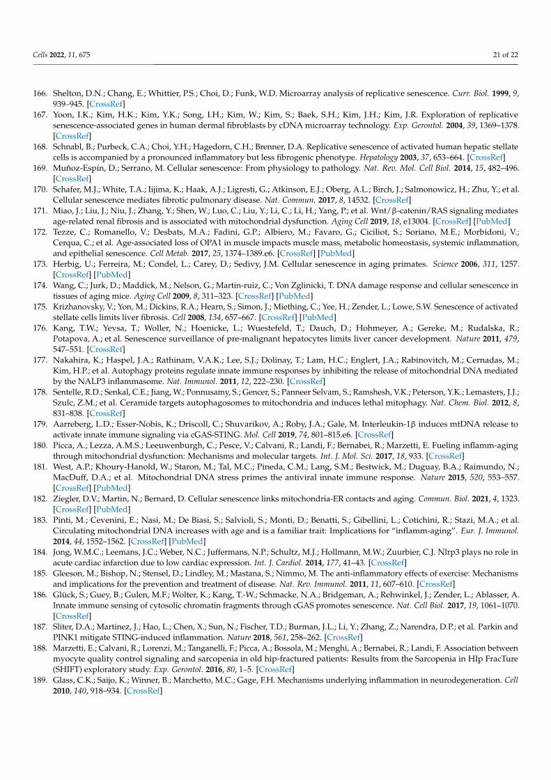
Cells 2022, 11, 675 21 of 22
166. Shelton, D.N.; Chang, E.; Whittier, P.S.; Choi, D.; Funk, W.D. Microarray analysis of replicative senescence. Curr. Biol. 1999, 9,939–945. [CrossRef]
167. Yoon, I.K.; Kim, H.K.; Kim, Y.K.; Song, I.H.; Kim, W.; Kim, S.; Baek, S.H.; Kim, J.H.; Kim, J.R. Exploration of replicativesenescence-associated genes in human dermal fibroblasts by cDNA microarray technology. Exp. Gerontol. 2004, 39, 1369–1378.[CrossRef]
168. Schnabl, B.; Purbeck, C.A.; Choi, Y.H.; Hagedorn, C.H.; Brenner, D.A. Replicative senescence of activated human hepatic stellatecells is accompanied by a pronounced inflammatory but less fibrogenic phenotype. Hepatology 2003, 37, 653–664. [CrossRef]
169. Muñoz-Espín, D.; Serrano, M. Cellular senescence: From physiology to pathology. Nat. Rev. Mol. Cell Biol. 2014, 15, 482–496.[CrossRef]
170. Schafer, M.J.; White, T.A.; Iijima, K.; Haak, A.J.; Ligresti, G.; Atkinson, E.J.; Oberg, A.L.; Birch, J.; Salmonowicz, H.; Zhu, Y.; et al.Cellular senescence mediates fibrotic pulmonary disease. Nat. Commun. 2017, 8, 14532. [CrossRef]
171. Miao, J.; Liu, J.; Niu, J.; Zhang, Y.; Shen, W.; Luo, C.; Liu, Y.; Li, C.; Li, H.; Yang, P.; et al. Wnt/β-catenin/RAS signaling mediatesage-related renal fibrosis and is associated with mitochondrial dysfunction. Aging Cell 2019, 18, e13004. [CrossRef] [PubMed]
172. Tezze, C.; Romanello, V.; Desbats, M.A.; Fadini, G.P.; Albiero, M.; Favaro, G.; Ciciliot, S.; Soriano, M.E.; Morbidoni, V.;Cerqua, C.; et al. Age-associated loss of OPA1 in muscle impacts muscle mass, metabolic homeostasis, systemic inflammation,and epithelial senescence. Cell Metab. 2017, 25, 1374–1389.e6. [CrossRef] [PubMed]
173. Herbig, U.; Ferreira, M.; Condel, L.; Carey, D.; Sedivy, J.M. Cellular senescence in aging primates. Science 2006, 311, 1257.[CrossRef] [PubMed]
174. Wang, C.; Jurk, D.; Maddick, M.; Nelson, G.; Martin-ruiz, C.; Von Zglinicki, T. DNA damage response and cellular senescence intissues of aging mice. Aging Cell 2009, 8, 311–323. [CrossRef] [PubMed]
175. Krizhanovsky, V.; Yon, M.; Dickins, R.A.; Hearn, S.; Simon, J.; Miething, C.; Yee, H.; Zender, L.; Lowe, S.W. Senescence of activatedstellate cells limits liver fibrosis. Cell 2008, 134, 657–667. [CrossRef] [PubMed]
176. Kang, T.W.; Yevsa, T.; Woller, N.; Hoenicke, L.; Wuestefeld, T.; Dauch, D.; Hohmeyer, A.; Gereke, M.; Rudalska, R.;Potapova, A.; et al. Senescence surveillance of pre-malignant hepatocytes limits liver cancer development. Nature 2011, 479,547–551. [CrossRef]
177. Nakahira, K.; Haspel, J.A.; Rathinam, V.A.K.; Lee, S.J.; Dolinay, T.; Lam, H.C.; Englert, J.A.; Rabinovitch, M.; Cernadas, M.;Kim, H.P.; et al. Autophagy proteins regulate innate immune responses by inhibiting the release of mitochondrial DNA mediatedby the NALP3 inflammasome. Nat. Immunol. 2011, 12, 222–230. [CrossRef]
178. Sentelle, R.D.; Senkal, C.E.; Jiang, W.; Ponnusamy, S.; Gencer, S.; Panneer Selvam, S.; Ramshesh, V.K.; Peterson, Y.K.; Lemasters, J.J.;Szulc, Z.M.; et al. Ceramide targets autophagosomes to mitochondria and induces lethal mitophagy. Nat. Chem. Biol. 2012, 8,831–838. [CrossRef]
179. Aarreberg, L.D.; Esser-Nobis, K.; Driscoll, C.; Shuvarikov, A.; Roby, J.A.; Gale, M. Interleukin-1β induces mtDNA release toactivate innate immune signaling via cGAS-STING. Mol. Cell 2019, 74, 801–815.e6. [CrossRef]
180. Picca, A.; Lezza, A.M.S.; Leeuwenburgh, C.; Pesce, V.; Calvani, R.; Landi, F.; Bernabei, R.; Marzetti, E. Fueling inflamm-agingthrough mitochondrial dysfunction: Mechanisms and molecular targets. Int. J. Mol. Sci. 2017, 18, 933. [CrossRef]
181. West, A.P.; Khoury-Hanold, W.; Staron, M.; Tal, M.C.; Pineda, C.M.; Lang, S.M.; Bestwick, M.; Duguay, B.A.; Raimundo, N.;MacDuff, D.A.; et al. Mitochondrial DNA stress primes the antiviral innate immune response. Nature 2015, 520, 553–557.[CrossRef] [PubMed]
182. Ziegler, D.V.; Martin, N.; Bernard, D. Cellular senescence links mitochondria-ER contacts and aging. Commun. Biol. 2021, 4, 1323.[CrossRef] [PubMed]
183. Pinti, M.; Cevenini, E.; Nasi, M.; De Biasi, S.; Salvioli, S.; Monti, D.; Benatti, S.; Gibellini, L.; Cotichini, R.; Stazi, M.A.; et al.Circulating mitochondrial DNA increases with age and is a familiar trait: Implications for “inflamm-aging”. Eur. J. Immunol.2014, 44, 1552–1562. [CrossRef] [PubMed]
184. Jong, W.M.C.; Leemans, J.C.; Weber, N.C.; Juffermans, N.P.; Schultz, M.J.; Hollmann, M.W.; Zuurbier, C.J. Nlrp3 plays no role inacute cardiac infarction due to low cardiac expression. Int. J. Cardiol. 2014, 177, 41–43. [CrossRef]
185. Gleeson, M.; Bishop, N.; Stensel, D.; Lindley, M.; Mastana, S.; Nimmo, M. The anti-inflammatory effects of exercise: Mechanismsand implications for the prevention and treatment of disease. Nat. Rev. Immunol. 2011, 11, 607–610. [CrossRef]
186. Glück, S.; Guey, B.; Gulen, M.F.; Wolter, K.; Kang, T.-W.; Schmacke, N.A.; Bridgeman, A.; Rehwinkel, J.; Zender, L.; Ablasser, A.Innate immune sensing of cytosolic chromatin fragments through cGAS promotes senescence. Nat. Cell Biol. 2017, 19, 1061–1070.[CrossRef]
187. Sliter, D.A.; Martinez, J.; Hao, L.; Chen, X.; Sun, N.; Fischer, T.D.; Burman, J.L.; Li, Y.; Zhang, Z.; Narendra, D.P.; et al. Parkin andPINK1 mitigate STING-induced inflammation. Nature 2018, 561, 258–262. [CrossRef]
188. Marzetti, E.; Calvani, R.; Lorenzi, M.; Tanganelli, F.; Picca, A.; Bossola, M.; Menghi, A.; Bernabei, R.; Landi, F. Association betweenmyocyte quality control signaling and sarcopenia in old hip-fractured patients: Results from the Sarcopenia in HIp FracTure(SHIFT) exploratory study. Exp. Gerontol. 2016, 80, 1–5. [CrossRef]
189. Glass, C.K.; Saijo, K.; Winner, B.; Marchetto, M.C.; Gage, F.H. Mechanisms underlying inflammation in neurodegeneration. Cell2010, 140, 918–934. [CrossRef]

Cells 2022, 11, 675 22 of 22
190. Sharma, M.; Rajendrarao, S.; Shahani, N.; Ramírez-Jarquín, U.N.; Subramaniam, S. Cyclic GMP-AMP synthase promotes theinflammatory and autophagy responses in Huntington disease. Proc. Natl. Acad. Sci. USA 2020, 117, 15989–15999. [CrossRef]
191. Nazmi, A.; Field, R.H.; Griffin, E.W.; Haugh, O.; Hennessy, E.; Cox, D.; Reis, R.; Tortorelli, L.; Murray, C.L.; Lopez-Rodriguez, A.B.; et al.Chronic neurodegeneration induces type I interferon synthesis via STING, shaping microglial phenotype and accelerating diseaseprogression. Glia 2019, 67, 1254–1276. [CrossRef] [PubMed]