Role of the miR-17∼92 cluster family in cerebellar and medulloblastoma development
9
RESEARCH ARTICLE Role of the miR-17,92 cluster family in cerebellar and medulloblastoma development Frederique Zindy 1 , Daisuke Kawauchi 1, *, Youngsoo Lee 2,` , Olivier Ayrault 1,§ , Leila Ben Merzoug 1 , Peter J. McKinnon 2 , Andrea Ventura 3 and Martine F. Roussel 1," ABSTRACT The miR-17,92 cluster family is composed of three members encoding microRNAs that share seed sequences. To assess their role in cerebellar and medulloblastoma (MB) development, we deleted the miR-17,92 cluster family in Nestin-positive neural progenitors and in mice heterozygous for the Sonic Hedgehog (SHH) receptor Patched 1 (Ptch1 +/2 ). We show that mice in which we conditionally deleted the miR-17,92 cluster (miR-17,92 floxed/ floxed ; Nestin-Cre + ) alone or together with the complete loss of the miR-106b,25 cluster (miR-106b,25 2/2 ) were born alive but with small brains and reduced cerebellar foliation. Remarkably, deletion of the miR-17,92 cluster abolished the development of SHH-MB in Ptch1 +/2 mice. Using an orthotopic transplant approach, we showed that granule neuron precursors (GNPs) purified from the cerebella of postnatal day 7 (P7) Ptch1 +/2 ; miR-106b,25 2/2 mice and overexpressing Mycn induced MBs in the cortices of naı ¨ve recipient mice. In contrast, GNPs purified from the cerebella of P7 Ptch1 +/2 ; miR-17,92 floxed/floxed ; Nestin-Cre + animals and overexpressing Mycn failed to induce tumors in recipient animals. Taken together, our findings demonstrate that the miR-17,92 cluster is dispensable for cerebellar development, but required for SHH-MB development. KEY WORDS: MicroRNA, miR-17,92 and miR-106b,25 clusters, Cerebellum, Development, Nestin, Medulloblastoma, Granule neuron progenitors (GNPs) INTRODUCTION The cerebellum develops in the mouse from embryonic day 9 (E9) with the cerebellar anlage forming from the roof (the alar plates) of the metencephalon. It is composed of different types of neurons that arise from the ventricular zone (VZ) of the cerebellar neuroepithelium localized on the roof of the fourth ventricle, and from the rostral rhombic lip (rRL), localized at the posterior edge of the cerebellar anlage (Hatten and Roussel, 2011). In the mouse, granule neuron progenitors (GNPs) are born in the rRL (E11– E16) and migrate along the surface of the developing cerebellum over the Purkinje cells, which are born in the VZ (E11–E13), to form the external granule layer (EGL) (E13–E16). After birth, Sonic Hedgehog (SHH), secreted by the Purkinje cells, promotes the proliferation of GNPs, which peaks between postnatal days 5 (P5) and P7. Subsequently, GNPs exit the cell cycle, migrate inwardly through the Purkinje cell layer and settle as postmitotic neurons in the internal granular layer (IGL). By 3 weeks, the mouse cerebellum is fully formed consisting of ten folia separated by fissures (Hatten and Roussel, 2011). Constitutive activation of the SHH signaling pathway leads to defects in cell cycle exit, migration and differentiation, which, in turn, induce medulloblastoma (MB). This SHH-subgroup of MBs (SHH- MB) represents ,25% of all human cases (Taylor et al., 2012). MicroRNAs (miRNAs) are ,22 nucleotides long non-coding RNAs. They are derived from pri-miRNAs that are processed by Drosha and DGCR8 into pre-miRNAs in the nucleus, and then translocated into the cytoplasm where they are converted into mature miRNAs by the processing enzyme Dicer. In turn, single-stranded miRNAs are loaded into the RNA-induced silencing complex (RISC) to bind the 39-untranslated region (39-UTR) of mRNAs to inhibit their translation or degradation (Carmell and Hannon, 2004; Kim, 2005). Abnormal expression of miRNAs is often seen in cancers. MicroRNAs encoded by the miR-17,92 cluster, also called oncomiR-1, are overexpressed in various cancers (Concepcion et al., 2012; Mogilyansky and Rigoutsos, 2013) including mouse and human medulloblastomas with constitutively activated SHH signaling (Uziel et al., 2009; Northcott et al., 2009). The miR- 17,92 cluster encoded by chromosome 14 in the mouse (13 in humans) has two paralogs, the miR-106b,25 and miR- 106a,363 clusters, each of which is located on different chromosomes (Fig. 1A). The miR-106b,25 cluster is encoded on chromosome 5 in the mouse (7 in humans) while the miR- 106a,363 cluster maps to chromosome X in mice and humans (Concepion et al., 2012; Mogilyansky and Rigoutsos, 2013). Mice lacking the miR-17,92 cluster die shortly after birth from lung and heart defects while mice lacking each of its two paralogs do not show any obvious phenotypes (Ventura et al., 2008). However, the miR-17,92 and miR-106b,25 clusters share overlapping functions since mice with combined deletion exhibit a more profound phenotype than those lacking miR- 17,92 alone (Ventura et al., 2008). The miR-17,92 cluster is a downstream target of Myc (c-Myc) (O’Donnell et al., 2005) and Mycn (Northcott et al., 2009; de Pontual et al., 2011). Mice lacking one copy of miR-17,92 show skeletal and growth 1 Department of Tumor Cell Biology, Danny Thomas Research Center, St Jude Children’s Research Hospital, Memphis, TN 38105-3678, USA. 2 Department of Genetics, Danny Thomas Research Center, St Jude Children’s Research Hospital, Memphis, TN 38105-3678, USA. 3 Cancer Biology and Genetics Program, Memorial Sloan Kettering Cancer Center, New York, NY 10065, USA. *Present address: Division of Pediatric Neurooncology, German Cancer Research Center (DKFZ), Im Neuenheimer Feld 580, 69120 Heidelberg, Germany. ` Present address: Genomic Instability Research Center, Ajou University, School of Medicine, Suwon 443-749, South Korea. § Present address: Institut Curie/CNRS UMR 3306/INSERM U1005 – Building 110 – Centre Universitaire, 91405 Orsay, Cedex, France. " Author for correspondence ([email protected]) This is an Open Access article distributed under the terms of the Creative Commons Attribution License (http://creativecommons.org/licenses/by/3.0), which permits unrestricted use, distribution and reproduction in any medium provided that the original work is properly attributed. Received 25 September 2013; Accepted 8 May 2014 ß 2014. Published by The Company of Biologists Ltd | Biology Open (2014) 3, 597–605 doi:10.1242/bio.20146734 597 Biology Open
-
Upload
independent -
Category
Documents
-
view
0 -
download
0
Transcript of Role of the miR-17∼92 cluster family in cerebellar and medulloblastoma development

RESEARCH ARTICLE
Role of the miR-17,92 cluster family in cerebellar andmedulloblastoma development
Frederique Zindy1, Daisuke Kawauchi1,*, Youngsoo Lee2,`, Olivier Ayrault1,§, Leila Ben Merzoug1,Peter J. McKinnon2, Andrea Ventura3 and Martine F. Roussel1,"
ABSTRACT
The miR-17,92 cluster family is composed of three members
encoding microRNAs that share seed sequences. To assess their
role in cerebellar and medulloblastoma (MB) development, we
deleted the miR-17,92 cluster family in Nestin-positive neural
progenitors and in mice heterozygous for the Sonic Hedgehog
(SHH) receptor Patched 1 (Ptch1+/2). We show that mice in which
we conditionally deleted the miR-17,92 cluster (miR-17,92floxed/
floxed; Nestin-Cre+) alone or together with the complete loss of the
miR-106b,25 cluster (miR-106b,252/2) were born alive but with
small brains and reduced cerebellar foliation. Remarkably, deletion
of the miR-17,92 cluster abolished the development of SHH-MB
in Ptch1+/2 mice. Using an orthotopic transplant approach, we
showed that granule neuron precursors (GNPs) purified from the
cerebella of postnatal day 7 (P7) Ptch1+/2; miR-106b,252/2 mice
and overexpressing Mycn induced MBs in the cortices of naıve
recipient mice. In contrast, GNPs purified from the cerebella of
P7 Ptch1+/2; miR-17,92floxed/floxed; Nestin-Cre+ animals and
overexpressing Mycn failed to induce tumors in recipient animals.
Taken together, our findings demonstrate that the miR-17,92
cluster is dispensable for cerebellar development, but required for
SHH-MB development.
KEY WORDS: MicroRNA, miR-17,92 and miR-106b,25 clusters,
Cerebellum, Development, Nestin, Medulloblastoma, Granule
neuron progenitors (GNPs)
INTRODUCTIONThe cerebellum develops in the mouse from embryonic day 9
(E9) with the cerebellar anlage forming from the roof (the alarplates) of the metencephalon. It is composed of different types ofneurons that arise from the ventricular zone (VZ) of the cerebellarneuroepithelium localized on the roof of the fourth ventricle, and
from the rostral rhombic lip (rRL), localized at the posterior edge
of the cerebellar anlage (Hatten and Roussel, 2011). In the mouse,
granule neuron progenitors (GNPs) are born in the rRL (E11–
E16) and migrate along the surface of the developing cerebellum
over the Purkinje cells, which are born in the VZ (E11–E13), to
form the external granule layer (EGL) (E13–E16). After birth,
Sonic Hedgehog (SHH), secreted by the Purkinje cells, promotes
the proliferation of GNPs, which peaks between postnatal days 5
(P5) and P7. Subsequently, GNPs exit the cell cycle, migrate
inwardly through the Purkinje cell layer and settle as postmitotic
neurons in the internal granular layer (IGL). By 3 weeks, the
mouse cerebellum is fully formed consisting of ten folia separated
by fissures (Hatten and Roussel, 2011). Constitutive activation
of the SHH signaling pathway leads to defects in cell cycle
exit, migration and differentiation, which, in turn, induce
medulloblastoma (MB). This SHH-subgroup of MBs (SHH-
MB) represents ,25% of all human cases (Taylor et al., 2012).
MicroRNAs (miRNAs) are ,22 nucleotides long non-coding
RNAs. They are derived from pri-miRNAs that are processed by
Drosha and DGCR8 into pre-miRNAs in the nucleus, and
then translocated into the cytoplasm where they are converted
into mature miRNAs by the processing enzyme Dicer. In turn,
single-stranded miRNAs are loaded into the RNA-induced
silencing complex (RISC) to bind the 39-untranslated region
(39-UTR) of mRNAs to inhibit their translation or degradation
(Carmell and Hannon, 2004; Kim, 2005).
Abnormal expression of miRNAs is often seen in cancers.
MicroRNAs encoded by the miR-17,92 cluster, also called
oncomiR-1, are overexpressed in various cancers (Concepcion
et al., 2012; Mogilyansky and Rigoutsos, 2013) including mouse
and human medulloblastomas with constitutively activated SHH
signaling (Uziel et al., 2009; Northcott et al., 2009). The miR-
17,92 cluster encoded by chromosome 14 in the mouse (13 in
humans) has two paralogs, the miR-106b,25 and miR-
106a,363 clusters, each of which is located on different
chromosomes (Fig. 1A). The miR-106b,25 cluster is encoded
on chromosome 5 in the mouse (7 in humans) while the miR-
106a,363 cluster maps to chromosome X in mice and humans
(Concepion et al., 2012; Mogilyansky and Rigoutsos, 2013).
Mice lacking the miR-17,92 cluster die shortly after birth from
lung and heart defects while mice lacking each of its two
paralogs do not show any obvious phenotypes (Ventura et al.,
2008). However, the miR-17,92 and miR-106b,25 clusters
share overlapping functions since mice with combined deletion
exhibit a more profound phenotype than those lacking miR-
17,92 alone (Ventura et al., 2008). The miR-17,92 cluster is a
downstream target of Myc (c-Myc) (O’Donnell et al., 2005) and
Mycn (Northcott et al., 2009; de Pontual et al., 2011). Mice
lacking one copy of miR-17,92 show skeletal and growth
1Department of Tumor Cell Biology, Danny Thomas Research Center, St JudeChildren’s Research Hospital, Memphis, TN 38105-3678, USA. 2Department ofGenetics, Danny Thomas Research Center, St Jude Children’s ResearchHospital, Memphis, TN 38105-3678, USA. 3Cancer Biology and GeneticsProgram, Memorial Sloan Kettering Cancer Center, New York, NY 10065, USA.*Present address: Division of Pediatric Neurooncology, German CancerResearch Center (DKFZ), Im Neuenheimer Feld 580, 69120 Heidelberg,Germany. `Present address: Genomic Instability Research Center, AjouUniversity, School of Medicine, Suwon 443-749, South Korea. §Present address:Institut Curie/CNRS UMR 3306/INSERM U1005 – Building 110 – CentreUniversitaire, 91405 Orsay, Cedex, France.
"Author for correspondence ([email protected])
This is an Open Access article distributed under the terms of the Creative Commons AttributionLicense (http://creativecommons.org/licenses/by/3.0), which permits unrestricted use, distributionand reproduction in any medium provided that the original work is properly attributed.
Received 25 September 2013; Accepted 8 May 2014
� 2014. Published by The Company of Biologists Ltd | Biology Open (2014) 3, 597–605 doi:10.1242/bio.20146734
597
BiologyOpen

defects recapitulating the Feingold syndrome observed inpatients harboring MYCN mutations or hemizygous deletion ofMIR-17,92 (de Pontual et al., 2011). The miR-17,92 cluster is
expressed in proliferative GNPs but not in post-mitotic granuleneurons. Overexpression of the miR-17,92 cluster in GNPsheterozygous for the SHH receptor, Patched 1 (Ptch1+/2),induces early onset of SHH-MB formation after orthotopic
transplant in the cortices of naıve recipient animals (Uziel et al.,2009). Similarly when overexpressed in wild-type GNPs, themiR-17,92 cluster collaborates with SHH signaling to provide
GNPs with a proliferative advantage (Northcott et al., 2009).These results suggested that, besides its role in SHH-MBs, the miR-17,92 cluster might play a role in cerebellar
development. We here show that the miR-17,92 cluster and itsparalog the miR-106b,25 cluster, are differently required forcerebellar and medulloblastoma development.
MATERIALS AND METHODSMiceMouse lines carrying conditional alleles of miR-17,92 (miR-17,92floxed/floxed) (Ventura et al., 2008), or lacking miR-106b,25 (miR-
106b,252/2) (Ventura et al., 2008) were generously provided by Dr
Tyler Jacks (Boston, MA, USA). The transgenic line in which the Cre
recombinase is expressed under the promoter of the rat Nestin gene
(Nestin-Cre) (stock number 003771) (Tronche et al., 1999) and C57BL/6
mice were obtained from the Jackson Laboratory (Bar Harbor, ME,
USA). Ptch1+/2 mice in a Cdkn2c-null background (Ptch1+/2;
Cdkn2c2/2) were previously described (Uziel et al., 2005). All mice
were maintained on a mixed 1296C57BL/6 background. CD-1 nu/nu
mice were obtained from Charles River (Wilmington, MA, USA). Mice
were housed in an accredited facility of the Association for Assessment
and Accreditation of Laboratory Animal Care International (AAALAC)
in accordance with National Institute of Health guidelines. The Animal
Care and Use Committee (ACUC) of St Jude Children’s Research
Hospital approved all procedures.
Histology, immunohistochemistry, proliferation assays and X-galstainingHeads of embryos or whole brains from mice were harvested, fixed
overnight in 4% paraformaldehyde (PFA) in phosphate buffered saline
(PBS) at 4 C, soaked in 30% sucrose in PBS until the tissues sank
to the bottom of the tube and embedded in Optimal Cutting
Temperature (OCT) compound. Frozen 12 mm sections were
collected on Fisherbrand superfrost plus slides using a cryostat.
Slides were stained with Hematoxylin and Eosin (H&E) or antibodies,
as previously described (Uziel et al., 2005). The following antibodies
used were: 5-Bromo-2-Deoxy-Uridine (BrdU) (SC-32323; Santa Cruz,
Dallas, TX, USA; 1/1000 dilution), p27Kip1 (610242; BD Biosciences,
San Jose, CA; 1/200 dilution), cyclin D2 (SC-593; Santa Cruz; 1/50
dilution), NeuN (MAB377; EMD Millipore, Billerica, MA, USA; 1/
200 dilution), Ki67 (NLC-Ki67p; Leica microsystems, Buffalo Grove,
IL; 1/1000 dilution) and GABA(A) receptor a6 subunit (AB5610;
EMD Millipore; 1/200 dilution). BrdU (100 mg/kg) was injected intra-
peritoneally two hours before harvesting the brain. For X-gal staining,
whole brains from 3-week-old animals were fixed in 2% PFA in PBS
at 4 C for 3 hours. Fixed brains were washed twice in PBS and
then incubated in the ‘‘Rinse’’ buffer (100 mM sodium phosphate
pH 7.3, 2 mM MgCl2, 0.01% sodium deoxycholate, 0.02% IGEPAL
CA-360) for 5 minutes at room temperature. Brains were stained
with X-gal staining solution (‘‘Rinse’’ buffer containing 5 mM
potassium ferricyanide, 5 mM potassium ferrocyanide, 1 mg/ml X-
galactoside 5-bromo-4-chloro-3-indolyl-beta-D-galactopyranoside (X-
gal) (Invitrogen, Life technologies, Grand Island, NY, USA), at 37 C
for 4 hours.
Quantitative-reverse transcriptase-polymerase chain reaction(Q-RT-PCR) and Affymetrix microarrays analysisPurification of GNPs from P7 cerebella, RNA extraction from dissected
embryonic and postnatal (P4 and P7) cerebella or from purified GNPs,
and Q-RT-PCR for miR-19a and miR-106b were performed, as
previously described (Uziel et al., 2005; Uziel et al., 2009). For
comparative gene expression analysis, RNAs from cerebella of P4
and P7 mice were subjected to hybridization using Affymetrix
Mouse GeneChip MG430PM (Affymetrix, Santa Clara, CA). Principal
component analysis (PCA) and gene set enrichment analysis (GSEA)
were performed using Partek Genomics Suite 6.6 software (St Louis,
MO) and GSEA software (Broad Institute, Cambridge, MA),
respectively.
Orthotopic transplantationGNPs were purified from P7 cerebella and infected with retroviruses
encoding Mycn and the red fluorescent protein (RFP), as previously
described (Kawauchi et al., 2012). 48 hours after infection, 26106
infected GNPs were injected into the cortices of naıve recipient CD-1 nu/
nu mice. The rate of infection was assessed by fluorescence activated cell
sorting (FACS). The percentage of RFP positive (RFP+) cells ranged
from 30 to 50%.
StatisticsStatistical significance was determined using GraphPad Prism software
(version 5.0). Data were shown as mean 6 s.e.m. P-values ,0.05 were
used as significance threshold from unpaired two-tailed Student’s t test.
For the survival curves, p-values were determined with a log-rank
(Mantel Cox) test.
Fig. 1. Expression of miR-19a and miR-106b encoded by the miR-
17,92 cluster family in cerebella during embryogenesis. (A) Schematicrepresentation of the three microRNA clusters. MiR-17,92, miR-106a,363
and miR-106b,25 clusters encode 6, 6 and 3 mature microRNAs (coloredboxes), respectively. MicroRNAs sharing the same seed sequence arerepresented by boxes of the same color. Relative levels of maturemicroRNAs miR-19a (B) and miR-106b (C) were determined by Q-RT-PCRon total RNA extracted from total cerebella of wild-type mice (lanes 1–3) atE14.5 (lanes 1), E16.5 (lanes 2) and E18.5 (lanes 3).
RESEARCH ARTICLE Biology Open (2014) 3, 597–605 doi:10.1242/bio.20146734
598
BiologyOpen
RESULTSCo-inactivation of the miR-17,92 and miR-106b,25 clustersreduced cerebellar size and foliation by limiting proliferationWe previously found that microRNAs encoded by the miR-
17,92 and miR-106b,25, but not the miR-106a,363, clustersare expressed in proliferating GNPs in the postnatal cerebellum
(Uziel et al., 2009). Because these miRNAs were expressed indeveloping cerebella from E14.5 to birth (Fig. 1B,C, lanes 1–3),we assessed their role during cerebellar development. Since miR-
17,92-null mice die shortly after birth (Ventura et al., 2008), the
miR-17,92 cluster was conditionally deleted in Nestin-positiveneural progenitors using a Nestin-Cre transgenic mouse (miR-
17,92floxed/floxed; Nestin-Cre+) in a wild-type miR-106b,25
(referred as miR-17,92cKO) or in a miR-106b,25-null (referredas miR-17,92cKO; miR-106b,25KO) background. We alsogenerated miR-17,92floxed/floxed; Nestin-Cre2; miR-106b,252/2
(referred as miR-106b,25KO) and miR-17,92floxed/floxed; Nestin-
Cre2; miR-106b,25+/+ (referred as control) littermates. Mice ofall genotypes were born at a Mendelian ratio.
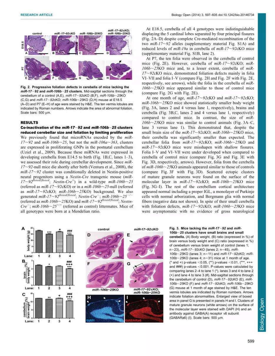
At E18.5, cerebella of all 4 genotypes were indistinguishabledisplaying the 5 cardinal lobes separated by four principal fissures
(Fig. 2A–D) despite complete Cre-mediated recombination of thetwo miR-17,92 alleles (supplementary material Fig. S1A) andreduced levels of miR-19a in cerebella of miR-17,92cKO mice(supplementary material Fig. S1B, lane 2).
At P7, the ten folia were observed in the cerebella of controlmice (Fig. 2E). However, cerebella of miR-17,92cKO; miR-
106b,25KO mice and, to a lesser extent, cerebella of miR-
17,92cKO mice, demonstrated foliation defects mainly in foliaVI–VII and folia I–V (compare Fig. 2H and Fig. 2F with Fig. 2E,respectively, see arrows), while the folia in the cerebella of miR-
106b,25KO mice appeared similar to those of control mice(compare Fig. 2G with Fig. 2E).
At one month of age, miR-17,92cKO and miR-17,92cKO;
miR-106b,25KO mice showed statistically smaller body weight(Fig. 3A, lanes 2 and 4 versus lane 1, respectively), brains andcerebella (Fig. 3B,C, lanes 2 and 4 versus lane 1, respectively)compared to control mice. In contrast, the size of miR-
106b,25KO mice was similar to control animals (Fig. 3A–C,lane 3 versus lane 1). This demonstrated that, despite thesmall brain size of the miR-17,92cKO; miR-106b,25KO mice,
their cerebella was significantly smaller than expected. Thecerebellar folia from miR-17,92cKO; miR-106b,25KO andmiR-17,92cKO mice were misshapen with shallow fissures.
Folia I–V and VI–VII were under developed when compared tocerebella of control mice (compare Fig. 3G and Fig. 3E withFig. 3D, respectively, arrows). However, folia from the cerebella
of miR-106b,25KO animals appeared similar to those of controls(compare Fig. 3F with Fig. 3D). Scattered ectopic clustersof mature granule neurons were found on the surface of themolecular layer in miR-17,92cKO; miR-106b,25KO mice
(Fig. 3G–I). The rest of the cerebellum cortical architectureappeared normal including a proper IGL, a monolayer of Purkinjecells with normal arborization, and Bergmann glia with normal
fibers (negative data not shown). In spite of their small cerebellawith foliation defects, miR-17,92cKO; miR-106b,25KO micewere asymptomatic with no evidence of gross neurological
Fig. 2. Progressive foliation defects in cerebella of mice lacking themiR-17,92 and miR-106b,25 clusters. Mid-sagittal sections through thecerebellum of a control (A,E), miR-17,92cKO (B,F), miR-106b,25KO(C,G) and miR-17,92cKO; miR-106b,25KO (D,H) mouse at E18.5(A–D) and P7 (E–H) of age were stained by H&E. The ten vermis lobules areindicated by Roman numbers. Arrows indicate the area of abnormal foliation.Scale bars: 500 mm.
Fig. 3. Mice lacking the miR-17,92 and miR-
106b,25 clusters have small brains and smallcerebella. (A) Body weight, (B) ratio (expressed in %) ofbrain versus body weight and (C) ratio (expressed in %)of cerebellum versus brain weight of control (lanes 1;n523), miR-17,92cKO (lanes 2; n545), miR-
106b,25KO (lanes 3; n511) and miR-17,92cKO; miR-
106b,25KO (lanes 4; n531) mice at 1 month of age.(* and +) p-values ,0.05, (**) p-values ,0.01, (***, +++and ###) p-values ,0.001. P-values were calculated bycomparing lanes 2–4 to lane 1 (*), lanes 3 and 4 to lane 2(+) and lane 4 to lane 3 (#). Mid-sagittal sections throughthe cerebellum of control (D), miR-17,92cKO (E), miR-
106b,25KO (F) and miR-17,92cKO; miR-106b,25KO(G) mouse at 1 month of age stained by H&E. The tenvermis lobules are indicated by Roman numbers. Arrowsindicate foliation abnormalities. Enlarged view of boxedarea in panel G is presented in panels H and I. Clusters ofmature granule neurons (white arrows) on the surface ofthe molecular layer were stained with DAPI (H) and anantibody against GABA(A) receptor a6 subunit(GABARa6) (I). Scale bars: 500 mm.
RESEARCH ARTICLE Biology Open (2014) 3, 597–605 doi:10.1242/bio.20146734
599
BiologyOpen

symptoms including motor coordination or balance defectsmeasured on a Rotarod (negative data not shown).
Cerebellar development of miR-17,92cKO; miR-106b,25KOmice appeared normal during embryogenesis until birth, a timewhen GNPs respond to SHH to rapidly proliferate with maximalproliferation between P5 and P7. At P7, we found a significant
reduction in BrdU incorporation in the EGL of cerebella frommiR-17,92cKO; miR-106b,25KO mice compared to controls(Fig. 4D versus Fig. 4A and Fig. 4E) consistent with the
requirement for massive proliferation of GNPs for properfoliation from birth until P7 (Lauder et al., 1974). The miR-
17,92 cluster not only regulates proliferation but also apoptosis
(Concepcion et al., 2012; Mogilyansky and Rigoutsos, 2013). Weobserved no significant differences in apoptosis by Terminaldeoxynucleotidyl transferase dUTP nick end labeling staining
between the cerebella of all 4 genotypes at P7 (negative data notshown). These results suggested that decreased proliferation ofGNPs from cerebella of miR-17,92cKO; miR-106b,25KO micemight account for the diminished pool of GNPs during post-
natal development. Consistent with this possibility, we observedpremature exhaustion of proliferative GNPs at P14 in miR-
17,92cKO; miR-106b,25KO mice. The EGL did not uniformly
cover the surface of the cerebellum in contrast to that of controlmice (compare Fig. 5G and Fig. 5I with Fig. 5A and Fig. 5C).The remaining cells in the EGL were mainly negative for BrdU
and cyclin D2 but positive for p27Kip1, suggesting premature exitfrom the cell cycle and migration defects (Fig. 5H and Fig. 5J–Lversus Fig. 5B and Fig. 5D–F).
The miR-17,92 cluster is required for medulloblastomaformationWe previously reported that GNPs purified from Ptch1+/2;Cdkn2c2/2 mice and overexpressing the miR-17,92 clusterinduced SHH-MB after transplant in the cortex of naıve CD-1 nu/
nu recipient mice (Uziel et al., 2009). However, unlike Mycn,
enforced expression of the miR-17,92 cluster in GNPs purifiedfrom the cerebella of Trp532/2; Cdkn2c2/2 mice failed to induceSHH-MBs after orthotopic transplantation demonstrating that
activation of the Patched signaling pathway was required for miR-
17,92 induction of SHH-MBs (Uziel et al., 2009). Toassess whether the miR-17,92 cluster was required for
MB formation, we bred miR-17,92floxed/+; Nestin-Cre+ withPtch1+/2; Cdkn2c2/2 mice. While, as expected, 45.5% (10/22) ofmiR-17,92+/+; Ptch1+/; Cdkn2c+/2 mice succumbed to SHH-
MBs, none (0/20) of the miR-17,92cKO; Ptch1+/2, Cdkn2c+/2
mice developed tumors over a period of 300 days (Fig. 6A).In wild-type mice, by 3 weeks after birth, all GNPs have exited
the cell cycle and migrated from the surface of the cerebellum
into the IGL. In contrast, in Ptch1+/2 mice, several GNPscontinue to divide and remain on the surface of the molecularlayer (ML), to form clusters of densely packed cells called pre-
neoplastic lesions (PNLs) (Goodrich et al., 1997; Kim et al.,
Fig. 4. Reduced proliferation in cerebella of mice lacking the miR-
17,92 and miR-106b,25 clusters. (A–D) Proliferation assessed by BrdUstaining (red) on mid-sagittal sections through P7 cerebellum. Nuclei werecounterstained with propidium iodide (blue). White bars indicate the EGL.(E) The percentage of BrdU+ cells in the EGL was quantified per 0.27 mm2.BrdU+ cells were counted in the EGL of lobule I and II from 2sections per animal. Three mice for each genotype were analyzed.(***) p-values ,0.001. (A and E, lane 1) control, (B and E, lane 2) miR-
17,92cKO, (C and E, lane 3) miR-106b,25KO and (D and E, lane 4)miR17,92cKO; miR-106b,25KO.
Fig. 5. Premature termination of EGL proliferation in mice lacking themiR-17,92 and miR-106b,25 clusters. Mid-sagittal sections through thecerebellum of P14 control (A–F) and miR-17,92cKO; miR-106b,25KO(G–L) mice stained with DAPI (A,C,G,I), or antibodies raised against BrdU(B,D,H,J), p27Kip1 (E,K) and cyclin D2 (F,L). Enlarged views of boxed areas inpanels A and G are presented in panels C–F and I–L, respectively. Arrowsindicate 2 clusters of granule neurons out of cycle, on the surface of thecerebellum, in miR-17,92cKO; miR-106b,25KO mice. Scale bars: 500 mm.
RESEARCH ARTICLE Biology Open (2014) 3, 597–605 doi:10.1242/bio.20146734
600
BiologyOpen

2003; Oliver et al., 2005). In 6 weeks old mice, the majority ofPNLs have regressed, while only few progress to MBs. Tovisualize PNLs, we stained the entire brains of 3 week old miR-
17,92cKO; Ptch1+/2, Cdkn2c+/2 and miR-17,92+/+; Ptch1+/2,Cdkn2c+/2 mice with X-gal, since a portion of the wild-type alleleof Patched is replaced by LacZ in the Ptch1+/2 mice (Goodrich et
al., 1997). We detected PNLs in 84.5% (11/13) miR-17,92+/+;Ptch1+/2, Cdkn2c+/2 mice (Fig. 6B,C,F). As expected, thosePNLs were Ki67 positive but negative for NeuN and GABA (A)receptor a6 subunit (Fig. 7C–E, arrows). However, only 33% (2/6)
of cerebella from miR-17,92cKO; Ptch1+/2; Cdkn2c+/2 miceshowed PNLs (p50.0248) (Fig. 6F). Scattered foci of non-proliferating, differentiated neurons (negative for Ki67 but
positive for NeuN and GABA (A) receptor a6 subunit) wereobserved on the surface of the cerebellar ML of miR-17,92cKO;Ptch1+/2; Cdkn2c+/2 mice (Fig. 6D,E, Fig. 7H–J, arrows). These
results suggest that the miR-17,92 cluster is required for SHH-induced PNL formation and for SHH-MB development.
Mycn is a direct target of SHH signaling and is required for
SHH-MB formation (Kenney et al., 2003; Hatton et al., 2006).Using an orthotopic transplantation approach, we previouslyshowed that enforced expression of Mycn in GNPs purified fromP7 cerebella of Ptch1+/2, Cdkn2c2/2 mice induces SHH-MBs
(Zindy et al., 2007; Kawauchi et al., 2012). GNPs purified fromthe cerebella of P7 miR-17,92cKO; Ptch1+/2; Cdkn2c+/2 and
miR-17,92+/+; Ptch1+/2; Cdkn2c+/2 mice were infected with
retroviruses encoding Mycn and the red fluorescence protein(RFP), and stereotactically implanted 2 days later into the corticesof naıve recipient animals. Mycn expression did not increase
apoptosis or cell cycle arrest in infected GNPs after three days inculture (negative data not shown). As expected, 100% (9/9) of theanimals transplanted with GNPs purified from the cerebella of P7
miR-17,92+/+; Ptch1+/2; Cdkn2c+/2 mice and overexpressingMycn developed SHH-MBs (Fig. 8A) (Zindy et al., 2007;Kawauchi et al., 2012). In contrast, none of the 12 mice
transplanted with GNPs purified from the cerebella of P7 miR-
17,92cKO; Ptch1+/2; Cdkn2c+/2 mice and expressing Mycn
developed medulloblastoma, 180 days post-implantation.However, 5/5 mice transplanted with GNPs purified from the
cerebella of P7 miR-106b,25KO; Ptch1+/2; Cdkn2c+/2 mice inwhich we enforced Mycn expression developed MBs (Fig. 8B).Pathological analysis of the tumors of all genotypes confirmed
that they were MBs of the SHH subtype with classic morphologyof round cells (Fig. 8C–H), as previously described (Zindy et al.,2007; Kawauchi et al., 2012).
These results point to an absolute requirement for the miR-
17,92, but not for the miR-106b,25, cluster in SHH-MBinitiation.
Targets of the miR-17,92 family clusterWe recently reported that the miR-17,92 cluster down-regulatesbone morphogenic protein (Bmp) receptor type 2 (Bmpr2), the
receptor for Bmp-2, -4 and -7 and that higher levels of Bmpr2 aredetected in cerebella of P7 miR-17,92cKO; miR-106b,25KOmice compared to those of controls (Murphy et al., 2013). To
Fig. 6. The mir-17,92 cluster is required for medulloblastomaformation. (A) Survival curves for Ptch1+/2; Cdkn2c+/2 mice with differentmiR-17,92 and Nestin-Cre genotypes. MiR-17,92cKO (blue line, n520)andmiR-17,92+/+ (red line, n522) mice. P-value ,0.001. (B–E) In situ X-galstaining of cerebella from a 3 week old Ptch1+/2; Cdkn2c+/2 mouse wildtype (B,C) or null (D,E) for the miR-17-92 cluster. Enlarged views of boxedareas in panels B and D are presented in panels C and E, respectively.(C) Arrows indicate 3 PNLs composed of densely packed cells. (E) Arrowindicates a region composed of scattered ectopic cells localized on thesurface of the cerebellum. (F) Number of 3 week old mice revealing PNLs onthe surface of their cerebella.
Fig. 7. Foci of differentiated neurons at the cerebellar surface of 3 weekold miR-17,92cKO; Ptch1+/2; Cdkn2c+/2 mice. Cerebella from 3 weekold Ptch1+/2; Cdkn2c+/2 mice wild type (A–E) or null (F–J) for the miR-
17,92 cluster were stained with H&E (A,B,F,G), an antibody against Ki67(C,H), NeuN (D,I), and GABA(A) receptor a6 subunit (GABARa6) (E,J).Enlarged views of boxed areas (A,F) are presented in panels B–E andG–J, respectively. PNL (arrow, B–E) is composed of densely packedundifferentiated and proliferating neurons. Foci of differentiated, non-proliferating neurons (arrows, G–J). Scale bars: 500 mm (A,F), 100 mm (B,G).
RESEARCH ARTICLE Biology Open (2014) 3, 597–605 doi:10.1242/bio.20146734
601
BiologyOpen
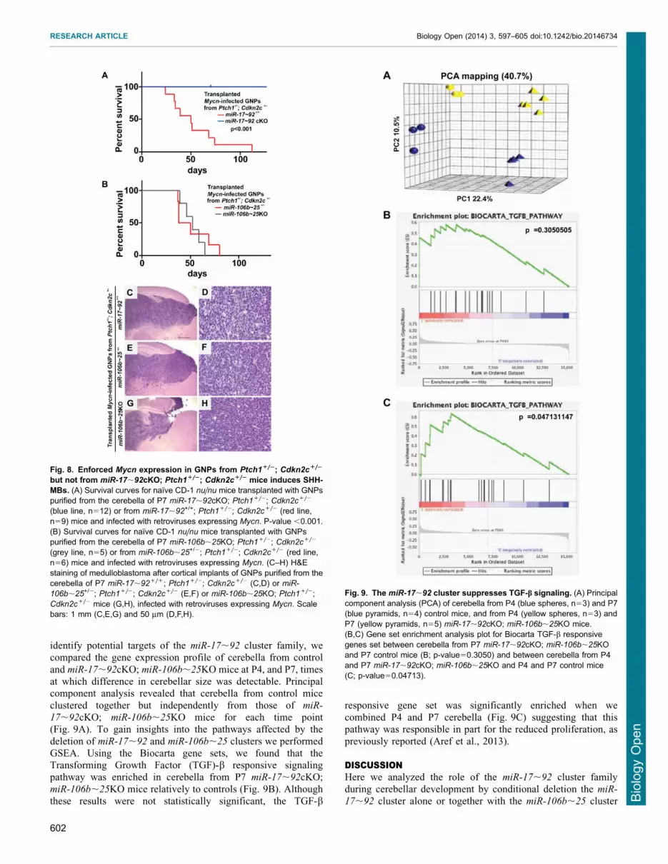
identify potential targets of the miR-17,92 cluster family, wecompared the gene expression profile of cerebella from control
and miR-17,92cKO; miR-106b,25KO mice at P4, and P7, timesat which difference in cerebellar size was detectable. Principalcomponent analysis revealed that cerebella from control mice
clustered together but independently from those of miR-
17,92cKO; miR-106b,25KO mice for each time point(Fig. 9A). To gain insights into the pathways affected by the
deletion of miR-17,92 and miR-106b,25 clusters we performedGSEA. Using the Biocarta gene sets, we found that theTransforming Growth Factor (TGF)-b responsive signalingpathway was enriched in cerebella from P7 miR-17,92cKO;
miR-106b,25KO mice relatively to controls (Fig. 9B). Althoughthese results were not statistically significant, the TGF-b
responsive gene set was significantly enriched when wecombined P4 and P7 cerebella (Fig. 9C) suggesting that thispathway was responsible in part for the reduced proliferation, as
previously reported (Aref et al., 2013).
DISCUSSIONHere we analyzed the role of the miR-17,92 cluster family
during cerebellar development by conditional deletion the miR-
17,92 cluster alone or together with the miR-106b,25 cluster
Fig. 8. Enforced Mycn expression in GNPs from Ptch1+/2; Cdkn2c+/2
but not from miR-17,92cKO; Ptch1+/2; Cdkn2c+/2 mice induces SHH-MBs. (A) Survival curves for naıve CD-1 nu/nu mice transplanted with GNPspurified from the cerebella of P7 miR-17,92cKO; Ptch1+/2; Cdkn2c+/2
(blue line, n512) or from miR-17,92+/+; Ptch1+/2; Cdkn2c+/2 (red line,n59) mice and infected with retroviruses expressing Mycn. P-value ,0.001.(B) Survival curves for naıve CD-1 nu/nu mice transplanted with GNPspurified from the cerebella of P7 miR-106b,25KO; Ptch1+/2; Cdkn2c+/2
(grey line, n55) or from miR-106b,25+/2; Ptch1+/2; Cdkn2c+/2 (red line,n56) mice and infected with retroviruses expressing Mycn. (C–H) H&Estaining of medulloblastoma after cortical implants of GNPs purified from thecerebella of P7 miR-17,92+/+; Ptch1+/2; Cdkn2c+/2 (C,D) or miR-
106b,25+/2; Ptch1+/2; Cdkn2c+/2 (E,F) or miR-106b,25KO; Ptch1+/2;Cdkn2c+/2 mice (G,H), infected with retroviruses expressing Mycn. Scalebars: 1 mm (C,E,G) and 50 mm (D,F,H).
Fig. 9. The miR-17,92 cluster suppresses TGF-b signaling. (A) Principalcomponent analysis (PCA) of cerebella from P4 (blue spheres, n53) and P7(blue pyramids, n54) control mice, and from P4 (yellow spheres, n53) andP7 (yellow pyramids, n55) miR-17,92cKO; miR-106b,25KO mice.(B,C) Gene set enrichment analysis plot for Biocarta TGF-b responsivegenes set between cerebella from P7 miR-17,92cKO; miR-106b,25KOand P7 control mice (B; p-value50.3050) and between cerebella from P4and P7 miR-17,92cKO; miR-106b,25KO and P4 and P7 control mice(C; p-value50.04713).
RESEARCH ARTICLE Biology Open (2014) 3, 597–605 doi:10.1242/bio.20146734
602
BiologyOpen

(miR-17,92cKO; miR-106b,25KO) in neural progenitors.While lack of the miR-106b,25 cluster had no obvious
phenotype, loss of the miR-17,92 cluster induced a reductionin cerebellar size and foliation. Loss of both clusters induced amore severe phenotype. Deletion of the miR-17,92 cluster wassufficient to completely abolish tumor development, pointing to
the absolute requirement for the miR-17,92 cluster, and to thelack of compensation by the miR-106b,25 cluster, for MBdevelopment in collaboration with constitutively activated SHH
signaling.
ThemiR-17,92 andmiR106b,25 clusters control the numberof GNPs during post-natal cerebellar developmentWe previously reported that microRNAs from the miR-106a,363
cluster are not expressed in wild-type cerebella (Uziel et al.,
2009). We found no significant up-regulation of microRNAsencoded by this cluster in cerebella lacking both miR-17,92 andmiR-106b,25 clusters (data not shown). This suggested that themiR-106a,363 cluster is unlikely to contribute to the phenotype
seen in miR-17,92cKO; miR-106b,25KO mice.Co-deletion of the two clusters miR-17,92 and miR-106b,25
led to the exhaustion of progenitor neurons in the cerebellum
resulting in reduction in cerebellar size and the number of folia.The miR-17,92 cluster controls the number of oligodendroglialcells (Budde et al., 2010), renal tubular cells (Patel et al., 2013),
neural stem cells (Bian et al., 2013) and cardiomyocytes (Chenet al., 2013). However, in contrast, the miR-17,92 cluster aloneor in combination with miR-106b,25 is dispensable during
retinal development (Conkrite et al., 2011; Nittner et al., 2012),mammary development (Feuermann et al., 2012) and Langerhanscells (Zhou et al., 2014). Thus, the requirement for microRNAsencoded by the miR-17,92 cluster is cell context specific.
The miR-17,92 and miR-106b,25 clusters are direct targetsof Myc and Mycn (O’Donnell et al., 2005; Northcott et al., 2009;de Pontual et al., 2011). In the central nervous system, the loss
of Mycn in Nestin-positive cells induces a reduced size andmisformed cerebellum (Knoepfler et al., 2002). However, in thesemice, the proliferation of the residual GNPs is driven by the up-
regulation of Myc that normally is not expressed in these neuronalprogenitors (Zindy et al., 2006). Expansion of GNPs in postnatalcerebella of mice deficient for both Mycn and Myc is severelyreduced leading to a cerebellum lacking all folia (Wey et al.,
2010). The fact that the loss of the miR-17,92 and miR-106b,25
clusters attenuated proliferation of GNPs but still caused foliaformation suggests that both clusters are required but not
sufficient to induce the cerebellar phenotype of Mycn; Myc
double-null mice.
The miR-17,92 cluster is absolutely required for SHH-MBdevelopmentMiRs encoded by the miR-17,92 cluster are overexpressed in
both mouse and human tumors (Uziel et al., 2009; Northcottet al., 2009). We previously found that enforced expression of themiR-17,92 cluster collaborates with SHH signaling to induceSHH-MB (Uziel et al., 2009). Strikingly, deletion of the miR-
17,92 cluster in Nestin-positive cells from Ptch1+/2; Cdkn2c+/2
mice completely abolished SHH-MB development. This wasassociated with a significant decrease in the number of 3 week old
Ptch1+/2; Cdkn2c+/2 mice lacking the miR-17,92 cluster thatexhibit PNLs on the surface of their cerebella. Therefore, themiR-17,92 cluster plays a critical role in GNPs during SHH-MB
initiation as was shown in retinoblastoma (Nittner et al., 2012).
Our data also showed that the miR-106b,25 did notcompensate for the loss of the miR-17,92 cluster in tumor
initiation. Analysis of the microRNAs expressed by the twoclusters revealed that miR-19a and miR-19b-1 that share the sameseed sequence are encoded only by the miR-17,92 but not themiR-106b,25 cluster (Fig. 10). This suggests that these two
microRNAs might be sufficient for tumor development and thatthey might recapitulate the oncogenic function of the miR-17,92
cluster, as reported previously for the development of Em-Myc B
cell lymphoma (Mu et al., 2009; Olive et al., 2009). Treatment ofSHH-MB with tiny LNAs directed against miR-17/20a/106b/93
and miR-19a/b-1 from the miR-17,92 cluster family inhibits
proliferation of SHH-MB in vitro and in vivo (Murphy et al.,2013). Therefore the miR-19a/b-1 might not be the onlymicroRNA required for MB progression, although this will
need further evaluation.Given the absolute requirement of the miR-17,92 cluster for
Ptch1+/2 induced SHH-MB formation, the identification of bonefide miR-17,92 targets is clearly warranted. Analysis of
previously published targets (Brock et al., 2009; Concepcionet al., 2012; Mogilyansky and Rigoutsos, 2013; Sun et al., 2013)revealed that Bmpr2, but not PTEN and p21Cip1, is regulated by
the miR-17,92 cluster (Murphy et al., 2013). We found thatBmpr2 was upregulated in the cerebella of miR-17,92cKO; miR-
106b,25KO mice, which might be responsible, in part, for
the cerebellar anomalies. Constitutive activation of Bmpr1a incerebella induced a simplified foliation pattern (Ming et al.,2002). These data are consistent with ours and others findings
showing that BMP signaling antagonizes the SHH pathway(Rios et al., 2004) and that genes in the BMP pathway aredownregulated in SHH-MB (Zhao et al., 2008). It has beenpreviously reported that the miR-17,92 cluster down-regulates
multiple components of the TGF-b pathway (Tagawa et al., 2007;Petrocca et al., 2008; Dews et al., 2010; Mestdagh et al., 2010;Concepion et al., 2012; Li et al., 2012; Mogilyansky and
Fig. 10. Schematic representation of the miR-17,92 cluster family andits regulation by SHH signaling. SHH signaling induces the transcription ofMycn, which, in turn, induces the expression of the miR-17,92 and miR-
106b,25 clusters that encode 6 and 3 microRNAs, respectively. MicroRNAssharing the same seed sequence are represented by boxes of the samecolor. X and Y represent putative targets. Loss of the miR-17,92 but not themiR-106b,25 cluster inhibits Ptch1+/2-induced SHH-MBs. Loss of bothclusters reduces cerebellar size.
RESEARCH ARTICLE Biology Open (2014) 3, 597–605 doi:10.1242/bio.20146734
603
BiologyOpen

Rigoutsos, 2013). In agreement with these reports, we found, byGSEA analysis, that the TGF-b pathway was upregulated in the
cerebella of miR-17,92cKO; miR-106b,25KO mice. TGF-bpathway is associated with SHH-MB pathogenesis with highlevels connoting a good prognosis (Aref et al., 2013). Our resultsare in agreement with a study suggesting that miR-17,92 cluster
downregulates two different signaling pathways, Bmp and TGF-bpathways (Brock et al., 2009). Further analysis using Clip-Seqapproaches will be required to identify the bona fide miR-17,92
targets in mouse and human MBs (Darnell, 2010).In summary, while the miR-17,92 and miR-106b,25 clusters
are essential for cerebellum development and homeostasis, the
miR-17,92, but not the miR-106b,25, cluster is required fortumor initiation in Ptch1+/2 mice.
AcknowledgementsWe are indebted to Tyler Jacks for graciously providing the conditional miR-
17,92 mice as well as the miR-106b,25-null animals. We thank ShellyWilkerson, Sarah Robinson, Dana Farmer and Jose Grenet for mousegenotyping, immunohistochemistry experiments, RNA extraction and mousecolony management. We thank Melissa Johnson and Shantel Brown for help withorthotopic transplants, Jerold Rehg for pathology analysis, Brandon Cox forperforming Rotarod testing, and David Finkelstein and Marie Morfouace formicroarray analysis. We also thank Charles J. Sherr, David Solecki, BrandonWainwright and all members of the laboratory for helpful discussions during thecourse of these experiments.
Competing interestsThe authors have no competing interests to declare.
Author contributionsM.F.R., F.Z. and A.V. developed concepts and approaches; F.Z., D.K., Y.L., O.A.and L.B.M. performed experiments; F.Z., D.K. and M.F.R. performed dataanalysis; M.F.R. and F.Z. wrote the manuscript; M.F.R., P.J.M. and A.V. edited themanuscript; all authors reviewed the manuscript.
FundingThis work was supported in part by National Institutes of Health Grant CA-096832(to M.F.R.), Core Grant CA-21765 (to M.F.R.), and the American Lebanese-SyrianAssociated Charities (ALSAC) of St Jude Children’s Research Hospital.
ReferencesAref, D., Moffatt, C. J., Agnihotri, S., Ramaswamy, V., Dubuc, A. M., Northcott,P. A., Taylor, M. D., Perry, A., Olson, J. M., Eberhart, C. G. et al. (2013).Canonical TGF-b pathway activity is a predictor of SHH-driven medulloblastomasurvival and delineates putative precursors in cerebellar development. BrainPathol. 23, 178-191.
Bian, S., Hong, J., Li, Q., Schebelle, L., Pollock, A., Knauss, J. L., Garg, V. andSun, T. (2013). MicroRNA cluster miR-17-92 regulates neural stem cellexpansion and transition to intermediate progenitors in the developing mouseneocortex. Cell Rep. 3, 1398-1406.
Brock, M., Trenkmann, M., Gay, R. E., Michel, B. A., Gay, S., Fischler, M.,Ulrich, S., Speich, R. and Huber, L. C. (2009). Interleukin-6 modulates theexpression of the bone morphogenic protein receptor type II through a novelSTAT3-microRNA cluster 17/92 pathway. Circ. Res. 104, 1184-1191.
Budde, H., Schmitt, S., Fitzner, D., Opitz, L., Salinas-Riester, G. and Simons,M. (2010). Control of oligodendroglial cell number by the miR-17-92 cluster.Development 137, 2127-2132.
Carmell, M. A. and Hannon, G. J. (2004). RNase III enzymes and the initiation ofgene silencing. Nat. Struct. Mol. Biol. 11, 214-218.
Chen, J., Huang, Z. P., Seok, H. Y., Ding, J., Kataoka, M., Zhang, Z., Hu, X.,Wang, G., Lin, Z., Wang, S. et al. (2013). mir-17-92 cluster is required for andsufficient to induce cardiomyocyte proliferation in postnatal and adult hearts.Circ. Res. 112, 1557-1566.
Concepcion, C. P., Bonetti, C. and Ventura, A. (2012). The microRNA-17-92family of microRNA clusters in development and disease. Cancer J. 18, 262-267.
Conkrite, K., Sundby, M., Mukai, S., Thomson, J. M., Mu, D., Hammond, S. M.and MacPherson, D. (2011). miR-17,92 cooperates with RB pathwaymutations to promote retinoblastoma. Genes Dev. 25, 1734-1745.
Darnell, R. B. (2010). HITS-CLIP: panoramic views of protein-RNA regulation inliving cells. Wiley Interdiscip Rev RNA 1, 266-286.
de Pontual, L., Yao, E., Callier, P., Faivre, L., Drouin, V., Cariou, S., VanHaeringen, A., Genevieve, D., Goldenberg, A., Oufadem, M. et al. (2011).Germline deletion of the miR-17,92 cluster causes skeletal and growth defectsin humans. Nat. Genet. 43, 1026-1030.
Dews, M., Fox, J. L., Hultine, S., Sundaram, P., Wang, W., Liu, Y. Y., Furth, E.,Enders, G. H., El-Deiry, W., Schelter, J. M. et al. (2010). The myc-miR-17,92axis blunts TGFbeta signaling and production of multiple TGFbeta-dependentantiangiogenic factors. Cancer Res. 70, 8233-8246.
Feuermann, Y., Robinson, G. W., Zhu, B. M., Kang, K., Raviv, N., Yamaji, D.and Hennighausen, L. (2012). The miR-17/92 cluster is targeted by STAT5 butdispensable for mammary development. Genesis 50, 665-671.
Goodrich, L. V., Milenkovic, L., Higgins, K. M. and Scott, M. P. (1997). Alteredneural cell fates and medulloblastoma in mouse patched mutants. Science 277,1109-1113.
Hatten, M. E. and Roussel, M. F. (2011). Development and cancer of thecerebellum. Trends Neurosci. 34, 134-142.
Hatton, B. A., Knoepfler, P. S., Kenney, A. M., Rowitch, D. H., de Alboran, I. M.,Olson, J. M. and Eisenman, R. N. (2006). N-myc is an essential downstreameffector of Shh signaling during both normal and neoplastic cerebellar growth.Cancer Res. 66, 8655-8661.
Kawauchi, D., Robinson, G., Uziel, T., Gibson, P., Rehg, J., Gao, C.,Finkelstein, D., Qu, C., Pounds, S., Ellison, D. W. et al. (2012). A mousemodel of the most aggressive subgroup of human medulloblastoma. CancerCell 21, 168-180.
Kenney, A. M., Cole, M. D. and Rowitch, D. H. (2003). Nmyc upregulation bysonic hedgehog signaling promotes proliferation in developing cerebellargranule neuron precursors. Development 130, 15-28.
Kim, V. N. (2005). MicroRNA biogenesis: coordinated cropping and dicing. Nat.Rev. Mol. Cell Biol. 6, 376-385.
Kim, J. Y., Nelson, A. L., Algon, S. A., Graves, O., Sturla, L. M., Goumnerova,L. C., Rowitch, D. H., Segal, R. A. and Pomeroy, S. L. (2003).Medulloblastoma tumorigenesis diverges from cerebellar granule celldifferentiation in patched heterozygous mice. Dev. Biol. 263, 50-66.
Knoepfler, P. S., Cheng, P. F. and Eisenman, R. N. (2002). N-myc is essentialduring neurogenesis for the rapid expansion of progenitor cell populations andthe inhibition of neuronal differentiation. Genes Dev. 16, 2699-2712.
Lauder, J. M., Altman, J. and Krebs, H. (1974). Some mechanisms of cerebellarfoliation: effects of early hypo- and hyperthyroidism. Brain Res. 76, 33-40.
Li, L., Shi, J. Y., Zhu, G. Q. and Shi, B. (2012). MiR-17-92 cluster regulates cellproliferation and collagen synthesis by targeting TGFB pathway in mousepalatal mesenchymal cells. J. Cell. Biochem. 113, 1235-1244.
Mestdagh, P., Bostrom, A. K., Impens, F., Fredlund, E., Van Peer, G., DeAntonellis, P., von Stedingk, K., Ghesquiere, B., Schulte, S., Dews, M. et al.(2010). The miR-17-92 microRNA cluster regulates multiple components of theTGF-b pathway in neuroblastoma. Mol. Cell 40, 762-773.
Ming, J. E., Elkan, M., Tang, K. and Golden, J. A. (2002). Type I bonemorphogenetic protein receptors are expressed on cerebellar granular neuronsand a constitutively active form of the type IA receptor induces cerebellarabnormalities. Neuroscience 114, 849-857.
Mogilyansky, E. and Rigoutsos, I. (2013). The miR-17/92 cluster: acomprehensive update on its genomics, genetics, functions and increasinglyimportant and numerous roles in health and disease. Cell Death Differ. 20,1603-1614.
Mu, P., Han, Y. C., Betel, D., Yao, E., Squatrito, M., Ogrodowski, P., deStanchina, E., D’Andrea, A., Sander, C. and Ventura, A. (2009). Geneticdissection of the miR-17,92 cluster of microRNAs in Myc-induced B-celllymphomas. Genes Dev. 23, 2806-2811.
Murphy, B. L., Obad, S., Bihannic, L., Ayrault, O., Zindy, F., Kauppinen, S. andRoussel, M. F. (2013). Silencing of the miR-17,92 cluster family inhibitsmedulloblastoma progression. Cancer Res. 73, 7068-7078.
Nittner, D., Lambertz, I., Clermont, F., Mestdagh, P., Kohler, C., Nielsen, S. J.,Jochemsen, A., Speleman, F., Vandesompele, J., Dyer, M. A. et al. (2012).Synthetic lethality between Rb, p53 and Dicer or miR-17-92 in retinalprogenitors suppresses retinoblastoma formation. Nat. Cell Biol. 14, 958-965.
Northcott, P. A., Fernandez-L, A., Hagan, J. P., Ellison, D. W., Grajkowska, W.,Gillespie, Y., Grundy, R., Van Meter, T., Rutka, J. T., Croce, C. M. et al. (2009).The miR-17/92 polycistron is up-regulated in sonic hedgehog-drivenmedulloblastomas and induced by N-myc in sonic hedgehog-treatedcerebellar neural precursors. Cancer Res. 69, 3249-3255.
O’Donnell, K. A., Wentzel, E. A., Zeller, K. I., Dang, C. V. and Mendell, J. T.(2005). c-Myc-regulated microRNAs modulate E2F1 expression. Nature 435,839-843.
Olive, V., Bennett, M. J., Walker, J. C., Ma, C., Jiang, I., Cordon-Cardo, C., Li,Q. J., Lowe, S. W., Hannon, G. J. and He, L. (2009). miR-19 is a key oncogeniccomponent of mir-17-92. Genes Dev. 23, 2839-2849.
Oliver, T. G., Read, T. A., Kessler, J. D., Mehmeti, A., Wells, J. F., Huynh, T. T.,Lin, S. M. and Wechsler-Reya, R. J. (2005). Loss of patched and disruption ofgranule cell development in a pre-neoplastic stage of medulloblastoma.Development 132, 2425-2439.
Patel, V., Williams, D., Hajarnis, S., Hunter, R., Pontoglio, M., Somlo, S. andIgarashi, P. (2013). miR-17,92 miRNA cluster promotes kidney cyst growth inpolycystic kidney disease. Proc. Natl. Acad. Sci. USA 110, 10765-10770.
Petrocca, F., Vecchione, A. and Croce, C. M. (2008). Emerging role of miR-106b-25/miR-17-92 clusters in the control of transforming growth factor betasignaling. Cancer Res. 68, 8191-8194.
Rios, I., Alvarez-Rodrıguez, R., Martı, E. and Pons, S. (2004). Bmp2antagonizes sonic hedgehog-mediated proliferation of cerebellar granuleneurones through Smad5 signalling. Development 131, 3159-3168.
RESEARCH ARTICLE Biology Open (2014) 3, 597–605 doi:10.1242/bio.20146734
604
BiologyOpen

Sun, Q., Mao, S., Li, H., Zen, K., Zhang, C. Y. and Li, L. (2013). Role of miR-17family in the negative feedback loop of bone morphogenetic protein signaling inneuron. PLoS ONE 8, e83067.
Tagawa, H., Karube, K., Tsuzuki, S., Ohshima, K. and Seto, M. (2007).Synergistic action of the microRNA-17 polycistron and Myc in aggressive cancerdevelopment. Cancer Sci. 98, 1482-1490.
Taylor, M. D., Northcott, P. A., Korshunov, A., Remke, M., Cho, Y. J., Clifford,S. C., Eberhart, C. G., Parsons, D. W., Rutkowski, S., Gajjar, A. et al. (2012).Molecular subgroups of medulloblastoma: the current consensus. ActaNeuropathol. 123, 465-472.
Tronche, F., Kellendonk, C., Kretz, O., Gass, P., Anlag, K., Orban, P. C., Bock,R., Klein, R. and Schutz, G. (1999). Disruption of the glucocorticoid receptorgene in the nervous system results in reduced anxiety. Nat. Genet. 23, 99-103.
Uziel, T., Zindy, F., Xie, S., Lee, Y., Forget, A., Magdaleno, S., Rehg, J. E.,Calabrese, C., Solecki, D., Eberhart, C. G. et al. (2005). The tumorsuppressors Ink4c and p53 collaborate independently with Patched tosuppress medulloblastoma formation. Genes Dev. 19, 2656-2667.
Uziel, T., Karginov, F. V., Xie, S., Parker, J. S., Wang, Y. D., Gajjar, A., He, L.,Ellison, D., Gilbertson, R. J., Hannon, G. et al. (2009). The miR-17,92 clustercollaborates with the Sonic Hedgehog pathway in medulloblastoma. Proc. Natl.Acad. Sci. USA 106, 2812-2817.
Ventura, A., Young, A. G., Winslow, M. M., Lintault, L., Meissner, A., Erkeland,S. J., Newman, J., Bronson, R. T., Crowley, D., Stone, J. R. et al. (2008).Targeted deletion reveals essential and overlapping functions of the miR-17through 92 family of miRNA clusters. Cell 132, 875-886.
Wey, A., Martinez Cerdeno, V., Pleasure, D. and Knoepfler, P. S. (2010). c- andN-myc regulate neural precursor cell fate, cell cycle, and metabolism to directcerebellar development. Cerebellum 9, 537-547.
Zhao, H., Ayrault, O., Zindy, F., Kim, J. H. and Roussel, M. F. (2008). Post-transcriptional down-regulation of Atoh1/Math1 by bone morphogenic proteinssuppresses medulloblastoma development. Genes Dev. 22, 722-727.
Zhou, L., Qi, R. Q., Liu, M., Xu, Y. P., Li, G., Weiland, M., Kaplan, D. H. and Mi,Q. S. (2014). microRNA miR-17-92 cluster is highly expressed in epidermalLangerhans cells but not required for its development. Genes Immun. 15, 57-61.
Zindy, F., Knoepfler, P. S., Xie, S., Sherr, C. J., Eisenman, R. N. and Roussel,M. F. (2006). N-Myc and the cyclin-dependent kinase inhibitors p18Ink4c andp27Kip1 coordinately regulate cerebellar development. Proc. Natl. Acad. Sci.USA 103, 11579-11583.
Zindy, F., Uziel, T., Ayrault, O., Calabrese, C., Valentine, M., Rehg, J. E.,Gilbertson, R. J., Sherr, C. J. and Roussel, M. F. (2007). Genetic alterations inmouse medulloblastomas and generation of tumors de novo from primarycerebellar granule neuron precursors. Cancer Res. 67, 2676-2684.
RESEARCH ARTICLE Biology Open (2014) 3, 597–605 doi:10.1242/bio.20146734
605
BiologyOpen