The continuum seabirds - humans Elisabet Moré Mir
280
ADVERTIMENT. Lʼaccés als continguts dʼaquesta tesi queda condicionat a lʼacceptació de les condicions dʼús establertes per la següent llicència Creative Commons: http://cat.creativecommons.org/?page_id=184 ADVERTENCIA. El acceso a los contenidos de esta tesis queda condicionado a la aceptación de las condiciones de uso establecidas por la siguiente licencia Creative Commons: http://es.creativecommons.org/blog/licencias/ WARNING. The access to the contents of this doctoral thesis it is limited to the acceptance of the use conditions set by the following Creative Commons license: https://creativecommons.org/licenses/?lang=en
-
Upload
khangminh22 -
Category
Documents
-
view
1 -
download
0
Transcript of The continuum seabirds - humans Elisabet Moré Mir

ADVERTIMENT. Lʼaccés als continguts dʼaquesta tesi queda condicionat a lʼacceptació de les condicions dʼúsestablertes per la següent llicència Creative Commons: http://cat.creativecommons.org/?page_id=184
ADVERTENCIA. El acceso a los contenidos de esta tesis queda condicionado a la aceptación de las condiciones de usoestablecidas por la siguiente licencia Creative Commons: http://es.creativecommons.org/blog/licencias/
WARNING. The access to the contents of this doctoral thesis it is limited to the acceptance of the use conditions setby the following Creative Commons license: https://creativecommons.org/licenses/?lang=en

A One Health approach into the epidemiology
of Campylobacter and Salmonella:
The continuum seabirds - humans
Elisabet Moré Mir
PhD Thesis
Bellaterra, 2018


A One Health approach into the epidemiology of Campylobacter
and Salmonella: the continuum seabirds - humans
Tesi doctoral presentada per na Elisabet Moré Mir per optar al grau de Doctora en el
marc del programa de Doctorat de Medicina i Sanitat Animals de la Facultat de
Veterinària de la Universitat Autònoma de Barcelona, sota la direcció de la Dra. Marta
Cerdà Cuéllar i la tutoria del Dr. Joaquim Segalés Coma.
Bellaterra, 2018
Dra. Marta Cerdà Cuéllar Dr. Joaquim Segalés Coma Elisabet Moré Mir
Directora Tutor Doctoranda

La Dra. Marta Cerdà Cuéllar, investigadora del Centre de Recerca en Sanitat Animal de
l’Institut de Recerca i Tecnologia Agroalimentàries (CReSA-IRTA), i el Dr. Joaquim
Segalés Coma, professor titular del Departament de Sanitat i d’Anatomia Animals de la
Facultat de Veterinària de la Universitat Autònoma de Barcelona i investigador adscrit
al CReSA-IRTA,
Informen:
Que la memòria titulada “A One Health approach into the epidemiology of
Campylobacter and Salmonella: the continuum seabirds - humans” presentada per na
Elisabet Moré Mir per a l’obtenció del grau de Doctora en Medicina i Sanitat Animal,
s’ha realitzat sota la seva direcció i supervisió i, considerant-la acabada, n’autoritzen la
seva presentació per tal de ser avaluada per la comissió corresponent.
I per tal que consti als efectes oportuns, signen el present declaració a Bellaterra
(Barcelona), a 20 de febrer de 2018.
Dra. Marta Cerdà Cuéllar Dr. Joaquim Segalés Coma
Directora Tutor
PhD studies presented by Elisabet Moré Mir were financially supported by the FI-DGR
Pre-Doctoral grant from the Catalan Governmment (Agència de Gestió d’Ajuts
Universitaris i de Recerca - AGAUR), reference: 2015 FI_B 00620, 2016 FI_B1 00168 and
2017 FI_B2 00080.
Printing of this thesis was financed by the Institut de Recerca i Tecnologia
Agroalimentàries (IRTA).
Cover design: Pau Bertran Grífols

Al meu avi,
l’home més bo que he conegut mai


Agraïments
Miro enrere després d’aquest llarg camí que m’ha portat fins aquí i me n’adono que hi
ha moltíssimes persones que m’han acompanyat i que gràcies a la seva ajuda han fet
possible aquesta tesis. És per això, que vull agrair:
En primer lloc a la meva directora Marta C., la teva confiança en mi per dur a terme
aquest projecte, haver-me guiat durant tots aquests anys i ensenyar-me com funciona
el món de la ciència. També a tots els que heu participat directament en els estudis de
la tesis ja sigui en els mostrejos, en el laboratori o en l’elaboració dels articles.
A tot el personal del CReSA, el bon ambient que s’hi respira, per fer que la feina del dia
a dia sigui més divertida i que les hores passin més despresa. Sobretot vull donar les
gràcies als tècnics, per la vostra paciència i tot el que m’heu ensenyat. En especial a la
Marta P., per la teva amistat i suport, i per tots els km fets juntes.
Als meus companys becaris/doctors, els grans moments que hem passat junts al
“despachito” i fora de la feina. Hem rigut molt i, sens dubte, heu sigut la millor
companyia que podria haver tingut. Gràcies per la vostra amistat. També vull agrair a la
Noelia A. els seus bons consells i haver-me aplanat el camí. Al meu “Compañerito”,
haver-me fet riure i enrabiar a parts iguals, els viatges junts i els “asaditos”. Moltes
gràcies també a la gent del SEFAS per haver-me acollit a Berlin com a una més.
Per últim, però no menys important, vull donar les gràcies a la meva família i als meus
amics per tot el suport que m’heu donat. Per estar sempre al meu costat, per animar-
me, per escoltar els meus rotllos de la tesis o evitar parlar-ne quan ha fet falta. I
especialment, a la meva germana Carla per ajudar-me amb l’anglès i a en Pau B. per
ensenyar-me a editar imatges.
Sense tots vosaltres no hauria pogut fer aquesta tesis. Tots hi heu contribuït d’una
manera o altra i, per tant, també és una mica vostra. Moltes gràcies de tot cor!

I
Table of contents
Figures and tables index .................................................................................................. V
List of abbreviations ........................................................................................................ IX
Summary ......................................................................................................................... XI
Resum .......................................................................................................................... XIV
Publications ................................................................................................................. XVII
CHAPTER 1
General Introduction ........................................................................................................ 1
1.1. ZOONOSES ......................................................................................................... 3
1.2. CAMPYLOBACTER .............................................................................................. 4
1.2.1. Discovery and taxonomy ............................................................................ 4
1.2.2. General characteristics ............................................................................... 8
1.2.3. Detection, isolation and confirmation ....................................................... 9
1.2.4. Clinical manifestations ............................................................................. 10
1.2.5. Epidemiology ............................................................................................ 11
1.2.6. Pathogenesis ............................................................................................ 13
1.2.6.1. Virulence factors ............................................................................... 16
1.3. SALMONELLA ................................................................................................... 18
1.3.1. Discovery and taxonomy .......................................................................... 18
1.3.2. General characteristics ............................................................................. 21
1.3.3. Detection, isolation and confirmation ..................................................... 21
1.3.4. Clinical manifestations ............................................................................. 23
1.3.5. Epidemiology ............................................................................................ 25
1.3.6. Pathogenesis ............................................................................................ 28
1.3.6.1. Virulence factors ............................................................................... 32
1.4. ANTIMICROBIAL RESISTANCE .......................................................................... 35
1.4.1. Campylobacter antimicrobial resistance .................................................. 36

II
1.4.2. Salmonella antimicrobial resistance .........................................................38
1.5. TYPING METHODS ...........................................................................................39
1.5.1. Enterobacterial repetitive intergenic consensus PCR ..............................40
1.5.2. PCR-Restriction fragment length polymorphism ......................................41
1.5.3. Pulsed-field gel electrophoresis ...............................................................41
1.5.4. Multilocus sequence typing ......................................................................42
1.5.5. Future prospects .......................................................................................44
1.6. ONE HEALTH ....................................................................................................44
1.7. WILD BIRDS AS RESERVOIRS OF ZOONOTIC BACTERIA ...................................47
CHAPTER 2
Objectives .......................................................................................................................51
CHAPTER 3
Study I: Seabirds (Laridae) as a source of Campylobacter spp., Salmonella spp. and
antimicrobial resistance in South Africa .........................................................................55
3.1. SUMMARY .......................................................................................................57
3.2. INTRODUCTION ...............................................................................................58
3.3. MATERIALS AND METHODS .............................................................................60
3.3.1. Sampling ...................................................................................................60
3.3.2. Campylobacter isolation and identification .............................................61
3.3.3. Salmonella isolation and identification ....................................................63
3.3.4. Molecular typing of the isolates ...............................................................63
3.3.4.1. flaA-RFLP ............................................................................................63
3.3.4.2. ERIC-PCR ............................................................................................64
3.3.4.3. PFGE ...................................................................................................64
3.3.5. Analysis and comparison of band patterns ..............................................65
3.3.6. Antimicrobial susceptibility testing ..........................................................65
3.3.7. Statistical analysis .....................................................................................66
3.4. RESULTS ...........................................................................................................67
3.4.1. Campylobacter and Salmonella occurrence .............................................67
III
3.4.2. Genetic diversity ....................................................................................... 68
3.4.3. Antimicrobial susceptibility ...................................................................... 70
3.5. DISCUSSION ..................................................................................................... 79
CHAPTER 4
Study II: Humans spread zoonotic enteric bacteria in Antarctica? ................................ 85
4.1. SUMMARY ....................................................................................................... 87
4.2. INTRODUCTION ............................................................................................... 88
4.3. MATERIAL AND METHODS .............................................................................. 89
4.3.1. Sampling ................................................................................................... 89
4.3.2. Bacterial isolation and identification........................................................ 90
4.3.3. Antimicrobial susceptibility testing .......................................................... 90
4.3.4. Salmonella and Campylobacter genotyping ............................................. 91
4.4. RESULTS ........................................................................................................... 96
4.4.1. Salmonella and Campylobacter spp. in seabirds ...................................... 96
4.4.2. Antimicrobial resistance ........................................................................... 96
4.4.3. Genetic diversity ....................................................................................... 97
4.5. DISCUSSION ................................................................................................... 100
CHAPTER 5
Study III: Genetic diversity, population structure and virulence potential of
Campylobacter and Salmonella spp. from Southern Ocean seabirds .......................... 105
5.1. SUMMARY ..................................................................................................... 107
5.2. INTRODUCTION ............................................................................................. 108
5.3. MATERIALS AND METHODS........................................................................... 110
5.3.1. Bacterial isolates..................................................................................... 110
5.3.2. Genotyping ............................................................................................. 111
5.3.3. Virulence-associated genes .................................................................... 112
5.4. RESULTS ......................................................................................................... 117
5.4.1. Genetic diversity and population structure ........................................... 117
5.4.2. Virulence-associated genes .................................................................... 120
IV
5.5. DISCUSSION ...................................................................................................133
CHAPTER 6
Study IV: Molecular comparative analysis of nontyphoidal Salmonella isolates from
humans, poultry and seagulls in Southwestern Europe ..............................................141
6.1. SUMMARY .....................................................................................................143
6.2. INTRODUCTION .............................................................................................144
6.3. MATERIALS AND METHODS ...........................................................................147
6.3.1. Bacterial isolates .........................................................................................147
6.3.2. PFGE ........................................................................................................151
6.3.3. MLST .......................................................................................................151
6.3.4. Virulence-associated genes ....................................................................152
6.3.5. Statistical analyses ..................................................................................152
6.4. RESULTS .........................................................................................................157
6.4.1. PFGE ........................................................................................................157
6.4.2. Multilocus sequence typing ....................................................................159
6.4.3. Virulence genes ......................................................................................163
6.5. DISCUSSION ...................................................................................................166
CHAPTER 7
General Discussion .......................................................................................................175
CHAPTER 8
Conclusions ...................................................................................................................185
REFERENCES .................................................................................................................189
ANNEX
Guide of studied wild birds ...........................................................................................237

V
Figures and tables index
Figures
Figure 1.1. Reported numbers and notification rates of confirmed human
zoonoses in the EU, 2016 ............................................................................................... 4
Figure 1.2. Pathogenesis of C. jejuni in human and chicken ......................................... 15
Figure 1.3. Taxonomic classification of Salmonella genus, sources and associated
diseases ......................................................................................................................... 20
Figure 1.4. The global burden of foodborne disease by regions (DALYs per
100,000 population) caused by enteric hazards, 2010 ................................................. 26
Figure 1.5. Distribution of the 10 most frequently reported Salmonella serovars
in humans in the EU in 2016 ......................................................................................... 27
Figure 1.6. Pathogenesis of nontyphoidal Salmonella (NTS) serovars and S. Typhi
in humans ...................................................................................................................... 31
Figure 1.7. Sources and transmission of Campylobacter and Salmonella .................... 45
Figure 3.1. Map locations of the sampled seabird colonies in Western Cape
(South Africa) ................................................................................................................. 62
Figure 3.2. PFGE combined dendrogram of SmaI and KpnI patterns of C. jejuni
isolates ........................................................................................................................... 75
Figure 4.1. Salmonella and Campylobacter findings in the Southern Ocean................ 92
Figure 4.2. PFGE combined dendrogram of SmaI and KpnI profiles of C. jejuni
isolates from wild and domestic birds .......................................................................... 98
Figure 4.3. PFGE combined dendrogram of SmaI and KpnI profiles of C. lari
isolates from wild and domestic birds .......................................................................... 99
Figure 5.1. Locations and host origin of Campylobacter and Salmonella isolates
from birds sampled in the Southern Ocean ................................................................ 114
VI
Figure 5.2. Combined dendrogram of SmaI and KpnI PFGE profiles of C. lari
isolates ......................................................................................................................... 123
Figure 5.3. Combined dendrogram of SmaI and KpnI PFGE profiles of C. jejuni
and C. coli isolates ....................................................................................................... 125
Figure 5.4. Maximum likelihood tree based on concatenated MLST loci of
Salmonella isolates and presence of virulence-associated genes ............................... 127
Figure 5.5. Minimum spanning tree showing the distribution of C. lari isolates
according to bird species and source of isolation ....................................................... 128
Figure 5.6. Minimum spanning tree showing the distribution of C. jejuni and C.
coli isolates according to bird species and source of isolation.................................... 129
Figure 6.1. Origin of Salmonella isolates from different hosts included in this
study ............................................................................................................................ 150
Figure 6.2. Combined dendrogram with XbaI and BlnI profiles of a selection of
Salmonella isolates from different hosts showing the same or highly similar
pulsotypes ................................................................................................................... 159
Figure 6.3. Salmonella isolates from different hosts with XbaI and BlnI PFGE
pulsotypes (XB) in common ......................................................................................... 162
Tables
Table 1.1. Described and validated Campylobacter species, their respective
sources and human-associated diseases ........................................................................ 4
Table 1.2. Most relevant Campylobacter virulence factors ........................................... 17
Table 1.3. Most relevant Salmonella virulence factors .................................................. 33
Table 3.1. Frequency of Campylobacter species and Salmonella subspecies and
serovars in kelp gull and greater crested tern chicks in the five colonies sampled
in the Western Cape, South Africa ................................................................................. 72

VII
Table 3.2. Salmonella pulsotypes and serovars from seabirds and their
relationship with clinical isolates ................................................................................... 76
Table 3.3. Antimicrobial resistance of Salmonella isolates according to the
seabird species and colony ............................................................................................. 78
Table 4.1. Sampled birds at the four studied localities and Campylobacter
occurrence ...................................................................................................................... 94
Table 5.1. PCR primers used for Campylobacter and Salmonella virulence-
associated gene detection........................................................................................... 115
Table 5.2. Occurrence of virulence-associated genes in C. jejuni and C. coli
isolates from different sources ................................................................................... 130
Table 5.3. Occurrence of virulence-associated genes in Salmonella isolates of
different subspecies and serovars ............................................................................... 131
Table 6.1. Number of Salmonella isolates of nontyphoidal serovars included in
the study and originating from different hosts and localities ..................................... 148
Table 6.2. Salmonella isolates analysed by PFGE with primary and secondary
enzymes (XbaI and BlnI) and the resulting pulsotypes ............................................... 153
Table 6.3. PCR primers used for Salmonella virulence-associated genes
detection ..................................................................................................................... 155

IX
List of abbreviations
AMR Antimicrobial resistance
API Analytical Profile Index
CC Clonal complex
CFU Colony-forming units
cgMLST Core-genome multilocus sequence typing
CPS Capsular polysaccharide
DNA Deoxyribonucleic acid
DR Direct repeats
eBG eBurst group
ECDC European Centre for Disease Prevention and Control
EEA European Economic Area
EFSA European Food Safety Authority
ERIC Enterobacterial repetitive intergenic consensus
EU European Union
FISH Fluorescence in situ hybridization
GALT Gut-associated lymphoid tissue
GBS Guillain-Barré syndrome
HIV Human immunodeficiency virus
iNTS Invasive nontyphoidal Salmonella
IS Insertion sequences
ISO International Organization for Standardization
LPS Lipopolysaccharide
LPSN List of prokaryotic names with standing in nomenclature
MCP Methyl-accepting chemotaxis protein
MDR Multidrug resistance
MLEE Multilocus enzyme electrophoresis
MLST Multilocus sequence typing
X
MPS Mononuclear phagocyte system
MUCAP 4-methylumbelliferyl caprylate
NGS Next-generation sequencing
NTS Nontyphoidal Salmonella
PCR Polymerase chain reaction
PFGE Pulsed-field gel electrophoresis
PMNL Polymorphonuclear leukocytes
qPCR Quantitative polymerase chain reaction
RFLP Restriction fragment length polymorphism
rMLST Ribosomal MLST
rRNA Ribosomal ribonucleic acid
SCV Salmonella-containing vacuole
SNP Single nucleotide polymorphism
SPI Salmonella pathogenicity island
ST Sequence type
SVR Short variable region
TCRS Two-component regulatory system
T3SS Type III secretion system
T4SS Type IV secretion system
T6SS Type VI secretion system
UK United Kingdom
USA United States of America
VBNC Viable but non-culturable
WHO World Health Organization
WHOCC-Salm WHO Collaborating Centre for Reference and Research on
Salmonella
wgMLST Whole genome multilocus sequence typing
WGS Whole genome sequencing
XI
Summary
Zoonotic thermophilic Campylobacter spp. and nontyphoidal Salmonella enterica are a
major cause of foodborne human gastroenteritis worldwide. Both bacteria are able to
infect a broad range of domestic and wild animals. A wide variety of wild birds,
especially gulls, have been reported as asymptomatic carriers of these zoonotic agents
in Europe, America and Australia. However, there is scarce information about these
reservoirs in Africa and remote regions of the Southern Ocean, and the role of wild birds
in the epidemiology of these pathogens is not fully understood. Thus, within the
framework of this PhD thesis we have investigated the occurrence, antimicrobial
susceptibility, virulence potential and population structure or genetic diversity of
Campylobacter and Salmonella spp. in seabird species along the western coast of South
Africa (near the Benguela Upwelling Region) and across the Antarctic and Subantarctic
region. We have also analysed the genetic relation and virulence potential of isolates of
Salmonella serovars from seabirds, poultry and humans, to assess whether common
strains are circulating among different niches in Southwestern Europe.
In Western Cape (South Africa), we detected thermophilic Campylobacter spp., mainly
C. jejuni, in kelp gulls and greater crested terns with similar prevalences. Most C. jejuni
sequence types (ST)s belonged to the clonal complex (CC)-1275, which is mainly related
to aquatic environments and wild birds. On the contrary, a higher occurrence of
Salmonella was observed in kelp gulls than in greater crested terns, which seems to be
related to the scavenging feeding habits of the former. Anatum, Enteritidis and Hadar
were the most frequent Salmonella serovars, although a great diversity of other
zoonotic serovars were found, especially in gull colonies near urban areas. The same or
highly similar pulsed-field gel electrophoresis genotypes (pulsotypes) were detected in
some Salmonella isolates from seabirds and humans presenting with salmonellosis in
Cape Town hospitals. Most S. Enteritidis and S. Typhimurium isolates belonged to ST-11
and ST-34, respectively, which are genotypes globally distributed in a broad range of
XII
hosts. In addition to virulence potential, both Campylobacter and Salmonella isolates
exhibited antimicrobial resistance to several agents, including critically important
antimicrobials (quinolones, tetracyclines and β-lactams), and multidrug resistance in
Salmonella serovars from kelp gulls.
Thermophilic Campylobacter spp. were also found in all sampled Antarctic and
Subantarctic islands, mainly C. lari, but also C. jejuni, specially in brown skuas, one of
the main opportunistic seabird species in the Southern Ocean region. It is noteworthy
that C. jejuni CC-21, CC-45 and CC-206, associated to domestic animals and human
infections, were isolated. However, Salmonella (mainly S. Enteritidis ST-11) was only
isolated from a few seabirds at Livingston Island (Antarctic Peninsula) suggesting this
bacterium is not indigenous to the region. The presence of C. jejuni and S. Enteritidis
genotypes commonly found in humans and domestic animals, suggests reverse zoonosis
(from humans to seabirds) probably through tourism and scientific activities.
Nevertheless, this pathogens introduction to remote regions by other sources, such as
the migration movements of seabirds, cannot be ruled out. We also show further spread
of the bacteria among Antarctic wildlife is facilitated by substantial connectivity among
populations of opportunistic seabirds, notably skuas.
On the other hand, in seagulls from Southwestern Europe we identified a high diversity
of exclusive Salmonella pulsotypes (mainly S. Typhimurium) compared to the more
predominant pulsotypes from poultry and humans, which likely indicates that seagulls
are exposed to a higher variety of contamination sources. However, we detected 30
pulsotypes in common among isolates from two or three different host niches belonging
to 12 different serovars: Bredeney, Derby, Enteritidis (ST-11), Grumpensis, Hadar,
Infantis, Kentucky, Kottbus, Mikawasima, Rissen, Typhimurium (ST-19 and ST-34) and
Virchow. This finding suggests the existence of generalist Salmonella strains circulating
among different compartments. In addition, the presence of a wide repertoire of
virulence-associated genes, regardless of the host of origin, may increase the capacity
of these strains to infect different hosts and to adapt to new environments.
XIII
Our results demonstrate that seabirds can be carriers of Campylobacter and Salmonella
strains of anthropogenic origin, some of them showing antimicrobial resistance and an
important virulence potential. Our findings support that seabirds contribute to the
amplification and maintenance of these pathogens in the environment. In addition,
given the foraging and migratory movements of seabirds, they may play an important
role in the spread of these zoonotic agents, but also of resistance and virulence genes
by mobile genetic elements, to remote geographical areas and new animal hosts. It is
necessary to increase the surveillance systems to wildlife, especially in seabirds, and to
establish stricter environmental policies for the management of human wastes to limit
the access of these birds to anthropogenic sources of contamination, which may help
to control the dissemination of strains with potential hazard for the public and animal
health.
XIV
Resum
Les espècies termòfiles de Campylobacter i serovars no tifoides de Salmonella enterica
són els principals agents causals de gastroenteritis humana transmesa pels aliments a
nivell mundial. Ambdós bacteris són capaços d’infectar un ampli ventall d’animals
domèstics i salvatges. Una gran varietat d’aus silvestres, especialment les gavines, són
portadores asimptomàtiques d’aquests agents zoonòtics a Europa, Amèrica i Austràlia.
Tot i així, hi ha poca informació sobre aquests reservoris a Àfrica i a les regions remotes
de l’Oceà Austral, i el paper de les aus silvestres en l’epidemiologia d’aquests patògens
no es coneix del tot. Per tant, en el marc d’aquesta tesis doctoral hem investigat la
prevalença, la susceptibilitat antimicrobiana, el potencial de virulència i l’estructura
poblacional o la diversitat genètica de Campylobacter i Salmonella en espècies d’aus
marines al llarg de la costa occidental de Sud-Àfrica i a les regions Antàrtica i
Subantàrtica. També hem analitzat la relació genètica i el potencial de virulència
d’aïllaments de diferents serovars de Salmonella procedents d’aus marines, aus de
corral i humans, per tal d’avaluar la potencial circulació de les mateixes soques entre els
diferents nínxols al sud-oest d’Europa.
A la província de Western Cape (Sud-Àfrica), vam detectar espècies termòfiles de
Campylobacter, principalment C. jejuni i amb prevalences similars, en gavians de
Lichtenstein i en xatracs crestats. La majoria de genotips (seqüències tipus o STs) de C.
jejuni pertanyien al complex clonal (CC)-1275, que està relacionat principalment amb
ambients aquàtics i aus salvatges. En canvi, vam observar una prevalença més alta de
Salmonella en gavians que en xatracs, probablement degut als hàbits carronyaires dels
gavians. Els serovars de Salmonella més freqüents van ser Anatum, Enteritidis i Hadar,
però també vam trobar una gran diversitat d’altres serovars zoonòtics, especialment en
colònies de gavines properes a zones urbanes. Mitjançant electroforesis en gel de camp
polsat vam detectar genotips (pulsotips) iguals o molt similars en alguns aïllaments de
Salmonella d’aus marines i d’altres d’origen clínic humà. La majoria dels aïllaments de

XV
S. Enteritidis i S. Typhimurium pertanyien al ST-11 i ST-34, respectivament, genotips que
es troben distribuïts globalment en una àmplia varietat d’hostes. A més del potencial de
virulència, tant els aïllaments de Campylobacter com de Salmonella van mostrar
resistència antimicrobiana a diversos agents, inclosos antimicrobians d’importància
crítica (quinolones, tetraciclines i β- lactàmics) i multi-resistències en el cas de serovars
de Salmonella aïllats de gavians.
També vam trobar espècies termòfiles de Campylobacter a totes les illes Antàrtiques i
Subantàrtiques mostrejades, principalment C. lari, però també C. jejuni, especialment
en paràsits subantàrtics, una de les principals espècies d’aus marines oportunistes a
l’Oceà Sud. Cal destacar que vam aïllar genotips de C. jejuni pertanyents als CC-21, CC-
45 i CC-206, que estan associats a animals domèstics i infeccions en humans. Tanmateix,
només vam aïllar Salmonella (principalment S. Enteritidis ST-11) d’unes poques aus
marines de l’illa de Livingston (Península Antàrtica), la qual cosa suggereix que aquest
bacteri no és autòcton de la regió. La presència de genotips de C. jejuni i S. Enteritidis
que habitualment es troben en humans i animals domèstics suggereix una zoonosi
inversa (des d’humans cap a aus marines) probablement a través del turisme i les
activitats científiques a la zona. Tot i així, no es pot descartar la introducció de patògens
a regions remotes a través d’altres fonts, com ara els moviments migratoris de les aus
marines. També vam observar una substancial connectivitat entre les poblacions d’aus
marines oportunistes, especialment els paràsits subantàrtics, que poden facilitar la
propagació dels bacteris entre la fauna silvestre de l’Antàrtida.
Per altra banda, vam identificar una gran diversitat de pulsotips únics de Salmonella
(principalment de S. Typhimurium) en gavines del sud-oest d’Europa, en comparació
amb els pulsotips predominants d’aus de corral i humans, la qual cosa probablement
indica que les gavines estan exposades a una major varietat de fonts de contaminació.
No obstant això, vam detectar 30 pulsotips en comú entre aïllaments de dos o tres
nínxols d’hoste diferents pertanyents a 12 serovars diferents: Bredeney, Derby,
Enteritidis (ST-11), Grumpensis, Hadar, Infantis, Kentucky, Kottbus, Mikawasima, Rissen,
XVI
Typhimurium (ST-19 i ST-34) i Virchow. Aquesta troballa suggereix l’existència de soques
generalistes de Salmonella que circulen entre diferents compartiments. A més, la
presència d’un ampli repertori de gens associats a la virulència, independentment de
l’hoste d’origen, pot augmentar la capacitat d’aquestes soques per infectar diferents
hostes i adaptar-se a nous entorns.
Els nostres resultats demostren que les aus marines poden ser portadores de soques de
Campylobacter i Salmonella d’origen antropogènic, algunes d’elles amb resistència
antimicrobiana i un important potencial de virulència. Les nostres troballes reforcen
l’argument que les aus marines contribueixen a l’amplificació i el manteniment
d’aquests patògens en el medi ambient. A més, degut als moviments migratoris i de
recerca d’aliment les aus marines poden exercir un important paper en la disseminació
d’aquests agents zoonòtics, però també de gens de resistència i virulència a través
d’elements genètics mòbils, a àrees geogràfiques remotes i nous hostes. És necessari
augmentar els sistemes de vigilància de la vida silvestre, especialment en aus marines, i
establir polítiques ambientals més estrictes pel maneig dels residus humans per tal de
limitar l’accés d’aquestes aus a font de contaminació antropogènica, la qual cosa pot
ajudar a controlar la disseminació de soques amb potencial perill per la salut pública i
animal.

XVII
Publications
The results presented in this thesis have been published or submitted for publication in
international scientific peer-reviewed journals:
Moré, E., Ayats, T., Ryan, P.G., Naicker, P.R., Keddy, K.H., Gaglio, D., Witteveen, M.,
Cerdà-Cuéllar, M. (2017) Seabirds (Laridae) as a source of Campylobacter spp.,
Salmonella spp. and antimicrobial resistance in South Africa. Environ. Microbiol.
19(10):4164-4176.
Cerdà-Cuéllar, M., Moré, E., Ayats, T., Aguilera, M., Muñoz-González, S., Antilles, N.,
Ryan, P.G., González-Solís, J. Humans spread zoonotic enteric bacteria in Antarctica.
Submitted.
Moré, E., Ryan, P.G., González-Solís, J., Cerdà-Cuéllar, M. Genetic diversity, population
structure and virulence potential of Campylobacter and Salmonella spp. from Southern
Ocean seabirds. In preparation.
Moré, E., Antilles, N., Biarnes, M., Ballester, F., Pérez-Moreno, M.O., Cerdà-Cuéllar, M.
Molecular comparative analysis of nontyphoidal Salmonella isolates from humans,
poultry and seagulls in Southwestern Europe. In preparation.


CHAPTER 1
General Introduction

General Introduction
3
1.1. ZOONOSES
Zoonoses are infectious diseases that can be transmitted directly or indirectly between
animals and humans. Diseases transmitted from animals to humans are of concern for
its impact in clinical medicine, while infections transmitted from humans to animals (i.e.
reverse zoonoses) may put at risk species conservation. More than the 60% of
pathogens affecting humans are shared with domestic or wild animals (Taylor et al.,
2001). The emergence of zoonoses is the result of the ecology and evolution of
pathogens which exploit new niches and adapt to new hosts. The underlying causes that
provide access to these novel niches seem to be mediated by human action in most
cases, including changes in land use, extraction of natural resources, human population
growth, animal production systems, antimicrobial drugs and vaccine use, international
travel and trade, etc. (Karesh et al., 2012).
According to the route of transmission, zoonotic diseases can be classified in vector
borne zoonoses (e.g. malaria, West Nile fever, Lyme disease), direct zoonoses (e.g.
influenza, Q fever, rabies) and indirect zoonoses (e.g. foodborne diseases). Nowadays,
foodborne diseases, acquired through consumption of contaminated food and water,
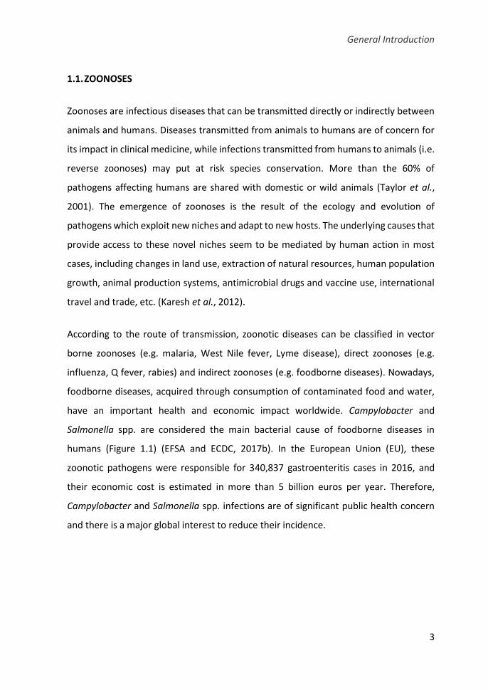
have an important health and economic impact worldwide. Campylobacter and
Salmonella spp. are considered the main bacterial cause of foodborne diseases in
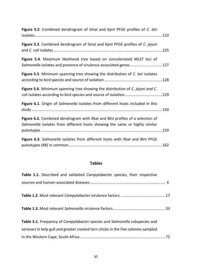
humans (Figure 1.1) (EFSA and ECDC, 2017b). In the European Union (EU), these
zoonotic pathogens were responsible for 340,837 gastroenteritis cases in 2016, and
their economic cost is estimated in more than 5 billion euros per year. Therefore,
Campylobacter and Salmonella spp. infections are of significant public health concern
and there is a major global interest to reduce their incidence.

Chapter 1
4
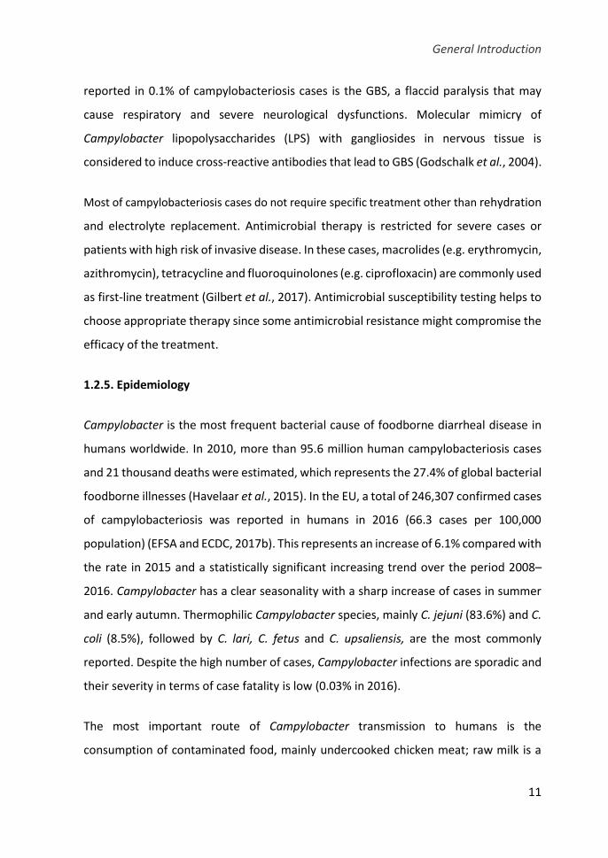
Figure 1.1. Reported numbers and notification rates of confirmed human zoonoses in
the EU, 2016. Total number of confirmed cases is indicated at the end each bar. Source:
EFSA and ECDC, 2017.
1.2. CAMPYLOBACTER
1.2.1. Discovery and taxonomy
It is believed that the first report regarding the bacterium that we now know as
Campylobacter dates back to 1886, when Escherich observed non-culturable spiral-
shaped bacteria in the colonic contents of children affected by what he called “cholera
infantum” (Escherich, 1886). However, the first reliable identification is attributed to
McFadyean and Stockman, who discovered a Vibrio-like bacterium in aborted ovine
fetuses in 1913 (McFadyean and Stockman, 1913). Few years later, Smith and Taylor
General Introduction
5
found the same spiral bacteria associated to infectious abortions of bovines for which
they proposed the name Vibrio fetus (Smith and Taylor, 1919). Afterward, closely
related organisms were detected in faeces of cattle and pigs with enterocolitis and were
classified as V. jejuni and V. coli, respectively (Jones et al., 1931; Doyle, 1944).
Initially, this bacterium was studied mainly in the veterinary field due to the economic
losses that it caused in livestock. A milk-borne outbreak of V. jejuni among prisoners in
USA in 1938 is considered the first well documented instance of Campylobacter human
infection (Levy, 1946). In 1957, King discriminated between V. fetus and the thermo-
tolerant V. jejuni and V. coli associating the latter with human enteric diseases (King,
1957). The genus Campylobacter (meaning “curved rod” in Greek) was first proposed by
Sebald and Véron in 1963, distinguishing them from Vibrio spp. by their DNA base
composition, non-fermentative metabolism and microaerophilic growth requirements
(Sebald and Véron, 1963). Later, Véron and Chatelain amended the taxonomy
considering four distinct species in the genus: C. fetus, C. jejuni, C. coli and C. sputorum
(Véron and Chatelain, 1973).
The difficulty of isolating and culturing Campylobacter from faeces, attributed to the
fastidious nature of these bacteria and the overgrowth of competing coliforms,
supposed a major hindrance to their research in human medicine. However,
Campylobacter was successfully isolated from stools of humans employing a filtration
method in 1972 (Dekeyser et al., 1972). Few years later, the isolation procedure was
refined using a selective medium supplemented with a mixture of antibiotics (Skirrow,
1977). The improvement in diagnostic methods represented an important
breakthrough and allowed the retrieval of Campylobacter from a wide range of human,
animal and environmental sources, and gradually novel taxa were proposed during the
1980s. Finally, Campylobacter became recognized as the main cause of bacterial
gastroenteritis in humans, despite having been ignored in clinical microbiology for so
many decades (Olson et al., 2008).
Chapter 1
6
In the wake of the description of new Campylobacter species, the taxonomic structure
was rearranged and the novel bacterial family Campylobacteriaceae was proposed,
which contains the genus Campylobacter, in addition to the closely related Arcobacter,
Dehalospirillum and Sulfurospirillum genera (Vandamme and De Ley, 1991). Ever since,
the taxonomy of Campylobacter genus has undergone many changes and, nowadays
there are still some controversies that remain to be resolved (Debruyne et al., 2008).
Currently, the genus consists of 28 species, nine subspecies and three biovars known
according to the LPSN (http://www.bacterio.net/campylobacter.html) (Table 1.1). The
species most commonly associated to human gastroenteritis are C. jejuni and C. coli, but
other species related to livestock animals, such as C. lari, C. upsaliensis, C. lanienae, C.
fetus, C. sputorum and C. hyointestinalis, can occasionally cause human infections as
well. Some non-zoonotic species isolated from humans are implicated in periodontal
diseases, for example, C. curvus, C. rectus, C. showae and C. concisus. However, other
species, such as C. canadensis, C. cuniculorum, C. iguaniorum and C. mucosalis, have
been isolated from animals but do not cause human illness.
The research of Campylobacter in hitherto little explored habitats, such as poles
ecosystems or new hosts in which the presence of the bacterium was suspect, has
revealed the existence of novel species. It is the case of C. peloridis found in molluscs,
C. volucris in gulls and C. subantarcticus in albatrosses and penguins from Antarctic
regions (Debruyne et al., 2009, 2010a, 2010b). However, novel species have also been
discovered in extensively researched hosts: C. avium in chickens and turkeys, and more
recently, C. hepaticus in chickens with spotty liver disease (Rossi et al., 2009; Van et al.,
2016).

General Introduction
7
Table 1.1. Described and validated Campylobacter species, their respective sources and
human-associated diseases.
Campylobacter species Source Human disease
C. avium Poultry NP
C. canadensis Wild birds NP
C. coli Pigs, sheep, cattle, poultry, wild birds G, S, M
C. concisus Humans, domestic pets G, P, A
C. corcagiensis Primates NP
C. cuniculorum Rabbits NP
C. curvus Humans P, G
C. fetus
subsp. fetus Cattle, sheep, reptiles G, S
subsp. testudium Reptiles G
subsp. veneralis Cattle, sheep S
C. geochelonis Reptiles NP
C. gracilis Humans P, A
C. helveticus Dogs, cats G
C. hepaticus Poultry NP
C. hominis Humans G
C. hyointestinalis
subsp. hyointestinalis Cattle, pigs G
subsp. lawsonii Pigs NP
C. iguaniorum Reptiles NP
C. insulaenigrae Marine mammals G
C. jejuni
subsp. doylei Humans G, S
subsp. jejuni Cattle, sheep, pigs, poultry, wild birds G, S, M, GBS
C. lanienae Cattle, pigs G
C. lari
subsp. concheus Shellfish G
subsp. lari Dogs, cats, poultry, wild birds G, S
C. mucosalis Pigs NP
C. peloridis Shellfish G
C. rectus Humans P, A
C. showae Humans P, A
C. sputorum
biovar faecalis Sheep, bulls NP
biovar paraureolyticus Cattle G
biovar sputorum Cattle, pigs A, G
C. subantarticus Wild birds NP
C. upsaliensis Dogs, cats G, S
C. ureolyticus Humans G, S, A
C. volucris Wild birds NP
A: abscesses; G: gastroenteritis; GBS: Guillain-Barré syndrome; M: meningitis; NP: none present as yet;
P: periodontal disease; S: septicemia.
Source: http://www.bacterio.net/campylobacter.html

Chapter 1
8
1.2.2. General characteristics
Campylobacter is a Gram-negative, small bacterium (0.2-0.8 μm x 0.5-5 μm) with a
slightly curved or spiral-shaped appearance, non-spore-forming, but in old cultures or
under stress conditions can take on coccoid body or viable but non-culturable (VBNC)
form (Rollins and Colwell, 1986). In general, cell has a single polar unsheathed flagellum
at one or both ends that enable to generate a corkscrew-like motion, while some species
are non-motile (C. gracilis, C. hominis) or have multiple flagella (C. showae) (Ferrero and
Lee, 1988; Etoh et al., 1993; Vandamme et al., 1995; Lawson et al., 2001).
This fastidious bacterium neither ferment nor oxidize carbohydrates, instead it obtains
energy from amino acids or tricarboxylic acid cycle intermediates. Most species have
catalase and oxidase but not urease activity (Debruyne et al., 2008). Campylobacter is
essentially microaerophilic, it grows at an atmosphere with reduced oxygen and
elevated carbon dioxide levels (5% O2, 10% CO2, and 85% N2) since it is susceptible to
oxygen radicals and peroxide (Garénaux et al., 2008). Moreover, several species of the
human oral cavity can grow in anaerobic conditions.
The growth temperature for Campylobacter is 30°C to 37°C, although thermotolerant
species grow better between 37°C and 42°C. Thermophilic species, including C. jejuni,
C. coli, C. lari and C. upsaliensis are causal agents of campylobacteriosis and their high
growth temperature may be a result of adaptation to warm-blooded animals. The
thermal stress response of bacteria is mostly due to the induction of the expression of
heat-shock proteins which promote the folding of cellular proteins and the proteolysis
of potentially deleterious proteins. Although unable to multiply below 30°C due to the
absence of cold-shock proteins, Campylobacter can survive to refrigeration and freezing
temperatures (Sampers et al., 2010). Campylobacter is sensitive to desiccation, heat,
ultra-violet radiation and other environmental stresses, even so it can persist in some
environments, such as manure, for prolonged periods (Inglis et al., 2010). The survival

General Introduction
9
time depends on the bacterial strain and the environmental conditions (e.g. light,
temperature, oxygen, nutrients and biotic interactions).
1.2.3. Detection, isolation and confirmation
Campylobacter is typically a fragile bacterium difficult to isolate and culture in
laboratory due to the special requirements for growth. The isolation methods are based
on complex selective media containing oxygen scavengers (horse or sheep blood,
charcoal), growth promoting reagents (ferrous sulphate, sodium metabisulphite,
sodium pyruvate) and antibiotics (cefoperazone, amphotericin B, polymyxin B,
cycloheximide, rifampicin, trimethoprim lactate and vancomycin). An enrichment step
in a liquid medium, prior to isolation on selective agar plates, usually provides better
recovery when cells are either low in number, injured or stressed (e.g. in food samples)
(Williams et al., 2009). Some of the most frequently employed enrichment broth media
are Bolton, Preston, Park-Sanders and Exeter. Numerous selective solid media also exist
for Campylobacter, some of the most common ones are: mCCDA (modified charcoal
cefoperazone deoxycholate), Preston, Skirrow, Butzler, Karmali and Campy-Cefex. Agar
plates with different selective principles in parallel can be used to increase the yield.
According to the standardized method for the detection and enumeration of
Campylobacter (ISO 10272-1:2017), Bolton broth is recommended for enrichment and
mCCDA is the selective agar of choice. Incubation is performed at 42°C in a microaerobic
atmosphere.
For confirmation, presumptive colonies can be stained and examined microscopically
regarding their morphology and motility, and biochemical (oxidase, catalase, nitrate
reductase) or serological (latex agglutination) tests can be performed. Hippurate
hydrolysis test can be used to discriminate between C. jejuni (positive) and C. coli
(negative), but some false-negatives could be wrongly classified (Adzitey and Corry,
2011). Since conventional phenotypic methods may be often atypical and difficult to
interpret, the use of molecular techniques is more reliable. The polymerase chain
Chapter 1
10
reaction (PCR), based on 16S rRNA, 23S rRNA, mapA, ceuE or lpxA genes, among others,
is more sensitive and specific and allows a rapid confirmation and identification of
Campylobacter species, and the detection of the bacteria without culture (Linton et al.,
1996; Fermér and Engvall, 1999; Denis et al., 2001; Klena et al., 2004; Katzav et al.,
2008). However, direct PCR amplification of Campylobacter from environmental
samples can be complicated due to the presence of low numbers of the bacteria or
inhibitory substances, and thus, a prior enrichment or DNA purification step,
respectively, may be necessary. Recently, more rapid and sensitive detection methods
have been developed such as real-time quantitative PCR (qPCR) or fluorescence in situ
hybridization (FISH) (Poppert et al., 2008; Leblanc-Maridor et al., 2011).
1.2.4. Clinical manifestations
Thermophilic Campylobacter spp. generally cause enteric infections in humans which
ranges from a mild watery diarrhoea to a severe inflammatory bloody diarrhoea.
Campylobacteriosis usually occurs within two to five days after exposure to the
pathogen and can be accompanied by other general symptoms including headache,
malaise, abdominal pain, cramping, nausea, vomiting and fever (van Vliet and Ketley,
2001). Some infected people do not have any symptoms. The illness is typically self-
limiting and lasts less than one week, but the bacterial shedding often persists after
clinical symptoms have ended. Campylobacteriosis may be more severe in infants,
elderly or immunocompromised patients, in which the pathogen occasionally spreads
to the bloodstream and causes a serious life-threatening infection (WHO, 2017a).
Complications such as bacteraemia, hepatitis, pancreatitis and miscarriage may occur,
but are uncommon especially when compared to those associated with Salmonella (see
section 1.3.4) (Moore et al., 2005). Campylobacter infection may also result in long-term
sequelae such as rheumatologic disorders (e.g. reactive arthritis) and peripheral
neuropathies (e.g. Guillain-Barré syndrome (GBS), Miller Fischer syndrome)
(Nachamkin, 2002). Probably, one of the most important immune-mediated disorders
General Introduction
11
reported in 0.1% of campylobacteriosis cases is the GBS, a flaccid paralysis that may
cause respiratory and severe neurological dysfunctions. Molecular mimicry of
Campylobacter lipopolysaccharides (LPS) with gangliosides in nervous tissue is
considered to induce cross-reactive antibodies that lead to GBS (Godschalk et al., 2004).
Most of campylobacteriosis cases do not require specific treatment other than rehydration
and electrolyte replacement. Antimicrobial therapy is restricted for severe cases or
patients with high risk of invasive disease. In these cases, macrolides (e.g. erythromycin,
azithromycin), tetracycline and fluoroquinolones (e.g. ciprofloxacin) are commonly used
as first-line treatment (Gilbert et al., 2017). Antimicrobial susceptibility testing helps to
choose appropriate therapy since some antimicrobial resistance might compromise the
efficacy of the treatment.
1.2.5. Epidemiology
Campylobacter is the most frequent bacterial cause of foodborne diarrheal disease in
humans worldwide. In 2010, more than 95.6 million human campylobacteriosis cases
and 21 thousand deaths were estimated, which represents the 27.4% of global bacterial
foodborne illnesses (Havelaar et al., 2015). In the EU, a total of 246,307 confirmed cases
of campylobacteriosis was reported in humans in 2016 (66.3 cases per 100,000
population) (EFSA and ECDC, 2017b). This represents an increase of 6.1% compared with
the rate in 2015 and a statistically significant increasing trend over the period 2008–
2016. Campylobacter has a clear seasonality with a sharp increase of cases in summer
and early autumn. Thermophilic Campylobacter species, mainly C. jejuni (83.6%) and C.
coli (8.5%), followed by C. lari, C. fetus and C. upsaliensis, are the most commonly
reported. Despite the high number of cases, Campylobacter infections are sporadic and
their severity in terms of case fatality is low (0.03% in 2016).
The most important route of Campylobacter transmission to humans is the
consumption of contaminated food, mainly undercooked chicken meat; raw milk is a
Chapter 1
12
common source of outbreaks (EFSA and ECDC, 2017b). International travels,
environmental exposure and direct contact with domestic animals are also important
risk factors for infection. The level of risk for travel-related campylobacteriosis appears
to be associated with the travel destination (Mughini-Gras et al., 2014). In developing
countries, Campylobacter is often hyperendemic and seasonality is less marked or
absent (Coker et al., 2002). Besides, asymptomatic infections are common and
diarrhoea is usually limited to children, suggesting that a high level of exposure in early
life leads to the development of protective immunity. Due to the ubiquitous nature of
the pathogen, risk factors in poor regions are more diffusely associated with exposure
to the environment, including contaminated drinking water. Although it is not as
common, person-to-person transmission via faecal-oral or fomites also occurs.
Campylobacter spp. normally inhabit the intestinal tract of warm-blooded animals, and
thus are frequently detected in foods derived from these animals (Horrocks et al., 2009;
Kaakoush et al., 2015). Poultry are the main reservoir of C. jejuni, C. coli, and to a lesser
extent C. lari, C. upsaliensis and C. concisus. In cattle, C. jejuni, C. coli, C. lari and C.
lanienae are frequently found, while pigs are more readily colonized by C. coli. Sheep
and goats have also been reported as carriers of Campylobacter species but with lower
prevalence. Campylobacter is also present in animal pets, such as dogs and cats (mainly
C. upsaliensis), hamsters, ferrets, rabbits and reptiles. Wild animals are potential
reservoirs of the pathogen, and among them, wild birds are most likely to carry
Campylobacter species (see section 1.7). Campylobacters have also been found in
shellfish.
Although Campylobacter is unable to grow outside of a suitable host, it can survive in
different environmental sources, including soil, manure and surface waters, which in
turn, are the most likely sources of infection to domestic and wild animals (Murphy et
al., 2006; Bronowski et al., 2014). This bacterium is found in abundance on farms and
their surrounding environment. Despite of biosecurity measures, the bacterium can
enter the farm, and both rodents and insects have also been identified as possible

General Introduction
13
vectors (Hald et al., 2008). Once established, the bacterium is difficult to eliminate since
transmission within individuals occurs rapidly, especially in poultry farms (Sahin et al.,
2002; Urdaneta, 2016). Water is also an effective vehicle of transmission of
Campylobacter to animals and humans. Campylobacter is omnipresent in rivers, ponds,
lakes, streams and coastal waters, mostly in those which are exposed to direct
contamination with animal faeces, agricultural run-off and sewage effluents (Whiley et
al., 2013).
1.2.6. Pathogenesis
Campylobacteriosis severity depends on the virulence of the strain and other host-
specific factors such as age, gastric acidity level and the host immune-response to the
infection. A low dose of Campylobacter, about 500 cells, is enough to induce infection
in humans (Kothary and Babu, 2001). Campylobacter enter through the oral route, cross
the stomach and attain the small intestine thanks to their resistance to gastric and bile
acids. At first, the bacterium colonizes the small intestine and then moves to the colon
that is the target organ. Motility is necessary to resist peristalsis and survive in the
gastrointestinal environment, as well as to circumvent the intestinal mucus layer.
Therefore, the flagella of Campylobacter play an essential role for intestinal
colonization, along with the bacterial chemosensory system that drives flagellar
movement based on the environmental signals.
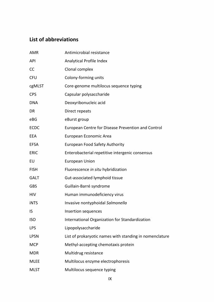
To establish infection, Campylobacter must attach to the intestinal epithelial cells and
subsequently invade them (Figure 1.2). Campylobacter adhesion is not mediated by
appendages like fimbria or pilus as occurs in Salmonella and Escherichia coli, although
the precise molecular mechanism of the attachment for Campylobacter is still unclear
(Rubinchik et al., 2012). It seems that outer membrane proteins of C. jejuni specifically
bind to fibronectin, a glycoprotein of the extracellular matrix, located on the basolateral
surface of epithelial cells. The mechanisms that controls the bacterial invasion are also
confuse and controverted since different results have been observed in vitro depending
Chapter 1
14
on the C. jejuni strain and the culture cell model used (Ó Cróinín and Backert, 2012). C.
jejuni effectors induce rearrangements of eukaryotic cell cytoskeleton to facilitate the
bacterium uptake. All strains require the polymerization of microtubules (tubulin
subunits) for maximal invasion, while some strains also require the polymerization of
microfilaments (actin subunits). Besides, it has been demonstrated that C. jejuni flagella
are involved not only in motility, but also in the secretion of flagellar proteins and
invasion effectors acting as a type III secretion system (T3SS) (Guerry, 2007).
Once internalized, a Campylobacter-containing vacuole is developed avoiding the
delivery into lysosomes. C. jejuni may evade the host immune response within the
endocytic vacuole although its role is not yet well established. Invasion by C. jejuni
induce interleukin (IL)-8, one of the earliest pro-inflammatory cytokines that sign the
recruitment of polymorphonuclear leukocytes (PMNL), mainly neutrophils, to the gut
lumen (Young et al., 2007; Janssen et al., 2008). The interaction of phagocytes, including
macrophages and dendritic cells, with the bacteria results in a massive pro-
inflammatory response and increases the cytokine production.
While adherence of Campylobacter and enterotoxins production alter the fluid
resorption of the intestine resulting in secretory diarrhoea, the intestinal inflammation
and the mucosal damage, probably along with the effect of bacterial cytotoxins, results
in the inflammatory diarrhoea frequently observed in humans (Wassenaar, 1997;
Janssen et al., 2008). The best characterized toxin of Campylobacter is the cytolethal
distending toxin (CDT) which arrests eukaryotic cell cycle inducing cellular distension
and apoptosis (Asakura et al., 2008).
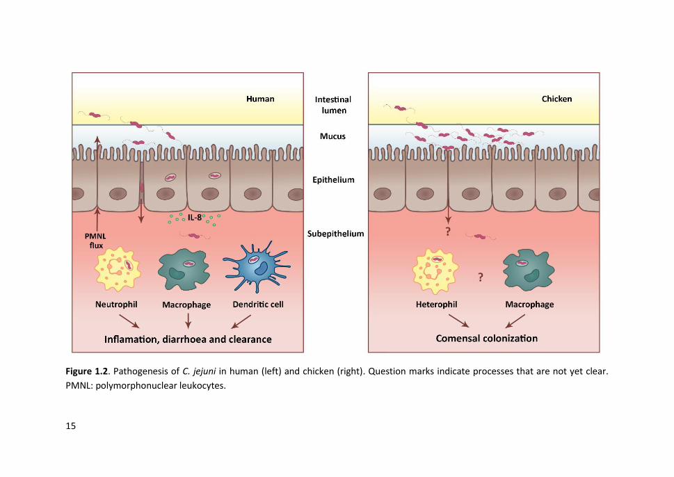
15
Figure 1.2. Pathogenesis of C. jejuni in human (left) and chicken (right). Question marks indicate processes that are not yet clear.
PMNL: polymorphonuclear leukocytes.
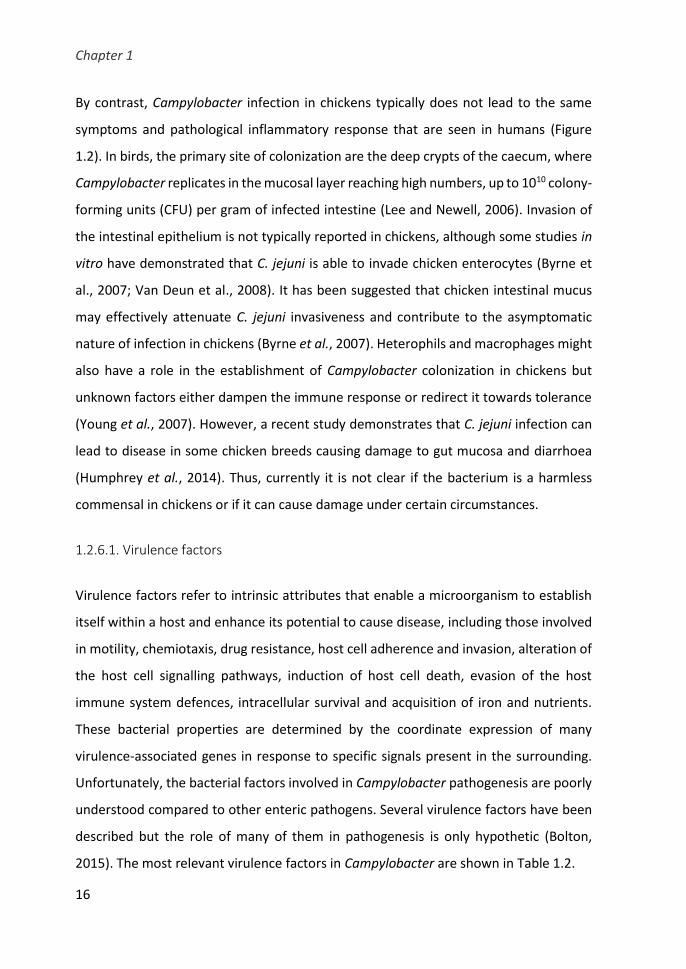
Chapter 1
16
By contrast, Campylobacter infection in chickens typically does not lead to the same
symptoms and pathological inflammatory response that are seen in humans (Figure
1.2). In birds, the primary site of colonization are the deep crypts of the caecum, where
Campylobacter replicates in the mucosal layer reaching high numbers, up to 1010 colony-
forming units (CFU) per gram of infected intestine (Lee and Newell, 2006). Invasion of
the intestinal epithelium is not typically reported in chickens, although some studies in
vitro have demonstrated that C. jejuni is able to invade chicken enterocytes (Byrne et
al., 2007; Van Deun et al., 2008). It has been suggested that chicken intestinal mucus
may effectively attenuate C. jejuni invasiveness and contribute to the asymptomatic
nature of infection in chickens (Byrne et al., 2007). Heterophils and macrophages might
also have a role in the establishment of Campylobacter colonization in chickens but
unknown factors either dampen the immune response or redirect it towards tolerance
(Young et al., 2007). However, a recent study demonstrates that C. jejuni infection can
lead to disease in some chicken breeds causing damage to gut mucosa and diarrhoea
(Humphrey et al., 2014). Thus, currently it is not clear if the bacterium is a harmless
commensal in chickens or if it can cause damage under certain circumstances.
1.2.6.1. Virulence factors
Virulence factors refer to intrinsic attributes that enable a microorganism to establish
itself within a host and enhance its potential to cause disease, including those involved
in motility, chemiotaxis, drug resistance, host cell adherence and invasion, alteration of
the host cell signalling pathways, induction of host cell death, evasion of the host
immune system defences, intracellular survival and acquisition of iron and nutrients.
These bacterial properties are determined by the coordinate expression of many
virulence-associated genes in response to specific signals present in the surrounding.
Unfortunately, the bacterial factors involved in Campylobacter pathogenesis are poorly
understood compared to other enteric pathogens. Several virulence factors have been
described but the role of many of them in pathogenesis is only hypothetic (Bolton,
2015). The most relevant virulence factors in Campylobacter are shown in Table 1.2.
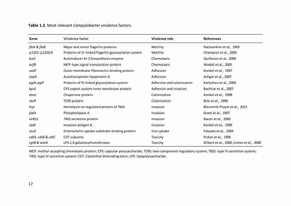
17
Table 1.2. Most relevant Campylobacter virulence factors.
Gene Virulence factor Virulence role References
flaA & flaB Major and minor flagellin proteins Motility Nachamkim et al., 1993
cj1321-cj1325/6 Proteins of O -linked flagellin glycosylation system Motility Champion et al., 2005
luxS Autoinducer AI-2 biosynthesis enzyme Chemotaxis Quiñones et al., 2009
acfB MCP-type signal transduction protein Chemotaxis Wodall et al., 2005
cadF Outer membrane fibronectin-binding protein Adhesion Konkel et al., 1997
capA Autotransporter lipoprotein A Adhesion Ashgar et al., 2007
pglA-pglF Proteins of N-linked glycosylation system Adhesion and colonization Karlyshev et al., 2004
kpsE CPS export system inner membrane protein Adhesion and invasion Bachtiar et al., 2007
dnaJ Chaperone protein Colonization Konkel et al., 1998
racR TCRS protein Colonization Brás et al., 1999
hcp Hemolysin co-regulated protein of T6SS Invasion Bleumink-Pluym et al., 2013
pldA Phospholipase A Invasion Grant et al., 1997
virB11 T4SS secretion protein Invasion Bacon et al., 2000
ciaB Invasion antigen B Invasion Konkel et al., 1999
ceuE Enterochelin uptake substrate-binding protein Iron uptake Palyada et al., 2004
cdtA, cdtB & cdtC CDT subunits Toxicity Picket et al., 1996
cgtB & wlaN LPS 1,3-galactosyltransferases Toxicity Gilbert et al., 2000; Linton et al., 2000
MCP: methyl-accepting chemotaxis protein; CPS: capsular polysaccharide; TCRS: two-component regulatory system; T6SS: type VI secretion system;
T4SS: type IV secretion system; CDT: Cytolethal distending toxin; LPS: lipopolysaccharide.
Chapter 1
18
1.3. SALMONELLA
1.3.1. Discovery and taxonomy
Throughout history, there have been a great number of dire outbreaks of typhoid fever.
Many scientists associated the disease with the consumption of contaminated food and
drinks, and unsuccessfully tried to found the causal agent during years. It was Eberth
who observed the bacillus for the first time in 1879 in mesenteric lymph nodes and
spleen from a patient that died due to typhoid fever. Few years later, Salmon and Smith
isolated the bacterium from pigs affected by hog cholera and it was consequently
named “Bacillus choleraesuis”. The genus Salmonella was proposed later in 1900 by
Lignières in honour to Salmon’s research group (Salmonella Subcommittee of the
Nomenclature Comm. Int. Soc. Microbiol., 1934).
In 1934, the first Kauffman-White classification scheme was established in the basis of
the serological identification of Salmonella surface structures (Kauffman, 1966).
Initially, each serotype or serovar was considered a separate species and was named
according to the caused disease or the animal from which the bacterium was isolated.
However, when the absence of host specificity was observed, the new serovars began
to be named according to the location at where they were isolated. Later, Crosa et al.
(1973) demonstrated by DNA-DNA hybridization experiments that all serovars belonged
to a single Salmonella species. As a result, the taxonomy of Salmonella underwent a
series of modifications and a new nomenclature was proposed. “Salmonella
choleraesuis” was the name accepted for the Salmonella type species and the six
subgenera were considered to subspecies (Le Minor et al., 1982). The only exception
was S. bongori which was separated from the other subspecies and recognized as a
distinct species (Reeves et al., 1989). As the term “S. choleraesuis”, which referred to
both a species and a serovar, caused confusion, it was suggested to be changed to S.
enterica since no serovar shared this name (Le Minor and Popoff, 1987). The
nomenclature of Le Minor and Popoff (1987) was widely accepted and used in certain

General Introduction
19
countries, even though it has not been recognized nor validated by the Judicial
Commission of the International Committee of Systematic Bacteriology. During years,
two different systems of nomenclature were in use despite the attempts to unify them.
Finally, the nomenclature of Salmonella was approved in 2005 and the White-Kauffman-
Le Minor classification scheme was established (Judicial Commission, 2005; Grimont
and Weill, 2007).
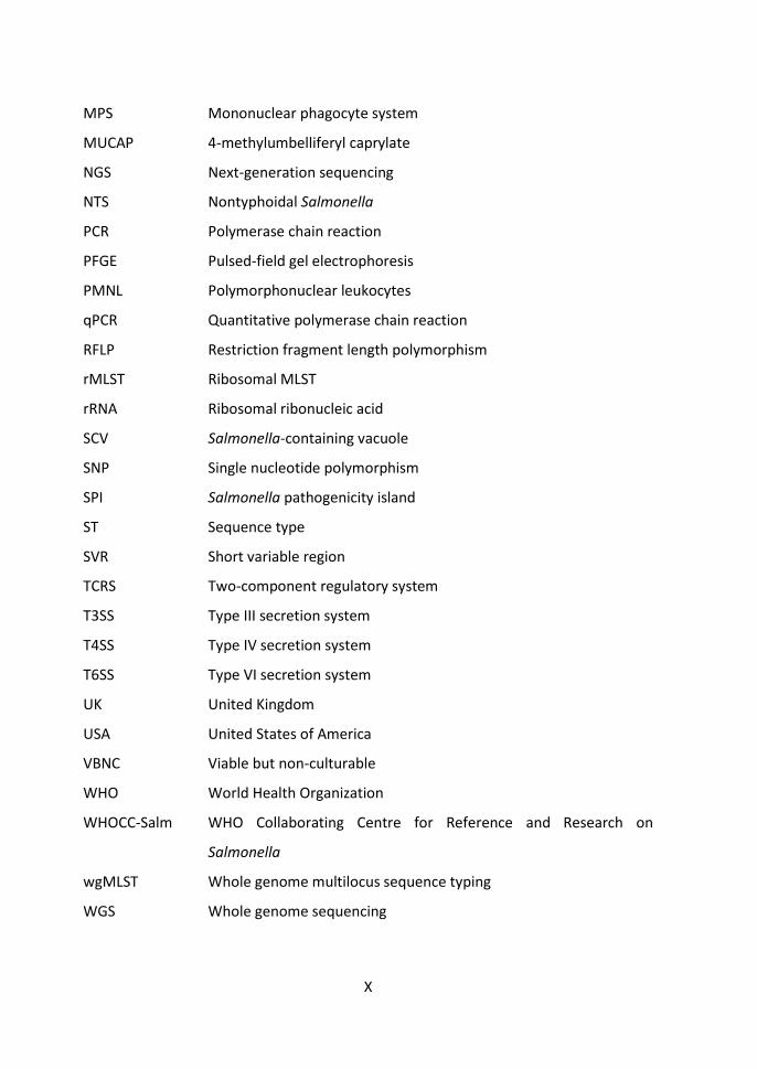
Currently, within the Enterobacteriaceae family, only two species comprise the genus
Salmonella: S. bongori and S. enterica. Furthermore, S. enterica is divided into six
subspecies: enterica (I), salamae (II), arizonae (IIIa), diarizonae (IIIb), houtenae (IV) and
indica (VI) (Figure 1.3). Salmonella subspecies are further subtyped into serovars
according to the immunological characterization of somatic (O), flagellar (H) and, to a
lesser extent, capsular (K; Vi) antigens. The antigenic formulae of Salmonella serovars
are available in the While-Kauffmann-Le Minor scheme, in continuous update by the
World Health Organization Collaborating Centre for Reference and Research on
Salmonella (WHOCC-Salm) of the Institut Pasteur (Issenhuth-Jeanjean et al., 2014). The
full name of a serovar is given as, for example, Salmonella enterica subsp. enterica
serovar Typhimurium, but can be abbreviated to S. Typhimurium. Serovars of other
subspecies are designated by their antigenic formulae, following the subspecies name.
Currently, the taxonomic group contains more than 2,700 serovars of Salmonella.
While most Salmonella subspecies are widely distributed in the environment and cold-
blooded animals, serovars belonging to S. enterica subsp. enterica can colonise a broad
range of animal hosts, including mammals and birds. These serovars can cause human
disease: serovars Typhi and Paratyphi are responsible for enteric fever, whereas the
other serovars, denominated nontyphoidal Salmonella (NTS), can be non-invasive or
invasive causing mild to moderate gastroenteritis or systemic infections, respectively
(Figure 1.3).

20
Figure 1.3. Taxonomic classification of Salmonella genus, sources and associated diseases. Blue, green and red colours represent
environmental and cold-blooded animals, warm-blooded animals, and human origin, respectively. In parenthesis, number of
serovars.
subsp. V (23)
subsp. IV houtenae
(77)
subsp. IIIb diarizonae
(346)
subsp. IIIa arizonae
(106)
subsp. II salamae
(528)
subsp. VI indica (13)
Typhoid fever Paratyphoid fever
S. Typhi S. Paratyphi A S. Paratyphi BS. Paratyphi C
Gastroenteritis Extra-intestinal
Self-limiting(non-invasive)
S. TyphimuriumS. Enteritidis+ 1,500 others
Bacteraemia (invasive)
S. TyphimuriumS. EnteritidisS. DublinS. VirchowS. Heidelberg
Bacteraemia
S. CholeraesuisS. TyphisuisS. TyphimuriumS. EnteritidisS. DublinS. VirchowS. HeidelbergS. Bovismorbificans
Cold-blooded animals and environment
subsp. I enterica (1,619)
Cold-blooded animals and environment Warm-blooded
Focal infection
S. CholeraesuisS. TyphisuisS. TyphimuriumS. EnteritidisS. Dublin
Salmonella(2,712)
Salmonella bongori Salmonella enterica
Typhoidal Salmonella Non-typhoidal Salmonella
Typhoid fever Paratyphoid fever

General Introduction
21
1.3.2. General characteristics
Salmonella is a Gram-negative, rod-shaped bacterium ranging 0.7-1.5 x 2.0-5.0 μm in
size and non-spore-forming. The bacillus is predominantly motile by peritrichous
flagella, except for the pathogenic avian-specific serovars Pullorum and Gallinarum, and
other non-motile variants. Salmonella is facultative anaerobic, chemoorganotrophic
bacterium, predominantly non-lactose fermenting and hydrogen sulphide producing. It
also presents catalase but not oxidase nor urease activity (Bell and Kyriakides, 2002).
Salmonella is considered mesophilic with an optimum growth at 35-37°C, but some
strains can survive at extremely low or high temperatures (2°C to 54°C). The induction
of the multigenic cold shock response and the heat stress response controlled by the
sigma factors allow a quick adaptation to temperature changes. These mechanisms can
increase Salmonella survival rates when treated at low temperature prior freezing, or
when exposed to heat treatment, especially in low water activity (aw) foods (Mattick et
al., 2001; Dominguez and Schaffner, 2009). Moreover, Salmonella is resistant to
desiccation, supports high salt concentrations (up to 4%) and can persist in extremely
acid environments (pH 3.0-4.0) (Álvarez-Ordóñez et al., 2012; Li et al., 2012). Salmonella
is incredibly adept and versatile in the strategies it employs to multiply or survive for
prolonged periods under unfavourable environmental conditions outside the living
hosts (e.g. faecal material, soil, water, pastures, foods) (Winfiel and Groisman, 2003;
Spector and Kenyon, 2012).
1.3.3. Detection, isolation and confirmation
Salmonella is a non-fastidious bacterium that can grow in a simple glucose-salts medium
or more rapidly in highly supplemented media. Standard Salmonella detection methods
include a non-selective pre-enrichment, an enrichment in a selective medium and the
subsequent plating onto two different selective media (ISO 6579-1:2017). Incubation in
a pre-enrichment liquid medium, usually buffered peptone water or modified tryptone

Chapter 1
22
soya broth, improves the recovery of bacteria when stressed, sub-lethally damaged or
in low accounts (Valentín-Bon et al., 2003). Subsequently, a selective enrichment broth,
typically Müller-Kauffmann tetrathionate or Rappaport-Vassiliadis soya peptone, is
used to favour the proliferation of Salmonella to the detriment of competing flora. The
modified semi-solid Rappaport-Vassiliadis medium, which allows Salmonella to be
distinguished from other non-motile bacteria, is the one demanded by ISO 6579-1:2017.
The next step is the selection and differentiation of Salmonella by sub-cultivation onto
different selective solid media, such as MacConkey, Xylose Lysine Desoxycholate (XLD),
Xylose Lysine-Tergitol 4 (XLT4), Brilliant Green, Hektoen-Enteric or Salmonella-Shigella,
among others. The production of hydrogen sulphide and the inability to ferment glucose
are the main characteristics of Salmonella used for their detection in which these media
are based.
Once Salmonella is isolated, its identity can be confirmed at subspecies level by
biochemical tests (e.g. API-E20, VITEK®2, MUCAP test) and serovar can be determined
by serological tests. Serotyping is performed by testing a bacterial suspension against
commercial anti-sera by means of a series of slide agglutination tests. The type, order
and repetition of sugar residues conforming the lipopolysaccharide (LPS) component of
the outer membrane determine the O antigens. The H antigens are defined by the
middle region of the flagellin protein constituting the bacterial flagellum. Monophasic
serovars produce flagella always with the same antigenic specificity; instead, diphasic
serovars can express in alternative phases two different flagellin types (H1 and H2).
Most of the serovars of S. enterica subsp. enterica are diphasic, however, some diphasic
serovars may become monophasic because of the loss or lack of expression of one of
the flagellin genes (e.g. S. Typhimurium). Only a few serovars present the K antigens,
which are polysaccharides located at the capsular surface, while the Vi antigens, a
special subtype of K antigens, are only found in some pathogenic serovars (e.g. Typhi,
Paratyphi C and Dublin). The combination of all these antigens, referred to as the
General Introduction
23
antigenic formula, is unique to each Salmonella serovar (Issenhuth-Jeanjean et al.,
2014).
Aside from the phenotyping methods that are laborious and time consuming, molecular
techniques such as PCR or real-time PCR, mostly based on the target gene invA, can also
be used for a rapid detection and confirmation of Salmonella (Malorny et al., 2003;
González-Escalona et al., 2012). Despite the high specificity and sensitivity of these
techniques, live and dead cells cannot be distinguished and some samples need to be
cultured to reach the detection limit threshold.
1.3.4. Clinical manifestations
In human infections, there are three clinical forms of salmonellosis: gastroenteritis,
bacteraemia and enteric fever. Besides, there may be cases of chronic asymptomatic
carriers of Salmonella. Gastroenteritis is the most common manifestation of food
poisoning typically caused by nontyphoidal Salmonella (NTS) serovars. Enterocolitis
symptoms usually begin six to 72 h after intoxication and include profuse water
diarrhoea, nausea, vomiting and abdominal cramps. Myalgia, headache, chills and fever
are also common in many patients. The symptoms can be mild to severe, but
gastroenteritis is usually self-limiting and may last between two to seven days.
Rehydration and electrolyte replacement is usually sufficient to overcome the bacterial
infection and most patients recover without antibiotic treatment. In immunocompetent
individuals, the disease typically remains localized in the terminal ileum, mesenteric
lymph nodes and colon. However, infant, elderly and immunocompromised patients are
more susceptible to NTS infections and have a higher risk of developing severe
symptoms and complications (WHO, 2017b).
Bacteraemia occurs when invasive nontyphoidal Salmonella (iNTS) pass through the
intestinal barrier, enter the bloodstream and disperse to other organs. Almost all
serovars can be invasive, but some of them, such as Choleraesuis, Dublin and Heidelberg

Chapter 1
24
are markedly more likely to cause hospitalization, systemic infections or death (Jones et
al., 2008). High fever is the characteristic symptom of bacteraemia which can lead to
septic shock and other severe extra-intestinal complications (e.g. infections of liver,
spleen, biliary or urinary tract, pneumonia, arthritis or endocarditis, meningitis)
(Hohmann, 2001). Some comorbidities predispose to iNTS diseases, especially human
immunodeficiency virus (HIV), malaria and malnutrition in developing countries (Feasey
et al., 2012).
Serovars Typhi and Paratyphi cause life threatening systemic infections, known as
enteric fever or typhoid fever, regardless of the immunocompetence of the patient.
Most infections concur five to nine days after contamination and are characterised by
prodromal symptoms (e.g. headache, malaise, myalgia, abdominal pain and diarrhoea
or constipation) followed by the onset of low fever that highly increases in the second
week. Besides fever, infected patients may also display bradycardia,
hepatosplenomegaly, rose spots on their chest and abdomen, meningism and
neuropsychiatric manifestations, among others (Kuvandik et al., 2009). Some
complications like those mentioned for Salmonella bacteraemia can occur, including
pancreatitis, hepatitis and cholecystitis. The most severe complications are
encephalopathy and haemorrhage due to gastrointestinal bleeding and intestinal
perforation (Huang and DuPont, 2005).
Currently, it is recommended to reserve the antimicrobial therapy for patients with
severe disease or with a high risk for invasive disease. In life-threatening infections,
fluoroquinolones and third-generation cephalosporins are administered for empiric
therapy until the antimicrobial susceptibilities of the pathogen agent are known (Gilbert
et al., 2017). Nowadays, two typhoid vaccines of demonstrated safety and efficacy are
available: the oral vaccine based on a live attenuated mutant strain of S. Typhi (Ty21a)
and the injectable Vi capsular polysaccharide vaccine (ViCPS) (WHO, 2008).

General Introduction
25
1.3.5. Epidemiology
The global human health impact of NTS is high, with an estimation of 78.7 million illness
cases and more than 59 thousand deaths in 2010 (Havelaar et al., 2015). Despite
improvements in sanitation and water supplies, the incidence of NTS infections
continues to increase worldwide, being one of the major causes of bacterial foodborne
diseases in both industrialized and developing countries (Figure 1.4) (WHO, 2015b). In
the EU, a total of 94,530 confirmed salmonellosis cases were reported in 2016 (20.4
cases per 100,000 population), which represents an increase of 1.9% compared with the
previous year (EFSA and ECDC, 2017b). However, there was an overall significant
declining trend of salmonellosis between 2008 and 2015, probably because of the
implementation of National Control Programmes, except in Czech Republic, France and
Spain where the trend was upward. Most cases were reported during summer months
following a seasonal pattern. Although the incidence of salmonellosis is lower than
campylobacteriosis, the fatality rate is higher (0.25% in 2016).
Salmonella is widely distributed in the environment (e.g. water, soil, faecal material,
foods) where it can survive for long time periods (Winfiel and Groisman, 2003).
However, its natural environment is the intestinal tract of a broad range of domestic
and wild animals which act as reservoirs and excrete the bacteria in their faeces for
weeks or months (Hoelzer et al., 2011; Hilbert et al., 2012). The main source of
Salmonella infection in humans is the consumption of contaminated food of animal
origin, mainly poultry meat, eggs and milk, or vegetables and water contaminated with
animal wastes (EFSA and ECDC, 2017b). Salmonella infection occurs when the bacteria
are capable of multiplying on foodstuffs due to inadequate storage temperatures,
insufficient cooking and cross-contamination of ready-to-eat food. To a lesser extent,
transmission through direct contact with infected animals, environment or person-to-
person can also occur.
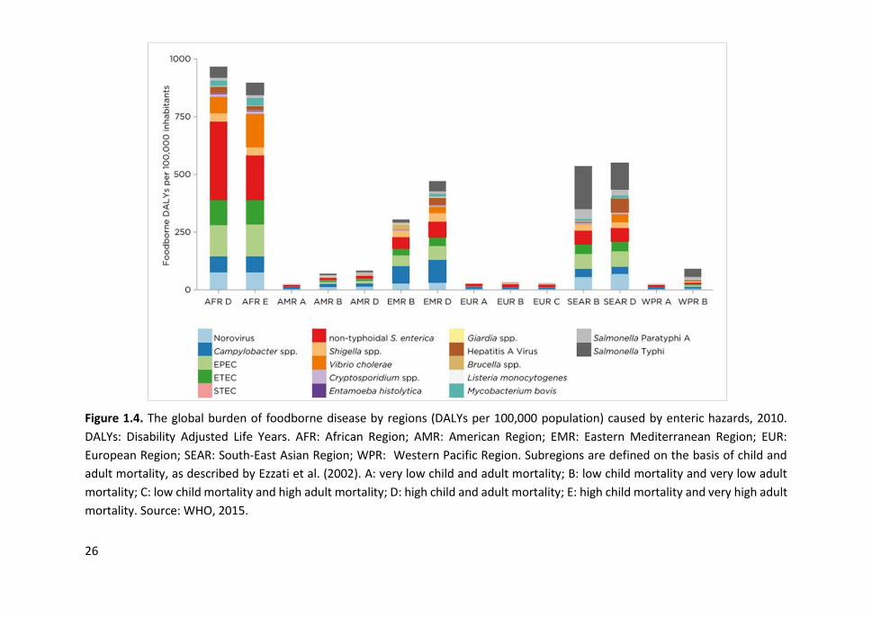
26
Figure 1.4. The global burden of foodborne disease by regions (DALYs per 100,000 population) caused by enteric hazards, 2010.
DALYs: Disability Adjusted Life Years. AFR: African Region; AMR: American Region; EMR: Eastern Mediterranean Region; EUR:
European Region; SEAR: South-East Asian Region; WPR: Western Pacific Region. Subregions are defined on the basis of child and
adult mortality, as described by Ezzati et al. (2002). A: very low child and adult mortality; B: low child mortality and very low adult
mortality; C: low child mortality and high adult mortality; D: high child and adult mortality; E: high child mortality and very high adult
mortality. Source: WHO, 2015.
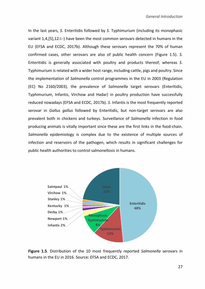
General Introduction
27
In the last years, S. Enteritidis followed by S. Typhimurium (including its monophasic
variant 1,4,[5],12:i:-) have been the most common serovars detected in humans in the
EU (EFSA and ECDC, 2017b). Although these serovars represent the 70% of human
confirmed cases, other serovars are also of public health concern (Figure 1.5). S.
Enteritidis is generally associated with poultry and products thereof; whereas S.
Typhimurium is related with a wider host range, including cattle, pigs and poultry. Since
the implementation of Salmonella control programmes in the EU in 2003 (Regulation
(EC) No 2160/2003), the prevalence of Salmonella target serovars (Enteritidis,
Typhimurium, Infantis, Virchow and Hadar) in poultry production have successfully
reduced nowadays (EFSA and ECDC, 2017b). S. Infantis is the most frequently reported
serovar in Gallus gallus followed by Enteritidis, but non-target serovars are also
prevalent both in chickens and turkeys. Surveillance of Salmonella infection in food
producing animals is vitally important since these are the first links in the food-chain.
Salmonella epidemiology is complex due to the existence of multiple sources of
infection and reservoirs of the pathogen, which results in significant challenges for
public health authorities to control salmonellosis in humans.
Figure 1.5. Distribution of the 10 most frequently reported Salmonella serovars in
humans in the EU in 2016. Source: EFSA and ECDC, 2017.
Enteritidis48%
Typhimurium13%
Monophasic Typhimurium
8%Infantis 2%
Newport 1%
Derby 1%
Kentucky 1%
Stanley 1%
Virchow 1%
Saintpaul 1% Other23%
Chapter 1
28
In contrast to the worldwide distribution of NTS infections, enteric fever occurs
predominantly in underdeveloped countries and it is associated with a high morbidity
and mortality rates. The WHO estimated more than 7.5 and 1.7 million cases of typhoid
and paratyphoid fever, respectively, which resulted in more than 52 and 12 thousand
deaths in 2010 (Havelaar et al., 2015). Typhoidal Salmonella serovars are endemic
throughout Africa and Asia and persists in the Middle East, Central and South America
and in some South-East European countries (Figure 1.4). Typhoidal Salmonella is carried
only by humans and is transmitted through the faecal–oral route usually by
contaminated water and food. The risk of infection is higher among impoverished and
overcrowded populations with unsanitary conditions and exposed to unsafe water
supplies due to ineffective sewage disposal. In Europe and USA, the incidence of enteric
fever is low and most cases are imported by foreigners or travellers returning from
endemic areas (Jensenius et al., 2013)
1.3.6. Pathogenesis
The pathogenicity of Salmonella depends on the serovar, the virulence of the strain, the
inoculum dose, the host-status and its immune response. The infective dose of
Salmonella is 103 to 109 cells, usually in the order of 106 cells (Blaser and Newman, 1982).
The inherent tolerance of Salmonella to the extreme stomach pH and the bile acids
allows it to reach the small intestine. The bacterium must also resist digestive enzymes,
secretory IgA, antimicrobial peptides and other innate immune defences and compete
with indigenous microbiota (Álvarez-Ordóñez et al., 2011). Flagella motility regulated
by a complex chemotaxis system enables the bacterium to penetrate the intestinal
mucus layer and colonize the ileum and to a lesser extent the colon.
In the lumen, Salmonella selectively attaches to the apical domain of enterocytes by
fimbrial adhesins and invade the cell by bacterial-mediated endocytosis, crossing the
intestinal barrier to reach the gut-associated lymphoid tissue (GALT) (Figure 1.6).
Salmonella preferentially enters through microfold (M) cells of the follicle-associated

General Introduction
29
epithelium, which transport it to the lymphoid cells (T and B) in the underlying Peyer’s
patches (Tahoun et al., 2012). Adhesion is a highly specific process and involves a
complete repertoire of fimbrial biosynthesis genes. The different Salmonella serovars
may possess a set of specific adhesins which facilitate the bacterial attachment to a
multitude of eukaryotic cell types encountered in various hosts (Wagner and Hensel,
2011).
Salmonella invasion of intestinal cells is mainly conferred by the T3SS injectosome which
allows the bacteria to mediate its own uptake. T3SS apparatus is composed of multi-
channel proteins assembled in a needle-like structure projected from the bacterial
surface that puncture the membrane of the host cell and inject effector proteins into
the cellular cytoplasm. These translocated proteins modulate actin polymerization and
trigger a local rearrangement of the host cell cytoskeleton leading to formation of
membrane ruffles that engulf the bacterium. Other effector proteins secreted by a
second T3SS modulate the Salmonella-containing vacuole (SCV) blocking the fusion of
the lysosomes which supports bacterial survival and multiplication within the host cell
(Haraga et al., 2008; Moest and Méresse, 2013).
Invasion by NTS serovars induces the expression of several cytokines, such as Toll-like
receptor (TLR)-dependent IL-8 and IL-12/interferon-gamma (IFN- γ), evoking an acute
inflammatory response. As a result, a transmigration of PMNL into the gut lumen takes
place, thereby preventing systemic spread of the bacteria (Patel and McCormick, 2014).
The secretion of fluids and the influx of electrolytes to the lumen evoke to the diarrhoea
onset. Some Salmonella strains can also produce enterotoxins that may stimulate
intestinal secretion increasing the intestinal damage. NTS serovars have evolved to use
inflammation-derived metabolites (e.g. nitrate, tetrathionate) enhancing their growth
in the inflamed intestine (Winter et al., 2010). In addition, Salmonella produces several
toxins in different amounts depending on the serovar, including the CDT (Miller and
Wiedmann, 2016).
Chapter 1
30
While the NTS infection generally elicits a local inflammatory reaction, typhoidal
serovars overcome the innate immune response in the intestinal mucosa and cause a
systemic infection in healthy individuals (Raffatellu et al., 2008; Gal-Mor et al., 2014)
(Figure 1.6). When S. Typhi crosses the epithelial barrier, it induces a recruitment of
intestinal macrophages. Bacteria can survive and multiply into the microbicidal
environment of these phagocytic cells and subsequently migrate to mesenteric lymph
nodes and disseminate to liver, spleen and bone marrow. The mononuclear phagocyte
system (MPS) provides the periodic recirculation of the pathogen to new foci of
infection. Some patients can become asymptomatic carriers of Salmonella and excrete
large amounts of bacteria in their faeces, having the potential to re-infect (Ruby et al.,
2012).
The carrier state has also been described in livestock animals and is associated with
foodborne outbreaks. Birds can be infected by host-specific and non-host-specific
Salmonella serovars showing differences in their pathogenesis. S. Gallinarum and S.
Pullorum are adapted and restricted to birds, especially poultry, in which they cause
fowl typhoid and pullorum disease, systemic infections with high mortality rates
(Shivaprasad and Barrow, 2008). Conversely, non-host-specific Salmonella serovars
readily colonize the cecal tonsils, the upper part of the small intestine, the gizzard and
proventriculus of birds, but only produce severe disease in some special cases (e.g.
during the laying period, after viral diseases or in two weeks-old chicks). Therefore, most
birds become symptomless carriers of Salmonella for some weeks or months and shed
it in the faeces sometimes intermittently (Revolledo and Ferreira, 2012).

31
Figure 1.6. Pathogenesis of nontyphoidal Salmonella (NTS) serovars (left) and S. Typhi (right) in humans. PMNL: polymorphonuclear
leukocytes

Chapter 1
32
1.3.6.1. Virulence factors
Virulence factors of Salmonella are under a complex genetic control and involve
multiple genes encoded both on the bacterial chromosome and on large plasmids. Most
virulence-associated genes are clustered together in distinct large regions (10-200 kb)
called Salmonella pathogenicity islands (SPIs) distributed along the chromosome. SPIs
have been acquired by horizontal gene transfer from other species, have a G+C content
different from that of the host core genome and often contain mobile genetic elements.
The number and content of the SPIs differ among Salmonella serovars, partly explaining
their heterogeneity in host specificities and disease outcome. Currently, 21 different
SPIs have been identified, some of them are conserved throughout the genus
Salmonella (e.g. SPI-1 and SPI-2), while others are specific for certain serovars (e.g. SPI-
7 of S. Typhi) (Sabbagh et al., 2010). The SPI-1 contains at least 35 genes encoding T3SS
and several effector proteins involved in bacterial invasion of host cells. A larger region
of SPI-2 encodes a second T3SS which secrete effector proteins implicated in Salmonella
intracellular survival and replication within the SCV. Other SPIs (e.g. SPI-3, SPI-4 and SPI-
5) also appear to facilitate the invasion and survival of the bacterium within host cells.
However, many SPIs have been recently identified and much less is known about their
distribution across serovars and the role they play in disease. In addition to SPIs,
virulence determinants can be encoded in pathogenicity islets (smaller clusters),
fimbrial operons, prophages or randomly dispersed in the genome, as well as in
virulence plasmids (Table 1.3).
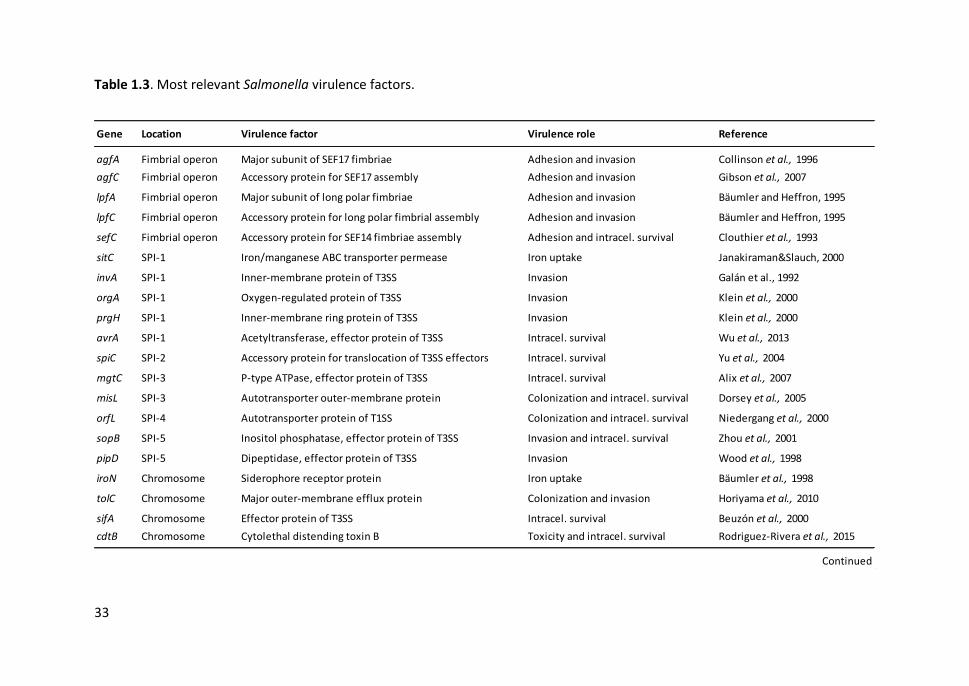
33
Table 1.3. Most relevant Salmonella virulence factors.
Gene Location Virulence factor Virulence role Reference
agfA Fimbrial operon Major subunit of SEF17 fimbriae Adhesion and invasion Collinson et al., 1996
agfC Fimbrial operon Accessory protein for SEF17 assembly Adhesion and invasion Gibson et al., 2007
lpfA Fimbrial operon Major subunit of long polar fimbriae Adhesion and invasion Bäumler and Heffron, 1995
lpfC Fimbrial operon Accessory protein for long polar fimbrial assembly Adhesion and invasion Bäumler and Heffron, 1995
sefC Fimbrial operon Accessory protein for SEF14 fimbriae assembly Adhesion and intracel. survival Clouthier et al., 1993
sitC SPI-1 Iron/manganese ABC transporter permease Iron uptake Janakiraman&Slauch, 2000
invA SPI-1 Inner-membrane protein of T3SS Invasion Galán et al., 1992
orgA SPI-1 Oxygen-regulated protein of T3SS Invasion Klein et al., 2000
prgH SPI-1 Inner-membrane ring protein of T3SS Invasion Klein et al., 2000
avrA SPI-1 Acetyltransferase, effector protein of T3SS Intracel. survival Wu et al., 2013
spiC SPI-2 Accessory protein for translocation of T3SS effectors Intracel. survival Yu et al., 2004
mgtC SPI-3 P-type ATPase, effector protein of T3SS Intracel. survival Alix et al., 2007
misL SPI-3 Autotransporter outer-membrane protein Colonization and intracel. survival Dorsey et al., 2005
orfL SPI-4 Autotransporter protein of T1SS Colonization and intracel. survival Niedergang et al., 2000
sopB SPI-5 Inositol phosphatase, effector protein of T3SS Invasion and intracel. survival Zhou et al., 2001
pipD SPI-5 Dipeptidase, effector protein of T3SS Invasion Wood et al., 1998
iroN Chromosome Siderophore receptor protein Iron uptake Bäumler et al., 1998
tolC Chromosome Major outer-membrane efflux protein Colonization and invasion Horiyama et al., 2010
sifA Chromosome Effector protein of T3SS Intracel. survival Beuzón et al., 2000
cdtB Chromosome Cytolethal distending toxin B Toxicity and intracel. survival Rodriguez-Rivera et al., 2015
Continued
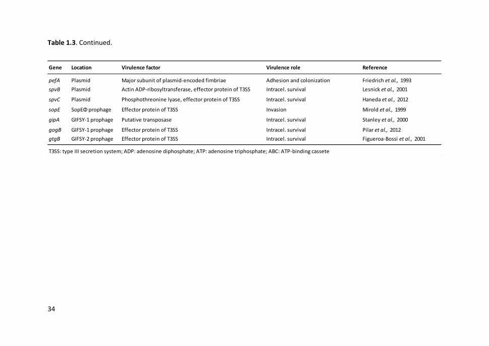
34
Table 1.3. Continued.
Gene Location Virulence factor Virulence role Reference
pefA Plasmid Major subunit of plasmid-encoded fimbriae Adhesion and colonization Friedrich et al., 1993
spvB Plasmid Actin ADP-ribosyltransferase, effector protein of T3SS Intracel. survival Lesnick et al., 2001
spvC Plasmid Phosphothreonine lyase, effector protein of T3SS Intracel. survival Haneda et al., 2012
sopE SopEΦ prophage Effector protein of T3SS Invasion Mirold et al., 1999
gipA GIFSY-1 prophage Putative transposase Intracel. survival Stanley et al., 2000
gogB GIFSY-1 prophage Effector protein of T3SS Intracel. survival Pilar et al., 2012
gtgB GIFSY-2 prophage Effector protein of T3SS Intracel. survival Figueroa-Bossi et al., 2001
T3SS: type III secretion system; ADP: adenosine diphosphate; ATP: adenosine triphosphate; ABC: ATP-binding cassete
General Introduction
35
1.4. ANTIMICROBIAL RESISTANCE
Emergence of resistance to antimicrobials is a natural process that has been observed
since the first antibiotics were discovered. However, antimicrobial resistance (AMR) has
become a growing concern in recent times since the overuse of antimicrobials in both
humans and food-producing animals has accelerated the selection and spread of
resistant bacteria. Consequently, antimicrobial drugs have become less effective or
even ineffective, limiting available treatment options. AMR affects every country
regardless of its level of income and development and it is estimated to account for
more than 700,000 deaths per year worldwide (O’Neill, 2016).
The quantity of antimicrobials used in poultry and livestock is dramatic and often
includes drugs important for the treatment of common human infections (Van Boeckel
et al., 2015). Worryingly, much of their use in food-producing animals is not to treat and
control disease, but instead either to prevent infections (sometimes to compensate for
poor farming practices) or for production purposes. In these cases, antimicrobials are
usually administered at lower and imprecise doses, via medicated feed or drinking
water, for long periods, potentially enhancing selection for AMR in bacterial
populations. The use of antimicrobials as prophylactic and growth promoters is not
allowed in EU since 2006, but are still used for this purpose in many countries including
USA, where they can be obtained without veterinary prescription (Maron et al., 2013).
Antimicrobial resistant bacteria from animal origin has significant public health
implications since they can be transmitted to humans through food products, the
environment or, less often, direct contact.
Bacterial resistance to antimicrobials can appear as a result of mutations or by
acquisition of resistance conferring genes via horizontal gene transfer (e.g. plasmids,
conjugative transposons) (Von Wintersdorff et al., 2016). The latter occurs frequently in
the host intestinal tract due to high amounts of different bacteria and the close contact
among them, but also in the environment, especially the aquatic one (e.g. sewage). The
Chapter 1
36
transfer of antimicrobial resistance genes among bacteria increase the dispersion of
AMR and contributes to the emergence of multi-resistant bacteria.
To combat this increasing concern, it is necessary to coordinate efforts of the different
institutions and apply international measures and national strategies. In 2015, the WHO
adopted a global action plan to: raise awareness of AMR, strengthen the surveillance
and research, reduce the incidence of infections through effective sanitation, hygiene
and infection prevention measures; optimize the use of antimicrobial agents in human
and animal health, and increase development of new diagnostic tools, vaccines and
other alternative products (e.g. phage therapy, lysins, probiotics) (WHO, 2015a).
1.4.1. Campylobacter antimicrobial resistance
Intrinsic resistance in C. jejuni and C. coli has been described against penicillins and most
of the cephalosporins, as well as trimethoprim, sulfamethoxazole, rifampicin and
vancomycin (Fitzgerald et al., 2008). On the other hand, C. lari belongs to the nalidixic
acid resistant thermophilic Campylobacter (NARTC) group (Benjamin et al., 1983).
In the early 1990s, when enrofloxacin was taken into use in animal production,
especially in poultry and pigs, Campylobacter fluoroquinolone resistance started to
increase worldwide among animals and human isolates at the same time (Engberg et
al., 2001). There are still growing trends in resistance to fluoroquinolones in EU, where
C. coli and C. jejuni strains present high resistance levels to ciprofloxacin (EFSA and
ECDC, 2017a). Given the high level of acquired resistance to fluoroquinolones in EU this
antimicrobial can no longer be considered appropriate for routine empirical treatment
of human Campylobacter infections.
By contrast, the macrolide resistance of Campylobacter has remained at a low and
stable level for a long time. However, there is also evidence that resistance rates to
erythromycin and other macrolides (e.g. azithromycin, clindamycin) are slowly
General Introduction
37
increasing in several countries, mostly among C. coli isolates from pigs (Gibreel and
Taylor, 2006; EFSA and ECDC, 2017a). It has been observed that the use of tylosin as
feed additive in veterinary medicine selects for a high level resistance to erythromycin
in the Campylobacter population (Ladely et al., 2007). Nevertheless, the acquisition of
this resistance is a stepwise process and requires prolonged exposure, in contrast to the
rapidly appearance of fluoroquinolone resistance, and may decrease in the absence of
antibiotic selection pressure (Luangtongkum et al., 2012). Since fluoroquinolone
resistance is common, macrolides have become critical antimicrobials for the treatment
of human campylobacteriosis. Even so, the recent identification of a transferable
macrolide resistance mechanism in Campylobacter may pose a rapid means of spread
of this resistance (Qin et al., 2014).
High levels of resistance to tetracyclines, which is often plasmid mediated, are
frequently observed in Campylobacter isolates from humans and animals in many EU
countries (EFSA and ECDC, 2017a). Although tetracyclines are considered as an
alternative to treat Campylobacter infections, in practice are not often used and are
contraindicated in young children. As mentioned above, Campylobacter is resistant to
many β-lactam antimicrobial agents, mainly penicillins and cephaloporins, but
carbapenems are an exception and may be effective in the treatment of
campylobacteriosis.
Multidrug resistance (MDR), defined as resistance to three or more families of
antimicrobial agents, has so far been infrequent in Campylobacter. However, it has
increased in recent years, probably due to the overuse of different antimicrobial agents
in animal production along with horizontal transfer of AMR, posing a serious risk of
treatment failures (Lin et al., 2002; Qin et al., 2014).
Chapter 1
38
1.4.2. Salmonella antimicrobial resistance
Several Salmonella strains with MDR emerged during the late 1990s, and since then they
have expanded worldwide. For instance, S. Typhimurium phage type DT104 is typically
resistant to ampicillin, chloramphenicol, streptomycin, sulphonamides and tetracycline
(ACSSuT), and is able to acquire additional resistance to other clinically important
antimicrobials (Helms et al., 2005). The appearance of this strain raised an important
public health concern because of its involvement in animals and human diseases.
Besides, the genomic elements that confer the penta-resistance profile or other AMR
(e.g. β-lactams) may spread horizontally among Salmonella serovars and other enteric
pathogens (Douard et al., 2010). Currently, the level of MDR in Salmonella clinical
isolates is high in EU, and some of them are resistant to up to eight different
antimicrobial classes (EFSA and ECDC, 2017a). MDR is especially notorious among
monophasic S. Typhimurium isolates from humans, with a prevalence of 81.1% in 2015.
In EU, the highest proportions of resistance in human Salmonella isolates in 2015 were
reported for sulphonamides/sulfamethoxazole (32.4%), tetracyclines (28.1%) and
ampicillin (27.8%) (EFSA and ECDC, 2017a). Resistance to these antimicrobials was also
observed among isolates from production animals and their derived products, probably
as a consequence of their broad use in veterinary medicine. Resistance to
fluoroquinolones (e.g. ciprofloxacin) and third-generation cephalosporins (e.g.
cefotaxime, ceftazidime), which represent the clinically most important antimicrobial
classes for treatment of human salmonellosis, was detected in 13.3% and 0.9% of
human isolates, respectively. AMR levels for Salmonella are greatly influenced by the
serovar, with some of them exhibiting high resistance to certain antimicrobials. For
instance, resistance to colistin was present in 11.4% of the isolates (belonging to
different serovars), mainly in S. Enteritidis which has been reported to have inherent
resistance to this antimicrobial agent (Agersø et al., 2012). However, S. Enteritidis is
often more susceptible to antimicrobials than other serovars. Emergence of AMR and
General Introduction
39
MDR in S. Typhi has also been described in Africa and Asia, causing increased treatment
failure cases (Zaki and Karande, 2011).
1.5. TYPING METHODS
Bacterial typing methods allow characterizing the intraspecies variability and are
essential tools for conducting epidemiological studies: for example, for comparing
different isolates to establish common origins and sources of infection for pathogenic
bacteria traceability or studying bacteria population dynamics and diversity of strains in
different geographical areas and host species. A proper typing technique must
accomplish the following characteristics: typeability, discriminatory power,
reproducibility and repeatability, epidemiological concordance and, if possible, speed
and automation (van Belkum et al., 2007). A wide variety of typing techniques are
currently in use, thus, the more suitable one or a combination of two or more must be
chosen according to the aims of the investigation and study design. Typing methods fall
into two broad categories: phenotypic and genotypic methods.
Phenotypic methods are based on the gene expression of bacteria and include
techniques such as serotyping, biotyping, phage typing, antimicrobial resistance typing,
multilocus enzyme electrophoresis (MLEE), etc. The serovar determination is essential
to unravel the epidemiology of Salmonella spp. (see section 1.3.3.), but it is ineffective
for Campylobacter spp. (Allos et al., 2004). The main drawback of phenotypic methods
is that genes expression can vary according to growth conditions and may not reflect
the genetic changes occurred in bacterial strains, therefore, they not provide a reliable
and stable epidemiological marker (van Belkum et al., 2007).
Genotypic methods are based on the analysis of bacterial genetic structure and provide
more sensitivity and discrimination power, as well as higher levels of standardization
Chapter 1
40
and reproducibility. Genotyping techniques can be classified in three categories
according to their bases: (I) restriction sites in the bacterial DNA, (II) PCR amplification
of genomic targets and, (III) polymorphisms in DNA sequences (Foley et al., 2009). In
this section, only the techniques used in this thesis for genotyping Campylobacter and
Salmonella isolates are described in detail: enterobacterial repetitive intergenic
consensus (ERIC)-PCR, PCR-restriction fragment length polymorphism (RFLP), pulsed-
field gel electrophoresis (PFGE) and multilocus sequence typing (MLST). All of them are
extensively used in current research of molecular epidemiology of foodborne bacteria.
1.5.1. Enterobacterial repetitive intergenic consensus PCR
The ERIC sequences are short imperfect palindromes (126 bp) with internal deletions or
insertions, repeatedly interspersed throughout the genome of different bacterial
species. These sequences were originally described in E. coli and Salmonella, and then
were found to be conserved in other members of the Enterobacteriaceae, and even in
unrelated bacteria from different phyla (Hulton et al., 1991). As the position and
number of copies of ERIC elements in bacterial genomes is variable, their PCR-
amplification generates specific patterns that allow strains within Gram-negative
enteric species to be distinguished (Versalovic et al., 1991).
Given the simplicity, speed and low cost of this technique, ERIC sequences have been
successfully used as a molecular marker in several Salmonella epidemiological studies
for a long time (Chmielewski et al., 2002; Ye et al., 2011; Turki et al., 2012; Wang et al.,
2014). However, other ERIC-PCR based-studies showed less discriminatory power
compared with other molecular techniques such as PFGE (Fendri et al., 2013; Almeida
et al., 2015). Moreover, it is not always possible to obtain repeatable profiles due to the
appearance and disappearance of minor bands. This poor repeatability and
reproducibility makes the comparison of ERIC-PCR results among different laboratories
very difficult (Swanenburg et al., 1998).

General Introduction
41
1.5.2. PCR-Restriction fragment length polymorphism
The flaA-RFLP technique consists in the amplification of a flaA gene region (1725 bp)
and their further restriction with the DdeI enzyme to generate a fingerprint profile
(Nachamkin et al., 1993; Harrington et al., 2003). The flaA gene, which encodes for the
flagellin protein of bacterial flagella, has a genetic variability region flanked by
conserved regions and provides a useful target for discriminating among Campylobacter
isolates.
In addition to the high-resolution power, the relatively simplicity and the low cost of the
flaA-RFLP technique has led to its widespread use for epidemiological studies of
Campylobacter. The flaA-RFLP has been employed to determine the genetic diversity of
Campylobacter in farms, to compare isolates from human and animal origin and to track
outbreaks of campilobacteriosis, among others (Ring et al., 2005; Corcoran et al., 2006;
Oporto et al., 2007; Heuvelink et al., 2009). However, the flaA gene is genetically
instable and not species-specific. The recombination between the flaA and flaB genes
within a strain can occur, as well as recombination intra and interspecies in co-
infections, finding some alleles in both C. jejuni and C. coli (Harrington et al., 1997;
Dingle et al., 2005). These limitations make this molecular typing unsuited for dynamic
population or longer-term investigations. Nevertheless, flaA-RFLP may be appropriate
as a first technique for distinguishing closely related strains of Campylobacter, especially
when used in combination with other additional methods based on more conserved
target genes, such as MLST (Behringer et al., 2011).
1.5.3. Pulsed-field gel electrophoresis
PFGE is based on the enzymatic digestion of entire chromosomal DNA and the
subsequent separation of restriction fragments of large size differences (30 - 1100 kb)
applying an electrical field of alternating polarity. PFGE uses endonucleases with
infrequent recognition sites to generate macrorestriction profiles: SmaI and KpnI are
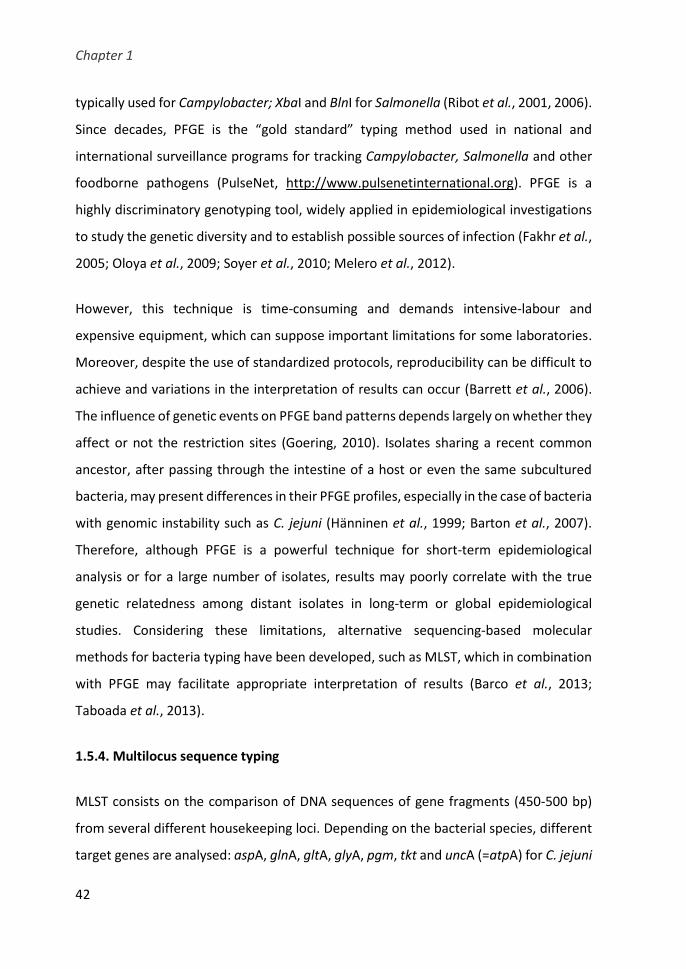
Chapter 1
42
typically used for Campylobacter; XbaI and BlnI for Salmonella (Ribot et al., 2001, 2006).
Since decades, PFGE is the “gold standard” typing method used in national and
international surveillance programs for tracking Campylobacter, Salmonella and other
foodborne pathogens (PulseNet, http://www.pulsenetinternational.org). PFGE is a
highly discriminatory genotyping tool, widely applied in epidemiological investigations
to study the genetic diversity and to establish possible sources of infection (Fakhr et al.,
2005; Oloya et al., 2009; Soyer et al., 2010; Melero et al., 2012).
However, this technique is time-consuming and demands intensive-labour and
expensive equipment, which can suppose important limitations for some laboratories.
Moreover, despite the use of standardized protocols, reproducibility can be difficult to
achieve and variations in the interpretation of results can occur (Barrett et al., 2006).
The influence of genetic events on PFGE band patterns depends largely on whether they
affect or not the restriction sites (Goering, 2010). Isolates sharing a recent common
ancestor, after passing through the intestine of a host or even the same subcultured
bacteria, may present differences in their PFGE profiles, especially in the case of bacteria
with genomic instability such as C. jejuni (Hänninen et al., 1999; Barton et al., 2007).
Therefore, although PFGE is a powerful technique for short-term epidemiological
analysis or for a large number of isolates, results may poorly correlate with the true
genetic relatedness among distant isolates in long-term or global epidemiological
studies. Considering these limitations, alternative sequencing-based molecular
methods for bacteria typing have been developed, such as MLST, which in combination
with PFGE may facilitate appropriate interpretation of results (Barco et al., 2013;
Taboada et al., 2013).
1.5.4. Multilocus sequence typing
MLST consists on the comparison of DNA sequences of gene fragments (450-500 bp)
from several different housekeeping loci. Depending on the bacterial species, different
target genes are analysed: aspA, glnA, gltA, glyA, pgm, tkt and uncA (=atpA) for C. jejuni
General Introduction
43
and C. coli; adk and pgi genes instead of aspA and gltA genes for C. lari; aroC, dnaN,
hemD, hisD, purE, thrA and sucA for Salmonella (Dingle et al., 2001; Miller et al., 2005;
Achtman et al., 2012). An allele number is assigned for each locus according to the allelic
sequences available in the corresponding bacterial database (PubMLST,
http://pubmlst.org; Enterobase, http://enterobase.warwick.ac.uk). Isolates that
possess identical alleles for the seven loci correspond to a common sequence type (ST).
In turn, STs sharing four or more identical alleles are grouped into clonal complexes
(CC)s on the basis of eBurst (Feil et al., 2004).
The main advantage of MLST compared to the abovementioned methods is that DNA
sequence data are unambiguous and is possible to objectively compare the allelic
profiles among different laboratories through an online global database. Since MLST
measures nucleotide polymorphisms in relatively-stable housekeeping genes, which
accumulate slowly and without selective pressure, it is a useful tool for long-term
investigations. MLST is extensively used to identify major infection sources and study
population structures and phylogeny of many bacteria including Campylobacter and
Salmonella (Hughes et al., 2010; Müllner et al., 2010; Nielsen et al., 2010; Antunes et
al., 2011; Keller and Shriver, 2014; Toboldt et al., 2014; Papadopoulos et al., 2016).
MLST has also been proposed as a method to replace Salmonella serotyping (Achtman
et al., 2012).
Nevertheless, MLST is an expensive and time-consuming technique that requires high
quality sequences. Besides, although MLST is very powerful in detecting groups of
related organisms belonging to the same lineage, it may lack capacity to distinguish
closely related isolates or highly clonal bacterial populations (Barco et al., 2013;
Taboada et al., 2013). To enhance the discriminatory power among epidemic strains
with common STs, the use of an additional subtyping method may be required, such as
the sequencing of the flagellin short variable region (flaA-SVR) for Campylobacter (Sails
et al., 2003; Clark et al., 2005).
Chapter 1
44
1.5.5. Future prospects
Nowadays, next-generation sequencing (NGS) technologies have made possible to
examine the complete or nearly entire genomes of bacterial isolates (Forde and O’Toole,
2013). Whole genome sequencing (WGS) methods can resolve isolates that differ at only
a single nucleotide, thereby providing the highest level of resolution and phylogenetic
accuracy for epidemiologic subtyping. Until recently, WGS-based analyses were limited
to the characterization of a small number of strains of interest. However, rapid advances
and relatively decreasing costs in NGS have made WGS increasingly accessible and
applicable for tracking disease outbreaks with successful results. The WGS approach
provides the possibility to obtain from one single assay many traditional typing results
and other more complete analysis that yield critical information for bacterial
surveillance. Some of them are the ribosomal MLST (rMLST) scheme, based on 53
ribosomal protein loci present in most bacteria; the core-genome MLST (cgMLST), which
provides high-resolution data in groups of related but not identical isolates; and the
whole genome MLST (wgMLST) that is applicable to single-clone bacteria or very closely
related strains (Jolley et al., 2012; Sheppard et al., 2012).
1.6. ONE HEALTH
The improvement in detection and genotyping techniques has contributed to a greater
understanding of the epidemiology of Campylobacter and Salmonella. However, the
existence of multiple sources of infection and reservoir hosts reveals the complexity of
the transmission cycle of these enteropathogens (Figure 1.7). It is well known that the
main source of infection is the consumption of contaminated food. However,
identification of the sources of environmental contamination may not always be
possible due to the large number of animal species involved in the transmission of these
zoonotic agents (Hoelzer et al., 2011; Whiley et al., 2013). Moreover, the bacterial
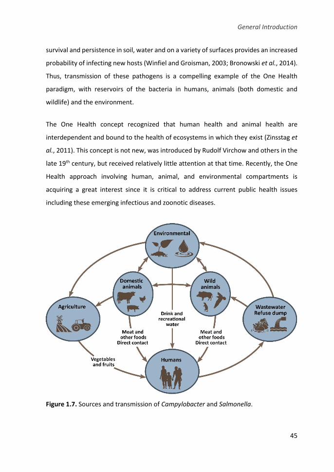
General Introduction
45
survival and persistence in soil, water and on a variety of surfaces provides an increased
probability of infecting new hosts (Winfiel and Groisman, 2003; Bronowski et al., 2014).
Thus, transmission of these pathogens is a compelling example of the One Health
paradigm, with reservoirs of the bacteria in humans, animals (both domestic and
wildlife) and the environment.
The One Health concept recognized that human health and animal health are
interdependent and bound to the health of ecosystems in which they exist (Zinsstag et
al., 2011). This concept is not new, was introduced by Rudolf Virchow and others in the
late 19th century, but received relatively little attention at that time. Recently, the One
Health approach involving human, animal, and environmental compartments is
acquiring a great interest since it is critical to address current public health issues
including these emerging infectious and zoonotic diseases.
Figure 1.7. Sources and transmission of Campylobacter and Salmonella.
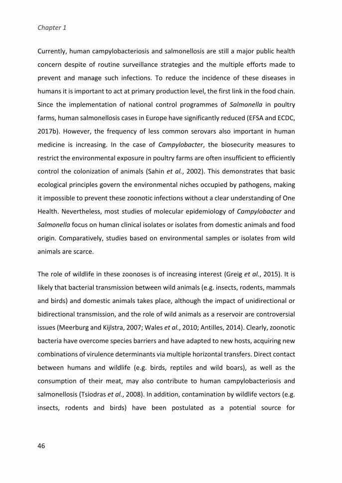
Chapter 1
46
Currently, human campylobacteriosis and salmonellosis are still a major public health
concern despite of routine surveillance strategies and the multiple efforts made to
prevent and manage such infections. To reduce the incidence of these diseases in
humans it is important to act at primary production level, the first link in the food chain.
Since the implementation of national control programmes of Salmonella in poultry
farms, human salmonellosis cases in Europe have significantly reduced (EFSA and ECDC,
2017b). However, the frequency of less common serovars also important in human
medicine is increasing. In the case of Campylobacter, the biosecurity measures to
restrict the environmental exposure in poultry farms are often insufficient to efficiently
control the colonization of animals (Sahin et al., 2002). This demonstrates that basic
ecological principles govern the environmental niches occupied by pathogens, making
it impossible to prevent these zoonotic infections without a clear understanding of One
Health. Nevertheless, most studies of molecular epidemiology of Campylobacter and
Salmonella focus on human clinical isolates or isolates from domestic animals and food
origin. Comparatively, studies based on environmental samples or isolates from wild
animals are scarce.
The role of wildlife in these zoonoses is of increasing interest (Greig et al., 2015). It is
likely that bacterial transmission between wild animals (e.g. insects, rodents, mammals
and birds) and domestic animals takes place, although the impact of unidirectional or
bidirectional transmission, and the role of wild animals as a reservoir are controversial
issues (Meerburg and Kijlstra, 2007; Wales et al., 2010; Antilles, 2014). Clearly, zoonotic
bacteria have overcome species barriers and have adapted to new hosts, acquiring new
combinations of virulence determinants via multiple horizontal transfers. Direct contact
between humans and wildlife (e.g. birds, reptiles and wild boars), as well as the
consumption of their meat, may also contribute to human campylobacteriosis and
salmonellosis (Tsiodras et al., 2008). In addition, contamination by wildlife vectors (e.g.
insects, rodents and birds) have been postulated as a potential source for
General Introduction
47
contamination of non-animal based food (e.g. fresh fruits, nuts and vegetables) (Jay-
Russell, 2013).
1.7. WILD BIRDS AS RESERVOIRS OF ZOONOTIC BACTERIA
Wild birds are important in the spread and maintenance of zoonotic pathogens in the
environment, such as influenza A virus, West Nile virus, Borrelia and enteric bacteria
(Bengis et al., 2004). Campylobacter and Salmonella have been detected with variable
prevalence in a wide range of wild birds including waterfowl, crows, sparrows, pigeons,
raptors and seagulls (Chuma et al., 2000; Molina-Lopez et al., 2011; Gargiulo et al., 2014;
Antilles et al., 2015; Jurado-Tarifa et al., 2016; Konicek et al., 2016).
Birds feeding habits appear to be the main factor influencing the exposure to these
zoonotic bacteria and the proximity to farms and grazing fields imply an added risk of
infection (Ramos et al., 2010; Hald et al., 2016). In some ecological guilds, such as
insectivores, granivores and birds feeding exclusively on vegetable matter, the presence
of enteropathogens is low or non-existent. On the contrary, high carriage rates have
been found in other guilds including ground-foraging, raptors and opportunistic feeders.
Ground-foraging bird species can become infected when ingesting food contaminated
with droppings or eat filter-feeding molluscs in sewage contaminated habitats. Raptors
can acquire these enteric bacteria from the intestines of their preys or from carcasses
when they scavenge on carrion. Opportunistic birds are drawn to sewage sludge and
refuse dumps that may be inherently rich sources of pathogenic agents (Raven and
Coulson, 2001). In particular, seagulls are one of the most documented carriers of
Campylobacter and Salmonella spp. probably due to their scavenging feeding habits.
Over the past decades, populations of several gull species have increased drastically
throughout Europe, North America and Australia, and have been largely attributed to
greater availability of food because of anthropogenic activity (Hatch, 1996). Due to the
Chapter 1
48
urbanization of coastal areas, interaction of seagulls with humans is increasingly close
since flocks gather at areas where food scraps are abundant, especially around landfills,
farms, processing factories and fishing boats.
Given the global loss of natural wetlands, seagulls and other seabirds have become
more dependent on alternative habitats, including wastewater treatment plants
(Murray and Hamilton, 2010). These behaviours may favour the transmission of
antimicrobial resistant enteric bacteria of anthropogenic origin to wild birds. At the
same time, seabirds are clearly of importance in maintaining these zoonotic bacteria in
the environment during routine movements centralized on the place of residence from
roosting or nesting sites to feeding sites. They often roost on nearby fields and pastures,
and wash in local water bodies, in a way that bacteria ingested at feeding sites may
enter again to the food chain, once excreted by the birds.
Furthermore, seabirds like other wild birds may act as effective dispersers of pathogens
via faecal contamination due to their ability to cover long distances during annual
movements. Twice a year, billions of wild birds, belonging to the 19% of extant species,
migrate across national and intercontinental borders contributing to the potential
establishment of new endemic foci of diseases (Reed et al., 2003). Migration is a regular
seasonal movement, often north and south along a flyway between breeding and
wintering grounds, driven primarily by availability of food. The migration patterns are
complex and variable between species or even different populations within the same
species (Hockey et al., 2005). For instance, Arctic tern (Sterna paradisaea) holds the
long-distance migration record for birds, travelling up to 80,000 km between northern
Scandinavia and Antarctica each year (Egevang et al., 2010). Other trans-equatorial
migrant birds are the long-tailed skua (Stercorarius longicaudus) and the Sabine’s gull
(Larus sabini), which breed in Arctic and spend the winter in the Southern Hemisphere,
in close association with the cold waters of the Benguela Upwelling in the south-west
coast of Africa (Stenhouse et al., 2012; Gilg et al., 2013). Bird migration involves long
journeys and long distances which results in physiologic stress, and thus, a greater
General Introduction
49
susceptibility to infectious diseases that facilitates birds becoming a reservoir of
pathogens. Moreover, the different ecosystems where birds stop over during the travel
can also increase the risk of exposure to reservoir hosts and sources of zoonotic
bacteria. For these reasons, migration may be a mechanism that facilitates the
geographic distribution of pathogens and AMR that can pose a risk for public and animal
health.


CHAPTER 2
Objectives

Objectives
53
Despite the importance of seabirds and other wild birds as reservoirs of zoonotic
bacteria and antimicrobial resistance traits, there are still gaps of knowledge of their
role in the epidemiology of Campylobacter and Salmonella spp. Thus, the global aim of
this thesis is to deepen the knowledge of the epidemiology of these zoonotic agents in
seabirds from remote regions in the Southern Hemisphere and in the neaest
Southwestern Europe.
In order to achieve this goal, the specific objectives are:
1. To determine the prevalence, genetic relatedness and antimicrobial resistance
of thermophilic Campylobacter spp. and non-typhoidal Salmonella serovars in
seabirds from the Western Cape coast of South Africa. Study I
2. To gain insight into the epidemiology, population structure and potential of
virulence of thermophilic Campylobacter spp. and non-typhoidal Salmonella
serovars from a range of seabird species from the Southern Ocean. Study II and
III.
3. To assess the genetic relatedness and virulence potential of Salmonella isolates
from human, poultry and seagulls from Southwestern Europe. Study IV.


CHAPTER 3
Study I: Seabirds (Laridae) as a source of
Campylobacter spp., Salmonella spp. and
antimicrobial resistance in South Africa

Study I
57
3.1. SUMMARY
Zoonotic thermophilic Campylobacter and nontyphoidal Salmonella enterica are a
major cause of foodborne human gastroenteritis worldwide. There is little information
about reservoirs of these zoonotic agents in Africa. Thus, chicks of kelp gulls (Larus
dominicanus, n=129) and greater crested terns (Thalasseus bergii, n=100) were studied
at five colonies on the Western Cape coast (South Africa) during summer 2013/2014.
Campylobacter spp. occurrence was 14.0% (CI95%: 9.9-19.3), with C. jejuni the most
frequently isolated species, whilst that of Salmonella was 27.5% (CI95%: 21.9-33.9)
overall, with a higher prevalence in gulls (43.0%, CI95%: 34.8-52.4) than terns (7.0%, CI95%:
3.1-14.4). Among the 16 different S. enterica serovars found, Anatum, Enteritidis and
Hadar were the most frequent. The same or highly similar pulsed-field gel
electrophoresis genotype was found in some Salmonella isolates from seabirds and
humans presenting with salmonellosis in Cape Town hospitals. Both Campylobacter and
Salmonella isolates exhibited antimicrobial resistance to several agents, including
critically important antimicrobials (quinolones, tetracyclines and β-lactams) and
multidrug resistance in Salmonella serovars from kelp gulls. Our results highlight the
importance of seabirds as reservoirs of Campylobacter and Salmonella resistant strains
and their role in the maintenance and transmission of these bacteria in the
environment, with implications for public health.
Chapter 3
58
3.2. INTRODUCTION
Thermophilic Campylobacter and nontyphoidal Salmonella enterica are considered the
most common causes of foodborne zoonotic infections worldwide, and are thus of
economic and public health concern (Thorns, 2000; Havelaar et al., 2015). These
infections are usually self-limiting but may become more severe, causing complications
such as dehydration, bacteraemia or occasionally long-term chronic sequels such as
reactive arthritis, inflammatory bowel disease or Guillain-Barré syndrome (Batz et al.,
2013).
The occurrence of Campylobacter and Salmonella in aquatic environments (Levantesi et
al., 2012; Pitkänen, 2013) and in wildlife reservoirs is well documented (Hilbert et al.,
2012; Greig et al., 2015). A wide variety of birds, including raptors, waterfowl, crows,
pigeons and gulls, have been reported as asymptomatic carriers of these zoonotic
agents (Molina-Lopez et al., 2011; Gargiulo et al., 2014; Antilles et al., 2015; Jurado-
Tarifa et al., 2016; Konicek et al., 2016), with their risk of infection linked to their
opportunistic feeding habits and their proximity to farms or livestock pastures (Ramos
et al., 2010; Hald et al., 2016). Although some Campylobacter and Salmonella strains
display an important host-specificity (Heithoff et al., 2008; Griekspoor et al., 2013),
many strains or serovars infectious to humans are adapted to a generalist lifestyle (e.g.,
certain C. jejuni and C. coli strains or Salmonella serovars Typhimurium and Enteritidis)
and have a broad-host range (Hoelzer et al., 2011; Dearlove et al., 2016).
At the same time, the widespread use of antimicrobials during the last decade has
selected for resistant microorganisms, with increasing reports of bacteria resistant to
critically important antimicrobials (NARMS-FDA, 2016; EFSA and ECDC, 2017b).
Antimicrobial resistant strains of zoonotic Campylobacter and Salmonella from
domestic animals and humans can spread to wild animals, creating new host reservoirs
of resistant bacteria in the environment. In addition, antimicrobial resistance genes can
be transmitted to other pathogens or commensal bacteria by horizontal transfer (Davies
Study I
59
and Davies, 2010), contributing to the rapid amplification and mobilization of drug
resistance (Carroll et al., 2015). An example of this is the emergence of the plasmid-
encoded mcr-1 gene conferring resistance to colistin that was initially reported in food
animals and humans (Liu et al., 2016). This new mechanism of resistance recently has
been reported in wild fauna, including gulls, revealing the emergence and dissemination
of this gene through wildlife (Liakopoulos et al., 2016; Ruzauskas and Vaskeviciute,
2016). Due to the ability of many bird species to travel long distances, they can be
effective spreaders of these zoonotic agents via faecal contamination of agricultural
lands and surface waters used for drinking, recreation or irrigation (Reed et al., 2003),
or they may come in contact with food production animals. Hence, some wild bird
species can play an important role in the epidemiology of humans and livestock
campylobacteriosis and salmonellosis and potentially contribute to the maintenance
and dissemination of antimicrobial resistance in the environment (Sippy et al., 2012;
Palomo et al., 2013; Cody et al., 2015).
Several studies have described the occurrence of Campylobacter and Salmonella,
including resistant strains, in certain wild birds (e.g. gulls, raptors) from Europe, America
and Australia (Hudson et al., 2000; Cízek et al., 2007; Dolejska et al., 2016; Jurado-Tarifa
et al., 2016; Migura-Garcia et al., 2017). However, there is little information about these
zoonotic bacteria in wild birds in Africa. To assess whether seabirds in South Africa may
play a role in the epidemiology of these zoonotic bacteria, we conducted a study in the
Western Cape Province where some seabirds breed close to human settlements within
the Benguela Upwelling Region. Here we report the presence, antimicrobial resistance
and genetic diversity of Campylobacter and Salmonella spp. in kelp gulls (Larus
dominicanus) and greater crested terns (Thalasseus bergii) along the southern coast of
Africa, in colonies from semi-rural and urban areas. Both species are resident within the
region, but some individuals undertake movements of hundreds to thousands of
kilometres (Hockey et al., 2005). We also investigated the genetic relatedness among

Chapter 3
60
Salmonella isolates found in these birds with those of human clinical isolates from Cape
Town, the major urban centre in the region.
3.3. MATERIALS AND METHODS
3.3.1. Sampling
A total of 229 kelp gull (N = 129) and greater crested tern (N = 100) chicks of 3-4 weeks
old were sampled in the Western Cape, South Africa, between December 2013 and April
2014. Kelp gulls were sampled at four colonies at coastal wetlands: two colonies in semi-
rural areas along the west coast (Velddrif and Yzerfontein) and two colonies close to
urban centres: Strandfontein in Cape Town and Keurbooms River Mouth near
Plettenberg Bay (Figure 3.1). The Velddrif colony of ca. 700 pairs is at a series of
commercial salt pans adjacent to St Helena Bay (32°43'S, 18°12'E), 5 km north of Velddrif
(population ca. 11,000 people in 2011; www.citypopulation.de/php/southafrica-
westerncape.php). The Yzerfontein colony (ca 120 pairs) breeds at a natural salt pan
(33°20'S, 18°10'E) ca. 1 km north of the small coastal resort town of Yzerfontein (1,100
people in 2011). Both these colonies are close to the coast in Strandveld vegetation,
with agricultural lands inland supporting a mix of cereal crops and small stock farming
(mainly sheep). The Strandfontein colony of ca. 1,250 pairs is on coastal dunes adjacent
to a sewage treatment plant and a large refuse dump on the False Bay coast (34°05'S,
18°34'E), within greater Cape Town (3.4 million people in 2011); most adjacent areas
are either green belts or developed for low income housing. Finally, ca. 1,400 pairs
breed among coastal dunes at the Keurbooms River Mouth (34°02'S, 23°22'E), 3 km east
of Plettenberg Bay (32,000 people in 2011), where non-breeding gulls also feed
extensively at the municipal refuse dump (Whittington et al., 2016; Witteveen et al.,
2017). Two colonies of greater crested terns were sampled on Robben Island, in Table
Bay 10 km off Cape Town (Figure 3.1), where 4,000-8,000 pairs breed in most years: 50
chicks were sampled from a large colony (ca. 8,000 pairs) breeding next to an area with

Study I
61
large numbers of kelp gulls, and 50 from a smaller colony (ca. 800 pairs) breeding in a
mixed-species colony with Hartlaub’s gulls. Many Hartlaub’s gulls commute to the city
each day to feed. Faecal samples from gull and tern chicks were collected in duplicate
using sterile swabs that were gently inserted into the cloaca, then placed in Amies
transport medium with charcoal (Deltalab, Barcelona, Spain) and refrigerated until they
were processed within two weeks of the date collected. Chicks were caught during a
single visit to each colony. Tern chicks were sampled during an annual ringing operation
that rounds up creching chicks into large holding pens, where they are held for up to 1-
2 h until they are processed and released back into the colony. During this time the
chicks continue to be fed by their parents. Kelp gull nests are more widely spaced, so
chicks were caught individually by hand as the team of two researchers moved
sequentially through each colony, sampling no more than one chick per brood. Chicks
were sampled because they are much easier to catch than adult birds. Field protocols
were approved by the University of Cape Town’s Science Faculty Animal Ethics
Committee (SFAEC 2013/V3/TC).
3.3.2. Campylobacter isolation and identification
Campylobacter isolation and identification from the swabs was performed as described
by Urdaneta et al. (2015) Blood-free selective medium (mCCDA, modified charcoal
cefoperazone desoxycholate agar, CM739 with selective supplement, SR0155E; Oxoid,
Basingstoke, UK) was used. We subcultured up to four Campylobacter-presumptive
colonies per positive bird onto blood agar plates (BioMérieux, Marcy l’Etoile, France)
and Campylobacter species were identified by multiplex PCR using primers targeting the
lipid A gene lpxA (Klena et al., 2004). All isolates were preserved in brain hearth infusion
broth with 20% of glycerol at -80°C for later analysis.

62
Figure 3.1. Map locations of the sampled seabird colonies in Western Cape (South Africa).

Study I
63
3.3.3. Salmonella isolation and identification
Salmonella isolation procedure was as described by Antilles et al. (2015). Briefly,
isolation was performed by using buffered peptone water (Oxoid, Basingstoke, UK) pre-
enrichment, followed by selective enrichment in Rappaport-Vassiliadis (Oxoid,
Basingstoke, UK) and subculturing onto xylose lysine Tergitol 4 agar (Merck, Darmstadt,
Germany); we subcultured up to four presumptive colonies onto MacConkey agar, and
confirmed the lactose-negative colonies as Salmonella spp. with the Mucap (Biolife,
Milano, Italy) and indole tests. All isolates were preserved in brain hearth infusion broth
with 20% of glycerol at -80°C for later analysis.
Salmonella serotyping was carried out according to the White-Kauffmann-Le Minor
scheme (Grimont and Weill, 2007) at the Laboratori Agroalimentari (Cabrils, Spain) of
the Departament d’Agricultura, Ramaderia, Pesca i Alimentació.
3.3.4. Molecular typing of the isolates
Two different subtyping methods were carried out for genotyping both Campylobacter
and Salmonella isolates. For Campylobacter, we used flaA-RFLP to determine the
genotypic diversity among Campylobacter isolates within an individual host and within
a gull colony. Isolates from the same bird showing identical flaA-RFLP profile were
considered as the same strain and only one of them was selected for PFGE typing. With
the same purpose, we conducted ERIC-PCR of all Salmonella isolates and representative
isolates from the different Salmonella ERIC-PCR patterns identified per bird were
analysed by PFGE.
3.3.4.1. flaA-RFLP
We carried out RFLP of the flaA gene following the CAMPYNET protocol as described
previously (Harrington et al., 2003). The flaA gene was amplified using the forward A1
(5’-GGA TTT CGT ATT AAC ACA AAT GGT GC-3’) and reverse A2 (5’-CTG TAG TAA TCT TAA
Chapter 3
64
AAC ATT TTG-3’) primers (Nachamkin et al., 1993). The amplified product of 1.7 kb PCR
fragment was digested using the restriction enzyme DdeI (HypF3I; FastDigest®, Thermo
Fisher Scientific, Waltham, MA, USA). Digest products were separated by
electrophoresis on 2.5% agarose gel in 1x TAE buffer at 90V for 3 h.
3.3.4.2. ERIC-PCR
We performed ERIC-PCR as previously described (Antilles et al., 2015), except that we
used a 50ºC annealing temperature that is more adequate for Enterobacteriaceae.
Primer pairs used were ERIC-F (5’-AAG TAA GTG ACT GGG GTG AGC G-3’) and ERIC-R (5’-
ATG TAA GCT CCT GGG GAT TCA C-3’) (Versalovic et al., 1991).
3.3.4.3. PFGE
Campylobacter and Salmonella isolates were typed by PFGE according to the standard
operating procedure of PulseNet (www.pulsenetinternational.org/protocols/pfge/).
Genomic DNA of Campylobacter isolates was digested with SmaI and KpnI restriction
enzymes (Roche Applied Science, Indianapolis, IN), whilst for Salmonella digestion XbaI
and BlnI enzymes (Roche Applied Science, Indianapolis, IN, USA) were used; the
secondary enzyme (BlnI) was used only in a selection of isolates which showed identical
XbaI-PFGE profiles. Electrophoresis was performed in a CHEF-DR III System (Bio-Rad,
Hercules, CA, USA).
Isolates from the same bird that showed the same PFGE pattern with the primary
restriction enzyme were considered the same clone, and only one was included in the
analyses with the secondary enzyme.
We compared Salmonella isolates from seabirds with 47 human clinical isolates of
serovars Enteritidis (n=24), Typhimurium (n=14), Hadar (n=8) and Anatum (n=1)
collected during 2014 from patients from eight different hospitals in the Cape Town
area. These isolates were randomly selected among those cases overlapping with the
Study I
65
bird sampling period. Human isolates were collected at the National Health Laboratory
Service Microbiology laboratory, Groote Schuur Hospital and University of Cape Town;
this study was approved by the University of Cape Town Human Research Ethics
Committee (HREC REF:653/2016).
3.3.5. Analysis and comparison of band patterns
The RFLP, ERIC and PFGE band patterns were analysed using Fingerprinting II v3.0
software (Bio-Rad, Hercules, CA, USA). Similarity matrices were calculated using the
Dice coefficient with tolerance and optimization values of 1.0%. A dendrogram was
constructed based on an unweighted-pair group method with arithmetic mean
(UPGMA) cluster analysis. A cut-off of 90% was used for the determination of the
different profiles. PFGE patterns generated with the primary and secondary restriction
enzymes were combined and named as pulsotype SK1, SK2 and so on for
Campylobacter; and XB1, XB2 and so on for Salmonella. Some Salmonella isolates were
genotyped only with the primary enzyme; in this case XbaI-PFGE profiles were named
as pulsotype X1, X2, etc.
3.3.6. Antimicrobial susceptibility testing
Antimicrobial susceptibility was performed according to the Clinical Laboratory and
Standard Institute (CLSI) disk diffusion method (M100-S26; CLSI, 2016) using Neo-
Sensitabs™ (Rosco Diagnostica, Taastrup, Denmark) with CLSI potencies and
interpretation zones according to the 2016 manufacturer’s instructions. One
Campylobacter and one Salmonella isolate per positive bird was tested, except in those
cases were more than one PFGE profile was found in a single bird, where one isolate per
profile was analysed.
Campylobacter was streaked to form a bacterial lawn onto Mueller-Hinton II agar
supplemented with 5% defibrinated sheep blood (BioMérieux, Marcy l'Etoile, France)
Chapter 3
66
and then incubated with antimicrobial disks at 37°C for 48 h under microaerobic
conditions. The diameter of the bacterial growth inhibition was measured and
designated as resistant or susceptible. Campylobacter isolates were tested for
susceptibility to 7 antimicrobial agents which included two quinolones: nalidixic acid (30
μg, R < 18 mm), ciprofloxacin (5 μg, R < 26 mm); one aminoglycoside: gentamicin (10
μg, R ≤ 14 mm); one macrolide: erythromycin (15 μg, R < 20 mm); two tetracyclines:
tetracycline (30 μg, R < 30 mm) and doxycycline (30 μg, R ≤ 20 mm); and one phenicol:
chloramphenicol (30 μg, R ≤ 20 mm). Similarly, Salmonella isolates were streaked onto
Mueller-Hinton agar (Difco, Madrid, Spain) to form a bacterial lawn and plates were
incubated a 37°C for 24 h. A panel of 18 antimicrobial agents were evaluated, including
three β-lactams: ampicillin (10 μg, R ≤ 13 mm), amoxycillin (30 μg, R ≤ 16 mm) and
amoxicillin-clavulanate (20 + 10 μg, R ≤ 13 mm); one cephalosporin: ceftiofur (30 μg, R
≤ 17 mm); four aminoglycosides: apramycin (40 μg; R ≤ 19 mm), gentamicin (10 μg; R ≤
12 mm), neomycin (120 μg, R ≤ 19 mm) and streptomycin (10 μg, R ≤ 11 mm); four
quinolones: nalidixic acid (30 μg, R ≤ 1 3mm); ciprofloxacin (5 μg, R ≤ 15 mm),
enrofloxacin (10 μg, R < 28 mm) and norfloxacin (10 μg, R ≤ 12 mm); two tetracyclines:
tetracycline (30 μg, R ≤ 11 mm) and doxycycline (30 μg, R ≤ 10 mm); one polymyxin:
colistin (150 μg, R < 15 mm); one phenicol: chloramphenicol (30 μg, R ≤ 12 mm); and
three other antimicrobials: nitrofurantoin (300 μg, R ≤ 14 mm), lincomycin-
spectinomycin (15 + 200 μg, R ≤ 16 mm), and trimethoprim-sulfamethoxazole (1.25 +
23.75 μg, R ≤ 10 mm).
3.3.7. Statistical analysis
The confidence intervals of Campylobacter and Salmonella prevalence were calculated
with the Wilson score with continuity correction. Pearson’s chi-squared test with Yates’s
correction for continuity was used to compare Campylobacter and Salmonella
frequencies between different bird species and among gull colonies. Fisher’s exact tests
were performed when one or more of the observed values was lower than 5. P < 0.05
Study I
67
was considered statistically significant. The Deducer GUI of R software (www.R-
project.org) was used for the statistical analysis.
3.4. RESULTS
3.4.1. Campylobacter and Salmonella occurrence
Thermophilic Campylobacter were isolated from 32 (14.0%, CI95%: 9.9-19.3) of the 229
chicks sampled: 12.4% (CI95%: 7.5-19.7) of kelp gulls and 16.0% (CI95%: 9.7-25.0) of greater
crested terns. As no significant differences between the two sampled colonies of terns
at Robben Island were found, both colonies were treated as a single entity.
Campylobacter was found in gulls from all colonies except Yzerfontein (semi-rural; only
a modest sample size) (Table 3.1), with a statistically significant higher frequency at
Velddrif (semi-rural) and Strandfontein (urban) than at Plettenberg Bay (urban), where
only one positive bird was detected. C. jejuni was the most frequently isolated species,
but C. lari was identified in one tern from Robben Island and one gull from
Strandfontein.
Salmonella was isolated in all seabird colonies studied with an overall occurrence of
27.5% (CI95%: 21.9-33.9), but was significantly more prevalent in kelp gulls (43.0%, CI95%:
34.8-52.4) than in greater crested terns (7.0%, CI95%: 3.1-14.4) (Table 3.1). S. enterica
subsp. salamae was isolated in gulls from Strandfontein (urban), Yzerfontein and
Velddrif (semi-rural). Sixteen different serovars of S. enterica subsp. enterica were
detected (14 in kelp gulls and four in greater crested terns) (Table 3.1). Serovars Anatum
and Enteritidis were the most frequent and were found at three of the four kelp gull
colonies. Hadar was the third most frequent serovar and was found in greater crested
terns as well as at two kelp gull colonies. Overall, most Salmonella positive birds carried
a single serovar, but a few gulls from urban colonies carried two serovars: at Plettenberg
Bay one gull was infected with both S. Manhattan and S. Anatum, and another gull
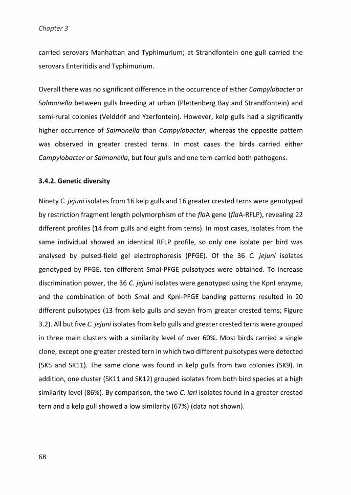
Chapter 3
68
carried serovars Manhattan and Typhimurium; at Strandfontein one gull carried the
serovars Enteritidis and Typhimurium.
Overall there was no significant difference in the occurrence of either Campylobacter or
Salmonella between gulls breeding at urban (Plettenberg Bay and Strandfontein) and
semi-rural colonies (Velddrif and Yzerfontein). However, kelp gulls had a significantly
higher occurrence of Salmonella than Campylobacter, whereas the opposite pattern
was observed in greater crested terns. In most cases the birds carried either
Campylobacter or Salmonella, but four gulls and one tern carried both pathogens.
3.4.2. Genetic diversity
Ninety C. jejuni isolates from 16 kelp gulls and 16 greater crested terns were genotyped
by restriction fragment length polymorphism of the flaA gene (flaA-RFLP), revealing 22
different profiles (14 from gulls and eight from terns). In most cases, isolates from the
same individual showed an identical RFLP profile, so only one isolate per bird was
analysed by pulsed-field gel electrophoresis (PFGE). Of the 36 C. jejuni isolates
genotyped by PFGE, ten different SmaI-PFGE pulsotypes were obtained. To increase
discrimination power, the 36 C. jejuni isolates were genotyped using the KpnI enzyme,
and the combination of both SmaI and KpnI-PFGE banding patterns resulted in 20
different pulsotypes (13 from kelp gulls and seven from greater crested terns; Figure
3.2). All but five C. jejuni isolates from kelp gulls and greater crested terns were grouped
in three main clusters with a similarity level of over 60%. Most birds carried a single
clone, except one greater crested tern in which two different pulsotypes were detected
(SK5 and SK11). The same clone was found in kelp gulls from two colonies (SK9). In
addition, one cluster (SK11 and SK12) grouped isolates from both bird species at a high
similarity level (86%). By comparison, the two C. lari isolates found in a greater crested
tern and a kelp gull showed a low similarity (67%) (data not shown).

Study I
69
A total of 235 Salmonella isolates from seabirds were analysed by enterobacterial
repetitive intergenic consensus (ERIC)-PCR and 43 different profiles were detected (38
from kelp gulls and five from greater crested terns). Ninety isolates representing the
different banding patterns identified were genotyped by PFGE using the XbaI enzyme.
Isolates that showed an identical XbaI-PFGE profile were further analysed with the
secondary enzyme BlnI. The combined analysis with XbaI and BlnI macrorestriction
profiles resulted in 29 PFGE pulsotypes (26 from kelp gulls and four from greater crested
terns) among the different serovars (Table 3.2). S. enterica subsp. salamae isolates from
kelp gulls presented three different pulsotypes with a similarity of 36%; one of them
(XB32) included isolates from three different gull colonies. The three S. Typhimurium
isolates showed three different PFGE profiles. Five distinct pulsotypes were detected
among the eight S. Enteritidis isolates, with one of them (XB7) found in both urban gull
colonies. Genetic diversity was low among S. Anatum isolates; 28 isolates were grouped
in three pulsotypes, with isolates from the two urban gull colonies clustering in one of
them (XB1). All seven S. Hadar isolates from both bird species and three different
colonies clustered together in the same pulsotype (XB12).
Genotyping of 47 human clinical isolates of Salmonella using the two restriction
enzymes revealed 14 different PFGE profiles (Table 3.2), with 70% of the human isolates
showing the same (23/47) or highly similar (10/47) pulsotype with bird isolates. The
single S. Anatum clinical isolate was non-typeable with BlnI. The XbaI-PFGE profile of
this isolate corresponded to the X1 pulsotype that included nine gull isolates, which also
were non-typeable with the secondary enzyme. All S. Enteritidis of human origin were
grouped in three pulsotypes which also included kelp gull isolates from urban colonies
with a similarity of ≥ 87%. Despite the genetic diversity detected among S. Typhimurium
isolates, a high similarity (84%) was observed between a kelp gull and a human isolate
(pulsotypes XB22 and XB23). It should be noted that half of the human S. Enteritidis
isolates and the human S. Typhimurium isolate that clustered together with isolates
from kelp gulls were invasive. Regarding S. Hadar, almost all clinical isolates belonged
Chapter 3
70
to the XB13 pulsotype which was closely related (88%) with XB12 that included isolates
from different colonies and seabird species.
3.4.3. Antimicrobial susceptibility
One isolate (both for Campylobacter and for Salmonella) per pulsotype from each
positive bird were tested for antimicrobial susceptibility to a panel of antimicrobials.
Most of the 33 Campylobacter isolates tested were pansusceptible (72.7%). However,
37.5% of kelp gull isolates and 17.6% of greater crested tern isolates were resistant to
at least one antimicrobial agent tested. All Campylobacter isolates were susceptible to
gentamicin, erythromycin, doxycycline and chloramphenicol. In kelp gulls, the main
antimicrobial resistances detected were to tetracycline (31.3%) and quinolones (12.5%).
Tetracycline resistance was mainly found in gulls from Velddrif (66.7%). At
Strandfontein, two isolates were resistant to both nalidixic acid and ciprofloxacin
(22.2%), and one was also resistant to tetracycline (11.1%). In greater crested terns,
three isolates showed resistance to quinolones (17.6%): one was resistant to nalidixic
acid and two were resistant to both nalidixic acid and ciprofloxacin. Among those
isolates resistant to nalidixic acid and ciprofloxacin, there were two C. lari from one kelp
gull and one greater crested tern.
Of the 66 Salmonella isolates analysed, 74.2% were pansusceptible. All Salmonella
isolates were susceptible to amoxicillin-clavulanate, ceftiofur, apramycin, gentamicin,
neomycin, ciprofloxacin, enrofloxacin, norfloxacin and colistin. The only resistance
detected in greater crested terns was to ampicillin and amoxicillin (14.3% each) (Table
3.3). The main resistance detected in kelp gulls were to tetracycline (13.6%) and
streptomycin (10.2%). Overall, resistance to antimicrobials was greater in urban gull
colonies (seven drugs) than gulls from semi-rural colonies (three drugs). Multidrug
resistance, defined as resistance to three or more classes of antimicrobial agents, was
present in three Salmonella isolates from kelp gulls, all from urban colonies. One S.
Idikan isolate from Strandfontein was resistant to six groups of antimicrobials

Study I
71
(streptomycin, tetracycline, doxycycline, chloramphenicol, lincomycin-spectinomycin
and trimethoprim-sulfamethoxazole), and two S. Paratyphi B var Java isolates from
Plettenberg Bay showed resistance to four antimicrobials (streptomycin, lincomycin-
spectinomycin, nitrofurantoin and trimethoprim-sulfamethoxazole).
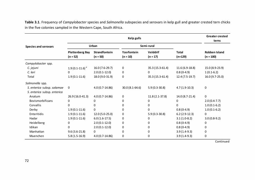
72
Table 3.1. Frequency of Campylobacter species and Salmonella subspecies and serovars in kelp gull and greater crested tern chicks
in the five colonies sampled in the Western Cape, South Africa.
Greater crested
terns
Plettenberg Bay
(n = 52)
Strandfontein
(n = 50)
Yzerfontein
(n = 10)
Velddrif
(n = 17)
Total
(n=129)
Robben Island
(n = 100)
Campylobacter spp.
C. jejuni 1.9 (0.1-11.6) a 16.0 (7.6-29.7) 0 35.3 (15.3-61.4) 11.6 (6.9-18.8) 15.0 (8.9-23.9)
C. lari 0 2.0 (0.1-12.0) 0 0 0.8 (0-4.9) 1 (0.1-6.2)
Total 1.9 (0.1-11.6) 18.0 (9.0-31.9) 0 35.3 (15.3-61.4) 12.4 (7.5-19.7) 16.0 (9.7-25.0)
Salmonella spp.
S. enterica subsp. salamae 0 4.0 (0.7-14.86) 30.0 (8.1-64.6) 5.9 (0.3-30.8) 4.7 (1.9-10.3) 0
S. enterica subsp. enterica
Anatum 26.9 (16.0-41.3) 4.0 (0.7-14.86) 0 11.8 (2.1-37.8) 14.0 (8.7-21.4) 0
Bovismorbificans 0 0 0 0 0 2.0 (0.4-7.7)
Corvallis 0 0 0 0 0 1.0 (0.1-6.2)
Derby 1.9 (0.1-11.6) 0 0 0 0.8 (0-4.9) 1.0 (0.1-6.2)
Enteritidis 1.9 (0.1-11.6) 12.0 (5.0-25.0) 0 5.9 (0.3-30.8) 6.2 (2.9-12.3) 0
Hadar 1.9 (0.1-11.6) 6.0 (1.6-17.5) 0 0 3.1 (1.0-8.2) 3.0 (0.8-9.2)
Heidelberg 0 2.0 (0.1-12.0) 0 0 0.8 (0-4.9) 0
Idikan 0 2.0 (0.1-12.0) 0 0 0.8 (0-4.9) 0
Manhattan 9.6 (3.6-21.8) 0 0 0 3.9 (1.4-9.3) 0
Muenchen 5.8 (1.5-16.9) 4.0 (0.7-14.86) 0 0 3.9 (1.4-9.3) 0
Species and serovars
Kelp gulls
Urban Semi-rural
Continued
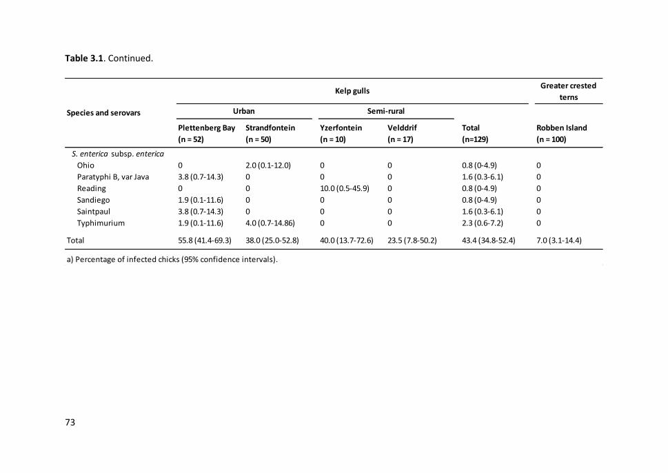
73
Table 3.1. Continued.
Greater crested
terns
Plettenberg Bay
(n = 52)
Strandfontein
(n = 50)
Yzerfontein
(n = 10)
Velddrif
(n = 17)
Total
(n=129)
Robben Island
(n = 100)
S. enterica subsp. enterica
Ohio 0 2.0 (0.1-12.0) 0 0 0.8 (0-4.9) 0
Paratyphi B, var Java 3.8 (0.7-14.3) 0 0 0 1.6 (0.3-6.1) 0
Reading 0 0 10.0 (0.5-45.9) 0 0.8 (0-4.9) 0
Sandiego 1.9 (0.1-11.6) 0 0 0 0.8 (0-4.9) 0
Saintpaul 3.8 (0.7-14.3) 0 0 0 1.6 (0.3-6.1) 0
Typhimurium 1.9 (0.1-11.6) 4.0 (0.7-14.86) 0 0 2.3 (0.6-7.2) 0
Total 55.8 (41.4-69.3) 38.0 (25.0-52.8) 40.0 (13.7-72.6) 23.5 (7.8-50.2) 43.4 (34.8-52.4) 7.0 (3.1-14.4)
a) Percentage of infected chicks (95% confidence intervals).
Species and serovars
Kelp gulls
Urban Semi-rural

PFGE SmaI+PFGE KpnI
SmaI&KpnI
100
90
80
70
60
PFGE SmaI PFGE KpnI
.
.
.
.
.
.
.
.
.
.
.
.
.
.
.
.
.
.
.
.
.
.
.
.
.
.
.
.
.
.
.
SD19-C1
SD38-C1
SD34-C2
VD11-C1
RB25-C1
RB66-C1
RB94-C4
RB49-C2
RB87-C1
RB89-C1
RB93-C1
SD41-C1
SD16-C1
VD8-C1
VD3-C1
VD16-C2
RB94-C1
RB99-C1
SD6-C1
RB33-C1
RB44-C2
RB88-C1
SD26-C1
RB18-C1
RB21-C1
RB38-C1
VD17-C1
RB98-C1
SD3-C1
VD10-C1
PD45-C1
.
.
.
.
.
.
.
.
.
.
.
.
.
.
.
.
.
.
.
.
.
.
.
.
.
.
.
.
.
.
.
.
.
.
.
.
.
.
.
.
.
.
.
.
.
.
.
.
.
.
.
.
.
.
.
.
.
.
.
.
.
.
Kelp gull
Kelp gull
Kelp gull
Kelp gull
Swift tern
Swift tern
Swift tern
Swift tern
Swift tern
Swift tern
Swift tern
Kelp gull
Kelp gull
Kelp gull
Kelp gull
Kelp gull
Swift tern
Swift tern
Kelp gull
Swift tern
Swift tern
Swift tern
Kelp gull
Swift tern
Swift tern
Swift tern
Kelp gull
Swift tern
Kelp gull
Kelp gull
Kelp gull
Strandfontein
Strandfontein
Strandfontein
Velddrif
Robben Island
Robben Island
Robben Island
Robben Island
Robben Island
Robben Island
Robben Island
Strandfontein
Strandfontein
Velddrif
Velddrif
Velddrif
Robben Island
Robben Island
Strandfontein
Robben Island
Robben Island
Robben Island
Strandfontein
Robben Island
Robben Island
Robben Island
Velddrif
Robben Island
Strandfontein
Velddrif
Plettenberg B.
.
.
.
.
.
.
.
.
.
.
.
.
.
.
.
.
.
.
.
.
.
.
.
.
.
.
.
.
.
.
.
.
.
.
.
.
.
.
.
.
.
.
.
.
.
.
.
.
.
.
.
.
.
.
.
.
.
.
.
.
.
.
.
.
.
.
.
.
.
.
.
.
.
.
.
.
.
.
.
.
.
.
.
.
.
.
.
.
.
S
S
S
Tet
S
S
S
S
S
S
CiprNal
S
S
Tet
S
Tet
S
S
S
S
S
S
S
S
Nal
S
Tet
S
CiprNalT.
Tet
S
637
4020
1223
1275
1268
1223
1268
2654
2654
1223
1292
1268
2353
572
1326
267
1275
1275
1275
1275
1275
1275
1275
1275
1275
1275
206
45
283
SmaI-PFGE KpnI-PFGE Pulsotype LocationIsolate Host
SD19-C1 SK1 Kelp gull Urb-Strandfontein
SD38-C1 SK2 Kelp gull Urb-Strandfontein
SD34-C2 SK3 Kelp gull Urb-Strandfontein
VD11-C1 SK4 Kelp gull Srur-Velddrif
RB25-C1 SK5 Greater crested tern Robben Island
RB66-C1 SK5 Greater crested tern Robben Island
RB94-C4 SK5 Greater crested tern Robben Island
RB49-C2 SK6 Greater crested tern Robben Island
RB87-C1 SK7 Greater crested tern Robben Island
RB89-C1 SK7 Greater crested tern Robben Island
RB93-C1 SK7 Greater crested tern Robben Island
SD41-C1 SK8 Kelp gull Urb-Strandfontein
SD16-C1 SK9 Kelp gull Urb-Strandfontein
VD8-C1 SK9 Kelp gull Srur-Velddrif
VD3-C1 SK9 Kelp gull Srur-Velddrif
VD16-C2 SK10 Kelp gull Srur-Velddrif
RB94-C1 SK11 Greater crested tern Robben Island
RB99-C1 SK11 Greater crested tern Robben Island
SD6-C1 SK12 Kelp gull Urb-Strandfontein
RB33-C1 SK13 Greater crested tern Robben Island
RB44-C2 SK13 Greater crested tern Robben Island
RB88-C1 SK13 Greater crested tern Robben Island
SD26-C1 SK14 Kelp gull Urb-Strandfontein
RB18-C1 SK15 Greater crested tern Robben Island
RB21-C1 SK15 Greater crested tern Robben Island
RB38-C1 SK15 Greater crested tern Robben Island
VD17-C1 SK16 Kelp gull Srur-Velddrif
RB98-C1 SK17 Greater crested tern Robben Island
SD3-C1 SK18 Kelp gull Urb-Strandfontein
VD10-C1 SK19 Kelp gull Srur-Velddrif
PD45-C1 SK20 Kelp gull Urb-Plettenberg Bay
90
10
0
80
70
60

75
Figure 3.2. PFGE combined dendrogram of SmaI and KpnI patterns of C. jejuni isolates. The similarity matrices were calculated using
the Dice coefficient and UPGMA clustering method. Profiles with a similarity ≥ 90% were considered same pulsotype. Urb: urban
colony; Srur: semi-rural colony.
PFGE SmaI+PFGE KpnI
SmaI&KpnI
100
90
80
70
60
PFGE SmaI PFGE KpnI
.
.
.
.
.
.
.
.
.
.
.
.
.
.
.
.
.
.
.
.
.
.
.
.
.
.
.
.
.
.
.
SD19-C1
SD38-C1
SD34-C2
VD11-C1
RB25-C1
RB66-C1
RB94-C4
RB49-C2
RB87-C1
RB89-C1
RB93-C1
SD41-C1
SD16-C1
VD8-C1
VD3-C1
VD16-C2
RB94-C1
RB99-C1
SD6-C1
RB33-C1
RB44-C2
RB88-C1
SD26-C1
RB18-C1
RB21-C1
RB38-C1
VD17-C1
RB98-C1
SD3-C1
VD10-C1
PD45-C1
.
.
.
.
.
.
.
.
.
.
.
.
.
.
.
.
.
.
.
.
.
.
.
.
.
.
.
.
.
.
.
.
.
.
.
.
.
.
.
.
.
.
.
.
.
.
.
.
.
.
.
.
.
.
.
.
.
.
.
.
.
.
Kelp gull
Kelp gull
Kelp gull
Kelp gull
Swift tern
Swift tern
Swift tern
Swift tern
Swift tern
Swift tern
Swift tern
Kelp gull
Kelp gull
Kelp gull
Kelp gull
Kelp gull
Swift tern
Swift tern
Kelp gull
Swift tern
Swift tern
Swift tern
Kelp gull
Swift tern
Swift tern
Swift tern
Kelp gull
Swift tern
Kelp gull
Kelp gull
Kelp gull
Strandfontein
Strandfontein
Strandfontein
Velddrif
Robben Island
Robben Island
Robben Island
Robben Island
Robben Island
Robben Island
Robben Island
Strandfontein
Strandfontein
Velddrif
Velddrif
Velddrif
Robben Island
Robben Island
Strandfontein
Robben Island
Robben Island
Robben Island
Strandfontein
Robben Island
Robben Island
Robben Island
Velddrif
Robben Island
Strandfontein
Velddrif
Plettenberg B.
.
.
.
.
.
.
.
.
.
.
.
.
.
.
.
.
.
.
.
.
.
.
.
.
.
.
.
.
.
.
.
.
.
.
.
.
.
.
.
.
.
.
.
.
.
.
.
.
.
.
.
.
.
.
.
.
.
.
.
.
.
.
.
.
.
.
.
.
.
.
.
.
.
.
.
.
.
.
.
.
.
.
.
.
.
.
.
.
.
S
S
S
Tet
S
S
S
S
S
S
CiprNal
S
S
Tet
S
Tet
S
S
S
S
S
S
S
S
Nal
S
Tet
S
CiprNalT.
Tet
S
637
4020
1223
1275
1268
1223
1268
2654
2654
1223
1292
1268
2353
572
1326
267
1275
1275
1275
1275
1275
1275
1275
1275
1275
1275
206
45
283
SmaI-PFGE KpnI-PFGE Pulsotype LocationIsolate Host
SD19-C1 SK1 Kelp gull Urb-Strandfontein
SD38-C1 SK2 Kelp gull Urb-Strandfontein
SD34-C2 SK3 Kelp gull Urb-Strandfontein
VD11-C1 SK4 Kelp gull Srur-Velddrif
RB25-C1 SK5 Greater crested tern Robben Island
RB66-C1 SK5 Greater crested tern Robben Island
RB94-C4 SK5 Greater crested tern Robben Island
RB49-C2 SK6 Greater crested tern Robben Island
RB87-C1 SK7 Greater crested tern Robben Island
RB89-C1 SK7 Greater crested tern Robben Island
RB93-C1 SK7 Greater crested tern Robben Island
SD41-C1 SK8 Kelp gull Urb-Strandfontein
SD16-C1 SK9 Kelp gull Urb-Strandfontein
VD8-C1 SK9 Kelp gull Srur-Velddrif
VD3-C1 SK9 Kelp gull Srur-Velddrif
VD16-C2 SK10 Kelp gull Srur-Velddrif
RB94-C1 SK11 Greater crested tern Robben Island
RB99-C1 SK11 Greater crested tern Robben Island
SD6-C1 SK12 Kelp gull Urb-Strandfontein
RB33-C1 SK13 Greater crested tern Robben Island
RB44-C2 SK13 Greater crested tern Robben Island
RB88-C1 SK13 Greater crested tern Robben Island
SD26-C1 SK14 Kelp gull Urb-Strandfontein
RB18-C1 SK15 Greater crested tern Robben Island
RB21-C1 SK15 Greater crested tern Robben Island
RB38-C1 SK15 Greater crested tern Robben Island
VD17-C1 SK16 Kelp gull Srur-Velddrif
RB98-C1 SK17 Greater crested tern Robben Island
SD3-C1 SK18 Kelp gull Urb-Strandfontein
VD10-C1 SK19 Kelp gull Srur-Velddrif
PD45-C1 SK20 Kelp gull Urb-Plettenberg Bay
90
10
0
80
70
60
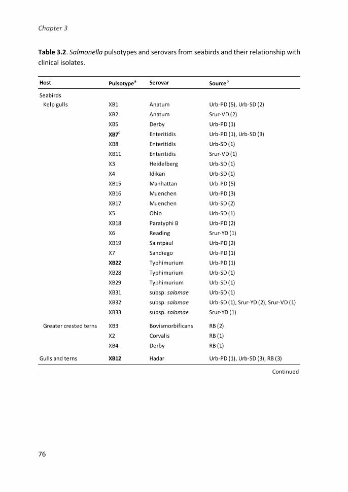
Chapter 3
76
Table 3.2. Salmonella pulsotypes and serovars from seabirds and their relationship with
clinical isolates.
Host Pulsotypea Serovar Sourceb
Seabirds
Kelp gulls XB1 Anatum Urb-PD (5), Urb-SD (2)
XB2 Anatum Srur-VD (2)
XB5 Derby Urb-PD (1)
XB7c Enteritidis Urb-PD (1), Urb-SD (3)
XB8 Enteritidis Urb-SD (1)
XB11 Enteritidis Srur-VD (1)
X3 Heidelberg Urb-SD (1)
X4 Idikan Urb-SD (1)
XB15 Manhattan Urb-PD (5)
XB16 Muenchen Urb-PD (3)
XB17 Muenchen Urb-SD (2)
X5 Ohio Urb-SD (1)
XB18 Paratyphi B Urb-PD (2)
X6 Reading Srur-YD (1)
XB19 Saintpaul Urb-PD (2)
X7 Sandiego Urb-PD (1)
XB22 Typhimurium Urb-PD (1)
XB28 Typhimurium Urb-SD (1)
XB29 Typhimurium Urb-SD (1)
XB31 subsp. salamae Urb-SD (1)
XB32 subsp. salamae Urb-SD (1), Srur-YD (2), Srur-VD (1)
XB33 subsp. salamae Srur-YD (1)
Greater crested terns XB3 Bovismorbificans RB (2)
X2 Corvalis RB (1)
XB4 Derby RB (1)
Gulls and terns XB12 Hadar Urb-PD (1), Urb-SD (3), RB (3)
Continued

Study I
77
Table 3.2. Continued.
Host Pulsotypea Serovar Sourceb
Humans and kelp gulls X1 Anatum Urb-PD (9), HCT (1)
XB9 Enteritidis Urb-SD (1), HCT(16)
XB10 Enteritidis Urb-SD (1), HCT (6)
Humans XB6 Enteritidis HCT (2)
XB13 Hadar HCT (7)
XB14 Hadar HCT (1)
XB20 Typhimurium HCT (2)
XB21 Typhimurium HCT (1)
XB23 Typhimurium HCT (1)
XB24 Typhimurium HCT (5)
XB25 Typhimurium HCT (1)
XB26 Typhimurium HCT (1)
XB27 Typhimurium HCT (1)
XB30 Typhimurium HCT (2)
a) Profiles with a similarity ≥ 90% were considered the same pulsotype. Pulsotypes referred as X were
assigned by XbaI macrorestriction profies; XB indicates the combination of XbaI and BlnI profiles.
b) Urb-PD: urban colony at Plettenberg Bay; Urb-SD: urban colony at Strandfontein; Srur-YD: semi-rural
colony at Yzerfontein; Srur-VD: semi-rural colony at Velddrif; RB: Robben Island colony. Number of
isolates is given in brackets.
c) Pulsotypes with similarities of 84-89% among isolates from seabirds and humans are highlighted in
bold: XB7, XB6 S. Enteritidis pulsotypes with 86% similarity; XB22, XB23 S. Typhimurium pulsotypes
with 84% similarity; XB12, XB13 S. Hadar with 88% similarity.

78
Table 3.3. Antimicrobial resistance of Salmonella isolates according to the seabird species and colony.
Amp Amx Str Nal Tet Dox Chl Ni Lisp Sxt
Kelp gulls
Urban
Plettenberg Bay 31 0c 0 2 (6.5) 0 5 (16.1) 0 0 2 (6.5) 3 (9.7) 2 (6.5)
Strandfontein 20 0 0 2 (10.0) 0 3 (15.0) 3 (15.0) 1 (5.0) 0 1 (5.0) 3 (15.0)
Semi-rural
Yzerfontein 4 0 0 2 (50.0) 0 0 0 0 0 0 0
Velddrif 4 0 0 0 1 (25.0) 0 0 0 1 (25.0) 0 0
Total 59 0 0 6 (10.2) 1 (1.7) 8 (13.6) 3 (5.1) 1 (1.7) 3 (5.1) 4 (6.8) 5 (8.5)
Greater crested terns
Robben Island 7 1 (14.3) 1 (14.3) 0 0 0 0 0 0 0 0
c) Number of isolates resistant (%).
Sampled colony Na
Antimicrobial agentsb
b) Amp: ampicil l in; Amx: amoxicil l in; Str: streptomycin; Nal: nalidixic acid; Tet: tetracycline; Dox: doxycycline; Chl: chloramphenicol; Ni:
nitrofurantoin; LiSp: lincomycin + spectinomycin; Sxt: trimethoprim + sulfamethoxazole. All isolates were susceptible to: amoxicil l in +
clavulanate, ceftiofur, apramycin, gentamicin, neomycin, ciprofloxacin, enrofloxacin, norfloxacin and colistin.
a) Number of isolates tested.

Study I
79
3.5. DISCUSSION
The presence of zoonotic bacteria in wild birds has been described in many studies, with
differences in the reported prevalence depending on the animal species and the
location (Benskin et al., 2009). However, little is known of the occurrence of zoonotic
agents in African free-living birds. We report the presence, genetic diversity and
antimicrobial resistance of the two most relevant foodborne zoonotic agents,
Campylobacter and Salmonella, in seabirds in the Western Cape, South Africa. Birds
have been reported as asymptomatic carriers of these pathogens, and seabirds nearby
human settlements may be more prone to carry these bacteria, particularly those with
scavenging feeding habits, such as gulls. Campylobacter occurrence in kelp gulls (12%)
and greater crested terns (16%) was similar to that reported in yellow-legged gulls Larus
michahellis in southern Europe (10%, Ramos et al., 2010), but lower than in other gulls
in the eastern coast of United States (laughing gulls Leucophaeus atricilla, 33%) and in
northern Europe (black-headed gulls Chroicocephalus ridibundus, 28-36%) (Broman et
al., 2002; Keller et al., 2011). The correlation between Campylobacter prevalence in wild
birds and their feeding habits (Sensale et al., 2010; Hald et al., 2016), points to foraging
in human waste and sewage as important risk factors of infection (Ramos et al., 2010).
Contrary to what would be expected, the highest Campylobacter prevalence was found
in a colony (Velddrif) from a semi-rural area. In this site, sampling took place later in the
season, during slightly warmer weather, when chicks were older on average and were
moving around in an area where other birds had been active for a longer period. Thus,
the area was more likely to be contaminated by these bacteria and could have
contributed to an increased Campylobacter occurrence in chicks from this colony. On
the other hand, Campylobacter species have been reported in livestock grazing in
pastures (Oporto et al., 2007; Moriarty et al., 2008; Sproston et al., 2011). Although no
information is available about Campylobacter occurrence in livestock from South Africa,
sheep present in the area may also have been the source of C. jejuni in this colony.
Chapter 3
80
We detected a higher occurrence of Salmonella spp. in kelp gulls (43%) than in greater
crested terns (7%). The prevalence of Salmonella shedding among wild birds appears to
be quite variable. For instance, a high Salmonella occurrence (up to 75%) in gulls has
been found in certain colonies of the western Mediterranean (Cerdà-Cuéllar,
unpublished data). In contrast, recent studies report a moderate (13%-17%) to low (3%)
Salmonella prevalence in other gull species in Australia and Europe (Palmgren et al.,
2006; Ramos et al., 2010; Dolejska et al., 2016; Masarikova et al., 2016). The difference
in Salmonella occurrence between kelp gulls and greater crested terns may be due to
the different location of these bird colonies and their feeding habits. The greater crested
tern’s diet consists predominantly of pelagic fish, other marine prey and occasionally
insects, whilst the kelp gull is a generalist that often scavenges at refuse dumps and
sewage treatment plants (Hockey et al., 2005; Gaglio et al., 2017), which are commonly
contaminated with pathogenic bacteria. Our results suggest that seabird feeding habits
affects the incidence of Salmonella more than Campylobacter. We found the highest
frequency of Salmonella at Plettenberg Bay, which is located close to a municipal refuse
dump (Witteveen et al., 2017) and a higher overall prevalence of Salmonella spp. than
Campylobacter spp., especially in kelp gulls from urban areas, probably due to the
increased presence and survival of Salmonella in gull breeding and feeding areas (Literák
et al., 1996). Also, the high survival rate of Salmonella in aquatic environments (Winfiel
and Groisman, 2003) and the presence of this pathogen in surface waters (Levantesi et
al., 2012), may facilitate seabird infections. Contrary to Salmonella, Campylobacter is
more susceptible to environmental stress (Murphy et al., 2006) and its survival in the
environment may be lower.
The wide variety of Salmonella serovars found in our study, particularly in urban kelp
gull colonies, suggests a lack of host-specificity and the presence of different sources of
Salmonella contamination. All the Salmonella serovars detected, except Corvallis and
Paratyphi B var Java, previously have been found in South African farm animals, where
the most common serovars are Typhimurium, Dublin, Enteritidis, Muenchen,
Study I
81
Heildelberg and Hadar (Magwedere et al., 2015). Typhimurium, Enteritidis and
Heidelberg are also the most frequently reported serovars in humans in South Africa
(GERMS-SA, 2014). Anatum, Enteritidis and Hadar were the most frequent isolated
serovars in our seabirds, all of them involved in salmonellosis outbreaks (Havelaar et al.,
2015). At the Strandfontein gull colony, located next to a major urban refuse dump,
there was a high diversity of Salmonella, including the highest frequency of zoonotic
serovars. This was also the case at the Plettenberg Bay gull colony, where we detected
S. Paratyphi B var Java. This serovar, which is thought to be non-zoonotic, is very rare in
South Africa (Smith et al., 2016) but has been reported in stool samples from humans
in the Western Cape in 2012 and the Eastern Cape in 2013 (GERMS-SA, 2013, 2014).
The high genetic diversity observed among Campylobacter isolates is common in this
bacterium because of its genetic instability (Wassenaar et al., 1998; Boer et al., 2002).
This genomic variability seems to be an important mechanism in environmental stress
situations, generating population heterogeneity to improve fitness and survival in
hostile environments and colonize other hosts (Ridley et al., 2008). In our study, a higher
genetic diversity was detected in C. jejuni isolates from kelp gulls than greater crested
terns: 13 and seven PFGE profiles from 15 gulls and 15 terns, respectively. This greater
Campylobacter genetic diversity in gulls may reflect different sources of infection
probably due to their trophic plasticity. Even so, we detected a pulsotype in common in
gulls from colonies located in semi-rural and urban areas, and two genetically close
isolates from a greater crested tern and a kelp gull. This suggests a degree of
connectivity among colonies and bird species, either directly or indirectly through a
common contamination source.
Ten S. Anatum isolates (nine from gulls and one human clinical isolate) had identical
XbaI-PFGE profiles, but none of them could be genotyped with the secondary restriction
enzyme BlnI. This may be due to the lack of restriction sites or the presence of
methylation patterns affecting the restriction enzyme recognition sites (Oyarzabal et al.,
2008). In Salmonella, genetic diversity depends largely on the serovar. Here, diversity
Chapter 3
82
was greater among isolates of S. Typhimurium and S. Enteritidis, whereas clonality was
frequent among S. Anatum and S. Hadar isolates. As for Campylobacter, the presence
of pulsotypes of Salmonella in common to different colonies and seabird species
indicates the likely transfer of strains among colonies or a common source of infection.
On the other hand, the finding that 70% of human Salmonella isolates had pulsotypes
the same or highly similar to those from bird isolates indicates an epidemiological link
between them. Our results suggest that gulls breeding near urban areas can acquire
human pathogenic Salmonella when they feed on anthropogenic food sources,
including urban waste landfills or sewage, as has previously have been reported in other
continents (Thorbjørn Refsum et al., 2002; Palmgren et al., 2006; Dolejska et al., 2016;
Hernandez et al., 2016). Thus, gulls act as healthy carriers of strains involved in human
disease and to some extent mirror Salmonella strains circulating in the humanised
environment.
Despite around 70% of the isolates being pansusceptible, the notable frequency of
antimicrobial resistant isolates detected is a cause of concern given that wild birds,
unlike production animals, are not directly exposed to them. The main Campylobacter
resistance detected was to quinolones and tetracycline (15%). Previous studies have
reported high levels of tetracycline (22-100%) and quinolone (8-60%) resistant
Campylobacter isolates in different production animals and farming systems in South
Africa (Jonker and Picard, 2010; Bester and Essack, 2012). The broad use of tetracyclines
in animal production systems in South Africa (Eagar et al., 2012) may account for the
high incidence of Campylobacter isolates resistant to this class of antimicrobial agents
in livestock animals, which in turn may influence the resistance found in wildlife. To a
lesser extent tetracyclines are also used in human medicine, although the drugs of
choice as first line therapy for bacterial gastroenteritis are fluoroquinolones and
macrolides. In recent years, there has been a dramatic increase in the proportion of
Campylobacter clinical isolates in Cape Town resistant to nalidixic acid and ciprofloxacin
(Lastovica, 2006). The simplicity of the resistance mechanism to quinolones and the

Study I
83
regular use of these agents may account for the rapid increase in Campylobacter
resistant strains. The frequent use of enrofloxacin in animal production induces
Campylobacter cross-resistance to other quinolones (Payot et al., 2002; Takahashi et al.,
2005). C. lari strains isolated from kelp gulls and greater crested terns showed resistance
to nalidixic acid, which is intrinsic in this Campylobacter species (Piddock et al., 2003),
and to ciprofloxacin; both resistance frequently are observed in the NARTC (nalidixic
acid resistant thermophilic Campylobacter) biotype (Leatherbarrow et al., 2007).
Even though Campylobacter and Salmonella share the same niche in birds, the use of
quinolones in South Africa has not produced a similar resistance pattern among them.
The main Salmonella resistance detected in our study were to tetracycline and
streptomycin in kelp gulls and β-lactams in one greater crested tern. It is noteworthy
that these drugs are used in human medicine (although tetracycline is not first choice).
In gull colonies near urban areas, we found a greater number of Salmonella isolates
resistant to a wide variety of antimicrobial drugs, including multidrug resistant strains,
which may be indicative of anthropogenic pressure. Although greater crested terns feed
on live prey captured mostly off shore, one isolate showed resistance to a β-lactam
(ampicillin); this resistance is also found with some frequency in human clinical isolates
(GERMS-SA, 2014). At Robben Island, greater crested terns often roost and breed in
association to Hartlaub’s gulls (Chroicocephalus hartlaubi) which are more commensal
with humans than kelp gulls in the greater Cape Town area. Their interaction with this
gull species may explain the ampicillin resistance found in terns.
In conclusion, our results suggest that kelp gulls and greater crested terns in the
Western Cape of South Africa are a source of zoonotic thermophilic Campylobacter
species and Salmonella serovars, with several Salmonella strains in common to both
humans and gulls. Many isolates are resistant and multidrug-resistant (in the case of
Salmonella) to antimicrobial agents, including critically important antimicrobials for
human medicine (WHO-AGISAR, 2017). Our results highlight the importance of seabirds
as reservoirs of Campylobacter and Salmonella resistant strains in the Western Cape

Chapter 3
84
and their potential role in the maintenance and transmission of these bacteria in the
environment, with significant implications for public health management. Further
studies are needed to identify where these seabirds have acquired the zoonotic
bacteria.

CHAPTER 4
Study II: Humans spread zoonotic enteric
bacteria in Antarctica?


Study II
87
4.1. SUMMARY
Reports of enteric bacteria in Antarctic wildlife have suggested its spread from people
to seabirds and seals, but evidence is scarce and fragmentary. We investigated the
occurrence of zoonotic bacteria in poultry and seabirds across the Antarctic and
Subantarctic region. Three findings suggest reverse zoonosis from humans to seabirds:
the detection of a zoonotic Salmonella serovar Enteritidis and Campylobacter species
(e.g. C. jejuni), typical of human infections; the resistance of C. lari isolates to
ciprofloxacin and enrofloxacin, antibiotics commonly used in human and veterinary
medicine; and most importantly, the presence of C. jejuni genotypes mostly found in
humans and domestic animals. We also show further spread of zoonotic agents among
Antarctic wildlife is facilitated by substantial connectivity among populations of
opportunistic seabirds, notably skuas (Stercorarius). Our results highlight the need for
even stricter biosecurity measures to limit human impacts in Antarctica.
Chapter 4
88
4.2. INTRODUCTION
The global spread of pathogens is a growing conservation concern because their
introduction into novel environments can have dramatic effects on wildlife (Van Riper
et al., 1986; Paxton et al., 2016). Pathogens have been dispersed by migratory birds,
fish, mammals and other taxa for millions of years, but in recent centuries humans have
also contributed to their dispersal (Altizer et al., 2011; Fuller et al., 2012). Antarctica is
the only continent where reverse zoonosis transmission has not been documented
(Messenger et al., 2014). Despite ongoing concern about human impacts in the region,
diseases have not been identified as significant threats (Chown, Huiskes, et al., 2012;
Chown, Lee, et al., 2012).
To date, the presence of pathogens in Antarctic wildlife has received limited attention
(Barbosa and Palacios, 2009; Kerry and Riddle, 2009). It has been assumed that the
region’s isolation and relatively recent exploration by humans have protected Antarctic
wildlife from novel pathogens, although there have been several outbreaks of infectious
diseases at Southern Ocean islands (Weimerskirch, 2004; Cooper et al., 2009; Kane et
al., 2012). The few surveys of pathogens in Antarctica have been opportunistic, and
investigations of occasional mass mortality events to date have not established clear
evidence of human-to-animal transmission (Gardner et al., 1997; Frenot et al., 2005;
Iveson et al., 2009; Kerry and Riddle, 2009; Vigo et al., 2011; Hernandez et al., 2012).
The mechanisms by which pathogens invaded the Southern Ocean wildlife remain
uncertain. Human-mediated transport may be a legacy of exposure in the last few
centuries to sealers and whalers or to their domestic animals (Gardner et al., 1997;
Griekspoor et al., 2010), but several studies indicate that the main risk of pathogen
invasion is the increase in tourism and research activities, which currently account for
tens of thousands of visitors each year (Curry et al., 2002; Hughes and Convey, 2010).
In this regard, the Protocol on Environmental Protection to the Antarctic Treaty (1991),
which came into force in 1996, included a number of measures to prevent the

Study II
89
introduction of novel pathogens (Committee for Environmental Protection, 2011).
However, it may be of limited value if Antarctic wildlife disperses to areas outside the
Antarctic region, where they can be exposed to a wide range of pathogens. Many
Antarctic seabirds disperse across the Southern Ocean, coming into contact with
domestic species in populated areas, and some species that visit the region during the
Antarctic summer spend the winter in the Northern Hemisphere (e.g. Arctic Terns
Sterna paradisaea and South Polar Skuas Stercorarius maccormicki). Such large-scale
movements may introduce pathogens to Antarctica, and disperse them within the
region. Climate change also may alter the migratory habits of animals, increasing the
spread and contact between Antarctic, Subantarctic and temperate wildlife (Altizer et
al., 2013).
The zoonotic bacteria Salmonella spp. and thermotolerant Campylobacter spp. are
amongst the most important foodborne diarrheal pathogens worldwide (Havelaar et al.,
2015). Both agents can spread rapidly in the environment through faecal contamination
and can persist in soil or water for long enough to infect wild fauna. We explore the
transfer of these zoonotic bacteria from humans and poultry to Subantarctic and
Antarctic by sampling 24 seabird species over a broad geographical range, identifying
bacterial species and comparing serovars and genotypes in seabirds with those
commonly found in humans and domestic animals, and by testing their resistance to
antibiotics commonly used in human and veterinary medicine. We also assess whether
these pathogens are spreading across wildlife of the Southern Ocean.
4.3. MATERIAL AND METHODS
4.3.1. Sampling
From 2008 to 2011 we collected faecal samples from adult seabirds at four Southern
Ocean localities: Livingston (Antarctica), Marion, Gough and the Falkland Islands (Figure
Chapter 4
90
4.1.A; Table 4.1). Additionally, we also sampled backyard poultry at the Falklands, which
support a permanent human settlement with a number of farms in close contact with
Subantarctic and Antarctic wildlife. Birds were caught by hand and faecal samples were
collected in duplicate using sterile swabs inserted into the cloaca. Samples were stored
refrigerated in Amies transport medium with charcoal (Deltalab, Barcelona, Spain),
transported to Spain within two to five weeks after the day of collection and cultured
immediately upon arrival to the laboratory.
4.3.2. Bacterial isolation and identification
We performed Salmonella and Campylobacter isolation and identification by standard
culture methods (Antilles et al., 2015). Salmonella serotyping was performed according
to the Kauffmann-White scheme (Grimont and Weill, 2007) and carried out at the
Laboratori Agroalimentari (Cabrils, Spain) of the Departament d’Agricultura, Ramaderia,
Pesca, Alimentació i Medi Natural. We identified Campylobacter isolates to species level
by PCR using primers based on the lpxA gene (Klena et al., 2004). A multiplex PCR for C.
jejuni and C. coli identification was performed using forward primers lpxA-Cjejuni (5’-
ACA ACT TGG TGA CGA TGT TGTA-3’) and lpxA-Ccoli (5’-AGA CAA ATA AGA GAG AGA
ATC AG-3’) and a common reverse primer (lpxARKK2m: 5’-CAA TCA TGD GCD ATA TGA
SAA TAH GCC AT-3’). C. lari identification was performed with a monoplex PCR using
primers lpxA-Clari (5’-TRC CAA ATG TTA AAA TAG GCG A-3’) and lpxARKK2m.
4.3.3. Antimicrobial susceptibility testing
We performed antimicrobial susceptibility testing for both Salmonella and
Campylobacter isolates following the Clinical Laboratory and Standard Institute (CLSI)
disc diffusion method (M100-S18) (CLSI, 2016) using Neo-Sensitabs™ (Rosco
Diagnostica, Denmark) with CLSI potencies according to the manufacturer’s
instructions. For Salmonella isolates, we used Mueller-Hinton agar (Difco, Madrid,
Spain) and plates were incubated at 37°C for 24 h. For Campylobacter isolates, we used
Study II
91
Mueller-Hinton II agar supplemented with 5% defibrinated sheep blood (BioMérieux,
Marcy l'Etoile, France) and plates were incubated at 37°C for 48 h under microaerobic
conditions.
Salmonella isolates were tested against 18 antimicrobials: ampicillin (33 µg), amoxicillin
(30 µg), amoxicillin-clavulanate (30 + 15 µg), ceftiofur (30 µg), apramycin (40 µg),
streptomycin (100 µg), gentamicin (40 µg), neomycin (120 µg), ciprofloxacin (10 µg),
enrofloxacin (10 µg), nalidixic acid (130 µg), norfloxacin (10 µg), colistin (150 µg),
chloramphenicol (60 µg), lincomycin-spectinomycin (15 + 200 µg), nitrofurantoin (260
µg), tetracycline (80 µg) and trimethoprim-sulfonamide (5.2 + 240 µg). Campylobacter
isolates were tested against seven antimicrobials: nalidixic acid (30 µg), ciprofloxacin (5
µg), enrofloxacin (10 µg), tetracycline (80 µg), chloramphenicol (60 µg), erythromycin
(15 µg) and gentamicin (10 µg).
4.3.4. Salmonella and Campylobacter genotyping
We typed representative bacterial isolates with pulsed-field gel electrophoresis (PFGE)
and multilocus sequence typing (MLST). PFGE was carried out according to the standard
operating procedure of PulseNet (www.pulsenetinternational.org). We performed
restriction enzyme digests for PFGE with XbaI and BlnI enzymes for Salmonella, and with
SmaI and KpnI enzymes for Campylobacter (Roche Applied Science, Indianapolis, IN,
USA). Salmonella Braenderup H9812 restricted with XbaI was used as molecular size
standard for both Campylobacter and Salmonella. We analysed the resulting PFGE
patterns using Fingerprinting II v3.0 software (Bio-Rad, Hercules, CA, USA). Banding
patterns were compared with the UPGMA (Unweighted Pair Group Method with
Arithmetic averages) clustering method using the Dice correlation coefficient with a
band position tolerance of 1%.
We further characterized S. enterica and thermotolerant Campylobacter using MLST,
which is based on sequencing of seven housekeeping genes (Dingle et al., 2001; Miller
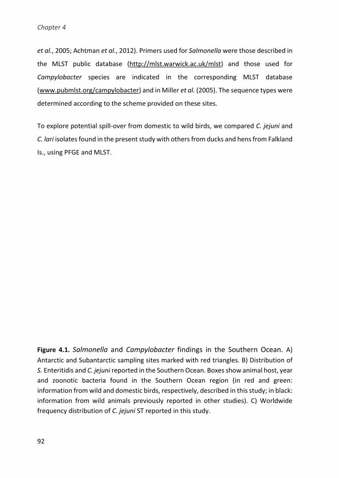
Chapter 4
92
et al., 2005; Achtman et al., 2012). Primers used for Salmonella were those described in
the MLST public database (http://mlst.warwick.ac.uk/mlst) and those used for
Campylobacter species are indicated in the corresponding MLST database
(www.pubmlst.org/campylobacter) and in Miller et al. (2005). The sequence types were
determined according to the scheme provided on these sites.
To explore potential spill-over from domestic to wild birds, we compared C. jejuni and
C. lari isolates found in the present study with others from ducks and hens from Falkland
Is., using PFGE and MLST.
Figure 4.1. Salmonella and Campylobacter findings in the Southern Ocean. A)
Antarctic and Subantarctic sampling sites marked with red triangles. B) Distribution of
S. Enteritidis and C. jejuni reported in the Southern Ocean. Boxes show animal host, year
and zoonotic bacteria found in the Southern Ocean region (in red and green:
information from wild and domestic birds, respectively, described in this study; in black:
information from wild animals previously reported in other studies). C) Worldwide
frequency distribution of C. jejuni ST reported in this study.
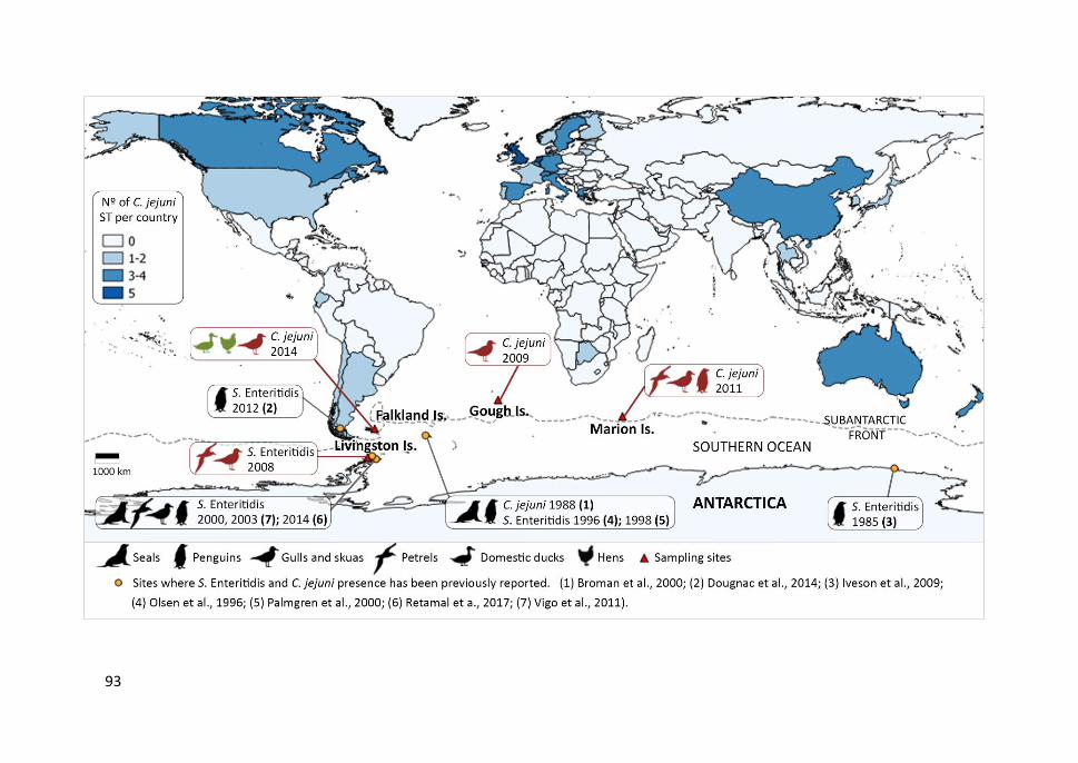
93
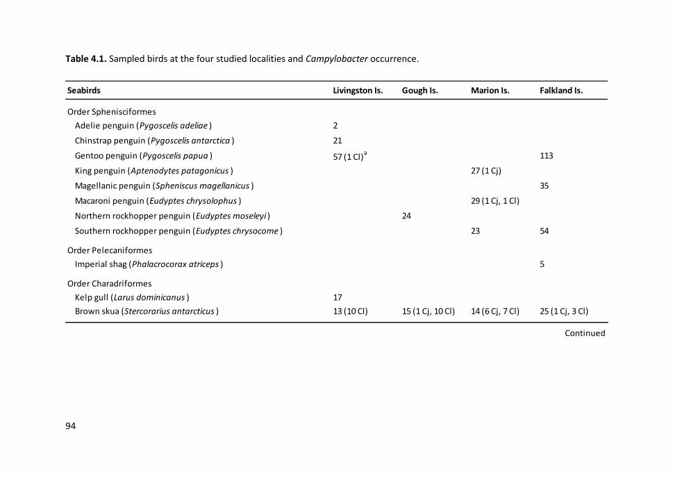
94
Table 4.1. Sampled birds at the four studied localities and Campylobacter occurrence.
Seabirds Livingston Is. Gough Is. Marion Is. Falkland Is.
Order Sphenisciformes
Adelie penguin (Pygoscelis adeliae ) 2
Chinstrap penguin (Pygoscelis antarctica ) 21
Gentoo penguin (Pygoscelis papua ) 57 (1 Cl)a 113
King penguin (Aptenodytes patagonicus ) 27 (1 Cj)
Magellanic penguin (Spheniscus magellanicus ) 35
Macaroni penguin (Eudyptes chrysolophus ) 29 (1 Cj, 1 Cl)
Northern rockhopper penguin (Eudyptes moseleyi ) 24
Southern rockhopper penguin (Eudyptes chrysocome ) 23 54
Order Pelecaniformes
Imperial shag (Phalacrocorax atriceps ) 5
Order Charadriformes
Kelp gull (Larus dominicanus ) 17
Brown skua (Stercorarius antarcticus ) 13 (10 Cl) 15 (1 Cj, 10 Cl) 14 (6 Cj, 7 Cl) 25 (1 Cj, 3 Cl)
Continued

95
Table 4.1. Continued.
Seabirds Livingston Is. Gough Is. Marion Is. Falkland Is.
Order Procellariiformes
Atlantic yellow-nosed albatross (Thalassarche chlororhynchos ) 13
Black-browed albatross (Thalassarche melanophris ) 20
Sooty albatross (Phoebetria fusca ) 5
Atlantic petrel (Pterodroma incerta ) 21
Soft-plumaged petrel (Pterodroma mollis ) 32
Northern giant petrel (Macronectes halli ) 16
Southern giant petrel (Macronectes giganteus ) 29 (1 Cl) 9 15 (2 Cl) 2
White-chinned petrel (Procellaria aequinoctialis ) 1
Broad-billed prion (Pachyptila vittata ) 3
Great shearwater (Ardenna gravis) 16
Sooty shearwater (Ardenna grisea ) 10
Total 139 138 125 264
a) Number of Campylobacter positive birds in brackets. Cj: C. jejuni , Cl: C. lari .

Chapter 4
96
4.4. RESULTS
4.4.1. Salmonella and Campylobacter spp. in seabirds
We sampled 666 seabirds from 24 species at Livingston (n= 139), Gough (n= 138),
Marion (n= 125) and the Falkland Islands (n= 264) (Figure 4.1.A; Table 4.1), and isolated
three Salmonella serovar Enteritidis, 10 C. jejuni and 35 C. lari. The only other Salmonella
serovar detected was one Oakey; no other thermotolerant Campylobacter species were
found.
We isolated Salmonella Enteritidis from two kelp gulls and one southern giant petrel
from Livingston Is.; C. jejuni from one macaroni penguin, one king penguin, and six
brown skuas at Marion Is. and from single brown skuas at Gough and the Falkland Is.
(Figure 4.1.B); and C. lari from one gentoo penguin, one southern giant petrel and 10
brown skuas at Livingston Is.; from one macaroni penguin, two southern giant petrels
and seven brown skuas at Marion Is.; from 10 brown skuas at Gough Is.; and from three
brown skuas at the Falkland Is. Marion Is. showed the highest diversity of positive
seabird species to Campylobacter and was the only locality where co-infections
occurred of both C. jejuni and C. lari (n=3 skuas).
4.4.2. Antimicrobial resistance
We did not detect any antimicrobial resistance in isolates of Salmonella or C. jejuni.
Among C. lari isolates, besides nalidixic acid resistance which is characteristic of this
species and was found in all tested isolates, we found ciprofloxacin resistance in isolates
from one macaroni penguin and two Subantarctic skuas from Marion Is., and from three
skuas from Gough Is. Ciprofloxacin and enrofloxacin resistance was detected in two C.
lari from skuas at Livingston Is.
Study II
97
4.4.3. Genetic diversity
All three Salmonella Enteritidis isolates exhibited identical PFGE patterns and MLST
sequence type (ST-11). Among C. jejuni isolates, PFGE analysis clustered together three
isolates: two from brown skuas from the Falklands and Marion Is. and one from a
domestic duck from the Falklands. MLST showed these isolates to belong to the
widespread ST-45. Four other C. jejuni ST (ST-137, ST-227, ST-696 and ST-883) were
isolated from skuas and penguins at Gough and Marion Is. (Figure 4.2). These ST have
been mostly reported in several hosts in developed countries of the Northern
Hemisphere, Australia and New Zealand (Figure 4.1.C).
Among C. lari isolates, PFGE genotyping showed highly similar isolates (> 80% similarity)
from several skuas at Livingston, Marion and Gough Is. and from a giant petrel at Marion
Is. One cluster was formed by three (GH128-C1, GH131-C1 and MAR5-C1) nearly
identical isolates (≥ 95% similarity) found in skuas from Gough and Marion Is. belonging
to the same novel ST (Figure 4.3). In addition, the same genotype was found in two
different seabird species, a brown skua and a gentoo penguin from Livingston Is.
(isolates AN138-C7 and AN32-C1), which were closely related (81% similarity) to an
isolate from a duck (FK72-C1) from the Falklands. One cluster grouped isolates from
distant localities, i.e. one isolate from a skua at the Falklands and one from a penguin at
Marion Is. (FK54-C1 and MAR18-C1), with an 88% similarity.
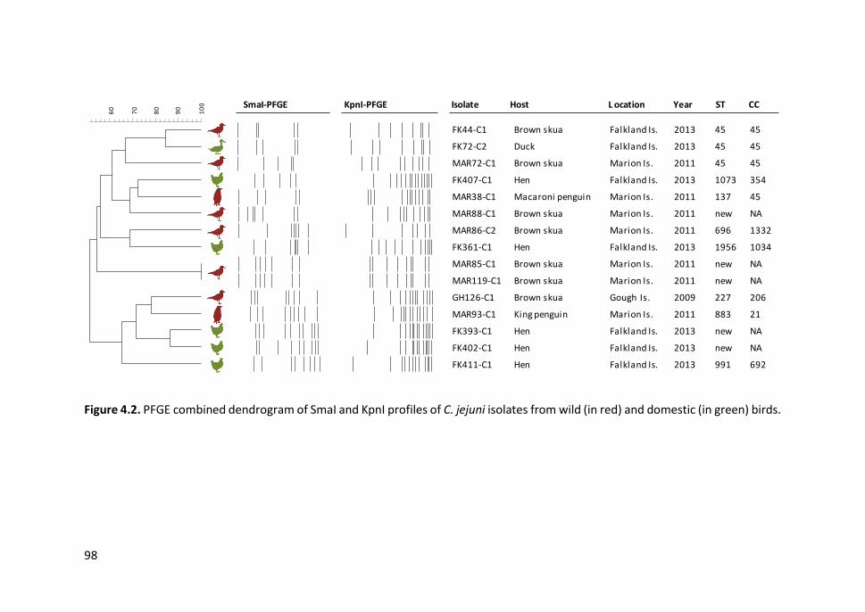
98
Figure 4.2. PFGE combined dendrogram of SmaI and KpnI profiles of C. jejuni isolates from wild (in red) and domestic (in green) birds.
SmaI-PFGE KpnI-PFGE Isolate Host L ocation Year ST CC
PFGE SmaI+PFGE KpnI
SmaI&KpnI
100
90
80
70
60
PFGE SmaI PFGE KpnI
.
.
.
.
.
.
.
.
.
.
.
.
.
.
.
FK44-C1
FK72-C2
MAR72-C1
FK407-C1
MAR38-C1
MAR88-C1
MAR86-C2
FK361-C1
MAR85-C1
MAR119-C1
GH126-C1
MAR93-C1
FK393-C1
FK402-C1
FK411-C1
.
.
.
.
.
.
.
.
.
.
.
.
.
.
.
.
.
.
.
.
.
.
.
.
.
.
.
.
.
.
Brown skua
Duck
Brown skua
Hen
Maccaroni penguin
Brown skua
Brown skua
Hen
Brown skua
Brown skua
Brown skua
King penguin
Hen
Hen
Hen
Falklands
Falklands
Marion
Falklands
Marion
Marion
Marion
Falklands
Marion
Marion
Gough
Marion
Falklands
Falklands
Falklands
.
.
.
.
.
.
.
2013
2013
2011
2013
2011
2011
2011
2013
2011
2011
2009
2011
2013
2013
2013
.
.
.
.
.
.
.
.
.
.
.
.
.
.
45
45
45
1073
137
new
696
1956
new
new
227
883
new
new
991
45
45
45
354
45
1332
1034
206
21
692
FK44-C1 Brown skua Falkland Is. 2013 45 45
FK72-C2 Duck Falkland Is. 2013 45 45
MAR72-C1 Brown skua Marion Is. 2011 45 45
FK407-C1 Hen Falkland Is. 2013 1073 354
MAR38-C1 Macaroni penguin Marion Is. 2011 137 45
MAR88-C1 Brown skua Marion Is. 2011 new NA
MAR86-C2 Brown skua Marion Is. 2011 696 1332
FK361-C1 Hen Falkland Is. 2013 1956 1034
MAR85-C1 Brown skua Marion Is. 2011 new NA
MAR119-C1 Brown skua Marion Is. 2011 new NA
GH126-C1 Brown skua Gough Is. 2009 227 206
MAR93-C1 King penguin Marion Is. 2011 883 21
FK393-C1 Hen Falkland Is. 2013 new NA
FK402-C1 Hen Falkland Is. 2013 new NA
FK411-C1 Hen Falkland Is. 2013 991 692
10
0
90
80
70
60

99
Figure 4.3. PFGE combined dendrogram of SmaI and KpnI profiles of C. lari isolates from wild (in red) and domestic (in green) birds.
SmaI-PFGE KpnI-PFGE Isolate Host Location Year
PFGE SmaI+PFGE KpnI
SmaI&KpnI
100
95
90
85
80
75
70
65
60
PFGE SmaI PFGE KpnI
.
.
.
.
.
.
.
.
.
.
.
.
.
.
.
.
.
.
.
.
.
.
.
AN138-C7
AN32-C1
FK72-C1
GH128-C1
GH131-C1
MAR5-C1
MAR119-C4
AN136-C5
GH127-C1
MAR81-C1
MAR120-C5
AN138-C2
FK57-C1
GH129-C4
MAR123-C5
AN135-C1
GH125-C1
FK54-C1
MAR18-C1
MAR5-C3
AN100-C1
AN102-C7
MAR85-C3
.
.
.
.
.
.
.
.
.
.
.
.
.
.
.
.
.
.
.
.
.
.
.
.
.
.
.
.
.
.
.
.
.
.
.
.
.
.
.
.
.
.
.
.
.
.
Brown skua
Gentoo penguin
Duck
Brown skua
Brown skua
Brown skua
Brown skua
Brown skua
Brown skua
Brown skua
Brown skua
Brown skua
Brown skua
Brown skua
Giant petrel
Brown skua
Brown skua
Brown skua
Maccaroni penguin
Brown skua
Brown skua
Brown skua
Brown skua
Livingston
Livingston
Falklands
Gough
Gough
Marion
Marion
Livingston
Gough
Marion
Marion
Livingston
Falklands
Gough
Marion
Livingston
Gough
Falklands
Marion
Marion
Livingston
Livingston
Marion
.
.
.
2008
2008
2013
2009
2009
2011
2011
2008
2009
2011
2011
2008
2013
2009
2011
2008
2009
2013
2011
2011
2008
2008
2011
.
.
.
.
.
.
AN138-C7 Brown skua Livingston Is. 2008
AN32-C1 Gentoo penguin Livingston Is. 2008
FK72-C1 Duck Falkland Is. 2013
GH128-C1 Brown skua Gough Is. 2009
GH131-C1 Brown skua Gough Is. 2009
MAR5-C1 Brown skua Marion Is. 2011
MAR119-C4 Brown skua Marion Is. 2011
AN136-C5 Brown skua Livingston Is. 2008
GH127-C1 Brown skua Gough Is. 2009
MAR81-C1 Brown skua Marion Is. 2011
MAR120-C5 Brown skua Marion Is. 2011
AN138-C2 Brown skua Livingston Is. 2008
FK57-C1 Brown skua Falkland Is. 2013
GH129-C4 Brown skua Gough Is. 2009
MAR123-C5 Southern giant petrel Marion Is. 2011
AN135-C1 Brown skua Livingston Is. 2008
GH125-C1 Brown skua Gough Is. 2009
FK54-C1 Brown skua Falkland Is. 2013
MAR18-C1 Macaroni penguin Marion Is. 2011
MAR5-C3 Brown skua Marion Is. 2011
AN100-C1 Brown skua Livingston Is. 2008
AN102-C7 Brown skua Livingston Is. 2008
MAR85-C3 Brown skua Marion Is. 2011
10
0
90
80
70
60
Chapter 4
100
4.5. DISCUSSION
Three lines of evidence strongly suggest a reverse zoonosis in Antarctica, whereby
zoonotic enteric bacteria have been introduced by humans to Southern Ocean
ecosystems: the detection in seabirds of Salmonella serovars (e.g. Enteritidis) or
Campylobacter species (e.g. C. jejuni) typically associated with humans (Figure 4.1.B),
the antibiotic resistance of some strains, and most importantly, the occurrence of
several Campylobacter genotypes (ST-45, ST-137, ST-227, ST-696 and ST-883) previously
reported almost exclusively in humans and domestic animals from developed countries.
Salmonella was only isolated from a few seabirds at Livingston Is. (Antarctic Peninsula),
suggesting Salmonella is not indigenous to seabirds in the region. Salmonella Enteritidis
serovar is, together with Typhimurium, the most common serovar causing salmonellosis
in humans worldwide (Hendriksen et al., 2011). Our results agree with the scarcity of
Salmonella isolates previously reported in seabirds and mammals of the Southern
Ocean, which mainly belong to serovars commonly found in humans (Figure 4.1B)
(Olsen et al., 1996; Palmgren et al., 2000; Iveson et al., 2009; Vigo et al., 2011; Dougnac
et al., 2015; Retamal et al., 2017). The Salmonella serovar found typically occurs in
scavenging birds associated with urban areas, such as gulls and raptors, and is relatively
uncommon in wildlife from less transformed areas (Ĉíẑek et al., 1994; Ramos et al.,
2010; Jurado-Tarifa et al., 2016). All our Salmonella isolates had the same PFGE
macrorestriction profile and the same MLST type (ST-11), which has also been reported
from seabirds and seals in the Antarctic Peninsula (Vigo et al., 2011), and it is the most
abundant and widespread ST of S. Enteritidis worldwide, further suggesting the clonal
spread of this serovar from other continents to Antarctica.
We found thermophilic Campylobacter species in all sampled localities, mainly C. lari,
but also C. jejuni, which is a major cause of foodborne diarrhoeal illness in humans
worldwide (Havelaar et al., 2015). C. jejuni has been isolated only once in penguins from
the same colony (3/100; 3/446 of all sampled birds) in the broader Antarctic region, at
South Georgia (Broman et al., 2000). In non-remote areas, prevalence of this

Study II
101
Campylobacter species from scavenging seabirds has been reported at much higher
rates (Kapperud and Rosef, 1983; Keller et al., 2011; authors, unpublished data). We
found C. jejuni mainly in brown skuas, one of the main opportunistic seabird species of
the Southern Ocean. When given the chance, skuas often scavenge on human waste,
providing a plausible mechanism for the transfer of C. jejuni to this species.
Antimicrobial resistance was generally low, but the presence of at least certain
resistance is worrying given that they were found in some of the most remote areas on
Earth. A few C. jejuni and C. lari isolates from poultry at the Falklands (authors,
unpublished data) and some C. lari isolates from a macaroni penguin and skuas from
three islands were resistant to fluoroquinolones (ciprofloxacin, enrofloxacin). These
agents belong to the so-called critically important antimicrobials and are therefore
seldom used in human or veterinary medicine (WHO AGISAR, 2017). As a result, the
development of resistance in backyard poultry or wild seabird populations is very
unlikely, strongly suggesting contamination by a resistant strain of anthropogenic origin.
Interestingly, the domestic duck which carried a C. lari resistant isolate was free ranging
most of the day, a practice that may facilitate transmission between the domestic and
the wildlife compartments. Resistance also may have developed through spontaneous
mutation, acquired by horizontal gene transfer from other microorganisms that
constitute natural sources of drug-resistant genes, or may have been imported into the
Southern Ocean through bird migration. However, the detection in skuas of several C.
jejuni genotypes almost exclusively found in humans and livestock supports the
likelihood of reverse zoonosis. MLST analysis showed some strains from skuas from
Marion Is. to belong to new STs. They could represent host specific strains or strains
endemic of the Southern Ocean. However, several other genotypes belonged to ST
almost exclusively associated with human disease and asymptomatic infection in
livestock (ST-45, ST-137, ST-227, ST-696 and ST-883) from northern developed
countries, strongly supporting their human origin. At Gough and Marion Is., introduction
likely occurred through personnel based at the South African scientific stations, despite
Chapter 4
102
strict biosecurity controls for more than two decades. The introduction of these human-
associated strains to these remote islands by migrating birds infected during migrating
movements cannot be ruled out, but seems less plausible.
The case of the Falkland Is. is particularly relevant, since ST-45 was isolated from a skua
and a domestic duck. This ST is very common in humans and livestock but has only been
reported once in a single bird in the Southern Ocean in a remote site of the Subantarctic
region (Olsen et al., 1996; Griekspoor et al., 2010), suggesting movement from the
domestic to the wildlife compartment. Inhabited areas close to the Antarctic region with
free-ranging livestock, such as Patagonia, the Falklands and Tristan da Cunha, are of
particular concern, since in these localities domestic animals come in close contact with
Antarctic wildlife, potentially facilitating the spread of infectious diseases. Many
Antarctic birds and mammals regularly visit these areas or mix with the local fauna in
common wintering grounds (Shirihai, 2007).
It is also plausible that zoonotic enteric bacteria and other pathogens can spread and
circulate through wildlife across the Southern Ocean. C. lari, the most abundant
Campylobacter species recovered at all four sites, has been reported previously in
Southern Ocean penguins, gulls, skuas and seals (Bonnedahl et al., 2005; Leotta et al.,
2006; García-Peña et al., 2010, 2017). The widespread distribution of C. lari among host
species and localities and its high genetic diversity suggest that it has long been
circulating in the region. The genetic similarities among isolates from skuas, penguins
and gulls in our study also suggest substantial connectivity across Southern Ocean
localities and therefore potential for spreading new pathogens.
Our results provide compelling evidence for reverse zoonosis of pathogens in Antarctica
and suggest that zoonotic enteric bacteria can be spread by wildlife across the Southern
Ocean. The increasing spread of pathogens, underpinned by globalization and climate
change, now affects the most remote areas on Earth. Strict measures to limit human

Study II
103
impacts in Antarctica (Chown, Huiskes, et al., 2012; Chown, Lee, et al., 2012) should be
expanded to zoonotic bacteria and to settled areas in the peri-Antarctic region.

CHAPTER 5
Study III: Genetic diversity, population
structure and virulence potential of
Campylobacter and Salmonella spp. from
Southern Ocean seabirds


Study III
107
5.1. SUMMARY
Seabirds can be carriers of zoonotic agents such as Campylobacter and Salmonella spp.,
which are the most reported cause of bacterial foodborne illnesses to humans
worldwide. Foraging and migrating movements of seabirds can contribute to the
dispersal of these pathogens. There is little information on the genetic diversity,
population structure and pathogenic potential of these agents of seabird origin,
especially in remote regions. We studied 77 Campylobacter (50 C. lari, 26 C. jejuni and
one C. coli) and 23 Salmonella spp. isolates from seabirds from several Southern Ocean
localities and the South African coast. The isolates were typed by pulsed-field gel
electrophoresis (PFGE) and multi-locus sequence typing (MLST) and the presence of
virulence-associated genes were investigated. A high genetic diversity and new
sequence types (ST) were observed in the C. lari population, although some common
genotypes were found among different bird species and islands. Most C. jejuni STs from
South African seabirds belonged to clonal complex CC-1275, which is mainly found in
aquatic environments and wild birds. Conversely, CC-45, CC-21 and CC-206, associated
with domestic animals and human infections, were often detected in C. jejuni isolates
from Southern Ocean seabirds. These seabirds could have acquired these strains during
their northward migration, although it is more likely that infection has occurred locally
due to the introduction by human activities in those remote localities. We also found
Salmonella serovar Enteritidis ST-11 and S. Typhimurium ST-34, among others; these
genotypes have a worldwide distribution and a broad-range of hosts. Many isolates of
C. jejuni and different Salmonella serovars presented multiple genes involved in
pathogenicity. Our results revealed that seabirds from remote regions of the Southern
Ocean and from South Africa can be carriers of genotypes of livestock and human origin
and show an important virulence potential.
Chapter 5
108
5.2. INTRODUCTION
Wild birds can be carriers of human pathogens such as Campylobacter and Salmonella
spp. which are major bacterial causes of gastroenteritis worldwide (Havelaar et al.,
2015). Laridae are amongst the wild birds most frequently reported to be infected with
these zoonotic agents in high levels (Ramos et al., 2010; Antilles, 2014; Dolejska et al.,
2016; Masarikova et al., 2016; Migura-Garcia et al., 2017). The habitats of these marine
birds often overlap with human activities and their scavenging feeding habits have been
associated with an increased risk of pathogen infection.
Campylobacter and Salmonella spp. have been found in seabirds from Western Cape
province of South Africa, with some Salmonella strains related to those causing human
salmonellosis in Cape Town hospitals (Moré et al., 2017). These pathogens have also
been found in seabirds from pristine environments in remote areas. Some surveys have
reported the presence of C. lari and S. enterica subsp. enterica serovar Enteritidis in
penguins, skuas, gulls and petrels from Antarctic regions and circumpolar islands
(Palmgren et al., 2000; Bonnedahl et al., 2005; Leotta et al., 2006; Iveson et al., 2009;
Vigo et al., 2011; García-Peña et al., 2017; Chapter 4). C. jejuni has seldom been detected
in penguins and brown skuas (Stercorarius antarcticus), one of the main opportunistic
seabird species of the Southern Ocean (Broman et al., 2000; Chapter 4).
The remoteness of Antarctica and the Subantarctic, and the recent arrival of humans to
the region, has not prevented the spread of novel pathogens to these areas. The
increase in human presence, due to scientific research and tourism, may be the main
reason why these pathogens invaded Southern Ocean wildlife (Curry et al., 2002; Smith
and Riddle, 2009). On the other hand, many seabirds come into contact with non-native
wildlife, domestic animals and humans outside the region, specifically in stopover sites
and wintering areas during their seasonal migrations. For instance, each year brown
skuas leave the Southern Ocean to spend the winter months in mid– to low-latitudes,
often reaching the coasts of Africa, South America and Australia; many south polar skuas
Study III
109
(Stercorarius maccormicki) cross the equator to winter in the Northern Hemisphere
(Weimerskirch et al., 2015; Delord et al., 2018). Moreover, climate change may have
contributed to an increase in pathogen dispersal by altering the migratory routes of wild
birds among Antarctic, Subantarctic and temperate regions and by allowing pathogens
to adapt to new environments (Altizer et al., 2013).
Many pelagic seabirds of the Southern Ocean migrate to the waters off South Africa in
the austral winter to feed in the nutrient-rich waters of the Benguela upwelling system,
where they come into contact with local seabirds (Crawford et al., 1991). Therefore, the
same or closely related Campylobacter and Salmonella strains found in seabirds from
the Southern Ocean could be found in seabirds from the Western Cape of South Africa.
Using a robust method such as multi-locus sequence typing (MLST) it is possible to
establish the genetic relatedness among these bacterial strains and to compare them
with those circulating at a global scale (Urwin and Maiden, 2003). This molecular
analysis can contribute to a better understanding of the global epidemiology of these
zoonotic agents.
Some Campylobacter and Salmonella spp. are generalists, widely distributed and
commonly related to human gastroenteritis (e.g. C. jejuni, C. coli; Salmonella serovars
Enteritidis and Typhimurium), while others are more adapted to certain hosts and only
occasionally cause human infections (e.g. C. lari in seabirds and marine mammals, S.
enterica subsp. salamae in reptiles) (Abbott et al., 2012; Miller et al., 2014; Havelaar et
al., 2015). The ability to cause infection in a particular host is mainly determined by the
virulence potential of the bacterial strain, in addition to host susceptibility. Virulence
factors involved in motility, adhesion, colonization, invasion and survival within host
cells are encoded in chromosomal and plasmid genes that can be conserved or specific
at species, serovar or genotype level, and can be transferred between them by mobile
genetic elements (Foley et al., 2013; Bolton, 2015).

Chapter 5
110
In this study, we investigated the genetic diversity, population structure and potential
pathogenicity of Campylobacter and Salmonella spp. isolates carried by seabirds from
remote Antarctic and Subantarctic islands, and from seabirds from the Western Cape
coast of South Africa where they breed close to human settlements within the Benguela
Upwelling Region.
5.3. MATERIALS AND METHODS
5.3.1. Bacterial isolates
Campylobacter and Salmonella spp. isolates were recovered from stock cultures stored
at −80°C in cryovials containing Brain Heart Infusion broth (BHI; Merck KGaA,
Darmstadt, Germany) supplemented with 20% glycerol. These isolates had been
recovered from cloacal swabs of seabird species sampled at various locations during
2008-2014 (Moré et al., 2017; Chapter 3) (Figure 5.1.). Overall, 110 Campylobacter (C.
lari, C. jejuni and C. coli) and 64 Salmonella isolates were analysed: 33 Campylobacter
isolates from kelp gulls (Larus dominicanus) and greater crested terns (Thalasseus
bergii) from the Western Cape (South Africa) and 77 Campylobacter isolates from kelp
gulls, brown skuas, southern giant petrels (Macronectes giganteus), gentoo penguins
(Pygoscelis papua), king penguins (Aptenodytes patagonicus) and macaroni penguins
(Eudyptes chrysolophus) from Southern Ocean islands (Falkland Is., Livingston Is., Gough
Is. and Marion Is.); Salmonella isolates included 64 Salmonella enterica (two subsp.
salamae and 62 subsp. enterica of different serovars) from kelp gulls and greater crested
terns from the Western Cape (South Africa) and kelp gulls and a southern giant petrel
from Livingston Is. For comparison purposes, isolates from domestic ducks (Anas
platyrhynchos) and hens (Gallus gallus) from the Falkland Is. were also included in the
analyses (Chapter 4). Fresh cultures of Campylobacter isolates were obtained by
streaking a loop of the frozen stock cultures onto blood agar plates (BioMérieux, Marcy
Study III
111
l’Etoile, France); plates were incubated at 37°C for 48 h under a microaerobic
atmosphere (85% N2, 10% CO2, 5% O2; Anaerocult©, Merck, Darmstadt, Germany).
Salmonella isolates were cultured onto Trypticase Soy Agar (TSA; Difco, Madrid, Spain)
at 37°C for 24 h in aerobic conditions.
5.3.2. Genotyping
Campylobacter and Campylobacter and Salmonella isolates had previously been
genotyped by pulsed-field gel electrophoresis (PFGE) using the restriction enzymes SmaI
and KpnI, and XbaI and BlnI, respectively, according to the PulseNet protocols (Moré et
al., 2017; Chapter 3). We performed a comparison analysis of all PFGE profiles using
Fingerprinting II v3.0 software. Similarity matrices were calculated by the Dice
coefficient (tolerance and optimization values of 1.0%) and dendrograms were
constructed using the UPGMA method. Isolates with a similarity ≥ 90% were considered
as the same pulsotype (referred to as SK for Campylobacter and XB for Salmonella). A
set of isolates with representative pulsotypes from the different bird species and
regions were selected for further analysis.
We characterized by MLST a selection of 77 Campylobacter isolates (41 C. lari, 35 C.
jejuni and one C. coli) and 23 Salmonella enterica isolates (two subsp. salamae and 21
subsp. enterica serovars Anatum, Bovismorbificans, Enteritidis, Hadar, Manhattan,
Muenchen, Saintpaul, Typhimurium and Paratatyphi B var Java). For Campylobacter,
PCR amplification and sequencing of fragments of seven housekeeping genes (aspA,
glnA, gltA, glyA, pgm tkt and uncA/atpA) were carried out using the primer set described
by Miller et al. (2005). We required alternative primers to amplify and sequence tkt and
pgm genes of some isolates (Korczak et al., 2009). In the case of the C. lari MLST scheme,
aspA and gltA genes were substituted by adk and pgi (Miller et al., 2005). Primers
employed to obtain sequences of Salmonella loci (aroC, dnaN, hemD, hisD, purE, sucA
and thrA) were those recommended in the Salmonella enterica MLST database
(http://mlst.warwick.ac.uk/mlst/dbs/Senterica). We used InstaGene® Matrix (Bio-Rad,

Chapter 5
112
Hercules, CA, USA) for DNA template extraction and the ultrafiltration kit NucleoFast®
96 PCR (Macherey-Nagel, Düren, Germany) for cleaning PCR products, according to the
manufacturer’s instructions. Sanger sequence data were analysed by Fingerprinting II
v3.0 software (Bio-Rad, Hercules, CA, USA). Alleles and sequence types (STs) were
assigned based on the MLST scheme provided on the Campylobacter PubMLST
databases (http://pubmlst.org/campylobacter), as well as on the Salmonella enterica
MLST database. Novel alleles and STs of Campylobacter were submitted to the database
mentioned above. To represent the relationship among Campylobacter isolates, we
generated a complete minimum spanning tree (MST) using the geoBURST algorithm
from the PHYLOViZ 2.0 software package (Nascimento et al., 2017). For Salmonella
isolates, the phylogenetic analysis was conducted with MEGA 7.0 software (Kumar et
al., 2016) using the maximum likelihood method based on the concatenated MLST loci.
The 16S rDNA sequencing of two C. lari isolates showing an allelic profile of seven
exclusive loci was performed to confirm species identity. Primers and PCR conditions
followed those described by Lane (1991). Sequences were analysed using BioEdit v7.0.5
software and compared with those from the National Center for Biotechnology
Information (NCBI) Database.
5.3.3. Virulence-associated genes
The presence of virulence-associated genes was studied in those isolates characterized
by MLST. For Campylobacter isolates, we analysed 14 virulence genes involved in cell
adhesion and colonization (flaA, flaB, cadF, dnaJ and racR), invasion (hcp, pdlA, virB,
ciaB and ceuE) and toxin production (cdtA, cdtB, cdtC and wlaN). For Salmonella, we
investigated 27 virulence genes, involved in adhesion, colonization, invasion, iron
uptake and intracellular survival and replication, located in the Salmonella
pathogenicity island (SPI)-1 (orgA, sitC, invA, prgH and avrA), SPI-2 (spiC), SPI-3 (misL,
mgtC), SPI-4 (orfL), SPI-5 (pipD and sopB), pathogenicity islets (tolC, iroN, sifA and cdtB),

Study III
113
fimbrial operons (agfA, agfC, lpfA, lpfC and sefC), plasmids (spvB, spvC and pefA) and
prophages (sopE, gipA, gogB and gtgB).
For both bacteria, PCRs were performed in a 25 μL reaction mixture that included 12.5
µL of a ready-to-use PCR Master Mix solution (Promega, Wisconsin, USA), 0.4 μM of
each primer and 2.5 µL of DNA. Amplification cycles were the following: initial
denaturation at 94°C for 5 min; 30 cycles of denaturation at 94°C for 30 s, annealing
temperature corresponding to each gene for 30 s, and extension at 72°C for 2 min; final
extension at 72°C for 10 min. Details of the primers employed alone or in multiplex PCRs
and annealing temperatures are listed in Table 5.1. Amplified products were separated
out by electrophoresis in a 1.8% agarose gel in 1x TAE buffer with 0.2 µg/ml of ethidium
bromide and viewed under UV light to determine the presence of a PCR product.
Isolates with the same combination of virulence-associated genes were considered to
have the same virulotype (VT).
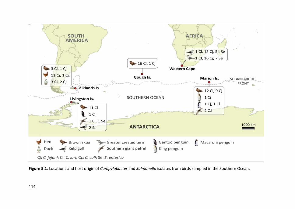
114
Figure 5.1. Locations and host origin of Campylobacter and Salmonella isolates from birds sampled in the Southern Ocean.
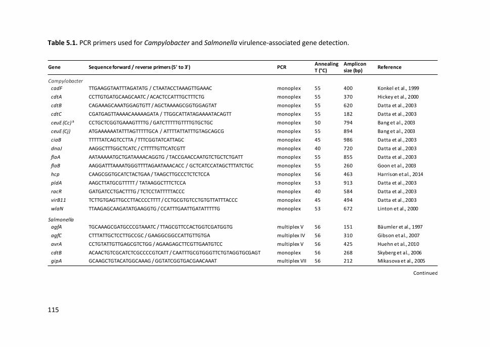
115
Table 5.1. PCR primers used for Campylobacter and Salmonella virulence-associated gene detection.
Gene Sequence forward / reverse primers (5' to 3') PCRAnnealing T (°C)
Amplicon size (bp)
Reference
Campylobacter
cadF TTGAAGGTAATTTAGATATG / CTAATACCTAAAGTTGAAAC monoplex 55 400 Konkel et al., 1999
cdtA CCTTGTGATGCAAGCAATC / ACACTCCATTTGCTTTCTG monoplex 55 370 Hickey et al., 2000
cdtB CAGAAAGCAAATGGAGTGTT / AGCTAAAAGCGGTGGAGTAT monoplex 55 620 Datta et al., 2003
cdtC CGATGAGTTAAAACAAAAAGATA / TTGGCATTATAGAAAATACAGTT monoplex 55 182 Datta et al., 2003
ceuE (Cc) a CCTGCTCGGTGAAAGTTTTG / GATCTTTTTGTTTTGTGCTGC monoplex 50 794 Bang et al., 2003
ceuE (Cj) ATGAAAAAATATTTAGTTTTTGCA / ATTTTATTATTTGTAGCAGCG monoplex 55 894 Bang et al., 2003
ciaB TTTTTATCAGTCCTTA / TTTCGGTATCATTAGC monoplex 45 986 Datta et al., 2003
dnaJ AAGGCTTTGGCTCATC / CTTTTTGTTCATCGTT monoplex 40 720 Datta et al., 2003
flaA AATAAAAATGCTGATAAAACAGGTG / TACCGAACCAATGTCTGCTCTGATT monoplex 55 855 Datta et al., 2003
flaB AAGGATTTAAAATGGGTTTTAGAATAAACACC / GCTCATCCATAGCTTTATCTGC monoplex 55 260 Goon et al., 2003
hcp CAAGCGGTGCATCTACTGAA / TAAGCTTGCCCTCTCTCCA monoplex 56 463 Harrison et al., 2014
pldA AAGCTTATGCGTTTTT / TATAAGGCTTTCTCCA monoplex 53 913 Datta et al., 2003
racR GATGATCCTGACTTTG / TCTCCTATTTTTACCC monoplex 40 584 Datta et al., 2003
virB11 TCTTGTGAGTTGCCTTACCCCTTTT / CCTGCGTGTCCTGTGTTATTTACCC monoplex 45 494 Datta et al., 2003
wlaN TTAAGAGCAAGATATGAAGGTG / CCATTTGAATTGATATTTTTG monoplex 53 672 Linton et al., 2000
Salmonella
agfA TGCAAAGCGATGCCCGTAAATC / TTAGCGTTCCACTGGTCGATGGTG multiplex V 56 151 Bäumler et al., 1997
agfC CTTTATTGCTCCTTGCCGC / GAAGGCGGCCATTGTTGTGA multiplex IV 56 310 Gibson et al., 2007
avrA CCTGTATTGTTGAGCGTCTGG / AGAAGAGCTTCGTTGAATGTCC multiplex V 56 425 Huehn et al., 2010
cdtB ACAACTGTCGCATCTCGCCCCGTCATT / CAATTTGCGTGGGTTCTGTAGGTGCGAGT monoplex 56 268 Skyberg et al., 2006
gipA GCAAGCTGTACATGGCAAAG / GGTATCGGTGACGAACAAAT multiplex VII 56 212 Mikasova et al., 2005
Continued
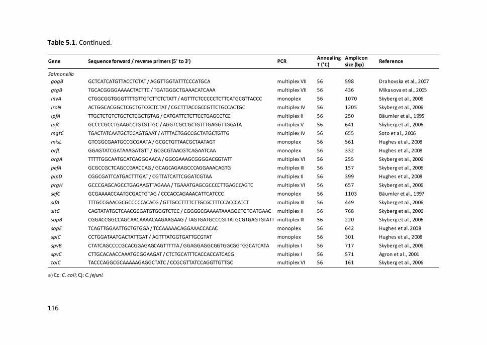
116
Table 5.1. Continued.
Gene Sequence forward / reverse primers (5' to 3') PCRAnnealing T (°C)
Amplicon size (bp)
Reference
Salmonella
gogB GCTCATCATGTTACCTCTAT / AGGTTGGTATTTCCCATGCA multiplex VII 56 598 Drahovska et al., 2007
gtgB TGCACGGGGAAAACTACTTC / TGATGGGCTGAAACATCAAA multiplex VII 56 436 Mikasova et al., 2005
invA CTGGCGGTGGGTTTTGTTGTCTTCTCTATT / AGTTTCTCCCCCTCTTCATGCGTTACCC monoplex 56 1070 Skyberg et al., 2006
iroN ACTGGCACGGCTCGCTGTCGCTCTAT / CGCTTTACCGCCGTTCTGCCACTGC multiplex IV 56 1205 Skyberg et al., 2006
lpfA TTGCTCTGTCTGCTCTCGCTGTAG / CATGATTCTCTTCCTGAGCCTCC multiplex II 56 250 Bäumler et al., 1995
lpfC GCCCCGCCTGAAGCCTGTGTTGC / AGGTCGCCGCTGTTTGAGGTTGGATA multiplex V 56 641 Skyberg et al., 2006
mgtC TGACTATCAATGCTCCAGTGAAT / ATTTACTGGCCGCTATGCTGTTG multiplex IV 56 655 Soto et al., 2006
misL GTCGGCGAATGCCGCGAATA / GCGCTGTTAACGCTAATAGT monoplex 56 561 Hughes et al., 2008
orfL GGAGTATCGATAAAGATGTT / GCGCGTAACGTCAGAATCAA monoplex 56 332 Hughes et al., 2008
orgA TTTTTGGCAATGCATCAGGGAACA / GGCGAAAGCGGGGACGGTATT multiplex VI 56 255 Skyberg et al., 2006
pefA GCGCCGCTCAGCCGAACCAG / GCAGCAGAAGCCCAGGAAACAGTG multiplex III 56 157 Skyberg et al., 2006
pipD CGGCGATTCATGACTTTGAT / CGTTATCATTCGGATCGTAA multiplex II 56 399 Hughes et al., 2008
prgH GCCCGAGCAGCCTGAGAAGTTAGAAA / TGAAATGAGCGCCCCTTGAGCCAGTC multiplex VI 56 657 Skyberg et al., 2006
sefC GCGAAAACCAATGCGACTGTAG / CCCACCAGAAACATTCATCCC monoplex 56 1103 Bäumler et al., 1997
sifA TTTGCCGAACGCGCCCCCACACG / GTTGCCTTTTCTTGCGCTTTCCACCCATCT multiplex III 56 449 Skyberg et al., 2006
sitC CAGTATATGCTCAACGCGATGTGGGTCTCC / CGGGGCGAAAATAAAGGCTGTGATGAAC multiplex II 56 768 Skyberg et al., 2006
sopB CGGACCGGCCAGCAACAAAACAAGAAGAAG / TAGTGATGCCCGTTATGCGTGAGTGTATT multiplex III 56 220 Skyberg et al., 2006
sopE TCAGTTGGAATTGCTGTGGA / TCCAAAAACAGGAAACCACAC monoplex 56 642 Hughes et al. 2008
spiC CCTGGATAATGACTATTGAT / AGTTTATGGTGATTGCGTAT monoplex 56 301 Hughes et al., 2008
spvB CTATCAGCCCCGCACGGAGAGCAGTTTTTA / GGAGGAGGCGGTGGCGGTGGCATCATA multiplex I 56 717 Skyberg et al., 2006
spvC CTTGCACAACCAAATGCGGAAGAT / CTCTGCATTTCACCACCATCACG multiplex I 56 571 Agron et al., 2001
tolC TACCCAGGCGCAAAAAGAGGCTATC / CCGCGTTATCCAGGTTGTTGC multiplex VI 56 161 Skyberg et al., 2006
a) Cc: C. coli; Cj: C. jejuni.
Study III
117
5.4. RESULTS
5.4.1. Genetic diversity and population structure
PFGE profiles of 110 Campylobacter and 64 Salmonella isolates from Southern Ocean
and South African seabirds were analysed and compared. Based on the results obtained,
we selected isolates representative of different bird species and locations to assess their
genetic relationship by MLST. In those cases where isolates from the same bird or the
same bird species and colony showed a pulsotype in common, only one isolate was
selected for further analysis. The selection included 77 Campylobacter and 23
Salmonella enterica isolates. Overall, 68 Campylobacter pulsotypes (SK) (35 C. lari, 32 C.
jejuni and one C. coli) and 20 Salmonella pulsotypes (XB) were identified (Figures 5.2,
5.3 and 5.4). One C. jejuni isolate (MAR119-C5) non-typeable by PFGE with either of the
two restriction enzymes was included in the selection. One isolate of S. Anatum non-
typeable with the secondary enzyme BlnI was also included.
MLST of Campylobacter isolates revealed 62 STs (35 of C. lari, 26 of C. jejuni and one of
C. coli), of which 65% corresponded to novel STs (33 of C. lari and seven of C. jejuni)
(Figures 5.5 and 5.6). We described 53 new C. lari alleles (five of adk, eight of atpA,
seven of glnA, three of glyA, nine of pgi, 11 of pgm and 10 of tkt genes) and four new C.
jejuni alleles (two of glnA, one of glyA and one of pgm genes). We also identified four C.
lari and four C. jejuni new allelic combinations. However, one C. lari isolate (FK57-C1)
was not typeable by MLST since it was not possible to obtain the sequence of the tkt
gene and thus the allelic profile was incomplete (adk63-atpA72-glnA61-glyA57-pgi72-
pgm67-tktX).
A high genetic diversity was observed among C. lari isolates, and most STs had three or
more loci variants (Figure 5.5). No C. lari ST belonged to a previously defined clonal
complex (CC). We found two STs, one in a southern giant petrel from Marion Is. (ST-54)
and one in a skua from the Falkland Is. (ST-62), with exclusive allelic profiles (i.e. all

Chapter 5
118
seven loci were different from those found in all other STs detected). The former was
identified by 16S rDNA sequence analysis as C. lari subsp. lari, whilst the latter was a C.
lari subsp. concheus. Despite the high genetic diversity, the same STs were found in
different seabird species and islands: ST-37 was detected in one skua and one gentoo
penguin from Livingston Is., ST-33 was found in three skuas from Gough Is. and Marion
Is., ST-43 was present in two skuas from Marion Is. and Livingston Is., and one southern
giant petrel carried ST-41, a single-locus variant (SLV) of ST-43. We also found other
SLVs, such as ST-27 and ST-35, in one skua and one macaroni penguin, respectively, from
Marion Is. Although most brown skuas carried unrelated STs, some clusters of SLVs and
double loci variants (DLVs) were observed in this bird species from Livingston Is. (ST-38,
ST-39 and ST-40) and Gough Is. (ST-47, ST-48 and ST-51). Interestingly, C. lari isolates
from domestic ducks at the Falkland Is. showed a low similarity between them and
among those from wild birds. We did not observe relevant genetic similarity among C.
lari isolates from Western Cape and Southern Ocean islands.
Regarding C. jejuni, the CCs most frequently observed were CC-1275 and CC-45,
followed by CC-21 and CC-206 (Figure 5.6). Five CCs (CC-283, CC-354, CC-692, CC-1034
and CC-1332) were represented by just one ST which appeared only once (ST-267, ST-
1073, ST-991, ST-1956 and ST-696, respectively), while six STs (ST-2353, ST-2654, ST-
8572, ST-8577, ST-8588 and ST-8590) belonged to unassigned CCs. The predominant CC-
1275 encompassed seven STs, all of which were detected in Western Cape seabirds. ST-
1275 is the central type of the CC, but ST-1223 and ST-1268 were the most prevalent.
The latter and ST-2654 were found in both Western Cape kelp gulls and greater crested
terns. ST-45 was detected in skuas from Marion and the Falkland Is., as well as one
domestic duck from the Falkland Is. ST-45 is the ancestral type of the CC-45 which
contains other SLVs, such as those found in one skua (ST-8589) and one macaroni
penguin (ST-137) from Marion Is., and one Western Cape greater crested tern (ST-1326).
CC-206 contained two SLVs isolated from one skua from Gough Is. (ST-227) and one
Western Cape kelp gull (ST-572). Moreover, one king penguin from Marion Is. carried

Study III
119
an isolate differing in two loci (DLV) from that found in a hen from the Falkland Is. (ST-
883 and ST-8570, respectively), both belonging to CC-21. However, the only C. coli
isolated from a hen corresponded to ST-899 (CC-828).
We observed few coincidences between STs and pulsotypes: two for C. lari (ST-37 and
SK34, ST-33 and SK36) and two for C. jejuni (ST-8588 and SK3, ST-2654 and SK13)
(Figures 5.2 and 5.3). By comparison, two STs of C. lari (ST-43 and ST-27) and four of C.
jejuni (ST-8570, ST-1268, ST-45 and ST-1223) grouped isolates showing two or three
different pulsotypes, some of them with a similarity level ˂65%. We also detected
pulsotypes in common among C. lari isolates assigned to different STs (SK41 in ST-46
and ST-55; SK55 in ST-39 and ST-38). However, most C. jejuni isolates belonging to the
same CC were clustered together by PFGE.
MLST of 23 Salmonella isolates showed 14 different STs, two of them with new allele
profiles (one of S. enterica subsp. salamae and one of S. enterica subsp. enterica serovar
Anatum) (Figure 5.4). A low genetic diversity was observed by MLST compared to PFGE
since we mostly detected one or two ST per serovar regardless of the pulsotype,
whereas several pulsotypes were distinguished within most serovars. Five S. Enteritidis
isolates from kelp gulls from the Western Cape and Livingston Is. corresponded to ST-
11 of eBG4 (eBurst groups are equivalent to Campylobacter clonal complexes), while
the remaining isolate was assigned to SLV ST-1949. Moreover, two S. Typhimurium
isolates belonged to ST-34 (eBG1) and one to SLV ST-1952. Three S. Anatum isolates
(one of them not typeable by BlnI-PFGE) pertained to ST-64 (eBG65) and the other
corresponded to a previously undescribed DLV of ST-64. According to the phylogenetic
tree generated by concatenated MLST loci, the S. Paratyphi B var Java isolate (ST-28,
eBG59) was the most divergent of the Salmonella isolates, including those belonging to
the subspecies salamae. This was not perceived in the PFGE dendrogram, although all
clusters grouped together isolates of the same serovar (data not shown).
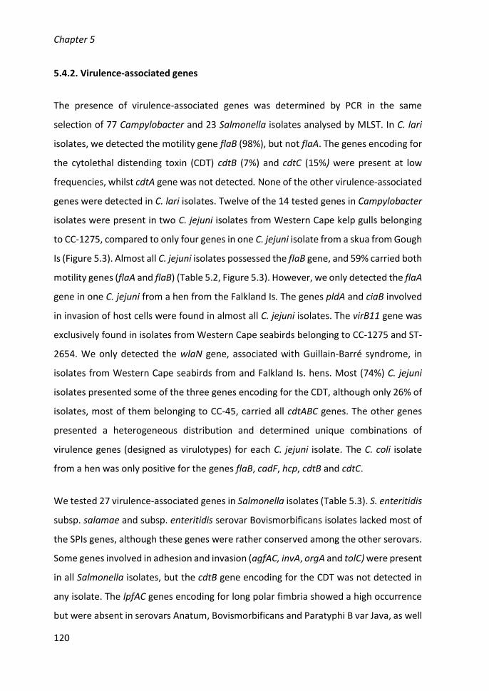
Chapter 5
120
5.4.2. Virulence-associated genes
The presence of virulence-associated genes was determined by PCR in the same
selection of 77 Campylobacter and 23 Salmonella isolates analysed by MLST. In C. lari
isolates, we detected the motility gene flaB (98%), but not flaA. The genes encoding for
the cytolethal distending toxin (CDT) cdtB (7%) and cdtC (15%) were present at low
frequencies, whilst cdtA gene was not detected. None of the other virulence-associated
genes were detected in C. lari isolates. Twelve of the 14 tested genes in Campylobacter
isolates were present in two C. jejuni isolates from Western Cape kelp gulls belonging
to CC-1275, compared to only four genes in one C. jejuni isolate from a skua from Gough
Is (Figure 5.3). Almost all C. jejuni isolates possessed the flaB gene, and 59% carried both
motility genes (flaA and flaB) (Table 5.2, Figure 5.3). However, we only detected the flaA
gene in one C. jejuni from a hen from the Falkland Is. The genes pldA and ciaB involved
in invasion of host cells were found in almost all C. jejuni isolates. The virB11 gene was
exclusively found in isolates from Western Cape seabirds belonging to CC-1275 and ST-
2654. We only detected the wlaN gene, associated with Guillain-Barré syndrome, in
isolates from Western Cape seabirds from and Falkland Is. hens. Most (74%) C. jejuni
isolates presented some of the three genes encoding for the CDT, although only 26% of
isolates, most of them belonging to CC-45, carried all cdtABC genes. The other genes
presented a heterogeneous distribution and determined unique combinations of
virulence genes (designed as virulotypes) for each C. jejuni isolate. The C. coli isolate
from a hen was only positive for the genes flaB, cadF, hcp, cdtB and cdtC.
We tested 27 virulence-associated genes in Salmonella isolates (Table 5.3). S. enteritidis
subsp. salamae and subsp. enteritidis serovar Bovismorbificans isolates lacked most of
the SPIs genes, although these genes were rather conserved among the other serovars.
Some genes involved in adhesion and invasion (agfAC, invA, orgA and tolC) were present
in all Salmonella isolates, but the cdtB gene encoding for the CDT was not detected in
any isolate. The lpfAC genes encoding for long polar fimbria showed a high occurrence
but were absent in serovars Anatum, Bovismorbificans and Paratyphi B var Java, as well
Study III
121
as in subsp. salamae. Other fimbrial genes were only present in specific serovars, such
as the sefC gene in ser. Enteritidis, and the plasmid-encoded pefA gene in serovar
Enteritidis, Typhimurium and Bovismorbificans. The spvBC plasmid-borne genes
involved in intracellular survival were found in all S. Enteritidis, S. Typhimurium and S.
Hadar isolates. The genes associated with the P2-like (SopEΦ) and lambda-like (Gifsy)
phages, involved in invasion and intracellular survival, respectively, were detected
mainly in isolates of S. Enteritidis (sopE, gtgB, gipA and gogB) and S. Typhimurium (sopE,
gtgB and gipA), but some were also present in isolates of other serovars, such as Hadar
(sopE), Muenchen (sopE), Saintpaul (gtgB) and Anatum (gtgB), as well as in subsp.
salamae (gipA). The same virulotype was observed in two isolates of S. Anatum ST-64
(VT16B), S. Typhimurium ST-34 (VT20A) and S. Enteritidis ST-11 (VT18C). The highest
number of virulence determinants (25 genes) was detected in S. Enteritidis ST-1949 and
S. Typhimurium ST-1942, (SLVs of ST-11 and ST-34, respectively), both isolated from
Western Cape kelp gulls. In addition, one Livingston Is. kelp gull carried a S. Enteritidis
ST-11 positive for 24 virulence determinants.
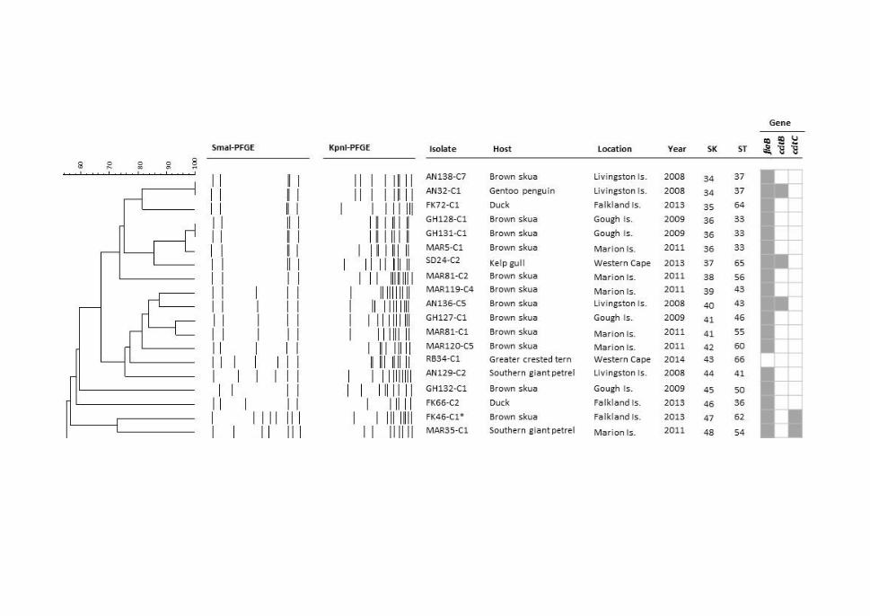
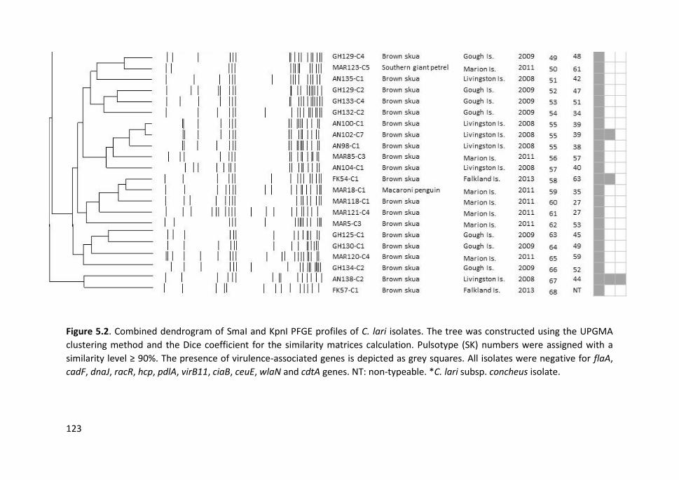
123
Figure 5.2. Combined dendrogram of SmaI and KpnI PFGE profiles of C. lari isolates. The tree was constructed using the UPGMA
clustering method and the Dice coefficient for the similarity matrices calculation. Pulsotype (SK) numbers were assigned with a
similarity level ≥ 90%. The presence of virulence-associated genes is depicted as grey squares. All isolates were negative for flaA,
cadF, dnaJ, racR, hcp, pdlA, virB11, ciaB, ceuE, wlaN and cdtA genes. NT: non-typeable. *C. lari subsp. concheus isolate.
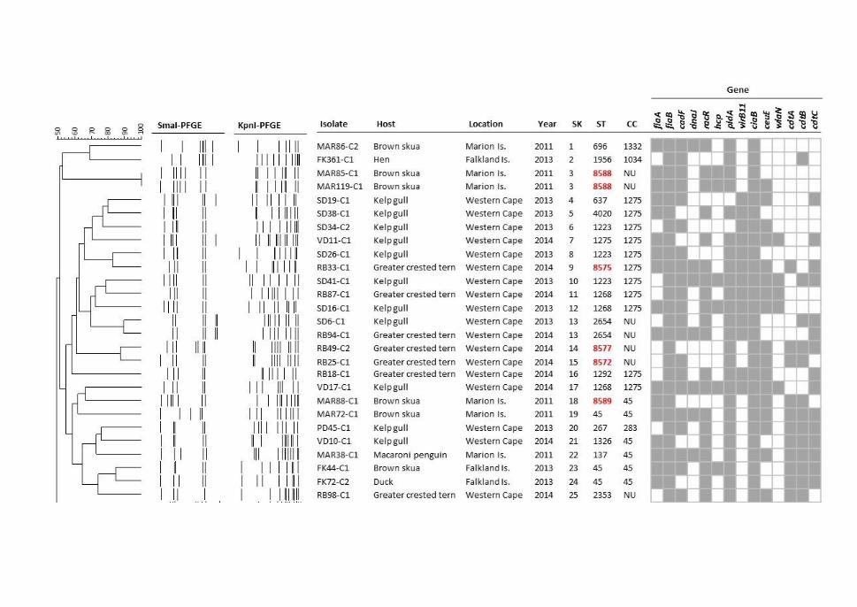
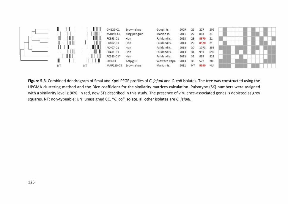
125
Figure 5.3. Combined dendrogram of SmaI and KpnI PFGE profiles of C. jejuni and C. coli isolates. The tree was constructed using the
UPGMA clustering method and the Dice coefficient for the similarity matrices calculation. Pulsotype (SK) numbers were assigned
with a similarity level ≥ 90%. In red, new STs described in this study. The presence of virulence-associated genes is depicted as grey
squares. NT: non-typeable; UN: unassigned CC. *C. coli isolate, all other isolates are C. jejuni.

Gene
SPI1-5 IsletFimbrial operon Plasmid Phage
Subsp. and serovar XB ST eBG VT
invA
orgA
prgH
sitC
avrA
spiC
mgt
C
mis
L
orfL
pipD
sopB
tolC
iroN
sifA
cdtB
agfA
agfC
lpfA
lpfC
sefC
pefA
spvB
spvC
sopE
gtgB
gipA
gogB
Enteritidis 17 11 4 21B
Enteritidis 16 11 4 18C
Enteritidis 17 11 4 24A
Enteritidis 18 11 4 16C
Enteritidis 15 11 4 18C
Enteritidis 14 1949 NU 25B
Saintpaul 10 27 14 19B
Typhimurium 9 1952 NU 25A
Typhimurium 8 34 1 20A
Typhimurium 7 34 1 20A
Manhattan 6 1540 27 19A
Muenchen 5 82 8 16A
Muenchen 4 82 8 18B
subsp. salamae 20 new NU 6A
subsp. salamae 19 53 214 11A
Anatum 12 new NU 9A
Anatum 11 64 65 16B
Anatum NT 64 65 16B
Anatum 11 64 65 17A
Hadar 1 473 22 18A
Hadar 1 473 22 21A
Bovismorbificans 3 142 34 7A
Paratyphi B, var Java 2 28 59 14A
*
*
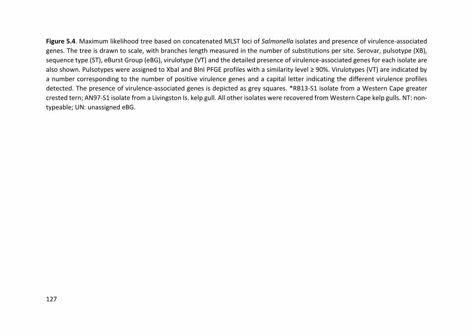
127
Figure 5.4. Maximum likelihood tree based on concatenated MLST loci of Salmonella isolates and presence of virulence-associated
genes. The tree is drawn to scale, with branches length measured in the number of substitutions per site. Serovar, pulsotype (XB),
sequence type (ST), eBurst Group (eBG), virulotype (VT) and the detailed presence of virulence-associated genes for each isolate are
also shown. Pulsotypes were assigned to XbaI and BlnI PFGE profiles with a similarity level ≥ 90%. Virulotypes (VT) are indicated by
a number corresponding to the number of positive virulence genes and a capital letter indicating the different virulence profiles
detected. The presence of virulence-associated genes is depicted as grey squares. *RB13-S1 isolate from a Western Cape greater
crested tern; AN97-S1 isolate from a Livingston Is. kelp gull. All other isolates were recovered from Western Cape kelp gulls. NT: non-
typeable; UN: unassigned eBG.
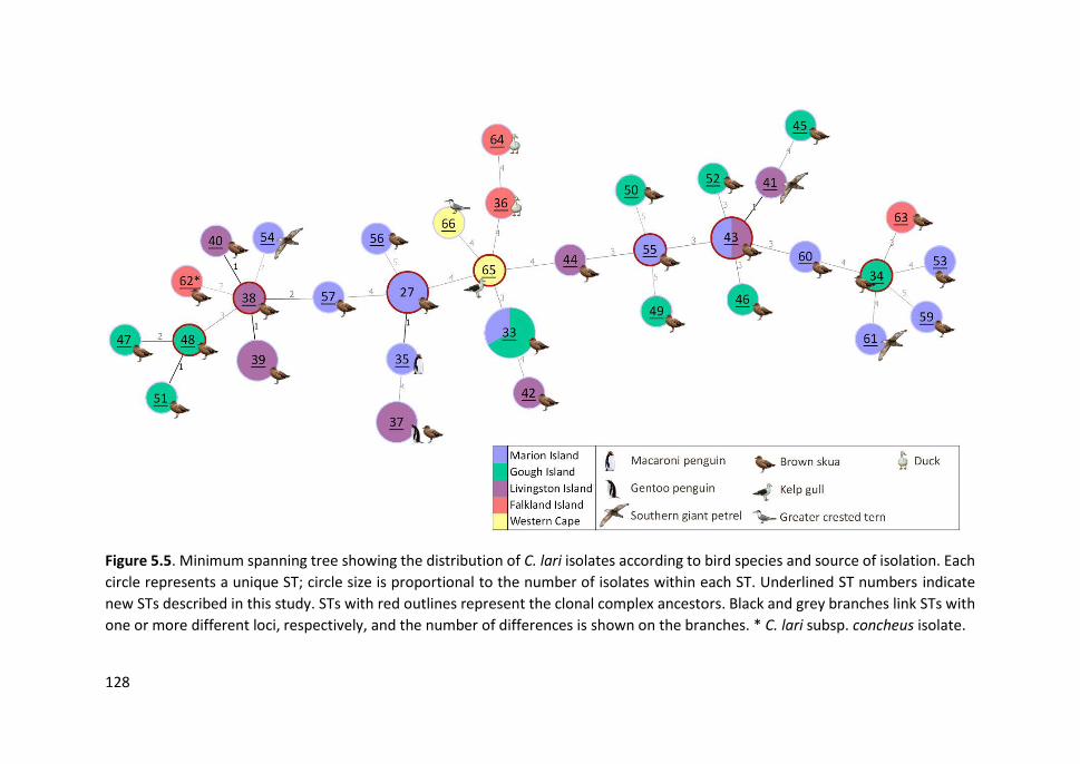
128
Figure 5.5. Minimum spanning tree showing the distribution of C. lari isolates according to bird species and source of isolation. Each
circle represents a unique ST; circle size is proportional to the number of isolates within each ST. Underlined ST numbers indicate
new STs described in this study. STs with red outlines represent the clonal complex ancestors. Black and grey branches link STs with
one or more different loci, respectively, and the number of differences is shown on the branches. * C. lari subsp. concheus isolate.

129
Figure 5.6. Minimum spanning tree showing the distribution of C. jejuni and C. coli isolates according to bird species and source of
isolation. Each circle represents a unique ST, the circle size is proportional to the number of isolates within each ST. Underlined ST
numbers indicate new STs described in this study. The single C. coli ST is indicated with an asterisk. STs with red outlines represent
the clonal complex ancestors. Black and grey branches link STs with one or more different loci, respectively, and the number of
differences is shown on the branches. * C. coli isolate, all other isolates are C. jejuni.
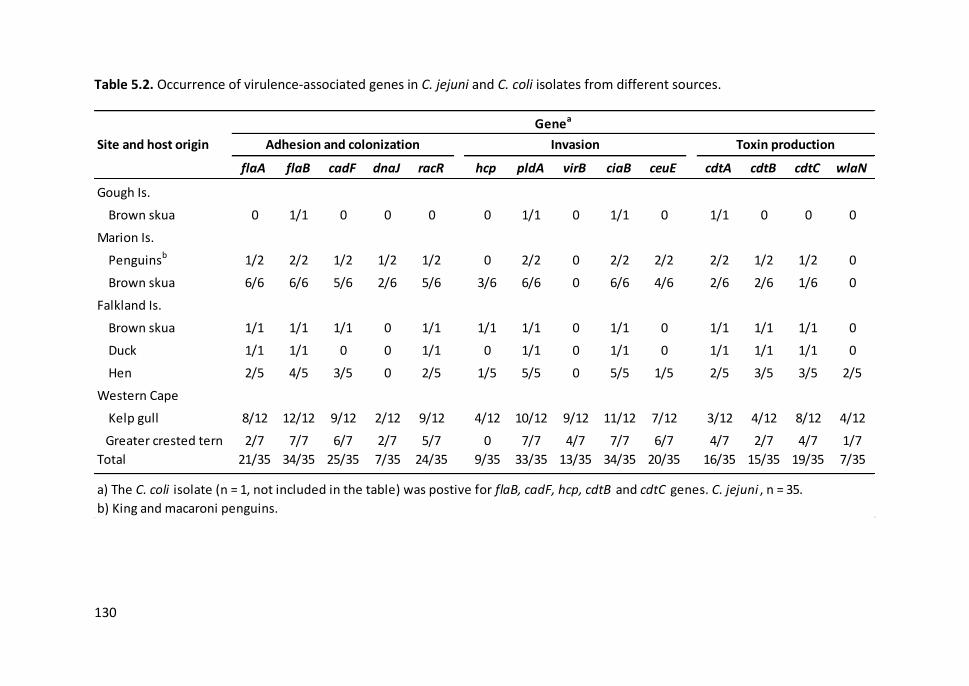
130
Table 5.2. Occurrence of virulence-associated genes in C. jejuni and C. coli isolates from different sources.
flaA flaB cadF dnaJ racR hcp pldA virB ciaB ceuE cdtA cdtB cdtC wlaN
Gough Is.
Brown skua 0 1/1 0 0 0 0 1/1 0 1/1 0 1/1 0 0 0
Marion Is.
Penguinsb 1/2 2/2 1/2 1/2 1/2 0 2/2 0 2/2 2/2 2/2 1/2 1/2 0
Brown skua 6/6 6/6 5/6 2/6 5/6 3/6 6/6 0 6/6 4/6 2/6 2/6 1/6 0
Falkland Is.
Brown skua 1/1 1/1 1/1 0 1/1 1/1 1/1 0 1/1 0 1/1 1/1 1/1 0
Duck 1/1 1/1 0 0 1/1 0 1/1 0 1/1 0 1/1 1/1 1/1 0
Hen 2/5 4/5 3/5 0 2/5 1/5 5/5 0 5/5 1/5 2/5 3/5 3/5 2/5
Western Cape
Kelp gull 8/12 12/12 9/12 2/12 9/12 4/12 10/12 9/12 11/12 7/12 3/12 4/12 8/12 4/12
Greater crested tern 2/7 7/7 6/7 2/7 5/7 0 7/7 4/7 7/7 6/7 4/7 2/7 4/7 1/7
Total 21/35 34/35 25/35 7/35 24/35 9/35 33/35 13/35 34/35 20/35 16/35 15/35 19/35 7/35
a) The C. coli isolate (n = 1, not included in the table) was postive for flaB, cadF, hcp, cdtB and cdtC genes. C. jejuni , n = 35.
b) King and macaroni penguins.
Site and host origin
Genea
Adhesion and colonization Invasion Toxin production

131
Table 5.3. Occurrence of virulence-associated genes in Salmonella isolates of different subspecies and serovars.
sub
sp. s
ala
ma
e
An
atu
m
Bo
vism
orb
ific
ans
Ente
riti
dis
Had
ar
Man
hat
tan
Mu
en
che
n
Par
atyp
hi B
,
var
Java
Sain
tpau
l
Typ
him
uri
um
Tota
l
SPI-1
invA Invasion 2/2 4/4 1/1 6/6 2/2 1/1 2/2 1/1 1/1 3/3 23/23
orgA Invasion 2/2 4/4 1/1 6/6 2/2 1/1 2/2 1/1 1/1 3/3 23/23
prgH Invasion 1/2 3/4 0 3/6 2/2 1/1 2/2 1/1 1/1 3/3 17/23
sitC Iron uptake 1/2 3/4 0 5/6 1/2 1/1 2/2 1/1 1/1 3/3 18/23
avrA Intracel. survival 0 4/4 0 6/6 1/2 1/1 2/2 0 1/1 1/3 16/23
SPI-2
spiC Intracel. survival 1/2 4/4 0 5/6 2/2 1/1 2/2 1/1 1/1 3/3 20/23
SPI-3
mgtC Intracel. survival 0 3/4 0 3/6 2/2 1/1 1/2 1/1 1/1 3/3 15/23
misL Colonization and intracel. survival 1/2 3/4 0 3/6 1/2 1/1 2/2 1/1 1/1 3/3 16/23
SPI-4
orfL Colonization and intracel. survival 0 3/4 0 6/6 2/2 1/1 2/2 0 1/1 3/3 18/23
SPI-5
pipD Invasion 1/2 4/4 0 6/6 2/2 1/1 2/2 1/1 1/1 3/3 21/23
sopB Invasion and intracel. survival 0 4/4 1/1 6/6 2/2 1/1 2/2 1/1 1/1 3/3 21/23
Islet
tolC Colonization and invasion 2/2 4/4 1/1 6/6 2/2 1/1 2/2 1/1 1/1 3/3 23/23
Subsp. and serovar
Gene Virulence role
Continued
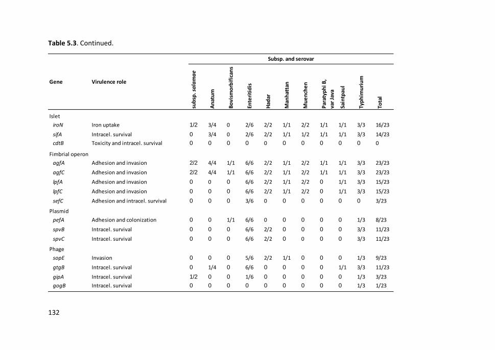
132
Table 5.3. Continued.
sub
sp. s
ala
ma
e
An
atu
m
Bo
vism
orb
ific
ans
Ente
riti
dis
Had
ar
Man
hat
tan
Mu
en
che
n
Par
atyp
hi B
,
var
Java
Sain
tpau
l
Typ
him
uri
um
Tota
l
Islet
iroN Iron uptake 1/2 3/4 0 2/6 2/2 1/1 2/2 1/1 1/1 3/3 16/23
sifA Intracel. survival 0 3/4 0 2/6 2/2 1/1 1/2 1/1 1/1 3/3 14/23
cdtB Toxicity and intracel. survival 0 0 0 0 0 0 0 0 0 0 0
Fimbrial operon
agfA Adhesion and invasion 2/2 4/4 1/1 6/6 2/2 1/1 2/2 1/1 1/1 3/3 23/23
agfC Adhesion and invasion 2/2 4/4 1/1 6/6 2/2 1/1 2/2 1/1 1/1 3/3 23/23
lpfA Adhesion and invasion 0 0 0 6/6 2/2 1/1 2/2 0 1/1 3/3 15/23
lpfC Adhesion and invasion 0 0 0 6/6 2/2 1/1 2/2 0 1/1 3/3 15/23
sefC Adhesion and intracel. survival 0 0 0 3/6 0 0 0 0 0 0 3/23
Plasmid
pefA Adhesion and colonization 0 0 1/1 6/6 0 0 0 0 0 1/3 8/23
spvB Intracel. survival 0 0 0 6/6 2/2 0 0 0 0 3/3 11/23
spvC Intracel. survival 0 0 0 6/6 2/2 0 0 0 0 3/3 11/23
Phage
sopE Invasion 0 0 0 5/6 2/2 1/1 0 0 0 1/3 9/23
gtgB Intracel. survival 0 1/4 0 6/6 0 0 0 0 1/1 3/3 11/23
gipA Intracel. survival 1/2 0 0 1/6 0 0 0 0 0 1/3 3/23
gogB Intracel. survival 0 0 0 0 0 0 0 0 0 1/3 1/23
Gene Virulence role
Subsp. and serovar
Study III
133
5.5. DISCUSSION
Despite the efforts taken to reduce the human incidence of Campylobacter and
Salmonella spp., these pathogens continue to be the most common causes of human
bacterial gastroenteritis worldwide. For an effective control of these zoonoses, it is
necessary to better understand the global epidemiology of these pathogens, including
the environmental reservoir with particular attention to wild animals that may
constitute another reservoir of these pathogens. Here, we investigated the diversity,
population structure and virulence potential of Campylobacter and Salmonella isolates
from seabirds from remote Southern Ocean islands and the Western Cape coast of
South Africa. We also included in the analysis Campylobacter isolates from domestic
poultry from the Falkland Is. We described 33 new STs of C. lari and seven of C. jejuni,
in addition to 53 and four new alleles, respectively. Novel MLST data, submitted to the
corresponding PubMLST database, represented an important contribution in the case
of C. lari, with only 32 STs described prior to our submission (January 2017). We also
found new STs of S. enterica subsp. salamae and subsp. enterica serovar Anatum.
The population structure of Campylobacter isolates from Southern Ocean seabirds
appeared to be highly diverse and genetically distant, especially in the case of C. lari
isolates since most of them belonged to new STs with unassigned CCs. Within the genus
Campylobacter, C. lari represent a closely related phylogenetic clade and their members
often share similar hosts (e.g. shorebirds, marine mammals and shellfish) and
environments (e.g. coastal regions and watersheds) (Miller et al., 2014). Therefore, the
genetic heterogenicity observed here may be due to the geographically distinct origin
of the isolate collection and the existence of a variety of infection sources. In addition,
the relatively high body temperature of birds could provide an optimal growth
environment for bacteria and favour their genetic diversification, which may explain the
detection of some SLVs and DLVs in brown skuas from the same islands.

Chapter 5
134
Despite the genetic diversity, the same C. lari STs were detected repeatedly in brown
skuas from different Southern Ocean islands: Marion Is. and Gough Is. (ST-33), Marion
Is. and Livingston Is. (ST-43). This suggests some connectivity among Subantarctic and
Antarctic islands, probably as a result of direct transmission by skuas or indirectly by
some common sources of infection of these seabirds. Moreover, the same STs or SLVs
present in skuas were found in other seabird species at the same island: gentoo penguin
(ST-37) and southern giant petrel (ST-41, a SLV of ST-43) at Livingston Is., and macaroni
penguin (ST-35, a SLV of ST-27) at Marion Is. Brown skuas are predators and scavengers,
frequently found close to penguin colonies, where they feed on penguin eggs and, like
giant petrels, chicks and sick or wounded adult penguins, which may explain the
similarities found among C. lari STs carried by these seabirds. No common STs of C. lari
STs were observed between our study and those previously reported in Antarctic
penguins, but it is difficult to compare across studies because authors described many
incomplete allelic profiles (García-Peña et al., 2017). According to the MLST database,
C. lari has been found mainly in aquatic environments, but also in human gastroenteritis
cases in some countries (USA, Canada, Belgium and France). An example of this is ST-27
detected in one skua from Marion Is. and also isolated in a human stool in France.
The population structure of C. jejuni was less heterogeneous than that of C. lari, with
most isolates structured into clusters of related lineages. CC-1275 occurred exclusively
in kelp gulls and greater crested terns from the Western Cape, and is mainly recorded
from environmental waters and wild birds in Europe, USA, Canada, New Zealand and
Australia (http://pubmlst.org/campylobacter). Several studies have described that most
C. jejuni genotypes from wild birds differ from those recovered from food-producing
animals and human campylobacteriosis (Broman et al., 2004; Sheppard et al., 2011;
Griekspoor et al., 2013). This agrees with what we observed in South African seabirds,
where 84% were carriers of C. jejuni genotypes mainly linked to aquatic environments,
despite inhabiting a populated region in close contact with livestock and human
activities.
Study III
135
Surprisingly, we found the opposite scenario in seabirds from remote Southern Ocean
islands. Almost all C. jejuni genotypes carried by skuas and penguins from Subantarctic
regions belong to CCs commonly associated with domestic animals, especially poultry,
and human gastroenteritis. One of the most prevalent CCs here is CC-45, widely
distributed in the Northern Hemisphere and locally in the Southern Hemisphere
(Argentina, Australia and New Zealand), followed by CC-21 and CC-206, usually reported
in Europe. These CCs have also been found, albeit less frequently, in natural
environments and wild birds, probably as a result of contamination from animal and
human waste (Kwan et al., 2008; Levesque et al., 2013; Cody et al., 2015). ST-45, the
central genotype of CC-45, is the only C. jejuni ST that has been reported previously from
the Subantarctic, specifically in a macaroni penguin from Bird Island (South Georgia),
suggesting human activities (wastes from scientific bases and passing ships) as possible
transmission routes (Griekspoor et al., 2010). Interestingly, we also detected ST-45 in
skuas and one domestic duck from the Falkland Is., one of the few areas close to
Antarctica (and South Georgia) with a resident human population and free-ranging
livestock, which may facilitate the contact between seabirds and domestic animals.
Although brown skuas are mainly predatory birds, they scavenge on human food waste,
providing a plausible route for C. jejuni infection, or they may be infected in wintering
and stopover areas where they mix with the local fauna (Shirihai, 2007). Skuas also may
spread pathogens around the Southern Ocean as we observed with some C. lari
genotypes. C. jejuni ST-45 and associated SLVs were found in one brown skua and one
macaroni penguin from Marion Is. supporting the hypothesis of connectivity among
distant islands. However, although several seabirds breeding in Antarctic and
Subantarctic islands spend the austral winter in the productive Benguela Upwelling
region in the western coast of South Africa (Crawford et al., 1991; Krietsch, 2014), few
genetic similarities were found between C. jejuni genotypes from Southern Ocean and
South African seabirds.
Chapter 5
136
Previous studies have demonstrated that C. jejuni genotypes from different bird host
species are genetically distinct, although it has been suggested that there is some niche-
association in genotypes from wild birds with similar feeding ecology (Sheppard et al.,
2011; Griekspoor et al., 2013). We did not observe C. jejuni genotypes clearly adapted
to Southern Ocean seabirds and closely related C. jejuni STs were detected in different
host species. For example, C. jejuni genotypes of the same CCs were present in brown
skuas, greater crested terns and macaroni penguins (CC-45), skuas and kelp gulls (CC-
206) and king penguins and domestic hens (CC-21) from different regions. The fact that
these seabirds carried C. jejuni genotypes commonly found in domestic animals and
known to cause human gastroenteritis, strongly suggest that the pathogens were
acquired from sporadic infections from anthropogenic sources. These poultry-adapted
genotypes probably encounter a similar ecological niche in the gastrointestinal tract of
wild birds allowing them to grow and proliferate rapidly.
With regards to Salmonella isolates, a single ST per serovar was observed in most cases,
except for serovars Typhimurium, Enteritidis and Anatum that displayed two SLVs each.
Almost all S. Typhimurium isolates corresponded to ST-34, one of the most predominant
ST within the eBG1, whilst S. Enteritidis isolates belonged to ST-11, the central ST of
eBG4 (Achtman et al., 2012; Ashton et al., 2016). Both STs are globally distributed in a
broad range of domestic animals and are commonly responsible for human
salmonellosis (Enterobase Database, http://enterobase.warwick.ac.uk/). Interestingly,
the isolate recovered from an Antarctic kelp gull from Livingston Is. also belonged to ST-
11. S. Enteritidis has been previously reported in Antarctic and Subantarctic wildlife
(seagulls, skuas, penguins, petrels and seals), although the ST was not determined
(Olsen et al., 1996; Palmgren et al., 2000; Iveson et al., 2009; Vigo et al., 2011; Dougnac
et al., 2015). The presence of this ST in seabirds from Western Cape and even from
remote regions of Antarctica reflects the rapid worldwide expansion of Salmonella
genotypes of public health concern. The remaining Salmonella serovar isolates were less
frequently reported STs, isolated from diverse origins such as livestock, reptiles, foods,
Study III
137
and humans. The only S. Paratyphi B var Java isolated (from a Western Cape kelp gull)
was assigned to ST-28 (eBG59), a genotype mainly found in poultry and not as
pathogenic for humans as other S. Paratyphi B genotypes (Toboldt et al., 2012).
Overall, molecular typing by PFGE allowed a slightly greater discrimination compared to
MLST, and isolates with same ST exhibited different pulsotypes in many cases. One
drawback of PFGE is determining the degree of relatedness among isolates that have
similar but distinguishable macrorestriction profiles. Genomic rearrangements may
hinder the interpretation of PFGE data, especially in the case of Campylobacter which
demonstrates considerable genetic instability (Ridley et al., 2008), and additional
sequence-based methods may be required to clarify phylogenetic relationships.
However, in our study, PFGE typing clustered Campylobacter isolates in the same way
as CCs, or eBGs and serovars in the case of Salmonella, which is consistent with previous
reports (McTavish et al., 2009; Shi et al., 2015). PFGE typing also was a helpful tool for
screening closely related genotypes, for instance within individuals or within islands,
and for selecting representative isolates for MLST analysis. Although MLST lacks the
resolution of PFGE, it provides a good epidemiological concordance in assessing the
evolution of bacterial strains, as well as information on source attribution (Barco et al.,
2013; Taboada et al., 2013). Nevertheless, it is important to consider that a few C. lari
isolates with the same pulsotypes turned out to be different STs, unlike what has been
observed in other Campylobacter species (Yabe et al., 2010; Lucarelli et al., 2016;
Cantero, 2017), probably as a result of single nucleotide changes that do not alter the
restriction sites and therefore are not reflected in band profiles.
Our study also provides information about the potential virulence of Campylobacter and
Salmonella spp. isolates. C. lari isolates were PCR-negative for almost all genes analysed,
probably because of significant differences among alleles rather than the absence of
most of these genes. Most C. jejuni isolates presented both flaA and flaB genes, while
C. coli and C. lari isolates lacked the flaB gene. Experiments with mutants have
demonstrated that flaA (but not flaB) is essential for chicken colonization, although
Chapter 5
138
probably both are needed for full motility (Wassenaar et al., 1993). However, the
truncated flagellar filament coded only by flaB is enough for secretion of effector
proteins such as CiaB required for the invasion of hosts cells (Konkel et al., 2004). While
ciaB and pldA genes were present in almost all C. jejuni isolates, other genes involved in
adhesion (cadF, dnaJ and racR) and invasion (hcp, virB11 and ceuE) were less conserved
and their heterogeneous distribution gave rise to a unique virulotype for each C. jejuni
isolate. The hcp gene was used as an indicator for the presence of T6SS, a secretion
system recently reported in Campylobacter that causes lysis of blood cells (Bleumink-
Pluym et al., 2013) and associated with severe cases of campilobacteriosis, especially in
Asia (Harrison et al., 2014). We found the hcp gene in C. jejuni from brown skuas and
kelp gulls, but also in a C. jejuni and a C. coli from domestic hens.
We also observed an association between virulence genes and genotypes. The virB11
gene, located in the virulence plasmid pVir, is involved in adhesion and invasion (Bacon
et al., 2000). Several studies have reported a low prevalence of this gene (˂ 15% or
absent) in isolates from humans and domestic animals (Datta et al., 2003; Müller et al.,
2006; Talukder et al., 2008; Koolman et al., 2015). We found a higher occurrence of
virB11 (37%), but it was only present in C. jejuni CC-1275 and ST-2654 from Western
Cape seabirds; these genotypes are mainly related to aquatic environments and wild
birds. A recent study found that the absence of virB11 did not reduce the colonization
ability of the bacteria, suggesting that it may not be a relevant virulence factor for
human infection (Biswas et al., 2011). Some genotypes of CC-1275, in addition to the
novel ST-8570 (CC-21) from the Falkland Is. hens, also possessed the wlaN gene which
is involved in the Guillian-Barré syndrome (Linton et al., 2000). The prevalence of wlaN
in isolates from seabirds (20%) was similar to that previously found in chickens and
humans (Datta et al., 2003; Cantero, 2017), but substantially higher than reported by
(Koolman et al., 2015). These C. jejuni genotypes carried by seabirds thus may pose an
important risk for human health. In addition, a 23% of C. jejuni isolates, mainly belonging
to CC-45, exhibited the complete cdtABC gene cluster required for producing
Study III
139
functionally active cytolethal distending toxin (CDT) (Asakura et al., 2008). These
genotypes were present in different seabird species (skuas, gulls, terns and penguins)
from three different regions as well as in backyard poultry, which suggest that the
presence of cdtABC genes may be associated with certain C. jejuni genotypes regardless
of host species or geographical location.
In the case of Salmonella, SPIs (invA, orgA, prgH, sitC, arvA, spiC, mgtC, misL, orfL, pipD,
sopB) and islet (tolC, iroN, sifA) genes were rather conserved, except in subsp. salamae
and S. Bovismorbificans which lacked most of these genes. By comparison, the cdtB
gene encoding for one of the CDT subunits was not detected in any Salmonella isolate.
Some variability was observed in the distribution of genes located in fimbrial operons,
plasmids and especially prophage regions, which can contribute to the diversity and
host adaptation of Salmonella serovars. While agfAC and lpfAC genes encoding for
SEF17 and long polar fimbriae, respectively, were present in all or almost all serovars,
the sefC gene encoding for the SEF14 fimbria was only detected in some S. Enteritidis
isolates. Also, the plasmid-borne fimbrial gene pefA was only found in isolates of
serovars Enteritidis, Typhimurium and Bovismorbificans. It seems that the acquisition
of different adhesion determinants may expand the host range of the bacteria, including
a greater ability to colonize birds and domestic animals. The pefA and spvBC genes,
often located in the same virulence plasmid (Skyberg et al., 2006), were found together
in S. Enteritidis isolates. However, some S. Typhimurium and S. Hadar isolates that
lacked the pefA gene also presented the spvBC genes involved in systemic survival
(Heithoff et al., 2008). Prophage genes were mainly found in S. Enteritidis and S.
Typhimurium isolates, especially gtgB and sopE. The existence of efficient mechanisms
for horizontal gene transfer between serovars or other Gram-negative bacteria,
including lysogenic conversion, may explain the extreme adaptability of Salmonella spp.
and the wide range of hosts that it can infect. The sopE gene is involved in invasion of
host cells and is carried by P2-like phages, but also on lambda-like phages that contain
the gtgB gene (Hoffmann et al., 2014). The lambda-like phage genes encode some

Chapter 5
140
virulence factors responsible for survival in Peyer’s patches, and have been reported
with high prevalence in S. Typhimurium isolates from humans and animals (Drahovská
et al., 2007). Other lambda-like phage genes (gipA and gogB) were present in SLVs of S.
Enteritidis ST-11 (ST-1949) and S. Typhimurium ST-34 (ST-1952). Interestingly, these
genotypes found in Western Cape kelp gulls exhibited the highest proportion of
virulence-associated genes, followed by S. Enteritidis ST-11 carried by a kelp gull from
Livingston Is. However, it is important to note that the detection of virulence-associated
genes by PCR does not consider possible deletions or insertions in gene sequences, nor
does it give information about expression levels. Despite this, the presence of multiple
genes potentially involved in pathogenic processes suggest that many Campylobacter
and Salmonella spp. genotypes found in seabirds may be infectious for humans and
other animals.
In conclusion, our results highlight that seabirds from Southern Ocean islands and the
South African coast can act as carriers of Campylobacter and Salmonella genotypes
associated with human gastroenteritis, some of which have a high virulence potential.
Therefore, these seabirds can constitute an important reservoir for Campylobacter and
Salmonella and may play an important role in their dispersion and in the global
epidemiology of these pathogens. Although some Southern Ocean seabirds could have
acquired these strains during their northward migration, it seems more likely that the
strains have been introduced by human activities such as tourism or scientific
expeditions.

CHAPTER 6
Study IV: Molecular comparative analysis of
nontyphoidal Salmonella isolates from
humans, poultry and seagulls in
Southwestern Europe

Study IV
143
6.1. SUMMARY
Salmonellosis, caused by non-typhoidal Salmonella (NTS), represents one of the most
common human foodborne zoonotic diseases in developed countries. Birds can
transmit this pathogen to humans, especially through contaminated poultry products.
Seagulls have an elevated risk of exposure to Salmonella sources due to their scavenging
feeding habits and can contribute to the dissemination of this agent in the environment
during their foraging or migrating movements. In this study, 742 isolates of 19 NTS
serovars from humans, poultry and seabirds from Southwestern Europe were
genotyped using PFGE. Overall, we detected a higher number of exclusive pulsotypes in
isolates from gulls (mainly S. Typhimurium) and more predominant pulsotypes in
isolates from poultry and humans. However, we also found 30 pulsotypes in common
among isolates from two or three different host niches (serovars Bredeney, Derby,
Enteritidis, Grumpensis, Hadar, Infantis, Kentucky, Kottbus, Mikawasima, Rissen,
Typhimurium and Virchow). Sequence type (ST) and virulotype were further determined
in a subset of Salmonella isolates from different hosts with pulsotypes in common. S.
Typhimurium belonged to ST19 and ST34, while S. Enteritidis corresponded to ST11. We
did not find statistically significant differences in the presence of virulence-associated
genes in isolates from a different source, except for the iroN gene, which is lacking in
most of seagull isolates. These findings further support the role of seagulls as reservoir
Salmonella strains of public health concern, including less common serovars.
Chapter 6
144
6.2. INTRODUCTION
Non-typhoidal Salmonella are the zoonotic agents responsible of one of the main
human foodborne gastroenteritis diseases in industrialized countries. Over the last
decade the incidence of human salmonellosis in the European Union (EU) has decreased
as a result of the interventions put in place for its control in the poultry sector. Despite
this, Salmonella infection still represents a major public health burden and a
considerable economic cost in many countries. In 2016, a total of 94,530 confirmed
salmonellosis cases were reported in the EU, being the serovars Enteritidis and
Typhimurium the most commonly reported (EFSA and ECDC, 2017b). Transmission of
Salmonella to humans is mainly due to consumption of contaminated food of animal
origin (mostly eggs, poultry meat and milk), even though other foods such as fruits,
vegetables or contaminated water have also been implicated in Salmonella outbreaks
(Pires et al., 2014). Besides, some human salmonellosis can also be attributed to direct
or indirect contact with infected animals and contaminated environments.
Salmonella are extensively distributed in the environment where they can survive and
persist for a long time (Winfiel and Groisman, 2003), but its natural environment is the
intestinal tract of a wide range of domestic and wild animals (Hoelzer et al., 2011;
Hilbert et al., 2012). Poultry (chicken and turkey particularly) are frequently colonized
with Salmonella without apparent clinical symptoms. Therefore, fowls can act as
intermittent or persistent healthy carriers and transmit the bacteria vertically and
horizontally to all the flock, spreading it also to the environment and farm surroundings
(Antunes et al., 2016).
Some wild birds such as seagulls, raptors, pigeons, crows and waterfowl can also be
Salmonella carriers and faecally excrete these bacteria (Lawson et al., 2010; Ramos et
al., 2010; Molina-Lopez et al., 2011; Fresno et al., 2013; Callaway et al., 2014; Gargiulo
et al., 2014; Jurado-Tarifa et al., 2016). Seagulls have adapted to an opportunistic
lifestyle in close proximity to humans and often complement their diet with leftovers
Study IV
145
from animal farms, garbage and waste products. Given their scavenging feeding habits,
seagulls have an increased risk of pathogen infection and are one of the most
documented carriers of Salmonella (Ferns and Mudge, 2000; Palmgren et al., 2006;
Ramos et al., 2010; Dolejska et al., 2016; Masarikova et al., 2016). Besides, gulls have
the ability to cover long distances and haunt diverse habitats, which may contribute to
the pathogen spread into the environment and its transmission to domestic animals or
humans via contamination of pastures and agricultural fields, as well as surface waters
(Daniels et al., 2003; Reed et al., 2003).
Nonetheless, Salmonella epidemiology is still not fully understood due to the multiple
infection sources, transmission routes and the wide variety of reservoirs implicated.
Pulsed-field gel electrophoresis (PFGE) and multi-locus sequence typing (MLST) are
powerful tools for establishing the genetic relatedness amongst strains and have been
used in numerous studies for linking human infections to specific sources (Barco et al.,
2013). However, the implications of Salmonella occurrence in wild birds is probably
underrated, as few attempts at identifying the role of these animal reservoirs have been
conducted successfully (Palomo et al., 2013).
Although some Salmonella serovars are adapted to humans (e.g. Typhi and Paratyphi)
or non-human hosts (e.g. Gallinarum in poultry, Dublin in cattle and Cholerasuis in pigs),
most serovars have a broad-host range and can be infectious to humans (Uzzau et al.,
2000). Enteritidis and Typhimurium are the most common zoonotic serovars, but others
such as Infantis, Newport, Derby, Kentucky, Virchow and Hadar, are also of public health
significance in the EU (EFSA and ECDC, 2017b). While host-adapted serovars produce
systemic infection in their natural hosts, generalists serovars commonly cause
gastroenteritis in infected hosts (Hoelzer et al., 2011). The severity of infection depends
largely on the susceptibility of the host and differs according to the serovar
pathogenicity and the virulence potential of the strain itself (Hohmann, 2001; Jones et
al., 2008). In humans, nontyphoidal Salmonella (NTS) infections are usually self-limiting

Chapter 6
146
but, in some cases, it can become invasive and cause complications such as
bacteraemia, focal systemic infections and long-term chronic sequels (Batz et al., 2013).
The virulence of the bacteria is determined by their ability to attach and invade the host
intestinal epithelium cells and to survive and replicate within macrophages. Virulence
factors involved in these processes are encoded by genes located in Salmonella
pathogenicity islands (SPIs), Salmonella genomic islets (SGIs), fimbrial operons,
prophage DNA integrated in the chromosome and virulence plasmids (Van Asten and
Van Dijk, 2005; Fàbrega and Vila, 2013). Some of these elements are conserved among
serovars, such as SPI-1 and SPI-2 (encoding factors required for invasion and
intracellular survival and replication, respectively), while others are adapted to specific
serovars. Furthermore, Salmonella are capable to acquire new virulence-associated
genes by the intra/inter-species horizontal transference of mobile genetic elements
which is associated with the emergence of novel pathogenicity phenotypes (Gyles and
Boerlin, 2014). Therefore, the expression of certain virulence genes can determine the
pathogenicity of Salmonella strains and their capacity to infect different hosts as well as
the severity of the infection.
Given the need to improve the understanding of the role of domestic and wild birds in
the epidemiology of Salmonella, we investigated isolates of a wide variety of zoonotic
serovars from different hosts niches (human, poultry and seagulls) to assess the
existence of common Salmonella genotypes circulating among compartments and to
determine the presence of virulence-associated genes in isolates from different origins
but with highly similar genotypes.
Study IV
147
6.3. MATERIALS AND METHODS
6.3.1. Bacterial isolates
A total of 742 Salmonella isolates of 19 different serovars from humans (N=155), poultry
(N=382) and seagulls (N=205) were characterized in this study (Table 6.1). Clinical
human isolates were obtained from gastroenteritis cases occurred during the period
2009-2014 in two hospitals (63 from hospital A and 94 from hospital B) from Catalonia
(Spain) (Figure 6.1). Isolates from poultry (laying hens, N= 137; chickens, N= 122;
turkeys, N= 49; quails, N=39; partridges, N=18; other domestic birds, N=17) from
different farms located in the Northeast of Spain (Catalonia, Valencia and Aragon) were
collected at the Poultry Health Centre of Catalonia and Aragon (CESAC) between 2008
and 2012. Additionally, we included in the study a collection of isolates from fledgling
seagulls of two different species from nine colonies of the Western Mediterranean and
Eastern Atlantic coasts, sampled between 2009 and 2011 (Antilles, 2014). These isolates
were obtained from yellow-legged gulls (Larus michahellis, N=173) from Ebro Delta,
Medes Islands, Columbretes Island, Dragonera Island, Zembra Island, Ons Island,
Lanzarote Island and Tenerife Island; and Audouin’s gulls (L. audouinii, N=32) from Ebro
Delta and Alboran Island.
Serotyping of Salmonella isolates was carried out according to the White-Kauffmann-Le
Minor scheme (Grimont and Weill, 2007). Salmonella isolates were preserved frozen at
-80° C in Brain Heart Infusion broth (BHI, Merck KGaA, Darmstadt, Germany)
supplemented with 20% glycerol. Fresh cultures of the isolates were prepared onto TSA
(Difco, Madrid, Spain), and plates were incubated at 37° C for 24 h. The extraction of
DNA templates from a bacterial suspension in PBS was performed using InstaGene®
Matrix (Bio-Rad, Hercules, CA, USA) according to the manufacturer’s instructions.
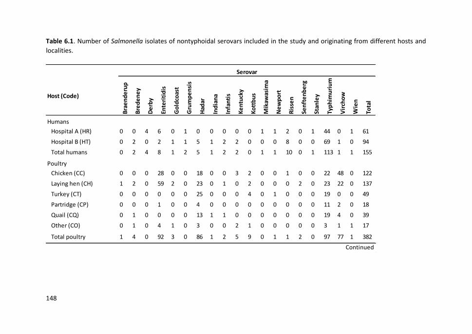
148
Table 6.1. Number of Salmonella isolates of nontyphoidal serovars included in the study and originating from different hosts and
localities.
Host (Code)
Bra
en
de
rup
Bre
de
ne
y
De
rby
Ente
riti
dis
Go
ldco
ast
Gru
mp
en
sis
Had
ar
Ind
ian
a
Infa
nti
s
Ke
ntu
cky
Ko
ttb
us
Mik
awas
ima
Ne
wp
ort
Ris
sen
Sen
fte
nb
erg
Stan
ley
Typ
him
uri
um
Vir
cho
w
Wie
n
Tota
l
Humans
Hospital A (HR) 0 0 4 6 0 1 0 0 0 0 0 1 1 2 0 1 44 0 1 61
Hospital B (HT) 0 2 0 2 1 1 5 1 2 2 0 0 0 8 0 0 69 1 0 94
Total humans 0 2 4 8 1 2 5 1 2 2 0 1 1 10 0 1 113 1 1 155
Poultry
Chicken (CC) 0 0 0 28 0 0 18 0 0 3 2 0 0 1 0 0 22 48 0 122
Laying hen (CH) 1 2 0 59 2 0 23 0 1 0 2 0 0 0 2 0 23 22 0 137
Turkey (CT) 0 0 0 0 0 0 25 0 0 0 4 0 1 0 0 0 19 0 0 49
Partridge (CP) 0 0 0 1 0 0 4 0 0 0 0 0 0 0 0 0 11 2 0 18
Quail (CQ) 0 1 0 0 0 0 13 1 1 0 0 0 0 0 0 0 19 4 0 39
Other (CO) 0 1 0 4 1 0 3 0 0 2 1 0 0 0 0 0 3 1 1 17
Total poultry 1 4 0 92 3 0 86 1 2 5 9 0 1 1 2 0 97 77 1 382
Serovar
Continued

149
Table 6.1. Continued.
Host (Code)
Bra
en
de
rup
Bre
de
ne
y
De
rby
Ente
riti
dis
Go
ldco
ast
Gru
mp
en
sis
Had
ar
Ind
ian
a
Infa
nti
s
Ke
ntu
cky
Ko
ttb
us
Mik
awas
ima
Ne
wp
ort
Ris
sen
Sen
fte
nb
erg
Stan
ley
Typ
him
uri
um
Vir
cho
w
Wie
n
Tota
l
Seagulls
Yellow-legged gulls
Columbretes Is. (CM) 2 1 3 3 0 0 1 0 1 4 2 0 2 1 0 0 23 0 0 43
Medes Is. (MM) 0 0 7 1 0 0 3 0 0 0 0 1 0 2 14 0 18 0 1 47
Ebro Delta (DM) 2 2 1 1 0 1 6 0 0 0 0 0 0 1 0 0 10 2 1 27
Dragonera Is. (DGM) 2 0 4 0 0 0 4 0 0 0 0 0 0 0 0 0 7 0 0 17
Ons Is. (GAM) 0 0 1 1 1 0 0 0 3 0 0 0 1 0 0 0 10 0 0 17
Tenerife (GM) 0 0 0 0 0 0 0 0 0 0 0 0 0 0 0 0 2 0 0 2
Lanzarote (MCM) 0 1 0 1 0 0 0 0 0 0 0 0 3 0 0 0 2 1 0 8
Zembra Is. (ZM) 0 0 0 3 0 0 1 0 0 3 0 0 0 0 0 0 5 0 0 12
Audouin's gulls
Alboran Is. (AA) 0 0 0 2 0 0 1 0 1 3 0 0 2 0 0 0 0 0 0 9
Ebro Delta (DA) 0 0 0 0 0 0 1 0 0 3 3 0 1 0 0 3 11 0 1 23
Total seagulls 6 4 16 12 1 1 17 0 5 13 5 1 9 4 14 3 88 3 3 205
Total 7 10 20 112 5 3 108 2 9 20 14 2 11 16 16 4 298 81 5 742
Serovar
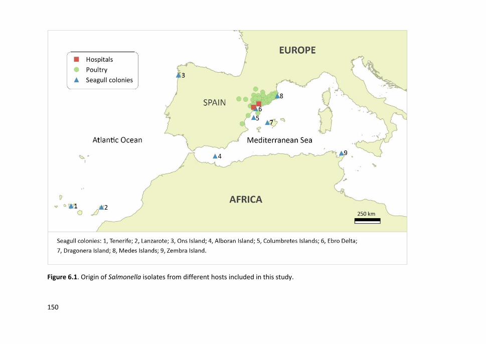
150
Figure 6.1. Origin of Salmonella isolates from different hosts included in this study.
Study IV
151
6.3.2. PFGE
Altogether 742 Salmonella isolates were analysed by PFGE using the restriction
endonuclease XbaI (Roche Applied Science, Indianapolis, IN, USA). A selection of 288
isolates from diverse sources, with same XbaI-PFGE profile, were further genotyped
using a secondary restriction enzyme (BlnI) to enhance the discrimination power (Table
6.2). PFGE was carried out according to the standard operating procedure of PulseNet
(www.pulsenetinternational.org) and using a CHEF-DR II System (Bio-Rad Laboratories,
Hercules, CA). Besides, thiourea was employed during gel electrophoresis when
necessary. Salmonella Braenderup H9812 restricted with XbaI was used as molecular
size standard. The resulting PFGE patterns were transferred to the Fingerprinting II v3.0
software (Bio-Rad, Hercules, CA, USA) for computer analysis. Similarities between
banding patterns was determined on the basis of the Dice coefficient with a tolerance
and optimization of 1.0%. Cluster analysis was performed by the Unweighted Pair Group
Method with Arithmetic averages (UPGMA). Isolates with a minimum level of similarity
≥ 85% were considered as the same pulsotype (XB). Simpson’s diversity index (D) of
XbaI-PFGE in each compartment (human, domestic and wild) was calculated by the
method of Hunter and Gaston (1988). The D-value ranges between 0 and 1; 0 value
indicates that all isolates are identical, while a D-value of 1 indicates a maximum
diversity.
6.3.3. MLST
A subset of 29 isolates of the most significant clinical serovars (21 of Typhimurium and
eight of Enteritidis) and covering human, poultry and seagull origins was chosen for
further genotyping by MLST. Fragments of seven housekeeping genes (aroC, dnaN,
hemD, hisD, purE, sucA and thrA) were amplified by PCR and sequenced following the
protocol and recommended primers at S. enterica MLST database
(http://mlst.warwick.ac.uk/mlst/dbs/Senterica). Sanger sequences were analysed using
the Fingerprinting II v3.0 software (Bio-Rad, Hercules, CA, USA). Alleles and sequence

Chapter 6
152
types (STs) were assigned based on the MLST scheme provided on the database at S.
enterica MLST web site.
6.3.4. Virulence-associated genes
A total of 58 isolates of 12 serovars from different hosts with XbaI and BlnI PFGE
pulsotypes in common were screened to characterize their potential pathogenicity. We
analysed the presence of 27 virulence-associated genes harboured in Salmonella
pathogenicity island (SPI)-1 (orgA, sitC, invA, prgH and avrA), SPI-2 (spiC), SPI-3 (misL,
mgtC), SPI-4 (orfL), SPI-5 (pipD and sopB), pathogenicity islets (tolC, iroN, sifA and cdtB),
fimbrial operons (agfA, agfC, lpfA, lpfC and sefC), plasmids (spvB, spvC and pefA) and
prophages (sopE, gipA, gogB and gtgB). The primer pairs employed alone or in multiplex
PCRs are compiled in Table 6.3. PCR was performed in a 25 μL reaction mixture that
included a ready-to-use PCR Master Mix solution (Promega, Wisconsin, USA), 0.4 μM of
each primer and and 2.5 µL of DNA. Amplification cycles were the following: initial
denaturation at 94°C for 5 min; 30 cycles of denaturation at 94°C for 30 s, annealing at
56°C for 30 s, and extension at 72°C for 2 min; and final extension at 72°C for 10 min.
Amplicons were detected by gel electrophoresis in a 1.8% agarose gels with 0.2 µg/ml
of ethidium bromide. Isolates with the same combination of virulence-associated genes
were considered as the same virulotype (VT).
6.3.5. Statistical analyses
Pearson’s chi-squared test with Yates’s correction for continuity was performed to
compare frequencies of virulence-associated genes among different Salmonella
serovars and host origins. Fisher’s exact tests were used when needed. P < 0.05 was
considered statistically significant. The Deducer GUI of R software (www.R-project.org)
was used for the statistical analysis.
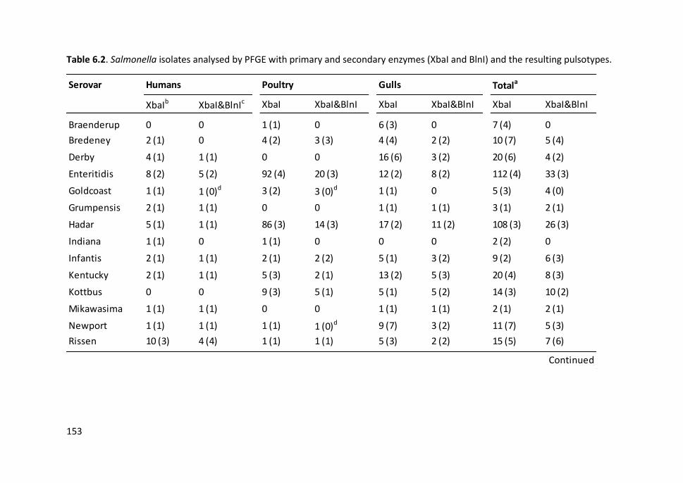
153
Table 6.2. Salmonella isolates analysed by PFGE with primary and secondary enzymes (XbaI and BlnI) and the resulting pulsotypes.
Serovar
XbaIb XbaI&BlnIc XbaI XbaI&BlnI XbaI XbaI&BlnI XbaI XbaI&BlnI
Braenderup 0 0 1 (1) 0 6 (3) 0 7 (4) 0
Bredeney 2 (1) 0 4 (2) 3 (3) 4 (4) 2 (2) 10 (7) 5 (4)
Derby 4 (1) 1 (1) 0 0 16 (6) 3 (2) 20 (6) 4 (2)
Enteritidis 8 (2) 5 (2) 92 (4) 20 (3) 12 (2) 8 (2) 112 (4) 33 (3)
Goldcoast 1 (1) 1 (0)d 3 (2) 3 (0)d 1 (1) 0 5 (3) 4 (0)
Grumpensis 2 (1) 1 (1) 0 0 1 (1) 1 (1) 3 (1) 2 (1)
Hadar 5 (1) 1 (1) 86 (3) 14 (3) 17 (2) 11 (2) 108 (3) 26 (3)
Indiana 1 (1) 0 1 (1) 0 0 0 2 (2) 0
Infantis 2 (1) 1 (1) 2 (1) 2 (2) 5 (1) 3 (2) 9 (2) 6 (3)
Kentucky 2 (1) 1 (1) 5 (3) 2 (1) 13 (2) 5 (3) 20 (4) 8 (3)
Kottbus 0 0 9 (3) 5 (1) 5 (1) 5 (2) 14 (3) 10 (2)
Mikawasima 1 (1) 1 (1) 0 0 1 (1) 1 (1) 2 (1) 2 (1)
Newport 1 (1) 1 (1) 1 (1) 1 (0)d 9 (7) 3 (2) 11 (7) 5 (3)
Rissen 10 (3) 4 (4) 1 (1) 1 (1) 5 (3) 2 (2) 15 (5) 7 (6)
Humans Poultry Gulls Totala
Continued
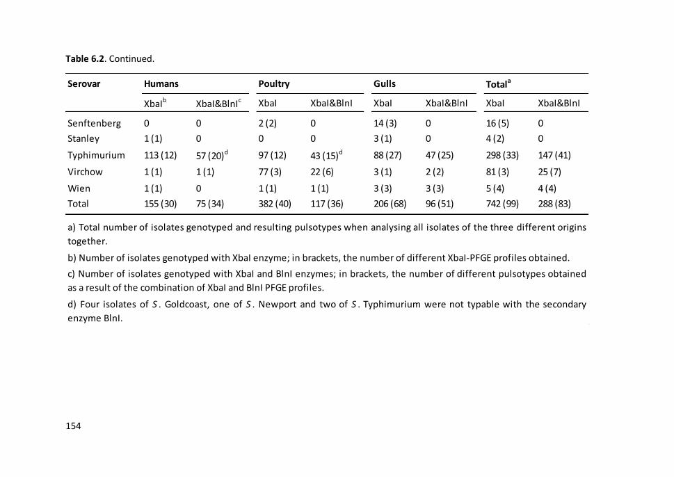
154
Table 6.2. Continued.
Serovar
XbaIb XbaI&BlnIc XbaI XbaI&BlnI XbaI XbaI&BlnI XbaI XbaI&BlnI
Senftenberg 0 0 2 (2) 0 14 (3) 0 16 (5) 0
Stanley 1 (1) 0 0 0 3 (1) 0 4 (2) 0
Typhimurium 113 (12) 57 (20)d 97 (12) 43 (15)d 88 (27) 47 (25) 298 (33) 147 (41)
Virchow 1 (1) 1 (1) 77 (3) 22 (6) 3 (1) 2 (2) 81 (3) 25 (7)
Wien 1 (1) 0 1 (1) 1 (1) 3 (3) 3 (3) 5 (4) 4 (4)
Total 155 (30) 75 (34) 382 (40) 117 (36) 206 (68) 96 (51) 742 (99) 288 (83)
b) Number of isolates genotyped with XbaI enzyme; in brackets, the number of different XbaI-PFGE profiles obtained.
a) Total number of isolates genotyped and resulting pulsotypes when analysing all isolates of the three different origins
together.
c) Number of isolates genotyped with XbaI and BlnI enzymes; in brackets, the number of different pulsotypes obtained
as a result of the combination of XbaI and BlnI PFGE profiles.
d) Four isolates of S . Goldcoast, one of S . Newport and two of S . Typhimurium were not typable with the secondary
enzyme BlnI.
Humans Poultry Gulls Totala
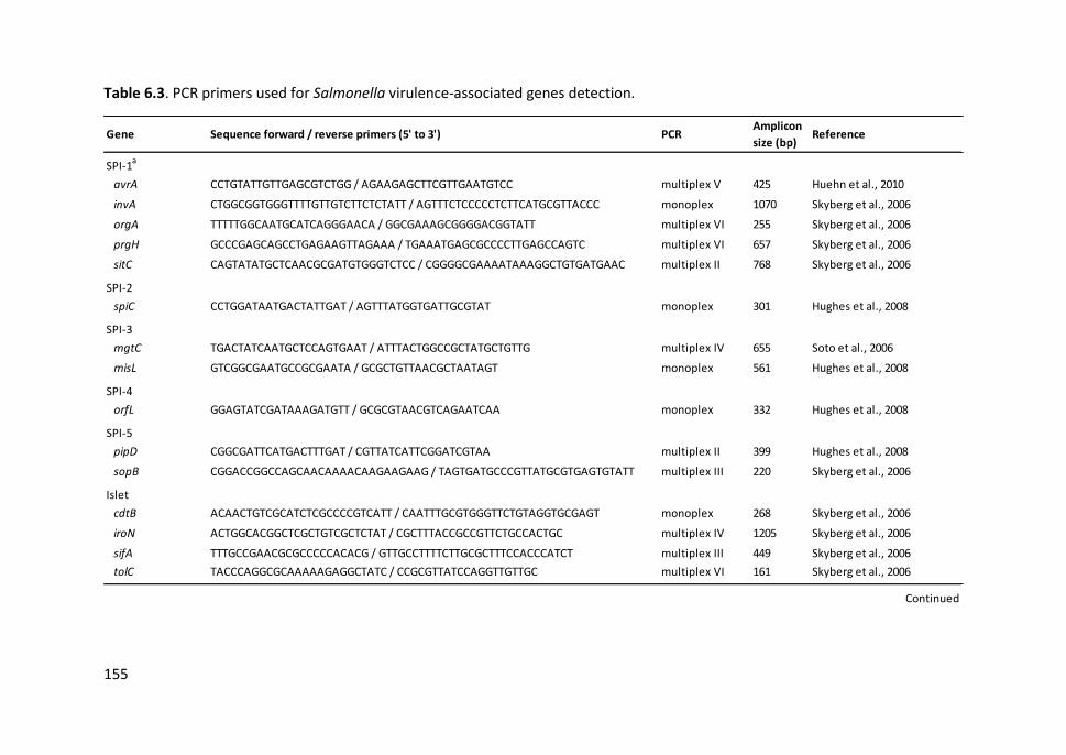
155
Table 6.3. PCR primers used for Salmonella virulence-associated genes detection.
Gene Sequence forward / reverse primers (5' to 3') PCRAmplicon
size (bp) Reference
SPI-1a
avrA CCTGTATTGTTGAGCGTCTGG / AGAAGAGCTTCGTTGAATGTCC multiplex V 425 Huehn et al., 2010
invA CTGGCGGTGGGTTTTGTTGTCTTCTCTATT / AGTTTCTCCCCCTCTTCATGCGTTACCC monoplex 1070 Skyberg et al., 2006
orgA TTTTTGGCAATGCATCAGGGAACA / GGCGAAAGCGGGGACGGTATT multiplex VI 255 Skyberg et al., 2006
prgH GCCCGAGCAGCCTGAGAAGTTAGAAA / TGAAATGAGCGCCCCTTGAGCCAGTC multiplex VI 657 Skyberg et al., 2006
sitC CAGTATATGCTCAACGCGATGTGGGTCTCC / CGGGGCGAAAATAAAGGCTGTGATGAAC multiplex II 768 Skyberg et al., 2006
SPI-2
spiC CCTGGATAATGACTATTGAT / AGTTTATGGTGATTGCGTAT monoplex 301 Hughes et al., 2008
SPI-3
mgtC TGACTATCAATGCTCCAGTGAAT / ATTTACTGGCCGCTATGCTGTTG multiplex IV 655 Soto et al., 2006
misL GTCGGCGAATGCCGCGAATA / GCGCTGTTAACGCTAATAGT monoplex 561 Hughes et al., 2008
SPI-4
orfL GGAGTATCGATAAAGATGTT / GCGCGTAACGTCAGAATCAA monoplex 332 Hughes et al., 2008
SPI-5
pipD CGGCGATTCATGACTTTGAT / CGTTATCATTCGGATCGTAA multiplex II 399 Hughes et al., 2008
sopB CGGACCGGCCAGCAACAAAACAAGAAGAAG / TAGTGATGCCCGTTATGCGTGAGTGTATT multiplex III 220 Skyberg et al., 2006
Islet
cdtB ACAACTGTCGCATCTCGCCCCGTCATT / CAATTTGCGTGGGTTCTGTAGGTGCGAGT monoplex 268 Skyberg et al., 2006
iroN ACTGGCACGGCTCGCTGTCGCTCTAT / CGCTTTACCGCCGTTCTGCCACTGC multiplex IV 1205 Skyberg et al., 2006
sifA TTTGCCGAACGCGCCCCCACACG / GTTGCCTTTTCTTGCGCTTTCCACCCATCT multiplex III 449 Skyberg et al., 2006
tolC TACCCAGGCGCAAAAAGAGGCTATC / CCGCGTTATCCAGGTTGTTGC multiplex VI 161 Skyberg et al., 2006
Continued
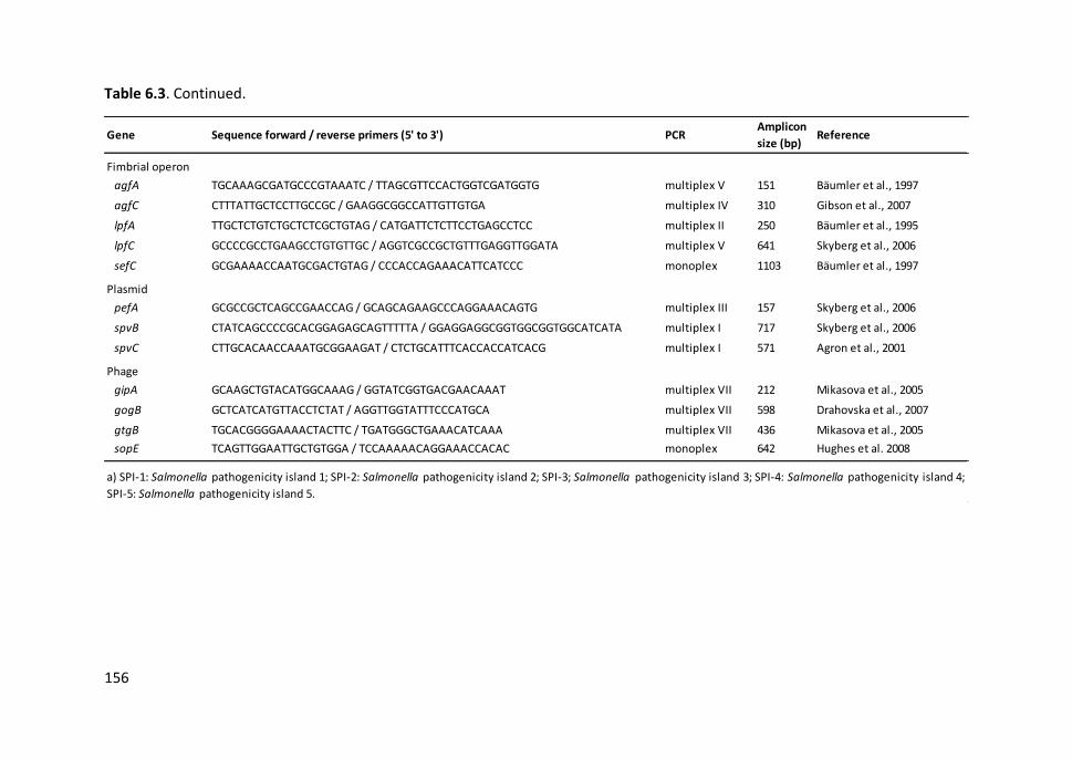
156
Table 6.3. Continued.
Gene Sequence forward / reverse primers (5' to 3') PCRAmplicon
size (bp) Reference
Fimbrial operon
agfA TGCAAAGCGATGCCCGTAAATC / TTAGCGTTCCACTGGTCGATGGTG multiplex V 151 Bäumler et al., 1997
agfC CTTTATTGCTCCTTGCCGC / GAAGGCGGCCATTGTTGTGA multiplex IV 310 Gibson et al., 2007
lpfA TTGCTCTGTCTGCTCTCGCTGTAG / CATGATTCTCTTCCTGAGCCTCC multiplex II 250 Bäumler et al., 1995
lpfC GCCCCGCCTGAAGCCTGTGTTGC / AGGTCGCCGCTGTTTGAGGTTGGATA multiplex V 641 Skyberg et al., 2006
sefC GCGAAAACCAATGCGACTGTAG / CCCACCAGAAACATTCATCCC monoplex 1103 Bäumler et al., 1997
Plasmid
pefA GCGCCGCTCAGCCGAACCAG / GCAGCAGAAGCCCAGGAAACAGTG multiplex III 157 Skyberg et al., 2006
spvB CTATCAGCCCCGCACGGAGAGCAGTTTTTA / GGAGGAGGCGGTGGCGGTGGCATCATA multiplex I 717 Skyberg et al., 2006
spvC CTTGCACAACCAAATGCGGAAGAT / CTCTGCATTTCACCACCATCACG multiplex I 571 Agron et al., 2001
Phage
gipA GCAAGCTGTACATGGCAAAG / GGTATCGGTGACGAACAAAT multiplex VII 212 Mikasova et al., 2005
gogB GCTCATCATGTTACCTCTAT / AGGTTGGTATTTCCCATGCA multiplex VII 598 Drahovska et al., 2007
gtgB TGCACGGGGAAAACTACTTC / TGATGGGCTGAAACATCAAA multiplex VII 436 Mikasova et al., 2005
sopE TCAGTTGGAATTGCTGTGGA / TCCAAAAACAGGAAACCACAC monoplex 642 Hughes et al. 2008
a) SPI-1: Salmonella pathogenicity island 1; SPI-2: Salmonella pathogenicity island 2; SPI-3; Salmonella pathogenicity island 3; SPI-4: Salmonella pathogenicity island 4;
SPI-5: Salmonella pathogenicity island 5.
Study IV
157
6.4. RESULTS
6.4.1. PFGE
All the 742 Salmonella isolates, belonging to 19 serovars, from different hosts included
in this study were analysed by PFGE using the restriction endonuclease XbaI to
determine the genetic relatedness amongst them. Up to 99 different XbaI-PFGE profiles
were obtained with a similarity level of 85% (Table 6.2). Overall, a high genetic diversity
was detected in Salmonella isolates from seagulls (68 profiles; D = 0.970) followed by
isolates from human gastroenteritis cases (30 profiles; D = 0.914). However, isolates
from poultry presented a lower diversity (40 profiles; D = 0.881) despite coming from
different domestic host species and farms distributed in a broad region of the
Northeastern Spain.
We found 30 XbaI-PFGE profiles grouping isolates from two or three different origins
(human, domestic and wild birds). The 88% of clinical human isolates (136/155) showed
profiles also detected in isolates from poultry (six profiles), seagulls (five profiles) or
both domestic and wild birds (nine profiles). Twenty-five profiles from poultry (80%,
305/382) overlapped with those from seagulls (ten profiles), humans or both.
Conversely, the 48% of isolates (98/205) from gulls exhibited unique profiles not
detected in other hosts.
Seventeen unique profiles were detected exclusively in S. Typhimurium isolates from
seagulls. One of these profiles was found in 14 isolates from six different far apart gull
colonies, whereas the others were present in a single or a few isolates from one or more
gull species and colonies. In spite of the overall genetic diversity observed in S.
Typhimurium isolates (33 profiles), five predominant profiles were detected in a high
number of isolates (11 to 90) from the three different niches. The most abundant profile
included the majority of S. Typhimurium isolates (67%, 65/97) from a wide range of
domestic birds (laying hens, chickens, turkeys, partridges and quails) in addition to some
isolates from humans (15/113) and seagulls (8/88) from different colonies. The other
Chapter 6
158
predominant profiles were mostly detected in S. Typhimurium clinical isolates (16 to 26)
besides other hosts. On the other hand, isolates of S. Enteritidis and S. Hadar showed
an overall low genetic diversity and predominant profiles were repeatedly observed in
a high number of isolates. In fact, most of S. Enteritidis isolates from poultry (77/92)
showed the same profile as isolates from humans (7/8) and seagulls (11/12) from
distinct locations.
Based on the XbaI PFGE results, a representative set of Salmonella isolates from
different hosts and sites with the same or highly similar profile were selected for further
genotyping with a secondary restriction enzyme (BlnI) to increase the discrimination
power. Thus, 288 isolates (117 from poultry, 96 from seagulls and 75 from humans) of
15 different serovars were analysed by BlnI-PFGE. Isolates of Braenderup, Indiana,
Senftenberg and Stanley serovars were not genotyped with BlnI because the low degree
of similarity detected using XbaI. Seven isolates (four of S. Goldcoast, two of S.
Typhimurium and one of S. Newport) were non-typable using BlnI. The combined
dendrogram with XbaI and BlnI band patterns revealed 83 distinct profiles (PFGE
pulsotypes) with a similarity of 85% (Table 6.2). Isolates were grouped according to their
serovar, except for serovars Hadar and Kottbus which were clustered together although
forming separate branches (Figure 6.2).
We detected 30 XbaI and BlnI PFGE pulsotypes containing isolates from two or three
different hosts (Figures 6.2 and 6.3). These pulsotypes in common corresponded mainly
to serovar Typhimurium, followed by Enteritidis, Virchow, and other serovars also of
public health significance (Bredeney, Derby, Grumpensis, Hadar, Infantis, Kentucky,
Kottbus, Mikawasima, and Rissen). Noteworthy, the XbaI profiles with a high similarity
in isolates of S. Goldcoast, S. Newport and S. Wien from different hosts were
distinguished using BlnI. At the same time, the restriction with BlnI increased the
resolution among isolates of other serovars and allowed to discriminate more
pulsotypes whilst maintaining similarities among the different host niches. S.
Typhimurium isolates from different niches shared a total of 16 pulsotypes; the most

Study IV
159
frequent of them (XB20) was detected in 31 isolates from the three different niches,
especially from poultry (22 isolates). Other pulsotypes with high occurrence were XB8
and XB1 which were found in 25 S. Enteritidis and 21 S. Hadar isolates, respectively,
from the three different compartments.
6.4.2. Multilocus sequence typing
MLST of 29 isolates of S. Typhimurium (N=21) and S. Enteritidis (N=8) from different
sources with the same or highly related pulsotypes (similarity over 80%) was performed
to determine the potential evolutionary relatedness among them. We detected two STs
of S. Typhimurium differing only by a single locus (dnaN gene: alleles 7 and 19),
belonging to the same clonal complex, also denominated eBurstGroup (eGB1) (Figure
6.2). Fourteen isolates were classified as ST34 and seven belonged to ST19. These STs
grouped five and three different pulsotypes, respectively, represented by isolates from
different hosts and sites far apart. All S. Enteritidis isolates originating from humans,
poultry and seagulls were clustered into a single sequence type ST11 although two
different pulsotypes were distinguished by PFGE (XB8 and XB9).
Figure 6.2. Combined dendrogram with XbaI and BlnI profiles of a selection of
Salmonella isolates from different hosts showing the same or highly similar pulsotypes.
The tree was constructed using the UPGMA clustering method and the Dice coefficient
for the similarity matrices calculation. Pulsotypes (XB) numbers were assigned to XbaI
and BlnI PFGE profiles with a similarity level of 85%. Sequence types (ST) and
eBurstGroups (eBG) not analysed are labelled as NA (Non-available). Virulotypes (VT)
are indicated by a number corresponding to the number of positive virulence genes and
a capital letter regarding to the different profiles detected. The presence of virulence-
associated genes is depicted as grey squares. Dark grey squares stand out isolates from
different hosts with XB and VT in common. YL gull: yellow-legged gull; AD gull: Audouin’s
gull.

PFGE XbaI+PFGE BlnI
XbaI&BlnI
10
0
90
80
70
60
50
40
PFGE XbaI PFGE BlnI
.
.
.
.
.
.
.
.
.
.
.
.
.
.
.
.
.
.
.
.
.
.
.
.
.
.
.
.
.
.
.
.
.
.
.
.
.
.
.
CM104-S1
DA187-S2
HT15
CS77
DA230-S4
CS10
CM214-S1
MM141-S1
CS49
HT9
CS382
DA216-S3
DGM64-S1
HR59
HT20
CS284
AA114-S1
MM207-S3
HR43
AA145-S1
CS218
ZM11-S1
HR41
CS7
MM212-S1
HR6
CS290
DM65-S1
HR2
MM257-S4
HT8
DM89-S4
CS252
HT18
CS297
DM197-S1
CS285
MCM2-S1
CS17
Hadar
Hadar
Hadar
Hadar
Kottbus
Kottbus
Kottbus
Hadar
Hadar
Kentucky
Kentucky
Kentucky
Derby
Derby
Infantis
Infantis
Infantis
Mikawasima
Mikawasima
Enteritidis
Enteritidis
Enteritidis
Enteritidis
Enteritidis
Enteritidis
Enteritidis
Enteritidis
Grumpensis
Grumpensis
Rissen
Rissen
Virchow
Virchow
Virchow
Virchow
Bredeney
Bredeney
Typhimirium mo.
Typhimurium
L. michahellis
L. audouinii
Human
Turkey
L. audouinii
Turkey
L. michahellis
L. michahellis
Chicken
Human
Chicken
L. audouinii
L. michahellis
Human
Human
Hen
L. audouinii
L. michahellis
Human
Audouin's gull
Chicken
Yellow-legged gull
Human
Hen
Yellow-legged gull
Human
Hen
L. michahellis
Human
L. michahellis
Human
L. michahellis
Hen
Human
Chicken
L. michahellis
Hen
Yellow-legged gull
Hen
Columbretes
Ebro Delta
Tortosa
Baix Camp
Ebro Delta
Alt Camp
Columbretes
Medes
Priorat
Tortosa
Baix Ebre
Ebro Delta
Dragonera
Reus
Tortosa
Unknown
Alboran
Medes
Reus
Alboran
Alt Penedès
Zembra
Reus
Terres Ebre
Medes
Reus
Baix Ebre
Delta Ebre
Reus
Medes
Tortosa
Ebro Delta
Unknown
Tortosa
Montsià
Delta Ebre
Garrigues
Lanzarote
Alt Penedès
2010
2011
2009
2010
2011
2008
2011
2010
2009
2011
2012
2011
2011
2013
2010
2010
2010
2011
2013
2010
2010
2009
2013
2008
2011
2013
2010
2009
2013
2011
2011
2010
2010
2010
2010
2011
2010
2010
2008
.
.
.
.
.
.
.
.
.
.
.
.
.
.
.
.
.
.
.
.
.
.
.
.
.
.
.
.
.
.
.
.
.
.
.
.
.
.
.
.
.
.
.
.
.
.
.
.
.
.
CS285
MCM2-S1
CS17
CS206
HT68
MM229-S1
HR30
CS14
DM178-S1
HT50
CM222-S1
CS165
HR17
MM213-S1
HT73
GAM133-S1
CS201
HR40
DM95-S1
CS209
CS163
HT77
Bredeney
Typhimirium mo.
Typhimurium
Typhimirium mo.
Typhimurium
Typhimirium mo.
Typhimurium
Typhimurium
Typhimirium mo.
Typhimurium
Typhimurium
Typhimurium
Typhimirium mo.
Typhimirium mo.
Typhimurium
Typhimurium
Typhimurium
Typhimurium
Typhimurium
Typhimurium
Typhimurium
Typhimurium
Hen
Yellow-legged gull
Hen
Chicken
Human
Yellow-legged gull
Human
Hen
Yellow-legged gull
Human
Yellow-legged gull
Chicken
Human
Yellow-legged gull
Human
Yellow-legged gull
Turkey
Human
Yellow-legged gull
Quail
Chicken
Human
Garrigues
Lanzarote
Alt Penedès
Unknown
Tortosa
Medes
Reus
Unknown
Delta Ebre
Tortosa
Columbrets
Solsonès
Reus
Medes
Tortosa
Ons
Baix Ebre
Reus
Delta Ebre
Pla d'Urgell
Alt Camp
Tortosa
2010
2010
2008
2011
2010
2011
2013
2008
2010
2009
2011
2010
2013
2011
2011
2011
2011
2013
2010
2011
2010
2011
.
.
.
.
.
.
.
.
.
.
.
.
ST19
ST19
ST19
ST34
ST11
50
60 70 80 90
10
0
Isolate Serovar Host Location Year XB ST eBG VT
Virulence-associated genes
SPI 1-5 IsletFimbrial operon
Plasmid Prophage
invA
org
Ap
rgH
sitC
avr
Asp
iCm
gtC
mis
Lo
rfL
pip
Dso
pB
tolC
iro
Nsi
fAcd
tBa
gfA
ag
fClp
fAlp
fCse
fCp
efA
spvB
spvC
sop
Eg
ipA
go
gB
gtg
B
CM104-S1 Hadar YL gull Columbretes Is. 2010 1 NA NA 20A
DA187-S2 Hadar AD gull Ebro Delta 2011 1 NA NA 20A
HT15 Hadar Human Catalonia 2009 1 NA NA 20B
CS77 Hadar Turkey Catalonia 2010 1 NA NA 20B
DA230-S4 Kottbus AD gull Ebro Delta 2011 2 NA NA 18A
CS10 Kottbus Turkey Catalonia 2008 2 NA NA 20B
CM214-S1 Kottbus YL gull Columbretes Is. 2011 2 NA NA 20B
MM141-S1 Hadar YL gull Medes Is. 2010 3 NA NA 16B
CS49 Hadar Chicken Catalonia 2009 3 NA NA 11A
HT9 Kentucky Human Catalonia 2011 4 NA NA 20B
CS382 Kentucky Chicken Catalonia 2012 4 NA NA 20B
DA216-S3 Kentucky AD gull Ebro Delta 2011 4 NA NA 18A
DGM64-S1 Derby YL gull Dragonera Is. 2011 5 NA NA 11B
HR59 Derby Human Catalonia 2013 5 NA NA 16C
HT20 Infantis Human Catalonia 2010 6 NA NA 20B
CS284 Infantis Hen Catalonia 2010 6 NA NA 20B
AA114-S1 Infantis AD gull Alboran Is. 2010 6 NA NA 20B
MM207-S3 Mikawasima YL gull Medes Is. 2011 7 NA NA 21A
HR43 Mikawasima Human Catalonia 2013 7 NA NA 21A
AA145-S1 Enteritidis AD gull Alboran Is. 2010 8 11 4 18B
CS218 Enteritidis Chicken Catalonia 2010 8 11 4 18C
ZM11-S1 Enteritidis YL gull Zembra 2009 8 11 4 20C
HR41 Enteritidis Human Catalonia 2013 8 11 4 25A
CS7 Enteritidis Hen Catalonia 2008 8 11 4 15A
MM212-S1 Enteritidis YL gull Medes Is. 2011 9 11 4 22C
HR6 Enteritidis Human Catalonia 2013 9 11 4 16D
CS290 Enteritidis Hen Catalonia 2010 9 11 4 20D
DM65-S1 Grumpensis YL gull Ebro Delta 2009 10 NA NA 16A
HR2 Grumpensis Human Catalonia 2013 10 NA NA 16A
MM257-S4 Rissen YL gull Medes Is. 2011 11 NA NA 6A
HT8 Rissen Human Catalonia 2011 11 NA NA 19A
DM89-S4 Virchow YL gull Ebro Delta 2010 12 NA NA 21B
CS252 Virchow Hen Aragon 2010 12 NA NA 22A
HT18 Virchow Human Catalonia 2010 13 NA NA 21C
CS297 Virchow Chicken Catalonia 2010 13 NA NA 22A
DM197-S1 Bredeney YL gull Ebro Delta 2011 14 NA NA 14A
CS285 Bredeney Hen Catalonia 2010 14 NA NA 16E
MCM2-S1 Typhimurium YL gull Lanzarote 2010 15 34 1 22B
CS17 Typhimurium Hen Catalonia 2008 15 34 1 22B
CS206 Typhimurium Chicken Catalonia 2011 16 34 1 24B
HT68 Typhimurium Human Catalonia 2010 16 34 1 24B
MM229-S4 Typhimurium YL gull Medes Is. 2011 17 34 1 22B
HR30 Typhimurium Human Catalonia 2013 17 34 1 23A
CS14 Typhimurium Hen Catalonia 2008 17 34 1 22B
DM178-S1 Typhimurium YL gull Ebro Delta 2010 17 34 1 23A
HT50 Typhimurium Human Catalonia 2009 17 34 1 24B
CM222-S1 Typhimurium YL gull Columbretes Is. 2011 18 34 1 22B
CS165 Typhimurium Chicken Catalonia 2010 18 34 1 21C
HR17 Typhimurium Human Catalonia 2013 18 34 1 21D
MM213-S1 Typhimurium YL gull Medes Is. 2011 19 34 1 22B
HT73 Typhimurium Human Catalonia 2011 19 34 1 21E
GAM133-S1 Typhimurium YL gull Ons Is. 2011 20 19 1 24A
CS201 Typhimurium Turkey Catalonia 2011 20 19 1 24A
HR40 Typhimurium Human Catalonia 2013 20 19 1 20E
DM95-S1 Typhimurium YL gull Ebro Delta 2010 21 19 1 24A
CS209 Typhimurium Quail Catalonia 2011 21 19 1 24A
CS163 Typhimurium Chicken Catalonia 2010 22 19 1 24A
HT77 Typhimurium Human Catalonia 2011 22 19 1 25B
40
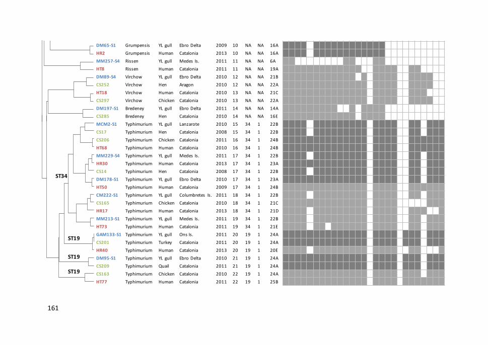
161
PFGE XbaI+PFGE BlnI
XbaI&BlnI
10
0
90
80
70
60
50
40
PFGE XbaI PFGE BlnI
.
.
.
.
.
.
.
.
.
.
.
.
.
.
.
.
.
.
.
.
.
.
.
.
.
.
.
.
.
.
.
.
.
.
.
.
.
.
.
CM104-S1
DA187-S2
HT15
CS77
DA230-S4
CS10
CM214-S1
MM141-S1
CS49
HT9
CS382
DA216-S3
DGM64-S1
HR59
HT20
CS284
AA114-S1
MM207-S3
HR43
AA145-S1
CS218
ZM11-S1
HR41
CS7
MM212-S1
HR6
CS290
DM65-S1
HR2
MM257-S4
HT8
DM89-S4
CS252
HT18
CS297
DM197-S1
CS285
MCM2-S1
CS17
Hadar
Hadar
Hadar
Hadar
Kottbus
Kottbus
Kottbus
Hadar
Hadar
Kentucky
Kentucky
Kentucky
Derby
Derby
Infantis
Infantis
Infantis
Mikawasima
Mikawasima
Enteritidis
Enteritidis
Enteritidis
Enteritidis
Enteritidis
Enteritidis
Enteritidis
Enteritidis
Grumpensis
Grumpensis
Rissen
Rissen
Virchow
Virchow
Virchow
Virchow
Bredeney
Bredeney
Typhimirium mo.
Typhimurium
L. michahellis
L. audouinii
Human
Turkey
L. audouinii
Turkey
L. michahellis
L. michahellis
Chicken
Human
Chicken
L. audouinii
L. michahellis
Human
Human
Hen
L. audouinii
L. michahellis
Human
Audouin's gull
Chicken
Yellow-legged gull
Human
Hen
Yellow-legged gull
Human
Hen
L. michahellis
Human
L. michahellis
Human
L. michahellis
Hen
Human
Chicken
L. michahellis
Hen
Yellow-legged gull
Hen
Columbretes
Ebro Delta
Tortosa
Baix Camp
Ebro Delta
Alt Camp
Columbretes
Medes
Priorat
Tortosa
Baix Ebre
Ebro Delta
Dragonera
Reus
Tortosa
Unknown
Alboran
Medes
Reus
Alboran
Alt Penedès
Zembra
Reus
Terres Ebre
Medes
Reus
Baix Ebre
Delta Ebre
Reus
Medes
Tortosa
Ebro Delta
Unknown
Tortosa
Montsià
Delta Ebre
Garrigues
Lanzarote
Alt Penedès
2010
2011
2009
2010
2011
2008
2011
2010
2009
2011
2012
2011
2011
2013
2010
2010
2010
2011
2013
2010
2010
2009
2013
2008
2011
2013
2010
2009
2013
2011
2011
2010
2010
2010
2010
2011
2010
2010
2008
.
.
.
.
.
.
.
.
.
.
.
.
.
.
.
.
.
.
.
.
.
.
.
.
.
.
.
.
.
.
.
.
.
.
.
.
.
.
.
.
.
.
.
.
.
.
.
.
.
.
CS285
MCM2-S1
CS17
CS206
HT68
MM229-S1
HR30
CS14
DM178-S1
HT50
CM222-S1
CS165
HR17
MM213-S1
HT73
GAM133-S1
CS201
HR40
DM95-S1
CS209
CS163
HT77
Bredeney
Typhimirium mo.
Typhimurium
Typhimirium mo.
Typhimurium
Typhimirium mo.
Typhimurium
Typhimurium
Typhimirium mo.
Typhimurium
Typhimurium
Typhimurium
Typhimirium mo.
Typhimirium mo.
Typhimurium
Typhimurium
Typhimurium
Typhimurium
Typhimurium
Typhimurium
Typhimurium
Typhimurium
Hen
Yellow-legged gull
Hen
Chicken
Human
Yellow-legged gull
Human
Hen
Yellow-legged gull
Human
Yellow-legged gull
Chicken
Human
Yellow-legged gull
Human
Yellow-legged gull
Turkey
Human
Yellow-legged gull
Quail
Chicken
Human
Garrigues
Lanzarote
Alt Penedès
Unknown
Tortosa
Medes
Reus
Unknown
Delta Ebre
Tortosa
Columbrets
Solsonès
Reus
Medes
Tortosa
Ons
Baix Ebre
Reus
Delta Ebre
Pla d'Urgell
Alt Camp
Tortosa
2010
2010
2008
2011
2010
2011
2013
2008
2010
2009
2011
2010
2013
2011
2011
2011
2011
2013
2010
2011
2010
2011
.
.
.
.
.
.
.
.
.
.
.
.
ST19
ST19
ST19
ST34
ST11
50
60 70 80 90
10
0
Isolate Serovar Host Location Year XB ST eBG VT
Virulence-associated genes
SPI 1-5 IsletFimbrial operon
Plasmid Prophage
invA
org
Ap
rgH
sitC
avr
Asp
iCm
gtC
mis
Lo
rfL
pip
Dso
pB
tolC
iro
Nsi
fAcd
tBa
gfA
ag
fClp
fAlp
fCse
fCp
efA
spvB
spvC
sop
Eg
ipA
go
gB
gtg
B
CM104-S1 Hadar YL gull Columbretes Is. 2010 1 NA NA 20A
DA187-S2 Hadar AD gull Ebro Delta 2011 1 NA NA 20A
HT15 Hadar Human Catalonia 2009 1 NA NA 20B
CS77 Hadar Turkey Catalonia 2010 1 NA NA 20B
DA230-S4 Kottbus AD gull Ebro Delta 2011 2 NA NA 18A
CS10 Kottbus Turkey Catalonia 2008 2 NA NA 20B
CM214-S1 Kottbus YL gull Columbretes Is. 2011 2 NA NA 20B
MM141-S1 Hadar YL gull Medes Is. 2010 3 NA NA 16B
CS49 Hadar Chicken Catalonia 2009 3 NA NA 11A
HT9 Kentucky Human Catalonia 2011 4 NA NA 20B
CS382 Kentucky Chicken Catalonia 2012 4 NA NA 20B
DA216-S3 Kentucky AD gull Ebro Delta 2011 4 NA NA 18A
DGM64-S1 Derby YL gull Dragonera Is. 2011 5 NA NA 11B
HR59 Derby Human Catalonia 2013 5 NA NA 16C
HT20 Infantis Human Catalonia 2010 6 NA NA 20B
CS284 Infantis Hen Catalonia 2010 6 NA NA 20B
AA114-S1 Infantis AD gull Alboran Is. 2010 6 NA NA 20B
MM207-S3 Mikawasima YL gull Medes Is. 2011 7 NA NA 21A
HR43 Mikawasima Human Catalonia 2013 7 NA NA 21A
AA145-S1 Enteritidis AD gull Alboran Is. 2010 8 11 4 18B
CS218 Enteritidis Chicken Catalonia 2010 8 11 4 18C
ZM11-S1 Enteritidis YL gull Zembra 2009 8 11 4 20C
HR41 Enteritidis Human Catalonia 2013 8 11 4 25A
CS7 Enteritidis Hen Catalonia 2008 8 11 4 15A
MM212-S1 Enteritidis YL gull Medes Is. 2011 9 11 4 22C
HR6 Enteritidis Human Catalonia 2013 9 11 4 16D
CS290 Enteritidis Hen Catalonia 2010 9 11 4 20D
DM65-S1 Grumpensis YL gull Ebro Delta 2009 10 NA NA 16A
HR2 Grumpensis Human Catalonia 2013 10 NA NA 16A
MM257-S4 Rissen YL gull Medes Is. 2011 11 NA NA 6A
HT8 Rissen Human Catalonia 2011 11 NA NA 19A
DM89-S4 Virchow YL gull Ebro Delta 2010 12 NA NA 21B
CS252 Virchow Hen Aragon 2010 12 NA NA 22A
HT18 Virchow Human Catalonia 2010 13 NA NA 21C
CS297 Virchow Chicken Catalonia 2010 13 NA NA 22A
DM197-S1 Bredeney YL gull Ebro Delta 2011 14 NA NA 14A
CS285 Bredeney Hen Catalonia 2010 14 NA NA 16E
MCM2-S1 Typhimurium YL gull Lanzarote 2010 15 34 1 22B
CS17 Typhimurium Hen Catalonia 2008 15 34 1 22B
CS206 Typhimurium Chicken Catalonia 2011 16 34 1 24B
HT68 Typhimurium Human Catalonia 2010 16 34 1 24B
MM229-S4 Typhimurium YL gull Medes Is. 2011 17 34 1 22B
HR30 Typhimurium Human Catalonia 2013 17 34 1 23A
CS14 Typhimurium Hen Catalonia 2008 17 34 1 22B
DM178-S1 Typhimurium YL gull Ebro Delta 2010 17 34 1 23A
HT50 Typhimurium Human Catalonia 2009 17 34 1 24B
CM222-S1 Typhimurium YL gull Columbretes Is. 2011 18 34 1 22B
CS165 Typhimurium Chicken Catalonia 2010 18 34 1 21C
HR17 Typhimurium Human Catalonia 2013 18 34 1 21D
MM213-S1 Typhimurium YL gull Medes Is. 2011 19 34 1 22B
HT73 Typhimurium Human Catalonia 2011 19 34 1 21E
GAM133-S1 Typhimurium YL gull Ons Is. 2011 20 19 1 24A
CS201 Typhimurium Turkey Catalonia 2011 20 19 1 24A
HR40 Typhimurium Human Catalonia 2013 20 19 1 20E
DM95-S1 Typhimurium YL gull Ebro Delta 2010 21 19 1 24A
CS209 Typhimurium Quail Catalonia 2011 21 19 1 24A
CS163 Typhimurium Chicken Catalonia 2010 22 19 1 24A
HT77 Typhimurium Human Catalonia 2011 22 19 1 25B
40

162
Figure 6.3. Salmonella isolates from different hosts with XbaI and BlnI PFGE pulsotypes (XB) in common.
Serovar and pulsotype Number of isolates
BredeneyDerby
Enteritidis
GrumpensisHadarInfantisKentuckyKottbusMikawasimaRissen
Typhimurium
Virchow
12
2516
8
8
104
0 5 10 15 20 25 30 35
XB14XB5XB8XB9XB10XB1XB3XB6XB4XB2XB7XB11XB15XB16XB17XB18XB19XB20XB21XB22XB23XB24XB25XB26XB27XB28XB29XB30XB12XB13
Humans
Poultry
Seagulls
Number of pulsotypes

Study IV
163
6.4.3. Virulence genes
We examined the presence of 27 virulence-associated genes in 58 NTS isolates from
different hosts showing the same or highly similar PFGE pulsotype. All isolates were
positive for six or more of the tested genes and at least 20 genes were present in most
isolates (78%, 42/58) (Figure 6.2). We observed a high prevalence (>76%) of genes
located in SPIs (SPI-1 to SPI-5) which were highly conserved among different NTS
serovars (Table 6.4). The invA and orgA genes were present in all studied isolates.
However, the avrA gene was absent in S. Grumpensis and showed a relative low
frequency in S. Typhimurium isolates (57%). The 59% of isolates possessed all SPIs genes
tested, but one S. Rissen isolate (MM257-S4) from a seagull was deficient in many SPI-
1 genes (8/11).
On the other hand, the islet gene tolC and the fimbrial operon genes agfA and agfC were
detected in all isolates. In contrast, the presence of other virulence-associated genes
was more variable. Some genes were found in almost all serovars except for Bredeney
(iroN and lpfAC genes), Grumpensis (lpfAC genes) and Rissen (sifA gene). However, other
genes were only detected in specific serovars, such as the fimbrial gene sefC in S.
Enteritidis. Moreover, the cdtB gene encoding for the Salmonella cytolethal distending
toxin was only found in four isolates of serovars Bredeney and Grumpensis. The target
genes for virulence determinants located in plasmids (spvB and spvC) were observed
with a high prevalence (84%) in isolates of many serovars, except for Bredeney, Derby
and Grumpensis. Yet the plasmid-located fimbrial gene pefA was only present in some
S. Enteritidis and Typhimurium isolates (75% and 29%, respectively). The prophage
genes were detected in a low frequency and only in a few serovars, mainly Enteritidis
and Typhimurium. The sopE gene involved in invasion of host cells was present in most
of S. Enteritidis isolates (88%) and in some isolates of S. Typhimurium (33%) and S. Hadar
(24%). The genes associated with the lamboid Gifsy-1 (gipA and gogB) and Gifsy-2 (gtgB)
phages were only found in S. Typhimurium (gipA, gogB and gtgB), S. Enteritidis (gogB
and gtgB) and S. Mikawasima (gtgB).
Chapter 6
164
We detected a high number of positive virulence-associated genes (21 to 25) in isolates
of serovars Enteritidis, Typhimurium and Virchow from the three different niches. No
statistically significant differences were detected in virulence gene prevalences or
distribution among isolates from humans, poultry and seagulls; except for the iroN gene
which showed a higher occurrence in isolates from humans compared to those from
gulls. The iroN gene was present in almost all human clinical isolates (16/17) and in most
isolates from poultry (14/18) but only in half of the isolates from seagulls (11/23) (Figure
6.2).
Based on the combination of virulence-associated genes, we observed 33 different
virulotypes (VT); ten of them in more than one isolate (two to nine) from different host
niches (Figure 6.2). We found the same virulotype (VT20B) in isolates of four serovars
(Hadar, Infantis, Kentucky and Kottbus) from different hosts. S. Hadar isolates with the
same pulsotype (XB1) displayed two different virulotypes: iroN gene was present in
isolates from human and poultry (VT20B), whilst isolates from seagulls lacked iroN and
possessed the prophage gene sopE (VT20A). Moreover, another virulotype (VT18A),
similar to VT20B but lacking genes involved in iron acquisition (iroN and sitC), was
detected in two isolates of different serovars (Kentucky and Kottbus) from the same
seagull colony. Noteworthy, S. Bredeney and S. Grumpensis isolates showed similar
virulotypes characterized by the lack of plasmid- and phage-borne virulence genes and
lacking also some fimbrial genes; conversely, cdtB gene was only detected in those
serovars.
We found a unique virulotype in each of the S. Enteritidis isolates, despite the low
diversity detected by PFGE and MLST, since they were grouped in two different
pulsotypes (XB8 and XB9) and a single ST (ST-11). Contrary, four different virulotypes
were repeatedly observed in the 76% of S. Typhimurium isolates. The same virulotype
(VT24A) was observed in five isolates from domestic and wild birds belonging to three
pulsotypes (XB20, XB21 and XB22) of the ST19, though it was not detected in closely
related isolates of human origin. The remaining three virulotypes (VT22B, VT24B and
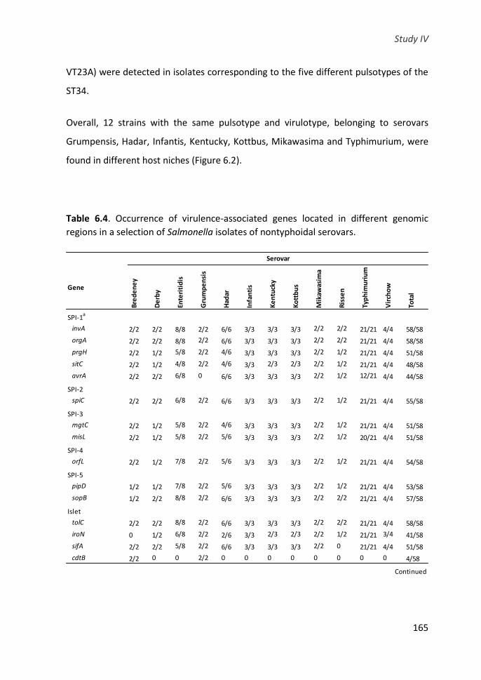
Study IV
165
VT23A) were detected in isolates corresponding to the five different pulsotypes of the
ST34.
Overall, 12 strains with the same pulsotype and virulotype, belonging to serovars
Grumpensis, Hadar, Infantis, Kentucky, Kottbus, Mikawasima and Typhimurium, were
found in different host niches (Figure 6.2).
Table 6.4. Occurrence of virulence-associated genes located in different genomic
regions in a selection of Salmonella isolates of nontyphoidal serovars.
Gene
Bre
de
ne
y
De
rby
Ente
riti
dis
Gru
mp
en
sis
Had
ar
Infa
nti
s
Ke
ntu
cky
Ko
ttb
us
Mik
awas
ima
Ris
sen
Typ
him
uri
um
Vir
cho
w
Tota
l
SPI-1a
invA 2/2 2/2 8/8 2/2 6/6 3/3 3/3 3/3 2/2 2/2 21/21 4/4 58/58
orgA 2/2 2/2 8/8 2/2 6/6 3/3 3/3 3/3 2/2 2/2 21/21 4/4 58/58
prgH 2/2 1/2 5/8 2/2 4/6 3/3 3/3 3/3 2/2 1/2 21/21 4/4 51/58
sitC 2/2 1/2 4/8 2/2 4/6 3/3 2/3 2/3 2/2 1/2 21/21 4/4 48/58
avrA 2/2 2/2 6/8 0 6/6 3/3 3/3 3/3 2/2 1/2 12/21 4/4 44/58
SPI-2
spiC 2/2 2/2 6/8 2/2 6/6 3/3 3/3 3/3 2/2 1/2 21/21 4/4 55/58
SPI-3
mgtC 2/2 1/2 5/8 2/2 4/6 3/3 3/3 3/3 2/2 1/2 21/21 4/4 51/58
misL 2/2 1/2 5/8 2/2 5/6 3/3 3/3 3/3 2/2 1/2 20/21 4/4 51/58
SPI-4
orfL 2/2 1/2 7/8 2/2 5/6 3/3 3/3 3/3 2/2 1/2 21/21 4/4 54/58
SPI-5
pipD 1/2 1/2 7/8 2/2 5/6 3/3 3/3 3/3 2/2 1/2 21/21 4/4 53/58
sopB 1/2 2/2 8/8 2/2 6/6 3/3 3/3 3/3 2/2 2/2 21/21 4/4 57/58
Islet
tolC 2/2 2/2 8/8 2/2 6/6 3/3 3/3 3/3 2/2 2/2 21/21 4/4 58/58
iroN 0 1/2 6/8 2/2 2/6 3/3 2/3 2/3 2/2 1/2 21/21 3/4 41/58
sifA 2/2 2/2 5/8 2/2 6/6 3/3 3/3 3/3 2/2 0 21/21 4/4 51/58
cdtB 2/2 0 0 2/2 0 0 0 0 0 0 0 0 4/58
Serovar
Continued

Chapter 6
166
Table 6.4. Continued.
6.5. DISCUSSION
It is well known that domestic and wild birds are important reservoirs of Salmonella spp.
Antilles (2014) reported that seagulls carried a wide variety of Salmonella serovars, most
of them associated with human outbreaks, and suggested the contribution of these
birds in the transmission of Salmonella. In order to investigate the genetic relatedness
among NTS present in these seagulls, as well as in poultry and human gastroenteritis
cases, we conducted the molecular typing of isolates of 19 serovars from these different
host niches.
PFGE revealed a high genetic diversity among S. Typhimurium isolates, probably
because of the vast number of isolates of this serovar that were analysed. However, this
GeneB
red
en
ey
De
rby
Ente
riti
dis
Gru
mp
en
sis
Had
ar
Infa
nti
s
Ke
ntu
cky
Ko
ttb
us
Mik
awas
ima
Ris
sen
Typ
him
uri
um
Vir
cho
w
Tota
l
Fimbrial operon
agfA 2/2 2/2 8/8 2/2 6/6 3/3 3/3 3/3 2/2 2/2 21/21 4/4 58/58
agfC 2/2 2/2 8/8 2/2 6/6 3/3 3/3 3/3 2/2 2/2 21/21 4/4 58/58
lpfA 0 1/2 8/8 0 5/6 3/3 3/3 3/3 2/2 1/2 21/21 4/4 51/58
lpfC 0 1/2 8/8 0 5/6 3/3 3/3 3/3 2/2 1/2 21/21 4/4 51/58
sefC 0 0 4/8 0 0 0 0 0 0 0 0 0 4/58
Plasmid
pefA 0 0 6/8 0 0 0 0 0 0 0 6/21 0 12/58
spvB 0 0 7/8 0 6/6 3/3 3/3 3/3 2/2 1/2 20/21 4/4 49/58
spvC 0 0 7/8 0 6/6 3/3 3/3 3/3 2/2 1/2 20/21 4/4 49/58
Phage
sopE 0 0 7/8 0 2/6 0 0 0 0 0 5/21 4/4 18/58
gipA 0 0 0 0 0 0 0 0 0 0 19/21 3/4 22/58
gogB 0 0 1/8 0 0 0 0 0 0 0 19/21 0 20/58
gtgB 0 0 8/8 0 0 0 0 0 2/2 0 21/21 0 31/58
a) SPI-1: Salmonella pathogenicity island 1; SPI-2: Salmonella pathogenicity island 2; SPI-3; Salmonella pathogenicity
island 3; SPI-4: Salmonella pathogenicity island 4; SPI-5: Salmonella pathogenicity island 5.
Serovar

Study IV
167
result may also reflect the heterogenicity of S. Typhimurium populations and the
different genotypes circulating in the environment. S. Typhimurium isolates from
seagulls displayed the greatest diversity and almost half of them showed unique profiles
not detected neither in poultry nor in humans. However, many of these pulsotypes were
found in more than one seagull of both species and colonies far from each other. This
may suggest the existence of strains adapted to seagulls, although it could also be due
to different infection sources, such as the environment and other livestock apart from
poultry. Salmonella is present in many animals and has a widespread distribution and
survivability in the environment (e.g. soil, animal feed and faeces, and surface waters)
(Winfiel and Groisman, 2003; Hoelzer et al., 2011; Levantesi et al., 2012). This, together
with the feeding habits and the foraging and migratory movements of seagulls promote
their exposure to a wider variety of Salmonella infection sources. Thus, these seabirds
can act as indicators of the environment Salmonella contamination, carrying many
different S. Typhimurium strains that reflect the genotypic diversity circulating in the
habitats where they move (wetlands, swamps and other aquatic ecosystems). Despite
yellow-legged gulls and Audouin’s gulls are usually resident in a particular region, some
juvenile individuals can move long distances, especially when resources are scarce
(Christel et al., 2012; Galarza et al., 2012; Arizaga et al., 2014). Also, the movements of
seagulls could explain the connectivity among distant colonies which highlight their
potential to disseminate the bacteria to other geographic areas.
We detected 17 pulsotypes in common in S. Typhimurium isolates from two or all three
different host niches. The XB20 and XB18 pulsotypes exhibited a high frequency in S.
Typhimurium isolates from poultry and humans, respectively, but were also present in
seagulls. With regard to S. Enteritidis, a low genetic diversity was observed and almost
all isolates carried by domestic and wild birds showed the same pulsotypes (XB8 and
XB9) as the clinical isolates causing human gastrointestinal cases. Whilst seagulls diet
consists mainly of fish, some marine and terrestrial invertebrates, small birds and plants,
an important alternative food source comes from human activities (Olsen and Larsson,

Chapter 6
168
2010). Audouin’s gulls feed on fisheries discards, marshes and rice fields (Navarro et al.,
2010), and yellow-legged gulls frequently scavenge in human waste and sewage that
suppose a high risk of becoming infected with pathogens from human and domestic
animal origin (Ramos et al., 2010). This behaviour and the occupation of habitats that
overlaps with anthropogenic activities, including animal production, could explain the
fact that seagulls carried pulsotypes detected with a higher frequency in isolates from
humans and poultry.
All S. Typhimurium pulsotypes belonged to ST19 and ST34, both predominant in eBG1
(Ashton et al., 2016), while pulsotypes of S. Enteritidis corresponded to ST11, the central
ST of eBG4 (Achtman et al., 2012). They all are globally spread, largely distributed in
animal hosts and their derived products (poultry, pig, cattle, fishmeal) and have been
commonly reported to cause human salmonellosis (Enterobase Database,
http://enterobase.warwick.ac.uk/). The presence of these ST in seagulls may have
contributed to the rapid expansion of these STs in European countries in the last years,
which represents a public health concern. Recently, the ST34 has been associated with
multidrug-resistant strains (ACSSuT resistance phenotype) carrying transferable
plasmids with the mcr-1 mediated colistin resistance gene (Campos et al., 2016;
Doumith et al., 2016). Similarly to the S. Enteritidis ST11, it has also been associated
with Salmonella strains producing extended-spectrum β-lactamases (Kim et al., 2011;
Yang et al., 2017).
The low diversity observed within the serovar Enteritidis in spite of using two restriction
enzymes, and the high genetic similarity detected among hosts may be due to the highly
clonal population structure of S. Enteritidis circulating in the studied compartments.
However, the limited discrimination power of PFGE in isolates of this serovar has been
reported in several studies (Boxrud et al., 2007; Ross and Heuzenroeder, 2009; Dewaele
et al., 2012). Source-tracking infection of S. Enteritidis can be complicated as some
pulsotypes may be predominant and widely distributed. In some cases, PFGE can be too
discriminatory distinguishing isolates that share a very recent common ancestor or, on

Study IV
169
the contrary, less discriminatory grouping isolates with no epidemiological linkages.
Therefore, to facilitate the interpretation of PFGE data, it is recommended to use an
additional typing method. MLST is a complementary tool that provides a good
epidemiological concordance in assessing the evolution of bacterial strains (Barco et
al., 2013). In this study, the use of MLST in combination with PFGE did not increase the
discriminatory power, supporting the hypothesis that these genotypes are very
common and shared among the studied hosts. However, more discriminatory methods,
such as whole genome sequence analysis, would be required for a finer genotyping of
this highly homogeneous serovar.
Overall, we detected 30 pulsotypes in common in different host niches not only
belonging to S. Typhimurium and S. Enteritidis, but also to other clinical relevant
serovars: Bredeney, Derby, Grumpensis, Hadar, Infantis, Kentucky, Kottbus,
Mikawasima and Rissen (EFSA and ECDC, 2017b). Of these, eight pulsotypes were
detected in the three host niches, including isolates of serovar Enteritidis, Hadar,
Kentucky, Kottbus and Typhimurium. Previous studies have reported same Salmonella
pulsotypes infecting humans and animals. In different studies conducted in USA, S.
Enteritidis and S. Typhimurium isolates from poultry and bovine, respectively,
presented common pulsotypes with clinical human isolates (Oloya et al., 2009; Soyer et
al., 2010; Mezal et al., 2014). In the same line, Hauser et al. (2011, 2012) identified S.
Derby and S. Infantis pulsotypes frequently isolated in pigs and humans in Germany.
They all concluded that these pulsotypes were spread from food-producing animals via
the food chain to humans. Moreover, Horton et al. (2013) found common S.
Typhimurium pulsotypes in livestock and different wild birds species and hypothesized
that infections in domestic animals could be caused by Salmonella strains carried by
these birds. Other epidemiological studies described the same pulsotypes of different
serovars (Anatum, Enteritidis, Hadar, Infantis, Mikawasima, Rissen and Typhimurium) in
humans, domestic birds and/or wild animals (Palomo et al., 2013). Our results are
consistent with those previously described and demonstrates an evident association

Chapter 6
170
between NTS strains from humans and animals (either domestic or wild). However,
identification of pulsotypes in common in multiple hosts may sometimes not represent
a causal relationship and it is not possible to ascertain whether the transmission was
from domestic to wild birds, birds to humans or vice-versa. Probably, seagulls get
infected when fed on human wastes and contaminated environments. Nevertheless,
the fact that seagulls carry NTS strains frequently infecting humans and poultry, suggest
that these wild birds may also act as a potential source of infection for livestock and as
a reservoir for gastrointestinal infections in humans, which may be regarded as an
important threat to public and animal health.
We further characterized the presence of virulence-associated genes in NTS isolates
with common pulsotypes in different hosts to assess their pathogenic capacity. As
expected, SPIs genes were highly conserved amongst serovars and most isolates
presented the complete set of SPIs genes. Nonetheless, we observed a certain degree
of variability in gene contents in some serovars, mainly in fimbrial, plasmid and
prophage regions.
All isolates possessed the agfAC genes encoding for SEF17 fimbriae that enhance
adherence to and invasion of eukaryotic cells but also that are involved in biofilm
formation and environmental persistence (Austin et al., 1998; Gibson et al., 2007). By
contrast, the sefC gene encoding for SEF14 fimbriae associated to avian-adapted
Salmonella serovars, was only present in some S. Enteritidis isolates from human and
bird origin. Another fimbrial gene, pefA, located in a serovar-associated virulence
plasmid, was only present in S. Enteritids and S. Typhimurium. These results are
consistent with those reported by Rahman et al. (2000) who found both sefC and pefA
genes in S. Enteritidis, while S. Typhimurium only carried the pefA gene. The fimbrial
pefA gene is often, but not always, harboured in the same virulence plasmid containing
the spv operon involved in intra-macrophage survival and multiplication (Skyberg et al.,
2006). However, we observed that the 76% of isolates positive for spv genes lacked the
pefA gene. The virulence plasmid carrying the spv operon has been previously identified

Study IV
171
in several serovars (Feng et al., 2012). We detected the spvBC genes, in almost all NTS
isolates (84%), except for serovars Bredeney, Derby and Grumpensis. These results
contrast with the findings of Huehn et al. (2010), who detected the spvC gene only in S.
Enteritidis and S. Typhimurium.
Regarding the genes of prophage origin, we detected them with a low prevalence and
only in S. Enteritidis, S. Typhimurium and few isolates of serovars Hadar and Virchow
(only gipA and sopE genes) or Mikawasima (only gtgB gene). The sopE gene encodes for
a T3SS effector protein involved in host cell invasion and has been related with some
epidemic strains of S. Typhimurium in humans (Mirold et al., 1999). However, we
detected this gene at a higher prevalence in S. Entertidis (88%) compared to S.
Typhimurium (24%), in agreement with other authors (Huehn et al., 2010; Capuano et
al., 2013). Contrary, the gene gogB was almost exclusive of S. Typhimurium, whilst the
gtgB gene was found in all tested isolates of both serovars, similarly to what has been
reported previously for that gene (Capuano et al., 2013). These genes encoding
virulence factors responsible for Salmonella survival in Peyer’s patches, like the sopE
gene, can be horizontally transferred by lysogenic conversion among Salmonella
serovars and also between distantly related Gram-negative bacteria (Figueroa-Bossi et
al., 2001). The inclusion and reassortment of phage-associated virulence genes could
enable the adaptation of certain Salmonella serovars to different environmental
conditions and new hosts.
Surprisingly, the presence of cdtB gene was detected in S. Bredeney and, for the first
time, in S. Grumpensis isolates from human, poultry and seagull origin, but was absent
in other serovars most common in gastroenteritis cases. Salmonella cytolethal
distending toxin was originally described in S. Typhi and cause cell-cycle arrest and
severe cell distention (Haghjoo and Galán, 2004). Later, this typhoid-related toxin has
also been identified in a variety of NTS serovars, including Bredeney, from humans and
poultry (Skyberg et al., 2006; Suez et al., 2013) and it likely induces DNA damage in
Chapter 6
172
eukaryotic cells in a manner similar to how it occurs in S. Typhi (Miller and Wiedmann,
2016).
In some cases, the same virulotype was detected in different pulsotypes, but the
opposite situation was also observed (isolates with the same pulsotype but different
virulotypes). Four different virulotypes were repeatedly observed in the 76% of S.
Typhimurium isolates. On the contrary, we found a unique virulotype in each S.
Enteritidis isolate in spite of the low diversity detected by PFGE and MLST. These results
indicate that genetic variability in S. Enteritidis may be related to the presence of
virulence-associated genes mainly located on mobile elements and the characterization
of these genes can be a good tool to discriminate isolates of this serovar. However,
regardless of the variability of S. Enteritidis in gene contents, there was no association
between the virulotypes and host origin of isolates.
Overall, we could not detect statistically significant differences in the presence of
virulence-associated genes in NTS isolates from different hosts, except for the iroN gene
that showed a lower frequency in seagulls compared with humans. This gene encodes
for a siderophore receptor protein that allows the iron acquisition when bound to the
host proteins lactoferrin or transferrin during infection, which is crucial for Salmonella
survival and proliferation within its host. Acquisition of the iroN gene along with the
iroBCDE operon by horizontal transfer has been proposed as one of the genetic changes
that enables Salmonella to benefit from T3SS mediated intestinal inflammation by
improving its capability to grow in the gut lumen and to compete with the microbiota
(Raffatellu and Bäumler, 2010). However, the existence of redundant iron uptake
systems in Salmonella suggest that other mechanisms, such as the iron transport system
sitABCD independent of siderophore (Janakiraman and Slauch, 2000), may be involved
in the iron metabolism and compensate for the absence of iro gene cluster.
Interestingly, the distribution of virulence-associated genes seems to depend to a
greater extent on the serovar than on the host. We did not analyse isolates showing
Study IV
173
host-exclusive pulsotypes; in this case we might have found more differences in the
virulence determinants repertoire within a serovar. On the other hand, despite the
detection of these genes by PCR does not guarantee the expression of the encoding
virulence factors, their presence highlights the potential virulence capacity of these
Salmonella strains. It is therefore tempting to speculate that many NTS strains carried
by seagulls may be infectious for humans.
Considering the combination of pulsotypes and virulotypes, we detected 13 overlaps of
isolates (including serovars Grumpensis, Hadar, Infantis, Kentucky, Kottbus,
Mikawasima and Typhimurium) from two or three different hosts. Thus, our data
support that there is a potential risk of virulent NTS strains transmission between
humans and birds. Nevertheless, further research is required to more accurately
determine the transmission direction and the relative risk of wild birds spreading the
pathogen, as well as to identify other many potential sources of Salmonella infection.
Meanwhile, the implementation of environmental policies for the management of
human wastes, such as refuse, farms and fishery discards, may reduce the source of
Salmonella infection of seagulls, and help to control the spread of some NTS strains with
potential hazard for the public health.


CHAPTER 7
General Discussion

General Discussion
177
Despite the multiple efforts made to prevent Campylobacter and Salmonella infections,
the incidence of these pathogens in humans is still a major public health concern (EFSA
and ECDC, 2017b). Most epidemiological studies on these enterobacteria focus on
infections in humans or food-producing animals. However, much less is known about
the presence of these pathogens in the environment and wild animals. Due to the
complexity of the transmission cycle of these zoonotic agents, with multiple sources of
infection and reservoir hosts, a One Health approach involving human, animal and
environmental niches is necessary to better understand their epidemiology, paying
special attention to wild animals that may constitute an important reservoir of
pathogens.
The presence of Campylobacter and Salmonella spp. has been previously described in
several wild birds, especially in seagulls, from Europe, America and Australia (Benskin et
al., 2009; Ramos et al., 2010; Molina-Lopez et al., 2011; Antilles, 2014; Gargiulo et al.,
2014; Dolejska et al., 2016; Jurado-Tarifa et al., 2016; Konicek et al., 2016).
Nevertheless, there is little information about these zoonotic pathogens in wild birds in
the African continent and in remote regions such as the Southern Ocean.
Whether wild birds are a source of infection for humans or domestic livestock or are
mainly recipients of environmental strains is not fully understood. Several studies have
reported that most of the genotypes carried by wild birds differ from those recovered
from food-producing animals or causing human campylobacteriosis and salmonellosis,
although strains in common also have been found in wild birds and domestic animals or
humans (Thorbjorn Refsum et al., 2002; Broman et al., 2004; Griekspoor et al., 2013;
Palomo et al., 2013; Retamal et al., 2015). To contribute to understand these issues,
there is a need to gain insight into the carriage, the genetic relation, the antimicrobial
resistance and the virulence potential of both bacteria in wild birds. In order to solve
some of these questions, in this thesis, several studies have been conducted to
determine the role of seabirds in the epidemiology of Campylobacter and Salmonella
spp.
Chapter 7
178
Thermophilic Campylobacter spp. were isolated in seabirds of the Western Cape coast
of South Africa, mainly C. jejuni. Similar prevalences were detected in both greater
crested terns and kelp gulls. On the contrary, a higher occurrence of Salmonella spp.
was found in kelp gulls than in greater crested terns, which seems to be related to the
scavenging feeding habits of the former. Serovars Anatum, Enteritidis and Hadar were
the most frequently isolated, but other serovars of public health concern were also
found (Havelaar et al., 2015). Kelp gulls often scavenge at refuse dumps and sewage
treatment plants commonly contaminated with pathogens, probably making them
more prone to carry this enterobacteria (Hockey et al., 2005). This may explain the high
prevalence and diversity of zoonotic serovars detected in gull colonies from urban areas.
Thermophilic Campylobacter spp. were also found in all sampled Antarctic and
Subantarctic islands, mainly C. lari, but also C. jejuni, specially in brown skuas, one of
the main opportunistic seabird species in the Southern Ocean. Salmonella was only
isolated from a few seabirds at Livingston Is. (Antarctic Peninsula) suggesting this
bacterium is not indigenous in the region.
Our results indicate that seabirds feeding habits affects the incidence of Salmonella
more than Campylobacter. This in part may be due to the increased presence and
survival of Salmonella in gull feeding and breeding areas (Literák et al., 1996). Most birds
become asymptomatic carriers of Salmonella for some weeks or months and shed it in
the faeces sometimes intermittently (Revolledo and Ferreira, 2012). Campylobacter is
more susceptible to environmental stress and its survival in the environment may be
lower, but once within the animal host it may establish a persistent colonization as part
of the intestinal microbiota (Murphy et al., 2006).
The frequency of antimicrobial resistant isolates found in seabirds was generally low,
especially in the Southern Ocean. However, the presence of at least certain resistance,
even in the most remote areas on Earth, is worrying given that wild birds are not directly
exposed to antimicrobial agents. On the other hand, the main Campylobacter resistance
detected in Western Cape seabirds was to tetracyclines and quinolones. The broad use

General Discussion
179
of tetracyclines in animal production systems in South Africa may account for the high
incidence of resistant Campylobacter isolates in livestock animals, which in turn may
influence the resistance found in wildlife (Eagar et al., 2012). In recent years, there has
been a dramatic increase in the proportion of Campylobacter clinical isolates resistant
to fluoroquinolones in Cape Town, a cause of concern since these are the drug of choice
as first line therapy for bacterial gastroenteritis (Lastovica, 2006). Although tetracyclines
and fluoroquinolones are highly persistent in the environment, given the absence of a
continuous antimicrobial pressure is more likely that seabirds have acquired AMR
strains from anthropogenic sources (Wellington et al., 2013). Regarding Salmonella,
resistance to a wide variety of antimicrobial drugs, mainly tetracyclines and
streptomycin, including multi-drug resistance, were detected specially in gull colonies
near urban areas, which also may be indicative of anthropogenic contamination. The
presence of bacteria harbouring resistance to critically important antimicrobials for
human medicine in wildlife evidences the wide degree of expansion of AMR in the
natural environment. Wild birds can contribute to the persistence and spread of AMR,
also via mobile genetic elements that can be passed to bacteria that are not closely
related, with serious implications for ecosystem function, animal disease and public
health (Wellington et al., 2013).
The molecular typing of Campylobacter and Salmonella spp. isolates from Western Cape
and Southern Ocean seabirds allowed us to analyse the population structure of these
bacteria in regions little investigated to date, establish the possible sources of infection
and contribute to the expansion of the MLST Database with 42 new STs described
(mainly of C. lari).
A major genetic heterogenicity was observed in C. lari population in Southern Ocean
seabirds, despite C. lari represents a highly related phylogenetic clade within the genus
Campylobacter (Miller et al., 2014). The widespread distribution of C. lari among host
species and localities and its high genetic diversity may be indicative that it has long
been circulating in the region. C. lari has been previously found in aquatic environments,
Chapter 7
180
including Southern Ocean penguins, gulls, skuas and seals (Bonnedahl et al., 2005;
Leotta et al., 2006; García-Peña et al., 2010, 2017), but some STs (e.g. ST-27) have also
been associated with human gastroenteritis cases in some countries (USA, Canada,
Belgium and France; MLST Database).
The genetic population of C. jejuni was less heterogeneous and was structured into
clusters of related lineages. Most of C. jejuni isolates from Western Cape seabirds
belonged to CCs (mainly CC-1275) related to aquatic environments. Although these
seabirds inhabit in a populated region in close contact with livestock and human
activities, they carry C. jejuni genotypes that differ from those retrieved from food-
producing animals and human campylobacteriosis cases, in agreement with previous
reports (Broman et al., 2004; Sheppard et al., 2011; Griekspoor et al., 2013).
Nevertheless, the opposite scenario was found in seabirds from remote Southern Ocean
islands, where almost all C. jejuni genotypes belonged to CCs (mainly CC-45, CC-21, CC-
206) commonly associated with domestic animals, especially poultry, and human
gastroenteritis cases, and widely distributed in the Northern Hemisphere. These CCs
have also been found, albeit less frequently, in natural environments and wild birds,
probably as a result of contamination from animal and human waste (Kwan et al., 2008;
Levesque et al., 2013; Cody et al., 2015). These results strongly suggest that Southern
Ocean seabirds have acquired these pathogens as a result of sporadic infections through
anthropogenic sources.
The presence of Campylobacter STs in common in seabirds from different Antarctic and
Subantarctic islands evidences certain degree of connectivity within the Southern Ocean
region either directly or indirectly through common contamination sources. In addition,
the same STs or SLVs were also found in different Southern Ocean seabird species,
similarly to what was also observed in Western Cape seabirds. However, no genetic
similarities were found between Campylobacter isolates from Western Cape and
Southern Ocean seabirds. Several seabirds breeding in Antarctic and Subantarctic
islands spend the austral winter in the productive Benguela Upwelling region in the
General Discussion
181
western coast of South Africa (Crawford et al., 1991; Krietsch, 2014), where they can
get in contact with other migrating birds, resident wildlife and even livestock. Southern
Ocean seabirds could have acquired these strains in wintering or stopover grounds.
However, attending to our results, the presence of C. jejuni in seabirds from these
remote regions does not seem to be due to their northward migration movements. It
seems more plausible that the bacteria have been introduced by human activities such
as tourism or scientific expeditions despite the strict biosecurity controls from more
than two decades.
With regards to Salmonella, almost all S. Typhimurium isolates found in Western Cape
seabirds corresponded to ST-34, one of the most predominant ST within the eBG1, while
S. Enteritidis isolates, also detected in a few Southern Ocean seabirds, belonged to ST-
11, the central ST of the eBG4 (Achtman et al., 2012; Ashton et al., 2016). Both STs are
globally distributed in a broad range of domestic animals and are commonly responsible
for human salmonellosis (Enterobase Database, http://enterobase.warwick.ac.uk/).
The presence of these STs in seabirds from Western Cape, and even from a remote
Antarctic island (Livingston Is.), reflects the rapid worldwide expansion of clonal
Salmonella genotypes of public health concern.
We observed that the majority of human Salmonella isolates, belonging to serovars
Anatum, Hadar, Enteritidis and Typhimurium, from Cape Town hospitals causing human
salmonellosis analysed in our study had identical or highly similar pulsotypes to those
isolates from Western Cape seabirds. In the same line, we also detected a total 30
pulsotypes in common among Salmonella isolates, belonging to serovars Bredeney,
Derby, Enteritidis, Grumpensis, Hadar, Infantis, Kentucky, Kottbus, Mikawasima, Rissen
and Typhimurium, from different hosts (seabirds, poultry and humans) in the southwest
of Europe. These results demonstrate an epidemiological link among NTS strains from
domestic, wild and human niches as previously reported for serovars Enteritidis and
Typhimurium (Palomo et al., 2013; Retamal et al., 2015). However, identification of
pulsotypes in common in multiple hosts may sometimes not represent a causal
Chapter 7
182
relationship and it is difficult to ascertain whether seabirds are only mirrors of the
strains circulating in the humanised environment or are relevant actors in the
transmission.
Disease transmission between wild birds and livestock or humans is often seen as a
unidirectional pathway, but, in fact, it should be considered bidirectional. The direct
transmission of Campylobacter and Salmonella from wild birds in general play a limited
role in human infectious diseases (Tsiodras et al., 2008). The potential participation of
wild birds as a source of infections to domestic animals and humans is mainly indirect,
linked to faecal contamination of water supplies, pastures and feed (Benskin et al.,
2009). In this context, it is important to underline that, in addition to wild birds, other
wild animals (such as insects, reptiles, bats, rodents, foxes, badgers and wild boars) may
also be involved in the transmission of these pathogens (Briones et al., 2004; Meerburg
and Kijlstra, 2007; Wales et al., 2010; Chiari et al., 2014; Whiley et al., 2016; Ruiz-Fons,
2017; Hazeleger et al., 2018). This may indirectly increase the risk of disease
transmission, for example when these bacteria are brought to an environment that is
more benign for it or are transmitted to another species that cohabit with poultry and
livestock. Several studies highlight the importance of proper on-farm biosecurity and
disease surveillance systems to ensure that production animals do not get in contact
with, or share pasture or water access with wild animals (Wiethoelter et al., 2015;
Sommer et al., 2016). However, currently in the EU animal husbandry is moving from
more intensive to more extensive farming systems, increasing interactions in the
wildlife-livestock interface.
On the other hand, the presence of multiple virulence-associated genes in
Campylobacter and Salmonella isolates from seabirds highlights their virulent potential
and their capacity to infect different hosts. Regardless of the variability in gene contents,
in our studies there was no association between the virulotypes and host origin of
isolates. Our results suggest that NTS isolates carried by seabirds can be able to infect
both poultry and humans, probably because, in fact, they have an anthropogenic origin.
General Discussion
183
However, the acquisition of virulence determinants by efficient mechanisms of
horizontal gene transfer may enable the adaptation of other non-clinical relevant
serovars to different environmental conditions and the infection of new hosts. It is also
important to mention that the detection of virulence-associated genes by PCR does not
guarantee the expression of the encoding virulence factors. Besides, in the case of
Campylobacter, the role of some virulence genes is only hypothetic and has not been
clearly demonstrated, therefore additional studies are needed to elucidate the exact
mechanisms of pathogenesis (Bolton, 2015).
Comparing the molecular genotyping techniques used in this thesis, PFGE displayed a
slightly greater discrimination than MLST. The main drawback of PFGE is determining
the degree of relatedness among isolates that have similar but distinguishable
macrorestriction profiles, since some genomic rearrangements may hinder the
interpretation of PFGE data. In some cases, PFGE can be too discriminatory
distinguishing isolates that share a very recent common ancestor, especially in the case
of Campylobacter which present a high genetic instability (Ridley et al., 2008). On the
contrary, sometimes PFGE may be less discriminatory grouping isolates with no
epidemiological linkages, as is the case of highly clonal Salmonella serovar Enteritidis
(Dewaele et al., 2012). In addition, several factors can alter the similarities detection
during the computer analysis of PFGE patterns: subjective differences in the designation
of bands position, parameters values of tolerance and optimization; correlation
coefficient used for patterns comparison, cut-off established for pulsotypes assignation,
etc.
Although PFGE proved to be a helpful tool for screening closely related genotypes in
short-term studies, for instance from the same individual or bird colony, an additional
sequence-based method may be required to clarify their phylogenetic relationships.
MLST does not have such a high resoluteness, but it provides a good epidemiological
concordance in assessing the evolution of bacterial strains in long-term studies and
brings important information on source attribution (Barco et al., 2013; Taboada et al.,

Chapter 7
184
2013). Source-tracking infection of S. Enteritidis can be complicated due to their highly
clonal population structure, in addition to the fact that some STs present a broad host
range and worldwide distribution. Therefore, more discriminatory methods such as
whole genome sequence (WGS) analyses would be required for reliable genotyping of
highly monomorphic genotypes like C. jejuni ST-45, S. Enteritidis ST-11 or S.
Typhimurium ST-34 (Achtman et al., 2012; Llarena et al., 2016). This technique opens
new avenues to differentiate among strains, and provides a much more detailed level
for assessing occurrence of particular traits, such as virulence markers or resistance
mechanisms, and thus allows a better understanding of disease dynamics and its effects.
However, WGS does not provide information about gene expression levels or synergistic
effects among different resistance mechanisms or virulence determinants. On the other
hand, given that bioinformatic highly-skilled personnel is required for the data analysis
and the lack of standardisation of the procedures, WGS implementation for the routine
surveillance of foodborne pathogens still remains a challenge (Rantsiou et al., 2017).
Nevertheless, the cost of WGS continues to decrease and has become a fast and
affordable tool for both reference and diagnostic laboratories, and it is expected that
this technology will increasingly be used either alone or in combination with
conventional methods.

CHAPTER 8
Conclusions

Conclusions
187
1. Kelp gulls and greater crested terns in the Western Cape of South Africa are carriers
of zoonotic thermophilic Campylobacter species and Salmonella serovars, some of
them resistant to critically important antimicrobials for human medicine.
2. The presence of zoonotic serovars of Salmonella and multi-drug resistant strains in
seagulls in Western Cape is likely related to their feeding habits and constitute a
public health risk, as demonstrated with the finding of same strains recovered from
seagulls and human salmonellosis cases in Cape Town.
3. Most of C. jejuni genotypes found in Western Cape seabirds are related to aquatic
environments, despite these seabirds inhabit in a populated region in close contact
with anthropogenic activities.
4. The presence of C. jejuni and S. Enteritidis genotypes mainly associated with
domestic animals and human gastroenteritis cases in seabirds from Antarctica and
Southern Ocean islands strongly suggests a reverse zoonosis on most remote regions
on Earth.
5. The widespread distribution of C. lari among seabird species in Southern Ocean
localities and its high genetic diversity may be indicative that it has long been
circulating in the region, with certain degree of connectivity among islands.
6. A One Health approach to study the epidemiology of Salmonella has allowed us to
demonstrate the circulation of a range of serovars and strains with virulence
potential among seagulls, poultry and humans in Southwestern Europe.
7. It is necessary to improve the environmental policies for the management of human
wastes to reduce the access of seagulls to this source of zoonotic agents, which may
help to control the spread of strains with potential hazard for the public health.


REFERENCES

References
191
Abbott, S.L., Ni, F.C.Y., and Janda, J.M. (2012) Increase in extraintestinal infections
caused by Salmonella enterica subspecies II-IV. Emerg. Infect. Dis. 18: 637–639.
Achtman, M., Wain, J., Weill, F.X., Nair, S., Zhou, Z., Sangal, V., et al. (2012) Multilocus
sequence typing as a replacement for serotyping in Salmonella enterica. PLoS
Pathog. 8: e1002776.
Adzitey, F. and Corry, J. (2011) A comparison between hippurate hydrolysis and
multiplex PCR for differentiating Campylobacter coli and Campylobacter jejuni.
Trop. Life Sci. Res. 22: 91–98.
Agersø, Y., Torpdahl, M., Zachariasen, C., Seyfarth, A., Hammerum, A.M., and Nielsen,
E.M. (2012) Tentative Colistin Epidemiological Cut-Off Value for Salmonella spp.
Foodborne Pathog. Dis. 9: 367–369.
Agron, P.G., Walker, R.L., Kinde, H., Sherilyn, J., Hayes, D.C., Wollard, J., et al. (2001)
Identification by Subtractive Hybridization of Sequences Specific for Salmonella
enterica Serovar Enteritidis Identification by Subtractive Hybridization of
Sequences Specific for Salmonella enterica Serovar Enteritidis. 67: 4984–4991.
Alix, E. and Blanc-Potard, A.B. (2007) MgtC: a key player in intramacrophage survival.
Trends Microbiol. 15: 252–256.
Allos, B.M., Moore, M.R., Griffin, P.M., and Tauxe, R. V (2004) Surveillance for sporadic
foodborne disease in the 21st century: the FoodNet perspective. Clin. Infect. Dis.
38: 115–120.
Almeida, F., Medeiros, M.I.C., Rodrigues, D. dos P., and Falcao, J.P. (2015) Genotypic
diversity, pathogenic potential and the resistance profile of Salmonella
Typhimurium strains isolated from humans and food from 1983 to 2013 in Brazil.
J. Med. Microbiol. 64: 1395–1407.
Altizer, S., Bartel, R., and Han, B.A. (2011) Animal migration and infectious disease risk.
Science 331: 296–302.
Altizer, S., Ostfeld, R.S., Johnson, P.T.J., Kutz, S., and Harvell, C.D. (2013) Climate Change
and Infectious Diseases: From Evidence to a Predictive Framework. Science. 341:
514–519.

References
192
Álvarez-Ordóñez, A., Begley, M., Prieto, M., Messens, W., López, M., Bernardo, A., and
Hill, C. (2011) Salmonella spp. survival strategies within the host gastrointestinal
tract. Microbiology 157: 3268–3281.
Álvarez-Ordóñez, A., Prieto, M., Bernardo, A., Hill, C., and López, M. (2012) The Acid
Tolerance Response of Salmonella spp.: An adaptive strategy to survive in stressful
environments prevailing in foods and the host. Food Res. Int. 45: 482–492.
Antilles, N. (2014) Epidemiology and antimicrobial resistance of Salmonella spp. and
Campylobacter spp. from wild birds and poultry reared outdoors. PhD thesis.
Universitat Autònoma de Barcelona, Bellaterra.
Antilles, N., Sanglas, A., and Cerdà-Cuéllar, M. (2015) Free-living Waterfowl as a Source
of Zoonotic Bacteria in a Dense Wild Bird Population Area in Northeastern Spain.
Transbound. Emerg. Dis. 62: 516–521.
Antunes, P., Mourão, J., Campos, J., and Peixe, L. (2016) Salmonellosis: The role of
poultry meat. Clin. Microbiol. Infect. 22: 110–121.
Antunes, P., Mourão, J., Pestana, N., and Peixe, L. (2011) Leakage of emerging clinically
relevant multidrug-resistant Salmonella clones from pig farms. J. Antimicrob.
Chemother. 66: 2028–2032.
Arizaga, J., Aldalur, A., Herrero, A., Cuadrado, J.F., Díez, E., and Crespo, A. (2014)
Foraging distances of a resident yellow-legged gull (Larus michahellis) population
in relation to refuse management on a local scale. Eur. J. Wildl. Res. 60: 171–175.
Asakura, M., Samosornsuk, W., Hinenoya, A., Misawa, N., Nishimura, K., Matsuhisa, A.,
and Yamasaki, S. (2008) Development of a cytolethal distending toxin (cdt) gene-
based species-specific multiplex PCR assay for the detection and identification of
Campylobacter jejuni, Campylobacter coli and Campylobacter fetus. FEMS
Immunol. Med. Microbiol. 52: 260–266.
Ashgar, S.S.A., Oldfield, N.J., Wooldridge, K.G., Jones, M.A., Irving, G.J., Turner, D.P.J.,
and Ala’Aldeen, D.A.A. (2007) CapA, an autotransporter protein of Campylobacter
jejuni, mediates association with human epithelial cells and colonization of the
chicken gut. J. Bacteriol. 189: 1856–1865.

References
193
Ashton, P.M., Nair, S., Peters, T.M., Bale, J.A., Powell, D.G., Painset, A., et al. (2016)
Identification of Salmonella for public health surveillance using whole genome
sequencing. PeerJ 4: e1752.
Van Asten, A.J.A.M. and Van Dijk, J.E. (2005) Distribution of “classic” virulence factors
among Salmonella spp. FEMS Immunol. Med. Microbiol. 44: 251–259.
Austin, J.W., Sanders, G., Y, W.W.K., and Collinson, S.K. (1998) Thin aggregative fimbriae
enhance Salmonella enteritidis biofilm formation. 162: 295–301.
Bachtiar, B.M., Coloe, P.J., and Fry, B.N. (2007) Knockout mutagenesis of the kpsE gene
of Campylobacter jejuni 81116 and its involvement in bacterium-host interactions.
FEMS Immunol. Med. Microbiol. 49: 149–154.
Bacon, D.J., Alm, R., Burr, D.H., Hu, L., Kopecko, D.J., Ewing, C.P., et al. (2000)
Involvement of a plasmid in virulence of Campylobacter jejuni 81-176. Infect.
Immun. 68: 4384–4390.
Bang, D.D., Nielsen, E.M., Scheutz, F., Pedersen, K., Handberg, K., and Madsen, M.
(2003) PCR detection of seven virulence and toxin genes of Campylobacter jejuni
and Campylobacter coli isolates from Danish pigs and cattle and cytolethal
distending toxin production of the isolates. J. Appl. Microbiol. 94: 1003–1014.
Barbosa, A. and Palacios, M.J. (2009) Health of Antarctic birds: A review of their
parasites, pathogens and diseases. Polar Biol. 32: 1095–1115.
Barco, L., Barrucci, F., Olsen, J.E., and Ricci, A. (2013) Salmonella source attribution
based on microbial subtyping. Int. J. Food Microbiol. 163: 193–203.
Barrett, T.J., Gerner-Smidt, P., and Swaminathan, B. (2006) Interpretation of pulsed-
field gel electrophoresis patterns in foodborne disease investigations and
surveillance. Foodborne Pathog. Dis. 3: 20–31.
Barton, C., Ng, L.K., Tyler, S.D., and Clark, C.G. (2007) Temperate bacteriophages affect
pulsed-field gel electrophoresis patterns of Campylobacter jejuni. J. Clin. Microbiol.
45: 386–391.
Batz, M.B., Henke, E., and Kowalcyk, B. (2013) Long-term consequences of foodborne
infections. Infect. Dis. Clin. North Am. 27: 599–616.

References
194
Bäumler, A.J., Gilde, A.J., Tsolis, R.M., Van Der Velden, A.W.M., Ahmer, B.M.M., and
Heffron, F. (1997) Contribution of horizontal gene transfer and deletion events to
development of distinctive patterns of fimbrial operons during evolution of
Salmonella serotypes. J. Bacteriol. 179: 317–322.
Bäumler, A.J. and Heffron, F. (1995) Identification and sequence analysis of lpfABCDE, a
putative fimbrial operon of Salmonella typhimurium. J. Bacteriol. 177: 2087–97.
Bäumler, A.J., Norris, T.L., Lasco, T., Voigt, W., Reissbrodt, R., Rabsch, W., and Heffron,
F. (1998) IroN, a novel outer membrane siderophore receptor characteristic of
Salmonella enterica. J. Bacteriol. 180: 1446–1453.
Behringer, M., Miller, W.G., and Oyarzabal, O.A. (2011) Typing of Campylobacter jejuni
and Campylobacter coli isolated from live broilers and retail broiler meat by flaA-
RFLP, MLST, PFGE and REP-PCR. J. Microbiol. Methods 84: 194–201.
van Belkum, A., Tassios, P.T., Dijkshoorn, L., Haeggman, S., Cookson, B., Fry, N.K., et al.
(2007) Guidelines for the validation and application of typing methods for use in
bacterial epidemiology. Clin. Microbiol. Infect. 13: 1–46.
Bell, C. and Kyriakides, A. (2002) Salmonella: A practical approach to the organism and
its control in foods. Blackwell Science Ltd, Oxford.
Bengis, R.G., Leighton, F. a, Fischer, J.R., Artois, M., Mörner, T., and Tate, C.M. (2004)
The role of wildlife in emerging and re-emerging zoonoses. Rev. Sci. Tech. 23: 497–
511.
Benjamin, J., Leaper, S., Owen, J., and Skirrow, M.B. (1983) Description of
Campylobacter laridis, a new species comprising the nalidixic acid resistant
thermophilic Campylobacter (NARTC) group. Curr Microbiol. 8: 231–238.
Benskin, C.M.H., Wilson, K., Jones, K., and Hartley, I.R. (2009) Bacterial pathogens in wild
birds: A review of the frequency and effects of infection. Biol. Rev. 84: 349–373.
Bester, L.A. and Essack, S.Y. (2012) Observational study of the prevalence and antibiotic
resistance of Campylobacter spp. from different poultry production systems in
KwaZulu-Natal, South Africa. J. Food Prot. 75: 154–159.
Beuzon, C.R. (2000) Salmonella maintains the integrity of its intracellular vacuole
References
195
through the action of SifA. EMBO J. 19: 3235–3249.
Biswas, D., Hannon, S.J., Townsend, H.G.G., Potter, A., and Allan, B.J. (2011) Genes
coding for virulence determinants of Campylobacter jejuni in human clinical and
cattle isolates from Alberta, Canada, and their potential role in colonization of
poultry. Int. Microbiol. 14: 25–32.
Blaser, M.J. and Newman, L.S. (1982) A review of human salmonellosis: I. Infective dose.
Rev. Infect. Dis. 4: 1096–1106.
Bleumink-Pluym, N.M.C., van Alphen, L.B., Bouwman, L.I., Wösten, M.M.S.M., and van
Putten, J.P.M. (2013) Identification of a Functional Type VI Secretion System in
Campylobacter jejuni Conferring Capsule Polysaccharide Sensitive Cytotoxicity.
PLoS Pathog. 9: 16–18.
Van Boeckel, T.P., Brower, C., Gilbert, M., Grenfell, B.T., Levin, S.A., Robinson, T.P., et al.
(2015) Global trends in antimicrobial use in food animals. Proc. Natl. Acad. Sci. 112:
5649–5654.
Boer, P. de, Wagenaar, J.A., Achterberg, R.P., Putten, J.P.M. van, Schouls, L.M., and
Duim, B. (2002) Generation of Campylobacter jejuni genetic diversity in vivo. Mol.
Microbiol. 44: 351–359.
Bolton, D.J. (2015) Campylobacter virulence and survival factors. Food Microbiol. 48:
99–108.
Bonnedahl, J., Broman, T., Waldenström, J., Palmgren, H., Niskanen, T., Olsen, B., et al.
(2005) In search of human-associated bacterial pathogens in Antarctic wildlife:
report from six penguin colonies regularly visited by tourists. Ambio 34: 430–432.
Boxrud, D., Pederson-Gulrud, K., Wotton, J., Medus, C., Lyszkowicz, E., Besser, J., and
Bartkus, J.M. (2007) Comparison of multiple-locus variable-number tandem repeat
analysis, pulsed-field gel electrophoresis, and phage typing for subtype analysis of
Salmonella enterica serotype enteritidis. J. Clin. Microbiol. 45: 536–543.
Brás, A.M., Chatterjee, S., Wren, B.W., Newell, D.G., and Ketley, J.M. (1999) A novel
Campylobacter jejuni two-component regulatory system important for
temperature-dependent growth and colonization. J. Bacteriol. 181: 3298–3302.

References
196
Briones, V., Téllez, S., Goyache, J., Ballesteros, C., Del Pilar Lanzarot, M., Domínguez, L.,
and Fernández-Garayzábal, J.F. (2004) Salmonella diversity associated with wild
reptiles and amphibians in Spain. Environ. Microbiol. 6: 868–871.
Broman, T., Bergström, S., On, S.L.W., Palmgren, H., McCafferty, D.J., Sellin, M., and
Olsen, B. (2000) Isolation and Characterization of Campylobacter jejuni subsp.
jejuni from Macaroni penguins (Eudyptes chrysolophus) in the Subantarctic Region.
Appl. Environ. Microbiol. 66: 449–452.
Broman, T., Palmgren, H., Bergström, S., Sellin, M., Waldenström, J., Danielsson-Tham,
M.L., and Olsen, B. (2002) Campylobacter jejuni in black-headed gulls (Larus
ridibundus): Prevalence, genotypes, and influence on C. jejuni epidemiology. J.
Clin. Microbiol. 40: 4594–4602.
Broman, T., Waldenström, J., Dahlgren, D., Carlsson, I., Eliasson, I., and Olsen, B. (2004)
Diversities and similarities in PFGE profiles of Campylobacter jejuni isolated from
migrating birds and humans. J. Appl. Microbiol. 96: 834–843.
Bronowski, C., James, C.E., and Winstanley, C. (2014) Role of environmental survival in
transmission of Campylobacter jejuni. FEMS Microbiol. Lett. 356: 8–19.
Byrne, C.M., Clyne, M., and Bourke, B. (2007) Campylobacter jejuni adhere to and invade
chicken intestinal epithelial cells in vitro. Microbiology 153: 561–569.
Callaway, T.R., Edrington, T.S., and Nisbet, D.J. (2014) Isolation of Escherichia coli
O157:H7 and Salmonella from Migratory Brown-Headed Cowbirds (Molothrus
ater), Common Grackles (Quiscalus quiscula), and Cattle Egrets (Bubulcus ibis).
Foodborne Pathog. Dis. 11: 791–794.
Campos, J., Cristino, L., Peixe, L., and Antunes, P. (2016) MCR-1 in multidrug-resistant
and copper-tolerant clinically relevant Salmonella 1,4,[5],12:i:- and S. Rissen
clones in Portugal, 2011 to 2015. Eurosurveillance 21. doi: 10.2807/1560-
7917.ES.2016.21.26.30270.
Cantero, J.G. (2017) Campylobacter spp. in broiler farms: genetic diversity, antimicrobial
resistance and virulence factors. PhD thesis. Universitat Autònoma de Barcelona,
Bellaterra.
References
197
Capuano, F., Mancusi, A., Capparelli, R., Esposito, S., and Proroga, Y.T.R. (2013)
Characterization of drug resistance and virulotypes of Salmonella strains isolated
from food and humans. Foodborne Pathog. Dis. 10: 963–968.
Carroll, D., Wang, J., Fanning, S., and Mcmahon, B.J. (2015) Antimicrobial Resistance in
Wildlife: Implications for Public Health. Zoonoses Public Health 62: 534–542.
Champion, O.L., Gaunt, M.W., Gundogdu, O., Elmi, A., Witney, A.A., Hinds, J., et al.
(2005) Comparative phylogenomics of the food-borne pathogen Campylobacter
jejuni reveals genetic markers predictive of infection source. Proc. Natl. Acad. Sci.
102: 16043–16048.
Chiari, M., Ferrari, N., Giardiello, D., Lanfranchi, P., Zanoni, M., Lavazza, A., and Alborali,
L.G. (2014) Isolation and identification of Salmonella spp. from red foxes (Vulpes
vulpes) and badgers (Meles meles) in northern Italy. Acta Vet. Scand. 56: 86.
Chmielewski, R., Wieliczko, A., Kuczkowski, M., Mazurkiewicz, M., and Ugorski, M.
(2002) Comparison of ITS profiling, REP- and ERIC-PCR of Salmonella Enteritidis
isolates from Poland. J. Vet. Med. Ser. B 49: 163–168.
Chown, S.L., Huiskes, A.H.L., Gremmen, N.J.M., Lee, J.E., Terauds, A., Crosbie, K., et al.
(2012) Continent-wide risk assessment for the establishment of nonindigenous
species in Antarctica. Proc. Natl. Acad. Sci. U. S. A. 109: 4938–4943.
Chown, S.L., Lee, J.E., Hughes, K.A., Barnes, J., Barrett, P.J., Bergstrom, D.M., et al. (2012)
Challenges to the future conservation of the Antarctic. Science 337: 158–159.
Christel, I., Navarro, J., del Castillo, M., Cama, A., and Ferrer, X. (2012) Foraging
movements of Audouin’s gull (Larus audouinii) in the Ebro Delta, NW
Mediterranean: A preliminary satellite-tracking study. Estuar. Coast. Shelf Sci. 96:
257–261.
Chuma, T., Hashimoto, S., and Okamoto, K. (2000) Detection of thermophilic
Campylobacter from sparrows by multiplex PCR: the role of sparrows as a source
of contamination of broilers with Campylobacter. J. Vet. Med. Sci. 62: 1291–1295.
Cízek, A., Dolejská, M., Karpíšková, R., Dědičová, D., and Literák, I. (2007) Wild black-
headed gulls (Larus ridibundus) as an environmental reservoir of Salmonella
References
198
strains resistant to antimicrobial drugs. Eur. J. Wildl. Res. 53: 55–60.
Ĉíẑek, A., Literák, I., Hejlíćek, K., Treml, F., Smola, J., Cizek, A., et al. (1994) Salmonella
Contamination of the Environment and its Incidence in Wild Birds. J. Vet. Med. Ser.
B 41: 320–327.
Clark, C.G., Bryden, L., Cuff, W.R., Johnson, P.L., Jamieson, F., Ciebin, B., and Wang, G.
(2005) Use of the Oxford multilocus sequence typing protocol and sequencing of
the flagellin short variable region to characterize isolates from a large outbreak of
waterborne Campylobacter spp. strains in Walkerton, Ontario, Canada. J. Clin.
Microbiol. 43: 2080–2091.
Clouthier, S.C., Muller, K.H., Doran, J.L., Collinson, S.K., and Kay, W.W. (1993)
Characterization of three fimbrial genes, sefABC, of Salmonella enteritidis. J.
Bacteriol. 175: 2523–2533.
CLSI (2016) Performance Standards for Antimicrobial Susceptibility Testing; Twenty-
sixth Informational Supplement (M100-S26) CLSI Publication, Wayne,
Pennsylvania.
Cody, A.J., Mccarthy, N.D., Bray, J.E., Wimalarathna, H.M.L., Colles, F.M., Jansen van
Rensburg, M.J., et al. (2015) Wild bird-associated Campylobacter jejuni isolates are
a consistent source of human disease, in Oxfordshire, United Kingdom. Environ.
Microbiol. Rep. 7: 782–788.
Coker, A.O., Isokpehi, R.D., Thomas, B.N., Amisu, K.O., and Larry Obi, C. (2002) Human
campylobacteriosis in developing countries. Emerg. Infect. Dis. 8: 237–243.
Collinson, S.K., Clouthier, S.C., Doran, J.L., Banser, P.A., and Kay, W.W. (1996) Salmonella
enteritidis agfBAC operon encoding thin aggregative fimbriae. J. Bacteriol. 178:
662–667.
Cooper, J., Crawford, R.J.M., De Villiers, M.S., Dyer, B.M., Hofmeyr, G.J.G., and Jonker,
A. (2009) Disease outbreaks among penguins at sub-Antarctic Marion Island: A
conservation concern. Mar. Ornithol. 37: 193–196.
Corcoran, D., Quinn, T., Cotter, L., Whyte, P., and Fanning, S. (2006) Antimicrobial
resistance profiling and fla-typing of Irish thermophillic Campylobacter spp. of
References
199
human and poultry origin. Lett. Appl. Microbiol. 43: 560–565.
Crawford, R.J., Ryan, P.G., and Williams, A.J. (1991) Seabird consumption and
production in the Benguela and Western Agulhas ecosystems. South African J.
Mar. Sci. 11: 357–375.
Crosa, J.H., Brenner, D.J., Ewing, W.H., and Falkow, S. (1973) Molecular Relationships
Among the Salmonelleae. J. Bacteriol. 115: 307–315.
Curry, C.H., McCarthy, J.S., Darragh, H.M., Wake, R. a, Todhunter, R., and Terris, J. (2002)
Could tourist boots act as vectors for disease transmission in Antarctica? J. Travel
Med. 9: 190–193.
Daniels, M.J., Hutchings, M.R., and Greig, A. (2003) The Risk of Disease Transmission to
Livestock Posed by Contamination of Farm Stored Feed by Wildlife Excreta.
Epidemiol. Infect. 130: 561–568.
Datta, S., Niwa, H., and Itoh, K. (2003) Prevalence of 11 pathogenic genes of
Campylobacter jejuni by PCR in strains isolated from humans, poultry meat and
broiler and bovine faeces. J. Med. Microbiol. 52: 345–348.
Davies, J. and Davies, D. (2010) Origins and Evolution of Antibiotic Resistance. Microbiol.
Mol. Biol. Rev. 74: 417–433.
Dearlove, B.L., Cody, A.J., Pascoe, B., Méric, G., Wilson, D.J., and Sheppard, S.K. (2016)
Rapid host switching in generalist Campylobacter strains erodes the signal for
tracing human infections. ISME J. 10: 721–729.
Debruyne, L., Broman, T., Bergström, S., Olsen, B., On, S.L.W., and Vandamme, P.
(2010a) Campylobacter volucris sp. nov., isolated from black-headed gulls (Larus
ridibundus). Int. J. Syst. Evol. Microbiol. 60: 1870–1875.
Debruyne, L., Broman, T., Bergström, S., Olsen, B., On, S.L.W., and Vandamme, P.
(2010b) Campylobacter subantarcticus sp. nov., isolated from birds in the sub-
Antarctic region. Int. J. Syst. Evol. Microbiol. 60: 815–819.
Debruyne, L., Gevers, D., and Vandamme, P. (2008) Taxonomy of the Family
Campylobacteraceae. In Nachamkin, I., Szymanski, C.M., and Blaser, M.J. (eds),
Campylobacter. ASM Press, Washington, pp. 3–25.
References
200
Debruyne, L., On, S.L.W., De Brandt, E., and Vandamme, P. (2009) Novel Campylobacter
lari like bacteria from humans and molluscs: Description of Campylobacter
peloridis sp. nov., Campylobacter lari subsp. concheus subsp. nov. and
Campylobacter lari subsp. lari subsp. nov. Int. J. Syst. Evol. Microbiol. 59: 1126–
1132.
Dekeyser, P., Gossuin-Detrain, M., Butzler, J.P., and Sternon, J. (1972) Acute enteritidis
due to related vibrio: First positive stool cultures. J. Infect. Dis. 125: 390–392.
Delord, K., Cherel, Y., Barbraud, C., Chastel, O., and Weimerskirch, H. (2018) High
variability in migration and wintering strategies of brown skuas (Catharacta
antarctica lonnbergi) in the Indian Ocean. Polar Biol. 41: 59–70.
Denis, M., Refrégier-Petton, J., Laisney, M.J., Ermel, G., and Salvat, G. (2001)
Campylobacter contamination in French chicken production from farm to
consumers. Use of a PCR assay for detection and identification of Campylobacter
jejuni and Campylobacter coli. J. Appl. Microbiol. 91: 255–267.
Dewaele, I., Rasschaert, G., Bertrand, S., Wildemauwe, C., Wattiau, P., Imberechts, H.,
et al. (2012) Enteritidis: Comparison of an Optimized Multi-Locus Variable-Number
of Tandem Repeat Analysis (MLVA) and Pulsed-Field Gel Electrophoresis.
Foodborne Pathog. Dis. 9: 885–895.
Dingle, K.E., Colles, F.M., Falush, D., Maiden, C.J., and Maiden, M.C.J. (2005) Sequence
Typing and Comparison of Population Biology of Campylobacter coli and
Campylobacter jejuni Sequence Typing and Comparison of Population Biology of
Campylobacter coli and Campylobacter jejuni. Society 43: 340–347.
Dingle, K.E., Colles, F.M., Wareing, D.R.A.A., Ure, R., Fox, A.J., Bolton, F.E., et al. (2001)
Multilocus Sequence Typing System for Campylobacter jejuni. J. Clin. Microbiol. 39:
14–23.
Dolejska, M., Masarikova, M., Dobiasova, H., Jamborova, I., Karpiskova, R., Havlicek, M.,
et al. (2016) High prevalence of Salmonella and IMP-4-producing
Enterobacteriaceae in the silver gull on Five Islands, Australia. J. Antimicrob.
Chemother. 71: 63–70.
Dominguez, S. a and Schaffner, D.W. (2009) Survival of Salmonella in processed chicken
References
201
products during frozen storage. J. Food Prot. 72: 2088–2092.
Dorsey, C.W., Laarakker, M.C., Humphries, A.D., Weening, E.H., and Bäumler, A.J. (2005)
Salmonella enterica serotype Typhimurium MisL is an intestinal colonization factor
that binds fibronectin. Mol. Microbiol. 57: 196–211.
Douard, G., Praud, K., Cloeckaert, A., and Doublet, B. (2010) The Salmonella genomic
island 1 is specifically mobilized in trans by the IncA/C multidrug resistance plasmid
family. PLoS One 5: e15302.
Dougnac, C., Pardo, C., Meza, K., Arredondo, C., Blank, O., Abalos, P., et al. (2015)
Detection of Salmonella enterica in Magellanic penguins (Spheniscus
magellanicus) of Chilean Patagonia: evidences of inter-species transmission.
Epidemiol. Infect. 143: 1187–1193.
Doumith, M., Godbole, G., Ashton, P., Larkin, L., Dallman, T., Day, M., et al. (2016)
Detection of the plasmid-mediated mcr-1 gene conferring colistin resistance in
human and food isolates of Salmonella enterica and Escherichia coli in England and
Wales. J. Antimicrob. Chemother. 71: 2300–2305.
Doyle, L.P. (1944) A vibrio associated with swine dysentery. Am. J. Vet. Res. 5: 3–5.
Drahovská, H., Mikasová, E., Szemes, T., Ficek, A., Sásik, M., Majtán, V., and Turna, J.
(2007) Variability in occurrence of multiple prophage genes in Salmonella
Typhimurium strains isolated in Slovak Republic. FEMS Microbiol. Lett. 270: 237–
244.
Eagar, H., Swan, G., and Van Vuuren, M. (2012) A survey of antimicrobial usage in
animals in South Africa with specific reference to food animals. J. S. Afr. Vet. Assoc.
83: 16.
EFSA and ECDC (2017a) European union summary report on antimicrobial resistance in
zoonotic and indicator bacteria from humans, animals and food 2015. EFSA Journal
2017 15: 4694.
EFSA and ECDC (2017b) The European Union summary report on trends and sources of
zoonoses, zoonotic agents and food‐borne outbreaks in 2016. EFSA Journal 2017
15: 5077.

References
202
Egevang, C., Stenhouse, I.J., Phillips, R.A., Petersen, A., Fox, J.W., and Silk, J.R.D. (2010)
Tracking of Arctic terns Sterna paradisaea reveals longest animal migration. Proc.
Natl. Acad. Sci. 107: 2078–2081.
Engberg, J., Aarestrup, F.M., Taylor, D.E., Gerner-Smidt, P., and Nachamkin, I. (2001)
Quinolone and macrolide resistance in Campylobacter jejuni and C. coli: Resistance
mechanisms and trends in human isolates. Emerg. Infect. Dis. 7: 24–34.
Escherich, T. (1886) Articles adding to the knowledge of intestinal bacteria, III. On the
existence of vibrios in the intestines and feces of babies. Munchener Medizinische
Wochenschrift 33: 815–817. In German.
Etoh, Y., Dewhirst, F.E., Paster, B.J., Yamamoto, A., and Goto, N. (1993) Campylobacter
showae sp. nov., isolated from the human oral cavity. Int. J. Syst. Bacteriol. 43:
631–639.
Ezzati, M., Lopez, A.D., Rodgers, A., Vander Hoorn, S., and Murray, C.J. (2002) Selected
major risk factors and global and regional burden of disease. Lancet 360: 1347–
1360.
Fàbrega, A. and Vila, J. (2013) Salmonella enterica serovar Typhimurium skills to succeed
in the host: Virulence and regulation. Clin. Microbiol. Rev. 26: 308–341.
Fakhr, M.K., Nolan, L.K., and Logue, C.M. (2005) Multilocus Sequence Typing Lacks the
Discriminatory Ability of Pulsed-Field Gel Electrophoresis for Typing. 43: 2215–
2219.
Feasey, N.A., Dougan, G., Kingsley, R.A., Heyderman, R.S., and Gordon, M.A. (2012)
Invasive non-typhoidal salmonella disease: An emerging and neglected tropical
disease in Africa. Lancet 379: 2489–2499.
Feil, E.J., Li, B.C., Aanensen, D.M., Hanage, W.P., and Spratt, B.G. (2004) eBURST:
Inferring Patterns of Evolutionary Descent among Clusters of Related Bacterial
Genotypes from Multilocus Sequence Typing Data. J. Bacteriol. 186: 1518–1530.
Fendri, I., Ben Hassena, A., Grosset, N., Barkallah, M., Khannous, L., Chuat, V., et al.
(2013) Genetic diversity of food-isolated Salmonella strains through Pulsed Field
Gel Electrophoresis (PFGE) and Enterobacterial Repetitive Intergenic Consensus

References
203
(ERIC-PCR). PLoS One 8: e81315.
Feng, Y., Liu, J., Li, Y.G., Cao, F.L., Johnston, R.N., Zhou, J., et al. (2012) Inheritance of the
Salmonella virulence plasmids: Mostly vertical and rarely horizontal. Infect. Genet.
Evol. 12: 1058–1063.
Fermér, C. and Engvall, E.O. (1999) Specific PCR Identification and Differentiation of the
Thermophilic Campylobacters, Campylobacter jejuni, C. coli, C. lari and C.
upsaliensis. 37: 3370–3373.
Ferns, P.N. and Mudge, G.P. (2000) Abundance, diet and Salmonella contamination of
gulls feeding at sewage outfalls. Water Res. 34: 2653–2660.
Ferrero, R.L. and Lee, A. (1988) Motility of Campylobacter jejuni in a viscous
environment: comparison with conventional rod-shaped bacteria. J. Gen.
Microbiol. 134: 53–59.
Figueroa-Bossi, N., Uzzau, S., Maloriol, D., and Bossi, L. (2001) Variable assortment of
prophages provides a transferable repertoire of pathogenic determinants in
Salmonella. Mol. Microbiol. 39: 260–271.
Fitzgerald, C., Wichard, J., and Nachamkin, I. (2008) Diagnosis and Antimicrobial
susceptibility of Campylobacter species. In Nachamkin, C.C., Szymanski, C.M., and
Blaser, M. (eds), Campylobacter. AMS Press, Washington, pp. 227–245.
Foley, S.L., Johnson, T.J., Ricke, S.C., Nayak, R., and Danzeisen, J. (2013) Salmonella
pathogenicity and host adaptation in chicken-associated serovars. Microbiol. Mol.
Biol. Rev. 77: 582–607.
Foley, S.L., Lynne, A.M., and Nayak, R. (2009) Molecular typing methodologies for
microbial source tracking and epidemiological investigations of Gram-negative
bacterial foodborne pathogens. Infect. Genet. Evol. 9: 430–440.
Forde, B.M. and O’Toole, P.W. (2013) Next-generation sequencing technologies and
their impact on microbial genomics. Brief. Funct. Genomics 12: 440–453.
Frenot, Y., Chown, S.L., Whinam, J., Selkirk, P.M., Convey, P., Skotnicki, M., and
Bergstrom, D.M. (2005) Biological invasions in the Antarctic: extent, impacts and
implications. Biol. Rev. Camb. Philos. Soc. 80: 45–72.

References
204
Fresno, M., Barrera, V., Gornall, V., Lillo, P., Paredes, N., Abalos, P., et al. (2013)
Identification of diverse Salmonella serotypes, virulotypes, and antimicrobial
resistance phenotypes in waterfowl from Chile. Vector borne zoonotic Dis. 13: 884-
887.
Friedrich, M.J., Kinsey, N.E., Vila, J., and Kadner, R.J. (1993) Nucleotide sequence of a
13.9 kb segment of the 90 kb virulence plasmid of Salmonella typhimurium: the
presence of fimbriai biosynthetic genes. Mol. Microbiol. 8: 543–558.
Fuller, T., Bensch, S., Müller, I., Novembre, J., Pérez-Tris, J., Ricklefs, R.E., et al. (2012)
The ecology of emerging infectious diseases in migratory birds: An assessment of
the role of climate change and priorities for future research. Ecohealth 9: 80–88.
Gaglio, D., Cook, T.R., Connan, M., Ryan, P.G., and Sherley, R.B. (2017) Dietary studies
in birds: Testing a non-invasive method using digital photography in seabirds.
Methods Ecol. Evol. 8: 214–222.
Gal-Mor, O., Boyle, E.C., and Grassl, G.A. (2014) Same species, different diseases: How
and why typhoidal and non-typhoidal Salmonella enterica serovars differ. Front.
Microbiol. 5: 391.
Galan, J.E., Ginocchio, C., and Costeas, P. (1992) Molecular and functional
characterization of the Salmonella invasion gene invA: Homology of InvA to
members of a new protein family. J. Bacteriol. 174: 4338–4349.
Galarza, A., Herrero, A., Domínguez, J.M., Aldalur, A., and Arizaga, J. (2012) Movements
of Mediterranean Yellow-legged Gulls Larus michahellis to the Bay of Biscay.
Ringing Migr. 27: 26–31.
García-Peña, F.J., Llorente, M.T., Serrano, T., Ruano, M.J., Belliure, J., Benzal, J., et al.
(2017) Isolation of Campylobacter spp. from Three Species of Antarctic Penguins
in Different Geographic Locations. Ecohealth 14: 78–87.
García-Peña, F.J., Pérez-Boto, D., Jiménez, C., San Miguel, E., Echeita, A., Rengifo-
Herrera, C., et al. (2010) Isolation and characterization of Campylobacter spp. from
Antarctic fur seals (Arctocephalus gazella) at Deception Island, Antarctica. Appl.
Environ. Microbiol. 76: 6013–6016.
References
205
Gardner, H., Kerry, K.R., Riddle, M., Brouwer, S., and Gleeson, L. (1997) Poultry virus
infection in Antarctic penguins. 387: 245.
Garénaux, A., Jugiau, F., Rama, F., de Jonge, R., Denis, M., Federighi, M., and Ritz, M.
(2008) Survival of Campylobacter jejuni strains from different origins under
oxidative stress conditions: effect of temperature. Curr. Microbiol. 56: 293–297.
Gargiulo, A., Russo, T.P., Schettini, R., Mallardo, K., Calabria, M., Menna, L.F., et al.
(2014) Occurrence of enteropathogenic bacteria in urban pigeons (Columba livia)
in Italy. Vector Borne Zoonotic Dis. 14: 251–255.
GERMS-SA (2013) GERMS-SA Annual Report 2012. Available at:
http://www.nicd.ac.za/assets/files/2012_GERMS-SA_Annual_Report.pdf
GERMS-SA (2014) GERMS-SA Annual Report 2013. Available at:
www.nicd.ac.za/assets/files/GERMS-SA%20AR%202013(1).pdf
Gibreel, A. and Taylor, D.E. (2006) Macrolide resistance in Campylobacter jejuni and
Campylobacter coli. J. Antimicrob. Chemother. 58: 243–255.
Gibson, D.L., White, A.P., Rajotte, C.M., and Kay, W.W. (2007) AgfC and AgfE facilitate
extracellular thin aggregative fimbriae synthesis in Salmonella Enteritidis.
Microbiology 153: 1131–1140.
Gilbert, D.N., Eliopoulos, G.M., Chambers, H.F., Saag, M.S., and Pavia, A.T. (2017)
Sanford Guide to Antimicrobial Therapy 2017 47th ed. Gilbert, D.N., Eliopoulos,
G.M., Chambers, H.F., Saag, M.S. and Pavia, A.T. (eds) Antimicrobial Therapy, Inc.,
Sperryville, Virginia.
Gilbert, M., Brisson, J., Karwaski, M., Michniewicz, J., Cunningham, A., Wu, Y., et al.
(2000) Biosynthesis of Ganglioside Mimics in Campylobacter jejuni OH4384. J. Biol.
Chem. 275: 3896–3906.
Gilg, O., Moe, B., Hanssen, S.A., Schmidt, N.M., Sittler, B., Hansen, J., et al. (2013) Trans-
Equatorial Migration Routes, Staging Sites and Wintering Areas of a High-Arctic
Avian Predator: The Long-tailed Skua (Stercorarius longicaudus). PLoS One 8:
e64614.
Godschalk, P.C.R., Heikema, A.P., Gilbert, M., Komagamine, T., Wim Ang, C., Glerum, J.,
References
206
et al. (2004) The crucial role of Campylobacter jejuni genes in anti-ganglioside
antibody induction in Guillain-Barré syndrome. J. Clin. Invest. 114: 1659–1665.
Goering, R. V. (2010) Pulsed field gel electrophoresis: A review of application and
interpretation in the molecular epidemiology of infectious disease. Infect. Genet.
Evol. 10: 866–875.
González-Escalona, N., Brown, E.W., and Zhang, G. (2012) Development and evaluation
of a multiplex real-time PCR (qPCR) assay targeting ttrRSBCA locus and invA gene
for accurate detection of Salmonella spp. in fresh produce and eggs. Food Res. Int.
48: 202–208.
Goon, S., Kelly, J.F., Logan, S.M., Ewing, C.P., and Guerry, P. (2003) Pseudaminic acid,
the major modification on Campylobacter flagellin, is synthesized via the Cj1293
gene. Mol. Microbiol. 50: 659–671.
Grant, K.A., Belandia, I.U., Dekker, N., Richardson, P.T., and Park, S.F. (1997) Molecular
characterization of pldA, the structural gene for a phospholipase A from
Campylobacter coli, and its contribution to cell-associated hemolysis. Infect.
Immun. 65: 1172–1180.
Greig, J., Rajić, A., Young, I., Mascarenhas, M., Waddell, L., and Lejeune, J. (2015) A
scoping review of the role of wildlife in the transmission of bacterial pathogens
and antimicrobial resistance to the food chain. Zoonoses Public Health 62: 269–
284.
Griekspoor, P., Colles, F.M., McCarthy, N.D., Hansbro, P.M., Ashhurst-Smith, C., Olsen,
B., et al. (2013) Marked host specificity and lack of phylogeographic population
structure of Campylobacter jejuni in wild birds. Mol. Ecol. 22: 1463–1472.
Griekspoor, P., Engvall, E.O., Olsen, B., and Waldenström, J. (2010) Multilocus sequence
typing of Campylobacter jejuni from broilers. Vet. Microbiol. 140: 180–185.
Grimont, P. and Weill, F.X. (2007) Antigenic formulae of the Salmonella servovars 9th
ed. WHO Collab. Cent. Ref. Res. Salmonella. Institut Pasteur, Paris.
Guerry, P. (2007) Campylobacter flagella: not just for motility. Trends Microbiol. 15:
456–461.
References
207
Gyles, C. and Boerlin, P. (2014) Horizontally transferred genetic elements and their role
in pathogenesis of bacterial disease. Vet. Pathol. 51: 328–340.
Haghjoo, E. and Galán, J.E. (2004) Salmonella typhi encodes a functional cytolethal
distending toxin that is delivered into host cells by a bacterial-internalization
pathway. Proc. Natl. Acad. Sci. U. S. A. 101: 4614–4619.
Hald, B., Skov, M.N., Nielsen, E.M., Rahbek, C., Madsen, J.J., Wainø, M., et al. (2016)
Campylobacter jejuni and Campylobacter coli in wild birds on Danish livestock
farms. Acta Vet. Scand. 58: 11.
Hald, B., Skovgård, H., Pedersen, K., and Bunkenborg, H. (2008) Influxed insects as
vectors for Campylobacter jejuni and Campylobacter coli in Danish broiler houses.
Poult. Sci. 87: 1428–1434.
Haneda, T., Ishii, Y., Shimizu, H., Ohshima, K., Iida, N., Danbara, H., and Okada, N. (2012)
Salmonella type III effector SpvC, a phosphothreonine lyase, contributes to
reduction in inflammatory response during intestinal phase of infection. Cell.
Microbiol. 14: 485–499.
Hänninen, M.L., Hakkinen, M., and Rautelin, H. (1999) Stability of related human and
chicken Campylobacter jejuni genotypes after passage through chick intestine
studied by pulsed-field gel electrophoresis. Appl. Environ. Microbiol. 65: 2272–
2275.
Haraga, A., Ohlson, M.B., and Miller, S.I. (2008) Salmonellae interplay with host cells.
Nat. Rev. Microbiol. 6: 53–66.
Harrington, C.S., Moran, L., Ridley, A.M., Newell, D.G., and Madden, R.H. (2003) Inter-
laboratory evaluation of three flagellin PCR/RFLP methods for typing
Campylobacter jejuni and C. coli: The CAMPYNET experience. J. Appl. Microbiol.
95: 1321–1333.
Harrington, C.S., Thomson-Carter, F.M., and Carter, P.E. (1997) Evidence for
recombination in the flagellin locus of Campylobacter jejuni: Implications for the
flagellin gene typing scheme. J. Clin. Microbiol. 35: 2386–2392.
Harrison, J.W., Dung, T.T.N., Siddiqui, F., Korbrisate, S., Bukhari, H., Vu Tra, M.P., et al.
References
208
(2014) Identification of possible virulence marker from Campylobacter jejuni
isolates. Emerg. Infect. Dis. 20: 1026–1029.
Hatch, J.J. (1996) Threats to public health from gulls (Laridae). Int. J. Environ. Health Res.
6: 5–16.
Hauser, E., Hebner, F., Tietze, E., Helmuth, R., Junker, E., Prager, R., et al. (2011) Diversity
of Salmonella enterica serovar Derby isolated from pig, pork and humans in
Germany. Int. J. Food Microbiol. 151: 141–149.
Hauser, E., Tietze, E., Helmuth, R., Junker, E., Prager, R., Schroeter, A., et al. (2012) Clonal
dissemination of Salmonella enterica serovar Infantis in Germany. Foodborne
Pathog. Dis. 9: 352–360.
Havelaar, A.H., Kirk, M.D., Torgerson, P.R., Gibb, H.J., Hald, T., Lake, R.J., et al. (2015)
World Health Organization Global Estimates and Regional Comparisons of the
Burden of Foodborne Disease in 2010. PLoS Med. 12: e1001923.
Hazeleger, W.C., Jacobs-reitsma, W.F., Lina, P.H.C., Boer, A.G. De, Bosch, T., Hoek,
A.H.A.M. Van, and Beumer, R.R. (2018) Wild insectivorous bats might be carriers
of Campylobacter spp . PLoS One 13: e0190647.
Heithoff, D.M., Shimp, W.R., Lau, P.W., Badie, G., Enioutina, E.Y., Daynes, R.A., et al.
(2008) Human Salmonella clinical isolates distinct from those of animal origin.
Appl. Environ. Microbiol. 74: 1757–1766.
Helms, M., Ethelberg, S., Mølbak, K., and Group, D.S. (2005) Typhimurium DT104
Infections, 1992–2001. Emerg. Infect. Dis. 11: 859–867.
Hendriksen, R.S., Vieira, A.R., Karlsmose, S., Lo Fo Wong, D.M. a, Jensen, A.B., Wegener,
H.C., and Aarestrup, F.M. (2011) Global monitoring of Salmonella serovar
distribution from the World Health Organization Global Foodborne Infections
Network Country Data Bank: results of quality assured laboratories from 2001 to
2007. Foodborne Pathog. Dis. 8: 887–900.
Hernandez, S.M., Keel, K., Sanchez, S., Trees, E., Gerner-Smidt, P., Adams, J.K., et al.
(2012) Epidemiology of a Salmonella enterica subsp. enterica serovar
Typhimurium strain associated with a songbird outbreak. Appl. Environ. Microbiol.
References
209
78: 7290–7298.
Hernandez, S.M., Welch, C.N., Peters, V.E., Lipp, E.K., Curry, S., Yabsley, M.J., et al. (2016)
Urbanized White Ibises (Eudocimus albus) as carriers of Salmonella enterica of
significance to public health and wildlife. PLoS One 11: e0164402.
Heuvelink, A.E., van Heerwaarden, C., Zwartkruis-Nahuis, A., Tilburg, J.J.H.C., Bos, M.H.,
Heilmann, F.G.C., et al. (2009) Two outbreaks of campylobacteriosis associated
with the consumption of raw cows’ milk. Int. J. Food Microbiol. 134: 70–74.
Hickey, T.E., McVeigh, A.L., Scott, D.A., Michielutti, R.E., Bixby, A., Carroll, S.A., et al.
(2000) Campylobacter jejuni cytolethal distending toxin mediates release of
interleukin-8 from intestinal epithelial cells. Infect. Immun. 68: 6535–6541.
Higgins, P.J. and Davies, S.J.J.F. (1996) Handbook of Australian, New Zealand & Antarctic
Birds. Volume 3, Snipe to pigeons Oxford University Press, Melbourne.
Hilbert, F., Smulders, F.J.M., Chopra-Dewasthaly, R., and Paulsen, P. (2012) Salmonella
in the wildlife-human interface. Food Res. Int. 45: 603–608.
Hockey, P.A.R., Dean, W.R.J., and Ryan, P.G. (2005) Roberts - Birds of Southern Africa
Cape Town: John Voelcker Bird Book Fund, Cape Town.
Hoelzer, K., Switt, A.I.M., and Wiedmann, M. (2011) Animal contact as a source of
human non-typhoidal salmonellosis. Vet. Res. 42: 34.
Hoffmann, M., Zhao, S., Pettengill, J., Luo, Y., Monday, S.R., Abbott, J., et al. (2014)
Comparative genomic analysis and virulence differences in closely related
Salmonella enterica serotype heidelberg isolates from humans, retailmeats, and
animals. Genome Biol. Evol. 6: 1046–1068.
Hohmann, E.L. (2001) Non-typhoidal salmonellosis. Clin. Infect. Dis. 32: 263–269.
Horiyama, T., Yamaguchi, A., and Nishino, K. (2010) TolC dependency of multidrug efflux
systems in Salmonella enterica serovar Typhimurium. J. Antimicrob. Chemother.
65: 1372–1376.
Horrocks, S.M., Anderson, R.C., Nisbet, D.J., and Ricke, S.C. (2009) Incidence and ecology
of Campylobacter jejuni and coli in animals. Anaerobe 15: 18–25.
References
210
Horton, R.A., Wu, G., Speed, K., Kidd, S., Davies, R., Coldham, N.G., and Duff, J.P. (2013)
Wild birds carry similar Salmonella enterica serovar Typhimurium strains to those
found in domestic animals and livestock. Res. Vet. Sci. 95: 45–48.
Huang, D.B. and DuPont, H.L. (2005) Problem pathogens: Extra-intestinal complications
of Salmonella enterica serotype Typhi infection. Lancet Infect. Dis. 5: 341–348.
Hudson, C.R., Quist, C., Lee, M.D., Keyes, K., Dodson, S. V., Morales, C., et al. (2000)
Genetic relatedness of Salmonella isolates from nondomestic birds in
southeastern United States. J. Clin. Microbiol. 38: 1860–1865.
Huehn, S., La Ragione, R.M., Anjum, M., Saunders, M., Woodward, M.J., Bunge, C., et al.
(2010) Virulotyping and antimicrobial resistance typing of Salmonella enterica
serovars relevant to human health in Europe. Foodborne Pathog. Dis. 7: 523–535.
Hughes, K.A. and Convey, P. (2010) The protection of Antarctic terrestrial ecosystems
from inter- and intra-continental transfer of non-indigenous species by human
activities: A review of current systems and practices. Glob. Environ. Chang. 20: 96–
112.
Hughes, L.A., Shopland, S., Wigley, P., Bradon, H., Leatherbarrow, A.H., Williams, N.J., et
al. (2008) Characterisation of Salmonella enterica serotype Typhimurium isolates
from wild birds in northern England from 2005 – 2006. BMC Vet. Res. 4: 4.
Hughes, L.A., Wigley, P., Bennett, M., Chantrey, J., and Williams, N. (2010) Multi-locus
sequence typing of Salmonella enterica serovar Typhimurium isolates from wild
birds in northern England suggests host-adapted strain. Lett. Appl. Microbiol. 51:
477–479.
Hulton, C.S.J., Higgins, C.F., and Sharp, P.M. (1991) ERIC sequence: a novel family of
repetitive elements in the genomes of Escherichia coli, Salmonella typhimurium
and other enterobacteria. Molecualar Microbiol. 5: 825–834.
Humphrey, S., Chaloner, G., Kemmett, K., Davidson, N., Williams, N., Kipar, A., et al.
(2014) Campylobacter jejuni is not merely a commensal in commercial broiler
chickens and affects bird welfare. MBio 5: e01364-14.
Hunter, P.R. and Gaston, M.A. (1988) Numerical index of the discriminatory ability of

References
211
typing systems: An application of Simpson’s index of diversity. J. Clin. Microbiol.
26: 2465–2466.
Inglis, G.D., McAllister, T.A., Larney, F.J., and Topp, E. (2010) Prolonged survival of
Campylobacter species in bovine manure compost. Appl. Environ. Microbiol. 76:
1110–1119.
ISO 10272-1:2017 (2017) Microbiology of the food chain. Horizontal method for
detection and enumeration of Campylobacter spp. Part 1: Detection Method.
Geneva.
ISO 6579-1:2017 (2017) Microbiology of the food chain. Horizontal method for the
detection, enumeration and serotyping of Salmonella. Part 1: Detection of
Salmonella spp. Genève.
Issenhuth-Jeanjean, S., Roggentin, P., Mikoleit, M., Guibourdenche, M., De Pinna, E.,
Nair, S., et al. (2014) Supplement 2008-2010 (no. 48) to the White-Kauffmann-Le
Minor scheme. Res. Microbiol. 165: 526–530.
Iveson, J.B., Shellam, G.R., Bradshaw, S.D., Smith, D.W., Mackenzie, J.S., and Mofflin,
R.G. (2009) Salmonella infections in Antarctic fauna and island populations of
wildlife exposed to human activities in coastal areas of Australia. Epidemiol. Infect.
137: 858–870.
Janakiraman, A. and Slauch, J.M. (2000) The putative iron transport system SitABCD
encoded on SPI1 is required for full virulence of Salmonella typhimurium. Mol.
Microbiol. 35: 1146–1155.
Janssen, R., Krogfelt, K.A., Cawthraw, S.A., Van Pelt, W., Wagenaar, J.A., and Owen, R.J.
(2008) Host-pathogen interactions in Campylobacter infections: The host
perspective. Clin. Microbiol. Rev. 21: 505–518.
Jay-Russell, M.T. (2013) What is the risk from wild animals in food-borne pathogen
contamination of plants? CAB Rev. 8: 1–16.
Jensenius, M., Han, P. V., Schlagenhauf, P., Schwartz, E., Parola, P., Castelli, F., et al.
(2013) Acute and potentially life-threatening tropical diseases in western travelers
- A GeoSentinel multicenter study, 1996-2011. Am. J. Trop. Med. Hyg. 88: 397–404.
References
212
Jolley, K.A., Bliss, C.M., Bennett, J.S., Bratcher, H.B., Brehony, C., Colles, F.M., et al.
(2012) Ribosomal multilocus sequence typing: Universal characterization of
bacteria from domain to strain. Microbiology 158: 1005–1015.
Jones, F.S., Orcutt, M., and Little, R.B. (1931) Vibrios (Vibrio jejuni, n. sp.) associated with
intestinal disorders of cows and calves. J. Exp. Med. 53: 853–864.
Jones, T.F., Ingram, L.A., Cieslak, P.R., Vugia, D.J., Angelo, M.T., Hurd, S., et al. (2008)
Salmonellosis outcomes differ substantially by serotype. J. Infect. Dis. 198: 109–
114.
Jonker, A. and Picard, J.A. (2010) Antimicrobial susceptibility in thermophilic
Campylobacter species isolated from pigs and chickens in South Africa. J. S. Afr.
Vet. Assoc. 81: 228–236.
Judicial Commission (2005) The type species of the genu Salmonella Lignières 1900 is
Salmonella enterica (ex Kauffmann and Edwards 1952) Le Minor and Popoff 1987,
with the type strain LT2T, and conservation of the epithet enterica in Salmonella
enterica over al. Int. J. Syst. Evol. Microbiol. 55: 519–520.
Jurado-Tarifa, E., Torralbo, A., Borge, C., Cerdà-Cuéllar, M., Ayats, T., Carbonero, A., and
García-Bocanegra, I. (2016) Genetic diversity and antimicrobial resistance of
Campylobacter and Salmonella strains isolated from decoys and raptors. Comp.
Immunol. Microbiol. Infect. Dis. 48: 14–21.
Kaakoush, N.O., Castaño-Rodríguez, N., Mitchell, H.M., and Man, S.M. (2015) Global
epidemiology of Campylobacter infection. Clin. Microbiol. Rev. 28: 687–720.
Kane, O.J., Uhart, M.M., Rago, V., Pereda, A.J., Smith, J.R., Van Buren, A., et al. (2012)
Avian pox in Magellanic Penguins (Spheniscus magellanicus). J. Wildl. Dis. 48: 790–
794.
Kapperud, G. and Rosef, O. (1983) Avian wildlife reservoir of Campylobacter fetus subsp.
jejuni, Yersinia spp., and Salmonella spp. in Norway. Appl. Environ. Microbiol. 45:
375–380.
Karesh, W.B., Dobson, A., Lloyd-Smith, J.O., Lubroth, J., Dixon, M.A., Bennett, M., et al.
(2012) Ecology of zoonoses: Natural and unnatural histories. Lancet 380: 1936–
References
213
1945.
Karlyshev, A. V., Everest, P., Linton, D., Cawthraw, S., Newell, D.G., and Wren, B.W.
(2004) The Campylobacter jejuni general glycosylation system is important for
attachment to human epithelial cells and in the colonization of chicks.
Microbiology 150: 1957–1964.
Katzav, M., Isohanni, P., Lund, M., Hakkinen, M., and Lyhs, U. (2008) PCR assay for the
detection of Campylobacter in marinated and non-marinated poultry products.
Food Microbiol. 25: 908–914.
Kauffman, F. (1966) The bacteriology of Enterobacteriaceae. Munksgaard Danmark,
Copenhagen.
Keller, J.I. and Shriver, W.G. (2014) Prevalence of three Campylobacter species, C. jejuni,
C. coli and C. lari using multilocus sequence typing in wild birds of the mid-atlantic
region, USA. J. Wildl. Dis. 50: 31–41.
Keller, J.I., Shriver, W.G., Waldenström, J., Griekspoor, P., and Olsen, B. (2011)
Prevalence of Campylobacter in wild birds of the mid-Atlantic region, USA. J. Wildl.
Dis. 47: 750–754.
Kerry, K.R. and Riddle, M. (2009) Health of Antarctic Wildlife. Kerry, K.R. and Riddle, M.
(eds). Springer, Berlin.
Kim, Y., Bae, I.K., Jeong, S.H., Lee, C.H., Lee, H.K., Ahn, J., et al. (2011) Occurrence of
incfii plasmids carrying the bla CTX-M-15 gene in Salmonella enterica serovar
enteritidis sequence type 11 in korea. Diagn. Microbiol. Infect. Dis. 71: 171–173.
King, E.O. (1957) Human infections with Vibrio fetus and a closely related vibrio. J. Infect.
Dis. 101: 119–128.
Klein, J.R., Fahlen, T.F., and Jones, B.D. (2000) Transcriptional organization and function
of invasion genes within Salmonella enterica serovar Typhimurium pathogenicity
island 1, including the prgH, prgI, prgJ, prgK, orgA, orgB, and orgC genes. Infect.
Immun. 68: 3368–3376.
Klena, J.D., Parker, C.T., Knibb, K., Claire Ibbitt, J., Devane, P.M.L., Horn, S.T., et al. (2004)
Differentiation of Campylobacter coli, Campylobacter jejuni, Campylobacter lari,

References
214
and Campylobacter upsaliensis by a multiplex PCR developed from the nucleotide
sequence of the lipid A gene lpxA. J. Clin. Microbiol. 42: 5549–5557.
Konicek, C., Vodrážka, P., Barták, P., Knotek, Z., Hess, C., Račka, K., et al. (2016) Detection
of Zoonotic Pathogens in Wild Birds in the Cross-Border Region Austria – Czech
Republic. J. Wildl. Dis. 52: 850–861.
Konkel, M.E., Garvis, S.G., Tipton, S.L., Anderson Jr., D.E., and Cieplak Jr., W. (1997)
Identification and molecular cloning of a gene encoding a fibronectin-binding
protein (CadF) from Campylobacter jejuni. Mol. Microbiol. 24: 953–963.
Konkel, M.E., Gray, S.A., Kim, B.J., Garvis, S.G., and Yoon, J. (1999) Identification of the
enteropathogens Campylobacter jejuni and Campylobacter coli based on the cadF
virulence gene and its product. J. Clin. Microbiol. 37: 510–517.
Konkel, M.E., Kim, B.J., Klena, J.D., and Young, C.R. (1998) Characterization of the
Thermal Stress Response of Campylobacter jejuni. Infect Immun. 66: 3666–3672.
Konkel, M.E., Kim, B.J., Rivera-Amill, V., and Garvis, S.G. (1999) Bacterial secreted
proteins are required for the internaliztion of Campylobacter jejuni into cultured
mammalian cells. Mol. Microbiol. 32: 691–701.
Konkel, M.E., Klena, J.D., Rivera-Amill, V., Monteville, M.R., Biswas, D., Raphael, B., and
Mickelson, J. (2004) Secretion of Virulence Proteins from Campylobacter jejuni is
dependent on a funtional flagellar exportus apparatus. Society 186: 3296–3303.
Koolman, L., Whyte, P., Burgess, C., and Bolton, D. (2015) Distribution of virulence-
associated genes in a selection of Campylobacter isolates. Foodborne Pathog. Dis.
12: 424–432.
Korczak, B.M., Zurfluh, M., Emler, S., Kuhn-Oertli, J., and Kuhnert, P. (2009) Multiplex
strategy for multilocus sequence typing, fla typing, and genetic determination of
antimicrobial resistance of Campylobacter jejuni and Campylobacter coli isolates
collected in Switzerland. J. Clin. Microbiol. 47: 1996–2007.
Kothary, M.H. and Babu, U.S. (2001) Infective Dose of Foodborne Pathogens in
Volunteers: a Review. J. Food Saf. 21: 49–68.
Krietsch, J. (2014) The migration behavior of seabirds from the Antarctic Ocean:
References
215
different ways to deal with the winter and how to investigate it in Antarctica.
Kumar, S., Stecher, G., and Tamura, K. (2016) MEGA7: Molecular Evolutionary Genetics
Analysis version 7.0 for bigger datasets. Mol. Biol. Evol. 33: 1870-1874.
Kuvandik, C., Karaoglan, I., Namiduru, M., and Baydar, I. (2009) Predictive value of
clinical and laboratory findings in the diagnosis of the enteric fever. New Microbiol
32: 25–30.
Kwan, P.S.L., Barrigas, M., Bolton, F.J., French, N.P., Gowland, P., Kemp, R., et al. (2008)
Molecular epidemiology of Campylobacter jejuni populations in dairy cattle,
wildlife, and the environment in a farmland area. Appl. Environ. Microbiol. 74:
5130–5138.
Ladely, S.R., Harrison, M.A., Fedorka-Cray, P.J., Berrang, M.E., Englen, M.D., and
Meinersmann, R.J. (2007) Development of macrolide-resistant Campylobacter in
broilers administered subtherapeutic or therapeutic concentrations of tylosin. J.
Food Prot. 70: 1945–1951.
Lane, D. (1991) 16S/23S rRNA sequencing. In Stackebrandt, E. and Goodfellow, M. (eds),
Techniques in Bacterial Systematics. John Wiley and Sons, New York, pp. 115–175.
Lastovica AJ (2006) Antibiotic resistance patterns of Campylobacter jejuni, C. concisus
and C. upsaliensis isolates from paediatric patients in Cape Town, South Africa,
1998–2005. In, 106th General Meeting of the American Society for Microbiology,
Orlando FL, 2006. Washington, DC, USA: American Society for Microbiology., p.
Poster presentation C-03.
Lawson, A.J., On, S.L.W., Logan, J.M.J., and Stanley, J. (2001) Campylobacter hominis sp.
nov., from the human gastrointestinal tract. Int. J. Syst. Evol. Microbiol. 51: 651–
660.
Lawson, B., Howard, T., Kirkwood, J.K., MacGregor, S.K., Perkins, M., Robinson, R.A., et
al. (2010) Epidemiology of salmonellosis in garden birds in England and Wales,
1993 to 2003. Ecohealth 7: 294–306.
Leatherbarrow, A.J.H., Griffiths, R., Hart, C.A., Kemp, R., Williams, N.J., Diggle, P.J., et al.
(2007) Campylobacter lari: genotype and antibiotic resistance of isolates from

References
216
cattle, wildlife and water in an area of mixed dairy farmland in the United Kingdom.
Environ. Microbiol. 9: 1772–1779.
Leblanc-Maridor, M., Beaudeau, F., Seegers, H., Denis, M., and Belloc, C. (2011) Rapid
identification and quantification of Campylobacter coli and Campylobacter jejuni
by real-time PCR in pure cultures and in complex samples. BMC Microbiol. 11: 113.
Lee, M.D. and Newell, D.G. (2006) Campylobacter in Poultry: Filling an Ecological Niche.
Avian Dis. 50: 1–9.
Leotta, G. a., Vigo, G.B., and Giacoboni, G. (2006) Isolation of Campylobacter lari from
seabirds in Hope Bay, Antarctica. Polish Polar Res. 27: 303–308.
Lesnick, M.L., Reiner, N.E., Fierer, J., and Guiney, D.G. (2001) The Salmonella spvB
virulence gene encodes an enzyme that ADP-ribosylates actin and destabilizes the
cytoskeleton of eukaryotic cells. Mol. Microbiol. 39: 1464–1470.
Levantesi, C., Bonadonna, L., Briancesco, R., Grohmann, E., Toze, S., and Tandoi, V.
(2012) Salmonella in surface and drinking water: Occurrence and water-mediated
transmission. Food Res. Int. 45: 587–602.
Levesque, S., Fournier, E., Carrier, N., Frost, E., D. Arbeit, R., and Michaud, S. (2013)
Campylobacteriosis in urban versus rural areas: A case-case study integrated with
molecular typing to validate risk factors and to attribute sources of infection. PLoS
One 8: 17–20.
Levy, A.J. (1946) A gastro-enteritis cutbreak probably due to a bovine strain of vibrio.
Yale J. Biol. Med. 18: 243–258.
Li, H., Bhaskara, A., Megalis, C., and Tortorello, M. Lou (2012) Transcriptomic Analysis of
Salmonella Desiccation Resistance. Foodborne Pathog. Dis. 9: 1143–1151.
Liakopoulos, A., Mevius, D.J., Olsen, B., and Bonnedahl, J. (2016) The colistin resistance
mcr-1 gene is going wild. J. Antimicrob. Chemother. 71: 2335–2336.
Lin, J., Michel, L.O., and Zhang, Q. (2002) CmeABC Functions as a Multidrug Efflux
System in Campylobacter jejuni CmeABC Functions as a Multidrug Efflux System in
Campylobacter jejuni. Society 46: 2124–2131.
References
217
Linton, D., Gilbert, M., Hitchen, P.G., Dell, A., Morris, H.R., Wakarchuk, W.W., et al.
(2000) Phase variation of a β-1,3 galactosyltransferase involved in generation of
the ganglioside GM1-like lipo-oligosaccharide of Campylobacter jejuni. Mol.
Microbiol. 37: 501–514.
Linton, D., Owen, R.J., and Stanley, J. (1996) Rapid identification by PCR of the genus
Campylobacter and of five Campylobacter species enteropathogenic for man and
animals. Res. Microbiol. 147: 707–718.
Literák, I., Cízek, A., and Smola, J. (1996) Survival of salmonellas in a colony of common
black-headed gulls Larus ridibundus between two nesting periods. Colon.
Waterbirds 19: 268–269.
Liu, Y.Y., Wang, Y., Walsh, T.R., Yi, L.X., Zhang, R., Spencer, J., et al. (2016) Emergence of
plasmid-mediated colistin resistance mechanism MCR-1 in animals and human
beings in China: A microbiological and molecular biological study. Lancet Infect.
Dis. 16: 161–168.
Llarena, A.-K., Zhang, J., Vehkala, M., Välimäki, N., Hakkinen, M., Hänninen, M.-L., et al.
(2016) Monomorphic genotypes within a generalist lineage of Campylobacter
jejuni show signs of global dispersion. Microb. Genomics 2: e000088.
Luangtongkum, T., Shen, Z., Seng, V.W., Sahin, O., Jeon, B., Liu, P., and Zhang, Q. (2012)
Impaired fitness and transmission of macrolide-resistant Campylobacter jejuni in
its natural host. Antimicrob. Agents Chemother. 56: 1300–1308.
Lucarelli, C., Dionisi, A.M., Trezzi, L., Farina, C., Passera, M., Kärki, T., et al. (2016)
Molecular and Epidemiological Analysis of a Campylobacter jejuni Outbreak in
Northern Italy in November 2013. Foodborne Pathog. Dis. 13: 490–494.
Magwedere, K., Rauff, D., De Klerk, G., Keddy, K.H., and Dziva, F. (2015) Incidence of
nontyphoidal Salmonella in food-producing animals, animal feed, and the
associated environment in South Africa, 2012-2014. Clin. Infect. Dis. 61: S283–
S289.
Malorny, B., Hoorfar, J., Bunge, C., and Helmuth, R. (2003) Multicenter Validation of the
Analytical Accuracy of Salmonella PCR : towards an International Standard
Multicenter Validation of the Analytical Accuracy of Salmonella PCR : towards an
References
218
International Standard. Appl. Environ. Microbiol. 69: 290–296.
Maron, D., Smith, T.J., and Nachman, K.E. (2013) Restrictions on antimicrobial use in
food animal production: an international regulatory and economic survey. Global.
Health 9: 48.
Masarikova, M., Manga, I., Cizek, A., Dolejska, M., Oravcova, V., Myskova, P., et al.
(2016) Salmonella enterica resistant to antimicrobials in wastewater effluents and
black-headed gulls in the Czech Republic, 2012. Sci. Total Environ. 542: 102–107.
Mattick, K.L., Jørgensen, F., Wang, P., Pound, J., Ward, L.R., Legan, J.D., and Humphrey,
T.J. (2001) Effect of Challenge Temperature and Solute Type on Heat Tolerance of
Salmonella Serovars at Low Water Activity Effect of Challenge Temperature and
Solute Type on Heat Tolerance of Salmonella Serovars at Low Water Activity. Appl.
Environ. Microbiol. 67: 4128–4136.
McFadyean, F. and Stockman, S. (1913) Report of the Deparmental Committee
Appointed by the Board of Agriculture and Fisheries to Enquire into Epizootic
Abortion, Part III: abortation in sheep. His Majesty's Stationery Office, London.
McTavish, S.M., Pope, C.E., Nicol, C., Campbell, D., French, N., and Carter, P.E. (2009)
Multilocus sequence typing of Campylobacter jejuni, and the correlation between
clonal complex and pulsed-field gel electrophoresis macrorestriction profile. FEMS
Microbiol. Lett. 298: 149–156.
Meerburg, B.G. and Kijlstra, A. (2007) Role of rodents in transmission of Salmonella and
Campylobacter. J. Sciece Food Agric. 87: 274–2781.
Melero, B., Juntunen, P., Hänninen, M.L., Jaime, I., and Rovira, J. (2012) Tracing
Campylobacter jejuni strains along the poultry meat production chain from farm
to retail by pulsed-field gel electrophoresis, and the antimicrobial resistance of
isolates. Food Microbiol. 32: 124–128.
Messenger, A.M., Barnes, A.N., and Gray, G.C. (2014) Reverse zoonotic disease
transmission (Zooanthroponosis): A systematic review of seldom-documented
human biological threats to animals. PLoS One 9: e89055.
Mezal, E.H., Sabol, A., Khan, M.A., Ali, N., Stefanova, R., and Khan, A.A. (2014) Isolation

References
219
and molecular characterization of Salmonella enterica serovar Enteritidis from
poultry house and clinical samples during 2010. Food Microbiol. 38: 67–74.
Migura-Garcia, L., Ramos, R., and Cerdà-Cuéllar, M. (2017) Antimicrobial Resistance of
Salmonella Serovars and Campylobacter spp. Isolated from an Opportunistic Gull
Species, Yellow-legged Gull (Larus michahellis). J. Wildl. Dis. 53: 148-152.
Mikasova, E., Drahovska, H., Szemes, T., Kuchta, T., Karpiskova, R., Sasik, M., and Turna,
J. (2005) Characterization of Salmonella enterica serovar Typhimurium strains of
veterinary origin by molecular typing methods. Vet. Microbiol. 109: 113–120.
Miller, R.A. and Wiedmann, M. (2016) The Cytolethal Distending Toxin Produced by
Nontyphoidal Salmonella Serotypes Javiana, Montevideo, Oranienburg, and
Mississippi Induces DNA Damage in a Manner Similar to That of Serotype Typhi.
MBio 7: e02109-16.
Miller, W.G., On, S.L.W., Wang, G., Fontanoz, S., Lastovica, A.J., and Mandrell, R.E.
(2005) Extended Multilocus Sequence Typing System for Campylobacter coli , C.
lari, C. upsaliensis, and C. helveticus. J. Clin. Microbiol. 43: 2315–2329.
Miller, W.G., Yee, E., Chapman, M.H., Smith, T.P.L., Bono, J.L., Huynh, S., et al. (2014)
Comparative genomics of the Campylobacter lari group. Genome Biol. Evol. 6:
3252–3266.
Le Minor, L. and Popoff, M.Y. (1987) Request for an Opinion Designation of Salmonella
enterica sp. nov., norn. rev., as the Type and Only Species of the Genus Salmonella.
Int. J. Syst. Bacteriol. 37: 465–468.
Le Minor, L., Verón, M., and Popoff, M. (1982) A proposal for Salmonella nomenclature.
Ann. Microbiol. (Paris). 133: 245–254. In French.
Mirold, S., Rabsch, W., Rohde, M., Stender, S., Tschäpe, H., Rüssmann, H., et al. (1999)
Isolation of a temperate bacteriophage encoding the type III effector protein SopE
from an epidemic Salmonella typhimurium strain. Proc. Natl. Acad. Sci. U. S. A. 96:
9845–9850.
Moest, T.P. and Méresse, S. (2013) Salmonella T3SSs: Successful mission of the
secret(ion) agents. Curr. Opin. Microbiol. 16: 38–44.
References
220
Molina-Lopez, R. a, Valverdú, N., Martin, M., Mateu, E., Obon, E., Cerdà-Cuéllar, M., and
Darwich, L. (2011) Wild raptors as carriers of antimicrobial-resistant Salmonella
and Campylobacter strains. Vet. Rec. 168: 565.
Moore, J.E., Corcoran, D., Dooley, J.S.G., Fanning, S., Lucey, B., Matsuda, M., et al. (2005)
Campylobacter. Vet. Res. 36: 351–382.
Moré, E., Ayats, T., Ryan, P.G., Naicker, P.R., Keddy, K.H., Gaglio, D., et al. (2017) Seabirds
(Laridae) as a source of Campylobacter spp., Salmonella spp. and antimicrobial
resistance in South Africa. Environ. Microbiol. 19: 4164-4176.
Moriarty, E.M., Sinton, L.W., Mackenzie, M.L., Karki, N., and Wood, D.R. (2008) A survey
of enteric bacteria and protozoans in fresh bovine faeces on New Zealand dairy
farms. J. Appl. Microbiol. 105: 2015–2025.
Müller, J., Schulze, F., Müller, W., and Hänel, I. (2006) PCR detection of virulence-
associated genes in Campylobacter jejuni strains with differential ability to invade
Caco-2 cells and to colonize the chick gut. Vet. Microbiol. 113: 123–129.
Müllner, P., Collins-Emerson, J.M., Midwinter, A.C., Carter, P., Spencer, S.E.F., Van Der
Logt, P., et al. (2010) Molecular epidemiology of Campylobacter jejuni in a
geographically isolated country with a uniquely structured poultry industry. Appl.
Environ. Microbiol. 76: 2145–2154.
Murphy, C., Carroll, C., and Jordan, K.N. (2006) Environmental survival mechanisms of
the foodborne pathogen Campylobacter jejuni. J. Appl. Microbiol. 100: 623–632.
Murray, C.G. and Hamilton, A.J. (2010) Perspectives on wastewater treatment wetlands
and waterbird conservation. J. Appl. Ecol. 47: 976–985.
Nachamkin, I. (2002) Chronic effects of Campylobacter infection. Microbes Infect. 4:
399–403.
Nachamkin, I., Bohachick, K., and Patton, C.M. (1993) Flagellin gene typing of
Campylobacter jejuni by restriction fragment length polymorphism analysis. J. Clin.
Microbiol. 31: 1531–1536.
NARMS-FDA (2016) The National Antimicrobial Resistance Monitoring Systems: Enteric
Bacteria. NARMS Integrated Report: 2014. Available at:

References
221
https://www.fda.gov/downloads/AnimalVeterinary/SafetyHealth/AntimicrobialR
esistance/NationalAntimicrobialResistanceMonitoringSystem/UCM528861.pdf
Nascimento, M., Sousa, A., Ramirez, M., Francisco, A.P., Carriço, J.A., and Vaz, C. (2017)
PHYLOViZ 2.0: Providing scalable data integration and visualization for multiple
phylogenetic inference methods. Bioinformatics 33: 128–129.
Navarro, J., Oro, D., Bertolero, A., Genovart, M., Delgado, A., and Forero, M.G. (2010)
Age and sexual differences in the exploitation of two anthropogenic food
resources for an opportunistic seabird. Mar. Biol. 157: 2453–2459.
Niedergang, F., Sirard, J.-C., Blanc, C.T., and Kraehenbuhl, J.-P. (2000) Entry and survival
of Salmonella typhimurium in dendritic cells and presentation of recombinant
antigens do not require macrophage-specific virulence factors. Proc. Natl. Acad.
Sci. 97: 14650–14655.
Nielsen, L.N., Sheppard, S.K., McCarthy, N.D., Maiden, M.C.J., Ingmer, H., and Krogfelt,
K.A. (2010) MLST clustering of Campylobacter jejuni isolates from patients with
gastroenteritis, reactive arthritis and Guillain-Barré syndrome. J. Appl. Microbiol.
108: 591–599.
O’Neill, J. (2016) Tackling Drug-Resistant Infections Globally: Final Report and
Recommendations. The Review on Antimicrobial Resistance. Available at:
https://amr-
review.org/sites/default/files/160518_Final%20paper_with%20cover.pdf
Ó Cróinín, T. and Backert, S. (2012) Host Epithelial Cell Invasion by Campylobacter jejuni:
Trigger or Zipper Mechanism? Front. Cell. Infect. Microbiol. 2: 25.
Oloya, J., Doetkott, D., and Khaitsa, M.L. (2009) Antimicrobial drug resistance and
molecular characterization of Salmonella isolated from domestic animals, humans,
and meat products. Foodborne Pathog. Dis. 6: 273–284.
Olsen, B., Bergström, S., McCafferty, D., Sellin, M., and Wiström, J. (1996) Salmonella
enteritidis in Antarctica: zoonosis in man or humanosis in penguins? Lancet 348:
1319–1320.
Olsen, K.M. and Larsson, H. (2010) Gulls of Europe, Asia and North America. Christopher

References
222
Helm, London.
Olson, C.K., Ethelberg, S., van Pelt, W., and Tauxe, R.V. (2008) Epidemiology of
Campylobacter jejuni infections in industrialized nations. In Nachamkin, I.,
Szymanski, C.M., and Blaser, M.J. (eds), Campylobacter 3rd ed. American Society
for Microbiology (ASM) Press, Washington DC, pp. 163–189.
Oporto, B., Esteban, J.I., Aduriz, G., Juste, R.A., and Hurtado, A. (2007) Prevalence and
strain diversity of thermophilic campylobacters in cattle, sheep and swine farms.
J. Appl. Microbiol. 103: 977–984.
Oyarzabal, O.A., Backert, S., Williams, L.L., Lastovica, A.J., Miller, R.S., Pierce, S.J., et al.
(2008) Molecular typing, serotyping and cytotoxicity testing of Campylobacter
jejuni strains isolated from commercial broilers in Puerto Rico. J. Appl. Microbiol.
105: 800–812.
Palmgren, H., Aspán, A., Broman, T., Bengtsson, K., Blomquist, L., Bergström, S., et al.
(2006) Salmonella in Black-headed gulls (Larus ridibundus); prevalence, genotypes
and influence on Salmonella epidemiology. Epidemiol. Infect. 134: 635–644.
Palmgren, H., McCafferty, D., Aspán, A., Broman, T., Sellin, M., Wollin, R., et al. (2000)
Salmonella in sub-Antarctica: low heterogeneity in Salmonella serotypes in South
Georgian seals and birds. Epidemiol. Infect. 125: 257–262.
Palomo, G., Campos, M.J., Ugarte, M., Porrero, M.C., Alonso, J.M., Borge, C., et al. (2013)
Dissemination of antimicrobial-resistant clones of Salmonella enterica among
domestic animals, wild animals, and humans. Foodborne Pathog. Dis. 10: 171–176.
Palyada, K., Threadgill, D., and Stintzi, A. (2004) Iron Acquisition and Regulation in
Campylobacter jejuni. J Bacteriol. 186: 4714–4729.
Papadopoulos, T., Petridou, E., Zdragas, A., Mandilara, G., Nair, S., and Peters, T. (2016)
Comparative study of all Salmonella enterica serovar Enteritidis strains isolated
from food and food animals in Greece from 2008 to 2010 with clinical isolates. Eur
J Clin Microbiol Infect Dis. 35: 741–746.
Patel, S. and McCormick, B.A. (2014) Mucosal inflammatory response to Salmonella
typhimurium infection. Front. Immunol. 5: 311.
References
223
Paxton, E.H., Camp, R.J., Gorresen, P.M., Crampton, L.H., Jr, D.L.L., and Vanderwerf, E.A.
(2016) Collapsing avian community on a Hawaiian island. Sci. Adv. 2: e1600029.
Payot, S., Cloeckaert, A., and Chaslus-Dancla, E. (2002) Selection and characterization of
fluoroquinolone-resistant mutants of Campylobacter jejuni using enrofloxacin.
Microb. Drug Resist. Epidemiol. Dis. 8: 335–343.
Pickett, C.L., Pesci, E.C., Cottle, D.L., Russell, G., Erdem, A.N., Pickett, C.L., et al. (1996)
Prevalence of cytolethal distending toxin production in Campylobacter jejuni and
relatedness of Campylobacter sp. cdtB gene. Prevalence of Cytolethal Distending
Toxin Production in Campylobacter jejuni and Relatedness of Campylobacter sp.
cdtB Genes. 64: 2070–2078.
Piddock, L.J. V, Ricci, V., Pumbwe, L., Everett, M.J., and Griggs, D.J. (2003)
Fluoroquinolone resistance in Campylobacter species from man and animals:
detection of mutations in topoisomerase genes. J. Antimicrob. Chemother. 51: 19–
26.
Pilar, A.V.C., Reid-Yu, S.A., Cooper, C.A., Mulder, D.T., and Coombes, B.K. (2012) GogB is
an anti-inflammatory effector that limits tissue damage during Salmonella
infection through interaction with human FBXO22 and Skp1. PLoS Pathog. 8:
e1002773.
Pires, S.M., Vieira, A.R., Hald, T., and Cole, D. (2014) Source Attribution of Human
Salmonellosis: An Overview of Methods and Estimates. Foodborne Pathog. Dis. 11:
667–676.
Pitkänen, T. (2013) Review of Campylobacter spp. in drinking and environmental waters.
J. Microbiol. Methods 95: 39–47.
Poppert, S., Haas, M., Yildiz, T., Alter, T., Bartel, E., Fricke, U., and Essig, A. (2008)
Identification of thermotolerant Campylobacter species by fluorescence in situ
hybridization. J. Clin. Microbiol. 46: 2133–2136.
Qin, S., Wang, Y., Zhang, Q., Zhang, M., Deng, F., Shen, Z., et al. (2014) Report of
ribosomal RNA methylase gene erm(B) in multidrug-resistant Campylobacter coli.
J. Antimicrob. Chemother. 69: 964–968.

References
224
Quiñones, B., Miller, W.G., Bates, A.H., and Mandrell, R.E. (2009) Autoinducer-2
production in Campylobacter jejuni contributes to chicken colonization. Appl.
Environ. Microbiol. 75: 281–285.
Raffatellu, M. and Bäumler, A.J. (2010) Salmonella’s iron armor for battling the host and
its microbiota. Gut Microbes 1: 70–72.
Raffatellu, M., Wilson, R.P., Winter, S.E., and Bäumler, A.J. (2008) Clinical pathogenesis
of typhoid fever. J. Infect. Dev. Ctries. 2: 260–266.
Rahman, H., Prager, R., and Tschape, H. (2000) Ocurrence of sef & pef genes among
different serovars of Salmonella. 111: 40–42.
Ramos, R., Cerdà-Cuéllar, M., Ramírez, F., Jover, L., and Ruiz, X. (2010) Influence of
refuse sites on the prevalence of Campylobacter spp. and Salmonella serovars in
seagulls. Appl. Environ. Microbiol. 76: 3052–3056.
Rantsiou, K., Kathariou, S., Winkler, A., Skandamis, P., Saint-Cyr, M.J., Rouzeau-
Szynalski, K., and Amézquita, A. (2017) Next generation microbiological risk
assessment: Opportunities of whole genome sequencing (WGS) for foodborne
pathogen surveillance, source tracking and risk assessment. Int. J. Food Microbiol.
doi: 10.1016/j.ijfoodmicro.2017.11.007
Raven, S.J. and Coulson, J.C. (2001) Effects of cleaning a tidal river of sewage on gull
numbers: A before-and-after study of the River Tyne, northeast England. Bird
Study 48: 48–58.
Reed, K.D., Meece, J.K., and Henkel, J.S. (2003) Birds, Migration and Emerging Zoonoses:
West Nile Virus, Lyme Disease, Influenza A and Enteropathogens. Clin. Med. Res.
1: 5–12.
Reeves, M.W., Evins, G.M., Heiba, A.A., Plikaytis, B.D., and Farmer, J.J. (1989) Clonal
nature of Salmonella typhi and its genetic relatedness to other salmonellae as
shown by multilocus enzyme electrophoresis, and proposal of Salmonella bongori
comb. nov. J. Clin. Microbiol. 27: 313–320.
Refsum, T., Handeland, K., Baggesen, D.L., Holstad, G., and Kapperud, G. (2002)
Salmonellae in avian wildlife in Norway from 1969 to 2000. Appl. Environ.
References
225
Microbiol. 68: 5595–5599.
Refsum, T., Heir, E., Kapperud, G., Vardund, T., and Holstad, G. (2002) Molecular
epidemiology of Salmonella enterica serovar typhimurium isolates determined by
pulsed-field gel electrophoresis: Comparison of isolates from avian wildlife,
domestic animals, and the environment in Norway. Appl. Environ. Microbiol. 68:
5600–5606.
Retamal, P., Fresno, M., Dougnac, C., Gutierrez, S., Gornall, V., Vidal, R., et al. (2015)
Genetic and phenotypic evidence of the Salmonella enterica serotype Enteritidis
human-animal interface in Chile. Front. Microbiol. 6: 464.
Retamal, P., Llanos-Soto, S., Salas, L.M., López, J., Vianna, J., Hernández, J., et al. (2017)
Isolation of drug-resistant Salmonella enterica serovar enteritidis strains in gentoo
penguins from Antarctica. Polar Biol. 40: 2531–2536.
Revolledo, L. and Ferreira, A.J.P. (2012) Current perspectives in avian salmonellosis:
Vaccines and immune mechanisms of protection. J. Appl. Poult. Res. 21: 418–431.
Ribot, E.M., Fair, M. a, Gautom, R., Cameron, D.N., Hunter, S.B., Swaminathan, B., and
Barrett, T.J. (2006) Standardization of pulsed-field gel electrophoresis protocols
for the subtyping of Escherichia coli O157:H7, Salmonella, and Shigella for
PulseNet. Foodborne Pathog. Dis. 3: 59–67.
Ribot, E.M., Fitzgerald, C., Kubota, K., Swaminathan, B., and Barrett, T.J. (2001) Rapid
Pulsed-Field Gel Electrophoresis protocol for subtyping of Campylobacter jejuni. J.
Clin. Microbiol. 39: 1889–1894.
Ridley, A.M., Toszeghy, M.J., Cawthraw, S.A., Wassenaar, T.M., and Newell, D.G. (2008)
Genetic instability is associated with changes in the colonization potential of
Campylobacter jejuni in the avian intestine. J. Appl. Microbiol. 105: 95–104.
Ring, M., Zychowska, M.A., and Stephan, R. (2005) Dynamics of Campylobacter spp.
Spread investigated in 14 broiler flocks in Switzerland. Avian Dis. 49: 390–396.
Van Riper, C., Van Riper, S.G., Goff, L.M., and Laird, M. (1986) The Epizootiology and
Ecological Significance of Malaria in Hawaiian Land Birds. Ecol. Monogr. 56: 327–
344.

References
226
Rodriguez-Rivera, L.D., Bowen, B.M., den Bakker, H.C., Duhamel, G.E., and Wiedmann,
M. (2015) Characterization of the cytolethal distending toxin (typhoid toxin) in
non-typhoidal Salmonella serovars. Gut Pathog. 7: 19.
Rollins, D.M. and Colwell, R.R. (1986) Viable but nonculturable stage of Campylobacter
jejuni and its role in survival in the natural aquatic environment. Appl. Environ.
Microbiol. 52: 531–538.
Ross, I.L. and Heuzenroeder, M.W. (2009) A comparison of two PCR-based typing
methods with pulsed-field gel electrophoresis in Salmonella enterica serovar
Enteritidis. Int. J. Med. Microbiol. 299: 410–420.
Rossi, M., Debruyne, L., Zanoni, R.G., Manfreda, G., Revez, J., and Vandamme, P. (2009)
Campylobacter avium sp. nov., a hippurate-positive species isolated from poultry.
Int. J. Syst. Evol. Microbiol. 59: 2364–2369.
Rubinchik, S., Seddon, A., and Karlyshev, A. V (2012) Molecular mechanisms and
biological role of Campylobacter jejuni attachment to host cells. Eur. J. Microbiol.
Immunol. (Bp). 2: 32–40.
Ruby, T., Mclaughlin, L., Gopinath, S., and Monack, D. (2012) Salmonella’s long-term
relationship with its host. FEMS Microbiol. Rev. 36: 600–615.
Ruiz-Fons, F. (2017) A Review of the Current Status of Relevant Zoonotic Pathogens in
Wild Swine (Sus scrofa) Populations: Changes Modulating the Risk of Transmission
to Humans. Transbound. Emerg. Dis. 64: 68–88.
Ruzauskas, M. and Vaskeviciute, L. (2016) Detection of the mcr-1 gene in Escherichia coli
prevalent in the migratory bird species Larus argentatus. J. Antimicrob. Chemother.
71: 2333–2334.
Sabbagh, S.C., Forest, C.G., Lepage, C., Leclerc, J.M., and Daigle, F. (2010) So similar, yet
so different: uncovering distinctive features in the genomes of Salmonella enterica
serovars Typhimurium and Typhi. FEMS Microbiol. Lett. 305: 1–13.
Sahin, O., Morishita, T.Y., and Zhang, Q. (2002) Campylobacter colonization in poultry:
sources of infection and modes of transmission. Anim. Heal. Res. Rev. 3: 95–105.
Sails, A.D., Swaminathan, B., and Fields, P.I. (2003) Utility of Multilocus Sequence Typing
References
227
as an Epidemiological Tool for Investigation of Outbreaks of Gastroenteritis Caused
by Campylobacter jejuni Utility of Multilocus Sequence Typing as an
Epidemiological Tool for Investigation of Outbreaks of Gastroente. J. Clin.
Microbiol. 41: 4733–4739.
Sampers, I., Habib, I., De Zutter, L., Dumoulin, A., and Uyttendaele, M. (2010) Survival
of Campylobacter spp. in poultry meat preparations subjected to freezing,
refrigeration, minor salt concentration, and heat treatment. Int. J. Food Microbiol.
137: 147–153.
Sebald, M. and Véron, M. (1963) Base DNA content and classification of Vibrios. Ann.
l’Institut Pasteur 105: 897–910. In French.
Sensale, M., Cuomo, A., Dipineto, L., Santaniello, A., Calabria, M., Menna, L.F., and
Fioretti, A. (2010) Survey of Campylobacter jejuni and Campylobacter coli in
different taxa and ecological guilds of migratory birds. Ital. J. Anim. Sci. 5: 291–294.
Sheppard, S.K., Colles, F.M., McCarthy, N.D., Strachan, N.J.C., Ogden, I.D., Forbes, K.J.,
et al. (2011) Niche segregation and genetic structure of Campylobacter jejuni
populations from wild and agricultural host species. Mol. Ecol. 20: 3484–3490.
Sheppard, S.K., Jolley, K.A., and Maiden, M.C.J. (2012) A gene-by-gene approach to
bacterial population genomics: Whole genome MLST of Campylobacter. Genes
(Basel). 3: 261–277.
Shi, C., Singh, P., Ranieri, M.L., Wiedmann, M., and Moreno Switt, A.I. (2015) Molecular
methods for serovar determination of Salmonella. Crit. Rev. Microbiol. 41: 309–
325.
Shirihai, H. (2007) A Complete Guide to Antarctic Wildlife: the birds and marine
mammals of the Antarctic Continent and the Southern Ocean 2nd ed. Princeton
University Press, Princeton, New Jersey.
Shivaprasad, H.L. and Barrow, P.A. (2008) Pullorum disease and fowl typhoid. In Saif,
Y.M., Fadly, A.M., Glisson, J.R., McDougald, L.R., Nolan, L.K., and Swayne, D.E.
(eds), Diseases of Poultry. Blackwell Publishing, Ames, Iowa, pp. 620–634.
Sippy, R., Sandoval-Green, C.M.J., Sahin, O., Plummer, P., Fairbanks, W.S., Zhang, Q.,
References
228
and Blanchong, J.A. (2012) Occurrence and molecular analysis of Campylobacter
in wildlife on livestock farms. Vet. Microbiol. 157: 369–375.
Skirrow, M.B. (1977) Campylobacter enteritis: a “new” disease. Br. Med. J. 2: 9–11.
Skyberg, J., Logue, C.M., and Nolan, L.K. (2006) Virulence genotyping of Salmonella spp.
with multiplex PCR. Avian Dis. 50: 77–81.
Mughini‐Gras, L., Smid, J.H., Wagenaar, J.A., de Boer, A., Havelaar, A.H., Friesema, I.H.,
French, N.P, Graziani, C., Busani, L., and van Pelt, W. (2014). Campylobacteriosis in
returning travellers and potential secondary transmission of exotic strains.
Epidemiol. Infect. 142: 1277–1288.
Smith, A.M., Nomsa, T., Arvinda, S., Keddy, K.H., and GERMS-SA Surveillance (2016)
Microbiological characterization of Salmonella enterica serotype Paratyphi , South
Africa , 2003 – 2014. J Med Microbiol. 64: 1450-1453.
Smith, J.J. and Riddle, M.J. (2009) Sewage disposal and wildlife health in Antarctica. In
Kerry, K.R. and Riddle, M.J. (eds), Health of Antarctic Wildlife: A Challenge for
Science and Policy. Springer, Berlin, pp. 271–315.
Smith, T. and Taylor, M.S. (1919) Some Morphological and Biological Characters of the
Spirilla (Vibrio fetus, N. Sp.) Associated With Disease of the Fetal Membranes in
Cattle. J. Exp. Med. 30: 299–311.
Sommer, H.M., Høg, B.B., Larsen, L.S., Sørensen, A.I.V., Williams, N., Merga, J.Y., et al.
(2016) Analysis of farm specific risk factors for Campylobacter colonization of
broilers in six European countries. Microb. Risk Anal. 2–3: 16–26.
Soto, S.M., Rodríguez, I., Rodicio, M.R., Vila, J., and Mendoza, M.C. (2006) Detection of
virulence determinants in clinical strains of Salmonella enterica serovar Enteritidis
and mapping on macrorestriction profiles. J. Med. Microbiol. 55: 365–373.
Soyer, Y., Alcaine, S.D., Schoonmaker-Bopp, D.J., Root, T.P., Warnick, L.D., McDonough,
P.L., et al. (2010) Pulsed-field gel electrophoresis diversity of human and bovine
clinical Salmonella isolates. Foodborne Pathog. Dis. 7: 707–717.
Spector, M.P. and Kenyon, W.J. (2012) Resistance and survival strategies of Salmonella
enterica to environmental stresses. Food Res. Int. 45: 455–481.
References
229
Sproston, E.L., Ogden, I.D., MacRae, M., Dallas, J.F., Sheppard, S.K., Cody, A.J., et al.
(2011) Temporal variation and host association in the Campylobacter population
in a longitudinal ruminant farm study. Appl. Environ. Microbiol. 77: 6579–6586.
Stanley, T.L., Ellermeier, C.D., and Slauch, J.M. (2000) Tissue-specific gene expression
identifies a gene in the lysogenic phage Gifsy-1 that affects Salmonella enterica
serovar typhimurium survival in Peyer’s patches. J. Bacteriol. 182: 4406–4413.
Stenhouse, I.J., Egevan, C., and Phillips, R.A. (2012) Trans-equatorial migration, staging
sites and wintering area of Sabine’s Gulls Larus sabini in the Atlantic Ocean. Ibis
(Lond. 1859). 154: 42–51.
Suez, J., Porwollik, S., Dagan, A., Marzel, A., Schorr, Y.I., Desai, P.T., et al. (2013)
Virulence Gene Profiling and Pathogenicity Characterization of Non-Typhoidal
Salmonella Accounted for Invasive Disease in Humans. PLoS One 8: e58449.
Swanenburg, M., Urlings, H. a P., Keuzenkamp, D. a, and Snijders, J.M. a (1998)
Validation of ERIC PCR as a tool in epidemiologic research of Salmonella in
slaughter pigs. J. Ind. Microbiol. Biotechnol. 21: 141–144.
Taboada, E.N., Clark, C.G., Sproston, E.L., and Carrillo, C.D. (2013) Current methods for
molecular typing of Campylobacter species. J. Microbiol. Methods 95: 24–31.
Tahoun, A., Mahajan, S., Paxton, E., Malterer, G., Donaldson, D.S., Wang, D., et al. (2012)
Salmonella transforms follicle-associated epithelial cells into M cells to promote
intestinal invasion. Cell Host Microbe 12: 645–656.
Takahashi, T., Ishihara, K., Kojima, A., Asai, T., Harada, K., and Tamura, Y. (2005)
Emergence of fluoroquinolone resistance in Campylobacter jejuni in chickens
exposed to enrofloxacin treatment at the inherent dosage licensed in Japan. J. Vet.
Med. Ser. B Infect. Dis. Vet. Public Heal. 52: 460–464.
Talukder, K.A., Aslam, M., Islam, Z., Azmi, I.J., Dutta, D.K., Hossain, S., et al. (2008)
Prevalence of virulence genes and cytolethal distending toxin production in
Campylobacter jejuni isolates from diarrheal patients in Bangladesh. J. Clin.
Microbiol. 46: 1485–1488.
Taylor, L.H., Latham, S.M., and woolhouse, M.E.J. (2001) Risk factors for human disease

References
230
emergence. Philos. Trans. R. Soc. B Biol. Sci. 356: 983–989.
The Salmonella Subcommittee of the Nomenclature Committee of the International
Society for Microbiology (1934) The Genus Salmonella Lignières, 1900. J. Hyg.
(Lond). 34: 333–350.
Thorns, C.J. (2000) Bacterial food-borne zoonoses. Rev. Sci. Tech. 19: 226–239.
Toboldt, A., Tietze, E., Helmuth, R., Fruth, A., Junker, E., and Malorny, B. (2012) Human
infections attributable to the d-tartrate-fermenting variant of Salmonella enterica
serovar paratyphi B in Germany originate in reptiles and, on rare occasions,
poultry. Appl. Environ. Microbiol. 78: 7347–7357.
Toboldt, A., Tietze, E., Helmuth, R., Junker, E., Fruth, A., and Malorny, B. (2014)
Molecular epidemiology of Salmonella enterica serovar Kottbus isolated in
Germany from humans, food and animals. Vet. Microbiol. 170: 97–108.
Tsiodras, S., Kelesidis, T., Kelesidis, I., Bauchinger, U., and Falagas, M.E. (2008) Human
infections associated with wild birds. J. Infect. 56: 83–98.
Turki, Y., Mehri, I., Cherif, H., Najjari, A., Ben Aissa, R., Hassen, A., and Ouzari, H. (2012)
Epidemiology and antibiotic resistance of Salmonella enterica Serovar Kentucky
isolates from Tunisia: The new emergent multi-drug resistant serotype. Food Res.
Int. 45: 925–930.
Urdaneta, S. (2016) Epidemiología de Campylobacter spp. en granas de pollos de
engorde: prevalencia, factores de riesgo y dinámica de infección. PhD thesis.
Universitat Autònoma de Barcelona, Bellaterra.
Urdaneta, S., Dolz, R., and Cerdà-Cuéllar, M. (2015) Assessment of two different types
of sample for the early detection and isolation of thermophilic Campylobacter in
broiler farms. Avian Pathol. 44: 103–105.
Urwin, R. and Maiden, M.C.J. (2003) Multi-locus sequence typing: A tool for global
epidemiology. Trends Microbiol. 11: 479–487.
Uzzau, S., Brown, D.J., Wallis, T., Rubino, S., Leori, G., Bernard, S., et al. (2000) Host
adapted serotypes of Salmonella enterica. Epidemiol. Infect. 125: 229–255.
References
231
Valentín-Bon, I.E., Brackett, R.E., Seo, K.H., Hammack, T.S., and Andrews, W.H. (2003)
Preenrichment versus direct selective agar plating for the detection of Salmonella
Enteritidis in shell eggs. J. Food Prot. 66: 1670–1674.
Van, T.T., Elshagmani, E., Gor, M.C., Scott, P.C., and Moore, R.J. (2016) Campylobacter
hepaticus sp. nov., isolated from chickens with spotty liver disease. Int. J. Syst. Evol.
Microbiol. 66: 4518–4524.
Vandamme, P., Daneshvar, M.I., Dewhirst, F.E., Paster, B.J., Kersters, K., Goossens, H.,
and Moss, C.W. (1995) Chemotaxonomic analysis of Bacteroides gracilis and
Bacteroides ureolyticus and reclassification of B. gracilis as Campylobacter gracilis
comb. nov. Int. J. Syst. Bacteriol. 45: 145–152.
Vandamme, P. and De Ley, J. (1991) Proposal for a New Family. Int J Syst Bacteriol. 41:
451-455.
Véron, M. and Chatelain, R. (1973) Taxonomic Study of the Genus Campylobacter Sebald
and Veron and Designation of the Neotype Strain for the Type Species,
Campylobacter fetus (Smith and Taylor) Sebald and Veron. Int. J. Syst. Bacteriol.
23: 122–134.
Versalovic, J., Koeuth, T., and Lupski, J.R. (1991) Distribution of repetitive DNA
sequences in eubacteria and application to fingerprinting of bacterial genomes.
Nucleic Acids Res. 19: 6823–6831.
Vigo, G.B., Leotta, G.A., Caffer, M.I., Salve, A., Binsztein, N., and Pichel, M. (2011)
Isolation and characterization of Salmonella enterica from Antarctic wildlife. Polar
Biol. 34: 675–681.
van Vliet, a H. and Ketley, J.M. (2001) Pathogenesis of enteric Campylobacter infection.
J. Appl. Microbiol. 90: 45S–56S.
Wagner, C. and Hensel, M. (2011) Adhesive Mechanisms of Salmonella enterica. Adv Exp
Med Biol. 715: 17–34.
Wales, A.D., Carrique-Mas, J.J., Rankin, M., Bell, B., Thind, B.B., and Davies, R.H. (2010)
Review of the carriage of zoonotic bacteria by arthropods, with special reference
to salmonella in mites, flies and litter beetles. Zoonoses Public Health 57: 299–314.
References
232
Wang, H., Shu, R., Zhao, Y., Zhang, Q., Xu, X., and Zhou, G. (2014) Analysis of ERIC-PCR
genomic polymorphism of Salmonella isolates from chicken slaughter line. Eur.
Food Res. Technol. 239: 543–548.
Wassenaar, T.M. (1997) Toxin production by Campylobacter spp. Clin.Microbiol.Rev. 10:
466–476.
Wassenaar, T.M., Geilhausen, B., and Newell, D.G. (1998) Evidence of genomic
instability in Campylobacter jejuni isolated from poultry. Appl. Environ. Microbiol.
64: 1816–1821.
Wassenaar, T.M., Van Der Zeijst, B.A., Ayling, R., and Newell, D.G. (1993) Colonization
of chicks by motility mutants of Campylobacter jejuni demonstrates the
importance of flagellin A expression. J. Gen. Microbiol. 139: 1171–1175.
Weimerskirch, H. (2004) Diseases threaten Southern Ocean albatrosses. Polar Biol. 27:
374–379.
Wellington, E.M.H., Boxall, A.B.A., Cross, P., Feil, E.J., Gaze, W.H., Hawkey, P.M., et al.
(2013) The role of the natural environment in the emergence of antibiotic
resistance in Gram-negative bacteria. Lancet Infect. Dis. 13: 155–165.
Weimerskirch, H., Tarroux, A., Chastel, O., Delord, K., Cherel, Y., and Descamps, S. (2015)
Population-specific wintering distributions of adult south polar skuas over three
oceans. Mar. Ecol. Prog. Ser. 538: 229–237.
Whiley, H., van den Akker, B., Giglio, S., and Bentham, R. (2013) The role of
environmental reservoirs in human campylobacteriosis. Int. J. Environ. Res. Public
Health 10: 5886–5907.
Whiley, H., McLean, R., and Ross, K. (2016) Detection of Campylobacter jejuni in Lizard
Faeces from Central Australia Using Quantitative PCR. Pathogens 6: 1.
Whittington, P. a, M Craw ford, R.J., Paul Martin, A., Randall, R.M., Brown, M., Ryan,
P.G., et al. (2016) Recent Trends of the Kelp Gull (Larus dominicanus) in South
Africa. Waterbirds 39: 99–113.
WHO-AGISAR Advisory Group on Integrated Surveillance of Antimicrobial Resistance
(2017) Critically Important Antimicrobials for Human Medicine - 5th Revision 2016,
References
233
Geneva. Available at:
http://apps.who.int/iris/bitstream/10665/255027/1/9789241512220-eng.pdf
WHO (2017a) Campylobacter. Fact sheet. Available at:
http://www.who.int/mediacentre/factsheets/fs255/en/
WHO (2015a) Global action plan on antimicrobial resistance. Geneva. Available at:
www.wpro.who.int/entity/drug_resistance/resources/global_action_plan_eng.p
df
WHO (2017b) Salmonella (non-typhoidal). Fact sheet. Available at:
http://www.who.int/mediacentre/factsheets/fs139/en/
WHO (2015b) WHO estimates of the global burden of foodborne diseases: Foodborne
Disease Burden Epidemiology Reference Group 2007-2015. WHO Press, Geneva.
Available at:
http://apps.who.int/iris/bitstream/10665/199350/1/9789241565165_eng.pdf
WHO (2008) Typhoid vaccines: WHO position paper. Weekly epidemiological record 83:
49-60.
Wiethoelter, A.K., Beltrán-Alcrudo, D., Kock, R., and Mor, S.M. (2015) Global trends in
infectious diseases at the wildlife–livestock interface. Proc. Natl. Acad. Sci. 112:
9662–9667.
Williams, L.K., Jørgensen, F., Grogono-Thomas, R., and Humphrey, T.J. (2009)
Enrichment culture for the isolation of Campylobacter spp: Effects of incubation
conditions and the inclusion of blood in selective broths. Int. J. Food Microbiol.
130: 131–134.
Winfiel, M.D. and Groisman, E.A. (2003) Role of Nonhost Enviroments in the Lifestyles
of Salmonella and E. coli. Appl. Environ. Mcrobiology 69: 3687–3694.
Winter, S.E., Thiennimitr, P., Winter, M.G., Butler, B.P., Huseby, D.L., Crawford, R.W., et
al. (2010) Gut inflammation provides a respiratory electron acceptor for
Salmonella. Nature 467: 426–429.
Von Wintersdorff, C.J.H., Penders, J., Van Niekerk, J.M., Mills, N.D., Majumder, S., Van
Alphen, L.B., et al. (2016) Dissemination of antimicrobial resistance in microbial
References
234
ecosystems through horizontal gene transfer. Front. Microbiol. 7: 137.
Witteveen, M., Brown, M., and Ryan, P.G. (2017) Anthropogenic debris in the nests of
kelp gulls in South Africa. Mar. Pollut. Bull. 114: 6–11.
Wood, M.W., Jones, M.A., Watson, P.R., Hedges, S., Wallis, T.S., and Galyov, E.E. (1998)
Identification of a pathogenicity island required for Salmonella
enteropathogenicity. Mol. Microbiol. 29: 883–891.
Woodall, C.A., Jones, M.A., Barrow, P.A., Hinds, J., Marsden, G.L., Kelly, D.J., et al. (2005)
Campylobacter jejuni gene expression in the chick cecum: Evidence for adaptation
to a low-oxygen environment. Infect. Immun. 73: 5278–5285.
Wu, H., Jones, R.M., and Neish, A.S. (2013) The Salmonella effector AvrA mediates
bacterial intracellular survival during infection in vivo Huixia. Cell Microbiol. 14:
28–39.
Yabe, S., Higuchi, W., Iwao, Y., Takano, T., Razvina, O., Reva, I., et al. (2010) Molecular
typing of Campylobacter jejuni and C. coli from chickens and patients with gastritis
or guillain-barré syndrome based on multilocus sequence types and pulsed-field
gel electrophoresis patterns. Microbiol. Immunol. 54: 362–367.
Yang, L., Hu, X., Xu, X., Yang, C., Xie, J., Hao, R., et al. (2017) Salmonella enterica serovar
Typhimurium ST34 co-expressing blaNDM-5 and blaCTX-M-55 isolated in China.
Emerg. Microbes Infect. 6: e61.
Ye, Y., Wu, Q., Zhang, J., Lu, J., and Lin, L. (2011) Isolation of Salmonella from Meat
Samples and Characterization by Enterobacterial Repetitive Intergenic Consensus–
Polymerase Chain Reaction and Antibiotics Test. Foodborne Pathog. Dis. 8: 935–
937.
Young, K.T., Davis, L.M., and Dirita, V.J. (2007) Campylobacter jejuni: molecular biology
and pathogenesis. Nat. Rev. Microbiol. 5: 665–679.
Yu, X.J., Liu, M., and Holden, D.W. (2004) SsaM and SpiC interact and regulate secretion
of Salmonella Pathogenicity Island 2 type III secretion system effectors and
translocators. Mol. Microbiol. 54: 604–619.
Zaki, S.A. and Karande, S. (2011) Multidrug-resistant typhoid fever: A review. J. Infect.

References
235
Dev. Ctries. 28: 324-337.
Zhou, D., Chen, L.M., Hernandez, L., Shears, S.B., and Galán, J.E. (2001) A Salmonella
inositol polyphosphatase acts in conjunction with other bacterial effectors to
promote host cell actin cytoskeleton rearrangements and bacterial internalization.
Mol. Microbiol. 39: 248–259.
Zinsstag, J., Schelling, E., Waltner-Toews, D., and Tanner, M. (2011) From “one
medicine” to “one health” and systemic approaches to health and well-being. Prev.
Vet. Med. 101: 148–156.

ANNEX
Guide of studied wild birds

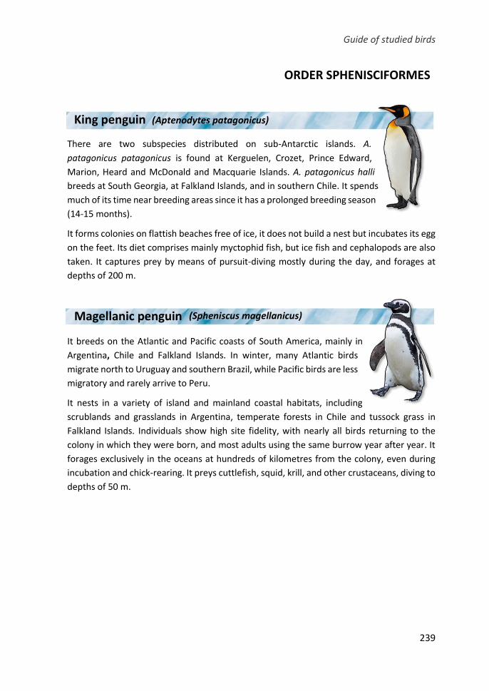
Guide of studied birds
239
ORDER SPHENISCIFORMES
There are two subspecies distributed on sub-Antarctic islands. A.
patagonicus patagonicus is found at Kerguelen, Crozet, Prince Edward,
Marion, Heard and McDonald and Macquarie Islands. A. patagonicus halli
breeds at South Georgia, at Falkland Islands, and in southern Chile. It spends
much of its time near breeding areas since it has a prolonged breeding season
(14-15 months).
It forms colonies on flattish beaches free of ice, it does not build a nest but incubates its egg
on the feet. Its diet comprises mainly myctophid fish, but ice fish and cephalopods are also
taken. It captures prey by means of pursuit-diving mostly during the day, and forages at
depths of 200 m.
It breeds on the Atlantic and Pacific coasts of South America, mainly in
Argentina, Chile and Falkland Islands. In winter, many Atlantic birds
migrate north to Uruguay and southern Brazil, while Pacific birds are less
migratory and rarely arrive to Peru.
It nests in a variety of island and mainland coastal habitats, including
scrublands and grasslands in Argentina, temperate forests in Chile and tussock grass in
Falkland Islands. Individuals show high site fidelity, with nearly all birds returning to the
colony in which they were born, and most adults using the same burrow year after year. It
forages exclusively in the oceans at hundreds of kilometres from the colony, even during
incubation and chick-rearing. It preys cuttlefish, squid, krill, and other crustaceans, diving to
depths of 50 m.
King penguin
penguin
(Aptenodytes patagonicus)
Magellanic penguin
penguin
(Spheniscus magellanicus)
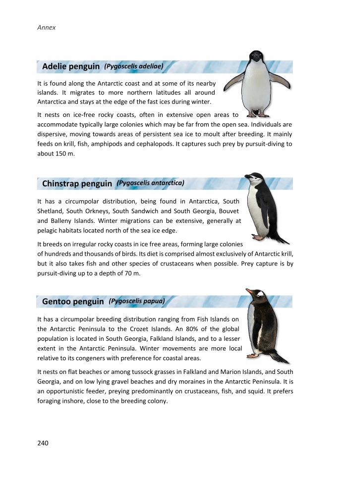
Annex
240
It is found along the Antarctic coast and at some of its nearby
islands. It migrates to more northern latitudes all around
Antarctica and stays at the edge of the fast ices during winter.
It nests on ice-free rocky coasts, often in extensive open areas to
accommodate typically large colonies which may be far from the open sea. Individuals are
dispersive, moving towards areas of persistent sea ice to moult after breeding. It mainly
feeds on krill, fish, amphipods and cephalopods. It captures such prey by pursuit-diving to
about 150 m.
It has a circumpolar distribution, being found in Antarctica, South
Shetland, South Orkneys, South Sandwich and South Georgia, Bouvet
and Balleny Islands. Winter migrations can be extensive, generally at
pelagic habitats located north of the sea ice edge.
It breeds on irregular rocky coasts in ice free areas, forming large colonies
of hundreds and thousands of birds. Its diet is comprised almost exclusively of Antarctic krill,
but it also takes fish and other species of crustaceans when possible. Prey capture is by
pursuit-diving up to a depth of 70 m.
It has a circumpolar breeding distribution ranging from Fish Islands on
the Antarctic Peninsula to the Crozet Islands. An 80% of the global
population is located in South Georgia, Falkland Islands, and to a lesser
extent in the Antarctic Peninsula. Winter movements are more local
relative to its congeners with preference for coastal areas.
It nests on flat beaches or among tussock grasses in Falkland and Marion Islands, and South
Georgia, and on low lying gravel beaches and dry moraines in the Antarctic Peninsula. It is
an opportunistic feeder, preying predominantly on crustaceans, fish, and squid. It prefers
foraging inshore, close to the breeding colony.
Chinstrap penguin
penguin
(Pygoscelis antarctica)
Gentoo penguin
penguin
(Pygoscelis papua)
Adelie penguin (Pygoscelis adeliae)
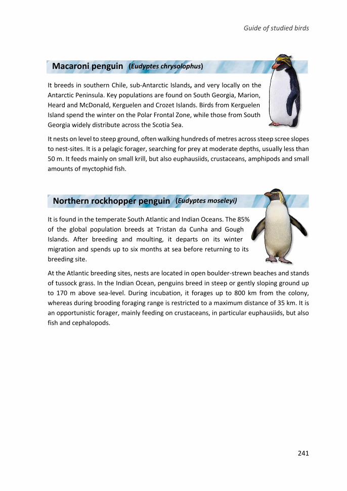
Guide of studied birds
241
It breeds in southern Chile, sub-Antarctic Islands, and very locally on the
Antarctic Peninsula. Key populations are found on South Georgia, Marion,
Heard and McDonald, Kerguelen and Crozet Islands. Birds from Kerguelen
Island spend the winter on the Polar Frontal Zone, while those from South
Georgia widely distribute across the Scotia Sea.
It nests on level to steep ground, often walking hundreds of metres across steep scree slopes
to nest-sites. It is a pelagic forager, searching for prey at moderate depths, usually less than
50 m. It feeds mainly on small krill, but also euphausiids, crustaceans, amphipods and small
amounts of myctophid fish.
It is found in the temperate South Atlantic and Indian Oceans. The 85%
of the global population breeds at Tristan da Cunha and Gough
Islands. After breeding and moulting, it departs on its winter
migration and spends up to six months at sea before returning to its
breeding site.
At the Atlantic breeding sites, nests are located in open boulder-strewn beaches and stands
of tussock grass. In the Indian Ocean, penguins breed in steep or gently sloping ground up
to 170 m above sea-level. During incubation, it forages up to 800 km from the colony,
whereas during brooding foraging range is restricted to a maximum distance of 35 km. It is
an opportunistic forager, mainly feeding on crustaceans, in particular euphausiids, but also
fish and cephalopods.
Macaroni penguin (Eudyptes chrysolophus)
Northern rockhopper penguin
penguin
(Eudyptes moseleyi)
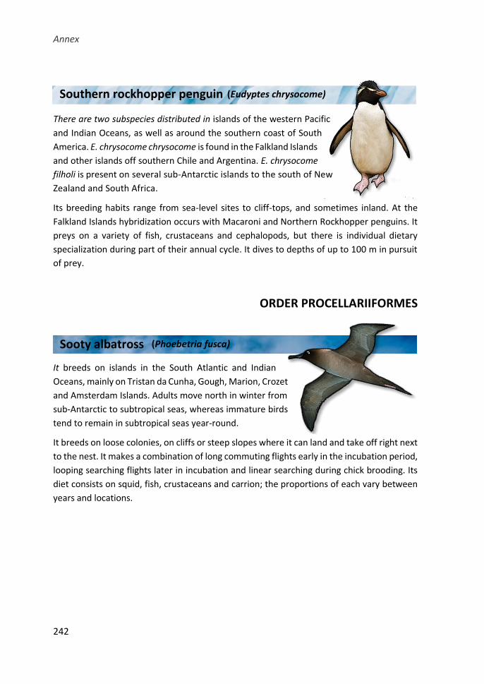
Annex
242
There are two subspecies distributed in islands of the western Pacific
and Indian Oceans, as well as around the southern coast of South
America. E. chrysocome chrysocome is found in the Falkland Islands
and other islands off southern Chile and Argentina. E. chrysocome
filholi is present on several sub-Antarctic islands to the south of New
Zealand and South Africa.
Its breeding habits range from sea-level sites to cliff-tops, and sometimes inland. At the
Falkland Islands hybridization occurs with Macaroni and Northern Rockhopper penguins. It
preys on a variety of fish, crustaceans and cephalopods, but there is individual dietary
specialization during part of their annual cycle. It dives to depths of up to 100 m in pursuit
of prey.
ORDER PROCELLARIIFORMES
It breeds on islands in the South Atlantic and Indian
Oceans, mainly on Tristan da Cunha, Gough, Marion, Crozet
and Amsterdam Islands. Adults move north in winter from
sub-Antarctic to subtropical seas, whereas immature birds
tend to remain in subtropical seas year-round.
It breeds on loose colonies, on cliffs or steep slopes where it can land and take off right next
to the nest. It makes a combination of long commuting flights early in the incubation period,
looping searching flights later in incubation and linear searching during chick brooding. Its
diet consists on squid, fish, crustaceans and carrion; the proportions of each vary between
years and locations.
Southern rockhopper penguin
penguin
(Eudyptes chrysocome)
Sooty albatross (Phoebetria fusca)
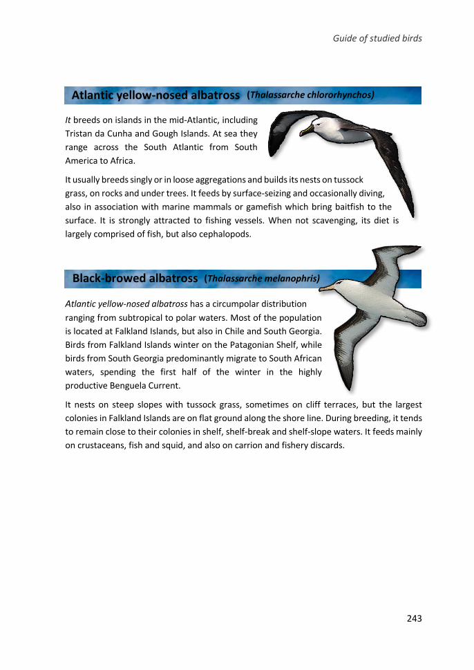
Guide of studied birds
243
It breeds on islands in the mid-Atlantic, including
Tristan da Cunha and Gough Islands. At sea they
range across the South Atlantic from South
America to Africa.
It usually breeds singly or in loose aggregations and builds its nests on tussock
grass, on rocks and under trees. It feeds by surface-seizing and occasionally diving,
also in association with marine mammals or gamefish which bring baitfish to the
surface. It is strongly attracted to fishing vessels. When not scavenging, its diet is
largely comprised of fish, but also cephalopods.
Atlantic yellow-nosed albatross has a circumpolar distribution
ranging from subtropical to polar waters. Most of the population
is located at Falkland Islands, but also in Chile and South Georgia.
Birds from Falkland Islands winter on the Patagonian Shelf, while
birds from South Georgia predominantly migrate to South African
waters, spending the first half of the winter in the highly
productive Benguela Current.
It nests on steep slopes with tussock grass, sometimes on cliff terraces, but the largest
colonies in Falkland Islands are on flat ground along the shore line. During breeding, it tends
to remain close to their colonies in shelf, shelf-break and shelf-slope waters. It feeds mainly
on crustaceans, fish and squid, and also on carrion and fishery discards.
Atlantic yellow-nosed albatross (Thalassarche chlororhynchos)
Black-browed albatross (Thalassarche melanophris)
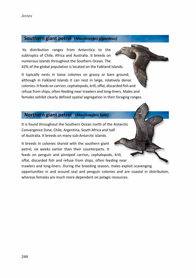
Annex
244
Its distribution ranges from Antarctica to the
subtropics of Chile, Africa and Australia. It breeds on
numerous islands throughout the Southern Ocean. The
42% of the global population is located on the Falkland Islands.
It typically nests in loose colonies on grassy or bare ground,
although in Falkland Islands it can nest in large, relatively dense
colonies. It feeds on carrion, cephalopods, krill, offal, discarded fish and
refuse from ships, often feeding near trawlers and long-liners. Males and
females exhibit clearly defined spatial segregation in their foraging ranges.
It is found throughout the Southern Ocean north of the Antarctic
Convergence Zone, Chile, Argentina, South Africa and half
of Australia. It breeds on many sub-Antarctic islands.
It breeds in colonies shared with the southern giant
petrel, six weeks earlier than their counterparts. It
feeds on penguin and pinniped carrion, cephalopods, krill,
offal, discarded fish and refuse from ships, often feeding near
trawlers and long-liners. During the breeding season, males exploit scavenging
opportunities in and around seal and penguin colonies and are coastal in distribution,
whereas females are much more dependent on pelagic resources.
Southern giant petrel
Northern giant petrel (Macronectes halli)
(Macronectes giganteus)
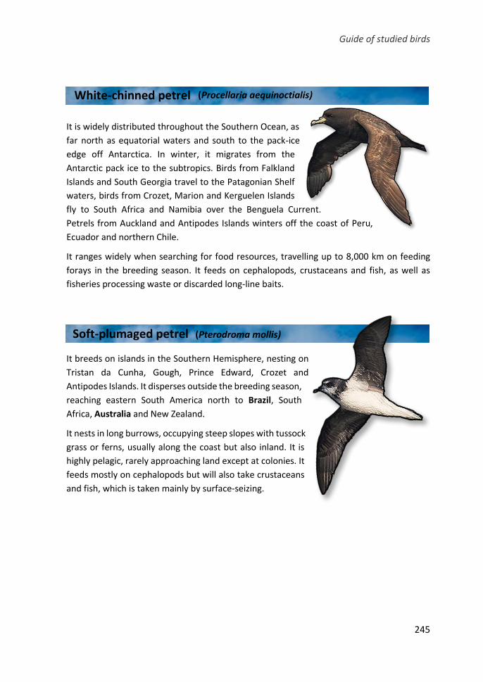
Guide of studied birds
245
It is widely distributed throughout the Southern Ocean, as
far north as equatorial waters and south to the pack-ice
edge off Antarctica. In winter, it migrates from the
Antarctic pack ice to the subtropics. Birds from Falkland
Islands and South Georgia travel to the Patagonian Shelf
waters, birds from Crozet, Marion and Kerguelen Islands
fly to South Africa and Namibia over the Benguela Current.
Petrels from Auckland and Antipodes Islands winters off the coast of Peru,
Ecuador and northern Chile.
It ranges widely when searching for food resources, travelling up to 8,000 km on feeding
forays in the breeding season. It feeds on cephalopods, crustaceans and fish, as well as
fisheries processing waste or discarded long-line baits.
It breeds on islands in the Southern Hemisphere, nesting on
Tristan da Cunha, Gough, Prince Edward, Crozet and
Antipodes Islands. It disperses outside the breeding season,
reaching eastern South America north to Brazil, South
Africa, Australia and New Zealand.
It nests in long burrows, occupying steep slopes with tussock
grass or ferns, usually along the coast but also inland. It is
highly pelagic, rarely approaching land except at colonies. It
feeds mostly on cephalopods but will also take crustaceans
and fish, which is taken mainly by surface-seizing.
Soft-plumaged petrel (Pterodroma mollis)
White-chinned petrel (Procellaria aequinoctialis)
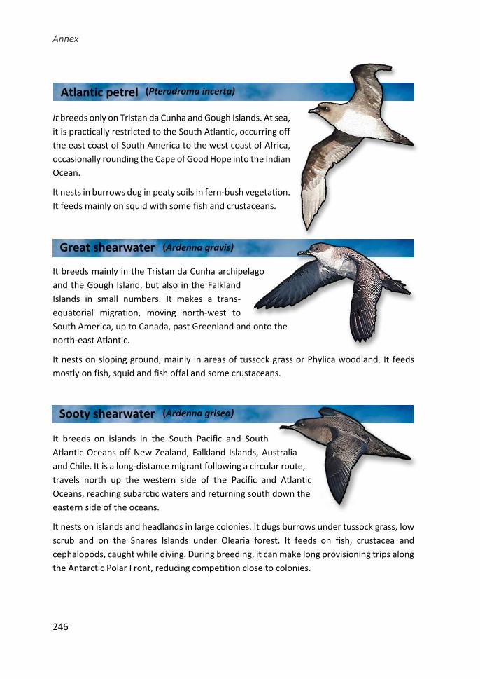
Annex
246
It breeds only on Tristan da Cunha and Gough Islands. At sea,
it is practically restricted to the South Atlantic, occurring off
the east coast of South America to the west coast of Africa,
occasionally rounding the Cape of Good Hope into the Indian
Ocean.
It nests in burrows dug in peaty soils in fern-bush vegetation.
It feeds mainly on squid with some fish and crustaceans.
It breeds mainly in the Tristan da Cunha archipelago
and the Gough Island, but also in the Falkland
Islands in small numbers. It makes a trans-
equatorial migration, moving north-west to
South America, up to Canada, past Greenland and onto the
north-east Atlantic.
It nests on sloping ground, mainly in areas of tussock grass or Phylica woodland. It feeds
mostly on fish, squid and fish offal and some crustaceans.
It breeds on islands in the South Pacific and South
Atlantic Oceans off New Zealand, Falkland Islands, Australia
and Chile. It is a long-distance migrant following a circular route,
travels north up the western side of the Pacific and Atlantic
Oceans, reaching subarctic waters and returning south down the
eastern side of the oceans.
It nests on islands and headlands in large colonies. It dugs burrows under tussock grass, low
scrub and on the Snares Islands under Olearia forest. It feeds on fish, crustacea and
cephalopods, caught while diving. During breeding, it can make long provisioning trips along
the Antarctic Polar Front, reducing competition close to colonies.
Atlantic petrel (Pterodroma incerta)
Great shearwater (Ardenna gravis)
Sooty shearwater (Ardenna grisea)
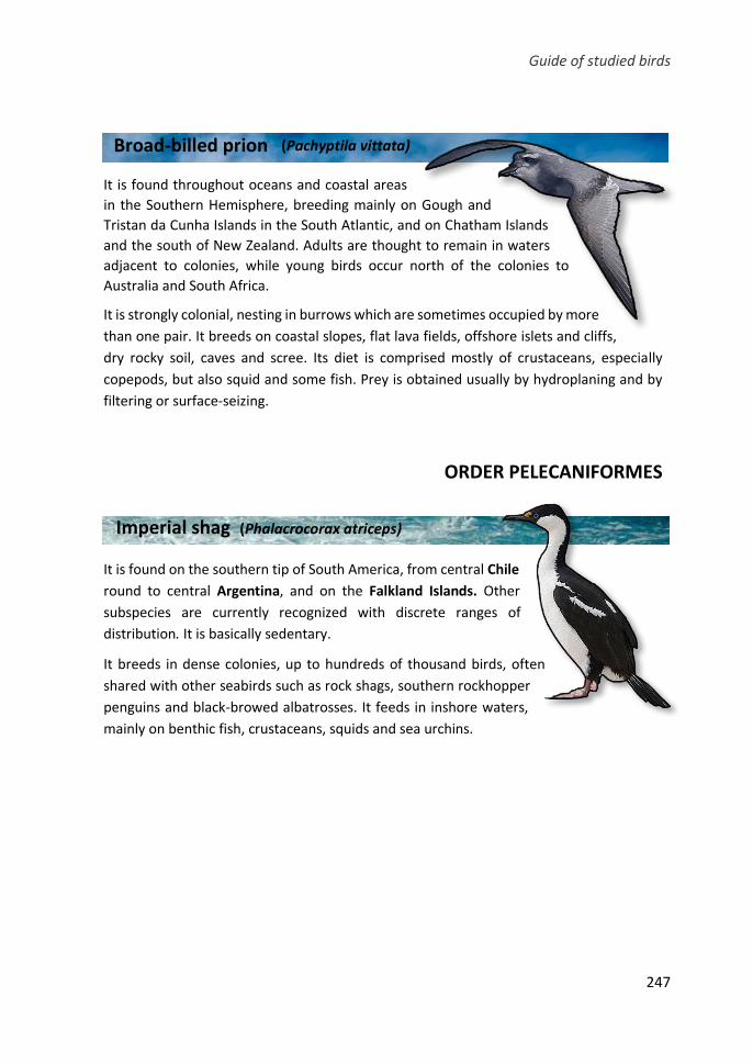
Guide of studied birds
247
It is found throughout oceans and coastal areas
in the Southern Hemisphere, breeding mainly on Gough and
Tristan da Cunha Islands in the South Atlantic, and on Chatham Islands
and the south of New Zealand. Adults are thought to remain in waters
adjacent to colonies, while young birds occur north of the colonies to
Australia and South Africa.
It is strongly colonial, nesting in burrows which are sometimes occupied by more
than one pair. It breeds on coastal slopes, flat lava fields, offshore islets and cliffs,
dry rocky soil, caves and scree. Its diet is comprised mostly of crustaceans, especially
copepods, but also squid and some fish. Prey is obtained usually by hydroplaning and by
filtering or surface-seizing.
ORDER PELECANIFORMES
It is found on the southern tip of South America, from central Chile
round to central Argentina, and on the Falkland Islands. Other
subspecies are currently recognized with discrete ranges of
distribution. It is basically sedentary.
It breeds in dense colonies, up to hundreds of thousand birds, often
shared with other seabirds such as rock shags, southern rockhopper
penguins and black-browed albatrosses. It feeds in inshore waters,
mainly on benthic fish, crustaceans, squids and sea urchins.
Broad-billed prion (Pachyptila vittata)
Imperial shag (Phalacrocorax atriceps)
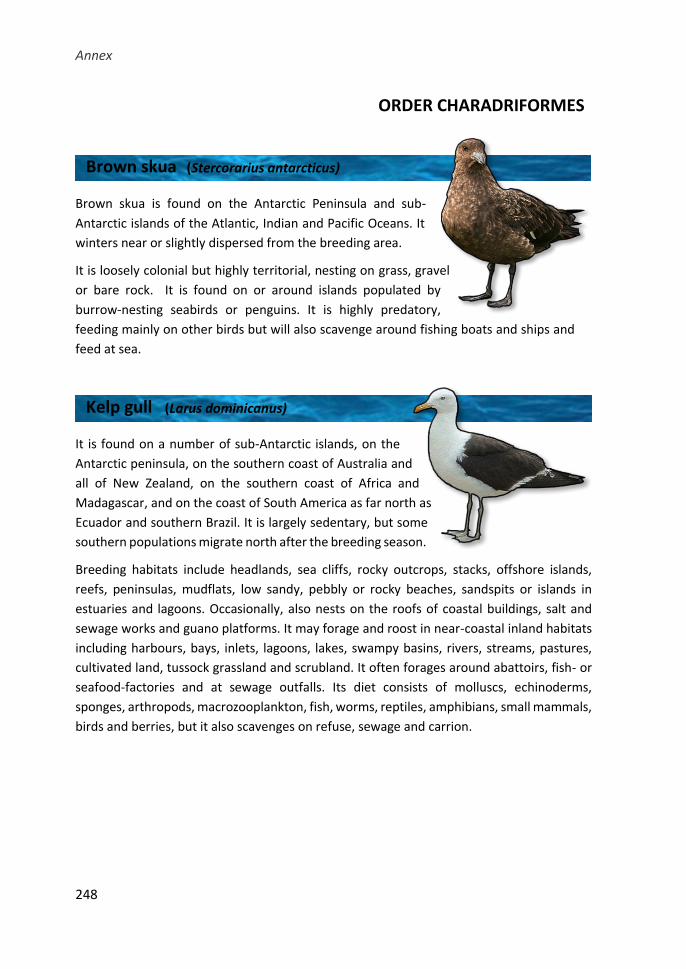
Annex
248
ORDER CHARADRIFORMES
Brown skua is found on the Antarctic Peninsula and sub-
Antarctic islands of the Atlantic, Indian and Pacific Oceans. It
winters near or slightly dispersed from the breeding area.
It is loosely colonial but highly territorial, nesting on grass, gravel
or bare rock. It is found on or around islands populated by
burrow-nesting seabirds or penguins. It is highly predatory,
feeding mainly on other birds but will also scavenge around fishing boats and ships and
feed at sea.
It is found on a number of sub-Antarctic islands, on the
Antarctic peninsula, on the southern coast of Australia and
all of New Zealand, on the southern coast of Africa and
Madagascar, and on the coast of South America as far north as
Ecuador and southern Brazil. It is largely sedentary, but some
southern populations migrate north after the breeding season.
Breeding habitats include headlands, sea cliffs, rocky outcrops, stacks, offshore islands,
reefs, peninsulas, mudflats, low sandy, pebbly or rocky beaches, sandspits or islands in
estuaries and lagoons. Occasionally, also nests on the roofs of coastal buildings, salt and
sewage works and guano platforms. It may forage and roost in near-coastal inland habitats
including harbours, bays, inlets, lagoons, lakes, swampy basins, rivers, streams, pastures,
cultivated land, tussock grassland and scrubland. It often forages around abattoirs, fish- or
seafood-factories and at sewage outfalls. Its diet consists of molluscs, echinoderms,
sponges, arthropods, macrozooplankton, fish, worms, reptiles, amphibians, small mammals,
birds and berries, but it also scavenges on refuse, sewage and carrion.
Brown skua (Stercorarius antarcticus)
Kelp gull (Larus dominicanus)
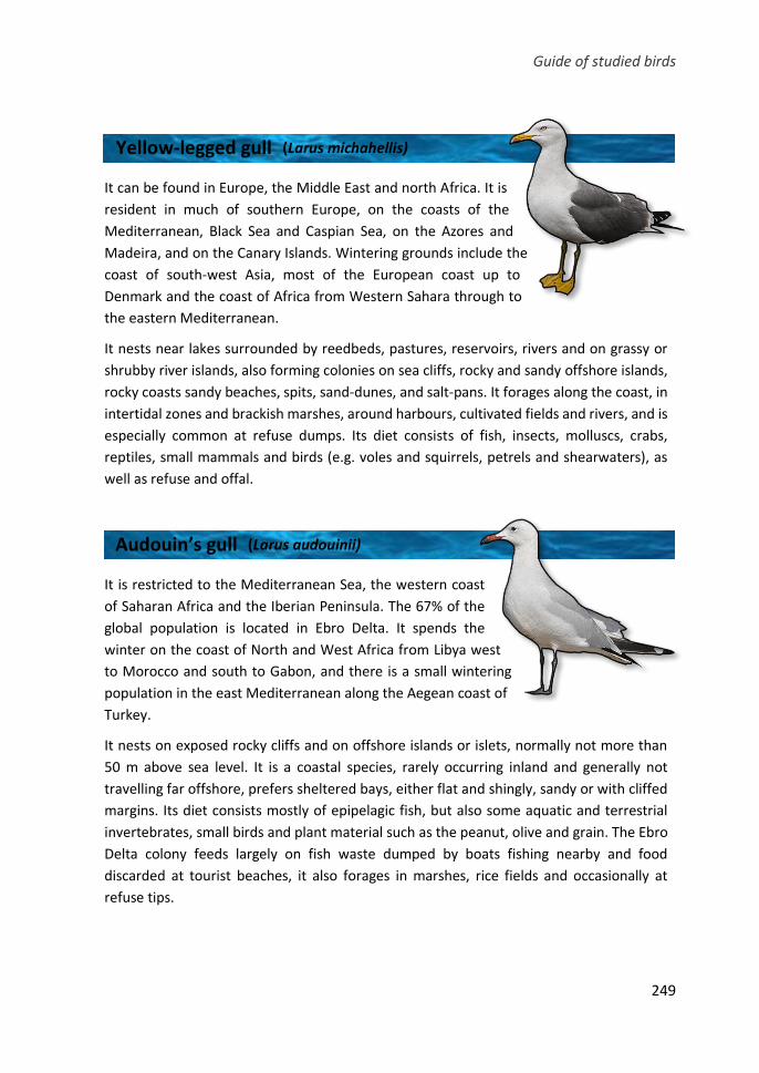
Guide of studied birds
249
It can be found in Europe, the Middle East and north Africa. It is
resident in much of southern Europe, on the coasts of the
Mediterranean, Black Sea and Caspian Sea, on the Azores and
Madeira, and on the Canary Islands. Wintering grounds include the
coast of south-west Asia, most of the European coast up to
Denmark and the coast of Africa from Western Sahara through to
the eastern Mediterranean.
It nests near lakes surrounded by reedbeds, pastures, reservoirs, rivers and on grassy or
shrubby river islands, also forming colonies on sea cliffs, rocky and sandy offshore islands,
rocky coasts sandy beaches, spits, sand-dunes, and salt-pans. It forages along the coast, in
intertidal zones and brackish marshes, around harbours, cultivated fields and rivers, and is
especially common at refuse dumps. Its diet consists of fish, insects, molluscs, crabs,
reptiles, small mammals and birds (e.g. voles and squirrels, petrels and shearwaters), as
well as refuse and offal.
It is restricted to the Mediterranean Sea, the western coast
of Saharan Africa and the Iberian Peninsula. The 67% of the
global population is located in Ebro Delta. It spends the
winter on the coast of North and West Africa from Libya west
to Morocco and south to Gabon, and there is a small wintering
population in the east Mediterranean along the Aegean coast of
Turkey.
It nests on exposed rocky cliffs and on offshore islands or islets, normally not more than
50 m above sea level. It is a coastal species, rarely occurring inland and generally not
travelling far offshore, prefers sheltered bays, either flat and shingly, sandy or with cliffed
margins. Its diet consists mostly of epipelagic fish, but also some aquatic and terrestrial
invertebrates, small birds and plant material such as the peanut, olive and grain. The Ebro
Delta colony feeds largely on fish waste dumped by boats fishing nearby and food
discarded at tourist beaches, it also forages in marshes, rice fields and occasionally at
refuse tips.
Yellow-legged gull (Larus michahellis)
Audouin’s gull (Larus audouinii)
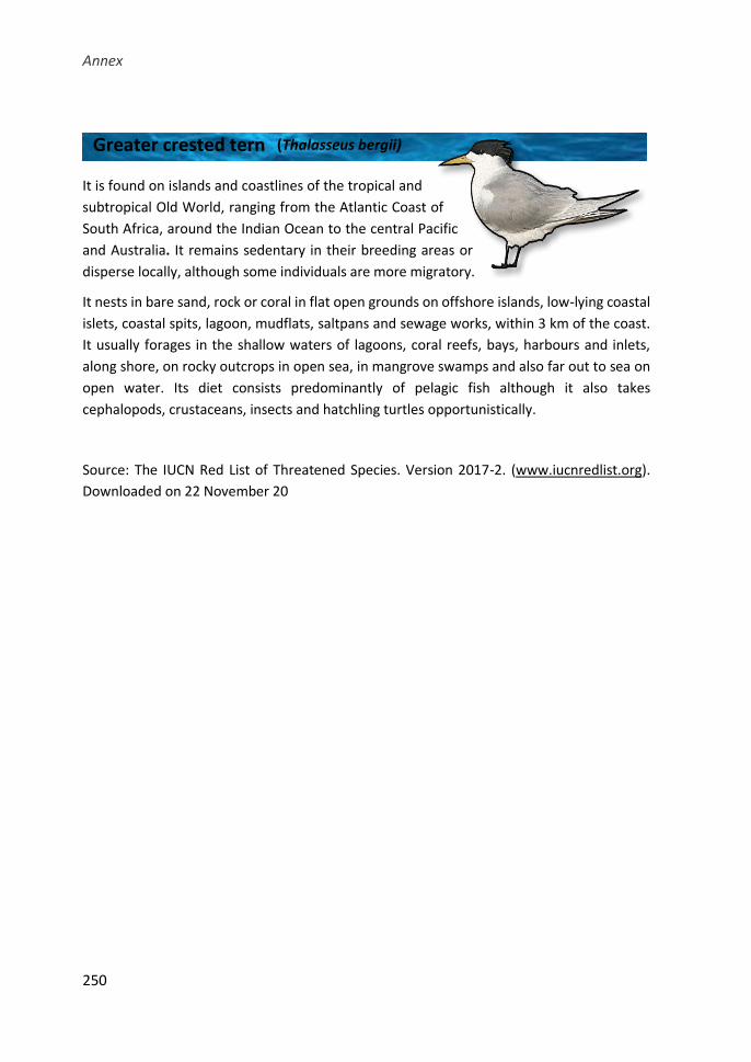
Annex
250
It is found on islands and coastlines of the tropical and
subtropical Old World, ranging from the Atlantic Coast of
South Africa, around the Indian Ocean to the central Pacific
and Australia. It remains sedentary in their breeding areas or
disperse locally, although some individuals are more migratory.
It nests in bare sand, rock or coral in flat open grounds on offshore islands, low-lying coastal
islets, coastal spits, lagoon, mudflats, saltpans and sewage works, within 3 km of the coast.
It usually forages in the shallow waters of lagoons, coral reefs, bays, harbours and inlets,
along shore, on rocky outcrops in open sea, in mangrove swamps and also far out to sea on
open water. Its diet consists predominantly of pelagic fish although it also takes
cephalopods, crustaceans, insects and hatchling turtles opportunistically.
Source: The IUCN Red List of Threatened Species. Version 2017-2. (www.iucnredlist.org).
Downloaded on 22 November 20
Greater crested tern (Thalasseus bergii)