Clinical relevance of microRNA miR-21, miR-31, miR-92a, miR-101, miR-106a and miR-145 in colorectal...
8
RESEARCH ARTICLE Open Access Clinical relevance of microRNA miR-21, miR-31, miR-92a, miR-101, miR-106a and miR-145 in colorectal cancer Kristina Schee 1* , Kjetil Boye 1,2 , Torveig Weum Abrahamsen 1 , Øystein Fodstad 1 and Kjersti Flatmark 1,3 Abstract Background: MicroRNAs (miRNAs) regulate gene expression by binding to mRNA, and can function as oncogenes or tumor suppressors depending on the target. In this study, using qRT-PCR, we examined the expression of six miRNAs (miR-21, miR-31, miR-92a, miR-101, miR-106a and miR-145) in tumors from 193 prospectively recruited patients with colorectal cancer, and associations with clinicopathological parameters and patient outcome were analyzed. The miRNAs were chosen based on previous studies for their biomarker potential and suggested biological relevance in colorectal cancer. Methods: The miRNA expression was examined by qRT-PCR. Associations between miRNA expression and clinicopathological variables were explored using Mann–Whitney U and Kruskal-Wallis test while survival was estimated using the Kaplan-Meier method and compared using the log-rank test. Results: MiR-101 was hardly expressed in the tumor samples, while for the other miRNAs, variable expression levels and expression ranges were observed, with miR-21 being most abundantly expressed relative to the reference (RNU44). In our study cohort, major clinical significance was demonstrated only for miR-31, as high expression was associated with advanced tumor stage and poor differentiation. No significant associations were found between expression of the investigated miRNAs and metastasis-free or overall survival. Conclusions: Investigating the expression of six miRNAs previously identified as candidate biomarkers in colorectal cancer, few clinically relevant associations were detected in our patient cohort. Our results emphasize the importance of validating potential tumor markers in independent patient cohorts, and indicate that the role of miRNAs as colorectal cancer biomarkers is still undetermined. Keywords: MiRNA, Colorectal cancer, Prognostic biomarker Background MicroRNAs (miRNAs) are a class of small, non-coding RNAs (19–22 nucleotides) that function as posttranscrip- tional gene regulators by binding to the 3’UTR of mRNA, and one miRNA may potentially down-regulate multiple mRNA targets. More than 1500 human miRNAs are cur- rently annotated in the miRBase [1], and it has been pre- dicted that as many as 30% of protein-encoding genes may be regulated by miRNAs [2]. The discovery that miRNAs may function as oncogenes or tumor suppressors depending on the target mRNA, has instigated intensive research to determine the role of these molecules in can- cer. MiRNAs are chemically very stable, and can be detected by a range of high-throughput detection methods in tissue, serum and plasma as well as in urine and feces, and are for these reasons considered to have great poten- tial as cancer biomarkers. In colorectal cancer (CRC), treatment decisions are still based essentially on anatomical extent of disease at diagnosis, and the search for better biomarkers is war- ranted. Several miRNAs with potential biological and clinical relevance have been identified and are being explored as diagnostic, prognostic and predictive bio- markers [3-6]. Based on previous studies and our recent * Correspondence: [email protected] 1 Departments of Tumor Biology, The Norwegian Radium Hospital, Oslo University Hospital, N-0310 Oslo, Norway Full list of author information is available at the end of the article © 2012 Schee et al.; licensee BioMed Central Ltd. This is an Open Access article distributed under the terms of the Creative Commons Attribution License (http://creativecommons.org/licenses/by/2.0), which permits unrestricted use, distribution, and reproduction in any medium, provided the original work is properly cited. Schee et al. BMC Cancer 2012, 12:505 http://www.biomedcentral.com/1471-2407/12/505
-
Upload
independent -
Category
Documents
-
view
8 -
download
0
Transcript of Clinical relevance of microRNA miR-21, miR-31, miR-92a, miR-101, miR-106a and miR-145 in colorectal...
Schee et al. BMC Cancer 2012, 12:505http://www.biomedcentral.com/1471-2407/12/505
RESEARCH ARTICLE Open Access
Clinical relevance of microRNA miR-21, miR-31,miR-92a, miR-101, miR-106a and miR-145 incolorectal cancerKristina Schee1*, Kjetil Boye1,2, Torveig Weum Abrahamsen1, Øystein Fodstad1 and Kjersti Flatmark1,3
Abstract
Background: MicroRNAs (miRNAs) regulate gene expression by binding to mRNA, and can function as oncogenesor tumor suppressors depending on the target. In this study, using qRT-PCR, we examined the expression of sixmiRNAs (miR-21, miR-31, miR-92a, miR-101, miR-106a and miR-145) in tumors from 193 prospectively recruitedpatients with colorectal cancer, and associations with clinicopathological parameters and patient outcome wereanalyzed. The miRNAs were chosen based on previous studies for their biomarker potential and suggestedbiological relevance in colorectal cancer.
Methods: The miRNA expression was examined by qRT-PCR. Associations between miRNA expression andclinicopathological variables were explored using Mann–Whitney U and Kruskal-Wallis test while survival wasestimated using the Kaplan-Meier method and compared using the log-rank test.
Results: MiR-101 was hardly expressed in the tumor samples, while for the other miRNAs, variable expression levelsand expression ranges were observed, with miR-21 being most abundantly expressed relative to the reference(RNU44). In our study cohort, major clinical significance was demonstrated only for miR-31, as high expression wasassociated with advanced tumor stage and poor differentiation. No significant associations were found betweenexpression of the investigated miRNAs and metastasis-free or overall survival.
Conclusions: Investigating the expression of six miRNAs previously identified as candidate biomarkers in colorectalcancer, few clinically relevant associations were detected in our patient cohort. Our results emphasize theimportance of validating potential tumor markers in independent patient cohorts, and indicate that the role ofmiRNAs as colorectal cancer biomarkers is still undetermined.
Keywords: MiRNA, Colorectal cancer, Prognostic biomarker
BackgroundMicroRNAs (miRNAs) are a class of small, non-codingRNAs (19–22 nucleotides) that function as posttranscrip-tional gene regulators by binding to the 3’UTR of mRNA,and one miRNA may potentially down-regulate multiplemRNA targets. More than 1500 human miRNAs are cur-rently annotated in the miRBase [1], and it has been pre-dicted that as many as 30% of protein-encoding genesmay be regulated by miRNAs [2]. The discovery thatmiRNAs may function as oncogenes or tumor suppressors
* Correspondence: [email protected] of Tumor Biology, The Norwegian Radium Hospital, OsloUniversity Hospital, N-0310 Oslo, NorwayFull list of author information is available at the end of the article
© 2012 Schee et al.; licensee BioMed Central LCommons Attribution License (http://creativecreproduction in any medium, provided the or
depending on the target mRNA, has instigated intensiveresearch to determine the role of these molecules in can-cer. MiRNAs are chemically very stable, and can bedetected by a range of high-throughput detection methodsin tissue, serum and plasma as well as in urine and feces,and are for these reasons considered to have great poten-tial as cancer biomarkers.In colorectal cancer (CRC), treatment decisions are
still based essentially on anatomical extent of disease atdiagnosis, and the search for better biomarkers is war-ranted. Several miRNAs with potential biological andclinical relevance have been identified and are beingexplored as diagnostic, prognostic and predictive bio-markers [3-6]. Based on previous studies and our recent
td. This is an Open Access article distributed under the terms of the Creativeommons.org/licenses/by/2.0), which permits unrestricted use, distribution, andiginal work is properly cited.
Schee et al. BMC Cancer 2012, 12:505 Page 2 of 8http://www.biomedcentral.com/1471-2407/12/505
review of this topic, six candidate miRNAs, miR-21,miR-31, miR-92a, miR-101, miR-106a and miR-145(Table 1), were chosen for analysis in a cohort of 193prospectively recruited patients receiving curative sur-gery for CRC [7-13]. Expression of the miRNA wasdetermined by qRT-PCR and associations with clinico-pathological parameters and outcome were analyzed.
MethodsPatient cohort316 patients, recruited from five hospitals in the Oslo re-gion between the year 1998 and 2000 [15], were pro-spectively included in the study at the time of primarysurgery for assumed or verified colorectal cancer. Thestudy was approved by the Regional Ethics Committee(Health Region II, Norway) and informed consent wasobtained from the patients. At surgery, resected speci-mens were routinely processed for histopathological as-sessment and additional tumor tissue was sampled andsnap-frozen in liquid nitrogen. A number of cases wereexcluded from statistical analysis for the following rea-sons: not invasive cancer (25), histology other thanadenocarcinoma (5), distant metastasis at the time ofsurgery (34, tissue samples not available), preoperativechemoradiotherapy (2), inadequate surgical margins (7),unknown stage of disease (1), freshly frozen tissue sam-ples not obtainable (46), and high Ct-values (>37; n=3).The study population thus consisted of 193 patients inTNM stage I-III (Table 2). Follow-up data was obtainedfrom the participating hospitals and from the generalpractitioners (for the patients not attending scheduledcontrols). Metastasis was verified by radiological examin-ation and survival data was obtained from the NationalRegistry of Norway and updated by October 1st 2008with the cause of death registered and classified as deathfrom colorectal cancer, death of other cause or death ofunknown cause.
Table 1 Mature sequence, miRBase accession number and pro
miRNA name Mature miRNAsequence
miRBaseAccessionnumber
Proposreleva
hsa-miR-21 UAGCUUAUCAGACUGAUGUUGA MIMAT0000076 Overall
hsa-miR-31 AGGCAAGAUGCUGGCAUAGCU MIMAT0000089 Tumor
hsa-miR-92a UAUUGCACUUGUCCCGGCCUGU MIMAT0000092 Plasma
hsa-miR-101 UACAGUACUGUGAUAACUGAA MIMAT0000099 Increas
hsa-miR-106a AAAAGUGCUUACAGUGCAGGUAG MIMAT0000103 Diseaseoverall
hsa-miR-145 GUCCAGUUUUCCCAGGAAUCCCU MIMAT0000437 Tumor
MiRNA selectionMiRNA selection was based on previous studies and ourliterature review [11], identifying miRNA with proposedclinical relevance in CRC, including published articlesleading up to the year 2009. We wished to examineselected miRNAs in our CRC cohort and their relevancewith clinicopathological data and outcome parameters(Table 2). The following six miRNAs were chosen foranalysis; miR-21, miR-31, miR-92a, miR-101, miR-106aand miR-145 [7-13].
Sample preparation and RNA isolationBiopsies were sampled and snap frozen in liquid nitrogenand stored at −80°C. The biopsies were sectioned using acryostat microtome and hematoxylin-eosin stained slideswere evaluated for tumor content by a pathologist (mediantumor content in the samples was 50%, range 30 - 80%).The tumor tissue was sliced into 10 μm sections using acryostat microtome, aliquoted into 1.5 ml Micro tubes(Sarstedt, Nümbrecht, Germany) and stored at −80°C.RNA was isolated from the tumor tissue using TriReagent(Ambion Inc, TX) according to the manufacturer’s proto-col and the total RNA concentration was measured byNanodrop (ND-1000).
qRT-PCRTotal RNA from 196 patients was used to reversely tran-scribe miRNAs using TaqMan MicroRNA assays (Ap-plied Biosystems, Foster City, CA). Each reversetranscriptase reaction contained 10 ng of total RNA(5μl), 0.15 μl dNTP (100 mM total), 1.0 μl MultiscribeRT enzyme (50 U/μl), 1.5 μl 10X RT buffer, 0.19 μlRNase Inhibitor (20 U/μl) , 4.16 μl nuclease free water(Sigma-Aldrich, Ayshire, UK) and 3.0 μl 5X RT Primer.The 15 μl reaction volumes were incubated in 8-well PCRstrip tubes (Sarstedt) in a GeneAmp PCR System 9700thermal cycler (Applied Biosystems) as follows; 30 min at
posed clinical relevance for the six chosen miRNAs
ed clinicalnce
Comment Reference
survival High expression associatedwith poor OS
[14]
stage/ differentiation High expression associatedwith advanced tumor stageand poorly differentiated tumors
[10]
marker Elevated levels as a possiblediagnostic marker
[13]
ed invasiveness Decreased expression associatedwith invasiveness
[8]
free andsurvival
Down-regulation associated withpoor disease free and overall survival.
[7]
size Low expression associated withlarge tumor size
[9]
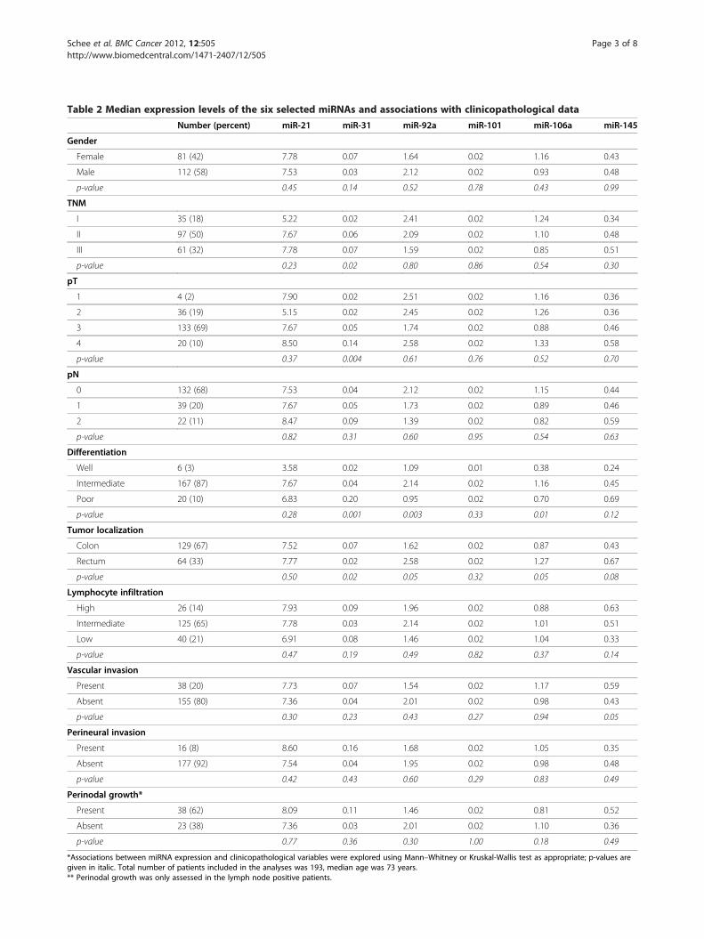
Table 2 Median expression levels of the six selected miRNAs and associations with clinicopathological data
Number (percent) miR-21 miR-31 miR-92a miR-101 miR-106a miR-145
Gender
Female 81 (42) 7.78 0.07 1.64 0.02 1.16 0.43
Male 112 (58) 7.53 0.03 2.12 0.02 0.93 0.48
p-value 0.45 0.14 0.52 0.78 0.43 0.99
TNM
I 35 (18) 5.22 0.02 2.41 0.02 1.24 0.34
II 97 (50) 7.67 0.06 2.09 0.02 1.10 0.48
III 61 (32) 7.78 0.07 1.59 0.02 0.85 0.51
p-value 0.23 0.02 0.80 0.86 0.54 0.30
pT
1 4 (2) 7.90 0.02 2.51 0.02 1.16 0.36
2 36 (19) 5.15 0.02 2.45 0.02 1.26 0.36
3 133 (69) 7.67 0.05 1.74 0.02 0.88 0.46
4 20 (10) 8.50 0.14 2.58 0.02 1.33 0.58
p-value 0.37 0.004 0.61 0.76 0.52 0.70
pN
0 132 (68) 7.53 0.04 2.12 0.02 1.15 0.44
1 39 (20) 7.67 0.05 1.73 0.02 0.89 0.46
2 22 (11) 8.47 0.09 1.39 0.02 0.82 0.59
p-value 0.82 0.31 0.60 0.95 0.54 0.63
Differentiation
Well 6 (3) 3.58 0.02 1.09 0.01 0.38 0.24
Intermediate 167 (87) 7.67 0.04 2.14 0.02 1.16 0.45
Poor 20 (10) 6.83 0.20 0.95 0.02 0.70 0.69
p-value 0.28 0.001 0.003 0.33 0.01 0.12
Tumor localization
Colon 129 (67) 7.52 0.07 1.62 0.02 0.87 0.43
Rectum 64 (33) 7.77 0.02 2.58 0.02 1.27 0.67
p-value 0.50 0.02 0.05 0.32 0.05 0.08
Lymphocyte infiltration
High 26 (14) 7.93 0.09 1.96 0.02 0.88 0.63
Intermediate 125 (65) 7.78 0.03 2.14 0.02 1.01 0.51
Low 40 (21) 6.91 0.08 1.46 0.02 1.04 0.33
p-value 0.47 0.19 0.49 0.82 0.37 0.14
Vascular invasion
Present 38 (20) 7.73 0.07 1.54 0.02 1.17 0.59
Absent 155 (80) 7.36 0.04 2.01 0.02 0.98 0.43
p-value 0.30 0.23 0.43 0.27 0.94 0.05
Perineural invasion
Present 16 (8) 8.60 0.16 1.68 0.02 1.05 0.35
Absent 177 (92) 7.54 0.04 1.95 0.02 0.98 0.48
p-value 0.42 0.43 0.60 0.29 0.83 0.49
Perinodal growth*
Present 38 (62) 8.09 0.11 1.46 0.02 0.81 0.52
Absent 23 (38) 7.36 0.03 2.01 0.02 1.10 0.36
p-value 0.77 0.36 0.30 1.00 0.18 0.49
*Associations between miRNA expression and clinicopathological variables were explored using Mann–Whitney or Kruskal-Wallis test as appropriate; p-values aregiven in italic. Total number of patients included in the analyses was 193, median age was 73 years.** Perinodal growth was only assessed in the lymph node positive patients.
Schee et al. BMC Cancer 2012, 12:505 Page 3 of 8http://www.biomedcentral.com/1471-2407/12/505
Rel
ativ
e Q
uant
ity (
log1
0)
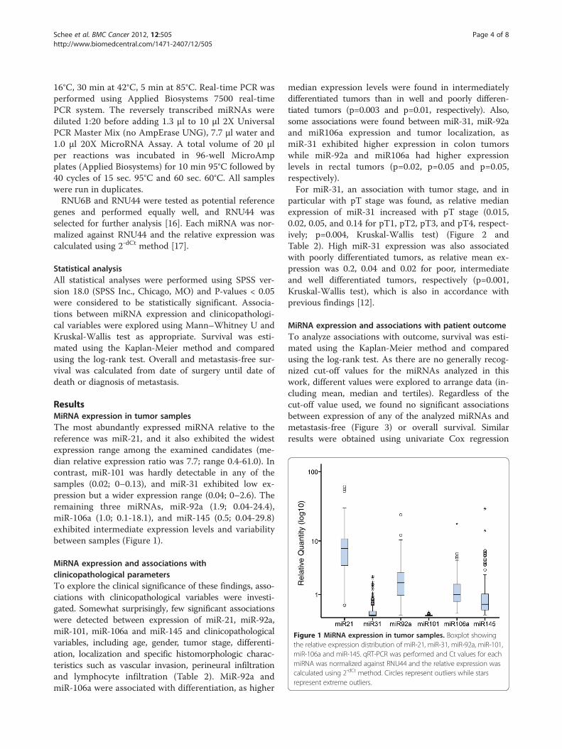
Figure 1 MiRNA expression in tumor samples. Boxplot showingthe relative expression distribution of miR-21, miR-31, miR-92a, miR-101,miR-106a and miR-145. qRT-PCR was performed and Ct values for eachmiRNA was normalized against RNU44 and the relative expression wascalculated using 2-dCt method. Circles represent outliers while starsrepresent extreme outliers.
Schee et al. BMC Cancer 2012, 12:505 Page 4 of 8http://www.biomedcentral.com/1471-2407/12/505
16°C, 30 min at 42°C, 5 min at 85°C. Real-time PCR wasperformed using Applied Biosystems 7500 real-timePCR system. The reversely transcribed miRNAs werediluted 1:20 before adding 1.3 μl to 10 μl 2X UniversalPCR Master Mix (no AmpErase UNG), 7.7 μl water and1.0 μl 20X MicroRNA Assay. A total volume of 20 μlper reactions was incubated in 96-well MicroAmpplates (Applied Biosystems) for 10 min 95°C followed by40 cycles of 15 sec. 95°C and 60 sec. 60°C. All sampleswere run in duplicates.RNU6B and RNU44 were tested as potential reference
genes and performed equally well, and RNU44 wasselected for further analysis [16]. Each miRNA was nor-malized against RNU44 and the relative expression wascalculated using 2-dCt method [17].
Statistical analysisAll statistical analyses were performed using SPSS ver-sion 18.0 (SPSS Inc., Chicago, MO) and P-values < 0.05were considered to be statistically significant. Associa-tions between miRNA expression and clinicopathologi-cal variables were explored using Mann–Whitney U andKruskal-Wallis test as appropriate. Survival was esti-mated using the Kaplan-Meier method and comparedusing the log-rank test. Overall and metastasis-free sur-vival was calculated from date of surgery until date ofdeath or diagnosis of metastasis.
ResultsMiRNA expression in tumor samplesThe most abundantly expressed miRNA relative to thereference was miR-21, and it also exhibited the widestexpression range among the examined candidates (me-dian relative expression ratio was 7.7; range 0.4-61.0). Incontrast, miR-101 was hardly detectable in any of thesamples (0.02; 0–0.13), and miR-31 exhibited low ex-pression but a wider expression range (0.04; 0–2.6). Theremaining three miRNAs, miR-92a (1.9; 0.04-24.4),miR-106a (1.0; 0.1-18.1), and miR-145 (0.5; 0.04-29.8)exhibited intermediate expression levels and variabilitybetween samples (Figure 1).
MiRNA expression and associations withclinicopathological parametersTo explore the clinical significance of these findings, asso-ciations with clinicopathological variables were investi-gated. Somewhat surprisingly, few significant associationswere detected between expression of miR-21, miR-92a,miR-101, miR-106a and miR-145 and clinicopathologicalvariables, including age, gender, tumor stage, differenti-ation, localization and specific histomorphologic charac-teristics such as vascular invasion, perineural infiltrationand lymphocyte infiltration (Table 2). MiR-92a andmiR-106a were associated with differentiation, as higher
median expression levels were found in intermediatelydifferentiated tumors than in well and poorly differen-tiated tumors (p=0.003 and p=0.01, respectively). Also,some associations were found between miR-31, miR-92aand miR106a expression and tumor localization, asmiR-31 exhibited higher expression in colon tumorswhile miR-92a and miR106a had higher expressionlevels in rectal tumors (p=0.02, p=0.05 and p=0.05,respectively).For miR-31, an association with tumor stage, and in
particular with pT stage was found, as relative medianexpression of miR-31 increased with pT stage (0.015,0.02, 0.05, and 0.14 for pT1, pT2, pT3, and pT4, respect-ively; p=0.004, Kruskal-Wallis test) (Figure 2 andTable 2). High miR-31 expression was also associatedwith poorly differentiated tumors, as relative mean ex-pression was 0.2, 0.04 and 0.02 for poor, intermediateand well differentiated tumors, respectively (p=0.001,Kruskal-Wallis test), which is also in accordance withprevious findings [12].
MiRNA expression and associations with patient outcomeTo analyze associations with outcome, survival was esti-mated using the Kaplan-Meier method and comparedusing the log-rank test. As there are no generally recog-nized cut-off values for the miRNAs analyzed in thiswork, different values were explored to arrange data (in-cluding mean, median and tertiles). Regardless of thecut-off value used, we found no significant associationsbetween expression of any of the analyzed miRNAs andmetastasis-free (Figure 3) or overall survival. Similarresults were obtained using univariate Cox regression
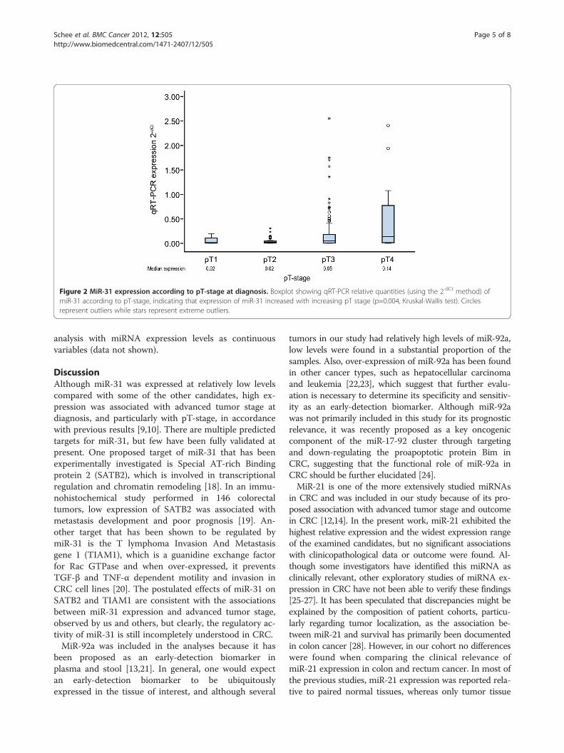
Figure 2 MiR-31 expression according to pT-stage at diagnosis. Boxplot showing qRT-PCR relative quantities (using the 2-dCt method) ofmiR-31 according to pT-stage, indicating that expression of miR-31 increased with increasing pT stage (p=0.004, Kruskal-Wallis test). Circlesrepresent outliers while stars represent extreme outliers.
Schee et al. BMC Cancer 2012, 12:505 Page 5 of 8http://www.biomedcentral.com/1471-2407/12/505
analysis with miRNA expression levels as continuousvariables (data not shown).
DiscussionAlthough miR-31 was expressed at relatively low levelscompared with some of the other candidates, high ex-pression was associated with advanced tumor stage atdiagnosis, and particularly with pT-stage, in accordancewith previous results [9,10]. There are multiple predictedtargets for miR-31, but few have been fully validated atpresent. One proposed target of miR-31 that has beenexperimentally investigated is Special AT-rich Bindingprotein 2 (SATB2), which is involved in transcriptionalregulation and chromatin remodeling [18]. In an immu-nohistochemical study performed in 146 colorectaltumors, low expression of SATB2 was associated withmetastasis development and poor prognosis [19]. An-other target that has been shown to be regulated bymiR-31 is the T lymphoma Invasion And Metastasisgene 1 (TIAM1), which is a guanidine exchange factorfor Rac GTPase and when over-expressed, it preventsTGF-β and TNF-α dependent motility and invasion inCRC cell lines [20]. The postulated effects of miR-31 onSATB2 and TIAM1 are consistent with the associationsbetween miR-31 expression and advanced tumor stage,observed by us and others, but clearly, the regulatory ac-tivity of miR-31 is still incompletely understood in CRC.MiR-92a was included in the analyses because it has
been proposed as an early-detection biomarker inplasma and stool [13,21]. In general, one would expectan early-detection biomarker to be ubiquitouslyexpressed in the tissue of interest, and although several
tumors in our study had relatively high levels of miR-92a,low levels were found in a substantial proportion of thesamples. Also, over-expression of miR-92a has been foundin other cancer types, such as hepatocellular carcinomaand leukemia [22,23], which suggest that further evalu-ation is necessary to determine its specificity and sensitiv-ity as an early-detection biomarker. Although miR-92awas not primarily included in this study for its prognosticrelevance, it was recently proposed as a key oncogeniccomponent of the miR-17-92 cluster through targetingand down-regulating the proapoptotic protein Bim inCRC, suggesting that the functional role of miR-92a inCRC should be further elucidated [24].MiR-21 is one of the more extensively studied miRNAs
in CRC and was included in our study because of its pro-posed association with advanced tumor stage and outcomein CRC [12,14]. In the present work, miR-21 exhibited thehighest relative expression and the widest expression rangeof the examined candidates, but no significant associationswith clinicopathological data or outcome were found. Al-though some investigators have identified this miRNA asclinically relevant, other exploratory studies of miRNA ex-pression in CRC have not been able to verify these findings[25-27]. It has been speculated that discrepancies might beexplained by the composition of patient cohorts, particu-larly regarding tumor localization, as the association be-tween miR-21 and survival has primarily been documentedin colon cancer [28]. However, in our cohort no differenceswere found when comparing the clinical relevance ofmiR-21 expression in colon and rectum cancer. In most ofthe previous studies, miR-21 expression was reported rela-tive to paired normal tissues, whereas only tumor tissue
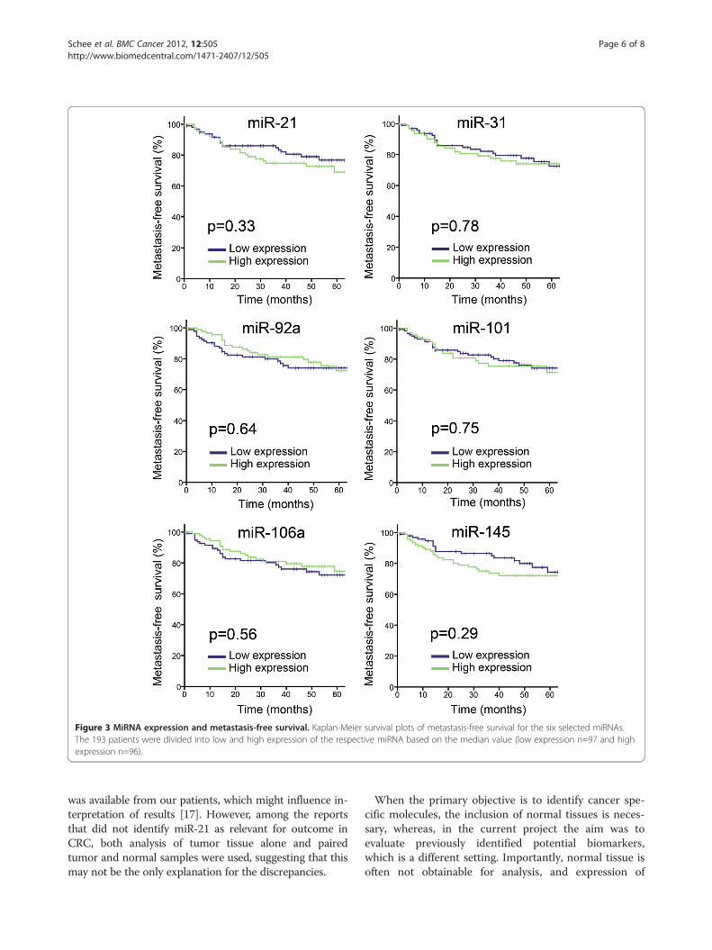
Figure 3 MiRNA expression and metastasis-free survival. Kaplan-Meier survival plots of metastasis-free survival for the six selected miRNAs.The 193 patients were divided into low and high expression of the respective miRNA based on the median value (low expression n=97 and highexpression n=96).
Schee et al. BMC Cancer 2012, 12:505 Page 6 of 8http://www.biomedcentral.com/1471-2407/12/505
was available from our patients, which might influence in-terpretation of results [17]. However, among the reportsthat did not identify miR-21 as relevant for outcome inCRC, both analysis of tumor tissue alone and pairedtumor and normal samples were used, suggesting that thismay not be the only explanation for the discrepancies.
When the primary objective is to identify cancer spe-cific molecules, the inclusion of normal tissues is neces-sary, whereas, in the current project the aim was toevaluate previously identified potential biomarkers,which is a different setting. Importantly, normal tissue isoften not obtainable for analysis, and expression of
Schee et al. BMC Cancer 2012, 12:505 Page 7 of 8http://www.biomedcentral.com/1471-2407/12/505
molecular targets in normal tissues might vary consider-ably between patients, and not necessarily in concert withthe corresponding tumor sample. Thus, it is probably bothpracticable and necessary to develop assays that are inde-pendent of normal tissue. Another related challenge con-cerns the definition of biologically relevant cut-off levels,which have not been determined for specific miRNA indifferent tissues. We explored multiple cut-off levels, butassociations with clinicopathological parameters and out-come for all the candidates remained relatively similar.MiR-101, miR-145 and miR-106a have previously been
associated with cancer-relevant biological processes,such as growth, proliferation and inhibition of apoptosis,or with clinical outcome in CRC [7-9], but few associa-tions with clinicopathological parameters or outcomewere found in our cohort. MiR-101 was hardly detect-able in tumor samples, which is in accordance with itsproposed function as a tumor suppressor that is lostduring tumorigenesis. Interesting recent findings in pan-creatic cancer suggest miR-101 as a key regulator ofstem cell protein markers; its loss favoring the stem cellphenotype and its re-expression constituting a possibletherapeutic strategy [29]. Down-regulation of miR-145was also identified as an early event in CRC carcinogen-esis, which might explain why associations with clinicalvariables in invasive tumors were absent in our tumorpanel. The biological relevance of miR-145 in CRC has,however, been repeatedly confirmed, and this miRNA isalso being explored as a therapeutic target [30,31].MiR-106a was in a recent review identified as consistentlyup-regulated in CRC (relative to normal colon) whichwould be in agreement with our findings [32]. It has alsobeen identified in stool samples in CRC patients, and hasbeen suggested as an early detection biomarker [33], buteven if extensively studied in several cancer forms, itsfunction and clinical relevance remain unclear.
ConclusionsIt has become evident over the last decade that miRNAscontribute to the pathogenesis of a broad variety of humandisease, including cancer. Their relatively small numbercombined with large potential downstream regulatoryeffects and unique chemical stability make these moleculesinteresting biomarker candidates. Although the miRNAsanalyzed in the present study were chosen on the basis ofbiomarker potential and biological relevance in CRC, majorclinical significance could only be confirmed for miR-31 inour study cohort. It seems clear that the role of miRNAs ascolorectal cancer biomarkers is still undetermined, empha-sizing the need for further investigations in the exploratorysetting and to validate potential biomarkers.
Competing interestsThe authors declare that they have no competing interests.
Authors’ contributionsKS carried out the qRT-PCR and normalization, performed parts of thestatistical analysis and drafted the manuscript. KB performed the mainstatistical analysis. TWA carried out the tissue preparation and RNA isolation.ØF contributed with critical revisions of the manuscript. KF participated inthe study design and coordination and helped draft the manuscript. Allauthors have read and approved the final manuscript.
AcknowledgementsThis work was supported by the South-Eastern Norway Regional HealthAuthority, by the “Legacy in support of cancer research” and by the TheNorwegian Radium Hospital Research Foundation.
Author details1Department of Tumor Biology, The Norwegian Radium Hospital, OsloUniversity Hospital, N-0310, Oslo, Norway. 2Department of Oncology, TheNorwegian Radium Hospital, Oslo University Hospital, N-0310, Oslo, Norway.3Department of Gastrointestinal Surgery, The Norwegian Radium Hospital,Oslo University Hospital, N-0310, Oslo, Norway.
Received: 27 January 2012 Accepted: 18 October 2012Published: 2 November 2012
References1. Griffiths-Jones S: The microRNA Registry. 2004, 32:D109–D111.2. Lewis BP, Burge CB, Bartel DP: Conserved seed pairing, often flanked by
adenosines, indicates that thousands of human genes are microRNAtargets. Cell 2005, 120:15–20.
3. Ahmed FE, Jeffries CD, Vos PW, Flake G, Nuovo GJ, Sinar DR, Naziri W,Marcuard SP: Diagnostic microRNA markers for screening sporadichuman colon cancer and active ulcerative colitis in stool and tissue.CANCER GENOMICS PROTEOMICS 2009, 6:281–295.
4. Bandres E, Agirre X, Bitarte N, Ramirez N, Zarate R, Roman-Gomez J, ProsperF, Garcia-Foncillas J: Epigenetic regulation of microRNA expression incolorectal cancer. Int J Cancer 2009, 125:2737–2743.
5. Shibuya H, Iinuma H, Shimada R, Horiuchi A, Watanabe T:Clinicopathological and prognostic value of microRNA-21 andmicroRNA-155 in colorectal cancer. Oncology 2010, 79:313–320.
6. Sarver AL, French AJ, Borralho PM, Thayanithy V, Oberg AL, Silverstein KA,Morlan BW, Riska SM, Boardman LA, Cunningham JM, et al: Human coloncancer profiles show differential microRNA expression depending onmismatch repair status and are characteristic of undifferentiatedproliferative states. BMC Cancer 2009, 9:401.
7. Diaz R, Silva J, Garcia JM, Lorenzo Y, Garcia V, Pena C, Rodriguez R, Munoz C,Garcia F, Bonilla F, Dominguez G: Deregulated expression of miR-106apredicts survival in human colon cancer patients. Genes ChromosomesCancer 2008, 47:794–802.
8. Strillacci A, Griffoni C, Sansone P, Paterini P, Piazzi G, Lazzarini G, Spisni E,Pantaleo MA, Biasco G, Tomasi V: MiR-101 downregulation is involved incyclooxygenase-2 overexpression in human colon cancer cells. Exp CellRes 2009, 315:1439–1447.
9. Wang CJ, Zhou ZG, Wang L, Yang L, Zhou B, Gu J, Chen HY, Sun XF:Clinicopathological significance of microRNA-31, -143 and −145expression in colorectal cancer. Dis Markers 2009, 26:27–34.
10. Bandres E, Cubedo E, Agirre X, Malumbres R, Zarate R, Ramirez N, Abajo A,Navarro A, Moreno I, Monzo M, Garcia-Foncillas J: Identification by Real-time PCR of 13 mature microRNAs differentially expressed in colorectalcancer and non-tumoral tissues. Mol Cancer 2006, 5:29.
11. Schee K, Fodstad O, Flatmark K: MicroRNAs as biomarkers in colorectalcancer. Am J Pathol 2010, 177:1592–1599.
12. Slaby O, Svoboda M, Fabian P, Smerdova T, Knoflickova D, Bednarikova M,Nenutil R, Vyzula R: Altered expression of miR-21, miR-31, miR-143 andmiR-145 is related to clinicopathologic features of colorectal cancer.Oncology 2007, 72:397–402.
13. Ng EK, Chong WW, Jin H, Lam EK, Shin VY, Yu J, Poon TC, Ng SS, Sung JJ:Differential expression of microRNAs in plasma of patients withcolorectal cancer: a potential marker for colorectal cancer screening.Gut 2009, 58:1375–1381.
14. Schetter AJ, Leung SY, Sohn JJ, Zanetti KA, Bowman ED, Yanaihara N, YuenST, Chan TL, Kwong DL, Au GK, et al: MicroRNA expression profiles
Schee et al. BMC Cancer 2012, 12:505 Page 8 of 8http://www.biomedcentral.com/1471-2407/12/505
associated with prognosis and therapeutic outcome in colonadenocarcinoma. JAMA 2008, 299:425–436.
15. Flatmark K, Pedersen KB, Nesland JM, Rasmussen H, Aamodt G, Mikalsen SO,Bjornland K, Fodstad O, Maelandsmo GM: Nuclear localization of themetastasis-related protein S100A4 correlates with tumour stage incolorectal cancer. J Pathol 2003, 200:589–595.
16. Andersen CL, Jensen JL, Orntoft TF: Normalization of real-timequantitative reverse transcription-PCR data: a model-based varianceestimation approach to identify genes suited for normalization, appliedto bladder and colon cancer data sets. Cancer Res 2004, 64:5245–5250.
17. Schmittgen TD, Livak KJ: Analyzing real-time PCR data by thecomparative C(T) method. Nat Protoc 2008, 3:1101–1108.
18. Aprelikova O, Yu X, Palla J, Wei BR, John S, Yi M, Stephens R, Simpson RM,Risinger JI, Jazaeri A, Niederhuber J: The role of miR-31 and its target geneSATB2 in cancer-associated fibroblasts. Cell Cycle 2010, 9:4387–4398.
19. Wang S, Zhou J, Wang XY, Hao JM, Chen JZ, Zhang XM, Jin H, Liu L, Zhang YF,Liu J, et al: Down-regulated expression of SATB2 is associated with metastasisand poor prognosis in colorectal cancer. J Pathol 2009, 219:114–122.
20. Cottonham CL, Kaneko S, Xu L: miR-21 and miR-31 converge on TIAM1 toregulate migration and invasion of colon carcinoma cells. J Biol Chem2010, 285:35293–35302.
21. Wu CW, Ng SS, Dong YJ, Ng SC, Leung WW, Lee CW, Wong YN, Chan FK, Yu J,Sung JJ: Detection of miR-92a and miR-21 in stool samples as potentialscreening biomarkers for colorectal cancer and polyps. Gut 2012, 61:739.
22. Ohyashiki JH, Umezu T, Kobayashi C, Hamamura RS, Tanaka M, Kuroda M,Ohyashiki K: Impact on cell to plasma ratio of miR-92a in patients withacute leukemia: in vivo assessment of cell to plasma ratio of miR-92a.BMC Res Notes, 3:347.
23. Shigoka M, Tsuchida A, Matsudo T, Nagakawa Y, Saito H, Suzuki Y, Aoki T,Murakami Y, Toyoda H, Kumada T, et al: Deregulation of miR-92aexpression is implicated in hepatocellular carcinoma development.Pathol Int 2010, 60:351–357.
24. Tsuchida A, Ohno S, Wu W, Borjigin N, Fujita K, Aoki T, Ueda S, Takanashi M,Kuroda M: miR-92 is a key oncogenic component of the miR-17-92cluster in colon cancer. Cancer Sci 2011, 102:2264–2271.
25. Akcakaya P, Ekelund S, Kolosenko I, Caramuta S, Ozata DM, Xie H, LindforssU, Olivecrona H, Lui WO: miR-185 and miR-133b deregulation isassociated with overall survival and metastasis in colorectal cancer.Int J Oncol 2011, 39:311–318.
26. Motoyama K, Inoue H, Takatsuno Y, Tanaka F, Mimori K, Uetake H, SugiharaK, Mori M: Over- and under-expressed microRNAs in human colorectalcancer. Int J Oncol 2009, 34:1069–1075.
27. Xi Y, Formentini A, Chien M, Weir DB, Russo JJ, Ju J, Kornmann M, Ju J:Prognostic Values of microRNAs in Colorectal Cancer. Biomark Insights2006, 2:113–121.
28. Nielsen BS, Jorgensen S, Fog JU, Sokilde R, Christensen IJ, Hansen U,Brunner N, Baker A, Moller S, Nielsen HJ: High levels of microRNA-21 in thestroma of colorectal cancers predict short disease-free survival in stage IIcolon cancer patients. Clin Exp Metastasis 2011, 28:27–38.
29. Bao B, Ali S, Banerjee S, Wang Z, Logna F, Azmi AS, Kong D, Ahmad A, Li Y,Padhye S, Sarkar FH: Curcumin Analogue CDF Inhibits Pancreatic TumorGrowth by Switching on Suppressor microRNAs and Attenuating EZH2Expression. Cancer Res 2012, 72:335–345.
30. Akao Y, Nakagawa Y, Hirata I, Iio A, Itoh T, Kojima K, Nakashima R, Kitade Y,Naoe T: Role of anti-oncomirs miR-143 and −145 in human colorectaltumors. Cancer Gene Ther 2010, 17:398–408.
31. Xu Q, Liu LZ, Qian X, Chen Q, Jiang Y, Li D, Lai L, Jiang BH: MiR-145 directlytargets p70S6K1 in cancer cells to inhibit tumor growth andangiogenesis. Nucleic Acids Res 2012, 2012:761.
32. Ma Y, Zhang P, Yang J, Liu Z, Yang Z, Qin H: Candidate microRNAbiomarkers in human colorectal cancer: Systematic review profilingstudies and experimental validation. Int J Cancer 2012, 130:2077.
33. Link A, Balaguer F, Shen Y, Nagasaka T, Lozano JJ, Boland CR, Goel A: FecalMicroRNAs as novel biomarkers for colon cancer screening. CancerEpidemiol Biomarkers Prev 2010, 19:1766–1774.
doi:10.1186/1471-2407-12-505Cite this article as: Schee et al.: Clinical relevance of microRNA miR-21,miR-31, miR-92a, miR-101, miR-106a and miR-145 in colorectal cancer.BMC Cancer 2012 12:505.
Submit your next manuscript to BioMed Centraland take full advantage of:
• Convenient online submission
• Thorough peer review
• No space constraints or color figure charges
• Immediate publication on acceptance
• Inclusion in PubMed, CAS, Scopus and Google Scholar
• Research which is freely available for redistribution
Submit your manuscript at www.biomedcentral.com/submit