Adenosine and the Control of Adrenergic Regulation of Adipose Tissue Lipolysis During Lactation

MicroRNAs Regulate Human Adipocyte Lipolysis: Effectsof miR-145 Are Linked to TNF-aSilvia Lorente-Cebrian1*, Niklas Mejhert1, Agne Kulyte1, Jurga Laurencikiene1, Gaby Astrom1,
Per Heden2, Mikael Ryden1, Peter Arner1*
1 Lipid Laboratory, Department of Medicine (H7) Huddinge, Karolinska Institute, Stockholm, Sweden, 2 Akademikliniken, Stockholm, Sweden
Abstract
MicroRNAs (miRNAs) are small non-coding RNAs that regulate gene expression and have multiple effects in various tissuesincluding adipose inflammation, a condition characterized by increased local release of the pro-lipolytic cytokine tumornecrosis factor-alpha (TNF-a). Whether miRNAs regulate adipocyte lipolysis is unknown. We set out to determine whethermiRNAs affect adipocyte lipolysis in human fat cells. To this end, eleven miRNAs known to be present in human adiposetissue were over-expressed in human in vitro differentiated adipocytes followed by assessments of TNF-a and glycerol levelsin conditioned media after 48 h. Three miRNAs (miR-145, -26a and let-7d) modulated both parameters in parallel. However,while miR-26a and let-7d decreased, miR-145 increased both glycerol release and TNF-a secretion. Further studies werefocused therefore on miR-145 since this was the only stimulator of lipolysis and TNF-a secretion. Time-course analysisdemonstrated that miR-145 over-expression up-regulated TNF-a expression/secretion followed by increased glycerolrelease. Increase in TNF-a production by miR-145 was mediated via activation of p65, a member of the NF-kB complex. Inaddition, miR-145 down-regulated the expression of the protease ADAM17, resulting in an increased fraction of membranebound TNF-a, which is the more biologically active form of TNF-a. MiR-145 overexpression also increased thephosphorylation of activating serine residues in hormone sensitive lipase and decreased the mRNA expression ofphosphodiesterase 3B, effects which are also observed upon TNF-a treatment in human adipocytes. We conclude that miR-145 regulates adipocyte lipolysis via multiple mechanisms involving increased production and processing of TNF-a in fatcells.
Citation: Lorente-Cebrian S, Mejhert N, Kulyte A, Laurencikiene J, Astrom G, et al. (2014) MicroRNAs Regulate Human Adipocyte Lipolysis: Effects of miR-145 AreLinked to TNF-a. PLoS ONE 9(1): e86800. doi:10.1371/journal.pone.0086800
Editor: Juergen Eckel, GDC, Germany
Received August 28, 2013; Accepted December 13, 2013; Published January 24, 2014
Copyright: � 2014 Lorente-Cebrian et al. This is an open-access article distributed under the terms of the Creative Commons Attribution License, which permitsunrestricted use, distribution, and reproduction in any medium, provided the original author and source are credited.
Funding: This work was supported by several grants from Swedish Research Council, The Swedish Diabetes Foundation, the Diabetes Program at KarolinskaInstitute, the Swedish Cancer Foundation, Diabetes Wellness, the Swedish Society of Medicine, Tore Nilsson Foundation, Foundation for Gamla Tjanarinnor andEASD/Lilly program and Novo Nordisk Foundation. The funders had no role in study design, data collection and analysis, decision to publish, or preparation of themanuscript.
Competing Interests: The authors have declared that no competing interests exist.
* E-mail: [email protected] (SLC); [email protected] (PA)
Introduction
Obesity and insulin resistance are characterized by several
disturbances in white adipose tissue (WAT) function including
increased basal (i.e. non-hormone stimulated) lipolysis and a
chronic low-grade inflammation. The latter results in an increased
release of pro-inflammatory factors including interleukin-6 (IL-6),
chemo-attractant protein chemokine (C-C motif) ligand 2 (CCL2,
also known as MCP-1) and tumour necrosis factor-alpha (TNF-a)
which can be produced by both adipocytes and infiltrating
leucocytes (e.g. macrophages) (see [1] for review). Among these,
TNF-a has gained considerable interest due to its multiple actions
on adipocyte function including increased basal lipolysis and
reduced insulin sensitivity which together result in a pernicious
metabolic profile (reviewed in [2]).
In adipocytes, TNF-a affects lipolysis via multiple mechanisms
mediated via its cognate receptor TNF-a-receptor-1 (TNFR1) [3]
which in turn activate two main intracellular pathways: the
mitogen activated protein kinases (MAPKs) (involving activation of
ERK1/2 and JNK but not p38) [3,4,5] and NF-kB [6]. This
results in increased phosphorylation and attenuated gene expres-
sion of perilipin-1 (PLIN1), a lipid droplet coating phosphoprotein
that controls triglyceride hydrolysis by regulating access of
hormone sensitive-lipase (HSL) to the lipid droplet surface [7].
TNF-a also affects HSL activity more directly by increasing
protein phosphorylation at the activating residues p-Ser552, p-
Ser649 and p-Ser650 and reducing it at the inactivating site p-
Ser554 [8]. Furthermore, TNF-a down-regulates phosphodiester-
ase 3B (PDE3B), the enzyme that catalyzes cAMP hydrolysis and
which mediates the antilipolytic effect of insulin [9].
The regulation of TNF-a production and secretion is complex
and involves an extensive cross-talk at the intra- and extracellular
level, including a self-regulatory loop [10,11,12]. TNF-a is
synthesized as a 26-kDa trans-membrane protein which is cleaved
by ADAM17, a member of the metalloproteinase family [13]. This
protein cleavage results in the release of the secreted 17-kDa form
of TNF-a from fat cells [14]. Although both forms of TNF-a (i.e.
secreted and membrane bound) are biologically active, studies
have shown that they have overlapping as well as differential
biological roles (reviewed in [15]).
MicroRNAs (miRNAs) are small non-coding RNAs that
regulate gene expression at the post-transcriptional level [16].
These molecules influence numerous cellular processes including
adipocyte function [17]. Recent studies have demonstrated that
PLOS ONE | www.plosone.org 1 January 2014 | Volume 9 | Issue 1 | e86800

miRNAs play an important role in the regulation of glucose
metabolism, adipogenesis and inflammation in adipose tissue
[18,19,20]. Interestingly, in non-adipose tissues several miRNAs
have also been shown to control TNF-a production, for instance
by regulating the expression of ADAM17 [21]. However, whether
miRNAs regulate adipocyte lipolysis and production of TNF-a is
not known. In this work, we screened eleven miRNAs previously
shown to be considerably present in WAT of a large number of
subjects [18] for their possible effects on TNF-a release and
lipolysis in human primary adipocytes. Our primary aim was to
identify miRNAs that could affect basal lipolysis primarily via
changes in TNF production/secretion.
Materials and Methods
Cell CultureExperimental (in vitro) studies were carried out in subcutaneous
WAT obtained from healthy men and women undergoing
liposuction for cosmetic reasons. None of the subjects was on
any regular medication and there was no selection for age, sex or
body mass index (BMI). Isolation, culture and in vitro differenti-
ation of human adipocyte progenitor cells from subcutaneous
WAT were performed as described previously [22]. Briefly,
subcutaneous WAT was washed, cut into small pieces and
digested with collagenase for 1 h at 37uC. The obtained cell
suspension was centrifuged at 2006g for 10 min and the
supernatant (containing mature adipocytes and collagenase
solution), was removed. The stroma-vascular fraction (containing
pre-adipocytes) was re-suspended in erythrocyte lysis buffer for
10 min, filtered through a nylon mesh and centrifuged as
described above. The supernatant was discarded and the cell
pellet was re-suspended in an inoculation DMEM/F12 medium
supplemented with 10% fetal bovine serum, 100 mg/mL penicil-
lin-streptomycin and was subsequently filtered through a 70 mm
pore size filter. Cells were plated at the density of 30.000–50.000
cells/cm2 in inoculation medium to allow cells attachment. After
24 h, the medium was changed to differentiation medium
(DMEM/F12 supplemented with 15 mM HEPES, 100 mg/ml
penicillin-streptomycin, 2.5 mg/ml amphotericin B, 66 nM hu-
man insulin, 1 nM triiodo-L-thyroine, 10 mg/ml human trans-
ferin, 33 mM biotin, 17 mM panthotenate, 100 nM cortisol and
10 mM rosiglitazone (BRL49653). Rosiglitazone was included first
3–6 days and then removed from the differentiation medium. In
vitro differentiated adipocytes obtained from different subjects were
not pooled. Each experiment was repeated in cells isolated from at
least three separate individuals. All subjects were informed about
this study and written consent was obtained. This study was
approved by the Research Ethics Committee at Karolinska
Institutet Huddinge (Sweden).
MicroRNA and siRNA TransfectionIn vitro differentiated adipocytes were treated with 40 nM of
miRIDIAN miRNA Mimics (Thermo Fisher Scientific, Lafayette,
CO) and HiperFect Transfection Reagent (Qiagen, Hilden,
Germany) at day 10–12 of differentiation according to the
manufacturers’ protocols. After appropriate incubation times (6–
12–24–48 h), cells were harvested for RNA or protein analysis and
conditioned media was collected. Optimal transfection conditions
were determined in separate titration experiments and transfection
efficiency was assessed with quantitative real-time PCR (qRT-
PCR) using miRNA probes (Applied Biosystems, Foster City, CA).
All experiments showed at least, 100–200 fold-change up-
regulation of the transfected miRNA. We did not observe any
morphological change on adipocyte phenotype and transfected
adipocytes remained functional 48 h post-transfection (values not
shown). To rule out unspecific effects, control cells were
transfected with miRIDIAN miRNA Mimic Negative Controls
(Thermo Fisher Scientific).
For RNAi experiments, human differentiated adipocytes were
transfected as described above with ON-TARGETplus SMART-
pool siRNA against TNFR1 (Thermo Fisher Scientific) and
appropriate negative control for 24 h prior to co-transfection with
miRIDIAN miRNA Mimics (Neg. Cntl/miR-145) for additional
48 h.
LipolysisGlycerol release into cell culture medium was determined as an
index of lipolysis using a bioluminescence method as described [3]
and/or using Free Glycerol Reagent (Sigma Aldrich, St. Louis,
MO) and Amplex UltraRedH (Invitrogen, Carlsbad, CA) accord-
ing to manufacturer instructions. Amplex Ultra Red was diluted
100-fold in Free Glycerol Reagent, mixed with 20 mL of
conditioned medium in 96-well plate and incubated at room
temperature for 15 min (protected from light). After incubation
period (15 mins), fluorescence was measured (ex/em 530/590)
using Infinite M200 plate reader (Tecan Group Ltd., Mannedorf,
Switzerland). Both methods yield similar results.
RNA Isolation, cDNA Synthesis and qRT-PCRCells were harvested in QIAzol Lysis Reagent (Qiagen) and
total RNA was isolated using miRNeasy Mini Kit (Qiagen)
according to manufacturer instructions. RNA concentration as
well as purity was measured spectrophotometrically using a
Nanodrop ND-2000 Spectrophotometer (Thermo Fisher Scientif-
ic) and high quality RNA was confirmed using the Agilent 2100
Bioanalyzer (Agilent Technologies, Palo Alto, CA). Reverse
transcription and qRT-PCR were performed as described [18].
Relative gene expression calculated using the comparative Ct-
method, i.e. 2DCt-target gene/2DCt-reference gene with 18S and/or
LRP10 as internal control. For miRNA expression experiments
(transfection efficiency), miRNA expression was normalized to
reference gene RNU48 [18].
Enzyme-linked Immunosorbent Assay (ELISA)TNF-a secretion levels were determined in conditioned media
from in vitro differentiated adipocytes by ELISA according to
manufacturer’s instructions (R&D Systems, Minneapolis, MN).
NF-kB Nuclear Translocation AssayTo determine NF-kB activation by miR-145, we used TransAM
NF-kB p65 kit from Active Motif (Tokyo, Japan). After
appropriate transfection time with mimics of miR-145, in vitro
differentiated adipocytes were harvested and nuclear extracts were
prepared as described previously [23] with some modifications.
Briefly, adipocytes were harvested and washed in PBS, resus-
pended to 16106 cells/mL in solution A [10 mM NaCl, 10 mM
Tris-HCl (pH 7.5), 3 mM MgCl2, 0.05% NP-40, 1 mM PMSF,
5 mM NaF, 1 mM Na3VO4 and protease inhibitor cocktail set V,
EDTA-free (Calbiochem, San Diego, CA)], and then allowed to
swell for 15 min at 0uC. Thereafter, the cell suspension was shaken
vigorously and mixed immediately to 1:1 (vol/vol) with solution B
(solution A supplemented with 0.6 M sucrose). The nuclei were
pelleted by centrifugation at 14.000 rpm for 90 sec. Nuclei were
examined under a light microscope for purity and integrity using
TrypanBlue/NaCl (150 mM) solution. Nuclei from 16106 cells
were subsequently re-suspended in 40–60 mL of nuclei lysis buffer
(provided in TransAM NF-kB p65 kit, Active Motif) supplemented
Regulation of Adipocyte Lipolysis by miR-145
PLOS ONE | www.plosone.org 2 January 2014 | Volume 9 | Issue 1 | e86800

Figure 1. Transfection screening of microRNAs identifies TNF-a as a key mediator of miRNA effects on human adipocyte lipolysis.(A) Human differentiated adipocytes were transfected with each individual miRNA Mimics or Negative Control (40 nM) for 48 h as described in
Regulation of Adipocyte Lipolysis by miR-145
PLOS ONE | www.plosone.org 3 January 2014 | Volume 9 | Issue 1 | e86800
with 5 mM NaF, 1 mM Na3VO4, protease inhibitor cocktail and
benzonase (Sigma). Concentration of nuclear proteins was
measured using BC/RC kit (Bio-Rad, Hercules, CA). Transloca-
tion assay was performed according to manufacturer instructions.
Nuclear extracts (2.5 mg) were used in binding reactions and
provided Jurkat nuclear extract (1.25 mg) was used as positive
control in translocation assay. Wild-type and mutated consensus
oligonucleotides were used in the assay as competitors of NF-kB
binding in order to assure specificity of the assay. Wild-type (but
not mutated) oligonucleotides inhibited NF-kB p65 activation.
Western BlotCells were lysed in RIPA buffer as described previously [24] and
15–20 mg of total protein was separated by SDS-PAGE. Western
blot was performed according to standard procedures. The
membranes were blocked in 3% ECL AdvanceTM Blocking Agent
(GE Healthcare, Buckinghamshire, UK). Primary antibodies
against TNF-a, p-HSL (Ser-563 and Ser-660; corresponding to
human Ser-552 and Ser-650), total HSL, tubulin were purchased
from Cell Signalling Technologies (Beverly, MA) and antibodies
against PLIN1 and b-actin were purchased from Progen
(Heidelberg, Germany) and Sigma-Aldrich, respectively. Second-
ary antibodies IgG-HRP (IgG conjugated to horse radish
peroxidase) were purchased from Sigma-Aldrich. Antibody-
antigen complexes were detected by chemiluminescence using
ECLTM Select Western Blotting Detection Kit (GE Healthcare) as
indicated in manufacturer’s instructions.
Statistical AnalysesData displayed in figures and tables are mean 6 standard error
of the mean (SEM). When appropriate, the data was 10log-
transformed to become normally distributed. Results were
analysed by unpaired Student t-test. In miR-145 over-expression
experiments, relative values of glycerol, TNF-a secretion and
mRNA expression were corrected by transfection efficiency and
analysed by linear regression.
Results
Screening of microRNAs Identifies TNF-a as One Mediatorof miRNA Effects on Human Adipocyte Lipolysis
In order to study whether microRNAs have an impact on
adipocyte lipolysis, we selected a set of eleven miRNAs that we
have previously described/identified as considerably and abun-
dantly expressed in human WAT and isolated fat cells of a large
number of subjects [18]. We performed a transfection screen
where individual miRNAs were over-expressed in human in vitro
differentiated adipocytes followed by measures of glycerol release
(an indicator of lipolysis). While five out of the eleven miRNAs had
no effect on adipocyte lipolysis, four (miR-30c, -652, -193b, -145)
significantly increased and two (miR-26a and let-7d) significantly
decreased glycerol release (Figure 1A).
We also determined TNF-a protein levels in the same samples
(Figure 1A). Seven miRNAs (miR-26a, let-7d, -143, -92a, let-7a, -
193a-5p, -193b) significantly decreased while only one (miR-145)
significantly increased TNF-a levels in the conditioned media. The
TNF-a concentrations in the conditioned media varied between
0.2 pg/mL (for miR-26a) and 1.160.2 pg/mL (for miR-145). For
three miRNAs, the effects on glycerol and TNF-a release were
parallel, i.e. both measures were either decreased (miR-26a and
let-7d) or increased (miR-145). Since it is more feasible to
determine mechanisms underlying increased (in contrast to
attenuated) basal lipolysis, further studies were focused on miR-
145.
To determine the temporal order of miR-145 effects on lipolysis
rate and TNF-a production, we performed a time-course
experiment where miR-145 was over-expressed and TNF-amRNA/protein secretion and glycerol release were measured at
several time-points (6, 12, 24 and 48 h) post-transfection
(Figure 1B). TNF-a mRNA expression levels started to increase
at 12 h and reached a plateau at 24 h (p,0.001). This was followed
by a time-dependent increase in TNF-a secretion from 12 h, 24 h
(p,0.05) to 48 h (p,0.001). These changes preceded the effects
observed on lipolysis since glycerol levels remained unchanged at
early time points (6 h –12 h –24 h) and were significantly
increased only after 48 h (p,0.001). MiR-145 over-expression
levels were similar at all studied time-points (see Figure S1). This
suggests that the effects of miR-145 on adipocyte lipolysis are
preceded by an increased production of TNF-a and that the
quantitative effects on glycerol release correlate with TNF-a levels.
MiR-145 Alters TNF-a Signaling and Induces TightlyCorrelated Changes in Lipolysis and TNF-a
NF-kB pathway is an important regulator for TNF-a-induced
lipolysis in human adipocytes [6]. We evaluated whether miR-
145-induced changes of TNF-a (mRNA and protein) were related
to increased transcriptional activity of the NF-kB complex by
assessing nuclear translocation of p65 following miR-145 over-
expression. A significant ,30% increase in p65 translocation was
observed 15 h after transfection of miR-145 mimics (Figure 2A).
As expected from the time-course data in Figure 1B, glycerol levels
remained unchanged at this early time-point (values not shown).
The results in Figures 1–2 indicate that the ability of miR-145 to
stimulate adipocyte lipolysis, may be secondary to increased TNF-
a production. Therefore, we next wanted to evaluate if the
increase on adipocyte lipolysis after miR-145 treatment was
directly mediated via the TNF-a signaling pathway. To test this
hypothesis, we attenuated the expression of human fat cell
TNFR1, the primary receptor mediating the lipolytic effect of
TNF-a in human fat cells [3], alone or in combination with miR-
145 over-expression, and assessed effects on basal lipolysis. We
could confirm that selective TNFR1 knock-down in human fat
cells (,90% down-regulation, p,0.001, Figure S2) caused a
significant decrease in basal glycerol levels (Figure 2B). Further-
more, TNFR1 down-regulation attenuated the effect of miR-145
experimental procedures. After 48 h, conditioned media was collected and glycerol release and TNF-a secretion were measured. Glycerol levels wereevaluated by a bioluminescence method in three to six biological/independent experiments. TNF-a secretion values were detected by ELISA in atleast, two biological/independent experiments. Values are shown as mean 6 SEM and expressed as relative fold change vs. Neg. Cntl. Statisticaldifferences were analyzed by Student t-test comparing Mimics Neg. Cntl vs. Mimics of each miRNA: *p,0.05; **p,0.01; ***p,0.001. (B) MiR-145 wasover-expressed in human differentiated adipocytes in a time-course experiment. Conditioned medium was collected at selected time-points post-transfection (6 h –12 h –24 h –48 h) for determination of glycerol (black line) and TNF-a secretion (black-broken line) levels. Cells were harvested forRNA and TNF-a mRNA expression (grey line) levels were determined. Results are indicative of three biological/independent experiments. Transfectionefficiency showed ,26104 fold-change up-regulation of individual miR-145 as compared to control (see Figure S1). Values are shown as mean 6 SEMand expressed as relative fold change vs. Neg. Cntl. of each time-point. Statistical differences were analyzed by Student t-test comparing Mimics miR-145 vs. corresponding Neg. Cntl at each indicated time-point: *p,0.05; **p,0.01; ***p,0.001.doi:10.1371/journal.pone.0086800.g001
Regulation of Adipocyte Lipolysis by miR-145
PLOS ONE | www.plosone.org 4 January 2014 | Volume 9 | Issue 1 | e86800

Figure 2. miR-145 alters TNF-a signaling and induces tightly correlated changes in lipolysis and TNF-a. (A) Human differentiatedadipocytes were transfected with mimics miR-145 or Neg. Cntl (40 nM) for 15 h. After incubation time (15 h), nuclei were isolated and 2.5 mg ofnuclear extract was used to perform p65 transactivation assay as described by manufacturer. Figure depicts representative wells of Neg. Cntl andmiR-145-treated adipocytes and its relative quantification graph of three biological/independent experiments. Values are shown as mean 6 SEM andexpressed as relative fold change vs. Neg. Cntl. Statistical differences were analyzed by Student t-test: *p,0.05. (B) TNF-a receptor 1 (TNFR1) was
Regulation of Adipocyte Lipolysis by miR-145
PLOS ONE | www.plosone.org 5 January 2014 | Volume 9 | Issue 1 | e86800

on glycerol release. This was observed despite a significant and
similar increase in TNF-a mRNA levels in miR-145 over-
expressing cells transfected with or without TNFR1 siRNA
(Figure 2C). Additional experiments demonstrated that there was
a positive and highly significant correlation between glycerol
release and the levels of TNF-a secretion (Figure 2D) or TNF-amRNA (Figure 2E) in miR-145 overexpressing adipocytes at 48 h.
ADAM17 is a miR-145 Target that Regulates TNF-aProcessing in Human Adipocytes
Recent studies have shown that miR-145 binds to the 39UTR of
ADAM17 mRNA and thereby regulates its expression [21,25].
Therefore, we investigated whether alterations in ADAM17
expression might be involved in the regulation of TNF-a secretion
by miR-145. In a time-course experiment, ADAM17 mRNA was
significantly down-regulated by miR-145 in a temporal manner
(Figure 3A) while over-expression of miR-145 lead to a marked
increase in the ratio of membrane-bound (26 KDa) vs. the soluble
form (17 KDa) of TNF-a at 48 h (Figure 3B).
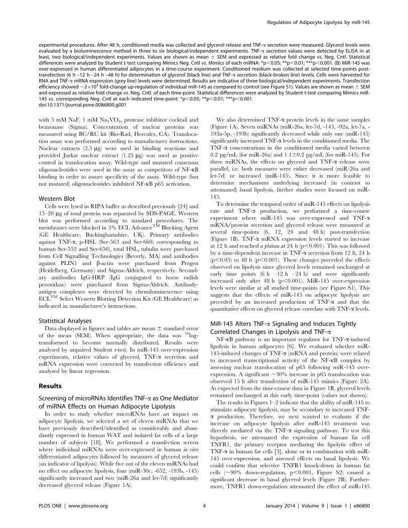
MiR-145 Regulates HSL (but not PLIN1) Phosphorylationand PDE3B mRNA Expression
It is well-established that TNF-a increases the phosphorylation
of HSL and PLIN1 [3,4,9,26] and decreases mRNA expression of
PDE3B [9]. Over-expression of miR-145 for 48 h induced a
significant increase in phosphorylation of HSL at activating
residues (Ser-552 and Ser-650) (Figure 4A and 4B) but did not
alter total HSL protein levels (Figure 4C). We also measured
PDE3B mRNA expression at different time points up to 48 h after
transfection with miR-145 mimics (Figure 4D). There was a
significant down-regulation in gene expression first observable at
24 h post-transfection (Figure S3). In contrast, we did not observe
any effects on PLIN1 phosphorylation status as miR-145 did not
induce any shift in the protein bands towards a higher molecular
weight. In addition, no significant changes were observed in total
PLIN1 protein content (Figure 5A and 5B).
Discussion
The present work demonstrates that miRNAs regulate adipo-
cyte lipolysis adding this metabolic action to the pleiotropic effects
of this class of molecules. We found that three (miR-26a, let-7d
and miR-145) of the eleven tested miRNAs had a concordant
effect on TNF-a secretion and glycerol release. This suggests
involvement of TNF-a in their lipolytic effect although we cannot
exclude TNF-a-independent mechanisms as well. Importantly, the
concentration of TNF-a in conditioned media from miR-26a, let-
7d and miR-145 overexpressing cells varied between 0.2 pg/mL
(for miR-26a) and 1.1 pg/mL (for miR-145) which is within the
physiological concentration interval of TNF-a in the interstitial
space of human adipose tissue [27,28]. This suggests that small
variations in local TNF-a concentrations lead to clearly detectable
changes in adipocyte lipolysis. On average, the pro-lipolytic effects
of miR-145 resulted in an approximate 50% increase in glycerol
levels in transfection screening (Figure 1).
In the over-expression experiments, miRNAs were .100 fold
up-regulated. At this high-level, there might be off-target effects.
This is generally a methodological issue in miRNA over-
expression experiments. However, we always compare cells treated
with specific mimics miRNA with mimics Negative Control.
Furthermore, in a recent study [18], we found no changes in fat
cell morphology or function due to specific miRNA over-
expression.
In order to study the axis miRNA-TNF-a lipolysis more in
detail, we selected miR-145 because it was the only miRNA that
stimulated both TNF-a and glycerol release. Several lines of
evidence suggest that miR-145 stimulates lipolysis by increasing
the endogenous production of TNF-a. First, there was a strong
correlation between changes in TNF-a mRNA/TNF-a secretion
and lipolysis following miRNA over-expression. Second, TNFR1
knock down abrogates miR-145-induced lipolysis suggesting a
causal relationship between the effects of miR-145 and TNFR1-
mediated signaling. Importantly, the finding that TNFR1 deple-
tion in fat cells attenuates lipolysis suggests that endogenous TNF-
a production is important for basal adipocyte lipolysis. Third,
miR-145-induced effects on adipocyte lipolysis occurred after
induction of TNF-a gene and protein secretion. Fourth, NF-kB,
which stimulates TNF-a transcription in an auto2/paracrine
loop, was activated prior to the peak in TNF-a secretion/mRNA
expression following miR-145 transfection. This indicates that NF-
kB induction may be a mechanism through which miR-145
regulates TNF-a expression. Moreover, since TNF-a induction by
miR-145 was not dependent on TNFR1 expression, it is plausible
that the TNF-a/NF-kB feedback loop might be mediated via
TNFR2 while TNF-a-induced lipolysis is primarily induced via
TNFR1. Nevertheless, the direct link between miR-145 and NF-
kB is not clear at the moment. Since miRNAs are down-regulators
of gene expression, there are probably several steps up-stream of
NF-kB activation linking miR-145 to TNF-a production which
could include one or more negative regulators of NF-kB.
However, to identify this/these candidates might prove difficult
and was out of the scope of this study.
TNF-a can either be present in soluble or a cell membrane-
bound state. Although both forms are biologically active,
membrane-bound TNF-a is thought to be responsible for cell-to-
cell interaction and activate TNF-a signaling pathways in adjacent
cells thereby promoting the development of an intense inflamma-
tory response within the cellular micro-environment [29]. Inter-
estingly, it has recently been shown in human non-adipose cells
(renal and hepatic cancer) that miR-145 decreases ADAM17
expression by direct binding to the 39UTR of the gene [21,25].
Although this interaction was not assessed in human adipocytes,
the fact that both cell systems were human, suggests that a similar
interaction may be present in other human cells including fat cells.
The observations that miR-145 over-expression resulted in a time-
dependent decrease in ADAM17 mRNA expression as well as a
significant increase in the ratio of membrane-bound/soluble TNF-
a, suggests that miR-145 may increase TNF-a activity in human
adipocytes, at least in part, through inhibition of ADAM17. This
fact further emphasizes that the increase in TNF-a production by
miR-145 is not only mediated by a direct effects on TNF-a
silenced with siRNA for 24 h prior to co-transfection with miRNA Mimics (Neg. Cntl/miR-145) for additional 48 h in human differentiated adipocytes.After incubation time, conditioned medium was collected to measure glycerol release and cells were harvested for (C) TNF-a mRNA measurements.Results are representative of three biological/independent experiments. Values are shown as mean 6 SEM. Statistical differences (vs. Neg. Cntl) wereanalyzed by Student t-test: **p,0.01; ***p,0.001. (D) Correlation between glycerol vs. TNF-a secretion (measured in conditioned medium/supernatant) and vs. (E) TNF-a mRNA levels in miR-145-transfected adipocytes in vitro for 48 h. Values are expressed as relative fold change vs. Neg.Cntl. (in D and E) after correction by transfection efficiency and were compared by linear regression. Each dot represents a technical replicate from 7–8 biological/independent experiments.doi:10.1371/journal.pone.0086800.g002
Regulation of Adipocyte Lipolysis by miR-145
PLOS ONE | www.plosone.org 6 January 2014 | Volume 9 | Issue 1 | e86800

transcription but also probably through an alteration of pre-
existing levels within the cell (e.g. TNF-a processing). In a broader
context, this would mean that adipocytes become more ‘‘sensitive’’
to certain pre-existing factors (such as TNF-a) and therefore, the
regulatory system develops and amplifies an inflammatory
response at the intra- and extracellular level, mediated in an
Figure 3. ADAM17 is a miR-145 target that regulates TNF-a processing in human adipocytes. (A) mRNA expression levels of ADAM17after miR-145 over-expression at 6 h –12 h –24 h –48 h. Results presented are obtained from three biological/independent experiments. Values areshown as mean 6 SEM and expressed as relative fold change vs. Neg. Cntl. at each corresponding time-point. (B) MiR-145 was over-expressed inhuman differentiated adipocytes for 48 h and whole cell lysates were analyzed by Western blot. TNF-a protein levels are expressed as ratio of TNF-amembrane bound (26 KDa) vs. soluble form (17 KDa). Results are representative of two biological/independent experiments. Values are shown asmean 6 SEM. Statistical differences (vs. Neg. Cntl) were analyzed by Student t-test: **p,0.01; ***p,0.001.doi:10.1371/journal.pone.0086800.g003
Regulation of Adipocyte Lipolysis by miR-145
PLOS ONE | www.plosone.org 7 January 2014 | Volume 9 | Issue 1 | e86800

auto2/paracrine manner by TNF-a. This finding would support
the hypothesis that the effect of miR-145 on lipolysis is complex,
involving several intermediates and complementary interactions
between them. However, we cannot exclude the possibility that
other genes targeted by miR-145 might contribute to regulate
TNF-a production as well and/or that miR-145 may also exert
TNF-a-independent effects on lipolysis.
Figure 4. miR-145 increases HSL phosphorylation at activating residues (but does not change total protein content of HSL) anddown-regulates PDE3B. (A) Representative blots of protein expression levels of phosphorylated HSL (Ser-552 and Ser-650) and total HSL in humandifferentiated adipocytes transfected with miR-145 mimics for 48 h. (B) Relative quantification by densitometry of above depicted blots for p-HSL(Ser-552, dark grey; Ser-650, light grey) and (C) total HSL. Equal amounts of total protein were loaded and separated by SDS-PAGE as indicated inexperimental procedures. Total HSL levels were corrected by tubulin expression and p-HSL levels were corrected by total HSL protein content. Valuesare shown as mean of pooling results from three biological/independent experiments. (D) Relative expression of PDE3B after miR-145 over-expressionin human differentiated adipocytes for 48 h. Results are representative of four biological/independent experiments. Values are shown as mean 6
SEM. Statistical differences (vs. Neg. Cntl) were analyzed by Student t-test: **p,0.01; ***p,0.001.doi:10.1371/journal.pone.0086800.g004
Regulation of Adipocyte Lipolysis by miR-145
PLOS ONE | www.plosone.org 8 January 2014 | Volume 9 | Issue 1 | e86800
Final steps in TNF-a-mediated lipolysis activation include HSL
[30] and PLIN1 phosphorylation [9]. Our results suggest that
HSL but not PLIN1 phosphorylation is involved in miR-145
stimulated lipolysis through TNF-a at least under the conditions
and time-points (48 h) used in this study. It could be speculated
that phosphorylation of HSL might constitute an earlier event
than PLIN1 phosphorylation. Indeed, we cannot rule out that
PLIN1 phosphorylation could be observed at later time-points
although total protein content remained unaltered. Finally, the
inhibition of PDE3B gene expression might constitute an
additional mechanism through which miR-145 affects adipocyte
lipolysis. However, this effect seems to be secondary to TNF-aexpression rather than a direct miRNA-mediated effect, since
PDE3B down-regulation occurred at later time-points (24 h and
48 h) after miR-145 over-expression.
Our screen included eleven miRNAs out of which only three
displayed concordant effects on lipolysis and TNF-a production.
For the remaining candidates, a few attenuated TNF-a without
altering lipolysis (miR-143, -92a and -193a-5p). This could
possibly depend on the fact that it is sometimes difficult to assess
reductions (compared with increases) in glycerol levels. It is
therefore possible that increasing the number of experiments
would identify (small) reductions in glycerol and TNF-a release
also with these miRNAs. Moreover, a few miRNAs increased
lipolysis without significant changes in TNF-a levels (miR-30c, -
652) or even with a reduction in its release (miR-193b). These data
suggest that specific miRNAs may alter lipolysis via TNF-a-
independent mechanisms. However, investigating these alternative
pathways was out of the scope of this present study.
On the basis of the present findings, we propose that TNF-aproduction and lipolysis can be regulated by miRNAs. Micro-
RNA-145 enhances the production/release of TNF-a and
increases the amount of membrane bound TNF-a via inhibition
of ADAM17. It also activates NF-kB, decreases PDE3B expression
and enhances HSL phosphorylation. All these events promote
lipolysis activation and taken together, suggest that the effects of
miR-145 on lipolysis are, at least in part, mediated by the effects
on TNF-a.
If the present findings are relevant for other species, remains to
be established. Future studies are also needed to elucidate the
mechanisms mediating the effects of the other miRNAs identified
in this study.
Supporting Information
Figure S1 Quantification of over-expression of miR-145in human differentiated pre-adipocytes. Human differen-
tiated adipocytes were transfected with miR-145 Mimics and
collected at several time-points post-transfection (6 h –12 h –24 h
–48 h) as described in material and methods. Cells were harvested
for RNA and relative miR-145 expression levels were determined.
Results are indicative of three biological/independent experi-
ments. Values are shown as mean 6 SEM and expressed as
relative fold change vs. Neg. Cntl. of each time-point.
(TIF)
Figure S2 Quantification of TNFR1 mRNA levels afterspecific gene silencing with siRNA. TNFR1 was silenced
with siRNA in human differentiated adipocytes as described in
material and methods. Cells were harvested for RNA and relative
TNFR1 mRNA expression levels were determined. Results are
indicative of three biological/independent experiments. Values are
shown as mean 6 SEM. Statistical differences were analyzed by
Student t-test: ***p,0.001.
(TIF)
Figure S3 Quantification of PDE3B mRNA levels miR-145 over-expression time-course. MiR-145 was over-
expressed in human differentiated adipocytes for 6 h –12 h –
24 h –48 h. Cells were harvested for RNA and PDE3B mRNA
expression levels were determined. Results are indicative of three
biological/independent experiments. Values are shown as mean 6
SEM. Statistical differences were analyzed by Student t-test:
**p,0.01.
(TIF)
Acknowledgments
We thank Eva Sjolin, Elisabeth Dungner, Kerstin Wahlen, Britt-Marie
Leijonhufvud, Katarina Hertel and Yvonne Widlund for excellent
technical assistance.
Figure 5. miR-145 does not affect phosphorylation and protein content of PLIN1. (A) Representative blot of total protein content of PLIN1after miR-145 over-expression in human differentiated adipocytes for 48 h. PLIN1 protein content was corrected by tubulin as described inexperimental procedures. (B) Relative quantification of PLIN1 vs. tubulin (n = 3).doi:10.1371/journal.pone.0086800.g005
Regulation of Adipocyte Lipolysis by miR-145
PLOS ONE | www.plosone.org 9 January 2014 | Volume 9 | Issue 1 | e86800
Author Contributions
Conceived and designed the experiments: SLC JL MR PA. Performed the
experiments: SLC GA AK. Analyzed the data: SLC NM PA. Contributed
reagents/materials/analysis tools: PH. Wrote the paper: SLC PA. Made
significant contributions and approved the final submission of the
manuscript: SLC JL MR PA AK NM GA PH.
References
1. Wellen KE, Hotamisligil GS (2003) Obesity-induced inflammatory changes in
adipose tissue. J Clin Invest 112: 1785–1788.2. Sethi JK, Hotamisligil GS (1999) The role of TNF alpha in adipocyte
metabolism. Seminars in cell & developmental biology 10: 19–29.3. Ryden M, Dicker A, van Harmelen V, Hauner H, Brunnberg M, et al. (2002)
Mapping of early signaling events in tumor necrosis factor-alpha -mediated
lipolysis in human fat cells. J Biol Chem 277: 1085–1091.4. Ryden M, Arvidsson E, Blomqvist L, Perbeck L, Dicker A, et al. (2004) Targets
for TNF-alpha-induced lipolysis in human adipocytes. Biochem Biophys ResCommun 318: 168–175.
5. Greenberg AS, Shen WJ, Muliro K, Patel S, Souza SC, et al. (2001) Stimulation
of lipolysis and hormone-sensitive lipase via the extracellular signal-regulatedkinase pathway. J Biol Chem 276: 45456–45461.
6. Laurencikiene J, van Harmelen V, Arvidsson Nordstrom E, Dicker A, BlomqvistL, et al. (2007) NF-kappaB is important for TNF-alpha-induced lipolysis in
human adipocytes. J Lipid Res 48: 1069–1077.7. Gasic S, Tian B, Green A (1999) Tumor necrosis factor alpha stimulates lipolysis
in adipocytes by decreasing Gi protein concentrations. J Biol Chem 274: 6770–
6775.8. Lorente-Cebrian S, Kulyte A, Heden P, Naslund E, Arner P, et al. (2011)
Relationship between site-specific HSL phosphorylation and adipocyte lipolysisin obese women. Obesity facts 4: 365–371.
9. Zhang HH, Halbleib M, Ahmad F, Manganiello VC, Greenberg AS (2002)
Tumor necrosis factor-alpha stimulates lipolysis in differentiated humanadipocytes through activation of extracellular signal-related kinase and elevation
of intracellular cAMP. Diabetes 51: 2929–2935.10. Ashall L, Horton CA, Nelson DE, Paszek P, Harper CV, et al. (2009) Pulsatile
stimulation determines timing and specificity of NF-kappaB-dependent tran-
scription. Science 324: 242–246.11. Hoffmann A, Levchenko A, Scott ML, Baltimore D (2002) The IkappaB-NF-
kappaB signaling module: temporal control and selective gene activation.Science 298: 1241–1245.
12. Nelson DE, Ihekwaba AE, Elliott M, Johnson JR, Gibney CA, et al. (2004)Oscillations in NF-kappaB signaling control the dynamics of gene expression.
Science 306: 704–708.
13. Peschon JJ, Slack JL, Reddy P, Stocking KL, Sunnarborg SW, et al. (1998) Anessential role for ectodomain shedding in mammalian development. Science 282:
1281–1284.14. Xu H, Uysal KT, Becherer JD, Arner P, Hotamisligil GS (2002) Altered tumor
necrosis factor-alpha (TNF-alpha) processing in adipocytes and increased
expression of transmembrane TNF-alpha in obesity. Diabetes 51: 1876–1883.15. Palladino MA, Bahjat FR, Theodorakis EA, Moldawer LL (2003) Anti-TNF-
alpha therapies: the next generation. Nature reviews Drug discovery 2: 736–746.
16. Bartel DP (2004) MicroRNAs: genomics, biogenesis, mechanism, and function.
Cell 116: 281–297.
17. Williams MD, Mitchell GM (2012) MicroRNAs in insulin resistance and obesity.
Experimental diabetes research 2012: 484696.
18. Arner E, Mejhert N, Kulyte A, Balwierz PJ, Pachkov M, et al. (2012) Adipose
tissue microRNAs as regulators of CCL2 production in human obesity. Diabetes
61: 1986–1993.
19. Sayed D, Abdellatif M (2011) MicroRNAs in development and disease.
Physiological reviews 91: 827–887.
20. Chen L, Song J, Cui J, Hou J, Zheng X, et al. (2013) microRNAs regulate
adipocyte differentiation. Cell biology international 37: 533–546.
21. Doberstein K, Steinmeyer N, Hartmetz AK, Eberhardt W, Mittelbronn M, et al.
(2013) MicroRNA-145 targets the metalloprotease ADAM17 and is suppressed
in renal cell carcinoma patients. Neoplasia 15: 218–230.
22. van Harmelen V, Skurk T, Hauner H (2005) Primary culture and differentiation
of human adipocyte precursor cells. Methods Mol Med 107: 125–135.
23. Kulyte A, Navakauskiene R, Treigyte G, Gineitis A, Bergman T, et al. (2002)
Characterization of human alpha-dystrobrevin isoforms in HL-60 human
promyelocytic leukemia cells undergoing granulocytic differentiation. Molecular
biology of the cell 13: 4195–4205.
24. Stenson BM, Ryden M, Venteclef N, Dahlman I, Pettersson AM, et al. (2011)
Liver X receptor (LXR) regulates human adipocyte lipolysis. J Biol Chem 286:
370–379.
25. Yang XW, Zhang LJ, Huang XH, Chen LZ, Su Q, et al. (2013) miR-145
suppresses cell invasion in hepatocellular carcinoma cells: miR-145 targets
ADAM17. Hepatology research : the official journal of the Japan Society of
Hepatology.
26. Zhang HH, Souza SC, Muliro KV, Kraemer FB, Obin MS, et al. (2003) Lipase-
selective functional domains of perilipin A differentially regulate constitutive and
protein kinase A-stimulated lipolysis. J Biol Chem 278: 51535–51542.
27. Pachler C, Ikeoka D, Plank J, Weinhandl H, Suppan M, et al. (2007)
Subcutaneous adipose tissue exerts proinflammatory cytokines after minimal
trauma in humans. Am J Physiol Endocrinol Metab 293: E690–696.
28. Clausen TS, Kaastrup P, Stallknecht B (2009) Proinflammatory tissue response
and recovery of adipokines during 4 days of subcutaneous large-pore
microdialysis. Journal of pharmacological and toxicological methods 60: 281–
287.
29. Kriegler M, Perez C, DeFay K, Albert I, Lu SD (1988) A novel form of TNF/
cachectin is a cell surface cytotoxic transmembrane protein: ramifications for the
complex physiology of TNF. Cell 53: 45–53.
30. Bezaire V, Langin D (2009) Regulation of adipose tissue lipolysis revisited. The
Proceedings of the Nutrition Society 68: 350–360.
Regulation of Adipocyte Lipolysis by miR-145
PLOS ONE | www.plosone.org 10 January 2014 | Volume 9 | Issue 1 | e86800
Copyright © 2022 FDOKUMEN