
MicroRNA cloning and sequencing in osteosarcoma cell lines: differential role of miR-93
13
ORIGINAL PAPER MicroRNA cloning and sequencing in osteosarcoma cell lines: differential role of miR-93 Luisa Montanini & Lisa Lasagna & Valeria Barili & Søren Peter Jonstrup & Alba Murgia & Laura Pazzaglia & Amalia Conti & Chiara Novello & Jørgen Kjems & Roberto Perris & Maria Serena Benassi Accepted: 26 August 2011 /Published online: 30 September 2011 # International Society for Cellular Oncology 2011 Abstract Background Studies show that abnormalities in non-coding genes can contribute to carcinogenesis; microRNA levels may modulate cancer growth and metastatic diffusion. Method MicroRNA libraries were built and sequenced from two osteosarcoma cell lines (MG-63 and 143B), which differ in proliferation and transmigration. By cloning and transfec- tion, miR-93, expressed in both cell lines, was then investigated for its involvement in osteosarcoma progression. Results Six of the 19 miRNA identified were expressed in both cell lines with higher expression levels of miR-93 in 143B and in primary osteosarcoma cultures compared to normal osteoblasts. Interestingly, levels of miR-93 were significantly higher in metastases from osteosarcoma than in paired primary tumours. When 143B and MG-63 were transfected with miR-93, clones appeared to respond differently to microRNA overexpression. Ectopic expres- sion of miR-93 more significantly increased cell prolifera- tion and invasivity in 143B than in MG-63 clones. Furthermore, increased mRNA and protein levels of E2F1, one of the potential miR-93 targets, were seen in osteosarcoma cellular clones and its involvement in 143B cell proliferation was confirmed by E2F1 silencing. Conclusion Although further studies are needed to evaluate miRNA involvement in osteosarcoma progression, miR-93 overexpression seems to play an important role in osteo- sarcoma cell growth and invasion. Keywords MicroRNA . Proliferation . Migration . Apoptosis . Osteosarcoma 1 Introduction Osteosarcoma (OS), the most common bone tumour, is a rare malignant neoplasm [1], for which biologic and pathologic information are still largely incomplete. In recent years surgery combined with chemotherapy has markedly im- proved patient survival [2]; however the use of anti-cancer drugs is still associated with serious problems, such as frequent acquisitions of drug-resistant phenotypes, highlight- ing the need for urgent novel treatment approaches [3]. A better understanding of the cellular and molecular mecha- nisms underlying pre- and post-surgery metastasis formation L. Montanini : L. Lasagna : V. Barili : A. Murgia : R. Perris Center for Molecular and Translational Oncology, Department of Genetics, Microbiology, Antropology and Evolution, University of Parma, Via G. P Usberti, 11/A, 43124 Parma, Italy L. Montanini Department of Paediatrics, University of Parma, Via Gramsci, 14, 43124 Parma, Italy S. P. Jonstrup : J. Kjems Department of Molecular Biology, University of Aarhus, C.F. Møllers Allé, Aarhus C, Denmark L. Pazzaglia : A. Conti : C. Novello : M. S. Benassi (*) Laboratory of Experimental Oncology, Rizzoli Orthopaedic Institute, Via di Barbiano 1/10, 40136 Bologna, Italy e-mail: [email protected] R. Perris Unit for Stem Cells and Cellular Therapy, Division for Experimental Oncology 2, The National Cancer Institute Aviano, CRO-IRCCS, Via Franco Gallini 2, 33081 Aviano, PN, Italy Cell Oncol. (2012) 35:29–41 DOI 10.1007/s13402-011-0059-z
-
Upload
independent -
Category
Documents
-
view
0 -
download
0
Transcript of MicroRNA cloning and sequencing in osteosarcoma cell lines: differential role of miR-93
ORIGINAL PAPER
MicroRNA cloning and sequencing in osteosarcoma celllines: differential role of miR-93
Luisa Montanini & Lisa Lasagna & Valeria Barili & Søren Peter Jonstrup &
Alba Murgia & Laura Pazzaglia & Amalia Conti & Chiara Novello & Jørgen Kjems &
Roberto Perris & Maria Serena Benassi
Accepted: 26 August 2011 /Published online: 30 September 2011# International Society for Cellular Oncology 2011
AbstractBackground Studies show that abnormalities in non-codinggenes can contribute to carcinogenesis; microRNA levelsmay modulate cancer growth and metastatic diffusion.Method MicroRNA libraries were built and sequenced fromtwo osteosarcoma cell lines (MG-63 and 143B), which differin proliferation and transmigration. By cloning and transfec-tion, miR-93, expressed in both cell lines, was theninvestigated for its involvement in osteosarcoma progression.
Results Six of the 19 miRNA identified were expressed inboth cell lines with higher expression levels of miR-93 in143B and in primary osteosarcoma cultures compared tonormal osteoblasts. Interestingly, levels of miR-93 weresignificantly higher in metastases from osteosarcoma thanin paired primary tumours. When 143B and MG-63 weretransfected with miR-93, clones appeared to responddifferently to microRNA overexpression. Ectopic expres-sion of miR-93 more significantly increased cell prolifera-tion and invasivity in 143B than in MG-63 clones.Furthermore, increased mRNA and protein levels ofE2F1, one of the potential miR-93 targets, were seen inosteosarcoma cellular clones and its involvement in 143Bcell proliferation was confirmed by E2F1 silencing.Conclusion Although further studies are needed to evaluatemiRNA involvement in osteosarcoma progression, miR-93overexpression seems to play an important role in osteo-sarcoma cell growth and invasion.
Keywords MicroRNA . Proliferation .Migration .
Apoptosis . Osteosarcoma
1 Introduction
Osteosarcoma (OS), the most common bone tumour, is a raremalignant neoplasm [1], for which biologic and pathologicinformation are still largely incomplete. In recent yearssurgery combined with chemotherapy has markedly im-proved patient survival [2]; however the use of anti-cancerdrugs is still associated with serious problems, such asfrequent acquisitions of drug-resistant phenotypes, highlight-ing the need for urgent novel treatment approaches [3]. Abetter understanding of the cellular and molecular mecha-nisms underlying pre- and post-surgery metastasis formation
L. Montanini : L. Lasagna :V. Barili :A. Murgia : R. PerrisCenter for Molecular and Translational Oncology,Department of Genetics, Microbiology,Antropology and Evolution, University of Parma,Via G. P Usberti, 11/A,43124 Parma, Italy
L. MontaniniDepartment of Paediatrics, University of Parma,Via Gramsci, 14,43124 Parma, Italy
S. P. Jonstrup : J. KjemsDepartment of Molecular Biology, University of Aarhus,C.F. Møllers Allé,Aarhus C, Denmark
L. Pazzaglia :A. Conti : C. Novello :M. S. Benassi (*)Laboratory of Experimental Oncology,Rizzoli Orthopaedic Institute,Via di Barbiano 1/10,40136 Bologna, Italye-mail: [email protected]
R. PerrisUnit for Stem Cells and Cellular Therapy,Division for Experimental Oncology 2,The National Cancer Institute Aviano, CRO-IRCCS,Via Franco Gallini 2,33081 Aviano, PN, Italy
Cell Oncol. (2012) 35:29–41DOI 10.1007/s13402-011-0059-z

and the development of chemo-resistance in these tumours arefundamental for prognosis and to unveil novel parameterspredicting therapeutic response [4]. Recently, much attentionhas focused on the impact of microRNA (miRNAs) ontumorigenesis and cancer progression. miRNAs, a class ofpost-transcriptional regulators, are single strand RNA mole-cules of about 22-nucleotides in length and regulate genetranslation into proteins acting either as tumour suppressorgenes or oncogenes, thus affecting mRNA stability andtranslation [5]. They are transcribed as pri-miRNA precursorsand cleaved by Drosha endonuclease in the nucleus, exportedinto cytoplasm by Exportin-5 as a pre-miRNA, prepared forentry into a large protein complex (RISC) by Dicerendonuclease-removing harping structure [6]. One of theRNA strands, corresponding to mature miRNA, is comple-mentary to mRNA target(s) and 30% of human protein-encoding genes are hypothesized to be regulated by miRNAs.miRNAs were stated as developmental modulators, with animportant regulatory role in a broad range of biologicalprocesses including development timing, cell differentiation,proliferation, apoptosis, and tumorigenesis [7–9]. Further-more, there is increasing evidence that abnormalities in certainmiRNAs play a role in the molecular mechanism involved inmalignant progression, acting as potential regulators of cellmotility and/or mediators of cancer spreading [10]. “Oncomir”ectopic expression appears to be associated with transforma-tion, metastatic progression, increased cell viability andproliferation in many solid tumours [11–15]. Other miRNAs,belonging to the tumour suppressor family, have been founddown-regulated in a variety of tumours, such as miR-Let-7 inbreast cancer [16], miR-335 required for metastasis formationof highly malignant cells [17], and miR-34a, a potent effectorof the p53 transcriptional network in uveal melanoma [18].Many in vitro studies demonstrated that up-regulation oftumour-suppressor miRNAs resulted in a decrease of cellmetastatic potential, modulating adhesion, migration, andinvasion pathways [19–22]. However, to date very few dataare reported in human sarcomas and cell lines [23, 24]. In thepresent study, microRNA libraries were built and sequencedfrom two OS cell lines, MG-63(p53−/−) and 143B(p53+/+),with different trans-endothelial migration and proliferationrate, in the effort to identify markers involved in OSprogression. We then investigated the role of miR-93, presentin both OS cell lines, in primary OS cultures and in clinicalsamples in relation to cell behaviour.
2 Materials and methods
2.1 Cell line and clinical specimens
Human osteosarcoma cell lines MG-63 (p53−/−), 143B(p53+/+) and Saos-2 (p53−/−), mesenchymal cells and
osteoblast cells were obtained from the American TypeCulture Collection (ATCC, Manassas, VA, USA; n°CRL-8303, n° CRL-1427 and n° HTB-85, n° CRL-1486and n° CRL-11372 respectively). Primary OS cell lines IOR/OS10, IOR/OS14, IOR/OS15, IOR/OS18 and IOR/MOSwere kindly provided by Dr Massimo Serra (Pharmacoge-nomics and Pharmacogenetics ResearchUnit, Rizzoli Institute,Bologna, Italy).
Cells were routinely cultured in humidified atmosphere(5% CO2) using D-MEM medium 10% FBS. Twenty-threeprimary classic OS samples, 11 paired normal tissues and 10OS lung metastases were referred to the Rizzoli OrthopaedicInstitute. Fresh non-necrotic tissue (≥90% viable tumour cells)was used and diagnosis based on hematoxylin-eosin stainedsamples according to histopathological criteria.
2.2 FATIMA system for cell migration detection
Cell migration assay was performed as previously described[25]. Cell transmigration kinetic was determined onstandard procedure-extracted HUVEC (Human UmbilicalVein Endothelial Cells) previously cultured in M199Endothelial medium (Cambrex—Rockland, USA) and20% FBS on 1% gelatine layer (37°C, 5% CO2 humidifiedatmosphere) by fluorescence plate reader (595 nm, TecanSpectra Fluor Instrument) at different times.
2.3 Small-RNA library construction
RNA was extracted from MG-63 and 143B osteosarcomacell lines by Trizol® total RNA isolation reagent (GibcoBRL, Life Technologies, Gaitherburg, MD, USA) accordingto the manufacturer’s instructions. The two librarieswere constructed as previously described by Elbashir etal. [26].
2.4 Sequencing of small-RNA cDNA libraries
Random selected colonies were picked up from ampicillin/kanamicin plate and resuspended in 20 μl of water. Cellswere lysed by heating in PCR machine (Gen Amp® PCRSystem 2700—Applied Biosystem), and DNA was ampli-fied using the following program 95°C/5′, (95°C/30″, 61°C/30″, 72°C/1′) ×30, 72°C/7′, 16°C/∞, Go Taq Polimerase(Promega) and the primers fwd GTT TTC CCA GTC ACGACG TTG TA and rev CAC AGG AAA CAG CTA TGACC. The longest amplification products were purified byusing Qiaquick Gel Extraction Kit (Qiagen GmbH, Hilden-Germany) and sequenced by DTCS Quick Start Master Mixof Genome Lab Dye Terminator Cycle Sequencing Kit(Beckmann). Termocycling was performed in Gen Amp®PCR System 2700 machine following the program (96°C/20″, 50°C/20″, 60°C/4′) ×30, 16°C/∞. Final products were
30 L. Montanini et al.

purified with standard procedure and analyzed in BeckmanCoulter CEQTM 2000 DNA Analysis System.
Each sequence identified by Chromas Lite Software wasanalyzed using megablast software (http://blast.ncbi.nlm.nih.gov/Blast.cgi). After vector and adapter sequence maskingand redundancy removing, 20–23 base inserts werecompared with the miRNA registry (http://microrna.sanger.ac.uk/sequences) and mapped into human genomeby using BLAST human genome (http://www.ncbi.nlm.nih.gov/genome/seq/HsBlast.html).
2.5 Cloning and transfection of the osteosarcoma cells
miR-93 was inserted in pCDBGW-miR vector by usinggateway system. In brief, two complementary strands of 60nt containing miR-93 were annealed for 4′ at 95°C thenligated overnight in a pENTRY vector. After checking thetransformation in DH5α subcloning cells and ligation withPvuII (Promega Corporation, Madison, WI, USA) andBsrgI (New England Biolabs) enzymes, a clonase reactionwas performed (LR Clonase—Invitrogen). Final ligation inpCDBGW-miR was tested withMscI restriction enzyme (NewEngland Biolabs). miR-93 vector was transfected in MG-63wild-type (wt), together with a vector containing G418resistance in the ratio of 5:1, by using Metafectene reagent(Biontex, Munchen—Germany) and following the manufac-turer’s protocol. For insertion in 143B wild-type (wt) cells,pCDBGWmiR-93 retrovirus was prepared by using CaPO4
and HEPES solution following standard procedures and theninfection was performed after transfection of a plasmidcontaining ecotropic receptor. After 2 days, green colonieswere picked, cultured and subsequently dispensed in a 96multi-plate (one cell every three wells) to obtain single cellclones (143Bcl4A, 143Bcl4B, MG-63clB and MG-63clDB).Cells infected with empty vector were used as control.
2.6 miR-93 expression analysis
Reverse transcription and RT-Real Time PCR were carriedout following TaqMan MicroRNA Assay Protocol (AppliedBiosystems, Foster City, CA) and miR-93 expression wasquantified using 2-ΔΔCT comparative method (AppliedBiosystems, User Bulletin N°2, P/N 4303859) and normalizedusing RNU6B and RNU44 as endogenous reference (TaqManmiRNA assay n° 001093 and n° 4373384 respectively—PEApplied Biosystems, Foster City, CA). cDNAs from mesen-chymal stem cells (MSC) and osteoblasts were used as relativecalibrators for OS cell lines and clinical samples. miRNAwasconsidered more expressed where the value of light emissioncalculated by the method 2-ΔΔCT was higher than the value1.5 SD or less expressed in all those cases in which the valuewas below 0.5 SD. miRNA average values were obtainedfrom replicates applying the 2-ΔΔCT method.
2.7 mRNA expression analysis of target genes
Reverse transcription of mRNA was carried out in 100 μlfinal volume from 400 ng total RNA using High CapacitycDNA Archive Kit (Applied Biosystems, Foster City, CA)according to manufacturer’s instructions.
Quantitative RT-PCR was performed using ABI 7900sequence detection system (Applied Biosystems, FosterCity, CA). Expression of target genes ANK2, E2F1 andLATS2 was quantified using TaqMan Expression Assays(Hs00153998_m1, Hs00153451_m1 and Hs00324396_m1respectively; Applied Biosystems, Foster City, CA) accordingto manufacturer’s protocol. For calculation of gene expressionwe used the 2-ΔΔCT comparative method. Expression oftarget genes was normalized to a housekeeping ACTB(Hs99999903_m1 gene; TaqMan Expression Assays-Applied Biosystems, Foster City, CA) and osteoblasts wereused as calibrator.
2.8 Immunoblot analysis
According to standard procedures, protein extracts wereprepared by mincing and homogenizing fresh samples inextraction buffer (50 mM Tris-HCl at pH 8, 150 mM NaCl,1 mM DTT, 50 mM NaF, 0.5% sodium deoxycholate, 0.1%SDS, 1% NP-40, 0.1 mM PMFS, 1 mg/ml aprotinin, 1 mg/ml leupeptin, 5 mM pepstatin, 0.1 mM NA3VO4, 1 mMmicrocystin). 50 microgram of protein extracts wereanalyzed by 10% SDS-PAGE and Western Blot (WB) wasperformed by using anti-E2F1 (Santa Cruz Biothech, CA,USA), dil 1:500. The signal was visualized by SupersignalWest Pico Chemiluminiscent Substrate (Pierce, Rockford,IL, USA) and quantified by using GS-670 imagingdensitometer (BioRad, Hercules, CA, USA). HELA lysatewas used as control.
2.9 Proliferation assay
The proliferation assay was performed in a 96-well formatusing CellTitre 96 One Solution Cell Proliferation Assay(Promega Corporation, Madison, WI, USA). In an individualexperiment, proliferation under each condition (0%, 0.1%,1%, 10% FBS) was studied in duplicate and the wholeexperiment was repeated at least three times.
2.10 Wound healing assay
2.0×10⁴ 143B cells and 2.5×10⁴ MG-63 cells were seededin Ibidi Culture Insert in DMEM with 1% of FBS. The platewas incubated overnight at 37°C in humidified atmosphere(5% of CO2). The following day the support was removed,cells were washed with PBS 1× and new DMEM with 1%of FBS was added. To investigate cell migration, the plate
MicroRNA Cloning and Sequencing in Osteosarcoma Cell Lines 31

was photographed at 0, 8, 24 h with 10× enlargement. Theexperiment was performed at least three times.
2.11 Migration assay
Cell migration assay was carried out using TranswellPermeable Supports (Corning Incorporated, Corning, NY,USA). Human osteosarcoma cell lines (MG-63 wt, MG-63clB, MG-63clDB, 143B wt, 143Bcl4A, 143Bcl4B) werecarefully transferred on the top chamber of each transwellapparatus at a density of 1,000,000 cells/ml (100 μl perchamber). Cells were allowed to migrate for 24 h at 37°C.Cells that had penetrated to the bottom of the membranewere then fixed in methanol, stained using hematoxylin,and countered at microscope. This experiment was per-formed three times.
2.12 Invasion assay
Cell invasion was analyzed using Cultrex 24 Well BMECell Invasion Assay (Trevigen, Inc, Gaithersburg, MD)according to standard procedures. Briefly, 103 cells/100 mlserum free media were seeded into the upper well oftranswells previously coated with Matrigel basementextract, while 500 μl of media were added in the bottomwell. After 24 h of CO2 incubation at 37°C, non-invasivecells on the upper surface were removed and the cellsmigrated to the lower surface were fixed in 500 μl of celldissociation solution/Calcein-AM, incubated at 37°C inCO2 for one hour and quantified by fluorimetric analysis(485 nm excitation, 520 nm emission).
2.13 TUNEL assay
10−3 OS cells grown on the slides were washed and fixedwith 4% paraformaldehyde in PBS pH 7.4 for 30 min,rinsed with PBS and permeabilized for 2 min on ice (0.1%Triton1 X–100, 0.1% sodium citrate). For the TUNELtechnique, all reagents were from in Situ Cell DeathDetection Kit (ROCHE Diagnostic, Mannheim, Germany)and procedures followed the manufacturer’s instructions.For negative control, 50 μl Label solution instead ofTUNEL reaction mixture was added. Positive control wastreated by DNase I recombinant for 10 min at 15–25°C toinduce DNA strand breaks, and then 50 μl TUNEL reactionmixture on every sample and on positive controls wasadded. Samples were incubated for 60 min at 37°C in adark humidified atmosphere and then rinsed three timeswith PBS. Finally, we added 50 μl Converter-POD onsamples incubated in a humidified chamber for 30 min at37°C, we rinsed slides three times with PBS, we added50 μl DAB substrate for 10 min at 15–25°C, we thenanalyzed samples under light microscope.
2.14 Small interfering RNA pool and transfection
A small interfering RNA (siRNA) pool targeting E2F1 (On-Target Plus SMART Pool, Thermo Scientific Dharmacon,Chicago, USA) was used in 143Bcl4B cell line. For cellgrowth experiment, cells were seeded in six well plates intriplicate (200.000 cells/well) and transfected 24 h later for5 h with specific siRNA or control (ctr) siRNA (50 nM)using DharmaFECT (Dharmacon), according to the manu-facturer’s protocol. At the end of transfection, freshmedium were placed and 24 h and 48 h later OS cellswere recovered with trypsin. As above described RNA andprotein extracts were analyzed by RT-PCR and WB toevaluate the efficiency of E2F1 target gene expressiondown-regulation. The remaining cells were seeded in 96-well flat-bottom plates at 103 cells per well and allowed toattach overnight and proliferation assay was performed.
2.15 Statistical analysis
miRNA expression data are shown as median (m) and25th–75th percentile for their strong non-Gaussian distri-bution. Non-parametric two-side Wicoxon’s rank sum testswere performed to compare miRNA expression profile inpaired samples; p values were set at <0.05.
3 Results
3.1 Transmigration and proliferation rate of OS cell lines
Cell kinetic was determined for different OS cell lines byFATIMA assay. Differences were found in cell migration.In particular, cells lacking p53, MG-63 and Saos-2, showeda faster trans-endothelial migration with respect to 143Bcells expressing wild-type p53. In particular, the kineticbehaviour of MG-63 and Saos-2 was similar, about doubleper time unit in percentage of migrated cells compared to143B (≈0.75 vs ≈0.4) (Fig. 1A, B).
On the other hand, OS cell growth rate using differentexperimental conditions, i.e. different serum concentrations,showed a direct correlation between cell proliferation andserum concentration with a higher proliferation rate of143B as compared with MG-63 and Saos-2 for up to 72 h atthe same conditions (Fig. 2A, B, C).
3.2 Screening of microRNA libraries
A number of miRNAs reported in the miRNA databaseregister were found expressed in MG-63 and 143B OS celllines (Table 1). Of the 19 miRNAs identified, the majorityact as tumour suppressor or oncogenes that control growthand apoptosis. Six out of 19 were expressed in both MG-63
32 L. Montanini et al.

and 143B (Table 1) and included miR-17-5p whichregulates cancer cell proliferation, miR-16-1 and −2 thatreside within the same cluster of miR-15 controlling anti-apoptotic gene BCL2, miR-130a that regulates expressionof homeoboxes GAX and HOXA5, and miR-93, belongingto the miR 17–92 family, that is recognized as an importantregulatory factor in various tumours. Other miRNAs werepresent in either MG-63 or 143B such as miR-21, miR-195,miR-106, miR-20b, miR-24 and miR-323.
A prediction of target genes for known miRNAs wasundertaken using software for miRNA targets version asMiranda, PicTar and Target Scan 5.1. In particular, thedatabases identified 968 candidate targets for miR-16a and -
16b including TMEM, SPRED1, KIFB, PLAG1, CCNE1and BCL2L2. In addition, the most important candidatetargets were ATXN1, PFN2, MAP3K2 and MYT1L formiR-17-5p, MI-ERI, MYBL1 and CFL2 genes for miR-130and LATS2, E2F1, ANK2, ZNFX1, ARID4B, HLF formiR-93.
3.3 miR-93 expression levels in different sarcoma cell linesand clinical specimens
Seeing its importance in different tumour types, we investi-gated miR-93 expression in sarcoma cell lines usingMSC andosteoblasts as calibrators. RT- PCR data showed that 143B
Fig. 1 Trans-endothelial migration of three OS cell lines. A Kinetics shows a higher migration through the endothelium layer ofMG-63 and Saos-2cells with respect to 143B. B Regression analysis. The data shown are mean values of at least three independent experiments
Fig. 2 Growth kinetics of MG-63 and 143B in different serum conditions. A 0–72 h, 1% serum. B 0–24 h, 1% serum; 24–72 h, 10% serum. C 0–72 h, 10% serum. The data shown are mean values of at least three independent experiments
MicroRNA Cloning and Sequencing in Osteosarcoma Cell Lines 33
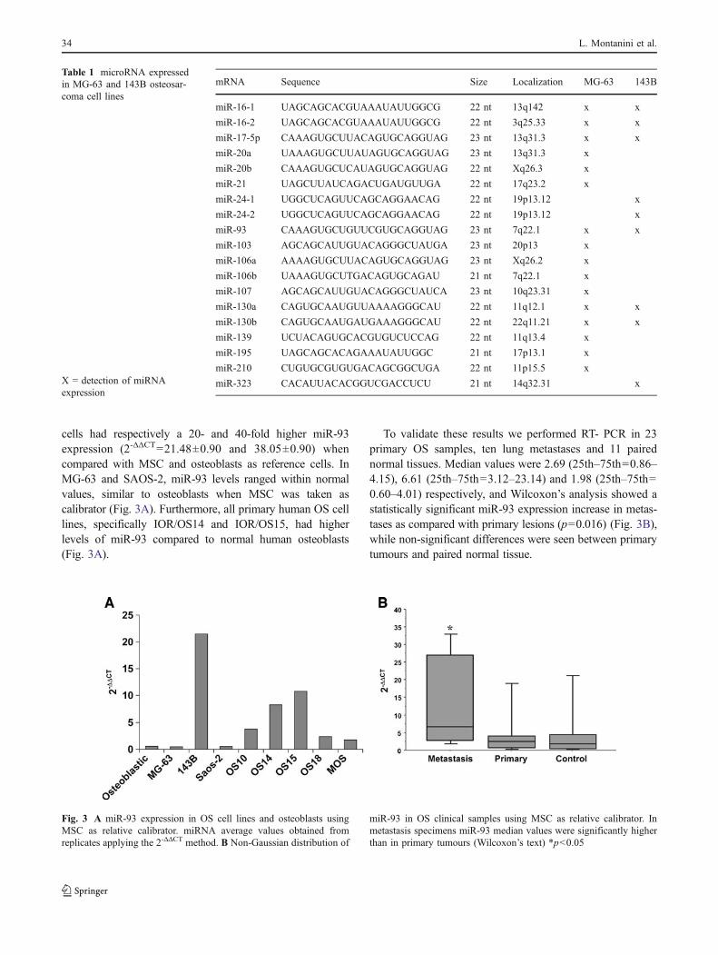
cells had respectively a 20- and 40-fold higher miR-93expression (2-ΔΔCT=21.48±0.90 and 38.05±0.90) whencompared with MSC and osteoblasts as reference cells. InMG-63 and SAOS-2, miR-93 levels ranged within normalvalues, similar to osteoblasts when MSC was taken ascalibrator (Fig. 3A). Furthermore, all primary human OS celllines, specifically IOR/OS14 and IOR/OS15, had higherlevels of miR-93 compared to normal human osteoblasts(Fig. 3A).
To validate these results we performed RT- PCR in 23primary OS samples, ten lung metastases and 11 pairednormal tissues. Median values were 2.69 (25th–75th=0.86–4.15), 6.61 (25th–75th=3.12–23.14) and 1.98 (25th–75th=0.60–4.01) respectively, and Wilcoxon’s analysis showed astatistically significant miR-93 expression increase in metas-tases as compared with primary lesions (p=0.016) (Fig. 3B),while non-significant differences were seen between primarytumours and paired normal tissue.
Table 1 microRNA expressedin MG-63 and 143B osteosar-coma cell lines
X = detection of miRNAexpression
mRNA Sequence Size Localization MG-63 143B
miR-16-1 UAGCAGCACGUAAAUAUUGGCG 22 nt 13q142 x x
miR-16-2 UAGCAGCACGUAAAUAUUGGCG 22 nt 3q25.33 x x
miR-17-5p CAAAGUGCUUACAGUGCAGGUAG 23 nt 13q31.3 x x
miR-20a UAAAGUGCUUAUAGUGCAGGUAG 23 nt 13q31.3 x
miR-20b CAAAGUGCUCAUAGUGCAGGUAG 22 nt Xq26.3 x
miR-21 UAGCUUAUCAGACUGAUGUUGA 22 nt 17q23.2 x
miR-24-1 UGGCUCAGUUCAGCAGGAACAG 22 nt 19p13.12 x
miR-24-2 UGGCUCAGUUCAGCAGGAACAG 22 nt 19p13.12 x
miR-93 CAAAGUGCUGUUCGUGCAGGUAG 23 nt 7q22.1 x x
miR-103 AGCAGCAUUGUACAGGGCUAUGA 23 nt 20p13 x
miR-106a AAAAGUGCUUACAGUGCAGGUAG 23 nt Xq26.2 x
miR-106b UAAAGUGCUTGACAGUGCAGAU 21 nt 7q22.1 x
miR-107 AGCAGCAUUGUACAGGGCUAUCA 23 nt 10q23.31 x
miR-130a CAGUGCAAUGUUAAAAGGGCAU 22 nt 11q12.1 x x
miR-130b CAGUGCAAUGAUGAAAGGGCAU 22 nt 22q11.21 x x
miR-139 UCUACAGUGCACGUGUCUCCAG 22 nt 11q13.4 x
miR-195 UAGCAGCACAGAAAUAUUGGC 21 nt 17p13.1 x
miR-210 CUGUGCGUGUGACAGCGGCUGA 22 nt 11p15.5 x
miR-323 CACAUUACACGGUCGACCUCU 21 nt 14q32.31 x
Fig. 3 A miR-93 expression in OS cell lines and osteoblasts usingMSC as relative calibrator. miRNA average values obtained fromreplicates applying the 2-ΔΔCT method. B Non-Gaussian distribution of
miR-93 in OS clinical samples using MSC as relative calibrator. Inmetastasis specimens miR-93 median values were significantly higherthan in primary tumours (Wilcoxon’s text) *p<0.05
34 L. Montanini et al.

3.4 miR-93 Target genes expression
mRNA expression of ANK2, E2F1 and LATS2 putativetarget genes was performed on miR-93 transfected cellclones of both 143B and MG-63 according to 2-ΔΔCT
comparative method.The analysis revealed that the E2F1 gene mRNA
expression increased in MG-63 and 143B clones (Table 2),the latter showing an evident increase also at protein levelas compared to wild-type cell line (Fig. 4).
3.5 Influence of miR-93 on OS cell proliferation, migrationand invasive behaviour
Effects of miR-93 on OS cell growth, migration andinvasiveness were then tested on infected/transfected cellclones of both 143B and MG-63.
RT-PCR analysis assessed significantly increased miR-93 expression levels in transfected clones when comparedwith controls (Fig. 5A, B).
miR-93 ectopic expression of significantly increased cellproliferation in 143Bcl4A and 143Bcl4B clones up to 72 h,only slightly affected cell growth of MG-63 clones(Fig. 6A, B).
The wound healing assay performed using serum contentrestrictions (1%) revealed that ectopic expression of miR-93induced migration decrease in 143B and MG-63 clones whencompared with wild-type and empty vector transfected cellsused as controls (Fig. 7A, B). However, migration throughpolycarbonate membrane and invasion through Matrigelbasement membrane showed that miR-93 overexpressionreduced 143B cell motility, but significantly increased cellinvasiveness in 143B clones 4A and 4B (p=0.01 and p=0.04respectively), without significantly affecting the ability ofMG-63 clones to migrate and invade through eithermembranes (Fig. 8A, B).
3.6 Apoptosis
After miR-93 transfection, 143B clones markedly decreasedapoptotic cell fraction measured by TUNEL assay that detects
DNA strand breakages in apoptotic cells. No significantvariations were seen in MG-63 clones when compared withnon-transfected cells (Fig. 9A, B).
3.7 E2F1 siRNA affects 143B cell proliferation
To confirm involvement of E2F1 in the proliferative responseof 143Bcl4B to miR-93 overexpression we used siRNAapproach to down-regulate E2F1expression. Transfection ofsiRNA targeting E2F1 markedly reduced its mRNA andprotein levels, with a concomitant significant decrease of cellproliferation after 24 h and 48 h from the end of transfection(Fig. 10A, B, C). A slight invasivity decrease (−10%) wasalso seen. E2F1 expression and proliferative rate were notaffected by control siRNA.
4 Discussion
miRNA deregulation has been identified in a variety ofcancers, where changes in specific miRNA expression maycontribute to tumour growth, progression, metastasis, anddrug resistance [27]. Deregulation of miRNAs occursfrequently during tumorigenesis [28], making them attrac-tive candidates for molecular detection of malignancy. Todate very few studies have been carried out on OS samplesand cell lines showing different miRNA expression relatedto metastatic potential and/or genetic background [29].
Table 2 mRNA expression ofmiR-93 target genes in MG-63and 143B
Calibrator: osteoblasts; house-keeping gene: ACTB
ANK2 (2-ΔΔCT±SD) E2F1 (2-ΔΔCT±SD) LATS2 (2-ΔΔCT±SD)
MG-63 wt 0.41±1.01 0.32±0.94 0.94±0.20
MG-63 clB 0.16±0.0 3.24±0.55 0.10±0.34
MG-63 clDB 0.80±0.11 0.10±0.0 0.30±0.11
143B wt 0.87±0.30 0.93±0.30 0.17±0.30
143B cl4A 0.99±0.16 2.14±0.26 0.32±0.22
143B cl4B 0.43±0.36 2.52±1.42 0.18±0.01
Fig. 4 E2F1 protein expression in MG-63wt, MG-63clB, MG-63clDB and in 143B wt, 143cl4A, 143Bcl4B by Western blotting.143B clones transfected with miR-93 show a more intense migratingband when compared to wild-type cells. Control loading is shown byactin. HELA lysate was used as control
MicroRNA Cloning and Sequencing in Osteosarcoma Cell Lines 35

In particular, miR-34 and miR-140 affecting target geneexpression in a p53 dependent manner induced chemo-sensitivity in wt-p53 human OS [24, 30].
A very recent study on sarcoma cell lines by Gougelet etal. [31] identified specific miRNAs as discriminating ofdrug-response using microfluidic PCR technology onTLDA platform preloaded with dehydrated specific primersand probes. In 7 OS samples, using miRNA microarraytechnology Maire et al. [32] found 38 miRNAs differen-tially expressed as compared with human osteoblasts.
With a different approach, we identified microRNAsrelated to a different proliferation and migration behaviorby preparing microRNA libraries from OS cell lines whichdiffer in p53 phenotype and in cell growth and migrationrate by transwell assay with HUVEC layer. Specifically,MG-63 (p53−/−) and Saos-2 (p53−/−) showed similartransmigration kinetics per time unit, whereas 143B (p53+/+)migrated at about half speed. On the other hand, whenproliferation rate was assessed data showed that cell growthof 143B was higher than MG-63 and Saos-2 in allexperimental serum conditions. Sequencing analysis identi-
fied 19 miRNA, of which 6 were present in both MG-63 and143B cell lines. Gao et al. [33] identified 25 microRNAs inSOPS-67 osteosarcoma cells. In both studies the sameidentification methods were used, and the discrepancy ofdata (19 versus 25) could be due to the loss of less expressedmicroRNAs during purification processes before cloningreaction.
MiR-21 and miR-106b, considered as the major micro-RNAs overexpressed in cancers, were expressed in MG-63but not in 143B. Recent studies showed overexpression ofmiR-21 in OS tissues and confirmed that miR-21 controlscell adhesion and migration, but not proliferation, throughrepression of tumour suppressor proteins such as TIMP1,MARCK5 PTEN [34, 35]. Thus, the role of miR-21appears in conflict with miR-143 that was found to reduceOS viability and metastasis by affecting Bcl2 and matrixmetalloproteinases-13 expression [29, 36].
All miRNAs found in the study appear related to cancerdevelopment. miR-106b that belongs to the cluster miR-106b-25, is necessary for cell proliferation and anchorage-independent growth [37], miR-17-5p has been characterized
Fig. 5 Increased expressionlevels of miR-93 in tumour cellclones. A MG-63 clones (clB,clDB); B 143B clones (cl4A,cl4B). In both experiments wild-type (wt) cell lines were used ascontrol. The data shown aremean values of at least threeindependent experiments
Fig. 6 Cell growth up 72 h of AMG-63 and B 143B wild-typecells and miR-93 transfectedclones. Cells infected withempty vector were used as con-trol (ctr). The data are expressedas mean ± SD of three indepen-dent experiments. * p<0.05
36 L. Montanini et al.

as microRNA associated to different cancer types [38], whilemiR-130a seems to regulate molecule expression inhibitingangiogenesis in vascular endothelial cells [39] and has beenfound down-regulated in ovarian cancer cell lines resistant tochemotherapy [40]. miR-16-1 and −2 and miR-15 belong toa cluster where deletion or down-regulation appear to be
associated with poor prognosis in chronic lymphocyticleukemia [41].
Many expressed miRNAs are “oncomirs” belongingto the 106b-25 microRNA cluster, a polycistronicmiRNA highly conserved in all vertebrates, implicatedin normal development of the heart, lungs, and immune
Fig. 7 Wound healing assays of A clB and clDB MG-63 clones and B cl4A and cl4B 143B clones overexpressing miR-93 in comparison with thecorresponding wild-type cell lines and controls (ctr)
MicroRNA Cloning and Sequencing in Osteosarcoma Cell Lines 37

system and recently found to be highly deregulated insome tumour types [42, 43]. Our attention was fixed onmiR-93, recently described as an important moleculeduring stem cell differentiation [44], involved in differentcancer types [45–47], but not yet investigated in sarcomacells.
RT-PCR specific analysis of miR-93 in the three OS cellsshowed that MG-63 and Saos-2 cells had miRNA expres-sion similar to that found in osteoblasts when mesenchymalcells were considered as calibrators, whereas a higherexpression of miR-93 was detected in 143B, which alsoshowed a higher growth rate. In addition, we assessed miR-93 levels in primary cultures of human OS, and found
higher expression levels as compared to osteoblasts. On thebasis of these findings, the role of miR-93 in OS cellbehaviour was further investigated. In this context, MG-63and 143B cell lines were transfected/infected by a plasmidcontaining this miRNA. Two independent cell clones fromeach OS cell line were obtained with miR-93 expressionhigher than in wild-type cells, but responding in a differentmanner to microRNA expression. 143B clones that expressp53 oncosuppressor genes increase cell proliferation andviability following miR-93 overexpression more than MG-63, which lack functional p53.
Although there is evidence that hundreds of targets arepredicted for each miRNA, recent observations in gastric
Fig. 9 Cell death of A MG-63 and B 143B and transfected clones measured by TUNEL assay that detects DNA strand breakages in apoptoticcells. Ectopic expression of miR-93 reduced the apoptotic cell fraction in 143B clones, without affecting MG-63
Fig. 8 A The migration assaythrough polycarbonate mem-branes in wild-type OS cells andmiR-93 transfected clones. BInvasion assay through a mem-brane coated with Matrigelbasement extract. The datashown are mean values ± SD ofthree independent experiments.**p<0.01, ***p<0.001
38 L. Montanini et al.

carcinoma showed that miR-93 cooperate to inhibit p21post-transcriptional expression through E2F1 regulation,also impairing the TGFb tumour suppressor pathway [45].It is well known that p21, an inhibitor of cyclin-dependentkinases and proliferating-cell nuclear antigen, is under thetranscriptional control of p53 suggesting that p21 mightpromote p53-dependent cell cycle arrest or apoptosis [48].Moreover, a recent study demonstrated that knockdown ofmiR-93 and miR-130b increased p53expression, consistentwith the increase of apoptotic rate [46]. Interestingly, wefound induction of E2F1 expression following ectopicexpression of miR-93 and demonstrated that silencingE2F1 by siRNA significantly decreased cell proliferationas compared to control siRNA transfected cells. Althoughfurther studies are needed, these observations suggest aninvolvement of miR-93 in E2F1 regulation and cell cyclecontrol of osteosarcoma cells.
On the other hand, the role of miR-93 in cell invasionand motility is unclear. Although ectopic miR-93 expres-sion resulted in a slower cell migration through polycar-bonate membrane, an increase in cell invasiveness was seenin 143B clones when Matrigel basement coated membrane
was used. The observation that in OS metastasis specimenslevels of miR-93 were significantly higher than in pairedprimary tumours might highlight the ability of miR-93 toconfer a highly malignant, metastatic phenotype. Inconclusion, we demonstrated differences in miRNA profil-ing of OS cell lines related to a different proliferation andmigration behaviour suggesting that up-regulation of miR-93 might be involved in E2F1 transcription factor regula-tion of p53-positive cells.
Ongoing studies directly addressing the actual targetsfor miR-93 cluster will be of interest to clarify mechanismsinvolved in this phenomenon and to better understandhow this alteration might contribute to developmentosteosarcoma.
Acknowledgements The authors thank Paola Spessotto for trans-well and invasion assays, Arcispedale Santa Maria Nuova forproviding umbilical cord, Prof. Paolo Malatesta for technical support,Cristina Ghinelli for the graphic work and Alba Balladelli, B.A. fortext revision. This work was supported by grants from the ItalianMinistry of Health (MSB, RP), Italian Istituto Superiore della Sanità(MSB, RP), EuroBoNeT Consortium (MSB) and Institutional fundsfrom the University of Parma (RP).
Fig. 10 Transfection effect of E2F1 siRNA in 143Bcl4B cellsincubated for 5 h with siRNA targeting E2F1 or ctr siRNA. A RT-PCR analysis of E2F1. 400 ng total RNAwere analyzed 24 h and 48 hfrom the end of transfection. Osteoblasts were used as calibrator. BWestern blot analysis of E2F1. 50 microgram of protein extracts were
analyzed 48 h later from the end of transfection. Control loading isshown by actin. HELA lysate was used as control. C Cell growthinhibition after transfection with E2F1 siRNA and control (ctr)siRNA. Each value indicates the average of three independentexperiments. **p<0.01
MicroRNA Cloning and Sequencing in Osteosarcoma Cell Lines 39

References
1. M. Campanacci, Bone and soft tissue tumors, 2nd edn. (Springer,Wien, 1999)
2. S.S. Bielack, B. Kempf-Bielack, G. Delling, G.U. Exner, S. Flege,K. Helmke, R. Kotz, M. Salzer-Kuntschik, M. Werner, W.Winkelmann, A. Zoubek, H. Jurgens, K. Winkler, Prognosticfactors in high-grade osteosarcoma of the extremities or trunk: ananalysis of 1,702 patients treated on neoadjuvant cooperativeosteosarcoma study group protocols. J. Clin. Oncol. 20, 776–790(2002)
3. M. Wachtel, B.W. Schäfer, Targets for cancer therapy in childhoodsarcomas. Cancer Treat. Rev. 36, 318–327 (2010)
4. J. Toguchida, T. Nakayama, Molecular genetics of sarcomas:applications to diagnoses and therapy. Cancer Sci. 100, 1573–1580 (2009)
5. R. Schieckel, B. Boyerinas, S.M. Park, M.E. Peter, MicroRNAs:key players in the immune system, differentiation, tumorigenesisand cell death. Oncogene 27, 5959–5974 (2008)
6. S.M. Hammond, Dicing and ilincing: the core machinery of theRNA interference pathway. FEBS Lett. 579, 5822–5829 (2005)
7. A. Fire, S. Xu, M.K. Montgomery, S.A. Kostas, S.E. Driver, C.C.Mello, Potent and specific genetic interference by double strandedRNA in Caenorhabditis elegans. Nature 391, 744–745 (1998)
8. I. Alvarez-Garcia, E.A. Miska, MicroRNAs functions in animaldevelopment and human disease. Development 132, 4653–4662(2005)
9. A. Esquela-Kerscher, F.J. Slack, Oncomirs-microRNAs with arole in cancer. Nat. Rev. Cancer 53, 627–635 (2006)
10. L. Ma, R.A. Weinberg, Micromanagers of malignancy: role ofmicroRNAs in regulating metastasis. Cell 24, 448–456 (2008)
11. P.M. Voorhoeve, C. le Sage, M. Schrier, A.J. Gillis, H. Stoop, R.Nagel, Y.P. Liu, J. van Duijse, J. Drost, A. Griekspoor, E.Zlotorynski, N. Yabuta, G. De Vita, H. Nojima, L.H. Looijenga,R. Agami, A genetic screen implicates miRNA-372 and miRNA-373 as oncogenes in testicular germ cell tumours. Cell 124, 1169–1181 (2006)
12. Q. Huang, K. Gumireddy, M. Schrier, C. le Sage, R. Nagel, S.Nair, D.A. Egan, A. Li, G. Huang, A.J. Klein-Szanto, P.A.Gimotty, D. Katsaros, G. Coukos, L. Zhang, E. Puré, R. Agami,The microRNAs miR-373 and mir-520c promote tumour invasionand metastasis. Nat. Cell. Biol. 10, 202–210 (2008)
13. J.A. Chan, A.M. Krichevsky, K.S. Kosik, MicroRNA-21 is ananti-apoptotic factor in human glioblastoma cell. Cancer Res. 65,6029–6033 (2005)
14. S.A. Ciafrè, S. Galardi, A. Mangiola, M. Ferracin, C.G. Liu, G.Sabatino, M. Negrini, G. Maira, C.M. Croce, M.G. Farace, Extensivemodulation of a set of microRNAs in primary glioblastoma.Biochem. Biophys. Res. Commun. 334, 1351–1358 (2005)
15. C. Roldo, E. Missiaglia, J.P. Hagan, M. Falconi, P. Capelli, S.Bersani, G.A. Calin, S. Volinia, C.G. Liu, A. Scarpa, C.M. Croce,MicroRNA expression abnormalities in pancreatic and acinartumors are associated with distinctive pathologic features andclinical behavior. J. Clin. Oncol. 24, 4677–4684 (2006)
16. F. Yu, H. Yao, P. Zhu, X. Zhang, Q. Pan, C. Gong, Y. Huang, X.Hu, F. Su, J. Lieberman, E. Song, Let-7 regulates self renewal andtumorigenicity of breast cancer cells. Cell 131, 1109–1123 (2007)
17. S.F. Tavazoie, C. Alarcon, T. Oskarsson, D. Padua, Q. Wang, P.D.Bos, W.L. Gerald, J. Massague, Endogenous human microRNAsthat suppress breast cancer metastasis. Nature 451, 147–152(2008)
18. D. Yan, X. Zhou, X. Chen, D.N. Hu, X.D. Dong, J. Wang, F. Lu,L. Tu, J. Qu, MicroRNA-34a inhibits uveal melanoma cellproliferation and migration thrugh down-regulation of c-Met.Invest. Ophthalmol. Vis. Sci. 50, 1559–1565 (2009)
19. P.A. Gregory, A.G. Bert, E.L. Paterson, S.C. Barry, A. Tsykin, G.Farshid, M.A.Y. Vadas, Y. Khew-Goodall, G.J. Goodall, The miR-200 family and miR-205 regulate epithelial to mesenchymaltransition by targeting ZEB1 and SIP1. Nat. Cell Biol. 10, 593–601 (2008)
20. M. Korpal, E.S. Lee, G. Hu, Y. Kang, The miR-200 familyinhibits epithelial-mesenchymal transition and cancer cell migra-tion by direct targeting of e-cadherin transcriptional repressorZEB1 and ZEB2. J. Biol. Chem. 283, 14910–14914 (2008)
21. M. Crawford, E. Brawner, K. Batte, L. Yu, M.G. Hunter, G.A.Otterson, G. Nuovo, C.B. Marsh, S.P. Nana-Sinkam, MicroRNA-126 inhibits invasion in non-small cell lung carcinoma cell lines.Biochem. Biophys. Res. Commun. 373, 607–612 (2008)
22. C. Evangelisti, M.C. Florian, I. Massimi, C. Dominici, G. Giannini,S. Galardi, M.C. Bluè, S. Massalini, H.P. McDowell, E. Messi, A.Gulino, M.G. Farace, S.A. Ciafrè, MiR-128 up-regulation inhibitsReelin and DCX expression and reduces neuroblastoma cell motilityand invasiveness. FASEB J. 23, 4276–4287 (2009)
23. S. Subramanian, W.O. Lui, C.H. Lee, I. Espinosa, T.O. Nielsen,M.C. Heinrich, C.L. Corless, A.Z. Fire, M. van de Rijn, Micro-RNA expression signature of human sarcomas. Oncogene 27,2015–2026 (2008)
24. C. He, J. Xiong, X. Xu, W. Lu, L. Liu, D. Xiao, D. Wang,Functional elucidation of miR-34 in osteosarcoma cells andprimary tumor samples. Biochem. Biophys. Res. Commun. 388,35–40 (2009)
25. P. Spessotto, K. Lacrima, P.A. Nicolosi, E. Pivetta, M. Scapolan,R. Perris, Fluorescence-based assays for in vitro analysis of celladhesion and migration. Methods Mol. Biol. 522, 221–250 (2009)
26. S.M. Elbashir, W. Lendeckel, T. Tuschl, RNA interference ismediated by 21- and 22-nucleotide RNAs. Genes Dev. 15, 188–200 (2001)
27. O.A. Kent, J.T. Mendell, A small piece in the cancer puzzle:microRNAs as tumor suppressors and oncogenes. Oncogene 25,6188–6196 (2006)
28. W. Zhang, J.E. Dahlberg, W. Tam, MicroRNAs in tumorigenesis:a primer. Am. J. Pathol. 171, 728–738 (2007)
29. M. Osaki, F. Takeshita, Y. Sugimoto, N. Kosaka, Y. Yamamoto, Y.Yoshioka, E. Kobayashi, T. Yamada, A. Kawai, T. Inoue, H. Ito,M. Oshimura, T. Ochiya, MicroRNA-143 regulates humanosteosarcoma metastasis by regulating matrix metalloproteinase-13 expression, Mol. Ther. Mar 22 (2011), [Epub ahead of print],PMID:21427707.
30. B. Song, Y. Wang, Y. Xi, K. Kudo, S. Bruheim, G.I. Botchkina, E.Gavin, Y. Wan, A. Formentini, M. Kornmann, O. Fodstad, J. Ju,Mechanism of chemoresistance mediated by miR-140 in humanosteosarcoma and colon cancer cells. Oncogene 28, 4065–4074(2009)
31. A. Gougelet, J. Perez, D. Pissaloux, A. Besse, A. Duc, A.V.Decouvelaere, D. Ranchere-Vince, J.Y. Blay, L. Alberti, miRNAProfiling: how to bypass the current difficulties in the diagnosis andtreatment of sarcomas, Sarcoma Feb 22(2011), PMID:21437224.
32. G. Maire, J.W. Martin, M. Yoshimoto, S. Chilton-MacNeill, M.Zielenska, A. Jeremy, Analysis of miRNA-gene expression-genomic profiles reveals complex mechanisms of microRNAderegulation in osteosarcoma. Cancer Genet. 204, 138–146 (2011)
33. J. Gao, T.T. Yang, X.C. Qiu, B. Yu, J.W. Han, Q.Y. Fan, B.A. Ma,Cloning and identification of microRNA from human osteosarco-ma cell line SOSP-9607. Ai Zheng 26, 561–565 (2007)
34. S. Baranwal, S.K. Alahari, MiRNA contro and tumour cellinvasion and metastasis. Int. J. Cancer 126, 1283–1290 (2010)
35. W. Ziyan, W. Shuhua, W. Xiufang, L. Xiaoyun, MicroRNA-p21 isinvolved in osteosarcoma cell invasion and migration, Med.Oncol. May 18 (2010), [Epub ahead of print] PMID: 20480266.
36. H. Zhang, X. Cai, Y. Wang, H. Tang, D. Tong, F. Ji, MicroRNA143, down-regulated in osteosarcoma, promotes apoptosis and
40 L. Montanini et al.
suppresses tumorigenicity by targeting Bcl-2. Oncol. Rep. 24,1363–1369 (2010)
37. Y. Li, W. Tan, T.W. Neo, M.O. Aung, S. Wasser, S.G. Lim, T.M.Tan, Role of miR-106b-25 microRNA cluster in hepatocellularcarcinoma. Cancer Sci. 100, 1234–1242 (2009)
38. C. Volinia, G. Calin, C.G. Liu, S. Ambs, A. Cimmino, F. Petrocca,R. Visone, M. Iorio, C. Roldo, M. Ferracin, R.L. Prueitt, N.Yanaihara, G. Lanza, A. Scarpa, A. Vecchione, M. Negrini, C.C.Harris, C.M. Croce, A microRNA expression segnature of humansolid tumors defines cancer gene targets. Proc. Natl. Acad. Sci.USA 103, 2257–2261 (2006)
39. Y. Chen, D.H. Gorski, Regulation of angiogenesis through amicroRNA (miR-130a) that down-regulates anti-angiogenic ho-meobox genes GAX and HOXA5. Blood 111, 1217–1226 (2008)
40. M.T. Van Joorsveld, J. Helleman, E.M. Berns, E.A. Wiemer,MicroRNAs in ovarian cancer biology and therapy resistance, Int.J. Biochem. Cell. Biol. (2010) 18 [Epub ahead of print] PMID:20083225.
41. G. Calin, A. Cimmino, M. Fabbri, M. Ferracin, S.E. Wojcik, M.Shimizu, C. Taccioli, N. Zanesi, R. Garzon, R.I. Aqeilan, H.Alder, S. Volinia, L. Rassenti, X. Liu, C.G. Liu, T.J. Kipps, M.Negrini, C.M. Croce, Mir-15a e Mir-16-1 clusters function inhuman leukaemia. Proc. Natl. Acad. Sci. USA 105, 5166–5171(2008)
42. A. Ventura, A.G. Young, M.M. Winslow, L. Lintault, A. Meissner,S.J. Erkeland, J. Newman, R.T. Bronson, D. Crowley, J.R. Stone,R. Jaenisch, P.A. Sharp, T. Jacks, Targeted dekletion reveals
essential and overlapping functions of the miR-17 trough 92family of miRNA clusters. Cell 132, 875–886 (2008)
43. C. Xiao, L. Srinivasan, D.P. Calado, H.C. Patterson, B. Zhang, J.Wang, J.M. Henderson, J.L. Kutok, K. Rajewsky, Lymphoprolferativedisease and autoimmunity in mice with increased miR-17-92expression in lymphocytes. Nat. Immunol. 9, 405–414 (2008)
44. K.M. Foshay, G.I. Gallicano, Small RNAs, big potential: the roleof MicroRNAs in stem cell function. Curr. Stem Cell Res. Ther. 2,264–271 (2007)
45. F. Petrocca, R. Visone, M.R. Onelli, M.H. Shah, M.S. Nicoloso, I.de Martino, D. Iliopoulos, E. Pilozzi, C.G. Liu, M. Negrini, L.Cavazzini, S. Volinia, H. Alder, L.P. Ruco, G. Baldassarre, C.M.Croce, A. Vecchione, E2F1-regulated microRNAs impairTGFbeta-dependent cell-cycle arrest and apoptosis in gastriccancer. Cancer Cell 13, 272–286 (2008)
46. M.L. Yeung, J. Yasunaga, Y. Bennasser, N. Dusetti, D. Harris, N.Ahmad, M. Matsuoka, K.T. Jeang, Roles of MicroRNAs, miR-93and miR-103b, and tumor protein 53-induced nuclear protein 1tumor suppressor in cell growth dysregulation by human T-celllymphotrophic virus 1. Cancer Res. 68, 8976–8985 (2008)
47. K.E. Resnick, H. Alder, J.P. Hagan, D.L. Richardson, C.M. Croce,D.E. Cohn, The detection of differentially expressed microRNAsfrom the serum of ovarian cancer patients using a novel real-timePCR platform. Gynecol. Oncol. 112, 55–59 (2009)
48. J. Brugarolas, C. Chandrasekaran, J.I. Gordon, D. Beach, T. Jacks,G.J. Hannon, Radiation-induced cell cycle arrest compromised byp21 deficiency. Nature 377, 552–557 (2002)
MicroRNA Cloning and Sequencing in Osteosarcoma Cell Lines 41