
miR-Sens--a retroviral dual-luciferase reporter to detect microRNA activity in primary cells
10
METHOD miR-Sens—a retroviral dual-luciferase reporter to detect microRNA activity in primary cells EMMANUEL BEILLARD, 1 SIAU CHI ONG, 1 ANTONIS GIANNAKAKIS, 2 ERNESTO GUCCIONE, 3 LEAH A. VARDY, 4 and P. MATHIJS VOORHOEVE 1,5,6 1 Department of Cancer and Stem Cell Biology, Duke-NUS Graduate Medical School, Singapore 169857, Singapore 2 Bioinformatics Institute, Matrix, Singapore 138671, Singapore 3 Institute of Molecular and Cell Biology, Proteos, Singapore 138673, Singapore 4 Institute of Medical Biology, Immunos, Singapore 138648, Singapore 5 Department of Biochemistry, National University of Singapore, Singapore 117597, Singapore ABSTRACT MicroRNA–mRNA interactions are commonly validated and deconstructed in cell lines transfected with luciferase reporters. However, due to cell type-specific variations in microRNA or RNA-binding protein abundance, such assays may not reliably reflect microRNA activity in other cell types that are less easily transfected. In order to measure miRNA activity in primary cells, we constructed miR-Sens, a MSCV-based retroviral vector that encodes both a Renilla luciferase reporter gene controlled by microRNA binding sites in its 39 UTR and a Firefly luciferase normalization gene. miR-Sens sensors can be efficiently transduced in primary cells such as human fibroblasts and mammary epithelial cells, and allow the detection of overexpressed and, more importantly, endogenous microRNAs. Notably, we find that the relative luciferase activity is correlated to the miRNA expression, allowing quantitative measurement of microRNA activity. We have subsequently validated the miR-Sens 39 UTR vectors with known human miRNA-372, miRNA-373, and miRNA-31 targets (LATS2 and TXNIP). Overall, we observe that miR- Sens-based assays are highly reproducible, allowing detection of the independent contribution of multiple microRNAs to 39 UTR–mediated translational control of LATS2. In conclusion, miR-Sens is a new tool for the efficient study of microRNA activity in primary cells or panels of cell lines. This vector will not only be useful for studies on microRNA biology, but also more broadly on other factors influencing the translation of mRNAs. Keywords: UTR; microRNA; retrovirus; primary cells; dual luciferase INTRODUCTION MicroRNAs (miRNAs) are short single-stranded RNAs bind- ing to mRNAs with partial complementarity in miRNA- binding sites, resulting in translational repression and/or mRNA destabilization. Most well-recognized targets are located in 39 UnTranslated Regions (UTRs), which medi- ate miRNA-dependent translational repression and mRNA destabilization (Bartel 2009). Several computational ap- proaches have been developed to predict miRNA targets based on seed (nucleotides 2–8 of the miRNA) sequence complementarity, combined with various additional criteria such as thermodynamic stability of the miRNA:mRNA pair, conservation of the target sequence, G:U pairing, and cal- culated accessibility of the site (Mazie `re and Enright 2007; Bartel 2009; Thomas et al. 2010). Unfortunately, these pre- diction algorithms are not always consistent with each other, and any given method has a significant rate of false-positive predictions (Mazie `re and Enright 2007; Voorhoeve 2010; Zhang and Verbeek 2010). Furthermore, all of these pre- dictions ultimately require functional validation in a cell- based assay to prove their validity (Thomas et al. 2010). One of the methods to show a direct interaction between miRNA and mRNA target site is using constructs contain- ing wild-type or mutant 39 UTRs fused behind a reporter, among which luciferase is the most convenient. Usually, the miRNA–mRNA interaction is tested in highly transfectable cell lines such as 293/T or MCF-7. The miRNA-mediated translational repression is measured by changes in activity of the 39 UTR–containing luciferase reporter in response to cotransfection with miRNAs. Correction for cell viability, transfection efficiency, and nonspecific effects on trans- lation is done through a second luciferase gene that lacks the 39 UTR, which can be present on a separate plasmid or on the same plasmid under a separate promoter. Notably, 6 Corresponding author. E-mail [email protected]. Article published online ahead of print. Article and publication date are at http://www.rnajournal.org/cgi/doi/10.1261/rna.031831.111. RNA (2012), 18:1091–1100. Published by Cold Spring Harbor Laboratory Press. Copyright Ó 2012 RNA Society. 1091
Transcript of miR-Sens--a retroviral dual-luciferase reporter to detect microRNA activity in primary cells

METHOD
miR-Sens—a retroviral dual-luciferase reporter
to detect microRNA activity in primary cells
EMMANUEL BEILLARD,1 SIAU CHI ONG,1 ANTONIS GIANNAKAKIS,2 ERNESTO GUCCIONE,3 LEAH A. VARDY,4
and P. MATHIJS VOORHOEVE1,5,6
1Department of Cancer and Stem Cell Biology, Duke-NUS Graduate Medical School, Singapore 169857, Singapore2Bioinformatics Institute, Matrix, Singapore 138671, Singapore3Institute of Molecular and Cell Biology, Proteos, Singapore 138673, Singapore4Institute of Medical Biology, Immunos, Singapore 138648, Singapore5Department of Biochemistry, National University of Singapore, Singapore 117597, Singapore
ABSTRACT
MicroRNA–mRNA interactions are commonly validated and deconstructed in cell lines transfected with luciferase reporters.However, due to cell type-specific variations in microRNA or RNA-binding protein abundance, such assays may not reliablyreflect microRNA activity in other cell types that are less easily transfected. In order to measure miRNA activity in primary cells,we constructed miR-Sens, a MSCV-based retroviral vector that encodes both a Renilla luciferase reporter gene controlled bymicroRNA binding sites in its 39 UTR and a Firefly luciferase normalization gene. miR-Sens sensors can be efficiently transducedin primary cells such as human fibroblasts and mammary epithelial cells, and allow the detection of overexpressed and, moreimportantly, endogenous microRNAs. Notably, we find that the relative luciferase activity is correlated to the miRNAexpression, allowing quantitative measurement of microRNA activity. We have subsequently validated the miR-Sens 39 UTRvectors with known human miRNA-372, miRNA-373, and miRNA-31 targets (LATS2 and TXNIP). Overall, we observe that miR-Sens-based assays are highly reproducible, allowing detection of the independent contribution of multiple microRNAs to 39UTR–mediated translational control of LATS2. In conclusion, miR-Sens is a new tool for the efficient study of microRNA activityin primary cells or panels of cell lines. This vector will not only be useful for studies on microRNA biology, but also more broadlyon other factors influencing the translation of mRNAs.
Keywords: UTR; microRNA; retrovirus; primary cells; dual luciferase
INTRODUCTION
MicroRNAs (miRNAs) are short single-stranded RNAs bind-ing to mRNAs with partial complementarity in miRNA-binding sites, resulting in translational repression and/ormRNA destabilization. Most well-recognized targets arelocated in 39 UnTranslated Regions (UTRs), which medi-ate miRNA-dependent translational repression and mRNAdestabilization (Bartel 2009). Several computational ap-proaches have been developed to predict miRNA targetsbased on seed (nucleotides 2–8 of the miRNA) sequencecomplementarity, combined with various additional criteriasuch as thermodynamic stability of the miRNA:mRNA pair,conservation of the target sequence, G:U pairing, and cal-culated accessibility of the site (Maziere and Enright 2007;
Bartel 2009; Thomas et al. 2010). Unfortunately, these pre-diction algorithms are not always consistent with each other,and any given method has a significant rate of false-positivepredictions (Maziere and Enright 2007; Voorhoeve 2010;Zhang and Verbeek 2010). Furthermore, all of these pre-dictions ultimately require functional validation in a cell-based assay to prove their validity (Thomas et al. 2010).
One of the methods to show a direct interaction betweenmiRNA and mRNA target site is using constructs contain-ing wild-type or mutant 39 UTRs fused behind a reporter,among which luciferase is the most convenient. Usually, themiRNA–mRNA interaction is tested in highly transfectablecell lines such as 293/T or MCF-7. The miRNA-mediatedtranslational repression is measured by changes in activityof the 39 UTR–containing luciferase reporter in response tocotransfection with miRNAs. Correction for cell viability,transfection efficiency, and nonspecific effects on trans-lation is done through a second luciferase gene that lacksthe 39 UTR, which can be present on a separate plasmid oron the same plasmid under a separate promoter. Notably,
6Corresponding author.E-mail [email protected] published online ahead of print. Article and publication date are
at http://www.rnajournal.org/cgi/doi/10.1261/rna.031831.111.
RNA (2012), 18:1091–1100. Published by Cold Spring Harbor Laboratory Press. Copyright � 2012 RNA Society. 1091

both luciferase activities can be tested sequentially on thesame sample, which greatly reduces variations due to cellnumber, viability, or reporter delivery (so-called dual-luciferase system) (Thomas et al. 2010).
However, miRNA–target interactions are not just de-pendent on the presence of the binding site and the miRNA,but are also influenced by the presence of other miRNAs orRNA-Binding Proteins (RBP) (van Kouwenhove et al. 2011).RBPs can be present in a cell type-specific fashion and in-hibit (Kedde et al. 2007), or enhance the activity of a miRNAtoward a target mRNA (Kedde et al. 2010). Additionally,RBPs such as hnRNP-L can be activated by specific con-ditions to neutralize miRNA activity (Jafarifar et al. 2011).Moreover, it is becoming apparent that the activity ofa given miRNA toward its target is affected by the pres-ence of competing endogenous mRNAs, whose pres-ence will be specific to each particular cell type (Salmenaet al. 2011). These and other findings indicate that thefunctional validation of miRNA–mRNA interactions shouldnot be agnostic of the cell type in which the miRNA isthought to exert its translational control on the presumedtarget.
Unfortunately, the dual-luciferase assay is presently lim-ited to cells that can be transfected with a reasonable ef-ficiency, and thus cannot be easily used in primary cellsor in cell lines with low-transfection efficiency. Efficienttransient transfection also has the disadvantage of pro-ducing relatively high levels of reporter mRNA, obscur-ing competitive effects with endogenous mRNAs. Theseobservations justify the development of a retroviral vectorfor the testing of miRNA-mediated translational repres-sion, which can be used in primary cells or other cells thatcannot be (efficiently) transfected.
To measure miRNA activity in human primary cells,we prepared a retroviral vector (miR-Sens) expressingboth a reporter luciferase (Renilla) and a control lucif-erase (Firefly). We found that efficient retroviral trans-duction requires the deletion of two strong internal PolyAdenylation Signals (PAS) from the original dual-lucif-erase construct. Although this extends the 39 UTR of thereporter luciferase, we could show that this vector stillsensitively detects miRNA activities. Since the controlluciferase is now expressed from the same integration sitesas the reporter, this system is more robust than whenusing two independent luciferase constructs. Consequently,the reproducibility of the retroviral dual-luciferase sys-tem is such that it is possible to detect the independentcontribution of multiple miRNAs on a target 39 UTR,and we show that the relative luciferase activity is cor-related to the miRNA expression, allowing quantitativemeasurement of miRNA activity. A combination of a ret-roviral vector and a dual-luciferase reporter thereforeprovides reproducibility in the detection of miRNA activityin primary cells or in panels of non-easily transfectable celllines.
RESULTS
Construction of the miR-Sens vector
We set out to construct a retroviral vector that allows easydetection of miRNA–mRNA interactions in primary cells(Fig. 1A). We therefore modified a MSCV-based vector tocontain two luciferase reporters inserted between the MSCVpackaging sequence and the 39 LTR (Vec_A) (Fig. 1A). Inthis vector, the Renilla luciferase expression—the reporter—is driven by the viral 59 LTR promoter, while the Fireflyluciferase expression depends on the Thymidine Kinasepromoter and is included as a normalization reference(Fig. 1A).
Efficient retroviral production requires the transcrip-tion of a long RNA that should not be abrogated by anypolyadenylation sequence before reaching the 39 Long Ter-minal Repeat (LTR) (Yang et al. 1999). The 39 LTR is re-quired for integration of the virus and itself contains a viralpolyadenylation sequence (Jern and Coffin 2008). As ex-pected, we found that cloning the two luciferase reportergenes, including their two polyadenylation sequences (PASs)that are used in the plasmid-based expression constructs toseparate the two luciferase transcripts, severely reduced viralRNA titers (Fig. 1C,D). To maintain the independence ofthe two luciferase reporter transcripts while attempting toincrease viral titers, we first deleted only the SV40 latepolyadenylation sequence (Vec_B). Moreover, the syntheticpolyadenylation signal (AATAAA), which terminates theRenilla transcript before the TK promoter and firefly-encoding sequences, was changed into an inactive poly-adenylation signal (ACTACA) (Retelska et al. 2006; Proudfoot2011) as shown in Figure 1A (miR-Sens). This later mod-ification results in the inclusion of the TK promoter andFirefly luciferase-encoding sequences into the 39 UTR ofthe Renilla reporter construct (predicted transcript T1)(Fig. 1A). Since the Firefly luciferase ORF is the second inthis transcript, we expect it not to be translated, and Fireflyluciferase activity to still be derived solely from the predictedtranscript T2, which does not contain the miRNA targetsequences to be tested (Fig. 1A).
Removal of both internal polyadenylation sequencesis required for efficient virus production
To test the effect of these changes on viral production, wehave compared the Renilla and Firefly luciferase activitiesfrom these vectors after transfection of 293/T packagingcells or after subsequent transduction of primary BJ fibro-blasts with equivalent amounts of packaging cell superna-tant containing the respective retroviruses (Fig. 1B). Aftertransient transfection, Renilla and Firefly luciferase activitiesdid not differ by more than twofold between the differentconstructs, indicating that all plasmids were transfected atsimilar levels, and that all mRNAs transcribed from the three
Beillard et al.
1092 RNA, Vol. 18, No. 5

constructs could be translated efficiently (Fig. 1C). In con-trast, we found that, as expected, the vectors containing oneor two internal polyadenylation sequences resulted in anaverage Renilla and Firefly luciferase activity after trans-duction that was >10-fold lower than for the miR-Sensvector (Fig. 1D; Blø et al. 2008). Having thus establisheda vector that produces sufficient titer to allow reliable mea-surement of the ratio of the reporter over the control lu-ciferase activity (RL/FL), we set out to prove that the Renillareporter is still sensitive to miRNA-mediated repressiondespite the necessary elongation of its 39 UTR.
The extended Renilla reporter transcript can stillfunction as an Ago-2-dependent microRNA sensor
To establish that the amount of Firefly luciferase translatedfrom transcript 2 (Fig. 1A) is not influenced by the fate ofthe partially overlapping transcript 1, we first inserted se-quences complementary to miRNAs in the multiple cloningsite of the miR-Sens vector behind the Renilla luciferaseORF (Fig. 1A). When a miRNA targets an Ago2-containingRISC to this sensor, the Renilla luciferase encoding mRNA
is cleaved. The transcript that encodes for the Firefly lucif-erase (transcript T2) (Fig. 1A) will not contain the miRNAsensor, and should thus be unaffected by the presence orabsence of the miRNA, which is important if the Fireflyluciferase activity is to be used as normalization control.
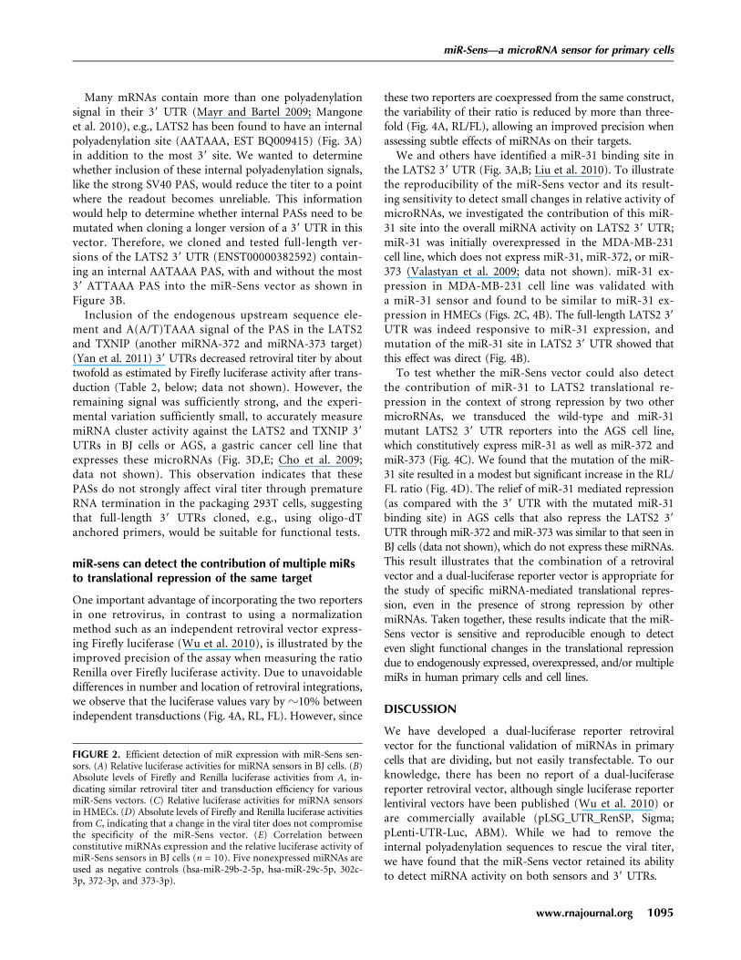
Indeed, Firefly luciferase activity was similar betweenconstructs containing sensors for miR-31, miR-155 (bothdetectable in primary BJ cells by microarray), and miR-372and miR-373 (both undetectable), indicating comparabletiters and transduction efficiencies for the generated ret-roviral sensors (Fig. 2B,D). In contrast, Renilla luciferaseactivity declined according to the presence of the corre-sponding miRNAs in the primary cells (Fig. 2A). Furthercorroboration that the miR-Sens vector could functionallydetect miRNAs in primary cells was obtained by trans-ducing these retroviral sensors in primary human epithe-lial cells (HMECs) (Fig. 2C), which express miR-31, butnot miR-373 or miR-155 (Ma et al. 2007; Valastyan et al.2009). The relative reduction in RL activity due to the pres-ence of a microRNA is even apparent when using a virus batchwith a lower titer (e.g., miR-Sens 31-5p and 373-3) (Fig. 2D)or after dilution of the virus (data not shown).
FIGURE 1. miR-Sens vector construction and validation. (A) Structure of the miR-Sens vector and sequence of the multiple cloning site (MCS).The MSCV packaging sequence between the 59 LTR and the RL is not shown on this graph. The vectors are not to scale. (B) Flowchart for thevalidation of the miR-Sens vector. Three independent 293/T transfections with the three vectors (Vec_A, Vec_B, and miR-Sens) were done onthree different days. Supernatants were collected on the third day post-transfection, and flash frozen. On the same day, the 293/T cells wereharvested and equal amounts of cells were flash-frozen in liquid nitrogen. (C) Results of the 293/T cell transfection. The 293/T transfected cellpellets were tested in triplicate on the same day. (D) Results of the BJ cell transduction. BJ cells were transduced in triplicate on the same day withthe three independent viral supernatants collected from the three 293/T cell supernatants. BJ cells were assayed 3 d after transduction as describedin the Materials and Methods. Results correspond to the Mean 6 SEM. (LTR) Long Terminal Repeat; (RL) Renilla luciferase; (PAS) syntheticPolyAdenylation Sequence; (T1) Expected Renilla luciferase transcript; (T2) Expected Firefly luciferase transcript; (TK) Thymidine Kinasepromoter; (FL) Firefly luciferase; (SV40 PAS) Simian Virus 40 late PolyAdenylation Signal.
miR-Sens—a microRNA sensor for primary cells
www.rnajournal.org 1093

Interestingly, when testing sensors against severalmicroRNAs, we observed that the log-transformed relativeluciferase activity of the respective sensor and miRNA levelsas measured by microRNA array (n = 10 different miRNAs)correlated linearly in BJ cells (Fig. 2E). This illustrates thatthe sensor, when expressed from the integrated virus, is re-sponsive over a wide range of microRNA levels. These findingsindicate that the miR-Sens sensors are suitable for quan-titative measurement of miRNA expression in primary cells.
Elongation of the reporter 39 UTR does notcompromise the detection of miRNA activityon 39 UTRs in BJ cells
Unlike Ago2-mediated cleavage of completely complemen-tary targets, microRNA targeted RISC represses translationand stability of mRNAs containing partially complemen-tary target sequences through a variety of mechanisms (Wuand Belasco 2008) that can be influenced by the presence ofRNA-binding proteins (van Kouwenhove et al. 2011) andRNA structural features such as 39 UTR length (Lewis et al.2003; Filipowicz et al. 2008; Mayr and Bartel 2009). Ef-ficient retroviral production requires the deletion of thestrong PAS that separates the reporter and normalizationtranscripts in the plasmid-based dual-luciferase system,resulting in a significant and constitutive extension of the39 UTR of the Renilla firefly encoding transcript (Fig. 1A).Thus, while the internal polyadenylation sequences of themiR-Sens are dispensable for Ago2-mediated cleavage ofmiRNA sensors, an extended heterologous 39 UTR down-stream from the 39 UTR of interest could possibly interferewith efficient miRNA activity.
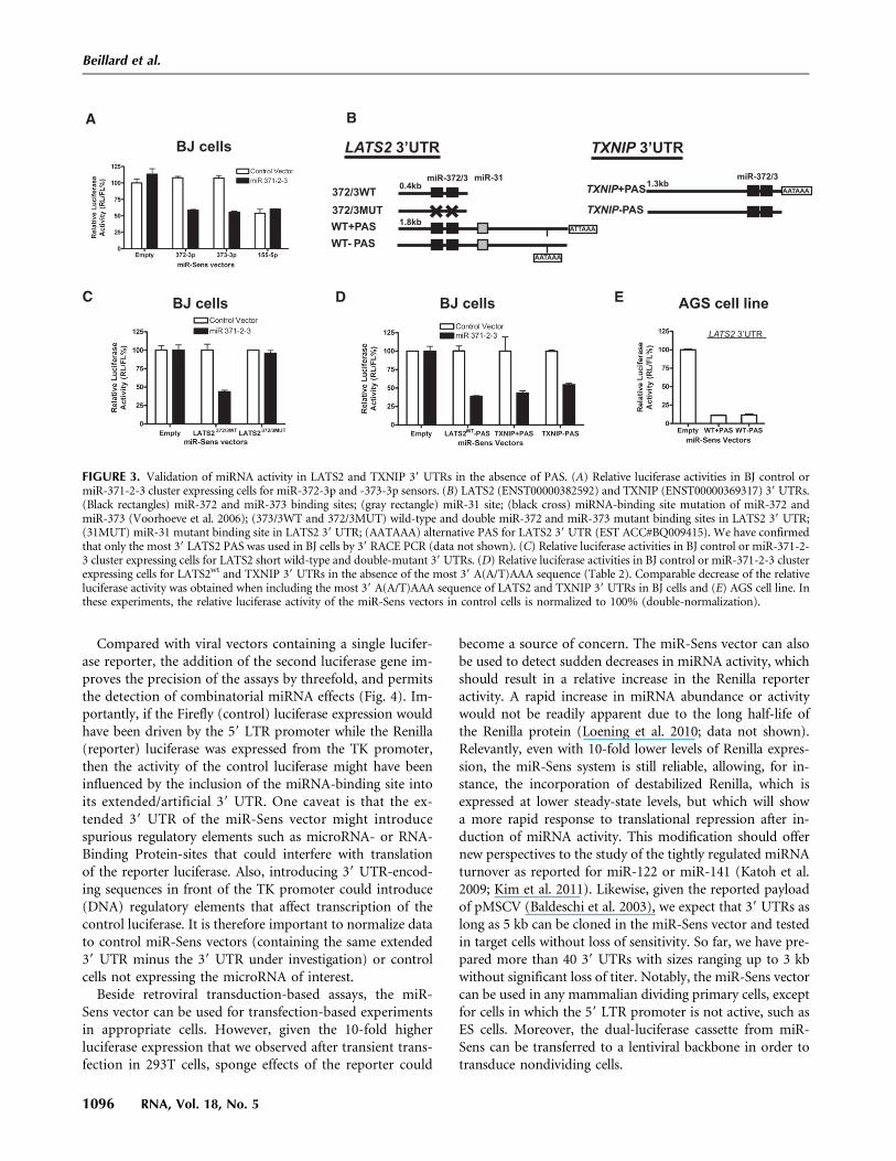
To be able to measure microRNA activity on 39 UTRs ofinterest and verify target predictions in primary cells, wehad to first address this possibility. We therefore preparedmiR-Sens constructs, in which we introduced behind thereporter luciferase the 39 UTR of LATS2, a known miR-372and miR-373 target whose repression by these microRNAsin primary BJ cells has been shown to contribute to theoncogenic transformation of these cells (Fig. 3A; Voorhoeveet al. 2006).
Primary BJ cells, transduced with miR-Vec 371-2-3, whichexpresses the oncogenic microRNAs 371, 372, and 373 undera strong promoter, were validated to express miR-372 andmiR-373 using a mir-Sens vector with the relevant comple-mentary sensors (Fig. 3A). miR-Sens vectors containing partof the wild-type LATS2 39 UTR or a mutant in which thetwo miR-372 and miR-373 binding sites have been inactivated(Voorhoeve et al. 2006) were then introduced (Fig. 3B). In-deed, the miRNA cluster overexpression decreased the rela-tive luciferase activity, which was dependent on miR-372 andmiR-373 binding sites in the 39 UTR (Fig. 3C), indicatingthat, despite the elongation of the reporter 39 UTR, miR-Sens can be used to detect miRNA activity on 39 UTRs ofrelevant target genes. FIGURE 2. (Legend on next page)
Beillard et al.
1094 RNA, Vol. 18, No. 5
Many mRNAs contain more than one polyadenylationsignal in their 39 UTR (Mayr and Bartel 2009; Mangoneet al. 2010), e.g., LATS2 has been found to have an internalpolyadenylation site (AATAAA, EST BQ009415) (Fig. 3A)in addition to the most 39 site. We wanted to determinewhether inclusion of these internal polyadenylation signals,like the strong SV40 PAS, would reduce the titer to a pointwhere the readout becomes unreliable. This informationwould help to determine whether internal PASs need to bemutated when cloning a longer version of a 39 UTR in thisvector. Therefore, we cloned and tested full-length ver-sions of the LATS2 39 UTR (ENST00000382592) contain-ing an internal AATAAA PAS, with and without the most39 ATTAAA PAS into the miR-Sens vector as shown inFigure 3B.
Inclusion of the endogenous upstream sequence ele-ment and A(A/T)TAAA signal of the PAS in the LATS2and TXNIP (another miRNA-372 and miRNA-373 target)(Yan et al. 2011) 39 UTRs decreased retroviral titer by abouttwofold as estimated by Firefly luciferase activity after trans-duction (Table 2, below; data not shown). However, theremaining signal was sufficiently strong, and the experi-mental variation sufficiently small, to accurately measuremiRNA cluster activity against the LATS2 and TXNIP 39
UTRs in BJ cells or AGS, a gastric cancer cell line thatexpresses these microRNAs (Fig. 3D,E; Cho et al. 2009;data not shown). This observation indicates that thesePASs do not strongly affect viral titer through prematureRNA termination in the packaging 293T cells, suggestingthat full-length 39 UTRs cloned, e.g., using oligo-dTanchored primers, would be suitable for functional tests.
miR-sens can detect the contribution of multiple miRsto translational repression of the same target
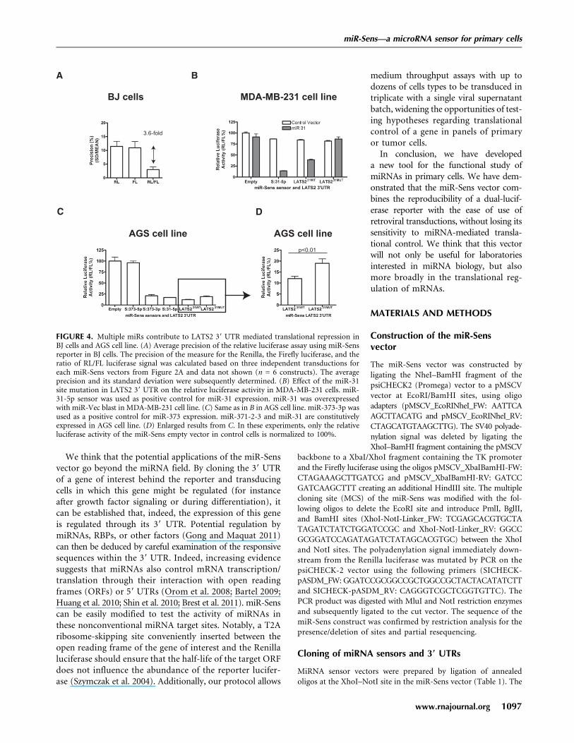
One important advantage of incorporating the two reportersin one retrovirus, in contrast to using a normalizationmethod such as an independent retroviral vector express-ing Firefly luciferase (Wu et al. 2010), is illustrated by theimproved precision of the assay when measuring the ratioRenilla over Firefly luciferase activity. Due to unavoidabledifferences in number and location of retroviral integrations,we observe that the luciferase values vary by z10% betweenindependent transductions (Fig. 4A, RL, FL). However, since
these two reporters are coexpressed from the same construct,the variability of their ratio is reduced by more than three-fold (Fig. 4A, RL/FL), allowing an improved precision whenassessing subtle effects of miRNAs on their targets.
We and others have identified a miR-31 binding site inthe LATS2 39 UTR (Fig. 3A,B; Liu et al. 2010). To illustratethe reproducibility of the miR-Sens vector and its result-ing sensitivity to detect small changes in relative activity ofmicroRNAs, we investigated the contribution of this miR-31 site into the overall miRNA activity on LATS2 39 UTR;miR-31 was initially overexpressed in the MDA-MB-231cell line, which does not express miR-31, miR-372, or miR-373 (Valastyan et al. 2009; data not shown). miR-31 ex-pression in MDA-MB-231 cell line was validated witha miR-31 sensor and found to be similar to miR-31 ex-pression in HMECs (Figs. 2C, 4B). The full-length LATS2 39
UTR was indeed responsive to miR-31 expression, andmutation of the miR-31 site in LATS2 39 UTR showed thatthis effect was direct (Fig. 4B).
To test whether the miR-Sens vector could also detectthe contribution of miR-31 to LATS2 translational re-pression in the context of strong repression by two othermicroRNAs, we transduced the wild-type and miR-31mutant LATS2 39 UTR reporters into the AGS cell line,which constitutively express miR-31 as well as miR-372 andmiR-373 (Fig. 4C). We found that the mutation of the miR-31 site resulted in a modest but significant increase in the RL/FL ratio (Fig. 4D). The relief of miR-31 mediated repression(as compared with the 39 UTR with the mutated miR-31binding site) in AGS cells that also repress the LATS2 39
UTR through miR-372 and miR-373 was similar to that seen inBJ cells (data not shown), which do not express these miRNAs.This result illustrates that the combination of a retroviralvector and a dual-luciferase reporter vector is appropriate forthe study of specific miRNA-mediated translational repres-sion, even in the presence of strong repression by othermiRNAs. Taken together, these results indicate that the miR-Sens vector is sensitive and reproducible enough to detecteven slight functional changes in the translational repressiondue to endogenously expressed, overexpressed, and/or multiplemiRs in human primary cells and cell lines.
DISCUSSION
We have developed a dual-luciferase reporter retroviralvector for the functional validation of miRNAs in primarycells that are dividing, but not easily transfectable. To ourknowledge, there has been no report of a dual-luciferasereporter retroviral vector, although single luciferase reporterlentiviral vectors have been published (Wu et al. 2010) orare commercially available (pLSG_UTR_RenSP, Sigma;pLenti-UTR-Luc, ABM). While we had to remove theinternal polyadenylation sequences to rescue the viral titer,we have found that the miR-Sens vector retained its abilityto detect miRNA activity on both sensors and 39 UTRs.
FIGURE 2. Efficient detection of miR expression with miR-Sens sen-sors. (A) Relative luciferase activities for miRNA sensors in BJ cells. (B)Absolute levels of Firefly and Renilla luciferase activities from A, in-dicating similar retroviral titer and transduction efficiency for variousmiR-Sens vectors. (C) Relative luciferase activities for miRNA sensorsin HMECs. (D) Absolute levels of Firefly and Renilla luciferase activitiesfrom C, indicating that a change in the viral titer does not compromisethe specificity of the miR-Sens vector. (E) Correlation betweenconstitutive miRNAs expression and the relative luciferase activity ofmiR-Sens sensors in BJ cells (n = 10). Five nonexpressed miRNAs areused as negative controls (hsa-miR-29b-2-5p, hsa-miR-29c-5p, 302c-3p, 372-3p, and 373-3p).
miR-Sens—a microRNA sensor for primary cells
www.rnajournal.org 1095
Compared with viral vectors containing a single lucifer-ase reporter, the addition of the second luciferase gene im-proves the precision of the assays by threefold, and permitsthe detection of combinatorial miRNA effects (Fig. 4). Im-portantly, if the Firefly (control) luciferase expression wouldhave been driven by the 59 LTR promoter while the Renilla(reporter) luciferase was expressed from the TK promoter,then the activity of the control luciferase might have beeninfluenced by the inclusion of the miRNA-binding site intoits extended/artificial 39 UTR. One caveat is that the ex-tended 39 UTR of the miR-Sens vector might introducespurious regulatory elements such as microRNA- or RNA-Binding Protein-sites that could interfere with translationof the reporter luciferase. Also, introducing 39 UTR-encod-ing sequences in front of the TK promoter could introduce(DNA) regulatory elements that affect transcription of thecontrol luciferase. It is therefore important to normalize datato control miR-Sens vectors (containing the same extended39 UTR minus the 39 UTR under investigation) or controlcells not expressing the microRNA of interest.
Beside retroviral transduction-based assays, the miR-Sens vector can be used for transfection-based experimentsin appropriate cells. However, given the 10-fold higherluciferase expression that we observed after transient trans-fection in 293T cells, sponge effects of the reporter could
become a source of concern. The miR-Sens vector can alsobe used to detect sudden decreases in miRNA activity, whichshould result in a relative increase in the Renilla reporteractivity. A rapid increase in miRNA abundance or activitywould not be readily apparent due to the long half-life ofthe Renilla protein (Loening et al. 2010; data not shown).Relevantly, even with 10-fold lower levels of Renilla expres-sion, the miR-Sens system is still reliable, allowing, for in-stance, the incorporation of destabilized Renilla, which isexpressed at lower steady-state levels, but which will showa more rapid response to translational repression after in-duction of miRNA activity. This modification should offernew perspectives to the study of the tightly regulated miRNAturnover as reported for miR-122 or miR-141 (Katoh et al.2009; Kim et al. 2011). Likewise, given the reported payloadof pMSCV (Baldeschi et al. 2003), we expect that 39 UTRs aslong as 5 kb can be cloned in the miR-Sens vector and testedin target cells without loss of sensitivity. So far, we have pre-pared more than 40 39 UTRs with sizes ranging up to 3 kbwithout significant loss of titer. Notably, the miR-Sens vectorcan be used in any mammalian dividing primary cells, exceptfor cells in which the 59 LTR promoter is not active, such asES cells. Moreover, the dual-luciferase cassette from miR-Sens can be transferred to a lentiviral backbone in order totransduce nondividing cells.
FIGURE 3. Validation of miRNA activity in LATS2 and TXNIP 39 UTRs in the absence of PAS. (A) Relative luciferase activities in BJ control ormiR-371-2-3 cluster expressing cells for miR-372-3p and -373-3p sensors. (B) LATS2 (ENST00000382592) and TXNIP (ENST00000369317) 39 UTRs.(Black rectangles) miR-372 and miR-373 binding sites; (gray rectangle) miR-31 site; (black cross) miRNA-binding site mutation of miR-372 andmiR-373 (Voorhoeve et al. 2006); (373/3WT and 372/3MUT) wild-type and double miR-372 and miR-373 mutant binding sites in LATS2 39 UTR;(31MUT) miR-31 mutant binding site in LATS2 39 UTR; (AATAAA) alternative PAS for LATS2 39 UTR (EST ACC#BQ009415). We have confirmedthat only the most 39 LATS2 PAS was used in BJ cells by 39 RACE PCR (data not shown). (C) Relative luciferase activities in BJ control or miR-371-2-3 cluster expressing cells for LATS2 short wild-type and double-mutant 39 UTRs. (D) Relative luciferase activities in BJ control or miR-371-2-3 clusterexpressing cells for LATS2wt and TXNIP 39 UTRs in the absence of the most 39 A(A/T)AAA sequence (Table 2). Comparable decrease of the relativeluciferase activity was obtained when including the most 39 A(A/T)AAA sequence of LATS2 and TXNIP 39 UTRs in BJ cells and (E) AGS cell line. Inthese experiments, the relative luciferase activity of the miR-Sens vectors in control cells is normalized to 100% (double-normalization).
Beillard et al.
1096 RNA, Vol. 18, No. 5
We think that the potential applications of the miR-Sensvector go beyond the miRNA field. By cloning the 39 UTRof a gene of interest behind the reporter and transducingcells in which this gene might be regulated (for instanceafter growth factor signaling or during differentiation), itcan be established that, indeed, the expression of this geneis regulated through its 39 UTR. Potential regulation bymiRNAs, RBPs, or other factors (Gong and Maquat 2011)can then be deduced by careful examination of the responsivesequences within the 39 UTR. Indeed, increasing evidencesuggests that miRNAs also control mRNA transcription/translation through their interaction with open readingframes (ORFs) or 59 UTRs (Orom et al. 2008; Bartel 2009;Huang et al. 2010; Shin et al. 2010; Brest et al. 2011). miR-Senscan be easily modified to test the activity of miRNAs inthese nonconventional miRNA target sites. Notably, a T2Aribosome-skipping site conveniently inserted between theopen reading frame of the gene of interest and the Renillaluciferase should ensure that the half-life of the target ORFdoes not influence the abundance of the reporter lucifer-ase (Szymczak et al. 2004). Additionally, our protocol allows
medium throughput assays with up todozens of cells types to be transduced intriplicate with a single viral supernatantbatch, widening the opportunities of test-ing hypotheses regarding translationalcontrol of a gene in panels of primaryor tumor cells.
In conclusion, we have developeda new tool for the functional study ofmiRNAs in primary cells. We have dem-onstrated that the miR-Sens vector com-bines the reproducibility of a dual-lucif-erase reporter with the ease of use ofretroviral transductions, without losing itssensitivity to miRNA-mediated transla-tional control. We think that this vectorwill not only be useful for laboratoriesinterested in miRNA biology, but alsomore broadly in the translational reg-ulation of mRNAs.
MATERIALS AND METHODS
Construction of the miR-Sensvector
The miR-Sens vector was constructed byligating the NheI–BamHI fragment of thepsiCHECK2 (Promega) vector to a pMSCVvector at EcoRI/BamHI sites, using oligoadapters (pMSCV_EcoRINheI_FW: AATTCAAGCTTACATG and pMSCV_EcoRINheI_RV:CTAGCATGTAAGCTTG). The SV40 polyade-nylation signal was deleted by ligating theXhoI–BamHI fragment containing the pMSCV
backbone to a XbaI/XhoI fragment containing the TK promoterand the Firefly luciferase using the oligos pMSCV_XbaIBamHI-FW:CTAGAAAGCTTGATCG and pMSCV_XbaIBamHI-RV: GATCCGATCAAGCTTT creating an additional HindIII site. The multiplecloning site (MCS) of the miR-Sens was modified with the fol-lowing oligos to delete the EcoRI site and introduce PmlI, BglII,and BamHI sites (XhoI-NotI-Linker_FW: TCGAGCACGTGCTATAGATCTATCTGGATCCGC and XhoI-NotI-Linker_RV: GGCCGCGGATCCAGATAGATCTATAGCACGTGC) between the XhoIand NotI sites. The polyadenylation signal immediately down-stream from the Renilla luciferase was mutated by PCR on thepsiCHECK-2 vector using the following primers (SICHECK-pASDM_FW: GGATCCGCGGCCGCTGGCCGCTACTACATATCTTand SICHECK-pASDM_RV: CAGGGTCGCTCGGTGTTC). ThePCR product was digested with MluI and NotI restriction enzymesand subsequently ligated to the cut vector. The sequence of themiR-Sens construct was confirmed by restriction analysis for thepresence/deletion of sites and partial resequencing.
Cloning of miRNA sensors and 39 UTRs
MiRNA sensor vectors were prepared by ligation of annealedoligos at the XhoI–NotI site in the miR-Sens vector (Table 1). The
FIGURE 4. Multiple miRs contribute to LATS2 39 UTR mediated translational repression inBJ cells and AGS cell line. (A) Average precision of the relative luciferase assay using miR-Sensreporter in BJ cells. The precision of the measure for the Renilla, the Firefly luciferase, and theratio of RL/FL luciferase signal was calculated based on three independent transductions foreach miR-Sens vectors from Figure 2A and data not shown (n = 6 constructs). The averageprecision and its standard deviation were subsequently determined. (B) Effect of the miR-31site mutation in LATS2 39 UTR on the relative luciferase activity in MDA-MB-231 cells. miR-31-5p sensor was used as positive control for miR-31 expression. miR-31 was overexpressedwith miR-Vec blast in MDA-MB-231 cell line. (C) Same as in B in AGS cell line. miR-373-3p wasused as a positive control for miR-373 expression. miR-371-2-3 and miR-31 are constitutivelyexpressed in AGS cell line. (D) Enlarged results from C. In these experiments, only the relativeluciferase activity of the miR-Sens empty vector in control cells is normalized to 100%.
miR-Sens—a microRNA sensor for primary cells
www.rnajournal.org 1097

39 UTRs were PCR amplified from human BJ cell genomic DNAwith the primers from Table 2 and cloned at the same site. miR-31site mutagenesis was done by PCR on the wild-type LATS2 39 UTRusing primers from Table 2. PCR amplification was carried out withiProof enzyme (Bio-Rad). The wild-type and mutant short LATS239 UTRs have been reported previously (Voorhoeve et al. 2006).Constructs identity and orientation were confirmed by sequencing.
miR-expressing vectors
The miR-Vec Blast retroviral vector was used for stable expressionof miRNAs after Blasticidin S selection (Invitrogen) of transducedcells (Voorhoeve et al. 2006). MiR-31 was cloned from BJ cellgenomic DNA with the following primers (MIR31S_FW: TAGATCGGATCCCATCTTCAAAAGCGGACACTCTA and MIR31S_RV:GATCGAATTCACAATACATAGCAGGACAGGAAGTAAG) be-tween the BamHI and EcoRI sites. The miRNA-371, miRNA-372,and miRNA-373 (subsequently referred to as miRNA 371-2-3cluster) expression vector has been reported before (Voorhoeveet al. 2006). The identity and orientation of the PCR productswere confirmed by sequencing.
Retroviral production and cell transduction
Retroviruses were prepared by transfection of 293/T cells withDNA precipitated using Calcium Phospate according to standardprocedures. Retroviruses were pseudotyped with either an eco-tropic (pCL-Eco) or amphotropic (pCL-Ampho, for miR-Sensonly) envelope. miR-Sens vector plasmid DNA was cotransfectedalong with a packaging plasmid and a pmCherry-C1 vector tomonitor transfection efficiency under a fluorescence microscope,the efficiency routinely exceeding 50%. Retroviral supernatantswere harvested at 48 and 72 h after transfection, pooled on ice,aliquoted and flash-frozen with liquid nitrogen. Cell transduction(20–50,000 cells) was carried out with 150 mL of viral supernantand 350 mL of media in the presence of eight microg/mL ofpolybrene (Sigma) in triplicate in a 24-well plate overnight. The
media was replaced the next morning with 0.5 mL of fresh media.Cells were assayed for luciferase activities after 72 h.
Cell lines
The primary BJ cells (human foreskin fibroblast cells) expressingRasV12ERTAM, GFP-st, and the murine ecotropic receptor, and theAGS cell line (human gastric cancer cell line) have been previouslyreported (Voorhoeve et al. 2006; Oh et al. 2011). The MDA-MB-231-eco cell line (human breast cancer cell line, kindly providedby Dr. D.M. Virshup ½Duke-NUS, Singapore�) and HMECs (hu-man mammary epithelial cells, purchased from ATCC) were engi-neered to express the murine ecotropic receptor. HMECs weresubsequently transduced with a retroviral vector expressing hTertas described previously (Voorhoeve et al. 2006). The 293/T cellline was provided by Dr. B. Reversade (IMB, Singapore). Cellswere grown according to standard procedures. BJ and MDA-MB-231-eco cells were transduced with ecotropic miR-Vec blast retro-viruses expressing miR-371-2-3, miR-31 or the inactive Telome-rase RNA component (TERC RNA nt 1–211) as reported before,and selected with Blasticidin S (Invitrogen) for 4–7 d (Voorhoeveet al. 2006). miR-31 expression was confirmed by quantitativePCR in MDA-MB-231 cells using EXIQON miR-31 assay with U6RNA as a control.
miRNA microarray data
Total BJ cell RNA from three independent cultures was isolated bycombining TRizol with a modified protocol from Invitrogen RNApureLink kit to allow for the extraction of RNAs smaller than 200nt. The quality of the total RNA was verified by an Agilent 2100Bioanalyzer profile. A total of 1000 ng of total RNA from BJ cellsand reference samples were labeled with Hy3 and Hy5 fluorescentlabel, respectively, using the miRCURY LNA Array power labelingkit (Exiqon) following the manufacturer’s instructions. The Hy3-labeled sample and a Hy5-labeled reference RNA sample weremixed pairwise and hybridized to the miRCURY LNA Array ver-sion fifth Generation (Exiqon), which contains capture probestargeting all registered human miRNAs from miRBASE version15.0. The hybridization was performed according to the miRCURYLNA array manual using a Tecan HS4800 hybridization station(Tecan). The miRCURY LNA array microarray slides were scannedusing the Agilent G2565BA Microarray Scanner System (AgilentTechnologies, Inc.) and the image analysis was carried out usingthe ImaGene 9.0 software (BioDiscovery Inc.). The raw Hy3 fluo-
TABLE 1. Oligos for the human miR sensors
TABLE 2. Oligos for the cloning of human 39 UTRs
(Underlined) NotI cloning site; (Bold) A(A/T)TAAA sequence of themost 39 polyadenylation signal.
Beillard et al.
1098 RNA, Vol. 18, No. 5

rescence intensities were used for the linear regression withoutbackground substraction.
Luciferase assays
The Dual-Luciferase Reporter kit (Promega) was used for thedetection of the luciferase activities according to the manufacturer’sprotocol. Cells were lysed briefly in 24-well plates for 20 min atroom temperature with 100 mL of 1X PLB buffer. Subsequently,10 mL of the lysate was tested with 50 mL of each reagent in a black96-well plate (NUNC). The luciferase activities were detected witha Tecan Infinite M200 microplate reader. The results (mean 6 SD)from three independent cell transductions are shown and ex-periments were repeated at least twice with similar results unlessotherwise stated. The threshold for a significant change in therelative Renilla over Firefly luciferase activity compared with con-trol vector was calculated based on the standard deviation of theratio from two independent transductions in triplicate in BJ andAGS cells (four experiments). Values outside of the mean 6 3SDrange are considered significantly changed from the control.
Statistics
The statistical analysis was done using Graphpad Prism V4(Graphpad). We have used the unpaired t-test or the one-wayANOVA test where appropriate for statistical comparison ofresults. The linearity was tested by linear regression. Precisionof a series of replicates was calculated as follows: Precision(%) =SD/mean.
ACKNOWLEDGMENTS
We acknowledge Dr. P. Tan, Dr. D.M. Virshup, and Dr. M. Fivaz(Duke-NUS), Dr. B. Reversade (IMB, Singapore) and Dr. J. Kluiver(UMCG, The Netherlands) for sharing reagents. We thank TedChang for critically reviewing of this manuscript. This work wassupported by the Association for International Cancer Research(Grant no. 09-0126) and Duke-NUS Graduate Medical School.
Received December 9, 2011; accepted February 1, 2012.
REFERENCES
Baldeschi C, Gache Y, Rattenholl A, Bouille P, Danos O, Ortonne J-P,Bruckner-Tuderman L, Meneguzzi G. 2003. Genetic correction ofcanine dystrophic epidermolysis bullosa mediated by retroviralvectors. Hum Mol Genet 12: 1897–1905.
Bartel DP. 2009. MicroRNAs: target recognition and regulatory func-tions. Cell 136: 215–233.
Blø M, Micklem DR, Lorens JB. 2008. Enhanced gene expression fromretroviral vectors. BMC Biotechnol 8: 19. doi: 10.1186/1472-6750-8-10.
Brest P, Lapaquette P, Souidi M, Lebrigand K, Cesaro A, Vouret-Craviari V, Mari B, Barbry P, Mosnier J-F, Hebuterne X, et al.2011. A synonymous variant in IRGM alters a binding site formiR-196 and causes deregulation of IRGM-dependent xenophagyin Crohn’s disease. Nat Genet 43: 242–245.
Cho WJ, Shin JM, Kim JS, Lee MR, Hong KS, Lee J-H, Koo KH, ParkJW, Kim K-S. 2009. miR-372 regulates cell cycle and apoptosis ofags human gastric cancer cell line through direct regulation ofLATS2. Mol Cells 28: 521–527.
Filipowicz W, Bhattacharyya SN, Sonenberg N. 2008. Mechanisms ofpost-transcriptional regulation by microRNAs: are the answers insight? Nat Rev Genet 9: 102–114.
Gong C, Maquat LE. 2011. lncRNAs transactivate STAU1-mediatedmRNA decay by duplexing with 39 UTRs via Alu elements. Nature470: 284–288.
Huang S, Wu S, Ding J, Lin J, Wei L, Gu J, He X. 2010. MicroRNA-181a modulates gene expression of zinc finger family members bydirectly targeting their coding regions. Nucleic Acids Res 38: 7211–7218.
Jafarifar F, Yao P, Eswarappa SM, Fox PL. 2011. Repression of VEGFAby CA-rich element-binding microRNAs is modulated by hnRNPL. EMBO J 30: 1324–1334.
Jern P, Coffin JM. 2008. Effects of retroviruses on host genomefunction. Annu Rev Genet 42: 709–732.
Katoh T, Sakaguchi Y, Miyauchi K, Suzuki T, Kashiwabara S-I, Baba T,Suzuki T. 2009. Selective stabilization of mammalian microRNAs by39 adenylation mediated by the cytoplasmic poly(A) polymeraseGLD-2. Genes Dev 23: 433–438.
Kedde M, Strasser MJ, Boldajipour B, Oude Vrielink JAF, Slanchev K,le Sage C, Nagel R, Voorhoeve M, van Duijse J, Ørom UA, et al.2007. RNA-binding protein Dnd1 inhibits microRNA access totarget mRNA. Cell 131: 1273–1286.
Kedde M, van Kouwenhove M, Zwart W, Oude Vrielink JAF, Elkon R,Agami R. 2010. A Pumilio-induced RNA structure switch in p27-39UTR controls miR-221 and miR-222 accessibility. Nat Cell Biol 12:1014–1020.
Kim Y-K, Yeo J, Ha M, Kim B, Kim VN. 2011. Cell adhesion-dependent control of microRNA decay. Mol Cell 43: 1005–1014.
Lewis BP, Shih I, Jones-Rhoades M, Bartel DP, Burge CB. 2003.Prediction of mammalian microRNA targets. Cell 115: 787–798.
Liu X, Sempere LF, Ouyang H, Memoli VA, Andrew AS, Luo Y,Demidenko E, Korc M, Shi W, Preis M, et al. 2010. MicroRNA-31functions as an oncogenic microRNA in mouse and human lungcancer cells by repressing specific tumor suppressors. J Clin Invest120: 1298–1309.
Loening AM, Dragulescu-Andrasi A, Gambhir SS. 2010. A red-shiftedRenilla luciferase for transient reporter-gene expression. Nat Methods7: 5–6.
Ma L, Teruya-Feldstein J, Weinberg RA. 2007. Tumour invasion andmetastasis initiated by microRNA-10b in breast cancer. Nature449: 682–688.
Mangone M, Manoharan AP, Thierry-Mieg D, Thierry-Mieg J, Han T,Mackowiak SD, Mis E, Zegar C, Gutwein MR, Khivansara V, et al.2010. The landscape of C. elegans 39UTRs. Science 329: 432–435.
Mayr C, Bartel DP. 2009. Widespread shortening of 39UTRs byalternative cleavage and polyadenylation activates oncogenes incancer cells. Cell 138: 673–684.
Maziere P, Enright AJ. 2007. Prediction of microRNA targets. DrugDiscov Today 12: 452–458.
Oh H-K, Tan AL-K, Das K, Ooi CH, Deng N-T, Tan IB, Beillard E, Lee J,Ramnarayanan K, Rha SY, et al. 2011. Genomic loss of miR-486regulates tumor progression and the OLFM4 antiapoptotic factor ingastric cancer. Clin Cancer Res 17: 2657–2667.
Orom UA, Nielsen FC, Lund AH. 2008. MicroRNA-10a binds the59UTR of ribosomal protein mRNAs and enhances their trans-lation. Mol Cell 30: 460–471.
Proudfoot NJ. 2011. Ending the message: Poly(A) signals then andnow. Genes Dev 25: 1770–1782.
Retelska D, Iseli C, Bucher P, Jongeneel CV, Naef F. 2006. Similaritiesand differences of polyadenylation signals in human and fly. BMCGenomics 7: 176. doi: 10.1186/1471-2164-7-176.
Salmena L, Poliseno L, Tay Y, Kats L, Pandolfi PP. 2011. A ceRNAhypothesis: The Rosetta Stone of a hidden RNA language? Cell146: 353–358.
Shin C, Nam J-W, Farh KK-H, Chiang HR, Shkumatava A, Bartel DP.2010. Expanding the microRNA targeting code: functional siteswith centered pairing. Mol Cell 38: 789–802.
Szymczak AL, Workman CJ, Wang Y, Vignali KM, Dilioglou S, VaninEF, Vignali DAA. 2004. Correction of multi-gene deficiency invivo using a single ‘‘self-cleaving’’ 2A peptide-based retroviralvector. Nat Biotechnol 22: 589–594.
miR-Sens—a microRNA sensor for primary cells
www.rnajournal.org 1099

Thomas M, Lieberman J, Lal A. 2010. Desperately seeking microRNAtargets. Nat Struct Mol Biol 17: 1169–1174.
Valastyan S, Reinhardt F, Benaich N, Wang ZC, Richardson AL,Weinberg RA. 2009. A pleiotropically acting MicroRNA, miR-31,inhibits breast cancer metastasis. Cell 137: 1032–1046.
van Kouwenhove M, Kedde M, Agami R. 2011. MicroRNA regulationby RNA-binding proteins and its implications for cancer. Nat RevCancer 11: 644–656.
Voorhoeve PM. 2010. MicroRNAs: Oncogenes, tumor suppressors ormaster regulators of cancer heterogeneity? Biochim Biophys Acta1805: 72–86.
Voorhoeve PM, le Sage C, Schrier M, Gillis AJM, Stoop H, Nagel R,Liu Y-P, van Duijse J, Drost J, Kulalert W, et al. 2006. A geneticscreen implicates miRNA-372 and miRNA-373 as oncogenes intesticular germ cell tumors. Cell 124: 1169–1181.
Wu L, Belasco JG. 2008. Let me count the ways: Mechanisms of generegulation by miRNAs and siRNAs. Mol Cell 29: 1–7.
Wu S, Huang S, Ding J, Zhao Y, Liang L, Liu T, Zhan R, He X. 2010.Multiple microRNAs modulate p21Cip1/Waf1 expression by di-rectly targeting its 39 untranslated region. Oncogene 29: 7211–7218.
Yan G-R, Xu S-H, Tan Z-L, Liu L, He Q-Y. 2011. Global identificationof miR-373-regulated genes in breast cancer by quantitativeproteomics. Proteomics 11: 912–920.
Yang S, Delgado R, King SR, Woffendin C, Barker CS, Yang ZY, Xu L,Nolan GP, Nabel GJ. 1999. Generation of retroviral vector for clinicalstudies using transient transfection. Hum Gene Ther 10: 123–132.
Zhang Y, Verbeek FJ. 2010. Comparison and integration of targetprediction algorithms for microRNA studies. J Integr Bioinform 7:127. doi: 10.2390/biecoll-jib-2010-127.
Beillard et al.
1100 RNA, Vol. 18, No. 5