Route of Entry-Dependent Blocks to Retroviral Replication
235
Route of Entry-Dependent Blocks to Retroviral Replication A thesis submitted to University College London in part fulfilment o f the requirements for the degree of Doctor of Philosophy July 2008 Eleanor Ruth Gray Division of Virology National Institute for Medical Research The Ridgeway M ill Hill London NW7 1AA
-
Upload
khangminh22 -
Category
Documents
-
view
0 -
download
0
Transcript of Route of Entry-Dependent Blocks to Retroviral Replication

Route of Entry-Dependent Blocks
to Retroviral Replication
A thesis submitted to University College London in part fulfilment o f the requirements for the degree o f Doctor o f Philosophy
July 2008
Eleanor Ruth Gray
Division o f Virology
National Institute for Medical Research
The Ridgeway
M ill H ill
London
NW7 1AA

UMI Number: U591600
All rights reserved
INFORMATION TO ALL USERS The quality of this reproduction is dependent upon the quality of the copy submitted.
In the unlikely event that the author did not send a complete manuscript and there are missing pages, these will be noted. Also, if material had to be removed,
a note will indicate the deletion.
Dissertation Publishing
UMI U591600Published by ProQuest LLC 2013. Copyright in the Dissertation held by the Author.
Microform Edition © ProQuest LLC.All rights reserved. This work is protected against
unauthorized copying under Title 17, United States Code.
ProQuest LLC 789 East Eisenhower Parkway
P.O. Box 1346 Ann Arbor, Ml 48106-1346

I, Eleanor Ruth Gray, confirm that the work presented in this thesis is my own.
Where information has been derived from other sources, I confirm that this has been
indicated in the thesis.

Abstract
Restriction factors are endogenous cellular proteins that block retroviral replication at
specific points in the life cycle. Those identified so far include F v l, Trim5a and
TrimCyp. Their characterisation has extended knowledge o f retroviral and cellular
functions, and has added a new branch to innate immunity.
Retroviral susceptibility to Fvl and Trim5a is determined by its capsid, and is
manifested in a pre- (Trim5a, TrimCyp) or post- (Fv l) reverse transcription block to
replication. Other blocks to replication have been postulated. For example, a novel
anti-viral factor, Lv2, is thought to block replication o f several primary isolates o f
HIV-2 in some cell lines.
Knowledge o f the early steps o f virus replication, between entry and nuclear import,
is critical to understanding restriction. The intention o f the studies described in this
thesis was to determine whether alternative routes o f virus trafficking might affect
susceptibility to Fvl and Trim5a, as well as to the putative Lv2. A system o f two
receptors was used, Tva800 and Tva950; both permit entry via ASLV envelope
protein, but take the virus into the cell by two different endocytic mechanisms.
The pathways traversed after binding to Tva800 and Tva950 were investigated and
shown not to reroute virions around restriction mediated by Fvl and Trim5a. When
virus titration curves were analysed, a distinctive pattern emerged suggesting that
entry via Tva800, but not Tva950, requires engagement o f more than one receptor-
envelope pair.
The block to replication caused by the putative factor Lv2 was also analysed. It was
concluded that a combination o f low surface CD4 expression and poor receptor
engagement are the cause o f low viral titres in some cell lines, rather than a cellular
anti-viral factor per se.

Acknowledgements
I owe so much to the support o f so many. Firstly to Jonathan, for so much patience,
for putting up with me and for encouraging excellent science. A huge debt o f
gratitude is also owing to the rest o f the Stoye lab, past and present, to Melvyn Yap,
Mark Dodding, Sada Okhura, Rebecca Butcher, Seti Grambas and Laura Hilditch, for
teaching me so much about being a good scientist, for being a pleasure to work with,
and also for some cracking recipes. Outside o f the lab and inside o f science I am also
grateful to M olly Strom, Barry Ely, Steve Wharton, and Srividya Sriskantharajah for
useful advice on vectors, immunology, and random odd chemicals.
Outside o f science the whole thesis process was made so much easier by the Lisbume
Ladies who never moved my papers around the lounge, so thanks to Anna-Lea Cooke
and Kate Davenport, and I am also particularly grateful to Caroline Goringe who
made the day to day process o f writing up pass far more enjoy ably than it really
should have done. After it was all done, Anna Hughes was extremely tactful and
utterly correct in her proof-reading. And how could I forget any o f my friends who
have listened to me talk about my work over the last few years without having a clue
what I was going on about - 1 honestly really appreciated it!
The support o f my family has been so important to me. Thanks to Morag and Serge
for being so encouraging and for the beautiful picture - this is for you (Simpson et al.
2006); I told you I ’d manage it somewhere! Thank you to Julia and Hazel, and to
Cameron and Margaret. And thanks are finally and most importantly due to my
parents, for rock-solid faith in me, and for being themselves so inspirational.


I am among those who think that science has great beauty. A scientist in his laboratory is not only a technician: he is also a child placed before natural
phenomena which impress him like a fairy tale.Marie Curie
May the words of my mouth and the meditation of my heart be pleasing in your sight. Ps 19vl4

Table of Contents
Abstract............................................................................................................................... 3Acknowledgements........................................................................................................... 4Table o f Contents................................................................................................................7Index o f Figures and Tables............................................................................................ 11Abbreviations.................................................................................................................... 14Introduction...................................................................................................................... 16
1.1 Virus Taxonomy...............................................................................................171.2 Genome Organisation...................................................................................... 181.3 Structure............................................................................................................ 201.4 Proteins.............................................................................................................21
1.4.1 PR .............................................................................................................211.4.2 R T .............................................................................................................221.4.3 IN ..............................................................................................................221.4.4 M A ............................................................................................................231.4.5 NC .............................................................................................................231.4.6 C A .............................................................................................................241.4.7 p6 ..............................................................................................................241.4.8 Env............................................................................................................251.4.9 Accessory proteins................................................................................... 26
1.5 Virus Infectivity Cycle.................................................................................... 271.5.1 B inding.....................................................................................................291.5.2 Fusion and Entry...................................................................................... 301.5.3 Reverse Transcription..............................................................................321.5.4 Nuclear Import......................................................................................... 351.5.5 Integration.................................................................................................361.5.6 Assembly and Exit...................................................................................38
1. 6 Vectors and Viral Pseudotypes........................................................................391.7 Entry o f A S LV -A .............................................................................................40
1.7.1 Tva800 and Tva950.................................................................................401.7.2 A S LV E nv ............................................................................................... 421.7.3 Mechanism o f entry via ASLV-A Env...................................................43
1. 8 Inhibition o f viral replication...........................................................................461.8.1 Fv Susceptibility Genes...........................................................................461.8.2 Trim 5a......................................................................................................471.8.3 APOBEC3G............................................................................................. 491.8.4 Lv2............................................................................................................ 501.8.5 Other Blocks to Retroviral Infection......................................................51
1.9 Other factors influencing viral entry.............................................................. 521.9.1 Endocytosis...............................................................................................531.9.2 Rab proteins..............................................................................................551.9.3 Lipid Rafts................................................................................................57
1.10 Aims o f this thesis............................................................................................59Materials and Methods.................................................................................................... 61
2.1 Cells...................................................................................................................612.1.1 Cell Culture...............................................................................................612.1.2 Preparation o f Tva800 and Tva950-expressing cell lines.....................61
2.2 Analyses............................................................................................................ 62

2.2.1 Cell sorting............................................................................................... 622.2.2 Fluorescent Activated Cell Sorting (FACS)...........................................622.2.3 Microscopy............................................................................................... 64
2.3 Viruses..............................................................................................................642.3.1 Env, Gag-pol and Vector Components................................................... 642.3.2 Virus Preparation............................................................................................6 6
2.3.3 Determination o f virus in fectiv ity ...........................................................6 6
2.4 Assays...............................................................................................................672.4.1 Infectivity assay........................................................................................672.4.2 Abrogation Assay.....................................................................................672.4.3 S iR N A...................................................................................................... 6 8
2.4.4 NH 4 CI treatment o f cells.......................................................................... 6 8
2.4.5 Inhibition o f infection by SUA-rlgG or empty vector........................... 6 8
2.4.6 Immunofluorescence............................................................................... 692.5 Protein Analysis............................................................................................... 71
2.5.1 Polyacrylamide gels..................................................................................712.5.2 Antibodies used for Western b lo t............................................................71
2.6 DNA Purification and Manipulation............................................................... 732.6.1 Agarose DNA gels....................................................................................732.6.2 DNA purification from agarose gels.......................................................732.6.3 Quantification o f D N A .............................................................................732.6.4 PCR............................................................................................................732.6.5 Quantitative PCR...................................................................................... 742.6.6 PCR Cloning............................................................................................. 742.6.7 QuikChange PCR..................................................................................... 752.6.8 Cloning via the Gateway System............................................................. 762.6.9 DNA precipitation.................................................................................... 762.6.10 Transformation......................................................................................... 762.6.11 Selection o f colonies.................................................................................... 772.6.12 Preparations o f D N A ................................................................................... 772.6.13 DNA Sequencing..........................................................................................772.6.14 Preparation o f m RNA.................................................................................. 782.6.15 Primers.......................................................................................................... 782.6.16 Primer Usage................................................................................................ 80
2.7 Calculations......................................................................................................81Routes o f Entry Via Tva800 and Tva950....................................................................... 82
Results..........................................................................................................................853.1 Mus dunni cells expressing Tva800 or Tva950 support infection andreplication by NB-M LV cores pseudotyped with ASLV envelope......................853.2 Viruses bound to Tva800 receptors remain infectious for 6 hours i f entryis blocked..................................................................................................................8 6
3.3 Viral particles co-localise with markers for the late, but not the earlyendosomes................................................................................................................903.4 Inhibition o f Rab5, but not Rab7, decreases entry via Tva800 and Tva950
973.5 Route o f entry via Tva800 or Tva950 receptor does not affect whetherNB-, N- or B-tropic M LV is affected by Fvl or Trim 5a.................................... 1003.6 ASLV envelope pseudotyped HIV, N- and B-tropic M LV are notrestricted by a range o f Trim proteins after entering cells via Tva800............... 104
Discussion................................................................................................................... 106

ASLV-A Must Bind More Than One Tva800 for Entry............................................. 114Results........................................................................................................................ 115
4.1 Entry o f ASLV pseudotyped virus via two clones o f Tva800 receptorvector gives two different titration curves.......................................................... 1154.2 Increasing the level o f eyp800 vector increases the proportion o f viralentry 1184.3 Viral entry dependent on receptor availability can be modelled using aPoisson distribution............................................................................................... 1194.4 Protein levels o f receptor made from AGG start codon are undetectableby Western b lotting............................................................................................... 1224.5 Levels o f Tva800 labelled by fluorescence on the surface o f cells arevisibly lower...........................................................................................................1254.6 Viral binding and entry can be blocked....................................................1304.7 Low levels o f Tva950 do not affect viral entry in the same w ay...........136
Discussion...................................................................................................................139Characterising a Block to Infection in HeLa CD4 Cells............................................. 144
Results........................................................................................................................ 1495.1 Replication o f MCR Env pseudotyped HIV and N B-M LV in HeLa CD4cells is inhibited in HeLa CD4 cells but not in NP2* cells.................................1495.2 ASLV Env NB-M LV and HIV-1 replicates to high titres on HeLa CD4cells when entering via both Tva800 and Tva950.............................................. 1525.3 Titres o f other CD4/CXCR4-tropic viruses envelopes are alsosignificantly reduced in HeLa CD4 cells..............................................................1545.4 The block to infection in HeLa CD4 cells cannot be abrogated 1565.5 Production o f strong stop DNA is reduced in HeLa CD4 cells challengedwith MCR pseudotyped H IV ................................................................................ 1585.6 Introduction o f Trims 1,18 and 34 into NP2* cells does not create ablock to infection................................................................................................... 1615.7 Expression o f anti-Trim 1 SiRNA does not diminish the block to infectionin HeLa CD4 cells................................................................................................. 165
Discussion................................................................................................................... 167The Block to Infection in HeLa CD4 Cells is at Entry................................................ 170
Results........................................................................................................................ 1706.1 CD4 and CXCR4 are expressed on HeLa CD4 cells................................1706.2 Progress o f fusion o f viral and cellular membranes can be monitored witha fluorescent protein targeted to the viral membrane.......................................... 1726.3 Membranes o f H IV pseudotyped with MCR do not undergo fusion withcellular membranes in proportion with a productive infection........................... 1776.4 Inhibition o f endocytosis or acidification o f endosomes causes a smallincrease in replication o f MCR NB-M LV in HeLa CD4 cells............................1806.5 Blocking action o f Rab5 causes a modest increase in productive infectiono f MCR Env H IV in HeLa CD4 cells, but has no effect in NP2* ce lls .............1846 . 6 Expression o f p56lck renders HeLa CD4 cells permissive to MCRpseudotyped virions............................................................................................... 186
Discussion................................................................................................................... 191Thesis Discussion...........................................................................................................196
Rationale.................................................................................................................196Results.................................................................................................................... 197
Conclusion..................................................................................................................202Further Work 202

References......................................................................................................................204Appendix 1 .................................................................................................................... 230Appendix 2 .................................................................................................................... 231Appendix 3 .................................................................................................................... 234

Index of Figures and Tables
1.2 M LV and H IV -1 genomes 181.3 A schematic structure o f a typical retrovirus particle 201.5 A schematic o f the retroviral infectious cycle 281.5.3 Outline o f the stages o f reverse transcription 341.7.1 Simplified outline o f the Tva800 and Tva950 proteins 411.7.2 A representation o f the structure o f ASLV Env 421.7.3 Fusion between viral and cellular membranes, mediated by ASLV 45
Env binding to receptor1.8.4 A pictorial representation o f the basic premise behind restriction by 50
Lv21.9.2 The major points o f entry into the cell, and known controlling factors 57
2.2 A typical 2-colour FACS profile 632.3.1 Schematic representations o f vectors used in the production o f 65
virions2.4.1 Chemicals used to affect cellular processes which were assayed for 67
their effects on viral infectivity2.4.5 Inhibition o f infection by SUA-rlgG 692.4.6 Primary and secondary antibodies used for immunofluorescence 602.5.2 Primary and secondary antibodies used for Western blotting 722.6.6 Published sequences for primer design 752.6.15 Primers sequences 782.6.16 Primer usage 80
3.1 Mus dunni cells expressing Tva800 or Tva950 support infection and 8 6
replication by NB-M LV cores pseudotyped with ASLV envelope3.2.1 ASLV Env pseudotyped viruses are unable to succesfully infect cells 87
in the presence o f 40mM NH 4 CI3.2.2 The proportion o f virions that remain infectious under an NH 4 C1- 8 8
induced block to infection is not the same for d800 and d950 cells3.2.3 Proportion o f virions bound via VS V-G envelope that are able to 89
complete an infectious cycle after being blocked by NH 4 C1 forvarying times
3.3.1 Fluorescent ASLV Env pseudotyped virions colocalised with 91markers for the late endosome/MVB in Tva800-expressing cells
3.3.2 Fluorescent ASLV Env pseudotyped virions colocalised with 93markers for the late endosome/MVB in Tva800 cells (quantitative analysis)
3.3.3 Co-localisation o f fluorescent ASLV Env pseudotyped virions with 95markers for cd63 at 30 mins and 4 hours
3.3.4 Co-localisation o f fluorescent ASLV Env pseudotyped virions with 96markers for cd63 at 30 mins and 4 hours (quantitative analysis)
3.4 Inhibition o f Rab5 alone decreases titres o f ASLV Env pseudotyped 99NB-M LV
3.5.1 Restriction o f N-, B- and NB-tropic M LV by Fvl and Trim5a is not 101

3.5.2
3.6
3.7
4.1
4.2
4.3
4.4.1
4.4.2
4.5.1
4.5.2
4.6.1
4.6.2
4.6.3
4.6.4
4.7.1
4.7.2
5
5.1.1
5.1.2
5.2
5.3
5.4
5.5.1
affected by entry via Tva800 or Tva950Restriction o f N-, B- and NB-tropic M LV by Fvl and Trim5a is not 103affected by entry via Tva800 or Tva950No new restriction is revealed when viruses encounter a panel o f 105Trim proteins in d800 cellsSchematic representation o f 3 possible entry pathways via VSV-G 112and receptor, ASLV Env and Tva800, or ASLV Env and Tva950
Increasing titres o f ASLV Env pseudotyped NB-M LV on cells 115expressing clone 1 gives an anomalous titration curveIncreasing the MOI o f Tva800-YFP vector in cells increases the 119number o f ASLV-Env/NB-MLV virions that are able to enterModelling using a Poisson distribution o f the relative levels o f cells 121infected when titrating virus onto cells expressing different levels o freceptorUse o f the HA-Tva800 vectors replicate the titration curves 123generated from Tva800ATG and Tva800AGGWestern blot o f Mus dunni cells transduced at different MOI with 124HA-Tva800 ATG and HA-Tva800 AGGTva receptors in d800 and d950 cells are visualised with SUA-rlgG 126and secondary antibodyFluorescent monitoring o f levels o f Tva receptors in Mus dunni cells 128transduced with Tva800ATG and Tva800AGGEmpty virions can successfully compete with virions carrying GFP 131vector, and can block infectionEmpty virions have little ability to compete with GFP virions for free 132Tva800 receptor, and are not effective in reducing infection Infection mediated by ASLV Env is inhibited by SUA-rlgG on cells 134transduced with Tva800AGGInfection by GFP vector is much less inhibited by SUA-rlgG on cells 135transduced with Tva800ATGIncreasing the MOI o f Tva950ATG vector on cells does not change 137the shape o f the titration curves o f ASLV-Env/NB-MLV Increasing MOI o f Tva950AGG also does not change the shape o f 138the titration curves o f ASLV Env pseudotyped NB-M LV
A schematic showing two potential entry pathways for MCR 147pseudotyped virus entering Lv2-positive cellsVSV-G pseudotyped HIV-1 and NB-M LV cores replicate to equally 150high titres on HeLa CD4 and NP2* cellsNP2* cells are up to 1 OOx more permissive to MCR pseudotyped 151virions compared to HeLa CD4 cellsHeLa CD4 cells expressing Tva800 and Tva950 can be successfully 153infected with ASLV Env pseudotyped virionsVirus pseudotyped with NL4-3 envelope is not able to replicate to 155high titres on HeLa CD4 cellsPre-incubation o f HeLa CD4 cells with restricted virus does not raise 157titres o f a second, challenge dose o f virusViruses pseudotyped with VSV-G, but not MCR, successfully 160reverse transcribe in HeLa CD4 cells

5.5.2
5.6.1
5.6.2
5.7
6.16 .2.1
6.2.2
6.2.3
6.3.1
6.3.2
6.4.1
6.4.2
6.5.1
6.5.2
6.6.1
6.6.2
6.6.3
The level o f early products o f reverse transcription from MCR virus 161in HeLa CD4 cells is 21% o f that from VSV-G pseudotyped virions A non-specific reduction in VSV-G-pseudotyped viral titres is seen 163on addition o f Trims 1,18, 34, or Tva950 to NP2* cells Long-term expression o f Trims 1, 18 or 34 does not lead to a 165consistent and significant decrease in virion infectivity on NP2* cells Expression o f anti-Trim 1 SiRNA in HeLa CD4 cells does not affect 166titres o f VSV-G or MCR
Western blot o f total levels o f CD4 and CXCR4 in cellular lysates 171Reducing the ratio o f sl5-mC:CSTKW increases the infectivity o f 173virions producedFluorescent viral particles pseudotyped with VSV-G were assessed 175for fusion in HeLa CD4 and U87* cellsVirions pseudotyped with VSV-G retain their viral (cherry red) 176membrane when NH4C1 blocks fusionA comparison o f the proportion o f virions that have successfully 178fused in HeLa CD4, NP2* and U87* cells when the viral envelope protein is MCRFluorescent viral particles pseudotyped with MCR were assessed for 179fusion in HeLa CD4, U87* and NP2* cellsInhibition o f endosomal acidification or endocytosis reduces titres o f 182VSV-pseudotyped virusInhibition o f endocytosis by N H 4CI or sucrose raises titres o f MCR 182pseudotyped virionsInhibition o f Rabs 5 and 7 does not inhibit the entry o f MCR 184pseudotyped N B-M LV into NP2* cellsExpression o f Rab5DN causes only a small increase in the number o f 185MCR pseudotyped viruses that infect the cellExpression o f p56lck renders HeLa CD4 cells over lOx more 187susceptible to MCR pseudotyped virusA comparison o f titres o f H IV -1 virions pseudotyped with three 188different envelope proteins in HeLa CD4 cells, and HeLa CD4 cells expressing p561ckA tyrosine kinase inhibitor, genistein, reduces titres o f MCR 190pseudotyped virions by 50% in NP2* cells

Abbreviations
AIDS acquired immunodeficiency syndromeAPOBEC apolipoprotein B mRNA-editing enzyme catalytic polypeptideASLV avian sarcoma and leucosis virusCA capsidCD cluster o f differentiationCMV cytomegalovirusCXCR CXC m otif chemokine receptord800 Mus dunni cells, expressing Tva800d950 Mus dunni cells, expressingTva950DMEM Dulbecco’s modified Eagle’s mediumDN dominant negativeDNA deoxyribonucleic acidDTT dithiothreitolds double strandedEEA1 early endosome antigen 1
ECL enhanced chemiluminescenteGFP enhanced green fluorescent proteinEnv envelopeER endoplasmic reticulumERV endogenous retrovirusFvl Friend virus restriction 1Gag group specific antigenGPI glycosylphosphatidylinositolGPI-AP glycosylphosphatidylinositol-anchored proteinHA haemagglutininHEPES 4-(2-hydroxyethy 1)-1 -piperazineethanesulfonic acidHIV human immunodeficiency virusHr hourIN integraseIRES internal ribosome entry siteLacZ p-galactosidaseLDL low density lipoproteinLTR long terminal repeatLvl/Lv2 lentivirus restriction 1 / 2
MA matrixmac rhesus macaqueMCR/MCN molecular clone restricted/non-restrictedMHR major homology regionMin minuteml m ililitrePi microlitreMLV murine leukaemia virusMOI multiplicity o f infectionMSD membrane-spanning domainNC nucleocapsidNef negative factorNMR nuclear magnetic resonance

ORF open reading framePAGE polyacrylamide gel electrophoresisPBS phosphate buffered salinepbs primer binding sitePIC pre-integration complexPol polymerasePR protease(q)PCR (quantitative) polymerase chain reactionRab Ras-related in brainRefl restriction factor 1
RNA ribonucleic acidRSV Rous sarcoma virusRT reverse transcriptaseSDS sodium dodecyl sulphateSecs secondsSiRNA small interfering ribonucleic acidSIV simian immunodeficiency virussmm sooty mangabey monkeyss single strandedSU surface subunit (o f envelope protein)SV40 ori origin o f replication, SV40 promoterTEMED N,N,N',N'-TetramethylethylenediamineTBE Tris/borate/EDTA bufferTM transmembrane region (o f envelope protein)Trim tripartite m otifTv(a) tumour virus (a)V if viral infectivity factorVpr viral protein RVpu viral protein uVpx viral protein xVSV vesicular stomatitis virusYFP yellow fluorescent protein

Chapte r 1 In t ro duc t io n : K c t r o \ i ruses and the i r reph ea t o e e> ele
Chapter 1
Introduction
Members o f the family Retroviridae, which is in the class o f Viruses, undergo a stage
in their infectious cycle whereby their single stranded RNA genome is converted into
double stranded DNA. This provides an exception to the central dogma o f molecular
biology as initially proposed by Francis Crick in 1958, which is that the normal
direction o f flow o f information in biology is from DNA—»RNA—►protein (Crick
1958; Crick 1970). Retroviruses have long been efficient invaders o f many species o f
plants and animals, and have left glimpses o f their co-evolution alongside hosts in the
now defunct viruses scattered as junk DNA across genomes (Griffiths 2001; Villesen
et al. 2004).
Viruses were first discovered in 1892 when the Russian botanist Iwanowski found
that the causative agent o f tobacco mosaic disease was small enough to pass through a
ceramic filter that would trap bacteria (Iwanowski 1892), although the idea o f a
disease causing agent that was separate from bacteria was defined more precisely a
few years later by Beijerinck (Beijerinck 1898). Retroviruses were first found not
long after during investigations o f diseases in chickens. Ellerman and Bang
demonstrated that leucosis in chickens was caused by a virus (avian leucosis virus,
ALV) (Ellerman and Bang 1908), and Rous showed that sarcoma in chickens could be
transmitted by a cell-free agent, named Rous sarcoma virus (RSV) (Rous 1911).
While these viruses were recognised to have an RNA genome, it was thought that like
a picomavirus (Landsteiner and Levaditi 1909; Baltimore and Franklin 1963; Warner
et al. 1963), the genome was the direct template for transcription o f mRNA, or itself
was used directly for translation o f viral proteins. However, experimental studies
failed to provide evidence o f necessary intermediates such as double-stranded RNA,
and additionally threw up confounding facts such as the sensitivity o f retroviral
replication to inhibitors o f DNA synthesis (Temin 1963; Temin 1964). Finally, these
data were drawn together when in 1964 Temin put forward the theory that these
viruses could reverse the standard flow o f information, and synthesise DNA from
16

C hapter I In t rod uc t i on : Ketro\ i ru^e.s and thei r rep l i ca t i ve cyc l e
RNA (Temin 1964). This idea was met with derision, and only rendered acceptable
when reverse transcriptase was discovered in retroviral virions in 1970 (Baltimore
1970; Temin and Mizutani 1970).
The discovery that retroviruses can acquire cellular genes which after subsequent
integration events and stepwise changes in cellular regulation can be oncogenic,
raised the stakes for discovery o f a cancer-causing human retrovirus, and human T-
cell leukaemia virus-1 (HTLV-1) was isolated in 1980 (Poiesz et al. 1980). Once
definitively linked with a human disease, interest in retroviruses was ensured. Much
o f the continuing research since has focussed on the currently most well known
retrovirus, human immunodeficiency virus (HIV). It was discovered as the causative
agent o f a mysterious immune deficiency first seen in the early 1980s, and rapidly
identified as a retrovirus (Barre-Sinoussi et al. 1983; Gallo et al. 1984). In 2007
acquired immunodeficiency syndrome (AIDS) caused by HIV-1 or HIV-2 was a
factor in the deaths o f 2.1 m illion people, 1.6 million in sub-Saharan Africa alone, and
up to 36 m illion further potentially infected (Joint United Nations Programme on
HIV/AIDS. 2007).
1.1 Virus Taxonomy
Retroviruses were initially classified according to the morphology o f the virion core
as seen under the electron microscope, so that those with similar core structures were
grouped together. Genera are now clustered according to genomic organisation and
the timing o f reverse transcription, as well as core morphology into two subfamilies,
and are as follows (with examples in parentheses): the orthoretrovirinae comprising
alpharetroviruses (avian sarcoma and leucosis virus, ASLV); betaretroviruses (mouse
mammary tumour virus); gammaretroviruses (murine leukaemia virus, M LV);
deltaretroviruses (HTLV); epsilonretroviruses (walleye dermal sarcoma virus);
lentiviruses (HIV-1); and the subfamily spumaretrovirinae, which contains only
spumaviruses (human foamy virus) (Hunter et al. 2000; Linial et al. 2005). Within
these species further divisions can be made. For example, HIV-1 worldwide can be
organised into genetically distinct subtypes, where virions in a subtype differ from the
other subtypes in amino acid composition by at least 2 0 % in the envelope region, and
15% in the Gag region (Robertson et al. 2000; Levy 2007).
17
(,'haplcr 1 In troduction : R e t i o v i r u s e s and their r e p l i c a i i x c c y d
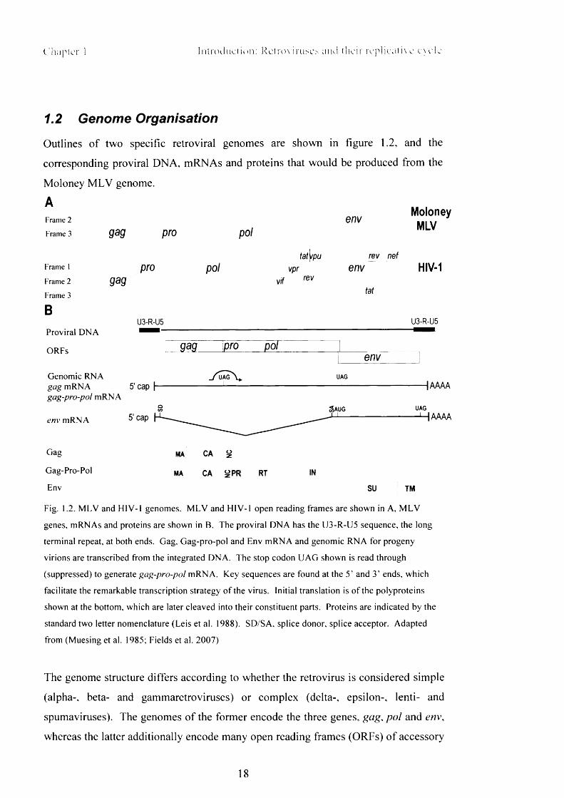
1.2 Genome Organisation
Outlines o f two specific retroviral genomes are shown in figure 1.2, and the
corresponding proviral DNA, mRNAs and proteins that would be produced from the
Moloney M L V genome.
AMoloney
Frame 2 enV Ml \ /Frame 3 gag pro pol
Frametat\/pu rev nef
pro pol vpr env HIV-1Frame 2 vif ^Frame 3
BProviral D N A
ORFs
tat
U3-R-U5 U3-R-U5
gag pro polenv
Genomic R N A . / u a g V . u a g
gag m RN A 5' cap-|----------------------------- 1-------------------------------------------------- ------------------------------------ 1AAAAgag-pro-pol m R N A
& AU G u a g
env m R N A 5 'cap H- H A A A A
Gag ma CA ^
Gag-Pro-Pol MA CA ^ P R RT IN
Env SU TM
Fig. 1.2. M L V and H IV -1 genomes. M L V and H IV -1 open reading frames are shown in A, M L V
genes, m R NAs and proteins are shown in B. The proviral D N A has the U 3 -R -U 5 sequence, the long
terminal repeat, at both ends. Gag, Gag-pro-pol and Env m R N A and genomic R N A for progeny
virions are transcribed from the integrated D N A . The stop codon U A G shown is read through
(suppressed) to generate gag-pro-pol m R N A . Key sequences are found at the 5 ’ and 3 ’ ends, which
facilitate the remarkable transcription strategy o f the virus. Initial translation is o f the polyproteins
shown at the bottom, which are later cleaved into their constituent parts. Proteins are indicated by the
standard two letter nomenclature (Leis et al. 1988). SD /SA , splice donor, splice acceptor. Adapted
from (Muesing et al. 1985; Fields et al. 2007)
The genome structure differs according to whether the retrovirus is considered simple
(alpha-, beta- and gammaretroviruses) or complex (delta-, epsilon-, lenti- and
spumaviruses). The genomes o f the former encode the three genes, gag, po l and env,
whereas the latter additionally encode many open reading frames (ORFs) o f accessory
18

Chapter 1 In t ro duc i io n : Rem>\ eiui thei r repl iea t i xe c \ d e
genes. Despite the epithet ‘accessory’ , most o f these are essential for infectivity, at
least in certain cell types and in vivo infection (Balliet et al. 1994; Anderson and Hope
2004). For all retroviruses, different mRNAs encoding single or combinations o f
these proteins are produced by splicing reactions. For M LV, a simple retrovirus, there
is just one splicing reaction that removes the entirety o f gag, most o f pol, and
produces a transcript o f just env (Shinnick et al. 1981). For H IV there are many more
splicing possibilities between different ORFs created by use o f several alternative
splice sites (Arrigo et al. 1990; Guatelli et al. 1990; Schwartz et al. 1990a). These
allow control o f production o f different proportions o f the viral proteins, and through
the accessory protein Rev, also control o f timing o f production (Malim et al. 1989;
Katz and Skalka 1990). As the genome is produced by the host cell it has several
standard modifications that would be present on host mRNAs, for example, it w ill be
capped at the 5’ end, and polyadenylated at the 3’ end (Green and Cartas 1972;
Furuichi et al. 1975; Reddy et al. 1980). In the mature retrovirus two copies o f the
genome w ill be packaged. During transcription, both copies can serve as a template
for reverse transcription, leading to deletions, insertions, and duplications. Template
switching between the two copies can also lead to exchange o f genetic markers
between clades, when one cell becomes infected with two viruses, resulting in inter
subtype recombinants. This causes problems when drug resistance mutations in genes
are exchanged.
Once processed, translation o f pro and pol is controlled by sequences at the gag-pro
and pro-pol junctions. A stop codon at the end o f gag may be read through to
produce Gag-Pro-Pol (e.g. M LV, (Yoshinaka et al. 1985), or a frameshift can occur,
which is when the ribosome slips back a nucleotide due to both stalling during reading
o f a UUUUUUA length and the encounter with secondary structures in the mRNA
(Hung et al. 1998). This also produces a Gag-Pro-Pol polyprotein (e.g.
alpharetroviruses, (Jacks and Varmus 1985). In some retroviruses (e.g. mouse
mammary tumour virus, and HTLV-1) two framshifts are required as gag, pro and pol
are all in separate reading frames. The efficiency with which the slippage or
frameshifting occurs determines the ratios o f shorter to full length polyproteins.
These initial products are large precursor proteins, cleaved into their constituent parts
by the viral protease at specific points later in the life cycle (Yoshinaka and Luftig
1977).
19
Chapter 1 Introduction: Retroviruses and their replicative cycle
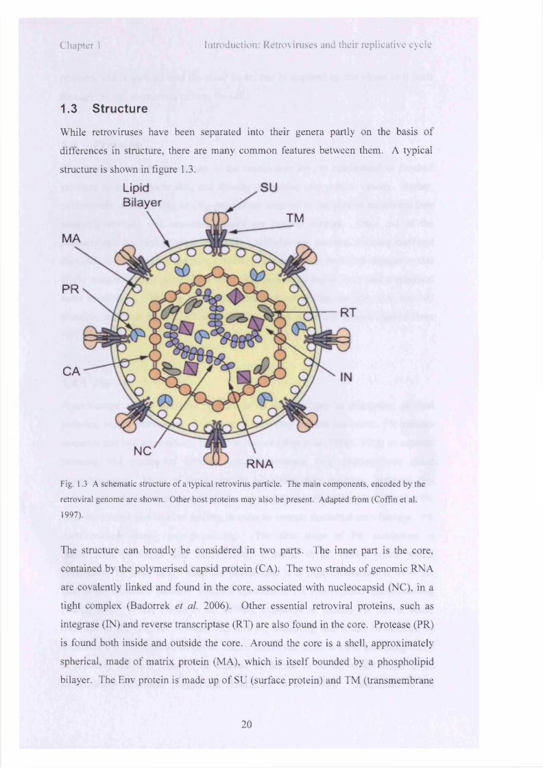
1.3 Structure
While retroviruses have been separated into their genera partly on the basis o f
differences in structure, there are many common features between them. A typical
structure is shown in figure 1.3.
LipidBilayer
TM
MA
PR
RT
CA
NCRNA
Fig. 1.3 A schematic structure o f a typical retrovirus particle. The main components, encoded by the
retroviral genome are shown. Other host proteins may also be present. Adapted from (Coffin et al.
1997).
The structure can broadly be considered in two parts. The inner part is the core,
contained by the polymerised capsid protein (CA). The two strands o f genomic RNA
are covalently linked and found in the core, associated w ith nucleocapsid (NC), in a
tight complex (Badorrek et al. 2006). Other essential retroviral proteins, such as
integrase (IN) and reverse transcriptase (RT) are also found in the core. Protease (PR)
is found both inside and outside the core. Around the core is a shell, approximately
spherical, made o f matrix protein (M A), which is itse lf bounded by a phospholipid
bilayer. The Env protein is made up o f SU (surface protein) and TM (transmembrane
20

protein), and is inserted into the outer layer; this is acquired by the v irion as it buds
through the cell membrane, exiting the cell.
1.4 Proteins
The structural proteins that make up the capsid core are not synthesised as finished
products in the producer cell, and directly assembled into mature viruses. Rather,
polyproteins o f either Gag or Gag-pro-pol are targeted to the plasma membrane (see
assembly section) and immature virions are in itia lly formed. Once out o f the
producer cell, the v ira l protease undergoes a self-cleavage reaction, excising itse lf and
then other components o f the polyproteins. The structure o f the v irion changes to that
o f the mature particle described above, w ith an ordered capsid core and a spherical
outer membrane. The major proteins processed out o f Gag are M A , CA, and NC
proteins, and from Pol PR, RT and IN. There is an additional protein cleaved from
H IV Gag, p6 .
1.4.1 PR
Autocleavage o f the retroviral protease is an essential step in maturation o f v ira l
particles, before cleavage o f all other units from Gag and Pol can occur. PR mutants
assemble and bud successfully, but fa il to mature (Wan et al. 1996). PR is an aspartic
protease, and studies o f cellular aspartic proteases, (e.g. pepsin) have aided
characterisation. Many o f these proteins have regulatory switches, however, this
switch is usually between completely on and completely o f f mechanisms, whereas PR
must have some low level o f activity in order to execute the in itia l autocleavage. PR
itse lf matures during auto-processing. The first stage o f PR maturation is
dimerisation (Wondrak and Louis 1996; Wondrak et al. 1996), which would
presumably be more like ly when the polyprotein is at high concentration, as is the
case in the immature particle. Regions upstream o f PR also regulate autoprocessing
(Gatlin et al. 1998a; Gatlin et al. 1998b). Inhibitors o f mature PR fa il to block
cleavage o f the Gag-pol between NC and p6 , indicating that it can be cleaved by
immature PR. Mature PR, however, cannot cleave this bond in vitro , indicating that it
is probably the first bond to be cleaved as failure to do so would result in an
incompletely processed polyprotein (Lindhofer et al. 1995). Sequences in the
21

Chapte r I In t ro duc t io n : Ketrov i ruscs and thei r rep l i ca t i ve cyc le
transframe region just upstream o f PR, comprising a transframe peptide (TFP) and
p6 * are important for PR regulation. Deletion studies o f p6 * showed that it has an
inhibitory effect on PR activity (Partin et al. 1991; Tessmer and Krausslich 1998). A
tripeptide just upstream o f p6 * in the TFP octapeptide is a potent inhibitor o f PR
(Louis et al. 1998), and may be a key element in the timing o f regulation o f activation
and subsequent deactivation o f PR, which is necessary once the viral particle has
matured.
1.4.2 RT
RT confers on the virus the ability to transcribe dsDNA from their ssRNA genomes.
The structure o f the enzyme varies between retroviruses. M LV RT is a monomer
with both DNA polymerase function and RNase H activity (Tanese and G off 1988).
ASLV RT is a heterodimer due to incomplete cleavage at the RT-IN boundary leading
to one subunit o f DNA polymerase-RNAse H-IN, and one o f DNA polymerase-
RNase H. The DNA polymerase uses either RNA or DNA as template, and a primer
o f host tRNA (Gilboa et a l 1979). As a polymerase it is slow, dissociates frequently,
and pausing is caused by secondary structures present in the templates (Harrison et al.
1998). RNase H acts on RNA in an RNA-DNA duplex, degrading the RNA so that
the remaining strand can act as a template for the DNA polymerase.
1.4.3 IN
Entry o f proviral DNA into the nucleus and its subsequent integration into the host
chromosome is an essential step for retroviral replication. Integration is primarily
mediated by integrase (Panganiban and Temin 1984; Quinn and Grandgenett 1988),
with other host proteins also playing a role (Bowerman et al. 1989; Turlure et al.
2004). Integrase enzyme is usually found as a dimer, each monomer consisting o f
three domains; one for Zn2+ binding, a catalytic core domain, and a C-terminal
domain. It catalyses the removal o f two nucleotides from the 3’ ends o f a proviral
DNA, and the attack and integration o f this prepared DNA on a phosphodiester bond
in the host chromosomal DNA (Fujiwara and Mizuuchi 1988; Brown et al. 1989;
Craigie et al. 1990). While integration can theoretically occur anywhere in the
genome, sites that are transcribed seem to be targeted for HIV, M LV and ASLV
22

C liapter I l n i r oJu c t io : i : R c l n n i r u s o aiui the i r fo p i i c n i i \ c c \ c i c
(Maxfield et al. 2005). However, the extent o f this preference differs. M LV also
shows a preference for integration near transcriptional start sites, whereas ASLV does
not, and only has a weak preference for active genes. The non-random nature o f
integration strongly suggests the involvement o f cellular factors, and indeed the
protein p75/LEDGF (lens-derived epithelium growth factor) has been highlighted as
essential (section 1.5.5) (Cherepanov et al. 2003; Llano et al. 2006). Genes that are
very highly transcribed seem to be selected less than those that are transcribed at a
low level (Maxfield et al. 2005).
1.4.4 MA
The matrix protein is made o f a monomer o f helices surrounding a hydrophobic core.
Sequences in M A are key to the targeting o f Gag to the membrane during assembly o f
nascent viral particles. I f the M domain, which is found at the N-terminus o f MA, is
mutated, assembly is defective, but it can be rescued with coexpression o f wild type
M A. Targeting requires myristoylation o f the M domain (addition o f a 14-carbon
fatty acid), and blockage o f this step abolishes viral assembly (Gottlinger et al. 1989;
Yuan et al. 1993). Depending on the conformation state o f MA, this myristoyl group
can be either exposed or sequestered. Binding o f PL.sPz to M A promotes an exposed
conformation, revealing a cluster o f basic residues in M A that binds to the membrane
(Zhou et al. 1994). H IV M A also has a nuclear localisation signal that may be
important for nuclear import, but it is not found in the pre-integration complex o f
other retroviruses.
1.4.5 NC
Nucleocapsid is a small, basic protein found associated with the RNA genome in the
retroviral core that binds a Zn2+ ion. Alteration or deletion o f this sequence causes
aberrations in RNA packaging (Aldovini and Young 1990). NC is also important in
early stages o f reverse transcription as it promotes the annealing o f primer tRNA to
the binding site, and strand transfer (Rong et al. 1998c).
23

Chapte r 1 In t r oduc t ion : Re trov i ruses and thei r rep l i ca t i ve evele
1.4.6 CA
The capsid protein is the largest o f the Gag proteins, and forms a polymerised shell
enclosing the core, a feature unknown in many other RNA viruses. In all retroviruses
(except spumaviruses) the one highly conserved sequence in Gag, the major
homology region (MHR), is found in CA and is important for virion assembly
(Mammano et al. 1994; Craven et al. 1995). The structure o f CA has been solved for
several viruses (e.g. H IV (Gamble et al. 1996; Gamble et al. 1997; Berthet-Colominas
et al. 1999). Structurally, CA is made up o f two domains, an N-terminal two-thirds
and C-terminal third, connected by a linker. The N-terminal domain o f HIV-1 is
mostly alpha-helical with an exposed loop that cyclophilin A binds, and an N-terminal
(3-hairpin. The MHR is found in the C-terminal domain.
In polymerised form the capsid core can be spherical (MLV), coffin- (H IV) or bar-
(Mason-Pfizer monkey virus) shaped. The role o f capsid in vivo requires a balancing
o f two seemingly opposite requirements; the core must be stable enough to keep the
viral genetic material safe during viral egress, maturation, binding and entry, and yet
also able to dissociate as soon as required in the infectious cycle after entry into a
naive cell. The capsid shell only forms after maturation, and after this point becomes
relatively unstable in detergents, indicating that it is then able to break down quickly
after entry (Wiegers et al. 1998). In the immature virion the core is round, with a
relatively sparse centre. During maturation Gag is cleaved into its constituent parts
and the core condenses, with capsid arranged as hexamers in a lattice (Mortuza et al.
2004; Ganser-Pomillos et al. 2007). Despite the apparent importance o f CA in
maturation, mutations that remove almost this entire domain in ASLV do not cause
loss o f budding.
A renewed focus on CA recently has come as a result o f its role as a determinant for
restriction by Fvl and Trim5a. The interaction o f these factors with CA is discussed
in later sections.
1.4.7 p6
The lentiviral p6 is a 6 kDa protein is found at the C-terminus o f Gag, and is a pro-rich
protein that functions late in the infectious cycle, during the assembly process o f HIV-
24

Chapter 1 In t roduc t ion : kct rov i ruse.s and the i r re p h e a t i \ e cyc le
1, incorporating vpr and vpx into the virions (Lu et al. 1993). Particles with defective
p6 remain tethered at the cell surface (Gottlinger et al. 1991). PT/SAP domains in p6
also promote viral release by binding to TsglOl, a component o f the cellular
machinery that mediates sorting into, and biogenesis of, multi-vesicular bodies
(Morita and Sundquist 2004). Another sorting protein, A L IX , can also bind to p6 o f
HIV, but this interaction is not critical for release (Strack et al. 2003).
1.4.8 Env
The env gene is found at the 3’ end o f the genome, and is produced from a singly
spliced mRNA, from which either the entire gag-pro-pol coding regions have been
removed (several retroviruses including M LV (Shinnick et al. 1981)), or the first 6
codons o f gag spliced to env, as is the case for ASLV (Hunter et al. 1983). Levels o f
Env vary amongst the retroviruses, from equimolar (Gag:Env) in gammaretroviruses
to much lower levels in the mature lentiviruses (Grief et al. 1989; Chertova et al.
2002; Zhu et al. 2003; Yuste et al. 2005). During production the nascent protein is
inserted into the ER after which it w ill be glycosylated and further modified in the
Golgi before being trafficked to the plasma membrane, as would occur for membrane
proteins o f the host cell. Env must oligomerise and fold correctly (e.g. ASLV Env
forms trimers (Einfeld and Hunter 1988)). It is important that Env does not interact
with the receptor when it is in the process o f being correctly constructed and
transported out to the cell surface. Different viruses have evolved different
mechanisms to ensure that this does not occur. For example, ASLV produces levels
o f Env that are vastly in excess o f the receptor (Bates et al. 1993; Young et al. 1993),
and HIV-1 Vpu binds to the cytoplasmic domain o f CD4 and induces its down-
regulation (Willey et al. 1992).
Cleavage o f the envelope protein into SU and TM occurs later, but while Env is still
in the Golgi, and is mediated by a host cell protease (Decroly et al. 1994; Gu et al.
1995). For ASLV, cleavage occurs at a di-basic pair, and leaves TM with a
hydrophobic N-terminus, the location o f the fusion peptide (Dong et al. 1992). Env is
then capable o f mediating fusion (Freed et al. 1995). Once at the cell surface, Env
proteins remain in the multimers o f the SU-TM dimer. Under the electron microscope
they appear as knobbly protrusions from the surface o f the virion. During binding and
25

C hapter I In t rod uc t ion : Ketrnx mise.s and the i r reph ea to e e> ele
entry several rearrangements o f intra- and inter-SU and TM bonds takes place, and
HIV-1 entry may involve SU removal.
1.4.9 Accessory proteins
In addition to those described above, a number o f several further retrovirally encoded
proteins are also found in H IV particles to varying degrees (Robert-Guroff et al. 1990;
Schwartz et al. 1990b). Mutations in these genes do not affect viral replication in
some cultured cells, but these proteins are essential in vivo for productive replicative
cycles.
V if (virus infectivity factor) interacts with APOBEC3G during assembly. This aspect
is discussed later, along with other restriction factors.
Vpr (viral protein r) is associated with the pre-integration complex, and may play a
role in its nuclear localisation (Heinzinger et al. 1994). It causes cell cycle arrest and
apoptosis, and also has a role in facilitating efficient infection o f non-dividing cells
(Andersen and Planelles 2005). An association with the cellular protein DDB1 is
necessary for the G2 arrest, and this association prevents DDB1 from carrying out its
normal cellular role o f repairing DNA damaged by UV. DDB1 also targets some
regulatory proteins o f the cell cycle for degradation, forming a ubiquitin ligase
complex with Cul4A and Rod (Schrofelbauer et al. 2007). Vpr is found in high copy
number in virions, persisting in association with proviral DNA up to and into the
nucleus, which has made fusions o f Vpr and fluorescent proteins useful in visualising
the viral core after entry (McDonald et al. 2002).
Vpu is found in HIV-1 and chimpanzee SIV, and bears homology to the potassium
ion channel, TASK-1 (Hsu et al. 2004). Vpu is not found in viral particles, and
negative mutants produce viral proteins normally, but are defective in virion
production in some cell types (Klimkait et al. 1990). Vpu induces CD4 degradation
by binding to the cytoplasmic tail o f CD4 in the ER, inducing its degradation (W illey
et al. 1992). This prevents re-infection o f the infected cell. It also counteracts the
anti-viral activity o f a set o f cellular proteins (or tetherins), including CD317, which
attach mature viral particles to the cell surface (Neil et al. 2008).
26

Chapter I Imi 'odi ic tU'n: k e t r m iruses and the i r repl icat iv c cyc le
Vpx is an additional gene found in viruses o f H IV -2 /S IV smm/S I V mac lineage. It plays a
similar role to vpr.
Nef (negative factor) is core-associated, and packaged into virions. It is essential for
sustaining viral production in vivo and acts by down regulating antigenic and viral
receptor cell-surface molecules, enhancing viral infectivity and modulating cellular
pathways (Schaeffer et al. 2001; Qi and Aiken 2007).
1.5 Virus Infectivity Cycle
Retroviruses must pass through a formidable array o f defences before they can
successfully enter the cell and complete a replicative cycle. First o f all they must
traverse the physical barriers; the viral core has to access the cytoplasm, and then,
while disassembling, pass through the thick barrage o f proteins and webs o f
membranes that make up the cytoplasm towards the nucleus, and the newly reverse
transcribed genetic material must finally traverse the nuclear membrane and
outmanoeuvre the histones and other DNA-associated proteins to integrate into the
host DNA. Secondly, they must overcome the cell’s biological defences. These can
be intrinsic defences or part o f the adaptive immune system. Different viruses have
evolved many strategies to avoid both physical and biological defences that the cell
raises.
The cycle can be broadly split into two stages. In the first, the virus enters the cell,
reverse transcribes and integrates its genetic material into the host DNA. These
processes are extraordinary for the cell, and use co-opted cellular proteins directed by
viral machinery. In the second half o f the cycle, progeny virions are produced,
assemble and exit the cell, using the routine cellular mechanisms o f translation,
transcription and transport. A basic outline o f the main steps is shown in figure l .5.
27

Chapter 1 Introduction: Retroviruses and their replicative cycle
Binding
• Uncoating and
*( f
Fusion
Reverse Transcription%
j £ o 6 t r
Nuclear Import
Integration
ja n r ja o r ^ n c ir jf jn r jr s o fjr jr
>g> Translation
^ V \ v/x^ - r -
^ v \Assembly
Budding
Fig. 1.5 A schematic o f the retroviral infectious cycle. In order to replicate, viruses must enter cells,
reverse transcribe their R N A genome, then integrate the newly synthesised D N A into the host
chromosomal D N A . From there cellular mechanisms transcribe, translate and partly process the
retroviral proteins. Virions assemble and bud from the plasma membrane, completing the cycle.
28

Chapte r 1 In t rod uc t io n : Rctrov innr .s and thei r rep l i ca t i ve cyc le
1.5.1 Binding
In order to initiate an infectious cycle, the viral envelope protein must first bind to a
receptor expressed on the cell surface. The combination o f receptor and envelope has
a profound effect on the ability o f the virus to enter different cell types, on the manner
in which the virus enters the cell, and therefore the success (or otherwise) o f the
replicative cycle.
Viruses have evolved to bind a huge range o f receptors, and related viruses may use
completely different ones. Receptors may be membrane proteins such as cell-cell
recognition molecules, membrane transporters, and some use proteins o f unknown
function (Marsh and Helenius 2006; Stewart and Nemerow 2007). The distribution o f
the receptor w ill obviously affect the range o f cells that the virus is able to infect, and
viruses are reliant on expression o f their receptor on a limited range o f target cells or
limited host range. For example, the main receptor for binding o f HIV-1 is CD4
(Maddon et al. 1986), a marker o f the immunoglobulin superfamily found on T-cells,
monocytes and macrophages, which limits the range o f cells that it can infect, but
clearly also holds advantage for the virus as it can hide itself within the immune
system. By contrast, Vesicular stomatitis virus (VSV), a rhabdovirus, is described as
pan-tropic, able to infect every type o f cell tested so far (Aiken 1997). The VSV
glycoprotein binds to membranes containing phosphatidylserine (Schlegel et al. 1983)
and viruses are taken up into the cell by clathrin-mediated endocytosis (Sun et al.
2005).
Once the virus is bound to the receptor, conformational changes are usually required
to bring about a fusion reaction between the viral and cellular membranes, creating a
pore so that the viral core can enter the cytoplasm. The triggering o f these
conformational changes and the fusion process is elaborate to prevent premature
initiation while the virus is not in suitable contact with a susceptible cell. This
requires the existence o f a trigger additional to receptor binding. For some viruses
this may be a change in the environment, such as a drop in pH; for some primate
lentiviruses it involves binding o f a coreceptor. HIV-1 w ill bind to, but not enter cells
expressing only its main receptor, CD4 (Maddon et al. 1986). Once the viral
glycoprotein, gpl20, is bound to CD4 conformational changes occur that permit it to
29

C hapter I In t rod uc t i on : Ketrov iruses and the i r r e p l ie a t i \ e cyc le
bind one o f its coreceptors, CXCR4 or CCR5 (Alkhatib et al. 1996; Deng et al. 1996;
Dragic et al. 1996; Feng et al. 1996). Once coreceptor is bound, further
conformational changes and fusion occur (Sullivan et al. 1998).
1.5.2 Fusion and entry
How is the fusion process affected by whether entry is considered to be pH-dependent
or (as for most retroviruses (McClure et al. 1990)) independent? Viral binding to the
cognate receptor must always occur for fusion to follow. As the receptors used by
virions to enter cells are there to perform some cell-specific function rather than for
the benefit o f the virion, the apparatus necessary for fusion is contained within the
viral envelope. However, the changes that activate Env subsequent to receptor
binding differ.
VSV is taken across the cell membrane by clathrin-mediated endocytosis (Sun et al.
2005) and the increasing acidity o f the endosomal environment primes the viral fusion
protein to initiate exit from the pathway. Entry into endosomes additionally removes
the virus quickly from the harsh extracellular environment, utilising the cell’s own
mechanisms to aid it in crossing the plasma membrane. Clathrin-mediated
internalisation is required for many other viruses (e.g. adenovirus (Meier et al. 2002),
while some others can also utilise clathrin-independent pathways (e.g. influenza
(Lakadamyali et al. 2004)).
Successful entry o f HIV, however, generally only follows fusion at the cell surface,
and is pH-independent (McClure et al. 1988). Endocytic processes usually lead to a
non-productive end for H IV as the virus cannot initiate exit from the lysosomal
pathway and w ill be degraded. This was found to be untrue in the exceptional case o f
human placental trophoblasts, which are polarised cells (Vidricaire et al. 2004).
Infection o f these cells is a key stage in mother to child transmission o f HIV, and the
virus may additionally be transcytosed whole across the placental cells and released
intact at the other side, ready to infect nearby foetal cells (Vidricaire et al. 2004).
Trophoblasts express little to none o f the receptors and co-receptors for H IV, yet they
can be productively infected to a moderate degree (Vidricaire and Tremblay 2005).
H IV is heavily endocytosed within these cells, and cytokines released during
30

Chapter 1 In t rod uc t i on : R c t n n i ruses and thei r rep l i ca t i ve e \e i e
pregnancy may create conditions temporarily favourable for escape from endosomes.
Inhibitors o f endocytosis completely block infection (Vidricaire and Tremblay 2005).
In this case, H IV is clearly able to exploit discrepancies in the cell biology o f
trophoblasts as compared to other cell lines in order to permit a-typical usage o f this
pathway.
Once the virus is bound by receptor and any requirements for fusion additional to
receptor binding have been met, the fusion mechanism is triggered. The fusion
reaction can take place at the cell surface (e.g. CD4/HIV-1) or in an endosomal
compartment (e.g. Tva/ASLV) (Stein et al. 1987; Sinangil et al. 1988; Mothes et al.
2000). For the envelope o f H IV -1, binding o f CD4 and a coreceptor are sufficient to
trigger the sequential conformational changes that eventually result in fusion. For
VSV-G, a drop in pH activates the receptor for fusion. In a remarkable twist, ASLV-
A Env appears to combine both strategies, using both conformational changes and a
drop in pH. Firstly, Env binds to the Tva receptor, causing a conformational change
to occur, but a drop in pH is necessary for full activation and completion o f fusion
(Barnard et al. 2006) (discussed in more detail below, in section 1.7).
The fusion reaction between viral and cellular membranes is highly energetically
unfavourable, involving extensive rearrangement o f the cellular membrane. The
conformational changes undergone by the viral envelope proteins drive these
rearrangements (Eckert and Kim 2001). Despite the difference in the conditions
under which fusion is triggered, the fusion mechanisms o f many viral envelope
proteins is very similar. Fusion proteins can be divided into two classes: class I and
class II. Class I proteins undergo a stage known as a 6-helix bundle, and class II
proteins do not (Colman and Lawrence 2003; Kielian 2006). After SU binding to the
cell-surface receptor, TM forms this 6-helix bundle in which it essentially bends back
onto itself. The N-terminal segments form a coiled-coil and the C-terminal segments
lie alongside the central coiled-coil in antiparallel configuration (Markosyan et al.
2003; Markosyan et al. 2004). Once formed, this six helix bundle is stable to well
above physiologically relevant temperatures (Lu et al. 1995). The viral and cellular
membranes are forced into close proximity as the fusion peptide o f the viral envelope
is inserted into the cellular membrane, and the membrane spanning segment is in the
viral membrane (Markosyan et al. 2003). Formation o f this bundle is an obligate step
31

Chapte r ! In t ro duc t ion : R e t r o v i r u s o and thei r rep l ica t i ve cxc le
and as such, is a suitable target for anti-HIV drugs. Enfuvirtide is a drug based on an
anti-retroviral peptide that binds to gp41, preventing structural rearrangements
(Greenberg and Cammack 2004). Class II proteins appear to show a similar approach
to fusion, but with unrelated intermediate protein structures.
One o f the best-understood series o f binding and fusion events, supported by a wealth
o f structural information, is that between the receptor protein o f influenza virus,
haemagglutinin (HA), and sialic acids on glycoproteins and glycolipids. HA exists as
a trimer o f heterodimers o f HA1 and HA2 (Wilson et al. 1981). Once bound to
receptor, the virus w ill be endocytosed, and HA is dependent on the consequent drop
in pH that occurs as the virus passes through the endosomal pathway to trigger a
conformational change (Skehel et al. 1982; Wharton et al. 1986). A fusion peptide in
HA2 is brought into close contact with, and inserted into, the target cell membrane
(Carr and Kim 1993; Bullough et al. 1994), and with the other end in the viral
membrane the Env-receptor complex undergoes a conformational change that pulls
the outer viral and cellular membranes close into a hemi-fused intermediate.
Subsequently the two progress together to full fusion, and eventually sufficient
enlargement o f the pore to permit the core o f the virus to pass through (Skehel and
Wiley 2002).
Models o f retroviral Env proteins in comparison to HA appear to share elements such
as potential fusion peptides, but differ in details, as would be expected from envelopes
designed to bind to different receptors.
1.5.3 Reverse transcription
Once fusion has been completed, whether from the extracellular milieu or endosomes,
the viral core is released into the cytoplasm and embarks on the next stage o f the
cycle. The core contains the viral genome, condensed by association with NC and RT
both o f which are essential components o f the reverse transcribing core. The capsid
structure enclosing the core must disassemble, as immature virions, in which the
capsid core is much more stable, are non-infectious. A very limited level o f reverse
transcription does occur in intact virions before entry, but only increases to significant
levels after core breakdown (Trono 1992; Zhang et al. 1993). Once the capsid shell
32

Chapte r 1 In t rod uc t ion : R e t r o v i r u s o and thei r rep l i ca t i ve cyc le
has been partly dismantled, however, components o f the core do not completely
disperse throughout the cell and have been tracked as bound to the proviral complex
up to, and even beyond, import into the nucleus.
DNA synthesis is initiated from the 3’ -OH o f a specific cellular tRNA bound to the
primer-binding site (pbs). A short DNA fragment is then synthesised up to the 5’ end
o f the genome, comprising 5’R-U5-pbs. This is known as strong stop DNA, and is a
useful marker for an indication o f viral entry, and commencement o f the earliest stage
o f the replicative cycle. The RNA template is then digested exo-or endo-
nucleolytically as far as U5 by RNaseH, and the new DNA ‘jumps’ to the 3’ end o f
the RNA template, binding the newly transcribed DNA R region to the 3’ RNA R
region . DNA synthesis continues, and the RNA template is concomitantly degraded
with short pieces remaining as primers for plus strand DNA (Haseltine et al. 1976;
Gilboa et al. 1979). During this process jumps between and within the two copies o f
RNA in the genome are frequent, leading to mutations and deletions or insertions.
When combined with an error rate o f ~10'4 for the polymerase, these jumps account
for the extremely high mutation rate o f many retroviruses (An and Telesnitsky 2001).
A purine-rich section near the 3’ end o f the genome that is relatively resistant to
RNAse H acts as a primer for plus strand DNA synthesis (Rattray and Champoux
1987; Rattray and Champoux 1989). The newly transcribed minus strand DNA acts
as a template for synthesis from this primer. Synthesis proceeds in the 5’ direction,
and includes the tRNA primer still attached to the pbs at the 5’ end, halting at a
modified base found in the tRNA. Once the tRNA is displaced (Pullen et al. 1992),
the complementary pbs sequences on the plus and minus strands pair to facilitate the
second strand transfer. These steps are illustrated in fig. 1.5.3.
33

Chapter 1 Introduction: Retroviruses and their replicative cycle
tRNA
R U5 PBS
- * A
PPT U3 RAAA 3*
ONAsynthesis
R US PBS PPT U3 RAAA
R N aseH
■vs PBS PPT 03 RAAA
Ftrst strand transfer
PBS PPT U3 R
i j ±__A— AAA
DNA synthesis R N aseH
.PPT U3 R US
PBS PPT
DNA synthesis RNase H
PPT U3 R U5
A
A>PBS
P ^
R N aseH
PPT U3 R U5
PPT U3 R UST
Second strand transfer
LTR LTR
Fig. 1.5.3 Outline o f the stages o f reverse transcription described in the text. Taken from (C offin et al.
1997).
34

Chapte r 1 In t ro duc t io n : Re t rov i ruses and the i r r e p l ie a t o e c \ v l e
The final product o f RT is a double stranded DNA copy o f the full genome, with
copies o f the LTR are present at both ends. For most retroviruses this finished
product can be found in the cytoplasm. However, A LV only completes reverse
transcription once in the nucleus (Lee and Coffin 1991), and RSV RT localises to the
nucleus (Werner et al. 2002), suggesting that there may be some advantage to
delaying termination o f reverse transcription until integration is imminent.
1.5.4 Nuclear import
In order to establish a stable infection, the viral DNA must first o f all cross the
nuclear membrane before it can undergo integrase-mediated incorporation. The
nuclear membrane presents a considerable barrier, and only the complex retroviruses
have evolved ways to cross it without requiring membrane breakdown during passage
through mitosis (M iller et al. 1990; Lewis et al. 1992; Lewis and Emerman 1994).
Simple retroviruses thus cannot replicate in cells without passage through mitosis
speedily following reverse transcription, as the viral DNA does not remain suitable
for integration for more than around 6 hours (M iller et al. 1990; Andreadis et al.
1997). Quiescence presents no barrier to lentiviruses and spumaviruses, however, and
this has stimulated much research into identifying which viral and cellular factors are
able to actively transport the viral DNA into the nucleus.
The pre-integration complex (PIC) is larger than the nuclear pores, and cannot enter
the nucleus by passive diffusion therefore active transport mechanisms must be
involved. Two separate nuclear localisation signals in the HIV-1 PIC, in M A and Vpr
were reported (Bukrinsky et al. 1993; Heinzinger et al. 1994), either one o f which
appeared to be able to mediate transport into the nucleus. However, this view is
disputed since viruses with mutations in both these regions are still able to localise the
PIC to the nucleus so other signals must exist (Freed et al. 1995). Host proteins, for
example the nuclear pore component Nup98 (Ebina et al. 2004), may play a role.
It has been shown recently that H IV CA is implicated in the cell-cycle independence
o f HIV. A viral chimera o f H IV with M LV CA, and H IV with mutations in CA were
both unable to infect non-dividing cells (Yamashita and Emerman 2004; Yamashita et
al. 2007). Markers o f nuclear import, 2LTR circles, were found in the nucleus,
35

Chapte r 1 In t rodu c t ion : Re t rov i ruses and thei r rep l i ca t i ve cyc le
indicating that it is not the physical access to the nucleus that is limiting, but a step
downstream o f this, possibly related to retention o f CA with the pre-integration
complex (Yamashita et al. 2007).
1.5.5 Integration
Production o f linear viral DNA alone by reverse transcription is not sufficient for
founding a long-term infection; integration into the host chromosomal DNA allows
the retrovirus to establish an infection on a permanent basis and is necessary for a
productive infection (Englund et al. 1995). At the ends o f the U3 and U5 sequences
are ~10bp sequences known as att sites, recognised by IN (Bushman and Craigie
1990), that contain a key CA dinucleotide pair. Two terminal nucleotides adjoining
this pair are removed by integrase prior to strand transfer. This CA pair thus w ill
ultimately define the 3’ ends o f the integrated viral DNA, and the LTRs control o f the
replication o f the viral genome. The second part o f the reaction, the strand transfer o f
the vDNA into the host DNA is via an attack o f the processed 3’OH ends on the target
DNA (Fujiwara and Mizuuchi 1988). Integrase is the only viral protein necessary to
carry out the reaction (Bushman et al. 1990; Craigie et al. 1990).
While there is no specific site in the host genome preferred for integration, the
distribution o f integrated provirus is not uniform. The tertiary structure o f DNA
appears to be more important than an extensive primary sequence in determining
whether a site is preferred or not (Pryciak et al. 1992). Preferences for integration
into certain areas may be retrovirus type-specific, for example, H IV appears to
integrate into sites o f active transcription, whereas M LV favours transcription start
sites and CpG islands (Schroder et al. 2002; Wu et al. 2003; Maxfield et al. 2005).
DNA in nucleosomes is targeted in preference to naked DNA, but other proteins
bound to DNA (e.g. in regions where DNA is highly transcribed) may decrease access
o f the provirus. A 5-bp sequence, with either end on the same side o f the double
helix, is targeted by HIV-1 during integration (Vincent et al. 1990). This sequence is
duplicated during this process, which together with the insertion o f the retroviral
genome explains the inherently mutagenic nature o f integration.
36

t iKipter ! In i ro t luc t ion : Re trov i ruses and the i r rep l ica t i ve cyc le
The fact that integration is not entirely random for some retroviruses indicated that a
cellular protein connected with transcription might be involved in binding IN to DNA.
In 2003 lens epithelium-derived growth factor (LEDGF/p75) was found in a complex
with HIV-1 IN (Cherepanov et al. 2003). LEDGF/p75 is involved in the cellular
stress response, and binds both chromosomal DNA and lentiviral IN, possibly acting
as a tethering factor (Maertens et al. 2003; Llano et al. 2004). Knockdown o f
LEDGF/p75 significantly inhibits replication o f H IV (Vandekerckhove et al. 2006).
When LEDGF/p75 was depleted from cells subsequently infected with HIV,
integration into transcription units and genes under transcriptional control o f
LEDGF/p75 was less frequent, and into CpG islands was more frequent indicating
that LEDGF/p75 affects the choice o f target site (Ciuffi et al. 2005).
Although vDNA can be found in circularised forms in the nucleus, it appears that only
linear DNA can be used for integration (Ellis and Bernstein 1989). Other forms found
in cells are made from circularisation o f the linear DNA at the LTRs, and while dead
end products for the virus, are useful analytical markers o f nuclear entry.
Once the viral DNA is integrated, there is little chance that it w ill be completely
excised, although low-frequency excision o f the full-length insertion by
recombination between the 5’ and 3’ LTRs does occur. This event leaves behind a
single LTR (a ‘solo LTR’), and has been mostly studied for retrotransposons in yeast
and Drosophila (Mager and Goodchild 1989). For germline cells, the integration o f
vDNA means that every past retroviral infection w ill leave a calling card o f viral
DNA in the host genome. Viruses that infect germline cells and pass down their
complete genome to progeny are known as endogenous retroviruses (ERVs). ERV
sequences are not essential for host cell function, and usually eventually acquire
mutations that knock out viral production, leaving just the traces behind. As much as
8% o f the human genome may be accounted for in this way (Smit 1996; Griffiths
2001), and the human genome project found evidence o f several thousand infections
by at least twenty-four different families (Griffiths 2001; Villarreal 2001).
37

Chapte r 1 In t rodu c t ion : K e t n n i ruses and the i r r c p l i c a t o e cyc le
1.5.6 Assembly and exit
Once integration is complete, the virus relies on standard host mechanisms for
transcription and translation. Both spliced and unspliced mRNAs are produced, the
former for production o f viral proteins, and the latter to serve as genomic RNA for
progeny virions. Both must be transported to the cytoplasm, and different retroviruses
have evolved different ways to achieve this, as unspliced mRNAs are normally
retained in the nucleus. Simple retroviruses contain c/s-acting elements in the RNA
that interact with cellular factors, or the splice site signals are relatively weak (Katz
and Skalka 1990; Ogert et al. 1996). In complex retroviruses the viral Rev or Rex
protein binds to their cis-acting elements, permitting transport without splicing
(Cullen 1992). Before significant quantities o f Rev are produced after infection,
multiply spliced small mRNAs predominate. After the build-up o f Rev to over a
threshold level, singly spliced and unspliced mRNAs begin to appear in the cytoplasm
(Kim et al. 1989). After this point, production o f retroviral protein and assembly o f
progeny virions can proceed.
Gag, which is at this point uncleaved, is the only retroviral protein needed to drive the
targeting o f the retroviral polyprotein to the plasma membrane, and the assembly into
immature particles (Freed 1998). A sequence within M A known as the M domain is
the membrane-binding domain. In HIV-1, myristoylation o f this domain and a cluster
o f basic residues together are both necessary for membrane targeting (Gottlinger et al.
1989; Zhou et al. 1994). Gag is also responsible for association o f the viral genome
with the nascent particle. Env proteins are synthesised into the ER membrane and
trafficked to the plasma membrane, enriched at budding sites.
Interacting Gag proteins form structures that bud out from the plasma membrane,
eventually pinching o ff as immature particles. This pinching o ff is inhibited by both
mutations in the L domain o f Gag and by cellular proteins known as tetherins. The
action o f tetherins can be overcome artificially by proteases, and are counteracted
during replication o f H IV -1 by Vpu (Neil et al. 2006; Neil et al. 2008). PR cleavage
o f the polyproteins occurs once the immature virion acquires a membrane via budding
and release through the cell surface. Protease processing occurs, the core condenses
and the newly matured virion is capable o f initiating a new infection.
38
Chapte r 1 In t rod uc t ion : Retrov i ruses and the i r rep l ica t i ve c_\eie
1.6 Vectors and Viral Pseudotypes
As retroviruses are capable o f inserting genes stably into the host chromosomal DNA,
they have been extensively researched and manipulated as gene delivery systems.
Retroviral vectors have been developed as a way o f generating replication defective
virus-like particles capable o f completing limited aspects o f the infectious cycle o f
retroviruses, without the risk o f associated undesirable aspects such as oncogenic
potential, and the spread o f the virus after an initial infection. The first stages o f
retroviral replication up to, and including integration, do not require any c/e novo viral
protein synthesis, so genes required for generation o f progeny virus, and cell exit can
be excised and supplied in trcrns in the producer cell (Mann et al. 1983; Watanabe and
Temin 1983). Instead, a selectable marker gene is inserted which w ill be translated by
the cell, so that an initial round o f replication can be detected (Palmer et al. 1987). A
packaging cell line is one that has integrated genetic material encoding the structural
proteins o f the virus and the viral envelope, however, they cannot transmit or package
the RNA that encodes these (Shank and Linial 1980). Alternatively, constituents o f
viral particles can be produced from two or more separate plasmids transiently
expressed in a producer cell (e.g. by transfection), encoding gag-pol, and the viral
envelope. A third plasmid encoding a gene o f interest, and potentially a selectable
marker can be co-transfected. This must contain a vj/ sequence between the promoter
and the inserted gene to direct uptake and packaging o f the RNA into virions, and ex
acting sequences that direct stages o f reverse transcription and integration (Soneoka et
al. 1995; McBride et al. 1997). A ll packaged vectors used to transfer genes in this
study used selectable markers driven from an IRES (internal ribosome entry site) to
assay for gene expression (Jang et al. 1990). Production o f the gene o f interest is
driven from the 5’ promoter sequence, and production o f the selectable marker from
an IRES located between the gene o f interest and the selectable marker. Protein
production from the IRES can be reasonably assumed to guarantee production o f the
gene o f interest. Selectable markers used in this way include fluorescent markers (e.g.
YFP), and neomycin resistance (see fig. 2.3.1 for schematic representations o f
vectors)
39

Chapter 1 In t rod uc t ion : K c t r o \ iruses run! thei r rep l i ca t ive exele
An advantage o f using separate gag-pol and env components to generate virus
particles is that component parts do not need to come from the same virus (Zavada
1972). That is, M LV gag-pol can be transfected along with env plasmid from HIV-1,
ASLV-A or, as is most widely used, VSV (Yee et al. 1994). The particles formed w ill
have the structural proteins encoded from the M LV gag-pol, and envelope from a
separate (retro)virus. This is known as pseudotyping, and is widely used to introduce
virions to a variety o f cell types not normally permissive to infection mediated by the
cognate envelope protein. An additional benefit is that the chance o f recombination to
generate replication competent virus, a key safety consideration, is reduced.
1.7 Entry of ASLV-A
A great deal o f the work presented here includes a viral envelope and receptors o f an
alpharetrovirus, ASLV. Infection by ASLV causes B-cell lymphomas. There are
currently 10 groups o f ASLVs, A-J, designated according to host range, receptor
subgroups and interference, and neutralisation by antibodies (Weiss 1993). Groups
A-E can be further grouped according to whether they are non-cytopathic (A, C and
E) or cytopathic (B and D) (Weller and Temin 1981). Three separate loci are
responsible for controlling susceptibility to these five groups: tva, tvb and tve (tv,
tumour virus). Tva and Tvc permit entry o f groups A and C, respectively, and two
alleles o f tvb permit entry to either B, D and E, or just B and D (Payne and Biggs
1964; Crittenden et al. 1967; D u ff and Vogt 1969). Tva and tvc are genetically linked
(Payne and Pani 1971). The normal functions o f Tva, Tvb and Tvc are not known.
1.7.1 Tva800 and Tva950
The receptors for ASLV-A were identified by examining chicken genomic DNA that
rendered African green monkey COS-7 cells susceptible to infection (Young et al.
1993). Further characterisation required examination o f the quail homologue, as the
chicken gene was not transcribed at a detectable level, and many ORFs were encoded.
Two alternatively spliced genes were identified from quail, both o f which encoded
proteins containing identical 83 amino acid sections and included an extracellular
receptor domain with an LDL-A (low density lipoprotein) motif. This m otif was first
found in the LDL-receptor, is approximately 40-amino acid long including seven
40

Chapter 1 Introduction: Retroviruses and their replicative cycle
ligand-binding repeats (~50% identical) and is also found in many other cellular
proteins (Tolleshaug et al. 1982; Herz 2001). The difference between the two arose in
their attachment to the membrane, by either a single membrane spanning region, or a
glycosylphosphatidylinositol (GPI) anchor (Bates et al. 1993). These were named
Tva950 (transmembrane linked protein) and Tva800 (GPI anchor). Both comprise an
N-terminal signal sequence (1-19) and the extracellular domain (20-102). Tva950
then consists o f a transmembrane domain (103-125) and a small cytoplasmic tail.
Tva800 has a C-terminal GPI anchor signal sequence: it is synthesised into the ER
like a transmembrane protein, but once completed a carboxy terminal signal directs
GPI-anchor attachment (Doering et al. 1990; Moran and Caras 1991). GPI anchors
share a common phosphoethanolamine-Mannose3 -phosphatidylinositol core, but
potential modifications o f side chains lead to a high level o f heterogeneity. A
schematic o f Tva800 and Tva950 is shown below in figure 1.7.1.
Tva800
LDL-A GPI
Tva950
LDL-A
20 30 70 103 125 157Figure 1.7.1 Simplified outline o f the Tva800 and Tva950 proteins. The two are identical up to amino
acid 103, after which Tva800 is modified to include a GPI anchor, whereas Tva950 has a single
membrane-spanning domain.
The LDL-A motif makes up the bulk o f the extracellular receptor structure at 47
residues, and is sufficient to mediate viral binding (Rong and Bates 1995; Damico et
al. 1998). Calcium is required for correct folding (Wang et al. 2001), and the tertiary
structure is also held in shape by three pairs o f disulphide bonds between six
invariable cysteine residues (Belanger et al. 1995). The structure o f this part o f Tva
was determined in solution by NMR spectroscopy (Tonelli et al. 2001; Wang et al.
2002a), which has aided elucidation o f the precise roles o f specific amino acids
identified as key determinants o f viral binding, although the two final structures
differed in interpretation at the N-termini. The key differences between Tva LDL-A
41

Chapte r 1 In t rod uc t i on : Re t ro \ i ruses and thei r rep l ica t i ve cyc le
and other LDL-A structures are concentrated at the N-terminus, and appear to arise
from extra amino acids in loops o f Tva. H38 and W48 were identified as critical viral
binding or entry residues (Zingler and Young 1996; Rong et al. 1998a; Rong et al.
1998b; Wang et a l 2002b), and in the solution structures lie at the exposed surface o f
the molecule, available to interact with the viral envelope. Interestingly, they are not
conserved amongst the LDL-A family and so must be essential for viral binding or
fusion events, rather than correct receptor folding. Other key residues identified are
buried deep within the molecule, and so are more likely to be important in folding or
maintaining the structure (Tonelli et al. 2001; Wang et al. 2002a). A hydrophobic
patch on the surface o f Tva has been suggested as a point o f contact with the envelope
protein, or as involved in correctly orientating the interaction. The natural function o f
Tva is not known, so it is currently impossible to compare the binding o f ASLV Env
with the physiological ligand to Tva.
1.7.2 ASLV Env
The ASLV envelope protein is similar in organisation to other viral fusion proteins. It
is synthesised as the precursor protein, Pr95, and a cleavage step releases the mature
gp85 (SU) and gp37 (TM) found as a trimer o f heterodimers, linked by a disulphide
bond (Hunter et al. 1983; Einfeld and Hunter 1988; Dong et al. 1992). There are five
host range determinant sequences, h rl, hr2, v r l, vr2 and vr3, found in the SU subunit
(Domer et al. 1985; Bova et al. 1988). There are several N-linked glycosylation sites
in SU, but none are in regions that form hrl or hr2 which suggests that
oligosaccharides are not involved in receptor recognition, although they may be
required for correct overall folding o f the protein, and therefore indirectly be
determinants for receptor binding (Delos et al. 2002).
1 341su Q Q D
342 545TM fp7 n
Fig 1.7.2 A representation o f the structure o f A SLV Env. The two parts are generated from cleavage of
a single polypeptide, and are linked by a disulphide bond, represented by a dashed line. The hashed
domains represent heptad repeats. SU - surface protein, T M - transmembrane, vr - variable region, hr
- host range, FP - fusion peptide, M SD - membrane spanning domain. Simplified from (Barnard and
Young 2003).
42

Chapter 1 In t roduc t ion : Ret rov i ruses ant' the i r rep l ica t i ve e \e l e
Viruses that have been selected for their ability to infect a wide range of host cells
carry mutations in the hr regions (Taplitz and Coffin 1997), and mixing and matching
sequences from these regions alone can have a significant effect on host permissivity
o f viruses. For example, insertion o f the hr2 sequence from ASLV-E Env into ASLV-
B Env permits a virus to infect both ASLV-E and ASLV-B receptor-expressing cells
(Tsichlis et al. 1980; Domer et al. 1985). Host range is not as affected by alterations
in v rl and vr2, but vr3 may act as a supplementary determinant to hr sequences in
some cases; the reason for the high level o f variability in v rl and vr2 is not clear.
Insertion o f a fusion peptide into the target cell membrane by the activated viral
envelope protein is a step common to enveloped viruses during entry. During entry
mediated by HA (Harter et al. 1989), HIV-1 (Gordon et al. 1992) and SIV (Horth et
al. 1991), the fusion peptide likely forms an amphipathic helix which exposes its
hydrophobic residues to the lipid bilayer. The likely fusion peptide in ASLV Env is
found in the N-terminus o f TM and like other viral fusion peptides, has a proline near
the centre. Mutations o f this proline do not abolish inter-Env interactions, but do
weaken them, and prevent ASLV entry.
1.7.3 Mechanism of entry via ASLV-A Env
Despite apparently contradictory data in recent years pertaining to the mechanism o f
ASLV-A fusion and entry, a general outline o f sequential steps that must occur during
entry now seems clear. Uniquely ASLV is not easily classified as using either a pH-
dependent or pH-independent route o f entry, but borrows from both mechanisms. The
envelope proteins o f viruses that are pH-dependent are usually activated by low pH,
as found in endosomes (e.g. influenza HA). Exposure o f such viruses to low pH in a
cell-free environment results in premature triggering o f the fusion mechanisms,
rendering the virus inactive (Stegmann et al. 1987). Similarly, exposure o f cells to
lysosomotropic agents that prevent acidification o f the endosomes causes the virus to
be blocked, trapped within the endocytic pathway, unable to initiate fusion. Viruses
that are pH-independent (e.g. HIV-1) initiate fusion at the cell surface, and are not
affected by changes in pH. Changes in Env structure that bring about fusion in this
latter group are initiated solely by receptor (and often co-receptor) binding (White
1990; Weiss 1993).
43

Chapter 1 Introduction: Retroviruses and their replicative cycle
Entry o f ASLV-A into cells can be blocked by the addition o f lysosomotropic agents,
and further exposure o f cells to low pH overcomes this block, suggesting that a low
pH step is necessary during entry (Mothes et al. 2000). However, ASLV-A virions
are not inactivated by low pH (Gilbert et al. 1990; Mothes et al. 2000), and lipid
mixing appears to occur at neutral pH (Gilbert et al. 1990; Hernandez et al. 1997).
The entry mechanism o f ASLV-A maps to the envelope protein alone, as
pseudotyping ASLV-A cores with ecotropic M LV Env, which mediates pH-
independent entry, abolishes the sensitivity to lysosomotropic agents (Mothes et al.
2000). In order to tie these data together, a mechanism o f entry was proposed which
involves a unique two-stage process (Mothes et al. 2000). First o f all, ASLV-A Env
binds to Tva at the cell surface, at neutral pH. The receptor-Env complex then
proceeds to an extremely stable stage that can be paused at 4°C. By binding to the
receptor, Env is primed for fusion, but w ill not proceed without a drop in pH. These
stages are illustrated in steps 1 and 2 o f figure 1.7.3.
Cellularmembrane
Env a dass 1 fusion protein
SU
TM
Viralm em brane
I Receptor y Priming
Fusionpeptide
» repeal
dom ains m
v m
i j Heptad' Heptad 'U ’Step 1: fusion
peptide insertion
M em branespanningdomain
R99 mblWlory — peptide I
Step 2: cold
arrested stage
44

Chapte r 1 In t rod uc t io n : Kel rov i ruses and the i r rep l i ca t i ve cyc le
I Low pH * Low temperature
Step 3: restricted
hemifusion
Step 4: unrestricted hemifusion
Step 5: fusion pore formation/ expansion
Fig. 1.7.3 Fusion between viral and cellular membranes, mediated by A S L V Env binding to receptor -
a pictorial representation o f the different stages described in the text. From (Barnard et al. 2006).
The peptide R99 inhibits ASLV infection by binding to an N-terminal heptad repeat
in TM, exposed during conformational changes, and has been used to track the stages
o f fusion (Netter et al. 2004). At the cold-arrested stage, infection is inhibited by R99
so the heptad repeat must be exposed (Netter et al. 2004), and the fusion peptide is
thought to be inserted into the membrane although no lipid mixing takes place
(Markosyan et al. 2004). For the next stage to occur, Env must be exposed to low pH
(Melikyan et al. 2004). At this point Env is very firm ly inserted into the membrane,
and when bound to Tva800 can be stably arrested here for over 6 hours by preventing
the necessary decrease in pH; the virus remains infectious (Mothes et al. 2000;
Narayan et al. 2003). After a drop in pH to below 5.5 (due to pumping o f protons into
the endosomal lumen by the H+-ATPase), the primed Env undergoes a conformational
change, and forms a 6-helix bundle typical o f class I viral fusion proteins (see section
1.5.2). The N and C termini o f the TM segment o f Env come into closer contact so
that they are lying against each other, resistant to SDS, and the viral and cellular
membranes are forced into closer proximity with each other. At 4°C restricted fusion
occurs between the outer viral and cellular lipid layers but no pore is formed. This
Physiologicaltemperature
45

Chapte r 1 In t ro duc t ion : K e i r o v im s e s and thei r rep I ieal i \ e cyc le
step can be tracked visually by the disappearance o f a fluorescent lipid label from the
virus membrane (Melikyan et al. 2005). A t 37°C, fusion occurs, and a pore between
the virus and the cell is gradually widened (Melikyan et al. 2005).
1.8 Inhibition of Viral Replication
Viruses are parasitic, and infection o f the host can be either commensalistic or fatal.
O f the latter, virus-induced diseases may be highly pathogenic and directly affect the
host within hours, for example, influenza, or indirectly over many years, as in HIV-
induced AIDS (Villarreal et al. 2000). Naturally, host organisms have evolved anti
viral strategies, and the immune system can produce a whole range o f defences at the
molecular level to fend o ff infections. Successful viruses have, o f course, frequently
found ways o f efficiently circumventing these defences. In studying these defence
mechanisms and the corresponding viral evasions much can be learnt about both
cellular and viral functions.
1.8.1 Fv susceptibility genes
Some o f the various intracellular defences were first described decades ago. In 1957
a cell-free extract was described, capable o f inducing erythroleukaemia in adult mice
(Friend 1957a; Friend 1957b). The aetiological root o f this disease, Friend virus, is a
complex o f two retroviruses, one o f which is the helper virus Fr-M LV (Kabat 1989;
Ben-David and Bernstein 1991) and the other the pathogenic but replication defective
spleen forming focus virus, which together cause leukaemia in spleens o f mice.
Studying resistance to Friend virus has identified several genes that confer resistance
to the host from pathogenesis in several different ways. Friend virus susceptibility 1
(F vl) was first described in the 70s (L illy 1970), and cloned in the 90s (Best et al.
1996), but the mechanism o f action remains unknown. Fv2 (or truncated Stk)
(Axelrad 1969; Persons et al. 1999) regulates cycling in early erythroid progenitors,
and also blocks proliferation o f Friend virus-infected cells (Persons et al. 1999).
Cloning o f the Fv4 locus revealed a defective endogenous provirus that encodes a
fragment o f envelope protein. When this is expressed, it binds to and downregulates
the receptor, so that incoming virus cannot bind and the cells are rendered resistant.
Fvl and Fv4 do not interact with cellular components, but block infection in isolation.
46

Chapter 1 In t rod uc t i on : Retrox i ruses and the i r rep l i ca t i ve cyc le
Cloning o f Fvl (Best et al. 1996) revealed a Gag-like protein, with sequence
similarity to a family o f endogenous retroviruses called HERV-L (Benit et al. 1997).
Inheritance o f resistance is dependent on a single locus, and so far has not been shown
to be dependent on any other cellular factor. There are at least two alleles that
provide resistance to certain strains o f M LV (Stevens et al. 2004). F v lb (from Balb/c
mice) confers resistance to N-tropic M LV, and F v ln (from NIH 3T3 mice) resistance
to B-tropic MLV. A third type o f M LV, NB-tropic (or Moloney M LV) is able to
infect both. The primary determinant o f restriction is found in the viral gag gene at
position 110 within CA, but determinants o f NB-tropism are also found elsewhere
within CA (DesGroseillers and Jolicoeur 1983; Boone et al. 1988). B-tropic M LV
has a glutamate at this site, and N-tropic M LV an arginine. Restricted viruses that
enter Fvl-positive cells are blocked post-reverse transcription (Jolicoeur 1979;
Pryciak and Varmus 1992), but despite a wealth o f genetic data and some structural
and biochemical data on CA (Mortuza et al. 2004) and Fvl (Bishop et al. 2006), the
mechanism and timing by which this occurs remains unclear. CA must be properly
processed (Dodding et al. 2005) and within the context o f a pre-integration complex,
but direct binding o f Fvl and CA has not yet been conclusively demonstrated. It
could occur as soon as immediately after entry, so that the virus is effectively doomed
despite continuing to synthesise products o f reverse transcription.
1.8.2 TrimSa
Pseudotyping M LV cores with VSV-G allows a range o f host cells other than murine
cells to be infected. A block to N-tropic M LV infection in human cells that was
similar to Fvl was seen, despite there being no homologue o f Fvl in human cells, and
was labelled Refl (Restriction factor 1) (Best et al. 1996; Towers et al. 2000).
Restriction o f HIV-1 in some nonhuman primates was called L v l (lentiviral
susceptibility factor 1) (Cowan et al. 2002; Munk et al. 2002). L v l and Refl were
later shown to be species-specific variants o f a single protein, Trim5a (Keckesova et
al. 2004; Stremlau et al. 2004; Yap et al. 2004). Since this time, several other blocks
have been described (Schmitz et al. 2004; Cutino-Moguel and Fassati 2006; Pineda et
al. 2007).
47

Chapte r 1 In t roduc t ion : Re t ro \ iruses and thei r rep l i ca t i ve cyc le
Many similarities exist between phenotypes exhibited by Trim5a- and Fvl-positive
cells when infected by restricted virus. The first clue that both were due to a
dominant factor expressed in restrictive cells was their saturable nature. Fvl
restriction in cells can be overcome at very high titres o f restricted virus (Hartley et al.
1970). Alternatively, cells expressing either Fvl or Trim5a can be pre-treated with
restricted virus at high MOI. A second round o f infection with a low MOI of
restricted virus w ill give high titres, indicating that the first round o f infection has
saturated all o f the pool o f the restricting factor in the cell. This is known as
abrogation (Duran-Troise et al. 1977; Bassin et al. 1978; Boone et al. 1990; Towers et
al. 2002). When restrictive and permissive cells were fused, infection o f these
heterokaryons was found to result in titration curves similar to the restricted cells
alone, which indicates that restriction is not due to absence o f a cellular factor
produced by permissive cells, but due to the presence o f a negative factor in
restrictive cells (Tennant et al. 1974; Cowan et al. 2002).
Trim5a was identified from a screen o f rhesus monkey cDNA, which identified a
gene that when introduced into permissive cells rendered them restrictive to HIV-1
replication (Stremlau et al. 2004). It is a member o f the tripartite m otif family o f
proteins, and contains RING (really interesting new gene), B-box, coiled-coil and
B30.2 (or SPRY) domains (Reymond et al. 2001). The B30.2 contains the
determinants for Trim5a specificity (Yap et al. 2005), and chimeras made o f CA-
binding proteins in place o f the B30.2 domain fused to the RBCC also function as
restriction factors, suggesting that it may be the overall structure o f the RBCC that
mediates restriction (Nisole et al. 2004; Sayah et al. 2004; Yap et al. 2006). Trim5a
blocks restricted virus at an earlier stage than Fvl, as no products o f reverse
transcription are made. The mechanism o f Trim5a mediated restriction remains
controversial, but studies o f the structure and mutational studies have shed light on
aspects o f the protein essential for function (Stremlau et al. 2005; Javanbakht et al.
2006; Ohkura et al. 2006). In owl monkeys, the B30.2 has been replaced by
cyclophilin A (CypA) (Nisole et al. 2004; Sayah et al. 2004), but the rest o f the RBCC
remains unaltered. This Trim5-CypA remains able to restrict HIV-1, but not HIV-1
G89V, which has a mutation in the CypA binding site. This implies that the B30.2
region contains crucial determinants for CA binding, and the RBCC mediates the
mechanism o f restriction.
48

C hapter I In t ro duc t ion : R e t r m ini>e> and thei r rep l i ca t i ve evele
Current models o f the mechanism o f restriction suggest that Trim5a binds to the
incoming core and either accelerates the uncoating with a rapidity that renders the
core unviable (Stremlau et al. 2006a), or causes sequestration and degradation by the
proteasome (Stremlau et al. 2004; Anderson et al. 2006; Campbell et al. 2007a).
Curiously, blocking the proteasome with MG 132 permits the virus to proceed with
reverse transcription, even though ultimately it remains restricted (Anderson et al.
2006). A similar shift in timing was also seen when artificial Trim-cyclophilin fusion
proteins were made from fusion o f CypA to either the end o f the coiled-coil (short), or
in place o f the B30.2 (long) o f African green monkey Trims 1,18 and 19 (Yap et al.
2006). A ll chimeras restricted HIV-1 replication, but assessment o f the timing
revealed that the ‘short’ versions o f Trims 1 and 18 fusions permitted reverse
transcription to be completed and nuclear entry to take place, whereas the Tong’
blocked pre-reverse transcription. These studies have shown that binding o f
restriction factor does not necessarily presuppose immediate cessation o f the viral life
cycle and also that the mechanism o f restriction probably involves a combination o f
events, all o f which are mediated by the same RBCC domains.
1.8.3 APOBEC3G
In the 1990s it was noted that the requirement for the HIV accessory gene v if was
producer cell dependent, and that the replication o f progeny virions was affected by
whether v if was present in the parent virion. A cDNA subtraction strategy was used
to identify genes expressed in a non-permissive cell line compared to a permissive
one (Sheehy et al. 2002). The factor Apolipoprotein B mRNA editing enzyme,
catalytic polypeptidelike 3G (APOBEC3G) was identified, which in the absence o f
V if is incorporated into new virions. In the next infection APOBEC3G converts
cytosines to uracils during minus-strand DNA synthesis (Yu et al. 2004). Up to 20%
o f minus-strand can be hypermutated in this way, and the destabilisation can lead to
degradation o f the new viral DNA. APOBECs are members o f DNA cytosine
deaminases, involved in mRNA editing and immunoglobulin gene diversification.
V if induces APOBEC3G degradation through the ubiquitin-proteasome pathway (Yu
et al. 2003; Mehle et al. 2004). Other APOBECs are also found in a cluster o f 5-7
APOBEC3 genes in primates at a single locus on chromosome 22 (Jarmuz et al.
2002). APOBEC3 A, B, C, F, G, and D and E are all known, but D and E don’t seem
49

Chapter 1 Introduction: Retroviruses and their replicative cycle
to give rise to any protein product. When originally described these were all just
classed as cytidine deaminases (Jarmuz et al. 2002), and the studies that lead to
revelation o f the anti-H IV -1 activity were entirely separate (Sheehy et al. 2002).
1.8.4 Lv2
In 2001 it was reported that some primary isolates o f H IV-2 could bind and enter, but
not replicate in certain human cell lines (McKnight et al. 2001). These H IV-2 isolates
were samples from The Gambia (Schulz et al. 1990), and predominantly used the
coreceptor CXCR4 (McKnight et al. 1998). It was noted that expression o f the
correct receptor/coreceptor combination, however, was not always sufficient to ensure
successful viral replication, suggesting that other factor(s) influenced viral replication
(McKnight et al. 1998). The cells in question were human cells (HeLa and U87*)
engineered to express CD4. U87* cells were also made to express CXCR4 (Clapham
et al. 1991; Deng et al. 1996), which HeLa cells express at a low level endogenously
(McKnight et al. 1997). Isolate virus could reportedly enter and reverse transcribe in
HeLa CD4 cells but progressed no further in the infectious cycle, but could complete
in U87* cells. However, isolate cores pseudotyped w ith VSV-G were able to infect
fu lly both cell types (McKnight et al. 2001).
Lv2-negative Lv2-positive
NP2U87
HeLa CD4
w **•■ + \ 4 » VSV-G pseudotyped HIV-1,
with eGPP vector
mm lm MCR pseudotyped HIV-1,
\ with eGPP vector
Fig. 1.8.4 A pictorial representation o f the basic premise behind restriction by Lv2. HeLa C D 4 cells
are resistant to infection by M C R pseudotyped, GFP-encoding vectors (red envelopes) but can be
infected by V S V -G pseudotypes (blue envelopes), detectable by eGFP expression in the cells upon
successful infection (green cells). N P 2 * and U 8 7 * cells are susceptible to infection by both
pseudotypes.
50

Chapte r ] im r o d u c i io n : Kci rox iruse> and the i r rephea t i xe e>elc
A molecular clone o f the virus restricted on HeLa CD4 cells was constructed and
analysed, and compared to a clone o f a virus that was able to replicate in an
unrestricted manner on both HeLa CD4 and U87* cells. The two were dubbed MCR
(molecular clone restricted, the primary isolate) and MCN (molecular clone non
restricted, the T-cell line adapted isolate) respectively (Schmitz et al. 2004). The
difference in replicative potential was mapped to key mutations in the gag
(specifically CA) and env genes. It was postulated that usage o f MCR Env or MCN
Env delivered the virus into the cell by one o f two different routes, potentially
delivering the virus into a cellular compartment where an anti-viral factor was able to
annul the infection. The CA sequence o f the virus determines whether once in this
intracellular compartment the virus w ill be susceptible to the anti-viral factor or not,
hence Gag is also a determinant o f restriction. It was concluded that a restriction
factor-like activity was at work, as the determinant o f Fvl and Trim5a restriction is
CA. However, this also represented a novel restriction as no previous route o f entry
component had been reported for other restrictions, and as such, it was named Lv2. A
subsequent paper confirmed the entry dependency, and classified the pathway that led
to Lv2 restriction as being lipid raft-dependent, pH independent and requiring
membrane cholesterol (Marchant et al. 2005). So far, the putative factor(s) or
conditions for Lv2 restriction have not been cloned or further described.
1.8.5 Other blocks to retroviral infection
Blocks to infection are by no means limited to restriction factors and their ilk. It has
been known for some time, for example, that rodent cells are only partially permissive
for H IV infection, partly due to an entry block and partly to intracellular inhibition
(Simon et al. 1994; Shacklett 2008). SIRC (rabbit) cells are permissive to fusion and
entry o f HIV-2 and SIV strains i f human CD4 is expressed, but lack a cellular factor,
resulting in incorrect trafficking o f cores subsequent to fusion. This could be a route
o f entry-related restriction, similar to Lv2. The encounter with, or bypassing of, cores
with this factor is not dependent on the receptor that the virus uses, and heterokaryons
o f human and rabbit cells were permissive to infection (Cutino-Moguel and Fassati
2006).
51

t hapter 1 In i r od i i c l i on : R c t r m iruses and the i r rep l i ca t ive cyc le
Studies to uncover novel restriction factors have frequently used gene libraries as a
way o f introducing high-level expression o f exogenous genes into a permissive cell-
type. Genes that render the permissive cells non-permissive to further infection are
isolated, and the inserted gene characterised. This is how Trim5a was identified
(Stremlau et al. 2004), and how a zinc-fmger protein ZAP (zinc-finger anti-viral
protein) was discovered (Gao et al. 2002). Expression o f ZAP specifically causes loss
o f cytoplasmic viral mRNAs, while nuclear mRNAs are unaffected.
In a slightly different approach, cells permissive to VSV-G pseudotyped M LV vector
were chemically mutagenised and those that were subsequently non-permissive to
infection were enriched by cell sorting. Cells were further engineered to express
Tva800 and those that were non-permissive to ASLV-A Env-pseudotyped M LV, but
permissive to ASLV-A vector were characterised (Bruce et al. 2005). A cell line with
over 10-fold difference in permissivity to M LV vector compared to ASLV-A vector
was selected. Expression o f another zinc-fmger protein, the transcription factor
ZASC-1 was identified as being disrupted in this cell line. This protein was shown to
act during or after integration, and transcription was reduced in ZASC-1 negative cell
lines. The selective effects against M LV but not ASLV arise as ZASC-1 can bind to
M LV U3, but not the ASLV U3 region (Bruce et al. 2007).
1.9 Other Factors Influencing Viral Entry
Uncovering the breadth o f mechanisms by which viruses can enter cells has
proceeded hand in hand with progress in understanding cellular pathways.
Surprisingly few appear to be able to initiate productive infection at the cell surface.
Most have deliberately evolved to use cellular uptake machinery. These mechanisms
enable them to bypass the plasma membrane and to be delivered into the cytosol from
further down the endocytic pathway as required. The number o f studies in the areas
o f intracellular trafficking and the different pathways o f viral entry is huge, and routes
differ not only between different virus families, but also within families, and even for
the same virus in different cell types. A summary o f the main pathways o f cellular
uptake o f external entities as w ill generally relate to receptors and viruses used in
these studies is presented, and how different virus families use these pathways.
52

Chapte r 1 In t ro duc t ion : Ke t ro \ i ruscs and thei r repl icat iv c c\ eie
As the virus passes through different cellular compartments, different challenges
present themselves according to the nature o f those compartments. Viruses that pass
through endosomes must be resistant to low pH, whereas viruses that fuse at the cell
surface must find ways to bind and initiate fusion in a potentially harsh extracellular
environment. Viruses that have evolved to use one route may not be able to use
another i f this route is not available, either because they cannot overcome the physical
barriers to replication or they encounter a biological agent that they have not evolved
resistance to. Viruses can be rerouted down different pathways by pseudotyping the
cores with different envelope proteins (see section 1.6). Experiments using viral cores
pseudotyped in this way can alter not just the route that the virus takes, but also the
timing o f the cycle, and potentially also the state in which the virus enters the
cytoplasm.
1.9.1 Endocytosis
Selective endocytosis is usually initiated by binding o f the virus to its cognate
receptor. One o f several distinct types o f events leading to uptake can then occur. A
major uptake pathway is mediated by clathrin (Royle 2006; Pauly and Drubin 2007;
Ungewickell and Hinrichsen 2007). Clathrin is a molecule that forms a coat around
pits in the plasma membrane, resulting in deformation and eventually vesicle scission
(Heuser 1980; Edeling et al. 2006). Some pre-assembled pits exist at the plasma
membrane, but typically a clathrin lattice w ill be assembled around membrane pits in
response to an internalisation signal in the cytoplasmic tail o f the receptor. There are
at least four types o f internalisation signals identified (Traub and Lukacs 2007),
recognised by adaptor proteins, which include AP-2, epsin, Epsl5, and most o f these
adaptor proteins can bind to clathrin and to each other (Ohno 2006; Ungewickell and
Hinrichsen 2007).
The assemblage then invaginates, and pinches o ff from the membrane (Merrifield et
al. 2005). Once entirely internalised, the clathrin coat disassembles and the vesicle
fuses with the endosomal pathway, delivering the contents to an early endosome
which progresses to a late endosome, with commensurate changes in the lumenal pH
accordingly (Edeling et al. 2006). Viruses that enter via clathrin-mediated
endocytosis either exclusively or optionally are legion, including Semliki Forest virus
53

Chapte r 1 In t roduc t ion : Retrov i ruses and the i r rep l ica t i ve cyc le
(Helenius et al. 1980), VSV (Sun et al. 2005), equine infectious anaemia virus
(Brindley and Maury 2008), Rice dwarf virus (a reovirus) (Wei et al. 2007), and
influenza (Sieczkarski and Whittaker 2002b; Lakadamyali et al. 2004). These viruses
usually have a fusion mechanism triggered by a drop in pH, which once reached in the
acidifying endosome w ill result in viral exit into the cytosol. Different viral fusion
proteins are triggered by different pHs, for example, SFV requires a pH o f less than
6.2 (White and Helenius 1980; Marsh and Helenius 1989), and is not affected by
inhibitors o f late endosome function (Sieczkarski and Whittaker 2003), indicating that
the site o f fusion out o f the endocytic pathway is at the early endosome. Tracking o f
influenza virus with conformation specific antibodies showed that haemagglutinin
was not competent for fusion until it reached the late endosome, where the pH is
typically closer to 5 (Sieczkarski and Whittaker 2003). VSV has a wider range for
endocytosis o f between 5.5-6.3, and so w ill fuse successfully somewhere between the
two (White et al. 1981; Matlin et al. 1982; Marsh and Helenius 1989). Fusion can be
induced at the plasma membrane by incubating cells in an acidified medium, but this
does not always result in a productive infection, as the viral Env may be prematurely
triggered while not in contact with the cell membrane. Other pathogens, such as
Listeria, also use clathrin and clathrin-associated rearrangements o f the cell
membrane to enter cells (Veiga et al. 2007).
Clathrin-independent forms o f endocytosis can be broadly grouped into two
categories; those that are dynamin-dependent and those that are dynamin-independent
(Mayor and Pagano 2007). Caveolar endocytosis is a clathrin-independent, dynamin-
dependent mechanism o f endocytosis that bypasses the acidifying endosomal system,
and caveolae are typically formed from lipid raft areas o f the plasma membrane,
where receptors for molecules that are taken up by caveolae are found (Conner and
Schmid 2003; Nichols 2003; Lajoie and Nabi 2007). A constituent component o f
caveolae is caveolin-1, which binds strongly to cholesterol and is highly resistant to
extraction by detergent, emphasising the strong link between caveolae and lipid rafts
(Sargiacomo et al. 1993). Internalisation is slower than clathrin-mediated
endocytosis, non-constitutive (Thomsen et al. 2002; Conner and Schmid 2003), and
directs the incoming virions to caveosomes. These represent a class o f internal
transport vesicles that are separate from endosomes (Nichols 2003). This distinction
is not trivial and could have implications in pathogenesis, for example, the chemokine
54

Chapte r 1 n i rodue t ion : Retrov n i ises and the i r r e p l i c a t i \ e cyc le
receptors CCR5 and CXCR4 that function as co-receptors for H IV are endocytosed
via caveosomes and clathrin-mediated uptake respectively (Venkatesan et al. 2003).
The virus classically studied in association with caveolae is SV40. The virus has been
visualised during binding to small pockets at the cell membrane, caveolin-1
recruitment, and virus internalisation (Anderson et al. 1996), and subsequent
trafficking along microtubules to the ER. Other viruses that use caveolae for entry
include BK virus (a polyomavirus) (Moriyama et al. 2007), and respiratory syncytial
virus may use internalisation by caveolae for transport directly to the ER (Shin and
Abraham 2001).
A separate form o f extracellular uptake is macropinocytosis, which occurs when
membrane protuberances enclose around a volume o f liquid, that then becomes a
vacuole (Conner and Schmid 2003). This is a non-specific form o f entry, which is
unlikely to be chosen as a favoured route by viruses. Vaccinia virus may be an
exception. This is a large virus, that can have one o f four combinations o f coats, and
in a curious twist to the uncoating process, appears to lose some o f these by
dissolution at the surface without fusion, after which the virus enters by
macropinocytosis, which could be virally stimulated (Law et al. 2006; Dodding 2008).
1.9.2 Rab proteins
For those virions that enter via the endocytic routes, intracellular travel may be
facilitated by Rab (Ras-related in brain) proteins. These are small guanosine
triphosphatases, members o f the wider Ras superfamily, found at characteristic sites
in the trafficking pathways, controlling fusion and docking o f vesicles. There are
over 70 Rab and Rab-like members found (Schwartz et al. 2007). They cycle between
GDP-bound inactive and GTP-bound active states, and conversion between the two
states is stimulated by guanine nucleotide exchange factors (Goody et al. 2005). Rab
proteins are found in almost all intracellular membranes and the plasma membrane
(Schwartz et al. 2007). The most studied function o f Rab proteins is their role in
controlling intracellular traffic. Rabs regulate endocytic and secretory routes,
anterograde and retrograde trafficking, and also organise the membranes o f
subcellular organelles into microdomains, ready for subsequent trafficking to different
destinations (Barbero et al. 2002; Vonderheit and Helenius 2005; Grosshans et al.
55

Chapter I In t iu d u c i io n : Ketrov i ruses and thei r re p l ie a t i \ e cyc le
2006). They play specific roles in different types o f cells, for example, in generation
o f polarity in epithelial cells by regulating junctions, and maintaining directional
flows in transport circuits to cilia (Schwartz et al. 2007).
Within the cell, Rabs are characteristically found associated with a specific population
o f sub-cellular organelles, and pathways can be distinguished according to the
presence or absence o f a particular Rab regulating them. For example, Rab5 is found
on early endosomes, is important for sequestering ligands into clathrin-coated pits,
and also regulates early endosome-early endosome fusion (Gorvel et al. 1991;
McLauchlan et al. 1998; Barbieri et al. 2000; Pelkmans et al. 2004). Rab7 is found on
late endosomes, and is important for regulation o f early-late endosomal traffic (Feng
et al. 1995; Vonderheit and Helenius 2005). Rabs 4 and 11 are involved in recycling
o f materials to the cell surface, and Rab 9 aids transport between late endosomes and
the Golgi (van der Sluijs et al. 1992a; van der Sluijs et al. 1992b; Lombardi et al.
1993; Khvotchev et al. 2003; Ganley et al. 2004).
Dominant negative (DN) forms o f these proteins have been important in elucidating
function (Feig 1999). These usually have a mutation in the active site and are unable
to bind GTP, and when overexpressed in a cell can down-regulate certain pathways,
although without completely blocking them. For example, using Rab5DN and
Rab7DN it was determined that Rab5, but not Rab7 is required for entry o f dengue
virus and West Nile virus (Krishnan et al. 2007), but entry o f influenza virus is
inhibited by down-regulation o f both (Sieczkarski and Whittaker 2003).
A summary o f current knowledge regarding the major pathways o f endocytosis and
cellular uptake is shown in figure 1.9.2. Key pathways controlled by Rab proteins are
indicated. It should be noted that just the main compartments and intracellular routes
are drawn, and other potential routes o f entry are by no means precluded.
56

Chapter 1 Introduction: Retroviruses and their replicative cycle
C itium
A p ica l m em brane
M e lanosom e
C o nstitu tive e nd o c y to s is
R ab 17R a b i iaR ab27
Rab 38 R ab32
R e gu la tede xocytosts
R a b 2 7 a bRab26R ab37R ab3a-oR a b i ib
R a b2 2 b R a b i ia R a b 2 5 '
R ab17R ab22aR ab14R ab€a
R ab14R ab22a
R ab 7b
E ndop lasm icre ticu lum
R ab23R ab 10
R ab 18
Lip iddrop le ts
R a b 2 l
Fig. 1.9.2. The many major points o f entry into the cell, each illustrated with some o f the known
controlling factors. The identity o f intracellular compartments can be somewhat fluid and less
precisely defined than is apparent from this diagram. However, key controllers such as the Rab
proteins are usually found in characteristic locations as shown. From (Schwartz et al. 2007).
Abbreviations: C C V , clathrin-coated vesicle; GAP, GTPase activating protein; G D F, G D I dissociating
factor; G D I, G DP dissociation inhibitor; G G T, geranylgeranyltransferase; IF T , intraflagellar transport;
M 6P, mannose 6-phosphate; M 6PR , cation-dependent mannose 6-phosphate receptor; M P C , motor
protein complex; REP, Rab escort protein; R ILP, Ras-interacting lysosomal protein; S N A R E , soluble
NSF attachment receptor; T IP47, tail-interacting protein 47kD .
1.9.3 Lipid rafts
Other aspects o f cellular structure and organisation can strongly influence the entry o f
viruses into host cells. Many receptors and co-receptors reside in regions o f the
plasma membrane known as lip id rafts (Simons and van Meer 1988; Simons and
Ikonen 1997). As mentioned earlier, these are often found to be sites o f caveolar
endocytosis, although localisation o f a receptor in these domains does not necessarily
signify usage o f a particular internalisation route (Deckert et al. 1996; Rodgers and
57

Rose 1996; Harder and Simons 1997; Rajendran and Simons 2005). L ip id rafts are
areas o f the plasma membrane enriched in certain types o f glycolipids, which alters
the local properties o f the membrane (Harder and Simons 1997; van Meer 2002;
Briggs et al. 2003). W hile the exact composition o f these microdomains is fluid, they
do appear to be maintained throughout the endocytic pathway, and have been detected
in early and late endosomes (Fivaz et al. 2002; Miaczynska and Zerial 2002; Sharma
et al. 2003). GPI-anchored proteins (e.g. Tva800) are found to be enriched in these
areas (Narayan et al. 2003).
The lip id distribution between the two layers o f the plasma membrane is
constitutively asymmetric, but as GPI anchors only span the outer leaflet o f the
plasma membrane only the composition o f this layer is described here. It is not
certain exactly how the presence o f a lip id raft on the outer layer o f a membrane
interacts with, or affects the properties of, the inner layer, although the inner side o f a
lip id raft is coupled to the raft by cholesterol (Gri et al. 2004). The principal lip id
group found generally in the outer layer o f the plasma membrane are types o f
phosphoglycerides, which are predominantly unsaturated phospholipids w ith fatty
acid chains o f between 16 and 22 C-atoms long and 1.1-1.5 unsaturated double bonds
per molecule (Harder and Simons 1997). Lipid raft microdomains, however, are
enriched for glycosphingolipids and sphingomyelin, which are usually 18-26 C atoms
long, w ith 0.1-0.35 unsaturated double bonds per molecule, and have a higher phase
transition temperature (Boggs 1987; Harder and Simons 1997). In this case the phase
change is between gel and liquid states, or in other words between an ordered rigid
state with sim ilar density and composition to a liquid but structurally related to a
solid, and the liquid state, in which components are much more diffuse and
disordered. So these areas tend to have the properties o f a gel, whereas the rest o f the
plasma membrane is more flu id (Boggs 1987; Brown and London 1998).
Phosphoglycerides have a small, polar headgroup o f phosphate or alcohol, whereas
glycosphingolipids have a sugar residue at the 1-OH position. This much larger
headgroup in the latter gives increased possibilities for bonding between
glycosphingolipids in the same raft (Bittman et al. 1994). The increased length and
almost totally saturated nature o f the fatty acid chain means that chains can be more
tightly packed together, w ith again more opportunities for van der Waals bonds
58

Chapter 1 In t r oduc t ion : K c t r o \ imses and thei r re p ! i c a t i \ c cyc le
between chains, and larger surface areas o f chain to interact with cholesterol which
can intercalate the phospholipids (Simons and Vaz 2004). GPI anchors would fit well
into these tightly packed regions, as the anchor is composed o f alkyl fatty acids or
ceramide (Ferguson 1999). These anchors do not normally penetrate further than the
outer leaflet o f the plasma membrane; some ceramide chains may reach into the inner
layer, but not beyond that into the cytoplasm. Tighter packing, increased bonding
both at the level o f the headgroups and between fatty acid chains, and increased
ordering resulting from close association o f cholesterol result in lipid rafts being
regions o f rigidity relative to the surrounding areas o f the plasma membrane. Lateral
diffusion within these areas is slow (Niemela et al. 2007).
Examples o f receptors localised here include CCR5 (Y i et al. 2006) and Tva800
(Narayan et al. 2003; Lim et al. 2004). Clustering o f receptors in these domains may
increase the chances o f multiple binding events, localise receptor and co-receptor to
the same region o f the plasma membrane, and affect both the method and kinetics o f
entry into the cell. Physical separation o f regulatory and effector proteins by partition
in raft and non-raft domains can also provide a means o f regulation, for example the
protein tyrosine kinase p56lck (raft) and its regulator, CD45 (non-raft) (Rodgers and
Rose 1996). Within the raft the p56lck is protected from the activity o f CD45, which
would otherwise de-phosphorylate and activate it (Rodgers and Rose 1996).
1.10 Aims of This Thesis
Over the last few years there have been several breakthroughs achieved in studies o f
the early stages o f retroviral replication, and also an important shift in perception o f
these early stages. The idea that cells simply need to express the right receptors in
order be permissive to infection by a retrovirus is long gone, as is also the idea that
cells are largely defenceless against these challenges. The potency o f some o f the
variants o f Trim5 against HIV-1 has refocused attention on innate immunity, and
inspired ongoing searches for other novel restriction factors.
Shortly before starting these studies, the idea was mooted that the route that a virus
takes into the cell could have a profound effect on ability to complete later stages o f
infection (Schmitz et al. 2004). The question then arose as to whether this could be
59

C i iaptcr 1 In t roduc t ion : Re l ro \ i ruscs and thei r rep l ica t i ve c \ c l c
true for restriction mediated by Fvl and Trim5a. Many previous studies had been
carried out using VSV-G, so any route o f entry dependence would not have been
revealed. A new system which could aid characterisation o f the early stages o f
retroviral replication had also just been described (Narayan et al. 2003; Narayan and
Young 2004), which took advantage o f the fact that ASLV Env can mediate entry
through one o f two receptors, which subsequently direct the virus down different
pathways. It was proposed to use this system first to try and assess route o f entry
implications for restriction mediated by Fvl and Trim5a (see chapter 3), and then to
study Lv2 restriction, as described in chapter 5 and 6 .
During cloning to establish cell lines expressing Tva800 and Tva950, a novel titration
curve sparked interest in the relationship o f the number o f receptors bound by a virus
to the ability o f that virus to enter the cell, and these studies are reported in chapter 4.
60

Chapter 2
Materials and Methods
2.1 Cells
2.1.1 Cell Culture
Mm dimni tail fibroblast (Lander and Chattopadhyay 1984), HT1080 (Rasheed et al.
1974), and 293T (Graham et al. 1977) cells were cultivated in 80cm2 flasks (Nunc) at
37°C under 5% CO 2 in an incubator (LEEC), in complete medium comprising
Dulbecco’s modified Eagle’ s Medium (DM EM ) (Sigma) supplemented w ith 10%
fetal ca lf serum (FCS) (PerBio), lOOU/ml penicillin-streptomycin (Sigma) and
passaged 1:5 twice weekly. The HOS/CD4/CXCR4 cell line (M cA llis te r et al. 1971;
Cecilia et al. 1998) was obtained from the AIDS reagent programme (ARP079) and
cultivated likewise. HeLa CD4 (Scherer et al. 1953; Maddon et al. 1986),
NP2/CD4/CXCR4 (NP2*) (Soda et al. 1999) and U87/CD4/CXCR4 (U87*) (Deng et
al. 1996) cells were gifts from Professor Aine M cKnight (Queen Mary, University o f
London), and were cultivated identically except that 5% FCS was used. During
passage cells were removed from the plate by incubation at 37°C w ith Trypsin-
Versene (NaCl 8 g, KC1 0.2g, Na2 H P0 4 1.15g, K H 2 P 04, 0.2g, EDTA O.lg, Trypsin
1.25g, phenol red 0.0lg to 1L in H20 pH7.8). Where necessary, medium was
supplemented with 1 mg/ml G418 (Melford) or 4pg/ml puromycin (Sigma).
2.1.2 Preparation of Tva800 and Tva950-expressing cell lines
Mus dunni cells were seeded at 2.5x10⁵ in a 6 -well dish (Corning). A fter 24 hours
cells were transduced w ith pLGateway800IRESG418 or pLGateway9501RESG418
(MLV-based vectors, to introduce genes for production o f Tva800 and Tva950.
Production o f bicistronic mRNA from these plasmids is driven from an M L V LTR. A
neomycin gene downstream o f the gene for the receptor, and separated by an internal
ribosome entry site (IRES), generates resistance to G418 (sections 2.3.1 and 2.6.8).
Cells were transduced with these vectors at M OI 0.01 to 10. A fter 72 hours cells were
passaged and replated at a density o f 4 x l0 5 per well. Medium was then supplemented
61

with G418 at 1 mg/ml and replaced daily. A fter >10 days o f selection and
considerable cell death, colonies were isolated by sealing a cloning ring (Sigma)
around a cell clump using silicone grease. Cells w ith in the ring were removed using
100 pi trypsin and replated in single wells o f a 24-well dish (Nunc). Cells were
grown for a further 2 weeks before transfer to 80cm“ flasks and further passage as
above.
2.2 Analyses
2.2.1 Cell sorting
Mus dunn'i or HeLa CD4 cells were seeded at 5x10⁴ in a 12 well dish and 24-hours
later were transduced w ith pLGatewayXIRESYFP, in which a bicistronic mRNA
encodes a gene o f interest (X) and downstream o f an IRES, yellow fluorescence
protein, YFP. A fter 72 hours cells were harvested by incubation at 37°C w ith trypsin-
versene and resuspended in complete DM EM at a density o f less than 2 x l0 7 cells per
ml. Cells were selected for YFP expression using a Dako Cytomation M oFlow (Dako
Ltd), replated as a pool o f clones in 25cm tissue culture flasks (Nunc), or an average
o f 1-cell per well, to be grown as single cell clones in a 96-well plate (Nunc), and
incubated at 37°C, 5% CO2 until confluent. From then on cells were transferred to
larger culture areas until 80cm flasks (Nunc) were reached, after which passage was
as described above. Sorted in this way were N P 2** cells expressing Trims 1,18 and
34.
2.2.2 Fluorescence activated cell sorting (FACS)
Cells were harvested by incubation with Trypsin-Versene at 37°C, resuspended by
repeated pipetting and fixed in 3mL 7% formaldehyde in PBS. Samples were
centrifuged at 1200xg for 8 minutes at 4°C, and resuspended in 50pl PBS. Analysis
o f 1-colour assays was by FACSCalibur (Becton Dickinson) and 2-colour assays by
LSR II (Becton Dickinson). Cells are taken up by the FACS machine in a thin stream,
and analysed in single file. A laser beam is directed at the stream, and the scattering
o f the light caused by cells is detected by a photocell. Scattering data were analysed
using FlowJo software v.8.4.5 (Tree Star Inc.). Cell populations were grouped
62

Chapter 2 Materials and Methods
according to YFP or GFP fluorescence and ratios o f the fluorescent populations
analysed as described (Bock et al. 2000) and as follows.
I f some cells in a sample express just one fluorescent protein, then two populations
w ill be detected; those that express the protein and those that do not. I f cells can
potentially express two colours (e.g. YFP and GFP), the sample w ill divide into 4
populations; those that express just YFP, just GFP, both and neither. The cells that
have been transduced w ith the gene o f interest are YFP positive. Those that have
been transduced with the challenge virus are GFP positive. The population o f cells
that are YFP positive lie in the upper right and lower right hand quadrants. The cells
that are GFP positive lie in the upper two quadrants o f the FACS profile. For a
sample o f cells, the proportions that lie in each o f the 4 quadrants can be compared
and analysed to give information about potential interactions between the gene o f
interest and the challenge virus.
For example, in fig. 2.2, YFP fluorescence is shown on the x, or FL1-H axis, and GFP
fluorescence is shown on the y, or FL2-H axis. The proportion o f cells in each
quadrant is given as a percentage and shown in the comer o f the quadrant. The
overall proportion o f cells successfully transduced w ith the gene o f interest (i.e. FL1-
positive) is 3.39% (top right) + 46.6% (bottom right), or 49.99%. The overall
proportion o f cells successfully infected by the challenge vims (i.e. FL2-positive) is
13% (top left) + 3.39% (top right) = 16.39%. 10<'
103 -
™ 102 -
101 -
Fig. 2.2 A typical 2-colour FACS profile
10° -
Comparing the percentage o f cells infected 1 0 ° 1 0 1 _̂io2̂ 1 0 3 1 0 4
with the challenge vims that carry the gene o f
interest and the percentage o f cells that do not, gives a ratio o f between 0 and 1. The
percentage o f cells infected w ith the challenge vims that do not carry the gene o f
63

t haptcr 2 Mater ia l s and Methods
interest is 13/(13+37), or 26%. The percentage o f cells infected with the challenge
virus that were transduced with the gene o f interest is 3.39/(3.39+46.6), or 6.78%. I f
these two ratios are compared by calculating the fold-difference between them, a
figure can be obtained between 0 and 1 that is indicative o f whether the exogenous
gene o f interest is having an effect on whether the virus can replicate or not. The ratio
obtained for the restriction o f N -M LV by rhesus macaque Trim5a is 0.3, and from
titration curves this is considered to be restriction This sets an upper lim it on the
value o f the ratio that can be considered a restriction, as stronger restrictions (e.g.
human Trim5a against N-M LV) generate a ratio o f less than this. A value between
0.7 and 1 is considered to show no restriction, as this is the value obtained with
factors that did not restrict when meansured by titration curves. Ratios between 0.3
and 0.7 are an intermediate phenotype that needs to be interpreted with caution (Yap
et al. 2005; Ohkura et al. 2006; Yap 2008). For the figure above, the value would be
0.0678/0.26, or 0.3. This indicates that the gene o f interest is restricting viral
replication in the YFP positive cells for the sample shown.
2.2.3 Microscopy
Slides were prepared as described in section 2.4.6, and were viewed on a Deltavision
Olympus 1X70 inverted microscope with lOOx lens and in itially analysed using
Softworx image acquisition software (Applied Precision). Where further study was
necessary, images were viewed and manipulated using Adobe Photoshop CS2 (Adobe
Systems).
2.3 Viruses
2.3.1 Env, Gag-pol and vector components
A ll experiments described here that involve viruses were carried out using retroviral
vectors, which only undergo a single round o f replication. Cells that readily take up
plasmids (in this case 293T cells) are transfected with three separate components:
gag-pol, env, and vector (Soneoka et al. 1995). The gag-pol and env are expressed
from CMV promoters in the transfected cell and drive assembly. The vector has a \\i
signal between the LTR and inserted gene, which directs uptake into the assembling
64

Chapter 2 Materials and Methods
progeny virions. The plasmids do not have a packaging signal, and so no carry over
o f these components occurs.
gag-polCMV SV40 ori
envCMV env SV40 ori
pLGatewaylRESYFP
51T% 96116 IRES eYFP 3'LTR PUC19(Amp")
pLGatewaylRESG4185'LTR gene Mpn R -t i t r
% y IRES Neo 'iL IK pUC19(% T
p0705GFP
CMV eGFP 3'LTR1 T— ________pUC19
(AmpR)pHIT111
CMV lacZ NeoR 3'LTR
Fig 2.3.1 Schematic representations o f vectors used in the production o f virions. Gag-pol constructs
used were p8.91 (H IV -1 ) (Naldini et al. 1996) and p H IT60 (M oloney M L V ) (Soneoka et al. 1995).
Env constructs include V S V -G (Bock et al. 2000), pH IT456 (amphotropic envelope) (Soneoka et al.
1995), M C R (molecular clone restricted), M C N (molecular clone non-restricted) (Schmitz et al. 2004)
and A S L V -A Env (pCB 6 ), which was a gift from P Bates. Packaged vectors into YFP-gateway were
Fvl", F v lb (Lindemann et al. 1997), T rim 5a (Yap et al. 2004), Tva800, Tva950, H A -Tva800, H A -
Tva950 and Rabs 5, 7, 9, 11 and the corresponding dominant negative constructs o f these proteins.
G 418 gateway was based on pLX SN to generate two constructs, encoding Tva800 and Tva950.
Reporter vectors used were p0705GFP, a gift from D Lindemann, and p H IT l 11 (Soneoka et al. 1995)
(both M LV-based), and CSG W (Bainbridge et al. 2001) (H IV-based). V ira l markers were GFP-vpr
(McDonald et al. 2002) and S15-mC (Campbell et al. 2007b).
65

( 'hapter 2 Mate r ia l s and Methods
The vector has a packaging signal, retroviral LTR and a selectable marker (commonly
G418 resistance or YFP/GFP fluorescence) that replaces the gag-pol-env genes o f the
original construct. Once the progeny virus has entered a target cell and reverse
transcribed, the novel 5’ LTR drives expression o f the selectable marker, so that cells
that have been successfully infected can be easily assayed. Where gene transfer alone
was required, plasmids were packaged using VSV-G and pHIT60.
2.3.2 Virus Preparation
Calcium phosphate transfections were carried out using the ProFection Mammalian
Transfection System (Promega). One day prior to transfection, 2 x l0 6 293T cells were
seeded in a 60mm dish (Coming). The medium was changed 3 hours before
transfection. DNA comprising equal quantities o f Env, Gag-pol and vector up to
21pg was mixed. For microscopy, additional vectors GFP-vpr and S15-mCherry
were also used. The DNA was mixed with 37pl 2M CaCh and DNase-free H 2 O to a
volume o f 300pl, and added dropwise to 300pl 2xHEPES-buffered saline (50mM
HEPES pH7.1, 280mM NaCl, 1.5mM Na2 P0 4 ). The transfection solution was then
incubated at room temperature for 30 minutes before spreading dropwise over cells.
Medium was replaced 16 hours post-transfection with fresh medium containing
lOmM sodium butyrate to enhance CMV promoter expression for 6 - 8 hours.
Supernatant fluid was collected 48 hours post-transfection, filtered through 0.45pm-
pore filter (Vivascience AG) and used or stored at -80°C, as necessary.
2.3.3 Determination of virus infectivity
Mus dunni Tva800 cells were used to titre all virus preparations except viruses
pseudotyped with MCR, in which case NP2* cells were used. 4 x l0 4 cells were
seeded per well. 24 hours later cells were transduced with increasing volumes o f
vims carrying eGFP or eYFP vector. Analysis was 3 days later by FACS, to
determine the percentage o f GFP or YFP positive cells. Titre was calculated using a
formula derived for M LV by Dr. M Bock based on the Poisson distribution. The MOI
is proportional to 21n(l-x) where x represents the proportion o f cells GFP or YFP
66

Chapter 2 Mater ia l s and M et hods
positive (0<x<l). The number o f infectious particles per ml can then be calculated by
multiplying MOI by number o f cells initially infected, and the dilution factor.
2.4 Assays
The assays detailed below were performed in 12-well dishes (Nunc) unless stated
otherwise, and analysis was by flow cytometry to assess levels o f eGFP or eYFP
fluorescence, except for cells prepared for microscopy.
2.4.1 Infectivity assay
5xl0 4 cells were seeded in 12-well dishes, and allowed to settle. 24 hours later cells
were incubated with DMEM + chemical reagents as shown in table 2.4.1 below, for
between 30 mins - 1 hour. Between lOO-lOOOpl virus was then added in DMEM with
the reagent, adjusted to keep the reagent at the appropriate concentration.
Name Provenance Diluent Stock Usage
Amantadine Sigma ddH20 150mM ImM
Bafilomycin A l Roche ddH20 5pM 50nM
Chloroquine Sigma ddH20 ImM 2.5pM
Chlorpromazine HC1 Sigma ddH20 1 Omg/ml 1 Opg/ml
Genistein Acros Organics DMSO 25mg/ml 5pg/ml
NH 4 C1 Sigma ddH20 500mM 40mM
Table 2.4.1 Chemicals used to affect cellular processes, which were assayed for their effects on viral
infectivity. Reagents were added at concentrations shown in the ‘usage’ column in D M E M .
2.4.2 Abrogation assay
Viruses were created via the calcium phosphate method described above with either
LacZ or eGFP vector. Cells were seeded at 5x l0 4 per well. After 24 hours medium
was removed and cells incubated with either supernatant from 293T cells that had
either not been transfected (mock) or viruses delivering LacZ vector at an MOI o f 10.
3 hours later this was removed and cells were incubated with eGFP virus at MOI 1 in
lm L DMEM. Cells were analysed 3 days later.
67

2.4.3 SiRNA
HeLa CD4 cells were plated at 5 x l0 4 cells per well, and transduced 24 hours later
w ith pLS iT lSN , a vector encoding anti-Trim 1 SiRNA, at M O I greater than 5. A fter
48 hours cells were passaged 1:10 into a 12 well plate, and after a further 24 hours
were challenged w ith MCR-pseudotyped viral eGFP vector. Analysis was carried out
after a further 3 days.
2.4.4 NH4CI treatment of cells
Mus dunni cells were seeded at 5 x l0 4 per well. A fter 24 hours the medium was
removed and replaced by complete DM EM + 40mM N H 4 CI. A fter 1 hour to
equilibrate, the medium was removed, and replaced by ASLVenv pseudotyped NB-
M LV (M O I 2-3) in DM EM + 40mM N H 4 C1. Virus was allowed to bind for 1 hour by
placing the plates at 4°C on an orbital mixer (Denley) after which the cells were
washed once w ith PBS + 40mM N H 4 CI, and then incubated at 37°C in complete
DM EM + 40mM N H 4 CI. A t preset time-points between 1-8 hours, this medium was
replaced by complete DM EM . Analysis was 3 days later.
2.4.5 Inhibition of infection by SUA-rlgG or empty vector
Cells were prepared that expressed Tva800 or Tva950 as described in 2.1.2, or as
follows. Mus dunni cells were plated at a density o f 5 x l0 4 in a 12-well dish, and 24
hours later were transduced w ith pLgatewayTva800IRESYFP or
pLgatewayTva950IRESYFP at M O I between 0.1 to 20. The SUA-rlgG is a fusion
protein made o f A S LV -A SU fused in-frame to a rabbit immunoglobulin constant
region (Zingler and Young 1996). Supernatant containing SUA-rlgG was harvested
from 293T cells transfected w ith SUA-rlgG vector, a g ift o f J Bruce. SUA-rlgG w ill
bind to Tva800 and Tva950, but is not able to initiate fusion. In order to assess the
effect o f inhibition on infection, samples containing preset constant levels o f eGFP
vector mixed w ith increasing volumes o f SUA-rlgG or empty vector were prepared as
shown in table 2.4.5. The eGFP vector was AS LV Env pseudotyped N B -M LV with
p0705GFP, and the empty vector the same, but w ithout p0705GFP.
68

Chapter 2 Mater ia l s and Me th ods
Well eGFP vector SUA-rlgG or PBS
(pl) empty vector (pi) (ftl)
neg 0 0 510
1 1 0 0 500
2 1 0 5 495
3 1 0 1 0 490
4 1 0 2 0 480
5 1 0 50 450
6 1 0 1 0 0 400
7 1 0 2 0 0 300
8 1 0 500 0
Table 2.4.5 Inhibition o f infection by SUA-rlgG. A preset volume of eGFP vector was mixed with
increasing levels o f SUA-rlgG in competition for binding to Tva800. Reagents were mixed on ice
before being added to cells.
These preparations were added to cells, and left for 6 hours, after which the medium
was replaced with fresh complete DMEM. Analysis o f infection by eGFP
fluorescence was after 3 days.
2.4.6 Immunofluorescence
Glass coverslips (Scientific Lab Supplies) were sterilised by autoclave, and placed at
the bottom o f wells in a 12-well dish (Nunc). HeLa CD4, U87*, NP2*, d800 or d950
cells were plated at between lx l0 5 and 5x l0 5 cells per well, and allowed to attach
overnight. 24 hours later cells were cooled to 4°C, then incubated with virus at MOI
between 1-5. After binding at 15°C for 2 hours while centrifuging at 1200xg
(experiments with HeLa CD4, U87* and NP2* cells) or binding at 4°C for 1 hour with
shaking (experiments with d800 and d950 cells), lm L pre-warmed DMEM ± 40mM
NH 4 C1 was added to each well, and infection allowed to proceed for preset time
periods. A ll virions used were HIV-1 based, and additionally carried GFP-vpr, which
binds to the viral RNA in high copy number, making the virions visible as puncta o f
GFP fluorescence. HeLa CD4, U87* and NP2* cells were infected with VSV-G and
MCR pseudotypes which were produced in cells that also expressed S15-mC, which
stains the virions with mCherry (red) as they exit the producer cell. After binding and
69

fusion w ith the membrane o f the target cell, this red signal is lost, allowing an
assessment o f fusion. The d800 and d950 cells were infected w ith AS LV -A Env
pseudotypes o f H IV -1. In order to stop the infection, cells were washed lx w ith PBS
and incubated w ith 4% paraformaldehyde in PBS at room temperature for 15 minutes.
Cells were then permeabilised in 0.2% Triton X-100 (B IO R AD ) in PBS for 15
minutes, blocked for 20 minutes w ith 1% BSA in PBS, stained w ith primary antibody
and secondary as appropriate (see table 2.4.6), both diluted in 1% BSA in PBS. When
possible, cells were also stained w ith DAPI (PerBio Ltd) used at lOpg/ml in PBS.
Finally, coverslips were prised o ff the plate and placed cell-side down in 20pl
C itifluor (Agar Scientific Ltd) on 76mm x 26mm x 1.0mm slides (Fisher Scientific)
and sealed around the edge w ith nail varnish.
Where the primary detection agent was SUA-rlgG, the fo llow ing procedure was
undertaken. The dish w ith cells on was cooled to 4°C and maintained on ice until
fixed. A fter cooling, cells were incubated for lh r at 4°C w ith 200pl SUA-rlgG-
containing supernatant prepared as follows: a 6 mm dish o f 293T cells plated for
transfection as described in 2.3.2 was transfected w ith lOpg SUA-rlgG D N A (Zingler
and Young 1996). 24 hours later cells were incubated for 6 hours w ith lOmM sodium
butyrate, after 48 hours the supernatant was harvested and filtered through a 20pM-
pore filte r (Vivascience AG), and used immediately or frozen at -80°C as required.
Antibodies used:
Target I Source Anim al/Type D ilu tion Company
1 ° EEA 1 Goat polyclonal 1 / 2 0 0 Santa Cruz
1 ° Lamp3 Mouse monoclonal 1 / 2 0 0 Santa Cruz
1 ° SUA-rlgG SU o f A S LV Env fused to rabbit heavy chain IgG
1 OOjLll o f supernatant
2 ° Alexa Fluor 594 Goat anti-rabbit 1/800 Invitrogen
2 ° Alexa Fluor 594 Rabbit anti-mouse 1/800 Invitrogen
2 ° Alexa Fluor 594 Donkey anti-goat 1/800 Invitrogen
Table 2.4.6 Primary and secondary antibodies used for immunofluorescence.
70

Chapter 2 Mate r ia l s and M e t h o d s
2.5 P ro te in A n a ly s is
2.5.1 Polyacrylamide gels
Samples were prepared as follows: 60mm dishes o f the cells to be assayed were
seeded and grown to confluency. lp l benzonase (Roche) in 500pl 95°C lxSDS
loading buffer (50mM Tris-HCl, lOOmM DTT, 2% SDS, 0.1% Bromophenol Blue,
1 0 % glycerol) was added, and cells were removed from the plate using a cell scraper
(Coming). Samples were incubated at 100°C for 10 minutes, then centrifuged at
>12,000xg for 10 minutes. The supernatant was removed, and 20pl applied to the gel.
5pl PageRuler Prestained protein ladder (Fermentas) was run alongside samples, with
markers at 170, 130, 100, 70, 55, 45, 35, 25, 15, and lOkDa.
Analytical denaturing gels were run on the mini-PROTEAN II system (BIO-RAD).
Gels consisted o f 1.5-2cm stacking gel o f 5% acrylamide (stock 30% Acrylamide/Bis
37.5:1, BIO-RAD), 124mM Tris-HCl pH 6 ,8 , 0.1% ammonium persulphate, 0.1%
SDS and lOpl N, N, N', N'-Tetra-methyl-ethylenediamine (TEMED, Sigma). The
separating gel was composed o f 10% acrylamide stock, 375mM Tris, pH 8 .8 , 0.1%
SDS, 0.1% ammonium persulfate and 4pl TEMED. Gels were run at 100V for up to
2 hours in running buffer (0.1% SDS, 25mM Tris, 250mM glycine, pH 8.3).
After separation, proteins were transferred to an Immobilon-P membrane (Millipore)
using a semidry electrotransfer apparatus (Ancos). The membrane was presoaked in
methanol, then in transfer buffer (20% methanol, 2.9g glycine, 0.36mL SDS, 5.8g
Tris in 11) surrounded by 4-6 pieces o f blotting paper and run for 1 hour at 20V. The
membrane was blocked in 5% non-fat m ilk in 0.1% polyoxyethylene-sorbitan
monolaurate (Tween 20)/PBS for a minimum o f 2 hours, or overnight.
2.5.2 Antibodies used for Western blot
Primary and secondary antibodies used to probe Western blots were as follows:
71

(
Target Source An im al/Type D ilu tio n Company
1 ° CD4 Sheep polyclonal sera (404) 1/500 ARP
1 ° CXCR4 Rabbit polyclonal 1 / 1 0 0 0 Merck
r HA Rabbit polyclonal 1/5000 Sigma
r a-Tubulin Mouse monoclonal 1/5000 Sigma
r GAPDH Mouse monoclonal 1/4000 Chemicon
r GFP (also YFP) Mouse monoclonal 1 / 1 0 0 0 0 Santa Cruz
2 ° Anti-Rabbit HRP Goat polyclonal 1 / 2 0 0 0 0 Pierce
2 ° Anti-Mouse HRP Rabbit polyclonal 1 / 1 0 0 0 0 Pierce
2 ° Anti-Sheep HRP Donkey polyclonal 1 / 1 0 0 0 0 Sigma
Table 2.5.2 Primary and secondary antibodies used for Western blotting.
The primary antibodies shown in table 2.5.2 were diluted in 5mL 5% non-fat m ilk in
0.1% Tween-20/PBS and bound to the membrane for 1-2 hrs at room temperature on
a suspension mixer (Luckham Ltd) except for antibodies to CD4 and CXCR4, which
were bound overnight at 4°C. Membranes were washed 3x in 0.5% non-fat m ilk in
0.1% Tween-20/PBS for 5 mins, then secondary antibody was bound, diluted and
incubated as for the primary to the concentrations shown in table 2.5.2. A fter 2
washes in 0.5% non-fat m ilk in 0.1% Tween-20/PBS and 1 wash in PBS, the level o f
HRP antibody bound to membranes was revealed by incubation in 2mL ECL reagent
(Chemilucent ECL, M illipore) and then exposure on K O D A K Biomax film for
between 5 seconds and 5 minutes, as appropriate.
For blots that were reprobed, stripping o f the in itia l primary and secondary antibodies
was done by incubation in Restore Western Blot stripping buffer (Pierce) at 37°C for
15 minutes. Removal o f antibody was assessed by incubation w ith ECL and exposure
to film , as before. Membranes were blocked again in 5% non-fat m ilk in 0.1%
Tween-20/PBS overnight, and re-probed.
72

2.6 DNA Purification and Manipulation
2.6.1 Agarose DNA gels
DN A samples were separated by size using horizontal slab agarose gels. 1% (w/v)
agarose (Melford) was dissolved in TBE (0.1M Tris, 0.1M Boric Acid, 5mM EDTA
in ddFLO) and 4pl Ethidium Bromide (BIO-RAD). Samples were applied in 10%
loading buffer (0.25% bromophenol blue, 0.25% xylene cyanol FF, 30% glycerol in
H 2 O) and run at 80V. DNA size markers used were either H ae lll digest o f cpX174,
H in d lll digestion o f X or Smartladder (Eurogentec).
2.6.2 DNA purification from agarose gels
In order to purify PCR products for further cloning, fragments were separated and
identified by size on agarose gels, then cut out using a scalpel and the DN A purified
using Q IAquick gel extraction k it (Qiagen), according to the manufacturer’ s protocol.
Briefly, the sample is dissolved in a high-salt buffer and bound to a silica membrane.
The sample is washed, then eluted in low-salt buffer.
2.6.3 Quantification of DNA
The concentration o f DNA samples was determined using a spectrophotometer
(Biophotometer, Eppendorf). Samples are automatically quantified based on the
premise that an absorbance o f 1.0 at 260nm in a 1cm cell is from 50pg/ml o f double
stranded DNA. The purity o f samples is estimated from the ratio o f the absorbances
260 and 280nm; an A 2 6 o'A 2 8 o o f >1.75 is assumed to be free from protein
contamination.
2.6.4 PCR
Taq (Abgene Ltd) and PfuUltra (Stratagene) amplifications were carried out
according to the manufacturers instructions using 0.2mL tubes (Alpha Laboratories)
using a PTC 100 thermal cycler (MJ Research).
Programmes used were as follows:
73

For amplification o f DN A fragments, samples were heated to 95°C for 2 minutes, then
subjected to 25 cycles o f 95°C 1 minute, 60°C 30 seconds, 72°C for 1 minute per kb o f
fragment amplified, 10 minutes at 72°C and cooled to 4°C.
For QuikChange mutagenesis 18 cycles were used as above, except w ith extension
times o f 1 minute per kb o f plasmid, plus an additional 2 minutes.
For insertional mutagenesis samples were heated to 94°C for 2 minutes, then 30
cycles o f 94°C for 1 minute, 60°C for 2 minutes, 72°C for 1 minute 30 seconds, then
72°C for 10 minutes and cooled to 4°C.
2.6.5 Quantitative PCR
2 x l0 5 cells were plated out in 60mm dishes (Coming). A fter 24 hours cells were
infected at M O I o f between 2 to 5 at 4°C for 1 hour w ith shaking on a rotary mixer
(Denley). Prewarmed medium was added at 37°C and incubated for either 6 or 18
hours incubation at 37°C. The total DNA o f the cells was extracted after 6 or 18
hours using QIAamp DNA mini k it (QIAGEN). HIV-1 early reverse transcription
products were detected using primers to amplify the RU5 region in D N A extracted at
6 hours. These primers were designated H IV eF and H IV eR. For D N A extracted at
18 hours, late products o f reverse transcription were quantified using primers H IV F
2LTR and H IV R 2LTR (Yap et al. 2006). Samples were normalised for cell
number by using an actin positive control (o -A l and o-A3). A ll primer sequences are
given in section 2.6.15. Analysis was carried out on lOpl (150ng) o f the extracted
DNA w ith 70nM primer and lx SYBR Green m ix (Abgene) in a volume o f 25pl,
using an A B I Prism 7000 Sequence Detection System (Applied Biosystems) and the
software as provided by the manufacturer. The program consisted o f an initial
incubation at 50°C for 2 min, then 95°C for 15 min, 40 cycles o f 95°C for 15 seconds
and 60°C for 1 min.
2.6.6 PCR cloning
Primers were designed to am plify genes o f interest using published sequences (table
2.6.6). A CACC tag was added to the 5’ end o f the forward primer to permit cloning
into TOPO D-entry vector (Invitrogen). Template D N A was prepared from harvested
cells using DNeasy k it (Qiagen).
74

Chapte r 2 Mate r ia l s and M e t hod s
Common Name GenelD
C D 4
C X C R 4
Rab5
Rab7
Rab9
R a b ll
M S V
p56 Ick
920
7852
5868
7879
9367
8766
2193426
3932
Tva800
Tva950
GenBank ID
403161
403162
Table 2.6.6 Published sequences used to design primers and to check sequences o f cloned products.
2.6.7 QuikChange PCR
Mutation o f 1-4 nucleotides was carried out using the QuikChange mutagenesis kit
(Stratagene), according to the manufacturer’s protocol. Briefly oligonucleotides
between 24-30 bases in length were designed that flanked the nucleotide(s) to be
changed, incorporating the desired mutation. PCR amplification would then be
carried out using the programme described above, with a reaction typically consisting
o f 40ng template DNA, 125ng o f each primer, 200pM dNTPs, lp l (2.5U) PfuUltra
polymerase and 5pl lOx polymerase buffer made up to 50pl with ddPhO. After
amplification samples were cooled, then digested and incubated at 37°C for 1 hour
with Dpnl, so removing the methylated template DNA. Samples were then
concentrated by sodium acetate/ethanol precipitation and resuspended in 5pi TE
(lOmM Tris, ImM EDTA pH8.0). An aliquot o f 2pl was used to transform 50pl One
Shot TOP 10 cells (Invitrogen). Successful transformations were selected overnight
on kanamycin (Sigma) or nafcilin/ampicilin (Sigma) plates, colonies picked, and the
mutation confirmed by DNA purification and sequencing.
75

2.6.8 Cloning via the Gateway system
The Gateway cloning system (Invitrogen) is based on phage X, and is designed to
allow genes o f interest to be easily transferred between different vector backbones.
First a PCR product is subcloned into pENTR-D-TOPO (entry vector) in a directional
reaction. Two sites, attL and attR, flank a lethal ccdB gene in this vector, which is
replaced by the PCR product. A second type o f vector, the destination vector, carries a
selective gene marker for either YFP fluorescence (pLgatewaylRESYFP) or G418
resistance (pLgatewayIRESG418). A recombination reaction catalysed by LR
clonase enzyme replaces ccdB in the destination vector w ith the gene o f interest (PCR
product) from the entry vector. Transformation o f the products o f this reaction into
TOP 10 chemically competent E.coli cells and selection by antibiotic marker selects
against cells carrying the original destination vector w ith the lethal ccdB gene, and for
successful recombinants carrying the gene o f interest. Vectors prepared by this
method include all cloned into pLgatewaylRESYFP (e.g. Tva800-YFP),
pLgatewayTva800IRESG418 and pLgatewayTva950IRESG418.
2.6.9 DNA precipitation
DNA samples were concentrated by precipitation w ith 2pl 3M sodium acetate pH5.2
and 2.5 volumes o f 100% ethanol, followed by at least 20 minutes incubation at -
80°C. DN A was pelleted by centrifugation at 16,000xg for 30 minutes at 4°C and
washed once w ith 70% ethanol.
2.6.10 Transformation
Up to 5 pi DNA was incubated w ith 50pl TOP 10 cells (Invitrogen) on ice for 30
minutes before heat shock at 42°C, recovery on ice for 2 minutes then incubation for 1
hr with 250pl SOC medium (20g bacto-tryptone, 5g bacto-yeast extract, 8.55mM
NaCl, 2.5mM KC1, lOmM MgC12, 20mM glucose made up to 1 litre) w ith shaking.
Cells were then spread on LB agar plates made up w ith the appropriate antibiotic
(50pg/ml kanamycin or nafcilin/ampicilin (Sigma) and incubated overnight at 37°C.
76

Chapter 2 Mate r ia l s and Method."
2.6.11 Selection of colonies
Bacterial samples were spread on agar plates with 50pg/ml selection agent
(kanamycin or nafcilin/ampicilin) and grown overnight. Colonies were selected and
grown in 2mL LB broth (lOg bacto-tryptone, 5g bacto-yeast extract, lOg NaCl made
up to 1 litre, pH7.5) in half-test tubes at 37°C with shaking.
2.6.12 Preparations of DNA
1.5mL o f culture from colonies transformed and selected as described above was
pelleted and DNA purified using Purelink Quick Plasmid Miniprep kit (Invitrogen)
according to the manufacturer’s protocol. Briefly, cells are collected through
centrifugation, lysed, neutralised, and chromosomal DNA pelleted. Plasmid DNA is
bound to a silica column, washed, and eluted.
Large-scale (midi) preparations o f DNA were carried out using a similar procedure
using the Purelink Quick Midi-prep kit (Invitrogen). lOOpl frozen or mini-prep
culture was used as a starter for lOOmL LB broth grown overnight at 37°C with the
appropriate antibiotic, Ampicilin/Nafcilin or Kanamycin at 50pg/ml, with shaking.
Cells were harvested and subsequently DNA was purified using the according to the
manufacturer’s protocol. Briefly, cells were centrifuged, lysed, neutralised and
chromosomal DNA pelleted. DNA is bound to an anion exchange column, impurities
washed away with buffer at neutral pH, and the DNA eluted with a high-salt buffer.
Eluted DNA is then precipitated and washed with ethanol.
2.6.13 DNA sequencing
Sequencing reactions were carried out using Big Dye Terminator Sequencing kit
(v3.1) (Applied Biosystems), according to the manufacturer’s protocol. Briefly,
350ng sample DNA, 3.2pmol primer, 3pl reaction buffer and 2pl Big Dye were mixed
in 0.2mL PCR tubes. Samples were subjected to 96°C for 1 minute prior to 30 cycles
o f 96°C for 10 seconds, 50°C for 4 seconds 60°C for 4 minutes. DNA was
concentrated using sodium acetate/ethanol precipitation before analysis on a
MegaBACE capillary sequencer (Pharmacia). Plasmids and samples longer than 2kb
were sequenced by GeneService Ltd (Medical Solutions pic).
77

Chapter 2 Mater ia l s and Me th od s
2.6.14 Preparation of mRNA
~5xl0 6 cells were harvested and pelleted by spinning at l,000xg for 10 minutes.
mRNA was then extracted and purified using RNeasy mini kit (Qiagen) according to
the manufacturer’s protocol. Briefly, cells are lysed and homogenised in an RNase-
inactivating denaturing buffer, RNA is bound to a membrane, washed, and eluted.
2.6.15 Primers
Primer Name
EG4
Sense/
Antisense
S
Sequence
CA CCA TG G CTAG TC G A G G CG CA AC
EG5 AS TTA G TTA C TA C A A C A C TG A TTC C
EG6 S C A C C A TG A C C TC TA G G A A G A A A G TG
EG7 AS A G A TC TTC A G C A A C TG C A G C TTTC TG
EG11 s C TG G A G TC G G G A A G A A C TC A C TC A TG A A C C A G
EG 12 AS C TG G TTC A TG A G TG A G TTC TTC C C G A C TC C A G
EG13 S CTCCATCCCCCCGTCTCTCCCCC
EG 14 AS C C A A C G A C TG A TTA A C TC TA C G TA C
EG 15 S CA CCATG G G CACCCG CG ACG ACG AG
EG 16 AS TTA G A TG TTC TG A C A G C A C TG C
EG 17 S C A C C A TG G C A G G A A A A TC TTC A C
EG 18 AS TC A A C A G C A A G A TG A G C TA G G C
EG21 AS C G A A G C A C TA G G C TG TTTTTG C C A A C A G C
EG22 S G C TG TTG G C A A A A A C A G C C TA G TG C TTC G
EG23 S G G A G TTG G G A A G A A TTC A C TTA TG A A C
EG24 AS G TTC A TA A G TG A A TTC TTC C C A A C TC C
EG25 S C TG G TG TTG G A A A G A A TA A TC TC C TG TC TC G
EG26 AS C G A G A C A G G A G A TTA TTC TTTC C A A C A C C A G
EG27 S G G TG G A G A AG CG ACC CCTG CG G G
EG28 AS A TC TTTA G C C C A G TG C C C C
EG39 S CCGCCCC CTTC ACC ATG G CG CG G C TG CTG CC
78
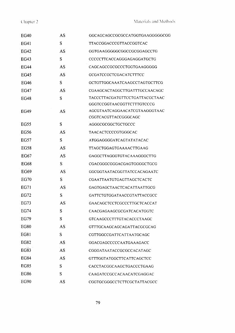
( hapter 2 Mate r ia l s and Me th od s
EG40
EG41
EG42
EG43
EG44
EG45
EG46
EG47
EG48
EG49
EG55
EG56
EG57
EG58
EG67
EG6 8
EG69
EG70
EG71
EG72
EG73
EG74
EG79
EG80
EG81
EG82
EG83
EG84
EG85
EG8 6
EG90
AS
S
AS
S
AS
AS
S
AS
S
AS
S
AS
S
AS
AS
S
AS
S
AS
S
AS
S
S
AS
S
AS
AS
AS
S
S
AS
G G C AG CAG CC G C G C CATG G TG AAG G G G G CG G
TTA C C G G A C C C G TTA C C G G TC A C
G G TG AAG G G G G CG G C CG CG G A G C CTG
CC CCC TTC A C C A G G G A G A G G A TG C TG
C A G C AG CCG CG CC CTG G TG A AG G G G G
G CG A TC C G C TC G A C A TC TTTC C
G C TG TTG G C A A A TC A A G C C TA G TG C TTC G
C G A A G C A C TA G G C TTG A TTTG C C A A C A G C
T A C C C TTA C G A TG TTC C TG A TTA C G C TA A C
G G G TC C G G TA A C G G TTC TTTG TC C C G
A G C G TA A TC A G G A A C A TC G TA A G G G TA A C
CG G TCACG TTACCG G G CAG C
AG GGCGCGGCTGCTGCCC
TAAC ACTCC CCG TG G G CAC
A TG G A G G G G A TC A G TA TA TA C A C
TTA G C TG G A G TG A A A A C TTG A A G
G A G G C TTA G G G TG TA C A A A G G G C TTG
CG ACGG G CG GG ACGAGTG GG G CTGCG
G G C G G TA A TA C G G TTA TC C A C A G A A TC
C G A A TTA A TG TG A G TTA G C TC A C TC
G A G TG A G C TA A C TC A C A TTA A TTG C G
G A TTC TG TG G A TA A C C G TA TTA C C G C C
G AAC AG C TC C TC G C C C TTG C TC A C C A T
C A A C G A G A A G C G C G A TC A C A TG G TC
G TC A A G C C C TTTG TA C A C C C TA A G C
G TTTG C A A G C A G C A G A TTA C G C G C A G
C G TTG G C C G A TTC A TTA A TG C A G C
G G A CG A G C C C C C A A TG A A A G A C C
CG G G A TA A TA C C G C G C C A C A TA G C
G TT T G G TA TG G C TTC A TTC A G C TC C
C A C C TA C G G C A A G C TG A C C C TG A A G
C A A G A TC C G C C A C A A C A TC G A G G A C
CG G TG C G G G C C TC TTC G C TA TTA C G C C
79

(. hapter 2 Mate r ia l s and Me th ods
EG91 S TCC G A C TTG TG G TC TC G C TG TTC C
EG92 S C A A C G C TC C G G C TC A G G TG TC A G G
EG93 S ATG G G C TG TG G C TG C A G C TC A C A C
EG94 AS TC A A G G C TG A G G C TG G TA C TG G
EG95 S C A C C A TG G G C TG TG G C TG C A G C TC
EG96 S CC G C CCC CTTC ACC G A CG CG CG G C TG CTG C CC
EG97 AS G GG CA G C AG CCG CG CG TCG G TG AAG G G G G CG G
TS221 S CACCATG G CG CG G CTG CTG CC
TS222 AS TCAG TC C C A TC TC A C C A G C TC
TS223 AS TC A G G A G A A C A A G TC TG C C TG
T- CD4 S C A C C A TG A A C C G G G G A G TC C C TTTTA G
CD4 AS CG G G A TC C TC A A A TG G G G C TA C A TG TC TTC
HIV eF S TCTG G C TA A C TA G G G A A C C C A
HIV eR AS CTG A C TA A A A G G G TC TG A G G
HIV F 2LTR S A A C TA G G G A A C C C A C TG C TTA A G
HIV R 2LTR AS TTG TC TTC G TTG G G A G TG A A TTA G
T-GFP S CACCA TG G TG AG CA AG G G CG AG G
GFP R AS C TC G A G TTA C TTG TA C A G C TC G TC C A TG
0-A1 S TG G G C A TG G G TC A G A A G G A T
0 - A3 AS CG G CCAG AG G CG TACAG G G A
2.6.16 Primer usage
Plasmid Name Usage Sense Anti-sense
pLgatewayR5IRESYFP sequencing/cloning EG4 EG5
pLgatewayR5IRESYFP QC mutation EG22 EG46 EG21 EG47
pLgatewayR7IRESYFP sequencing/cloning EG6 EG7
pLgatewayR7IRESYFP QC mutation EG11 EG 12
pLgatewayR9IRESYFP sequencing/cloning EG 17 EG 18
pLgatewayR9IRESYFP QC mutation EG23 EG24
pLgatewayRl 1IRESYFP sequencing/cloning EG 15 EG 16
pLgatewayRl 1IRESYFP QC mutation EG25 EG26
80

Chapter 2 Mater ials ; and M et hods
a-trim l siRNA plasmid sequencing EG 13 EG14
pHIT60 sequencing EG27 EG28
pLgatewayTva800IRESYFP sequencing EG6 8 EG70 EG41 EG42
EG72 EG74 EG45 EG67
EG79 EG81 EG69 EG71
EG91 EG92 EG73 EG80
EG85 EG8 6 EG82 EG83
TS221 EG84 EG90
TS222 GFP-R
EG91 EG92
T-GFP
pLgatewayT va800IRES YFP qPCR EG55 EG56
pLgatewayTva800IRESYFP QC mutation EG39EG43 EG40 EG44
EG96 EG97
pLgatewayTva800IRESYFP HA tag insertion EG48 EG49
pLgatewayTva800IRESYFP fragment cloning EG8 8 EG89
pLgatewayTva950IRESYFP sequencing TS221 TS223
pLgatewayTva950IRESYFP Fragment cloning EG8 8 EG89
pLgatewayp561ckIRESYFP cloning/sequencing EG93 EG95 EG94
CXCR4 detection EG57 EG58
CD4 detection T-CD4 CD4R
CD4 sequencing T-CD4 CD4 R EG54
QPCR actin o-A l o-A2
CSGW ss qPCR (early) H IV eF H IV eR
2.7 C a lc u la tio n s
The standard deviation o f the mean (sdm) and standard error o f the mean (sem),
where used, were calculated using Microsoft Excel.
81

Chapter 3 Routes o l 'en l rx \ i a I \ aSOO u i r I T n a^hO
Chapter 3
Routes of Entry Via Tva800 and Tva950
L v l is the activity o f a non-human primate factor that restricts HIV-1 (Cowan et al.
2002), and Refl is the activity found in human cells that blocks replication o f N -M LV
(Towers et al. 2000). In early 2004, Trim5a was reported to be the cellular factor
responsible for both L v l and Refl restriction. It was discovered by expressing genes
from a rhesus macaque gene library in a population o f HeLa cells. The gene that
rendered the HeLa cells resistant to HIV-1 infection was selected and cloned, and
found to encode Trim5a (Stremlau et al. 2004). Primate variants o f Trim5a affect
different restriction activities; human Trim5a restricts N -M LV but not HIV-1, but
rhesus macaque Trim5a can restrict HIV-1 and N-M LV (Stremlau et al. 2004; Yap et
al. 2004).
During these experiments, the HIV-1 virions were pseudotyped with VSV-G protein,
which directs the virus into the cell via clathrin-mediated endocytosis. Viruses enter
the endocytic pathway, and the fusion reaction o f the viral and cellular membranes is
triggered by the decreasing pH o f the endosomes (Aiken 1997; Sun et al. 2005).
Narayan et al. had reported in 2003 that the ASLV envelope could direct a virus into
the cell by two different routes depending on which o f its two cognate receptors,
Tva800 or Tva950, were used (Narayan et al. 2003). These two receptors are formed
o f identical viral binding domains but are attached to the cell surface in different
ways. Tva800 is a GPI-anchored protein, whereas Tva950 has a single membrane-
spanning domain. These different attachments mean that the receptors reside in
different regions o f the cell membrane. The plasma membrane o f the cell is not
uniform, but composed o f different microdomains (Harder and Simons 1997). These
microdomains have varying localised concentrations o f different phospholipids, fatty
acids and membrane proteins. GPI-anchored proteins tend to be found in lipid rafts,
whereas proteins with trans-membrane spanning regions are usually excluded from
these regions. This was found experimentally to be true for Tva800 and Tva950
(Narayan et al. 2003).
82

Unlike the fusion reaction subsequent to H IV binding o f CD4 which occurs at the cell
surface, fusion mediated by A S LV Env and Tva800 or Tva950 requires a low pH
step, and both receptors must therefore direct the virus through the endosomal
pathway, in which it w ill experience an increasingly acidic environment. The
initiation o f fusion occurs in a two-step fashion, unique to A S LV Env. The virus
must bind to the receptor, and be taken up into endosomes. Binding o f the virus
triggers a conformational change in Env that exposes the fusion peptide, which is then
inserted into the cellular membrane. Env is now sensitised to low pH, and exposure
in the endosomes w ill complete the reaction, resulting in fusion o f viral and cellular
membranes. Preventing the acidification o f the endosomes by a lysosomotropic
agent, for example NH4CI, blocks fusion, as the viral envelope protein cannot undergo
a conformation change necessary to complete fusion, and the virus w ill be unable to
enter the cell. Lysosomotropic agents are weak bases that can diffuse across
membranes. Once in an acidic environment they rapidly become protonated,
neutralising the drop in endosomal pH. Maintaining this block for extended periods
o f time renders viruses that have bound to Tva950 non-viable. Viruses that have
bound to Tva800, however, remain infectious for up to 6 hours in the presence o f
NH4CI and can be released to continue down the infectious pathway by simply
lowering the concentration to <10mM. The disparity between these two results has
been attributed to the difference in membrane attachment between the two receptors,
and how these affect the endosomal compartments the virus-receptor complex travels
through subsequent to binding (Narayan et al. 2003).
A similar case was reported by Keller et al. (Keller et al. 1992), in which one viral
binding domain was attached by two different methods to the plasma membrane. The
wild-type CD4 has a single transmembrane spanning domain, but by fusing the
extracellular viral ligand-binding domain o f CD4 to the C-terminus o f decay-
accelerating factor, DAF, which mediates transferral to a GPI anchor, a GPI-anchored
version was created. The kinetics o f uptake and the endocytic pathways embarked on
via this receptor, CD4-DAF, and the wild-type form, CD4-TM , were investigated.
CD4-TM was found to cluster into pits and was taken up by clathrin-mediated
endocytosis, whereas CD4-DAF was taken up by a clathrin-independent pathway
from microinvaginations at the cell surface at a slower rate o f about 32% that
mediated by CD4-TM. This corresponds well w ith the 2.3-fold higher rate o f
83

endocytosis for Tva950 (transmembrane) relative to Tva800 (GPI-anchored),
calculated by Lim et al. (L im et al. 2004), although it was not directly shown that
Tva800 or Tva950 is exclusively associated w ith either pathway.
Another difference between the two potential CD4 pathways was revealed by
incubation w ith the weak base, primaquine. Addition o f primaquine to cells
neutralises endosomal pH, and inhibits recycling o f receptors back to the cell surface
resulting in intracellular accumulation (van Weert et al. 2000). The intracellular pool
o f CD4-TM almost doubled when primaquine was added, whereas intracellular CD4-
DAF levels did not change. This indicates that the endocytic pathways taken by the
receptors lead to compartments w ith different sensitivities to pH neutralisation by
primaquine. Both receptors normally recycle back to the cell surface. Gold particles
incubated w ith either CD4-TM or CD4-DAF eventually associated with
multivesicular bodies, suggesting that the pathways converge (Keller et al. 1992).
The similarities between this example and the Tva800/950 pathways are evident.
Both Tva800 and Tva950 can ultimately direct the virus towards a successful
infection. However, as for CD4-DAF and CD4-TM, the routes that are taken after
binding to Tva800 or Tva950 prior to entry o f the virus into the cytoplasm are thought
to differ. Both routes are blocked by the addition o f a dominant negative form o f
dynamin, a component o f endocytosis mediated by both clathrin and caveolae
(Mothes et al. 2000).
A ll recent work w ith Trim5a and Fv l was carried out using VSV-G pseudotyped
virions. VSV-G directs virions into the cell via clathrin-mediated endocytosis (Aiken
1997). While certain events that occur between a retrovirus binding to a cell-surface
receptor and integration o f the proviral DNA into the host cell genome are described
to a certain extent, many aspects remain unclear. For example the precise sequence,
location, and extent o f events that occur on the route the virus takes between the site
o f entry into the cell cytoplasm and the cytoplasmic/nuclear boundary remain
undefined. How does the site o f fusion (at the cell surface or out o f an endocytic
compartment) and therefore the site o f viral entry into the cytoplasm affect the
inhibitory factors and other defence mechanism the cell mounts to try and prevent
retroviral infection? What events are concurrent w ith trafficking to the nucleus?
84

Chapter 3 Routes o f e n t r \ via l vaSOOant l l \ a R 5 0
How do these vary between different retroviruses? A system that allows the user to
vary entry events and conditions would aid clarification o f some o f these issues.
At the commencement o f these studies Fvl was known to be located to the cytoplasm,
but Trim5a was less well locally defined (Yap and Stoye 2003). It was o f interest to
see i f taking a different route to the cytoplasm could allow the virus time to mature
past a point o f susceptibility to restriction, or bypass the restriction altogether. I f
Tva800 and Tva950 do send the virus on significantly different routes o f entry into
the cell, then they could be useful in analysing whether restriction by Trim5a and Fvl
has a route-of-entry component that can be circumvented. For this, at least one
pathway would need to be distinct from that mediated by VSV-G, with different
biochemical or kinetic parameters. The receptors were therefore cloned and the
routes taken by virions subsequent to binding to Tva800 or Tva950 were investigated.
Results
3.1 Mus dunn i cells expressing Tva800 or Tva950 support
infection and replication by NB-MLV cores pseudotyped with ASLV
envelope
Mus dunni tail fibroblast cells are not known to harbour any endogenous restriction
factors, and they support replication o f MLV. They were therefore used as the basis
for these studies. To ensure that they were permissive to viruses carrying ASLV
envelope, Mus dunni cell lines expressing Tva800 or Tva950 were prepared. The
parental cell line was transduced with the vector pLgateway800IRESG418 or
pLgateway950IRESG418, which encode Tva800 or Tva950 respectively, and
neomycin resistance (see chapter 2 for all vector maps). Three days after infection
daughter cells were replated, and single cell clones were selected by addition o f G418
to the medium for two weeks and thereafter. 5x l0 4 Mus dunni cells stably expressing
Tva800 (d800), Tva950 (d950), or the parental cell line, were infected with between
2-250pl virus comprising NB-M LV core, ASLV envelope and carrying vector
encoding eGFP. Three days later cells were harvested, fixed and prepared for
analysis by FACS.
85

Chapter 3 Routes of entry via TvaSOO and Tva950
100
♦ dunni
— d800
—■— d950
0 100 200 300
vol virus (p i)
Fig. 3.1 Mus dunni cells expressing Tva800 or Tva950 support infection by N B -M L V cores
pseudotyped w ith A S L V envelope. 5x10⁴ d800, d950 and Mus dunni cells were transduced with
increasing volumes (l-2 5 0 p l) o f A S L V Env pseudotyped N B -M L V . The percentage o f cells
successfully infected was assayed by FACS for GFP expression 3 days later. Unfortunately no
standard cell line was readily available to titrate the virus. The parental Mus dunni cell line was
completely uninfectable by A S L V Env pseudotyped virus, but expression o f either Tva800 or Tva950
rendered the cells permissive. The experiment was repeated twice w ith the same clones w ith identical
results, so these clones were named d800 and d950 and used throughout. The d800 cell line was
further used subsequently to standardise stocks o f A S L V env pseudotyped virus.
The results o f this experiment presented in figure 3.1, show that the parental cell line
is totally non-permissive to AS LV Env pseudotyped M L V cores, but introduction o f
Tva800 or Tva950 is sufficient to render the cells susceptible to infection. Thus, these
cell lines can be used as a basis for further experiments. H IV-1 cores pseudotyped
with ASLV Env were also able to infect d800 and d950 cells (not shown).
3.2 Viruses bound to Tva800 receptors remain infectious for 6
hours if entry is blocked
To ensure that these receptors functioned in Mus dunni cells, as previously described
(Narayan et al. 2003; Narayan and Young 2004), the effects o f N H 4 C1 on viral entry
were investigated. 5x l0 4 d800 or d950 cells were plated and allowed to settle. 24
86

Chapter 3 Routes of entry via Tva800 and Tva950
hours later cells were incubated for 1 hour w ith 40mM NH4CI, prior to binding o f
ASLV envelope pseudotyped N B -M LV at 4°C for one hour. In control experiments
infection was initiated by addition o f medium at 37°C w ith 40mM NH4CI, and this
concentration o f NH4CI was maintained for the entire duration o f the experiment.
A fter 3 days the percentage o f cells that were infected was assayed by FACS for
eGFP expression. Cells that underwent identical infection procedures but did not
have any NH4CI in the medium at any time set the maximum level o f infection.
100
80
No Virus No NH4CI A lw ays N H 4C I
Fig 3.2.1 A S L V Env pseudotyped viruses are unable to infect cells succesfully in the presence o f
40m M N H 4CI. d800 or d950 cells were plated, and 24 hours later cells were incubated in medium
containing 40 m M N H 4C1 for 30 minutes. Cells were transduced w ith 200pl A S L V Env pseudotyped
N B -M L V in lm L medium containing 40m M N H 4C1. Results shown are combined from 3 separate
experiments, and error bars show sem.
Data in figure 3.2.1 show that when NH4CI is maintained in the cell medium for the
duration o f the experiment viral infection is entirely blocked, there is no difference in
infection levels (i.e. GFP expression) between cells infected w ith virus under an
NH4CI block and cells that do not have any virus on them at all.
In a second set o f experiments, cells were incubated w ith virus and infection was
initiated as described above. A t preset timepoints after infection o f between 1 and 8
hours the NFLCl-containing medium was removed, and replaced w ith DM EM only,
potentially releasing virions from the effects o f a block caused by inhibition o f
acidification o f the endosomes.
87

Chapter 3 Routes of entry via Tva800 and Tva950
140
120 \w302 1 0 0 ju■c
80 i01cEm 60E0)</)C 40 j0
;c> 20 j
0
-20 Jtim e of NH4CI rem oval (hours)
Fig. 3.2.2 The proportion of virions that remain infectious under an N H 4Cl-induced block to infection
is not the same for d800 and d950 cells. d800 or d950 cells were plated out and incubated in 40m M
N H 4CI for 30 mins. 250pl ASLV Env N B -M L V was adsorbed at 4°C for 1 hour in the presence o f
40mM N H 4CI. Medium containing 40mM N H 4C1 was then added, and cells were incubated at 37° C
for between 1-8 hours. Titres obtained were compared to cells incubated solely with medium. Results
are combined from 3 separate experiments, and error bars show sdm.
Figure 3.2.2 shows the effect o f removing the NH4CI block from d800 and d950 cells
between 1-8 hours after infection. The percentage o f GFP-expressing cells is an
indication o f the percentage o f virions remaining viable at the time o f N H 4C 1 removal.
Removal o f this block at set time points after infection reveals the length o f time that
infection can be arrested while viral particles still remain viable. In d950 cells
infectivity decreases rapidly with incubation under N H 4CI, showing that the virus is
no longer able to complete the infectious cycle. However, when virions have bound
to Tva800, >80% remained infectious even when the infectious pathway was blocked
for 8 hours.
In order to see how the d800 and d950 infectivity periods compare with another
receptor also blocked by NH4CI treatment, an identical experiment was carried out as
d 800
d 950
88

Chapter 3 Routes of entry via Tva800 and Tva950
described above with NB-M LV pseudotyped with VSV-G. The results are shown in
figure 3.2.3.
120 i
</)g 1 0 0
80
60
40
06 7 82 3 4 5No 1
NH4CI
time of NH4Cl removal (hours)
Fig. 3.2.3 Proportion of virions bound via VSV-G envelope that are able to complete an infectious
cycle after being blocked by NH4CI for varying times. The experiment was carried out as described in
the legend to figure 3.2.2. Results shown are combined from 3 experiments, and error bars show sdm.
As can be seen, the decrease in infectivity is even more dramatic than that for d950
cells, with fewer than 15% of virions remaining infectious after only 2 hours NH 4 CI
treatment. This confirms that the stability seen with ASLV pseudotyped virions
bound to Tva800 is indeed exceptional. The instability seen with d950 cells could be
either because the viral particle itself is not stable under conditions where the
acidification o f subcellular organelles is blocked, or because the virus-receptor
complex in the compartment it is trapped in is somehow unstable. Over 80% of
viruses that have bound to d800 cells remain viable when they are trapped for 8 hours,
suggesting that the degradation under blockage experienced by viruses bound to
Tva950 is not related to instability o f the virus per se, but to the cellular environment
in which it is bound and held.
The viral binding domains o f the receptors Tva800 and Tva950 (contained entirely
within the first 1 0 2 amino acids) are identical, therefore the difference between the
two pathways must relate to their attachment to the cell membrane, the
89

Chapte r 3 Routes ot ent! \ \ ia T\aSOO and TvaOM)
microenvironment o f the membrane in which the receptors are found, and the way
these two factors affect the route by which virions binding to the receptors enter the
cell.
These studies are instructive in trying to assess the differences in Tva800 and Tva950
endocytic pathways. The question next addressed was i f viral particles entering by
either one o f the receptors could be co-localised with a marker for a cellular
compartment, and i f this would reveal a convergence o f the two pathways.
3.3 Viral particles co-localise with markers for the late, but not
the early endosomes
Both Tva800 and Tva950 receptors function effectively as ports o f entry for the virus.
The differences between Tva800 and Tva950 correspond to those found for CD4-
DAF and CD4-TM, and this, combined with the fact that regardless o f receptor type,
the ASLV envelope needs low pH to trigger the fusion reaction, suggests that at some
point the pathways taken by the virus after entry through one receptor or the other
must pass through a low pH compartment, and could conceivably converge. In order
to try and ascertain where the block to infection in the presence o f NH 4 C1, and
potentially where this point o f convergence might be, viral particles were made with
an NL4.3 (HIV-1) core, ASLV envelope, and GFP-vpr ( ‘green H IV ’). GFP-vpr is a
fusion protein made o f the accessory protein vpr, and GFP, which renders it
fluorescent (McDonald et al. 2002). GFP-vpr, like vpr, is taken up in high copy
number by viral particles, and usefully remains associated with the viral ribonucleic
acid, so viral particles can be tracked by this fluorescence as they move through the
cell. The green HIV was concentrated, placed on d800 cells at 4°C, and allowed to
bind for an hour in the presence o f 40mM NH 4 CI. After one hour the cells were
washed and warm (37°C) medium + 40mM NH 4 CI was added, and the cells incubated
for 10, 30, 60 or 120 minutes. The infection was halted by removal o f medium and
addition o f 4% formaldehyde in PBS. Cells were then permeabilised and stained red
for either early endosomes (anti-EEAl) or late endosomes/ multivesicular bodies
(MVB) (anti-lamp3), and visualised under the microscope. A t least four pictures were
taken for each condition, and one example is shown in figure 3.3.1. Green viral
particles (top pictures) were scored as co-localised with a red endosomal marker
90

Chapter 3 Routes of entry via TvaSOO and Tva950
(middle pictures) i f signals from the two markers overlay each other (bottom
pictures), as analysed in Adobe Photoshop.
Time EEA1 (early endosome) cd63 (late endosome)

Chapter 3 Routes of entry via TvaSOO and Tva950
Fig. 3.3.1 Fluorescent A S L V Env pseudotyped virions colocalised with markers for the late
endosome/MVB in Tva800 cells. lx lO 5 d800 cells were plated on glass coverslips, and infected at
M O I 5 in the presence o f 40m M N H 4C1. After between 10-120 minutes cells were fixed and stained
with anti-EEA l (early endosomal marker) or anti-cd63 (found on the late endosomes and the M V B ).
92

Chapter 3 Routes of entry via Tva8()() and Tva950
Co-localisation was scored using Adobe Photoshop software; the green channel was
removed from pictures, and i f a red fluorescent mark found to be underneath co
localisation was confirmed. A rough assessement can be made by looking for yellow
marks but this does not always reveal faint red fluorescence, and can be skewed by a
high background o f red that is non-specific staining. Between 4 and 6 pictures were
taken for each condition, w ith at least 40 virions per condition, although only one
picture is shown as an example. The green virions were only rarely seen co-localised
with the early endosomes stained by EEA1, so that the left-hand column o f figure
3.3.1 reveals few yellow puncta. For cd63, however, while the red staining did in
general appear to be higher than staining with E E A 1, there was a disproportionately
higher level o f co-localisation seen with the green virions, resulting in many yellow
puncta in these pictures. A ll pictures taken were scored in this way to obtain a
quantitative assessment o f co-localisation, shown in figure 3.3.2.
60
□ EEAl (early)
■ cd63 (la te )
10 mins 30 rrins 1 hr 2 hrs
tim e-point
Fig. 3.3.2 A quantitative analysis o f the colocalisation o f fluorescent A S L V Env pseudotyped virions
with markers for the late endosom e/MVB in Tva800 cells. Cells were scored for the percentage o f
virions found colocalised with these markers, i.e. the red and green markers significantly overlay when
analysed with Adobe Photoshop. Error bars represent the standard deviation o f the mean number o f
virions colocalised across the 4-6 pictures taken for each sample.
93

C 'hapter 3 Route.s o f entry v ia l vaSOO and Tvn^fO
The time at which the highest percentage o f virions were found to be co-localised
with markers for EEA1 (found on the early endosome) was when virions had been
allowed to infect cells for a total o f 10 minutes. Even then, only 12% o f virions co
localised. This does not mean that the virion does not pass through the early
endosome, rather that i f it does, it must pass through very rapidly and not be held
there for any significant length o f time. The number o f virions found co-localised
with cd63 was consistently higher, between 45 and 50% for every time period
analysed up to 2 hours.
The experimentally determined pH at which ASLV-A Env mediated fusion out o f the
endocytic pathway occurs is <pH5.5 (Mothes et al. 2000). The pH o f early
endosomes is usually above this, between 5.9-6.0, not dropping to below 5.5 until the
late endosome is forming, indicating ASLV-A exit at these later stages. Together
with these data, it would seem to indicate that under a block o f NH4CI the virion can
still progress rapidly through the endocytic pathway up to the late endosome/MVB,
but at this point it pauses, probably while initiating the fusion reaction.
A comparison was then carried out for co-localisation o f virions with cd63 markers
after binding to Tva800 or Tva950, to see i f patterns o f co-localisation differ after
binding to these receptors, and to look at a longer timescale. A similar experiment to
that described above was carried out on both d800 and d950 cells, and viral infection
in the presence and absence o f NH4CI was analysed after 30 mins and 4 hours. Co
localisation with cd63 markers was analysed as before. The results are shown in
figures 3.3.3 and 3.3.4.
94
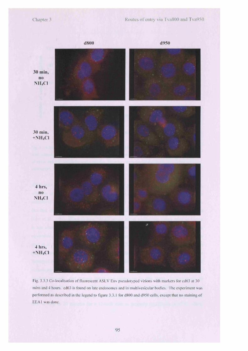
Chapter 3 Routes of entry via Tva800 and Tva950
d800 d950
NH4CI
30 min, +NH4CI
NH4CI
4 hrs, +NH4CI
Fig. 3.3.3 Co-localisation o f fluorescent A S L V Env pseudotyped virions with markers for cd63 at 30
mins and 4 hours. cd63 is found on late endosomes and in multivesicular bodies. The experiment was
performed as described in the legend to figure 3.3.1 for d800 and d950 cells, except that no staining o f
E E A 1 was done.
95

Chapter 3 Routes of entry via Tva800 and Tva950
120
0 £
1 80
0)Q.£ 40o
>»♦-oI -<D-QE3C
f l f i
i
Aneg plus neg plus neg plus neg plus
30 mins 4 hours 30 mins 4 hours
d800 d950
c e ll/t im e /c o n d itio n
■ CD63-ve
□ CD63+ve
Fig. 3.3.4 A quantitative analysis o f the co-localisation o f fluorescent A S L V Env pseudotyped virions
with markers for cd63 at 30 mins and 4 hours. Pictures were randomised and scored blind for overlap
o f virion and cd63 signals for each condition. Error bars show the standard error o f the count for each
photograph.
d800 cells had on average over 50 virions per picture after 30 minutes o f infection,
whether or not NH4CI was present. The d950 cells had almost double this average
number o f virions after 30 minutes when NH4CI was not present. This may reflect the
fact that entry via Tva950 is on average 2.6-fold faster compared to entry via Tva800
(Lim et al. 2004), although how NH4CI would affect trafficking stages prior to fusion
is not clear. A fter 4 hours the levels in d800 and d950 NH4C 1-negative cells are
equivalent, and for both cell types total numbers o f virions are under 50% o f those
seen at 30 minutes. For d950 cells under N H 4 C1, the levels o f virions are comparable
to those seen after 30 minutes under NH4CI. For d800 cells, however, the levels are
increased by ~2 .5-fold, which implies that in the extra 3% hours greater numbers o f
virions are able to both enter the cell (and so not be washed o ff in steps preparatory to
binding antibody) and remain intact and infectious in this time. In retrospect 30
minutes probably was not sufficient time for a first time-point. Virus was bound at
4°C for an hour, and although at time zero pre-warmed medium is added, it can still
take as long as 20 minutes for a 12-well dish to properly equilibrate at 37°C, which
96

Chapter 3 Routes o f entrs v ia T\aS()( ) and 1 \ a 9 5 0
does not really allow the virions sufficient time to initiate entry before the experiment
was halted.
Given the timecourse data shown in figure 3.2.2, it is also surprising that so many
virions do appear to remain sufficiently intact in d950 cells under an NfUCl-block
after 4 hours. It could be, o f course, that these are no longer infectious virions and
would not be capable o f completing an infectious cycle. This would mean that
fluorescence per se is not a good indicator o f potential infectivity.
As virions that have entered using both Tva800 and Tva950 are found co-localised
with cd63, either any differences between the routes used by the two receptors must
be prior to fusion with this compartment, or there is more than one type o f cd63-
positive compartment.
3.4 Inhibition of Rab5, but not Rab7, decreases entry via Tva800
and Tva950
Having established that the pathways o f entry o f the virus subsequent to binding and
entry via Tva800 or Tva950 converge on the late endosome, the question then arises
as to the differences between Tva800 and Tva950 pathways before the late endosome.
I f pathways subsequent to binding are different, would alteration o f cellular
conditions affect the virus as it traffics prior to this point to differentiate the two
pathways? To address this question, a set o f cellular traffic controllers, the Rab
proteins, were considered. Rabs are small GTPases that are found at several points in
the vesicle trafficking pathways o f the cell. Crucially, one Rab protein is
characteristically associated with a subset o f endocytic vesicles, or a single stage o f a
pathway. For example, Rab5 is found on endocytic vesicles prior to fusion with early
endosomes and on early endosomes themselves, and Rab7 is involved in late
endosome maturation and lysosome biogenesis. Rab9 controls transport between the
late endosome and the golgi, and Rabs 4 & 11 are involved in recycling back to the
plasma membrane (see figure 1.9.2 for an illustration o f these pathways).
Functional Rabs exchange GDP for GTP as part o f their cycle from inactive to active,
respectively. Dominant negative forms o f these Rab proteins that preferentially bind
97

Chapter 3 Routes o f ent rv \ ia TvaNOO and Tva95()
GDP have long been used to study the effects o f their inhibition (Sieczkarski and
Whittaker 2002a). Due to GDP binding, the dominant negative Rab is permanently
switched off. When a dominant negative version o f a Rab protein is over-expressed
in a cell it swamps the wild-type functional Rab, down-modulating its activity.
To assay the effects o f expression o f an exogenous factor on a large population o f
cells swiftly, an assay was developed that allows a non-clonal population o f cells
expressing this exogenous factor to be directly compared with factor-nai've cells in a
single well. The use o f single cell clones can sometimes lead to false conclusions
being drawn, i f a single cell colony is picked that is genetically positive for the
exogenous factor, but shows no effects. The use o f a non-clonal cell population in
which there is a range o f expression levels suppresses rare effects and emphasises
general trends (Bock et al. 2000; Bishop 2001). The gene o f interest is cloned into the
plasmid pLgatewayXIRESYFP. The gene o f interest is inserted at the X, and
expression is driven by the M LV LTR. YFP expression is driven from an IRES.
Cells transduced with this vector that are positive for YFP expression also express the
inserted gene of interest. Three days after the initial transduction cells are further
infected with the challenge virus, which carries EGFP vector. After a further three
days, four populations o f cells can be seen on FACS analysis. Double negative cells
are infected with neither the factor under consideration nor challenge virus, cells
expressing EYFP only have not been infected with the challenge virus, those cells
expressing EGFP only are factor negative but challenge virus positive, and double
positive cells for EYFP and EGFP express factor and were infected by the challenge
virus. The proportion o f cells that expressed the gene o f interest that were
successfully infected can be compared to the proportion that were infected in the
population that did not express any extra factor. Comparison o f proportions o f these
populations o f cells allows the effects o f exogenous factors on viral replication to be
determined (see section 2 .2 . 2 for a worked example).
This assay was used to assess the effects o f the dominant negative Rab proteins on
viral replication. Primers to amplify the ORFs o f Rabs 5, 7, 9 and 11 were designed
from published sequences (GenelDs 5868, 7879, 9367 and 8766, respectively) and
amplified out o f a HeLa library. Dominant negatives Rab5 S34N, Rab7 T22N, Rab9
S21N and Rabl 1 S25N have been designed and characterised previously (Riederer et
98

Chapter 3 Routes of entry via Tva800 and Tva950
al. 1994; Stenmark et al. 1994; Ullrich et al. 1996; Vidricaire and Tremblay 2005).
These mutations were introduced into the plasmids by QuikChange mutagenesis.
Challenge virus titres on cells expressing the dominant negative Rab proteins (and
EYFP) were compared with titres in Rab DN null cells. The results are shown in
figure 3.4. A fold-difference o f 1 indicates that titres o f challenge virus were not
altered and <0.7 indicates attenuation o f viral titre through inhibition o f viral
replication by the expression o f the dominant negative Rab. Titres are not normally
completely reduced by dominant negative Rabs as a significant population o f wild-
type, functional Rab protein remains in the cell.
1.6 n
1.4
Rab
Fig 3.4 Inhibition o f Rab5 alone decreases titres o f A S LV Env pseudotyped N B -M L V . 5 x l0 4 d800 or
d950 cells were plated and transduced with Rab vector at M O I 1. 2 days later cells were split, and after
1 further day challenged with A SLV Env pseudotyped N B -M L V . The control experiment for Rab5DN
carried out using VSV -G pseudotyped N B -M L V is also shown (Rab5DN Ctl). Inhibition was
monitored by calculating the ratio o f the percentage o f cells expressing Rab compared to the percentage
of negative cells, for each well. Results shown are combined from two separate experiments, and error
bars show sdm.
As can be seen from figure 3.4, only Rab5DN decreased viral titres to any extent, and
the effect was identical for both Tva800 and Tva950 cells. As Rab5 is involved in
regulating traffic from the plasma membrane to early endosomes this would indicate
that part o f this pathway is crucial for viral entry via both Tva800 and Tva950. After
the virus has been endocytosed, Rab5 is probably therefore involved in directing the
99

endocytic vesicle to the early endosome, or in the fusion reaction between the
endocytic vesicle and the early endosome. Given that the microscopy data presented
in figs 3.3.1 to 3.3.4 earlier would indicate that viruses colocalise substantially with
markers for the late endosome/MVB, and that Rab7 is substantially involved in
trafficking to, and is a marker for, the late endosome, it is surprising that no inhibitory
effect is seen w ith Rab7DN. Fusion o f the virion via A S LV Env out o f the endosomal
pathway is triggered at a pH o f below 5.5 (Mothes et al. 2000), which is lower than
the pH reached in the early endosome. Control experiments to assess the inhibitory
effects o f Rab7DN on influenza infection have been carried out by previous groups
that used the same inactivation mutation (Sieczkarski and Whittaker 2003). Our
experiments assessing the effects o f Rab7DN expression on in fectiv ity o f influenza A
(PR8) in MDCKs were hard to quantify, possibly due to toxic effects (experiments
carried out by Seti Grambas). Addition o f dominant negatives does not reduce the
level o f functional Rab proteins in the cell, but rather floods the cell w ith defective
versions, which are more like ly to be interacted with. Pathways are, therefore, not
totally inhibited, but downregulated. A better way to assess effects would be to use
SiRNA to reduce levels o f Rabs 5 and 7 in the cells more substantially. This should
be done before making any firm conclusions about either Rab protein and their effects
on entry via Tva800 and Tva950.
3.5 Route of entry via Tva800 or Tva950 receptor does not affect
whether NB-, N- or B-tropic MLV is affected by Fv1 or Trim5a
One reason to investigate the differences between Tva800 and Tva950 was to assess
the effects o f route o f entry on restriction by F v l and Trim5a. It could be
hypothesised that forcing the virus to take an alternative route into the cell to its usual
course could permit it to evade restriction activity. In order to assess whether this
could be the case for Tva800 and Tva950, the 2-colour assay was adapted, w ith d800
and d950 cells described above used as the basis for this assay. The d800 or d950
cells were transduced at M OI ~1 w ith F v ln, F v lb or Trim5a. This M OI results in
approximately 40% o f the target cell population becoming YFP positive, and by
extension restriction factor positive. Three days later, cells were then challenged with
AS LV envelope-pseudotyped NB-, N- or B-tropic M L V w ith between 2-100pl.
Results were analysed by FACS three days later.
100

Chapter 3 Routes of entry via TvaSOO and Tva950
ASLVenv/NB- MLV - o - ASLVenv/N- MLV ASLVenv/B- MLV
TVA800, No restriction factor TVA950, No restriction factor
100 100
10
100 100
TVA800, F v ln TVA 950 F v ln
100 100
1010
1 1100 100c
Wl
TVA800 F v lb TVA950 F v lb
100100
10
100 100
0.1
vol applied (pi)
Fig. 3.5.1 Restriction o f N -, B - and NB-tropic M L V by F v l and T rim 5a is not affected by entry via
Tva800 or Tva950. d800 and d950 cells were plated out and transduced with F v l" , F v lb or Trim 5a.
Two days later cells were passaged and challenged with 2-100p l A S L V Env pseudotyped N -, B-, or
N B -M L V . The restriction o f the virions by the restriction factor was compared in each cell line. The
x-axis shows the volume o f virus applied, in pi, and the y-axis the percentage o f cells infected. One
experiment is shown as an example.
101

n hData in fig 3.5.1 show the percentage o f cells expressing F v l , Fv l or Trim5a that
permitted infection by NB-, N- or B-tropic viruses. Cells that expressed no restriction
factor reveal a difference between the two Gag-Pol vectors used in this experiment,
which were pHIT60 for NB-tropic virus and pCIG3N or pCIG3B for N- or B-tropic
virus. pCIG3N and pCIG3B were prepared by subcloning o f constructs encoding N-
or B-tropic gag-pol into pCIG2, which encodes GFP fo llow ing an internal ribosome
entry site (Bock et al. 2000). Virus constructed w ith pHIT60 has been consistently
found to replicate to higher titres, but this difference is consistent throughout the
experiment and w ith previous experience, and the use o f ratios in further analysis
normalises the difference.
For the d800 and d950 cell populations, the ratio o f viral titres on restriction factor
positive cells: restriction factor negative cells was calculated (as detailed in Materials
and Methods section 2.2.2). In order to obtain one value for each virus/restriction
factor, all points on the titration curves shown in figure 3.5.1 and obtained in one
other experiment were used, where the percentage o f cells in all 4 populations was
>1% (the reliable lower detection lim it by fluorescence analysis). A combined fold-
difference o f RF positive/RF negative o f <0.3 indicates restriction activity, as this
would suggest that when the restriction factor is present less than 30% o f the cells
become infected compared to when the restriction factor is not present. Between 0.3-
0.7 indicates an intermediate level o f restriction, and over 0.7 there is considered to be
no significant activity.
102

Chapter 3 Routes of entry via Tva800 and Tva950
d 8 0 0 ce lls
a 0.8
w 0.6
0.4
0.2♦3 I..Ill NB
□ N
Fvln Fvlb Trim5a
d 9 5 0 ce lls
T3O8 0.8
j£ 0.6 8̂
0.4
ooa■
0.2 -I
0 I li .1 ll■ NB
□ N
■ B
Fvln Fvlb
restriction factor
Trim5a
Fig 3.5.2 Restriction o f N -, B- and NB-tropic M L V by Fvl and Trim5a is not affected by entry via
Tva800 or Tva950. The experiment was carried out as described in legend to figure 3.5.1. Columns
plotted are the proportion o f Fvl or Trim5a positive cells infected compared to negative cells. Results
shown are combined from 2 separate experiments, and all points o f the titration curves obtained used to
generate the above data as long as the percentage o f cells in each quadrant (RF and virus-negative, RF-
positive virus-negative, RF-negative virus-positive, and RF and virus-positive) was >1%. Error bars
represent sdm.
The decreases in viral titres caused by Fvl or Trim5a were as expected, and generally
in accordance with previously reported values. More specifically: F v ln or Trim5a did
not restrict NB-tropic M LV; F v lb and Trim5a restricted N-tropic M LV; and F v ln
restricted B-tropic MLV. As seen in previous assays, F v lb also strongly restricted
NB-tropic M LV (Bock et al. 2000). However, all o f these restrictions and
103

I 'hapter 3 Routes o l e n n a \ ia I \uSOu and
permissivities were reproduced for both d800 and d950 cells, with no significant
difference seen between them. Therefore, no difference in restriction was detected in
this system when ASLV Env pseudotyped virions entered cells by either route. As
these results are also commensurate with those obtained from previous studies where
virions pseudotyped with VSV-G were used, this implies that in terms o f the effects
on restriction by Fvl and Trim5a there is no functional difference between any o f the
pathways studied here. This would indicate either that it is not possible for the virions
to mature past a point o f susceptibility to the restriction factors, or that i f it could do
so, it is only by using another route o f entry not considered here.
3.6 ASLV envelope pseudotyped HIV, N- and B-tropic MLV are
not restricted by a range of Trim proteins after entering cells via
Tva800
Trim5a is just one isoform o f one member o f a family o f over 70 proteins containing
all or parts o f the tripartite m otif and the B30.2 domain (Reymond et al. 2001), o f
which at least two other members have been confirmed to exhibit anti-viral activity.
Trim l has anti-N-MLV activity, and Trim 19 (PML), found in nuclear bodies, is
relocated under the influence o f several viral infections, and is able to repress human
foamy virus gene expression (Regad et al. 2001). Having confirmed that no novel
anti-viral activity o f Trim5a is evident when the virus entered via the two receptors, it
was o f interest to see i f any other Trim proteins exhibited anti-viral activity i f the
virus took the more unusual post-entry route subsequent to binding to Tva800.
Human Trim proteins 1,6, 15, 18, 21, 22, 26, 27, 31, 34, 38, and 68 were cloned into
pLgatewaylRESYFP by Dr. Melvyn Yap. 5x l04 d800 cells were plated and
transduced at MOI ~1 with one o f the human Trim proteins listed above. Three days
later these were challenged with ASLV envelope-pseudotyped HIV, G89V HIV, NB-
MLV, N -M LV and B-MLV. G89V HIV is mutated in the CA domain that binds
cyclophilinA, so that it can no longer bind CA. This mutation has been noted to
abolish restriction by Trim-Cyp, but is not absolutely required for Trim5a restriction
(Stremlau et al. 2006b). After three days the percentage o f virally infected d800
Trim-positive and d800 Trim-negative cells was assessed by FACS. The ratio o f
Trim-positive cells infected was divided by Trim-negative cells infected and again, a
value o f lower than 0.3 indicates significant restriction activity.
104

£hapter 3 Routes of entry via Tva800 and Tva950
10
0.01T rim
■ H IV (A S L V ) ■ H IV G 8 9 V (A S L V ) □ N B -M L V (A S L V )
■ N -M L V (A S L V ) ■ B- M L V (A S L V )
Figure 3.6 N o new restriction is revealed when viruses encounter a panel o f Trim proteins in d800
cells. d800 cells were plated and transduced with the trim proteins shown at M O I 1. 2 days later cells
were split and challenged with A S L V Env pseudotyped H IV , H IV G 89V , N B -M L V , N -M L V and B-
M L V . Results shown are presented as the ratio o f (proportion o f trim positive cells infected)/(trim
negative cells infected). A ratio o f <0.3 reveals restriction activity.
As expected, expression o f Trim5a led to a considerable drop in titres o f H IV , N-
M LV and G89V H IV. Titres o f N -M LV were also reduced in cells expressing T rim l
as previously reported (Yap et al. 2004). For all other trim proteins used in this panel
no effects on replication on any o f the viruses could be detected. This would, o f
course, not completely preclude the possibility that anti-viral activity could manifest
itself under different conditions, or when a combination o f Trim proteins was
expressed in the same cell.
An extensive screen has been recently carried out in which 55 Trim proteins were
assessed for their abilities to alter ASLV Env pseudotyped H IV and M L V viral entry
through the Tva950 receptor, as well as viral release (Uchil et al. 2008). No Trim
proteins surveyed were found to have significant effects (> 10-fold inhibition) against
H IV or N -M LV entry, w ith the exception o f human Trims 1 and 5 against N -M LV ,
confirming the results reported here. When release o f virions was investigated,
105

however, 19 different Trim proteins inhibited M L V release by at least 10-fold (human
Trims 1, 5, 8, 11, 13, 15, 19, 21, 25, 26, 27, 31, 32, 62, and mouse Trims 8, 11, 19, 25,
and 27), and 5 sim ilarly affected H IV release (human Trims 11 and 15, and mouse
Trims 11, 25 and 56). As the fam ily o f Trim proteins consists o f many diverse
proteins linked only by their sim ilar structures, it is to be expected that they w ill be
found in many different cellular locations w ith a variety o f functions, and could
therefore exert effects on retroviral replication in a number o f different ways at
several points in the viral replicative cycle.
D iscussion
These experiments were designed to aid clarification o f the relationship o f the route o f
entry that a virus takes into the cell w ith restriction by F v l and Trim5a. Firstly the
receptors Tva800 and Tva950 were expressed in Mus dunni cells. The effects o f
N H 4 C I on A S LV Env-mediated entry into these cells was investigated, revealing a
significant difference in the effects on entry via Tva800 and Tva950.
Mus dunni cells engineered to express Tva800 or Tva950 are fu lly permissive to
infection w ith A S LV Env pseudotyped M L V cores. Entry via either receptor permits
successful infection. However, when the infection is blocked using NH 4CI ,
differences in the pathways used subsequent to receptor binding emerge. Viruses that
have bound to Tva800 are stable for up to 6 hours under an N H 4 C I block and can still
initiate a productive infection, whereas those that bound to Tva950, like virions bound
to VSV-G, lose in fectiv ity after 2 hours. There is no difference in the envelope
protein used between Tva800 and Tva950 and therefore no difference between the
fusion peptides, which is what anchors the envelope protein and virus into the target
cell membrane. Therefore the difference must be due to the method o f attachment o f
the receptor to the cell membrane, or the localised m ilieu o f the plasma membrane in
which the receptor resides and the way in which this affects the manner and rate o f
internalisation. This latter possibility is explored and discussed further in chapter 4.
GFP-labelled virions co-localise w ith cd63-positive compartments under an N H4 C I-
induced block, however it has not been possible to confirm the exact identity o f this
106

intracellular compartment. V irions w ith a red fluorescent membrane label and GFP-
vpr labelled nucleic acid can be used to detect when membrane fusion has occurred
(w ith loss o f the red label), and thus when a productive infection is like ly to occur
(used in experiments described in chapter 5). Usage o f these dually labelled virions
would help to guard against fo llow ing virions through intracellular pathways that had
been non-specifically endocytosed and were destined for lysosomal destruction. By
infecting cells w ith these virions under an NH4CI block, and then fix ing the cells and
staining subcellular compartments w ith EEA1, cd63 or other markers, and then a third
colour (blue), it might be possible to detect more specifically which subcellular
compartment virions were in. A comparison o f cells which had a constant NH4CI block up to fixation, and cells in which this block was removed for a short time before
fixation to allow fusion, would hopefully reveal v irion fusion and release near one
type o f compartment. Such experiments using a secondary antibody w ith emitted
light wavelength 350nm, as this was the only third colour that did not carry over into
different channels and could be satisfactorily resolved by the filterset on the
deltavision microscope, were attempted. However, these experiments could not be
pursued as the staining pattern o f early and late endosomes was too indistinct to be
able to draw any definite conclusions.
Ammonium chloride may have effects on the morphology o f intracellular
compartments and on trafficking routes that are as yet undefined. Another chemical
that inhibits the acidification o f endosomes is bafilomycin A l , which acts by blocking
proton ATPases, but it has also been shown to block transport from early to late
endosomes (Bayer et al. 1998). Thus inhibition o f a virus by bafilom ycin A l could be
due to the prevention o f viral uncoating, or in an entirely separate manner to the
inability o f the virus to continue down the endosomal pathway and access the late
endosome. Considering the results presented here and the A S LV -A Env-Tva receptor
system, however, it is unlikely that in ammonium chloride-treated cells the virus is
being blocked by anything other than an inability to initiate fusion in the endosomes.
A S LV-A Env has been shown to require low pH for the conformational change
necessary for fusion, and so even i f ammonium chloride does affect trafficking
downstream o f this point o f fusion, in the context o f these experiments this would be
irrelevant as the virus would be blocked in the endosome (Mothes et al. 2000; Barnard
et al. 2006).
107

C hapier \ ui I \ aN')<) kuu! I wX-
Examination o f other systems where one receptor has two modes o f attachment to the
cell membrane indicate that the difference in attachment is not necessarily trivial, and
there are difference in cellular pathways after entry that can significantly affect the
timing and success o f the infection. Viruses may have evolved to exploit a niche in
these pathways, or be able to make use o f several. There is no indication, however,
that there is any physiological relevance o f the usage o f Tva800 or Tva950.
It has not been possible to pinpoint all the commonalities and differences in entry
pathways subsequent to Tva800 and Tva950 binding. Both pathways are
downregulated by dominant negative Rab5, which is involved in clathrin-mediated
endocytosis (van der Bliek 2005), suggesting that this pathway plays at least some
role in uptake via Tva800 and Tva950. This is consistent with previous data that
showed dominant negative forms o f dynamin, which mediates entry via both clathrin-
mediated endocytosis and via caveolae, inhibited entry mediated by ASLV-A by
between 60-80% (Mothes et al. 2000). However, this would mitigate the similarity
somewhat o f Tva800 and Tva950 to CD4-DAF and CD4-TM, as CD4-DAF was not
taken up by a clathrin-mediated pathway. No differences were seen in the infectivity
rate between the two receptors when the cells expressed dominant negative forms o f
Rabs 7, 9 and 11, which modulate later stages o f the cellular pathways. The lack o f
effect seen with Rab7DN is surprising, given that this is thought to modulate traffic
from the early to late endosomes. This may indicate the presence and usage o f other
trafficking pathways, or the insufficient down-regulation o f Rab7 to generate a
significant effect. Rabs 9 and 11 moderate traffic from the late endosome to the trans-
Golgi network, and recycling between the trans-Golgi network and the early
endosome respectively. It would therefore not be expected that virions bound to
Tva800 or Tva950 would use pathways controlled by these Rab proteins during a
successful infection, and therefore dominant negative inhibitors o f these pathways
would have no effect. Microscopy data indicate that Tva800 and Tva950 both deliver
virions to late endosomes/MVB. NH 4 CI inhibits the fusion event between the viral
and cellular membranes, the stage just before entry into the cytosol common to both
receptors after binding to ASLV-A Env, so it is to be expected that the differences
between the Tva800 and Tva950 pathways would be before this common point o f
fusion.
108

C hapter a o! c i u r \ \ ia I \ aSi )() and 1 \ al)50
The possibility o f either Tva800 or Tva950 rerouting ASLV Env pseudotyped virions
around a block to infection in restrictive cells was assessed. F v ln, F v lb and Trim5a
were expressed in d800 and d950 cells, which were then infected with N-, B- and NB-
MLV. The pattern o f restriction was identical between the two receptors, which
indicates that restriction is occurring either on both pathways or after the two
pathways have combined, so that neither pathway permits the virus to evade the
restriction. Testing a panel o f Trim proteins against ASLV Env pseudotyped virions
also failed to reveal any novel restriction, although all combinations o f Trims and
intracellular conditions cannot be exhaustively tested to rule out all anti-viral activity
altogether.
There remains much to be clarified concerning the early stages o f retroviral
replication, and how these stages affect restriction. These studies, however, suggest
that it is unlikely that variation in the route o f entry would permit a virus to evade
restriction factors, at least those o f the type currently known and exemplified by Fvl
and Trim5a. Fvl is associated with the trans-Golgi network (TGN) (Yap and Stoye
2003). Previous experiments using viral envelopes that delivered the virus either by
internalisation or by direct fusion at the plasma membrane (ecotropic envelope or
VSV-G and amphotropic envelope) did not reveal any difference in restriction activity
o f Fvl against N- and B-tropic M LV that was route o f entry-dependent (Jolicoeur
1979; Bock et al. 2000; Yap and Stoye 2003). This would suggest that an interaction
o f the virus with the TGN is crucial to a successful infection and occurs no matter
how the virus enters the cell. Thus a restriction factor that is found in, or associated
with, a subcellular compartment that a virus is obligated to pass through w ill be
impossible for the virus to circumvent. During the period o f these studies it was
confirmed that Trim5a is cytoplasmic, and found in structures known as cytoplasmic
bodies, although the role o f these bodies in restriction is debated (Xu et al. 2003;
Song et al. 2005; Campbell et al. 2007a). As the virus must penetrate the cytoplasm
in order to undergo essential steps in replication, it is conceptually difficult to
understand how any differences in route o f entry prior to this stage would confer
resistance to, or allow the virus to otherwise avoid, the restrictive effects o f Trim5a.
The obligation to penetrate the cytoplasm makes it likely that restriction factors in
general are very difficult, i f not impossble, for the virus to evade, and adds to their
109

( i iaptcr 3 Routes o f cni rv \ ia I \ aS()() anJ T \ al-*30
potency as anti-virals. It is, however, possible that a type o f anti-viral with different
characteristics to currently known restriction factors may be described, and that this
could have a route o f entry component. This would probably require the anti-viral to
be non-cytoplasmic, and reside in a cellular compartment that the virus could pass
through or evade, with different outcomes o f infection accordingly.
Certain questions arise from these studies about the importance o f viral binding
domain and mode o f uptake in determining viral entry characteristics. Tva800 and
Tva950 have identical virus binding domains, but different entry kinetics and the
uptake route differs. Viruses entering via VSV-G are also taken up by endosomes,
and infection is blocked by NH4C1, although there is no two-step fusion process, as
for ASLV Env. Some similarities are seen between the receptor for VSV-G and
Tva950. Both are single membrane-spanning proteins. The different restriction
profiles shown in figure 3.5.2 for both Tva800 and Tva950 correspond well to
published data on Fvl and Trim5a restriction against viruses pseudotyped with VSV-
G (Bock et al. 2000). When a timed infection is carried out under an NFLCl-induced
block, however, differences emerge (figures 3.2.2 and 3.2.3). The decrease in
infectivity seen with viral binding to VSV-G is even more dramatic than that seen for
Tva950. With VSV-G, after 1 hour approximately 40% o f virions remain infectious,
2 hours 15% and 3 hours 6%. For Tva950 the figures are after 2 hours 74% o f virions
are infectious, and 4 hours 19%. There is a much more dramatic difference seen
when either Tva950 or VSV-G is compared with Tva800, where 60-80% o f virions
remain infectious for 8 hours, but the difference between Tva950 and VSV-G is
probably still significant.
Under normal cellular conditions, after exposure to low pH in the endosome, a
hydrophobic region o f VSV-G is exposed that would interact with the target
membrane (Durrer et al. 1995). The insertion o f the ASLV Env fusion peptide,
however, takes places at neutral pH (Barnard et al. 2006) and the shift to lower pH
merely completes the fusion reaction. This means that under an NH4Cl-induced
block, the envelope proteins would not be found in equivalent conformations. ASLV
Env would already have been activated, and the fusion peptide firm ly inserted into the
target endosomal membrane. Fusion would not be completed until the pH drops, but
in the interim the ASLV Env complex is deeply embedded into the membrane. As
110

I haptci a K t 'UU^ ol cni iA \ in I v nSD Om ul l \ a ^ 5 0
VSV-G does not expose its hydrophobic region apart from at low pH, under
endosomal neutralisation by NH4CI the virus-VSV-G-receptor complex would still be
in the same conformation as it was at the plasma membrane, on the exterior o f the
cell, with no direct attachment between the virus and the target membrane, only
indirectly through the receptor. The small difference in the stabilities o f virions
bound to Tva950 and VSV-G could be largely due to this difference.
The extraordinary stability o f virions bound to Tva800 then comes into question, as
the receptor is clearly significantly different to Tva950 despite interacting with the
same viral envelope protein, the conformation o f which would obviously be the same
under an NH 4 Cl-induced block. The stability must relate to either the method o f
membrane attachment, or to the indirect effects o f this attachment, namely the area o f
the plasma membrane where these receptors reside. GPI-anchored proteins (GPI-
APs) cluster in lipid rafts, and Tva800 has been shown to localise to these rafts
(Mothes et al. 2000). After caveolin-1-mediated endocytosis, GPI-APs have been
suggested to pass through a variety o f endosomal compartments, the exact nature o f
which probably depends on the specific cell type under investigation. It was shown
that in BHK cells some GPI-APs localise to late endosomes, and that when purified
these contained lipid rafts (Fivaz et al. 2002). This would suggest that the integrity o f
lipid rafts is to some extent maintained after endocytosis from the plasma membrane,
while passing through intracellular compartments (Miaczynska and Zerial 2002).
This lipid raft, therefore, would be the target membrane into which ASLV Env inserts
the fusion peptide after binding to Tva800. The different characteristics o f this type
o f membrane compared to non-raft areas w ill affect the durability o f the interaction,
and probably also the persistence o f the virion under a block to infection. Crucially, it
has been suggested that lipid rafts are excluded from entering degradative
compartments in some cell types, which fits well with viruses bound to the lipid raft-
associated Tva800 being less susceptible to degradation than Tva950 under the NH4C1
block (Kobayashi et al. 1998; Mayor et al. 1998).
A diagrammatic conclusion is shown in fig 3.7. This is a schematic o f the three
pathways, and shows the three potential receptors for ASLV Env and VSV-G
pseudotyped virions. Also shown are the routes taken after binding to one o f these
receptors, and what is known o f the controlling or inhibitory factors for each route.
I l l

Chapter 3 Routes of entry via Tva800 and Tva950
The intracellular compartments accessed by the different pathways are shown in
different colours, although it should be noted that these could be different areas o f the
same compartment. For example, the Tva800 receptor could be found in a
microdomain o f a sub-cellular compartment, w ith Tva950 and the receptor for VSV-G
outside these microdomains. When vesicles form from areas o f this compartment for
transport and degradation in the lysosome, the lip id raft microdomain and Tva800
would be excluded, Tva950 and the receptor for VSV-G included. Thus despite
accessing the same intracellular compartment, their ultimate fate would be different.
Tva950
Tva800
dynamin sensitive" Rab5 sensitive
cav-1 involved? dynamin sensitive
Rab5 sensitive
cdc42+ve?
late endosome? multivesicular body?
nucleus
Fig. 3.7 Schematic representation o f three possible entry pathways via V S V -G and receptor, A S L V
Env and Tva800, or A S L V Env and Tva950. The little that is known about each route is shown, but
cellular compartments remain to be clarified. Each compartment shown in a different colour represents
a potentially separate identity and constitution. References for each pathway are as follows: Tva800
(K eller et al. 1992; Mothes et al. 2000; Sabharanjak et al. 2002; Narayan et al. 2003), Tva950
(Narayan et al. 2003), V S V -G (Aiken 1997; Sun et al. 2005)
The difference between Tva800 and Tva950 may be explained by the different type o f
membrane in which the receptors reside which have different biophysical properties,
112

Chapte r 3 R o m o o f cn t r \ \ in i \ aSUO and l \ n h M >
and different intracellular fates after sorting. The difference between Tva950 and the
receptor for VSV-G, despite the two receptors residing in the same area o f membrane,
is due largely to the conformational state in which the envelope protein that binds to
these receptors is in when acidifcation o f the endosome is prevented.
113
Chapter 4
ASLV-A Must Bind More Than One Tva800 for Entry
As discussed in the introduction and chapter 3, Tva800 is the GPI-anchored form o f
the receptor that mediates entry o f A S LV -A into cells. Fusion mediated by ASLV-A
Env occurs via a unique two-step process, in which binding o f envelope to the
receptor triggers conformational changes that expose the fusion peptide, which is
inserted into the cell membrane. Completion o f fusion requires a drop in pH, such as
is found in the acidic environment o f endosomes (Mothes et al. 2000; Barnard et al.
2006). Tva950 is a receptor that shares 100% identity w ith Tva800 in the viral
binding domain, but differs in the way in which the receptor is attached to the cell
membrane at its C-terminus. In chapter 3 it was shown that although Tva800 and
Tva950 bind A S LV Env identically, and exactly the same fusion mechanism follows,
the pathway by which the virus then enters the cell differs, due to this difference in
attachment.
During these studies, routine cloning was carried out in order to insert the Tva800
receptor into the vector pLgatewaylRESYFP, creating pLgatewayTva800IRESYFP
(called Tva800-YFP hereafter). In order to assess whether expression o f Tva800 in
Mus dunni cells was sufficient to permit entry o f A S LV Env pseudotyped viruses, a
panel o f Mus dunni cell lines were created that had been transduced w ith clones o f
this plasmid. One cell line generated from a clone o f the plasmid (clone 1) gave
strikingly different titration curves o f ASLV Env pseudotyped N B -M L V compared to
the other clones. As this difference was replicable, the clone was investigated further
and compared to one o f the other clones (clone 4), which generated a more generic
titration curve. The results are discussed here in relation to a possible model for
ASLV entry.
114

Chapter 4 ASLV-A must bind more than one Tva800 for entry
R e su lts
4.1 Entry of ASLV pseudotyped virus via two clones of Tva800
receptor vector gives two different titration curves
In order to ensure that Mus dunni cells could express Tva800 and that it was able to
function as a receptor for ASLV Env pseudotyped viruses, the vector Tva800-YFP
was made to allow transient tests for receptor function. This was performed before
the construction o f the d800 cell line described in chapter 3. Successful transduction
o f the cell, and by extension receptor production, can be detected by assay for YFP
expression. Several clones were tested, and one clone in particular (clone 1) gave a
strikingly different titration curve when increasing volumes o f ASLV-Env/NB-MLV
were added to cells, as shown in figure 4.1.
clone 1 clone 4
800
Fig. 4.1 Increasing titres o f A SLV Env pseudotyped N B -M L V on cells expressing clone 1 gives an
anomalous titration curve. Mus dunni cells were plated at 5 x l0 4 cells per well, and transduced with
Tva800-YFP clone 1 or Tva800-YFP clone 4 vectors at M O I 1. Two days later cells were passaged,
and after one further day were challenged with increasing volumes o f A S L V -E n v/N B -M L V . The
percentage o f cells successfully infected as a function o f YFP positive cells is shown.
o>jg‘35oaa.U-0£a?03
£v>a50 a> > +31 o a .CLUL>-
100
80
60
40
20
00 200 400 600
vol challenge virus (pi)
115

( i u i p k T 4 A S I A - A m u M h i iu i m o r e in, in one i \ iMM‘
It would normally be expected that increasing the volume o f virus applied to the cells
would increase the proportion o f cells becoming infected. A t low volumes this
appeared to be the case, and the increase from 2-10pl appeared to correspondingly
increase the proportion o f virus infecting cells for both clones. Above this volume,
the graphs diverge. A t titres above 20pl for cells transduced w ith Tva800-YFP clone
1, the percentage o f cells that were successfully infected began to fall, until at
volumes over 150pl less than 15% o f cells were successfully infected. However, for
cells transduced w ith clone 4 increasing titres continued to increase the percentage o f
cells infected to over 95% with 200pl. As the odd curve w ith clone 1 was obtained in
two independent experiments carried out w ith different batches o f vector, it was
investigated further.
The clones were sequenced, and both found to be identical to each other in both
coding sequences and promoters and to the published sequence for Tva800 (GenBank
ID : 403161), except in one respect. Clone 1 had a mutation in the start codon o f
T—>G, so that in place o f a methionine codon o f ATG, an arginine AGG was instead
encoded. It would be expected that this would abolish all translation from this site,
yet Mus dunni cells have no endogenous receptor for A S LV -A (see chapter 3), and as
addition o f this vector renders the cells susceptible to entry via A S LV -A Env
pseudotyped viruses it must be that some small amount o f receptor is somehow being
made.
Production o f the mRNA from these vectors is driven by the M L V LTR, and therefore
the ATG/AG G start codon should not make a difference to the levels o f m RNA seen
in cells transduced w ith one o f these two vectors. As the YFP is produced from an
IRES, initiation o f translation o f YFP is entirely separate from that o f Tva800, despite
being from the same mRNA. It would, therefore, be expected that the number o f cells
YFP-positive after infection w ith the same volumes o f Tva800ATG and Tva800AGG
would be roughly equivalent, and this is what was found (not shown). However,
initiation o f translation o f Tva800 would differ, as this is the stage at which the AGG
mutation would manifest itself, and affect protein production. It is d ifficu lt to
understand conceptually how any protein is produced at all, given that non-canonical
start codons are rare, yet as virions are patently able to enter cells transduced with
116

I 'ha pier 4 AS I Y - A muM baul more than m»e ! \ aMH ' a '
Tva800AGG the evidence remains that some protein must be produced. It could then
be asked whether the (unknown) consequences o f this codon mutation are responsible
for the odd shape o f the titration curve seen.
In the paper describing the characterisation o f the quail homologue o f Tva it was
reported that during cloning the promoter was accidentally deleted, but that even with
the resultant inefficient expression o f Tva the cells were s till susceptible to infection
mediated by A S LV Env (Bates et al. 1993). Over-expression o f the receptor in
naturally permissive avian cells rendered them lOOx more sensitive. Thus, there is a
precedent for small amounts o f receptor being sufficient for a baseline level o f viral
entry. However, this does not explain why increasing viral titre at first increases, but
then reduces the actual level o f virus getting into the cells.
There are previously described atypical viral titration curve phenomena in the
literature. One example occurs during antibody dependent enhancement (ADE) o f
infection o f dengue virus, in which addition o f increasing antibody causes at first an
increase, and then a subsequent decrease in infection levels o f a fixed titre o f virus on
Fc-receptor bearing cells in vitro (Halstead 1982; Morens et al. 1987; Goncalvez et al.
2007). This is because the antibody surrounds the viral particle in itia lly enhances the
interaction w ith Fc-receptors on the target cells, or induces uptake by endocytosis. As
the level o f antibody increases beyond this, however, the interaction o f the dengue
virus w ith the target cell is increasingly blocked and titres start to fa ll rapidly.
In another example, when amphotropic M L V particles are titrated onto cells,
increasing the number o f M L V envelope proteins per virion increases the number o f
cells successfully infected up to a point, after which a decrease in infected cells is
seen as envelope numbers further increase (Landazuri and Doux 2007). When low
titres were used, the latter decrease was not seen, solely a plateau in the number o f
cells infected (Bachrach et al. 2000). The high doses o f virus like ly contain a large
amount o f inhib itory soluble Env, which would be diluted out in the end point
titration assays used in the second study. However, studies in which viral stocks were
purified o f soluble factors (designed to m im ic conditions o f gene therapy protocols)
also reproduced these results, indicating that virus-associated membrane proteins are
117

C hapter 4 A S l .Y - A must bind more than one TvaSOO tor entr\
capable o f blocking infection o f neighbouring virions by steric hindrance, as well as
soluble Env proteins (Landazuri and Doux 2007).
However, these examples are different from the phenomenon described here, as in
both examples the level o f virus stays the same, but other factors change
concentration e.g. antibody, envelope number per virion. In experiments described
here, the variable is the actual volume o f virus placed on the cell, so the differences
seen in the number o f cells infected must directly relate to the number o f virions
potentially able to infect the cell or some other factor in the medium that influences
infection.
4.2 Increasing the level of eyp800 vector increases the
proportion of viral entry
I f the assumption is made that the AGG mutation results in a very limited level o f
Tva800 at the cell surface, it can be hypothesised that this is a lim iting factor causing
the rate-dependent step o f viral entry. In this case, an excess o f virus at the cell
surface (as occurs when the volume is >10pl) results in a block to entry. An excess o f
virus should result in up to 100% receptor occupancy, and the lack o f unoccupied
receptor would prevent further viral entry. It is then logical to suggest that i f virus
binding o f one receptor is insufficient for successful viral entry (c.f. less than 15%
cells transduced at titres over 150(il) then multiple receptor binding events by one
virus are necessary for entry, presumably mediated by multiple Env trimers on the
virus.
A very simple way to test this hypothesis would be to increase the level o f Tva800 in
the cells to see i f the titration curve approaches 100% infected cells at higher levels o f
Tva800 and challenge virus. It would be assumed that this is actually the case for
clone 4 (the clone with the canonical ATG start codon), but it should be possible to
show this with clone 1. Four populations o f cells were transduced with clone 1
(Tva800AGG) vector at MOI o f 1, 5, 10 or 20, and then challenged with ASLV Env
pseudotyped NB-MLV. The resulting titration curves are shown in fig 4.2
118

Chapter 4 ASLV-A must bind more than one Tva800 for entry
100
8o 80
O2*3 60CO
£w0)<■> 40o > g to o a
20
Fig. 4.2 Increasing the M O I o f Tva800-YFP vector in cells increases the number o f A S LV -Env/N B -
M L V virions that are able to enter. Mus dunni cells were plated and transduced with Tva800-YFP
AGG vector (i.e. clone 1) at different M O I as shown. Cells were split and challenged after 3 days with
ASLV -Env/N B -M LV . Results shown are combined from two separate experiments, and error bars
represent sdm.
From these data it can be clearly seen that as the level o f receptor in the cell increases,
the permissivity o f that cell to high titres o f viruses increases, which supports the
assumption that low receptor level is the limiting factor o f viral entry at high virus
levels.
4.3 Viral entry dependent on receptor availability can be
modelled using a Poisson distribution
Given the above experimental data, it should be possible to model the percentage o f
virus entering cells mathematically as a function o f the level o f Tva800 at the cell
surface, and the total volume o f virus. The modelling detailed below was done by
Professor John M Coffin, Tufts University, USA.
200 400 600
vol challenge virus (pi)
MOI 5MO110MOI 20
119

( hapter 4 A S L V -A must hind more than one l'vaSOO tor entr\
Several assumptions and simplifications must be made in order to render the variables
manageable in equation form. Firstly, that across a given population o f cells, the
number o f proviruses that infect each cell is uniform (although in reality there w ill be
a Poisson distribution o f the proviruses across the cells). Secondly, that the number o f
receptors generated per provirus is the same. The level o f receptors is altered by
changing the level o f Tva vector in a cell population, as indicated by the change in the
percentage o f cells positive for YFP expression, and these levels must be assumed to
be proportional. Thirdly, that within one virus preparation the proportion o f virions
that are non-infectious is constant, although across preparations and with freeze-thaw
cycles it w ill differ, and for the purposes o f modelling this proportion is assumed to be
zero.
The proportion o f receptors that are available to interact with virions is given by r,
which varies in a Poisson distribution on the ratio o f virions to receptors for each
target cell. Each receptor: virion ratio is given by
(relative no. receptors per cell x no. receptors per MOI)
(titre o f challenge virus x no. virions per pi).
The number o f cells that are successfully infected is related to the number o f virions
that can interact with the required number o f receptors on those target cells, which in
these models is taken to be between 1 and 4. Thus, this number o f target cells which
have a virion interacting with the required number o f receptors is given by
1-1 (Poisson terms 0 ...r-l for each receptor: virion ratio), and the proportion o f cellsinfected is given by l^MOIGFPVoportionofcdkwitfi available receptors) (p|ease ^ appendix ,
for a fuller explanation o f these equations and workings, with symbols).
Using the above equations a theoretical experiment can be carried out in which the
level o f Tva800 is varied between cell populations, and different titration curves
obtained. The results are shown in figure 4.3.
120

% ce
lls
infe
cted
Chapter 4 ASLV-A must bind more than one Tva800 for entry
1 Receptor Needed 2 Receptors Needed
1.2 1.2
1.2
0.6
0 .4
0.2
0 200 40 0 600 800
1
0.8
0.6
0 .4
0.2
0200 400 6000 800
3 Receptors Needed
—♦— moi 1—■— moi 5
moi 10—x — moi 20
•moi 1 moi 5 moi 10
■moi 20
v o l v iru s ( t i l )
0.8
0.6
0 .4
0.2
0 200 4 0 0 600
1.2
1
0.8
0.6
0 .4
0.2
0200 40 0 6000
moi 1moi 5moi 10
—X— moi 20
4 Receptors Needed
■moi 1 moi 5 moi 10
•moi 20
800
Fig. 4.3 M odelling using a Poisson distribution o f the relative levels o f cells infected (Y -ax is ) when
titrating virus (pi, X -axis) onto cells expressing different levels o f receptor. The top-left graph assumes
that binding o f 1 Tva800 receptor is necessary for a virus to enter a cell, the top right that 2 receptors
are needed, and the bottom left and right 3 and 4 receptors respectively.
There is one major assumption used in the calculations, which is the actual number o f
Tva800 receptors for every M OI YFP, assumed to be 1.5 in the graphs shown above.
This could potentially vary between experiments but it is d ifficu lt to know exactly
what it is, aside from that is must be >0, as otherwise no receptor would be produced
at all, but it is d ifficu lt to see how it could be » 1 due to the AGG mutation. An
illustration o f how this factor changes the shape o f the curves obtained is shown in
appendix 2.
121

Chapte r 4 A S L V - A must b ind mor e than one T\aS()() tor ent r \
A superficial comparison between data shown in figures 4.2 and 4.3 indicates that the
modelled graph that bears most resemblance to the experimental data is when binding
o f 2 receptors are needed for entry. Due to the uncertainty in the ratio o f Tva800
receptors: YFP and the effects this has on the graph, further, more quantitative
experiments are required to verify this. However, it can be safely stated that binding
o f more that one receptor per virion is needed for successful entry via Tva800, and
further, that a lack o f free receptor at high virion levels inhibits entry.
4.4 Protein levels of receptor made from AGG start codon are
undetectable by Western blotting
I f the Tva800AGG vector does result in very low levels o f Tva800 receptor being
made, it would be useful to know more precisely what the relative amounts o f Tva800
being made from the ATG and AGG start codons actually are. This could be done by
ascertaining the level o f Tva800 protein in the cell by indirectly detecting the protein
with an antibody. There is no commercially available antibody to Tva800, so in order
to detect the receptor by Western blot, an HA tag was engineered by insertional PCR
into the receptor. This could not be put at the C-terminus as this end is post-
translationally altered and the GPI anchor attached. Likewise, it could not be put at
the extreme N-terminus as the first 19 amino acids comprise the signal sequence,
which is cleaved o ff by signal peptidases after sorting, and thus would not be found in
the mature protein. Insertion o f YPYDVPDYA directly after the signal sequence,
between residues 19 (glycine) and 20 (asparagine) did not interfere with receptor
function and by extension, receptor production and conformation. The new sequence
was run through SignalP 3.0, which detects signal peptide cleavage sites (Bendtsen et
al. 2004), and the output was identical to the original sequence indicating that the HA
tag should not interfere with signal peptide processing. Similarly, when the start
codon o f the HA-Tva800 sequence was AGG, cells transduced with this vector
produced the same a-typical titration curve as Tva800AGG (figure 4.4.1), and so was
deemed suitable for use in detection o f receptor levels. HA-Tva950 ATG and HA-
Tva950 AGG were also constructed.
122

Chapter 4 ASLV-A must bind more than one Tva800 for entry
100
o£wCB£jrt oQj ~ w 35 0) o > a'■J3moa,
>->5
80
60
40
20
Fig. 4.4.1 Use o f the HA-Tva800 vectors replicate the titration curves generated from Tva800ATG and
Tva800AGG. Mus dunni cells were transduced with the HA-Tva800 vectors, then challenged 3 days
later with AS LV Env pseudotyped N B -M L V . The legend shows whether the start codon o f the Tva800
vector was A TG or AGG, and the M O I at which the cells were transduced. Results shown are
representative o f three independent experiments, as variations in M O I obtained each time made them
difficult to combine.
Mus dunni cells were transduced with HA-Tva800ATG and HA-Tva800AGG at
different MOI and harvested after 3 days. Cells were lysed, and assayed by Western
blot for protein expression. The cells transduced with HA-Tva800ATG had final
MOI o f between 0.006-6, and those transduced with HA-Tva800AGG o f between
0.02-20, both assayed by YFP expression. Samples were probed with anti-HA and
GFP antibodies, then stripped and normalised by anti-GAPDH. A sample o f
untransduced Mus dunni cells was included as a control.
0 50 100 150 200 250 300
vol challenge virus (pi)
■ ATG 3.1
■ AG G 2.4
123

Chapter 4 ASLV-A must bind more than one Tva800 for entry
HA-Tva800ATG HA-Tva800AGG Musdunni
MOI 0.006| 0.06 | 0.6 | 6 0.02 | 0.2 | 2 | 20
70kDa
55kDa
40kDa
35kDa
40kDa
35kDa
25kDa
35kDa
25kDa
Fig. 4.4.2 Western blot o f Mus dunni cells transduced at different M O I with H A -Tva800 A T G and H A -
Tva800 AG G . Mus dunni cells were seeded in 60mm dishes and transduced at the M O I shown with one o f
the two vectors. Cells were grown to confluence, then lysed with lxS D S loading buffer, denatured, and
separated on a gel as shown above. The blot was probed with anti-H A (top row), an ti-G A P D H (middle
row, loading control) and anti-GFP (bottom row). Markers shown M W .
No HA or YFP expression was detected in lanes that were applied w ith Mus dunni cell
lysates, which were used as a negative control. O f the transduced samples, only those
cells transduced w ith HA-Tva800ATG were detectable by Western blot for HA
expression (i.e. for receptor expression) and only when the M O I was at least 0.6. Cells
transduced with HA-Tva800AGG did not produce detectable levels o f receptor, even
when the MOI was ~20. The intensity o f the bands produced by probing for YFP
expression are o f the relative intensities that would be expected from the MOIs o f the
124

( hapter 4 A S I A - A nui.st bind more lhan one I \aS00 lor entr\
transducing vectors; the bands appear to increase in intensity between 0.6 to 2, 2 to 6
and 6 to 20. The uniformity o f the GAPDH band across all samples confirms that
equal levels o f protein were loaded.
The HA-Tva800 protein was detected in the band at around 55kDa. In all Western
blots done with this protein the size varied between approximately 45 to 55kDa,
which is larger than expected from the amino acid sequence alone (around 15kDa,
without the GPI anchor). Previous blots o f Tva950 identified the protein in bands that
ran between 29 and 43kDa, which were reduced in size when produced in cells with
chemical inhibitors o f N-linked glycosylation present by about lOkDa (Rong and
Bates 1995; Balliet et al. 1999). Glycosylation prediction software predicts 3 and 6
sites o f N- and O-linked glycosylation, respectively (Bendtsen et al. 2004). Digestion
o f HA-Tva800 cell lysates with N-glycosidase F reduced the size o f the protein band
seen by between 8 and 13kDa, but digestion with O-glycosidase did not appear to
have any effect (not shown). This is consistent with previous results suggesting that
N- but not O-linked glycosylation significantly contributes to the size o f Tva800,
although it does not fully explain why the size is still about 8-lOkDa larger than
expected.
4.5 Levels of Tva800 labelled by fluorescence on the surface of
cells are visibly lower
Probing by Western blot permits a comparison o f total protein content between lysates
from different cells. As Tva800 is a cell-surface receptor, it was o f interest to see i f
there was a visible difference between levels at the cell surface, and i f the difference
in total Tva800 protein synthesis between cells transduced with Tva800ATG and
Tva800AGG also translated to the cell surface levels. In order to do this, an hybrid
protein comprised o f the SU o f ASLV Env linked to rabbit immunoglobulin G (SUA-
rlgG) was used as a primary antibody. This was developed to assess the influence o f
different mutations in Tva on binding coefficients (Zingler and Young 1996). SUA-
rlgG binds to Tva receptors but lacks the fusion peptide so w ill not initiate any further
events typical o f ASLV Env. Mus dunni, d800 or d950 cells were plated sparsely on
coverslips and allowed to attach overnight before staining at 4°C the following day.
SUA-rlgG was bound and then a fluorescent secondary antibody (Alexa Fluor 594)
125

Chapter 4 ASLV-A must bind more than one Tva800 for entry
raised against rabbit IgG was bound. Cells were inspected under the microscope to
assess the different levels o f red staining seen on the cell surface. The
immunoadhesin was generously provided by James Bruce.
b. d800 cells w ith 2’ antibody alonea. dunni cells with SUA and 2’ antibod
c. d800 d. d950
Figure 4.5.1 Specific staining o f the Tva800 and Tva950 receptors can be achieved using the
immunoadhesin S UA -rlgG and an anti-rabbit secondary antibody. Cells were plated at about 20%
confluency onto glass coverslips, and allowed to settle overnight. They were washed, blocked at 4°C
with 1% BSA in PBS, then incubated at 4°C in medium containing excess S U A -rlgG , washed again 3x
with 1% BSA in PBS, then incubated with 0.8pg Alexa Fluor 594 in 0.5ml 1% BSA in PBS at 4°C for
40 minutes. Cells were then fixed, permeabilised and stained with D A P I. Shown are a) dunni cells
stained with both S U A -rlgG and secondary antibody (negative control), b) d800 cells stained with
secondary antibody only, c) and d) d800 and d950 cells, respectively, both stained with S U A -rlgG and
secondary antibody.
126

C hapter 4 A S I A ’- A must h ind mor e than one lvaXOO fo r ent ry
The pictures in figure 4.5.1 show the control slides prepared to assess background
staining and positive controls. The dunni cells stained with SUA-rlgG and secondary
antibody and the d800 cells stained with secondary only provide proof o f specific
binding both o f the SUA-rlgG, and o f the secondary antibody to the SUA-rlgG. The
contrast between these two pictures and the staining o f the d800 and d950 cells is
clear; in the latter pictures there is punctate red staining around every single cell in a
radius that is approximately where the plasma membrane o f the cells would be
expected. Every cell should be stained as these cells are single cell clones, and the
plasmid maintained by selection in G418. These, then, are the positive controls for
staining o f Tva receptors by SUA-rlgG.
This staining was then applied to try to see how much receptor was produced in cells
expressing the Tva receptors from the mutant AGG start codon. Mus dunni cells
transduced with HA-Tva800 ATG, HA-Tva800AGG, HA-Tva950 ATG and HA-
Tva950 AGG were prepared. Cells were transduced with HA-Tva800 ATG at MOI
o f <1, HA-Tva800 AGG at MOI o f >20, HA-Tva950 ATG at MOI <1 and HA-
Tva950 AGG at MOI >20. Once plated cells were stained as described for figure
4.5.1.
127

Chapter 4 ASLV-A must bind more than one Tva800 for entry
a. HA-Tva800 ATG, low red b. HA-Tva800 ATG, high red
c. HA-Tva800 AGG, low red d. HA-Tva800 AGG, high red
e. HA-Tva950 ATG, low red f. HA-Tva950 AGG, low red
g. dunni, high red
Fig. 4.5.2 Dunni cells transduced with Tva800 A T G , Tva800 A G G , Tva950 A T G and Tva950 AG G
reveal different levels o f red fluorescence correlating to different levels o f Tva receptor at the cell
surface. Cells were prepared as described for figure 4.5.1. During analysis o f the red signal, the
picture was either left un-enhanced or artificially brightened for the ‘ low ’ and "high’ descriptors
respectively, to highlight low levels o f fluorescence. Shown are Mus dunni cells transduced with: a)
H A -Tva800 A T G , low red; b) H A -Tva800 A T G , high red; c) H A -Tva800 A G G , low red; d) the same
frame as c, high red; e) HA -Tva950 A T G , low red; f) H A -Tva950 A G G , low red. A picture o f Mus dunni cells stained with S U A -rlgG and secondary antibody, with a signal enhanced to similar levels to
those in b and d is shown for comparison in g. Size bars are 5pM (b and g) or 40 pM (a, c, d, e and f).
128

Chapte r 4 A S L V - A must b ind more than one I \ a 8 0 0 lo r ent r \
Photographs were taken with a fixed time o f 0.562 secs for the red channel, but then
visualised either at ‘ low red’ (un-enhanced), or ‘high red’ , where the red channel was
enhanced to try to make low-level staining artificially visible. This allows the
contrast between cells expressing high and low levels o f the receptors to be
highlighted.
The first notable point is that as the cells transduced with the ATG plasmids (HA-
Tva800 ATG and HA-Tva950 ATG) were infected at MOI <1, this is equivalent to up
to 40% o f the cells becoming infected. Therefore about 40% o f the cells in the
pictures a) and e) are capable o f expressing Tva800 or Tva950. Red fluorescence,
indicating easily detectable levels o f Tva800 or Tva950, is seen in XA o f the cells in
4.5.2.a and e. This suggests that i f a cell is infected by vector encoding either o f the
two receptors with an ATG start codon then the receptors are readily and efficiently
detectable.
However, the cells transduced with the AGG plasmids were infected at MOI >20,
which means that the percentage o f cells able to express the Tva receptors is >95%.
Cells infected with HA-Tva800 AGG, however, showed no sign o f red staining when
photographed under the ‘ low red’ conditions, which brought up clearly visible
staining for HA-Tva800 ATG. When the red signal was artificially raised ( ‘high
red’), staining became faintly discernible, as shown in picture 4.5.2.d. The
background fluorescence o f the slide also becomes visible, although the staining o f
the cells would appear to be somewhat distinguishable from this by the heightened
fluorescent shapes around the cells. A ll cells appear to have this penumbra, which
would be expected from the high MOI at which they were infected i f this was indeed
genuine staining o f Tva receptors. However, it would also be expected from a low
level o f non-specific binding. Hence, it is difficult to know from this study alone how
significant the shadowy halo o f red staining actually is. Cells that are not expressing
any Tva receptors exhibit only a non-specific level o f staining (for example, the dunni
cells stained in picture 4.5.1.a). When pictures o f cells infected with HA-Tva800
ATG are adjusted to the ‘high red’ level, the signal from Tva800-expressing cells
overwhelms the camera and appears as an overexposed red blob, for example as in
4.5.2.b. In that same picture is a cell that was probably not expressing Tva800 (given
that <40% o f the cells in this sample were positive). However, the heightened red
129

( haptcr 4 A S I A - A must b ind mo re t l iau one IvaSOO to r ent r \
signal does appear to show a similar punctate pattern o f staining similar to that seen
for the HA-Tva800 AGG infected cells. This makes it very difficult to know how
significant the faint staining in picture 4.5.2.d really is, and whether it is actually any
different from background staining. Hence, while it is not possible to quantitate the
difference in levels from these pictures, it is clear that it is significant. A similar case
can be made for the cells infected with HA-Tva950 ATG and AGG plasmids, shown
in the pictures e and f in figure 4.5.2.
4.6 Viral binding and entry can be blocked
In the experiments described in sections above, viral doses comprised solely of
virions carrying GFP vector, which were competing with each other for receptor.
Presumably, these doses also carried a proportion o f non-infectious vector but this is
very difficult to calculate for, as it w ill not be the same between batches o f virus and
experimental conditions. In order to try and quantitate the numbers o f receptors at the
cell surface and also the numbers o f virions able to enter, an experiment was carried
out in which a fixed volume o f virions carrying GFP vector was titrated onto cells in
competition with virions carrying no vector, so called ‘empty virions’ . These empty
virions w ill be able to bind to, and enter cells, but as they carry no GFP vector the
infection w ill not be recorded. Thus the empty virions can compete with GFP virions
for receptors. Increasing the level o f empty virions compared to a fixed volume of
GFP virions should result in increasing competition for free receptor, and hence a
decrease in GFP fluorescence. Ideally this would be done with three colours; YFP to
monitor Tva800 levels, and a comparison between the fixed volume o f GFP virions
and an increasing volume o f virions o f a third colour made, but this is not currently
possible. Cells were challenged with mixes o f different proportions o f GFP and
empty virions made up to a fixed volume in PBS, and the percentage o f GFP virions
entering successfully was assayed.
130

Chapter 4 ASLV-A must bind more than one Tva800 for entry
100
(AOaa.u.OSIiq
■wIQ
r .
vuV>sV)oaQ.u.>■#
10
10 100 vol e m p ty v irions (p i)
1000
Fig. 4.6.1 Empty virions can successfully compete with virions carrying GFP vector, and can block
infection. As the empty vector will still initiate entry into cells, receptors are only temporarily blocked.
Mus dunni cells were transduced with Tva800 AGG vector at M O I 0.2, 1.6 and 8.1, and infected with
510pl fluid containing lOpl GFP virus mixed with between 0-500p.l empty virions. Shown is the
proportion o f cells that became GFP positive as a function o f YFP positive cells. The experiment was
repeated twice but due to differences in YFP fluorescence levels obtained each time, the data from one
experiment are shown.
The data in figure 4.6.1 show that titres o f virus carrying GFP vector can indeed be
successfully reduced by competition with empty virus. Titres were an initial level o f
45-50% on cells expressing YFP, at all MOIs o f Tva800 transduction. When there
was a 50x proportion o f empty virus compared to GFP virus (i.e. when the volume o f
empty virus was 500pl), titres were then reduced to <10% for the lowest levels o f
Tva800 AGG, (MOI 0.2), to 11% for MOI 1.6, and 32% for MOI 8.1. This indicates
that the more Tva800 there is on the cell surface, the less susceptible that cell is to
competition from empty virus.
131

Chapter 4 ASLV-A must bind more than one Tva800 for entry
100
MOaaU.<52!
ro£4-1JAa3ua) >
(AOa
#
10 0.434
50 100 150 200 250vol e m p ty v irions (p i)
300
Fig. 4.6.2. Empty virions have little ability to compete out GFP virions for free Tva800 receptor in cells
transduced with vector encoding HA-Tva800ATG, and are not effective in reducing infection. Cells
were plated, transduced and infected as described in the legend to figure 4.6.1, except that Tva800
ATG was used. Lower M O I were assessed, but levels o f YFP fluorescence were <1% , making analysis
unreliable. Results from one o f two experiments are shown.
However, as shown in figure 4.6.2, for cells transduced with HA-Tva800 ATG,
increasing the level o f empty virus did not decrease titres, even though cells were
transduced with very low levels o f the HA-Tva800 ATG vector (at MOI o f 0.4).
Around 50% o f YFP expressing cells were infected with GFP vector at all volumes o f
empty virion used. This suggests that Tva800 is much more efficiently expressed
from the vector with the wild-type start codon, as might be expected, and that when
greater levels o f Tva800 are present the empty virus has less o f an inhibitory effect.
Another way to block binding o f Tva800 receptor to ASLV Env is to use SUA-rlgG.
This hybrid protein is made o f SU o f ASLV Env linked to rabbit immunoglobulin G.
132

Chapte r 4 A S I A - A must h ind more than one 1 vaSOO lor ent r \
The fusion peptide, which mediates the fusion o f viral and cellular membranes, is
found in TM o f ASLV Env so although SUA-rlgG binds to Tva800, no further fusion
is initiated and the receptor w ill remain blocked from taking part in further
interactions. SUA-rlgG is smaller sterically than the empty virus, and so binding o f
SUA-rlgG would be expected to be more specific in blocking binding o f GFP virus,
as a smaller surface area o f the cell, and hence other binding sites, is covered by the
molecule.
In these experiments, the ASLV Env pseudotyped virions and SUA-rlgG are
competing against each other for Tva800 binding sites on the cells. Binding o f the
virion does not irrevocably prevent the receptor from interacting with virus as the
virions can initiate internalisation, cellular entry and fusion, after which the receptor
w ill be recycled back to the cell surface. For SUA-rlgG it is less clear for how long
the receptor w ill be unable to rebind. There is no fusion peptide so the binding
reaction o f SU alone is less strong than when the entire ASLV Env is involved. I f the
binding o f SUA-rlgG triggers internalisation, dissociation could occur in endosomes
or lysosomes, which would return the Tva800 to the recycling receptor pool. I f no
internalisation is triggered, however, the Tva800-SUA-rIgG could remain bound at
the cell surface.
Mus dunni cells were transduced with Tva800 ATG or Tva800 AGG, and
subsequently challenged with the fixed volume o f 10pl GFP virus, mixed with
increasing volumes o f SUA-rlgG, made up to a fixed volume o f 51 Opil in PBS.
133

Chapter 4 ASLV-A must bind more than one Tva800 for entry
100
V)oa 10Q.LL.
V)
aiua>>
inoao.Li. 100>-
0.01vol S U A -rlgG (p i)
0 14.
1 A
1 1* 1 1
1000
Fig. 4.6.3 Infection mediated by ASLV Env is inhibited by SUA-rlgG on cells transduced with
Tva800AGG. Mus dunni cells were plated at 5x l0 4 cells per well and transduced with Tva800 AGG at
M O I 0.1-11. Cells were then challenged with lOpl AS LV Env pseudotyped N B -M L V GFP vector
mixed with 0-500jil SUA-rlgG containing-supematant. The percentage o f cells successfully infected
with GFP vector was assessed after three days by FACS. Representative results from one o f three
experiments are shown.
The data in fig. 4.6.3 were obtained for cells transduced with Tva800 AGG, where
receptor levels would be expected to be limited. Increasing the volume o f SUA-rlgG
has an increasingly inhibitory effect on the ability o f a constant volume (10pl) o f
ASLV Env pseudotyped virus to enter cells. The decrease seen is equivalent for cells
transduced with Tva800AGG at MOI 0.14 and 1.4, as in practice these MOI would
both lead to cells being transduced with either one virus or none (in the case o f an
MOI o f 1.4 is is simply that the proportion transduced with one virus w ill be higher),
and hence cells that are expressing receptor w ill be expressing the same level. I f cells
are transduced at MOI 11 the inhibition remains considerable, although the curve
134

Chapter 4 ASLV-A must bind more than one Tva800 for entry
begins with a short lag period o f non-inhibition (<20pl), indicating that there is
probably more receptor expressed on these cells.
The results o f the equivalent experiment performed for cells transduced with Tva800
ATG are shown in fig. 4.6.4.
100
</)oaa.u.U£re
V)oaa.LL.>*
10
10 100
vol SU A -rlgG (p i)
1000
Fig. 4.6.4 Infection by GFP vector is much less inhibited by SUA-rlgG on cells transduced with
Tva800ATG. Mus dunni cells were plated and transduced with Tva800ATG at M O I 0.16-8, after
which infections were carried out as described in the legend to figure 4.6.3. The experiment was
performed four times, results from one experiment are shown.
The curves obtained from the data presented in figure 4.6.4 are almost horizontal,
indicating that the presence o f SUA-rlgG is not inhibiting binding o f ASLV Env
pseudotyped GFP vector. A decrease in GFP expression is still seen for cells
transduced with Tva800ATG at MOI 0.16. The percentage o f cells expressing GFP
decreases from 60% to 25% when the ratio o f GFP vector: SUA-rlgG increases from
135

Chapte r 4 A S I A - A must b ind more than one l 'xaSOO to r ent r \
1:1 to 1:50, a decrease o f 2.4-fold. However, for cells transduced with Tva800AGG
the decrease is over 70-fold at all MOIs tested, which is clearly much more dramatic.
When higher MOIs o f Tva800ATG were tested (1.6 and 8), no decrease was seen,
indicating that sufficiently high levels o f Tva800 are produced that binding and entry
o f virus is uninhibited by SUA-rlgG.
These data strongly suggest that there is a discrepancy between the two vectors in the
way that the amount o f YFP expressed in the cells relates to the level o f Tva800
produced. It is not clear, therefore, what the relationship between the levels o f
Tva800 and YFP produced is for Tva800AGG. It is also not conclusive that where
cells are infected at a certain MOI with Tva800AGG, and then a second set o f cells
with lOx this MOI, that the second set o f cells w ill necessarily produced lOx the level
o f Tva800 compared to the first set, although the data in figures 4.6.3 and 4.6.4
suggest that some proportionality is likely to be involved.
4.7 Low levels of Tva950 do not affect viral entry in the same
way
Tva800 and Tva950 have identical N-termini, viruses bind to them using the same
envelope protein, and they differ only in their C-terminal attachment to the cell
membrane. I f low expression levels o f Tva800 reveal a requirement for binding o f
more than one receptor for productive entry, it would be o f interest to see whether low
levels o f Tva950 also give similar titration curves. I f not, then the requirement for
multiple binding events must be related to the mechanism o f membrane attachment.
A point mutation was inserted into the vector pLgateway950IRESYFP at the start
codon o f Tva950 to give an AGG triplet (Tva950AGG). This was then transduced
into Mus dunni cells, and successfully transduced cells detected by YFP expression.
Titres o f ASLV Env pseudotyped NB-M LV were then compared to cells transduced
with the non-mutant Tva950ATG vector. The results are shown in fig 4.7.1.
136

Chapter 4 ASLV-A must bind more than one Tva800 for entry
100
.1‘55 80oaCLU.U£re 604-1re£
reusSoaau.>#
4 0
20
—♦— 0 .1 7
- * - 0 . 6 9
- * - 2.8
0 5 0 1 0 0 15 0 2 0 0 2 5 0 3 0 0
vo l cha llen g e v irus (p i)
Fig 4.7.1 Increasing the M O I o f Tva950ATG vector on cells does not change the shape o f the titration
curves obtained from application o f increasing volumes o f A S L V -E n v/N B -M L V onto those cells. Mus
dunni cells were plated and transduced with Tva950ATG vector at M O I 0.17-2.8. Cells were split and
challenged after 3 days with ASLV-Env pseudotyped NB-MLV-encoding eGFP, and the percentage o f
cells infected analysed by FACS. Results shown are from one experiment o f three perforrned.
When cells were transduced with low MOI Tva950ATG vector, there does not appear
to be any limitation to viral entry at high titres o f challenge virus, indicating that there
are high levels o f free receptor on the cell surface that are able to bind the high levels
o f incoming virus. In fact, when cells are transduced with higher MOI o f
Tva950ATG it appears that there is insufficient virus to infect all the cells that
potentially could be infected, i.e. are expressing Tva950 (and YFP).
137

Chapter 4 ASLV-A must bind more than one Tva800 for entry
100
(AOaa.u.W
(AOaau.>-#
80
60
40
20
00 100 200 300 400 500 600
vol challenge virus (pi)
—♦— 0.15
—a — 2.5
— * — 6.2 —* — 15
—• — 30
Fig. 4.7.2 Increasing M O I o f Tva950AGG also does not change the shape o f the titration curves
obtained from A S LV Env pseudotyped N B -M L V . Mus dunni cells were plated and transduced with
Tva950AGG at M O I between 0.15 to approximately 30, challenged and analysed as described in the
legend to figure 4.7.1. Results shown are combined from one experiment with two separate
Tva950AGG clones.
When the mutated vector Tva950AGG was used and titres o f challenge virus were
over 20pl a steady further increase was seen. Titres do not reach 100% for cells
transduced with vector at MOI below 15% probably because o f the high number o f
cells that are YFP positive but not receptor positive. I f this graph is compared to the
models shown in fig 4.3, the curves show the most similarity to those seen when it is
modelled that binding o f 1 receptor is sufficient for entry, although the fit is not as
close as that for Tva800 and the 2-receptor model. I f it is borne in mind that virions
entering via Tva800 need to bind more than one receptor, probably two, for successful
entry (fig. 4.2), this would indicate a major difference in mechanistic steps between
Tva800 and Tva950 subsequent to binding o f ASLV Env.
138

Chapte r 4 A S I A - A must b ind mo re than one I'vaSOO for entry
Discussion
Theoretical models presented here show how low surface receptor expression could
affect viral binding and entry. Data presented here show how these models relate to
experimental data obtained for ASLV-A pseudotyped virions and their receptors,
Tva800 and Tva950. A significant barrier to fusion and entry arises from the
considerable permutations required to disorder and rearrange viral and cellular
membranes. I f this energy barrier to entry is such that binding o f insufficient numbers
o f receptors w ill mean that the virus cannot overcome it, then at high viral titres when
all receptors are occupied by a single virion and there is no free receptor available, the
virus w ill remain on the outside o f the cell. This is now discussed in relation to
Tva800, and reasons why two nearly identical receptors produce such different
kinetics are proposed.
One issue that is obviously raised by these experiments is how translation is initiated
from a non-AUG codon. It is clear that Mus dunni cells alone are utterly non-
permissive to infection mediated by ASLV Env, and that the addition o f the vector
Tva800 AGG renders them permissive. The sequencing o f the start site was carried
out four times by in-house and commercial sequencing methods, and unambiguously
found to be indeed AGG. Close examination o f sequences in the vector that lie
upstream o f the start site did not reveal any in-frame start sites, and indeed there were
several in-frame stop sites within 200bp. Interestingly, the mutation o f T to G in the
start codon would form a splice acceptor site (CACCAGG) but as any upstream start
sites between the major splice donor site (just downstream o f the primer binding site)
and this site would be spliced out it is not likely that this is the reason why translation
occurs. Start sites removed would include the major gag and glyco-gag initiation
sites. Non-canonical start codons are by no means unknown, but are more usually
found in plant genomes, bacteria, yeast, and viruses, and initiation is also much
weaker than from AUG (Kozak 1983; Gren 1984; Cherpillod et al. 2004; Depeiges et
al. 2006). Usage o f these sites is very dependent on other sequences surrounding the
altered start site (Chen et al. 2008; Wegrzyn et al. 2008). In one study o f initiator
codons o f the mouse dihydrofolate reductase gene a total o f nine variants were
considered, where each variant differs from AUG by only one nucleotide (Peabody
139

C hapter 4 A ^ l . V - A must h ind more than one TvaSOO for ent r \
1989). Each combination was able to direct synthesis o f an apparently normal protein
with some level o f efficiency, except the codons AAG and AGG, and used the
methionine initiator codon. Despite the base mismatch, the level o f initiation from the
codon ACG reached 10-15% that from AUG; this is much higher than the level o f
mismatch permitted during elongation and must represent some level o f permissivity
peculiar to the start site, as only tRNAjMet is able to gain access. Thus while it is
surprising that Tva800 protein is produced from the AGG codon, it is not completely
beyond the realms o f possibility. Very little protein w ill be produced, which forms
part o f the basis o f the explanation o f the odd-shaped titration curves obtained using
the Tva800 AGG vector.
The next question that must be considered is how similar or different the mechanisms
o f the two receptors, Tva800 and Tva950, really are. In chapter 3 the routes o f entry
mediated by these two receptors were discussed. Here, the possibility is raised that
the lipid raft location o f Tva800 has a significant effect on the binding requirements
for entry. A comparison o f the graphs in figures 4.2 (Tva800), 4.7.1 and 4.7.2
(Tva950) with the theoretical models shown in figure 4.3 would suggest that there is a
significant difference between the two. The graphs for Tva800 appear to resemble the
models for which >1, probably 2 receptors need to be bound for entry. For Tva950,
however, the percentage o f virus infecting the cells increases as the volume o f viruses
increases. There is no significant change in the shape o f the titration curve seen as the
level o f Tva950 in the cells increases, suggesting that the presence o f an increased
number o f free Tva950 receptors has no effect on the ability o f ASLV Env
pseudotyped virions to enter the cell. This strongly suggests that a single free Tva950
receptor is sufficient for the virus to enter the cell.
The two forms o f the receptor differ only in their attachment to the membrane and the
localised milieu o f the plasma membrane (Tva800 resides in lipid rafts (Narayan et al.
2003), and one or both o f these clearly have a profound effect on subsequent steps o f
viral entry. Data presented in chapter 3 would strongly suggest that the situation o f
Tva800 in lipid rafts, in contrast to Tva950 which is found outside, is responsible for
the stability o f bound virus under an NH 4 Cl-induced block. As described in the
introduction, lipid rafts are areas o f high cholesterol, and tight packing o f lipids and
fatty acyl chains, and the structure is more akin to a gel than the rest o f the membrane,
140

Chapte r 4 A S L V - A must h ind more than one IVaKOO to r ent r \
which resembles energetically the state o f a liquid. As discussed in chapter 3, these
microenvironments are generally preserved after internalisation o f areas o f the plasma
membrane. These data presented here would further suggest that this difference in
localisation o f the receptor at the cell surface has a profound effect on downstream
events, not only on the stability o f the receptor under NH 4CI in an intracellular
compartment, but also on the activation energy level required to overcome the barrier
to induce fusion between the viral and cellular membranes. I f binding o f more than
one Tva800 receptor is necessary for entry then it would suggest that there is a higher
activation energy to overcome the barrier to fusion within lipid rafts; the tighter
association necessary from the extra binding, and additional energy provided by more
than one fusion protein and receptor driving the reaction process, is necessary to
overcome this. Plasma membrane proteins attached via a GPI anchor are only
associated with the outer leaflet o f the plasma membrane whereas the anchor of
transmembrane proteins (by very definition) span both. It is feasible that causing the
perturbations to the cell membrane necessary for fusion o f viral and cellular
membranes, and hence viral entry, is easier when the receptor spans both leaflets, as is
the case for Tva950. It is also possible that this is linked to the fact that as Tva800 is
not anchored to both leaflets, it is more prone to being extruded from the cell
membrane due to the force exerted on it subsequent to virus binding, in comparison to
Tva950.
These data and this hypothesis would fit in well with time course data by Lim et al.,
who calculated that the initial stages o f entry (to the point o f fusion) via Tva800 took
2-3 times longer than when Tva950 was used, but entry overall was just as efficient
(Lim et al. 2004). This is not an increase in timescale likely to be significantly
detrimental to the overall probability o f success o f the infectious cycle o f the virus,
especially i f having an alternative route o f entry into the cell might render those cells
permissive to the virus.
For the cell there are very real advantages in organising the cell membrane so that
clustering o f certain types o f receptor into small regions bring them into close
proximity, and so it is hardly surprising that pathogens have evolved to use these
microdomains to their advantage. Lipid rafts create platforms for sorting and signal
transduction, the latter o f which is particularly relevant in cells o f the immune system
141

C hapter 4 A S 1 . V - A must b ind mor e than one l vaSOO for ent r \
where colocalisation o f receptors can concentrate signals and generate positive
feedback, resulting in amplification. There may be significant advantages to a virus
in using raft-associated proteins to enter cells, as this then directs the virus into certain
sorting pathways, or could provide colocalisation o f receptors and coreceptors into the
same region o f the plasma membrane, although this is not the case for ASLV, as no
coreceptor is used. The current physiological significance o f the presence o f both
types o f receptor is not currently known. For ASLV, it could be an advantage to be
able to use either Tva800 or Tva950, depending on which species or cell type is
targeted. It is also possible that being able to use one could be an irrelevant by
product o f being able to use the other.
Overall it can be concluded from the data presented here that low levels o f viral
receptor expression can prevent virus being able to enter cells when a high titre o f
virus is used, and the virus needs to bind more than one receptor in order to be able to
enter cells. This is not a particularly novel concept in either virology or retrovirology.
The rabies glycoprotein is organised into trimers and mediates binding and fusion.
An estimate o f the number o f these glycoproteins involved in fusion (after binding)
has put the number between 13 and 19 (Roche and Gaudin 2002). In retrovirology, a
mathematical analysis o f binding and entry o f H IV -1 via gpl20 and CD4 indicated
that while binding o f one gpl20 may be sufficient to mediate viral binding, the
involvement o f further gpl20-CD4 pairs is necessary to mediate viral penetration
(Layne et al. 1990). At low concentrations o f inhibitory soluble CD4, when free
gpl20 is readily available on the virion, each gpl20 acts independently in
contributing to infection. Once concentrations o f soluble CD4 are sufficiently high to
block about half o f the gpl20 sites, addition o f further CD4 blocks infection
synergistically, indicating that cooperative effects then come into play (Layne et al.
1990).
Studying these effects is important for applications involving gene therapy in clinical
settings. Traditionally it has been assumed that a high viral titre is necessary in order
for entry o f proportions sufficient to achieve the desired effect. However, this
assumption and strategy could be counter-productive i f viral envelopes and receptor
combinations with entry requirements such as ASLV Env and Tva800 are used, as
excessive numbers o f virions would actually inhibit entry rather than increase titres.
142

Chapte r 4 A S I A - A mus t b ind more than one lvaSOO for en tr \
The simplicity o f the ASLV Env/Tva800 system lies in the involvement o f a
conclusively defined receptor with no coreceptors. These would be significant
advantages for a model system which could be used to further characterise
cooperativity in viral entry.
143

Chapter 5 Characterising a block to infection in HeLa C l)4 cells
Chapter 5Characterising a Block to Infection in HeLa CD4 Cells
Trim5a was isolated and confirmed to act as a restriction factor in 2004 (Stremlau et
al. 2004). It was first reported in 2000 that a block to infection similar to the
restriction o f N -M LV by F v lb occurred in certain mammalian species (Towers et al.
2000). As Fvl is found only in mouse cells, this was an indication that Fvl could be
the first o f many potential restriction phenomena. Primate variants o f Trim5a were
eventually revealed as the causative agent o f both H IV restriction in rhesus macaques
(Stremlau et al. 2004), and N -M LV restriction in primate cell lines (Keckesova et al.
2004; Yap et al. 2004) and searches for other factors continue (Gao et al. 2002; Bruce
et al. 2005).
Characteristic traits o f Fvl and Trim5a include that they can both be abrogated, and
that the capsid sequence o f the incoming virus determines whether or not it w ill be
restricted; for example, N-M LV, but not B-M LV is restricted by Trim5a. Where
restriction by Fvl and Trim5a differ, however, is in the timing o f the block to
restriction. The stages o f infection that the virus traverses can be followed by
assessing the products o f reverse transcription. Restricted virus entering Fv l positive
cells completes reverse transcription, as shown by equal levels o f reverse transcription
products in restrictive and non-restrictive cells, and accumulation o f 2LTR circles in
the nucleus (Jolicoeur and Rassart 1980; Yang et al. 1980). In Trim5a positive cells,
however, the blockage is before reverse transcription (Munk et al. 2002; Stremlau et
al. 2004). It is not currently known with any precision what the mechanisms o f Fvl
and Trim5a are, whether similar mechanisms with small perturbations cause the
difference in timing, or i f the modes o f action are altogether different.
Now that it has been demonstrated that Fvl is much less a unique oddity than a trend
setter, and with the suggestion that discovery o f further restriction factors could have
therapeutic value, there has been interest in scrutinising cell types from a variety o f
species for signs o f similar innate (or artificially induced) activity (Gao and G off
1999; Bruce et al. 2005). Cells that exhibit resistance to infection under certain
144

Chapte r 5 ( haracter i s ine a b lo c k to in fec t ion in H e L a C D 4 cel ls
circumstances have been discovered both by serendipity and rigorous screens, and
novel genes uncovered that induce resistance (Gao and G off 1999; Bruce et al. 2005;
Pineda et al. 2007). The Trim family has come under particular scrutiny. O f the >70
members o f this family (Reymond et al. 2001), many currently have no known or
essential function in the cell, and it has been suggested that while they initially arose
through gene duplication (Towers 2007), many may have persisted due to antiviral
activity conferred. Evidence o f protective functions o f Trims other than Trims 5
(Stremlau et al. 2004) and 1 (Yap et al. 2004) is gradually being uncovered, as well as
variants o f Trim5 itself in which CypA replaces the B30.2 region (Sayah et al. 2004;
Newman et al. 2008; Wilson et al. 2008). A screen o f 36 human Trim proteins
revealed some that actually appeared to enhance viral release (Trims 25, 31 and 62),
and several that had inhibitory effects also acted late in the replicative cycle (Uchil et
al. 2008). Other Trims appear to first need activation, for example Trim22 is
upregulated by the interferon response, and inhibits HIV-1 particle release (Barr et al.
2008).
In chapter 3 it was shown that Fv l and Trim5a-mediated restrictions to infection are
not affected by the route that the virus takes into the cell, and it was hypothesised that
this might generally be true for other restriction factors as virions have to pass through
the cytoplasm. In 2004 a new block to replication o f HIV-2 was described, and
characterised as having both capsid and envelope dependent components (Schmitz et
al. 2004). The capsid sequence o f the virus altered susceptibility to Lv2, so it was
suggested that it could be caused by a restriction factor like Fvl or Trim5a, although
inhibition o f Trim5a activity did not increase replication in restrictive cells.
However, there was also a degree o f envelope dependence since a virus with a
susceptible capsid sequence could still replicate i f it was pseudotyped by VSV-G
envelope protein, which directs the virus into a pH-dependent endocytic route o f
entry. This envelope dependence, along with the separation o f the effect from Refl
activity, suggested that a new restriction had been uncovered, and it was named Lv2
(lentiviral restriction factor 2). The change in Lv2 activity seen when different
envelope proteins were used, taking the virus into the cell by different routes, implies
that the nature o f intracellular compartments, with all the associated diversities in pH,
proteases, proteins, the structure o f the compartment and other components, might
have a profound effect on the ability o f a virus to replicate successfully.
145

C hapter 5 C ha rac te r i s ing a b lo c k to in fec t ion in H e L a ( '1 )4 cel ls
Lv2 was characterised in four cell types; HeLa CD4 cells (Scherer et al. 1953;
Maddon et al. 1986) and Ghost/CXCR4 (Cecilia et al. 1998) restrict Lv2-susceptible
viruses, whereas NP2*/CD4/CXCR4 (NP2*) (Soda et al. 1999; Schmitz et al. 2004;
Marchant 2006) and U87*/CD4/CXCR4 (U87*) (Bjomdal et al. 1997) do not. This
means that VSV-G pseudotypes can replicate in all cell types, but that virions with an
Lv2-susceptible envelope can only replicate successfully in U87* or NP2* (Lv2-
negative) cells. The phenomenon o f Lv2 was initially spotted by investigation o f a
primary isolate (prCBL-23) and a T-cell line adapted isolate (CBL-23), but studies
were continued using their equivalent molecular clones known as MCR and MCN,
molecular clone restricted (Lv2 sensitive) and non-restricted (Lv2 insensitive)
respectively (Schmitz et al. 2004). These names refer to the whole clone, Env and
Gag-pol together.
When the Gag-pol and Env o f MCR and MCN were swapped, the different
contributions o f Env and Gag were assessed (Schmitz et al. 2004; Reuter et al. 2005).
Virions composed o f MCR Gag-pol and Env replicated to between 50 to 60-fold
higher titres on U87* cells compared to HeLa CD4 (Schmitz et al. 2004) or restrictive
Ghost/X4 cells (Reuter et al. 2005). MCR Env pseudotypes conferred 20-fold
restriction on MCN Gag (Schmitz et al. 2004), with an additional restriction to
infection (19-fold (Reuter et al. 2005) or 5-fold (Schmitz et al. 2004)) in HeLa CD4
cells compared to U87* cells attributable to Gag. Production o f virus from several
different cell types had no effect on susceptibility to Lv2 (Schmitz et al. 2004). Env
and Gag act in concert to determine the extent to which the virus is restricted. The
determinant in Gag is in CA, at position 207 where there is an isoleucine (I) and
valine (V) seen in MCR and MCN respectively (Schmitz et al. 2004). The Env
determinant was mapped to position 74 in SU; glutamic acid (E) for MCR and glycine
(G) for MCN (Reuter et al. 2005).
The restriction activity o f Lv2 does not require an HIV-2 core to be effective; MCR
Env pseudotyped 8.91 core (i.e. HIV-1) plated on HeLa CD4 cells give low titres
equivalent to MCR core (Marchant et al. 2005), showing that Env is a key
determinant and can confer sensitivity on an unrelated core. Therefore throughout
146

Chapter 5 Characterising a block to infection in HeLa CD4 cells
this chapter experiments were carried out w ith HIV-1 (8.91) cores, and H IV denotes
HIV-1 unless otherwise stated.
Additional studies also characterised more precisely the nature o f the cellular
pathways by which Lv2 restriction operates. As VSV-G pseudotypes o f M CR were
successful in replicating, the conclusion was drawn that entry via an endocytic route
to both Lv2-negative and Lv2-positive cells is productive for the virus, and permits it
access to the nucleus subsequent to entry and reverse transcription (Schmitz et al.
2004). Fusion via CD4 and CXCR4 in cells where Lv2 is operative, on the other hand
delivers the virus into a cellular compartment where Lv2 is active and able to suppress
infection. A fter manipulation o f cellular tra ffick ing routes using NH4CI, bafilomycin
A l (prevents the acidification o f endosomes), hypertonic sucrose (inhibits
endocytosis) and p-methylcyclodextrin (depletes membrane cholesterol) it was
concluded that Lv2 is active after delivery via a lip id-raft dependent, pH-independent
endocytic route, which requires membrane cholesterol (Marchant et al. 2005). A
summary o f these criteria is shown in the fo llow ing diagram.
Figure 5 A schematic showing two potential entry pathways for MCR-pseudotyped virions entering
Lv2-positive cells.
As the coreceptor CXCR4 is required, presumably this plays a role in triggering
fusion along this pathway (McKnight et al. 1998). I f these pathways are inhibited by
the chemicals mentioned above, restriction by Lv2 is reduced, and replication o f MCR
on HeLa CD4 cells is as productive as M CN. Although NH 4 CI rescues infection, it
Lipid rafts intact Presence of membrane cholesterol
pH independent route
Lipid rafts dispersed Absence of membrane cholesterol
Successful infection
147

C hapter 5 Charac te r i s ing a b lo ck to in fec t ion in He l a C D 4 cel ls
was determined that this is not related to the inhibition o f lysosomal degradation, as
similar experiments with bafilomcin A l which also prevents acidification o f
endosomes and lysosomes had no effect (Marchant et al. 2005). A ll o f these
treatments had no significant effect on infection by MCN.
An indication o f whether restriction is dominant or recessive can be drawn from
experiments in which heterokaryons o f restrictive and susceptible cells are made, and
then infected (Cowan et al. 2002). Successful infection indicates a recessive factor,
whereas infection levels similar to those seen in restrictive cells alone would have
indicated a dominant factor. Such experiments with Lv2-positive and negative cells,
however, did not give conclusive results, indicating that the integrity o f the plasma
membrane (which is seriously comprised during pertubations to create heterokaryons)
is necessary for Lv2 restriction (Schmitz et al. 2004). Alternatively, i f the block to
infection can be abrogated, that is, overcome with high titres o f susceptible virus, then
this indicates that the block is caused by a dominant, saturable factor. Abrogation o f
Lv2 was possible to a very limited extent; incubation with restricted virus raised titres
by between 3- and 6-fold (Marchant 2006).
Several lines o f evidence have been put forward to support the hypothesis that Lv2 is
a post-entry restriction (McKnight et al. 2001; Schmitz et al. 2004) and not a block at
entry or fusion:
1. Cells infected with prCBL-23 could induce syncytia with HeLa CD4 or HOS
cells in an overnight culture. Although successful cell-cell fusion does not
necessarily guarantee that the envelope can initiate fusion between a virus and
a cell, it is an indication that the receptor and co-receptor necessary for
binding and fusion are present and active.
2. PCR analysis o f LTR-gag showed reverse transcription occurred to at least
some extent in HeLa CD4 and HOS cells, taken as evidence o f entry.
3. Live virus derived from cells infected with both ROD (HIV-2 prototype) and
prCBL-23 generate hybrid particles with two envelopes. These virions
replicate to high titre on HeLa CD4 cells. I f antibody is added to block entry
by ROD Env, titres are not reduced, and are equivalent to the infectivity o f the
co-pseudotyped virus alone with no antibody, indicating that the prCBL-23
Env is fusion-competent.
148

C hapter 5 Charac te r i s ing a M o c k to in fec t ion in H e L a ( 1 ) 4 cel ls
4. qPCR data by Schmitz et al. are interpreted to support the hypothesis that
MCR and MCN are both equivalent in infectious capacity and ability to infect
cells, and that the block to infection by MCR must therefore be post-reverse
transcription (Schmitz et al. 2004). These data are discussed at length in
section 5.5.
These then, were the conclusions from previous studies o f the restrictive nature o f
HeLa CD4 cells to infection by MCR pseudotypes. I f the block is post-entry, then the
question arises as to where, when, and how the block manifests itself. Schmitz et al.
suggest that the virus is being directed into an intracellular compartment with no
access to the nucleus. Taking these studies further would reveal much about viral
entry, and the nature o f intracellular compartments. I f Lv2 represents a novel
mechanism o f restriction, it would represent a significant exception to the apparent
non-route o f entry dependence o f other restriction factors investigated so far and
discussed in chapter 3. In this chapter, I describe work done to try and further
understand this block to retroviral infection.
R e s u lts
5.1 Replication of MCR Env pseudotyped HIV and NB-MLV in
HeLa CD4 cells is inhibited in HeLa CD4 cells but not in NP2* cells
Previous experiments comparing titres o f MCR, MCN and VSV-G on HeLa CD4 and
U87* or NP2* cells were done by counting foci o f infection. Analysis by FACS is a
less user-subjective method that also allows analysis o f many more cells, so an
experiment was carried out to assess the relative infectivities o f VSV-G and MCR
pseudotypes on Lv2-positive and Lv2-negative cells as assayed by FACS. This
would also permit verification o f a basic premise o f Lv2 restriction, that MCR Env
pseudotypes are competent for the early stages o f replication in some cell types but
not in others. Both cell types should be permissive to VSV-G pseudotypes.
The permissive (Lv2-negative) U87* cell line was used in previous reports to
demonstrate the functionality o f MCR Env pseudotyped H IV cores. However, these
cells could not be used in our assays, as there was a small but persistent level o f auto-
149
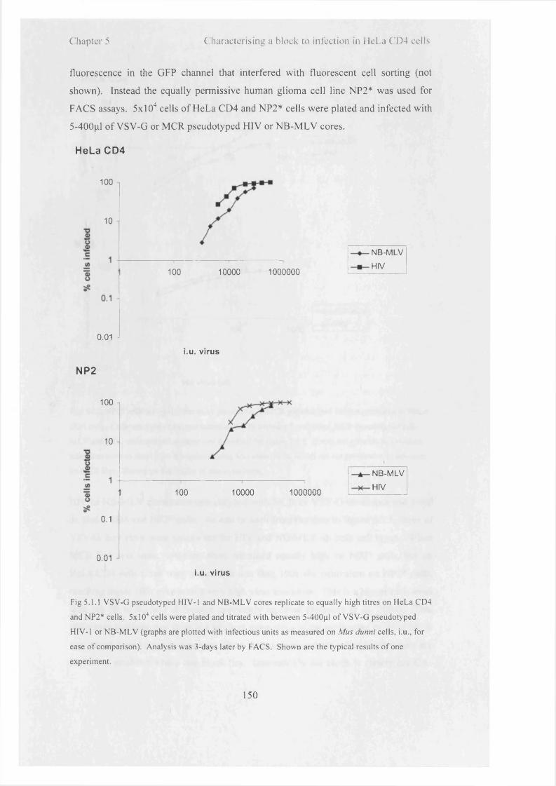
Chapter 5 Characterising a block to infection in HeLa CD4 cells
fluorescence in the GFP channel that interfered with fluorescent cell sorting (not
shown). Instead the equally permissive human glioma cell line NP2* was used for
FACS assays. 5xl04 cells o f HeLa CD4 and NP2* cells were plated and infected with
5-400pl o f VSV-G or MCR pseudotyped H IV or NB-M LV cores.
HeLa CD4
100
10
I
10000100 1000000
0.01 -
NP2
100
10
0.1 -
0.01
NB-M LV
HIV
i.u. virus
100 10000 1000000
NB-M LV
HIV
i.u. virus
Fig 5.1.1 VSV-G pseudotyped HIV-1 and N B -M L V cores replicate to equally high titres on HeLa CD4
and NP2* cells. 5x l0 4 cells were plated and titrated with between 5-400pl o f V S V-G pseudotyped
HIV-1 or N B -M L V (graphs are plotted with infectious units as measured on Mus dunni cells, i.u., for
ease o f comparison). Analysis was 3-days later by FACS. Shown are the typical results o f one
experiment.
150

Chapter 5 Characterising a block to infection in HeLa CD4 cells
HeLa C D 4
100 n
0.01
1000
NB-MLV
HIV
NP2
100
■§ 10
II 10) oa? 0.1
0.01
vol virus (|jl)
Fig. 5.1.2 NP2* cells are up to lOOx more permissive to M CR pseudotyped virions compared to HeLa
CD4 cells. Cells were plated out and transduced with between 5 and 500ul M C R pseudotyped NB -
M L V and H IV , and analysed as otherwise described for figure 5.1.1. It was not possible to calculate
infectious units as these were normalised using Mus dunni cells, which are not permissive to infection
by M C R Env. Shown are the results o f one experiment.
HIV or NB-M LV cores were pseudotyped with MCR or VSV-G envelopes and titred
on HeLa CD4 and NP2* cells. As can be seen from the data in figure 5.1.1, titres o f
VSV-G Env virus were equivalent for HIV and NB-M LV on both cell types. When
MCR Env was used, however, titres remained equally high on NP2* cells, but on
HeLa CD4 cells titres were reduced to less than lOOx the equivalent on NP2* cells,
reaching under 10% even with a very high virus inoculum. This is a bigger ratio even
than the 50 to 60-fold difference previously reported (Schmitz et al. 2004). This
difference indicates that there is a block at some stage o f the replicative cycle o f MCR
Env pseudotyped cores that prevents them from infecting these cells. Further tests are
needed to establish where this block lies. Interestingly the block is clearly not CA-
151

Chapter 5 ( 'h a ra c le r is in u a b lock to in fe c t io n in I le i .a ( 1 ) 4 cells
sequence specific, as both NB-M LV and H IV cores were equally inhibited. Lv2 was
defined as having a CA-dependent component; i f this is the case the component o f
restriction mediated by Gag must be the same for both the HIV-1 and NB-M LV cores
used here, unless this component o f the restriction also only operates down certain
entry pathways. Alternatively the restriction to infection in HeLa CD4 cells has no
effect on the core but is entirely mediated by the envelope protein. Although this is
not entirely consistent with published studies o f Lv2, the latter seems the more
probable explanation.
5.2 ASLV Env NB-MLV and HIV-1 replicates to high titres on
HeLa CD4 cells when entering via both Tva800 and Tva950
The envelope dependence o f Lv2 has been postulated to be relevant to the manner in
which the virus enters the cell (Schmitz et al. 2004). Assessment o f the route by
which Lv2 acts would be aided by study o f a system in which the same infectious
viral population is used for both Lv2-sensitive and insensitive routes, and receptor
proteins that were previously characterised as directing the virus into the cell by
different endocytic pathways. As the Tva800 and Tva950 receptors specifically
fulfilled these criteria, it was o f interest to see i f a virus that entered via either one o f
the receptors expressed in HeLa CD4 cells would encounter Lv2-type restriction or
not. Entry via Tva800 would be additionally interesting as it is a GPI-linked receptor,
and resides in lipid rafts, which are important for Lv2-sensitive entry. Studies o f Lv2
would be greatly enhanced i f the same virus could be used for assessment o f different
pathways, rather than using different viral envelopes. To assess their permissivity,
HeLa CD4 cells were transduced with high volumes o f vector encoding Tva800 or
Tva950, and three days later challenged with ASLV Env pseudotyped NB-M LV.
152

Chapter 5 Characterising a block to infection in HeLa CD4 cells
- 10
100
■ Tva800
• Tva950
10
vol virus (pi)
100
Fig. 5.2 HeLa CD4 cells expressing Tva800 and Tva950 can be successfully infected with A S LV Env
pseudotyped virions. 5 x l0 4 cells were plated and transduced with Tva800 or Tva950 vector, then
challenged with l-200pl AS LV Env pseudotyped N B -M L V . Analysis was by FACS. Shown are the
results o f one experiment with percentages o f GFP positive cells as a function o f all cells rather than
just the cells expressing the receptors. The YFP fluorescence was faint and the receptor/YFP-positive
population hard to separate, hence titres do not approach 100%.
Fig. 5.2 shows that virus enters and replicates successfully in HeLa CD4 cells after
entering via either Tva800 or Tva950. There is hence no difference shown between
these two routes o f entry. There are many other routes into the cell, however, and this
experiment does not preclude the possibility that Lv2 may operate down a third route,
separate from those o f Tva800 and Tva950.
Usage o f MCN Env did not give consistent results either as rendering a virus Lv2
sensitive or insensitive. In HeLa CD4, NP2* and U87* cells virus pseudotyped with
MCN was consistently unable to infect >5%, even at high viral titres (E. Gray and M.
Yap, data not shown). It is not entirely clear why this is the case, given that the
laboratory where it was cloned has used it to pseudotype a variety o f viral cores
successfully (Schmitz et al. 2004). 293T cells were used in virus production both
previously and for experiments described here, so a difference in efficiency o f
production is unlikely to be the case. MCN differs from MCR by 7 amino acids in
SU, and also at the C-terminus, as MCN has an extra 118 amino acids (Reuter et al.
2005). Ideally an investigation into MCR would use MCN as a counterpart, but
because it was not possible to use MCN to generate viable virus in our lab, further
153

C hapter 5 C'haracterisiiiii a block to infection in HeLa (1 )4 cells
experiments that required an envelope protein that does not deliver the virus to a
compartment where Lv2 is encountered used VSV-G.
The block to replication o f MCR Env pseudotypes in HeLa CD4 cells could
hypothetically be due to a defect in the virions used such as low Env expression, but
as the same batch o f MCR pseudotyped virus was used on both HeLa CD4 and NP2*
cells this is unlikely to be the case. The difference in titres seen on the two cell types
with MCR pseudotyped virus mirrors those seen in earlier studies (McKnight et al.
2001; Schmitz et al. 2004). In these studies it was reported that the earliest products
o f reverse transcription could be detected after 18 hours in both HeLa CD4 cells and
NP2* cells, which suggested to the authors that the virus was competent to both enter
and reverse transcribe and that the block in HeLa CD4 cells is not due to an entry
defect.
5.3 Titres of other CD4/CXCR4-tropic viruses envelopes are also
significantly reduced in HeLa CD4 cells
In order to assess whether Lv2 uniquely affects MCR pseudotyped virions in HeLa
CD4 cells, or whether another viral envelope that uses CD4 and CXCR4 as receptor
and co-receptor also makes the virus susceptible to Lv2, NL4-3 Env, was used. 5-
500pl virus made with HIV core and NL4-3 Env was titrated onto HeLa CD4 and
NP2* cells.
154

Chapter 5 Characterising a block to infection in HeLa CD4 cells
100
10
1 -
0.1 -j
0.01 j
0.001
10000
vol virus (pi)
HeLa CD4
— a — NP2
Fig. 5.3 Virus pseudotyped with NL4-3 envelope is not able to replicate to high titres on HeLa CD4
cells. 5x l04 HeLa CD4 and NP2* cells were plated out, and infected with between 5-500pl NL4-3
pseudotyped H IV . The infection was analysed 3 days later by FACS. Shown are the results o f two
experiments (for HeLa CD4 cells) and one for NP2* cells, error bars represent sdm.
These results show that i f any volume o f input NL4-3 Env pseudotyped virus is
considered, NP2* cells are between 10- to 100-fold more permissive than HeLa CD4
cells. For example, when 500jil o f virus was used, the percentages o f HeLa CD4 and
NP2* cells infected were 3.8% and 38.2% respectively, a difference o f 10-fold. The
difference was greater at lower viral titres. These results are in agreement with
similar experiments conducted by Schmitz et al., but the interpretation o f those data
offered here w ill differ from theirs (Schmitz et al. 2004). From their data, HIV-1
cores pseudotyped either with MCR and NL4-3 envelopes reveal differences in titres
o f approximately 40- and 10-fold respectively. The conclusion drawn in (Schmitz et
al. 2004) was that usage o f NL4-3 envelope ‘rescues the block to replication’
compared to MCR Env. However, this conclusion cannot be supported for either their
data or for these presented here. The ‘rescue’ in both cases is minor, o f the order o f 4-
fold for their data (i.e. a reduction o f a 40-fold to a 10-fold block to infection). For
my data the difference between titres o f MCR pseudotypes on HeLa CD4 and NP2*
cells was between 40-50-fold, and therefore using NL4-3 Env made no significant
difference. Whether data presented here or those from previous studies are
considered, there remains at least a 10-fold difference between titres o f NL4-3 Env
155

C h a p te r 5 C h a ra c te r is in g a M o c k to i n fe c t i o n n: \ 'c l a ( i )■< cc!Is
pseudotypes on the two cell lines, which is considerable. Therefore Lv2 restriction is
not unique to MCR Env, but acts on other viral envelopes that use CD4 and CXCR4
to enter cells.
5.4 The block to infection in HeLa CD4 cells cannot be abrogated
In previous studies the saturation o f cells w ith particles that would abrogate F v l and
Trim5a had no effect on titres o f Lv2-restricted virus, so it was concluded that Lv2
activity is separate from them (Schmitz et al. 2004). It has been reported that Lv2
activity can be diminished when an in itia l titre o f restricted challenge virus is overlaid
with a saturating dose o f (potentially) abrogating particles (Marchant 2006), and both
abrogation and challenge dose enter the cell simultaneously.
These previous abrogation experiments for Lv2 were carried out w ith initial
application o f the challenge dose at 4°C for lh r, after which abrogating dose was
applied for 30 min, as it was suggested that binding o f the abrogation dose first could
occlude receptors at the cell surface (Marchant 2006). An increase in titres o f
restricted virus o f between 3- to 6-fold in HeLa CD4 cells was revealed. However,
this moderate increase does not reflect the magnitude o f the difference in titres
between permissive and non-permissive cells (>60x), although it could be indicative
o f multiple blocks to replication. In experiments reported below, the abrogating dose
was applied 3 hours prior to challenge, as previously described (Towers et al. 2002;
Dodding et al. 2005), so that the potential saturability o f the Lv2 factor(s) could be
assessed in a direct comparison w ith that o f F v l and Trim5a. The volume o f
abrogating virus used was equivalent to that used to saturate F v l and Trim 5a (Towers
et al. 2002; Dodding et al. 2005). Receptor recycling would be complete in the 3
hours that the cells spend at 37°C between abrogating and challenge doses.
156

Chapter 5 Characterising a block to infection in HeLa CD4 cells
VSV-G MCR
■ mock
□ pHmn■ mock
□ p H IT l l l
HCD4 NP2 HCD4 NP2
c e l l c e l l
Fig 5.4 Pre-incubation o f HeLa CD4 cells with restricted virus does not raise titres o f a second,
challenge dose o f virus. Titres on NP2* cells are also unaffected, as are titres o f V S V -G pseudotypes.
The titres obtained from a dose o f GFP vector-carrying challenge virus after pre-incubation with either
293T-cell extract (mock) or p H IT l 11 vector-carrying virus is shown for when both viral and challenge
particles were pseudotyped with V SV -G or M CR Env. 500pl o f a neat abrogation dose was overlaid
onto 1x10s cells plated 24 hours earlier, and incubated at 37°C for 3 hours. Cells were then washed
gently with PBS, and challenged with fixed volumes o f GFP challenge virus. Titres were determined 3
days later by FACS. Results shown are from one experiment, and equivalent results were obtained by
Dr Melvyn Yap. Error bars show standard error.
The data in figure 5.4 show that saturating cells at MOI o f over 10 with viral particles
before challenge with a second dose o f GFP virus reduces titres on both HeLa CD4
and NP2* cells by 20-25% virus when the envelope protein is VSV-G, but has no
clear effect when the envelope protein is MCR. Titres o f MCR pseudotyped virions
on NP2* cells did not change significantly, with a small increase o f 6.6%, as might be
expected for restriction factor negative cells. Titres on HeLa CD4 cells were well
below 1%, that is, below the reliable analytical detection level o f FACS, even after
pre-challenge with saturating levels o f MCR Env-pseudotyped viral particles. This
makes any assessment o f fold-change in titres difficult, and would strongly suggest
that the nature o f the block to infection in HeLa CD4 cells is such that it is not a
saturable factor, or that any effect is insignificant. Abrogation o f F v ln, F v lb or
Trim5a, on the other hand, increases titres o f challenge virus by approximately 50-,
157

Chapter 5 Characterising a block to infection in Mel.a CD4 cells
10- and 50-fold respectively (Dodding et al. 2005). Thus it must be concluded that
Lv2 restriction is not caused by a saturable factor.
The existence o f a dominant, saturable factor that was ultimately found to be Trim5a
was also strongly suggested by experiments in which challenge o f heterokaryons
made from fusion o f Refl-negative and Refl-positive cells gave titres equivalent to
those seen in Refl-positive cells alone (Cowan et al. 2002). Schmitz et al. reported
that attempts to assess the dominance o f Lv2 restriction by creation o f heterokaryons
were unsuccessful and uninterpretable, as Lv2 restriction was not evident in
heterokaryons, but was also quashed in homokaryons created from fusion o f Lv2-
restrictive cells (Schmitz et al. 2004). This would suggest that either plasma
membrane disturbances or intracellular rearrangements from this process were
responsible for any Lv2 knockdown, rather than alteration o f a restriction factor. It is
therefore important to consider the stage o f the infectious cycle at which MCR is
blocked.
5.5 Production of strong stop DNA is reduced in HeLa CD4 cells
challenged with MCR pseudotyped HIV
Schmitz et al. detailed experiments with quantitative PCR, designed to compare ratios
o f early transcripts obtained in HeLa CD4 and U87* cells, for both MCR and MCN
viruses (Schmitz et al. 2004). Data shown were the ratio o f copies in U87* cells
compared to HeLa CD4 cells (U87*: HeLa CD4), for both MCR and MCN at 4
timepoints; 1, 2, 6 and 18 hours. After 1 hour, there were 8x as many strong stop
transcripts in U87* cells compared to HeLa CD4 for both MCR and MCN. This
means that there are 8x as many MCN strong stop transcripts in U87* cells as in HeLa
CD4 cells, and 8x as many MCR strong stop transcripts in U87* cells compared to
HeLa CD4 cells. The ratio is the same for both viral clones after 1 hour. After 2
hours, the same ratio o f U87*: HeLa CD4 strong stop transcripts for MCN is 20x, and
for MCR, 8x. Therefore, it would appear that after 2 hours replication o f MCN is
more dramatically inhibited than that o f MCR in HeLa CD4 cells. A t both the 6 and
18 hour timepoints, however, there is no difference in the U87*: HeLa CD4 ratio o f
transcripts for MCR and MCN. What is not commented on, however, is that for both
MCR and MCN this ratio is 25. This means that there are 25x more transcripts seen
158

C hapter 5 C ha rac te r is ing a b lo ck to in fe c t io n in H e L a C l ) 4 cells
in U87* cells compared to HeLa CD4 cells. Considering this ratio for MCN, it is not
entirely clear how the virus then continues to generate an equivalent titre on HeLa
CD4 and U87* cells. The conclusion drawn from these data in the paper is that
‘equivalent levels o f transcripts were observed at the 6- and 18-h timepoints, therefore
MCR and MCN represent prCBL-23 and CBL23 described previously in (McKnight
et al. 2001)’ . However, the reference in this statement to ‘equivalent levels’ refers
exclusively to the proportions o f MCN and MCR (i.e. the fact that at the timepoints o f
6 and 18 hours, both MCR and MCN produced 25x more strong stop transcripts on
U87* cells compared to HeLa CD4 so MCR and MCN were equivalent to each other).
This does not seem to support a conclusion o f unhindered entry o f either MCR or
MCN to HeLa CD4 cells as there are 25x less strong stop transcripts than in U87*
cells.
It is not possible to directly compare ratios obtained in experiments described here to
these data, as MCN appeared unviable. On consideration, however, an important
conclusion to have drawn from the previous MCR data alone would have been the
obvious one, that as there are between 8-25x less strong stop transcripts made in
HCD4 cells compared to U87* cells and that as these are the earliest product o f
reverse transcription, it cannot be maintained that reverse transcription is occurring at
equivalent levels in HeLa CD4 and U87* cells.
In consideration o f how a pre-reverse transcription block would manifest itself in
qPCR data, an important question is what sort o f difference in levels o f early RT
transcripts would be expected in cells that were restricted pre-reverse transcription
compared to cells that were either restricted after reverse transcription or fully
permissive? Data are presented in references (Yap et al. 2007) and (Yap et al. 2006)
that show levels o f reverse transcription products in cells expressing Owl Monkey
Trim5CypA or one o f several artificial constructs, all o f which block pre-reverse
transcription, compared to cells which express no restriction factor. The difference in
the levels o f strong stop products between the two is between 6 and 9-fold. Taking
this as a standard for the expected difference in levels between a pre-reverse
transcription block and permissive cells, MCR is most definitely restricted before
reverse transcription in HeLa CD4 cells compared to U87* cells, and by the same
159

Chapter 5 Characterising a block to infection in HeLa CD4 cells
token, MCN is moderately inhibited (~2-3x, (Marchant 2006) or heavily so (5-25x
(Schmitz et al. 2004).
< 600 Q
HeLa CD4 NP2
cell type
Fig. 5.5.1 Viruses pseudotyped with VSV-G , but not MCR, successfully reverse transcribe in HeLa
CD4 cells. 2 x l0 5 HeLa CD4 and N P2* cells were plated out. 24 hours later cells were overlaid with
virions at M O I 0.8-1, and bound at 4*C for one hour. Infection was initiated by addition o f medium
and transfer to 37°C. After 6 hours cells were harvested, and DN A extracted using QIAamp D N A mini
kit. The number of cells present was normalised by using ratios o f actin for each sample run in parallel
with early RT. Results shown are combined from 4 independent experiments, and error bars show
sdm.
The data in figure 5.5.1 are the actual numbers o f reverse transcription products
obtained in HeLa CD4 and NP2* cells after infection with VSV-G or MCR
pseudotyped virus. Levels o f both virions produced equivalent levels o f strong-stop
transcripts in NP2* cells. In HeLa CD4 cells levels o f strong stop products produced
after MCR virus infection were 24% o f those obtained from an infection with VSV-G
pseudotyped virus.
A comparison was done for HeLa CD4 cells alone, for MCR and VSV-G pseudotyped
virus at time points o f 6 and 18 hours (still detecting strong stop transcripts) and the
results shown in figure 5.5.2.
160

Chapter 5 Characterising a block to infection in HeLa C.D4 cells
1.2
D early RT products
V S V 6 MCR 6 V S V 1 8 M CR 18hrs hrs hrs hrs
virus/ timepoint
Fig. 5.5.2 The percentage o f early products of reverse transcription seen in HeLa CD4 cells infected
with M CR pseudotyped virions is 21% o f those seen when cells are infected with VS V -G pseudotyped
virions. This percentage does not increase over time. Data were processed from the experiments
described in the legend to figure 5.5.1.
These data show that the appearance o f appreciable levels o f strong stop transcripts is
not simply delayed in HeLaCD4 cells, but that it does not occur at all. Levels o f
MCR transcripts reach 21% o f those o f VSV-G (which is set to 100%) at both
timepoints. From these data the former presumption that MCR pseudotyped virus is
capable o f efficient entry into HeLa CD4 cells must be questioned.
5.6 Introduction of Trims 1, 18 and 34 into NP2* cells does not
create a block to infection
Human and rhesus macaque Trim5a are the causative agents o f Refl and L v l
respectively (Keckesova et al. 2004; Perron et al. 2004; Stremlau et al. 2004; Yap et
al. 2004), and Trim l has anti-N-MLV activity (Yap et al. 2004). As there are over 70
members o f the Trim family o f proteins, and many have as yet unidentified roles in
the cell, there may be more with anti-viral activity (Yap et al. 2004; Nisole et al.
2005). It was suggested that Trim proteins may be responsible for Lv2, and a study
done on members o f this family that, like Trim5a, contain a SPRY domain (Marchant
2006), Trims 1,6, 18 and 34.
161

( hapter 5 ( h a r a c te n s in e a b lo c k to in fe c t io n in H eLa ( '1 )4 cells
It was reported that RNAi directed against Trims 1,18 and 34 (specifically isoforms
Trims 1(3, 18(3 and 34a) in U87* cells had no effect on titres o f MCR pseudotyped
HIV-2, but in HeLa CD4 cells T rim l RNAi recovered titres o f HIV-2 15x, and Trims
18 and 34 RNAi both recovered titres 5x each (Marchant 2006). Conversely, an
increased restriction to replication was seen in NP2* cells over-expressing Trim 1,
Trim 18 or 34. Trim l decreased titres by 11-fold, and Trims 18 and 34 each
decreased titres by 6-fold. It was not reported what effect expression o f all 3 proteins
together in NP2* cells had, or whether the effect was additive or not (Marchant 2006).
It is plausible that Trim proteins could block viral replication at any point in the life
cycle, or even affect surface expression o f CD4 or CXCR4, and this could explain
data presented in 5.1-5.5 i f these Trims were found to be expressed at elevated levels
in HeLa CD4 cells compared to U87* or NP2* cells. Trims lp , 18a and 34a (referred
to in experiments discussing my data as 1, 18 and 34) were cloned into
pLgatewaylRESYFP by Dr Melvyn Yap, and transduced into NP2* and HT1080
cells. The plasmid pLTva950IRESYFP was used as a control, to assess the effects o f
transduction on the cells, as expression o f Tva950 should not affect titres o f virions
that do not use this receptor. 3 days later cells were challenged with either VSV-G or
MCR pseudotyped NB-M LV and HIV.
162

Chapter 5 Characterising a block to infection in HeLa CD4 cells
MCR
10 ,in2+->vca)oc2£V2o
0.1
0.01
vsv
10
0)c 1 2 £V2o
0.1
- P i
□ M CR /N B-M LV
□ M CR /H IV
eyp950 Triml Trim18 Trim34
&
□ V S V /N B -M L V
□ V S V /H IV
eyp950 Triml Trim18 Trim34
Fig 5.6.1 A non-specific reduction in VSV-G-pseudotyped viral titres is seen on addition o f Trims 1,
18, 34, or Tva950 to NP2* cells. This reduction was seen whether the viral envelope was M C R (i.e.
Lv2 sensitive) in the top graph, or VSV-G (i.e. Lv2-insensitive) in the bottom graph, and for both H IV -
1 and N B -M L V cores. Fold-difference was calculated from the ratio o f viral titres on cells expressing
Trim or eyp950, compared to cells that were not expressing an extra factor. The results shown are
combined from 3 separate experiments apart from eyp950, which was done twice.
As is clear from the size o f the error bars in the MCR graph (particularly for H IV) the
results varied considerably between experiments. Cells expressing eyp950 were
included to ensure that there was no non-specific effect on titres from prior challenge
with vector. However, as challenge with eyp950 vector effected a similar reduction in
titres o f MCR pseudotyped virus compared to any o f the three Trim proteins, a non
specific effect seems to be what is shown rather than any specific block.
Additionally, as shown in fig 5.6.1, VSV-G pseudotyped virus titres in Trim-
expressing cells are similarly reduced to between 34 to 56% o f those in non Trim-
163

Chapter 5 Characterising a block to infection in HeLa CD4 cells
expressing cells. Virus pseudotyped with VSV-G is unrestricted by Lv2 therefore this
lowering o f titres cannot be an Lv2 specific effect.
This type o f non-specific effect has not been seen before in the years o f using the 2-
colour FACS assay for analysis. In order to redo these results, and avoid any
activation o f anti-viral cellular defences raised by the initial transduction o f the Trim
proteins close in time to challenge with MCR pseudotyped proteins, three stable cell
lines were created. NP2* cells were transduced with the vector
pLgatewayTrimlRESYFP (where Trim is Trim l, Trim 18 or Trim34) and sorted for
YFP expression (work done by Dr Melvyn Yap). After passage for several weeks,
cells were mixed 1:1 with non-transduced NP2* cells, plated, and challenged with
MCR or VSV-G pseudotyped NB-MLV or HIV. The Trim-expressing cells express
YFP and hence can be easily differentiated from the non-fluorescent, Trim-negative
NP2* cells. The percentages o f these two populations o f cells that were successfully
infected with virus (and express GFP) were compared.
MCR
10
*5C
C<u 1□ M CR /N B-M LV
□ M CR/H IV
T3O
0.1N P 2-/T1 N P 2-/T 18 N P 2-/T 34
164

Chapter 5 Characterising a block to infection in HeLa CD4 cells
vsv
10
0)
c 1sf
0.1 JNP2 -/T1 N P 2-/T 18 N P 2-/T 34
□ V S V /N B -M L V
□ V S V /H IV
Fig 5.6.2 Long-term expression o f Trims 1, 18 or 34 does not lead to a consistent and significant
decrease in virion infectivity on NP2* cells. NP2* cells were transduced with trims 1,18 and 34,
sorted for YFP expression, and passaged for several weeks. They were mixed 1:1 with the parental
NP2* cells and challenged with V S V or M CR pseudotyped N B -M L V or H IV -1 . Shown are the
combined results o f two independent experiments. Error bars represent sdm.
As can be seen from data in figure 5.6.2, expression o f any o f these Trim proteins in
NP2* cells does not cause a clear and significant reduction in MCR pseudotyped virus
titres. At most, MCR titres were reduced in Trim-expressing NP2* cells by a factor
o f 2, which is not in any way comparable to the >50x reduction in titres in HeLa CD4
cells. The results for cells expressing Trim 1 were less consistent than for Trims 18 or
34, but this was true for both VSV-G and MCR pseudotyped virus. I f there is no
specific reduction shown for MCR pseudotyped virus then T rim l cannot be connected
with Lv2.
5.7 Expression of anti-Trim1 SiRNA does not diminish the block
to infection in HeLa CD4 cells
As Trim lp had been previously reported to cause the highest drop in titres when
expressed in NP2* cells compared to Trims 18 and 34 (Marchant 2006), and the
results seen here had been somewhat varied, it was important to ensure that these
were reliable, preferably by another method. SiRNA directed against T rim l was
designed and verified by Dr Melvyn Yap and transduced into HeLa CD4 cells. Three
days later cells were challenged with MCR pseudotyped HIV and titres compared to
165

Chapter 5 Characterising a block to infection in HeLa CD4 cells
cells that had been mock transduced. The titres on the two cell types with and without
SiRNA were compared, and the resulting fold-difference are shown in figure 5.7.
VSV-G/HIV MCR/HIV
virus
Fig. 5.7 Expression o f anti-Trim 1 SiRNA in HeLa CD4 cells does not affect titres o f V S V -G or M C R
pseudotyped H IV -1. HeLa CD4 cells were transduced with anti-Trim 1/Y FP vector, and then
challenged with VSV-G and M CR pseudotyped H IV -1 . The fold-difference in titres between cells
expressing the SiRNA and null cells was calculated, and is shown above. A fold-difference o f 1
indicates that the SiRNA has no effect, and >1 that the SiRNA has increased titres. Shown are the
combined results from two experiments. Error bars represent the variation in ratios from several
volumes used within the experiments.
Titres o f both the control VSV-G and the MCR pseudotyped H IV are
indistinguishable across the two sets o f HeLa CD4 cells, regardless o f the SiRNA
against Trim l in one set. These data indicate that T rim l does not play a role in
reducing titres o f MCR pseudotyped virus in HeLa CD4 cells. This, and the previous
experiment, reinforces the likelihood that Trim proteins 1, 18 and 34 are not the
causative agents for the Lv2 phenotype but that different factors or mechanisms are
responsible.
I f this is the case, then the question now arises as to when the block to infection
actually occurs, and what could possibly be the cause. This was investigated in
experiments reported in chapter 6.
166

( hapter 5 C ha rac te r is ing a b lo ck to in fec t ion in I le i .a C l ) 4 cells
Discussion
Although cells can be artificially engineered to express combinations o f retroviral
receptors, expression alone is not necessarily sufficient to render these cells
permissive to infection by the relevant viruses. HeLa CD4 cells are fully permissive
to infection by HIV-1 cores pseudotyped with VSV-G, but not to virions pseudotyped
with an envelope protein from a primary isolate, MCR. U87* and NP2* cells,
however, are fully permissive to viruses pseudotyped with both envelope proteins.
A ll cell types are clearly capable o f supporting retroviral replication. It has been
suggested that the difference between them is due to an intracellular factor present in
a specific compartment o f HeLa CD4 cells that acts on viral cores in a similar manner
to Trim5a or Fvl. Rerouting virus around this compartment (e.g. by VSV-G) would
enable the virus to avoid this block. In experiments detailed in this chapter I have
studied the phenomenon carefully by standard and new techniques, and come to the
conclusion that while there is a block to infection in HeLa CD4 cells, this is unlikely
to be caused by a Fvl - or Trim5a-like restriction factor.
It should be noted that experiments that compare VSV-G and MCR are at best
relative, as two different virus populations must be used with different efficiencies o f
production and binding. Experiments with MCN were completely unsuccessful, as it
appeared to be totally non-functional on all cell types; viruses pseudotyped with this
envelope failed to enter any cells. An attempt to recapitulate the Lv2 phenomenon
using Tva800 and Tva950 in HeLa CD4 cells was also unsuccessful, as no difference
in viral replication was seen with either receptor. Thus all experiments described here
were done with VSV-G and MCR envelopes.
A characteristic o f restriction by Fvl and Trim5a is that the block to infection is
saturable. This is highly indicative that a specific factor is causing the block to
infection in an infectious cycle that could otherwise be productive. As the block in
HeLa CD4 cells cannot be overcome with high titres o f viruses, it is hard to conclude
that it must be caused by a specific factor, as there is no indication that the infection
167

Chapte r 5 C ha ra c te r is in g a b lo ck to in fe c t io n in H eLa 0 ) 4 cells
would otherwise be successful. The use o f the envelope protein VSV-G merely
confirms that the cell is able to support viral replication. It does not prove or disprove
the existence o f a cel 1-compartment specific inhibitory factor.
The detection o f significant levels o f reverse transcription intermediates, while not
infallible, is an indication that entry is occurring at levels relevant to a successful
infection. Previous experiments that investigated levels o f products o f reverse
transcription showed either that they simply could be detected (McKnight et al. 2001),
or compared levels obtained with MCN and MCR-pseudotyped virions (Schmitz et al.
2004). In the former, levels o f reverse transcripts were not equivalent in U87*, HeLa
CD4 and HOS cells. Results were displayed as a PCR gel analysis o f LTR-gag from
between 100 and 10,000 cells. Levels o f transcripts were approximately equivalent
for 100 U87* and 10,000 HeLa CD4 cells, i.e. they were lOOx lower in HeLa CD4
cells. Levels in HOS cells were somewhat higher, and levels on the lane obtained
from 3,333 HOS cells were equivalent to 333 U87* cells. It was not discussed
whether reverse transcription occurring pre-infection (i.e. within the virions) or in
endosomes before lysosomal destruction could be sufficient in the larger samples o f
HeLa CD4 cells to produce a false positive, but this could indeed be the case (Trono
1992; Zhang et al. 1993). In the (Schmitz et al. 2004) reference, levels o f reverse
transcription intermediates were equivalent for MCN and MCR, as discussed in
section 5.5. These results were presented as a ratio o f reverse transcription quantities
obtained on U87* and HeLa CD4 cells for the two envelopes. This ratio varied
between 8-fold at 1-2 hours, to 25-fold at 6-18 hours. Therefore while levels o f
reverse transcription intermediates may be equivalent between infections mediated by
the MCR and MCN envelopes, levels between the two cell types are not. That is,
there are between 8 to 25-fold fewer products o f reverse transcription being
synthesised on HeLa CD4 cells compared to U87* cells. The pre-reverse
transcription block seen in Trim5Cyp-positive cells with susceptible virus leads to a
drop in reverse transcription intermediates o f at least 5-fold, so an 8 to 25-fold block
is commensurate with a lack o f significant synthesis o f reverse transcription products
(Yap et al. 2006). This conclusion, then, precludes the hypothesis o f a factor as
responsible for a specifically post reverse transcription block to infection in HeLa
CD4 cells.
168

C hapter 5 C harac te r is ing a b lo ck to in fe c t io n in H eLa 0 ) 4 cells
This means that the phenomenon described as Lv2 must be reconsidered. As
originally characterised, this was described as a post-entry lentiviral restriction with
components o f the restriction attributable to both Gag and Env, that was due to the
targeting o f the virus into an inappropriate sub-cellular compartment (Schmitz et al.
2004). Pseudotyping with VSV-G overcame the Env-dependent component o f the
restriction, and also removed the Gag-dependency. Experiments described here
confirm the original premise on which Lv2 is based, namely that some cells can be
infected by MCR pseudotyped virus, and that other cells from the same species
expressing the same combination o f receptor and co-receptor cannot. As described,
the block does not fall under the current understanding o f a restriction factor (Goff
2004; Sorin and Kalpana 2006). However, this would simply mean that the definition
o f what constitutes a restriction factor would have to be extended i f it could be proven
conclusively after an elimination o f all other possibilities in a sort o f molecular
biology version o f Occam’s razor, that the block to infection could not be attributable
to any other factors. The conclusion from a re-interpretation o f results from previous
studies and those described here does not suggest that this elimination has been fully
carried out, and cannot support a hypothesis that a restriction factor is definitely
responsible for the block to infection. The name Lv2, standing as it does for
lentivirus restriction factor 2, is probably a misnomer. The block is not saturable,
affects other CD4/CXCR4-tropic envelopes and does not permit entry o f virions into
the cell at levels commensurate with a successful infection. These issues do not point
towards a restriction factor being responsible, and indeed do not rule out other
irregularities or differences in cell biology between NP2*, U87* and HeLa CD4 cells
as being primarily responsible. This implies that the reason why HeLa CD4 cells are
refractory to infection by MCR pseudotyped virions must be sought elsewhere.
169

Chapte r 6 I he b loek to in fe c t io n in H eLa C I )4 cells is at entiy
Chapter 6
The Block to Infection in HeLa CD4 Cells is at Entry
It was shown in chapter 5 that viral cores pseudotyped with MCR are able to infect
NP2* cells, but not HeLa CD4 cells. This block to infection was previously attributed
to an intracellular restriction factor similar to Fvl and Trim5a but with a route o f
entry component. Experiments described in chapter 5 show that conclusive evidence
o f reverse transcription, and therefore entry o f the virus into the cell was not found,
leading to the conclusion that the block may not be due to a restriction factor at all.
The primary cause o f the block to replication probably lies in some aspect o f the
differences in cell biology between HeLa CD4, U87* and NP2* cells. This being the
case, further experiments were carried out to try and ascertain the nature o f this block,
how it prevents the virus from infecting the cell, and at what point in the infectious
cycle it does this. These experiments are described in this chapter.
R e s u lts
6.1 CD4 and CXCR4 are expressed on HeLa CD4 cells
In order to ensure that the cells I was using expressed sufficient amounts o f the
necessary receptor and co-receptor combination for entry using MCR (McKnight et
al. 1998), confluent flasks o f HeLa CD4, U87* and NP2* cells were harvested, lysed
and sampled by Western blot for CD4 and CXCR4 expression. HOS CD4/CXCR4
(HOS) cells were obtained from the AIDS Reagent program (NIBSC, Potters Bar,
UK) and were used to provide a positive control for CXCR4 expression. Mus dunni
cells were also assayed as a negative control for both proteins. Input protein level was
checked by probing for GAPDH.
170

Chapter 6 The block to infection in HeLa CD4 cells is at entry
M.d. - U 8 7 * - N P 2 * - H C D 4
GAPD
CD4
b
M .d.- U 8 7 * - N P 2 *- H C D 4 -H O S
CXCR4
Fig. 6.1 Western blot o f total levels o f CD 4 and C X C R 4 in cellular lysates. Samples were probed for
a) C D 4, b) G A P D H and c) C X C R 4. The same blot in a) was stripped and reprobed for b).
The Western blot shows that although CD4 is a rtific ia lly expressed in all cell lines,
levels do differ between cells. The levels appear to be highest in U87* cells, which
are permissive to MCR Env, but the lower levels seen in HeLa CD4 cells are still
readily detectable. HeLa CD4 cells express CXCR4 endogenously and the parental
HeLa cell line is suggested as a positive control for CXCR4 expression in published
protocols for several CXCR4 antibodies. From this Western blot, total levels o f
CXCR4 across the 4 cell lines appear to be broadly similar. Specific primary
antibodies that readily bind to CXCR4 are not available, and when blots are incubated
at 4°C overnight some non-specific binding occurs, as the bands at around lOOkDa
(seen in Mus dunni cells) and possibly 70kDa show. So, total levels o f CXCR4 are
similar across U87*, NP2* and HeLa CD4 cells, but levels o f CD4 vary, although
there is no guarantee that expression o f either CD4 or CXCR4 means that the protein
is being properly processed and trafficked in a particular cells line.
There is evidence that levels o f CD4 and CCR5 necessary for entry must be over a
threshold level and that levels o f CCR5 can be much lower i f CD4 levels are high
(Platt et al. 1998). It could be argued that part o f the difference in titres on these two
cell types compared to HeLa CD4 cells may relate to the elevated level o f artificial
CXCR4 over-expression in U87* and NP2* cells compared to HeLa CD4 cells, in
which CXCR4 is produced endogenously. If, however, as is true for CD4 and CCR5
171

C hapter 6 I he b lo c k to in fe c t io n in H eLa C D 4 cells is at entry
that higher levels o f one o f either the receptor or co-receptor can compensate for
levels o f the other being low, higher levels o f CD4 should compensate for low
CXCR4 levels. CD4 is overexpressed in all the cell lines, and should be sufficiently
high as to not lim it binding, and the reason why HeLa CD4 are non-permissive to
MCR Env pseudotyped virus must be sought elsewhere.
6.2 Progress of fusion of viral and cellular membranes can be
monitored with a fluorescent protein targeted to the viral
membrane
In the judgement o f whether the block to infection in HeLa CD4 cells is post-entry or
not, it is o f key importance to know whether MCR pseudotyped virus is able to enter
cells. In order to ascertain this, a different method o f assessing entry to PCR was
called for. In 2007 it was reported that red membrane-labelled virions are produced in
cells that express a fluorescent fusion protein comprised o f mCherry and the 15 N-
terminal amino acids o f Src (sl5-mC) (Campbell et al. 2007b). These 15 amino acids
target mCherry to the plasma membrane o f the producer cell, and the viral particles
are coated with red membrane as they leave the cell (Rodgers 2002; Campbell et al.
2007b). The coating is maintained as the viral envelope binds to its receptor on a
target cell, and disperses after fusion between the viral and cellular membranes has
taken place. I f the virus is endocytosed but fusion out o f the endosomes is prevented
(for example when fusion o f VSV-G envelope is blocked by bafilomycin A l) then the
red signal remains intact (Campbell et al. 2007b). This system was chosen to aid
analysis o f whether fusion was successfully taking place between MCR pseudotyped
viruses and HeLa CD4 cells. I f the proportion o f virions that lost their red coating
after binding remained unchanged between NP2*, U87* and HeLa CD4 cells then,
this would indicate that fusion was occurring at equivalent levels in the different cells,
and the blockage to replication in HeLa CD4 cells was further downstream in the
infectious pathway after entry.
In order to monitor viral particles after the fusion event, they needed to be dually
labelled with GFP-vpr, which labels the viral cores, as well as with the S15-mC dye
(McDonald et al. 2002). Viral particles outside the cell, and those bound to cells pre
fusion would be both green and red; those that had successfully completed a fusion
172

Chapter 6 The block to infection in HeLa CD4 cells is at entry
event would be green only. Counting the proportions o f each should reveal in which
cells fusion was blocked. Repeated washing was carried out pre-incubation and pre
fixation to ensure that only virions tightly bound to receptor and not free virions
comprised the green and red populations present. In order to ensure that as few
virions as possible progressed beyond dispersal o f the GFP-vpr signal and hence
beyond detection, infections were initially carried out for half an hour. However, this
short time frame did not allow a sufficient number o f virions to fuse to differentiate
between those that were inhibited, and those that were not, so an infection period o f 2
hours was eventually used.
293T cells were initially transfected with 5 plasmids (Gag-pol, Env, package
(CSTKW), GFP-vpr and sl5-mC) at equal ratios but this did not result in any virus
being produced, as high levels o f sl5-mC seem to have a negative impact on viral
viability. Varying the ratio o f sl5-mC to the other plasmids (from 1:1 to 1:10)
resulted in viruses that were increasingly infectious as the proportion o f sl5-mC
decreased, as shown in figure 6.2.1.
100
80
I60
cJ2a> 40o35
20 J
0 4- 0
Fig. 6.2.1 Reducing the ratio o f sl5-mC:CSGW increases the infectivity o f virions produced. 293T
cells were transfected with gag-pol, VSV-G , CSGW, GFP-vpr and sl5-m C at ratios varying between
1:1:1:1:1 to 1:1:1:1:10, as shown in the key to the graph above. 5-125jxl virus was titrated on 5 x l0 4
dunni cells, and the results analysed by FACS (for GFP expression) three days later. Shown are the
results o f one experiment, typical o f 3 carried out.
50 100 150vol virus (pi)
173

Chapter 6 I lie b lo ck to in fe c t io n in H eLa C D 4 cells is at entry
In order to ensure that none o f the nascent viral particles should escape being labelled
red so that particles would be erroneously considered to have undergone fusion, it was
deemed desirable to keep the concentration o f sl5-mC as high as possible. Because
o f this, although a further decrease in the ratio o f sl5-mC to CSGW to 1:20 yielded
particles with slightly higher infectivities compared to a 1 : 1 0 ratio, the ratio o f 1 : 1 0
was decided upon for further experiments.
In order to gauge the utility o f this method for assessing whether fusion had been
completed or not, green and red viral particles were pseudotyped with VSV-G
envelope protein, bound to HeLa CD4 or U87* cells at 15°C for two hours, and then
incubated at 37°C for two hours, all steps in the presence or absence o f NH 4 CI.
NH 4 CI is well documented to prevent initiation o f fusion by the VSV-G envelope
(Aiken 1997), and the virus remains trapped in endosomes. After infections, cells
were fixed and stained with DAPI, and at least 10 pictures were taken using a lOOx
lens at discrete points on the slide. Files were randomised by a colleague before the
pictures were opened in Adobe photoshop, and scored blind for overlapping red and
green, or green alone, signals. Examples o f pictures used to obtain data are shown in
6 .2 .2 , and the percentage o f green vs. green and red particles in each o f the different
conditions in 6.2.3.
174

Chapter 6 The block to infection in HeLa CD4 cells is at entry
b. V ira l particles, red channel.a. Viral particles.
c. HeLa CD4 cells and VSV-G pseudotypes. d. As c, red channel.
e. HeLa CD4 cells, VSV-G pseudotypes incubated with N H 4 CI.
f. As e, red channel.
Fig. 6.2.2 V iral particles can be localised with GFP-vpr, and their membranes stained with S I 5-
mCherry. These particles permit assessment o f fusion with cellular membranes. Nuclei are stained in
blue (where shown) with DA P I, viral particles (fused) are shown by green, and viral membranes red in
the left-hand pictures. In pictures on the right-hand side, the blue and green channels are removed and
the red signal only is shown, in black and white for clarity, a) and b) show the viral supernatant only.
A ll green particles also had a corresponding red signal, so were deemed suitable for use in assessment
o f fusion, c-f) Cells were spinoculated with virus at M O I >5 for 2 hours at 15°C, then incubated at
37°C for 2 hours before fixation with paraformaldehyde. Scoring o f fusion was done blind by
comparison o f the location o f the green and red signals for a minimum o f 10 pictures per cell type per
condition after pictures were randomised by a colleague. Shown are HeLa C D 4 cells, but similar
pictures were obtained and scored for U 8 7* cells as well.
175

Chapter 6 The block to infection in HeLa CD4 cells is at entry
0.8
■o
■ fused virions
HeLa CD4 U87 HeLa CD4 + U87 + NH4CINH4CI
cell type
Fig. 6.2.3 Virions pseudotyped with VSV-G retain their viral (cherry red) membrane when N H 4C1
blocks fusion. Shown are the proportions o f virions that have successfully fused with the cell, that is,
they have lost their red membrane signal and evince GFP fluorescence only. The proportion o f virions
fusing in the presence and absence o f N H 4C1 is the same for HeLa CD4 and U 87* cells. The results
shown are from 10 separate pictures taken per condition (examples are shown in figure 6.2.2), with
over 500 virions per condition counted blind and scored for red or green fluorescence.
The data in figure 6.2.3 show that after 2 hours o f incubation with media alone, 62
and 59% o f viral particles had undergone fusion in HeLa CD4 and U87* cells
respectively. These were the particles that were only fluorescent green, having lost
their red membranes. I f NH4CI was present, which blocks entry by VSV-G,
successful fusion events were seen in only 12% o f particles tallied, for both HeLa
CD4 and U87* cells. As it is much harder to detect reliably the absence o f a red label
than its presence, it may be that the percentage o f fusion events in cell is slightly
lower than these percentages. However, even with that caveat there is still a clear and
significant difference between the percentages o f VSV-G viral particles that have lost
their red virion membrane label in the presence or absence o f NH4CI.
176

C hapte r 6 I he b lo ck to in fe c t io n in H eLa C D 4 eells is at entry
6.3 Membranes of HIV pseudotyped with MCR do not undergo
fusion with cellular membranes in proportion with a productive
infection
This method provides an alternative way to qPCR to assess whether virions have
entered cells. Furthermore it permits assessement o f a stage before reverse
transcription, that o f the binding o f the viral envelope and the receptor and co
receptor, and fusion o f the viral and cellular membranes. Hence it was used further to
assess what proportion o f MCR pseudotyped viral particles underwent fusion in HeLa
CD4, NP2* and U87* cells. Two sets o f cells that are permissive to viruses
pseudotyped with MCR were assessed to ensure that there was no significant
difference between levels o f fusion in permissive cells. Example pictures are shown
in figure 6.3.1. A ll pictures taken were were randomised by a colleague, and viral
particles scored blind for red and green fluorescence. The quantitative data are shown
in figure 6.3.2.
177

Chapter 6 The block to infection in HeLa CD4 cells is at entry
ma. HeLa CD4 cells, all channels. b. HeLa CD4 cells, red channel only.
mc. NP2* cells, all channels. d. NP2* cells, red channel only.
e. U87* cells, all channels. f. U87* cells, red channel only.
Fig. 6.3.1 M C R pseudotyped viral particles do not initiate successful fusion with the plasma
membrane o f HeLa C D 4 cells. Particles applied to U 8 7 * or N P 2 * cells show fusion rates o f over 60%,
within the 2 hour time-frame. For HeLa CD4 cells the proportion fused was 15% o f particles. Samples
were prepared and assessed as described in the legend to fig 6.2.2. A ll pictures show M C R
pseudotyped virions with different cell lines: a and b, HeLa C D 4 cells; c and d, N P 2 * cells; e and f,
U 87* cells.
178

Chapter 6 Tie block to infection in HeLa CD4 cells is at entry
0.8
5 0.6 •c >0§ 0.4
0.2 -I
lbHeLa CD4
I fused wrions
NP2
c e l l t y p e
U87
Fig. 6.3.2 A comparison o f the proportion o f virions that have successfully fused in HeLa CD4, NP2*
and U87* cells when the viral envelope protein is M CR. Shown are the proportions o f virions that
have lost their cherry red label. Data encompass the results from at least 10 pictures taken per cell
type, and scored blind for red and green fluorescence.
Once bound and visualised, the proportion o f MCR virions that underwent fusion was
15%, 60%, and 75% for HeLa CD4, NP2* and U87* cells, respectively. Student’s t-
test was used to determine whether the difference between the number o f fused
virions was significant in pairs o f pictures: NP2* and U87* cells for MCR; HeLa CD4
and either NP2* or U87* cells for MCR; HeLa CD4 cells with MCR or VSV-G
pseudotyped virions when the latter were held under an NH 4 Cl-block to fusion. The
r-values obtained suggested that the difference between fusion levels in NP2* and
U87* cells with MCR pseudotyped virus was not significant. The difference between
fusion levels in either HeLa CD4 and U87* cells, or HeLa CD4 and NP2* cells was
significant (see appendix 3 for numbers). Furthermore, the difference in fusion levels
between HeLa CD4 cells with MCR pseudotyped virus, or HeLa CD4 cells with
VSV-G pseudotyped virus held under an NH4Cl-induced block to infection was not
significant, i.e. there was effectively no difference in the proportion o f virions fused.
This means that the level o f fusion in HeLa CD4 cells incubated with MCR virus is
commensurate with that o f VSV-G virions under an NH4Cl-induced blockage. This
similarity strongly suggests that fusion o f MCR pseudotyped virions with HeLa CD4
179

( h a p ie r 0 1 he b lo c k to i n fe c t i o n in 1 le i a i I u ce l ls is a! c iv c '■
cells is prohibitively blocked, and entry is not happening to levels significant enough
for a productive infection, so that virions are not able to infect the cells at all.
Interestingly, the average number o f M CR virions per picture o f HeLa CD4 cells was
21.5. This was much lower than for VSV-G virions w ith HeLa CD4 cells (93.2), or
MCR virions w ith NP2* cells (81.7) and U87* cells (46.2) (not detailed here, but see
appendix 3 for numbers). This would in itia lly suggest that CD4 binding o f MCR Env
is easily disrupted, perhaps by the washing steps. Antibody binding studies by Reuter
et al. (Reuter et al. 2005), however, showed that M CR Env actually conferred a lOx-
resistance to inhibition o f infection by anti-CD4 antibody compared to unrestricted
envelope (MCN), concluding that MCR binds to CD4 more tightly. This difference
was pinpointed to a glutamic acid residue at position 74 in M CR, compared to a
glycine in MCN (Reuter et al. 2005). So it is more like ly that M CR does not readily
bind in the first place. As CD4 expression is foreign to all o f the three cell types
studied here, it is not immediately apparent why there should be such a difference in
in itia l binding, whether it is due to in itia l expression and processing o f the protein, or
expression at the cell surface, or differences in cell type that become apparent after
virus binding.
6.4 Inhibition of endocytosis or acidification of endosomes
causes a small increase in replication of MCR NB-MLV in HeLa
CD4 cells
After MCR binds to CD4 and CXCR4 at the cell surface, during a productive
infection pH-independent fusion w ill occur without endocytosis. Endocytosis is not a
productive route o f entry into the majority o f cells for virions pseudotyped w ith H IV
envelopes (Pelchen-Matthews et al. 1995; Fredericksen et al. 2002). This is in direct
contrast to the A S LV -A envelope and Tva800 and Tva950 receptors used in
experiments discussed in chapters 3 and 4, which require entry into the endocytic
pathway and a drop in pH to trigger fusion. The fact that virions pseudotyped with
MCR are not able to infect HeLa CD4 cells means that the question must be asked as
to whether instead o f subsequent fusion and entry into the cytosol, endocytosis is
instead occurring, the virus is unable to exit the endocytic pathway, and ends up
degraded in the lysosomes.
180

Chapter 6 The b lo ck to in fe c t io n in H eLa C l ) 4 eel Is is at entrx
This question was addressed by two previous studies into entry mediated by MCR
Env (Marchant et al. 2005; Reuter et al. 2005). In one experiment, a concentration of
1 OmM NH 4 CI (which raises the pH inside lysosomes) was sufficient to raise titres on
restrictive Ghost/X4 cells by lOx, while increasing titres on U87* cells by only 2x
(Reuter et al. 2005). In a second study, addition o f NH 4 CI decreased the difference in
titres o f MCR pseudotyped virus between HeLa CD4 cells and U87* cells from 30- to
5-fold, and hypertonic sucrose (which inhibits endocytosis from the cell surface)
reduced restriction from 40-fold to 5-fold (Daukas and Zigmond 1985; Heuser and
Anderson 1989; Marchant et al. 2005). However, addition o f bafilomycin A l, an
inhibitor o f the H+-ATPase, did not increase titres on HeLa CD4 cells. Both NH 4 C1
and sucrose were purported to increase the level o f virus infecting HeLa CD4 cells,
rather than to reduce the levels successfully infecting U87* cells. These results
suggested to the authors o f the second study that “ Lv2 occurs along a pH-dependent
route” (Marchant et al. 2005).
In an effort to address possible differences in cell biology between cell types, a study
o f the basal rate o f uptake o f CD4 by endocytosis in restrictive Ghost/X4 and U87*
cells was carried out (Reuter et al. 2005). It was found to be similar in the two cell
types, with 48% and 53% o f CD4 molecules remaining at the cell surface after 40
minutes for Ghost/X4 and U87* cells respectively. Previously, a separate study had
found the rate o f recycling o f CD4 to be 1.5-2% per minute in HeLa CD4 cells which
is potentially somewhat higher (Pelchen-Matthews et al. 1989), equating to 60-80%
over 40 minutes. Neither study took account o f any alteration (i.e. stimulation) in
endocytic rate that would be occasioned by viral binding.
In order to assess how these data could fit together, infections were carried out o f
HeLa CD4 cells by MCR pseudotyped virus in the presence o f the endocytic
inhibitors NH 4 CI and sucrose. Control experiments with VSV-G pseudotyped virus in
HeLa CD4 and NP2* cells were also carried out.
181
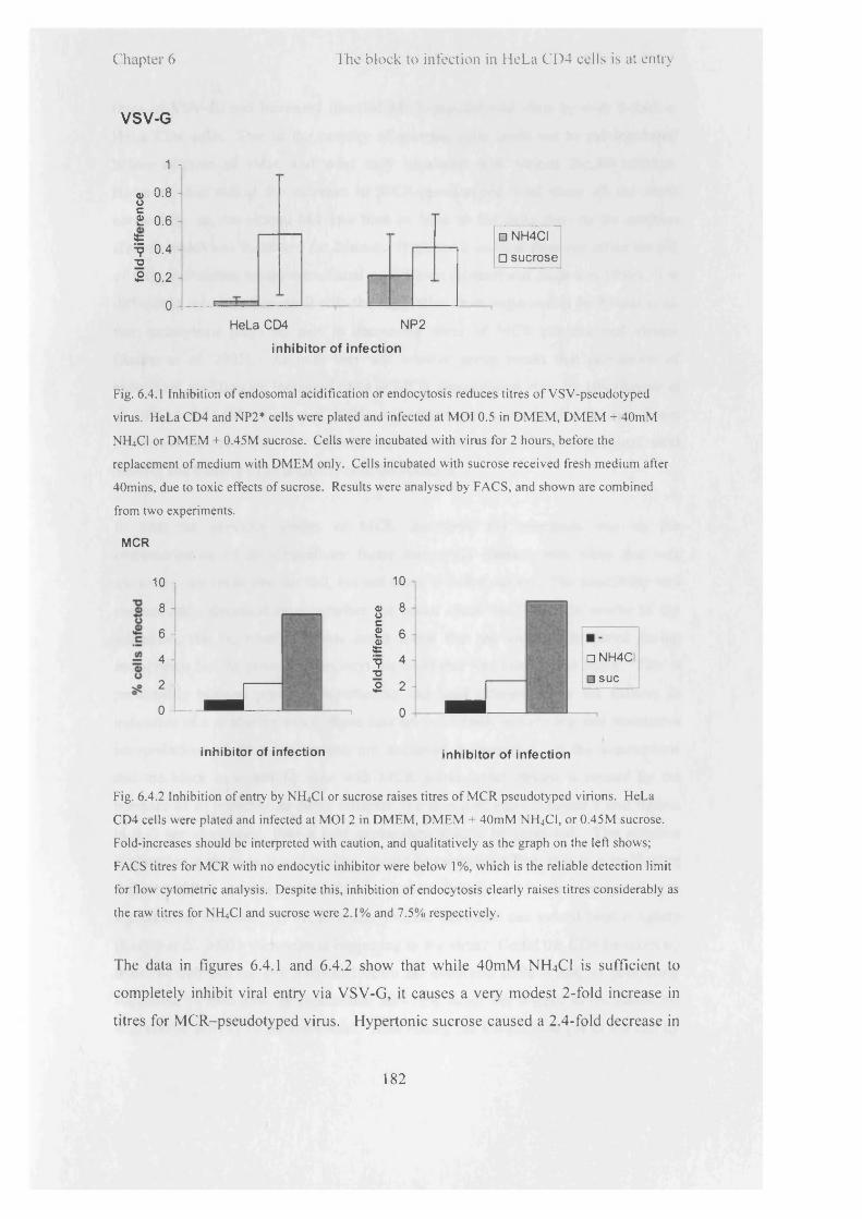
Chapter 6 The block to infection in HeLa CD4 cells is at entry
VSV-G
1 n
o> 0.801 0.6st^ 0.4 t j
£ 0.2
0 i
□ NH4CI
□ sucrose
HeLa CD4 NP2
inhibitor of infection
Fig. 6.4.1 Inhibition o f endosomal acidification or endocytosis reduces titres o f VSV-pseudotyped
virus. HeLa CD4 and NP2* cells were plated and infected at M O I 0.5 in D M E M , D M E M + 40mM
N H 4C1 or D M E M + 0.45M sucrose. Cells were incubated with virus for 2 hours, before the
replacement o f medium with D M E M only. Cells incubated with sucrose received fresh medium after
40mins, due to toxic effects o f sucrose. Results were analysed by FACS, and shown are combined
from two experiments.
MCR
10
8
6
4
2
0
1 10
a 8 01 c£ 6 £=9 4 tj
L -j fo
O M
_
□ NH4CI
□ sue
inhibitor of infection inhibitor of infection
Fig. 6.4.2 Inhibition o f entry by N H 4C1 or sucrose raises titres o f M C R pseudotyped virions. HeLa
CD4 cells were plated and infected at M O I 2 in D M E M , D M E M + 40m M N H 4C1, or 0.45M sucrose.
Fold-increases should be interpreted with caution, and qualitatively as the graph on the left shows;
FACS titres for M CR with no endocytic inhibitor were below 1%, which is the reliable detection limit
for flow cytometric analysis. Despite this, inhibition o f endocytosis clearly raises titres considerably as
the raw titres for N H 4CI and sucrose were 2.1% and 7.5% respectively.
The data in figures 6.4.1 and 6.4.2 show that while 40mM NH4CI is sufficient to
completely inhibit viral entry via VSV-G, it causes a very modest 2-fold increase in
titres for MCR-pseudotyped virus. Hypertonic sucrose caused a 2.4-fold decrease in
182

C hapter 6 I he b lo c k to in fe c t io n in H eLa ( 1 ) 4 cells is at entiy
titres o f VSV-G, and increased titres o f MCR-pseudotyped virus by over 8-fold in
HeLa CD4 cells. Due to the toxicity o f sucrose, cells could not be pre-incubated
before addition o f virus, and were only incubated with virions for 40 minutes.
However, this makes the increase in MCR-pseudotyped viral titres all the more
noteworthy, as the virions had less time to bind to the cells than in the positive
control, which was incubated for 2 hours. Hypertonic sucrose does not affect the pH
o f cells, but inhibits receptor-mediated endocytosis (Heuser and Anderson 1989). It is
difficult to reconcile this result with the suggestion from experiments by Reuter et al.
that endocytosis plays no part in decreasing titres o f MCR pseudotyped virions
(Reuter et al. 2005). As both they and another group report that prevention o f
endosomal acidification increased titres o f MCR pseudotyped virus by lOx (Reuter et
al. 2005) and 5x (Marchant et al. 2005), it is tempting to speculate that the significant
difference between the two experiments represents the difference between viral
binding and binding o f an antibody onto the receptor.
In both the previous studies o f MCR described, the emphasis was on the
characterisation o f an intracellular factor that could interact with virus that was
traversing one route into the cell, but not when it took a detour. The possibility was
not seriously discussed as to whether the actual route itself was the source o f the
inhibition, that is, whether it was not a factor that the virus encountered during
endocytosis but the process o f endocytosis itself that was causing the block. This is
presumably because previous experiments had been interpreted by the authors as
indicative o f a post-entry block; these data are not wholly convincing and alternative
interpretations o f these experiments are discussed in chapter 5. I f the assumptions
that the block to infectivity seen with MCR pseudotyped virions is caused by the
presence o f an intracellular factor (chapter 5) and is post-entry (chapter 5 and figures
in 6.3) are abandoned, then a new explanation should be proposed. The simplest
explanation with roots in cell biology should be considered first, that is, a trafficking
defect. MCR is not able to initiate fusion at the cell surface at levels corresponding to
a productive infection. I f MCR is able to bind CD4 and can indeed bind it tightly
(Reuter et al. 2005), then what is happening to the virus? Could the CD4 be taken up
from the cell surface and trafficked down an endocytic route - not, as previously
suggested, to encounter an intracellular anti-viral factor but instead to encounter the
degradative activity o f the lysosome? Thus raising the intracellular pH o f the cell by
183

Chapter 6 The block to infection in HeLa CD4 cells is at entry
NH4CI could increase infectivity by two ways; by inhibiting trafficking pathways into
the cell, or by blocking the degradative action o f the lysosomes. In the first case, the
virus would spend more time at the cell surface or en route along the pathways, with
an extended chance to initiate fusion. In the second case, the virus would again have
an extended time in which to initiate fusion, but from within the endocytic pathway.
The lack o f effect o f bafilomycin A l on titres o f MCR pseudotyped virus is somewhat
baffling, but would suggest that the former is more likely.
6.5 Blocking action of Rab5 causes a modest increase in
productive infection of MCR Env HIV in HeLa CD4 cells, but has no
effect in NP2* cells
For virions that are taken up by endocytosis, many can be tracked through the
endocytic pathway by mutational analysis o f the different Rab proteins. These data
presented here (and (Marchant et al. 2005) show that MCR Env mediates entry via
fusion at the cell surface, and strongly imply that the inability o f HeLa CD4 cells to
mediate entry via MCR Env is partly due to a block at this level. Additional evidence
for a surface fusion event is found in studies o f the effects o f dominant negative Rab5
in NP2* cells, shown in the data in figure 6.5.1
R5 R5 DN R7 R7 DN
over-expressed Rab protein
Fig. 6.5.1 Inhibition o f Rabs 5 and 7 does not inhibit the entry o f M C R pseudotyped N B -M L V into
NP2* cells. NP2* cells were plated and transduced with Rab vectors detailed in the graph. Three days
later cells were challenged with M CR pseudotyped N B -M L V . Infection was assayed by FACS 3 days
later. Shown is the proportion o f cells expressing the rab proteins that were infected by the challenge
virus compared to negative cells. Results shown are from one experiment.
184

Chapter 6 The block to infection in HeLa CD4 cells is at entrv
Dominant negative Rab5 inhibits transport to the early endosome. I f endocytosis was
a productive route o f entry for M CR pseudotyped H IV in NP2* cells, then over
expression o f Rab5 DN should reduce titres, as seen for VSV-G pseudotyped virions
(chapter 3). Down-regulation o f Rab5 appears, i f anything, to slightly increase titres
by 20%, however, supporting the case for productive entry via M CR Env as via fusion
at the cell surface.
The experiment above could not be carried out in precisely the same way in HeLa
CD4 cells, as titres are too low by FACS to give a sufficient number o f positives cells
for a background infection level to calculate the increase in titres. An older viral titre
assay was therefore reverted to, and titres o f M CR pseudotyped viruses delivering
LacZ vector were assessed by counting fixed and stained blue cells.
# blue cells
No virus +virus +virus, +Rab5DN fold-increase
Expt 1 o ° 217 7.2
Expt 2 0 49i
132 2.7
Table 6.5.2 Expression o f Rab5DN causes only a small increase in the number o f M C R pseudotyped
viruses that infect the cell. HeLa C D 4 cells were plated and infected with either R ab5D N or a mock
(null) vector. Three days later cells were challenged with M C R pseudotyped N B -M L V carrying
p H lT l 11 vector, which encodes lacZ-containing genome. The presence o f infected cells can be
assayed three days later by fixing and staining for |3-galactosidase. Data shown were obtained in two
independent experiments.
A modest increase was seen o f between 2.7 to 7.2-fold for two different experiments.
Combining these data w ith the results o f the chemical inh ib ition o f endocytosis and
the microscopic fusion assay would suggest that the block to entry via M CR might be
two-fold. First o f all, virus is not readily binding to CD4. This is suggested by the
much lower viral count from microscopy data. This could be either due to a lack o f
CD4 at the cell surface, or to the fact that as M CR is derived from a primary H IV-2
isolate, the envelope is il l adapted for rapid binding under conditions o f in vitro cell
culture. Secondly, once a few viral particles have bound, they are not successfully
185

C hapter 6 I he b lo ck to in fec t ion in H eLa ( D 4 cells is at ontrv
initiating fusion and entry into the cytoplasm from the cell surface. Either MCR is
binding too tightly (Reuter et al. 2005) or it is not initiating fusion in time to prevent
CD4 endocytosis and destruction in the lysosomes. Preventing endocytosis by
addition o f inhibitors o f endocytosis (chemical or biological) would therefore have
only a small effect, as it would only affect that small percentage o f virions that had
successfully bound. It was therefore decided to investigate what factor(s) could be
affecting CD4 expression at the cell surface.
6.6 Expression of p56lck renders HeLa CD4 cells permissive to MCR pseudotyped virions
CD4 is a marker o f differentiation found primarily on T cells, and interacts with class
II major histocompatibility complex antigens during antigen presentation. Expression
is linked to the stage o f positive selection that thymocytes have reached; they w ill
pass through double negative, double positive, and finally singly positive stages for
CD4 and CD8. As non-thymocytes, CD4 would not be expected to be expressed in
the HeLa CD4, NP2* or U87* cells, however, so the regulatory mechanisms that
would normally control CD4 expression and internalisation may also be awry or
absent.
Control o f CD4 levels at the cell surface is mediated by the tyrosine kinase p56lck.
p56,ck is involved in T-cell development at several stages, including signalling to halt
(3-chain gene rearrangement, as well as playing a key role in signalling pathways after
activation o f CD4, in conjunction with components o f the T-cell receptor. It has also
been shown to be a key regulator o f the endocytosis o f CD4 (Pelchen-Matthews et al.
1991; Pelchen-Matthews et al. 1992; Pelchen-Matthews et al. 1995). In cells that
express p56lck, it is constitutively found associated with the cytoplasmic domain o f
CD4. p56lck itself has regulatory tyrosine residues, is activated by phosphorylation o f
the kinase domain and inactivated by phosphorylation o f the carboxy terminus, which
is reversed by the phosphatase CD45 during signalling. Dissociation o f p56lck and
CD4, and phosphorylation o f two serine residues in the cytoplasmic domain o f CD4
lead to its endocytosis (Pitcher et al. 1999). CD4 that has no cytoplasmic domain and
cannot interact with p56lck is likewise constitutively endocytosed (Pelchen-Matthews
et al. 1989; Pelchen-Matthews et al. 1995). As unsuccessful entry o f MCR virus
186

Chapter 6 The block to infection in HeLa CD4 cells is at entry
could be due to either extensive endocytosis o f receptor-virus complex before
initiation o f fusion can occur or low expression levels o f CD4 at the cell surface, it
would be worthwhile considering i f p56lck could affect either o f these contributions to
the Lv2 phenotype.
Human p56lck was cloned into the vector pLgatewaylRESYFP, creating a vector
expressing p56lck, and YFP from an IRES. HeLa CD4 cells were transduced with the
vector, then challenged with MCR pseudotyped virus. The effects o f p56lck on titres
are shown in figure 6.6.1.
10
9
8
7
1 6$
—
■ HeLa CD4
□ HeLa CD4 p56
no \rirus + varus + PBS + varus + genistein
condition
Fig. 6.6.1 Expression o f p56lck renders HeLa CD4 cells over lOx more susceptible to M CR
pseudotyped virus. 5 x l0 4 HeLa CD4 or HeLa CD4 p56lck cells were plated, pre-incubated with PBS or
genistein, and infected with M CR pseudotyped H IV-1 at M O I 1. Genistein is an inhibitor o f tyrosine
kinase, and would be expected to negate any effects o f pSb1̂ on titres. The medium/genistein/PBS
w as replaced after 2 hours, and the extent o f infection was assayed 3 days later by FACS. Shown are
the combined results o f two experiments.
When p56lck is expressed in HeLa CD4 cells, titres increase dramatically from ~1%
(i.e. at the lim it o f detection) to 9-fold higher. However, i f the tyrosine kinase
inhibitor genistein is added, titres are reduced. It was noticeable that the addition o f
187
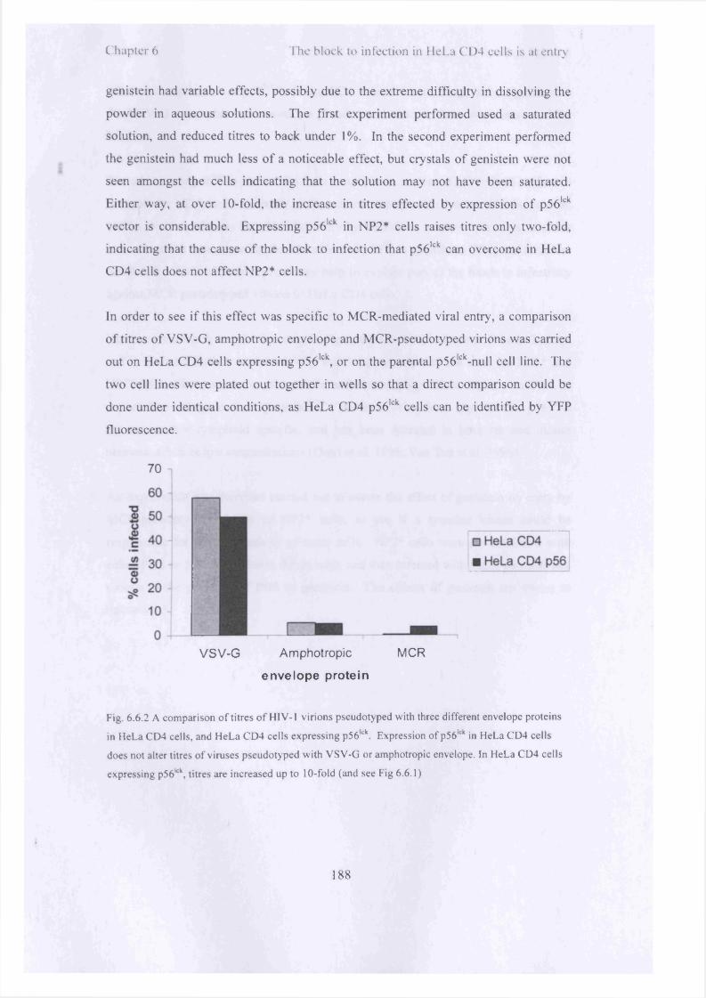
Chapter 6 The block to infection in HeLa CD4 cells is at entry
genistein had variable effects, possibly due to the extreme difficulty in dissolving the
powder in aqueous solutions. The first experiment performed used a saturated
solution, and reduced titres to back under 1%. In the second experiment performed
the genistein had much less o f a noticeable effect, but crystals o f genistein were not
seen amongst the cells indicating that the solution may not have been saturated.
Either way, at over 10-fold, the increase in titres effected by expression o f p56lck
vector is considerable. Expressing p56,ck in NP2* cells raises titres only two-fold,
indicating that the cause o f the block to infection that p56lck can overcome in HeLa
CD4 cells does not affect NP2* cells.
In order to see i f this effect was specific to MCR-mediated viral entry, a comparison
o f titres o f VSV-G, amphotropic envelope and MCR-pseudotyped virions was carried
out on HeLa CD4 cells expressing p56lck, or on the parental p56lck-null cell line. The
two cell lines were plated out together in wells so that a direct comparison could be
done under identical conditions, as HeLa CD4 p56lck cells can be identified by YFP
fluorescence.
70
VSV-G Amphotropic MCR
envelope prote in
Fig. 6.6.2 A comparison o f titres o f H IV - 1 virions pseudotyped with three different envelope proteins
in HeLa CD4 cells, and HeLa CD4 cells expressing p56lck. Expression o f p56lck in HeLa CD4 cells
does not alter titres o f viruses pseudotyped with V SV -G or amphotropic envelope. In HeLa CD4 cells
expressing p56kk, titres are increased up to 10-fold (and see Fig 6.6.1)
188

C hapte r 6 I lie b lo c k to in fe c t io n in H eLa ( '1 )4 cells is at entrv
While the expression o f p56lck in HeLa CD4 cells did not change titres significantly
for VSV-G or amphotropic envelope pseudotyped virions, titres were increased for
MCR.
The addition o f p56lck to HeLa CD4 cells results in an increased ratio o f cell surface to
intracellular CD4, and a decrease in the rate o f endocytosis (Pelchen-Matthews et al.
1995). Neither o f these should affect entry by VSV-G or amphotropic virus, and this
was indeed the case, showing that the effect o f p56lck is specific to CD4, and therefore
MCR Env. Both o f these aspects may help to explain part o f the block to infectivity
against MCR pseudotyped virions in HeLa CD4 cells.
However, the block to infection seen in HeLa CD4 cells is only part o f the Lv2 story,
and it must be further explained why this is apparently not the case for NP2* and
U87* cells. These are astrocytoma cells, and as non-lymphoid in origin, would not be
expected to endogenously express p56lck. However, it has been reported that p56lck is
not necessarily lymphoid specific, and has been detected in both rat and mouse
neurons, albeit at low concentrations (Omri et al. 1996; Van Tan et al. 1996).
An experiment was therefore carried out to assess the effect o f genistein on entry by
MCR-pseudotyped virions to NP2* cells, to see i f a tyrosine kinase could be
responsible for the permissivity o f these cells. NP2* cells were pre-incubated with
either PBS or lOOpM genistein for an hour, and then infected with MCR-pseudotyped
virions in the presence o f PBS or genistein. The effects o f genistein are shown in
figure 6.6.3.
189

Chapter 6 The block to infection in HeLa CD4 cells is at entry
80 i
70 j
60 i
■o« 50 1u
J 40
Vu# 30
20 j
10 |o I-------------
NP2 cells only NP2 + virus + PBS NP2 + virus +Genistein
cell conditions
Fig. 6.6.3 A tyrosine kinase inhibitor, genistein, reduces titres o f M C R pseudotyped virions by half in
NP2* cells. Cells were infected as described in the legend to figure 6.6.1. Shown are the results o f one
experiment. In a separate experiment, no decrease in titres on addition o f DM SO or ethanol (solvents
o f genistein) was seen (not shown).
When NP2* cells were incubated with genistein before infection with MCR-
pseudotyped virus, titres were reduced by 50%. This would imply that the difference
between NP2* and HeLa CD4 cells in their permissivity to MCR Env pseudotyped
virus may be partly, but not solely related to increased surface expression o f CD4
alone. In fact, even when expressing high levels o f p56lck, HeLa CD4 cells were still
significantly less permissive than NP2* cells, for example, the data in figure 6.6.1
shows that titres on HeLa CD4 p56lck-expressing cells reach 10%; titres on NP2* cells
with the volume o f virus used in this experiment typically reach over 70% cells
infected. There w ill be other reasons why HeLa CD4 cells are so refractory to
infection by MCR Env pseudotypes, and these effects combine to reduce titres so
drastically compared to NP2* cells.
190

C hapter 6 1'he block to infection in HeLa ( ’1)4 cells is at entry
Discussion
As it has been shown that a restriction factor is not responsible for the block to
infection in HeLa CD4 cells, it was necessary to consider what other factor(s) could
be responsible for low viral titres. Experiments described in chapter 5 show that
MCR-pseudotyped virions did not, in fact, appear to be able to enter HeLa CD4 cells
successfully, and therefore the process o f entry had to be considered in these cells.
Two receptors, CD4 and CXCR4, are necessary for entry via MCR (McKnight et al.
1998). Both o f these receptors are expressed in CD4 cells as detected by Western blot
o f whole cell lysates. Cloning and sequencing o f the CD4 receptor from HeLa CD4
cells showed no accumulated mutations (not shown). As levels o f CD4 and CXCR4
appear to be as expected, attention was then turned to whether a defect in receptor
usage could be detected.
In order to do this, a recently developed fusion protein, S15-mCherry was used.
When expressed in producer cells that synthesise virus, S15-mCherry labels the viral
membranes. As can be seen from the pictures and the data in sections 6.2 and 6.3,
this label provides a method o f assessing membrane fusion, the first stage o f a
productive infection. The data clearly show that MCR pseudotyped virions are not
fusing with HeLa CD4 cells, and in fact also suggest that a reduced absolute number
o f virions (about a quarter and a half o f the number o f virions compared to NP2* and
U87* cells respectively) is binding to HeLa CD4 cells (see appendix 3).
I f successful fusion is not taking place, this could be due to several factors. The
virions could themselves be defective, but this is unlikely given the high titres
obtained with the same batch o f virus on NP2* and U87* cells. There could be a
problem with CD4 expression or processing in HeLa CD4 cells, but this is also
unlikely given that it was detected in a Western blot o f whole cell lysates, and HeLa
CD4 cells are routinely used in viral titre assays for CD4-tropic virus. Alternatively,
it could be that CD4 is not found at high concentration at the cell surface, due to rapid
endocytosis, and that when it is trafficked out to the cell surface, the maladapted MCR
is not able to bind with enough rapidity before CD4 disappears again. Another
explanation is that MCR is binding, and binding too tightly (as suggested by Reuter et
191

C hapter 6 l hc b lo c k to in fec t ion in H eLa ( '1 )4 cells is at entrv
al. (Reuter et al. 2005)), and is not initiating fusion at the cell surface before the virus-
CD4 complex is endocytosed. This is not a productive entry pathway for HIV. The
small increase in titres in HeLa CD4 cells in which Rab5 is down regulated, or
acidification o f endosomes prevented, support the suggestion that endocytosis plays a
role, albeit along with other factors.
It should be borne in mind that MCR was derived from a primary isolate, and
therefore may be adapted for physiological conditions rather than the tissue culture
conditions described in these experiments. Certainly the cells that the MCR envelope
would be interacting with in an in vivo infection would be cells o f lymphocytic origin,
which carry a plethora o f regulatory mechanisms to ensure CD4 remains at the cell
surface. These mechanisms are thought to be largely lacking in cells o f non-
lymphocytic origin. Experiments described here in which titres o f MCR pseudotyped
virions are increased over 10-fold by expression o f p56lck support the suggestion that
lack o f CD4-regulatory mechanisms in HeLa CD4 cells are an important part o f the
reason for low titres. Within the physiological context, tight binding o f MCR Env to
CD4 on lymphocytes would probably be advantageous in the harsh extracellular
environment, and as p56lck would ensure that CD4 is present at the cell surface, the
likelihood o f this tight binding leading to undesired endocytosis is lowered.
With this hypothesis in mind, the question remains why NP2* and U87* cells, also o f
non-lymphoid origin, are apparently able to maintain CD4 at the cell surface. It has
been suggested that p56lck is expressed in rat and mouse brains (Omri et al. 1996; Van
Tan et al. 1996). Preliminary Western blots o f NP2* and U87* cells gave a
tantalising hint that there could be miniscule level o f p56lck protein in these cells (not
shown), but this does not preclude another CD4-regulatory mechanism being active as
well. Different cell lines from the same species can have utterly different intracellular
pathways, optimised for their particular role in the organism, and experiments in cell
biology and trafficking in one cell type cannot be presumed to reveal generalities for
all cell types from that species. The fact that MCR pseudotyped virions are able to
infect NP2* and U87* cells successfully should not be taken as evidence that they are
able to infect all human cells, but simply that they are competent for infection per se,
and in some cell lines the infection is successful.
192
C hapte r 6 I he b lo c k to in fe c t io n in H eLa ( 1)4 ce l ls is at entry
In March 2007, towards the end o f this period o f work, a new restriction to infection
was described that also has a route o f entry component, and dubbed Lv3 (Pineda et al.
2007). A rhesus macaque epithelial cell line expressing human CD4 (sMAGI)
exhibited two levels o f post-entry restriction to HIV-1, the first level being Trim5a.
The second level was operative against HIV-1 core pseudotyped with SIV envelope,
or an envelope from an R5 clone, SF162. The cells were not infected even at the
highest doses, indicating that the block to restriction was not caused by a saturable
factor, and addition o f SiRNA against Trim5a had no effect on titres. The block in
sMAGI cells could be circumvented by pseudotyping with VSV-G, or when sMAGI
cells expressed human CCR5, i f Trim5a was also abrogated. A threshold o f virus
entry which was detectable by qPCR data (-25 copies) was reached at l-2ng/ml Gag;
at all doses levels were ~100x lower in sMAGI cells compared to sMAGI-CCR5.
Levels o f the early products o f reverse transcription can be usefully used as markers
o f viral entry when there is a significant increase over baseline levels. However, as
some reverse transcription can occur in the retroviral particle pre-entry low levels o f
reverse transcription products cannot be reliably assumed to be solely due to post
entry events.
For example, on non-permissive sMAGI cells, 2ng/ml Gag equates to an MOI o f
-0.32, and 50 copies o f early RT products per 20,000 cells. The highest level that is
reached is 1000 copies per 20,000 cells at 40ng/ml i.e. MOI o f -6.4. Considering that
there is such a high MOI o f virus laid on the cells, the number o f copies per cell
seems unreasonably low, i f the desired point to be derived from the data was that
entry was uninhibited (compare to the lOOx higher levels in the unrestricted sMAGI-
CCR5 cells). Cells were washed before analysis which would suggest that the low-
level reverse transcription that occurs in free virions could not be responsible (Trono
1992; Zhang et al. 1993). However, in this particular experiment virions would not
necessarily be readily removed by washing even i f they were not able to enter the cell.
I f the block to infection were indeed at entry due to lack o f an optimised co-receptor,
virions would still bind to CD4 and thus be resistant to removal by washing. It is
conceivable that the reverse transcription products detected from the sMAGI cells are,
therefore, due to reverse transcription occurring in these bound extracellular virions.
193

C hapte r 6 I he b lo ck to in fe c t io n in H eLa C l ) 4 ce l ls is al entry
A cell-cell fusion assay to test the efficiency o f fusion o f the endogenous rhesus co
receptor revealed a part o f the block, and concluded that SF162 envelope was reduced
in efficiency for fusion with sMAGI cells compared to cells expressing CCR5,
although the relevance o f cell-cell fusion for assessment o f viral envelope viability is
not necessarily confirmed. Another viral envelope from SIV, however, was equally
efficient for fusion on both cell lines, but was similarly restricted during the infectious
cycle on sMAGI cells, again by a block that was unsaturable by nature. It was not
reported how levels o f early RT products compared for this envelope.
In order to be completely certain that a block was post-entry it would need to be
ascertained that fusion and entry o f the virus were uninhibited, and at comparable
levels to unrestricted virions. For example, for F v ln this was done by comparing
levels o f both early and late reverse transcription products for N-(unrestricted) and B-
(restricted) tropic MLV. Regarding rhTrim5a, levels o f early products o f restricted
and unrestricted virus were not comparable as the block is pre-reverse transcription,
but the saturable nature o f the block overwhelmingly indicated that a post-entry,
cytosolic restriction was involved, rather than a block at the level o f fusion and entry.
For Lv3 there is more uncertainty that fusion is sufficient with the SF162 envelope
and the putative rhesus co-receptor (expressed endogenously on sMAGI cells), and
comparisons between titres on sMAGI and sMAGI-CCR5 cells are only questionably
relevant. Certainly the sMAGI cells engineered to express the human CCR5 may
artificially increase the levels o f virions entering the cells by virtue o f the over
expression o f co-receptor, but this would simply reflect the difference between cells
able to take up artificially large levels o f virions and cells with levels o f viral entry
that have not been thus amplified, rather than revealing a bona fide restriction.
In conclusion, HeLa CD4 cells are poorly infectable by virions pseudotyped by MCR
Env but this is primarily due to inhibition o f the viral lifecycle at the level o f entry, to
a combination o f poor availability o f CD4 at the cell surface and sub-optimal
interaction o f MCR Env and CD4/CXCR4, rather than a post-entry block caused by a
specific anti-viral factor. While the type o f block to infection suggested by Lv2 as a
route o f entry-dependent restriction is not by any means impossible, it is intuitively
unlikely, and experiments in order to uncover a post-entry restriction using MCR are
difficult, given the overwhelming effect o f the entry block. In order to reverse
194

C hapter 6 1 he b lo c k to in fec t ion in H eLa C 1)4 cells is at entry
transcribe over a baseline level, virions must access the pool o f dNTPs in the
cytoplasm. I f they have reached the cytoplasm and reverse transcribed, the idea that
they would then somehow re-enter a subcellular compartment (i.e. one in which an
anti-retroviral such as Lv2 might be active) in order to access the nucleus is not
probable based on current knowledge. It is not, however, impossible that a factor
could bind to the virion as it passed through various endocytic compartments on its
way into the cell cytoplasm, and that this factor would only manifest an effect later on
in the viral lifecycle. This would then be in effect a route o f entry-dependent
restriction, as it would depend on the route that the virus took to get into the
cytoplasm even though the block was later on. For HeLa CD4 cells and MCR Env,
however, this is manifestly not the case, as the virus is not even entering the cell.
195

C hapter 7 Thesis d iscussion
Chapter 7Thesis Discussion
Rationale
During these studies, the overarching aim has been to shed light on the earliest events
o f retroviral replication. It is unfortunate that the commonest methods o f detecting
successful entry rely on all stages o f the early life cycle being completed, and the
virion being able to integrate successfully. Thus, from an initial FACS assay, an entry
block appears very similar to a block at integration as in both cases no fluorescent
signal is produced. However, in the former the virus has not even penetrated the cell,
whereas in the latter, the virus has entered, reverse transcribed and accessed the
nucleus. Other techniques must be employed to f ill in the missing details. In this
thesis a variety o f methods were used both to try and control variations in the early
stages o f the lifecycle, and also to detect success at each o f these stages. For example,
different combinations o f envelope proteins and receptors were used to try and direct
the virus into the cell by different routes, and the success o f attempted entry o f the
virus monitored by tracking loss o f a fluorescent signal from a protein in the viral
membrane. These techniques aid localisation o f blocks to replication more precisely
in both time and the space o f the cell and the infection process.
The characteristics o f the entry pathways mediated by the Tva800 and Tva950 after
binding o f ASLV Env were investigated. The differences between the two pathways
were probed using chemical and microscopic methods, and an attempt made to assess
how these differences might be manifest.
During the course o f these studies, a novel titration curve obtained using Tva800 as a
receptor for viral entry inspired a series o f experiments designed to clarify
quantitative aspects o f viral binding and fusion.
A previously described block to replication, Lv2, was re-examined and the principles
o f this phenomenon (namely that variation in route o f entry into the cell can have a
profound effect on subsequent steps o f replication) were assessed.
196

Chapte r 7 Thesis d iscussion
Results
It was found that Fvl and Trim5a restriction did not have a route o f entry dependence
that was revealed by these experiments. This is intuitively what would be expected,
as restriction factors do not act on whole virus, but after the membrane has been lost,
on the incoming core. Irrespective o f the current debate over whether Trim5a effects
restriction via a more rapid disassembly or via degradation o f the core by another
method, it is difficult to conceptualise how any binding sites would be revealed until
the viral membrane has been lost and the core delivered into the cytoplasm. Any
variations taken in the pathway up to that point o f fusion would, therefore, be
irrelevant, as long as the virus did ultimately reach it. Hence, although Tva800 and
Tva950 appear to traffic the virus into the cell by slightly different routes, they both
deliver the virus to a functional entry point (the late endosome?) at which fusion can
occur. After the virus is delivered to the cytoplasm, subsequent actions on the core
would affect those that entered via Tva800 and Tva950 equally, and this is what was
found with the experiments described here with Fv l and Trim5a. For these two
restriction factors, it is probable that there is no route o f entry component.
The routes that virions take after binding to Tva800 and Tva950 remain to be fully
described. Differences in the susceptibility o f the virus to degradation under an
hnTjCl-induced block to infection imply that either the micro-location o f the receptor,
the structure o f the receptor or the different routes that the receptor directs the virus
down after binding have a profound effect on subsequent events. GPI-linked
receptors located in lipid rafts are thought to direct ligands down caveolae-mediated
endocytic routes which have slower kinetic parameters than clathrin-mediated
endocytosis, and direct to caveosomes, caveolin-1 containing endosomes. The lipid
raft areas o f the membrane are enriched for cholesterol, glycosphingolipids and
sphingomyelin, which collude to stiffen the membrane. During endocytosis the
membrane w ill invaginate and pinch off, travelling into the cell, and the caveosome
dynamics may be affected by the composition o f the membrane it has been formed
from, namely the lipid raft area. Lipids themselves play key roles in the targeting o f
intracellular organelles and in fusion dynamics, for example, phosphoinositide 3-
phosphate is an important targeting signal for endosomal traffic (Hurley and Meyer
197

Chapter 7 Thesis diseussion
2001). In itself, it is not enough to target an early endosome; Rab5 is needed, but is
part o f the overall recognition o f the docking site. The bulkiness o f the lipids forming
the caveosome could have a profound effect on targeting and fusion dynamics o f the
caveosome, the release o f ligands bound at the cell surface, on viral penetration o f the
caveosome membrane to enter the cytoplasm, and could be a key feature in the
different viral entry kinetics and susceptibility to NH4C1 for Tva800 and Tva950. A ll
this is speculative, but as the binding domains o f the receptors are identical, and
clearly the envelope protein o f the virus is likewise, so the functional difference in the
two receptors must relate somehow to their attachment and localisation.
The local environment o f Tva800 or Tva950 in the cell membrane may also explain a
further difference between the two receptors, namely the requirement that a virus
binds >1 Tva800 receptor in order to be able to enter a cell successfully, but that
binding o f only one is necessary for entry via Tva950. Entry o f the virus requires a
considerable input o f activation energy to perturb the cell membrane sufficiently; the
decreased fluidity o f the cell membrane in lipid rafts may require the extra energy
input provided from extra receptor binding and fusion activation in order to overcome
the extra activation energy required for the stiffer membranes.
Virions carry multiple copies o f envelope proteins, although there is considerable
debate both about precisely how many extra copies they carry, and whether all o f
these are functional or not. Chertova et al. reported that HIV-1 virions contained
Gag:Env ratios that corresponded to between 7-14 trimers per virion, although the
simple presence o f a trimer is no guarantee that it is functional (Chertova et al. 2002).
Using antibody neutralising studies, Yang et al. concluded that only one functional
trimer is necessary to mediate viral infectivity for HIV-1, amphotropic M LV and
ASLV-A (with Tva950) (Yang et al. 2005a; Yang et al. 2005b). ASLV-A entry with
Tva800 was not studied (Yang et al. 2005b). Relative infectivities o f virions
possessing different ratios o f dominant negative mutant Env to wild-type Env were
plotted, and compared to the curve expected from a model where one functional
trimer is needed for infectivity. From another study by the same authors it was
furthermore concluded that only two wild-type units within a trimer were required for
that trimer to be functional (Yang et al. 2006).
198

C hapter 7 Thesis d iseussion
However, these findings have been disputed with alternative accounts o f the data,
which primarily address key assumptions that interpretation o f the experimental data
relied on. For example, a key assumption is that the number o f infectious virions in a
stock is as low as the number that actually infect cells under normal conditions. But
viral titres can be raised when interactions with cells are increased, as infectious
virions are lost to unproductive interactions with the cell or are endocytosed. Thus
the small percentage that infects is not the same as the proportion that is infectious.
The assumption that each virion only has the minimal number o f trimers required for
infection is also unsubstantiated, as functional Env may not be the dominant
limitation o f infection. Klasse carried out a mathematical investigation o f two types
o f models, in which the number o f Env trimers required for infection is either over a
minimum threshold or whether interaction o f each successive trimer incrementally
raises the chance o f a successful infection (Klasse 2007). The most likely scenario
that best-fit models suggested is a combination o f these two possibilities, which could
vary for different viruses, and mutations o f Env. Models explored the redundancy o f
Env protomers to generate a functional trimer unit, and the number o f functional
trimers required for a functional virion. It could be the case, for example, that rather
than an all-or-nothing scenario in which 2 non-functioning protomers render a trimer
inactive but 1 non-functioning protomer does not, it would be much more likely that
each protomer adds to the probability o f a trimer being functional, but a virion with
(for example) 7 semi-functional trimers is much less likely to be infectious than one
with 7 fully-functional trimers, hence the threshold levels o f activation come into
play. A model to demonstrate this, however, may not be readily distinguishable,
especially as both fully and partially functional trimers may contribute to cell binding,
i f not to infectivity.
In short, the best interpretation is that differences between virions and Env mutations
contribute to a blurring o f potential models, but that a combination o f incremental
effects o f each protomer to trimer function exists, and an increasing number o f
functional trimers eventually take a virion over a real threshold o f activation (Klasse
2007). This threshold cannot be equivalent to 1 trimer, as other data rule out this
option. For example, the phenomenon o f antibody-dependent neutralisation described
for dengue virus and HIV-1 (Schutten et al. 1995; Goncalvez et al. 2007) rules out the
possibility that a virion could have only one trimer (functional or not). This
199

C hapter 7 Thesis d iseussion
phenomenon describes how increasing concentrations o f antibody against envelope
increase the proportion o f virus entering a cell up to a point, after which further
increases in antibody block entry. It is thought that up to the turning point, antibody
against envelope neutralises negative charges on the virion, which would exert some
level o f repulsion away from the cell surface, so increasing viral titres. After the
turning point, however, addition o f further antibody starts to neutralise the viral
envelope protein to the extent that the virion is no longer able to initiate binding and
entry. So this explanation requires that in addition to the envelope protein(s) that
mediate entry, at the very least the virion would require additional trimers sufficiently
functional as to be capable o f binding antibody.
Studies o f H IV are complicated by the requirement o f receptor and coreceptor, and
additionally by the interdependence o f levels o f CD4 and CCR5 or CXCR4.
However, studies done argue against formation o f an entry complex and virion pore
mediated by a single trimer as HIV-1 could initiate infection with low levels o f CCR5
co-receptor i f levels o f CD4 were sufficiently high (or vice-versa), and that between
4-6 CCR5 molecules were required to bind the virion (Kuhmann et al. 2000).
Within this framework, the data presented in chapter 4 are not surprising. The
concept o f multiple receptor binding events being required for successful virion entry
is one that sits easily with the current models and data for other viral envelope-
receptor pairs. The simplicity o f the ASLV Env, Tva800 and Tva950 system lends
itself as a model system to facilitate further studies in this area. The interaction does
not require any additional factors or coreceptors, which simplifies things
considerably. Additionally, the mechanism o f entry is via endocytosis, which means
that there are no issues with virions being considered non-infectious when they have
simply embarked on a non-productive route o f entry, as is found for virions with a
requirement for fusion at the cell surface that get endocytosed.
A ll o f the studies presented in this thesis have proceeded from the basic premise that
the basic requirements o f a virion and a cell, expressing the cognate receptor, are not
necessarily a sufficient prelude to infection. However, it is also undoubtedly true that
not all barriers to infection necessarily have anything to do with restriction factors.
To give a well-known example, HIV-1 is able to initiate fusion into the cell from
200
C hapter 7 Thesis d iscussion
receptors at the cell membrane. If, however, the virion starts down the endocytic
pathway then the infection is unproductive as in most cell types it w ill be degraded in
the acidic environment. This is a major reason for the extremely low particle:
infectivity ratio for HIV-1. However, this does not in any way fall under the category
o f restriction as the virus is simply not managing to penetrate the cell successfully,
and there is no specific factor that is acting on the virus and blocking replication. As
the search for anti-retroviral agents continues, the need for new ways to counteract
infection grows, but this should not warp perspectives so much that cellular and viral
mismatches are not first considered thoroughly, before a novel mechanism is
proclaimed.
That said, the possibility should not be discounted, however, that among the myriad o f
cellular compartments that a virus could pass through on its way into the cell, there
could be those that contain anti-viral proteins or conditions that would irreversibly
alter the virus. Although the virus may be able to proceed a little further in the
infectious cycle, it is rendered unfit for completion. An example o f something along
these lines would be the Trim5a-mediated restriction o f H IV -1 with and without the
influence o f MG 132, a proteasome inhibitor (Anderson et al. 2006). When MG 132 is
absent, the viral infectious cycle and reverse transcription are both inhibited. When it
is present, reverse transcription occurs so that products o f it are found, but overall the
infectious cycle is still blocked.
There is so much about the early stages o f retroviral infection that remains to be
understood, and cellular factors used by the virus that remain to be identified. A list
o f host proteins involved in HIV-1 infection was identified through a large scale
SiRNA screen, and published towards the end o f writing o f this thesis (Brass et al.
2008). The authors identified over 250 host proteins involved at all stages from entry
to exit, many o f which were not previously known to be associated with HIV-1
infection. Further analysis o f the proteins listed that were involved in stages o f
infection between entry and integration would be an excellent starting point in a
search for restriction factor-like agents, or potential targets for anti-virals.
201

C hapter 7 Thesis d iscussion
Conclusion
Finally, it is evident that Fv l and Trim5a are merely forebears o f what may be a
whole range o f intracellular anti-virals, and many more are sure to be discovered.
While these offer potentially vast rewards in terms o f information revealed about the
viral life-cycle, and the workings o f the cell, several criteria must be satisfied for a
block to be considered both a restriction and significant in terms o f potential
therapeutics. Firstly, a significant decrease in viral titres must be evident. Previous
work assigned the term restriction only to decreases o f 100-fold or more in viral titres
(Hartley et al. 1970). A decrease o f less than lOx must be viewed with extreme
scepticism to ensure that different cell or experimental conditions are not causing a
variation in titres consistent with experimental error. Secondly, before putative
restriction factors are invoked it must be proven beyond any reasonable doubt that the
virus is indeed entering the cell at unrestricted levels, and that the block to infection is
due to a process significantly downstream o f entry. This could be done by detection
o f later products o f reverse transcription, or alternatively by abrogation o f the block.
Thirdly, the block should ideally be relevant on a physiological level. For example,
rhesus macaques were shown to be refractory to infection by HIV-1 long before the
discovery o f Trim5a, and the first discoveries about Fv l were made using whole
mice, rather than the cell lines. I f at least the first two criteria are satisfied then a
block to infection may be worth pursuing, and classifying as a restriction.
Further Work
During the course o f the work described in this thesis many questions were raised,
some o f which remain unanswered. The nature o f the differences between Tva800
and Tva950 has been partly clarified, yet the way in which these differences affect the
routes into the cell taken from the receptors is still not clear. Ideally further tracking
studies could be done to trace virions as they pass into the cell, potentially
highlighting other important intracellular compartments. The use o f a broad range o f
dominant negative inhibitors would also aid characterisation.
In the studies o f how many Tva800 receptors are needed for viral entry, a more
quantitative system was sought throughout. A tetracycline expression system,
202

( hapter 7 Thesis d iscussion
whereby expression o f a gene is controlled by the level o f this antibiotic in the
medium was made and tested, but was not functional in time for experiments to be
included in this thesis. Further experiments in this area must generate more
quantitative data, and a precise assessment o f numbers o f Tva800 at the cell surface in
further studies is needed.
Further experiments to investigate the refractory nature o f HeLa CD4 cells to
infection by some CD4-tropic viruses, e.g. MCR, would have to compare HeLa CD4
cells to cells that would express CD4 in a physiological context. A thorough
investigation o f mechanisms that normally regulate CD4 cell-surface expression in
lymphocytic cells is called for, with a view to understanding how these could be
potentially be present in NP2* and U87* cells, even though non-lymphocytic in
origin.
A ll o f these would contribute significantly to the body o f knowledge that must be
built up in order to better understand aspects o f retroviral infection that could be
targeted in future therapies.
203

References
Aiken, C. (1997). "Pseudotyping human immunodeficiency virus type 1 (H IV-1) by the glycoprotein o f vesicular stomatitis virus targets HIV-1 entry to an endocytic pathway and suppresses both the requirement for N e f and the sensitivity to cyclosporin A ." J V iro l 71(8): 5871-7.
A ldovini, A. and R. A. Young (1990). "Mutations o f RNA and protein sequences involved in human immunodeficiency virus type 1 packaging result in production o f noninfectious virus." J V iro l 64(5): 1920-6.
Alkhatib, G., C. Combadiere, C. C. Broder, Y. Feng, P. E. Kennedy, P. M. Murphy and E. A. Berger (1996). "CC CKR5: a RANTES, MIP-1 alpha, M IP-lbeta receptor as a fusion cofactor for macrophage-tropic H IV -1." Science 272(5270): 1955-8.
An, W. and A. Telesnitsky (2001). "Frequency o f direct repeat deletion in a human immunodeficiency virus type 1 vector during reverse transcription in human cells." V iro logy 286(2): 475-82.
Andersen, J. L. and V. Planelles (2005). "The role o f Vpr in HIV-1 pathogenesis." Curr H IV Res 3(1): 43-51.
Anderson, H. A., Y. Chen and L. C. Norkin (1996). "Bound simian virus 40translocates to caveolin-enriched membrane domains, and its entry is inhibited by drugs that selectively disrupt caveolae." M ol B io l Cell 7(1 1): 1825-34.
Anderson, J. L., E. M. Campbell, X. Wu, N. Vandegraaff, A. Engelman and T. J.Hope (2006). "Proteasome inhibition reveals that a functional preintegration complex intermediate can be generated during restriction by diverse TRIM5 proteins." J V iro l 80(19): 9754-60.
Anderson, J. L. and T. J. Hope (2004). "H IV accessory proteins and surviving the host cell." Curr H IV /A IDS Rep 1(1): 47-53.
Andreadis, S. T., D. Brott, A. O. Fuller and B. O. Palsson (1997). "Moloney murine leukemia virus-derived retroviral vectors decay intracellularly w ith a half-life in the range o f 5.5 to 7.5 hours." J V iro l 71(10): 7541-8.
Arrigo, S. J., S. Weitsman, J. A. Zack and I. S. Chen (1990). "Characterization and expression o f novel singly spliced RNA species o f human immunodeficiency virus type 1." J V iro l 64(9): 4585-8.
Axelrad, A. (1969). "Genetic and cellular basis o f susceptibility or resistance toFriend leukemia virus infection in mice." Proc Can Cancer Conf 8: 313-43.
Bachrach, E., M. Marin, M. Pelegrin, G. Karavanas and M. Piechaczyk (2000)."Efficient cell infection by Moloney murine leukemia virus-derived particles requires minimal amounts o f envelope glycoprotein." J V iro l 74(18): 8480-6.
Badorrek, C. S., C. M. Gherghe and K. M. Weeks (2006). "Structure o f an RNA switch that enforces stringent retroviral genomic RNA dimerization." Proc Natl Acad Sci U S A 103(37): 13640-5.
Bainbridge, J. W., C. Stephens, K. Parsley, C. Demaison, A. Halfyard, A. J. Thrasher and R. R. A li (2001). "In vivo gene transfer to the mouse eye using an H IV- based lentiviral vector; efficient long-term transduction o f corneal endothelium and retinal pigment epithelium." Gene Ther 8(21): 1665-8.
Balliet, J. W., J. Berson, C. M. D'Cruz, J. Huang, J. Crane, J. M. Gilbert and P. Bates (1999). "Production and characterization o f a soluble, active form o f Tva, the subgroup A avian sarcoma and leukosis virus receptor." J V iro l 73(4): 3054- 61.
204
Balliet, J. W., D. L. Kolson, G. Eiger, F. M. Kim , K. A. McGann, A. Srinivasan and R. Collman (1994). "D istinct effects in primary macrophages and lymphocytes o f the human immunodeficiency virus type 1 accessory genes vpr, vpu, and nef: mutational analysis o f a primary HIV-1 isolate." V iro logy 200(2): 623-31.
Baltimore, D. (1970). "RNA-dependent D N A polymerase in virions o f RNA tumour viruses." Nature 226(5252): 1209-11.
Baltimore, D. and R. M. Franklin (1963). "A New Ribonucleic Acid Polymerase Appearing after Mengovirus Infection o f L-Cells." J B io l Chem 238: 3395- 400.
Barbero, P., L. Bittova and S. R. Pfeffer (2002). "Visualization o f Rab9-mediated vesicle transport from endosomes to the trans-Golgi in liv ing cells." J Cell Biol 156(3): 511-8.
Barbieri, M. A., R. L. Roberts, A. Gumusboga, H. H ighfie ld, C. Alvarez-Dominguez, A. Wells and P. D. Stahl (2000). "Epidermal growth factor and membrane trafficking. EGF receptor activation o f endocytosis requires Rab5a." J Cell B M 151(3): 539-50.
Barnard, R. J., D. Elleder and J. A. Young (2006). "Avian sarcoma and leukosis virus- receptor interactions: from classical genetics to novel insights into virus-cell membrane fusion." V iro logy 344(1): 25-9.
Barnard, R. J. and J. A. Young (2003). Alpharetrovirus envelope-receptor interactions. Curr Top M icrobio l Im m unol. 281: 107-36.
Barr, S. D., J. R. Smiley and F. D. Bushman (2008). "The Interferon ResponseInhibits H IV Particle Production by Induction o f TR IM 22." PLoS Pathogens 4(2): el000007.
Barre-Sinoussi, F., J. C. Chermann, F. Rey, M. T. Nugeyre, S. Chamaret, J. Gruest, C. Dauguet, C. Axler-B lin, F. Vezinet-Brun, C. Rouzioux, W. Rozenbaum and L. Montagnier (1983). "Isolation o f a T-lymphotropic retrovirus from a patient at risk for acquired immune deficiency syndrome (A ID S)." Science 220(4599): 868-71.
Bassin, R. H., G. Duran-Troise, B. I. Gerwin and A. Rein (1978). "Abrogation o f Fv- lb restriction with murine leukemia viruses inactivated by heat or by gamma irradiation." J V iro l 26(2): 306-15.
Bates, P., J. A. Young and H. E. Varmus (1993). "A receptor for subgroup A Rous sarcoma virus is related to the low density lipoprotein receptor." Cell 74(6): 1043-51.
Bayer, N., D. Schober, E. Prchla, R. F. Murphy, D. Blaas and R. Fuchs (1998)."Effect o f bafilomycin A l and nocodazole on endocytic transport in HeLa cells: implications for viral uncoating and infection." J V iro l 72(12): 9645-55.
Beijerinck, M. (1898). "Concerning a contagium vivum flu idum as cause o f the spot disease o f tobacco leaves." Verhandelingen der Koninkyke akademie Wettenschappen te Amsterdam 65: 3-21.
Belanger, C., K. Zingler and J. A. Young (1995). "Importance o f cysteines in the LDLR-related domain o f the subgroup A avian leukosis and sarcoma virus receptor for viral entry." J V iro l 69(2): 1019-24.
Ben-David, Y. and A. Bernstein (1991). "Friend virus-induced erythroleukemia and the multistage nature o f cancer." Cell 66(5): 831-4.
Bendtsen, J. D., H. Nielsen, G. von Heijne and S. Brunak (2004). "Improved prediction o f signal peptides: SignalP 3.0." J M ol B iol 340(4): 783-95.
Benit, L., N. De Parseval, J. F. Casella, I. Callebaut, A. Cordonnier and T. Heidmann (1997). "Cloning o f a new murine endogenous retrovirus, M uERV-L, with
205

strong sim ilarity to the human HERV-L element and w ith a gag coding sequence closely related to the F v l restriction gene." J V iro l 71(7): 5652-7.
Berthet-Colominas, C., S. Monaco, A. Novelli, G. Sibai, F. Mallet and S. Cusack (1999). "Head-to-tail dimers and interdomain flex ib ility revealed by the crystal structure o f H IV -1 capsid protein (p24) complexed with a monoclonal antibody Fab." Embo J 18(5): 1124-36.
Best, S., P. Le Tissier, G. Towers and J. P. Stoye (1996). "Positional cloning o f the mouse retrovirus restriction gene F v l." Nature 382(6594): 826-9.
Bishop, K. N. (2001). The interactions o f the viral restriction gene, F v l, and its target. London, UCL: 197.
Bishop, K. N., G. B. Mortuza, S. Howell, M . W. Yap, J. P. Stoye and I. A. Taylor (2006). "Characterization o f an amino-terminal dimerization domain from retroviral restriction factor F v l." J V iro l 80(16): 8225-35.
Bittman, R., C. R. Kasireddy, P. Mattjus and J. P. Slotte (1994). "Interaction o fcholesterol with sphingomyelin in monolayers and vesicles." Biochemistry 33(39): 11776-81.
Bjomdal, A., H. Deng, M. Jansson, J. R. Fiore, C. Colognesi, A. Karlsson, J. Albert, G. Scarlatti, D. R. Littman and E. M. Fenyo (1997). "Coreceptor usage o f primary human immunodeficiency virus type 1 isolates varies according to biological phenotype." J V iro l 71(10): 7478-87.
Bock, M., K. N. Bishop, G. Towers and J. P. Stoye (2000). "Use o f a transient assay for studying the genetic determinants o f F v l restriction." J V iro l 74(16): 7422- 30.
Boggs, J. M. (1987). "L ip id intermolecular hydrogen bonding: influence on structural organization and membrane function." Biochim Biophys Acta 906(3): 353- 404.
Boone, L. R., P. L. Glover, C. L. Innes, L. A. N iver, M. C. Bondurant and W. K.Yang (1988). "Fv-1 N- and B-tropism-specific sequences in murine leukemia virus and related endogenous proviral genomes." J V iro l 62(8): 2644-50.
Boone, L. R., C. L. Innes and C. K. Heitman (1990). "Abrogation o f Fv-1 restriction by genome-deficient virions produced by a retrovirus packaging cell line." J V iro l 64(7): 3376-81.
Bova, C. A., J. C. Olsen and R. Swanstrom (1988). "The avian retrovirus env genefamily: molecular analysis o f host range and antigenic variants." J V iro l 62(1): 75-83.
Bowerman, B., P. O. Brown, J. M. Bishop and H. E. Varmus (1989). "Anucleoprotein complex mediates the integration o f retroviral D N A." Genes Dev 3(4): 469-78.
Brass, A. L., D. M. Dykxhoom, Y. Benita, N. Yan, A. Engelman, R. J. Xavier, J.Lieberman and S. J. Elledge (2008). "Identification o f host proteins required for H IV infection through a functional genomic screen." Science 319(5865): 921-6.
Briggs, J. A., T. W ilk and S. D. Fuller (2003). "Do lip id rafts mediate virus assembly and pseudotyping?" J Gen V iro l 84(Pt 4): 757-68.
Brindley, M. A. and W. Maury (2008). "Equine infectious anemia virus entry occurs through clathrin-mediated endocytosis." J V iro l 82(4): 1628-37.
Brown, D. A. and E. London (1998). "Functions o f lip id rafts in biological membranes." Annu Rev Cell Dev Biol 14: 111-36.
206

Brown, P. O., B. Bowerman, H. E. Varmus and J. M. Bishop (1989). "Retroviral integration: structure o f the in itia l covalent product and its precursor, and a role for the viral IN protein." Proc Natl Acad Sci U S A 86(8): 2525-9.
Bruce, J. W., P. Ahlquist and J. A. Young (2007). ZASC1 is a novel cellulartranscription factor that modulates M L V infection. Cold Spring Harbor, NY, USA.
Bruce, J. W., K. A. Bradley, P. Ahlquist and J. A. Young (2005). "Isolation o f celllines that show novel, murine leukemia virus-specific blocks to early steps o f retroviral replication." J V iro l 79(20): 12969-78.
Bukrinsky, M. I., S. Haggerty, M. P. Dempsey, N. Sharova, A. Adzhubel, L. Spitz, P. Lewis, D. Goldfarb, M. Emerman and M. Stevenson (1993). "A nuclear localization signal w ith in HIV-1 matrix protein that governs infection o f nondividing cells." Nature 365(6447): 666-9.
Bullough, P. A., F. M. Hughson, J. J. Skehel and D. C. W iley (1994). "Structure o f influenza haemagglutinin at the pH o f membrane fusion." Nature 371(6492): 37-43.
Bushman, F. D. and R. Craigie (1990). "Sequence requirements for integration o f Moloney murine leukemia virus D N A in v itro ." J V iro l 64(11): 5645-8.
Bushman, F. D., T. Fujiwara and R. Craigie (1990). "Retroviral D N A integration directed by H IV integration protein in v itro ." Science 249(4976): 1555-8.
Campbell, E. M., M. P. Dodding, M. W. Yap, X. Wu, S. Gallois-Montbrun, M. H.Malim , J. P. Stoye and T. J. Hope (2007a). "TR IM 5 alpha cytoplasmic bodies are highly dynamic structures." M ol B io l Cell 18(6): 2102-11.
Campbell, E. M., O. Perez, M. Melar and T. J. Hope (2007b). "Labeling HIV-1virions w ith two fluorescent proteins allows identification o f virions that have productively entered the target cell." V iro logy 360(2): 286-93.
Carr, C. M. and P. S. Kim (1993). "A spring-loaded mechanism for theconformational change o f influenza hemagglutinin." Cell 73(4): 823-32.
Cecilia, D., V. N. KewalRamani, J. O'Leary, B. Volsky, P. Nyambi, S. Burda, S. Xu, D. R. Littman and S. Zolla-Pazner (1998). "Neutralization profiles o f primary human immunodeficiency virus type 1 isolates in the context o f coreceptor usage." J V iro l 72(9): 6988-96.
Chen, S. J., G. Lin, K. J. Chang, L. S. Yeh and C. C. Wang (2008). "Translational efficiency o f a non-AUG initiation codon is significantly affected by its sequence context in yeast." J B io l Chem 283(6): 3173-80.
Cherepanov, P., G. Maertens, P. Proost, B. Devreese, J. Van Beeumen, Y.Engelborghs, E. De Clercq and Z. Debyser (2003). "H IV-1 integrase forms stable tetramers and associates w ith LEDGF/p75 protein in human cells." J B iol Chem 278(1): 372-81.
Cherpillod, P., L. Zipperle, R. W ittek and A. Zurbriggen (2004). "An mRNA region o f the canine distemper virus fusion protein gene lacking AUG codons can promote protein expression." Arch V iro l 149(10): 1971-83.
Chertova, E., J. W. Bess Jr, Jr., B. J. Crise, I. R. Sowder, T. M. Schaden, J. M.Hilburn, J. A. Hoxie, R. E. Benveniste, J. D. Lifson, L. E. Henderson and L. O. Arthur (2002). "Envelope glycoprotein incorporation, not shedding o f surface envelope glycoprotein (gpl20/SU), Is the primary determinant o f SU content o f purified human immunodeficiency virus type 1 and simian immunodeficiency virus." J V iro l 76( 11): 5315-25.
207

Ciuffi, A., M. Llano, E. Poeschla, C. Hoffmann, J. Leipzig, P. Shinn, J. R. Ecker and F. Bushman (2005). "A role for LEDGF/p75 in targeting H IV DNA integration." Nat Med 11(12): 1287-9.
Clapham, P. R., D. Blanc and R. A. Weiss (1991). "Specific cell surface requirements for the infection o f CD4-positive cells by human immunodeficiency virus types 1 and 2 and by Simian immunodeficiency virus." V iro logy 181(2): 703- 15.
Coffin, J. M., S. H. Hughes and H. Varmus (1997). Retroviruses. Plainview, N.Y., Cold Spring Harbor Laboratory Press.
Colman, P. M. and M. C. Lawrence (2003). "The structural biology o f type I viral membrane fusion." Nat Rev M ol Cell B io l 4(4): 309-19.
Conner, S. D. and S. L. Schmid (2003). "Regulated portals o f entry into the cell." Nature 422(6927): 37-44.
Cowan, S., T. Hatziioannou, T. Cunningham, M . A. Muesing, H. G. Gottlinger and P. D. Bieniasz (2002). "Cellular inhibitors w ith F v l- lik e activity restrict human and simian immunodeficiency virus tropism." Proc Natl Acad Sci U S A 99(18): 11914-9.
Craigie, R., T. Fujiwara and F. Bushman (1990). "The IN protein o f Moloney murine leukemia virus processes the viral D N A ends and accomplishes their integration in vitro." Cell 62(4): 829-37.
Craven, R. C., A. E. Leure-duPree, R. A. Weldon, Jr. and J. W. W ills (1995)."Genetic analysis o f the major homology region o f the Rous sarcoma virus Gag protein." J V iro l 69(7): 4213-27.
Crick, F. (1970). "Central dogma o f molecular biology." Nature 227(5258): 561-3.Crick, F. H. (1958). "On protein synthesis." Svmp Soc Exp B io l 12: 138-63.Crittenden, L. B., H. A. Stone, R. H. Reamer and W. Okazaki (1967). "Two loci
controlling genetic cellular resistance to avian leukosis-sarcoma viruses." J V iro l 1(5): 898-904.
Cullen, B. R. (1992). "Mechanism o f action o f regulatory proteins encoded by complex retroviruses." M icrobiol Rev 56(3): 375-94.
Cutino-Moguel, T. and A. Fassati (2006). "A phenotypic recessive, post-entry block in rabbit cells that results in aberrant tra ffick ing o f H IV -1." Traffic 7(8): 978- 92.
Damico, R. L., J. Crane and P. Bates (1998). "Receptor-triggered membraneassociation o f a model retroviral glycoprotein." Proc Natl Acad Sci U S A 95(5): 2580-5.
Daukas, G. and S. H. Zigmond (1985). "Inhib ition o f receptor-mediated but not fluid- phase endocytosis in polymorphonuclear leukocytes." J Cell B io l 101(5 Pt 1): 1673-9.
Deckert, M., M. Ticchioni and A. Bernard (1996). "Endocytosis o f GPI-anchored proteins in human lymphocytes: role o f glycolipid-based domains, actin cytoskeleton, and protein kinases." J Cell B io l 133(4): 791-9.
Decroly, E., M. Vandenbranden, J. M. Ruysschaert, J. Cogniaux, G. S. Jacob, S. C. Howard, G. Marshall, A. Kompelli, A. Basak, F. Jean and et al. (1994). "The convertases furin and PCI can both cleave the human immunodeficiency virus (H IV )-l envelope glycoprotein gp l60 into gp l20 (HIV-1 SU) and gp41 (H IV- I TM )." J Biol Chem 269(16): 12240-7.
Delos, S. E., M. J. Burdick and J. M. White (2002). "A single glycosylation site w ithin the receptor-binding domain o f the avian sarcoma/leukosis virus glycoprotein is critical for receptor binding." V iro logy 294(2): 354-63.
208

Deng, H., R. Liu, W. Ellmeier, S. Choe, D. Unutmaz, M. Burkhart, P. D i Marzio, S.Marmon, R. E. Sutton, C. M . H ill, C. B. Davis, S. C. Peiper, T. J. Schall, D. R. Littman and N. R. Landau (1996). "Identification o f a major co-receptor for primary isolates o f H I V - 1 Nature 381(6584): 661-6.
Depeiges, A., F. Degroote, M . C. Espagnol and G. Picard (2006). "Translationinitiation by non-AUG codons in Arabidopsis thaliana transgenic plants."Plant Cell Rep 25(1): 55-61.
DesGroseillers, L. and P. Jolicoeur (1983). "Physical mapping o f the Fv-1 tropism host range determinant o f B A LB /c murine leukemia viruses." J V iro l 48(3): 685-96.
Dodding, M. P. (2008). Personal communication.Dodding, M. P., M. Bock, M. W. Yap and J. P. Stoye (2005). "Capsid processing
requirements for abrogation o f F v l and R efl restriction." J V iro l 79(16): 10571-7.
Doering, T. L., W. J. Masterson, G. W. Hart and P. T. Englund (1990). "Biosynthesis o f glycosyl phosphatidylinositol membrane anchors." J B io l Chem 265(2): 611-4.
Dong, J. Y., J. W. Dubay, L. G. Perez and E. Hunter (1992). "Mutations w ith in the proteolytic cleavage site o f the Rous sarcoma virus glycoprotein define a requirement for dibasic residues for intracellular cleavage." J V iro l 66(2): 865- 74.
Domer, A. J., J. P. Stoye and J. M. Coffin (1985). "M olecular basis o f host range variation in avian retroviruses." J V iro l 53(1): 32-9.
Dragic, T., V. L itw in, G. P. Allaway, S. R. Martin, Y. Huang, K. A. Nagashima, C. Cayanan, P. J. Maddon, R. A. Koup, J. P. Moore and W. A. Paxton (1996). "HIV-1 entry into CD4+ cells is mediated by the chemokine receptor CC- CKR-5." Nature 381(6584): 667-73.
Duff, R. G. and P. K. Vogt (1969). "Characteristics o f two new avian tumor virus subgroups." V irology 3961): 18-30.
Duran-Troise, G., R. H. Bassin, A. Rein and B. I. Gerwin (1977). "Loss o f Fv-1restriction in Balb/3T3 cells fo llow ing infection w ith a single N tropic murine leukemia virus particle." Cell 10(3): 479-88.
Durrer, P., Y. Gaudin, R. W. Ruigrok, R. G raf and J. Brunner (1995). "Photolabeling identifies a putative fusion domain in the envelope glycoprotein o f rabies and vesicular stomatitis viruses." J B io l Chem 270(29): 17575-81.
Ebina, H., J. Aoki, S. Hatta, T. Yoshida and Y. Koyanagi (2004). "Role o f Nup98 in nuclear entry o f human immunodeficiency virus type 1 cDN A." Microbes Infect 6(8): 715-24.
Eckert, D. M. and P. S. K im (2001). "Mechanisms o f viral membrane fusion and its inhibition." Annu Rev Biochem 70: 777-810.
Edeling, M. A., C. Smith and D. Owen (2006). "L ife o f a clathrin coat: insights from clathrin and AP structures." Nat Rev M ol Cell B io l 7(1): 32-44.
Einfeld, D. and E. Hunter (1988). "O ligomeric structure o f a prototype retrovirus glycoprotein." Proc Natl Acad Sci U S A 85(22): 8688-92.
Ellerman, V. and O. Bang (1908). "Experimentelle Leukamie bei Huhnern."Zentralbl. Bakteriol. Parasitenkd. Infectionskr. Hyg. Abt. Orig. 46: 595-609.
Ellis, J. and A. Bernstein (1989). "Retrovirus vectors containing an internalattachment site: evidence that circles are not intermediates to murine retrovirus integration." J V iro l 63(6): 2844-6.
209

Englund, G., T. S. Theodore, E. O. Freed, A. Engelman and M. A. Martin (1995). "Integration is required for productive infection o f monocyte-derived macrophages by human immunodeficiency virus type 1." J V iro l 69(5): 3216- 9.
Feig, L. A. (1999). "Tools o f the trade: use o f dominant-inhibitory mutants o f Ras- family GTPases." Nat Cell B io l 1(2): E25-7.
Feng, Y., C. C. Broder, P. E. Kennedy and E. A. Berger (1996). "HIV-1 entrycofactor: functional cDNA cloning o f a seven-transmembrane, G protein- coupled receptor." Science 272(5263): 872-7.
Feng, Y., B. Press and A. Wandinger-Ness (1995). "Rab 7: an important regulator o f late endocytic membrane tra ffic ." J Cell B io l 131(6 Pt 1): 1435-52.
Ferguson, M. A. (1999). "The structure, biosynthesis and functions o fglycosylphosphatidylinositol anchors, and the contributions o f trypanosome research." J Cell Sci 112 ( Pt 17): 2799-809.
Fields, B. N., D. M. Knipe and P. M. Howley (2007). Fields' v iro logy. Philadelphia, Lippincott W illiams & W ilkins.
Fivaz, M., F. V ilbois, S. Thumheer, C. Pasquali, L. Abram i, P. E. Bickel, R. G. Parton and F. G. van der Goot (2002). "D ifferentia l sorting and fate o f endocytosed GPI-anchored proteins." Embo J 21(15): 3989-4000.
Fredericksen, B. L., B. L. Wei, J. Yao, T. Luo and J. V. Garcia (2002). "Inhibition o f endosomal/lysosomal degradation increases the in fectiv ity o f human immunodeficiency virus." J V iro l 76(22): 11440-6.
Freed, E. O. (1998). "HIV-1 gag proteins: diverse functions in the virus life cycle." V iro logy 251(1): 1-15.
Freed, E. O., G. Englund and M. A. M artin (1995). "Role o f the basic domain o f human immunodeficiency virus type 1 matrix in macrophage infection." J V iro l 69(6): 3949-54.
Friend, C. (1957a). "Cell-free transmission in adult Swiss mice o f a disease having the character o f a leukemia." J Exp Med 105(4): 307-18.
Friend, C. (1957b). "Leukemia o f adult mice caused by a transmissible agent." Ann N Y Acad Sci 68(2): 522-32.
Fujiwara, T. and K. Mizuuchi (1988). "Retroviral D N A integration: structure o f an integration intermediate." Cell 54(4): 497-504.
Furuichi, Y., A. J. Shatkin, E. Stavnezer and J. M. Bishop (1975). "Blocked, methylated 5'-terminal sequence in avian sarcoma virus RNA." Nature 257(5527): 618-20.
Gallo, R. C., S. Z. Salahuddin, M. Popovic, G. M . Shearer, M . Kaplan, B. F. Haynes, T. J. Palker, R. Redfield, J. Oleske, B. Safai and et al. (1984). "Frequent detection and isolation o f cytopathic retroviruses (H T L V -III) from patients with AIDS and at risk for A IDS." Science 224(4648): 500-3.
Gamble, T. R., F. F. Vajdos, S. Yoo, D. K. Worthylake, M. Houseweart, W. I.Sundquist and C. P. H ill (1996). "Crystal structure o f human cyclophilin A bound to the amino-terminal domain o f H IV -1 capsid." Cell 87(7): 1285-94.
Gamble, T. R., S. Yoo, F. F. Vajdos, U. K. von Schwedler, D. K. Worthylake, H.Wang, J. P. McCutcheon, W. I. Sundquist and C. P. H ill (1997). "Structure o f the carboxyl-terminal dimerization domain o f the HIV-1 capsid protein." Science 278(5339): 849-53.
Ganley, I. G., K. Carroll, L. Bittova and S. Pfeffer (2004). "Rab9 GTPase regulates late endosome size and requires effector interaction for its stability." Mol Biol Cell 15(12): 5420-30.
210
Ganser-Pomillos, B. K., A. Cheng and M. Yeager (2007). "Structure o f full-length HIV-1 CA: a model for the mature capsid lattice." Cell 131(1): 70-9.
Gao, G. and S. P. G o ff (1999). "Somatic cell mutants resistant to retrovirusreplication: intracellular blocks during the early stages o f infection." Mol Biol Cell 10(6): 1705-17.
Gao, G., X. Guo and S. P. G o ff (2002). "Inh ib ition o f retroviral RNA production by ZAP, a CCCH-type zinc finger protein." Science 297(5587): 1703-6.
Gatlin, J., S. J. Arrigo and M. G. Schmidt (1998a). "H IV-1 protease regulation: therole o f the major homology region and adjacent C-terminal capsid sequences." J Biomed Sci 5(4): 305-8.
Gatlin, J., S. J. Arrigo and M. G. Schmidt (1998b). "Regulation o f intracellular human immunodeficiency virus type-1 protease activ ity." V iro logy 244(1): 87-96.
Gilbert, J. M., D. Mason and J. M. White (1990). "Fusion o f Rous sarcoma virus with host cells does not require exposure to low pH." J V iro l 64(10): 5106-13.
Gilboa, E., S. W. Mitra, S. G o ff and D. Baltimore (1979). "A detailed model o f reverse transcription and tests o f crucial aspects." Cell 18(1): 93-100.
Goff, S. P. (2004). "Retrovirus restriction factors." M ol Cell 16(6): 849-59.Goncalvez, A. P., R. E. Engle, M. St Claire, R. H. Purcell and C. J. Lai (2007).
"Monoclonal antibody-mediated enhancement o f dengue virus infection in vitro and in vivo and strategies for prevention." Proc Natl Acad Sci U S A 104(22): 9422-7.
Goody, R. S., A. Rak and K. Alexandrov (2005). "The structural and mechanistic basis for recycling o f Rab proteins between membrane compartments." Cell M ol L ife Sci 62(15): 1657-70.
Gordon, L. M., C. C. Curtain, Y. C. Zhong, A. K irkpatrick, P. W. Mobley and A. J. Waring (1992). "The amino-terminal peptide o f H IV -1 glycoprotein 41 interacts with human erythrocyte membranes: peptide conformation, orientation and aggregation." Biochim Biophys Acta 1139(4): 257-74.
Gorvel, J. P., P. Chavrier, M. Zerial and J. Gruenberg (1991). "rab5 controls early endosome fusion in vitro." Cell 64(5): 915-25.
Gottlinger, H. G., T. Dorfman, J. G. Sodroski and W. A. Haseltine (1991). "Effect o f mutations affecting the p6 gag protein on human immunodeficiency virus particle release." Proc Natl Acad Sci U S A 88(8): 3195-9.
Gottlinger, H. G., J. G. Sodroski and W. A. Haseltine (1989). "Role o f capsidprecursor processing and myristoylation in morphogenesis and in fectiv ity o f human immunodeficiency virus type 1." Proc Natl Acad Sci U S A 86(15): 5781-5.
Graham, F. L., J. Smiley, W. C. Russell and R. Naim (1977). "Characteristics o f a human cell line transformed by D N A from human adenovirus type 5." J Gen V iro l 36(1): 59-74.
Green, M. and M. Cartas (1972). "The genome o f RNA tumor viruses contains polyadenylic acid sequences." Proc Natl Acad Sci U S A 69(4): 791-4.
Greenberg, M. L. and N. Cammack (2004). "Resistance to enfuvirtide, the first H IV fusion inhibitor." J Antim icrob Chemother 54(2): 333-40.
Gren, E. J. (1984). "Recognition o f messenger RNA during translational initiation in Escherichia coh." Biochimie 66(1): 1-29.
Gri, G., B. Molon, S. Manes, T. Pozzan and A. V io la (2004). "The inner side o f T cell lip id rafts." Immunol Lett 94(3): 247-52.
211

Grief, C., D. J. Hockley, C. E. Fromholc and P. A. K itchin (1989). "The morphology o f simian immunodeficiency virus as shown by negative staining electron microscopy." J Gen V iro l 70 ( Pt 8): 2215-9.
Griffiths, D. J. (2001). "Endogenous retroviruses in the human genome sequence." Genome B iol 2(6): REVIEWS1017.
Grosshans, B. L., D. Ortiz and P. N ovick (2006). "Rabs and their effectors: achieving specificity in membrane tra ffic ." Proc Natl Acad Sci U S A 1 0 3 ( 3 2 ) : 11821-7.
Gu, M., J. Rappaport and S. H. Leppla (1995). "Furin is important but not essential for the proteolytic maturation o f gp 160 o f H IV -1." FEBS Lett 365( 1): 95-7.
Guatelli, J. C., T. R. Gingeras and D. D. Richman (1990). "Alternative splice acceptor utilization during human immunodeficiency virus type 1 infection o f cultured cells." J V iro l 64(9): 4093-8.
Halstead, S. B. (1982). "Immune enhancement o f v ira l infection." Prog A llergy 31: 301-64.
Harder, T. and K. Simons (1997). "Caveolae, DIGs, and the dynamics o fsphingolipid-cholesterol microdomains." Curr Opin Cell B io l 9(4): 534-42.
Harrison, G. P., M. S. Mayo, E. Hunter and A. M. Lever (1998). "Pausing o f reverse transcriptase on retroviral RNA templates is influenced by secondary structures both 5' and 3' o f the catalytic site." Nucleic Acids Res 26(14): 3433-42.
Harter, C., P. James, T. Bachi, G. Semenza and J. Brunner (1989). "Hydrophobic binding o f the ectodomain o f influenza hemagglutinin to membranes occurs through the "fusion peptide"." J B io l Chem 264(11): 6459-64.
Hartley, J. W., W. P. Rowe and R. J. Huebner (1970). "Host-range restrictions o fmurine leukemia viruses in mouse embryo cell cultures." J V iro l 5(2): 221-5.
Haseltine, W. A., D. G. Kleid, A. Panet, E. Rothenberg and D. Baltimore (1976)."Ordered transcription o f RNA tumor virus genomes." J M ol B io l 106(1): 109-31.
Heinzinger, N. K., M. I. Bukinsky, S. A. Haggerty, A. M. Ragland, V. Kewalramani, M. A. Lee, H. E. Gendelman, L. Ratner, M . Stevenson and M. Emerman (1994). "The Vpr protein o f human immunodeficiency virus type 1 influences nuclear localization o f viral nucleic acids in nondividing host cells." Proc Natl Acad Sci U S A 9 1 0 5 ) : 7311-5.
Helenius, A., J. Kartenbeck, K. Simons and E. Fries (1980). "On the entry o f Semliki forest virus into BHK-21 cells." J Cell B io l 84(2): 404-20.
Hernandez, L. D., R. J. Peters, S. E. Delos, J. A. Young, D. A. Agard and J. M. White(1997). "Activation o f a retroviral membrane fusion protein: soluble receptor- induced liposome binding o f the A LS V envelope glycoprotein." J Cell Biol 139(6): 1455-64.
Herz, J. (2001). "Deconstructing the LD L receptor—a rhapsody in pieces." Nat Struct B io l 8(6): 476-8.
Heuser, J. (1980). "Three-dimensional visualization o f coated vesicle formation in fibroblasts." J Cell B iol 84(3): 560-83.
Heuser, J. E. and R. G. Anderson (1989). "Hypertonic media inhib it receptor-mediated endocytosis by blocking clathrin-coated pit formation." J Cell Biol 10 8 (2 ): 389-400.
Horth, M., B. Lambrecht, M. C. Khim, F. Bex, C. Thiriart, J. M. Ruysschaert, A.Burny and R. Brasseur (1991). "Theoretical and functional analysis o f the SIV fusion peptide." Embo J 1 0 ( 1 0 ) : 2747-55.
212

Hsu, K., J. Seharaseyon, P. Dong, S. Bour and E. Marban (2004). "Mutual functional destruction o f H IV -1 Vpu and host TASK-1 channel." M ol Cell 14(2): 259-67.
Hung, M., P. Patel, S. Davis and S. R. Green (1998). "Importance o f ribosomalframeshifting for human immunodeficiency virus type 1 particle assembly and replication." J V iro l 72(6): 4819-24.
Hunter, E., J. Casey, B. Hahn, M . Hayami, B. Korber, R. Kurth, J. Neil, A. Rethwilm, P. Sonigo and J. Stoye (2000). The Retroviridae. Virus Taxonomy: Seventh Report o f the International Committee on Taxonomy o f Viruses. M. H. V. van Regenmortel, Fauquet, C.M ., Bishop, D.H.L., Carstens, E.B., Estes, M.K., Lemon, S.M., M an ilo ff, J., Mayo, M .A ., McGeoch, D.J., Pringle, C.R., Wickner, R.B.,. San Diego, Academic Press: 369-387.
Hunter, E., E. H ill, M . Hardw ick, A. Bhown, D. E. Schwartz and R. Tizard (1983). "Complete sequence o f the Rous sarcoma virus env gene: identification o f structural and functional regions o f its product." J V iro l 46(3): 920-36.
Hurley, J. H. and T. Meyer (2001). "Subcellular targeting by membrane lipids." Curr Qpin Cell B io l 13(2): 146-52.
Iwanowski, D. (1892). "Concerning the mosaic disease o f the tobacco plant." St. Petersb. Acad.Imp. Sci. Bui. 63: 67-70.
Jacks, T. and H. E. Varmus (1985). "Expression o f the Rous sarcoma virus pol gene by ribosomal frameshifting." Science 230(4731): 1237-42.
Jang, S. K., T. V. Pestova, C. U. Hellen, G. W. Withered and E. W immer (1990)."Cap-independent translation o f picornavirus RNAs: structure and function o f the internal ribosomal entry site." Enzyme 44(1-4): 292-309.
Jarmuz, A., A. Chester, J. Bayliss, J. Gisboume, I. Dunham, J. Scott and N.Navaratnam (2002). "A n anthropoid-specific locus o f orphan C to U RNA- editing enzymes on chromosome 22." Genomics 79(3): 285-96.
Javanbakht, H., W. Yuan, D. F. Yeung, B. Song, F. D iaz-Griffero, Y. L i, X. L i, M. Stremlau and J. Sodroski (2006). "Characterization o f TRIM5alpha trimerization and its contribution to human immunodeficiency virus capsid binding." V iro logy 353(1): 234-46.
Joint United Nations Programme on H IV /A ID S . (2007). A IDS epidemic update, UNAIDS: 60.
Jolicoeur, P. (1979). "The Fv-1 gene o f the mouse and its control o f murine leukemia virus replication." Curr Top M icrob io l Immunol 86: 67-122.
Jolicoeur, P. and E. Rassart (1980). "E ffect o f Fv-1 gene product on synthesis o f linear and supercoiled viral D N A in cells infected w ith murine leukemia virus." J V iro l 33(1): 183-95.
Kabat, D. (1989). "Molecular biology o f Friend vira l erythroleukemia." Curr Top M icrobiol Immunol 148: 1-42.
Katz, R. A. and A. M. Skalka (1990). "Control o f retroviral RNA splicing throughmaintenance o f suboptimal processing signals." M ol Cell B iol 10(2): 696-704.
Keckesova, Z., L. M. Y linen and G. J. Towers (2004). "The human and African green monkey TRIM5alpha genes encode R efl and L v l retroviral restriction factor activities." Proc Natl Acad Sci U S A 101(29): 10780-5.
Keller, G. A., M. W. Siegel and I. W. Caras (1992). "Endocytosis o fglycophospholipid-anchored and transmembrane forms o f CD4 by different endocytic pathways." Embo J 11(3): 863-74.
Khvotchev, M. V., M. Ren, S. Takamori, R. Jahn and T. C. Sudhof (2003)."Divergent functions o f neuronal Rabl lb in Ca2+-regulated versus constitutive exocytosis." J Neurosci 23(33): 10531-9.
213

Kielian, M. (2006). "Class II virus membrane fusion proteins." V iro logy 344(1): 38- 47.
Kim, S. Y., R. Bym , J. Groopman and D. Baltimore (1989). "Temporal aspects o f D N A and R N A synthesis during human immunodeficiency virus infection: evidence for d ifferentia l gene expression." J V iro l 63(9): 3708-13.
Klasse, P. J. (2007). "M odeling how many envelope glycoprotein trimers per virion participate in human immunodeficiency virus in fectiv ity and its neutralization by antibody." V iro logy 369(2): 245-62.
Klim kait, T., K. Strebel, M . D. Hoggan, M. A. M artin and J. M. Orenstein (1990). "The human immunodeficiency virus type 1-specific protein vpu is required for efficient virus maturation and release." J V iro l 64(2): 621-9.
Kobayashi, T., F. Gu and J. Gruenberg (1998). "Lipids, lip id domains and lipid-protein interactions in endocytic membrane tra ffic ." Semin Cell Dev Biol 9(5): 517-26.
Kozak, M. (1983). "Comparison o f in itia tion o f protein synthesis in procaryotes, eucaryotes, and organelles." M icrob io l Rev 47(1): 1-45.
Krishnan, M. N., B. Sukumaran, U. Pal, H. Agaisse, J. L. Murray, T. W. Hodge and E. Fikrig (2007). "Rab 5 is required for the cellular entry o f dengue and West Nile viruses." J V iro l 81(9): 4881-5.
Kuhmann, S. E., E. J. Platt, S. L. Kozak and D. Kabat (2000). "Cooperation o f multiple CCR5 coreceptors is required for infections by human immunodeficiency virus type 1." J V iro l 74(15): 7005-15.
Lajoie, P. and I. R. Nabi (2007). "Regulation o f raft-dependent endocytosis." J Cell M ol Med 11(4): 644-53.
Lakadamyali, M., M. J. Rust and X. Zhuang (2004). "Endocytosis o f influenza viruses." Microbes Infect 6(10): 929-36.
Landazuri, N. and J. M. Doux (2007). "Am photropic retrovirus transduction isinhibited by high doses o f particle-associated envelope proteins." Biotechnol Bioeng.
Lander, M. R. and S. K. Chattopadhyay (1984). "A Mus dunni cell line that lackssequences closely related to endogenous murine leukemia viruses and can be infected by ectropic, amphotropic, xenotropic, and m ink cell focus-forming viruses." J V iro l 52(2): 695-8.
Landsteiner, K. and C. Levaditi (1909). "La paralysie infantile experimentale."Compt. Rend. Soc. B io l. 67: 787-789.
Law, M., G. C. Carter, K. L. Roberts, M . Hollinshead and G. L. Smith (2006)."Ligand-induced and nonfusogenic dissolution o f a viral membrane." Proc Natl Acad Sci U S A 103(15): 5989-94.
Layne, S. P., M. J. Merges, M . Dembo, J. L. Spouge and P. L. Nara (1990). "H IV requires multiple gp l20 molecules for CD4-mediated infection." Nature 346(6281): 277-9.
Lee, Y. M. and J. M. C offin (1991). "Relationship o f avian retrovirus D N A synthesis to integration in v itro ." M ol Cell B io l 11(3): 1419-30.
Leis, J., D. Baltimore, J. M. Bishop, J. Coffin, E. Fleissner, S. P. Goff, S. Oroszlan, H. Robinson, A. M. Skalka, H. M. Temin and et al. (1988). "Standardized and simplified nomenclature for proteins common to all retroviruses." J V iro l 62(5): 1808-9.
Levy, J. A. (2007). H IV and the pathogenesis o f A ID S . Washington, D.C., ASM Press.
214

Lewis, P., M. Hensel and M . Emerman (1992). "Human immunodeficiency virus infection o f cells arrested in the cell cycle." Embo J 11(8): 3053-8.
Lewis, P. F. and M . Emerman (1994). "Passage through mitosis is required for oncoretroviruses but not for the human immunodeficiency virus." J V iro l 68(1): 510-6.
L illy , F. (1970). "Fv-2: identification and location o f a second gene governing the spleen focus response to Friend leukemia virus in mice.” J Natl Cancer Inst 45(1): 163-9.
Lim, K. I., S. Narayan, J. A . Young and J. Y in (2004). "Effects o f lip id rafts ondynamics o f retrovira l entry and traffick ing: Quantitative analysis." Biotechnol Bioeng 86(6): 650-60.
Lindemann, D., M. Bock, M . Schweizer and A. Rethwilm (1997). "Efficientpseudotyping o f murine leukemia virus particles w ith chimeric human foamy virus envelope proteins." J V iro l 71(6): 4815-20.
Lindhofer, H., K. von der Helm and H. Nitschko (1995). "In vivo processing o fPrl60gag-pol from human immunodeficiency virus type 1 (H IV ) in acutely infected, cultured human T-lymphocytes." V iro logy 214(2): 624-7.
Linial, M., H. Fan, B. Hahn, R. Lower, S. Neil, S. Quackenbush, A. Rethwilm, P. Sonigo, J. Stoye and M . Tristem (2005). Retroviridae. Virus Taxonomy.VIHth Report o f the International Committee on Taxonomy o f Viruses. C. M. Fauquet, M. A. Mayo, J. M an ilo ff, U. Desselberger and L. A. Ball. San Diego, CA, Elsevier Academic Press: 421-440.
Llano, M., D. T. Saenz, A . Meehan, P. Wongthida, M. Peretz, W. H. Walker, W. Teo and E. M. Poeschla (2006). "A n essential role for LEDGF/p75 in H IV integration." Science 314(5798): 461-4.
Llano, M ., M . Vanegas, O. Fregoso, D. Saenz, S. Chung, M . Peretz and E. M. Poeschla (2004). "LEDGF/p75 determines cellular tra ffick ing o f diverse lentiviral but not murine oncoretroviral integrase proteins and is a component o f functional lentiviral preintegration complexes." J V iro l 78(17): 9524-37.
Lombardi, D., T. Soldati, M . A. Riederer, Y. Goda, M . Zerial and S. R. Pfeffer(1993). "Rab9 functions in transport between late endosomes and the trans Golgi network." Embo J 12(2): 677-82.
Louis, J. M ., F. Dyda, N. T. Nashed, A. R. K im m el and D. R. Davies (1998)."Hydrophilic peptides derived from the transframe region o f Gag-Pol inhibit the HIV-1 protease." Biochemistry 37(8): 2105-10.
Lu, M., S. C. Blacklow and P. S. K im (1995). "A trim eric structural domain o f the HIV-1 transmembrane glycoprotein." Nat Struct B io l 2(12): 1075-82.
Lu, Y. L., P. Spearman and L. Ratner (1993). "Human immunodeficiency virus type 1 viral protein R localization in infected cells and virions." J V iro l 67(11): 6542- 50.
Maddon, P. J., A. G. Dalgleish, J. S. McDougal, P. R. Clapham, R. A. Weiss and R. Axel (1986). "The T4 gene encodes the A IDS virus receptor and is expressed in the immune system and the brain." Cell 47(3): 333-48.
Maertens, G., P. Cherepanov, W. Pluymers, K. Busschots, E. De Clercq, Z. Debyser and Y. Engelborghs (2003). "LEDGF/p75 is essential for nuclear and chromosomal targeting o f H IV -1 integrase in human cells." J B iol Chem 278(35): 33528-39.
Mager, D. L. and N. L. Goodchild (1989). "Homologous recombination between the LTRs o f a human retrovirus-like element causes a 5-kb deletion in two siblings." Am J Hum Genet 45(6): 848-54.
215

Malim, M. H., J. Hauber, S. Y . Le, J. V. Maizel and B. R. Cullen (1989). "The HIV-1 rev trans-activator acts through a structured target sequence to activate nuclear export o f unspliced v ira l m RNA." Nature 338(6212): 254-7.
Mammano, F., A. Ohagen, S. Hoglund and H. G. Gottlinger (1994). "Role o f the major homology region o f human immunodeficiency virus type 1 in virion morphogenesis." J V iro l 68(8): 4927-36.
Mann, R., R. C. M ulligan and D. Baltimore (1983). "Construction o f a retroviruspackaging mutant and its use to produce helper-free defective retrovirus." Cell 33(1): 153-9.
Marchant, D. (2006). Characterisation o f a post-entry restriction to H IV in human cells. Infection and Im m un ity . London, University College London: 241.
Marchant, D., S. J. Ne il, K. Aubin, C. Schmitz and A. M cKnight (2005). "An envelope-determined, pH-independent endocytic route o f viral entry determines the susceptibility o f human immunodeficiency virus type 1 (H IV- 1) and H IV -2 to Lv2 restriction." J V iro l 79(15): 9410-8.
Markosyan, R. M., P. Bates, F. S. Cohen and G. B. M elikyan (2004). "A study o f low pH-induced refo lding o f Env o f avian sarcoma and leukosis virus into a six- helix bundle." Biophys J 87(5): 3291-8.
Markosyan, R. M ., F. S. Cohen and G. B. M elikyan (2003). "H IV-1 envelope proteins complete their fo ld ing into six-helix bundles immediately after fusion pore formation." M ol B io l Cell 14(3): 926-38.
Marsh, M. and A. Helenius (1989). "V irus entry into animal cells." Adv Virus Res 36: 107-51.
Marsh, M. and A. Helenius (2006). "V irus entry: open sesame." Cell 124(4): 729-40.Matlin, K. S., H. Reggio, A. Helenius and K. Simons (1982). "Pathway o f vesicular
stomatitis virus entry leading to infection." J M o l B io l 156(3): 609-31.Maxfield, L. F., C. D. Fraize and J. M . C o ffin (2005). "Relationship between
retroviral DNA-integration-site selection and host cell transcription." Proc Natl Acad Sci U S A 102(5): 1436-41.
Mayor, S. and R. E. Pagano (2007). "Pathways o f clathrin-independent endocytosis." Nat Rev M ol Cell B io l 8(8): 603-12.
Mayor, S., S. Sabharanjak and F. R. M axfie ld (1998). "Cholesterol-dependentretention o f GPI-anchored proteins in endosomes." Embo J 17(16): 4626-38.
M cAllister, R. M ., M. B. Gardner, A . E. Greene, C. Bradt, W. W. Nichols and B. H. Landing (1971). "Cultivation in v itro o f cells derived from a human osteosarcoma." Cancer 27(2): 397-402.
McBride, M. S., M. D. Schwartz and A. T. Panganiban (1997). "E ffic ientencapsidation o f human immunodeficiency virus type 1 vectors and further characterization o f cis elements required for encapsidation." J V iro l 71(6): 4544-54.
McClure, M. O., M . Marsh and R. A. Weiss (1988). "Human immunodeficiency virus infection o f CD4-bearing cells occurs by a pH-independent mechanism."Embo J 7(2): 513-8.
McClure, M. O., M. A. Sommerfelt, M . Marsh and R. A. Weiss (1990). "The pHindependence o f mammalian retrovirus infection." J Gen V iro l 71 ( Pt 4): 767- 73.
McDonald, D., M. A. Vodicka, G. Lucero, T. M . Svitkina, G. G. Borisy, M. Emerman and T. J. Hope (2002). "Visualization o f the intracellular behavior o f H IV in liv ing cells." J Cell B io l 159(3): 441-52.
216

McKnight, A., M. T. D ittm ar, J. Moniz-Periera, K. Ariyoshi, J. D. Reeves, S. Hibbitts, D. W hitby, E. Aarons, A . E. Proudfoot, H. W hittle and P. R. Clapham (1998). "A broad range o f chemokine receptors are used by primary isolates o f human immunodeficiency virus type 2 as coreceptors w ith CD4." J V iro l 72(5): 4065- 71.
McKnight, A., D. J. G riffiths, M. D ittmar, P. Clapham and E. Thomas (2001). "Characterization o f a late entry event in the replication cycle o f human immunodeficiency virus type 2." J V iro l 75(15): 6914-22.
McKnight, A., D. W ilkinson, G. Simmons, S. Talbot, L. Picard, M. Ahuja, M. Marsh, J. A. Hoxie and P. R. Clapham (1997). "Inh ib ition o f human immunodeficiency virus fusion by a monoclonal antibody to a coreceptor (CXCR4) is both cell type and virus strain dependent." J V iro l 71(2): 1692-6.
McLauchlan, H., J. Newell, N. M orrice, A. Osborne, M. West and E. Smythe (1998). "A novel role for Rab5-GDI in ligand sequestration into clathrin-coated pits." Curr B iol 8(1): 34-45.
Mehle, A., B. Strack, P. Ancuta, C. Zhang, M. McPike and D. Gabuzda (2004). " V if overcomes the innate antivira l activ ity o f APOBEC3G by promoting its degradation in the ubiquitin-proteasome pathway." J B io l Chem 279(9): 7792- 8 .
Meier, O., K. Boucke, S. V. Hammer, S. Keller, R. P. S tidw ill, S. Hemmi and U. F.Greber (2002). "Adenovirus triggers macropinocytosis and endosomal leakage together w ith its clathrin-mediated uptake." J Cell B io l 158(6): 1119-31.
Melikyan, G. B., R. J. Barnard, L. G. Abrahamyan, W. Mothes and J. A. Young(2005). "Imaging individual retroviral fusion events: from hemifusion to pore formation and growth." Proc Natl Acad Sci U S A 102(24): 8728-33.
Melikyan, G. B., R. J. Barnard, R. M . Markosyan, J. A . Young and F. S. Cohen (2004). "Low pH is required for avian sarcoma and leukosis virus Env- induced hemifusion and fusion pore formation but not for pore growth." J V iro l 78(7): 3753-62.
M errifie ld, C. J., D. Perrais and D. Zenisek (2005). "Coupling between clathrin- coated-pit invagination, cortactin recruitment, and membrane scission observed in live cells." Cell 121(4): 593-606.
Miaczynska, M. and M. Zerial (2002). "Mosaic organization o f the endocytic pathway." Exp Cell Res 272(1): 8-14.
M iller, D. G., M. A. Adam and A. D. M ille r (1990). "Gene transfer by retrovirus vectors occurs only in cells that are actively replicating at the time o f infection." M ol Cell B io l 10(8): 4239-42.
Moran, P. and I. W. Caras (1991). "A nonfunctional sequence converted to a signal for glycophosphatidylinositol membrane anchor attachment." J Cell Biol 115(2): 329-36.
Morens, D. M., S. B. Halstead and N. J. Marchette (1987). "Profiles o f antibody- dependent enhancement o f dengue virus type 2 infection." M icrob Pathog 3(4): 231-7.
Morita, E. and W. I. Sundquist (2004). "Retrovirus budding." Annu Rev Cell Dev Biol 20: 395-425.
Moriyama, T., J. P. Marquez, T. Wakatsuki and A. Sorokin (2007). "Caveolarendocytosis is critical for B K virus infection o f human renal proximal tubular epithelial cells." J V iro l 81(16): 8552-62.
217

Mortuza, G. B., L. F. Haire, A. Stevens, S. J. Smerdon, J. P. Stoye and I. A. Taylor(2004). "H igh-resolution structure o f a retroviral capsid hexameric amino- terminal domain." Nature 431(7007): 481-5.
Mothes, W., A. L. Boerger, S. Narayan, J. M. Cunningham and J. A. Young (2000). "Retroviral entry mediated by receptor prim ing and low pH triggering o f an envelope glycoprotein." Cell 103(4): 679-89.
Muesing, M. A., D. H. Smith, C. D. Cabradilla, C. V. Benton, L. A. Lasky and D. J. Capon (1985). "Nucleic acid structure and expression o f the human AIDS/lymphadenopathy retrovirus." Nature 313(6002): 450-8.
Munk, C., S. M. Brandt, G. Lucero and N. R. Landau (2002). "A dominant block to HIV-1 replication at reverse transcription in simian cells." Proc Natl Acad Sci U S A 99(21): 13843-8.
Naldini, L., U. Blomer, P. Gallay, D. Ory, R. M ulligan, F. H. Gage, I. M . Verma and D. Trono (1996). "In v ivo gene delivery and stable transduction o f nondividing cells by a lentiv ira l vector." Science 272(5259): 263-7.
Narayan, S., R. J. Barnard and J. A . Young (2003). "Two retroviral entry pathways distinguished by lip id raft association o f the v ira l receptor and differences in viral in fectiv ity." J V iro l 77(3): 1977-83.
Narayan, S. and J. A. Young (2004). "Reconstitution o f retroviral fusion anduncoating in a cell-free system." Proc Natl Acad Sci U S A 101(20): 7721-6.
Neil, S. J., S. W. Eastman, N. Jouvenet and P. D. Bieniasz (2006). "H IV-1 Vpupromotes release and prevents endocytosis o f nascent retrovirus particles from the plasma membrane." PLoS Pathog 2(5): e39.
Neil, S. J., T. Zang and P. D. Bieniasz (2008). "Tetherin inhibits retrovirus release and is antagonized by H IV -1 Vpu." Nature 451(7177): 425-30.
Netter, R. C., S. M. Amberg, J. W. Ballie t, M . J. Biscone, A. Vermeulen, L. J. Earp, J. M . White and P. Bates (2004). "Heptad repeat 2-based peptides inhib it avian sarcoma and leukosis virus subgroup a infection and identify a fusion intermediate." J V iro l 78(24): 13430-9.
Newman, R. M., L. Hall, A. K irm aier, L. A. Pozzi, E. Pery, M. Farzan, S. P. O'Neil and W. Johnson (2008). "Evolution o f a TR IM 5-C ypA splice isoform in old world monkeys." PLoS Pathog 4(2): e l000003.
Nichols, B. (2003). "Caveosomes and endocytosis o f lip id rafts." J Cell Sci 116(Pt 23): 4707-14.
Niemela, P. S., S. O llila , M. T. Hyvonen, M . Karttunen and I. Vattulainen (2007)."Assessing the nature o f lip id raft membranes." PLoS Comput B io l 3(2): e34.
Nisole, S., C. Lynch, J. P. Stoye and M . W. Yap (2004). "A Trim 5-cyclophilin A fusion protein found in ow l monkey kidney cells can restrict H IV -1." Proc Natl Acad Sci U S A 101(36): 13324-8.
Nisole, S., J. P. Stoye and A. Saib (2005). "TR IM fam ily proteins: retroviral restriction and antiviral defence." Nat Rev M icrob io l 3(10): 799-808.
Ogert, R. A., L. H. Lee and K. L. Beemon (1996). "Avian retroviral RNA elementpromotes unspliced RNA accumulation in the cytoplasm." J V iro l 70(6): 3834-43.
Ohkura, S., M. W. Yap, T. Sheldon and J. P. Stoye (2006). "A ll three variable regions o f the TRIM5alpha B30.2 domain can contribute to the specificity o f retrovirus restriction." J V iro l 80(17): 8554-65.
Ohno, H. (2006). "Physiological roles o f clathrin adaptor AP complexes: lessons from mutant animals." J Biochem 139(6): 943-8.
218

Omri, B., P. Crisanti, M . C. M arty, F. A llio t, R. Fagard, T. M olina and B. Pessac(1996). "The Lck tyrosine kinase is expressed in brain neurons." J Neurochem 67(4): 1360-4.
Ou, C. Y., L. R. Boone, C. K . Koh, R. W. Tennant and W. K. Yang (1983)."Nucleotide sequences o f gag-pol regions that determine the Fv-1 host range property o f B A LB /c N -trop ic and B-tropic murine leukemia viruses." J V iro l 48(3): 779-84.
Palmer, T. D., R. A . Hock, W. R. Osborne and A. D. M ille r (1987). "E fficientretrovirus-mediated transfer and expression o f a human adenosine deaminase gene in d ip lo id skin fibroblasts from an adenosine deaminase-deficient human." Proc Natl Acad Sci U S A 84(4): 1055-9.
Panganiban, A. T. and H. M . Temin (1984). "The retrovirus pol gene encodes aproduct required for D N A integration: identification o f a retrovirus int locus." Proc Natl Acad Sci U S A 81(24): 7885-9.
Partin, K., G. Zybarth, L. Ehrlich, M . DeCrombrugghe, E. W immer and C. Carter (1991). "Deletion o f sequences upstream o f the proteinase improves the proteolytic processing o f human immunodeficiency virus type 1." Proc Natl Acad Sci U S A 88(11): 4776-80.
Pauly, B. S. and D. G. Drubin (2007). "C lathrin: an amazing multifunctional dreamcoat?" Cell Host M icrobe 2(5): 288-90.
Payne, L. N. and P. M . Biggs (1964). "Differences between H ighly Inbred Lines o f Chickens in the Response to Rous Sarcoma Virus o f the Chorioallantoic Membrane and o f Embryonic Cells in Tissue Culture." V iro logy 24: 610-6.
Payne, L. N. and P. K. Pani (1971). "Evidence for linkage between genetic locicontrolling response o f fow l to subgroup A and subgroup C sarcoma viruses." J Gen V iro l 13(2): 253-9.
Peabody, D. S. (1989). "Translation in itia tion at non-AUG triplets in mammalian cells." J B iol Chem 264(9): 5031-5.
Pelchen-Matthews, A., J. E. Armes, G. G riffiths and M. Marsh (1991). "D ifferential endocytosis o f CD4 in lym phocytic and nonlymphocytic cells." J Exp Med 173(3): 575-87.
Pelchen-Matthews, A., J. E. Armes and M . Marsh (1989). "Internalization andrecycling o f CD4 transfected into HeLa and N IH3T3 cells." Embo J 8(12): 3641-9.
Pelchen-Matthews, A., I. Boulet, D. R. Littman, R. Fagard and M. Marsh (1992)."The protein tyrosine kinase p561ck inhibits CD4 endocytosis by preventing entry o f CD4 into coated pits." J Cell B io l 117(2): 279-90.
Pelchen-Matthews, A., P. Clapham and M. Marsh (1995). "Role o f CD4 endocytosis in human immunodeficiency virus infection." J V iro l 69(12): 8164-8.
Pelkmans, L., T. Burli, M . Zerial and A. Helenius (2004). "Caveolin-stabilized membrane domains as m ultifunctional transport and sorting devices in endocytic membrane tra ffic ." Cell 118(6): 767-80.
Perron, M. J., M. Stremlau, B. Song, W. U lm , R. C. M ulligan and J. Sodroski (2004). "TRIM5alpha mediates the postentry block to N-tropic murine leukemia viruses in human cells." Proc Natl Acad Sci U S A 101(32): 11827-32.
Persons, D. A., R. F. Paulson, M . R. Loyd, M. T. Herley, S. M. Bodner, A. Bernstein, P. H. Correll and P. A. Ney (1999). "Fv2 encodes a truncated form o f the Stk receptor tyrosine kinase." Nat Genet 23(2): 159-65.
219

Pineda, M. J., B. R. Orton and J. Overbaugh (2007). "A TRIM5alpha-independentpost-entry restriction to H IV-1 infection o f macaque cells that is dependent on the path o f entry." V iro logy 363(2): 310-8.
Pitcher, C., S. Honing, A. Fingerhut, K. Bowers and M. Marsh (1999). "Cluster o f differentiation antigen 4 (CD4) endocytosis and adaptor complex binding require activation o f the CD4 endocytosis signal by serine phosphorylation." M ol B io l Cell 10(3): 677-91.
Platt, E. J., K. W ehrly, S. E. Kuhmann, B. Chesebro and D. Kabat (1998). "Effects o f CCR5 and CD4 cell surface concentrations on infections by macrophagetropic isolates o f human immunodeficiency virus type 1." J V iro l 72(4): 2855-64.
Poiesz, B. J., F. W. Ruscetti, A. F. Gazdar, P. A . Bunn, J. D. M inna and R. C. Gallo (1980). "Detection and isolation o f type C retrovirus particles from fresh and cultured lymphocytes o f a patient w ith cutaneous T-cell lymphoma." Proc Natl Acad Sci U S A 77(12): 7415-9.
Pryciak, P. M., H. P. M u lle r and H. E. Varmus (1992). "Simian virus 40minichromosomes as targets for retroviral integration in vivo." Proc Natl Acad Sci U S A 8909): 9237-41.
Pryciak, P. M. and H. E. Varmus (1992). "Fv-1 restriction and its effects on murine leukemia virus integration in v ivo and in v itro ." J V iro l 66(10): 5959-66.
Pullen, K. A., L. K. Ishimoto and J. J. Champoux (1992). "Incomplete removal o f the RNA primer for minus-strand D N A synthesis by human immunodeficiency virus type 1 reverse transcriptase." J V iro l 66(1): 367-73.
Qi, M. and C. A iken (2007). "Selective restriction o f Nef-defective humanimmunodeficiency virus type 1 by a proteasome-dependent mechanism." J V iro l 81(3): 1534-6.
Quinn, T. P. and D. P. Grandgenett (1988). "Genetic evidence that the avian retrovirus D N A endonuclease domain o f pol is necessary for v ira l integration." J V iro l 62(7): 2307-12.
Rajendran, L. and K. Simons (2005). "L ip id rafts and membrane dynamics." J Cell Sci 118(Pt 6): 1099-102.
Rasheed, S., W. A. Nelson-Rees, E. M . Toth, P. Am stein and M . B. Gardner (1974). "Characterization o f a new ly derived human sarcoma cell line (HT-1080)." Cancer 33(4): 1027-33.
Rattray, A. J. and J. J. Champoux (1987). "The role o f M oloney murine leukemiavirus RNase H activity in the formation o f plus-strand primers." J V iro l 61(9): 2843-51.
Rattray, A. J. and J. J. Champoux (1989). "Plus-strand prim ing by Moloney murine leukemia virus. The sequence features important for cleavage by RNase H." J M ol B io l 208(3): 445-56.
Reddy, E. P., M. J. Smith, E. Canaani, K. C. Robbins, S. R. Tronick, S. Zain and S. A. Aaronson (1980). "Nucleotide sequence analysis o f the transforming region and large terminal redundancies o f Moloney murine sarcoma virus." Proc Natl Acad Sci U S A 77(9): 5234-8.
Regad, T., A. Saib, V. Lallemand-Breitenbach, P. P. Pandolfi, H. de The and M. K. Chelb i-A lix (2001). "PM L mediates the interferon-induced antiviral state against a complex retrovirus via its association w ith the viral transactivator." E m b o ! 20(13): 3495-505.
Reuter, S., P. Kaumanns, S. B. Buschhorn and M. T. D ittm ar (2005). "Role o f HIV-2 envelope in Lv2-mediated restriction." V iro logy 332(1): 347-58.
220

Reymond, A., G. Meroni, A . Fantozzi, G. Merla, S. Cairo, L. Luzi, D. Riganelli, E.Zanaria, S. Messali, S. Cainarca, A. Guffanti, S. M inucci, P. G. Pelicci and A. Ballabio (2001). "The tripartite m o tif fam ily identifies cell compartments." Embo J 20(9): 2140-51.
Riederer, M. A., T. Soldati, A . D. Shapiro, J. L in and S. R. Pfeffer (1994). "Lysosome biogenesis requires Rab9 function and receptor recycling from endosomes to the trans-Golgi network." J Cell B io l 125(3): 573-82.
Robert-Guroff, M ., M . Popovic, S. Gartner, P. Markham, R. C. Gallo and M. S. Reitz (1990). "Structure and expression o f tat-, rev-, and nef-specific transcripts o f human immunodeficiency virus type 1 in infected lymphocytes and macrophages." J V iro l 64(7): 3391-8.
Robertson, D. L., J. P. Anderson, J. A . Bradac, J. K. Carr, B. Foley, R. K.Funkhouser, F. Gao, B. H. Hahn, M . L. Kalish, C. Kuiken, G. H. Learn, T. Leitner, F. McCutchan, S. Osmanov, M. Peeters, D. Pieniazek, M. Salminen, P. M. Sharp, S. W olinsky and B. Korber (2000). "H IV-1 nomenclature proposal." Science 288(5463): 55-6.
Roche, S. and Y. Gaudin (2002). "Characterization o f the equilibrium between thenative and fusion-inactive conformation o f rabies virus glycoprotein indicates that the fusion complex is made o f several trimers." V iro logy 297(1): 128-35.
Rodgers, W. (2002). "M aking membranes green: construction and characterization o f GFP-fusion proteins targeted to discrete plasma membrane domains." Biotechniques 32(5): 1044-6, 1048, 1050-1.
Rodgers, W. and J. K. Rose (1996). "Exclusion o f CD45 inhibits activity o f p561ckassociated w ith glycolipid-enriched membrane domains." J Cell B io l 135(6 Pt 1): 1515-23.
Rong, L. and P. Bates (1995). "Analysis o f the subgroup A avian sarcoma andleukosis virus receptor: the 40-residue, cysteine-rich, low-density lipoprotein receptor repeat m o tif o f Tva is sufficient to mediate v ira l entry." J V iro l 69(8): 4847-53.
Rong, L., K. Gendron and P. Bates (1998a). "Conversion o f a human low-densitylipoprotein receptor ligandbinding repeat to a virus receptor: identification o f residues important for ligand specificity." Proc Natl Acad Sci U S A 95(15): 8467-72.
Rong, L., K. Gendron, B. Strohl, R. Shenoy, R. J. W ool-Lewis and P. Bates (1998b). "Characterization o f determinants for envelope binding and infection in tva, the subgroup A avian sarcoma and leukosis virus receptor." J V iro l 72(6): 4552-9.
Rong, L., C. Liang, M. Hsu, L. Kleiman, P. Petitjean, H. de Rocquigny, B. P. Roques and M. A. Wainberg (1998c). "Roles o f the human immunodeficiency virus type 1 nucleocapsid protein in annealing and in itia tion versus elongation in reverse transcription o f viral negative-strand strong-stop D N A." J V iro l 72(11): 9353-8.
Rous, P. (1911). "A Sarcoma o f the Fowl Transmissible by an Agent Separable from the Tumour Cells." J. Exp. Med. 13(4): 397-411.
Royle, S. J. (2006). "The cellular functions o f clathrin." Cell M ol L ife Sci 63(16): 1823-32.
Sabharanjak, S., P. Sharma, R. G. Parton and S. M ayor (2002). "GPI-anchoredproteins are delivered to recycling endosomes via a distinct cdc42-regulated, clathrin-independent pinocytic pathway." Dev Cell 2(4): 411-23.
221

Sargiacomo, M ., M. Sudol, Z. Tang and M . P. Lisanti (1993). "Signal transducingmolecules and glycosyl-phosphatidylinositol-linked proteins form a caveolin- rich insoluble complex in M D C K cells." J Cell B io l 122(4): 789-807.
Sayah, D. M ., E. Sokolskaja, L. Berthoux and J. Luban (2004). "Cyclophilin Aretrotransposition into TR IM 5 explains ow l monkey resistance to HIV-1." Nature 430(6999): 569-73.
Schaeffer, E., R. Geleziunas and W. C. Greene (2001). "Human immunodeficiencyvirus type 1 N e f functions at the level o f virus entry by enhancing cytoplasmic delivery o f v irions." J V iro l 75(6): 2993-3000.
Scherer, W. F., J. T. Syverton and G. O. Gey (1953). "Studies on the propagation in vitro o f po liom yelitis viruses. IV . V ira l m ultip lication in a stable strain o f human malignant epithelial cells (strain HeLa) derived from an epidermoid carcinoma o f the cervix." J Exp Med 97(5): 695-710.
Schlegel, R., T. S. Tralka, M . C. W illingham and I. Pastan (1983). "Inhib ition o f VSV binding and in fec tiv ity by phosphatidylserine: is phosphatidylserine a VSV- binding site?" Cell 32(2): 639-46.
Schmitz, C., D. Marchant, S. J. N e il, K. Aubin, S. Reuter, M . T. Dittmar and A. M cKnight (2004). "Lv2, a novel postentry restriction, is mediated by both capsid and envelope." J V iro l 78(4): 2006-16.
Schroder, A. R., P. Shinn, H. Chen, C. Berry, J. R. Ecker and F. Bushman (2002). "HIV-1 integration in the human genome favors active genes and local hotspots." Cell 110(4): 521-9.
Schrofelbauer, B., Y. Hakata and N. R. Landau (2007). "H IV-1 Vpr function is mediated by interaction w ith the damage-specific DN A-binding protein DDB1." Proc Natl Acad Sci U S A 104(10): 4130-5.
Schulz, T. F., D. W hitby, J. G. Hoad, T. Corrah, H. W hittle and R. A. Weiss (1990). "Biological and molecular variab ility o f human immunodeficiency virus type 2 isolates from The Gambia." J V iro l 64(10): 5177-82.
Schutten, M., A. C. Andeweg, M . L. Bosch and A. D. Osterhaus (1995)."Enhancement o f in fec tiv ity o f a non-syncytium inducing HIV-1 by sCD4 and by human antibodies that neutralize syncytium inducing H IV -1 ." Scand J Immunol 41(1): 18-22.
Schwartz, S., B. K. Felber, D. M . Benko, E. M . Fenyo and G. N. Pavlakis (1990a)."C loning and functional analysis o f m u ltip ly spliced m RNA species o f human immunodeficiency virus type 1." J V iro l 64(6): 2519-29.
Schwartz, S., B. K. Felber, E. M. Fenyo and G. N. Pavlakis (1990b). "Env and Vpu proteins o f human immunodeficiency virus type 1 are produced from multiple bicistronic mRNAs." J V iro l 64(11): 5448-56.
Schwartz, S. L., C. Cao, O. Pylypenko, A. Rak and A. Wandinger-Ness (2007). "Rab GTPases at a glance." J Cell Sci 120(Pt 22): 3905-10.
Shacklett, B. L. (2008). "Can the new humanized mouse model give H IV research a boost." PLoS Med 5(1): e l3 .
Shank, P. R. and M. L in ia l (1980). "Avian oncovirus mutant (SE21Qlb) deficient in genomic RNA: characterization o f a deletion in the provirus." J V iro l 36(2): 450-6.
Sharma, D. K., A. Choudhury, R. D. Singh, C. L. Wheatley, D. L. Marks and R. E. Pagano (2003). "G lycosphingolipids internalized via caveolar-related endocytosis rapidly merge w ith the clathrin pathway in early endosomes and form microdomains for recycling." J B io l Chem 278(9): 7564-72.
222

Sheehy, A. M ., N. C. Gaddis, J. D. Choi and M. H. M alim (2002). "Isolation o f a human gene that inhib its H IV-1 infection and is suppressed by the viral V if protein." Nature 418(6898): 646-50.
Shin, J. S. and S. N. Abraham (2001). "Cell biology. Caveolae—not just craters in the cellular landscape." Science 293(5534): 1447-8.
Shinnick, T. M ., R. A. Lem er and J. G. Sutcliffe (1981). "Nucleotide sequence o f Moloney murine leukaemia virus." Nature 293(5833): 543-8.
Sieczkarski, S. B. and G. R. W hittaker (2002a). "Dissecting virus entry via endocytosis." J Gen V iro l 83(Pt 7): 1535-45.
Sieczkarski, S. B. and G. R. W hittaker (2002b). "Influenza virus can enter and infect cells in the absence o f clathrin-mediated endocytosis." J V iro l 76(20): 10455- 64.
Sieczkarski, S. B. and G. R. W hittaker (2003). "D ifferentia l requirements o f Rab5 and Rab7 for endocytosis o f influenza and other enveloped viruses." Traffic 4(5): 333-43.
Simon, J. H., G. A. Schockmel, P. I lle i and W. James (1994). "A rodent cell line permissive for entry and reverse transcription o f human immunodeficiency virus type 1 has a pre-integration block to productive infection." J Gen V iro l 75 (P t 10): 2615-23.
Simons, K. and E. Ikonen (1997). "Functional rafts in cell membranes." Nature 387(6633): 569-72.
Simons, K. and G. van Meer (1988). "L ip id sorting in epithelial cells." Biochemistry 27(17): 6197-202.
Simons, K. and W. L. Vaz (2004). "M odel systems, lip id rafts, and cell membranes." Annu Rev Biophys B iom ol Struct 33: 269-95.
Simpson, P., M. Lewis and J. Richardson (2006). "Conservation o f upstreamregulators o f scute on the notum o f cyclorraphous Diptera." Dev Genes Evol 216(7-8): 363-71.
Sinangil, F., A. Loyter and D. J. Vo lsky (1988). "Quantitative measurement o f fusion between human immunodeficiency virus and cultured cells using membrane fluorescence dequenching." FEBS Lett 239(1): 88-92.
Skehel, J. J., P. M. Bayley, E. B. Brown, S. R. M artin, M . D. Waterfield, J. M. White, I. A. Wilson and D. C. W iley (1982). "Changes in the conformation o f influenza virus hemagglutinin at the pH optimum o f virus-mediated membrane fusion." Proc Natl Acad Sci U S A 79(4): 968-72.
Skehel, J. J. and D. C. W iley (2002). "Influenza haemagglutinin." Vaccine 20 Suppl 2: S51-4.
Smit, A. F. (1996). "The orig in o f interspersed repeats in the human genome." Curr Opin Genet Dev 6(6): 743-8.
Soda, Y., N. Shimizu, A. Jinno, H. Y. L iu , K. Kanbe, T. Kitamura and H. Hoshino (1999). "Establishment o f a new system for determination o f coreceptor usages o f H IV based on the human glioma NP-2 cell line." Biochem Biophys Res Commun 258(2): 313-21.
Soneoka, Y., P. M. Cannon, E. E. Ramsdale, J. C. G riffiths, G. Romano, S. M.Kingsman and A. J. Kingsman (1995). "A transient three-plasmid expression system for the production o f high titer retroviral vectors." Nucleic Acids Res 23(4): 628-33.
Song, B., F. D iaz-Griffero, D. H. Park, T. Rogers, M. Stremlau and J. Sodroski(2005). "TRIM 5alpha association w ith cytoplasmic bodies is not required for antiretroviral activ ity." V iro logy 343(2): 201-11.
223

Sorin, M. and G. V. Kalpana (2006). "Dynamics o f virus-host interplay in HIV-1 replication." Curr H IV Res 4t2J: 117-30.
Stegmann, T., F. P. Booy and J. W ilschut (1987). "Effects o f low pH on influenza virus. Activa tion and inactivation o f the membrane fusion capacity o f the hemagglutinin." J B io l Chem 262(36): 17744-9.
Stein, B. S., S. D. Gowda, J. D. Lifson, R. C. Penhallow, K. G. Bensch and E. G.Engleman (1987). "pH-independent H IV entry into CD4-positive T cells via virus envelope fusion to the plasma membrane." Cell 49(5): 659-68.
Stenmark, H., R. G. Parton, O. Steele-Mortimer, A. Lutcke, J. Gruenberg and M.Zerial (1994). "Inh ib ition ofrab5 GTPase activ ity stimulates membrane fusion in endocytosis." Embo J 13(6): 1287-96.
Stevens, A., M. Bock, S. E llis, P. LeTissier, K. N. Bishop, M. W. Yap, W. Taylor and J. P. Stoye (2004). "Retroviral capsid determinants o f F v l NB and NR tropism." J V iro l 78( 18): 9592-8.
Stewart, P. L. and G. R. Nemerow (2007). "Cell integrins: commonly used receptors for diverse viral pathogens." Trends M icrob io l 15(11): 500-7.
Strack, B., A. Calistri, S. Craig, E. Popova and H. G. Gottlinger (2003). "A IP 1 /A L IX is a binding partner fo r H IV-1 p6 and E IA V p9 functioning in virus budding." Cell 114(6): 689-99.
Stremlau, M., C. M. Owens, M . J. Perron, M. Kiessling, P. Autissier and J. Sodroski (2004). "The cytoplasmic body component TRIM5alpha restricts HIV-1 infection in Old W orld monkeys." Nature 427(6977): 848-53.
Stremlau, M ., M. Perron, M . Lee, Y. L i, B. Song, H. Javanbakht, F. D iaz-Griffero, D. J. Anderson, W. I. Sundquist and J. Sodroski (2006a). "Specific recognition and accelerated uncoating o f retroviral capsids by the TRIM 5 {alpha} restriction factor." Proc Natl Acad Sci U S A .
Stremlau, M., M. Perron, S. W elikala and J. Sodroski (2005). "Species-specificvariation in the B30.2(SPRY) domain o f TRIM 5alpha determines the potency o f human immunodeficiency virus restriction." J V iro l 79(5): 3139-45.
Stremlau, M., B. Song, H. Javanbakht, M . Perron and J. Sodroski (2006b)."Cyclophilin A: an auxiliary but not necessary cofactor for TRIM5alpha restriction o f H IV -1." V iro logy 351(1): 112-20.
Sullivan, N., Y. Sun, Q. Sattentau, M . Thali, D. Wu, G. Denisova, J. Gershoni, J. Robinson, J. Moore and J. Sodroski (1998). "CD4-Induced conformational changes in the human immunodeficiency virus type 1 gp l20 glycoprotein: consequences for virus entry and neutralization." J V iro l 72(6): 4694-703.
Sun, X., V. K. Yau, B. J. Briggs and G. R. W hittaker (2005). "Role o f clathrin-mediated endocytosis during vesicular stomatitis virus entry into host cells." V iro logy 338(1): 53-60.
Tanese, N. and S. P. G o ff (1988). "Domain structure o f the Moloney murine leukemia virus reverse transcriptase: mutational analysis and separate expression o f the DNA polymerase and RNase H activities." Proc Natl Acad Sci U S A 85(6): 1777-81.
Taplitz, R. A. and J. M . C o ffin (1997). "Selection o f an avian retrovirus mutant with extended receptor usage." J V iro l 71(10): 7814-9.
Temin, H. M. (1963). "The Effects o f Actinom ycin D on Growth o f Rous Sarcoma Virus in V itro ." V iro logy 20: 577-82.
Temin, H. M. (1964). "The Participation o f D N A in Rous Sarcoma Virus Production." V iro logy 23: 486-94.
224

Temin, H. M . and S. M izutani (1970). "RNA-dependent D N A polymerase in virions o f Rous sarcoma virus." Nature 226(5252): 1211-3.
Tennant, R. W., F. E. M yer and L. McGrath (1974). "Effect o f the Fv-1 gene on leukemia virus in mouse cell heterokaryons." Int J Cancer 14(4): 504-13.
Tessmer, U. and H. G. Krausslich (1998). "Cleavage o f human immunodeficiencyvirus type 1 proteinase from the N -term inally adjacent p6* protein is essential for efficient Gag polyprotein processing and vira l in fectiv ity." J V iro l 72(4): 3459-63.
Thomsen, P., K. Roepstorff, M . Stahlhut and B. van Deurs (2002). "Caveolae arehighly immobile plasma membrane microdomains, which are not involved in constitutive endocytic tra ffick ing ." M o l B io l Cell 13(1): 238-50.
Tolleshaug, H., J. L. Goldstein, W. J. Schneider and M . S. Brown (1982)."Posttranslational processing o f the LD L receptor and its genetic disruption in fam ilial hypercholesterolemia." Cell 30(3): 715-24.
Tonelli, M., R. J. Peters, T. L. James and D. A. Agard (2001). "The solution structure o f the viral binding domain o f Tva, the cellular receptor for subgroup A avian leukosis and sarcoma virus." FEBS Lett 509(2): 161-8.
Towers, G., M. Bock, S. M artin , Y. Takeuchi, J. P. Stoye and O. Danos (2000). "A conserved mechanism o f retrovirus restriction in mammals." Proc Natl Acad Sci U S A 97(22): 12295-9.
Towers, G., M. Collins and Y. Takeuchi (2002). "Abrogation o f Refl retrovirus restriction in human cells." J V iro l 76(5): 2548-50.
Towers, G. J. (2007). "The control o f v ira l infection by tripartite m o tif proteins and cyclophilin A ." Retrovirology 4: 40.
Traub, L. M. and G. L. Lukacs (2007). "Decoding ubiquitin sorting signals forclathrin-dependent endocytosis by CLASPs." J Cell Sci 120(Pt 4): 543-53.
Trono, D. (1992). "Partial reverse transcripts in virions from humanimmunodeficiency and murine leukemia viruses." J V iro l 66(8): 4893-900.
Tsichlis, P. N., K. F. Conklin and J. M . C o ffin (1980). "Mutant and recombinant avian retroviruses w ith extended host range." Proc Natl Acad Sci U S A 77(1): 536- 40.
Turlure, F., E. Devroe, P. A. S ilver and A . Engelman (2004). "Human cell proteins and human immunodeficiency virus D N A integration." Front Biosci 9: 3187- 208.
Uchil, P. D., B. D. Quinlan, W. T. Chan, J. M . Luna and W. Mothes (2008). "TR IM E3 Ligases Interfere w ith Early and Late Stages o f the Retroviral L ife Cycle." PLoSJPathog 4(2): e l6.
U llrich, O., S. Reinsch, S. Urbe, M . Zerial and R. G. Parton (1996). "Rabl 1 regulates recycling through the pericentriolar recycling endosome." J Cell B io l 135(4): 913-24.
Ungewickell, E. J. and L. Hinrichsen (2007). "Endocytosis: clathrin-mediated membrane budding." Curr Opin Cell B io l 19(4): 417-25.
van der Bliek, A. M. (2005). "A sixth sense for Rab5." Nat Cell B iol 7(6): 548-50.van der Sluijs, P., M. H u ll, L. A. Huber, P. Male, B. Goud and I. Mellman (1992a).
"Reversible phosphorylation-dephosphorylation determines the localization o f rab4 during the cell cycle." Embo J 11(12): 4379-89.
van der Sluijs, P., M. H u ll, P. Webster, P. Male, B. Goud and I. Mellman (1992b). "The small GTP-binding protein rab4 controls an early sorting event on the endocytic pathway." Cell 70(5): 729-40.
225

Van Laethem, F., X . Liang, F. Andris, J. Urbain, M. Vandenbranden, J. M.Ruysschaert, M . D. Resh, T. M. Stulnig and O. Leo (2003). "Glucocorticoids alter the lip id and protein composition o f membrane rafts o f a murine T cell hybridoma." J Imm unol 170(6): 2932-9.
van Meer, G. (2002). "Cell b iology. The different hues o f lip id rafts." Science 296(5569): 855-7.
Van Tan, H., G. A llee, C. Benes, J. V. Barnier, J. D. Vincent and R. Fagard (1996). "Expression o f a novel form o f the p561ck protooncogene in rat cerebellar granular neurons." J Neurochem 67(6): 2306-15.
van Weert, A. W., H. J. Geuze, B. Groothuis and W. Stoorvogel (2000). "Primaquine interferes w ith membrane recycling from endosomes to the plasma membrane through a direct interaction w ith endosomes which does not involve neutralisation o f endosomal pH nor osmotic swelling o f endosomes." Eur J Cell B iol 79(6): 394-9.
Vandekerckhove, L., F. Christ, B. Van Maele, J. De R ijck, R. Gijsbers, C. Van denHaute, M. W itvrouw and Z. Debyser (2006). "Transient and stable knockdown o f the integrase cofactor LEDGF/p75 reveals its role in the replication cycle o f human immunodeficiency virus." J V iro l 80(4): 1886-96.
Veiga, E., J. A. Guttman, M . Bonazzi, E. Boucrot, A. Toledo-Arana, A. E. Lin, J.Enninga, J. Pizarro-Cerda, B. B. Finlay, T. Kirchhausen and P. Cossart (2007). "Invasive and adherent bacterial pathogens co-Opt host clathrin for infection." Cell Host Microbe 2(5): 340-51.
Venkatesan, S., J. J. Rose, R. Lodge, P. M . M urphy and J. F. Foley (2003). "D istinct mechanisms o f agonist-induced endocytosis for human chemokine receptors CCR5 and CXCR4." M o l B io l Cell 14(8): 3305-24.
Vidricaire, G., M. Imbeault and M . J. Tremblay (2004). "Endocytic host cellmachinery plays a dominant role in intracellular tra ffick ing o f incoming human immunodeficiency virus type 1 in human placental trophoblasts." J V iro l 78(21): 11904-15.
Vidricaire, G. and M. J. Tremblay (2005). "Rab5 and Rab7, but not ARF6, govern the early events o f H IV -1 infection in polarized human placental cells." J Immunol 175(10): 6517-30.
Vidricaire, G. and M. J. Tremblay (2007). "A clathrin, caveolae, and dynamin-independent endocytic pathway requiring free membrane cholesterol drives HIV-1 internalization and infection in polarized trophoblastic cells." J Mol B io l 368(5): 1267-83.
Villarreal, L. (2001). "Persisting Viruses Could Play Role in D riv ing Host Evolution." ASM N ew s 87(10): 501-507.
Villarreal, L. P., V. R. D efilipp is and K. A. Gottlieb (2000). "Acute and persistent viral life strategies and their relationship to emerging diseases." V iro logy 272(1): 1-6.
Villesen, P., L. Aagaard, C. W iu f and F. S. Pedersen (2004). "Identification o fendogenous retroviral reading frames in the human genome." Retrovirology 1:32.
Vincent, K. A., D. York-H iggins, M . Quiroga and P. O. Brown (1990). "Hostsequences flanking the H IV provirus." Nucleic Acids Res 18(20): 6045-7.
Vonderheit, A. and A. Helenius (2005). "Rab7 associates w ith early endosomes tomediate sorting and transport o f Semliki forest virus to late endosomes." PLoS B iol 3(7): e233.
226

Wan, M., M . Takagi, B. N. Loh, X . Z. Xu and T. Imanaka (1996). "Autoprocessing: an essential step for the activation o f H IV -1 protease." Biochem J 316 ( Pt 2): 569-73.
Wang, Q. Y., K. Dolmer, W. Huang, P. G. Gettins and L. Rong (2001). "Role o f calcium in protein fo ld ing and function o f Tva, the receptor o f subgroup A avian sarcoma and leukosis virus." J V iro l 75(5): 2051-8.
Wang, Q. Y., W. Huang, K. Dolmer, P. G. Gettins and L. Rong (2002a). "Solution structure o f the v ira l receptor domain o f Tva and its implications in viral entry." J V iro l 76(6): 2848-56.
Wang, Q. Y., B. Manicassamy, X. Yu, K. Dolmer, P. G. Gettins and L. Rong (2002b). "Characterization o f the L D L -A module mutants o f Tva, the subgroup A Rous sarcoma virus receptor, and the implications in protein folding." Protein Sci 11(11): 2596-605.
Warner, J., M. J. Madden and J. E. Darnell (1963). "The interaction o f poliovirus RNA w ith Escherichia coli ribosomes." V iro logy 19: 393-9.
Watanabe, S. and H. M. Temin (1983). "Construction o f a helper cell line for avian reticuloendotheliosis virus cloning vectors." M ol Cell B io l 3(12): 2241-9.
Wegrzyn, J. L., T. M. Drudge, F. Valafar and V. Hook (2008). "B ioinform atic analyses o f mammalian 5'-UTR sequence properties o f mRNAs predicts alternative translation in itia tion sites." BM C Bioinform atics 9(1): 232.
Wei, T., H. Chen, T. Ichiki-Uehara, H. H ib ino and T. Omura (2007). "Entry o f Rice dwarf virus into cultured cells o f its insect vector involves clathrin-mediated endocytosis." J V iro l 81(14): 7811-5.
Weiss, R. A. (1993). Cellular receptors and vira l glycoproteins involved in retrovirus entry. Retroviridae. J. A. Levy. New York, Plenum Press. 2: 1-108.
Weller, S. K. and H. M. Temin (1981). "Cell k illin g by avian leukosis viruses." J V iro l 39(3): 713-21.
Werner, S., P. Hindmarsh, M . Napirei, K. Vogel-Bachmayr and B. M. Wohrl (2002). "Subcellular localization and integration activities o f rous sarcoma virus reverse transcriptase." J V iro l 76(12): 6205-12.
Wharton, S. A., J. J. Skehel and D. C. W iley (1986). "Studies o f influenzahaemagglutinin-mediated membrane fusion." V iro logy 149(1): 27-35.
White, J. and A. Helenius (1980). "pH-dependent fusion between the Semliki Forest virus membrane and liposomes." Proc Natl Acad Sci U S A 77(6): 3273-7.
White, J., K. M atlin and A. Helenius (1981). "Cell fusion by Semliki Forest, influenza, and vesicular stomatitis viruses." J Cell B io l 89(3): 674-9.
White, J. M. (1990). "V ira l and cellular membrane fusion proteins." Annu Rev Physiol 52: 675-97.
Wiegers, K., G. Rutter, H. Kottler, U. Tessmer, H. Hohenberg and H. G. Krausslich(1998). "Sequential steps in human immunodeficiency virus particle maturation revealed by alterations o f individual Gag polyprotein cleavage sites." J V iro l 72(4): 2846-54.
W illey, R. L., F. M aldarelli, M. A. M artin and K. Strebel (1992). "Humanimmunodeficiency virus type 1 Vpu protein induces rapid degradation o f CD4." J V iro l 66(12): 7193-200.
Wilson, I. A., J. J. Skehel and D. C. W iley (1981). "Structure o f the haemagglutinin membrane glycoprotein o f influenza virus at 3 A resolution." Nature 289(5796): 366-73.
227

Wilson, S. J., B. L. Webb, L. M . Ylinen, E. Verschoor, J. L. Heeney and G. J. Towers (2008). "Independent evolution o f an antiviral TR IM Cyp in rhesus macaques." Proc Natl Acad Sci U S A 105(9): 3557-62.
Wondrak, E. M. and J. M. Louis (1996). "Influence o f flanking sequences on the dimer stability o f human immunodeficiency virus type 1 protease." Biochemistry 35(39): 12957-62.
Wondrak, E. M ., N. T. Nashed, M . T. Haber, D. M. Jerina and J. M. Louis (1996). "A transient precursor o f the H IV -1 protease. Isolation, characterization, and kinetics o f maturation." J B io l Chem 271(8): 4477-81.
Wu, X., Y. L i, B. Crise and S. M . Burgess (2003). "Transcription start regions in the human genome are favored targets for M L V integration." Science 300(5626): 1749-51.
Xu, L., L. Yang, P. K. M oitra , K. Hashimoto, P. Rallabhandi, S. Kaul, G. Meroni, J.P. Jensen, A. M. Weissman and P. D 'Arpa (2003). "BTBD1 and BTBD2 colocalize to cytoplasmic bodies w ith the RBCC/tripartite m o tif protein, TRIM5delta." Exp Cell Res 288(1): 84-93.
Yamashita, M. and M. Emerman (2004). "Capsid is a dominant determinant o f retrovirus in fectiv ity in nondivid ing cells." J V iro l 78(11): 5670-8.
Yamashita, M., O. Perez, T. J. Hope and M. Emerman (2007). "Evidence for direct involvement o f the capsid protein in H IV infection o f nondividing cells."PLoS Pathog 3(10): 1502-10.
Yang, W. K., J. O. Kiggans, D. M . Yang, C. Y. Ou, R. W. Tennant, A. Brown and R.H. Bassin (1980). "Synthesis and circularization o fN - and B-tropic retroviral DNA Fv-1 permissive and restrictive mouse cells." Proc Natl Acad Sci U S A 77(5): 2994-8.
Yang, X., S. Kurteva, S. Lee and J. Sodroski (2005a). "Stoichiometry o f antibodyneutralization o f human immunodeficiency virus type 1." J V iro l 79(6): 3500- 8 .
Yang, X., S. Kurteva, X. Ren, S. Lee and J. Sodroski (2005b). "Stoichiometry o fenvelope glycoprotein trimers in the entry o f human immunodeficiency virus type 1." J V iro l 79(19): 12132-47.
Yang, X., S. Kurteva, X. Ren, S. Lee and J. Sodroski (2006). "Subunit stoichiometry o f human immunodeficiency virus type 1 envelope glycoprotein trimers during virus entry into host cells." J V iro l 80(9): 4388-95.
Yap, M. W. (2008). Personal communication.Yap, M. W., M. P. Dodding and J. P. Stoye (2006). "Trim -cycloph ilin A fusion
proteins can restrict human immunodeficiency virus type 1 infection at two distinct phases in the v ira l life cycle." J V iro l 80(8): 4061-7.
Yap, M. W., G. B. Mortuza, I. A . Taylor and J. P. Stoye (2007). "The design o f artificial retroviral restriction factors." V iro logy 365(2): 302-14.
Yap, M. W., S. Nisole, C. Lynch and J. P. Stoye (2004). "Trim5alpha protein restricts both HIV-1 and murine leukemia virus." Proc Natl Acad Sci U S A 101(29): 10786-91.
Yap, M. W., S. Nisole and J. P. Stoye (2005). "A single amino acid change in the SPRY domain o f human Trim5alpha leads to HIV-1 restriction." Curr Biol 15(1): 73-8.
Yap, M. W. and J. P. Stoye (2003). "Intracellu lar localisation o f F v l ." V iro logy 307(1): 76-89.
Yee, J. K., A. Miyanohara, P. LaPorte, K. Bouic, J. C. Burns and T. Friedmann(1994). "A general method for the generation o f high-titer, pantropic retroviral
228

vectors: h igh ly effic ient infection o f primary hepatocytes." Proc Natl Acad Sci U S A 91(20): 9564-8.
Y i, L., J. Fang, N. Isik, J. Chim and T. Jin (2006). "H IV gpl20-induced interactionbetween CD4 and CCR5 requires cholesterol-rich microenvironments revealed by live cell fluorescence resonance energy transfer imaging." J B io l Chem 281(46): 35446-53.
Yoshinaka, Y., I. Katoh, T. D. Copeland and S. Oroszlan (1985). "Murine leukemia virus protease is encoded by the gag-pol gene and is synthesized through suppression o f an amber termination codon." Proc Natl Acad Sci U S A 82(6): 1618-22.
Yoshinaka, Y. and R. B. L u ftig (1977). "Properties o f a P70 proteolytic factor o f murine leukemia viruses." Cell 12(3): 709-19.
Young, J. A., P. Bates and H. E. Varmus (1993). "Isolation o f a chicken gene thatconfers susceptibility to infection by subgroup A avian leukosis and sarcoma viruses." J V iro l 67(4): 1811-6.
Yu, Q., R. Konig, S. P illa i, K. Chiles, M . Kearney, S. Palmer, D. Richman, J. M. Coffin and N. R. Landau (2004). "Single-strand specificity o f APOBEC3G accounts for minus-strand deamination o f the H IV genome." Nat Struct M ol B iol 11(5): 435-42.
Yu, X., Y. Yu, B. Liu, K. Luo, W. Kong, P. Mao and X. F. Yu (2003). "Induction o f APOBEC3G ubiquitination and degradation by an HIV-1 Vif-Cul5-SCF complex." Science 302(5647): 1056-60.
Yuan, X., X. Yu, T. H. Lee and M . Essex (1993). "Mutations in the N-term inal region o f human immunodeficiency virus type 1 m atrix protein block intracellular transport o f the Gag precursor." J V iro l 67(11): 6387-94.
Yuste, E., W. Johnson, G. N. Pavlakis and R. C. Desrosiers (2005). "V irion envelope content, in fectivity, and neutralization sensitivity o f simian immunodeficiency virus." J V iro l 79(19): 12455-63.
Zavada, J. (1972). "Pseudotypes o f vesicular stomatitis virus w ith the coat o f murine leukaemia and o f avian myeloblastosis viruses." J Gen V iro l 15(3): 183-91.
Zhang, H., Y. Zhang, T. P. Spicer, L. Z. Abbott, M . Abbott and B. J. Poiesz (1993)."Reverse transcription takes place w ith in extracellular HIV-1 virions: potential biological significance." A IDS Res Hum Retroviruses 9(12): 1287-96.
Zhou, W., L. J. Parent, J. W. W ills and M . D. Resh (1994). "Identification o f a membrane-binding domain w ith in the amino-terminal region o f human immunodeficiency virus type 1 Gag protein which interacts w ith acidic phospholipids." J V iro l 68(4): 2556-69.
Zhu, P., E. Chertova, J. Bess, Jr., J. D. Lifson, L. O. Arthur, J. Liu, K. A. Taylor and K. H. Roux (2003). "Electron tomography analysis o f envelope glycoprotein trimers on H IV and simian immunodeficiency virus virions." Proc Natl Acad Sci U S A 100(26): 15812-7.
Zingler, K. and J. A. Young (1996). "Residue Trp-48 o f Tva is critical for viral entry but not for h igh-a ffin ity binding to the SU glycoprotein o f subgroup A avian leukosis and sarcoma viruses." J V iro l 70(11): 7510-6.
229

Appendix I
Appendix 1
A ratio can be calculated that relates the absolute number o f receptors on a cell and
the absolute number o f infectious virions hitting that cell as follows:
no. receptors3 : inoculumb
aabs. no. receptors calculated by relative no. (1, 5, 10, 20)*preset ratio - 1.5
binoculum = ul used*no infectious virions u l'1
Next assumption: the availability o f receptors necessary for an interaction on the
target cell surface is given by a Poisson distribution, dependent on the above ratio
Or, in Excel, POISSON[theorised no receptors, ratio, TRUE]
The proportion o f cells with available receptors is
l-(sum o f these terms) for each rec: virion ratio
ie l-I(Poisson terms 0...r-l)
Therefore the proportion o f infected cells is:
1 _ p > (M O I GFP*prop. o f cells with available receptors)
X
Ik= 0
230

Appendix 2
Appendix 2
Illustration o f the shapes o f graphs obtained i f the ratio between the number o f
receptors and M OI YFP is varied between 0.5 and 5.
Ratio: 0.5
1 Receptor Needed 2 Receptors Needed
■moi 1 ■moi 5 moi 10
•moi 20
3 Receptors Needed
1 . 2 -I
1 -I
0.8
0.6
0.4
0.2
0
—•— moi 1moi 5moi 10
—X— moi 20
4 Receptors Needed
1.2
1
0.8
0.6
0 .4
0.2
00 200 400 600 800
♦ moi 1—■— moi 5
moi 10
t 3 o 20
2 3 1

Appendix 2
Ratio 1.0
1 Receptor Needed
1.2
0.8
0.6
0.4
0.2
00 200 400 600 800
3 Receptors Needed
1.2
1
0.8
0.6
0.4
0.2
00 200 400 600 800
—♦— moi 1-m—moi 5
moi 10—X— moi 20
—♦— moi 1moi 5moi 10
—X— moi 20
4 Receptors Needed
1.2
1
0.8
0.6
0.4
0.2
00 200 400 600 800
♦ moi 1—■— moi 5
moi 10x moi 20
2 Receptors Needed
♦ moi 1 —■— moi 5
moi 10 —X— moi 201
232

Appendix 2
Ratio 5.0
1.2
0.2
1.2
1 Receptor Needed
1 \ f *0 . 8 JJ“ I
—•— moi 1—■— moi 5
moi 10If 3 o 20
0 200 400 600 800
3 Receptors Needed
0.8
0.6
0.4
0.2
200 400 6000
moi 1■ moi 5
moi 10— x— moi 20
1.2
1.2
2 Receptors Needed
0.8
0.6
0.4
0.2 ;
—•— moi 1—■— moi 5
moi 10x moi 20
0 200 400 600 800
4 Receptors Needed
0.6
0.4
0.2
200 400 600 8000
—♦— moi 1—■— moi 5
moi 10—x— moi 20
233

Appendix 3
Absolute number o f pictures and virions taken for the analyses presented in figures
6.2.3 and 6.3.2
Cell Env N H 4C1 pics Virions Fused virions Mean virions/pic
H CD4 MCR - 17 366 55 21.5
VSV - 10 930 576 93.0
VSV + 10 642 80 64.2
NP2* MCR - 10 817 488 81.7
VSV - 10 805 459 80.5
U87* MCR - 10 462 345 46.2
VSV - 10 516 307 51.6
VSV + 10 524 62 52.4
Statistical analysis - Student’ s /-test.
Significance is given to a 99.5% confidence lim it.
Class 1 Class 2 degrees of
freedom
Standard error of
difference between
means
/-value Significant?
H CD4 VH CD4 V +
N H 4 CI18 0.0390 12.7 ✓
U 87* VU 87* V + N H 4 CI
18 0.0490 9.72 V
H C D 4 M N P 2* M 25 0.0629 -7.11 V
HCD4 M U 87* M 25 0.0484 -12.3 ✓
N P2* M N P 2* V 18 0.0658 0.412 X
U87* M U 87* V 18 0.0613 2.48 X
H C D 4 M H C D 4 V 25 0.0456 -10.3 ✓
H C D 4 MH C D 4 V +
N H 4 CI25 0.0305 0.841 X
234