Novel endogenous type D retroviral particles expressed at high levels in a SCID mouse thymic...
8
JOURNAL OF VIROLOGY, 0022-538X/99/$04.0010 June 1999, p. 4662–4669 Vol. 73, No. 6 Copyright © 1999, American Society for Microbiology. All Rights Reserved. Novel Endogenous Type D Retroviral Particles Expressed at High Levels in a SCID Mouse Thymic Lymphoma SIKA RISTEVSKI, 1 * DAMIAN F. J. PURCELL, 2 JOHN MARSHALL, 3 DANIELLA CAMPAGNA, 2 SARA NOURI, 1 SIMON P. FENTON, 2,4 DALE A. MCPHEE, 2 AND GEORGE KANNOURAKIS 1,2,4 * L.A.R.C.H. Cancer Research Unit, Royal Children’s Hospital, Parkville, Victoria 3052, 1 AIDS Cellular Biology Unit, Macfarlane Burnet Centre for Medical Research, Fairfield, Victoria 3078, 2 Victorian Infectious Diseases Reference Laboratory, North Melbourne, Victoria 3051, 3 and Fiona Elsey Cancer Research Laboratory, Cancer Research Centre, University of Ballarat, St. John of God Hospital, Ballarat, Victoria 3350, 4 Australia Received 2 October 1998/Accepted 19 February 1999 A xenograft model of the human disease Langerhans cell histiocytosis (LCH) was investigated with severe combined immunodeficiency (SCID) mice. Transplantation of human LCH biopsy material into SCID mice resulted in the generation of mouse tumors resembling lymphomas. A thymoma cell line (ThyE1M6) was generated from one of these mice and found to display significant levels of Mg 21 -dependent reverse transcrip- tase activity. Electron microscopy revealed particles with type D retroviral morphology budding from ThyE1M6 cells at a high frequency, whereas control cultures were negative. Reverse transcription-PCR of virion RNA with degenerate primers for conserved regions of various mouse, human, and primate retroviruses amplified novel sequences related to primate type D retroviruses, murine intracisternal A particles, Jaagsiekte sheep retrovirus, and murine long interspersed nuclear elements but not other retroviral classes. We demonstrate that these sequences represent a novel group of endogenous retroviruses expressed at low levels in mice but expressed at high levels in the ThyE1M6 cell line. Furthermore, we propose that the activation of endogenous retroviral elements may be associated with a high incidence of thymomas in SCID mice. Langerhans cell histiocytosis (LCH) is a human disease of unknown etiology characterized by the accumulation of clonally derived Langerhans cells (65, 69). In addition, there is an accumulation of inflammatory cells, including T cells, mac- rophages, eosinophils, neutrophils, giant cells, and plasma cells (24, 66). The clinical spectrum of the disease varies and in- cludes isolated, benign lesions of bone called eosinophilic granuloma (33, 44), multifocal disease (32, 58), and severe, life-threatening disseminated disease (7, 20, 28, 55). A study of the etiology of LCH has been hindered by the limited availability of disease material, particularly progressive disease material. Consequently, sporadic biopsy samples are commonly used for research, and progress in defining the mechanisms of pathogenesis is complicated by the lack of con- sistent materials. In order to further characterize LCH, we xenografted human LCH biopsy material into severe combined immunodeficiency (SCID) mice with a view to observing the induction of LCH-type pathogenesis. SCID mice lack the B and T lymphocytes required for an immune response to allo- or xenografts (5, 10, 11) and have been used to establish success- ful long-term engraftment of human tissues (23, 46). A SCID mouse injected with LCH biopsy material developed a lym- phoma. A cell line, ThyE1M6, was established from this lym- phoma. Subsequent passage of this cell line in SCID mice resulted in disseminated cellular infiltrates distinct from lym- phoma but similar to the multifocal LCH observed in human patients. Here we examine the ThyE1M6 cell line and consider the possibility that xenotransplantation of human LCH tissue transmitted the LCH disease phenotype from humans to mice. During our analysis, we observed novel viral particles, resem- bling the type D retroviruses of primates, budding from the ThyE1M6 cell line. Since there are no type D retroviruses yet characterized from mice and many enigmatic accounts of type D retroviruses from human tumor cell lines (2, 16, 27, 41, 48, 64) and an immunocompromised patient (4), we characterized these particles at the molecular level. MATERIALS AND METHODS Xenografting of SCID mice. Six- to 8-week-old female SCID mice of the original C.B-17 strain background were used in all experiments. A thymus biopsy sample from a 13-year-old female patient diagnosed with LCH (LCH patient B) was teased into a single-cell suspension and continuously cultured in the pres- ence of 25 ng of the inflammatory cytokines tumor necrosis factor alpha and granulocyte-macrophage colony-stimulating factor (Boehringer Mannheim Bio- chemica, Mannheim, Germany) per ml for 35 days. Live cells were purified on a Ficoll gradient, and 6.7 3 10 4 cells in saline containing tumor necrosis factor alpha and granulocyte-macrophage colony-stimulating factor were injected sub- cutaneously into each of three SCID mice. Cytokine injections were repeated daily for 5 days only. Three additional SCID mice were injected with cytokines only as a control. Organs were harvested from the mice after 9 weeks for histopathologic examination. One of the xenotransplanted mice developed a thymic mass, with widespread involvement of lymphomatous infiltrates of liver, spleen, lymph nodes, lungs, and bone marrow. The lymphoid cells from the enlarged thymus were placed into a culture, and a nonclonal cell line, ThyE1M6, was obtained. A different xenotransplanted mouse developed an ovarian tumor with the histological appearance of a lymphoma. Cell cultures. All cells were grown in Iscove’s modified Dulbecco’s medium (Gibco BRL, Gaithersburg, Md.) supplemented with 10% fetal calf serum (Gibco BRL) at 37°C. Biopsy samples derived from two patients diagnosed with LCH were cultured to establish the cell lines LCH A and LCH B used in this study. A control cell line, STh1a, from a spontaneous thymoma from an aging SCID mouse was cultured and cloned twice in agar (cell line kindly provided by Ian Radford, Peter McCallum Cancer Institute, Melbourne, Victoria, Australia). The Ann-1 cell line (9) is chronically infected with Abelson murine leukemia virus (A-MuLV) and Moloney murine leukemia virus (Mo-MuLV) and was used * Corresponding author. Present address for Sika Ristevski: Insti- tute of Reproduction and Development, Monash Medical Centre, Clayton Rd., Clayton, Victoria 3168, Australia. Phone: 61-3-9594 7225. Fax: 61-3-9594 7211. E-mail: [email protected]. Mailing address for George Kannourakis: Fiona Elsey Cancer Re- search Laboratory, Cancer Research Centre, University of Ballarat, St. John of God Hospital, 1002 Mair St., Ballarat, Victoria 3350, Austra- lia. Phone: 61-353-334 811. Fax: 61-353-334 813. E-mail: g.kannour @ballarat.edu.au. 4662 on February 12, 2016 by guest http://jvi.asm.org/ Downloaded from
Transcript of Novel endogenous type D retroviral particles expressed at high levels in a SCID mouse thymic...
JOURNAL OF VIROLOGY,0022-538X/99/$04.0010
June 1999, p. 4662–4669 Vol. 73, No. 6
Copyright © 1999, American Society for Microbiology. All Rights Reserved.
Novel Endogenous Type D Retroviral Particles Expressed atHigh Levels in a SCID Mouse Thymic Lymphoma
SIKA RISTEVSKI,1* DAMIAN F. J. PURCELL,2 JOHN MARSHALL,3 DANIELLA CAMPAGNA,2
SARA NOURI,1 SIMON P. FENTON,2,4 DALE A. MCPHEE,2 AND GEORGE KANNOURAKIS1,2,4*
L.A.R.C.H. Cancer Research Unit, Royal Children’s Hospital, Parkville, Victoria 3052,1 AIDS Cellular Biology Unit,Macfarlane Burnet Centre for Medical Research, Fairfield, Victoria 3078,2 Victorian Infectious Diseases Reference
Laboratory, North Melbourne, Victoria 3051,3 and Fiona Elsey Cancer Research Laboratory, Cancer ResearchCentre, University of Ballarat, St. John of God Hospital, Ballarat, Victoria 3350,4 Australia
Received 2 October 1998/Accepted 19 February 1999
A xenograft model of the human disease Langerhans cell histiocytosis (LCH) was investigated with severecombined immunodeficiency (SCID) mice. Transplantation of human LCH biopsy material into SCID miceresulted in the generation of mouse tumors resembling lymphomas. A thymoma cell line (ThyE1M6) wasgenerated from one of these mice and found to display significant levels of Mg21-dependent reverse transcrip-tase activity. Electron microscopy revealed particles with type D retroviral morphology budding from ThyE1M6cells at a high frequency, whereas control cultures were negative. Reverse transcription-PCR of virion RNAwith degenerate primers for conserved regions of various mouse, human, and primate retroviruses amplifiednovel sequences related to primate type D retroviruses, murine intracisternal A particles, Jaagsiekte sheepretrovirus, and murine long interspersed nuclear elements but not other retroviral classes. We demonstratethat these sequences represent a novel group of endogenous retroviruses expressed at low levels in mice butexpressed at high levels in the ThyE1M6 cell line. Furthermore, we propose that the activation of endogenousretroviral elements may be associated with a high incidence of thymomas in SCID mice.
Langerhans cell histiocytosis (LCH) is a human disease ofunknown etiology characterized by the accumulation ofclonally derived Langerhans cells (65, 69). In addition, there isan accumulation of inflammatory cells, including T cells, mac-rophages, eosinophils, neutrophils, giant cells, and plasma cells(24, 66). The clinical spectrum of the disease varies and in-cludes isolated, benign lesions of bone called eosinophilicgranuloma (33, 44), multifocal disease (32, 58), and severe,life-threatening disseminated disease (7, 20, 28, 55).
A study of the etiology of LCH has been hindered by thelimited availability of disease material, particularly progressivedisease material. Consequently, sporadic biopsy samples arecommonly used for research, and progress in defining themechanisms of pathogenesis is complicated by the lack of con-sistent materials. In order to further characterize LCH, wexenografted human LCH biopsy material into severe combinedimmunodeficiency (SCID) mice with a view to observing theinduction of LCH-type pathogenesis. SCID mice lack the Band T lymphocytes required for an immune response to allo- orxenografts (5, 10, 11) and have been used to establish success-ful long-term engraftment of human tissues (23, 46). A SCIDmouse injected with LCH biopsy material developed a lym-phoma. A cell line, ThyE1M6, was established from this lym-phoma. Subsequent passage of this cell line in SCID miceresulted in disseminated cellular infiltrates distinct from lym-
phoma but similar to the multifocal LCH observed in humanpatients.
Here we examine the ThyE1M6 cell line and consider thepossibility that xenotransplantation of human LCH tissuetransmitted the LCH disease phenotype from humans to mice.During our analysis, we observed novel viral particles, resem-bling the type D retroviruses of primates, budding from theThyE1M6 cell line. Since there are no type D retroviruses yetcharacterized from mice and many enigmatic accounts of typeD retroviruses from human tumor cell lines (2, 16, 27, 41, 48,64) and an immunocompromised patient (4), we characterizedthese particles at the molecular level.
MATERIALS AND METHODS
Xenografting of SCID mice. Six- to 8-week-old female SCID mice of theoriginal C.B-17 strain background were used in all experiments. A thymus biopsysample from a 13-year-old female patient diagnosed with LCH (LCH patient B)was teased into a single-cell suspension and continuously cultured in the pres-ence of 25 ng of the inflammatory cytokines tumor necrosis factor alpha andgranulocyte-macrophage colony-stimulating factor (Boehringer Mannheim Bio-chemica, Mannheim, Germany) per ml for 35 days. Live cells were purified on aFicoll gradient, and 6.7 3 104 cells in saline containing tumor necrosis factoralpha and granulocyte-macrophage colony-stimulating factor were injected sub-cutaneously into each of three SCID mice. Cytokine injections were repeateddaily for 5 days only. Three additional SCID mice were injected with cytokinesonly as a control. Organs were harvested from the mice after 9 weeks forhistopathologic examination. One of the xenotransplanted mice developed athymic mass, with widespread involvement of lymphomatous infiltrates of liver,spleen, lymph nodes, lungs, and bone marrow. The lymphoid cells from theenlarged thymus were placed into a culture, and a nonclonal cell line, ThyE1M6,was obtained. A different xenotransplanted mouse developed an ovarian tumorwith the histological appearance of a lymphoma.
Cell cultures. All cells were grown in Iscove’s modified Dulbecco’s medium(Gibco BRL, Gaithersburg, Md.) supplemented with 10% fetal calf serum(Gibco BRL) at 37°C. Biopsy samples derived from two patients diagnosed withLCH were cultured to establish the cell lines LCH A and LCH B used in thisstudy. A control cell line, STh1a, from a spontaneous thymoma from an agingSCID mouse was cultured and cloned twice in agar (cell line kindly provided byIan Radford, Peter McCallum Cancer Institute, Melbourne, Victoria, Australia).The Ann-1 cell line (9) is chronically infected with Abelson murine leukemiavirus (A-MuLV) and Moloney murine leukemia virus (Mo-MuLV) and was used
* Corresponding author. Present address for Sika Ristevski: Insti-tute of Reproduction and Development, Monash Medical Centre,Clayton Rd., Clayton, Victoria 3168, Australia. Phone: 61-3-9594 7225.Fax: 61-3-9594 7211. E-mail: [email protected] address for George Kannourakis: Fiona Elsey Cancer Re-search Laboratory, Cancer Research Centre, University of Ballarat, St.John of God Hospital, 1002 Mair St., Ballarat, Victoria 3350, Austra-lia. Phone: 61-353-334 811. Fax: 61-353-334 813. E-mail: [email protected].
4662
on February 12, 2016 by guest
http://jvi.asm.org/
Dow
nloaded from

as a type C retrovirus control. The MiCl1 (S1 L2) mink (Mustela vison) lung cellline (infected with Moloney murine sarcoma virus) was grown in RPMI medium(Gibco BRL) supplemented with 10% fetal calf serum.
FACS analysis of the ThyE1M6 cell line. The surface antigen phenotype ofThyE1M6 cells was determined by staining with monoclonal antibodies to mu-rine antigens CD4, CD8, H2b, Ly-2, Thy-1, F4/80, N418, Mac-1, and NLDC145as well as to several human leukocyte antigens (including CD4, CD8, CD45,CD19, and CD14) (all listed antibodies were supplied by Becton Dickinson/Pharminogen, San Jose, Calif.). Fluorescence-activated cell sorter (FACS) anal-ysis was performed with a FACStar II apparatus (Becton Dickinson Immunocy-tometry Systems, San Jose, Calif.).
PERT assay. Culture supernatants from ThyE1M6 cells were centrifuged at16,000 3 g for 10 min to remove cellular debris and then were filtered througha 0.2-mm-pore-size filter (Millipore, Bedford, Mass.). Virions were pelleted from5 ml by ultracentrifugation in an SW56.1 rotor (Beckman, Fullerton, Calif.) at70,000 3 g for 90 min. The pellet was resuspended in 30 ml of lysis buffer,consisting of 100 mM KCl, 25 mM Tris-HCl (7.8), 10 mM dithiothreitol, 0.25 mMEDTA, 0.6% Triton X-100, and 50% glycerol. Viral reverse transcriptase (RT)activity was assayed by the ability to generate cDNA from an MS-2 phage RNAtemplate primed with an MS-2-specific primer. A 112-bp portion of the MS-2cDNA was amplified by PCR (25 cycles of 1 min at 94°C, 1 min at 55°C, and 1min at 72°C), quantified by Southern blot hybridization with 5 ng of biotinylatedRT-3 (59), an internal MS-2-specific probe, and then a streptavidin-horseradishperoxidase conjugate (DAKO, Carpinteria, Calif.), and detected with chemilu-minescence (ECL kit; Amersham, Buckinghamshire, England). In addition tobeing assayed by the highly sensitive PCR-enhanced RT (PERT) assay, RTactivity was assayed by measuring the incorporation of [32P]TTP with an oli-go(dT) z poly(rA) template in the presence of 0.6 mM Mn21 or 5 mM Mg21 asthe divalent cation as previously described (43, 53).
Electron microscopy. Pellets of ThyE1M6 cells were fixed with glutaraldehydeand osmium tetroxide and analyzed by transmission electron microscopy asdescribed by Lee et al. (30).
Virion RNA and cDNA preparation. Virions were pelleted from 180 ml ofsupernatant from early passages of ThyE1M6 cells by ultracentrifugation for 1 hat 100,000 3 g in an SW28 rotor (Beckman, Buckinghamshire, England). TheRNA was extracted from the pellet by the method of Chomczynski and Sacchi(6). First-strand cDNA was prepared from viral RNA by use of 2 U of avianmyeloblastosis virus RT (Boehringer) per ml in the supplied buffer, which wassupplemented with 1 U of RNasin (Promega, Madison, Wis.) per ml, 5 pmol ofrandom hexamers (Promega) per ml, and 1 mM deoxynucleoside triphosphate(dNTP) mix (Gibco BRL); the mixture was incubated at 42°C for 1 h, andinactivated at 70°C for 5 min.
Degenerate PCR of novel retroviral sequences. Degenerate primers for thehuman retroviruses human immunodeficiency virus type 1 (HIV-1) (45) andhuman T-cell leukemia virus (HTLV) type 1 (HTLV-1) (29) and for human (56)and mouse (51) type C retroviruses were used for PCR. In addition, primers foramplifying all existing type D viruses were identified from multiple alignments ofsimian retrovirus (SRV) type 1 (SRV-1) and type 2 (SRV-2), Mason-Pfizermonkey virus (MPMV), and squirrel monkey retrovirus (SMRV). Conservedregions were used to design degenerate type D virus PCR primers for gag [gag1,59 AGGGGCCAGCCCCAGG(C/G)CCC; gag2, 59 GAGGTCCA(A/G)TCCTGCACT] and pro (pro1, 59 GG(A/C)AGTGCAGGA(T/C)TGGACCTC(T/A)GT;pro2, 59 AGT(A/GA/G)CATC(A/T/G)GC(T/C)CC(C/A/T)GTATC], whichwere used to amplify the respective genes from the viral cDNA; nucleotides inparentheses represent alternative nucleotides incorporated during oligonucleo-tide synthesis to account for sequence variations at these positions. The predictedPCR product sizes for the gag1-gag2 primer pair are 119 bp for SRV-1, SRV-2,and MPMV and 157 bp for SMRV. The predicted PCR product size for thepro1-pro2 primer pair is 443 bp. The PCR products generated with the gag1-pro2primer pair are 544 bp for SRV1, SRV2, and MPMV and 583 bp for SMRV.Hot-start PCR amplification was performed with Taq DNA polymerase (Perkin-Elmer/Roche, Branchburg, N.J.) in a 1.5 mM Mg21 reaction buffer with 25 pmolof each primer. Forty amplification cycles at 94°C for 1 min, 45°C for 1 min, and72°C for 1 min and a final extension at 72°C for 7 min were performed with aPerkin-Elmer 9600 Thermocycler. PCR products were subsequently treated with1.25 U of Pfu polymerase (Stratagene, La Jolla, Calif.) per reaction for 30 min at72°C to create blunt ends, gel purified, and ligated into pCR-Script [StratagenepCR-Script SK(1) cloning kit] according to the manufacturer’s instructions.
Plasmid sequencing. Plasmid sequencing was performed by use of an ABIPrism Dye Terminator Cycle Sequencing Ready Reaction Kit with AmplitaqDNA polymerase, and sequences were analyzed by use of an Applied Biosystems373 DNA sequencer (Perkin-Elmer/Applied Biosystems).
Southern analysis. Genomic DNA was prepared from cultured cells andmouse tissues by the methods of Maniatis et al. (37) and digested with Boehr-inger restriction enzymes. Digestion products were separated on a 1% agarose–Tris-acetate gel and transferred to a Hybond N1 nylon membrane (Amersham)in 0.4 M NaOH. The membrane was prehybridized with 1 M NaCl–10% dextransulfate–1% sodium dodecyl sulfate (SDS)–100 mg of salmon sperm DNA per mlfor 1 h at 42°C and then hybridized overnight under the same conditions with adegenerate gag-pro PCR product labeled by four extra cycles of PCR with[a-32P]dCTP. The membrane was washed at 65°C under the following condi-tions: once in 23 SSC (13 SSC is 0.15 M NaCl plus 0.015 M sodium citrate) for
30 min, once in 13 SSC for 30 min, and once in 0.23 SSC for 30 min; themembrane was then exposed to Kodak X-Omat film.
Northern analysis. Total cellular RNA was extracted from cell culture pelletsby the method of Chomczynski and Sacchi (6). Ten micrograms of each samplewas loaded onto a 1.6% agarose–formamide gel as described by Maniatis et al.(37) and, following separation, RNA was transferred to a Hybond N1 membranein 103 SSC. The membrane was prehybridized at 65°C for 30 min with 0.5 Msodium phosphate buffer (pH 7.0)–1 mM EDTA–0.1% bovine serum albu-min–7% SDS and then hybridized overnight under the same conditions with adegenerate gag-pro PCR product labeled by four extra cycles of PCR with[a-32P]dCTP. The membrane was washed twice for 30 min each time at 65°C in40 mM sodium phosphate (pH 7.0)–1 mM EDTA–1% SDS and exposed toKodak X-Omat film.
RESULTS
Generation of a murine thymocytic lymphoma cell line,ThyE1M6, from SCID mice. A thymic mass removed at biopsyfrom a human patient with LCH was continuously cultured inthe presence of TNF-a and GM-CSF and, after 35 days, wasinjected subcutaneously into SCID mice. Two of three miceinjected with the cultured cells developed a lymphoma,whereas mice receiving only cytokines were normal. The rolethat the injection of LCH biopsy material played in the induc-tion of these tumors is unclear. A cell line, ThyE1M6, wasgenerated from one of these mice. Immunophenotyping of theThyE1M6 cell line demonstrated that it was a mouse-derivedpre-T-lymphocyte thymoma bearing murine CD4inter andCD8hi surface markers and showing low expression of theF4/80low antigen (Fig. 1). Antibodies to mouse dendritic cells(NLDC145; major histocompatibility complex class II) and
FIG. 1. FACS analysis of the ThyE1M6 cell line. The shaded area representsthe test antibody, and the dotted line represents the isotype-matched negativecontrol. MHC, major histocompatibility complex; FITC, fluorescein isothiocya-nate; PE, phycoerythrin; APC,
VOL. 73, 1999 ENDOGENOUS TYPE D RETROVIRUS OF MICE 4663
on February 12, 2016 by guest
http://jvi.asm.org/
Dow
nloaded from
macrophages (Mac-1) (Fig. 1) and to human antigens (CD4,CD8, CD45, CD19, CD14) (data not shown) were negative.The first passage of the ThyE1M6 cell line into SCID miceresulted in inflammatory infiltrates in the liver, spleen, bonemarrow, intestines, and mesenteric rudimentary lymph nodesand at the site of injection. Histopathologic examination of theinfiltrated tissues demonstrated the presence of cell types as-sociated with LCH, such as granulocytes, eosinophils, macro-phages, giant cells, mast cells, and lymphocyte-like cells (datanot shown).
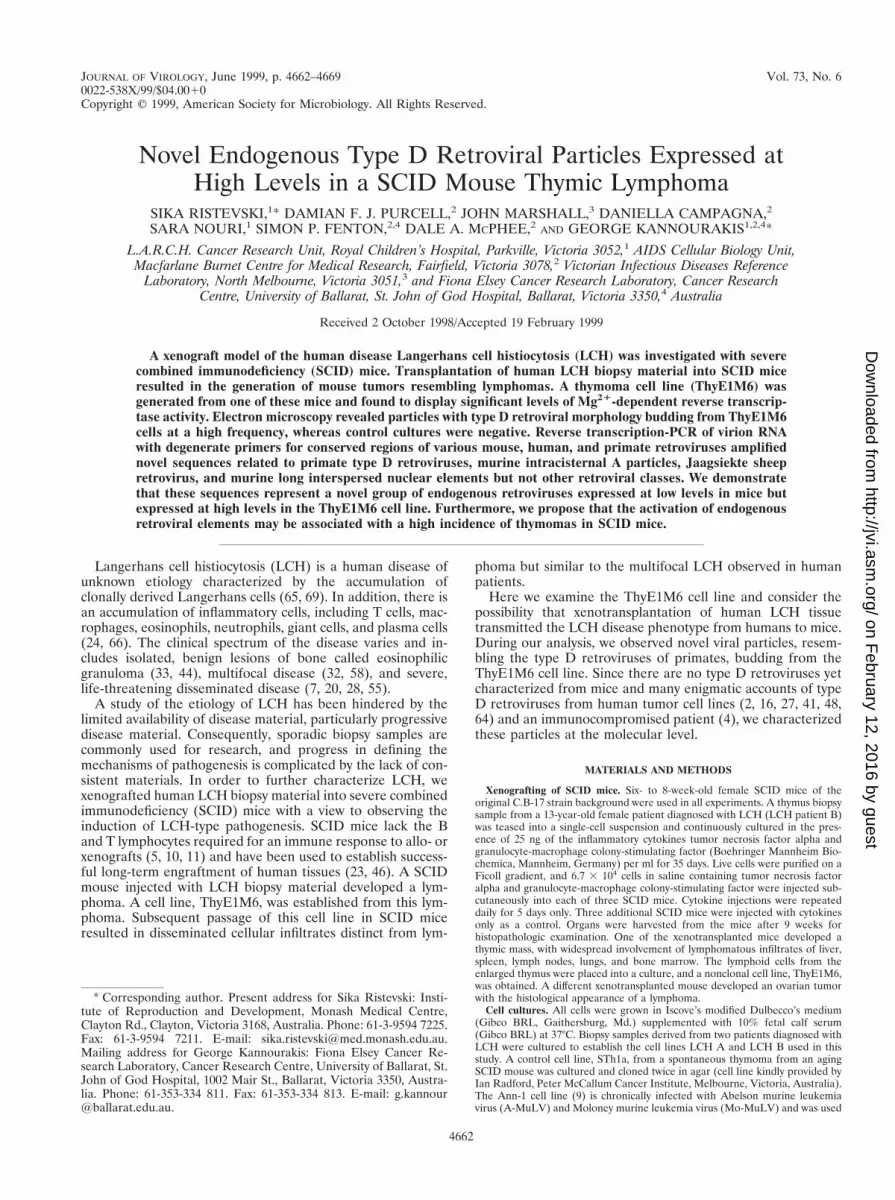
The ThyE1M6 cell line has a high level of Mg21-dependentRT activity. The ThyE1M6 cell line was tested for RT activitywith the PERT assay (52). Cell culture supernatants were har-vested from the ThyE1M6 cell line and a virus-infected controlcell line, Ann-1, an A-MuLV-transformed NIH 3T3 clone su-perinfected with cloned Mo-MuLV (9). After ultracentrifuga-tion to concentrate virus particles, pellets were tested for RTactivity with the PERT assay, as shown in Fig. 2A. In this assay,the presence of RT enzyme is detected by PCR amplificationof cDNA reverse transcribed from an MS-2 RNA template.The expected 112-bp PCR product was obtained from both theThyE1M6 and the Ann-1 cell supernatants but not from su-pernatants from cultured LCH biopsy material or from humanosteosarcoma, rhabdomyosarcoma, and retinoblastoma pri-mary cultures. Furthermore, there was an increase in RT ac-tivity with continuous culturing (3 to 144 h) of the ThyE1M6cell line, suggesting an accumulation of viral particles withculturing (Fig. 2A). Since the cell culture supernatant derivedfrom LCH B (which was used in the genesis of the ThyE1M6cell line) (Fig. 2A, lanes 14 to 18) was negative, it is unlikelythat the particle-associated RT activity was transmitted fromthe biopsy material to SCID mice. Since the PERT assay is ahighly sensitive, nonquantitative PCR-based method, we car-ried out RT assays which involved measuring the incorporationof [32P]TTP with an oligo(dT) z poly(rA) template in the pres-ence of either Mn21 or Mg21 as the divalent cation to confirmthe presence of RT activity in ThyE1M6 cells, as shown in Fig.2B. This assay confirmed RT activity with a preference forMg21, which is commonly associated with the retroviruses of
primates, whereas most murine type C viruses have a prefer-ence for Mn21 (61).
Electron microscopic analysis reveals a high frequency ofbudding type D retrovirus particles. We carried out electronmicroscopy of the ThyE1M6, Ann-1, and STh1a (a SCID-derived spontaneous thymoma) cell lines, spleen and bonemarrow derived from normal SCID mice, skin biopsy materialfrom LCH patient B, and cultured thymus cells from LCHpatient B. We were able to detect budding and mature virusparticles with different morphologies and frequencies from allthree cell lines, but no particles were observed from the mouseor human tissues (Fig. 3 and Table 1). Novel particles wereevident from the ThyE1M6 cell line and would best be classi-fied as type D retrovirus like on the basis of their size androd-like core morphology (17); however, unlike the results forsimian type D retroviruses, no intracellular precursor particleswere observed. The ThyE1M6 particles were observed at afrequency of 1 per 24 sectioned cells examined, formed at theplasma membrane, and were round or ovoid, with a tubularcore. Two matrix layers (shells) surrounded the core, a thininner layer, and a thicker outer layer. The frequency of theseparticles decreased with increasing passage number. The par-ticles observed from the STh1a thymoma cell line that spon-taneously arose in SCID mice would also best be classified astype D retrovirus like. Compared to the ThyE1M6 particles,the STh1a particles had similar dimensions and morphology,including the presence of two matrix layers, although corestructures could not be discerned. Therefore, the type D virusobserved in the ThyE1M6 cell line was morphologically similarto but distinct from the virus observed in the STh1a cell line.As expected, type C retrovirus-like particles were observedbudding from Ann-1 (infected with A-MuLV and Mo-MuLV),and these virions were clearly distinguishable from the parti-cles budding from the SCID thymoma cell lines. Compared tothe type C retrovirus-like Ann-1 particles, the type D retrovi-rus-like ThyE1M6 and STh1a particles were larger and had anadditional matrix layer surrounding the core (Fig. 3 and Table1).
We were unable to detect mature virus particles in SCID
FIG. 2. RT activity in the ThyE1M6 cell line culture supernatant, as demonstrated by the PERT assay. In all cases, culture supernatants were used to prepareconcentrated extracts used in the PERT assay. Lanes 1 and 23 are blank (H2O) PCR controls, lanes 19 and 26 are culture medium controls, lane 24 represents RT fromthe positive control Ann-1, and lanes 2 and 25 represent RT activity detected in supernatant preparations from confluent ThyE1M6 cells. Lanes 3 to 8 represent LCHA, and lanes 14 to 18 represent LCH B. Lanes 9 to 13 represent an RT activity time course for ThyE1M6 cells in which the supernatant was removed after 3, 24, 48,120, and 144 h of continuous cell culturing and virion preparations were assayed for RT activity. Lanes 4 to 8 and 14 to 18 represent equivalent time courses for LCHA and LCH B, respectively. Lane 3 represents a confluent culture of the LCH A cell line. Human osteosarcoma, rhabdomyosarcoma, and retinoblastoma cell lines arerepresented in lanes 20 to 22, respectively. (B) RT activity in the ThyE1M6 cell culture supernatant, as determined by measurement of [32P]TTP incorporation withan oligo(dT) z poly(rA) template in the presence of either Mg21 or Mn21 as the divalent cation. HIV type 1 (HIV-1) and HTLV-1 supernatants were used forcomparison. A background value was determined and was subtracted to give the values shown.
4664 RISTEVSKI ET AL. J. VIROL.
on February 12, 2016 by guest
http://jvi.asm.org/
Dow
nloaded from

mouse spleen or bone marrow samples. We cannot exclude thepossibility that these particles are present at a frequency toolow for detection by electron microscopy. Low-level RT activ-ity from cultured SCID spleen and bone marrow in the PERTassay would support the presence of infrequent particles (datanot shown). In addition, we were unable to detect mature virusparticles in LCH patient B skin biopsy material or in cellscultured from a thymic mass from this patient. The LCH B cellline was also negative for RT activity, as determined by thePERT assay (Fig. 2A, lanes 14 to 18). Therefore, it is likely thatthe novel type D retrovirus particles are associated with aSCID mouse-derived thymocytic lymphoma, and we were in-terested in testing the possibility that these particles were ofendogenous origin.
Degenerate PCR of virus preparations from ThyE1M6 cellsreveals the expression of novel type D retrovirus sequences. Inorder to identify the type D particles produced by theThyE1M6 cell line at the molecular level, we carried out de-generate PCR cloning. Since type D virus morphology and apreference for Mg21-dependent RT activity were observed, wedesigned PCR primers for highly conserved sequences of
SRV-1, SRV-2, MPMV, and SMRV. Primers were designedfor several regions, including the gag and pro genes, and wereused to yield three overlapping PCR products of the predictedsizes. A gag region product of 119 bp, a pro region product of443 bp, and a continuous gag-pro product of 544 bp wereobserved. Particles were pelleted from ThyE1M6 and STh1acell culture supernatants. RNA was extracted from virions,reverse transcribed to cDNA, and subjected to PCR with thegag, pro, and gag-pro primer pairs. Bands of the predicted sizeswere observed in both ThyE1M6 and STh1a samples, as shownin Fig. 4. All PCR products were cloned, sequenced, and sum-marized according to their sequence similarities (Table 2).These results showed that the PCR products contained severalgroups of novel sequences related to the simian type D retro-viruses, murine intracisternal alpha-particle elements (IAPE)(1, 28, 36, 42), Jaagsiekte sheep retrovirus (JSRV) (67, 68), andmurine long interspersed nuclear elements (LINE) (14). Anumber of sequence variants were identified in both ThyE1M6and STh1a samples: those related to type D simian retroviruses(represented by clones 3.12, 3.21, 3.23, and 2.53) and thoserelated to the IAPE (represented by clones 2.27 and 2.29).
FIG. 3. Electron microscopy analysis of virus particles produced by ThyE1M6, STh1a, and Ann-1 cell lines. Mature type D retrovirus particles were produced byThyE1M6 cells (a) and by STh1a cells (b). Mature type C retrovirus particles were produced by Ann-1 cells (c). Bar, 100 nm.
TABLE 1. Characterization of the viral particles produced by the ThyE1M6, STh1a, and Ann-1 cell lines
Cell line or tissueexamined Particles detected
Particle measurements, in mean 6 SD nm (n)a
Core structureDiam Core tubule Shell width
Total Core Width Length Inner Outer
ThyE1M6 Type D retrovirus(1 particle/24cells examined)
103.8 6 7.4 (10) 50.0 6 5.7 (5) 14.6 (2) 43.8 (2) 3.9 6 1.3 (4) 19.1 6 2.2 (4) Tubular
STh1a Type D retrovirus 105.1 6 7.1 (8) 47.4 6 6.4 (4) 4.9 6 1.0 (5) 20.9 6 1.2 (5) Not observedAnn-1 Type C retrovirus 98.2 6 8.1 (12) 68.3 6 7.5 (12) SphericalSCID spleen Not detected in
170 separatecell sectionsexamined
N/A N/A N/A N/A N/A N/A N/A
SCID bone marrow Not detected in170 separatecell sectionsexamined
N/A N/A N/A N/A N/A N/A N/A
LCH patient B skinbiopsy material
Not detected in atissue sectionarea covering8,500 mm2
N/A N/A N/A N/A N/A N/A N/A
LCH patient B thymicmass culture
Not detected in40 separate cellsectionsexamined
N/A N/A N/A N/A N/A N/A N/A
a n, number of particles measured in order to derive a mean particle size. N/A, not applicable.
VOL. 73, 1999 ENDOGENOUS TYPE D RETROVIRUS OF MICE 4665
on February 12, 2016 by guest
http://jvi.asm.org/
Dow
nloaded from

Each of these sequences was found to be present in both theThyE1M6 and the STh1a cell lines.
A number of other PCR primers were designed for charac-terized retroviral sequences, such as MPMV gag, MPMV env,degenerate SRV env, degenerate SRV pol, HTLV-1, murineleukemia virus, type C virus pol, degenerate pol, VL30, MLVenv, and primate foamy virus. However, these primers did notgenerate PCR products of the expected sizes relative to theknown retroviruses (data not shown).
The type D retrovirus-related genes are endogenous geneticelements expressed in mice. In order to investigate if the viral
sequence elements that we identified by PCR were derivedfrom endogenous mouse sequences, Southern blot analysis wasperformed. The type D retrovirus-related sequence gag-proproduct (generated by degenerate PCR from a ThyE1M6 cellculture supernatant [Fig. 4]) was used to screen a panel ofmouse-derived genomic DNAs from ThyE1M6 cells, STh1acells, SCID mice, and Mus dunni mice and is shown in Fig. 5.A band of approximately 1.7 kb and two less prominent bandsof approximately 2 and 3 kb were seen in all mouse-derivedgenomic DNAs digested with HindIII, including that from thewild mouse M. dunni. No cross-hybridizing bands of any sizewere detected in rat, hamster (CHO cells), human (HeLa cellsand LCH patient tissues), and mink (MiCl1 [S1 L2]) genomicDNAs (data not shown). Therefore, the gag-pro PCR productwas likely to have been derived from RNA expressed from anendogenous mouse element that is widespread across strains.
The endogenous murine type D retrovirus-related elementsare expressed as large RNA molecules. The gag-pro PCR prod-uct amplified from ThyE1M6 cDNA was radiolabelled andused as a probe in Northern blot experiments. Total RNAsfrom ThyE1M6, NIH 3T3, and M. dunni cell cultures weretested, and a band of approximately 8.5 kb was observed in the
FIG. 4. RT-PCR cloning of the gag-pro genes of type D retrovirus expressedby the ThyE1M6 and STh1a cell lines. Lanes 1 and 8, molecular size markers(100-bp ladder); lanes 2 to 4, gag, pro, and gag-pro PCR products, respectively,derived from the virus from ThyE1M6 cells; lanes 5 to 7, gag, pro, and gag-proPCR products, respectively, derived from the virus from STh1a cells. The pre-dicted sizes were 119 bp for gag, 443 bp for pro, and 544 bp for gag-pro. Theprimers used to amplify these sequences are indicated at the bottom of the figure.
TABLE 2. BLAST analysis of gag-pro PCR products generated by RT-PCR cloning from the ThyE1M6 cell line
Group Clone (bp) GenBank accessionno. BLAST sequence analysis search resultsa
Primate type D retrovirus (nine clonessequenced)
3.12 (551) AF093700 64% nt identity with SRV-2 and MPMV; significant identity withSRV-1, JSRV, SMRV, and LINE
3.21 (527) AF093701 62% nt identity with SRV-2 and MPMV; significant identity withSRV-1, SMRV, JSRV, and LINE
3.23 (450) AF093699 60% nt identity with SRV-1 and JSRV; significant identity withSRV-2 and SMRV
2.53 (403) AF093698 61% nt identity with SRV-1, MPMV, and SRV-2; significantidentity with SMRV, JSRV, and LINE
Retrovirus-like IAPE (seven clonessequenced)
2.27 (524) AF093697 85% nt identity with Mus musculus IAPE-Y retroviral genome;significant nt identity with M. musculus type IIB IAPE;significant nt identity with SRV-1, JSRV, SRV-2, SMRV, andMPMV
2.29 (545) AF093696 92% nt identity with M. musculus LEC-A retroviral genome;significant nt identity with M. musculus type IIB IAPE;significant nt identity with SRV-1, SRV-2, JSRV, SMRV, andMPMV
a nt, nucleotide.
FIG. 5. Southern analysis of mouse-derived genomic DNAs extracted fromThyE1M6 and STh1a cells and from SCID mouse and M. dunni tissues andprobed with gag-pro. Restriction endonuclease digests with EcoRI (E), BamHI(B), and HindIII (H) are shown. A prominent band of approximately 1.7 kb andtwo less prominent bands of approximately 2 and 3 kb were present in all samplesdigested with HindIII.
4666 RISTEVSKI ET AL. J. VIROL.
on February 12, 2016 by guest
http://jvi.asm.org/
Dow
nloaded from

mouse-derived cells, with the highest abundance in theThyE1M6 cell line (in which several bands were observed)(Fig. 6). No gag-pro-hybridizing bands were observed in normalhuman, LCH patient, and rat RNA samples (data not shown).Thus, the type D retrovirus-related endogenous elements areabundantly expressed as large RNA molecules that may rep-resent a complete viral genome. This type D retrovirus-relatedgenome may account for the type D retrovirus particles ob-served budding from the SCID-derived cell lines ThyE1M6and STh1a.
DISCUSSION
In this study, during the process of attempting to establish axenograft model for the human disease LCH in SCID mice, athymocytic lymphoma cell line, ThyE1M6, was generated. TheThyE1M6 cell line was shown to be of mouse origin and toexpress budding type D retrovirus particles containing RT ac-tivity. Degenerate PCR amplification and sequencing of thepredominant encapsidated viral RNAs demonstrated a signif-icant relationship of most PCR products to the type D retro-viruses at the level of gene sequence and arrangement as wellas homology to IAPE, LINE, and JSRV sequences. We haveshown by hybridization analysis that the genome for these viralRNAs contains endogenous elements common to all murinestrains, including the wild mouse M. dunni.
Further analysis of the full genome sequence of the virusfrom ThyE1M6 cells will be required to clarify whether LCHpatient cells have contributed any sequences to the newly iden-tified mouse type D retrovirus. The data presented here do notsupport genetic recombination with human LCH-derived ge-netic elements contributing to the gag-pro region of the novelvirus but do support the activation of a novel endogenousmouse retrovirus. Human DNA, including LCH patient DNA,does not contain type D virus gag-pro-related sequences; fur-thermore, human tissues were also negative for RT activity.However, it will be interesting to address this question withfurther analysis of the viral genome.
The type D class of SRVs has been extensively character-ized. These viruses were initially identified in studies of simianacquired immunodeficiency syndrome (38, 39, 49, 59, 62). Inaddition, type D retrovirus particles have been observed in avariety of transformed human cell lines (2, 16, 27, 41, 48, 64).These are presumed to be a culture contaminant (2, 48), al-though the activation of endogenous elements has not beenruled out. Furthermore, type D retrovirus particles have beenobserved in the serum of a human AIDS patient (4). A novelexogenous sequence related to type B and type D retroviruseshas been identified in patients with the autoimmune condition
Sjogren’s syndrome (19). In addition, the suspected etiologicalagent of ovine pulmonary carcinoma, JSRV, is an exogenousand endogenous type D and type B retrovirus of the ovine andcaprine species (47, 67, 68). It has been speculated that JSRVmay be a helper virus for an oncogenic replication-defectiveretrovirus, although this hypothesis is difficult to test, sinceJSRV cannot be grown in vitro. To our knowledge, this is thefirst report of the observation in mice of type D retrovirusparticles, which are present as endogenous elements and areactive in a mouse-derived thymocytic lymphoma.
The expression of type D retrovirus particles may be a com-mon feature of SCID mouse-derived thymocytic lymphoma,and we speculate whether it is associated with the generationof lymphoma in the SCID mouse genetic background. SCIDmice are susceptible to spontaneous thymoma, the incidence ofwhich can be significantly increased by breeding with strainssuch as the NOD (nonobese diabetic) mouse strain, which isnot usually susceptible to spontaneous thymoma. However,SCID/NOD mice have a 67% incidence compared to a 15 to20% incidence for mice with the original SCID mutation back-ground (C.B-17) (50). Differential endogenous proviral genesoften account for strain-specific susceptibility or resistance tospontaneous lymphomagenesis; for SCID/NOD mice, thy-momagenesis was associated with the expression of a NODmouse-unique endogenous ecotropic murine leukemia provi-rus locus (Emv-30) (50). Recombination between an ecotropicvirus and one or more endogenous nonecotropic proviral se-quences is likely a causative agent for the high incidence ofspontaneous lymphoma in strains such as AKR/J (8, 13, 18, 21,35, 60).
Molecular genetic analysis indicates a strong association be-tween a high spontaneous incidence of hemopoietic neoplasmsand the activation of endogenous murine leukemia viruses.This hypothesis is supported by cross-strain breeding experi-ments (such as those with SCID/NOD mice), in which murinestrains carrying different endogenous elements are broughttogether in one genome, facilitating potential cross-activationand recombination.
It has been demonstrated that the DNA-dependent kinase(p350) gene is the candidate gene for the SCID phenotype,resulting in an impairment of the double-strand break recom-bination repair pathway (15, 26). Therefore, in addition to adeficiency in B and T lymphocytes, SCID mice are highlysusceptible to DNA damage, as demonstrated by hypersensi-tivity to ionizing radiation (3, 34). This impairment confers ahigh potential for mutagenesis, genomic instability, and tumor-igenesis and may be linked to the occurrence of spontaneousthymomas in SCID mice. Therefore, there may be an increasedsusceptibility to retroviral activation in these mice.
The homology observed between the virus from ThyE1M6cells and LINE is intriguing in the context of recent reports ofthe FHIT gene. It has been demonstrated that the humanFHIT gene encompasses the common human fragile siteFRA3B and is often inactivated or deleted in tumor cells (57).FRA3B contains a high incidence of LINE insertions (22).LINE are capable of retrotransposition (54) and have beenshown to be involved in the insertional mutation of a numberof genes (12, 25, 40, 63). It has been proposed that carcinogendamage to DNA results in rearrangement of the FHIT gene byhomologous recombination with LINE sequences. FHIT func-tions as a tumor suppressor and thus may act early in tumordevelopment. The induced expression of LINE sequences maylead to increased recombination across the SCID mouse ge-nome and may be an early event leading to the generation ofthymomas.
We have identified a novel endogenous type D retrovirus of
FIG. 6. Northern analysis of total cellular RNAs extracted from the mouse-derived cell lines ThyE1M6, NIH 3T3, and M. dunni. A band of approximately8.5 kb was detected in all cells when probed with the ThyE1M6 gag-pro PCRproducts.
VOL. 73, 1999 ENDOGENOUS TYPE D RETROVIRUS OF MICE 4667
on February 12, 2016 by guest
http://jvi.asm.org/
Dow
nloaded from
mice which is expressed at high levels by the ThyE1M6 cellline. The results presented in this paper suggest that the acti-vation of this endogenous virus might have been associatedwith the genesis of a thymocytic lymphoma in a SCID mouse.It will be interesting to determine whether the virus identifiedhere is involved in the pathogenesis of spontaneous thymomasin other SCID mice. We aim to further characterize this en-dogenous murine retrovirus by cloning and sequencing of thefull viral genome.
ACKNOWLEDGMENTS
We thank Wendy Cook for the Ann-1 cell line and Ian Radford forthe STh1a cell line. We thank Len C. Harrison and Kaku Nakagawafor helpful discussions and Ken Shortman and Frank Battey for FACSanalysis of the ThyE1M6 cell line. We thank Thulasi Murughiah,Rodney Daly, Soong Ling, and Thuy Diem for excellent assistance withSCID mice, animal experimentation, and technical assistance andAnthea Ramos for technical assistance.
D.F.J.P. was supported by the NHMRC of Australia and by Mac-farlane Burnet Centre for Medical Research funds. This work wassupported by grants to G.K. from the Histiocytosis Association ofAmerica, the NHMRC of Australia, CICA (Ballarat, Victoria, Aus-tralia), and St. John of God Hospital (Ballarat, Victoria, Australia).
REFERENCES
1. Aota, S.-I., T. Gojobori, K. Shigesada, H. Ozeki, and T. Ikemura. 1987.Nucleotide sequence and molecular evolution of mouse retrovirus-like IAPelements. Gene 56:1–12.
2. Asikainen, K., M. Vesanen, T. Kuittinen, and A. Vaheri. 1993. Identificationof human type D retrovirus as a contaminant in a neuroblastoma cell line.Arch. Virol. 129:357–361.
3. Biedermann, K. A., J. Sun, A. J. Giaccia, L. M. Tosto, and J. M. Brown. 1991.scid mutation in mice confers hypersensitivity to ionizing radiation and adeficiency in DNA double-strand break repair. Proc. Natl. Acad. Sci. USA88:1394–1397.
4. Bohannon, R. C., L. A. Donehower, and R. J. Ford. 1991. Isolation of a typeD retrovirus from B-cell lymphomas of a patient with AIDS. J. Virol. 65:5663–5672.
5. Bosma, G. C., R. P. Custer, and M. J. Bosma. 1983. A severe combinedimmunodeficiency mutation in the mouse. Nature 301:527–530.
6. Chomczynski, P., and N. Sacchi. 1987. Single-step method of RNA isolationby acid guanidinium thiocyanate-phenol-chloroform extraction. Anal. Bio-chem. 162:156–159.
7. Christian, H. A. 1920. Defects in membranous bones, exophthalmos anddiabetes insipidus; an unusual syndrome of dyspituitarism: a clinical study.Med. Clin. North Am. 3:849–871.
8. Cloyd, M. W., J. W. Hartley, and W. P. Rowe. 1980. Lymphomagenicity ofrecombinant mink cell focus-inducing murine leukemia viruses. J. Exp. Med.151:542–552.
9. Cook, W. D. 1982. Rapid thymomas induced by Abelson murine leukemiavirus. Proc. Natl. Acad. Sci. USA 79:2917–2921.
10. Custer, R. P., G. C. Bosma, and M. J. Bosma. 1985. Severe combinedimmunodeficiency (SCID) in the mouse. Pathology, reconstitution, neo-plasms. Am. J. Pathol. 93:464–477.
11. Dorshkind, K., G. M. Keller, R. A. Phillips, R. G. Miller, G. C. Bosma, M.O’Toole, and M. J. Bosma. 1984. Functional status of cells from lymphoidand myeloid tissues in mice with severe combined immunodeficiency disease.J. Immunol. 132:1804–1808.
12. Drechsler, M., and B. Royer-Pokora. 1996. A LINE element is present at thesite of a 300-kb deletion starting in intron 10 of the PAX6 gene in a case offamilial aniridia. Hum. Genet. 98:297–303.
13. Elder, J. H., J. W. Gautsch, F. C. Jensen, R. A. Lerner, J. W. Hartley, andW. P. Rowe. 1997. Biochemical evidence that MCF murine leukemia virusesare envelope (env) gene recombinants. Proc. Natl. Acad. Sci. USA 74:4676–4680.
14. Fanning, T. G., and M. F. Singer. 1987. LINE-1: a mammalian transposableelement. Biochim. Biophys. Acta 910:203–212.
15. Fulop, G. M., and R. A. Phillips. 1990. The scid mutation in mice causes ageneral defect in DNA repair. Nature 347:479–482.
16. Gelderblom, H., H. Bauer, H. Ogura, R. Wigand, and A. B. Fischer. 1974.Detection of oncornavirus-like particles in HeLa cells. I. Fine structure andcomparative morphological classification. Int. J. Cancer 13:246–253.
17. Gelderblom, H. 1987. Oncoviridae: type D oncovirus, p. 289–293. In M. V.Nermut and A. C. Steven (ed.), Animal virus structure. Elsevier SciencePublishers, Amsterdam, The Netherlands.
18. Green, N., H. Hiai, J. H. Elder, R. S. Schwartz, R. H. Khiroya, C. Y. Thomas,P. N. Tsichlis, and J. M. Coffin. 1980. Expression of leukemogenic recom-
binant viruses associated with a recessive gene in HRS/J mice. J. Exp. Med.152:249–264.
19. Griffiths, D. J., P. J. W. Venables, R. A. Weiss, and M. T. Boyd. 1997. A novelexogenous retrovirus sequence identified in humans. J. Virol. 71:2866–2872.
20. Hand, A. 1921. Defects in membranous bones, exophthalmos, and polyuria inchildhood: is it dyspituitarism? Am. J. Med. Sci. 162:509.
21. Hartley, J. W., N. K. Wolford, L. J. Old, and W. P. Rowe. 1977. A new classof murine leukemia virus associated with the development of spontaneouslymphomas. Proc. Natl. Acad. Sci. USA 74:789–792.
22. Inoue, H., H. Ishii, H. Alder, E. Snyder, T. Druck, K. Huebner, and C. M.Croce. 1997. Sequence of the FRA3B common fragile region: implicationsfor the mechanism of FHIT deletion. Proc. Natl. Acad. Sci. USA 94:14584–14589.
23. Kamel-Reid, S., and J. E. Dick. 1988. Engraftment of immune-deficient micewith human hematopoietic stem cells. Science 242:1706–1709.
24. Kannourakis, G., and A. Abbas. 1994. The role of cytokines in the patho-genesis of Langerhans cell histiocytosis. Br. J. Cancer 70(Suppl. XXIII):S37–S40.
25. Kingsmore, S. F., B. Giros, D. Suh, M. Bieniarz, M. G. Caron, and M. F.Seldin. 1994. Glycine receptor b-subunit gene mutation in spastic mouseassociated with LINE-1 element insertion. Nat. Genet. 7:136–141.
26. Kirchgessner, C. U., C. K. Patil, J. W. Evans, C. A. Cuomo, L. M. Fried, T.Carter, M. A. Oettinger, and J. M. Brown. 1995. DNA-dependent kinase(p350) as a candidate gene for the murine SCID defect. Science 267:1178–1183.
27. Krause, H., V. Wunderlich, and W. Uckert. 1989. Molecular cloning of a typeD retrovirus from human cells (PMFV) and its homology to simian acquiredimmunodeficiency type D retroviruses. Virology 173:214–222.
28. Kuff, E. L., A. Feenstra, K. Lueders, L. Smith, R. Hawlay, N. Hozumi, andM. Shulman. 1983. Intracisternal A-particle genes as movable elements inthe mouse genome. Proc. Natl. Acad. Sci. USA 80:1992–1996.
29. Kwok, S., G. Ehrlich, B. Poiesz, R. Kalish, and J. J. Sninsky. 1988. Enzy-matic amplification of HTLV-1 viral sequences from peripheral blood mono-nuclear cells and infected tissues. Blood 72:1117–1123.
30. Lee, J. Y., D. S. Bowden, and J. A. Marshall. 1996. Membrane junctionsassociated with rubella virus infected cells. J. Submicrosc. Cytol. Pathol.28:101–108.
31. Leibold, D. M., G. D. Swergold, M. F. Singer, R. E. Thayer, B. A. Dombroski,and T. G. Fanning. 1990. Translation of LINE-1 DNA elements in vitro andin human cells. Proc. Natl. Acad. Sci. USA 87:6990–6994.
32. Letterer, E. 1924. Aleukamische retikulose (ein Beitrag zu den proliferativenErkrankungen des Retikuendosthelisalapparates). Frankf. Z. Pathol.30:377–394.
33. Lichenstein, L., and H. L. Jaffe. 1940. Eosinophilic granuloma of bone.Am. J. Pathol. 16:595–604.
34. Lieberman, M., G. A. Hansteen, E. K. Waller, I. L. Weissman, and A.Sen-Majumdar. 1992. Unexpected effects of the severe combined immuno-deficiency mutation on murine lymphomagenesis. J. Exp. Med. 176:399–405.
35. Lilly, F., M. L. Duran-Reynals, and W. P. Rowe. 1975. Correlation of earlymurine leukemia virus titer and H-2 type with spontaneous leukemia in miceof the BALB/c 3 AKR cross: a genetic analysis. J. Exp. Med. 141:882–889.
36. Lueders, K. K., and E. L. Kuff. 1977. Sequences associated with intracisternalA particles are reiterated in the mouse genome. Cell 12:963–972.
37. Maniatis, T., E. F. Fritsch, and J. Sambrook. 1982. Molecular cloning: alaboratory manual. Cold Spring Harbor Laboratory, Cold Spring Harbor,N.Y.
38. Marx, P. A., D. H. Maul, K. G. Osborn, N. W. Lerche, P. Moody, L. J.Lowenstine, R. V. Henrickson, L. O. Arthur, R. V. Gilden, M. Gravell, W. T.London, J. L. Sever, J. A. Levy, R. J. Munn, and M. B. Gardner. 1984. SimianAIDS: isolation of a type-D retrovirus and transmission of the disease.Science 223:1083–1086.
39. Marx, P. A., M. L. Bryant, K. G. Osborn, D. H. Maul, N. W. Lerche, L. J.Lowenstine, J. D. Kluge, C. P. Zaiss, R. V. Henrickson, S. M. Shiigi, B. J.Wilson, A. Malley, L. C. Olson, W. P. McNulty, L. O. Arthur, R. V. Gilden,C. S. Barker, E. Hunter, R. J. Munn, G. Heidecker, and M. B. Gardner. 1985.Isolation of a new serotype of simian acquired immune deficiency syndrometype D retrovirus from Celebes black macaques (Macaca nigra) with immunedeficiency and retroperitoneal fibromatosis. J. Virol. 56:571–578.
40. McNaughton, J. C., G. Hughes, W. A. Jones, P. A. Stockwell, H. J. Klamut,and G. B. Petersen. 1997. The evolution of an intron: analysis of a long,deletion-prone intron in the human dystrophin gene. Genomics 40:294–304.
41. Oda, T., S. Ikeda, S. Watanabe, M. Hatsushika, K. Akiyama, and F. Mit-sunobu. 1988. Molecular cloning, complete nucleotide sequence, and genestructure of the provirus genome of a retrovirus produced in a humanlymphoblastoid cell line. Virology 167:468–478.
42. Ono, M., M. D. Cole, A. T. White, and R. C. C. Huang. 1980. Sequenceorganization of cloned intracisternal A particle genes. Cell 21:465–473.
43. Oroszlan, S., M. Barbacid, T. D. Copeland, S. A. Aaronson, and R. V. Gilden.1981. Chemical and immunological characterization of the major structuralprotein (p28) of MMC-1, a rhesus monkey endogenous type C virus: homol-ogy with the major structural protein of avian reticuloendotheliosis virus.J. Virol. 39:845–854.
4668 RISTEVSKI ET AL. J. VIROL.
on February 12, 2016 by guest
http://jvi.asm.org/
Dow
nloaded from
44. Otani, S., and J. C. Ehrlich. 1940. Solitary granuloma of bone. Am. J. Pathol.16:595–604.
45. Ou, C. Y., S. Kwok, S. W. Mitchell, D. H. Mack, J. J. Sninsky, J. W. Krebs,P. Feorino, D. Warfield, and G. Schochetman. 1988. DNA amplification fordirect detection of HIV-1 in DNA of peripheral blood mononuclear cells.Science 239:295–297.
46. Paine-Murrieta, G. D., C. W. Taylor, R. A. Curtis, M. H. A. Lopez, R. T.Dorr, C. S. Johnson, C. Y. Funk, F. Thompson, and E. M. Hersh. 1997.Human tumor models in the severe combined immune deficient (scid)mouse. Cancer Chemother. Pharmacol. 40:209–214.
47. Payne, A.-L., D. W. Verwoerd, and H. M. Garnett. 1983. The morphology andmorphogenesis of Jaagsiekte retrovirus (JSRV). Onderstepoort J. Vet. Res.50:317–322.
48. Popovic, M., V. S. Kalyanaraman, M. S. Reitz, and M. G. Sarngadharan.1982. Identification of the RPMI 8226 retrovirus and its dissemination as asignificant contaminant of some widely used human and marmoset cell lines.Int. J. Cancer 30:93–99.
49. Power, M. D., P. A. Marx, M. L. Bryant, M. B. Gardner, P. J. Barr, and P. A.Luciw. 1986. Nucleotide sequence of SRV-1, a type D simian acquiredimmune deficiency syndrome retrovirus. Science 231:1567–1572.
50. Prochazka, M., H. R. Gaskins, L. D. Shultz, and E. H. Leiter. 1992. Thenonobese diabetic scid mouse: model for spontaneous thymomagenesis as-sociated with immunodeficiency. Proc. Natl. Acad. Sci. USA 89:3290–3294.
51. Purcell, D. F. J., C. M. Broscius, E. F. Vanin, C. E. Buckler, A. W. Nienhuis,and M. A. Martin. 1996. An array of murine leukemia virus-related elementsis transmitted and expressed in a primate recipient of retroviral gene trans-fer. J. Virol. 70:887–897.
52. Pyra, H., J. Boni, and J. Schupbach. 1994. Ultrasensitive retrovirus detectionby a reverse transcriptase assay based on product enhancement. Proc. Natl.Acad. Sci. USA 91:1544–1548.
53. Rabin, H., C. V. Benton, M. A. Tainsky, N. R. Rice, and R. V. Gilden. 1979.Isolation and characterization of an endogenous type C virus of rhesusmonkeys. Science 204:841–842.
54. Sassaman, D. M., B. A. Dombroski, J. V. Moran, M. L. Kimberland, T. P.Naas, R. J. DeBerardinis, A. Gabriel, G. D. Swergold, and H. H. Kazazian.1997. Many human L1 elements are capable of retrotransposition. Nat.Genet. 16:37–43.
55. Schuller, A. 1915. Uber eigenartige Schadeldefekte im Jugendalter.Fortschr. Rontgenstr. 23:12–18.
56. Shih, A., R. Misra, and M. G. Rush. 1989. Detection of multiple, novel
reverse transcriptase coding sequences in human nucleic acids: relation toprimate retroviruses. J. Virol. 63:64–75.
57. Siprashvili, Z., G. Sozzi, L. D. Barnes, P. McCue, A. K. Robinson, V. Eryo-min, L. Sard, E. Tagliabue, A. Greco, L. Fusetti, G. Schwartz, M. Pierotti,C. M. Croce, and K. Huebner. 1997. Replacement of Fhit in cancer cellssuppresses tumorigenicity. Proc. Natl. Acad. Sci. USA 94:13771–13776.
58. Siwe, S. A. 1933. Die Reticuloendotheliose—ein neues Krankheitsbild unterden Hepatosplenomegalien. Z. Kinderheilkd. 55:212–247.
59. Sonigo, P., C. Barker, E. Hunter, and S. Wain-Hobson. 1986. Nucleotidesequence of Mason-Pfizer monkey virus: an immunosuppressive D-type ret-rovirus. Cell 45:375–385.
60. Stoye, J. P., C. Moroni, and J. M. Coffin. 1991. Virological events leading tospontaneous AKR thymomas. J. Virol. 65:1273–1285.
61. Teich, N. 1982. Taxonomy of retroviruses, p. 25–208. In R. Weiss, N. Teich,H. Varmus, and J. Coffin (ed.), RNA tumor viruses: molecular biology oftumor viruses. Cold Spring Harbor Laboratory, Cold Spring Harbor, N.Y.
62. Thayer, R. M., M. D. Power, M. L. Bryant, M. B. Gardner, P. J. Barr, andP. A. Luciw. 1987. Sequence relationships of type D retroviruses which causesimian acquired immunodeficiency syndrome. Virology 157:317–329.
63. Toriello, H. V., T. W. Glover, K. Takahara, P. H. Byers, D. E. Miller, J. V.Higgins, and D. S. Greenspan. 1996. A translocation interrupts the COL5A1gene in a patient with Ehlers-Danlos syndrome and hypomelanosis of Ito.Nat. Genet. 13:361–365.
64. Uckert, W., M. Fleischhacker, and R. Kettmann. 1986. Isolation and char-acterisation of covalently closed circular proviral DNA molecules of severaltype D retroviruses isolated from human cell lines. Virology 155:742–746.
65. Willman, C. L., L. Busque, B. B. Griffin, B. E. Favara, K. L. McKlain, M. H.Duncan, and D. G. Gilliland. 1994. Langerhans cell histiocytosis (histiocy-tosis X): a clonal proliferative disease. N. Engl. J. Med. 331:154–160.
66. Writing Group of the Histiocyte Society. 1987. Histiocytosis syndromes inchildren. Lancet i:208–209.
67. York, D. F., R. Vigne, D. W. Verwoerd, and G. Querat. 1991. Isolation,identification, and partial cDNA cloning of genomic RNA of Jaagsiekteretrovirus, the etiological agent of sheep pulmonary adenomatosis. J. Virol.65:5061–5067.
68. York, D. F., R. Vigne, D. W. Verwoerd, and G. Querat. 1992. Nucleotidesequence of the Jaagsiekte retrovirus, an exogenous and endogenous type Dand B retrovirus of sheep and goats. J. Virol. 66:4930–4939.
69. Yu, R. C., C. Chu, L. Bulewela, and A. C. Chu. 1994. Clonal proliferation ofLangerhans cell histiocytosis. Lancet 343:767–768.
VOL. 73, 1999 ENDOGENOUS TYPE D RETROVIRUS OF MICE 4669
on February 12, 2016 by guest
http://jvi.asm.org/
Dow
nloaded from