The thymic repertoire of neuroendocrine self-antigens: physiological implications in T-cell life and...
6
IMMUNOLOGY TODAY The thymic repertoi se If-at+i ns I I T- phy cell I !sio I Ife an 13 lthough the thymus has A / long been considered to be a part of the endocrine sys- tem, it is difficult to apply the model of endocrine cell-cell signalling to the process of intrathymic T-cell differen- tiation. This might be due, at least in part, to the importance of the thymus as the pri- mary lymphoid organ responsible for T-cell differentiation, such that its immune prop- erties have overshadowed its endocrine role. However, these two functions are in- timately ‘linked, and neuroendocrine- immune interactions in T-cell education have important physiological and pathological implications. During phylogeny aud oufogeny, the tlyntts appears as a crucial wetiq point befzueert the newomdocrine aud itnrnt~ne system; through cqptocrine . i~~tcrcelltrlar comrtcnication, tlynic Ilelr roetzdocr.irlc-related preciuw3 can inflrtetrce the early steps of the imni~nc response, while T-cell precrrrsors are educated to recognize the principal ueuroeudocriw families. Here -we smma.+ze the ol~ser~~atious that suppol? the dual role of the thynic repertoire of ueuroendocrine-related polypeptidt7 precursors in T-cell differentiation. The neurohypophysial hormone family The neurohypophysial (NHP) hormones constitute a family of nona- peptides that are highly conserved throughout evolution’ and all have cysteine residues in positions 1 and 6 forming a disulfide bridge. They can be divided in two lineages corresponding to the oxytocin (OT)-like and vasopressin (VP)-like peptides. Both of the lineages are thought to have arisen from the duplication of a single ancestral gene’. In mammalian vertebrates, OT-like peptides are im- plicated in the control of reproduction, whereas VP-like peptides regulate water homeostasis and also have some cardiovascular functions. All known NHP hormones are synthesized as larger pre- cursors containing a neurophysin domain (2 10 kDa), the biologi- cal function of which remains unclear. and monoclonal antibodies (mAbs) against different epitopes of the NHP family, we confirmed that the dominant thymic NHP- related epitope belongs to the OT lineage al- though both pro-OT and pro-VP gene tran- scripts are detected at low levels within human and murine thymus extract&. A striking example of the intimate neuro- endocrine-immune interactions within the thymus is provided by thymic nurse cells (TNC), which synthesize NH&related pep- tides and express on their surface markers of the neuroendocrine system such as A2B5 and neuron-specific enolase7. Primary cul- tures of TEC/TNC do not secrete NHP- related peptides and a recent ultrastructure study confirmed the presence of immuno- reactive (IR)-OT in the cytosol, clear vesicles and perimembrane space of murine TEC/ TNC, but not within large dense secretory granules such as those in the hypothalamo-NHP axisa. A nonclassical distribution of NHF related mRNA and peptides has also been detected in eosinophils of the mouse spleen9. The term ‘cryptocrine’ has been introduced in endocrinology to describe a particular type of cell-cell interaction*OJ1. Cryptocrine cell-cell signalling occurs in microenvironments characterized by large ‘nursing’ epithelial cells (like TEC/TNC in the thymus or Sertoli cells in the testis) that enclose the cell populations (T cells and spermatids, respectively) that differentiate in close vicinity of these ‘nurse’ cells. Thymic NHP-related signals As early as 1910, Ott and Scott showed that thymic extracts could induce milk ejection (galactokinesis) when injected into goats. The causative agent was identified by du Vigneaud’s group in the 1950s as OT. Subsequent studies have confirmed that thymic epithelial cells (TEC) from human and different animal species ar > a site of synthesis of peptides related to the NHP family, with marked dominance for those of the OT lineage2-s. Using various polyclonal Cop,rg?t 0 I996 EWU er Sctence Ltc AlI r~pms me*& 0 167.5699196d I5 00 Thymic NHP peptide receptors Functional NHP-type receptors are expressed by a variety of T-cell lines12-15, with a predominance of Vl-type receptors on pre-T cells (murine RL,,NP cell line) and OT-type receptors on differentiated cytotoxic T cells (murine CTL-L, cellsP. Two recent studies also found an NHP peptide receptor of the Vlb subtype in the human and rat thymus16J7. Within the thymic microenvironment, the physicochemical con- ditions are conducive to functional cryptocrine signalling between TEC/TNC and pm-T cells’*. Moreover, NHP peptides increase tritiated PII 50167.5699(96) 10023.2
Transcript of The thymic repertoire of neuroendocrine self-antigens: physiological implications in T-cell life and...
IMMUNOLOGY TODAY
The thymic repertoi se If-at+i ns I I
T-
phy cell I
!sio I
Ife an
13 lthough the thymus has
A / long been considered to be
a part of the endocrine sys- tem, it is difficult to apply
the model of endocrine cell-cell signalling
to the process of intrathymic T-cell differen-
tiation. This might be due, at least in part, to
the importance of the thymus as the pri-
mary lymphoid organ responsible for T-cell
differentiation, such that its immune prop-
erties have overshadowed its endocrine
role. However, these two functions are in-
timately ‘linked, and neuroendocrine-
immune interactions in T-cell education have
important physiological and pathological
implications.
During phylogeny aud oufogeny,
the tlyntts appears as a crucial
wetiq point befzueert the
newomdocrine aud itnrnt~ne
system; through cqptocrine .
i~~tcrcelltrlar comrtcnication,
tlynic Ilelr roetzdocr.irlc-related
preciuw3 can inflrtetrce the early
steps of the imni~nc response, while
T-cell precrrrsors are educated to
recognize the principal
ueuroeudocriw families. Here -we
smma.+ze the ol~ser~~atious that
suppol? the dual role of the thynic
repertoire of ueuroendocrine-related
polypeptidt7 precursors in T-cell
differentiation.
The neurohypophysial hormone family The neurohypophysial (NHP) hormones constitute a family of nona-
peptides that are highly conserved throughout evolution’ and all
have cysteine residues in positions 1 and 6 forming a disulfide
bridge. They can be divided in two lineages corresponding to the
oxytocin (OT)-like and vasopressin (VP)-like peptides. Both of the
lineages are thought to have arisen from the duplication of a single
ancestral gene’. In mammalian vertebrates, OT-like peptides are im-
plicated in the control of reproduction, whereas VP-like peptides
regulate water homeostasis and also have some cardiovascular
functions. All known NHP hormones are synthesized as larger pre-
cursors containing a neurophysin domain (2 10 kDa), the biologi-
cal function of which remains unclear.
and monoclonal antibodies (mAbs) against
different epitopes of the NHP family, we
confirmed that the dominant thymic NHP-
related epitope belongs to the OT lineage al-
though both pro-OT and pro-VP gene tran-
scripts are detected at low levels within
human and murine thymus extract&. A
striking example of the intimate neuro-
endocrine-immune interactions within the
thymus is provided by thymic nurse cells
(TNC), which synthesize NH&related pep-
tides and express on their surface markers
of the neuroendocrine system such as A2B5
and neuron-specific enolase7. Primary cul-
tures of TEC/TNC do not secrete NHP-
related peptides and a recent ultrastructure
study confirmed the presence of immuno-
reactive (IR)-OT in the cytosol, clear vesicles
and perimembrane space of murine TEC/
TNC, but not within large dense secretory granules such as those in
the hypothalamo-NHP axisa. A nonclassical distribution of NHF
related mRNA and peptides has also been detected in eosinophils
of the mouse spleen9.
The term ‘cryptocrine’ has been introduced in endocrinology to
describe a particular type of cell-cell interaction*OJ1. Cryptocrine
cell-cell signalling occurs in microenvironments characterized by
large ‘nursing’ epithelial cells (like TEC/TNC in the thymus or
Sertoli cells in the testis) that enclose the cell populations (T cells
and spermatids, respectively) that differentiate in close vicinity of
these ‘nurse’ cells.
Thymic NHP-related signals As early as 1910, Ott and Scott showed that thymic extracts could
induce milk ejection (galactokinesis) when injected into goats. The
causative agent was identified by du Vigneaud’s group in the 1950s
as OT. Subsequent studies have confirmed that thymic epithelial
cells (TEC) from human and different animal species ar > a site of
synthesis of peptides related to the NHP family, with marked dominance for those of the OT lineage2-s. Using various polyclonal
Cop,rg?t 0 I996 EWU er Sctence Ltc AlI r~pms me*& 0 167.5699196d I5 00
Thymic NHP peptide receptors Functional NHP-type receptors are expressed by a variety of T-cell
lines12-15, with a predominance of Vl-type receptors on pre-T cells
(murine RL,,NP cell line) and OT-type receptors on differentiated
cytotoxic T cells (murine CTL-L, cellsP. Two recent studies also
found an NHP peptide receptor of the Vlb subtype in the human
and rat thymus16J7.
Within the thymic microenvironment, the physicochemical con-
ditions are conducive to functional cryptocrine signalling between
TEC/TNC and pm-T cells’*. Moreover, NHP peptides increase tritiated
PII 50167.5699(96) 10023.2

~~ IMMUNOLOGY TODAY
thymidine (13HlTdR) incorporation into freshly isolated murine and
human thymocytes” and induce the phosphorylation of the focal
adhesion kinase ~125 FAK (Ref. 18) (H. Martens et al., unpublished).
Thus, there are considerable data supporting thymic cryptocrine
signalling mediated by NHP-related peptides.
hymic as the self-antigen of the family In the thymus, cryptocrine signalling is intimately associated with
the presentation of self-antigens to developing T cells. This action
was long thought to be mediated by interdigitating thymic cells
only, but there is now evidence that TEC/TNC are also actively in-
volved in the thymic induction of immunological self-tolerancer9.
At the molecular level, major histocompatibility complex (MHC)
class I molecules are involved in central tolerance since they present
peptides derived from the processing of endogenous proteins to -;w
T-cell receptor (TCR) on CD8’ T cells2”. The OT amino acid sequence
CYIQNCPLG possesses appropriately positioned hydrophobic ty-
rosine (Y) and leucine (L) residues, which permit it to be anchored
to the groove of some MHC class I molecules2i. Therefore, it is hy-
pothesized that in human and other mammalian species, thymic OT
is the self-antigen of the NHP family.
The biochemical pathways leading to the presentation of OT by
TECITNC
Human TEC surface membranes were purified by ultracentrifu-
gation and dissolved in a non-ionic detergent. The solution was
then run down an affinity column prepared with a mAb di,-acted to
human MHC class I molecules. Following SDS-PAGE, instead of the
expected 45 kDa fractions (the molecular mass of MHC class I
heavy chain), western blot analyses revealed a 55 kDa fraction that
could be labelled both by anti-MHC class I mAb and by an anti-
serum against the highly conserved part of neurophysins. This pro-
tein was still present after mercaptoethanol and/or heat denatu-
ration, and the antiserum to neurophysin did not recognize
@microglobulin. Given these data, we interpret the thymic mem-
brane 55 kDa protein as a chimeric or a hybrid precursor including
neurophysin- (10 kDa), as well as MHC class I heavy chain (45 kDa)-
related domains. This 55 kDa protein differs from the hypothalamic
OT precursor (16 kDa) and probably reflects a strong binding of
the neurophysin domain to MHC class I heavy chain at a post-
transcriptional levelz2.
Until recently, the binding of OT or VP to neurophysins for
transport along the NHP axis provided a useful model for study-
ing the interaction of a short peptide with a larger protein. Several
studies have pointed out the importance of the tyrosine residue in
position 2 of OT and VP for this binding23*24. Analogous biochemi-
cal principles seem to rule the binding of antigens to the groove of
some MHC class I molecules, including the presence of a tyrosine
residue in position 2 (Ref. 21). So, by analogous binding, neuro-
physin transports OT along axons of the NHP axis while the neuro-
physin domain of the TEC membrane chimeric 55 kDa protein
could be implicated in the yresentntiun of thymic OT to pre-T cells.
Although thymic MHC class 1 pathways are involved in the pro-
cess, it appears that TEC/TNC T-cell education to OT is not re-
stricted in an allelic fashion in contrast with the peripheral presen-
tation of alloantigens by dedicated antigen-presenting cells.
III LY 1996

Another selective advantage resides in the potential presentation
of the characteristic cyclic structure of the NHP family to pre-T cells.
In addition to our findings, a recent study also suggests a distinc-
tion between thymic T-cell education and antigen-presenting
functionsz5.
Furthermore, our most recent results show that OT is colocalized
in human cultured TEC with interleukin lp (IL-l@, IL-6 and
leukaemia-inhibitory factor (LIF) (Fig. 1). Oxytocin is specifically
recognized at the outer surface of human TEC by mAbs 013 and 033
directed against the linear C-terminal and the cyclic part of the OT
molecule, respectively; this recognition markedly enhanced the
secretion of IL-6 and LIF (but not IL-lp) by primary cultures of
human TEC, while the addition of mAbs to VP did not exert any
effecP. These data provide strong evidence of the full processing of
OT expressed at the outer surface of the TEC membrane, and sup-
port the hypothesis of thymic OT as the self-antigen of the NHI’
family.
Self-antigens of tachykinin and insulin families Neurokinin A (NKA) is the peptide of the tachykinin family en-
coded by the preprotachykinin A (NT-A) gene in human and rat
TEC (Ref. 26). Thymic ITT-A expression was shown to be glucocor-
ticoid dependent since adrenalectomy of Sprague-Hawley rats
markedly enhanced the levels of thymic ITT-A mRNA (A. Ericsson
and V. Geenen, unpublished). In contrast to
other members of the tachykinin family,
NKA also exerts mitogenic effects on
murine thymocytes. This IL-l-type bioactiv-
ity of NKA suggests an interaction with spe-
cific receptors expressed by pre-T cells that
could be another accessory pathway in
T-cell maturation. The amino acid sequence
of NKA (HKTNSFVGLM) has the same
C-terminal epitope as other members of the
tachykinin family. This epitope possesses a
leucine residue in position 9 that could be
used in the binding to some MHC class I
molecules; thus NKA can be predicted to be
a T-cell epitope.
We have recently shown that insulin-like
growth factor II (IGF-II) is one peptide of the
insulin family that is expressed by human
and rat TEC/TNC (Ref. 27). IR-IGF-I is also
detected in human and rat thymus extracts
but in lower concentrations. Although IR-
IGF-II is expressed by TEC/TNC, it is not
secreted into the supernatant of human or
rat primary TEC cultures. Interestingly,
overexpression of IGF-II in transgenic mice
was shown to induce thymic hyperplasia
and to increase thymic cellularity?“. Our re-
cent studies have shown that IR-IGF-II can
be detected in confocal microscopy at the
outer surface of human TEC membranes and thus can be presented
to pre-T cells during their differentiation (I. Achour cf nl., unpub-
lished). The question of c? central T-cell tolerance of the insulin fam-
ily is of major physiological significance since it could lead to T-cell
tolerance of many components of the pancreatic islet 6 cell, the site
of synthesis and endocrine secretion of insulin, and the target of
diabetogenic insulitis.
Tcell tolerance of neuroendocrine families: implications in pathogeny of autoimmune endocrine diseases Since the dominant thymic peptide of the NHP family belongs to
the OT lineage, it is logical to conclude that the OT-mediated neuro-
endocrine functions are better tolerated than the VP-mediated ones.
Thus, a strong immunological tolerance protects the OT lineage,
rather than the VP one, from autoimmune aggression. Indeed, some
cases of idiopathic diabetes insipidus have been shown to result
from an autoimmune hypothalamitis specifically oriented toward
VP-producing neurons 2”~3u Given the importance of the OT lineage . in the control of several levels of the reproductive process (parturition,
maternal behaviour, lactation and paracrine regulation of gonadal
functions), a stronger tolerance of this lineage is expected to be cru-
cial for the preservation of the species. So, in the NHP family, OT
behaves as the self-antigen, while VP could be the prospective
I u LY 1996
IMMUNOLOGY TODAY
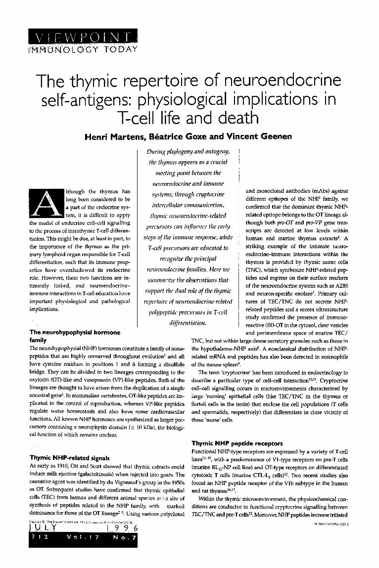
Fig. 2. Ultmstrt~cturnl image from the mrine tlytnic cortex. Pre-embedding lnbellillg zoith pol~yclor~nl mti-OT K31 at 2:700 (6. ]irikozcsk!y, Mmicld
and perosidnse-anti-peroxihse. Diffuse IR-OT appews in the qtosol of a TEC/TNC process of the szrbcorticnl ore0 zuith muificatiom moq pocked
thytnocytes CT). Some points of focd ndhesiorz cm be obserzmf betzuren TEC arrd T cells: T cells appmr 11s thoqh thy zwre ‘srrckliq’ the IR-OT-
prodming TECITNC. The economical principle of thr model is based upon the homology of peptide scqz~cr~cs befz(~eell crrdocr~irw signals md relntcd
thynic peptides tlmt me synthcsi~ed by TEC/TNC nnd presented to the differentidirq T cells. This lromolr~py Woql!y supports 17 dun1 role for fhe tlymic
repertoirr of uewoendocrirze precursors (pro X). First, thy cmstittrte II tolerogmic so~uw of ilerr~ocrzrfocrifzc self-mfigem (self-X). T11ew wlf-nrlti,qtwic
seqimces are lfi@y conserved tliro~~glzoi~t evolution of their fmil!y. On the other sicle, tlzymic rzerrroendocrirze-relntecl precursors nre the source qf
sigrds (signnl X) thnt provide nccessory pdzvays in the process of T-cell positive selection followirzg their intermtiou with uewoerdocrine-type
receptors (X receptor) expressed by developing T cells. Abbreviations: IR-OT, irtzmurzore~ctiz7e osytociu; MHC, mjor- lzistocoqmtibilit!y cotnplc.~ TCR,
T-cell receptor; TEC, tlynzic epithelid cells; TNC, thyrnic nurse cells.
target autoantigen of autoimmune processes. An infiltration of the
hypothalamo-NHP tract by inflammatory mononuclear cells has
been repeatedly observed, either after active immunization against
VP (Ref. 31) or in spontaneous autoimmune diabetes insipidus”O.
Together, these data support the idea that hypothalamic neurons
express some surface antigens representative of their neurosecre-
tory activity.
The breakdown of immunological tolerance of endocrine pan-
creatic islet p cells is becoming increasingly recognized as a major
etiopathogenic event in the emergence of autoimmune insulin-
dependent diabetes mellitus (IDDM)“*. This breakdown is thought
to be followed by an autoimmune cascade of events leading finally
to the disappearance of insulin-secreting islet p cells; consequently
insulin is more often accepted as a major autoantigen of the diabeto-
genie autoimmune process. The sequence 7-15 (CGSHLVEAL)
from the B domain of bovine insulin has been identified as a
target autoantigen for H-2Kh-restricted cytotoxlc T cells”“. However,
the biochemical identity between this insulin-derived autoantigen
and the corresponding sequence of IGF-II (CGGELVDTL) is nnt
complete. This difference in amino acid sequence could be impor-
tant for the nature of the T-cell responses (activation vs. tolerogenic
effect) elicited either by insulin- or IGF-II-derived peptide
sequences. As a further argument for its tolerogenic properties,
the production of IGF-II-specific antibodies is known to be more
difficult than those specific for IGF-I (Ref. 34). Thymic IGF-II
expression in diabetes-prone and diabetes-resistant BB rats is
under current investigation to elucidate its precise role in the patho-
genesis of IDDM.
Evolutionary aspects of neuroendocrine-immune interactions During the evolution of the endocrine system, various forms of
cell-cell signalling have appeared: (1) the primitive stages of

-
NHP ancestral VP lineage
-
precursor Water metabolism
OT lineage
Thymic NHP/MHC class I Reproduction
hybrid membrane protein A
/ 1 I 1 I I I I I
-700
\
-600 -500 -400 -300 -200 -100 Years
THYMUS (XiOs)
‘\ Agnatba Elasmobranchs Homo sapiens
Adhesion IglMHCITCRISupetfamily -~--
molecules
Recombinases (RAG1 and RAG2)
\ -I-
,- MHC Class I
---<- MHC Class II
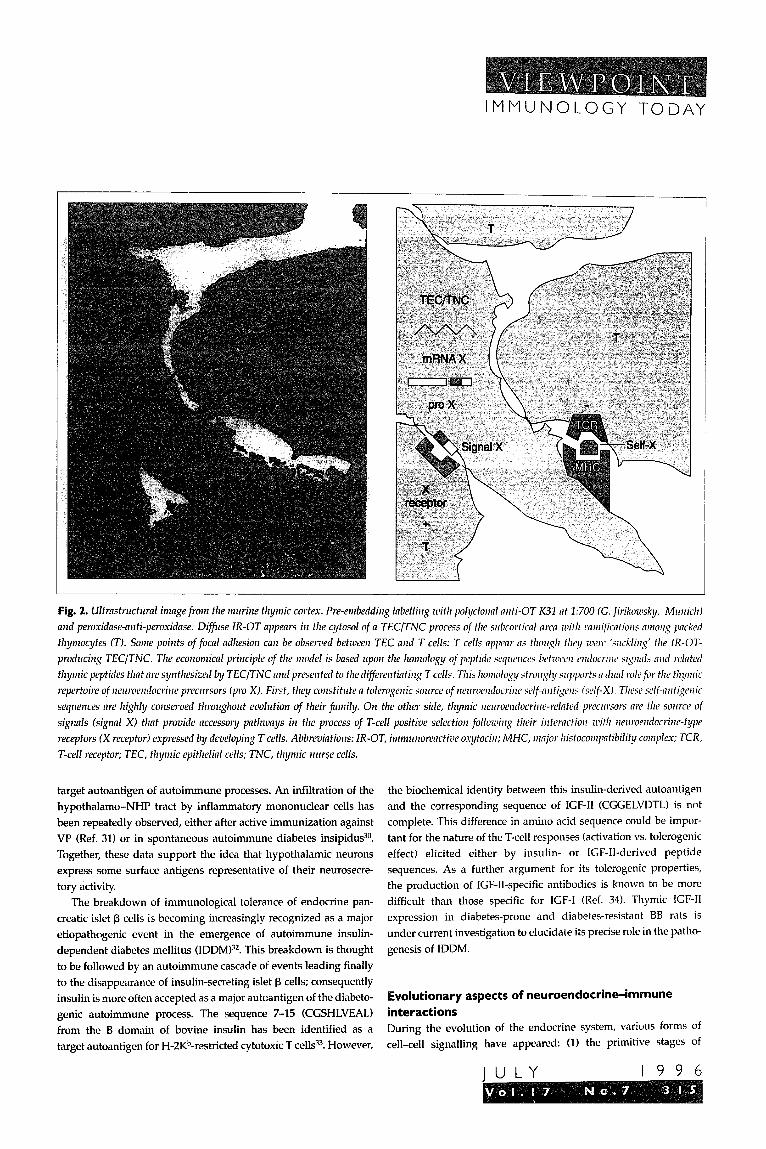
Fig. 3. Pwnllel evolutions nrzd interncfiom between the NHP mcl lg/MHC/TCR fmilies. The tlynic OT chinleric 55 kDn precursor corrld he nppeared after the dupficatiorz of the cow~~zo1~
mcestor of the ,Ieuro/z?lpo~~z~s/sinlf~nli[~, before or nt the sme time RS the @nit orgmz. It most prob- nbly prececied duplicntiorl of MHC-derizred profeins in class 1 RIIC/ class II, which occurred zvith the
first rlnsmobrmzchs. The ertrrme degree of diversificntiort and the potent properties of moleccrrlnr
recopitiorz chmcteri.zi~~,g most of the members of the Ig/MHC/TCR superfamily originnte from
nmdom recombinntioll of segment genes coding for Igs or TCRs. Abbreviations: Ig, i?,rnlrrtlo~Zobrllill;
MHC, rmjor Iristocompntibi~it!y complex; NHP, rlelfro/z~pop~rysinI; OT, osytocin; TCR, T-cell receptor; VP, vnsopressiii.
autocrine signalling (for cells in isolation) and intercellular adhesion
(which followed the first cell divisions in the conceptus); (2) the
more differentiated steps of paracrine and (neuro)endocrine sig-
nailing; and (3) the most advanced forms of synapses in neural net-
works which have allowed development of cognitive functions. It is
assumed that the cryptocrine mode occurs at the rather primitive
stage between cell adhesion and paracrine exchanges of soluble sig-
nals35. In parallel with these distinct structural levels, the com-
ponents of the genome coding intercellular messengers have
evolved to deal with organizational systems of increasing complex-
ity and have been progressively organized into separate families,
each containing distinct members in charge of the different types of
cell-cell signalling.
Converse!y, the immune system has evolved to protect the in-
tegrity of self against aggression from infectious nonself. Because
of the common peptidic nature of many alloantigens and self-anti-
gens, the immune system must have been educated to recognize
and to tolerate the molecular structure of the internal body.
Despite the increasing attention paid to peripheral pathways of
T-cell tolerance”‘, the thymus is clearly the primary organ purging
the immune system of self-reactive T cells, which could represent a
potential threat to survival. The thymic repertoire of neuroen-
docrine-related self-peptide precursors constitutes an original
model that underlines, at the molecular level, the dual role of the
W LY 1996
thymus in T-cell differentiation (Table 1).
According to this model, thymic neuroen-
docrine-related polypeptide precursors are
either the source of accessory signals for posi-
tive selection of T cells, or the source of self-
peptides able to induce the negative selec-
tion of self-reactive T cells bearing a
randomly rearranged TCR oriented against
their respective family (Fig. 2). This model
concurs with recent reports that have shown
that a single peptide may mediate both posi-
tive and negative selection of T cells de-
pending on the avidity or affinity of TCR for
the peptide used7i,3H’.
The hypothesis of a phylogenetic contin-
uum also arises from these observations
(Fig. 3). As discussed earlier, the NHP fam-
ily is highly conserved throughout evolu-
tion, even in invertebrates. By contrast,
the expansion of the immunoglobulin
(Ig)/MHC/TCR superfamily began at the
level of the primitive vertebrates some 550
million years ago at the latest39. Thus, the
foundations of the NHP family preceded
diversification of the Ig/MHC superfamily
The classical feature of neurophysins (two
variable sides connected to a greater central
constant region) has already been described
with the identification of their primary
structure40, and this constitutes another
structural relationship with members of the Ig/MHC superfamily.
Moreover, beyond the observation that some structural motifs of
the NHP family might have served as guides for further develop-
ment of the Ig/MHC superfamily, it is noteworthy that the thymic
OT self-antigen precursor apparently contains both a neuro-
endocrine- and an MHC class I-related domain. This illustrates the
intimacy of cooperation between a classical neuroendocrine family
and the Ig/MHC/TCR superfamily, as well as a plausible common
ancestral origin of these two families implicated in intercellular sig-
nalling and molecular recognition.
This manuscript is dedicated to our mentor and friend, Paul Franchimont, who was a pioneer of radioimmunology in Eumpe and founded the laboratory whew
most of these studies have been performed. The advanced stage of this work is
patly indebted to his encouragement and support since its :nitiation. These stud-
ies wele supported by gxmts horn the Sp&al Research Fund of the University of
Li+ge Medical School, the Fund for Scientific Medical ReseaKh of Belgium, the
Association contre le Cancer, the Belgian University Foundation, the International
Juvenile Diabetes Foundation, and the European Scienoz Foundation. Our grati-
tude goes to Prof. J. Urbain (Free University of Brussels) for his generous gift of
mAbs to oxytocin, to B. Malgrange (University of Li+ge) who performed the con-
focal microscopy analysis of Fig. 1, to A. Godard mSERM U.437) for antis-
against leukaemia inhibitory factor, and to M. Wiemann (university of Miinster,
Germany) who provided us with the ultrastructural image of Fig. 2.

IMMUNOLOGY TODAY
Henri Martens, Z%aatrice Goxe nixi Vincent Geertert nre nt the UniversitJy
of Li&e Medical School, Institute of Pafholopj CHU-B23, Laboratory of
Radio-lmmul~olog!l and Nezlroefzllocrirle-lnzn1Ur20logy, B-4000 Sart
Tilman, Belgium.
References
1 Acher, R. (1993) Regzd. Peptides 45,1-13
2 Geenen, V., Legros, J.J., Franchimont, P., Baudrihaye, M.F., Defresne,
M.l? and Boniver, J. (1986) Scielzce 232,508-511
3 Mall, U.M., Lane, B.L., Robert, F.R., Geenen, V. and Legros, J.J. (1988)
HisfochenGstry 89‘385-390
4 Melis, M.R., Mauri, A. and Argiolas, A. (1995) Regld. Pepides 59,335-340
5 Robert, F.R., Martens, H., Cormann, N., Benhida, A., Schoenen, J. and
Geenen, V. (1992) Dezl. l~nmu~zol. 2, 131-140
6 Geenen, V., Vandersmissen, E., Martens, H. et RI. (1995) in Nezllalzypophysis:
Recerlt Progress of Vnsopressill nrld Os!/tociw Rescnrch (Saito, T., Kurokawa, K.
and Yoshida, S., eds), pp. 309-319, Elsevier
7 Geenen, V., Defresne, M.P., Robert, F., Legros, J.J., Franchimont, I? and
Boniver, J. (1988) Nelrroclldocrilzolc/ 47, 365-368
8 Wiemann, M. and Ehret, G. (1993) Cell Tisslle Res. 273, 79-87
9 Kumamoto, K., Matsuura, T., Amagai, T. and Kawata, M. (1995) Cell
Tiswe Res. 281, l-10
10 Funder, J.W. (1990) Mol. Cell. Endocriizol. 70, C21-C24
11 Geenen, V., Robert, F., Martens, H. et al. (1991) Mol. Cell. Endocritzol. 76,
C27-C31
12 Martens, H., Robert, F., Legros, J.J., Geenen, V and Franchimont, l?
(1992) Prog. NellroelzdocrirziflzInllnol. 5, 31-39
13 Torres, B.A. and Johnson, H.M. (1988) I. Immtrr!ol. 140,2179-2183
14 Elands, J., Resink, A. and De Kloet, E.R. (1990) EfzdocrinoloR/ 126,
2703-2710
15 Caldwell, J.D., Musiol, I.M., Walker, C.H., Pedersen, C.A. and
Mason, G.A. (1993) Awz. New York Acod. Sci. 689, 575-577
16 Sugimoto, T., Saito, M., Mochizuki, S. et RI. (1995) in ,V~flroh!/po~ll!/sis:
Recent Progress of Voso,uressin and Os~ytocin Rese&l (Saito, T., Kurokawa, K.
and Yoshida, S., eds), pp. 409-413, Elsevier
17 Lolait, S. et al. (1995) Proc. Nnfl Acnd. Sci. USA 92,6783-6787
18 Schaller, M.D., Borgman, C.A., Cobb, B.S., Vines, R.R., Reynolds, A.B.
and Parsons, J.T. (1992) hoc. NotI Acod. Sci. USA 89,5192-5196
19 Bonomo, A. and Matzinger, I? (1993) 1. Exp. Med. 177, 1153-1164
20 Townsend, A. and Bodmer, H. (1989) AWW. Rea. Z~~IIWZO/. 7, 235-238
21 Rammensee, H.G., Falk, K. and Riitzschke, 0. (1993) AWE. Re:,.
Inlllll4nol. 11, 213-244
22 Geenen, V, Vandersmissen, E., Martens, H. et nl. (1993) Thp~s 22,55_66
23 Griffin, J.H., Alazard, R. and Cohen, I? (1973) 1. Biol. C/ton. 248,7975-7978
24 Breslow, E. and Burman, S. (1990) Adu. Enzy~~ol. 63,1-67
25 Simpson, E., Robinson, P.J., Chandler, l? et al. (1994) Imlnlrwolog!~ 81,
132-136
26 Ericsson, A., Geenen, V., Robert, F. et RI. (1990) Mol. Endocrinol. 4,
1211-1218
27 Geenen, CI., Achour, I., Robert, F. rt nl. (1993) Tlnprs 21, 115-127
28 Kooijman, R., van Buul-Offers, C., Scholtens, L.E. et nl. (1995) 1. I,nnlrlrol.
154,5736-5745
29 Scherbaum, W.A. and Bottazzo, G.F. (1983) Lnlxet i, 897-901
30 Imura, H., Nakao, K., Shimatsu, A. rt RI. (1993) Neil1 Ellgl. 1, Med. 329,
683-689
31 Cau, I? and Rougon-Capuzzi, G. (1979) Bruilz Res. 177,265-271
32 Castano, L. and Eisenbrrth, G.S. (1990) AIIIIII. RCP. Iw~~cr~ol. 8,
647-649
33 Sheil, J.M., Shepherd, S.E., Klimo, G.F. and Paterson, Y. (1992) I. Esp.
Med. 175,545-552
34 Zapf, J., Walter, H. and Froesch, E.R. (1981) I. Clilz. bzz~st. 68, 1321-1330
35 Geenen, V., Cormann-Goffin, N., Martens, H. et al. (1993) ReguI. Peytides
45,273-278
36 Geenen, V. and Kroemer, G. (1993) Onn?zrnol. Todq 14, 573-575
37 Sebzda, E., Wallace, VA., Mayer, J., Yeung, R.S.M., Mak, T.W. and
Ohashi, P.S. (1994) Scimce 263,1615-1618
38 Allen, P.M. (1994) Cell 76,593-596
39 Bartl, S., Baltimore, D. and Weissman, I.L. (199-l) P~oc. Nd Acod. Sci.
USA 91,10769-l 0770
40 Capra, J.D. and Walter, R. (1975) Ann. Nenj York Acod. Sci. 248,397-407
41 Kramer S., Reynolds, F.H., Jr, Castillo, M., Valenzuela, D.M., Thorikay, M.
and Sorvillo, J.M. (1991) ElrdocrinologJy 128,1927-1937
42 Bulloch, K., Radjocic, T., Yu, R., Hausman, J., Lenhard, L. and Baird, S.
(1993) Prog. Nezlroendocrilzimnzur~ol. 4, 186-194
43 Vollmar, A.M. and Schulz, R. (1990) Eadocrinology 126,2277-2281
44 Martens, H., Malgrange, B., Robert, F. et al. Regul. Pep. (in press)
, journals j @,y@ocrtf! ctieq,y+ct@pf$in I:a potential regulator ofmonocyte recruitment in inflammatory disease, B.J. Rollins
* I ( I99bi fit&i&. M&&e 7iiay 2 ‘(5)j I9&208 ” * _ : 1 ,
~~&SthktW +ai'id s t&&ion. ok the” T~atural-resistance-associated macrophage pmein (Nramp I), a candidate protein for T ’ ~“i~f$~~ti~~~~ autbrmmiine dis&e susceptibility, J.M. klackwell (1996) Molecu/~r Medicine Today 2 (S), 205-21 I > IS -i , z,._:- ' I _1 ‘ ,,: "a" - .I .,>i>
0 &$k eerapy again& canier and-HIV infection using the gene encoding herpes simplex virus thymidine kinase, M. Caruso IzI _.(l~~>_M&&w Medicke Today 2 (5), 213-217
I” ,Y _ , I*i_ : ” :*i: ~~~~@&pk~+ctio~ of therapeu@c cell transplants by encapsulation, Rj. Morris (1996) Trends in Biote&nology 14 (5), 163-l 67 _” (. ?i -I._ %
L /;;&an’immu& response to MSP- I, A.A. Holder and E.M. Riley (I 996) Pur&o/ogy Todq I2 (5), 173-l 74 -1
I.’ 0; @laiwia and onchocerciasis: on HLA and reked matters, C.G. Meyer and F!G. Kremsner ( 1996) fur~sitology Today 12 (5). * - _ .&I$6 -._ c ,L- 1_1”
I u LY 1996