Multiple Sclerosis-Associated Retrovirus Particles Cause T Lymphocyte-Dependent Death with Brain...
15
Journal of NeuroVirology, 9: 79–93, 2003 c ≈ 2003 Taylor & Francis ISSN 1355-0284/03 $12.00+.00 DOI: 10.1080/13550280390173328 Multiple sclerosis–associated retrovirus particles cause T lymphocyte–dependent death with brain hemorrhage in humanized SCID mice model R Firouzi, 1 A Rolland, 2 M Michel, 3 E Jouvin-Marche, 2 JJ Hauw, 4 C Malcus-Vocanson, 5 F Lazarini, 4 L Gebuhrer, 6 JM Seigneurin, 3 JL Touraine, 1 K Sanhadji, 1 PN Marche, 2 and H Perron 5 1 Laboratoire des d ´ eficits Immunitaires, Facult ´ e de M ´ edecine La ¨ ennec, Lyon, France; 2 INSERM, Grenoble, France; 3 Laboratoire de Virologie, Facult´ e de M ´ edecine de Grenoble, La Tronche, France; 4 Laboratoire de Neuropathologie Raymond Escourolle, H ˆ opital de la Salpˆ etri ` ere, Paris, France; 5 BioM ´ erieux, Research and Development Department, Chemin de l’Orme, Marcy l’Etoile, France; and 6 Laboratoire d’Histocompatibilit ´ e/EFS, Lyon, France A retroviral element (multiple sclerosis–associated retrovirus, MSRV) defining a family of genetically inherited endogenous retroviruses (human endogenous retrovirus type W, HERV-W) has been characterized in cell cultures from pa- tients with multiple sclerosis. Recently, MSRV retroviral particles or the enve- lope recombinant protein were shown to display superantigen activity in vitro, but no animal model has yet been set up for studying the pathogenicity of this retrovirus. In the present study, the pathogenicity of different sources of MSRV retroviral particles has been evaluated in a hybrid animal model: severe com- bined immunodeficiency (SCID) mice grafted with human lymphocytes and injected intraperitoneally with MSRV virion or mock controls. MSRV-injected mice presented with acute neurological symptoms and died within 5 to 10 days post injection. Necropsy revealed disseminated and major brain hemorrhages, whereas control animals did not show abnormalities (P < .001). In ill animals, reverse transcriptase–polymerase chain reaction (RT-PCR) analyses showed circulating MSRV RNA in serum, whereas overexpression of proinflamma- tory cytokines such as tumor necrosis factor (TNF)-γ and interferon (IFN)-γ was evidenced in spleen RNA. Neuropathological examination confirmed that hemorrhages occurred prior to death in multifocal areas of brain parenchyma and meninges. Further series addressed the question of immune-mediated pathogenicity, by inoculating virion to SCID mice grafted with total and T lymphocyte–depleted cells in parallel: dramatic and statistically significant reduction in the number of affected mice was observed in T-depleted series (P < .001). This in vivo study suggests that MSRV retroviral particles from MS cultures have potent immunopathogenic properties mediated by T cells compatible with the previously reported superantigen activity in vitro, which appear to be mediated by an overexpression of proinflammatory cytokines. Journal of NeuroVirology (2003) 9, 79–93. Keywords: animal model; blood-brain barrier; cytokine; endogenous retro- virus; superantigen Address correspondence to Herve Perron, BioM´ erieux, Re- search and Development Department, Chemin de l’Orme, 69280 Marcy l’Etoile, France. E-mail: [email protected] The authors thank Dr. Simone Peyrol and Ingrid Berger for their assistance in the electron microscopy study performed by Marl ` ene Michel, during her PhD thesis research, supported by a grant from the M´ erieux foundation. This study was supported by BioM ´ erieux SA. Received 17 July 2002; revised 26 August 2002; accepted 11 September 2002. Introduction Multiple sclerosis (MS) is a multifactorial inflam- matory disease of the central nervous system char- acterized by plaques of demyelination associated blood-brain barrier breakdown followed by intra- parenchymal lymphocytic infiltrates and prominent T-cell activation. Epidemiological and clinical data suggest that both genetic and environmental factors
-
Upload
independent -
Category
Documents
-
view
1 -
download
0
Transcript of Multiple Sclerosis-Associated Retrovirus Particles Cause T Lymphocyte-Dependent Death with Brain...

Journal of NeuroVirology, 9: 79–93, 2003c≈ 2003 Taylor & Francis ISSN 1355-0284/03 $12.00+.00DOI: 10.1080/13550280390173328
Multiple sclerosis–associated retrovirus particlescause T lymphocyte–dependent death with brainhemorrhage in humanized SCID mice model
R Firouzi,1 A Rolland,2 M Michel,3 E Jouvin-Marche,2 JJ Hauw,4 C Malcus-Vocanson,5 F Lazarini,4
L Gebuhrer,6 JM Seigneurin,3 JL Touraine,1 K Sanhadji,1 PN Marche,2 and H Perron5
1Laboratoire des deficits Immunitaires, Faculte de Medecine Laennec, Lyon, France; 2INSERM, Grenoble, France;3Laboratoire de Virologie, Faculte de Medecine de Grenoble, La Tronche, France; 4Laboratoire de NeuropathologieRaymond Escourolle, Hopital de la Salpetriere, Paris, France; 5BioMerieux, Research and Development Department,Chemin de l’Orme, Marcy l’Etoile, France; and 6Laboratoire d’Histocompatibilite/EFS, Lyon, France
A retroviral element (multiple sclerosis–associated retrovirus, MSRV) defininga family of genetically inherited endogenous retroviruses (human endogenousretrovirus type W, HERV-W) has been characterized in cell cultures from pa-tients with multiple sclerosis. Recently, MSRV retroviral particles or the enve-lope recombinant protein were shown to display superantigen activity in vitro,but no animal model has yet been set up for studying the pathogenicity of thisretrovirus. In the present study, the pathogenicity of different sources of MSRVretroviral particles has been evaluated in a hybrid animal model: severe com-bined immunodeficiency (SCID) mice grafted with human lymphocytes andinjected intraperitoneally with MSRV virion or mock controls. MSRV-injectedmice presented with acute neurological symptoms and died within 5 to 10 dayspost injection. Necropsy revealed disseminated and major brain hemorrhages,whereas control animals did not show abnormalities (P< .001). In ill animals,reverse transcriptase–polymerase chain reaction (RT-PCR) analyses showedcirculating MSRV RNA in serum, whereas overexpression of proinflamma-tory cytokines such as tumor necrosis factor (TNF)-γ and interferon (IFN)-γwas evidenced in spleen RNA. Neuropathological examination confirmed thathemorrhages occurred prior to death in multifocal areas of brain parenchymaand meninges. Further series addressed the question of immune-mediatedpathogenicity, by inoculating virion to SCID mice grafted with total and Tlymphocyte–depleted cells in parallel: dramatic and statistically significantreduction in the number of affected mice was observed in T-depleted series(P< .001). This in vivo study suggests that MSRV retroviral particles fromMS cultures have potent immunopathogenic properties mediated by T cellscompatible with the previously reported superantigen activity in vitro, whichappear to be mediated by an overexpression of proinflammatory cytokines.Journal of NeuroVirology (2003) 9, 79–93.
Keywords: animal model; blood-brain barrier; cytokine; endogenous retro-virus; superantigen
Address correspondence to Herve Perron, BioMerieux, Re-search and Development Department, Chemin de l’Orme, 69280Marcy l’Etoile, France. E-mail: [email protected] authors thank Dr. Simone Peyrol and Ingrid Berger for their
assistance in the electronmicroscopy study performed byMarleneMichel, during her PhD thesis research, supported by a grant fromthe Merieux foundation. This study was supported by BioMerieuxSA.Received 17 July 2002; revised 26 August 2002; accepted
11 September 2002.
Introduction
Multiple sclerosis (MS) is a multifactorial inflam-matory disease of the central nervous system char-acterized by plaques of demyelination associatedblood-brain barrier breakdown followed by intra-parenchymal lymphocytic infiltrates and prominentT-cell activation. Epidemiological and clinical datasuggest that both genetic and environmental factors
Immune-mediated neuropathogenicity of MS retrovirus80 R Firouzi et al
could be involved in the etiology of this disease. MSpathogenesis is thought to consist in an autoim-mune process directed against myelin components,probably triggered by environmental factors, amongwhich viruses are favored candidates. Viruses such asherpesviruses (Ascherio and Munch, 2000; Ferranteet al, 2000; Soldan et al, 1997; Wandinger et al, 2000)and retroviruses (Haahr et al, 1991; Koprowski et al,1985; Perron et al, 1991b) have been suggested to beassociated with MS, and different groups have de-tected retroviral particles in cultured cells from MSpatients (Haahr et al, 1991; Lan et al, 1994; Perronet al, 1991b).Our previous studies on RNA associated with
viral particles produced in choroid plexus/leptomeningeal cell or B-lymphocyte culturesfrom patients with MS had provided sequencescorresponding to overlapping regions of a retroviralgenome (Komurian-Pradel et al, 1999; Perron et al,1997b), which was provisionally named MSRV (formultiple sclerosis–associated retrovirus element).MSRV revealed to have genetically homologouselements in human DNA, defining a novel familyof human endogenous retroviruses, HERV-W, forhuman endogenous retrovirus type W (Blond et al,1999; Perron et al, 1997b).This HERV-W family comprises multiple copies of
a prototypic retroviral genome that probably enteredthe germ line cell lineage by an infectious route morethan 25 million years ago, before the species radia-tion of the ancestors of the old world monkeys (Kimet al, 1999; Voisset et al, 1999). Consequently, mem-bers of this endogenous family can be found in nor-mal DNA of old world monkeys, apes, and humans,but are absent in all other animal species, includingnew world monkeys. In normal human DNA, mostHERV-W copies are truncated and/or lack open read-ing frames (orf), but several chromosomal copies haveretained potential orfs for retroviral proteins (Vois-set et al, 2000). A complete HERV-W provirus onchromosome 7q, in a region associated with geneticsusceptibility to MS (Perron et al, 2000), encodesan envelope protein strongly expressed in placenta(Blond et al, 1999). This protein is involved in thephysiological process of syncytiotrophoblast fusion(Blond et al, 2000). The HERV-W7q provirus is notfully functional and cannot account for virion pro-duction as observed in MS (Perron et al, 2000). Inaddition, the HERV-W copy number in human DNAappears to vary between individuals and with eth-nical origin (Mirsattari et al, 2001). Therefore, MSretroviral particles are not likely to be encoded by thenormal inherited HERV-W copies, but may originatefrom a “modified” (e.g., retrotransposed and/or re-combined) or exogenous member of the same family(Komurian-Pradel et al, 1999; Perron et al, 2000; Vois-set et al, 2000). Particle-associated retroviral RNAhasnow been detected in MS sera by different groups(Dolei et al, 2002; Garson et al, 1998; Olsson et al,1999; Serra et al, 2001); these data also appear to sig-
nificantly correlate particular features of the clinicalhistory.The pathogenicity of retroviral elements belonging
to genetic families with endogenous copies usuallyresults from interactions between pathogenic mem-bers and the “normal” endogenous gene homologs(Contag and Plagemann, 1989; Gardner, 1990). Thishas also been reported for the superantigen encodedby members of the mouse mammary tumor virus(MMTV) family in mice (Kubo et al, 1996; Xuet al, 1996). Our recent findings of superantigenactivity associated with virions and encoded byMSRV envelope protein (Lafon et al, 2002; Perronet al, 2001) therefore suggest that, apart from amajor contribution in immune dysfunctions of MSdisease, immunopathogenic properties of humanMSRV/HERV-W members could also result fromsuch retroviral interactions. Given recent evidenceof active HERV-W proviruses in human cells (Yiet al, 2002), together with variability of DNA copynumber among humans (Mirsattari et al, 2001),retrotransposition—and possibly recombination—events should occur within the HERV-W familyunder certain circumstances. Consequently, mostanimal models that could be set up for studying thepathogenicity of these MSRV particles might appearinappropriate because, as mentioned above, mostanimals lack these endogenous ERV-W elements intheir genome, or, when present, e.g., in old worldmonkeys, these ERV-W copies are not identical totheir human counterpart, HERV-W (Kim and Crow,1999; Kim et al, 1999; Voisset et al, 1999).In order to address the potential pathogenicity of
MSRV virion in vivo, we have chosen a hybrid hu-man/animal model: the severe combined immuno-deficiency (SCID) mouse humanized with humanlymphocytes, called hu-SCID mouse. This T cell–and B cell–deficient mouse supports grafting of func-tional human lymphoid cells from human peripheralblood mononuclear cells (PBMCs) that will surviveand circulate in place of the mouse lymphoid cells.Such a model, called hu-PBL-SCID (considering pe-ripheral blood lymphocytes as major elements of thegraft), allows studies of human immunopathologicalmechanisms in vivo (Chargui et al, 1995; Okamotoet al, 1998; Persidsky, 1999; Scaramuzzino et al, 2000;Vanzieleghem et al, 2000). In our present protocol,the survival of the engrafted human cells was opti-mized by previous irradiation and anti–natural killer(NK) cell treatment, and therefore allowed studiesover a rather long period (generally, up to 3 monthswith good confidence).As previously discussed, mice do not harbor
any chromosomal copy of the ERV-W family. TheHu-PBL-SCID mouse therefore allows the study ofpathogenic mechanisms that may involve interac-tions between MSRV virus particles and endogenousHERV-W expression, within the human cells only. Inaddition, physiopathological effects within mice tis-sues should mostly result from retroviral expression

Immune-mediated neuropathogenicity of MS retrovirusR Firouzi et al 81
and/or immune response linked to human lymphoidcells.
Results
Different series of humanized mice have been pre-pared according to the protocol presented in Figure 1.In humanPBMCs prior to engraftment, the CD3/CD45ratio ranged from 50% to 80%, the CD4/CD45 ratiofrom 30% to 45%, and the CD19/CD45 ratio from10% to 25%. Five days after engraftment with humanPBMCs, peritoneal fluid from all mice was puncturedand analyzed by cytofluorometry: CD3 ranged from35% to 75%, CD4 from 20% to 30%, and CD19 from10% to 25%. Mice series once found with less than25% of CD3 and less than 10% of CD4 were not in-cluded in the study.Occasionally, a mouse died prior to virus injection
(three cases in all series), as normally observed withlow frequency within the first days after x-ray irra-diation, anti-NK treatment, and human PBMC graft-ing. In order to control the rate of such “acciden-tal” death during the “viral” study period in ourseries, phosphate-buffered saline (PBS)-inoculatedmice were always kept in parallel with mice from thesame pre-engraftment group, grafted with the samePBMC sample, but inoculated with MSRV virion ormock preparation. During this period (after the firstweek post engraftment), we did not observe death ofPBS-injected animals in our series.As presented in Table 1, summarizing successive
series of experiments, mice were grafted with PBMCsfrom five different blood donors with various humanleukocyte antigen (HLA) class II phenotypes.MS virions, as visualized by electron microscopy
shown in Figure 2, were prepared from different so-urces of cell culture (Perron et al, 1989, 1991a, 1997a).They were pelleted from supernatants presenting apeak of reverse transcriptase (RT) activity above thebackground signal (mean + 3 SD determined fromcell type–matched, non-MS control cultures), as pre-viously described (Perron et al, 1991b, 1993, 1997a).In addition, most cellular debris were eliminated byprecentrifugation (prior to freezing) and by using aglycerol cushion at the bottom of ultracentrifugationtubes, collection of the material pelleted under thisglycerol barrier avoided eventual soluble factors fromculture medium. Also, virion particles were preser-ved by adding 10% of glycerol in culture supernata-nts and resuspended pellets prior to any freezingstep.The RT activity count in the samples injected to
mice was about 10,000 dpm and approximately cor-responded to 104 particles, as determined elsewherewith negative-stained preparations on electron mi-croscope (EM) grids. Our recent data (unpublished)indicate that MSRV particles can be detected in serafrom MS patients at concentrations 3 logs below thatof the suspensions injected to hu-SCID mice.
As indicated in Material and methods, cell cul-tures and virion batches were screened for possiblecontaminants and all samples were obtained withthe same batch of culture medium. As expected, noEpstein-Barr virus genome was detected in choroidsplexus cultures from MS or non-MS control. In ad-dition, preliminary experiments on few nonhuman-ized SCIDmice had shownno clinically detectable ef-fect during 3weeks after intraperitoneal (IP) injectionof all preparations used for the humanized (hu-PBL-SCID) series (not shown). Consequently pathogeniccontaminants that could have directly affected SCIDmice were absent from our preparations.Circulating particle-associatedMSRVRNAwas de-
tected in cell-free serum collected from cardiac bloodat necropsy, according to J. Garson’s protocol (Garsonet al, 1998), as indicated in Table 1. These data con-firmed virion access to the bloodstream after IP in-oculation of virion preparations, but its absence inmock-infected mice.
Hu-PBL-SCID mice with MSRV virion diewith brain hemorrhageIn mice series grafted with total PBMCs (not T-depleted), we could observe a striking effect thatrapidly appeared in all mice inoculated with MS-virion preparation from both sources (choroid plexusor B cells): mice presented with neurological symp-toms such as partial or generalized paralysis followedby death of the animals. Such features were not ob-served in animals injected with mock preparationsfrom non-MS cultures, nor in animals injected withPBS, during the few months they were kept alive.This was similarly reproduced in separate experi-ments: onewith donors 1 and 2, anotherwith donor 3,and one with T-depleted versus nondepleted PBMCsfrom donors 4 and 5 (presented in Table 1). A firstseries with lower numbers of animals (not shown inTable 1) had also included mock virion from non-MSchoroid plexus cultures, in parallel with the otherpreparations mentioned in Table 1. These hu-PBL-SCID mice injected with mock choroid plexus virionsurvived as well as the PBS controls (over 2 months).The difference between the numbers of ill/dead
animals in virus-inoculated animals and mock- orPBS-injected controls, appears significant in all cases:comparing single donors (e.g., 1, 2, or 3) with onesource of virion versus mock controls (5/5 versus0/5) yields a chi-square value with Yate’s correctionof 6.4 (P < .02); comparing both sources of virionversus mock controls for single donors in the sameseries (10/10 versus 0/5) yields a chi-square valuewith Yate’s correction of 10.84 (P < .01); comparingall virus-injected hu-PBL-SCIDmice versus all mock-controls in Table 1 (46/46 versus 0/23, excludingT-depleted series) yields a chi-square value of 69(P < .001).Most virion-inoculated mice were examined post-
mortem, but a few ill mice, in the last series, weresacrificed when neurological symptoms appeared.

Immune-mediated neuropathogenicity of MS retrovirus82 R Firouzi et al
Figure 1 Hu-PBL-SCID grafting with human PBMCs followed by intraperitoneal injection of virus or control preparations: schematicrepresentation of the experimental protocol.

Tabl
e1Hu-PBL-SCIDseriesgraftedwithPBMCsfrom
humanblooddonorswithvariousHLA-IIphenotypes
1:D
R2/
DR
42:
DR
2/D
R4
3:D
R2/
DR
24:
DR
2/D
R13
4:T-
Dep
lete
d5:
DR
7/D
R11
5:T-
Dep
lete
dD
onor
+H
LA-D
RD
RB
1*02
/04
DR
B1*
02/0
4D
RB
1*02
DR
B1*
1501
/130
3D
RB
1*15
01/1
303
DR
B1*
1101
/07
DR
B1*
1101
/07
Inoculum
MSRVvirion
Deathwithbrain
5/5mice
5/5mice
5/5mice
4/4mice
1/4mice
4/4mice
0/4mice
from
MS
haemorrhage
5–10daysp.i.
5–10daysp.i.
5–10daysp.i.
5–10daysp.i.
5–10daysp.i.
5–10daysp.i.
alive
>2mo.
ChoroidPlexus
MSRVRT-PCR
++
+N
DN
DN
DN
Dcultures
MSRVvirion
Deathwithbrain
5/5mice
5/5mice
5/5mice
4/4mice
0/4mice
4/4mice
1/4mice
from
MS
haemorrhage
5–10daysp.i.
5–10daysp.i.
5–10daysp.i.
5–10daysp.i.
alive
>2mo.
5–10daysp.i.
5–10daysp.i.
B-Cellcultures
MSRVRT-PCR
++
+N
DN
DN
DN
DMockvirion
Deathwithbrain
0/5mice
0/5mice
0/5mice
0/4mice
0/4mice
0/4mice
0/4mice
from
Non-MS
haemorrhage
alive
>3mo.
alive
>3mo.
alive
>3mo.
alive
>2mo.
alive>2mo.
alive
>2mo.
alive
>2mo.
B-Cellcultures
MSRVRT-PCR
≈≈
≈N
DN
DN
DN
DPBSalone
Deathwithbrain
0/4mice
0/4mice
0/4mice
0/3mice
0/3mice
0/3mice
0/3mice
haemorrhage
alive
>3mo.
alive
>3mo.
alive
>3mo.
alive
>2mo.
alive
>2mo.
alive
>2mo.
alive
>2mo.
MSRVRT-PCR
≈≈
≈N
DN
DN
DN
D
Not
e.Hu-PBL-SCIDmicewereinoculatedintraperitoneallyinparallelwith(i)MSRVvirionpreparationsfrom
twodifferentcellcultures(choroidplexusandBcells);(ii)mock
virionfrom
non-MSBcells(seriesincludingnon-MSchoroidplexusmockpreparationarenotpresentedinthistablebecausecomplete(seriesonly)areconsideredhere);and
(iii)PBSusedtoresuspendvirionormockvirionultracentrifugationpellets.Threesuccessiveseriesofexperimentsaregrouped:firstseriescorrespondtodonors1and2with
identicalDRB1phenotypes;secondseriescorrespondtodonor3;thirdseriescorrespondtodonors4and5withparallelT-celldepletiononnylonmembranesofPBMCsbefore
grafting.Whenmentioned,theperiodofdeathcorrespondstothatofanimalsthatwerenotsacrificedbeforethefataloutcome.Similarly,theperiodduringthatmicewerekeptand
survivedinahealthystateexcludesanimalsthatweresacrificedinparallelwithillones,forcomparativehistopathologicalexamination.Inaddition,allsurvivinganimalsfrom
controlseriesweresacrificedaftertheperiodmentionedinthetableandnecropsied.Noneofsuchmicepresentedpathologicalsignsatnecropsicexamination.
ND:notdone.
83

Immune-mediated neuropathogenicity of MS retrovirus84 R Firouzi et al
Figure 2 Retroviral particles (MSRV) in multiple sclerosis cell cultures. Electron microscopy. No retroviral particle could be seen eitherin the cytoplasm or at the cell surface in control B-cell or choroid plexus (leptomeningeal cells) cultures. In MS cultures studied over a3-week period, retrovirus particles in isolated cells were observed one or two times corresponding to peaks of RT activity. Long periodsof observation were necessary for such findings. Extracellular particles were observed at the surface of the cytoplasmic membrane inisolated cells, whereas clusters of retrovirus-like particles in vacuoles were also occasionally seen. (A) Extracellular virion particles at thesurface of the cell membrane; MS leptomeningeal cells. (B) Arrows indicate retrovirus-like particles at the surface of the cell membraneor in the extracellular space; MS B-cell culture. (C) Extracellular virion particle at the surface of the cell membrane; MS B-Cell culture.(D) Cluster of electron-dense retrovirus-like particles (capsids) within an MS B cell. The bar represents 100 nm.
Control mice were sacrificed in parallel at the sameday, and others were kept in order to estimate theirsurvival time.As shown in Figure 3, dissection of mice regularly
showed a marked splenomegaly and relative hep-atomegaly inMSRV virion–inoculatedmice, whereasspleen from controls only reflected successful hu-manization with human lymphoid cells. Surpris-ingly, no other significant finding was made either inthe abdominal cavity or in the chest. When the cra-nial cavity was dissected, we were greatly surprisedto observe macroscopic and frequently massive brainhemorrhages in all dead MSRV-injected animals. Asshown in Figure 4, displacement of the brain some-
times revealed massive hemorrhage with blood col-lected at the bottomof the cranial cavity, thus stronglysuggesting that these animals could have died fromcerebral compression as well as from the cerebralhemorrhage itself. This was not seen in control ani-mals (mock-infected or PBS-inoculated), either sacri-ficed on the day when another mice died in infectedseries, or sacrificed after 2 or 3 months of survival.In ill mice sacrificed before death, hemorrhage ap-peared less important at the brain surface but mostlyas disseminated patches at the meningeal surface.In order to confirm the circumstances in which
these brain hemorrhages occurred, the animals fromthe last series were formalin-fixed immediately

Immune-mediated neuropathogenicity of MS retrovirusR Firouzi et al 85
Figure 3 Autopsic examination: Abdomen and chest. Parallel dissection of dead animals from the virion-injected series and micesacrificed the same day in mock-infected series revealed marked splenomegaly and relative hepatomegaly only in MSRV + hu-PBL-SCIDmice.
after death without dissection and examined by aneuropathologist expert (Prof. J. J. Hauw, l’HopitalSalpetriere, Paris, France).As shown in Figure 5, autopsic examination per-
formed in appropriate conditions confirmed theabsence of visible abnormalities in control brains(Figure 5A), the presence of hemorrhagic patches atthe brain surface (upper or lower side) of virion-injected animals sacrificed prior to death when neu-rological symptomswere detected (Figure 5B, C), andthe presence of massive hemorrhage involving largeportions of the brain with important blood leakage inthe cranial cavity, after death (Figure 5D–F).Neurohistological examination of brain sections
from these brains (Figure 6) confirmed that the hem-orrhagic phenomenon was not artefactual and oc-curred before death. Leptomeningeal (Figure 6A)as well as intraparenchymal (Figure 6B, C) hem-orrhage was observed and anoxi-ischemic neuronswere found in gray matter as well as multifocalnecrotic areas (Figure 6C, D). These features indicatethat corresponding hemorrhages must have occurredat least 6 h prior to death.
Figure 4 Autopsic examination: Head. Dissection of dead ani-mals from the virion-injected series revealed major brain hem-orrhages, with massive blood leakage into the cranial cavity inMSRV + hu-PBL-SCID mice. Parallel necropsy of control mock-infected hu-PBL-SCID mice was normal.

Immune-mediated neuropathogenicity of MS retrovirus86 R Firouzi et al
Figure 5 Neuropathological examination: Formalin-fixed heads. Dead animals from the virion-injected series and mice sacrificed thesame day in mock-infected series were formalin-fixed prior to dissection. (A) Mock-infected control. No abnormality on the brain or inhead structures can be noticed. (B, C) MSRV virion–injected hu-PBL-SCID mice presenting neurological symptoms (marked paralysis ofhind legs and tremor) sacrificed before death. Multiple petechia can be seen all over the surface of the brain, at the level of the meninges;B, top side; C, bottom side. (D) MSRV virion–injected hu-PBL-SCID mice fixed after death. A major meningeal hemorrhage can be seenin the posterior part of the brain. (E, F) MSRV virion–injected hu-PBL-SCID mice fixed after death. An important hemorrhage is seen atthe surface of the brain (E) and blood collected in the bottom of the cranial cavity is seen after displacement of the brain (F). The featuresobserved in this case are clearly similar to the one (non-formalin fixed) shown in Figure 4.
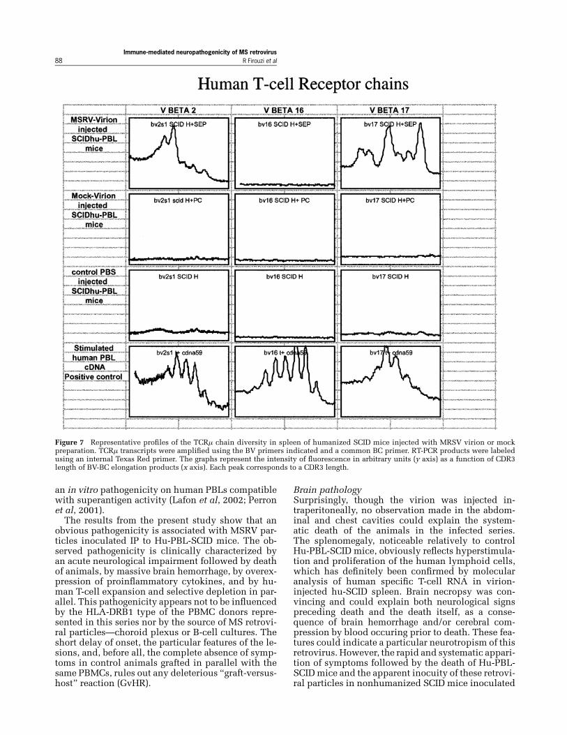
Pathogenicity correlates with overexpressionof proinflammatory cytokines in spleenBecause we had observed in vitro that MSRV viri-ons induced proinflammatory cytokine productionin association with polyclonal expansion or deple-tion of human T lymphocytes, mainly skewed to-wards Vμ16 subpopulation, we have analyzed theseparameters by quantitative reverse transcriptase–polymerase chain reaction (RT-PCR) in RNA ex-tracted from ≈80≈C frozen spleen samples.As shown in Table 2, the level of human T cells
detected in control mice spleen was either below thedetection limit or rather low, whereas it was signif-icantly elevated in virion-injected mice. Human tu-mor necrosis factor (TNF)-μ and interferon (IFN)-μRNA were not detected in spleen RNA from any con-
trol (including unstimulated normal human periph-eral blood lymphocyte [PBL] cDNA) but were read-ily detected at significant levels in virion-injected illmice.In Table 3, human T-cell receptor (TCR) Vμ RNA
was under the level of detection in control mice. Inspleen from ill mice, all Vμ tested were detected asin normal human PBL cDNA but, interestingly, Vμ16remained selectively undetectable.In Figure 7, the length polymorphism of human
TCRVμ transcriptswas plotted and reflects the clonaldiversity of each subpopulation tested, as previouslydescribed (Garban et al, 2000). As expected, thegaussian distribution found in phytohemaglutininA (PHA)-stimulated normal human PBLs reflects apolyclonal T-cell expansion. The baseline signal in

Immune-mediated neuropathogenicity of MS retrovirusR Firouzi et al 87
Figure 6 Neuropathological examination: Hematoxylin-eosincoloration of fixed brain sections. (A) MSRV virion–injected hu-PBL-SCID mouse no. A97003 (corresponds to Figure 5D). Lep-tomeningeal hemorrhage on the cortex surface. (B–D) MSRVvirion-injected hu-PBL-SCID mouse no. A97004 (corresponds toFigure 5B, C). (B) Intraparenchymal hemorrhage in the cerebel-lum. (C, D) Intraparenchymal hemorrhage in the cortex with focalzone of necrosis. Anoxi-ischemic neurons (appeared >6 h beforedeath) with few apoptotic-like cells can be seen.
control mice corroborates the undetectable level ofhuman T-lymphocyte RNA within these spleens, asobserved previously.Interestingly, virion-injected mice showed a wide
polyclonal Vμ 17 expansion, an oligoclonal Vμ 2
Table 2 Quantitative real time RT-PCR of TCRμ, CD4, CD8μ,IFN-μ, and TNF-μ transcripts in spleen samples of differentseries of humanized mice
Cytokines and T-cells RNA
CD4 CD8 TCR A C TNF-μ INF-μ
MSRV-Virion 4.01 1214 10000 91 120Mock-Virion 0 0 0 0 0human PBL cDNA 34550 24970 409600 0 0mouse PBL cDNA 3.5 0 73 ND ND
Note. cDNA from one representative spleen sample of each condi-tion tested in donor 2 series was amplified using oligonucleotidespecific for the different transcripts analysed. Amplification wasfollowed in real time on a LightCycler. Quantification of tran-scripts was expressed as copy number of target molecule. Resultswere confirmed for two to three individuals and three independentquantitative assays.CD4: human T-helper lymphocytes; CD8: human T-cytotoxic lym-phocytes; TCR: T-cell receptor; IFN-μ interferon gamma; TNF-μ:tumour necrosis factor alpha.
Table 3 Summary of human TCRμ chains found in spleen ofhumanized SCID mice inoculated with MRSV virion or mockpreparation
Human PBL-SCID mice injected intraperitoneally with:
TCR Vμ huPBL cDNA MSRV-virion Mock-virion Control PBS
2 + + ≈ ≈7 + + ≈ ≈14 + + ≈ ≈16 + ≈ ≈ ≈17 + + ≈ ≈4 + + ≈ ≈12 + + ≈ ≈
Note. The TCRμ transcripts analyzed are listed. Results were con-firmed for two individual mice (in donor 2 series) and two inde-pendent amplification. + = detected; ≈ = nondetected.
population, and as also shown in Table 3, a selectivedepletion of the whole Vμ 16 subpopulation.
Pathogenicity reveals mediated by T cellsIn such retroviral systems, the immune responseplays an important role in pathogenicity (Choi et al,1992; Marrack et al, 1991; Myer et al, 1988; Ortinet al, 1998; Rudge, 1991). Therefore, in order to fur-ther evaluate the relative contributions of the virus in-fection versus the immune response, we have graftedtotal PBMCs from two donors in parallel with anequivalent number of cells from the same PBMCpreparation, but previously depleted in T lympho-cytes on nylon membranes. Control cytofluorometricanalyses confirmed the efficient T-cell depletion inthe PBMC graft (≈80% depletion, compared to non-depleted PBMCs), and even increased depletion inthe resulting graft after 5 days in Hu-PBL-SCID mice(≈95%, when compared to mice grafted with non–T-depleted PBMCs).As shown in Table 1, this last series with donors
4 and 5 reproduced the previous observations inmice grafted with total PBMCs. However, a highrate of survival was observed among mice graftedwith T-depleted PBMCs from the same donors (andfrom the same samples). All mice grafted with totalPBMCs from both donors and injected with MSRVvirion from both sources died with brain hemorrhagewithin 2 weeks post injection. In parallel, only 10%of virion- injected mice with T-depleted grafts (onewith each donor) died and showed less importanthemorrhage than in previous cases. In parallel, con-trol mice—with mock virion or PBS—showed sur-vival similar to previous series.
Discussion
The purpose of this study was a preliminary evalua-tion of an in vivo experimental pathogenicity associ-ated with retroviral particles (MSRV) produced fromMS cell cultures, after having provided evidence for
Immune-mediated neuropathogenicity of MS retrovirus88 R Firouzi et al
Figure 7 Representative profiles of the TCRμ chain diversity in spleen of humanized SCID mice injected with MRSV virion or mockpreparation. TCRμ transcripts were amplified using the BV primers indicated and a common BC primer. RT-PCR products were labeledusing an internal Texas Red primer. The graphs represent the intensity of fluorescence in arbitrary units (y axis) as a function of CDR3length of BV-BC elongation products (x axis). Each peak corresponds to a CDR3 length.
an in vitro pathogenicity on human PBLs compatiblewith superantigen activity (Lafon et al, 2002; Perronet al, 2001).The results from the present study show that an
obvious pathogenicity is associated with MSRV par-ticles inoculated IP to Hu-PBL-SCID mice. The ob-served pathogenicity is clinically characterized byan acute neurological impairment followed by deathof animals, by massive brain hemorrhage, by overex-pression of proinflammatory cytokines, and by hu-man T-cell expansion and selective depletion in par-allel. This pathogenicity appears not to be influencedby the HLA-DRB1 type of the PBMC donors repre-sented in this series nor by the source of MS retrovi-ral particles—choroid plexus or B-cell cultures. Theshort delay of onset, the particular features of the le-sions, and, before all, the complete absence of symp-toms in control animals grafted in parallel with thesame PBMCs, rules out any deleterious “graft-versus-host” reaction (GvHR).
Brain pathologySurprisingly, though the virion was injected in-traperitoneally, no observation made in the abdom-inal and chest cavities could explain the system-atic death of the animals in the infected series.The splenomegaly, noticeable relatively to controlHu-PBL-SCID mice, obviously reflects hyperstimula-tion and proliferation of the human lymphoid cells,which has definitely been confirmed by molecularanalysis of human specific T-cell RNA in virion-injected hu-SCID spleen. Brain necropsy was con-vincing and could explain both neurological signspreceding death and the death itself, as a conse-quence of brain hemorrhage and/or cerebral com-pression by blood occuring prior to death. These fea-tures could indicate a particular neurotropism of thisretrovirus. However, the rapid and systematic appari-tion of symptoms followed by the death of Hu-PBL-SCIDmice and the apparent inocuity of these retrovi-ral particles in nonhumanized SCID mice inoculated

Immune-mediated neuropathogenicity of MS retrovirusR Firouzi et al 89
for our preliminary evaluations were not a prioriin favor of an explanation based on neurotropismand interspecies retrovirus transmission. More no-tably, the role of grafted human T cells appeareddecisive.
T cell–mediated pathogenicityGiven the low RT activity of our virion samplesand the estimated low number of particles injected(approximately 104), we were greatly surprised toobtain such a dramatic effect in thismodel.We conse-quently suspected an immunopathological amplifica-tion as encountered with superantigens or other im-munopathogenic molecules, known to be producedby bacteria or by such retroviral elements (Choi et al,1991; Kotzin et al, 1993; Kubo et al, 1996; Lafon et al,1992; Marrack et al, 1991; Marrack et al, 1993; Pullenet al, 1992; White et al, 1989). We already had thein vitro confirmation that superantigen effects wereassociated with MSRV virions and revealed to beinduced by MSRV-encoded envelope protein itself(Lafon et al, 2002; Perron et al, 2001). The limitednumber of blood donors who gave the PBMCs usedfor mice immunization provides few different HLAhaplotypes, as indicated in Table 1, but no major dif-ference in the results was noticed between the repre-sented DR specificities. This is in agreement with thesignificant data obtained in vitro on a larger pannelof HLA DR phenotypes, which indicated a HLA DR-independent polyclonal activation of Vμ16 T cells byMSRV virions, compatible with a superantigen activ-ity (Perron et al, 2001).Indeed, our results with T-depleted human lym-
phoid grafts confirmed the predominant (if not sole)contribution of human T cells to the pathogeniceffects observed in our model. Because T celldepletion—though rather drastic—was never ab-solute, the existence of residual and attenu-ated pathogenicity (12%) in Hu-PBL-SCID withT-depleted grafts can be considered as logically oc-curring. The difference between T-depleted and non-depleted series is clearly significant (2/16 versus16/16; chi-square test with Yate’s correction for lownumbers: 21.5; P < .001). We can therefore considerthat grafted human T cells are mediating this dra-matic effect of MS retroviral particles on SCID micebrains. Given results from molecular analysis ofTNF-μRNA transcripts in hu-SCID spleen, such hem-orrhagic effects are likely to result from an immuneamplification phenomenon causing huge proinflam-matory cytokine production in the bloodstream, asobserved in septic shocks caused by superantigens(Bernal et al, 1999; DeWinter et al, 1999; Krakauer,1999; Muraille et al, 1999; Schlievert et al, 2000).Interestingly, TNF-μ has been directly involved inhemorrhages targeting the brain endothelium in cere-bral malaria (Porta et al, 1993; Turner, 1997), thoughit revealed not sufficient by itself to induce simi-lar pathogenicity in healthy animal models (Kidoet al, 1991). A particular association between infected
blood cells and high TNF-μ levels in the case of cere-bralmalaria is likely account for the brain endothelialcell tropism and the hemorrhagic phenomenon (Portaet al, 1993). Such pathogenic mechanisms are likelyto be relevant in our observations.Nonetheless, we cannot exclude a possible con-
tribution of a gliotoxic factor, previously describedfrom human MS macrophages expressing MSRV par-ticles and RT activity (Menard et al, 1997), whichwas also shown to cause blood-brain barrier (BBB)breakdown in Lewis rats (Rieger et al, 1996). This pa-rameter could not, however, be adequately dosed inhu-SCID mouse sera with our present bioassay tech-nique (Malcus-Vocanson et al, 2001).Consequently, from the results of both in vitro
(Lafon et al, 2002; Perron et al, 2001) and in vivo(present report) studies, we can now assume thatthese retroviral particles from MS cultures havepotent immunopathogenic properties, mediated byT cells, involving an MSRV envelope protein dis-playing superantigen-like effects on T cells andcausing oversecretion of proinflammatory cytokinessuch as TNF-μ. This immune-mediated inflamma-tory pathogenicity may cause BBB disruption whencertain cytokine levels are reached after systemicvirion inoculation, as in this model. Alternatively,the gliotoxic factor coexpressed with MSRV virionsin human macrophages from MS patients (Menardet al, 1997) may account—at least partially—forthis MSRV-induced brain hemorrhage; but, in thiscase, the effect of T-cell depletion would meanthat gliotoxin production from MSRV-expressingmacrophages would require T-cell presence and/orthat gliotoxin has synergistic effects on BBB with T-cell activation products such as IFN-μ. The ongoingmolecular characterization of this gliotoxin (Malcus-Vocanson et al, 2001), and our efforts for setting upits immunodosage with appropriate monoclonal an-tibodies, may help addressing this particular point infurther hu-SCID series.
Material and methods
Choroid plexus cell cultures for virionor mock preparationsChoroid plexus (CP) cells from two MS patientsand one control without neuropathological abnor-malities at necropsy were obtained from the brain-cell library (Prof. Hauw), Hopital de la Salpetriere,Paris, France. CP cells were cultured as previouslydescribed (Perron et al, 1991a, 1997a) with rabbitpolyclonal antibody against leucocyte-produced in-terferon (Sigma), at a final neutralizing activity of100U/ml in the culture fluids, freshmediumadded ateach renewal of the corresponding culture medium.These cultures were controlled by PCR for Epstein-Barr virus (EBV) genome detection and were con-firmed negative. CP cultures from MS patients, butnot from the non-MS control, produced retroviral

Immune-mediated neuropathogenicity of MS retrovirus90 R Firouzi et al
particles as already described (Perron et al, 1991a,1997a).
B-cell cultures for virion or mock preparationsBlood from patients with definite MS were obtainedfrom Grenoble University Hospital, Department ofNeurology (Prof. Pellat). Blood from healthy controlswas obtained from blood transfusion center (EFS)in Lyon (Dr. Gebuhrer). B-cell cultures from MS pa-tients, but not from the non-MS control, producedretroviral particles as already described (Perron et al,1997a, 1997b).The protocol used for the establishment of EBV-
transformed B-cell lines, either from MS patients orhealthy controls, was as follows.Human lymphocytes from heparinized blood di-
luted 1:2 with RPMI 1640 separated by Ficoll den-sity gradient centrifugation were collected from thebuffy coat and from occasional cellular aggregatesfloating underneath. Deviceswith filters or separatingmembranes were avoided. Cells were washed twicein RPMI 1640 and resuspended to 2 ≈ 106 cells/mlin RPMI 1640 with 200 U/mL penicillin, 200 mg/Lstreptomycin, 6 mM L-glutamin, 1% sodium pyru-vate, 1% nonessential aminoacids, and 20% heatin-activated fetal calf serum (FCS). The cell flasks wereincubated at 37≈C and inoculated with 1 ml (105 viralparticles for [4–5]≈ 106 total lymphocytes) of filteredsupernatant from the EBV B95-8 productive culturein the presence of 200μl (2μg cyclosporin A for [4–5]≈ 106 total lymphocytes) of Cyclosporin A (Sandoz)for 3 to 5 days. Medium was then changed twice aweek with same medium supplemented with rab-bit polyclonal antibody against leucocyte-producedinterferon (Sigma), at a final neutralizing activityof 100 U/ml. Cells were maintained in the origi-nal flask until a significant number of proliferatingclones formed aggregates and were further passagedafter mechanical dissociation with a split ratio of1:2. All the cell lines were obtained by immortal-ization with the same EBV B95-8 strain, cultured inthe Department of Virology (Prof. J. M. Seigneurin),CHU, Grenoble. In control analyses, SMRV-H (Squir-rel Monkey RetroVirus, human isolate) sequences,which can be found integrated in the B-95 EBVgenome as described Sun et al (1995), were searchedfor by PCRwith two pairs of specific SMRV-Hprimersin the different B-cell cultures used in this study, aswell as in B-95 B-cell culture used for the produc-tion of immortalizing EBV virion. No amplificationwas obtained in any case, thus excluding such retro-viral contamination. Possible HHV-6 coinfection ofB-cell cultures was also searched for by PCRwith hu-manherpesvirus 6 (HHV-6)-specificprimers (Wilbornet al, 1994), but negative results were obtained inall cultures. The constant absence of mycoplasmawas confirmed in all cultures with an enzyme-linked immunosorbent assay (ELISA) detection kit(Roche).
RT activityFor RT-activity measurement, 30 ml of culture super-natants were centrifuged, first at 3000 rpm for 30 minat 4≈C to eliminate the cell debris, and then for 1 h30 min at 3,5000 rpm at 4≈C. The pellets were re-suspended in 100 μl 0.05 M pH 8.3 Tris-HCl and a50-μl aliquot was used for RT-activity test as pre-viously described (Perron et al, 1993). The cut-offvalue was calculated in order to discriminate specificactivity from background signal and represents themean value plus 3 standard deviations of all pointsobtained from control cell lines.
Virion preparation from either CP or B-cellculture supernatantsVirion (from previously characterized virion-producing MS cultures) as well as control prepara-tions (frompreviously characterized negative-controlcultures) were similarly prepared. All culture media(obtained from B-cell and CP cultures at passages10 to 20) were changed and collected twice a week,centrifuged at 3000 rpm for 30 min, and frozen at≈80≈C after addition of glycerol (10%). RT activitywas measured in culture supernatants as previouslymentioned. Supernatants were pooled in order toobtain a homogeneous preparation of about 500 mlfrom either MS B cells, MS CP, control B cells,or control CP. One large-volume fixed-angle rotorwas used for each homogeneous batch that wasdistributed in polycarbonate tubes. Five milliliter, ofa “cushion” consisting of PBS buffer with 30% glyc-erol was deposited at the bottom of each tube. Thesupernatants were ultracentrifuged at 100,000≈ g for2 h and a 30-min period of slow deceleration. Pelletswere collected from each tube, resuspended in 100μl of PBS buffer, pooled, and gently homogeneizedwith glycerol (10%, final concentration). Pooledpellets were aliquoted (100 μl), stored at ≈80≈C,and further used for inoculation in normal PBLs(100 μl/2≈ 106 cells). Importantly, all freezing stepsused glycerol in order to obtain a sufficient propor-tion of intact viral particles (sucrose gradients wereavoided).
Transmission electron microscopyFrom MS cultures 107 cells were collected and fixedfor 30minwith 4% glutaraldehyde prepared in 0.2Mcacodylate buffer (pH 7.4). Cells were rinsed 3 timesin a solution (v/v) of 0.2 M cacodylate (pH 7.4) and0.4M saccharose, post-fixed in a solution (v/v) of 2%osmium and 0.3 M cacodylate (pH 7.4) for 30 minat 4≈C and rinsed in distilled water. For dehydrat-ing, cellswere treatedwith graded ethanol series. Thesubstitution, impregnation, and inclusionweremadein Epon for 3 days. Sections were stained with uranylacetate and subsequently examined with an electronmicroscope Jeol 1200 EX (Tokyo, Japan).
Protocol for the preparation of Hu-PBL-SCID miceThis protocol, which corresponds to previously de-scribed Hu-PBL-SCID model preparation (Chargui

Immune-mediated neuropathogenicity of MS retrovirusR Firouzi et al 91
et al, 1995; Sanhadji et al, 2000), is presented inFigure 1, with conditions indicated for each step.Human blood was obtained from healthy blood
donors and was HLA-typed in the histocompatibil-ity laboratory of the blood transfusion center (EFS,Lyon). PBMCs were separated by centrifugation onFicoll gradients, as previously described for B-cellcultures, but were not cultured and were used di-rectly for mice humanization within a 2 to 3-h delay.In these conditions, MSRV particle production hasnever been detected in primary PBMC preparationsfrom healthy blood donors.
MSRV RT-PCRRT-PCR was performed according to the protocol al-ready described by Garson et al (1998) on serum ob-tained from cardiac blood puncture in dead or sacri-ficed animals. This nested RT-PCR protocol did notallow quantitative estimations of virion load in testedmice.
Cytokine and T-cell receptor Vμ chain RT-PCRon spleen cellsRNA extraction and cDNA synthesis: Total RNAwas extracted from frozen spleen using a acid guani-dium thiocyanate/phenol/chloroform method (RNA-NOWkit; Biogentex-Ozyme,Montigny-le Bx, France)according to the the manufacturer’s instructions.cDNA synthesis was performed with 1 μg of totalRNA using the SuperScript II Reverse transcriptasekit (Life Technologies–Gibco BRL, Cergy Pontoise,France).
Primers: Primers pairs used in CDR3 analysis aspreviously described in Even et al (1995). Primerspairs used in PCR quantitative assays were de-signed using Primers Select software (DNASTAR Inc,Lasergene, Madison, USA) and were synthesised byGenset (France). The primers were designated to am-plify specifically human TCRμ, CD4, CD8μ INF-μ,and TNF-μ transcripts and were chosen in two inde-pendent exons in order to avoid amplification of pu-tative DNA contamination in RNA preparations. ForTCRμ oligonucleotide sequences were as previouslydescribed (Jouvin-Marche et al, 2001), the remainingsequences were as follows (5′ → 3′):CD4sense: ACCGAAGGCGCCAAGCAGAGantisense: GAGCAGTGGGGAGAGGGTCAGAGA
References
Ascherio A, Munch M (2000). Epstein-Barr virus and mul-tiple sclerosis. Epidemiology 11: 220–224.
Bernal A, Proft T, et al (1999). Superantigens in humandisease. J Clin Immunol 19: 149–157.
Blond JL, Beseme F, et al (1999). Molecular characteriza-tion and placental expression of HERV-W, a new humanendogenous retrovirus family. J Virol 73: 1175–1185.
Blond JL, Lavillette D, et al (2000). An envelope glycopro-tein of the human endogenous retrovirus HERV-W is
CD8sense: GGGGCGGGGTGGGAAAGATTAantisense:AGACAGGGGCCTCGGAAAGAAAGAC
INF-μsense: AGCTCTGCATCGTTTTGGGTTCTantisense: ACACTCTTTTGGATGCTCTGGTCA
TNF-μsense: AGGGCTCCAGGCGGTGCTTGTTantisense: ACGGCGATGCGGCTGATGGT
Real time quantitative RT-PCR assay: Real time PCRwas carried out with the LightCycler system (RocheMolecular Biochemicals, Mannheim, Germany) us-ing the following reaction mixture: 1≈ of LightCyclerDNAmaster SYBRGreen I, 22 ng of TaqStart Antibody(TaqStart Antibody, Clontech), 3 mM of MgCl2, 0.5μM of each primers, and 2 μl of cDNA preparationor DNA external standards appropriate dilutions, ina total volume of 20 μl. After an initial denaturationat 95≈C for 2 min, amplification were cycled 38 to42 times with a 95≈C denaturation for 5 s, anneal-ing as follows: 60≈C 10 s for CD4 amplification, 62≈C10 s for CD8 amplification, 65≈C for INF-μ amplifi-cation, 69≈C 10 s for TNF-μ, and elongation at 72≈Cranging from 10 to 18 s according to the size of the tar-get sequence. Product specificity was determined bymelting curve analysis as described in the LightCy-cler handbook and visualization of PCR product on1.5% agarose gels staining with ethidium bromide.The TCRμ, CD4, CD8μ, IFN-μ, and TNF-μ quantifi-cation of transcripts were made with a DNA externalstandard generated by PCR as described in Jouvin-Marche et al (2001).
T- lymphocyte repertoire analysisTCR diversity was analyzed by monitoring the junc-tional variability of μ-chain transcripts as previ-ously described (Pannetier et al, 1995; Perron et al,2001). Briefly, cDNA was amplified using BV andBC specific primers over 35 cycles: 94≈C 30 s, 60≈C30 s, 72≈C 1 min, with a GeneAmp PCR System9600 (Perkin Elmer, Saint-Quentin, France). Ampli-fied products were used as template for an elongationreaction with Texas-Red labelled internal BC primer.CDR3 size distribution determination of BV-BC prod-ucts was performed using ImageQuaNT and Frag-mentNT software (Amersham Pharmacia Biotech,Orsay, France).
expressed in thehumanplacenta and fuses cells express-ing the type Dmammalian retrovirus receptor. J Virol 74:3321–3329.
Chargui J, Dye D, et al (1995). The humanized severe com-bined immunodeficient mouse as a model for primaryhuman humoral response against HIV1 peptides. J Im-munol Methods 181: 91–100.
Choi Y, Kappler JW, et al (1991). A superantigen encodedin the open reading frame of the 3′ long terminal repeat

Immune-mediated neuropathogenicity of MS retrovirus92 R Firouzi et al
of mouse mammary tumour virus. Nature 350: 203–207.
Choi Y, Marrack P, et al (1992). Structural analysis of amouse mammary tumor virus superantigen. J Exp Med175: 847–852.
Contag CH, Plagemann PG (1989). Age-dependent po-liomyelitis of mice: expression of endogenous retro-virus correlates with cytocidal replication of lac-tate dehydrogenase-elevating virus in motor neurons.J Virol 63: 4362–4369.
DeWinter LM, Low DE, et al (1999). Virulence of Strepto-coccus canis from canine streptococcal toxic shock syn-drome and necrotizing fasciitis. Vet Microbiol 70: 95–110.
Dolei A, Serra C, et al (2002). Multiple sclerosis-associatedretrovirus (MSRV) in Sardinian MS patients. Neurology58: 471–473.
Even J, Lim A, et al (1995). T-cell repertoires in healthy anddiseased human tissues analysed by T-cell receptor beta-chain CDR3 size determination: evidence for oligoclonalexpansions in tumours and inflammatory diseases. ResImmunol 146: 65–80.
Ferrante P, Mancuso R, et al (2000). Molecular evidencesfor a role of HSV-1 in multiple sclerosis clinical acuteattack. J NeuroVirol 6(Suppl 2): S109–S114.
Garban F, Maigrhead G, et al (2000). Immunotherapy bynon-myeloablaative stem cell transplantation: study ofthe immune reconstitution in two cases. Arguments fordistinct cell subsets in skin and blood.Hematol J 1: 274–281.
Gardner M (1990). Genetic resistance to a retroviral neuro-logic disease in wild mice. In: Retrovirus infections ofthe nervous system, vol 16. Oldstone M, Koprowski H(eds). Springer-Verlag: Berlin, pp 3–10.
Garson JA, Tuke PW, et al (1998). Detection of virion-associated MSRV-RNA in serum of patients with mul-tiple sclerosis. Lancet 351: 33.
Jouvin-Marche E, Vigan I, et al (2001). Quantitative RT-PCR for the detection of T cell receptor transcripts inT lymphocytes populations using LightCycler. In: rapidcycle real-time PCR. methods and applications. MeuerS, Wittwer C, Nakagawara KI (eds). Springer Verlag:Heidelberg.
Haahr S, Sommerlund M, et al (1991). Just another dubi-ous virus in cells from a patient with multiple sclerosis?Lancet 337: 863–864.
Kido G, Wright JL, et al (1991). Acute effects of humanrecombinant tumor necrosis factor-alpha on the cerebralvasculature of the rat in both normal brain and in anexperimental glioma model. J Neurooncol 10: 95–109.
Kim H, Crow TJ (1999). Identification and phylogeny ofnovel human endogenous retroviral sequences belong-ing to the HERV-W family on the human X chromosome.Arch Virol 144: 2403–2413.
Kim HS, Takenaka O, et al (1999). Isolation and phy-logeny of endogenous retrovirus sequences belonging tothe HERV-W family in primates. J Gen Virol 80: 2613–2619.
Komurian-Pradel F, Paranhos-Baccala G, et al (1999).Molecular cloning and characterization ofMSRV-relatedsequences associated with retrovirus-like particles.Virology 260: 1–9.
Koprowski H, DeFreitas E, et al (1985). Multiple sclero-sis and human T-cell lymphotropic retroviruses. Nature318: 154–160.
Kotzin BL, Leung DY, et al (1993). Superantigens and theirpotential role in human disease. Adv Immunol 54: 99–166.
Krakauer T (1999). Immune response to staphylococcalsuperantigens. Immunol Res 20: 163–173.
Kubo Y, Kakimi K, et al (1996). Possible origin of murineAIDS (MAIDS) virus: conversion of an endogenous retro-viral p12gag sequence to aMAIDS-inducing sequence byframeshift mutations. J Virol 70: 6405–6409.
Lafon M, Jouvin-Marche E, et al (2002). Human viral su-perantigens: to be or not to be transactivated? TrendsImmunol 23: 238–239.
Lafon M, Lafage M, et al (1992). Evidence for a viral super-antigen in humans [see comments]. Nature 358: 507–510.
LanX, ZengY, et al (1994). Establishment of a humanmalig-nant T lymphoma cell line carrying retrovirus-like par-ticles with RT activity. Biomed Environ Sci 7: 1–12.
Malcus-Vocanson C, Giraud P, et al (2001). Glial toxicity inurine and multiple sclerosis. Multiple Sclerosis 7: 383–388.
Marrack P, Kushnir E, et al (1991). A maternally inheritedsuperantigen encoded by a mammary tumour virus [seecomments]. Nature 349: 524–526.
Marrack P, Winslow GM, et al (1993). The bacterial andmouse mammary tumor virus superantigens; two dif-ferent families of proteins with the same functions.Immunol Rev 131: 79–92.
Menard A, Amouri R, et al (1997). Gliotoxicity, reversetranscriptase activity and retroviral RNA in mono-cyte/macrophage culture supernatants from patientswith multiple sclerosis. FEBS Lett 413: 477–485.
Mirsattari SM, Johnston JB, et al (2001). Aboriginals withmultiple sclerosis: HLA types and predominance of neu-romyelitis optica. Neurology 56: 317–323.
Muraille E, Pajak B, et al (1999). Role and regulation of IL-12 in the in vivo response to staphylococcal enterotoxinB. Int Immunol 11: 1403–1410.
Myer MS, Huchzermeyer HF, et al (1988). The possi-ble involvement of immunosuppression caused by alentivirus in the aetiology of jaagsiekte and pasteurel-losis in sheep. Onderstepoort J Vet Res 55: 127–133.
Okamoto Y, Eda Y, et al (1998). In SCID-hu mice, passivetransfer of a humanized antibody prevents infection andatrophic change of medulla in human thymic implantdue to intravenous inoculation of primary HIV-1 isolate.J Immunol 160: 69–76.
Olsson P, Ryberg B, et al (1999). Retroviral RNA related toERV9/MSRV in a human serum: a new sequence variant.AIDS Res Hum Retroviruses 15: 591–593.
Ortin A, Minguijon E, et al (1998). Lack of a specific im-mune response against a recombinant capsid proteinof Jaagsiekte sheep retrovirus in sheep and goats nat-urally affected by enzootic nasal tumour or sheep pul-monary adenomatosis. Vet Immunol Immunopathol 61:229–237.
Pannetier C, Even J, and Kourilsky P (1995). T-cell reper-toire diversity and clonal expansions in normal and clin-ical samples. Immunol Today 16: 176–181.
Perron H, Geny C, et al (1989). Leptomeningeal cell linefrom multiple sclerosis with reverse transcriptase activ-ity and viral particles. Res Virol 140: 551–561.
Perron H, Geny C, et al (1991a). Isolations of an un-known retrovirus from CSF, blood and brain from pa-tients with multiple sclerosis. In: Current concepts in

Immune-mediated neuropathogenicity of MS retrovirusR Firouzi et al 93
multiple sclerosis. Wietholter H (ed). Elsevier: Amster-dam, pp 111–116.
Perron H, Firouzi R, et al (1997a). Cell cultures and asso-ciated retroviruses in multiple sclerosis. CollaborativeResearch Group on MS. Acta Neurol Scand Suppl 169:22–31.
Perron H, Garson JA, et al (1997b). Molecular identifica-tion of a novel retrovirus repeatedly isolated from pa-tients with multiple sclerosis. The Collaborative Re-search Group on Multiple Sclerosis. Proc Natl Acad SciUSA 94: 7583–7588.
Perron H, Jouvin-Marche E, et al (2001). Multiple sclerosisretrovirus particles and recombinant envelope trigger anabnormal immune response in vitro, by inducing poly-clonal Vbeta16 T-lymphocyte activation. Virology 287:321–332.
Perron H, Lalande B, et al (1991b). Isolation of retrovirusfrom patients with multiple sclerosis. Lancet 337: 862–863.
Perron H, Perin JP, et al (2000). Particle-associated retro-viral RNA and tandem RGH/HERV-W copies on hu-man chromosome 7q: possible components of a ‘chain-reaction’ triggered by infectious agents in multiplesclerosis? J NeuroVirol 6: S67–S75.
Perron H, Suh M, et al (1993). Herpes simplex virus ICP0and ICP4 immediate early proteins strongly enhance ex-pression of a retrovirus harboured by a leptomeningealcell line from a patient with multiple sclerosis. J GenVirol 74: 65–72.
Persidsky Y (1999). Model systems for studies of leukocytemigration across the blood-brain barrier. J NeuroVirol 5:579–590.
Porta J, Carota A, et al (1993). Immunopathological changesin human cerebral malaria. Clin Neuropathol 12: 142–146.
Pullen AM, Choi Y, et al (1992). The open reading frames inthe 3′ long terminal repeats of several mouse mammarytumor virus integrants encode V beta 3-specific super-antigens. J Exp Med 175: 41–47.
Rieger F, Amouri R, et al (1996). [Gliotoxic factor and mul-tiple sclerosis]. C R Acad Sci III 319: 343–350.
Rudge P (1991). Does a retrovirally encoded superantigencause multiple sclerosis? J Neurol Neurosurg Psychiatry54: 853–855.
Sanhadji K, Grave L, et al (2000). Gene transfer of anti-gp41antibody and CD4 immunoadhesin strongly reduces theHIV-1 load in humanized severe combined immunode-ficient mice. AIDS 14: 2813–2822.
Scaramuzzino DA, Mcniff JM, et al (2000). Humanized invivomodel for streptococcal impetigo. Infect Immun 68:2880–2887.
Schlievert PM, Jablonski LM, et al (2000). Pyrogenic toxinsuperantigen site specificity in toxic shock syndromeand food poisoning in animals. Infect Immun 68: 3630–3634.
Serra C, Sotgiu S, et al (2001). Multiple sclerosis and mul-tiple sclerosis-associated retrovirus in Sardinia. NeurolSci 22: 171–173.
Soldan S, Berti R, et al (1997). Association of human herpesvirus 6 (HHV-6) with multiple sclerosis: increased IgMresponse to HHV-6 early antigen and detection of serumHHV-6 DNA. Nat Med 3: 1394–1397.
Sun R, Grogan E, et al (1995). Transmissible retrovirus inEpstein-Barr Virus-producer B95-8 cells. Virology 209:374–383.
Turner G (1997). Cerebralmalaria.Brain Pathol 7: 569–582.Vanzieleghem B, Gilles JG, et al (2000). Humanized severecombined immunodeficient mice as a potential modelfor the study of tolerance to factor VIII.Thromb Haemost83: 833–839.
Voisset C, Blancher A, et al (1999). Phylogeny of a novelfamily of human endogenous retrovirus sequences,HERV-W, in humans and other primates.AIDS Res HumRetroviruses 15: 1529–1533.
Voisset C, Bouton O, et al (2000). Chromosomal distri-bution and coding capacity of the human endogenousretrovirus HERV-W family. AIDS Res Hum Retroviruses16: 731–740.
Wandinger K, Jabs W, et al (2000). Association betweenclinical disease activity and Epstein-Barr virus reactiva-tion in MS [see comments]. Neurology 55: 178–184.
White J, Herman A, et al (1989). The V beta-specific su-perantigen staphylococcal enterotoxin B: stimulation ofmature T cells and clonal deletion in neonatal mice.Cell56: 27–35.
Wilborn F, Schmidt CA, et al (1994). A potential role forhuman herpesvirus type 6 in nervous system disease.J Neuroimmunol 49: 213–214.
Xu L, Wrona TJ, et al (1996). Exogenous mouse mam-mary tumor virus (MMTV) infection induces en-dogenous MMTV sag expression. Virology 215: 113–123.
Yi JM, Kim HM, et al (2002). Molecular cloning and phy-logenetic analysis of new human endogenous retro-virus HERV-W family in cancer cells. Curr Microbiol 44:216–220.