Supplemental Data GGA and Arf Proteins Modulate Retrovirus Assembly and Release
Upload
independentCategory
view
3download
0
The Ability of Multimerized Cyclophilin A to Restrict RetrovirusInfection
Hassan Javanbakhta, Felipe Diaz-Grifferoa, Wen Yuana, Darwin F. Yeunga, Xing Lia,Byeongwoon Songa, and Joseph Sodroskia,b,*
a Department of Cancer Immunology and AIDS, Dana-Farber Cancer Institute, Division of AIDS, HarvardMedical School, Boston, MA 02115
b Department of Immunology and Infectious Diseases, Harvard School of Public Health, Boston, MA 02115
AbstractIn owl monkeys, the typical retroviral restriction factor of primates, TRIM5α, is replaced byTRIMCyp. TRIMCyp consists of the TRIM5 RING, B-box 2 and coiled-coil domains, as well as theintervening linker regions, fused with cyclophilin A. TRIMCyp restricts infection of retroviruses,such as human immunodeficiency virus (HIV-1) and feline immunodeficiency virus (FIV), withcapsids that can bind cyclophilin A. The TRIM5 coiled coil promotes the trimerization of TRIMCyp.Here we show that cyclophilin A that is oligomeric as a result of fusion with a heterologous multimerexhibits substantial antiretroviral activity. The addition of the TRIM5 RING, B-box 2 and Linker 2to oligomeric cyclophilin A generated a protein with antiretroviral activity approaching that of wild-type TRIMCyp. Multimerization increased the binding of cyclophilin A to the HIV-1 capsid,promoting accelerated uncoating of the capsid and restriction of infection.
KeywordsCyclophilin A; HIV-1; FIV; Retroviral Capsid; Coiled-Coil Domain; Multimerization; Restriction;TRIM5; TRIMCyp
IntroductionTRIM5α mediates early, post-entry blocks to the infection of cells by retroviruses, includinghuman immunodeficiency virus (HIV-1) (Stremlau et al., 2004; Perron et al., 2004;Hatziioannou et al., 2004; Keckesova et al., 2004; Yap et al., 2004). TRIM5α is a member ofa family of proteins that contain a tripartite motif, hence the designation TRIM (Reymond etal., 2001). The tripartite motif includes a RING domain, B-box 2 domain and coiled-coil (cc)domain; TRIM proteins have also been called RBCC proteins. TRIM proteins exhibit thepropensity to form cytoplasmic or nuclear bodies (Reymond et al., 2001). Many cytoplasmicTRIM proteins contain a C-terminal B30.2 or SPRY domain. The coiled-coil domain of TRIM5is necessary for multimerization (Javanbakht et al., 2005; Perez-Caballero et al., 2005a).TRIM5α proteins from different species have been shown to form trimers (Mische et al.,
*Corresponding author: Joseph G. Sodroski, M.D., Department of Cancer Immunology and AIDS, Dana-Farber Cancer Institute, 44Binney Street, JFB 824, Boston, MA 02115, Tel.: 617-632-3371, Fax: 617-632-4338, E-Mail: [email protected]'s Disclaimer: This is a PDF file of an unedited manuscript that has been accepted for publication. As a service to our customerswe are providing this early version of the manuscript. The manuscript will undergo copyediting, typesetting, and review of the resultingproof before it is published in its final citable form. Please note that during the production process errors may be discovered which couldaffect the content, and all legal disclaimers that apply to the journal pertain.
NIH Public AccessAuthor ManuscriptVirology. Author manuscript; available in PMC 2008 October 10.
Published in final edited form as:Virology. 2007 October 10; 367(1): 19–29.
NIH
-PA Author Manuscript
NIH
-PA Author Manuscript
NIH
-PA Author Manuscript

2005). Changes in the rhesus monkey TRIM5α (TRIM5αrh) coiled coil or adjacent linker 2(L2) region can disrupt trimerization (Javanbakht et al., 2006). TRIM5α variants that restrictHIV-1 infection specifically interact with HIV-1 capsid-nucleocapsid (CA-NC) complexesassembled in vitro (Stremlau et al., 2006a; Li et al., 2006). An intact B30.2/SPRY domain,which is thought to contact the capsid directly, and the ability to trimerize are required forefficient TRIM5α interaction with HIV-1 CA-NC complexes (Javanbakht et al., 2006;Stremlau et al., 2006). Trimerization may allow TRIM5α to interact with sites on the surfacelattice of the retroviral capsid that exhibit trimeric pseudosymmetry (Mische et al., 2005).Significant gains in avidity would accrue to the interactions of two oligomeric complexes withcompatible symmetry. Thus, TRIM5α trimerization contributes to avidity for the retroviralcapsid, and to the ability to restrict virus infection (Javanbakht et al., 2006; Stremlau et al.,2006).
In owl monkeys, TRIMCyp, which is the result of a retrotransposition event, replaced theTRIM5 gene (Nisole et al., 2004; Ribeiro et al., 2005; Sayah et al., 2004). The encodedTRIMCyp protein contains the RBCC domains of TRIM5 fused to cyclophilin A. TRIMCypcan restrict some retroviruses, including HIV-1, by virtue of a specific recognition of the viralcapsid protein by the cyclophilin A moiety (Nisole et al., 2004; Sayah et al., 2004; Towers etal., 2003). Robust TRIMCyp-mediated restriction involves at least two functions: (1) capsidbinding, which occurs most efficiently for trimeric TRIMCyp proteins that retain the coiled-coil and cyclophilin A domains, and (2) an effector function that depends upon the B-box 2domain (Diaz-Griffero et al., 2006). Although the cyclophilin A moiety of TRIMCyp interactsat low affinity with the monomeric HIV-1 capsid protein, trimerization of TRIMCyp mediatedby its coiled coil contributes to the interaction with the processed multimeric capsid (Diaz-Griffero et al., 2006). Here we investigate the structural requirements for cyclophilin A tofunction as a retroviral restriction factor. We created cyclophilin A fusion proteins containingcoiled coils derived from the GCN4 transcription factor. GCN4 coiled coils were chosenbecause they are structurally well-characterized, can assume different oligomerization statesdepending on previously defined amino acid changes, and are derived from a protein unrelatedto TRIM proteins (Harbury et al., 1993; Lumb and Kim, 1995; Weissenhorn et al., 1997). Weshow that a multimeric cyclophilin A protein can partially restrict retrovirus infection. Theefficient interaction of multimeric cyclophilin A with the retroviral capsid complex promotescapsid uncoating and the retrovirus-restricting ability of these multimers. The addition of theTRIM5 RING, B-box 2 and Linker 2 (L2) regions to oligomeric cyclophilin A generated arestriction factor with a potency approaching that of TRIMCyp.
ResultsConstruction of multimeric cyclophilin A
To investigate the effect of multimerization on cyclophilin A (CypA), we constructed chimericproteins containing elements of TRIMCyp, including the CypA moiety, fused with GCN4variants that form either trimers or dimers (Harbury et al., 1993; Lumb and Kim, 1995;Weissenhorn et al., 1997). The following proteins were designed: 1) RBGCN4TriL2Cyp, inwhich the coiled-coil domain of TRIMCyp has been replaced by a heterologous trimeric coiledcoil derived by modification of the GCN4 transcription factor (Harbury et al., 1993; Lumb andKim, 1995; Weissenhorn et al., 1997); 2) RBGCN4TriCyp, a construct in which the coiled coiland Linker 2 regions of TRIMCyp are replaced by the trimeric GCN4 coiled-coil; 3)GCN4TriL2Cyp, which consists of a trimeric GCN4 motif fused at the N-terminus of the Linker2 (L2) region of TRIMCyp; 4) GCN4TriCyp, which consists of a trimeric GCN4 motif fusedat the N-terminus of the cyclophilin A domain of TRIMCyp; 5) GCN4DiCyp, which containsa dimeric GCN4 motif at the N-terminus of the cyclophilin A domain of TRIMCyp; and 6)Cyp A, the monomeric cyclophilin A moiety of the TRIMCyp protein (Figure 1A).
Javanbakht et al. Page 2
Virology. Author manuscript; available in PMC 2008 October 10.
NIH
-PA Author Manuscript
NIH
-PA Author Manuscript
NIH
-PA Author Manuscript

All of the proteins, as well as rhesus monkey TRIM5α (TRIM5αrh) as a control, were expressedstably in Cf2Th canine thymocytes. The Cyp A, TRIMCyp, TRIM5α and GCN4 fusion proteinshave an influenza hemagglutinin (HA) epitope tag at their carboxyl termini. All of the proteinswere expressed at least as well as the wild-type TRIMCyp proteins; CypA was expressed at alevel higher than those observed for the fusion proteins and wild-type TRIMCyp (Figure 1B).
To examine the oligomerization state of the CypA and the GCN4 fusion proteins, cross-linkingwith increasing concentrations of ethylene glycol-bis(succinimidyl succinate) (EGS) wasemployed. CypA exhibited only monomers, even at high concentrations of crosslinker (Figure2A). The GCN4DiCyp protein efficiently formed dimers. Crosslinked dimers of theCN4TriCyp protein were evident; trimers and higher-order forms of GCN4TriCyp were lessefficiently crosslinked. RBGCN4TriCyp exhibited trimers and higher-order multimers aftercrosslinking. GCN4DiCyp and GCN4TriL2Cyp exhibited evidence of dimer formation (Figure2B). The RBGCN4TriL2Cyp exhibited trimeric and higher-order forms after crosslinking. Thewild-type TRIMCyp protein crosslinked into trimers. Thus, all of the fusion proteinsoligomerize.
We examined the subcellular localization of TRIMCyp, CypA, GCN4DiCyp and the fusionproteins by staining cells expressing these proteins with an antibody directed against the HAepitope tag. As previously demonstrated (Diaz-Griffero et al., 2006; Perez-Caballero et al.,2005b), TRIMCyp formed cytoplasmic bodies and also exhibited diffuse cytoplasmic staining(Figure 2C). In contrast to the wild-type TRIMCyp protein, the Cyp A protein exhibited adiffuse pattern of expression throughout the cells. The intracellular localization ofRBGCN4TriL2Cyp was very similar to that of wild-type TRIMCyp. Diffuse intracellularlocalization, in some cases with nuclear exclusion, was seen for RBGCN4TriCyp,GCN4TriL2Cyp, GCN4TriCyp, and GCN4DiCyp. The intensity of the staining reflected theprotein expression level (See Figure 1B). These results indicate that artificial multimerizationis compatible with cytoplasmic localization of the Cyp A protein.
The ability of multimeric cyclophilin A protein to restrict retrovirus infectionWe examined the ability of the cyclophilin A fusion proteins to inhibit infection by retrovirusesthat are sensitive to TRIMCyp restriction (Diaz-Griffero et al., 2006). Cf2Th caninethymocytes expressing TRIM5αrh, TRIMCyp, CypA, RBGCN4TriL2Cyp, RBGCN4TriCyp,GCN4TriL2Cyp, GCN4TriCyp, and GCN4DiCyp or transduced with the empty LPCX vectorwere challenged with VSV G glycoprotein-pseudotyped recombinant retroviruses (HIV-1-GFP and FIV-GFP) expressing green fluorescent protein (GFP). As an indicator of successfulinfection, GFP-positive cells were scored (Figure 3A). Cells expressing TRIMCyp andTRIM5αrh proteins potently resisted HIV-1 infection, whereas cells expressing Cyp A wereinfected at least as well as the control cells transduced with the empty LPCX vector. Cf2Thcells expressing GCN4TriCyp and GCN4DiCyp exhibited a partial block to HIV-1 infection.RBGCN4TriCyp inhibited HIV-1 infection less efficiently than either GCN4TriCyp orGCN4DiCyp. However, Cf2Th cells expressing GCN4TriL2Cyp and RBGCN4TriL2Cyprestricted HIV-1 more potently than cells expressing GCN4TriCyp. Thus, although cyclophilinA fusion with the heterologous dimeric or trimeric GCN4 motifs was sufficient for some levelof anti-HIV-1 activity, the addition of the RING/B-box2 and Linker 2 (L2) sequences ofTRIMCyp increased the potency of the HIV-1 restriction.
The susceptibility of the Cf2Th cells expressing the various constructs to infection byrecombinant FIV-GFP is shown in Figure 3B. The wild-type TRIM5αrh and TRIMCyp proteinspotently restricted FIV infection. The Cf2Th cells expressing Cyp A were slightly moresusceptible to FIV infection than the control cells transduced with the empty LPCX vector. Allof the multimeric cyclophilin proteins restricted FIV-GFP infection with an efficiency at leastas great as that seen for HIV-1 infection. A similar rank order of potency of the multimeric
Javanbakht et al. Page 3
Virology. Author manuscript; available in PMC 2008 October 10.
NIH
-PA Author Manuscript
NIH
-PA Author Manuscript
NIH
-PA Author Manuscript

proteins was observed for the two viruses; for example, RBGCN4TriL2Cyp andGCN4TriL2Cyp exhibited the most potent activity against both HIV-1 and FIV infections.Thus, FIV-GFP infection is susceptible to restriction by multimeric forms of cyclophilin A.
The capsids of murine leukemia viruses, N-MLV and B-MLV, are not known to bindcyclophilin A and thus these viruses are not sensitive to TRIMCyp restriction (Perron et al.,2004; Yap et al., 2004; Keckesova et al., 2004; Hatziioannou et al., 2004; Nisole et al.,2004). To examine whether the antiretroviral activity of the multimeric GCN4-Cyp A fusionproteins is specific, we challenged Cf2Th cells expressing these proteins with N-MLV-GFPand B-MLV-GFP. Cells expressing TRIM5αrh or TRIMCyp were tested in parallel. AlthoughN-MLV infection is potently restricted by TRIM5αhu, TRIM5αrh only partially restricts N-MLV infection (Hatziioannou et al., 2004; Perron et al., 2004; Stremlau et al., 2004; Yap etal., 2004). As expected, partial inhibition of N-MLV infection was observed in Cf2Th cellsexpressing TRIM5αrh (Figure 3C). Neither TRIMCyp nor the GCN4-CypA fusion proteinsinhibited N-MLV infection. None of the proteins tested affected the efficiency of B-MLVinfection (Figure 3D). Apparently, multimerization of CypA promotes inhibitory activityagainst retroviruses like HIV-1 and FIV that bind CypA (Diaz-Griffero et al., 2006; Lin andEmerman, 2006), and not against viruses that do not bind CypA.
Multimeric CypA-mediated restriction requires CypA-capsid interactionTo investigate whether the ligand-binding capability of the cyclophilin A moiety in the fusionproteins contributes to the observed retroviral restriction, we challenged Cf2Th cells expressingthe fusion proteins with HIV-1-GFP and FIV-GFP in the presence of cyclosporine A.Cyclosporine A has been shown to bind cyclophilin A and inhibit its binding to HIV-1 andFIV capsid proteins (Bosco and Kern, 2004; Diaz-Griffero et al., 2006; Luban et al., 1993; Linand Emerman, 2006). Figure 4A shows that Cf2Th cells expressing TRIMCyp and GCN4-CypA fusion proteins were infected by HIV-1 at least as efficiently as the control cellstransduced with the empty LPCX vector. By contrast, TRIM5αrh retained most of its anti-HIV-1 activity in presence of cyclosporine A, as expected (Stremlau et al., 2006b). Similarresults were observed for FIV infection (Figure 4B). We conclude that retroviral restriction bythe multimeric CypA constructs requires a CypA domain capable of interacting with its ligand,presumably the retroviral capsid in this case.
The ability of multimeric cyclophilin A constructs to bind HIV-1 CA-NC complexesTRIM5αrh and TRIMCyp gain significant avidity for the retroviral capsid by virtue of thetrimeric state (Diaz-Griffero et al., 2006; Javanbakht et al., 2006). TRIM5αrh trimerization hasbeen suggested to facilitate multivalent association with the HIV-1 capsid (Mische et al.,2005). To investigate the potential contribution of multimerization to the interaction ofcyclophilin A with the HIV-1 capsid, we utilized a recently established capsid-binding assay(Stremlau et al., 2006) to evaluate the multimeric cyclophilin A proteins. In this assay, HIV-1capsid-like complexes are assembled in vitro from purified HIV-1 capsid-nucleocapsid (CA-NC) proteins (Ganser et al., 1999). After incubation with lysates from cells expressing arestriction factor, the CA-NC complexes are allowed to sediment through 70% sucrosecushions. The restriction factor co-sediments through the sucrose cushion only if it associateswith the HIV-1 CA-NC complexes (Stremlau et al., 2006). As expected (Stremlau et al.,2006a; Diaz-Griffero et al., 2006), TRIMCyp interacted efficiently with the HIV-1 CA-NCcomplexes (Figure 5). Cyclophilin A is known to interact with the HIV-1 capsid proteinmonomer (Braaten, Ansari, and Luban, 1997; Franke, Yuan, and Luban, 1994; Bosco and Kern,2004), but the affinity of this interaction is low (Yoo et al., 1997). Consistent with previousresults (Diaz-Griffero et al., 2006), the CypA interaction with the HIV-1 CA-NC complexeswas weak (compare Input and Bound in Figure 5). By contrast, all of the multimeric GCN4-CypA fusion proteins that we tested exhibited a higher affinity for HIV-1 CA-NC complexes.
Javanbakht et al. Page 4
Virology. Author manuscript; available in PMC 2008 October 10.
NIH
-PA Author Manuscript
NIH
-PA Author Manuscript
NIH
-PA Author Manuscript

These data suggest that multimerization increases the avidity of Cyp A fusion proteins forHIV-1 capsid complexes.
Disruption of the oligomerization of a multimeric CypA constructTo confirm that multimerization of CypA was responsible for the observed increases in HIV-1capsid binding and restriction, we replaced three of the isoleucine residues important fortrimerization of the GCN4 moiety in GCN4TriCyp with hydrophilic residues. TheGCN4TriCyp protein with isoleucines 15, 18 and 22 of trimeric GCN4 converted to serines isdesignated MutGCN4TriCyp. The ability of the GCN4TriCyp protein to form oligomers wasdramatically reduced by the modification of GCN4 isoleucine residues (Figure 6A). TheMutGCN4TriCyp protein did not detectably bind HIV-1 CA-NC complexes (Figure 6B) norrestrict HIV-1 infection (Figure 6C). We conclude that multimerization of CypA is importantfor efficient binding to the HIV-1 capsid and HIV-1 inhibition.
Effect of a multimeric CypA protein on cytosolic HIV-1 capsidsPrevious studies have suggested that retroviral capsids undergo accelerated uncoating afterthey enter the cytosol of cells expressing a restricting TRIM5α protein (Stremlau et al., 2006;Perron et al., 2007). To investigate whether TRIMCyp or the RBGCN4TriL2Cyp protein,which both efficiently restrict HIV-1 infection, might affect HIV-1 capsid stability, weexamined the fate of the HIV-1 capsid in cells expressing these proteins. Control cellstransduced with the empty LPCX vector and cells expressing TRIMCyp or RBGCN4TriL2Cypwere incubated with recombinant HIV-1-GFP pseudotyped with the VSV G glycoprotein or,as a negative control, lacking envelope glycoproteins. In parallel, a mutant (G89A) HIV-1-GFP virus, which is resistant to TRIMCyp restriction (Towers et al., 2003; Sayah et al.,2004), was studied. Sixteen hours after virus-cell incubation, the cells were lysed and the celllysates analyzed for particulate and soluble HIV-1 p24 capsid protein. No cytosolic HIV-1capsid protein was detected in cells incubated with viruses lacking envelope glycoproteins, asexpected (Figure 7). Particulate capsid proteins from the wild-type and G89A HIV-1-GFPviruses were detected in the control LPCX cells. The amounts of particulate wild-type HIV-1capsid protein in the cytosol of cells expressing TRIMCyp and RBGCN4TriL2Cyp weresignificantly decreased. By contrast, the levels of particulate G89A mutant HIV-1 capsidprotein were comparable in the control LPCX-transduced cells and the cells expressing theTRIMCyp and RBGCN4TriL2Cyp proteins. We conclude that expression of the TRIMCypand RBGCN4TriL2Cyp proteins leads to decreases in the amounts of particulate, cytosolicHIV-1 capsids in the target cells.
DiscussionIn this study, we investigated the effect of multimerization on the ability of cyclophilin A torestrict retroviruses. We show that multimerization of cyclophilin A is sufficient to allow somelevel of inhibition of particular retroviruses. Both HIV-1 and FIV are restricted by TRIMCypin a manner inhibitable by cyclosporine A (Diaz-Griffero et al., 2006; Luban et al., 1993). Theinhibitory activity likely depends upon the interaction of the CypA moiety with the retroviralcapsid. Although cyclophilin A is known to interact with both the unprocessed HIV-1 Gagpolyprotein and the processed capsid protein with low affinity (Franke et al., 1994; Thali etal., 1994; Gamble et al., 1996; Yoo et al., 1997), we show here that the ability of multimericcyclophilin A to restrict retroviruses correlates with an increased avidity for the processed andassembled capsid. Multimerization of CypA likely increases avidity for the assembledretroviral capsid rather than creating a new mode of binding. In support of this assertion,GCN4TriCypA and GCN4DiCypA restrict the same subset of retroviruses (HIV-1, FIV) asTRIMCyp. Moreover, the ability of the GCN4-CypA fusion proteins to restrict theseretroviruses is sensitive to cyclosporine A.
Javanbakht et al. Page 5
Virology. Author manuscript; available in PMC 2008 October 10.
NIH
-PA Author Manuscript
NIH
-PA Author Manuscript
NIH
-PA Author Manuscript

Although both TRIM5α and TRIMCyp are naturally trimeric (Mische et al., 2005; Diaz-Griffero et al., 2006), we did not observe significant differences between the ability ofGCN4TriCyp and GCN4DiCyp to restrict retroviruses. GCN4TriCypA and GCN4DiCyp weredesigned to form trimers and dimers, respectively. Trimeric restriction factors may have anavidity advantage in interacting with the pseudosymmetric trimeric structures on the assembledretroviral capsid (Mische et al., 2005). One explanation for the functional equivalence ofGCN4TriCyp and GCN4DiCyp could be that the GCN4TriCyp protein only inefficiently formstrimers or that the trimers formed are unstable.
Multimerization apparently promotes retrovirus restriction by increasing the association ofCypA moieties and the viral capsid. This implies that an increased binding of CypA to theincoming capsid may be detrimental to infection. Indeed, instances have been reported wherean abundance of CypA in the target cell appears to inhibit HIV-1 infection (Yin et al., 1998;Hatziioannou et al., 2005; Diaz-Griffero et al., 2006). In addition, qualitative differences incapsid binding by CypA might be promoted by multimerization. For example, the order inwhich the capsid proteins are bound by CypA might influence whether an enhancement or aninhibition of viral replicative events ensues.
The potent HIV-1 restriction mediated by wild-type TRIMCyp and the RBGCN4TriL2Cypproteins was accompanied by a decrease in particulate HIV-1 capsids in the cytosol. Theseresults are consistent with a growing body of evidence supporting accelerated uncoating of theretroviral capsid as a mechanism of TRIM5α-mediated restriction (Stremlau et al., 2006; Perronet al., 2007). That CypA oligomerization might also lead to retrovirus restriction by acceleratingcapsid disassembly implies that CypA itself may promote uncoating, presumably through itsprolyl isomerase activity. In this model, multimerization of CypA results in an increasedefficiency of capsid binding and augments the consequences of CypA-capsid association. Sucha model explains why multimerization of the TRIM5α B30.2(SPRY) domain is apparentlyinsufficient for retrovirus restriction. For example, TRIM5α proteins with certain B-box 2domain alterations trimerize and bind retroviral capsids efficiently, yet are devoid of restrictingactivity (Javanbakht et al., 2005; Diaz-Griffero et al., 2006; Perez-Caballero et al., 2005a;Stremlau et al., 2006; Diaz-Griffero et al., submitted). Of interest, expression of these B-box2 mutants of TRIM5α in target cells does not result in accelerated uncoating of the capsids ofretroviruses infecting those cells (Perron et al., 2007; Diaz-Griffero et al., submitted). Thus, aB-box 2 domain function appears to be essential for TRIM5α-mediated capsid disassemblyand viral restriction, perhaps by recruiting cellular cofactors. By contrast, RING and B-box 2functions are not absolutely essential for TRIMCyp-mediated retroviral restriction, althoughthey contribute to the efficiency of the restriction. A capsid-uncoating/restriction activityintrinsic to the CypA domain itself would explain the different dependencies of TRIM5α andCypA-based restriction factors on the B-box 2 domain and associated cellular cofactors.
The RBGCN4TriCyp and GCN4TriCyp proteins, although able to restrict infection by HIV-1and FIV, did so less efficiently than TRIMCyp. The addition of the Linker 2 (L2) region tothese proteins augmented their restricting ability for both HIV-1 and FIV. The Linker 2 regionplays a major role in trimerization of TRIM5α (Javanbakht et al., 2006) and may modulate thespatial relationship of the Cyp A moiety with the remainder of the TRIMCyp protein. Thephenotypes with respect to restriction enhancement for the RING/B-box 2 and L2 additionswere interdependent, consistent with such a structural interaction. The precise role played bythe RING and B-box 2 domains and the Linker 2 region in retrovirus restriction by TRIMCypand TRIM5α is a subject for further investigation.
Javanbakht et al. Page 6
Virology. Author manuscript; available in PMC 2008 October 10.
NIH
-PA Author Manuscript
NIH
-PA Author Manuscript
NIH
-PA Author Manuscript
Materials and MethodsPlasmid construction
The plasmids expressing the wild-type and mutant TRIMCyp proteins were constructed usingPCR-directed mutagenesis. The TRIMCyp cDNA was PCR-amplified (Stremlau et al., 2004)and digested with EcoRI and ClaI, sites that were introduced by each of the PCR primers. Thesefragments were cloned into the EcoRI and ClaI sites of pLPCX (Stratagene). The trimericGCN4 protein sequence reads as follows: MKQIEDKIEEIESKIKKIENEIARIKKLIGEG.The MutGCN4TriCyp protein has an altered GCN4 sequence:MKQIEDKIEEIESKSKKSENESARIKKLIGEG.
Creation of cells stably expressing TRIMCyp variantsRetroviral vectors encoding wild-type or mutant TRIMCyp proteins were created using thepLPCX plasmid (Stremlau et al., 2004). The LPCX vectors contain only the amino acid-codingsequence of the TRIMCyp variants. Recombinant viruses were produced in 293T cells bycotransfecting the pLPCX plasmids with the pVPack-GP and pVPack-VSV-G packagingplasmids (Stratagene). The pVPack-VSV-G plasmid encodes the vesicular stomatitis virus(VSV) G envelope glycoprotein, which allows efficient entry into a wide range of vertebratecells (Yee et al., 1994). The resulting virus particles were used to transduce approximately 1× 10⁶ Cf2Th cells in the presence of 5 μg/ml polybrene. Cells were selected in 5 μg/mlpuromycin (Sigma).
Infection with viruses expressing green fluorescent protein (GFP)Recombinant HIV-1, FIV, N-MLV and B-MLV expressing GFP were prepared as described(Stremlau et al., 2004). HIV-1 viral stocks were quantified by measuring reverse transcriptase(RT) activity. For infections, 3 × 10⁴ Cf2Th cells seeded in 24-well plates were incubated inthe presence of virus for 24 hours. Cells were washed and returned to culture for 48 hours, andthen subjected to FACS analysis with a FACScan (Becton Dickinson).
Capsid-binding assayPurification of the HIV-1 CA-NC protein expressed in E. coli was carried out as previouslydescribed (Ganser et al., 1999). High-molecular-weight HIV-1 capsid complexes wereassembled using 300 μM CA-NC protein and 60 μM (TG)50 DNA oligonucleotide in a volumeof 100 microliters of 50 mM Tris-HCl (pH 8.0) and 500 mM NaCl. The reaction was allowedto proceed overnight at 4°C, and the assembled CA-NC complexes were stored at 4°C untilneeded.
For a source of TRIMCyp and GCN4-CypA fusion proteins, 106 293T cells seeded in a 6-welldish were transfected with 1 μg of the appropriate pLPCX plasmid, using Lipofectamine 2000.Forty-eight hours later, the cells were harvested in PBS containing 5 mM EDTA andresuspended in 250 μl of hypotonic lysis buffer (10 mM Tris-HCl pH 8.0, 10 mM KCl, 1 mMEDTA) and placed on ice for 15 minutes. The cells were lysed by using a 2-ml Douncehomogenizer (Pestle B, 15 strokes) and the cell debris removed by centrifugation at 4°C for10 minutes at maximum speed (14,000 × g) in an Eppendorf microfuge. Fifty microliters oflysate were saved for assessment of the input amount of TRIMCyp or CypA derivative in theassay. Two-hundred microliters of the cleared cell lysate were combined with 5 μl of HIV-1CA-NC complexes from the assembly reaction and the concentration of NaCl adjusted to 150mM. The mixture was incubated for one hour at room temperature with gentle mixing. Afterincubation, the mixture was layered onto a 2-ml 70% sucrose cushion (prepared in 1X PBS)and centrifuged at 110,000 × g for one hour at 4°C in a Beckman SW55Ti rotor. The pellet
Javanbakht et al. Page 7
Virology. Author manuscript; available in PMC 2008 October 10.
NIH
-PA Author Manuscript
NIH
-PA Author Manuscript
NIH
-PA Author Manuscript

was resuspended in 50 μl of 1X SDS sample buffer and subjected to SDS-PAGE and Westernblotting.
Protein analysisCellular proteins were extracted with radioimmunoprecipitation assay (RIPA) buffer (10 mMTris, pH 7.4; 100 mM NaCl; 1% sodium deoxycholate; 0.1% sodium dodecyl sulfate [SDS];1% NP-40; 2 mg of aprotinin/ml; 2 mg of leupeptin/ml; 1 mg of pepstatin A/ml; 100 mg ofphenylmethylsulfonyl fluoride/ml). The cell lysates were analyzed by SDS-PAGE (10%acrylamide), followed by blotting onto nitrocellulose membranes (Amersham PharmaciaBiotech). Detection of protein by Western blotting utilized monoclonal antibodies that arespecifically reactive with the HA (Roche) or V5 (Invitrogen) epitope tags, and monoclonalantibodies to β-actin (Sigma). Detection of proteins was performed by enhancedchemiluminescence (NEN Life Sciences Products), using the following secondary antibodiesobtained from Amersham Pharmacia Biotech: anti-mouse (β-actin) and anti-rat (for HA).
Cross-linkingThe HA-tagged proteins were expressed transiently in 293T cells or stably in Cf2Th cells. Cellswere washed in phosphate-buffered saline (PBS) and lysed in NP40 lysis buffer (0.5% NonidetP40 (NP40), 1× protease inhibitor (complete EDTA-free, Roche Diagnostics) in PBS) for 45min at 4°C. Lysates were centrifuged at 14,000 × g for 15 min at 4°C. The cleared lysates werenot stored or frozen, but rather were directly cross-linked. Approximately 100–200 μl of clearedlysates were diluted with PBS/1 mM EDTA to a final volume of 400 μl. Lysates were cross-linked with varying concentrations (up to 2 mM) of ethylene glycol-bis(succinimidyl succinate)(EGS) for 30 min at room temperature and centrifuged briefly in a table-top centrifuge. Thereaction mix was quenched with 0.1 M Tris–HCl, pH 7.5 and briefly centrifuged. The cleared,cross-linked lysates were precipitated with the anti-HA antibody HA.11 (Covance) and proteinA-Sepharose beads (Amersham) for 2 h at 4 C; final volumes for the immunoprecipitation weregreater than 700 μl. The beads were washed four times with NP40 wash buffer (10 mM Tris–HCl, pH 7.5, 0.5 M NaCl, 0.5% NP40) and boiled in LDS sample buffer (106 mM Tris–HCl,141 mM Tris Base, pH 8.5, 0.51 mM EDTA, 10% glycerol, 2% LDS, 0.22 mM SERVA BlueG250, 0.175 mM phenol red (Invitrogen)) with a final concentration of 1.2% β-mercaptoethanol (β-ME) for 10 min. Precipitated proteins were separated on 8% or 12% Tris–glycine gels, transferred to a PVDF membrane, and detected with the horseradish peroxidase-conjugated 3F10 anti-HA antibody (Roche Diagnostics) and the ECL Plus Western BlottingDetection System (Amersham).
Immunofluorescence confocal microscopyCf2Th cells expressing the HA epitope-tagged TRIMCyp, Cyp A or variant Cyp A proteinswere grown overnight on Lab-Tek II Chamber Slides (Nalge Nunc International). Followingfixation for 15 minutes in Cytofix/Cytoperm (BD Biosciences) and permeabilization for 15minutes in Perm/Wash (BD Biosciences), the cells were incubated for 1 hour with rat anti-HA3F10 antibody (Roche). The cells were then incubated with anti-rat IgG conjugated with FITC(Santa Cruz) to detect the expression of the TRIMCyp or CypA variants, and examined byconfocal fluorescence microscopy.
Fate-of-capsid assayThe assay to examine the fate of HIV-1 capsids in the cytosol of control and TRIMCyp-expressing cells was performed as described by Stremlau et al., 2006.
Javanbakht et al. Page 8
Virology. Author manuscript; available in PMC 2008 October 10.
NIH
-PA Author Manuscript
NIH
-PA Author Manuscript
NIH
-PA Author Manuscript

Acknowledgements
We thank Ms. Yvette McLaughlin and Elizabeth Carpelan for manuscript preparation. This work was supported bygrants from the National Institutes of Health (AI063987, HL54785 and a Center for AIDS Research Award AI28691),the International AIDS Vaccine Initiative, the Bristol-Myers Squibb Foundation, The William A. Haseltine Foundationfor the Arts and Sciences, and the late William F. McCarty-Cooper. H.J. was supported by a fellowship from theCanadian Institutes of Health Research.
ReferencesBosco DA, Kern D. Catalysis and binding of cyclophilin A with different HIV-1 capsid constructs.
Biochemistry 2004;43(20):6110–9. [PubMed: 15147195]Braaten D, Ansari H, Luban J. The hydrophobic pocket of cyclophilin is the binding site for the human
immunodeficiency virus type 1 Gag polyprotein. J Virol 1997;71(3):2107–13. [PubMed: 9032343]Diaz-Griffero F, Vandegraaff N, Li Y, McGee-Estrada K, Stremlau M, Welikala S, Si Z, Engelman A,
Sodroski J. Requirements for capsid-binding and an effector function in TRIMCyp-mediatedrestriction of HIV-1. Virology 2006;351(2):404–19. [PubMed: 16650449]
Diaz-Griffero F, Kar A, Perron M, Xiang S-H, Javanbakht H, Li X, Sodroski J. Modulation of retroviralrestriction and proteasome inhibitor-resistant turnover by changes in the TRIM5α B-box 2 domain.manuscript submitted
Franke EK, Yuan HE, Luban J. Specific incorporation of cyclophilin A into HIV-1 virions. Nature1994;372(6504):359–62. [PubMed: 7969494]
Gamble TR, Vajdos FF, Yoo S, Worthylake DK, Houseweart M, Sundquist WI, Hill CP. Crystal structureof human cyclophilin A bound to the amino-terminal domain of HIV-1 capsid. Cell 1996;87:1285–94. [PubMed: 8980234]
Ganser BK, Li S, Klishko VY, Finch JT, Sundquist WI. Assembly and analysis of conical models for theHIV-1 core. Science 1999;283(5398):80–3. [PubMed: 9872746]
Harbury PB, Zhang T, Kim PS, Alber T. A switch between two-, three-, and four-stranded coiled coilsin GCN4 leucine zipper mutants. Science 1993;262(5138):1401–7. [PubMed: 8248779]
Hatziioannou T, Perez-Caballero D, Yang A, Cowan S, Bieniasz PD. Retrovirus resistance factors Ref1and Lv1 are species-specific variants of TRIM5alpha. Proc Natl Acad Sci U S A 2004;101(29):10774–9. [PubMed: 15249685]
Hatziioannou T, Perez-Caballero D, Cowan S, Bieniasz PD. Cyclophilin interactions with incominghuman immunodeficiency virus type 1 capsids with opposing effects on infectivity in human cells.J Virol 2005;79(1):176–83. [PubMed: 15596813]
Javanbakht H, Diaz-Griffero F, Stremlau M, Si Z, Sodroski J. The contribution of RING and B-box 2domains to retroviral restriction mediated by monkey TRIM5alpha. J Biol Chem 2005;280(29):26933–40. [PubMed: 15897199]
Javanbakht H, Yuan W, Yeung DF, Song B, Diaz-Griffero F, Li Y, Li X, Stremlau M, Sodroski J.Characterization of TRIM5alpha trimerization and its contribution to human immunodeficiency viruscapsid binding. Virology. 2006
Keckesova Z, Ylinen LM, Towers GJ. The human and African green monkey TRIM5alpha genes encodeRef1 and Lv1 retroviral restriction factor activities. Proc Natal Acad Sci U S A 2004;101(29):10780–5.
Li Y, Li X, Stremlau M, Lee M, Sodroski J. Removal of arginine 332 allows human TRIM5α to bindhuman immunodeficiency virus (HIV-1) capsids and to restrict infection. J Virol 2006b;80:6738–44. [PubMed: 16809279]
Lin TY, Emerman M. Cyclophilin A interacts with diverse lentiviral capsids. Retrovirology 2006;3:70.[PubMed: 17038183]
Luban J, Bossolt KL, Franke EK, Kalpana GV, Goff SP. Human immunodeficiency virus type 1 Gagprotein binds to cyclophilins A and B. Cell 1993;73(6):1067–78. [PubMed: 8513493]
Lumb KJ, Kim PS. A buried polar interaction imparts structural uniqueness in a designed heterodimericcoiled coil. Biochemistry 1995;34(27):8642–8. [PubMed: 7612604]
Mische CC, Javanbakht H, Song B, Diaz-Griffero F, Stremlau M, Strack B, Si Z, Sodroski J. Retroviralrestriction factor TRIM5alpha is a trimer. J Virol 2005;79(22):14446–50. [PubMed: 16254380]
Javanbakht et al. Page 9
Virology. Author manuscript; available in PMC 2008 October 10.
NIH
-PA Author Manuscript
NIH
-PA Author Manuscript
NIH
-PA Author Manuscript

Nisole S, Lynch C, Stoye JP, Yap MW. A trim5-cyclophilin A fusion protein found in owl monkey kidneycells can restrict HIV-1. Proc Natl Acad Sci U S A 2004;101:13318–24. [PubMed: 15326287]
Perez-Caballero D, Hatziioannou T, Yang A, Cowan S, Bieniasz PD. Human tripartite motif 5alphadomains responsible for retrovirus restriction activity and specificity. J Virol 2005a;79(14):8969–78. [PubMed: 15994791]
Perez-Caballero D, Hatziioannou T, Zhang F, Cowan S, Bieniasz PD. Restriction of humanimmunodeficiency virus type 1 by TRIM-CypA occurs with rapid kinetics and independently ofcytoplasmic bodies, ubiquitin, and proteasome activity. J Virol 2005b;79(24):15567–72. [PubMed:16306627]
Perron MJ, Stremlau M, Song B, Ulm W, Mulligan RC, Sodroski J. TRIM5alpha mediates the postentryblock to N-tropic murine leukemia viruses in human cells. Proceedings of the National Academy ofSciences of the United States of America 2004;101(32):11827–32. [PubMed: 15280539]
Perron MJ, Stremlau M, Lee M, Javanbakht H, Song B, Sodroski J. The human TRIM5α restriction factormediates accelerated uncoating of the N-tropic murine leukemia virus capsid. J Virol 2007;81:2138–48. [PubMed: 17135314]
Reymond A, Meroni G, Fantozzi A, Merla G, Cairo S, Luzi L, Riganelli D, Zanaria E, Messali S, CainarcaS, Guffanti A, Minucci S, Pelicci PG, Ballabio A. The tripartite motif family identifies cellcompartments. EMBO J 2001;20(9):2140–51. [PubMed: 11331580]
Ribeiro JP, Menezes AN, Moreira MA, Bonvicino CR, Seuanez HN, Soares MA. Evolution of cyclophilinA and TRIMCyp retrotransposition in New World primates. J Virol 2005;79(23):14998–5003.[PubMed: 16282502]
Sayah DM, Sokolskaja E, Berthoux L, Luban J. Cyclophilin A retrotransposition into TRIM5 explainsowl monkey resistance to HIV-1. Nature 2004;430(6999):569–73. [PubMed: 15243629]
Stremlau M, Owens CM, Perron MJ, Kiessling M, Autissier P, Sodroski J. The cytoplasmic bodycomponent TRIM5alpha restricts HIV-1 infection in Old World monkeys. Nature 2004;427(6977):848–53. [PubMed: 14985764][see comment]
Stremlau M, Perron M, Lee M, Yuan L, Song B, Javanbakht H, Diaz-Griffero F, Anderson D, SundquistWI, Sodroski J. Specific recognition and accelerated uncoating of retroviral capsids by theTRIM5α restriction factor. Proc Natl Acad Sci U S A 2006;103:5514–9. [PubMed: 16540544]
Stremlau M, Song B, Javanbakht H, Sodroski J. Cyclophilin A: an auxiliary but not necessary cofactorfor TRIM5α restriction of HIV-1. Virology 2006b;351:120–20.
Thali M, Bukovsky A, Kondo E, Rosenwirth B, Walsh C, Sodroski J, Göttlinger H. Functional associationof Cyclophilin A with HIV-1 virions. Nature 1994;372:363–5. [PubMed: 7969495]
Towers GJ, Hatziioannou T, Cowan S, Goff SP, Luban J, Bieniasz PD. Cyclophilin A modulates thesensitivity of HIV-1 to host restriction factors. Nature Med 2003;9(9):1138–43. [PubMed: 12897779]
Weissenhorn W, Calder LJ, Dessen A, Laue T, Skehel JJ, Wiley DC. Assembly of a rod-shaped chimeraof a trimeric GCN4 zipper and the HIV-1 gp41 ectodomain expressed in Escherichia coli. Proc NatlAcad Sci U S A 1997;94(12):6065–9. [PubMed: 9177169]
Yap MW, Nisole S, Lynch C, Stoye JP. Trim5alpha protein restricts both HIV-1 and murine leukemiavirus. Proceedings of the National Academy of Sciences of the United States of America 2004;101(29):10786–91. [PubMed: 15249690][see comment]
Yee JK, Friedmann T, Burns JC. Generation of high-titer pseudotyped retroviral vectors with very broadhost range. Methods Cell Biol 1994;43(Pt A):99–112. [PubMed: 7823872]
Yin L, Braaten D, Luban J. Human immunodeficiency virus type 1 replication is modulated by hostcyclophilin A expression levels. J Virol 1998;72(8):6430–6. [PubMed: 9658084]
Yoo S, Myszka DG, Yeh C, McMurray M, Hill CP, Sundquist WI. Molecular recognition in the HIV-1capsid/cyclophilin A complex. J Mol Biol 1997;269:780–95. [PubMed: 9223641]
Javanbakht et al. Page 10
Virology. Author manuscript; available in PMC 2008 October 10.
NIH
-PA Author Manuscript
NIH
-PA Author Manuscript
NIH
-PA Author Manuscript

Figure 1. Structure and expression of wild-type cyclophilin A and oligomeric cyclophilin A proteinsA) The wild-type owl monkey cyclophilin A protein is depicted, aligned with the fusionproteins containing different oligomerization domains. The RING (R), B-box 2 (B), coiled-coil (CC) and Linker 2 (L2) regions are derived from TRIMCyp. The GCN4 trimer and dimersequences (Harbury et al., 1993; Lumb and Kim, 1995) are shown. The C termini of the proteinscontain hemagglutinin (HA) epitope tags. B) Cf2Th cells were transduced with pLPCX vectorsexpressing the indicated proteins. Cell lysates were Western blotted and probed with antibodiesdirected against either the HA epitope tag (top panels) or β-actin (bottom panels).
Javanbakht et al. Page 11
Virology. Author manuscript; available in PMC 2008 October 10.
NIH
-PA Author Manuscript
NIH
-PA Author Manuscript
NIH
-PA Author Manuscript

Javanbakht et al. Page 12
Virology. Author manuscript; available in PMC 2008 October 10.
NIH
-PA Author Manuscript
NIH
-PA Author Manuscript
NIH
-PA Author Manuscript

Javanbakht et al. Page 13
Virology. Author manuscript; available in PMC 2008 October 10.
NIH
-PA Author Manuscript
NIH
-PA Author Manuscript
NIH
-PA Author Manuscript

Figure 2. Oligomerization states of Cyp A, TRIMCyp and GCN4-CypA fusion proteinsA,B) Lysates from 293T cells expressing the indicated proteins were crosslinked withincreasing concentrations of EGS and were subjected to Western blotting with an anti-HAantibody. Monomeric (m), dimeric (d), trimeric (t) and higher-order (h) forms of the proteinsare indicated. C) Cf2Th cells stably expressing the indicated HA-tagged proteins were fixedand stained using a FITC-conjugated anti-HA antibody, as described in Materials and Methods.Representative confocal microscopic images of the Cf2Th cells expressing the indicatedproteins are shown.
Javanbakht et al. Page 14
Virology. Author manuscript; available in PMC 2008 October 10.
NIH
-PA Author Manuscript
NIH
-PA Author Manuscript
NIH
-PA Author Manuscript

Javanbakht et al. Page 15
Virology. Author manuscript; available in PMC 2008 October 10.
NIH
-PA Author Manuscript
NIH
-PA Author Manuscript
NIH
-PA Author Manuscript
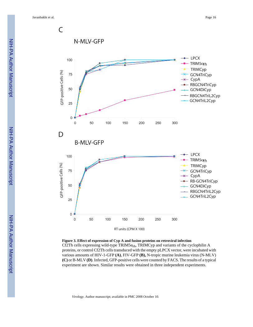
Figure 3. Effect of expression of Cyp A and fusion proteins on retroviral infectionCf2Th cells expressing wild-type TRIM5αrh, TRIMCyp and variants of the cyclophilin Aproteins, or control Cf2Th cells transduced with the empty pLPCX vector, were incubated withvarious amounts of HIV-1-GFP (A), FIV-GFP (B), N-tropic murine leukemia virus (N-MLV)(C) or B-MLV (D). Infected, GFP-positive cells were counted by FACS. The results of a typicalexperiment are shown. Similar results were obtained in three independent experiments.
Javanbakht et al. Page 16
Virology. Author manuscript; available in PMC 2008 October 10.
NIH
-PA Author Manuscript
NIH
-PA Author Manuscript
NIH
-PA Author Manuscript
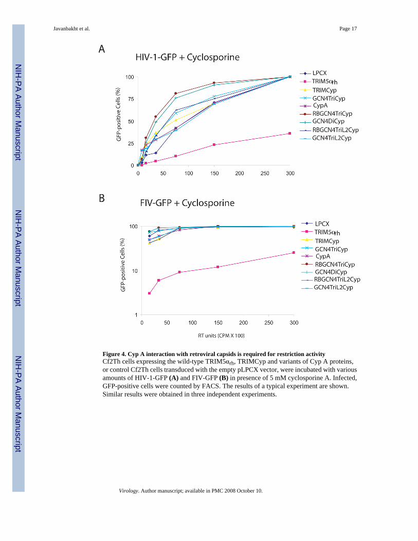
Figure 4. Cyp A interaction with retroviral capsids is required for restriction activityCf2Th cells expressing the wild-type TRIM5αrh, TRIMCyp and variants of Cyp A proteins,or control Cf2Th cells transduced with the empty pLPCX vector, were incubated with variousamounts of HIV-1-GFP (A) and FIV-GFP (B) in presence of 5 mM cyclosporine A. Infected,GFP-positive cells were counted by FACS. The results of a typical experiment are shown.Similar results were obtained in three independent experiments.
Javanbakht et al. Page 17
Virology. Author manuscript; available in PMC 2008 October 10.
NIH
-PA Author Manuscript
NIH
-PA Author Manuscript
NIH
-PA Author Manuscript

Figure 5. Specific association of TRIMCyp and Cyp A variants with HIV-1 CA-NC complexesIn vitro assembled CA-NC complexes (Ganser et al., 1999) were mixed with 293T lysatescontaining TRIMCyp, cyclophilin A or the indicated cyclophilin A variants and layered onto70% sucrose cushions before centrifugation. In some experiments (left panel), CA-NCcomplexes were not added, serving as a negative control. Immediately prior to mixing, analiquot of the cell lysate was removed and blotted with α-HA antibodies to determine thesteady-state expression levels of the different cyclophilin A variants (Input). Aftercentrifugation, the pellet was resuspended in SDS sample buffer and analyzed by Westernblotting using an anti-HA antibody (for Bound protein) or an anti-p24 antibody (to detect CA-NC).
Javanbakht et al. Page 18
Virology. Author manuscript; available in PMC 2008 October 10.
NIH
-PA Author Manuscript
NIH
-PA Author Manuscript
NIH
-PA Author Manuscript

Figure 6. Effects of disruption of oligomerization of the GCN4TriCyp proteinA. The oligomerization of the GCN4TriCyp and Mut GCN4TriCyp proteins was studied byEGS crosslinking and SDS-PAGE. Monomeric (m), dimeric (d), trimeric (t) and higher-order(h) forms of the proteins are indicated. B. Cell lysates containing the indicated proteins wereincubated with HIV-1 CA-NC complexes. The mixtures were pelleted through 70% sucrosecushions. The amount of each protein in the input lysate and the CA-NC-bound pellet is shown.The amount of HIV-1 CA-NC protein detected in the pellet is shown in the bottom row. C.Cf2Th cells expressing the indicated proteins or control LPCX-transduced cells were exposedto recombinant HIV-1-GFP. The percentage of GFP-positive cells at 72 hours after virus-cellincubation is shown. The experiment was repeated with comparable results.
Javanbakht et al. Page 19
Virology. Author manuscript; available in PMC 2008 October 10.
NIH
-PA Author Manuscript
NIH
-PA Author Manuscript
NIH
-PA Author Manuscript

Figure 7. Fate of the wild-type and G89A HIV-1 capsids in target cells expressing TRIMCyp andRBGCN4TriL2CypVSV G-pseudotyped, wild-type (wt) HIV-1-GFP and mutant HIV-1-GFP containing a G89Acapsid change were incubated with Cf2Th cells expressing TRIMCyp or RBGCN4TriL2Cyp,or control cells transduced with the empty LPCX vector. In parallel LPCX-transduced cellswere also exposed to these viruses lacking envelope glycoproteins (Env−). Sixteen hours later,the cells were lysed and the cell lysates were analyzed on 50% sucrose cushions, as described(Stremlau et al., 2006). The HIV-1 p24 capsid protein detected in the particulate fraction(Pellet) and supernatant (Sup.) is shown.
Javanbakht et al. Page 20
Virology. Author manuscript; available in PMC 2008 October 10.
NIH
-PA Author Manuscript
NIH
-PA Author Manuscript
NIH
-PA Author Manuscript
Copyright © 2022 FDOKUMEN