Validation of the SCID-hu Thy/Liv Mouse Model with Four Classes of Licensed Antiretrovirals
11
Validation of the SCID-hu Thy/Liv Mouse Model with Four Classes of Licensed Antiretrovirals Cheryl A. Stoddart 1¤ *, Cheryl A. Bales 1¤ , Jennifer C. Bare 1 , George Chkhenkeli 1¤ , Sofiya A. Galkina 1¤ , April N. Kinkade 1 , Mary E. Moreno 1¤ , Jose ´ M. Rivera 1¤ , Rollie E. Ronquillo 1¤ , Barbara Sloan 1¤ , Paul L. Black 2 1 Gladstone Institute of Virology and Immunology, University of California at San Francisco, San Francisco, California, United States of America, 2 Targeted Interventions Branch, Division of AIDS, National Institute of Allergy and Infectious Diseases, National Institutes of Health, Bethesda, Maryland, United States of America Background. The SCID-hu Thy/Liv mouse model of HIV-1 infection is a useful platform for the preclinical evaluation of antiviral efficacy in vivo. We performed this study to validate the model with representatives of all four classes of licensed antiretrovirals. Methodology/Principal Findings. Endpoint analyses for quantification of Thy/Liv implant viral load included ELISA for cell-associated p24, branched DNA assay for HIV-1 RNA, and detection of infected thymocytes by intracellular staining for Gag-p24. Antiviral protection from HIV-1-mediated thymocyte depletion was assessed by multicolor flow cytometric analysis of thymocyte subpopulations based on surface expression of CD3, CD4, and CD8. These mice can be productively infected with molecular clones of HIV-1 (e.g., the X4 clone NL4-3) as well as with primary R5 and R5X4 isolates. To determine whether results in this model are concordant with those found in humans, we performed direct comparisons of two drugs in the same class, each of which has known potency and dosing levels in humans. Here we show that second-generation antiretrovirals were, as expected, more potent than their first-generation predecessors: emtricitabine was more potent than lamivudine, efavirenz was more potent than nevirapine, and atazanavir was more potent than indinavir. After interspecies pharmacodynamic scaling, the dose ranges found to inhibit viral replication in the SCID-hu Thy/Liv mouse were similar to those used in humans. Moreover, HIV-1 replication in these mice was genetically stable; treatment of the mice with lamivudine did not result in the M184V substitution in reverse transcriptase, and the multidrug-resistant NY index case HIV-1 retained its drug- resistance substitutions. Conclusion. Given the fidelity of such comparisons, we conclude that this highly reproducible mouse model is likely to predict clinical antiviral efficacy in humans. Citation: Stoddart CA, Bales CA, Bare JC, Chkhenkeli G, Galkina SA, et al (2007) Validation of the SCID-hu Thy/Liv Mouse Model with Four Classes of Licensed Antiretrovirals. PLoS ONE 2(8): e655. doi:10.1371/journal.pone.0000655 INTRODUCTION The SCID-hu mouse model, in which human lymphoid organs are implanted into severe-combined immunodeficient (SCID) mice, was designed by McCune et al. [1] to provide a small animal model for the study of human hematopoiesis. It has also facilitated study of the pathogenesis of HIV-1 in human hematolymphoid organs [2–7] and evaluation of anti-HIV-1 compounds in vivo [2,8,9]. In this model, SCID mice are implanted with a variety of human fetal organs, including bone, liver, thymus, lymph node, and spleen. The fetal implants become tolerant of the mouse environment, and re- ciprocally, growth of the human tissue is permitted by the immunocompromised status of the recipient SCID mouse [6]. The SCID-hu Thy/Liv mouse, first reported by Namikawa et al. in 1990 [10], is generated by coimplanting human fetal thymus and liver beneath the mouse kidney capsule. In a highly reproducible manner, these organs fuse, become vascularized, and grow into a stable organ termed ‘‘Thy/Liv,’’ reaching a total mass of 100–300 6 10 6 human cells in 18 weeks. The Thy/Liv implant reproduces the differentiation, proliferation, and function of human hematopoietic progenitor cells derived from the fetal liver within the human thymus. The implants possess histologically normal cortical and medullary compartments that sustain multi- lineage human hematopoiesis for 6–12 months [10,11], generating a continuous source of CD4-expressing thymocytes that can serve as target cells for HIV-1 infection and replication. Importantly for a model of antiviral chemotherapy, 50–60 SCID-hu Thy/Liv mice can be made with tissues from a single fetal donor, and the Thy/ Liv implant is amenable to experimental manipulation and infection with HIV-1. The Thy/Liv implants support viral replication after inocula- tion of HIV-1 by direct injection [3], and thymocyte depletion occurs with both molecular clones and clinical isolates of HIV-1 in 3–5 weeks [12–16]. This depletion includes loss of CD4 + CD8 + (double-positive, DP) immature cortical thymocytes and a decrease in the CD4/CD8 ratio in the thymic medulla. There is evidence for both indirect, apoptotic destruction of uninfected thymocytes and direct infection and destruction of CD3 2 CD4 + CD8 2 intrathymic T-progenitor cells, which severely disrupts thymocyte maturation [17,18]. Infection of human thymus with HIV-1 induces IFN-a secretion from plasmacytoid dendritic cells, which upregulates MHC class I (MHC-I) expression on DP thymocytes, where expression is normally low [19]. The administration of nucleoside and nonnucleoside reverse transcriptase (RT) inhibitors to these mice results in dose-dependent Academic Editor: Linqi Zhang, AIDS Research Center, Chinese Academy of Medical Sciences and Peking Union Medical College, China Received April 18, 2007; Accepted June 20, 2007; Published August 1, 2007 Copyright: ß 2007 Stoddart et al. This is an open-access article distributed under the terms of the Creative Commons Attribution License, which permits unrestricted use, distribution, and reproduction in any medium, provided the original author and source are credited. Funding: This project has been funded in whole or in part with Federal funds from NIAID, NIH, under contract no. N01-AI-05418. Competing Interests: The authors have declared that no competing interests exist. * To whom correspondence should be addressed. E-mail: cheryl.stoddart@ucsf. edu ¤ Current address: Division of Experimental Medicine, Department of Medicine, University of California at San Francisco, San Francisco, California, United States of America PLoS ONE | www.plosone.org 1 August 2007 | Issue 8 | e655
Transcript of Validation of the SCID-hu Thy/Liv Mouse Model with Four Classes of Licensed Antiretrovirals
Validation of the SCID-hu Thy/Liv Mouse Model withFour Classes of Licensed AntiretroviralsCheryl A. Stoddart1¤*, Cheryl A. Bales1¤, Jennifer C. Bare1, George Chkhenkeli1¤, Sofiya A. Galkina1¤, April N. Kinkade1, Mary E. Moreno1¤, Jose M.Rivera1¤, Rollie E. Ronquillo1¤, Barbara Sloan1¤, Paul L. Black2
1 Gladstone Institute of Virology and Immunology, University of California at San Francisco, San Francisco, California, United States of America,2 Targeted Interventions Branch, Division of AIDS, National Institute of Allergy and Infectious Diseases, National Institutes of Health, Bethesda,Maryland, United States of America
Background. The SCID-hu Thy/Liv mouse model of HIV-1 infection is a useful platform for the preclinical evaluation of antiviralefficacy in vivo. We performed this study to validate the model with representatives of all four classes of licensedantiretrovirals. Methodology/Principal Findings. Endpoint analyses for quantification of Thy/Liv implant viral load includedELISA for cell-associated p24, branched DNA assay for HIV-1 RNA, and detection of infected thymocytes by intracellularstaining for Gag-p24. Antiviral protection from HIV-1-mediated thymocyte depletion was assessed by multicolor flowcytometric analysis of thymocyte subpopulations based on surface expression of CD3, CD4, and CD8. These mice can beproductively infected with molecular clones of HIV-1 (e.g., the X4 clone NL4-3) as well as with primary R5 and R5X4 isolates. Todetermine whether results in this model are concordant with those found in humans, we performed direct comparisons of twodrugs in the same class, each of which has known potency and dosing levels in humans. Here we show that second-generationantiretrovirals were, as expected, more potent than their first-generation predecessors: emtricitabine was more potent thanlamivudine, efavirenz was more potent than nevirapine, and atazanavir was more potent than indinavir. After interspeciespharmacodynamic scaling, the dose ranges found to inhibit viral replication in the SCID-hu Thy/Liv mouse were similar to thoseused in humans. Moreover, HIV-1 replication in these mice was genetically stable; treatment of the mice with lamivudine didnot result in the M184V substitution in reverse transcriptase, and the multidrug-resistant NY index case HIV-1 retained its drug-resistance substitutions. Conclusion. Given the fidelity of such comparisons, we conclude that this highly reproducible mousemodel is likely to predict clinical antiviral efficacy in humans.
Citation: Stoddart CA, Bales CA, Bare JC, Chkhenkeli G, Galkina SA, et al (2007) Validation of the SCID-hu Thy/Liv Mouse Model with Four Classes ofLicensed Antiretrovirals. PLoS ONE 2(8): e655. doi:10.1371/journal.pone.0000655
INTRODUCTIONThe SCID-hu mouse model, in which human lymphoid organs are
implanted into severe-combined immunodeficient (SCID) mice, was
designed by McCune et al. [1] to provide a small animal model for
the study of human hematopoiesis. It has also facilitated study of the
pathogenesis of HIV-1 in human hematolymphoid organs [2–7] and
evaluation of anti-HIV-1 compounds in vivo [2,8,9]. In this model,
SCID mice are implanted with a variety of human fetal organs,
including bone, liver, thymus, lymph node, and spleen. The fetal
implants become tolerant of the mouse environment, and re-
ciprocally, growth of the human tissue is permitted by the
immunocompromised status of the recipient SCID mouse [6].
The SCID-hu Thy/Liv mouse, first reported by Namikawa et
al. in 1990 [10], is generated by coimplanting human fetal thymus
and liver beneath the mouse kidney capsule. In a highly
reproducible manner, these organs fuse, become vascularized,
and grow into a stable organ termed ‘‘Thy/Liv,’’ reaching a total
mass of 100–3006106 human cells in 18 weeks. The Thy/Liv
implant reproduces the differentiation, proliferation, and function
of human hematopoietic progenitor cells derived from the fetal
liver within the human thymus. The implants possess histologically
normal cortical and medullary compartments that sustain multi-
lineage human hematopoiesis for 6–12 months [10,11], generating
a continuous source of CD4-expressing thymocytes that can serve
as target cells for HIV-1 infection and replication. Importantly for
a model of antiviral chemotherapy, 50–60 SCID-hu Thy/Liv mice
can be made with tissues from a single fetal donor, and the Thy/
Liv implant is amenable to experimental manipulation and
infection with HIV-1.
The Thy/Liv implants support viral replication after inocula-
tion of HIV-1 by direct injection [3], and thymocyte depletion
occurs with both molecular clones and clinical isolates of HIV-1 in
3–5 weeks [12–16]. This depletion includes loss of CD4+CD8+
(double-positive, DP) immature cortical thymocytes and a decrease
in the CD4/CD8 ratio in the thymic medulla. There is evidence
for both indirect, apoptotic destruction of uninfected thymocytes
and direct infection and destruction of CD32CD4+CD82
intrathymic T-progenitor cells, which severely disrupts thymocyte
maturation [17,18]. Infection of human thymus with HIV-1
induces IFN-a secretion from plasmacytoid dendritic cells, which
upregulates MHC class I (MHC-I) expression on DP thymocytes,
where expression is normally low [19].
The administration of nucleoside and nonnucleoside reverse
transcriptase (RT) inhibitors to these mice results in dose-dependent
Academic Editor: Linqi Zhang, AIDS Research Center, Chinese Academy ofMedical Sciences and Peking Union Medical College, China
Received April 18, 2007; Accepted June 20, 2007; Published August 1, 2007
Copyright: � 2007 Stoddart et al. This is an open-access article distributedunder the terms of the Creative Commons Attribution License, which permitsunrestricted use, distribution, and reproduction in any medium, provided theoriginal author and source are credited.
Funding: This project has been funded in whole or in part with Federal fundsfrom NIAID, NIH, under contract no. N01-AI-05418.
Competing Interests: The authors have declared that no competing interestsexist.
* To whom correspondence should be addressed. E-mail: [email protected]
¤ Current address: Division of Experimental Medicine, Department of Medicine,University of California at San Francisco, San Francisco, California, United States ofAmerica
PLoS ONE | www.plosone.org 1 August 2007 | Issue 8 | e655
inhibition of HIV-1 replication, prevention of IFN-a-mediated
MHC-I upregulation, and protection of CD4+ cells within the
implanted human tissue [8,9,20]. The model has been also used to
evaluate new classes of HIV-1 inhibitors, such as bicyclam [21] and
oligonucleotide [22] inhibitors of HIV-1 entry, the nucleoside analog
29-deoxy-39-oxa-49-thiocytidine (dOTC) [20], and an oxime-piper-
idine CCR5 antagonist [23]. This is the first animal model in which
the action of candidate anti-HIV-1 compounds can be tested within
the setting of an intact HIV-1-infected human target organ.
We use a standardized protocol described by Rabin et al. [8] for
evaluation of antiviral agents against HIV-1 in the SCID-hu Thy/
Liv mouse model. Drugs can be administered by various routes
(oral, subcutaneous, intraperitoneal, or by mini-osmotic pump)
before or after virus inoculation and continued for 2–6 weeks.
Mice are challenged with titrated inocula of various HIV-1
isolates, and endpoint analyses include quantification of viral
protein by p24 ELISA and viral RNA by branched DNA assay,
quantification and isolation of replication-competent virus by
cocultivation after limiting dilution, and assessment of the effects
on human thymocyte subpopulations by flow cytometry [8].
The SCID-hu Thy/Liv model has also been employed by us and
by others for in vivo studies of HIV-1 pathogenesis. The roles of HIV-
1 nef [24–26], other accessory genes [24], and the Rev–Rev
responsive element [27] have been explored as well as the expression
of HIV-1 coreceptors (CXCR4 and CCR5) on thymocytes [28,29]
and the effects of coreceptor usage on viral tropism and pathogenesis
[16,28,30–32]. The CXCR4-utilizing (X4) strain NL4-3 replicates
quickly and extensively in thymocytes in the cortex and medulla,
causing significant thymocyte depletion [16]. In contrast, the CCR5-
utilizing (R5) strain Ba-L initially infects stromal cells, including
macrophages, in the thymic medulla without any obvious pathologic
consequences for 3–4 weeks, after which the infection slowly spreads
through the thymocyte populations and results in slowly progressing
thymocyte depletion after 6 weeks. In addition, three R5 viruses
isolated from rapid AIDS progressors lacking X4 strains were much
less pathogenic than NL4-3 in SCID-hu Thy/Liv mice, suggesting
that R5 virus-mediated rapid disease progression is associated with
host, not viral, factors [30]. The lab-adapted LAI/IIIB isolate and its
associated infectious molecular clones (e.g., HXB2) fail to replicate in
Thy/Liv implants, and this impairment has been mapped to
a specific amino acid change in the crown region of V3 in Env
[33,34]. The model has also been used to study immune
reconstitution and recovery of hematopoietic colony-forming activity
in HIV-1-infected implants after treatment of mice with potent
combination antiretroviral therapy [35–37].
Given the potential correlations between virus-host interactions
in the Thy/Liv implant and in humans, we wanted to determine
whether the SCID-hu Thy/Liv mouse model predicts antiretro-
viral efficacy in humans for representatives of all four classes of
currently licensed drugs. Does it, for instance, yield data that
reflect the greater potency of more recently developed drugs than
their first-generation predecessors in each class? In vivo models are
far more stringent than cell culture-based assays for demonstrating
drug activity, particularly in the case of orally delivered drugs as
they must be absorbed by the gastrointestinal tract without
degradation, enter the blood and circulate with sufficient half-lives,
and penetrate solid target organs at concentrations high enough to
be potently efficacious. The greater potency of the second-
generation drugs is likely the result of a combination of greater in
vitro potency and superior pharmacokinetics. In vitro virus-cell
culture systems would likely predict greater potency, but only in
vivo studies can demonstrate sustained plasma half-life or superior
oral bioavailability. To determine such relative potency in vivo,
head-to-head comparisons of two drugs in the same class were
performed in SCID-hu Thy/Liv mouse cohorts each made with
tissues from a single donor. We observed that the more recent
drugs in each class were significantly more potent than their
predecessors. We conclude from such comparisons with licensed
antiretrovirals that this well-established model is likely to predict
clinical antiviral efficacy in humans for other members of these
classes and possibly other drug classes as well.
MATERIALS AND METHODS
VirusesThe following reagents were obtained through the AIDS Research
and Reference Reagent Program, Division of AIDS, NIAID, NIH:
pNL4-3 [38] from Malcolm Martin and HIV-1 Ba-L [39] from
Suzanne Gartner, Mikulas Popovic, and Robert Gallo. A working
stock of NL4-3 was prepared in 293T cells by calcium phosphate
transfection. A T-20-sensitive NL4-3 (NL4-3 D36G) was altered
by site-directed mutagenesis to match the consensus sequence at
amino acid position 36 (aspartic acid replaced by glycine) [40,41].
The R5X4 AZT-resistant clinical HIV-1 isolate JD [42] was
kindly provided by Mike McCune, and the multidrug-resistant
(MDR) R5X4 NY index case isolate [43] by Hiroshi Morhi and
Martin Markowitz. The clinical isolates were prepared and titrated
by limiting dilution in phytohemagglutin-activated peripheral
blood mononuclear cells.
Antiretroviral drugs and mouse dosingThe antiretroviral drugs lamivudine (3TC), emtricitabine [(–)-
FTC], nevirapine, efavirenz, indinavir, and atazanavir were kindly
supplied by Opendra Sharma and the AIDS Reagent Program.
Dosing solutions were prepared in sterile water for injection or
Dulbecco’s phosphate-buffered saline, and groups of 5–8 mice
were treated by oral gavage (200 ml per dose) with an 18-gauge62-
inch feeding needle twice daily at 7:00 AM and 6:00 PM for the
duration of the infection period (2–6 weeks depending on the
HIV-1 isolate). Enfuvirtide (T-20) was purchased from a pharmacy,
prepared in sterile water, and administered by twice-daily
subcutaneous injection (200 ml per dose).
Implantation and inoculation of SCID-hu Thy/Liv
miceHuman fetal thymus and liver were obtained through services
provided by a nonprofit organization (Advanced Bioscience
Resources, Alameda, CA) in accordance with federal, state, and
local regulations. Coimplantation of thymus and liver fragments
under the kidney capsule to create SCID-hu Thy/Liv mice and
inoculation of the Thy/Liv implants with HIV-1 was carried out
as described [8,10]. Male C.B-17 SCID (model #CB17SC-M,
homozygous, C.B-Igh-1b/IcrTac-Prkdcscid) mice were obtained at
6–8 weeks of age from Taconic (Germantown, NY), and cohorts
of 50–60 SCID-hu Thy/Liv mice were implanted with tissues
from a single donor. Implants were inoculated 18 weeks after
implantation with 50 ml of stock virus (500–2,000 TCID50) or
RPMI 1640 medium (mock infection) by direct injection after
surgical exposure of the implanted kidney of anesthetized mice.
Animal protocols were approved by the UCSF Institutional
Animal Care and Use Committee.
Implant collection and viral load quantificationThe Thy/Liv implants were collected from euthanized mice, and
single-cell suspensions were prepared by dispersing the implant
through nylon mesh and processed for p24 ELISA, bDNA assay,
and FACS analysis as described [8,20]. Unless specified otherwise,
Validation of SCID-hu Model
PLoS ONE | www.plosone.org 2 August 2007 | Issue 8 | e655
implants were collected 21 days after inoculation with NL4-3,
14 days after JD inoculation, and 42 days after Ba-L inoculation.
Flow cytometryImplant cells were stained with phycoerythrin cyanine dye CY7-
conjugated anti-CD4 (BD Biosciences, San Jose, CA), phycoerythrin
cyanine CY5.5-conjugated anti-CD8 (Caltag Laboratories, Burlin-
game, CA), allophycocyanin cyanine CY7-conjugated anti-CD3
(eBiosciences, San Diego, CA), and phycoerythrin-conjugated anti-
W6/32 (DakoCytomation, Glostrup, Denmark. Cells were fixed and
permeabilized with 1.2% paraformaldehyde and 0.5% Tween 20,
stained with fluorescein isothiocyanate-conjugated anti-p24 (Beck-
man Coulter, Fullerton, CA), and analyzed on an LSR II (BD
Biosciences). After collecting 100,000 total cell events, percentages of
marker-positive (CD4+, CD8+, and DP) thymocytes in the implant
samples were determined by first gating on a live lymphoid cell
population identified by forward- and side-scatter characteristics and
then by CD3 expression. W6/32-positive mean fluorescence
intensity (MFI) of DP thymocytes was determined, and CD4/CD8
ratios were calculated by dividing the percentage of CD4+ cells by
the percentage of CD8+ cells for each individual implant.
Statistical analysisResults are expressed as the mean6SEM for each mouse group.
Nonparametric statistical analyses were performed by use of the
Mann-Whitney U test. Data for mice in each group were
compared to those for untreated infected mice, and p values
#0.05 were considered statistically significant.
RESULTS
Viral load and prophylactic antiviral efficacy are
highly reproducible among mouse cohorts each
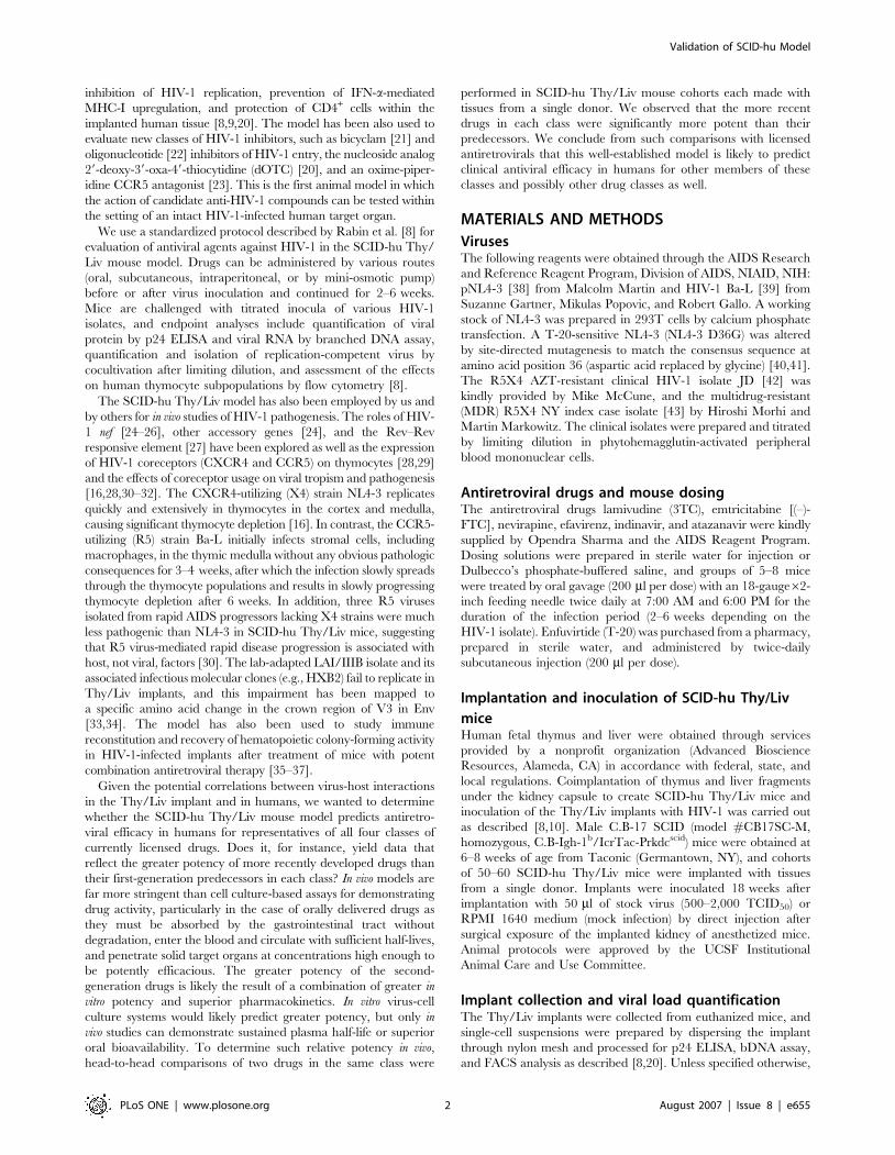
made with tissues from a single donorThe levels of HIV-1 RNA and p24 in the Thy/Liv implants of
untreated- and 3TC-treated SCID-hu Thy/Liv mice were highly
reproducible among seven different mouse cohorts infected with
the same stock of HIV-1 NL4-3 (Figure 1). In seven consecutive
experiments, with mice in a given cohort constructed from human
fetal tissue from a single donor, mean cell-associated HIV-1 RNA
ranged from 5.2–6.3 log10 copies per 106 cells (Figure 1A), and
mean p24 ranged from 640–1,700 pg per 106 cells (Figure 1B).
There was also good correspondence between viral RNA and p24
Figure 1. Implant viral loads in untreated- and 3TC-treated SCID-hu Thy/Liv mice are highly reproducible, and 3TC treatment after HIV-1inoculation is nearly as effective as prophylactic treatment. A, HIV-1 RNA in Thy/Liv implants of untreated mice and mice treated by twice-daily oralgavage with 3TC at 30 mg/kg per day beginning 1 day before inoculation with NL4-3 (means6SEM) Each experiment was performed in a mousecohort made with tissues from a single donor. B, Implant p24 for the same groups as in panel A. C, Implant HIV-1 RNA in mice treated with 3TCbeginning on day 21, day +1, day +3, and day +7 with respect to inoculation with the indicated HIV-1 strains. D, Implant p24 for the same groups asin panel C. Implants were collected 21 days after inoculation for NL4-3, 14 days for JD, and 42 days for Ba-L. *p#0.05, compared with untreated micefor 5–7 mice per group.doi:10.1371/journal.pone.0000655.g001
Validation of SCID-hu Model
PLoS ONE | www.plosone.org 3 August 2007 | Issue 8 | e655
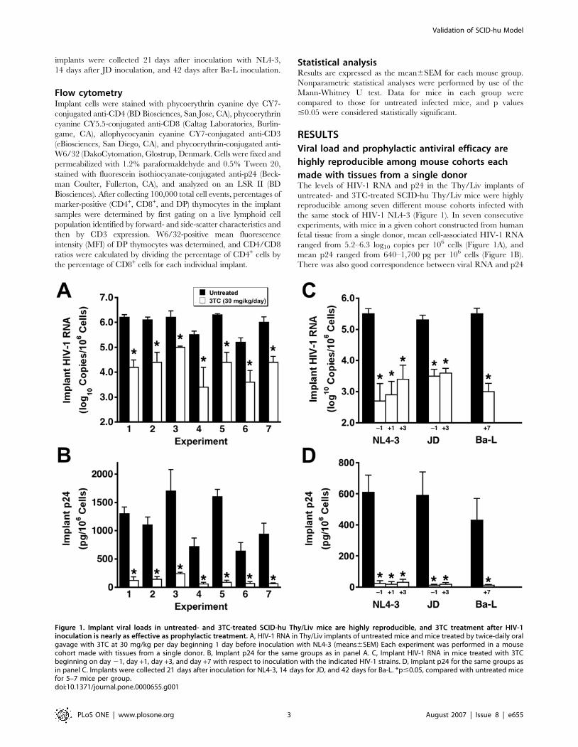
Figure 2. 3TC treatment of HIV-1 JD-infected SCID-hu Thy/Liv mice inhibits viral replication and protects implants from virus-mediatedthymocyte depletion. Mice were treated by twice-daily oral gavage with 3TC at 300 mg/kg per day beginning on day +1 after inoculation, and Thy/Liv implants were collected 14, 21, and 28 days after inoculation. Antiviral efficacy was assessed by determining HIV-1 RNA (A), p24 (B), Gag-p24+
thymocytes (C), and MHC-I expression on DP thymocytes (D). Thymocyte protection was assessed by total implant cellularity (E), thymocyte viability(F), percentage of DP thymocytes (G), and CD4/CD8 ratio (H) for 3TC-treated mice versus untreated mice (means6SEM). *p#0.05, compared withuntreated mice for 5–7 mice per group.doi:10.1371/journal.pone.0000655.g002
Validation of SCID-hu Model
PLoS ONE | www.plosone.org 4 August 2007 | Issue 8 | e655
means of implants in the same groups (i.e., lowest for untreated
mice in experiment 6 and highest for those in experiments 3 and
5). In the seven experiments, 3TC treatment beginning 1 day
before virus inoculation reduced HIV-1 RNA in the implants by
1.2–2.1 log10 (94%–99%) (Figure 1A) and p24 by 77%–92%
(Figure 1B) compared to untreated mice in the same experiment.
Importantly, HIV-1 NL4-3 replication in these mice was
genetically stable over the 21-day infection period; there was no
evidence of the RT M184V substitution in viral RNA amplified by
RT-PCR and sequenced from 32 of 32 Thy/Liv implants
collected from mice treated with 3TC in these seven experiments
(data not shown). The lack of resistance development is most likely
the result of the limited time before virus-mediated thymocyte
depletion becomes severe enough to limit the number of target
cells available for infection. We view the genetic stability of the
viruses over several weeks as an asset because our results are not
confounded by the rapid development of antiviral resistance,
particularly against drugs for which only one amino acid
substitution is sufficient for high-level resistance.
Postexposure dosing also reduces viral load and
prevents virus-mediated thymocyte depletionInitiation of 3TC treatment 1 or 3 days after NL4-3 inoculation
was nearly as effective at reducing implant viral RNA (Figure 1C)
and p24 (Figure 1D) as was prophylactic treatment with treatment
beginning 1 day before inoculation. This was also true for mice
inoculated with the clinical R5X4 isolate JD and treated with 3TC
beginning 3 days after inoculation. Treatment of mice inoculated
with the R5 isolate Ba-L, which replicates in Thy/Liv implants
with slower kinetics than does X4 HIV-1 NL4-3 [16], was highly
effective at reducing implant viral RNA and p24 even when
initiation was delayed until 7 days after inoculation.
To determine whether postexposure dosing also prevents
thymocyte depletion, SCID-hu mice were inoculated with HIV-
1 JD and treated twice daily with 3TC at 300 mg/kg per day in
a time-course study in which implants were collected at weekly
intervals up to 28 days after inoculation. By that point, the
implants from untreated mice had become severely depleted of
thymocytes. As expected, 3TC-treated mice had significant
reductions in viral RNA (Figure 2A), p24 (Figure 2B), Gag-p24+
thymocytes (Figure 2C), and IFN-a-induced MHC-I expression on
DP thymocytes (Figure 2D), compared to untreated mice at all
three times. Reductions in viral load by 3TC were accompanied
by virtually complete protection of the Thy/Liv implants from
thymocyte depletion, in terms of total cellularity (Figure 2E),
thymocyte viability (Figure 2F), percentage of DP thymocytes
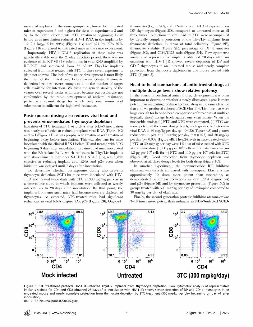
(Figure 2G), and CD4/CD8 ratio (Figure 2H). Flow cytometric
analysis of representative implants obtained 28 days after in-
oculation with HIV-1 JD showed severe depletion of DP and
CD4+ thymocytes in an untreated mouse and nearly complete
protection from thymocyte depletion in one mouse treated with
3TC (Figure 3).
Head-to-head comparisons of antiretroviral drugs at
multiple dosage levels show relative potencyIn the course of preclinical antiviral drug development, it is often
important to determine whether a newly discovered agent is more
potent than an existing, perhaps licensed, drug in the same class. To
that end, we produced cohorts of SCID-hu Thy/Liv mice that were
large enough for head-to-head comparisons of two drugs at multiple
(typically three) dosage levels against one virus isolate. When the
nucleoside analogs (–)-FTC and 3TC were compared, (–)-FTC was
more potent at the same dosage levels, with greater reductions in
viral RNA at 30 mg/kg per day (p = 0.035) (Figure 4A) and greater
reductions in p24 at 10 mg/kg per day (p = 0.025) and 30 mg/kg
per day (p = 0.009) (Figure 4B). The p24 levels in mice treated with (–
)-FTC at 30 mg/kg per day were 1% that of mice treated with 3TC
at the same dose (1,300 pg per 106 cells in untreated mice versus
1.2 pg per 106 cells for (–)-FTC and 110 pg per 106 cells for 3TC)
(Figure 4B). Good protection from thymocyte depletion was
observed at all three dosage levels for both drugs (Figure 4C).
In another experiment, the nonnucleoside RT inhibitor
efavirenz was directly compared with nevirapine. Efavirenz was
approximately 10 times more potent than nevirapine, as
demonstrated by similar reductions in viral RNA (Figure 5A)
and p24 (Figure 5B) and by thymocyte protection (Figure 5C) in
groups treated with 300 mg/kg per day of nevirapine compared to
30 mg/kg per day of efavirenz.
Finally, the second-generation protease inhibitor atazanavir was
3–10 times more potent than indinavir in NL4-3-infected SCID-
Figure 3. 3TC treatment protects HIV-1 JD-infected Thy/Liv implants from thymocyte depletion. Flow cytometric analysis of representativeimplants stained for CD4 and CD8 obtained 28 days after inoculation with HIV-1 JD shows severe depletion of DP and CD4+ thymocytes in anuntreated mouse and nearly complete protection from thymocyte depletion by 3TC treatment (300 mg/kg per day beginning on day +1 afterinoculation).doi:10.1371/journal.pone.0000655.g003
Validation of SCID-hu Model
PLoS ONE | www.plosone.org 5 August 2007 | Issue 8 | e655
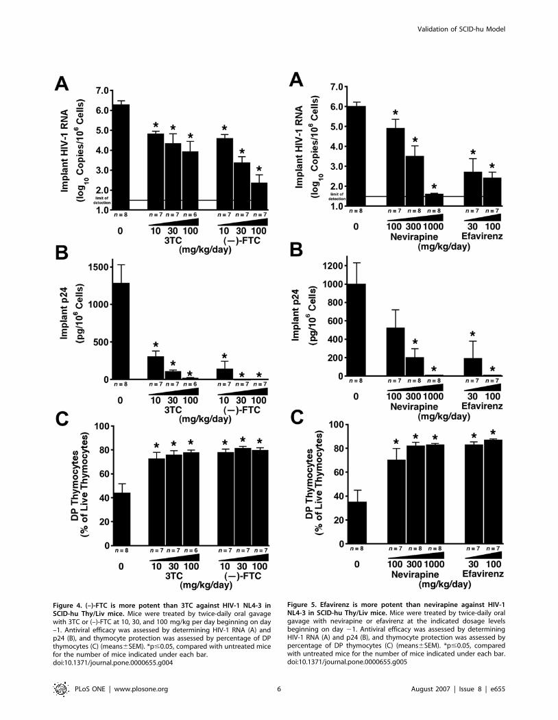
Figure 4. (–)-FTC is more potent than 3TC against HIV-1 NL4-3 inSCID-hu Thy/Liv mice. Mice were treated by twice-daily oral gavagewith 3TC or (–)-FTC at 10, 30, and 100 mg/kg per day beginning on day–1. Antiviral efficacy was assessed by determining HIV-1 RNA (A) andp24 (B), and thymocyte protection was assessed by percentage of DPthymocytes (C) (means6SEM). *p#0.05, compared with untreated micefor the number of mice indicated under each bar.doi:10.1371/journal.pone.0000655.g004
Figure 5. Efavirenz is more potent than nevirapine against HIV-1NL4-3 in SCID-hu Thy/Liv mice. Mice were treated by twice-daily oralgavage with nevirapine or efavirenz at the indicated dosage levelsbeginning on day 21. Antiviral efficacy was assessed by determiningHIV-1 RNA (A) and p24 (B), and thymocyte protection was assessed bypercentage of DP thymocytes (C) (means6SEM). *p#0.05, comparedwith untreated mice for the number of mice indicated under each bar.doi:10.1371/journal.pone.0000655.g005
Validation of SCID-hu Model
PLoS ONE | www.plosone.org 6 August 2007 | Issue 8 | e655
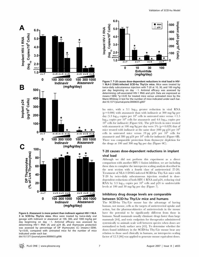
hu mice, with a 3.1 log10 greater reduction in viral RNA
(p = 0.006) with atazanavir than with indinavir at 300 mg/kg per
day (5.3 log10 copies per 106 cells in untreated mice versus ,1.5
log10 copies per 106 cells for atazanavir and 4.6 log10 copies per
106 cells for indinavir) (Figure 6A). The p24 levels in mice treated
with atazanavir at 100 mg/kg per day were 5% (p = 0.029) that of
mice treated with indinavir at the same dose (400 pg p24 per 106
cells in untreated mice versus 19 pg p24 per 106 cells for
atazanavir and 380 pg p24 per 106 cells for indinavir) (Figure 6B).
There was comparable protection from thymocyte depletion for
the drugs at 100 and 300 mg/kg per day (Figure 6C).
T-20 causes dose-dependent reductions in implant
viral loadAlthough we did not perform this experiment as a direct
comparison with another HIV-1 fusion inhibitor, we are including
these data to complete the interspecies scaling analysis described in
the next section with a fourth class of antiretroviral (T-20).
Treatment of NL4-3 D36G-infected SCID-hu Thy/Liv mice with
T-20 by twice-daily subcutaneous injection resulted in dose-
dependent reductions of both HIV-1 RNA and p24, reducing viral
RNA by 3.3 log10 copies per 106 cells and p24 to undetectable
levels at 100 and 30 mg/kg per day (Figure 7).
Inhibitory drug dosage levels are comparable
between SCID-hu Thy/Liv mice and humansThe SCID-hu Thy/Liv mouse has the advantage of having
human, not mouse, cells as the targets of antiretroviral uptake and
action, but the pharmacokinetics of antiretrovirals in the mouse
have the potential to be significantly different from those in
humans. Small mammals usually eliminate drugs faster than large
mammals [44], and toxic endpoints for therapeutics administered
systemically to animals scale well between species when doses are
normalized to body surface area [45]. To determine whether the
doses found inhibitory in the SCID-hu Thy/Liv mouse bear any
relation to those used clinically in humans, an interspecies scaling
factor of 12.3 [46] was applied to generate mouse equivalent doses.
Figure 6. Atazanavir is more potent than indinavir against HIV-1 NL4-3 in SCID-hu Thy/Liv mice. Mice were treated by twice-daily oralgavage with indinavir or atazanavir at 100, 300, and 1000 mg/kg perday beginning on day 21. Antiviral efficacy was assessed bydetermining HIV-1 RNA (A) and p24 (B), and thymocyte protectionwas assessed by percentage of DP thymocytes (C) (means6SEM).*p#0.05, compared with untreated mice for the number of miceindicated under each bar.doi:10.1371/journal.pone.0000655.g006
Figure 7. T-20 causes dose-dependent reductions in viral load in HIV-1 NL4-3 D36G-infected SCID-hu Thy/Liv mice. Mice were treated bytwice-daily subcutaneous injection with T-20 at 10, 30, and 100 mg/kgper day beginning on day 21. Antiviral efficacy was assessed bydetermining cell-associated HIV-1 RNA and p24. Data are expressed asmeans6SEM; *p#0.05 for treated mice versus untreated mice by theMann-Whitney U test for the number of mice indicated under each bar.doi:10.1371/journal.pone.0000655.g007
Validation of SCID-hu Model
PLoS ONE | www.plosone.org 7 August 2007 | Issue 8 | e655
This factor reflects the 12.3-fold difference in surface area-to-body
weight ratio between mice (0.0066 m2/0.02 kg) and humans
(1.6 m2/60 kg); consequently, 12.3 times more drug is required in
the mouse to be comparable to the dose in humans. When
currently approved dosage levels for humans of each antiretroviral
drug are adjusted for this difference, the adjusted dosage levels
were similar to those that cause 0.9–3.7 log10 reductions in viral
RNA in the SCID-hu Thy/Liv mouse (Table 1). Thus, not only is
the model predictive of relative potency within these drug classes,
it might also be useful in determining approximate dosing ranges
for effective use in humans.
Multidrug-resistant NY index case HIV-1 replicates
and depletes thymocytes with kinetics comparable
to HIV-1 NL4-3A key test of any new class of antiretroviral drug is a demonstration
of potent activity against HIV-1 with known drug-resistance
mutations. We reported the activity of the 3TC analog dOTC
against 3TC-resistant HIV-1 NL4-3/M184V in the SCID-hu
Thy/Liv model [20]. Here we report that the MDR NY index
case HIV-1, which was isolated from a patient with highly rapid
progression to AIDS [43], replicates with kinetics similar to HIV-1
NL4-3 in SCID-hu Thy/Liv mice (Figure 8A–D) resulting in
severe thymocyte depletion by 28 days after inoculation
(Figure 8E–H). The lower viral loads for the NY index case
isolate compared to NL4-3 at day 28 are most likely the result of
somewhat faster thymocyte depletion and thus greater loss of
target cells for viral replication.
Additional evidence that HIV-1 replication is genetically stable
within the 28-day infection period was provided by sequence
analysis of implant viral RNA showing that the MDR NY index
case isolate [43] retained the full complement of drug-resistance
amino acid substitutions of the original inoculum (data not shown).
These data show that this MDR clinical isolate can readily be used
as a challenge virus in the SCID-hu Thy/Liv model for in vivo
studies of new classes of antiretroviral drugs with activity against
HIV-1 that is resistant to currently licensed antiretrovirals.
DISCUSSIONThe SCID-hu Thy/Liv mouse is an efficient model for the study of
HIV-1 infection and for the preclinical evaluation of potential
antiretrovirals. Here we show that this model provides a validated
platform for reproducible viral infectivity and drug-mediated
reductions in viral load in vivo. We also show that the model is
especially valuable for direct, head-to-head comparisons of the
potency and effective dose ranges of existing antiretroviral drugs.
In consecutive experiments performed in different SCID-hu
mouse cohorts infected with the same stock of HIV-1 NL4-3, we
observed a range in mean HIV-1 RNA of only 1.1 log10 copies per
106 cells. Reproducible reductions in viral load were also observed
after oral administration of 3TC. Although our standardized
protocol calls for prophylactic treatment 1 day before virus
inoculation to maximize the potential for discerning antiviral
efficacy of new drugs in initial in vivo tests, we show here that
postexposure drug initiation (beginning 1, 3, or 7 days after HIV-1
inoculation) also renders potent antiviral efficacy. Importantly,
potent therapeutic efficacy, in terms of both inhibition of viral
replication and virtually complete protection from virus-mediated
cytopathicity and thymocyte depletion, was also demonstrated in
3TC-treated mice infected with the highly cytopathic R5X4
isolate, JD. In addition, we have minimized one potential
experimental variable by inoculating the mice at a uniform time
point 18 weeks after implantation.
The goal of this study was to validate the SCID-hu Thy/Liv
model for use in drug discovery efforts by determining the efficacy
of different classes of FDA-approved antiretroviral drugs. Antiviral
efficacy in this model is demonstrated by dose-dependent
reductions in viral load as well as dose-dependent protection of
CD4+ T cells from virus-mediated depletion. Assessment of
thymocyte percentage and viability is particularly important for
proving that the observed antiviral activity is not merely the result
of cytotoxicity to the Thy/Liv implant just as the selectivity index
(ratio of cytotoxic to inhibitory concentration) is important for
assessing antiviral activity in vitro.
As new antiretroviral drugs in existing drug classes are
developed to improve upon antiviral potency or to limit in vivo
toxicity, direct comparisons of the existing drug and the new drug
in single SCID-hu mouse cohorts represent a powerful preclinical
tool for the prediction of improved efficacy in human patients. To
accomplish this, we produced cohorts of SCID-hu Thy/Liv mice
that were large enough for comparison of two drugs at three
dosage levels against one virus isolate. For studies of viral
pathogenesis and hematopoietic reconstitution, a relatively small
number of animals can generate sufficient replicate points for
analysis. In contrast, preclinical antiviral evaluations require
groups of animals sufficiently large to demonstrate statistical proof
of efficacy in vivo. Two-drug, three-dose comparisons are made
possible by our unique ability to construct large (50–60-mouse)
SCID-hu cohorts with tissues from a single donor, which permits
Table 1. Antiretroviral drug dosage levels adjusted for difference in surface area-to-body weight ratio between mice and humans. . . . . . . . . . . . . . . . . . . . . . . . . . . . . . . . . . . . . . . . . . . . . . . . . . . . . . . . . . . . . . . . . . . . . . . . . . . . . . . . . . . . . . . . . . . . . . . . . . . . . . . . . . . . . . . . . . . . . . . . . . . . . . . . . . . . . . . . . . . . . . . . . .
Drug Human dosage regimenaHuman dosageb
(mg/kg per day)Mouse equivalent dosagec
(mg/kg per day)Calculated reduction in implantHIV-1 RNAd (log10 copies)
3TC 150 mg twice daily 5.0 62 1.9
(–)-FTC 200 mg once daily 3.3 41 2.3
nevirapine 400 mg once daily 6.7 82 0.9
efavirenz 600 mg once daily 10 123 3.7
indinavir 800 mg three times daily 40 490 1.2
atazanavir 400 mg once daily 6.7 82 1.4
T-20 90 mg twice daily 3.0 37 3.2
aFrom ref. [56].bBased on 60 kg adult human weight and 20 g mouse weight.cHuman dosage612.3 (fold difference in surface area to body weight between mice and humans; from ref. [45,46].dCalculated by linear regression of the viral RNA data shown in Figures 3–6 at the equivalent mouse dosage level.doi:10.1371/journal.pone.0000655.t001....
....
....
....
....
....
....
....
....
....
....
....
....
...
Validation of SCID-hu Model
PLoS ONE | www.plosone.org 8 August 2007 | Issue 8 | e655
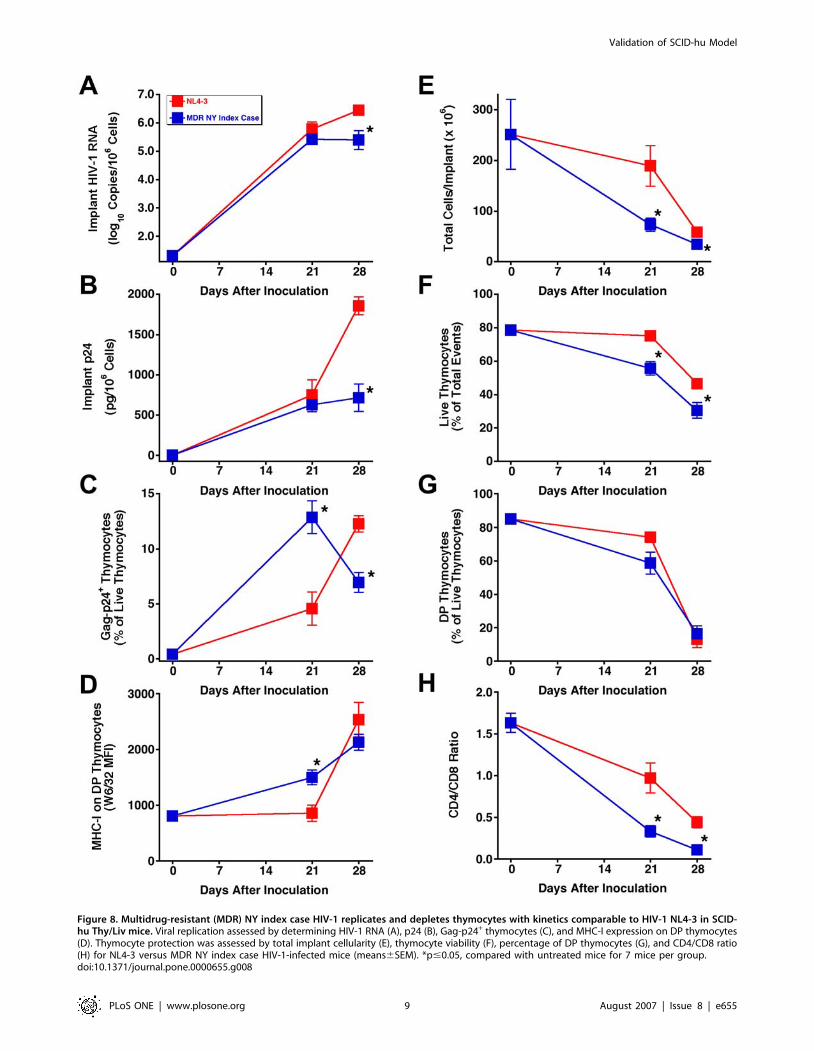
Figure 8. Multidrug-resistant (MDR) NY index case HIV-1 replicates and depletes thymocytes with kinetics comparable to HIV-1 NL4-3 in SCID-hu Thy/Liv mice. Viral replication assessed by determining HIV-1 RNA (A), p24 (B), Gag-p24+ thymocytes (C), and MHC-I expression on DP thymocytes(D). Thymocyte protection was assessed by total implant cellularity (E), thymocyte viability (F), percentage of DP thymocytes (G), and CD4/CD8 ratio(H) for NL4-3 versus MDR NY index case HIV-1-infected mice (means6SEM). *p#0.05, compared with untreated mice for 7 mice per group.doi:10.1371/journal.pone.0000655.g008
Validation of SCID-hu Model
PLoS ONE | www.plosone.org 9 August 2007 | Issue 8 | e655
six or seven dosing groups of 5–8 mice for optimal statistical
power. Given such group sizes and appropriate controls, it is
possible to assign effective antiviral dose ranges with statistical
precision [8]. Among animal models for HIV-1, this ability is
unique: nonhuman primate models (i.e., SIV- or SHIV-infected
rhesus macaques) cannot provide dosing groups of such size and
cannot achieve the same statistical power and relatively lower cost
per animal. There are several recent reports on the infection of
humanized Rag22/2cc2/2 (RAG-hu) [47–50] and NOD/SCID
BLT mice [51] with HIV-1, but there are no reports yet published
on the evaluation of antiretroviral efficacy in these models, and it
has not yet been demonstrated that 50–60 mice can be generated
with the CD34+ hematopoietic stem cells from a single donor. In
a recent review [52], Manz states that one of the challenges these
CD34+-reconstituted mouse models face is that usually less than 10
mice can be transplanted from one human graft.
In the experiments presented here, the second-generation
antiretrovirals in each drug class were more potent than their
first-generation predecessors. Thus, (–)-FTC was more potent than
3TC, the nonnucleoside RT inhibitor efavirenz was more potent
than nevirapine, and the protease inhibitor atazanavir was more
potent than indinavir. The greater potency of the newer drugs in
SCID-hu Thy/Liv mice is consistent with their observed greater
potency in HIV-1-infected individuals. The nonnucleoside RT
inhibitor (–)-FTC has greater potency than 3TC because the (–)-
FTC-triphosphate is incorporated 10 times more efficiently than
3TC-triphosphate during HIV-1 RT-catalyzed RNA-dependent
DNA synthesis [53]. The 10-fold greater potency of efavirenz
compared to nevirapine in SCID-hu mice is entirely predictive of
the superior oral bioavailability and plasma half-life characteristics
of efavirenz [54], which allow for once-daily dosing in humans as
the current standard of care compared with twice-daily dosing for
nevirapine. Finally, the greater potency of atazanavir over
indinavir in SCID-hu mice can be explained by a combination
of its greater in vitro potency and oral bioavailability, allowing
once-daily dosing [55]. Taken together, these comparisons of
licensed antiretrovirals in each major class suggest that the SCID-
hu Thy/Liv model may also predict clinical antiviral efficacy in
humans. These results extend an earlier validation of the model
with the nucleoside analogs zidovudine and dideoxyinosine [8].
The predictive nature of SCID-hu mouse efficacy is further
supported by the dosage levels required to achieve log order
reductions in implant viral load when adjusted for the difference in
surface area-to-body weight ratio between mice and humans.
Upon initial examination, the effective dosage level of 1,000 mg/
kg per day for indinavir in the mice seems overly high until such
an adjustment is made and the very high human daily dosage of
2.4 g (800 mg three times a day) is taken into consideration.
Predictiveness of the SCID-hu Thy/Liv model for the behavior
of HIV-1 in humans can be extended beyond antiretroviral
efficacy to simple kinetics of viral replication and virus-mediated
cytopathicity. AZT-resistant (JD) and MDR NY index case R5X4
clinical isolates replicate and cause thymocyte depletion with
kinetics comparable to HIV-1 NL4-3. The data presented here
show that drug-resistant clinical isolates can readily be used as
challenge viruses in the SCID-hu Thy/Liv model for in vivo studies
of new classes of antiretroviral drugs with activity against HIV-1
that is resistant to currently licensed antiretrovirals.
ACKNOWLEDGMENTSWe thank Sandra Bridges for longstanding support of the SCID-hu Thy/
Liv model, Opendra Sharma for supplying multiple-gram quantities of
antiretroviral drugs, Tim Schmidt for performing p24 ELISA, and Mike
McCune for helpful discussions.
Author Contributions
Conceived and designed the experiments: CS PB. Performed the
experiments: CB JB GC SG AK MM JR RR BS. Analyzed the data:
CS MM RR. Wrote the paper: CS.
REFERENCES1. McCune JM, Namikawa R, Kaneshima H, Shultz LD, Lieberman M, et al.
(1988) The SCID-hu mouse: murine model for the analysis of human
hematolymphoid differentiation and function. Science 241: 1632–1639.
2. Kaneshima H, Shih CC, Namikawa R, Rabin L, Outzen H, et al. (1991)
Human immunodeficiency virus infection of human lymph nodes in the SCID-
hu mouse. Proc Natl Acad Sci U S A 88: 4523–4527.
3. Namikawa R, Kaneshima H, Lieberman M, Weissman IL, McCune JM (1988)
Infection of the SCID-hu mouse by HIV-1. Science 242: 1684–1686.
4. Kitchen SG, Zack JA (1998) HIV type 1 infection in lymphoid tissue: natural
history and model systems. AIDS Res Hum Retroviruses 14 Suppl 3: S235–239.
5. McCune JM (1996) Development and applications of the SCID-hu mouse
model. Semin Immunol 8: 187–196.
6. McCune JM (1997) Animal models of HIV-1 disease. Science 278: 2141–2142.
7. McCune J, Kaneshima H, Krowka J, Namikawa R, Outzen H, et al. (1991) The
SCID-hu mouse: a small animal model for HIV infection and pathogenesis.
Annu Rev Immunol 9: 399–429.
8. Rabin L, Hincenbergs M, Moreno MB, Warren S, Linquist V, et al. (1996) Use
of standardized SCID-hu Thy/Liv mouse model for preclinical efficacy testing of
anti-human immunodeficiency virus type 1 compounds. Antimicrob Agents
Chemother 40: 755–762.
9. McCune JM, Namikawa R, Shih CC, Rabin L, Kaneshima H (1990) Suppression
of HIV infection in AZT-treated SCID-hu mice. Science 247: 564–566.
10. Namikawa R, Weilbaecher KN, Kaneshima H, Yee EJ, McCune JM (1990)
Long-term human hematopoiesis in the SCID-hu mouse. J Exp Med 172:
1055–1063.
11. Krowka JF, Sarin S, Namikawa R, McCune JM, Kaneshima H (1991) Human
T cells in the SCID-hu mouse are phenotypically normal and functionally
competent. J Immunol 146: 3751–3756.
12. Bonyhadi ML, Rabin L, Salimi S, Brown DA, Kosek J, et al. (1993) HIV induces
thymus depletion in vivo. Nature 363: 728–732.
13. Kaneshima H, Su L, Bonyhadi ML, Connor RI, Ho DD, et al. (1994) Rapid-high,
syncytium-inducing isolates of human immunodeficiency virus type 1 induce
cytopathicity in the human thymus of the SCID-hu mouse. J Virol 68: 8188–8192.
14. Stanley SK, McCune JM, Kaneshima H, Justement JS, Sullivan M, et al. (1993)
Human immunodeficiency virus infection of the human thymus and disruption
of the thymic microenvironment in the SCID-hu mouse. J Exp Med 178:
1151–1163.
15. Aldrovandi GM, Feuer G, Gao L, Jamieson B, Kristeva M, et al. (1993) The
SCID-hu mouse as a model for HIV-1 infection. Nature 363: 732–736.
16. Berkowitz RD, Alexander S, Bare C, Linquist-Stepps V, Bogan M, et al. (1998)
CCR5- and CXCR4-utilizing strains of human immunodeficiency virus type 1
exhibit differential tropism and pathogenesis in vivo. J Virol 72: 10108–10117.
17. Su L, Kaneshima H, Bonyhadi M, Salimi S, Kraft D, et al. (1995) HIV-1-
induced thymocyte depletion is associated with indirect cytopathogenicity and
infection of progenitor cells in vivo. Immunity 2: 25–36.
18. Jamieson BD, Uittenbogaart CH, Schmid I, Zack JA (1997) High viral burden
and rapid CD4+ cell depletion in human immunodeficiency virus type 1-infected
SCID-hu mice suggest direct viral killing of thymocytes in vivo. J Virol 71:
8245–8253.
19. Keir ME, Stoddart CA, Linquist-Stepps V, Moreno ME, McCune JM (2002)
IFN-alpha secretion by type 2 predendritic cells up-regulates MHC class I in the
HIV-1-infected thymus. J Immunol 168: 325–331.
20. Stoddart CA, Moreno ME, Linquist-Stepps VD, Bare C, Bogan MR, et al.
(2000) Antiviral activity of 29-deoxy-39-oxa-49-thiocytidine (BCH-10652) against
lamivudine-resistant human immunodeficiency virus type 1 in SCID-hu Thy/
Liv mice. Antimicrob Agents Chemother 44: 783–786.
21. Datema R, Rabin L, Hincenbergs M, Moreno MB, Warren S, et al. (1996)
Antiviral efficacy in vivo of the anti-human immunodeficiency virus bicyclam
SDZ SID 791 (JM 3100), an inhibitor of infectious cell entry. Antimicrob Agents
Chemother 40: 750–754.
22. Stoddart CA, Rabin L, Hincenbergs M, Moreno M, Linquist-Stepps V, et al.
(1998) Inhibition of human immunodeficiency virus type 1 infection in SCID-hu
Thy/Liv mice by the G-quartet-forming oligonucleotide, ISIS 5320. Antimicrob
Agents Chemother 42: 2113–2115.
23. Strizki JM, Xu S, Wagner NE, Wojcik L, Liu J, et al. (2001) SCH-C (SCH
351125), an orally bioavailable, small molecule antagonist of the chemokine
Validation of SCID-hu Model
PLoS ONE | www.plosone.org 10 August 2007 | Issue 8 | e655
receptor CCR5, is a potent inhibitor of HIV-1 infection in vitro and in vivo.
Proc Natl Acad Sci U S A 98: 12718–12723.24. Aldrovandi GM, Zack JA (1996) Replication and pathogenicity of human
immunodeficiency virus type 1 accessory gene mutants in SCID-hu mice. J Virol
70: 1505–1511.25. Aldrovandi GM, Gao L, Bristol G, Zack JA (1998) Regions of human
immunodeficiency virus type 1 nef required for function in vivo. J Virol 72:7032–7039.
26. Stoddart CA, Geleziunas R, Ferrell S, Linquist-Stepps V, Moreno ME, et al.
(2003) Human immunodeficiency virus type 1 Nef-mediated downregulation ofCD4 correlates with Nef enhancement of viral pathogenesis. J Virol 77:
2124–2133.27. Valentin A, Aldrovandi G, Zolotukhin AS, Cole SW, Zack JA, et al. (1997)
Reduced viral load and lack of CD4 depletion in SCID-hu mice infected withRev-independent clones of human immunodeficiency virus type 1. J Virol 71:
9817–9822.
28. Berkowitz RD, Beckerman KP, Schall TJ, McCune JM (1998) CXCR4 andCCR5 expression delineates targets for HIV-1 disruption of T cell differenti-
ation. J Immunol 161: 3702–3710.29. Kitchen SG, Zack JA (1997) CXCR4 expression during lymphopoiesis:
implications for human immunodeficiency virus type 1 infection of the thymus.
J Virol 71: 6928–6934.30. Berkowitz RD, van’t Wout AB, Kootstra NA, Moreno ME, Linquist-Stepps VD,
et al. (1999) R5 strains of human immunodeficiency virus type 1 from rapidprogressors lacking X4 strains do not possess X4-type pathogenicity in human
thymus. J Virol 73: 7817–7822.31. Berkowitz RD, Alexander S, McCune JM (2000) Causal relationships between
HIV-1 coreceptor utilization, tropism, and pathogenesis in human thymus.
AIDS Res Hum Retroviruses 16: 1039–1045.32. Jamieson BD, Pang S, Aldrovandi GM, Zha J, Zack JA (1995) In vivo
pathogenic properties of two clonal human immunodeficiency virus type 1isolates. J Virol 69: 6259–6264.
33. Miller ED, Duus KM, Townsend M, Yi Y, Collman R, et al. (2001) Human
immunodeficiency virus type 1 IIIB selected for replication in vivo exhibitsincreased envelope glycoproteins in virions without alteration in coreceptor
usage: separation of in vivo replication from macrophage tropism. J Virol 75:8498–8506.
34. Su L, Kaneshima H, Bonyhadi ML, Lee R, Auten J, et al. (1997) Identificationof HIV-1 determinants for replication in vivo. Virology 227: 45–52.
35. Amado RG, Jamieson BD, Cortado R, Cole SW, Zack JA (1999) Reconstitution
of human thymic implants is limited by human immunodeficiency virusbreakthrough during antiretroviral therapy. J Virol 73: 6361–6369.
36. Koka PS, Fraser JK, Bryson Y, Bristol GC, Aldrovandi GM, et al. (1998)Human immunodeficiency virus inhibits multilineage hematopoiesis in vivo.
J Virol 72: 5121–5127.
37. Withers-Ward ES, Amado RG, Koka PS, Jamieson BD, Kaplan AH, et al.(1997) Transient renewal of thymopoiesis in HIV-infected human thymic
implants following antiviral therapy. Nat Med 3: 1102–1109.38. Adachi A, Gendelman HE, Koenig S, Folks T, Willey R, et al. (1986) Production
of acquired immunodeficiency syndrome-associated retrovirus in human andnonhuman cells transfected with an infectious molecular clone. J Virol 59:
284–291.
39. Gartner S, Markovits P, Markovitz DM, Kaplan MH, Gallo RC, et al. (1986)
The role of mononuclear phagocytes in HTLV-III/LAV infection. Science 233:215–219.
40. Greenberg ML, Cammack N (2004) Resistance to enfuvirtide, the first HIV
fusion inhibitor. J Antimicrob Chemother 54: 333–340.41. Rimsky LT, Shugars DC, Matthews TJ (1998) Determinants of human
immunodeficiency virus type 1 resistance to gp41-derived inhibitory peptides.J Virol 72: 986–993.
42. Kovalev G, Duus K, Wang L, Lee R, Bonyhadi M, et al. (1999) Induction of
MHC class I expression on immature thymocytes in HIV-1-infected SCID-huThy/Liv mice: evidence of indirect mechanisms. J Immunol 162: 7555–7562.
43. Markowitz M, Mohri H, Mehandru S, Shet A, Berry L, et al. (2005) Infectionwith multidrug resistant, dual-tropic HIV-1 and rapid progression to AIDS:
a case report. Lancet 365: 1031–1038.44. Mordenti J (1986) Man versus beast: pharmacokinetic scaling in mammals.
J Pharm Sci 75: 1028–1040.
45. Freireich EJ, Gehan EA, Rall DP, Schmidt LH, Skipper HE (1966) Quantitativecomparison of toxicity of anticancer agents in mouse, rat, hamster, dog, monkey,
and man. Cancer Chemother Rep 50: 219–244.46. Bast RC, Holland JF, Frei E, American Cancer Society (2000) Cancer medicine
e.5 online. 5th ed. Hamilton, Ont. AtlantaGA: B.C. Decker; American Cancer
Society.47. Baenziger S, Tussiwand R, Schlaepfer E, Mazzucchelli L, Heikenwalder M, et
al. (2006) Disseminated and sustained HIV infection in CD34+ cord blood cell-transplanted Rag2-/-gamma c-/- mice. Proc Natl Acad Sci U S A 103:
15951–15956.48. Berges BK, Wheat WH, Palmer BE, Connick E, Akkina R (2006) HIV-1
infection and CD4 T cell depletion in the humanized Rag2-/-gamma c-/-
(RAG-hu) mouse model. Retrovirology 3: 76.49. Gorantla S, Sneller H, Walters L, Sharp JG, Pirruccello SJ, et al. (2007) Human
immunodeficiency virus type 1 pathobiology studied in humanized BALB/c-Rag2-/-gammac-/- mice. J Virol 81: 2700–2712.
50. Zhang L, Kovalev GI, Su L (2007) HIV-1 infection and pathogenesis in a novel
humanized mouse model. Blood 109: 2978–2981.51. Sun Z, Denton PW, Estes JD, Othieno FA, Wei BL, et al. (2007) Intrarectal
transmission, systemic infection, and CD4+ T cell depletion in humanized miceinfected with HIV-1. J Exp Med 204: 705–714.
52. Manz MG (2007) Human-hemato-lymphoid-system mice: opportunities andchallenges. Immunity 26: 537–541.
53. Feng JY, Shi J, Schinazi RF, Anderson KS (1999) Mechanistic studies show that
(-)-FTC-TP is a better inhibitor of HIV-1 reverse transcriptase than 3TC-TP.FASEB J 13: 1511–1517.
54. Young SD, Britcher SF, Tran LO, Payne LS, Lumma WC, et al. (1995) L-743,726 (DMP-266): a novel, highly potent nonnucleoside inhibitor of the human
immunodeficiency virus type 1 reverse transcriptase. Antimicrob Agents
Chemother 39: 2602–2605.55. Robinson BS, Riccardi KA, Gong YF, Guo Q, Stock DA, et al. (2000) BMS-
232632, a highly potent human immunodeficiency virus protease inhibitor thatcan be used in combination with other available antiretroviral agents.
Antimicrob Agents Chemother 44: 2093–2099.56. De Clercq E (2004) Antiviral drugs in current clinical use. J Clin Virol 30:
115–133.
Validation of SCID-hu Model
PLoS ONE | www.plosone.org 11 August 2007 | Issue 8 | e655


![Elisabetta Cecconi, "Old England of thy sins in time repent [...]": Religious lexis and discourse in 17th century Broadside Ballads](https://static.fdokumen.com/doc/165x107/631a9f70d5372c006e039424/elisabetta-cecconi-old-england-of-thy-sins-in-time-repent-religious-lexis.jpg)