Th1 and Th17 immunocompetence in humanized NOD/SCID/IL2rγ null mice
20
Th1 and Th17 Immunocompetence in Humanized NOD/SCID/γC- KO mice Deepika Rajesh a,* , Ying Zhou a , Ewa Jankowska-Gan a , Drew Allan Ronneburg a , Melanie M Dart a , Jose Torrealba b , and William J Burlingham a Department of Surgery, Transplant Division, University of Wisconsin School of Medicine and Public Health, Madison WI 53706 b Department of Pathology and Laboratory Medicine University of Wisconsin School of Medicine and Public Health, Madison WI 53706 Abstract We evaluated the immunocompetence of human T cells in humanized NOD-scid IL2r-γ-null (Hu— NSG) mice bearing a human thymic organoid, after multilinegage reconstitution with isogeneic human leukocytes. Delayed type hypersensitivity (DTH) response was assessed by a direct footpad challenge of the immunized hu-NSG host, or by transfer of splenocytes from immunized hu-NSG, along with antigen, into footpads of CB17 SCID mice [trans-vivo (tv) DTH]. Both methods revealed cellular immunity to tetanus toxoid (TT) or collagen type V (ColV). Immunohistochemical analysis of the swollen footpads revealed infiltration of human CD45 + cells, including CD3 + T cells, CD68 + macrophages and murine Ly6G + neutrophils. We observed a significant correlation between % circulating human CD4 + cells and the direct DTH swelling response to TT. The tvDTH response to TT was inhibited by anti-IFNγ, while the tvDTH response to collagen V was inhibited by anti IL-17 antibody, mimicking the cytokine bias of adult human T cells to these antigens. Hu-NSG mice were also capable of mounting a B cell response (primarily IgM) to TT antigen. The activation of either Th1- or Th17 - dependent cellular immune response supports the utility of Hu-NSG mice as a surrogate model of allograft rejection and autoimmunity. Keywords Humanized mice; T cell response; Delayed Type Hypersensitivity; Trans-vivo Delayed Type Hypersensitivity; Th1 and Th17 response Introduction The utility of humanized mouse models in human immunobiology research depends on their ability to mimic a functional human immune system. Previous models have often fallen short due to the lack of strong HLA-restricted, T cell-mediated immune responses. The recently developed NOD/SCID/γc/KO (NSG) mouse, exhibits enhanced ability to support the © 2009 American Society for Histocompatibility and Immunogenetics. Published by Elsevier Inc. All rights reserved. Address for Correspondence: Dr William J Burlingham, Department of Surgery, Transplant Division, H4/749 Clinical Science Center, 600 Highland Avenue, Madison, WI 53792-7375. Tel. 608-263-0119; Fax 608-263-7652; [email protected]. * current address:Senior Scientist, Cellular Dynamics Inc, 525 Science Drive, Madison WI 53711 Publisher's Disclaimer: This is a PDF file of an unedited manuscript that has been accepted for publication. As a service to our customers we are providing this early version of the manuscript. The manuscript will undergo copyediting, typesetting, and review of the resulting proof before it is published in its final citable form. Please note that during the production process errors may be discovered which could affect the content, and all legal disclaimers that apply to the journal pertain. NIH Public Access Author Manuscript Hum Immunol. Author manuscript; available in PMC 2011 June 1. Published in final edited form as: Hum Immunol. 2010 June ; 71(6): 551–559. doi:10.1016/j.humimm.2010.02.019. NIH-PA Author Manuscript NIH-PA Author Manuscript NIH-PA Author Manuscript
Transcript of Th1 and Th17 immunocompetence in humanized NOD/SCID/IL2rγ null mice
Th1 and Th17 Immunocompetence in Humanized NOD/SCID/γC-KO mice
Deepika Rajesha,*, Ying Zhoua, Ewa Jankowska-Gana, Drew Allan Ronneburga, Melanie MDarta, Jose Torrealbab, and William J BurlinghamaDepartment of Surgery, Transplant Division, University of Wisconsin School of Medicine and PublicHealth, Madison WI 53706bDepartment of Pathology and Laboratory Medicine University of Wisconsin School of Medicine andPublic Health, Madison WI 53706
AbstractWe evaluated the immunocompetence of human T cells in humanized NOD-scid IL2r-γ-null (Hu—NSG) mice bearing a human thymic organoid, after multilinegage reconstitution with isogeneichuman leukocytes. Delayed type hypersensitivity (DTH) response was assessed by a direct footpadchallenge of the immunized hu-NSG host, or by transfer of splenocytes from immunized hu-NSG,along with antigen, into footpads of CB17 SCID mice [trans-vivo (tv) DTH]. Both methods revealedcellular immunity to tetanus toxoid (TT) or collagen type V (ColV). Immunohistochemical analysisof the swollen footpads revealed infiltration of human CD45+ cells, including CD3+ T cells,CD68+ macrophages and murine Ly6G+ neutrophils. We observed a significant correlation between% circulating human CD4+ cells and the direct DTH swelling response to TT. The tvDTH responseto TT was inhibited by anti-IFNγ, while the tvDTH response to collagen V was inhibited by antiIL-17 antibody, mimicking the cytokine bias of adult human T cells to these antigens. Hu-NSG micewere also capable of mounting a B cell response (primarily IgM) to TT antigen. The activation ofeither Th1- or Th17 - dependent cellular immune response supports the utility of Hu-NSG mice asa surrogate model of allograft rejection and autoimmunity.
KeywordsHumanized mice; T cell response; Delayed Type Hypersensitivity; Trans-vivo Delayed TypeHypersensitivity; Th1 and Th17 response
IntroductionThe utility of humanized mouse models in human immunobiology research depends on theirability to mimic a functional human immune system. Previous models have often fallen shortdue to the lack of strong HLA-restricted, T cell-mediated immune responses. The recentlydeveloped NOD/SCID/γc/KO (NSG) mouse, exhibits enhanced ability to support the
© 2009 American Society for Histocompatibility and Immunogenetics. Published by Elsevier Inc. All rights reserved.Address for Correspondence: Dr William J Burlingham, Department of Surgery, Transplant Division, H4/749 Clinical Science Center,600 Highland Avenue, Madison, WI 53792-7375. Tel. 608-263-0119; Fax 608-263-7652; [email protected].*current address:Senior Scientist, Cellular Dynamics Inc, 525 Science Drive, Madison WI 53711Publisher's Disclaimer: This is a PDF file of an unedited manuscript that has been accepted for publication. As a service to our customerswe are providing this early version of the manuscript. The manuscript will undergo copyediting, typesetting, and review of the resultingproof before it is published in its final citable form. Please note that during the production process errors may be discovered which couldaffect the content, and all legal disclaimers that apply to the journal pertain.
NIH Public AccessAuthor ManuscriptHum Immunol. Author manuscript; available in PMC 2011 June 1.
Published in final edited form as:Hum Immunol. 2010 June ; 71(6): 551–559. doi:10.1016/j.humimm.2010.02.019.
NIH
-PA Author Manuscript
NIH
-PA Author Manuscript
NIH
-PA Author Manuscript

engraftment of human hematopoietic stem cells (HSCs) [1–4]. The NSG mouse lacks maturelymphocytes, NK cells, and yet is relatively long-lived, permitting long-term studies ofengraftment, infection, and immunity. Garcia [2,5–6] and Yang [2,5–6] have successfullygenerated humanized mice by transplanting human fetal liver HSCs into a NOD/SCID/γc/KO[NSG] mouse previously implanted with fetal thymic and liver tissues from the same donor.Both groups observed long-term, systemic human T cell homeostasis along with the systemicrepopulation with human B cells, monocytes/macrophages, and dendritic cells (DC) in therecipient mice. The success of this model has been attributed to the generation of a humanthymic organoid, derived from fetal tissue implanted underneath the kidney capsule, thatpermits the selection of developing T cells on autologous human Class I and Class II MHCantigen presenting molecules [2,5–6]. T cells play a central role in the development of immuneresponse. A crucial step in T-cell-mediated response is the recognition of antigen presentingcells and recruitment of antigen specific T cells to the respective target organ. The ability ofhumanized mice to mediate T cell–dependent responses has been modestly characterized inthe NSG mice. The presence of cellular immune responses against Epstein-Barr virus (EBV)toxic shock syndrome toxin-1 (TSST-1).and HIV antigens has been documented by severalgroups [6–7]. Here we have address the immunocompetence of the engrafted human T cellsby eliciting a delayed-type hypersensitivity (DTH) response to both Th-1 [Tetanus Toxoid/TT]and Th-17 [Collagen V/col(V)] antigens [8].
A DTH response is an in vivo index of cell-mediated immune response induced by antigenspecific T memory cells. An antigen is introduced to the host in the priming phase, and thenafter re-challenge subcutaneously or intradermally, the DTH reaction is initiated when antigensare presented by antigen-presenting cells (APCs) to sensitized memory T cells in the draininglymph node and at the challenge site. T-cell activation elicits an influx of macrophages,monocytes, and lymphocytes at the site of antigen exposure resulting an induration anderythema between 24–48 hours post injection. A positive swelling reaction typically requiresimmunization 2–4 weeks prior to DTH testing; after the antigen challenge, a transient swellingusually peaks at 24–48 h. The DTH reaction is termed direct if the swelling response is recordedafter subcutaneous or intradermal antigen injection into the primed individual. An alternativemethod for DTH testing involves the transfer of peripheral blood mononuclear cells plusantigen from the primed individual into the pinnae or footpads of naive mice. This indirect or“trans-vivo” (tv) DTH response is elicited by the localized human immune reaction and therecruitment and activation of murine PMNs, which produces a measurable swelling responsewithin 24 hrs [9][26]. Minimally, the interaction of memory T cells with antigen-pulseddendritic cells (DC) is sufficient to initiate a swelling response [26], while the interaction ofTreg cells with DC is sufficient to prevent it [27]. Because of its exquisite sensitivity to detectantigen-specific regulatory and effector T cells, the tvDTH assay has proven useful formonitoring the immune status of clinical transplant recipients [12,17,18].
In humans the DTH response to a recall antigen such as TT involves the activation of MHCclass II restricted CD4 T memory cells, as well as antigen presenting cells [APC] at the site ofantigen injection. After encountering the antigen-loaded APC, a few antigen-specific T cellsrecognize their cognate MHC/peptide ligand, become activated, and secrete cytokines: IFN-γ and IL-2 for a Th-1 type response [10–11] and 1L-17 or IL-22 for Th-17 type responses[12]. The release of proinflammatory cytokines and chemokines secreted by the activated Tcells, APC, and resident endothelial cells provide the amplification signals which recruitneutrophils, monocytes and more T cells, both antigen-specific and non-specific, to the site ofinflammation. This leads to a second wave of amplification, disrupting the collagen bundlesof the dermis 12–18 hours, and leading to a peak swelling response between 24–48 hrs afterantigen challenge.
Rajesh et al. Page 2
Hum Immunol. Author manuscript; available in PMC 2011 June 1.
NIH
-PA Author Manuscript
NIH
-PA Author Manuscript
NIH
-PA Author Manuscript
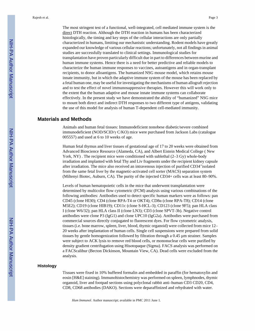
The most stringent test of a functional, well-integrated, cell mediated immune system is thedirect DTH reaction. Although the DTH reaction in humans has been characterizedhistologically, the timing and key steps of the cellular interactions are only partiallycharacterized in humans, limiting our mechanistic understanding. Rodent models have greatlyexpanded our knowledge of various cellular reactions; unfortunately, not all findings in animalstudies are successfully translated to clinical settings. Immunological studies fortransplantation have proven particularly difficult due in part to differences between murine andhuman immune systems. Hence there is a need for better predictive and reliable models tocharacterize the human immune responses to vaccines, autoantigens and in organ-transplantrecipients, to donor alloantigens. The humanized NSG mouse model, which retains mouseinnate immunity, but in which the adaptive immune system of the mouse has been replaced bya fetal human one, may be useful for investigating the mechanisms of human allograft rejectionand to test the effect of novel immunosuppressive therapies. However this will work only tothe extent that the human adaptive and mouse innate immune systems can collaborateeffectively. In the present study we have demonstrated the ability of “humanized” NSG miceto mount both direct and indirect DTH responses to two different type of antigens, validatingthe use of this model for analysis of human T-dependent cell-mediated immunity.
Materials and MethodsAnimals and human fetal tissues: Immunodeficient nonobese diabetic/severe combinedimmunodeficient (NOD/SCID/γ C/KO) mice were purchased from Jackson Labs (catalogue005557) and used at 6 to 10 weeks of age.
Human fetal thymus and liver tissues of gestational age of 17 to 20 weeks were obtained fromAdvanced Bioscience Resource (Alameda, CA), and Albert Eistein Medical College ( NewYork, NY) . The recipient mice were conditioned with sublethal (2–3 Gy) whole-bodyirradiation and implanted with fetal Thy and Liv fragments under the recipient kidney capsuleafter irradiation. The mice also received an intravenous injection of purified CD34+isolatedfrom the same fetal liver by the magnetic-activated cell sorter (MACS) separation system(Miltenyi Biotec, Auburn, CA). The purity of the injected CD34+ cells was at least 80–90%.
Levels of human hematopoietic cells in the mice that underwent transplantation weredetermined by multicolor flow cytometric (FCM) analysis using various combinations of thefollowing antibodies: Antibodies used to detect specific human markers were as follows: panCD45 (clone HI30); CD4 (clone RPA-T4 or OKT4); CD8α (clone RPA-T8); CD14 (cloneM5E2); CD19 (clone HIB19); CD11c (clone S-HCL-3); CD123 (clone 9F5); pan HLA classI (clone W6/32); pan HLA class II (clone LN3); CD3 (clone SPVT-3b). Negative controlantibodies were clone P3 (IgG1) and clone UPC10 (IgG2a). Antibodies were purchased fromcommercial sources directly conjugated to fluorescent dyes. For flow cytometric analysis,tissues (i.e. bone marrow, spleen, liver, blood, thymic organoid) were collected from mice 12–20 weeks after implantation of human cells. Single cell suspensions were prepared from solidtissues by gentle homogenization followed by filtration through a 0.45 µm strainer. Sampleswere subject to ACK lysis to remove red blood cells, or mononuclear cells were purified bydensity gradient centrifugation using Hisotopaque (Sigma). FACS analysis was performed ona FACScalibur (Becton Dickinson, Mountain View, CA). Dead cells were excluded from theanalysis.
HistologyTissues were fixed in 10% buffered formalin and embedded in paraffin (for hematoxylin andeosin [H&E] staining). Immunohistochemistry was performed on spleen, lymphnodes, thymicorganoid, liver and footpad sections using polyclonal rabbit anti–human CD3 CD20, CD4,CD8, CD68 antibodies (DAKO). Sections were deparaffinized and rehydrated with water.
Rajesh et al. Page 3
Hum Immunol. Author manuscript; available in PMC 2011 June 1.
NIH
-PA Author Manuscript
NIH
-PA Author Manuscript
NIH
-PA Author Manuscript

Antigen retrieval was performed using rodent decloaker (Biocare Medical) with sectionssubsequently incubated with primary Abs: CD4, CD8, CD45 (Biocare Medical), CD20 &Ly6G (BD Pharmingen), CD3 & CD68 (Dako). Antibodies were detected using a mouse onmouse HRP-polymer kit (Biocare Medical) and visualized with DAB.
ELISA for human Immunoglobulins (IgM and IgG) quantification was performed using humanIgM and human IgG enzyme-linked immunosorbent assay (ELISA) kits (Bethyl Laboratories,Montgomery, TX) according to the manufacturer‘s instructions . For the TT specific ELISA ,microwells were coated with purified 10µg/ml TT(Sanofi-Aventis Pasteur) in PBS overnightat 4°C , then blocked with 2% bovine serum albumin for 1 hr, and washed 3× in PBS/.05%Tween-20. Serum samples were applied at varying dilutions to each well for 1 hr; afteradditional 3× wash in PBS/Tween, human IgM or IgG were detected using HRP-coupledisotype-specific secondary antibodies, 3× washes and addition of the chromogenic substrateo-phenylenediamine(0.55 mg/ml) and 0.1% H2O2 for 5–10 minutes, followed by 1 MH2SO4.
Tetanus/Diphtheria immunizationMice were immunized with 1.5 limits of flocculation (lf) of pediatrics tetanus toxoid anddiphtheria (TT/DT) vaccine (Adventis-Pasteur) subcutaneously in the inguinal pouch region.
Collagen V immunizationMice were immunized with 100ug of bovine collagen V (col V) (Gift from Dr. David Wilkes)in Complete Fruend‘s Adjuvant (Sigma) subcutaneously in the inguinal pouch region. Two-three weeks post immunization mice were tested for a response to collagen V either directly(direct DTH assay) or splenocytes were collected and used in the trans-vivo DTH assay.
Direct DTHMice previously immunized to TT/DT and/or col V were monitored for a response by injecting .25lf TT/DT+PBS or 20ug of bovine col V+PBS subcutaneously into the right hind footpad. Tocontrol for swelling caused by the injection itself, PBS alone was injected into the left hindfootpad. Changes in footpad swelling were measured 24 hours post injection using a dial-thickness gauge. Pre-injection measurements were subtracted from post-injectionmeasurements to obtain specific swelling values. DTH reactivity is shown as the change inthickness, using units of 10−4 inches.
Trans-vivo DTHFor all TV- DTH assays, 4–8 × 10⁶ splenocytes were injected into footpads of naïveCB17.SCID recipients along with co-injection inoculum. DTH reactivity was measured as thechange in footpad thickness 24 hours post-injection over the pre-injection reading using a dial-thickness gauge and swelling is expressed in 10-4 inches. Co-injection inoculums include:PBS, 20µg donor antigen, 0.25lf TT/DT (Aventis-Pasteur Inc.) vaccine, 20µg col V,10µg anti-IFN-γ (BD Biosciences), 10µg anti-IL-1β (eBioscience), 10µg anti-TNF-α (eBiosciences), and10µg anti-IL-17A (BD Biosciences)
ResultsMulti-lineage human hematopoietic reconstitution NOD/SCID/γC/KO mice via implantationof human Thy/Liv fragments along with CD34+ cells
Humanized-mice were generated by implanting human fetal thymus and liver under the kidneycapsule of 6–8 week old NSG mice, followed by injection of autologous CD34+ cells asdescribed elsewhere (5). Typically we recovered sufficient cells and tissue to reconstitute 6–
Rajesh et al. Page 4
Hum Immunol. Author manuscript; available in PMC 2011 June 1.
NIH
-PA Author Manuscript
NIH
-PA Author Manuscript
NIH
-PA Author Manuscript

18 mice per human donor. Flow cytometry analysis was performed to detect the presence ofthe circulating human and murine cells in the peripheral blood of mice from 8–28 weeks postinjection. The results (Figure 1A) showed an increase in the proportion of human cells incirculation (HLA-ABC+) from 7% at 6 weeks to >50% at 14–30 weeks. HLA class II+ (HLA-DR+) cells increased from 8 to 30% from 6–14 weeks, gradually declining to 15% at 30 weekspost engraftment. The human cell engraftment was accompanied by a decline in the levels ofmurine class I+ (H2-KdDd) expressing cells from 80% to 24% between weeks 6 and 32. Avariable portion of mice, ≤5% of total, developed up to 90% human HLA-ABC+ circulatingcells, along with signs of graft-versus host [GVH] disease [data not shown]; these mice weresacrificed after 22 weeks. These exceptionally high % HLA-ABC+ values are not included inFig.1A.
The peripheral blood of the hu-NSG mice showed multi-lineage engraftment by human CD45+ cells (Figure 1B). B cells [CD19+] appeared rapidly and were the most abundant lymphocytesubset at 12 wks. T cells (CD4+ and CD8+) gradually overtook the B cells, surpassing them at18 weeks (27% T vs. 20% B). We also observed a low level of circulating CD14+ monocytes(2–6%) and CD56+ NK cells (< 1%; data not shown) between 12 and 20 wks afterreconstitution.
Evidence of muilti-lineage engraftment was also evident in the spleen (Figures 1C) and bonemarrow (Figure 1D). Human CD45+ cells comprised around 50% of both spleen and bonemarrow cells at 12 wks. Subset analysis of human CD45+ cells present in the marrow revealedthe presence of CD34+ stem/progenitor cells [data not shown], CD14+ monocytes, CD19+ Bcells and T cells (CD4+ and CD8+). B cells were relatively more abundant than T cells in thehu-NSG bone marrow at 12 weeks; the reverse (T>B) was true in spleen. Importantly,professional antigen presenting cells (APC), including CD14+ monocytes, and both myeloid(CD11c+) and plasmacytoid (CD123+) dendritic cells (DC) were present in both spleen andbone marrow. The relative proportions of pDCs and mDCs varied between spleen and bonemarrow, with the highest proportion of pDC being found in the hu-NSG spleen.
Representative immunohistochemical analyses depicting the histology of human cells in thespleen are shown in Figures 2A–2F. Note (Figure 2D) that human CD45+ leukocytes wereinterspersed with CD45neg (presumably murine) cells. CD4 (Figure 2B) and CD8 (Figure 2C)T cells are found scattered throughout the spleen, whereas phagocytic cells (CD68+) (Figure2E) and B cells (Figure 2F) tended to be more clustered within the splenic pulp.
To determine whether engrafted human B cells were functional, we quantified the humanimmunoglobulins (Ig) in the serum of hu-NSG mice reconstituted with cells from severaldifferent fetal donors. At 12–14 weeks, the average serum levels of total human IgM and humanIgG were 55 µg/ml and 165 µg/ml, respectively (Figure 3A). The timing of the appearance ofthe serum IgG and IgM in relation to circulating B cells was determined in a subset of mice atdifferent time points from 4–24 weeks (Figure 3B). B cells (CD19+) appeared at 4 wks inperipheral blood, and was soon followed by the appearance of serum IgM at 6 weeks. HumanIgG was not detectable until 8 weeks, and increased steadily through the observation period(to 20 weeks). To determine if antigen-specific B cell responses could be elicited, hu-NSGmice were immunized with tetanus toxoid (TT) in alum adjuvant at 12 weeks post engraftmentand subsequently given a booster dose of the antigen by footpad injection [DTH assay] at 14wks. Serum analysis of 2× immunized mice at 16 wks did not reveal anti-TT antibody [datanot shown]. However at 24 wks, 10 wks after the TT booster injection, anti-TT human IgMwas now detected in 3/3 hu-NSG mice(Table1).The amount was roughly 10% of the anti-TTIgM level found in an adult human serum, based on OD vs. serum titration curve analysis [datanot shown] . Anti-TT IgG was found in 1/3 mice tested, at the lower limit of detection (Table1).Consistent with a long-lived B cell response to antigen, the sera of the same 3 hu-NSG mice
Rajesh et al. Page 5
Hum Immunol. Author manuscript; available in PMC 2011 June 1.
NIH
-PA Author Manuscript
NIH
-PA Author Manuscript
NIH
-PA Author Manuscript

contained human B cell activating factor (BAFF), a TNF family protein essential for B cellsurvival, at levels comparable to that found in a normal adult human serum (Table 1).
Analysis of cell types within the thymic organoidA thymic organoid was generated by co-injection of human thymus and liver fragments underthe mouse kidney capsule. Figure 4A shows the gross appearance of the kidney at 12 weeks,the white area indicating the thymus organoid. Flow cytometric analysis of the thymic organoidrevealed the presence of human MHC class I+ cells, some of which co-expressed class II HLA-DR (Figure 4B, lower right panel). The organoid also contained human T cells, the majoritybeing immature CD4+/CD8+ double positive thymocytes (upper right panel , Fig. 4B) alongwith a low number of CD20+ B cells (Figure 4B, lower left). The majority of thymic cells wereconventional TCRαβ+ T cells (>60% of total), a low % of which co-stained for TCRγδ; a few(≤1%) were purely TCRγδ + TCRαβ− (Figure 4B, upper left). When we analyzed the humanthymic organoid (Figure 4C–4K) by immunohistochemistry, we found a clear separation ofthymus from mouse kidney parenchyma and both a dense cortex and a sparsely populatedmedullary region, along with areas resembling the Hassal‘s corpuscles seen in human (but notmurine) thymus [arrow, Fig 4C]. We observed CD4+ and CD8+ T cells in the cortex (Figures4G, 4H) as well as some B cells (Figure 4I), DC-SIGN+ dendritic cells (Figure 4J), andCD68+ macrophages (Figure 4K). The DC-SIGN+ cells were confined to the cortical region;however, CD1a+ DC were present in the medulla (data not shown). Human CD45+ cells wereobserved in both the cortex and medullary regions (Figure 4F). The organoid contained thymicstromal protein AE-1/4 (Figure 4D) indicating abundant thymic epithelium; however theorganoid was devoid of mature hepatocytes , as indicated by absence of hepatocyte surfaceantigen (Hep Ag) staining(Figure 4E).
Function of Human T cells assessed by delayed-type hypersensitivity (DTH)Although human thymopoiesis can be achieved by transplantation of human CD34+ cord bloodcells into neonatal BALB/c-RAG-2null or NOD/SCID null newborn mice, these models, whichrequire human T cells to mature in the mouse thymus, have generated suboptimal human T-cell immunocompetence [13–15]. Because the autologous thymic organoid supplies all theimmunogenetic components [for ex., HLA-A,B,C and DR antigens, Fig 4B] necessary fornormal human T cell development, the adaptive immune system of the hu-NSG mice ought toprovide a rigorous test to see if one could elicit an antigen-specific human T cell-mediatedimmune response in vivo.
Twelve weeks after reconstitution, humanized NSG mice were either left untreated, orimmunized with tetanus toxoid (TT) in alum [n=240 mice]; alternatively, some mice [n=8]were immunized with a “self” antigen, collagen type V (col V), administered in completeFreund‘s adjuvant. Two weeks later, the direct DTH reaction [16] was assessed by measuringchange in footpad thickness 24 hrs after challenge with antigen alone, in the absence ofadjuvant. As shown in Figure 5A, we observed a variable DTH response to each antigen.Control non-humanized mice, and humanized mice that were not immunized with the relevantantigen prior to challenge, had no swelling response at 24 hrs. after footpad challenge.
By immunohistochemistry, we found increased recruitment of human CD45+ cells scatteredin the subcutaneous tissue in the footpads injected with TT as compared to PBS control(compare Figure 5C vs. Figure 5I). CD3+ T cells were clearly present among the CD45+ humancells recruited to the TT challenge site (compare Fig 5D vs. 5J), as were CD68 macrophages(compare Fig 5F vs. 5L), whereas CD20+ B cells were absent (Fig 5E). Influx of murine PMNs(Ly6G+) cells was also observed at the site of injection with TT but not in the PBS controlfootpad (compare Fig 5G vs. 5M).
Rajesh et al. Page 6
Hum Immunol. Author manuscript; available in PMC 2011 June 1.
NIH
-PA Author Manuscript
NIH
-PA Author Manuscript
NIH
-PA Author Manuscript
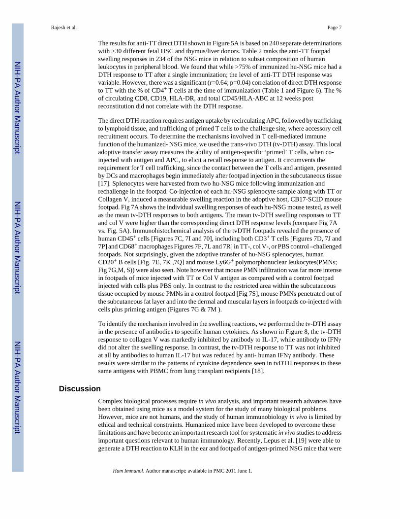
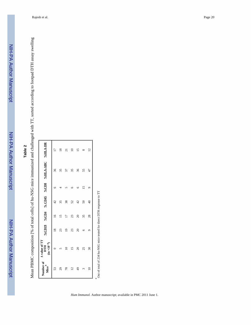
The results for anti-TT direct DTH shown in Figure 5A is based on 240 separate determinationswith >30 different fetal HSC and thymus/liver donors. Table 2 ranks the anti-TT footpadswelling responses in 234 of the NSG mice in relation to subset composition of humanleukocytes in peripheral blood. We found that while >75% of immunized hu-NSG mice had aDTH response to TT after a single immunization; the level of anti-TT DTH response wasvariable. However, there was a significant (r=0.64; p=0.04) correlation of direct DTH responseto TT with the % of CD4+ T cells at the time of immunization (Table 1 and Figure 6). The %of circulating CD8, CD19, HLA-DR, and total CD45/HLA-ABC at 12 weeks postreconstitution did not correlate with the DTH response.
The direct DTH reaction requires antigen uptake by recirculating APC, followed by traffickingto lymphoid tissue, and trafficking of primed T cells to the challenge site, where accessory cellrecruitment occurs. To determine the mechanisms involved in T cell-mediated immunefunction of the humanized- NSG mice, we used the trans-vivo DTH (tv-DTH) assay. This localadoptive transfer assay measures the ability of antigen-specific ‘primed’ T cells, when co-injected with antigen and APC, to elicit a recall response to antigen. It circumvents therequirement for T cell trafficking, since the contact between the T cells and antigen, presentedby DCs and macrophages begin immediately after footpad injection in the subcutaneous tissue[17]. Splenocytes were harvested from two hu-NSG mice following immunization andrechallenge in the footpad. Co-injection of each hu-NSG splenocyte sample along with TT orCollagen V, induced a measurable swelling reaction in the adoptive host, CB17-SCID mousefootpad. Fig 7A shows the individual swelling responses of each hu-NSG mouse tested, as wellas the mean tv-DTH responses to both antigens. The mean tv-DTH swelling responses to TTand col V were higher than the corresponding direct DTH response levels (compare Fig 7Avs. Fig. 5A). Immunohistochemical analysis of the tvDTH footpads revealed the presence ofhuman CD45+ cells [Figures 7C, 7I and 70], including both CD3+ T cells [Figures 7D, 7J and7P] and CD68+ macrophages Figures 7F, 7L and 7R] in TT-, col V-, or PBS control –challengedfootpads. Not surprisingly, given the adoptive transfer of hu-NSG splenocytes, humanCD20+ B cells [Fig. 7E, 7K ,7Q] and mouse Ly6G+ polymorphonuclear leukocytes(PMNs;Fig 7G,M, S)) were also seen. Note however that mouse PMN infiltration was far more intensein footpads of mice injected with TT or Col V antigen as compared with a control footpadinjected with cells plus PBS only. In contrast to the restricted area within the subcutaneoustissue occupied by mouse PMNs in a control footpad [Fig 7S], mouse PMNs penetrated out ofthe subcutaneous fat layer and into the dermal and muscular layers in footpads co-injected withcells plus priming antigen (Figures 7G & 7M ).
To identify the mechanism involved in the swelling reactions, we performed the tv-DTH assayin the presence of antibodies to specific human cytokines. As shown in Figure 8, the tv-DTHresponse to collagen V was markedly inhibited by antibody to IL-17, while antibody to IFNγdid not alter the swelling response. In contrast, the tv-DTH response to TT was not inhibitedat all by antibodies to human IL-17 but was reduced by anti- human IFNγ antibody. Theseresults were similar to the patterns of cytokine dependence seen in tvDTH responses to thesesame antigens with PBMC from lung transplant recipients [18].
DiscussionComplex biological processes require in vivo analysis, and important research advances havebeen obtained using mice as a model system for the study of many biological problems.However, mice are not humans, and the study of human immunobiology in vivo is limited byethical and technical constraints. Humanized mice have been developed to overcome theselimitations and have become an important research tool for systematic in vivo studies to addressimportant questions relevant to human immunology. Recently, Lepus et al. [19] were able togenerate a DTH reaction to KLH in the ear and footpad of antigen-primed NSG mice that were
Rajesh et al. Page 7
Hum Immunol. Author manuscript; available in PMC 2011 June 1.
NIH
-PA Author Manuscript
NIH
-PA Author Manuscript
NIH
-PA Author Manuscript
humanized at birth by intrahepatic injection of human CD34+ HSCs. While these results wereencouraging, no quantitative analysis of the swelling reactions was reported. In the absence ofa human thymic organoid to restrict T cell development to autologous HLA molecules, it isunlikely that such mice, whose T cells mature in a murine thymus, can develop a fully HLA-restricted T cell repertoire necessary to respond to human-restricted viruses[2,5–6]. These micein the present study had stable multi-lineage engraftment 10–12 weeks post engraftment anda well developed thymic organoid, complete with Hassal‘s corpuscles characteristic of humanthymus. Despite the very early stage of development of the human immune system [still “pre-natal” in total age] the ability to generate antigen-specific humoral [IgM] and cellular [DTH]responses confirms the functionality of a “blended” immune system, consisting of a remnantpopulation of approximately 40% murine, primarily innate immune leukocytes, and 60% Tcells, B cells, myeloid, NK and dendritic cells of human origin. Importantly, the hu-NSG micewere able to mount cell-mediated immune responses to both a Th1-type (TT) and a Th17 type(col V) antigen. We have recently found that these antigens elicit similar Th1 and Th17-typepolarized responses in both humans [18] and immunized C57BL/6 mice (M. Dart, A. David ,and W. Burlingham , manuscript in preparation), suggesting that this polarization may be afundamental property of the mammalian cellular immune response to these particular antigens.To the extent that innate immune mechanisms may direct the T cell response into a Th1 orTh17 pathway, the commonality between human and mouse DTH responses could explain whythe response to TT and col V elicited a Th1 and Th17 response in the hu-NSG, even thoughthe innate and adaptive arms of the immune system come from 2 different species.
The pattern of cytokine dependence of tv-DTH response to col V in the hu-NSG mice wassimilar to that seen in col V-sensitized humans and mice, with one interesting exception. Inhuman tv-DTH using PBMC from col V-reactive lung transplant recipients and patients withidiopathic pulmonary fibrosis [12,18] , besides a requirement for human IL17, there was apronounced dependence of the swelling reaction upon human IL1β as well as TNFα. Weconcluded from analysis of the cellular requirements for this response that monocyteproduction of IL1β and TNFα was a critical downstream response to the IL17 produced byCD4 col V reactive Th-17 cells. The lack of IL1β dependence of the tv-DTH response in thecol V- immunized NSG suggests that a) monocytes/macrophages in the spleen of the hu-NSGmice do not play any role in the Th-17 response as was seen with human PBMC [18]or b) thatmouse , and not human monocytes/macrophages present in the hu-NSG spleen play this keyaccessory role.
As noted above, the direct DTH assay is a relatively high level test of the function of the humanadaptive immune system. It also requires establishment and maintenance of a memorylymphocyte pool. Once naïve T cells recognize antigenic peptides in proper MHC [HLA]context, they will clonally expand and give rise to a population of T memory cells specific forthe antigen. Upon rechallenge, it must mobilize these T cells along with accessory cells inresponse to antigen in the tissues. Without a human thymus organoid, T cells of neonatal NSGmice reconstituted with CD34+ human HSCs mediated a suboptimal DTH response to TNBSsensitization and ear challenge; exogenous recombinant human IL-7, a T cell survival factor,was required for optimal strength of response [20]. While we did not test for IL-7 levels in ourmodel, the serum levels of human BAFF, which specifically promotes proliferation andsurvival of activated B cells [21] were near to that found in normal human serum [Table 1].This may account for the ability to detect anti-TT IgM responses in hu-NSG mice 10 weeksafter immunization [Fig 3.C]. The lack of antigen-specific IgG responses indicates that the hu-NSG mouse, at least at 24 weeks post-reconstitution, still lacks optimal conditions for germinalcenter formation and Ig class switch, despite adequate BAFF levels.
In summary, the response to a known Th-1 antigen, TT, both at the B cell[IgM] and T cell[direct DTH] level, and the response to a known Th-17 antigen, col(V), indicate that T helper
Rajesh et al. Page 8
Hum Immunol. Author manuscript; available in PMC 2011 June 1.
NIH
-PA Author Manuscript
NIH
-PA Author Manuscript
NIH
-PA Author Manuscript

cells educated in the autologous human thymus organoid are functional and provide anadequate level of immunocompetence for study of a wide range of human immunologicprocesses.
AcknowledgmentsMERC foundation of Wisconsin Madison (Grant number 133HG98) awarded to Dr. Burlingham provided the majorfunding for this research. We also thank Dr. Debra Bloom for performing human BAFF analysis, and Drs. WmWeidanz, Jenny Gumperz, Shannon Kenney, Clive Svensen, and Tim Kamp for their support in the development ofthe model
References1. Shultz LD, Ishikawa F, Greiner DL. Humanized mice in translational biomedical research. Nat Rev
Immunol 2007;7(2):118. [PubMed: 17259968]2. Melkus MW, Estes JD, Padgett-Thomas A, Gatlin J, Denton PW, Othieno FA, Wege AK, Haase AT,
Garcia JV. Humanized mice mount specific adaptive and innate immune responses to EBV andTSST-1. Nat Med 2006;12(11):1316. [PubMed: 17057712]
3. Larochelle A, Vormoor J, Lapidot T, Sher G, Furukawa T, Li Q, Shultz LD, Olivieri NF,Stamatoyannopoulos G, Dick JE. Engraftment of immune-deficient mice with primitive hematopoieticcells from beta-thalassemia and sickle cell anemia patients: implications for evaluating human genetherapy protocols. Hum Mol Genet 1995;4(2):163. [PubMed: 7757063]
4. Shultz LD, Schweitzer PA, Christianson SW, Gott B, Schweitzer IB, Tennent B, McKenna S,Mobraaten L, Rajan TV, Greiner DL, et al. Multiple defects in innate and adaptive immunologicfunction in NOD/LtSz-scid mice. J Immunol 1995;154(1):180. [PubMed: 7995938]
5. Lan P, Tonomura N, Shimizu A, Wang S, Yang YG. Reconstitution of a functional human immunesystem in immunodeficient mice through combined human fetal thymus/liver and CD34+ celltransplantation. Blood 2006;108(2):487. [PubMed: 16410443]
6. Wege AK, Melkus MW, Denton PW, Estes JD, Garcia JV. Functional and phenotypic characterizationof the humanized BLT mouse model. Curr Top Microbiol Immunol 2008;324:149. [PubMed:18481459]
7. Yajima M, Imadome KI, Nakagawa A, Watanabe S, Terashima K, Nakamura H, Ito M, Shimizu N,Honda M, Yamamoto N, Fujiwara S. A New Humanized Mouse Model of Epstein-Barr Virus InfectionThat Reproduces Persistent Infection, Lymphoproliferative Disorder, and Cell-Mediated and HumoralImmune Responses. J Infect Dis. 2008
8. Lopez M, Aguilera R, Perez C, Mendoza-Naranjo A, Pereda C, Ramirez M, Ferrada C, Aguillon JC,Salazar-Onfray F. The role of regulatory T lymphocytes in the induced immune response mediated bybiological vaccines. Immunobiology 2006;211(1–2):127. [PubMed: 16446177]
9. Burlingham WJ, Jankowska-Gan E. Mouse strain and injection site are crucial for detecting linkedsuppression in transplant recipients by trans-vivo DTH assay. Am J Transplant 2007;7(2):466.[PubMed: 17173656]
10. Laub R, Dorsch M, Meyer D, Ermann J, Hedrich HJ, Emmrich F. A multiple transgenic mouse modelwith a partially humanized activation pathway for helper T cell responses. J Immunol Methods2000;246(1–2):37. [PubMed: 11121545]
11. Riemer AB, Klinger M, Wagner S, Bernhaus A, Mazzucchelli L, Pehamberger H, Scheiner O,Zielinski CC, Jensen-Jarolim E. Generation of Peptide mimics of the epitope recognized bytrastuzumab on the oncogenic protein Her-2/neu. J Immunol 2004;173(1):394. [PubMed: 15210798]
12. Bobadilla JL, Love RB, Jankowska-Gan E, Xu Q, Haynes LD, Braun RK, Hayney MS, Munoz delRio A, Meyer K, Greenspan DS, Torrealba J, Heidler KM, Cummings OW, Iwata T, Brand D, PressonR, Burlingham WJ, Wilkes DS. Th-17, monokines, collagen type V, and primary graft dysfunctionin lung transplantation. Am J Respir Crit Care Med 2008;177(6):660. [PubMed: 18174545]
13. Legrand N, Weijer K, Spits H. Experimental model for the study of the human immune system:production and monitoring of "human immune system" Rag2−/−gamma c−/− mice. Methods MolBiol 2008;415:65. [PubMed: 18370148]
Rajesh et al. Page 9
Hum Immunol. Author manuscript; available in PMC 2011 June 1.
NIH
-PA Author Manuscript
NIH
-PA Author Manuscript
NIH
-PA Author Manuscript
14. Kuruvilla JG, Troyer RM, Devi S, Akkina R. Dengue virus infection and immune response inhumanized RAG2(−/−)gamma(c)(−/−) (RAG-hu) mice. Virology 2007;369(1):143. [PubMed:17707071]
15. Berges BK, Wheat WH, Palmer BE, Connick E, Akkina R. HIV-1 infection and CD4 T cell depletionin the humanized Rag2−/−gamma c−/− (RAG-hu) mouse model. Retrovirology 2006;3:76. [PubMed:17078891]
16. Black CA. Delayed type hypersensitivity: current theories with an historic perspective. DermatolOnline J 1999;5(1):7. [PubMed: 10673450]
17. VanBuskirk AM, Burlingham WJ, Jankowska-Gan E, Chin T, Kusaka S, Geissler F, Pelletier RP,Orosz CG. Human allograft acceptance is associated with immune regulation. J Clin Invest 2000;106(1):145. [PubMed: 10880058]
18. Burlingham WJ, Love RB, Jankowska-Gan E, Haynes LD, Xu Q, Bobadilla JL, Meyer KC, HayneyMS, Braun RK, Greenspan DS, Gopalakrishnan B, Cai J, Brand DD, Yoshida S, Cummings OW,Wilkes DS. IL-17-dependent cellular immunity to collagen type V predisposes to obliterativebronchiolitis in human lung transplants. J Clin Invest 2007;117(11):3498. [PubMed: 17965778]
19. Lepus CM, Gibson TF, Gerber SA, Kawikova I, Szczepanik M, Hossain J, Ablamunits V, Kirkiles-Smith N, Herold KC, Donis RO, Bothwell AL, Pober JS, Harding MJ. Comparison of human fetalliver, umbilical cord blood, and adult blood hematopoietic stem cell engraftment in NOD-scid/gammac−/−, Balb/c-Rag1−/−gammac−/−, and C.B-17-scid/bg immunodeficient mice. HumImmunol 2009;70(10):790. [PubMed: 19524633]
20. Unsinger J, McDonough JS, Shultz LD, Ferguson TA, Hotchkiss RS. Sepsis-induced humanlymphocyte apoptosis and cytokine production in "humanized" mice. J Leukoc Biol 2009;86(2):219.[PubMed: 19369639]
21. Schmidt MR, Appel MC, Giassi LJ, Greiner DL, Shultz LD, Woodland RT. Human BLyS facilitatesengraftment of human PBL derived B cells in immunodeficient mice. PLoS One 2008;3(9):e3192.[PubMed: 18784835]
Rajesh et al. Page 10
Hum Immunol. Author manuscript; available in PMC 2011 June 1.
NIH
-PA Author Manuscript
NIH
-PA Author Manuscript
NIH
-PA Author Manuscript

Figure 1.Quantification of engraftment in peripheral blood of NOD/SCID/γC/KO mice. Peripheralblood was collected from reconstituted mice at the indicated times after treatment and stainedwith anti-human specific antibodies for human pan HLABC, human Class II HLADR, MurineH2KD (A), human CD45 CD3 CD4 CD8 and CD19 (B). by flow cytometry. Each data pointin Figures A and B represents the average n (`~30) ± SE values. Cells isolated from spleen (C)and bone marrow (D) from mice 12 week post implantation of the human tissues and werestained for the presence of human T cells (CD4, CD8), B cells (CD19), monocytes (CD14) anddendritic cells defined as lineage-negative and HLA-DR positive cells expressing either CD123or CD11c. Data presented in C and D are obtained from six mice obtained from a single donor.FACS analysis was performed on a FACScalibur (Becton Dickinson, Mountain View, CA). ).FACS analysis was performed on a FACS calibur (Becton Dickinson, Mountain View, CA).Dead cells were excluded from the analysis.
Rajesh et al. Page 11
Hum Immunol. Author manuscript; available in PMC 2011 June 1.
NIH
-PA Author Manuscript
NIH
-PA Author Manuscript
NIH
-PA Author Manuscript

Figure 2.Human reconstitution in the spleen was confirmed by Hematoxylin and Eosin staining (A)followed by Immunoperoxidase staining of human cells in sections of spleens 12 wk afterengraftment. Presence of human, CD4 (B), CD8 (C), CD45 (D), CD68 (E), CD20 (F wereobserved in the spleen sections.
Rajesh et al. Page 12
Hum Immunol. Author manuscript; available in PMC 2011 June 1.
NIH
-PA Author Manuscript
NIH
-PA Author Manuscript
NIH
-PA Author Manuscript
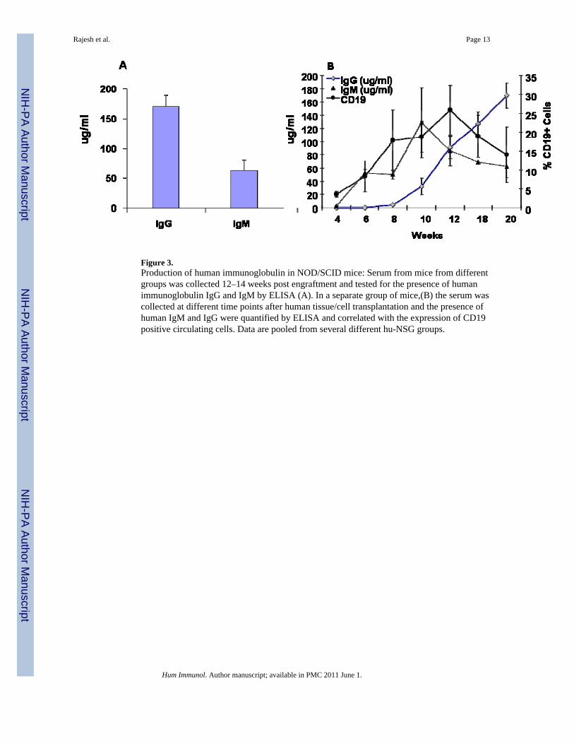
Figure 3.Production of human immunoglobulin in NOD/SCID mice: Serum from mice from differentgroups was collected 12–14 weeks post engraftment and tested for the presence of humanimmunoglobulin IgG and IgM by ELISA (A). In a separate group of mice,(B) the serum wascollected at different time points after human tissue/cell transplantation and the presence ofhuman IgM and IgG were quantified by ELISA and correlated with the expression of CD19positive circulating cells. Data are pooled from several different hu-NSG groups.
Rajesh et al. Page 13
Hum Immunol. Author manuscript; available in PMC 2011 June 1.
NIH
-PA Author Manuscript
NIH
-PA Author Manuscript
NIH
-PA Author Manuscript

Figure 4.Analysis of human thymic organoid: Gross Macroscopic image of the human thymic organoiddissected from the kidney 12 weeks post engraftment (A). Representative flow cytometric datashowing the presence of human MHC class I (HLABC), MHC class II (HLA-DR), T cells(CD4, CD8, TCR alpha beta, and B cells (CD19). Hematoxylin and eosin (H&E) of the thymicorganoid revealing the presence of hassal‘s corpuscles (C) Immunohistochemical analysis ofthe thymic organoid using human specific antibodies. for thymic stromal antigen (AE-1/AE-4)(D), hepatocyte specific antigen (HSA) (E), CD45 (F) ,CD4 (G),CD8 (H), CD20 (I), DC-SIGN(J) and CD68 (H). All images are shown at a magnification of 200×.
Rajesh et al. Page 14
Hum Immunol. Author manuscript; available in PMC 2011 June 1.
NIH
-PA Author Manuscript
NIH
-PA Author Manuscript
NIH
-PA Author Manuscript

Figure 5.DTH assay: The mice were immunized using 0.25 lf Tetanus Toxoid/Diphtheria Toxoid (TT/DT) vaccine in alum, Collagen V in CFA. Mice were tested for the response to the antigens ina direct DTH assay two weeks later. The swelling responses to the antigens after subtractingthe change in the PBS injected are compiled in A. After recording the response, the footpadswere harvested and stained during heamtoxylin and eosin (B, H) murine Ly6G antigens(G,M)and human specific CD45 (C,I), CD3 (D,J), CD68 (F,L), CD20(E,K). B–F represent thenegative response to PBS and H to M represent the DTH response to TT antigen. All imagesare shown at a magnification of 200×.
Rajesh et al. Page 15
Hum Immunol. Author manuscript; available in PMC 2011 June 1.
NIH
-PA Author Manuscript
NIH
-PA Author Manuscript
NIH
-PA Author Manuscript

Figure 6.DTH assay statistics: Data from Table 2 are plotted, showing the relationship between thepercentage of circulating human CD4 T cells and response to TT challenge in the footpad at12 weeks post engraftment. Linearity/r2 and p values are shown.
Rajesh et al. Page 16
Hum Immunol. Author manuscript; available in PMC 2011 June 1.
NIH
-PA Author Manuscript
NIH
-PA Author Manuscript
NIH
-PA Author Manuscript

Figure 7.Tv DTH response in humanized mice: Splenocytes from humanized mice immunized with TTor Collagen V antigens were isolated and injected along with TT or Collagen V antigens in tothe footpads CB17.SCID mice. The net DTH response was recorded after subtracting the PBScontrols. After recording the response, the footpads were harvested and stained duringheamtoxylin and Eosin (B, H, N) murine Ly6G antigens (G, M,S) human specific CD45 (C,I,O)CD3 (D, J,P) , CD20 (E,K,Q) and CD68 (F, L R) B,-G represent the Tv-DTH response to ColV,H to M represent the DTH response to TT antigen.N to S represent the DTH response to PBS.All images are shown at a magnification of 200×.
Rajesh et al. Page 17
Hum Immunol. Author manuscript; available in PMC 2011 June 1.
NIH
-PA Author Manuscript
NIH
-PA Author Manuscript
NIH
-PA Author Manuscript

Figure 8.TV-DTH was performed by co-transfer of splenocytes pooled from n=4 humanized NOD/SCID mice, along with antigen used for priming, into the footpads of CB17SCID mice asdescribed previously. Control values of swelling from transfer of hu-NSG splenocytes alone,w/o antigen, were subtracted to obtained net swelling values. The TV-DTH assay wasperformed using a mixture of cells , tetanus and diphtheria toxoid (TT/DT) or Collagen V,along with co injection of antibodies to human IFNγ, TNF-α, IL-1β , or IL-17. Values shownare mean ± SD of n=3 determinations.
Rajesh et al. Page 18
Hum Immunol. Author manuscript; available in PMC 2011 June 1.
NIH
-PA Author Manuscript
NIH
-PA Author Manuscript
NIH
-PA Author Manuscript

NIH
-PA Author Manuscript
NIH
-PA Author Manuscript
NIH
-PA Author Manuscript
Rajesh et al. Page 19
Table 1
Serum anti-TT specific antibody in 24 week reconstituted hu-NSG mice, 10 weeks after priming and boostingwith TT
Hu-NSG mice Adult HumanControl
TT specific IgM (OD value) 0.23, 0.34, 0.49 *1 0.83*2
TT specific IgG (µg/ml) 0.03, 0, 0 648.85
BAFF in serum (pg/ml) 48.2, 19.6, 31.8 67.0
*1Mouse serum was diluted at 1:100.
*2Human plasma was diluted at 1:160.
Hum Immunol. Author manuscript; available in PMC 2011 June 1.
NIH
-PA Author Manuscript
NIH
-PA Author Manuscript
NIH
-PA Author Manuscript
Rajesh et al. Page 20
Tabl
e 2
Mea
n PB
MC
com
posi
tion
[% o
f tot
al c
ells
] of h
u-N
SG m
ice
imm
uniz
ed a
nd c
halle
nged
with
TT,
sorte
d ac
cord
ing
to fo
otpa
d D
TH a
ssay
swel
ling
Num
ber
ofM
ice*
Δ va
lue
of T
TD
TH
(in ×
10−4
)%
CD
19%
CD
4%
CD
45%
CD
8%
HL
A-A
BC
%H
LA
-DR
530
1816
426
3617
295
2315
354
3518
7810
1917
385
3721
1215
2323
526
3510
4920
2020
426
3615
325
635
5015
558
1030
928
409
4712
* Out
of t
otal
of 2
34 h
u-N
SG m
ice
test
ed fo
r dire
ct D
TH re
spon
se to
TT
Hum Immunol. Author manuscript; available in PMC 2011 June 1.