Transient B-Cell Depletion with Anti-CD20 in Combination with Proinsulin DNA Vaccine or Oral...
10
Transient B-Cell Depletion with Anti-CD20 in Combination with Proinsulin DNA Vaccine or Oral Insulin: Immunologic Effects and Efficacy in NOD Mice Ghanashyam Sarikonda 1 , Sowbarnika Sachithanantham 1 , Yulia Manenkova 1 , Tinalyn Kupfer 2 , Amanda Posgai 3 , Clive Wasserfall 3 , Philip Bernstein 4 , Laura Straub 4 , Philippe P. Pagni 1 , Darius Schneider 1 , Teresa Rodriguez Calvo 1 , Marilyne Coulombe 2 , Kevan Herold 9 , Ronald G. Gill 2 , Mark Atkinson 3 , Gerald Nepom 10 , Mario Ehlers 4 , Teodora Staeva 5 , Hideki Garren 6 , Lawrence Steinman 7,6 , Andrew C. Chan 8 , Matthias von Herrath 1 * 1 La Jolla Institute for Allergy and Immunology, Diabetes Center, La Jolla, California, United States of America, 2 University of Colorado Denver, Aurora, Colorado, United States of America, 3 University of Florida, Gainesville, Florida, United States of America, 4 Immune Tolerance Network, San Francisco, California, United States of America, 5 JDRF International, New York, New York, United States of America, 6 Bayhill Therapeutics, San Mateo, California, United States of America, 7 Stanford University, Stanford, California, United States of America, 8 Genentech, Inc, South San Francisco, California, United States of America, 9 Yale University, New Haven, Connecticut, United States of America, 10 Benaroya Research Institute, Seattle, Washington, United States of America Abstract A recent type 1 diabetes (T1D) clinical trial of rituximab (a B cell-depleting anti-CD20 antibody) achieved some therapeutic benefit in preserving C-peptide for a period of approximately nine months in patients with recently diagnosed diabetes. Our previous data in the NOD mouse demonstrated that co-administration of antigen (insulin) with anti-CD3 antibody (a T cell- directed immunomodulator) offers better protection than either entity alone, indicating that novel combination therapies that include a T1D-related autoantigen are possible. To accelerate the identification and development of novel combination therapies that can be advanced into the clinic, we have evaluated the combination of a mouse anti-CD20 antibody with either oral insulin or a proinsulin-expressing DNA vaccine. Anti-CD20 alone, given once or on 4 consecutive days, produced transient B cell depletion but did not prevent or reverse T1D in the NOD mouse. Oral insulin alone (twice weekly for 6 weeks) was also ineffective, while proinsulin DNA (weekly for up to 12 weeks) showed a trend toward modest efficacy. Combination of anti-CD20 with oral insulin was ineffective in reversing diabetes in NOD mice whose glycemia was controlled with SC insulin pellets; these experiments were performed in three independent labs. Combination of anti-CD20 with proinsulin DNA was also ineffective in diabetes reversal, but did show modest efficacy in diabetes prevention (p = 0.04). In the prevention studies, anti-CD20 plus proinsulin resulted in modest increases in Tregs in pancreatic lymph nodes and elevated levels of proinsulin-specific CD4+ T-cells that produced IL-4. Thus, combination therapy with anti-CD20 and either oral insulin or proinsulin does not protect hyperglycemic NOD mice, but the combination with proinsulin offers limited efficacy in T1D prevention, potentially by augmentation of proinsulin-specific IL-4 production. Citation: Sarikonda G, Sachithanantham S, Manenkova Y, Kupfer T, Posgai A, et al. (2013) Transient B-Cell Depletion with Anti-CD20 in Combination with Proinsulin DNA Vaccine or Oral Insulin: Immunologic Effects and Efficacy in NOD Mice. PLoS ONE 8(2): e54712. doi:10.1371/journal.pone.0054712 Editor: Paolo Fiorina, Children’s Hospital Boston/Harvard Medical School, United States of America Received October 23, 2012; Accepted December 17, 2012; Published February 6, 2013 Copyright: ß 2013 Sarikonda et al. This is an open-access article distributed under the terms of the Creative Commons Attribution License, which permits unrestricted use, distribution, and reproduction in any medium, provided the original author and source are credited. Funding: This work was supported by a subaward from the University of Colorado, Denver (UCD) to MvH, issued under NIH award number 5U19AI050864-08 to UCD under the direction of Dr. George S. Eisenbarth, P.I. GS was a recipient of a T1D center of San Diego Postdoctoral fellowship; the ITN-JDRF (Immune Tolerence Network) Partnership supported members of the ITN-JDRF preclinical consortium, including TK, AP, CW, RG, MA, ME and MvH. The funders had no role in study design, data collection and analysis, decision to publish, or preparation of the manuscript. Competing Interests: Andrew Chan is the director of research at Genentech Inc. Hideki Garren and Lawrence Steinman are founding members of Bayhill Therapeutics Inc. All other authors have declared no competing interests exist. This does not alter the authors’ adherence to all the PLOS ONE policies on sharing data and materials. * E-mail: [email protected] Introduction In type 1 diabetes (T1D) antigen-specific immunotherapy (ASI) is a desirable goal because it offers the prospect of inducing immune tolerance with a good safety profile [1]. To date, however, clinical trials of ASI in the prevention or treatment of T1D have shown little or no efficacy, despite encouraging preclinical data. Success in the clinic may require optimization of dose, frequency, route of administration, and choice of antigen/epitope and adjuvant [2]. In addition, it is possible that in human T1D, ASI alone is not sufficient to induce tolerance but requires combination with an appropriate immune modulator that can enhance regulatory T cell (Treg) function and reduce the load of effector cells. This approach was recently validated in the NOD mouse, in which combination of non-Fc receptor binding anti-CD3 Mab with nasal proinsulin was more effective in reversing diabetes than either agent alone [3]. This has prompted strong interest in combination therapies, particularly those in which the individual components have already shown safety or efficacy in human trials [4]. Based on these considerations we explored the combination of an insulin-based antigen with anti-CD20 Mab in the NOD mouse. PLOS ONE | www.plosone.org 1 February 2013 | Volume 8 | Issue 2 | e54712
Transcript of Transient B-Cell Depletion with Anti-CD20 in Combination with Proinsulin DNA Vaccine or Oral...

Transient B-Cell Depletion with Anti-CD20 inCombination with Proinsulin DNA Vaccine or OralInsulin: Immunologic Effects and Efficacy in NOD MiceGhanashyam Sarikonda1, Sowbarnika Sachithanantham1, Yulia Manenkova1, Tinalyn Kupfer2,
Amanda Posgai3, Clive Wasserfall3, Philip Bernstein4, Laura Straub4, Philippe P. Pagni1,
Darius Schneider1, Teresa Rodriguez Calvo1, Marilyne Coulombe2, Kevan Herold9, Ronald G. Gill2,
Mark Atkinson3, Gerald Nepom10, Mario Ehlers4, Teodora Staeva5, Hideki Garren6,
Lawrence Steinman7,6, Andrew C. Chan8, Matthias von Herrath1*
1 La Jolla Institute for Allergy and Immunology, Diabetes Center, La Jolla, California, United States of America, 2University of Colorado Denver, Aurora, Colorado, United
States of America, 3University of Florida, Gainesville, Florida, United States of America, 4 Immune Tolerance Network, San Francisco, California, United States of America,
5 JDRF International, New York, New York, United States of America, 6 Bayhill Therapeutics, San Mateo, California, United States of America, 7 Stanford University,
Stanford, California, United States of America, 8Genentech, Inc, South San Francisco, California, United States of America, 9 Yale University, New Haven, Connecticut,
United States of America, 10 Benaroya Research Institute, Seattle, Washington, United States of America
Abstract
A recent type 1 diabetes (T1D) clinical trial of rituximab (a B cell-depleting anti-CD20 antibody) achieved some therapeuticbenefit in preserving C-peptide for a period of approximately nine months in patients with recently diagnosed diabetes. Ourprevious data in the NOD mouse demonstrated that co-administration of antigen (insulin) with anti-CD3 antibody (a T cell-directed immunomodulator) offers better protection than either entity alone, indicating that novel combination therapiesthat include a T1D-related autoantigen are possible. To accelerate the identification and development of novel combinationtherapies that can be advanced into the clinic, we have evaluated the combination of a mouse anti-CD20 antibody witheither oral insulin or a proinsulin-expressing DNA vaccine. Anti-CD20 alone, given once or on 4 consecutive days, producedtransient B cell depletion but did not prevent or reverse T1D in the NOD mouse. Oral insulin alone (twice weekly for 6weeks) was also ineffective, while proinsulin DNA (weekly for up to 12 weeks) showed a trend toward modest efficacy.Combination of anti-CD20 with oral insulin was ineffective in reversing diabetes in NOD mice whose glycemia wascontrolled with SC insulin pellets; these experiments were performed in three independent labs. Combination of anti-CD20with proinsulin DNA was also ineffective in diabetes reversal, but did show modest efficacy in diabetes prevention (p = 0.04).In the prevention studies, anti-CD20 plus proinsulin resulted in modest increases in Tregs in pancreatic lymph nodes andelevated levels of proinsulin-specific CD4+ T-cells that produced IL-4. Thus, combination therapy with anti-CD20 and eitheroral insulin or proinsulin does not protect hyperglycemic NOD mice, but the combination with proinsulin offers limitedefficacy in T1D prevention, potentially by augmentation of proinsulin-specific IL-4 production.
Citation: Sarikonda G, Sachithanantham S, Manenkova Y, Kupfer T, Posgai A, et al. (2013) Transient B-Cell Depletion with Anti-CD20 in Combination withProinsulin DNA Vaccine or Oral Insulin: Immunologic Effects and Efficacy in NOD Mice. PLoS ONE 8(2): e54712. doi:10.1371/journal.pone.0054712
Editor: Paolo Fiorina, Children’s Hospital Boston/Harvard Medical School, United States of America
Received October 23, 2012; Accepted December 17, 2012; Published February 6, 2013
Copyright: � 2013 Sarikonda et al. This is an open-access article distributed under the terms of the Creative Commons Attribution License, which permitsunrestricted use, distribution, and reproduction in any medium, provided the original author and source are credited.
Funding: This work was supported by a subaward from the University of Colorado, Denver (UCD) to MvH, issued under NIH award number 5U19AI050864-08 toUCD under the direction of Dr. George S. Eisenbarth, P.I. GS was a recipient of a T1D center of San Diego Postdoctoral fellowship; the ITN-JDRF (Immune TolerenceNetwork) Partnership supported members of the ITN-JDRF preclinical consortium, including TK, AP, CW, RG, MA, ME and MvH. The funders had no role in studydesign, data collection and analysis, decision to publish, or preparation of the manuscript.
Competing Interests: Andrew Chan is the director of research at Genentech Inc. Hideki Garren and Lawrence Steinman are founding members of BayhillTherapeutics Inc. All other authors have declared no competing interests exist. This does not alter the authors’ adherence to all the PLOS ONE policies on sharingdata and materials.
* E-mail: [email protected]
Introduction
In type 1 diabetes (T1D) antigen-specific immunotherapy (ASI) is
a desirable goal because it offers the prospect of inducing immune
tolerance with a good safety profile [1]. To date, however, clinical
trialsofASI in thepreventionor treatmentofT1Dhaveshownlittleor
no efficacy, despite encouragingpreclinical data. Success in the clinic
mayrequireoptimizationofdose, frequency, routeofadministration,
and choice of antigen/epitope and adjuvant [2]. In addition, it is
possible that in human T1D, ASI alone is not sufficient to induce
tolerance but requires combination with an appropriate immune
modulator that can enhance regulatory T cell (Treg) function and
reduce the loadof effector cells.This approachwas recently validated
in theNODmouse, inwhichcombinationofnon-Fcreceptorbinding
anti-CD3Mab with nasal proinsulin was more effective in reversing
diabetes than either agent alone [3]. This has prompted strong
interest in combination therapies, particularly those in which the
individual components have already shown safety or efficacy in
human trials [4]. Based on these considerations we explored the
combination of an insulin-based antigen with anti-CD20Mab in the
NODmouse.
PLOS ONE | www.plosone.org 1 February 2013 | Volume 8 | Issue 2 | e54712

Among ASI options for T1D, antigens based on insulin have
received the most attention in the clinic. Both oral and nasal
insulin have been evaluated in T1D prevention trials [5,6], while
nasal insulin, DNA encoding proinsulin, proinsulin peptide, and
insulin B-chain formulated in adjuvant have been administered in
new-onset and established T1D [7–10]. Overall, results have been
disappointing but there have been signals of efficacy in defined
subpopulations as well as encouraging immunologic changes;
safety and tolerability have been good, with no signs of disease
exacerbation. Insulin is an important auto-antigen in human T1D
and a high proportion of auto-reactive, islet-infiltrating CD8 T
cells, which selectively destroy insulin producing b-cells [11], areinsulin-reactive [12]. Insulin is also the primary antigen leading to
targeted islet cell destruction in the NOD mouse [13]. In mouse
models, administration of insulin or insulin peptides increases the
numbers of antigen-specific Treg cells that can prevent T1D [14–
16]. DNA vaccination with insulin B-chain prevented diabetes
onset in NOD [17] and RIP-NP mice [18] through a mechanism
involving IL-4 production [17,18], and administration of a DNA
vaccine encoding proinsulin was effective in both prevention and
reversal of diabetes in NOD mice [9].
Among antigen-nonspecific, targeted immunomodulation ap-
proaches for T1D, many have been evaluated in the clinic
(reviewed in [2]) but thus far only three have shown a sign of
efficacy in well-controlled phase 2 trials: FcR-nonbinding anti-
CD3 Mab [19,20], anti-CD20 Mab [21], and CTLA4-Ig [22].
While anti-CD3 treatment appears to exert its effect through the
induction of IL-10-producing Tregs [23], the mechanism of action
of anti-CD20 is not clear. B-cells participate in most autoimmune
diseases through production of autoantibodies [24], but in T1D
they likely promote disease by functioning as antigen presenting
cells (APCs) that specifically and efficiently capture beta-cell
proteins, including insulin [25–29]. Studies in NOD mice using
anti-CD20 antibody have shown contrasting effects of B-cell
depletion on reversal or prevention of T1D [30–32]. While one
group found induction of regulatory B-cells (Bregs) and Tregs [30],
another group did not [32]. In reports that used nontransgenic
NOD mice, therapeutic efficacy of B-cell depletion on T1D after
onset was not observed [32]. B-cells that infiltrate the pancreas
may lose CD20 expression and gain a plasma cell phenotype,
which could explain the loss of anti-CD20-mediated protection in
mice at later stages of the disease [31]. In transgenic NOD mice,
transient anti-CD20 treatment in very young mice (newborn), but
not in older mice, prevented autoimmune disease onset later in life
[33]. Collectively, the data suggest that antigen-specific B cells
facilitate and enhance disease development and progression in
T1D.
In this study, we hypothesized that co-administration of a B cell-
depleting anti-CD20 Mab and either a proinsulin DNA vaccine or
oral insulin will offer synergistic protection before and after T1D
onset in NOD mice.
Materials and Methods
Mice MaintenanceNOD/LtJ, NOD.SCID (NOD.CB17-Prkdcscid/J) or C57BL/6
mice were purchased from the Jackson Laboratory (Bar Harbor,
ME, USA). NOD/LtJ and C57BL/6 mice were maintained in
specific pathogen-free conditions at the three participating
institutions while NOD.SCID mice were maintained in a sterile
suite at the La Jolla Institute for Allergy and Immunology animal
facility.
For animal care and use at LIAI, La Jolla Institute For Allergy
And Immunology Animal Care Committee approved the work
carried out in this manuscript, under our protocol titled: Immune
Mediated Diabetes, Reference: Ap151-Mvh8-510. Animal welfare
is governed by the guidelines established through ‘‘The Guide’’
(The Guide For The Care And Use Of Laboratory Animals) and
the AVMA panel on euthanasia.
For animal care and use at UC Denver, mice were housed,
cared for, and used in experiments in accordance with the eighth
edition of the Guide for the Care and Use of Laboratory Animals
as described in protocol B-75411(05)1E and as approved by the
University of Colorado’s Institutional Animal Care and Use
Committee. The IACUC-approved pain management adminis-
tered following insulin pellet implantation was the opioid analgesic
buprenorphine. Buprenorphine (0.2 mg/kg SC) was given just
prior to surgery and continued every 6–12 hours for the next 18–
24 hours based on pain assessment. The IACUC-approved pain
management administered following retro-orbital blood collection
was proparacaine ophthalmic solution. Isoflurane anesthesia was
administered prior to both procedures. The IACUC-approved
euthanasia method during this study was CO2 asphyxiation
followed by cervical dislocation.
For animal care and use at University of Florida, mice were
housed, cared for, and used in experiments in accordance with the
Guide for the Care and Use of Laboratory Animals as approved
by the University of Florida’s Institutional Animal Care and Use
Committee. The IACUC-approved anesthesia administered prior
to and during insulin pellet implantation was isoflurane inhalation
(5% induction and 1–2% maintenance checked by toe pinch
reflex). Tail vein blood collections were performed rather than
retro-orbital blood collection; thus, a pain management solution
was not needed for blood collections. The IACUC-approved
euthanasia method during this study was isoflurane inhalation.
The toe pinch reflex was used to confirm that the animal is
completely unconscious. Cardiac exsanguination was performed
while under deep anesthesia followed by cervical dislocation. At
this point, thoracotomy with full necropsy was performed.
Blood Glucose MonitoringBlood glucose values (BGV) were monitored biweekly with
OneTouch Ultra blood glucose monitoring system (LifeScan Inc.,
Milpitas, CA, USA). For studies involving the combination of anti-
CD20 and oral insulin, NOD mice were considered to be diabetic
when two consecutive (day 1 and 2; day 1 is the day when
hyperglycemia was first noted) BGV$250 mg/dL; for the anti-
CD20 plus proinsulin studies the diabetes threshold was
.200 mg/dL and hyperglycemia was defined as BGV values
.180 mg/dL on two consecutive occasions.
Anti-CD20 Mab, Oral Insulin, Proinsulin Plasmid, and Anti-CD3 MabAnti-CD20 Mab and proinsulin DNA vaccine were obtained
from Genentech Inc, (San Francisco, CA) and Bayhill Therapeu-
tics (San Mateo, CA), respectively. Proinsulin plasmid has been
described before [9]. The novel anti-CD20 antibody, clone 5D2, is
of mIgG2a isotype; the isotype control antibody was IgG2a non-B-
cell-depleting C1.18 Mab (Bio-X-Cell). Oral insulin was human
recombinant insulin (Humulin R U-500, Eli Lilly & Co,
Indianapolis, IN); the diluent given as the oral insulin control
consisted of glycerin 16 mg/mL, metacresol 2.5 mg/mL, and zinc
oxide (total zinc content 0.017 mg/100 U). Anti-CD3 Mab
consisted of the F(ab)2 fragments from the T-cell-depleting anti-
CD3 Mab 145-2C11 (hamster IgG).Recent-onset Diabetes Treat-
ments
The anti-CD20 plus oral insulin studies were performed in three
independent labs (La Jolla, Denver and Gainesville); all animals
Combination Therapy Provides Limited Protection
PLOS ONE | www.plosone.org 2 February 2013 | Volume 8 | Issue 2 | e54712

(except the anti-CD3 control group) received an insulin pellet
implant (LinBit, Linshin Canada, Inc., Toronto, Ontario) on day 1
of confirmed diabetes and a second implant if necessary (2
consecutive BGV$250 mg/dL). NOD mice with new-onset
diabetes were allocated to 6 treatment groups. Group A: isotype
control antibody and oral diluent; group B: oral insulin (1 mg
twice weekly66 weeks) starting 2–5 days after diabetes onset;
group C: anti-CD20 Mab (10 mg/kg IP on days 1–4); group D:
oral insulin plus anti-CD20 Mab (regimens as for groups B and C);
group E: same as group D but oral insulin was delayed to start on
day 21; and group F: treatment with anti-CD3 Mab (5 mg on days
1–5) as positive control.
For the anti-CD20 plus proinsulin studies, NOD mice were
treated with anti-CD20 only, proinsulin plasmid only, or
a combination of both anti-CD20 and proinsulin plasmid. In
mice treated with anti-CD20 only, varying doses of anti-CD20
(indicated in the figure legends) were given in 200 mL PBS via i.v.,
tail vein injections, administered four times on consecutive days
(days 1–4). In mice treated with proinsulin plasmid only, 50 mgproinsulin plasmid was administered in 200 mL PBS containing
0.9 M CaCL2 via i.m. route (100 mL or 25 mg in each large thigh
muscle) as described earlier [9]. For combination therapy treated
mice, anti-CD20 and proinsulin treatments were given simulta-
neously; anti-CD20 antibody was given on days 1–4 while
proinsulin plasmid was administered once every week beginning
on day 1. After treatment with anti-CD20 mono- or combination
therapy, BGV measurements were performed until the values were
above 250 mg/dL for more than two weeks (no protection) and/or
if they stayed ,250 mg/dL with no signs of diabetes relapse
(protected). Treated NOD mice that showed fluctuations in BGV
were monitored for longer time periods to confirm that the mice
were either protected or not, sometimes resulting in non-uniform
X-axes. In the positive control anti-CD3 treated group, once the
diabetes reversal rate stabilized with no mice showing relapse,
BGV measurements were stopped, accounting for the difference in
follow-up time periods between different treatment groups. In our
previous (Bresson D, et.al, JCI 2006) and current experiments (not
shown), NOD mice treated with anti-CD3 that have reversed
diabetes without relapse for two or more weeks do not relapse,
obviating the need for longer-term monitoring.
Prevention TherapyNormoglycemic 8–10-week old NOD mice were treated with
varying doses (indicated in figure legends) of anti-CD20 alone or in
combination with proinsulin plasmid. Anti-CD20 treatments were
performed as above in diabetic NOD mice, except that the dose of
anti-CD20 used was 10 or 50 mg/mouse in 200 mL PBS. In mice
treated with proinsulin plasmid only, 50 mg proinsulin plasmid was
administered as above either only once, or four times, or
continuously throughout the experiment, at weekly intervals. In
combination therapy treated mice, anti-CD20 and proinsulin
treatments were given simultaneously; anti-CD20 antibody was
given only on day 1 while proinsulin plasmid was administered
once every week for four weeks. Unmanipulated NOD mice were
used as controls. After treatment with anti-CD20 mono- or
combination therapy, BGV measurements were performed until
the age of the NOD mice was 40 weeks or until they turn diabetic,
to confirm that the protected mice did not turn diabetic with
longer observation.
Flow CytometryFlow cytometry was performed as previously described [34].
mAbs specific for anti-IgM, -B220, -CD1d, -CD21, -CD23, -CD3,
-CD4, -CD5, -CD8, -CD25, -CD62L, -CD127, IL-4, IL-10, IFN-
c, and control isotypes were purchased from BD Biosciences.
Foxp3 staining was performed using Foxp3 staining kit, according
to the manufacturer’s instructions (eBioscience, San Diego, CA).
Data were acquired on an LSR-II (BD Biosciences, San Diego,
CA) and analyzed using Flowjo software (Treestar Inc., NJ).
Multiplex Cytokine MeasurementSerum was obtained from untreated, anti-CD20 only, or
combination therapy treated mice retroorbitally, at weekly
intervals. TNF-a, IFN-c, IL4, IL-10, and IL-17 titers in the
serum were determined using a commercially available multiplex
cytokine assay kit (Bio-Rad, Hercules, CA) and Bio-Plex 200
instrument.
B-cell and CD4+ T-cell Purification from NOD MouseSplenocytesNOD mouse splenocytes were prepared by homogenization in
red blood cell lysis buffer. From pooled splenocytes, B-cells were
purified using CD19+ B-cell enrichment kit (Miltenyi Biotec Inc.,
Auburn, CA). CD4+ cells were purified using CD4+CD25+ Treg
isolation kit (Miltenyi Biotec Inc., Auburn, CA), by stopping the
isolation protocol after the depletion step.
Adoptive Transfers2–106106 purified B-cells or CD4+ T-cells from anti-CD20
mono- or combination therapy treated NOD mice were trans-
ferred into recipient NOD.SCID mice via tail vein i.v. in 100 mlPBS. The recipient NOD.SCID mice also received an equal
number of splenocytes obtained from diabetic NOD mice, in
100 mL PBS (diabetogenic splenocytes).
ELISpotELISpot was performed as previously described [34]. Briefly,
purified CD4+ T-cells were prepared from splenocytes and plated
in a 96 well PVDF membrane-bound plate (Millipore Inc., USA)
coated with cytokine capture antibodies, at 5–256104 cells/well in
triplicate. T-cell-depleted splenocytes (APC) were prepared from
6–8-week old NOD mice and added at 56104/well along with
50 mg/mL test peptides (proinsulin peptide, and a mutated Insulin
B:9–23 B16:A peptide [34]) or medium only. Peptides were used at
high purity (.95% by HPLC, Abgent Inc., USA). ELISpot
reactions were carried out for 3 days, when the cells were removed
followed by the addition of detection antibodies and spot
development using 3-amino-9-ethylcarbazole (AEC) substrate
(Sigma-Aldrich, USA). Cytokine spots were enumerated using
the KS ELISpot reader, software version 4.
Statistical AnalysisData are expressed as means6SEM. The statistical significance
of the difference between means was determined using the
unpaired Student’s t test. Statistical tests were performed using
PRISM software (Graphpad, San Diego, CA). For diabetes
incidence measurement, statistical significance was determined
by Kaplan Meier analysis.
Results
Novel Murine Anti-CD20 Preferentially Depletes Follicularand Immature B-cells in the Spleen and Spares MarginalZone B-cellsPrevious studies have shown inconsistent results in replicating
the effects of rituximab (anti-human CD20) with murine anti-
CD20 antibodies in mice [32]. Here, we investigated the efficacy
Combination Therapy Provides Limited Protection
PLOS ONE | www.plosone.org 3 February 2013 | Volume 8 | Issue 2 | e54712

of a novel anti-CD20 antibody, clone 5D2, in preventing or
reversing diabetes in NODmice, in combination with a proinsulin-
expressing plasmid DNA vaccine or oral insulin.
To determine the efficiency of B-cell depletion using this novel
antibody, prediabetic eight to ten week old female NOD mice
were treated with a one-time administration of 50-mg/mouse anti-
CD20 intravenously (i.v.). Seven days post anti-CD20 treatment,
relative proportions and numbers of different B-cell subsets were
determined in the spleen by gating on B220 positive B-cells. We
observed significant reductions in both frequencies (Fig. 1A) and
numbers (Fig. 1B) of total B220+ B cells obtained from spleens of
anti-CD20 treated mice, compared to untreated NOD mice.
These reductions were found in follicular (FO) B-cells
(B220+IgMhiCD21int) and immature B-cells (B220+IgMhiCD212)
but not marginal zone (MZ) (B220+IgMhiCD21hi) B-cells (Fig. 1B).
Similarly, efficient depletion of B-cells was also observed in
peripheral blood, accompanied by an increase in CD4+ and
CD8+ T-cell frequencies (Fig. 1C). The lowest dose tested, 5 mganti-CD20, achieved 70–80% depletion, while 50–100 mg of anti-
CD20 resulted in maximal depletion of B cells (90%) in peripheral
blood (Fig. 1D). Following anti-CD20 administration, B-cell
numbers were reduced by one week, and stayed reduced for the
next two weeks. By eight weeks post treatment, circulating B-cell
frequencies returned to levels observed in untreated mice (Fig. 1D).
We attempted to maintain a continuous B-cell depletion in
a cohort of NOD mice by giving multiple doses of anti-CD20
antibody separated by either one or three weeks, but these
treatment regimens induced rapid death of the mice. Therefore, as
perpetual B-cell depletion could not be achieved with the novel
anti-CD20 antibody, we administered 50 mg of anti-CD20 on days
1, 2, 3, and 4, which achieved similar levels of B-cell depletion
(data not shown). Cytokine analysis of serum samples from anti-
CD20-treated animals after one, two, and three weeks post
treatment did not show changes in levels of IL-2, TNF-a, IL-4, IL-10 and IL-17 compared to untreated mice (Fig. S1). Taken
together, these findings suggest that (a) anti-CD20 administration
given either only on day 1 or on days 1, 2, 3 and 4 achieved similar
levels of B-cell depletion, and (b) perpetual B-cell depletion could
not be achieved in NOD mice using the novel anti-CD20
antibody.
Transient B-cell Depletion alone or in Combination withOral Insulin does not Reverse Diabetes in NOD MiceNOD mice with new-onset diabetes ($250 mg/dL on 2
consecutive days) were allocated to 6 treatment groups. Group
A: isotype control antibody and oral diluent; group B: oral insulin
(1 mg twice weekly66 weeks) starting 2–5 days after diabetes
onset; group C: anti-CD20 Mab (10 mg/kg IP on days 1–4); group
D: oral insulin plus anti-CD20 Mab (regimens as for groups B and
C); group E: same as group D but oral insulin was delayed to start
on day 21; and group F: treatment with anti-CD3 Mab (5 mg on
days 1–5) as positive control. In all groups, except group F,
hyperglycemia was controlled with a subcutaneous insulin pellet
on day 1 (repeated if subsequent blood glucose values $250 mg/
dL on 2 consecutive readings).
Compared to controls (group A), treatment with oral insulin
alone (group B), anti-CD20 alone (group C), or the combination
(groups D and E) did not induce a significant reversal of
hyperglycemia in NOD mice with new-onset diabetes (Fig. 2). In
contrast, treatment with anti-CD3 Mab (group F, positive control)
resulted in reversal in ,60% of animals, consistent with published
results. To control for the effect of glycemic control with insulin
pellets, a separate cohort of mice (see below) was treated with anti-
CD20 alone (single injection, either 100 mg or 250 mg per mouse)
in the absence of insulin pellets. Again, no consistent reversal of
diabetes was noted (Fig. 3B).
Taken together, these results suggest that treatment with a short
course of anti-CD20 alone, 6 weeks of oral insulin alone, or the
combination, fails to reverse hyperglycemia in new-onset diabetic
NOD mice.
Transient B-cell Depletion Abrogates Proinsulin PlasmidDNA Mediated Protection in Hyperglycemic NOD MiceTo determine the therapeutic efficacy of proinsulin plasmid
alone compared to anti-CD20 alone in diabetes reversal, we
administered either 50 mg of proinsulin plasmid (pBHT568) at
weekly intervals, or anti-CD20 daily for four consecutive days, in
a new cohort of hyperglycemic NOD mice (.180 mg/dL; not
treated with insulin pellets). Although we did not reach the same
level of protection as had been observed previously by Solvason
et al., (75%, [9]) 33% (3/9) of mice that received continuous
administration of pBHT568 plasmid were protected at 12 weeks
post treatment (Fig. 3A). As observed in our first cohort, B-cell
depletion with anti-CD20 offered little protection, if any, in
hyperglycemic NOD mice (Fig. 3B); only 17% of mice (3/18) that
received 100 mg (upper panel) and 8% of mice (2/26) that received
250 mg (lower panel) anti-CD20 showed improvement.
To determine whether combined administration of anti-CD20
and proinsulin plasmid can offer synergistic or additive protection
in hyperglycemic NOD mice, we administered anti-CD20
antibody on four consecutive days in combination with proinsulin
plasmid given at weekly intervals. Consistent with the oral insulin
data, this combination did not offer any protection in hypergly-
cemic NOD mice (Fig. 3C). Thus, transient B-cell depletion, with
or without proinsulin plasmid DNA vaccination, did not offer
therapeutic efficacy in hyperglycemic NOD mice.
Transient B-cell Depletion with Anti-CD20 inCombination with Proinsulin Plasmid AdministrationOffers Protection from T1D OnsetTo test its efficacy in prevention of diabetes onset in NOD mice,
proinsulin plasmid was administered in 8–10-week old prediabetic
NOD mice once, four times, or continuously, at weekly intervals.
Among these different treatment regimens, we found that only
continuous administration of proinsulin plasmid showed a trend
for protecting NOD mice from diabetes onset (not significant;
Fig. 4A). Administration of proinsulin plasmid only once or for
four times had no effect on diabetes development in NOD mice.
To determine whether transient depletion of B-cells could
protect NOD mice from diabetes onset, multiple cohorts of 8–10-
week old NOD mice were given a one-time administration of
varying doses of anti-CD20 antibody (10, 50, 100 or 250 mg per
mouse) and diabetes progression was monitored. Single adminis-
tration of anti-CD20 antibody had no significant effect on diabetes
onset (Fig. 4B, and data not shown). There was also no significant
protection from diabetes onset when anti-CD20 was given four
times (on days 1–4, data not shown). Thus, transient B-cell
depletion with anti-CD20 does not have any protective effect on
diabetes onset in NOD mice.
To determine whether the combination therapy can offer
protection from diabetes onset, 8–10-week old prediabetic NOD
mice were treated with a low-dose (50 mg) one-time administration
of anti-CD20 antibody in combination with four weekly injections
of proinsulin plasmid. In contrast to anti-CD20 or proinsulin
plasmid alone, combination therapy resulted in a reduced in-
cidence of T1D in NOD mice (Fig. 4C). More frequent
administration of anti-CD20 (on days 1, 2, 3 and 4) or
Combination Therapy Provides Limited Protection
PLOS ONE | www.plosone.org 4 February 2013 | Volume 8 | Issue 2 | e54712

administration of a higher dose of anti-CD20 did not increase the
level of protection observed (data not shown). Therefore, transient
B-cell depletion with low-dose anti-CD20 in combination with
proinsulin plasmid offers modest protection from T1D onset in
NOD mice.
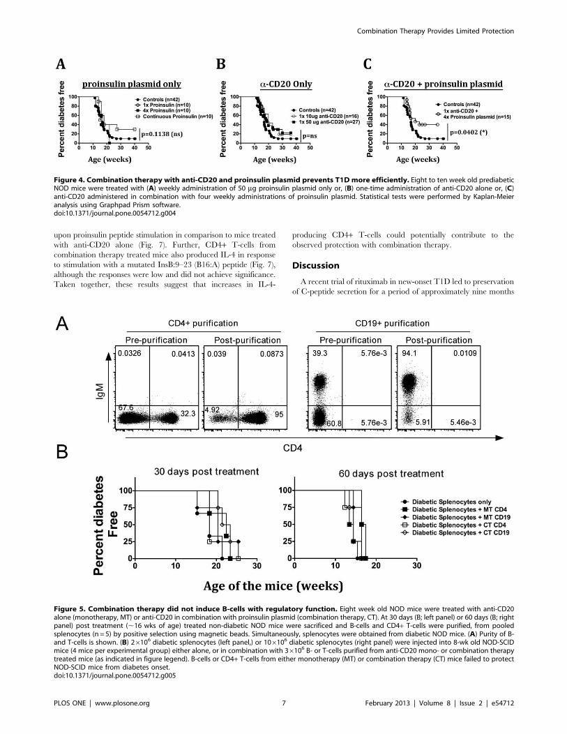
Combination Therapy did not Induce Regulatory B-cellsRegulatory B-cells can either be defined by expression of certain
cell surface markers (phenotype) or by their ability to suppress T
cell activity through IL-10 production or Fas-FasL interaction
(function) (discussed in [35]). Using all three phenotypic
characterizations [35], we did not detect any increases in
regulatory B-cells following anti-CD20 mono- or combination
therapy (data not shown). Next, we explored the possibility that
functional B-regulatory cells with T cell suppressive activity (not
restricted to above phenotypes) were induced upon anti-CD20
treatment. As it is difficult to isolate only functional B-regulatory
cells, at four or eight weeks following treatment initiation we
purified all B-cells from spleens of anti-CD20-treated non-diabetic
mice (with or without proinsulin plasmid co-administration), and
adoptively transferred them into NOD.SCID mice along with
diabetogenic splenocytes from control NOD mice. B-cells from
mono- or combination therapy treated mice did not alter the
course of diabetes development in recipient NOD.SCID mice
(Fig. 5A, B).
Combination Therapy Induces Modest Increases in Tregsin Pancreas Draining Lymph NodeWe next sought to determine if phenotypic or functional Tregs
are induced upon mono- or combination therapy. At eight weeks,
but not at four weeks post treatment, there was a modest increase
in CD4+FoxP3+ cell frequencies in PDLNs of combination
Figure 1. Novel murine anti-CD20 administration preferentially depletes follicular but not marginal zone B-cells. Eight week oldfemale NOD mice were treated with 50 mg anti-CD20 and 7 days later various B-cell subsets in the spleen were analyzed. Gating strategy is shown inthe top panel. Lymphocytes were gated on (A) B220+ (total B-cells) and further classified as FO B-cells (B220+ IgMhi, CD21int), immature B-cells (B220+IgMhi CD212), and MZ B-cells (B220+ IgMhi CD21hi). Total B220+ B-cell numbers were reduced in the spleen with significant reductions observed innumber of follicular and immature B-cells but not MZ B-cells (B). Eight week old female NOD mice were given anti-CD20 i.v. Seven days post anti-CD20 treatment, frequencies of B- and T-cells in peripheral blood were determined by staining with anti-IgM/B220 for B-cells or anti-CD4/CD8 for T-cells; loss of circulating B-cells was associated with compensatory increases in CD4+ and CD8+ T-cell frequencies in NOD mice (C). Frequencies ofcirculating B-cells returned to similar levels as in untreated NOD mice by 60 days post-treatment (D). Data are means 6 SEM. Representative datafrom 3 independent experiments with similar results are shown. Statistical analysis was performed using unpaired students t-test using GraphpadPrism software.doi:10.1371/journal.pone.0054712.g001
Combination Therapy Provides Limited Protection
PLOS ONE | www.plosone.org 5 February 2013 | Volume 8 | Issue 2 | e54712

therapy treated mice in comparison to mice that received
monotherapy, but this did not achieve statistical significance
(Fig. 6). At eight weeks post therapy there were significant
decreases in B-cells accompanied by increases in CD4+ T-cells in
PDLN but not in spleens of combination therapy treated mice.
Since protection offered by combination therapy was sustained
long-term (Fig. 4), but there was only a modest increase in Tregs in
spleen or PDLN at eight weeks, we sought to determine whether
this protection could be mapped to induction of dominant
tolerance in all CD4+ T-cells. At four weeks or eight weeks after
combination therapy, purified CD4+ T-cells (purity shown in
Fig. 5A) from protected mice were adoptively transferred into
eight-week-old NOD.SCID mice along with an equal number of
splenocytes from diabetic NOD mice. Purified CD4+ T-cells from
mono- or combination therapy treated mice failed to protect
NOD.SCID from T1D onset (Fig. 5B), suggesting a lack of
dominant tolerance induction in CD4+ T-cells.
Combination Therapy Increases the Number ofProinsulin-specific IL-4-producing CD4+ T-cellsWe have shown that antigen-specific DNA vaccination with
plasmid expressing an insulin B chain peptide, InsB:9-23, offers
protection via IL-4 production [17]. Therefore, we sought to
determine whether proinsulin-specific immune responses were
modulated with the combination therapy. At two and eight weeks
post anti-CD20 treatment with or without proinsulin plasmid
administration, purified CD4+ T-cells were tested for their ability
to respond to proinsulin peptide stimulation in an ELISpot assay.
Purified CD4+ T-cells were stimulated with either proinsulin
peptide or a modified InsB9-23 peptide (B16:A [13]). At two weeks
post treatment, CD4+ T-cells from anti-CD20 mono- or
combination therapy treated mice did not produce any IL-4 in
response to proinsulin peptide stimulation (data not shown),
suggesting a lack of proinsulin specific T-cell expansion at this time
point. Interestingly, at eight weeks post combination therapy,
a significantly higher number of CD4+ T-cells produced IL-4
Figure 2. Transient B-cell depletion alone or in combination with oral insulin does not reverse diabetes in NOD mice. NOD mice withrecent onset diabetes (2 consecutive readings of BGV.250 mg/dl), were treated with either anti-CD20 alone, oral insulin alone or a combination ofboth treatments, along with isotype control antibody treatment, oral diluent alone or anti-CD3 mAb treatment, as indicated in figures. Theseexperiments were carried out in three independent labs (indicated in materials and methods) and the combined results were pooled to obtain theresults shown here. The number of mice assigned to each treatment group is shown in the figure.doi:10.1371/journal.pone.0054712.g002
Figure 3. Combination therapy with anti-CD20 and proinsulin plasmid does not offer protection after T1D onset. NOD mice withhyperglycemia (2 consecutive readings of BGV.180 mg/dl), were treated with either (A) weekly administration of 50 mg proinsulin plasmid or (B) 100(upper panel) or 250 mg of anti-CD20 alone or (C) 50 (upper panel) or 250 mg of anti-CD20 antibody in combination with weekly administration 50 mgof proinsulin plasmid. Each line represents one individual mouse.doi:10.1371/journal.pone.0054712.g003
Combination Therapy Provides Limited Protection
PLOS ONE | www.plosone.org 6 February 2013 | Volume 8 | Issue 2 | e54712
upon proinsulin peptide stimulation in comparison to mice treated
with anti-CD20 alone (Fig. 7). Further, CD4+ T-cells from
combination therapy treated mice also produced IL-4 in response
to stimulation with a mutated InsB:9–23 (B16:A) peptide (Fig. 7),
although the responses were low and did not achieve significance.
Taken together, these results suggest that increases in IL-4-
producing CD4+ T-cells could potentially contribute to the
observed protection with combination therapy.
Discussion
A recent trial of rituximab in new-onset T1D led to preservation
of C-peptide secretion for a period of approximately nine months
Figure 4. Combination therapy with anti-CD20 and proinsulin plasmid prevents T1D more efficiently. Eight to ten week old prediabeticNOD mice were treated with (A) weekly administration of 50 mg proinsulin plasmid only or, (B) one-time administration of anti-CD20 alone or, (C)anti-CD20 administered in combination with four weekly administrations of proinsulin plasmid. Statistical tests were performed by Kaplan-Meieranalysis using Graphpad Prism software.doi:10.1371/journal.pone.0054712.g004
Figure 5. Combination therapy did not induce B-cells with regulatory function. Eight week old NOD mice were treated with anti-CD20alone (monotherapy, MT) or anti-CD20 in combination with proinsulin plasmid (combination therapy, CT). At 30 days (B; left panel) or 60 days (B; rightpanel) post treatment (,16 wks of age) treated non-diabetic NOD mice were sacrificed and B-cells and CD4+ T-cells were purified, from pooledsplenocytes (n = 5) by positive selection using magnetic beads. Simultaneously, splenocytes were obtained from diabetic NOD mice. (A) Purity of B-and T-cells is shown. (B) 26106 diabetic splenocytes (left panel,) or 106106 diabetic splenocytes (right panel) were injected into 8-wk old NOD-SCIDmice (4 mice per experimental group) either alone, or in combination with 36106 B- or T-cells purified from anti-CD20 mono- or combination therapytreated mice (as indicated in figure legend). B-cells or CD4+ T-cells from either monotherapy (MT) or combination therapy (CT) mice failed to protectNOD-SCID mice from diabetes onset.doi:10.1371/journal.pone.0054712.g005
Combination Therapy Provides Limited Protection
PLOS ONE | www.plosone.org 7 February 2013 | Volume 8 | Issue 2 | e54712

[21]. Similarly, administration of a proinsulin DNA vaccine has
shown promising results in NOD mouse studies [9] and is
currently being tested in a phase I/II clinical trial [36]. However,
it is evident from over 40 clinical trials conducted in the last three
decades that the use of a single immunotherapeutic agent is
insufficient to confer protection in a majority of T1D patients and
that combination approaches will be required (discussed in [4]).
Thus combination therapies with multiple therapeutic agents need
Figure 6. Combination therapy increases numbers of CD4+ Foxp3+ cells in pancreas draining lymph node 60-days post treatment.Eight to ten week old prediabetic NOD mice were given 50 mg of anti-CD20 either alone or in combination with weekly administration of 50 mgproinsulin plasmid for four weeks. Splenocytes and PDLN cells from mono- or combination therapy treated mice were stained with anti-IgM, -B220 (B-cells), -CD4, -CD8, -CD25 and –Foxp3. B-cell frequencies were determined by gating on IgM+ B220+ cells. Frequency of Foxp3+ cells among CD4+ T-cells was determined by gating on CD4+ cells. Cumulative frequencies of B-cells (left panel), CD4+ T-cells (middle panel), and CD4+Foxp3+ cells (rightpanel) in PDLN (upper panels) or spleen (lower panels) are shown at 30 days and 60 days post combination therapy. Representative data from threeindependent experiments with similar results is shown. Statistical analysis was performed using unpaired students t-test using Graphpad Prismsoftware. Each dot represents one mouse, with mean value indicated on the graph.doi:10.1371/journal.pone.0054712.g006
Figure 7. T-cells from combination therapy treated mice produce IL-4 in response to proinsulin peptide stimulation. At 60-days postanti-CD20 mono- (MT, open bars) or combination therapy (CT, filled black bars), CD4+ T-cells were purified from treated non-diabetic NOD mice.Simultaneously, T-depleted splenocytes (TDS) were obtained from 8–10 week old NOD mice and used as APCs. 250,000 purified CD4+ T-cells wereincubated with proinsulin peptide or a mutated insulin B:9–23 (B16:A) peptide in the presence of 56104 APCs. Following 3-day incubation, IL-4production was determined by ELISpot assay as previously described [34]. Background (media) subtracted spot numbers are shown on the Y-axis.Increased IL-4 production in response to proinsulin peptide was seen in combination therapy treated mice. Representative means 6 SEM data fromone of two independent experiments with similar results are shown. Statistical analysis was performed using unpaired students t-test using GraphpadPrism software.doi:10.1371/journal.pone.0054712.g007
Combination Therapy Provides Limited Protection
PLOS ONE | www.plosone.org 8 February 2013 | Volume 8 | Issue 2 | e54712

to be tested in preclinical studies to accelerate their translation into
human trials. In this study, we combined the administration of a B-
cell depleting anti-CD20 antibody with either proinsulin plasmid
or oral insulin to determine the efficacy of these combinations in
protecting hyperglycemic NOD mice. Our results show that
neither combination offers protection after onset of hyperglycemia,
but that anti-CD20 plus proinsulin plasmid prevents diabetes onset
in a modest but significant proportion of animals. These
observations provide insights for the design of future clinical trials
involving a combination of anti-CD20 and insulin-based antigens.
Previous studies evaluating anti-CD20 antibody to deplete B-
cells have found varying effects on T1D onset. While one group
found therapeutic efficacy in transgenic NOD mice that express
human CD20 [30], other groups failed to observe any protection
after T1D onset [32]. A recent study found that continuous
depletion of B-cells in NOD mice offered protection from T1D
onset in IAA-negative NOD mice [31]. However, because
repeated administration of our novel anti-CD20 antibody sepa-
rated by more than one week led to the death of NOD mice, our
own observations are limited to transient depletion of B-cells. Such
transient B-cell depletion did not protect NOD mice from T1D
onset.
In agreement with Serreze et al. [31], we found that most of the
B-cells that entered the pancreas lost their CD20 expression (data
not shown) and therefore were not depleted after anti-CD20
treatment. In addition, we found that anti-CD20 treatment did not
have any effect on circulating levels of IAA (Fig. S2), consistent
with reports that anti-CD20 treatment depletes short-lived plasma
B-cells [37], whereas IAA-is produced predominantly by long-
lived plasma B-cells. This lack of long-lived plasma B-cell depletion
may explain why monotherapy with rituximab shows only limited
efficacy in a majority of T1D subjects [18].
In our experiments, continuous proinsulin plasmid administra-
tion offered only limited protection in hyperglycemic NOD mice,
less than that observed by others previously [9], possibly due to the
autoantibody status of the NOD mice. Solvason et al. used only
IAA+ mice in their study [9] while we used all NOD mice
irrespective of their autoantibody status.
Anti-CD20 treatment was protective in IAA-negative NOD
mice [31] while proinsulin plasmid therapy was effective in IAA-
positive mice [9]. In our study we combined anti-CD20 and
proinsulin plasmid, and therefore we used mice regardless of IAA
status. Whether the lack of therapeutic efficacy in hyperglycemic
NOD mice could be attributed to differential effects of these agents
depending on the IAA autoantibody status cannot be answered in
our experimental setup.
While most anti-CD20 treatments require continuous admin-
istration [31,32] to reduce T1D incidence, we observed limited but
significant protection with a one-time anti-CD20 administration
combined with four doses of proinsulin plasmid. It appears that
this combination therapy can induce long-term antigen (pro-
insulin) specific tolerance in a proportion of mice, likely through
IL-4 production. However, involvement of additional mechanisms
playing a role in increased protection cannot be ruled out since
increases in IL-10 production were found in NOD mice treated
only with proinsulin plasmid [9]. Previous work in our lab showed
that DNA vaccination with insulin B:9–23 provided protection in
T1D through the production of IL-4 from CD4+ T-cells [17,18].
Thus it is possible that increased antigen-specific IL-4 production
could be a common feature of DNA vaccination-mediated
protection in T1D.
Islet antigen-specific Tregs operate locally in PDLNs and islets
by recognizing their cognate antigens, thus induction of such
Tregs is highly desirable through islet antigen specific therapies.
However, while monotherapy with islet antigens such as insulin
shows promise in animal models, it has little efficacy in human
trials [2], possibly because the duration of immunotherapy was too
short or because the complexity of human autoimmune diabetes
requires addition of an immune modulator to the antigen.
Therefore, to improve efficacy in T1D protection, novel combi-
nation therapies have to be optimized, ideally in mice first. We
have previously shown that combination therapy with anti-CD3
and proinsulin peptide (hpllp) achieved much better protection in
NOD mice with recent-onset diabetes than either of these reagents
alone [3].
Using combinations of reagents that are in clinical trials as T1D
monotherapies (anti-CD20, oral insulin, and proinsulin plasmid)
we only found modest protective efficacy in prevention of T1D
onset with the combination of anti-CD20 and proinsulin, but no
therapeutic efficacy in hyperglycemic NOD mice with either
combination. Our results suggest that transient B-cell depletion
using anti-CD20 alone or in combination with proinsulin plasmid
or oral insulin are not effective T1D therapeutic strategies. These
results further emphasize the importance of rigorously testing
novel therapeutic approaches in relevant animal models and
underscore the need for identifying novel combination therapeutic
approaches that can be moved into the clinic.
Supporting Information
Figure S1 B-cell depletion does not result in a systemi-cally altered cytokine milieu in peripheral blood. Eight toten week old prediabetic NOD mice were given 50 mg of anti-
CD20. At one, two or three weeks post treatment serum was
obtained and circulating levels of different cytokines (TNF-a, L-2,IL-4, IL-10 and IL-17) were determined by cytokine-multiplex
technology. For each cytokine, the samples were analyzed in
triplicates and the mean values are plotted as dot plots with each
dot representing one individual mouse. The experiment was
repeated twice with similar results and data from one represen-
tative experiment is shown.
(TIF)
Figure S2 Anti-CD20 treatment does not diminishcirculating levels of IAA auto-antibodies in NOD mice.Serum was collected from eight week old NOD mice that were
either untreated (A) or treated with anti-CD20 antibody (B) before
the initiation of anti-CD20 treatment (at 8-weeks of age) and at 1,
2, 3 and 4 weeks post anti-CD20 treatment. Levels of circulating
IAA antibody were determined by radioimmunoassay, at Barbara
Davis Center, Colorado. Anti-CD20 treatment did not cause
a drop in the levels of circulating IAA autoantibodies.
(TIF)
Acknowledgments
We would like to thank Malina Mclure for assistance with our mouse
colony, Priscilla Colby for managerial assistance, and Jeremy Pettus (all at
La Jolla Institute for Allergy and Immunology) for his critical review of this
manuscript.
Author Contributions
Oversee ITN-preclinical consortium projects including initiation of
projects, operations and guiding them to completion: LS PB GN TS.
Conceived and designed the experiments: RG MA MvH LS AC HG.
Performed the experiments: GS SS YM TK AP PP DS TRC MC.
Analyzed the data: GS MEMvH. Contributed reagents/materials/analysis
tools: RG MAMvH AC KH CW LS HG. Wrote the paper: GS MEMvH.
Combination Therapy Provides Limited Protection
PLOS ONE | www.plosone.org 9 February 2013 | Volume 8 | Issue 2 | e54712

References
1. Peakman M, von Herrath M (2010) Antigen-specific immunotherapy for type 1
diabetes: maximizing the potential. Diabetes 59: 2087–2093.2. Gallagher MP, Goland RS, Greenbaum CJ (2011) Making progress: preserving
beta cells in type 1 diabetes. Ann N Y Acad Sci 1243: 119–134.3. Bresson D, Togher L, Rodrigo E, Chen Y, Bluestone JA, et al. (2006) Anti-CD3
and nasal proinsulin combination therapy enhances remission from recent-onset
autoimmune diabetes by inducing Tregs. J Clin Invest 116: 1371–1381.4. Matthews JB, Staeva TP, Bernstein PL, Peakman M, von Herrath M (2010)
Developing combination immunotherapies for type 1 diabetes: recommenda-tions from the ITN-JDRF Type 1 Diabetes Combination Therapy Assessment
Group. Clin Exp Immunol 160: 176–184.
5. Nanto-Salonen K, Kupila A, Simell S, Siljander H, Salonsaari T, et al. (2008)Nasal insulin to prevent type 1 diabetes in children with HLA genotypes and
autoantibodies conferring increased risk of disease: a double-blind, randomisedcontrolled trial. Lancet.
6. Skyler JS, Krischer JP, Wolfsdorf J, Cowie C, Palmer JP, et al. (2005) Effects oforal insulin in relatives of patients with type 1 diabetes: The Diabetes Prevention
Trial–Type 1. Diabetes Care 28: 1068–1076.
7. Fourlanos S, Perry C, Gellert SA, Martinuzzi E, Mallone R, et al. (2011)Evidence that nasal insulin induces immune tolerance to insulin in adults with
autoimmune diabetes. Diabetes 60: 1237–1245.8. Orban T, Farkas K, Jalahej H, Kis J, Treszl A, et al. (2010) Autoantigen-specific
regulatory T cells induced in patients with type 1 diabetes mellitus by insulin B-
chain immunotherapy. J Autoimmun 34: 408–415.9. Solvason N, Lou YP, Peters W, Evans E, Martinez J, et al. (2008) Improved
efficacy of a tolerizing DNA vaccine for reversal of hyperglycemia throughenhancement of gene expression and localization to intracellular sites. J Immunol
181: 8298–8307.10. Thrower SL, James L, Hall W, Green KM, Arif S, et al. (2009) Proinsulin
peptide immunotherapy in type 1 diabetes: report of a first-in-man Phase I safety
study. Clin Exp Immunol 155: 156–165.11. von Herrath M, Homann D (2004) Islet regeneration needed for overcoming
autoimmune destruction - considerations on the pathogenesis of type 1 diabetes.Pediatr Diabetes 5 Suppl 2: 23–28.
12. Coppieters KT, Dotta F, Amirian N, Campbell PD, Kay TW, et al. (2012)
Demonstration of islet-autoreactive CD8 T cells in insulitic lesions from recentonset and long-term type 1 diabetes patients. J Exp Med 209: 51–60.
13. Nakayama M, Abiru N, Moriyama H, Babaya N, Liu E, et al. (2005) Prime rolefor an insulin epitope in the development of type 1 diabetes in NOD mice.
Nature 435: 220–223.14. Martinez NR, Augstein P, Moustakas AK, Papadopoulos GK, Gregori S, et al.
(2003) Disabling an integral CTL epitope allows suppression of autoimmune
diabetes by intranasal proinsulin peptide. J Clin Invest 111: 1365–1371.15. Homann D, Dyrberg T, Petersen J, Oldstone MB, von Herrath MG (1999)
Insulin in oral immune ‘‘tolerance’’: a one-amino acid change in the B chainmakes the difference. J Immunol 163: 1833–1838.
16. von Herrath MG, Dyrberg T, Oldstone MB (1996) Oral insulin treatment
suppresses virus-induced antigen-specific destruction of beta cells and preventsautoimmune diabetes in transgenic mice. J Clin Invest 98: 1324–1331.
17. Bot A, Smith D, Bot S, Hughes A, Wolfe T, et al. (2001) Plasmid vaccinationwith insulin B chain prevents autoimmune diabetes in nonobese diabetic mice.
J Immunol 167: 2950–2955.18. Coon B, An LL, Whitton JL, von Herrath MG (1999) DNA immunization to
prevent autoimmune diabetes. J Clin Invest 104: 189–194.
19. Herold KC, Hagopian W, Auger JA, Poumian-Ruiz E, Taylor L, et al. (2002)Anti-CD3 monoclonal antibody in new-onset type 1 diabetes mellitus.
N Engl J Med 346: 1692–1698.
20. Herold KC, Gitelman SE, Masharani U, Hagopian W, Bisikirska B, et al. (2005)
A single course of anti-CD3 monoclonal antibody hOKT3gamma1(Ala-Ala)
results in improvement in C-peptide responses and clinical parameters for at
least 2 years after onset of type 1 diabetes. Diabetes 54: 1763–1769.
21. Pescovitz MD, Greenbaum CJ, Krause-Steinrauf H, Becker DJ, Gitelman SE, et
al. (2009) Rituximab, B-lymphocyte depletion, and preservation of beta-cell
function. N Engl J Med 361: 2143–2152.
22. Orban T, Bundy B, Becker DJ, DiMeglio LA, Gitelman SE, et al. (2011) Co-
stimulation modulation with abatacept in patients with recent-onset type 1
diabetes: a randomised, double-blind, placebo-controlled trial. Lancet 378: 412–
419.
23. Waldron-Lynch F, Henegariu O, Deng S, Preston-Hurlburt P, Tooley J, et al.
(2012) Teplizumab induces human gut-tropic regulatory cells in humanized mice
and patients. Sci Transl Med 4: 118ra112.
24. Wong FS, Wen L (2005) B cells in autoimmune diabetes. Rev Diabet Stud 2:
121–135.
25. Hulbert C, Riseili B, Rojas M, Thomas JW (2001) B cell specificity contributes to
the outcome of diabetes in nonobese diabetic mice. J Immunol 167: 5535–5538.
26. Silveira PA, Johnson E, Chapman HD, Bui T, Tisch RM, et al. (2002) The
preferential ability of B lymphocytes to act as diabetogenic APC in NOD mice
depends on expression of self-antigen-specific immunoglobulin receptors.
Eur J Immunol 32: 3657–3666.
27. Bour-Jordan H, Salomon BL, Thompson HL, Santos R, Abbas AK, et al. (2007)
Constitutive expression of B7–1 on B cells uncovers autoimmunity toward the B
cell compartment in the nonobese diabetic mouse. J Immunol 179: 1004–1012.
28. Falcone M, Lee J, Patstone G, Yeung B, Sarvetnick N (1998) B lymphocytes are
crucial antigen-presenting cells in the pathogenic autoimmune response to
GAD65 antigen in nonobese diabetic mice. J Immunol 161: 1163–1168.
29. Noorchashm H, Lieu YK, Noorchashm N, Rostami SY, Greeley SA, et al.
(1999) I-Ag7-mediated antigen presentation by B lymphocytes is critical in
overcoming a checkpoint in T cell tolerance to islet beta cells of nonobese
diabetic mice. J Immunol 163: 743–750.
30. Hu CY, Rodriguez-Pinto D, Du W, Ahuja A, Henegariu O, et al. (2007)
Treatment with CD20-specific antibody prevents and reverses autoimmune
diabetes in mice. J Clin Invest 117: 3857–3867.
31. Serreze DV, Chapman HD, Niens M, Dunn R, Kehry MR, et al. (2011) Loss of
intra-islet CD20 expression may complicate efficacy of B-cell-directed type 1
diabetes therapies. Diabetes 60: 2914–2921.
32. Xiu Y, Wong CP, Bouaziz JD, Hamaguchi Y, Wang Y, et al. (2008) B
lymphocyte depletion by CD20 monoclonal antibody prevents diabetes in
nonobese diabetic mice despite isotype-specific differences in Fc gamma R
effector functions. J Immunol 180: 2863–2875.
33. Yu S, Ellis JS, Dunn R, Kehry MR, Braley-Mullen H (2012) Transient depletion
of B cells in young mice results in activation of regulatory T cells that inhibit
development of autoimmune disease in adults. Int Immunol.
34. Fousteri G, Dave A, Bot A, Juntti T, Omid S, et al. (2010) Subcutaneous insulin
B:9–23/IFA immunisation induces Tregs that control late-stage prediabetes in
NOD mice through IL-10 and IFNgamma. Diabetologia 53: 1958–1970.
35. Mauri C, Ehrenstein MR (2008) The ‘short’ history of regulatory B cells. Trends
Immunol 29: 34–40.
36. http://www.clinicaltrials.gov/ct2/home. Date accessed: September 12th, 2012.
37. Huang H, Benoist C, Mathis D (2010) Rituximab specifically depletes short-lived
autoreactive plasma cells in a mouse model of inflammatory arthritis. Proc Natl
Acad Sci U S A 107: 4658–4663.
Combination Therapy Provides Limited Protection
PLOS ONE | www.plosone.org 10 February 2013 | Volume 8 | Issue 2 | e54712