GVHD after chemotherapy conditioning in allogeneic transplanted mice

Cell Transplantation, Vol. 15, pp. 711–721, 2006 0963-6897/06 $90.00 + .00Printed in the USA. All rights reserved. E-ISSN 1555-3892Copyright 2006 Cognizant Comm. Corp. www.cognizantcommunication.com
Immunologic Consequences of Multiple, High-Dose Administration ofAllogeneic Mesenchymal Stem Cells to Baboons
Kirstin J. Beggs,* Alex Lyubimov,† Jade N. Borneman,* Amelia Bartholomew,† Annemarie Moseley,*Robert Dodds,* Michael P. Archambault,* Alan K. Smith,* and Kevin R. McIntosh*
*Osiris Therapeutics, Inc., Baltimore, MD, USA†University of Illinois, Chicago, IL, USA
Mesenchymal stem cells (MSCs) express low immunogenicity and demonstrate immunomodulatory proper-ties in vitro that may safely allow their transplantation into unrelated immunocompetent recipients withoutthe use of pharmacologic immunosuppression. To test this hypothesis, three groups of baboons (three ani-mals per group) were injected as follows: group 1 animals were injected with vehicle; group 2 animals wereinjected IV with DiI-labeled MSCs (5 × 10⁶ MSCs/kg body weight) followed 6 weeks later by IM injectionsof DiO-labeled MSCs (5 × 10⁶ MSCs/kg) from the same donor; and group 3 animals were treated similarlyas group 2 except that MSCs were derived from two different donors. Muscle biopsies, performed 4 weeksafter the second injection of MSCs, showed persistence of DiO-labeled MSCs in 50% of the recipients.Blood was drawn at intervals for evaluation of basic immune parameters (Con A mitogen responsiveness,PBMC phenotyping, immunoglobulin levels), and to determine T-cell and alloantibody responses to donoralloantigens. Host T-cell responses to donor alloantigens were decreased in the majority of recipients withoutsuppressing the overall T-cell response to Con A, or affecting basic parameters of the immune system. Allrecipient baboons produced alloantibodies that reacted with donor PBMCs. Two of six animals producedalloantibodies that reacted with MSCs. We conclude that multiple administrations of high doses of allogeneicMSCs affected alloreactive immune responses without compromising the overall immune system of recipientbaboons. The induction of host T-cell hyporesponsiveness to donor alloantigens may facilitate MSC survival.
Key words: Mesenchymal stem cells; Baboons; Immunogenicity; Allogeneic; T-cell hyporesponsiveness;Alloantibody
INTRODUCTION MSCs exhibit certain properties that suggest theymay be transplanted across genetic barriers. These cellsdo not express cell surface markers that are highly stim-Bone marrow-derived mesenchymal stem cells (MSCs)
have the capacity to differentiate into a variety of tissues ulatory to the immune system, such as MHC class II andcostimulatory molecules comprising CD40, CD80, andincluding bone, cartilage, stroma, fat, muscle, and ten-
don (13,29,30). These cells exist at very low frequency CD86 (18,32). Furthermore, MSCs do not stimulatealloreactive T-cell proliferation in one-way mixed lym-in bone marrow, and can be isolated and greatly ex-
panded (billions of MSCs can be produced from a small phocyte reaction (MLR) cultures (9,11,18,20,27,32).Furthermore, human MSCs actively suppressed primarybone marrow aspirate) without losing the ability to dif-
ferentiate into multiple lineages. Thus, these cells have MLR responses when added at the initiation of cultureor during an ongoing response (9,11,18,20,27,32). Sup-clinical potential for repair or replacement of damaged
tissues (7). The clinical application of MSCs for tissue pression was not genetically restricted because MSCsfrom third-party donors were as suppressive as MSCsregeneration could be most readily achieved with an al-
logeneic product; that is, cells derived from one donor matched to responder or stimulator cells used in theMLR. The suppressive characteristic of MSCs is notthat could be used in multiple recipients. A potential
limitation to this “universal donor” concept is rejection unique to human MSCs. Suppression of alloreactivity byMSCs derived from mice (12,14) and baboons (2) hasof donor cells by the recipient’s immune system, partic-
ularly after multiple administrations. been reported previously and we have documented allo-
Received October 6, 2005; final acceptance May 29, 2006.Address correspondence to Kevin R. McIntosh, Ph.D., Cognate BioServices, Inc., 1448 S. Rolling Road, Baltimore, MD 21227, USA. Tel: 410-455-5550; Fax: 410-455-5551; E-mail: [email protected]
711

712 BEGGS ET AL.
suppression by MSCs from rats, dogs, and goats (unpub- MATERIALS AND METHODSlished observations). The mechanism of suppression by
AnimalsMSCs involves a soluble molecule(s) as MSCs suppressed
Nine female baboons (Papio anubis), at least 11mixed lymphocyte cultures across a transwell membraneyears old, were obtained from Charles River Breeding(11,18,32). Candidate molecules implicated in suppres-Laboratories (Fort Washington, NY or Houston, TX),sion include transforming growth factor-β and hepato-Health Sciences Center (University of Oklahoma, Okla-cyte growth factor (11), prostaglandin E2 (1), and indole-homa City, OK), and Columbia University (New York,amine 2,3-dioxygenase (28).NY) and used as recipients. All animals in this studySeveral reports suggest that MSCs may mediate sup-were quarantined for at least 2 months in the Universitypression in vivo. Bartholomew et al. demonstrated thatof Illinois, Chicago (UIC) baboon breeding colony. Ani-baboons injected with MSCs exhibited delayed rejectionmal weights at the initiation of study ranged from 13.5responses to donor-matched and third-party skin graftsto 25.3 kg. Six juvenile male baboons were bone mar-(2). Intriguing data from phase I clinical trials and a caserow donors for the MSCs. All of the donors were spumareport suggest that MSCs may suppress graft-versus-negative and were housed at the Southwest Foundationhost disease (GVHD) in humans. Patients undergoingfor Biomedical Research (San Antonio, TX). All animalHLA-identical, sibling-matched hematopoietic stem cellwork was performed under institutionally approved ani-transplants plus MSC infusion (up to 5 × 10⁶ MSCs/kgmal protocols.body weight) exhibited a lower incidence of both acute
and chronic graft versus host disease than expected (19).Study DesignSimilarly, a patient with severe acute GVHD recovered
after he was treated with haploidentical MSCs (21). Re- MSC donors were purposefully mismatched to recipi-cently, MSCs have been reported to prevent the onset ent animals by HLA typing (minimum of three mis-of experimental autoimmune encephalomyelitis in mice matches at HLA-A, HLA-B, and HLA-DR). Molecularwith the subsequent induction of T cell anergy (33). HLA typing was performed by the Tissue Typing Labo-
Can the low immunogenicity of MSCs, coupled with ratory, Department of Transplantation Medicine at UIC.their immunosuppressive properties, permit their sur- Human class I and class II leukocyte antigen typing wasvival in genetically disparate immunocompetent recipi- performed via sequence specific primer amplificationent animals? Few studies have addressed this point. Al- PCR (SSP UniTray, PelFreez Clinical Systems, Brownlogeneic baboon MSCs were detected in various tissues Deer, WI). Typing results for the donor–recipient com-in a nonconditioned recipient animal 9 months following binations of animals used in this study are shown in Ta-IV infusion (10). Xenotransplantation of human MSCs ble 1.into immunocompetent fetal lambs has been successful, The nine animals were divided into three groups ofas the cells engrafted and exhibited site-specific differ- three animals per group (see Fig. 1).entiation into chondrocytes, adipocytes, myocytes, cardio-
Group 1. Control baboons received an initial IV in-myocytes, and stroma (23). Similarly, in nonconditionedjection of 95% Plasma-Lyte (Baxter, Deerfield, IL)rodent models, mouse MSCs have been shown to engraftplus 5% human albumin (American Red Cross, Rock-in rat bone marrow and heart muscle for at least 13ville, MD) in a volume of 20 ml. Six weeks later theweeks (31).animals received a second IM injection of vehicle. TheThe purpose of the present study was to investigateIM injections were administered to eight sites at 2 ml/the survival of transplanted allogeneic MSCs in noncon-site as follows: one injection bilaterally into the triceps,ditioned baboons, and to evaluate the cellular and hu-two injections bilaterally into the quadriceps, and onemoral immune responses evoked in these animals frominjection bilaterally into the biceps femoris (hamstring)primary and secondary challenge of cells. A secondarymuscles. Each injection site was marked with a tattoo togoal of the study was to determine the safety of multiplelocalize subsequent biopsy.injections of clinically relevant doses of allogeneic
MSCs by assessing broad immune parameters such as Group 2. Baboons received an initial IV injection ofallogeneic MSCs in vehicle at a dose of 5 × 10⁶ MSCs/mitogen responsiveness, PBMC phenotyping, and im-
munoglobulin levels. MSCs were administered by IV kg body weight (approximately 100 × 106 MSCs/ani-mal). Six weeks later, the animals received a second IMand IM routes; the latter in an attempt to maximize in-
duction of an immune response. We chose the baboon injection of MSCs (5 × 10⁶ MSCs/kg body weight) fromthe same donor as the cells used for the IV injection.model for these experiments, both for clinical relevance
and data showing that baboon MSCs exhibited similar We have designated this group “AA,” because both in-jections of MSCs were derived from the same donor.immunosuppressive properties to human MSCs (2).

IMMUNE RESPONSES TO ALLOGENEIC MSCs IN BABOONS 713
Table 1. HLA Designations of Baboons Used as Donors and Recipients
Group Animal No. Class 1A Class 1B Class IIDR Class IIDQ
2 (AA) 6214 (recip) 66, unk 15,44 6 or 8, unk 5, unk14682 (dnr A) 23 or 24, 32 37, unk 14 or 18, 14 5, unk
6309 (recip) unk, unk 44,unk 14 or 18, 6 or 8 5, 614862 (dnr A) 24, unk 37, 44 14,18 5, unk
6908 (recip) 66,unk 35, 44 14 or 18, unk 5, 614891 (dnr A) unk, unk 37, 35 14 or 18, 14 6, unk
3 (AB) 5500 (recip) 66, unk 35, unk 6 or 8, unk 5, unk14518 (dnr A) 24, unk 44, unk 14, unk 6, unk14485 (dnr B) unk, unk 55, unk 14, unk 5,unk
6115 (recip) 11, 66 15, unk 14 or 18, 6 or 8 5, 914682 (dnr A) 23 or 24, 32 37, unk 14 or 18, 14 5, unk14485 (dnr B) unk, unk 55, unk 14, unk 5, unk
6907 (recip) 66, unk unk, unk 14 or 18, 6 or 8 5, 614403 (dnr A) 24, unk 44, unk 14, unk 5, 814485 (dnr B) unk, unk 55, unk 14, unk 5, unk
Baboon recipients in group 2 (AA) received two injections from the same donor “A.” Recipients ingroup 3 (AB) received a first injection from donor “A” and a second injection from an unrelateddonor “B.” Nonreactivity to available primer pairs is depicted as unknown (unk); multiple reactivitiesto different primer pairs are shown with an “or” spacer.
Figure 1. Study design. See text for details.

714 BEGGS ET AL.
Intramuscular injections were distributed over eight sites general histology. For confocal microscopy, the tissuewas placed into cold PBS and examined as soon as pos-as described for group 1.sible (within 2 days). For histology, the tissue was fixedGroup 3. Baboons were treated as described forin 10% formalin overnight at room temperature and sub-group 2 with the exception that the second IM injectionsequently placed in cold PBS. The tissue was sectionedconsisted of MSCs from a different donor than was usedand stained with hematoxylin and eosin by traditionalfor the IV injection. We have designated this groupmethods.“AB,” because MSCs were derived from different do-
nors. Donor “B” was HLA disparate at a minimum ofMixed Lymphocyte Reaction
three loci with both the recipient and donor “A.”Baboon peripheral blood mononuclear cells (PBMCs)Recipient baboons were bled prior to the initial vehi-
were prepared from blood samples by centrifugationcle or MSC injections, and at intervals thereafter to as-over Ficoll Hypaque (Amersham-Pharmacia Biotech,sess T-cell reactivity to donor antigens, alloantibody,Piscataway, NJ) according to the manufacturer’s instruc-and peripheral blood phenotype. Muscle biopsies weretions. One-way MLRs were performed in flat-bottomperformed at 10 weeks and 6 months (at necropsy) at96-well microtiter plates by culturing recipient PBMCsthe site of injection.(2 × 10⁵/well) with irradiated donor PBMCs (1 × 10⁵/
Production of Dye-Labeled Baboon MSCs well) or autologous PBMCs (1 × 10⁵/well) as controls.PBMCs were irradiated with 4000 rads of X-irradiationAspirates (2–4 ml) were drawn from the iliac crest(Faxitron X Ray, Buffalo Grove, IL) to prevent T cellsin heparin and processed as described for human cellsin the stimulator PBMC population from reacting(30). Cells were cultured in Dulbecco’s low-glucoseagainst responder cells. Recipient responder cells (2 ×medium containing 10% selected fetal bovine serum105/well) were also cultured with the T-cell mitogen,(HyClone, Logan, UT) plus 1% antibiotic/antimycoticconcanavalin A (Con A, 10 µg/ml, Sigma) to evaluate(all culture reagents from Invitrogen, Carlsbad, CA, ex-the proliferative activity of total T cells. Cultures werecept as indicated). At the end of primary culture (P0),performed in Iscove’s modified Dulbecco’s mediumMSCs were cryopreserved in 90% FBS plus 10%(IMDM, Invitrogen) containing 5% human AB serumDMSO. To produce cells for experiments, P0 cells were(Pel-Freez Biologicals, Rogers, AK), 0.1 mM nonessen-thawed and subsequently expanded through three pas-tial amino acids, 1 mM sodium pyruvate, 5.5 × 10⁻⁵ Msages in MSC medium using Nunc 10-stack cell factor-2-mercaptoethanol, and 1% antibiotic/antimycotic (allies (VWR Scientific Products, West Chester, PA).culture reagents from Invitrogen, except as indicated).Greater than 95% of the cells stained positively withMLR assays were set up in duplicate for harvesting atSH3 antibody, similar to human MSCs (15), and lesstwo time points, days 3 and 7, so as not to miss peakthan 5% of the cells stained positive for myeloid andMLR activity due to potential recipient priming. Mito-lymphocyte-specific antibodies. Baboon MSCs were la-gen (Con A) stimulation was measured at the typicalbeled with the fluorescent dyes DiI or DiO (Molecularpeak of the response, on day 3. T-cell proliferation toProbes, Eugene, OR), according to the manufacturer’salloantigens or mitogen was determined by addition ofinstructions prior to cryopreservation in a cocktail of[3H]thymidine ([3H]TdR, Amersham-Pharmacia Bio-85% Plasma-LyteA, 10% DMSO (Sigma, St. Louis,tech) to wells at 1 µCi/well for the final 18 h of culture.MO), and 5% human serum albumin. Cells were cryo-The cells were harvested using a Mach III 96-well cellpreserved in 50 ml cryocyte bags (Baxter) using a pro-harvester (Tomtec, Hamden, CT) and radioactivity in-grammable Cryomed freezer (Forma Scientific, Mari-corporated into cellular DNA was determined by scintil-etta, OH). MSCs were shipped to UIC in a dry shipperlation counting using a Microbeta Trilux liquid scintilla-(Custom Biogenic Systems, Shelby Township, MI) thattion counter (Wallac Inc., Gaithersburg, MD). MLR datamaintains liquid nitrogen vapor temperatures for at leastwere calculated as ∆cpm ± SD of three to four wells,1 week. MSCs, recovered after thawing the bags in asubtracting autologous counts (autologous irradiatedwater bath at 37°C, were greater than 85% viable byPBMCs as stimulators) from allogeneic counts (donortrypan blue exclusion, with a recovery efficiency greaterirradiated PBMCs as stimulators). Mitogen data werethan 85%.calculated as ∆cpm ± SD of three to four wells, subtract-
Biopsy Processing ing spontaneous proliferation of responder cells culturedin medium alone.At 10 weeks, 4 weeks after the IM injections of
MSCs, surgical muscle biopsies were performed at four To determine whether significant differences existedbetween pretreatment and posttreatment responses, anal-of the eight injection sites. The remaining four sites
were biopsied at necropsy. The biopsy specimen was yses of variance were conducted and used to perform t-tests to determine whether MLR and Con A responsesdivided for confocal microscopy (Nikon PCM 200) and

IMMUNE RESPONSES TO ALLOGENEIC MSCs IN BABOONS 715
at each time point were significantly different (p < 0.05) ance followed by Dunnett’s procedure was used toevaluate the differences in means for each parameter an-from their pretreatment levels. Due to large differences
in these responses between individual animals, data alyzed between control and experimental groups.were log-transformed prior to statistical analysis. Data
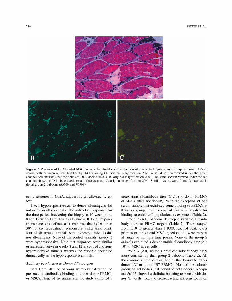
RESULTSrepresenting group responses were normalized as “per-Persistence of Donor MSCs in Musclecent of control” where the control was the pretreatment
MLR response. Four weeks after the second administration of alloge-neic MSCs, muscle biopsies were performed at four of
Alloantibody Assay eight sites of IM injections. The biopsies were evaluatedSera were collected from recipient baboons prior to for the presence of DiO-labeled cells using confocal mi-
MSC injection and at intervals after injection as depicted croscopy. Positive biopsies containing dye-labeled cellsin the tables. These sera were assessed for binding to were found for baboons in group 2 (#6309, #6908) andeither donor PBMCs or donor MSCs by a flow cytome- group 3 (#5500), or 50% of the treated animals. In eachtry method (17). Briefly, donor cells were incubated animal, only one of four biopsy specimens showed evi-with various dilutions of recipient serum (1:10, 1:100, dence of donor cells. The histological results from recip-1:1000) for 1 h at 4°C, washed, and reincubated with a ient #5500 were typical of the positive biopsies and aresecondary biotinylated mouse anti-baboon immunoglob- shown in Figure 2. Serial sections stained with H&E andulin antibody for an additional 60 min at 4°C. Isotype an unstained section viewed under fluorescent light incontrols were run at each dilution using biotinylated nor- the green channel demonstrated DiO-positive donormal mouse immunoglobulin. After 1-h incubation with MSCs between muscle bundles that were homogenoussecondary antibody, FITC-streptavidin, was added for in appearance and uniformly labeled with dye. There30 min at 4°C. Cells were washed and fluorescence was was no evidence of a host cell response to the allogeneicdetermined on a FACS Calibur flow cytometer (BD Bio- MSCs in the form of lymphocyte, macrophage, or neu-sciences, San Jose, CA). The percentage of positive cells trophil infiltration. Viewing the section in the red chan-was determined by analysis using CellQuest software. nel showed no fluorescence, indicating both the absencePositive titers were defined as exceeding a threshold of of DiI-labeled MSCs from the initial IV injection, andmean baseline titers plus 3 SD. Baseline titers were de- the lack of autofluorescence. Muscle samples taken fromtermined for each target cell population using naive se- the injection sites at necropsy (20 weeks after IM injec-rum obtained from nine animals. Sera that did not ex- tion) were negative for DiI- and DiO-labeled cells.hibit significant binding to target cells at a 1:10 dilution
T-Cell Reactivity to Donor Alloantigenswere considered negative.To determine whether allogeneic MSCs induced a
Additional Immunological Parameters systemic T-cell response to donor alloantigens, one-wayMLR assays were performed on blood samples obtainedBlood samples were obtained from recipient baboons
prior to MSC treatment, and at 6, 12, and 26 weeks post- prior to cell infusion and at various intervals after infu-sion. Each MLR assay was harvested at two time points,treatment and evaluated for immunoglobulin levels, anti-
nuclear antibodies, and percentage of various mononu- days 3 and 7, to assess forward shifts in temporal kinet-ics due to T-cell priming in vivo. However, we observedclear cell populations. IgG and IgM immunoglobulins
were quantified by immunoprecipitate turbidity on a Hi- no shift in kinetics: day 3 responses were always lessthan day 7 responses, usually by 75% or greater. There-tachi 717 analyzer using the SPQ Kit System (Diasorin,
Stillwater, MN). Antinuclear antibodies were assessed fore, only day 7 results are reported.Group 1 vehicle control animals did not show signifi-by ELISA (EL-ANA Screen ELISA kit, TheraTest Labs,
Lombard, IL). Analysis of cell surface markers on PBMCs cant increases or decreases in MLR or Con A activityrelative to pretreatment values throughout the initial 12-was performed using a panel of fluorochrome-labeled
monoclonal antibodies diluted and used according to the week period (Fig. 3A). At 16 weeks, there was a signifi-cant drop in both MLR and Con A responsiveness, sug-manufacturer’s instructions (BD Biosciences, San Diego,
CA). The antibody panel consisted of individual or com- gesting that the blood samples had been compromised,possibly during shipping. Therefore, the 16-week sam-binations of antibodies directed to CD3, CD19, CD14,
CD56, CD3/CD25, CD3/CD4, and CD3/CD8. For all ples were omitted from analysis.Group 2 (AA) and group 3 (AB) baboons showedexperiments, nonspecific fluorescence was determined
by substitution with appropriate isotype-matched control decreasing MLR responses to donor alloantigens afterthe second injection of MSCs that attained statistical sig-monoclonal antibodies. Data were analyzed by collect-
ing 10,000 events on a Becton Dickinson Vantage in- nificance at 12 weeks (Fig. 3B and C, respectively). Thesuppressed MLR response was not reflected in the mito-strument using Cell-Quest software. Analysis of vari-
716 BEGGS ET AL.
Figure 2. Presence of DiO-labeled MSCs in muscle. Histological evaluation of a muscle biopsy from a group 3 animal (#5500)shows cells between muscle bundles by H&E staining (A, original magnification 20×). A serial section viewed under the greenchannel demonstrates that the cells are DiO-labeled MSCs (B, original magnification 20×). The same section viewed under the redchannel shows no DiI-labeled cells or autofluorescence (C, original magnification 20×). Similar results were found for two addi-tional group 2 baboons (#6309 and #6908).
genic response to ConA, suggesting an allospecific ef- preexisting alloantibody titer (≥1:10) to donor PBMCsor MSCs (data not shown). With the exception of onefect.
T-cell hyporesponsiveness to donor alloantigens did serum sample that exhibited some binding to PBMCs at8 weeks, group 1 vehicle control sera were negative fornot occur in all recipients. The individual responses for
the time period bracketing the biopsy at 10 weeks (i.e., binding to either cell population, as expected (Table 2).Group 2 (AA) baboons developed variable alloanti-8 and 12 weeks) are shown in Figure 4. If T-cell hypore-
sponsiveness is defined as a response that is less than body titers to PBMC targets (Table 2). Titers rangedfrom 1:10 to greater than 1:1000, reached peak levels30% of the pretreatment response at either time point,
four of six treated animals were hyporesponsive to do- prior to or the second MSC injection, and were presentat single or multiple time points. None of the group 2nor alloantigens. None of the control animals (group 1)
were hyporesponsive. Note that responses were similar animals exhibited a demonstrable alloantibody titer (≥1:10) to MSC target cells.or increased between weeks 8 and 12 in control and non-
hyporesponsive animals, whereas the response decreased Group 3 (AB) animals produced alloantibody titersmore consistently than group 2 baboons (Table 2). Alldramatically in the hyporesponsive animals.three animals produced antibodies that bound to either
Antibody Production to Donor Alloantigens donor “A” or donor “B” PBMCs. Most of the animalsproduced antibodies that bound to both donors. Recipi-Sera from all nine baboons were evaluated for the
presence of antibodies binding to either donor PBMCs ent #6115 showed a definite boosting response with do-nor “B” cells, likely to cross-reacting antigens found onor MSCs. None of the animals in the study exhibited a

IMMUNE RESPONSES TO ALLOGENEIC MSCs IN BABOONS 717
both “A” and “B” cells. Also supporting the notion of ciated with immunoincompetence are usually associatedwith a decrease in the CD3+/CD4+ subsets, and a de-cross-reactivity, administration with “A” induced a
higher titer to “B” than “A” from 3 to 6 weeks in two crease in the CD3+CD4+/CD3+CD8+ ratio (16), unlikethe results reported here. The percentage of CD3+, CD3+/out of three animals, prior to injection with “B” cells.
When MSCs were used as targets, low alloantibody ti- CD25+, CD14+, CD19+, and CD56+ cells was similar be-tween all groups at all time points.ters were exhibited by two out of three animals, and in
one of these animals the titer was present at only oneDISCUSSIONtime point.
This study was undertaken to determine the immuno-Other Immunological Parameters logic consequences and safety of administering multiple
doses of allogeneic MSCs to normal, immunocompetentThere were no significant treatment-related changesin hematology, IgM, and IgG immunoglobulin levels, or animals at clinically relevant doses. Routes of MSC ad-
ministration, particularly the IM route, were chosen toantinuclear antibodies between any of the groups, at anytime point (data not shown). FACS analysis of PBMCs maximize activation of the immune system. We found
that, after injection of MSCs, alloreactive T-cell responsesshowed one minimal, but statistically significant, changein the form of higher percentage of CD3+CD4+ cells to donor alloantigens decreased in most recipients
whereas alloantibody responses were induced. Multiple(48.8 ± 4.7%) and lower percentage of CD3+CD8+ (34.0 ±4.8%) in group 3 baboons at 12 weeks compared to injections of MSCs did not affect the overall immune
system, however, as reflected by broader immunologicgroup 1 control animals (35.9 ± 6.6% and 45.3 ± 4.5%,respectively). The ratio of these subsets was maintained parameters including T-cell responsiveness to mitogen,
peripheral blood mononuclear cell phenotype, immuno-and the total number of lymphocytes was unchanged,compared to the vehicle control group. Conditions asso- globulin levels, and generation of aberrant antibodies
Figure 3. Multiple injections of MSCs induce T-cell hyporesponsiveness to donor alloantigens. Recipient baboon PBMCs wereobtained prior to treatment with MSCs, and at 3, 8, and 12 weeks after treatment. PBMCs were cultured with irradiated donorPBMCs for one-way MLR assays or they were cultured with Con A to determine overall T-cell reactivity. Data are expressed aspercent of pretreatment values, averaged from three animals per group, for group 1 (A), group 2 (B), and group 3 (C). Bars labeledwith an asterisk represent significantly lower mean responses compared to pretreatment values (p < 0.05).

718 BEGGS ET AL.
Figure 4. Individual MLR responses to donor alloantigens. Recipient baboon responses are shownfor 8 and 12 weeks after MSC treatment (see Fig. 3 legend). Data are expressed as percent ofpretreatment values, averaged from triplicate wells. Negative values are due to the response beinghigher to autologous PBMCs than to allogeneic PBMCs. Individual pretreatment responses (alloge-neic CPM response minus autologous CPM response), which represent 100% of the control re-sponse, are as follows (results expressed as mean ∆CMP ± SD): group 1—#5997: 21,051 ± 7592,#6036: 69,696 ± 17,128, #6909: 75,393 ± 15,398; group 2—#6214: 15,300 ± 5570, #6309: 33,056 ±6088, #6908: 41,022 ± 3369; group 3 versus A—#5500: 18,929 ± 650, #6115: 21,537 ± 6612,#6907: 125,441 ± 22,071; group 3 versus B—#5500: 14,092 ± 2705, #6115: 12,489 ± 2072,#6907: 124,291 (no SD, 1 well).
(antinuclear antibodies). The more specific effects of dicated that MSCs can induce T-cell hyporespon-siveness in vivo. In one study, tolerance developed as aMSCs on cellular and humoral alloreactivity did not
cause any apparent health problems throughout the 6- consequence of MSC engraftment and the developmentof mixed chimerism in lethally irradiated mice reconsti-month study period, indicating that the treatment regi-
men used in this study was safe. MSCs were found in tuted with syngeneic bone marrow (8). In a secondstudy, T-cell anergy developed in mice with experimen-half of the treated recipients at 1 month postinjection,
which is considerably longer than the 1–2-week period tal autoimmune encephalomyelitis that were treated withMSCs (33). Unresponsiveness to restimulation in vitroexpected for rejection of differentiated mesenchymal
lineage cells (4,25). All muscle samples taken at nec- was reversed by the addition of IL-2 to the cultures. Thediscrepancy between in vitro and in vivo results may beropsy at 6 months were negative for donor MSCs.
Using the MLR assay as an indicator of T-cell allore- attributed to the complexity of whole animal and lym-phoid organ systems as well as long-term consequencesactivity, we did not observe enhanced recipient T-cell
responses to donor alloantigens in any of the MSC- of donor cell engraftment. Our results support the exist-ing data that MSCs can induce T-cell hyporesponsivenesstreated animals that would suggest priming; in fact, most
recipients demonstrated a progressive loss of alloreactiv- in vivo.Alloantibodies were produced by baboons injectedity with time after treatment while maintaining an over-
all normal T-cell response to Con A. The suppressed with MSCs. Although the role of alloantibodies has beenwell established in solid organ allograft rejection [re-response to donor cells suggests that the recipients were
potentially tolerized by treatment with MSCs, but this viewed in (26)], the role of alloantibodies in stem cellrejection is only starting to be investigated (3). Antibod-remains to be further elucidated as the specificity of the
hyporesponsive state was not determined by analyzing ies can destroy a graft by direct lysis involving the com-plement system or indirectly via antibody-dependent,responses to third-party cells. The results showing that
T-cell hyporesponsiveness developed after administra- cell-mediated cytotoxicity (ADCC) mechanisms. Preex-isting antibody can mediate hyperacute and accelerated/tion of MSCs was not expected, as previous in vitro
studies have shown that MSCs did not tolerize naive acute rejection, which was not the situation in our ani-mals because they did not have alloantibodies reactiveT cells (11,18), and even primed them for secondary
responses (18). However, several recent studies have in- to donor strain antigens in their pretreatment sera but

IMMUNE RESPONSES TO ALLOGENEIC MSCs IN BABOONS 719
developed them after immunization. This response may injection of MSCs (i.e., antibody titers remained consis-tent). Interestingly, all three animals in group 3 had sim-be species related as rats injected intravenously with al-
logeneic MSCs did not develop alloantibodies when ilar or higher titers to donor “B” PBMCs than to donor“A” PBMCs, even prior to being injected with donorevaluated 4 weeks after injection (22).
In our study, most of the baboons produced alloanti- “B” cells. This is probably due to a higher level of ex-pression of MHC molecules on donor “B” PBMCs,bodies in response to injection with allogeneic MSCs.
The single exception was baboon #6907 (group 3), which were used for the second injection in all threeanimals (donor #14485, see Table 1). Antibody titerswhich did not produce any antibody to the immunizing
alloantigen from donor “A.” In most animals, there was were always higher against PBMC targets than MSC tar-gets, likely due to higher expression of MHC moleculesnot a boosting effect on antibody titer after the second
Table 2. Alloantibody Response to Donor Cells
Baboon Baboon Baboon#5997 #5036 #6909
Group 1 (Random) PBMCs MSCs PBMCs MSCs PBMCs MSCs
3 weeks <1:10 <1:10 <1:10 <1:10 <1:10 <1:106 weeks <1:10 <1:10 <1:10 <1:10 <1:10 <1:108 weeks <1:10 <1:10 1:10 <1:10 <1:10 <1:1012 weeks <1:10 <1:10 <1:10 <1:10 <1:10 <1:1016 weeks <1:10 <1:10 <1:10 <1:10 <1:10 <1:10
Baboon Baboon Baboon#6214 #6309 #6908
Group 2 (Donor 11A”) PBMCs MSCs PBMCs MSCs PBMCs MSCs
3 weeks ≥1:1000 <1:10 1:10 <1:10 <1:10 <1:106 weeks 1:10 <1:10 1:10 <1:10 <1:10 <1:108 weeks 1:100 <1:10 <1:10 <1:10 ≥1:1000 <1:1012 weeks <1:10 <1:10 <1:10 <1:10 <1:10 <1:1016 weeks 1:10 <1:10 <1:10 <1:10 <1:10 <1:10
Baboon Baboon Baboon#5500 #6115 #6907
Group 3 (Donor “A”) PBMCs MSCs PBMCs MSCs PBMCs MSCs
3 weeks 1:10 <1:10 1:100 <1:10 <1:10 <1:106 weeks 1:10 <1:10 <1:10 <1:10 <1:10 <1:108 weeks 1:10 <1:10 ≥1:1000 1:10 <1:10 <1:1012 weeks <1:10 <1:10 ≥1:1000 1:10 <1:10 <1:1016 weeks <1:10 <1:10 ≥1:1000 <1:10 <1:10 <1:10
Baboon Baboon Baboon#5500 #6115 #6907
Group 3 (Donor “B”) PBMCs MSCs PBMCs MSCs PBMCs MSCs
3 weeks 1:100 <1:10 1:100 <1:10 1:10 <1:106 weeks 1:100 <1:10 <1:10 <1:10 1:100 <1:108 weeks 1:100 1:10 ≥1:1000 1:10 1:10 <1:1012 weeks <1:10 <1:10 ≥1:1000 <1:10 1:10 <1:1016 weeks 1:100 <1:10 ≥1:1000 <1:10 1:10 <1:10
Alloantibody binding titers were determined from blood samples obtained from recipient baboonsat the times indicated post-MSC treatment. PBMCs and MSCs were used as target cells in theassay. Group 1 sera were evaluated using target cells from random animals, group 2 sera weretested against cells from donor “A,” and group 3 sera were evaluated against both donors “A”and “B” because these animals were injected with MSCs derived from both donors.

720 BEGGS ET AL.
Table 3. Individual Responses of Baboons to Allogeneic MSC Transplantation
T-Cell
Alloantibody
Hyporesp. State
Titers
PBMC MSCBaboon MSC in
Group No. Biopsy 8 Weeks 12 Weeks 8 Weeks 12 Weeks 8 Weeks 12 Weeks
2 (AA) 6214 Neg Neg Neg 1:100 Neg Neg Neg6309 Pos Inter Inter Neg Neg Neg Neg6908 Pos Inter High 1:1000 Neg Neg Neg
3 (AB) 5500 Pos Inter Inter 1:100 Neg 1:10 Neg6115 Neg Neg Neg 1:1000 1:1000 1:10 Neg6907 Neg High High 1:10 1:10 Neg Neg
MSC biopsy positivity was determined on week 10 of study by confocal microscopy to DiO-labeled MSCs. T-cellhyporesponsive state on weeks 8 and 12 was arbitrarily rated according to the following scale: Negative (Neg, >75%of control), Intermediate (Inter, 25–75% of control), and High (<25% of control). Alloantibody titers are shown toPBMC and MSC target cells (donor “A” for group 2 and donor “B” for group 3).
on the PBMC population. Alternatively, it is possible for at least 1 month in muscle biopsies of half of thesenormal recipient animals. It seems likely that the immu-that contaminating PBMCs present in the MSC popula-
tion were responsible for inducing PBMC-specific anti- nomodulatory properties of MSCs, such as the pre-viously reported ability of MSCs to suppress alloreactivebodies.
Does tolerance or alloantibody response account for T cells in vitro and in vivo, and the newly describedability of these cells to induce T-cell hyporespon-the success or failure of survival of the injected MSCs?
A summary of data for the most relevant time points siveness to donor alloantigens in vivo, contributed to themaintenance of the allogeneic MSCs. Our failure tobracketing the muscle biopsy for individual baboons is
shown in Table 3. In all three animals in which cell demonstrate MSCs in the muscle biopsies at extendedtime points could be due to cell migration, loss of fluo-survival was demonstrated, T-cell hyporesponsiveness
to donor alloantigens was induced, whereas in two out rescent label, chronic rejection response by cells or anti-bodies, or lack of engraftment into a normal, noninjuredof three animals in which MSCs were not detected in
the biopsy specimens, hyporesponsiveness was not in- environment. The baboon model used in our study wasnot one of functional tissue repair; it is possible that induced. In the single animal in which MSCs were not
detected but hyporesponsiveness was induced (group 3 an injury model, MSCs may be recruited to the site ofinjury and persist for longer periods of time in this envi-baboon #6907), biopsy sampling error may have re-
sulted in missing the intramuscular depot of cells as only ronment. Clinically, these results are encouraging for asafe “universal cell” approach in which MSCs expandedone in four biopsies taken from positive baboons showed
evidence of donor MSCs. Overall, our results suggest a from a single donor could be used in a variety of patientsfor tissue repair, particularly in acute settings when au-relationship between hyporesponsiveness and cell survival.
The relationship between MSC survival and alloanti- tologous isolation and expansion is not feasible.body production is more tenuous. Two of three animals ACKNOWLEDGMENTS: The authors would like to thank Dr.with MSC-positive biopsies had sera at 8 weeks that Marla McIntosh for performing statistical analyses on the
MLR data. We are indebted to Mark Pittenger for review ofcontained alloantibodies that bound to PBMCs; one ofthe manuscript.these sera contained antibodies that bound to donor
MSCs (Table 2). We speculate that the antibodies mayREFERENCESnot have inflicted damage to the MSCs due to low titer
(1:10) or to antibody-mediated enhancement of graft 1. Aggarwal, S.; Pittenger, M. F. Human mesenchymal stemsurvival (6). cells modulate allogeneic immune cell responses. Blood
105:1815–1822; 2005.In summary, IV infusion of high, clinically relevant2. Bartholomew, A.; Sturgeon, C.; Siatskas, M.; Ferrer, K.;doses of mismatched, allogeneic MSCs followed by IM
McIntosh, K. R.; Patil, S.; Hardy, W.; Devine, S.; Ucker,injection of MSCs from the same mismatched donor orD.; Deans, R.; Moseley, A.; Hoffman, R. Mesenchymal
a third mismatched donor did not result in any changes stem cells suppress lymphocyte proliferation in vitro andin the overall health or immune status of the recipient prolong skin graft survival in vivo. Exp. Hematol. 30:42–animals. Transplanted allogeneic MSCs were detected 48; 2002.
IMMUNE RESPONSES TO ALLOGENEIC MSCs IN BABOONS 721
3. Bradley, J. A.; Bolton, E. M.; Pedersen, R. A. Stem cell 20. Le Blanc, K.; Tammik, L.; Sundberg, B.; Haynesworth,S. E.; Ringden, O. Mesenchymal stem cells inhibit andmedicine encounters the immune system. Nat. Rev. Im-
munol. 2:859–871; 2002. stimulate mixed lymphocyte cultures and mitogenic re-sponses independently of the major histocompatibility4. Camirand, G.; Caron, N.; Asselin, I.; Tremblay, J. P.
Combined immunosuppression of mycophenolate mofetil complex. Scand. J. Immunol. 57:11–20; 2003.21. Le Blanc, K.; Rasmusson, I.; Sundberg, B.; Gotherstrom,and FK506 for myoblast transplantation in mdx mice.
Transplantation 72:38–44; 2001. C.; Hassan, M.; Uzunel, M.; Ringden, O. Treatment ofsevere acute graft- versus-host disease with third party5. Caplan, F Mesenchymal stem cells. J. Orthop. Res. 9:
641–650; 1991. haploidentical mesenchymal stem cells. Lancet 363:1439–1441; 2004.6. Carpenter, C. B.; d’Apice, A. J. F.; Abbas, A. K. The role
of antibodies in the rejection and enhancement of organ 22. Li, Y.; McIntosh, K.; Chen, J.; Zhang, C.; Gao, Q.; Borne-man, J.; Raginski, K.; Mitchell, J.; Shen, L.; Zhang, J.;allografts. Adv. Immunol. 22:1–65; 1976.
7. Deans, R. J.; Moseley, A. B. Mesenchymal stem cells: Lu, D.; Chopp, M. Allogeneic bone marrow stromal cellspromote glial-axonal remodeling without immunologicBiology and potential clinical uses. Exp. Hematol. 28:
875–884; 2000. sensitization after stroke in rats. Exp. Neurol. 198:313–325; 2006.8. Deng, W.; Han, Q.; Liao, L.; Li, C.; Ge, W.; Zhao, Z.;
You, S.; Deng, H.; Zhao, C. H. Allogeneic bone marrow- 23. Liechty, K. W.; MacKenzie, T. C.; Shaaban, A. F.; Radu,A.; Moseley, A. M.; Deans, R.; Marshak, D. R.; Flake,derived flk-1+Sca-1- mesenchymal stem cells leads to sta-
ble mixed chimerism and donor-specific tolerance. Exp. A. W. Human mesenchymal stem cells engraft and dem-onstrate site-specific differentiation after in utero trans-Hematol. 32:861–867; 2004.
9. Devine, S. M.; Peter, S.; Martin, B. J.; Barry, F.; McIn- plantation in sheep. Nat. Med. 6:1282–1286; 2000.24. Majumdar, M. K.; Keane-Moore, M.; Buyaner, D.; Hardy,tosh, K. R. Mesenchymal stem cells: Stealth and suppres-
sion. Cancer J. 7:S76–82; 2001. W. B.; Moorman, M. A.; McIntosh, K. R.; Mosca, J. D.Characterization and functionality of cell surface mole-10. Devine, S. M.; Cobbs, C.; Jennings, M.; Bartholomew, A.;
Hoffman, R. Mesenchymal stem cells distribute to a wide cules on human mesenchymal stem cells. J. Biomed. Sci.10:228–241; 2003.range of tissues following systemic infusion into non-
human primates. Blood 101:2999–3001; 2003. 25. Malejczyk, J.; Osiecka, A.; Hyc, A.; Moskalewski, S. Ef-fect of immunosuppression on rejection of cartilage11. Di Nicola, M.; Carlo-Stella, C.; Magni, M.; Milanesi, M.;
Longoni, P. D.; Matteucci, P.; Grisanti, S.; Gianni, A. M. formed by transplanted allogeneic rib chondrocytes inmice. Clin. Orthop. 269:266–273; 1991.Human bone marrow stromal cells suppress T-lymphocyte
proliferation induced by cellular or nonspecific mitogenic 26. Mason, D. W.; Morris, P. J. Effector mechanisms in allo-graft rejection. Annu. Rev. Immunol. 4:119–145; 1986.stimuli. Blood 99:3838–3843; 2002.
12. Djouad, F.; Plence, P.; Bony, C.; Tropel, P.; Apparailly, 27. McIntosh, K.; Bartholomew, A. Stromal cell modulationof the immune system. A potential role for mesenchymalF.; Sany, J.; Noel, D.; Jorgensen, C. Immunosuppressive
effect of mesenchymal stem cells favors tumor growth in stem cells. Graft 3:324–328; 2000.28. Meisel, R.; Zibert, A.; Laryea, M.; Gobel, U.; Daubener,allogeneic animals. Blood 102:3837–3844; 2003.
13. Friedenstein, A. J. Precursor cells of mechanocytes. Int. W.; Dilloo, D. Human bone marrow stromal cells inhibitallogeneic T-cell responses by indoleamine 2,3-dioxygen-Rev. Cytol. 47:327–359; 1976.
14. Glennie, S.; Soeiro, I.; Dyson, P. J.; Lam, E. W.; Dazzi, ase-mediated tryptophan degradation. Blood 103:4619–4621; 2004.F. Bone marrow mesenchymal stem cells induce division
arrest anergy of activated T cells. Blood 105:2821–2827; 29. Owen, M. E. Marrow stromal cells. J. Cell Sci. 10:63–76;1988.2005.
15. Haynesworth, S. E.; Baber, M. A.; Caplan, A. I. Cell sur- 30. Pittenger, M. F.; Mackay, A. M.; Beck, S. C.; Jaiswal,R. K.; Douglas, R.; Mosca, J. D.; Moorman, M. A.; Simo-face antigens on human bone marrow-derived mesenchy-
mal stem cells are detected by monoclonal antibodies. netti, D. W.; Craig, S.; Marshak, D. R. Multilineage po-tential of adult human mesenchymal stem cells. ScienceBone 13:69–80; 1992.
16. Huston, D. P. The biology of the immune system. JAMA 284:143–147; 1999.31. Saito, T.; Kuang, J-Q.; Bittira, B.; Al-Khaldi, A.; Chiu,278:1804–1814; 1997.
17. Kawai, T.; Poncelet, A.; Sachs, D. H.; Mauiyyedi, S.; R. C-J. Xenotransplant cardiac chimera: immune toleranceof adult stem cells. Ann. Thorac. Surg. 74:19–24; 2002.Boskovic, S.; Wee, S. L.; Ko, D. S.; Bartholomew, A.;
Kimikawa, M.; Hong, H. Z.; Abrahamian, G.; Colvin, 32. Tse, W. T.; Pendleton, J. D.; Bever, W. M.; Egalka,M. C.; Guinan, E. C. Suppression of allogeneic T-cell pro-R. B.; Cosim,i A. B. Long-term outcome and alloantibody
production in a non-myeloablative regimen for induction liferation by human marrow stromal cells: Implications intransplantation. Transplantation 75:389–397; 2003.of renal allograft tolerance. Transplantation 68:1767–
1775; 1999. 33. Zappia, E.; Casazza, S.; Pedemonte, E.; Benvenuto, F.;Bonanni, I.; Gerdoni, E.; Giunti, D.; Ceravolo, A.; Caz-18. Klyushnenkova, E.; Mosca, J. D.; Zernetkina, V.;
Majumdar, M. K.; Beggs, K. J.; Simonetti, D. W.; Deans, zanti, F.; Frassoni, F.; Mancardi, G.; Uccelli, A. Mesen-chymal stem cells ameliorate experimental autoimmuneR. J.; McIntosh, K. R. T cell responses to allogeneic hu-
man mesenchymal stem cells: Immunogenicity, tolerance, encephalomyelitis inducing T cell anergy. Blood 106:1755–1761; 2005.and suppression. J. Biomed. Sci. 12:47–57; 2005.
19. Lazarus, H. M.; Koc, O. N. Culture-expanded human mar-row-derived MSCs in clinical hematopoietic stem celltransplantation. Graft 3:329–333; 2000.

Copyright © 2022 FDOKUMEN