Melatonin regulates glucocorticoid receptor: an answer to its antiapoptotic action in thymus

T H E ROLE OF T H E THYMUS I N D E V E L O P M E N T OF IMMUNO- LOGIC CAPACITY I N RABBITS AND MICE*
BY ROBERT A. GOOD,:~ M.D., AGUSTIN P. DALMASSO,§ M.D., CARLOS MARTINEZ,] I M.D., OLGA K. ARCHER,¶ JAMES C. PIERCE,** M.D.,
AI~D B E N W. PAPERMASTER,$:~
(From the Pediatric Research Laboratories of the Variety Club Heart Hospital, and the Departments of Physiology and Surgery, University of Minnesota, Minneapolis)
PLATES 104 ~O 107
(Received for publication, July 5, 1962)
Although the thymus gland has been known from antiquity, its role in the body economy has remained enigmatic until recently. During the past year, however, a flurry of reports have given the first indications of the role of this organ in developmental biology. In mammals the thymus develops from the third and fourth pharyngeal pouches in early embryonic life, reaches maximal relative size near the time of birth, and then undergoes a gradual involution. In man the thymus appears in the 10 mm embryo, reaches maximal relative size in the neonate, attains maximal absolute size in the 12-year-old child, and then ..... gradually decreases in relative and absolute size as maturity is reached (1).
Our interest in the thymus developed in 1953 when we (2, 3) discovered that a patient with "acquired agammaglobulinemia" had developed a marked immunologic deficiency and an abnormality of the thymus--a benign thymoma--at about the same time. Removal of the thymoma, which was primarily an epithelial stromal overgrowth of the thymus pathologically, failed to alter either the protein abnormality or the immunologic defect. Since that time seven cases of the combined occurrence of these two disorders have been reported (4-8), and in no instance has removal of the thymic tumor restored immunologic function or the protein deficit. Nonetheless, it seems clear that the association of these two rare conditions has been far too frequent to be ex- plained by chance alone.
* Aided by grants from the National Foundation, the United States Public Health Service, the American Cancer Society, the Minnesota Heart Association, and the American Heart Association.
:~ American Legion Memorial Heart Research Professor of Pediatrics. § Postdoctoral Cancer Research Trainee, Department of Physiology, NIH Training Grant
CRT-5023. ]] American Cancer Society Professor of Physiology. ¶ Research Fellow, Department of Pediatrics, NIH Training Grant HTS-5462. ** Medical Fellow, Department of Surgery. $$ United States Public Health Service Graduate Student Trainee, Department of Micro-
biology. 773
on August 26, 2016
jem.rupress.org
Dow
nloaded from
Published November 1, 1962

774 THY~[US IN DEVELOPMENT OF IMMUNOLOGIC CAPACITY
In an attempt to exploit the initial clinical observation, we (9, 10) studied the effect of thymectomy on the immunologic capacity of adult rabbits. In these experiments, which confirmed the earlier findings of Harris et al. (11) we could demonstrate no effect of thymectomy on immunologic capacity. These observations seemed consonant with earlier findings (12-14) that the thymus, unlike other lymphoid organs, remains aloof from antigenic stimulation in adult animals and does not seem to form antibody, except perhaps when antigen is injected directly into its substance (15).
When it became apparent that the bursa of Fabricins (16, 17), a thymus4ike organ of the chicken, plays an important role in the early development of immunologic potential in that animal, we turned to study of the effect of thymectomy in newly born animals on the subsequent development of immunologic capacity. Archer and Pierce, from this laboratory, presented the first results of these studies at the meeting of the American Society of Immunologists in 1961 (18). In substance, we found that thymectomy in newborn rabbits had an effect on development of immunologic poten- tial. Fichtelius et al. (19), in work with guinea pigs, and more recently Miller (20) and Martinez et al. (21), in independent studies of inbred strains of mice, made similar observations.
I t is the purpose of this repor t to present our findings linking the thymus to the development of immunologic capaci ty in the mouse and the rabbit . I t will be shown tha t in both of these species, bu t most consistently and most com- pletely in the mouse, the thymus plays an essential role in matura t ion of im- munologic function and in the development of the anatomic integr i ty of the lymphoid tissues. Fur ther , evidence will be presented indicating tha t thymec- tomy in mice as late as 35 to 40 days after b i r th may affect capaci ty to reject skin homotransplants and m a y also affect resistance of F1 hybr id mice to the development of immunologic runt disease (homologous disease) (22, 23).
Materials and Methods
Rabbits:
New Zealand albino rabbits were obtained from a single local breeder in the terminal stages of gestation, allowed to deliver in secluded cages in our animal quarters, and the young were divided into litter mate groups. The young were kept with the dam until self-sufficient, and then separated and fed Purina rabbit chow, greens, and water ad libitum.
Thymectomy in Rabbits.--Rabbits were anesthetized with ether and thymectomized before the 7th day of life through a median incision of the chest wall. The thymus was removed with a glass aspirating pipette or by surgical dissection, and the incision closed with silk suture.
Antigenic Stimulation and Antibody Production in Rabbit.--~ two experiments, antigenic stimulation was provided with bovine serum albumin (BSA) in a dosage of 50 mg per kg, given intravenously when the rabbits were 5 to 8 weeks of age. Anti-BSA content of the serum was measured qualitatively by the method of Swift et al. (24) on the 14-day serum samples, and quantitatively by the ammonium sulfate precipitation method of Farr (25) on 14- and 21-day serum samples. In a third group of animals, antigenic stimulation was provided by 8 > 10 ~ particles per kg of T2 coliphage, given intravenously at 14 to 18 weeks of age. Phage neutralization procedures (26) were carried out on 7-day serum samples. All blood was ob- tained by cardiac puncture.
on August 26, 2016
jem.rupress.org
Dow
nloaded from
Published November 1, 1962

GOOD, DALM.ASSO, ~[ARTINEZ, ARCHER, PIERCE, PAPERMASTER 775
Transplantation Immunity in Rabb/ts.--Skin homografting was performed essentially by the method described by Stetson and Demopoulos (27). Each grafted rabbit received 2 auto- grafts, 1 on each ear, and 2 homografts, 1 from another New Zealand rabbit and the other from a Dutch rabbit. Mice:
Mice of the following strains were used in these studies: C67B1/1, CsH, Ce, DBA/2, A, Balb/C, (A x CffI)F,, (A x Cs~BI)F,, (Baib/C x DBA/2)F, , (Call x DBA/2)F, , and (Balb/C x A)F, . The CJ-I mice were obtained from the stock originally maintained by Dr. J. J. Bittner. Newborn mice remained with the mother until old enough to eat by themselves, and were then fed Purina fox chow and water nd libitum.
Thym~tomy in'Mice.--Mico were thymectomized by a modification of the technique used by Gross (28). This modification is essentially the same as that reported by Dischler and Rudali (29).1
Antigenic Stimulation and Antibody Production in Mice.--Ia mice the antigenic stimulation was provided by intraperitoneal injection of 2 × 101° particles of bacteriophage T~ at 2 months of age. The animals were bled 7 days later from the retro-orbital venous plexus into capillary tubes. The tubes were centrifuged at 4°C at 1500 G, and the serum removed and tested for virus neutralization at 37°C. Serum samples were diluted 1:50 in 20 per cent normal rabbit serum in 0.9 per cent NaCI, and incubated with 105 particles of bacteriophage per ml in the reaction tubes. The bacteriophage particles remaining active after 2 and 24 hours of incubation were assayed in the usual way (26). The mice were sacrificed and autopsied 2 weeks after bleeding.
Transplantation Immunity in Mice.--Skin homotransplantation immunity was tested by placing 1.5 by 2 cm skin homografts on the back of the tested animals. The grafting technique has been described (30).2 Tumor transplantation immunity was studied by taking a mammary adenocarcinoma which originated spontaneously in an A breeder female, and transplanting pieces of this tumor into the subcutaneous tissue of C+H mice. "Takes" were recorded when the tumor in the foreign host had reached 1 cm in size and was growing.
Growth of Mioe.--The mice were weighed at frequent intervals on a Mettler balance, and the weights of thymectomized and sham-operated animals compared at 4, 8, and 12 weeks.
Immunologic Activity of Spleen and Lymph Node Cdls in M/ce.--Mice of A, Balb/C, and CaI-I strains were thymectomized or sham-operated during the 1st or 6th day of life and sacrificed 2 months later. Their spleens or lymph nodes were made into suspensions in Locke- Ringer's saline solution. Each dose of 0.1 cc contained 10 million viable spleen cells. The hybrid recipients, which were 8 days old at the time of transfer, were sacrificed 8 days later, and the Simonsen assay of graft versus host reaction was determined (31).
Runt Disease in Mice.--(A x Call)F, and (A x C67B1)F1 mice were thymectomized or sham- operated at birth or at 40 days of age. Those thymectomized in the newborn period were given A strain spleen cells intraperitoneally at 35 days of age; those thymectomized at 40 days of
1 The immediate surgical mortality of mice and rabbits in the early experiments was often quite high, in some experiments as high as 90 per cent. More recently, the immediate mortality of the thymectomy procedure in newborn mice in our laboratories has been in the neighborhood of 10 to 20 per cent.
The criteria of skin homograft acceptance varied with the conditions of the individukl study, and details are furnished in the text and/or table in the Results section. When mice thymeetemized at birth were grafted, however, the observation period always continued to clear-cut slough of the graft or the death of the animal. These animals are short-lived, and no mouse is included in the tabulations as a graft survival unless it lived for at least 35 days after grafting.
on August 26, 2016
jem.rupress.org
Dow
nloaded from
Published November 1, 1962

776 THYMUS Ii~ DEVELOPMENT OF IMMUNOLOGIC CAPACITY
age were given adult A strain spleen cells intravenously or intraperitoneally 10 days after the surgery.
Histologi~ Stud/es.--Spleen, lymph node, thymus, and gut tissue were taken at birth and at intervals after birth from thymectomized and control mice, placed in either 10 per cent neutral formalin or 100 per cent ethyl alcohol, sectioned, and stained with hematoxylin and eosin, or methyl green-pyronin. The methyl green-pyronin staining technique used in our laboratory has been previously described (32).
TABLE I
Effect of Tkymectomy in RabMts Less Tkan 5 Days of Age on Production of Antibody to Bo~ne Serum Albumin*
14 day bleeding 21 day bleeding
Group
Thymectomized
Unthymectomized (sham-operated and non-operated)
Num- ber of
rabbits
7
I0
AntJbody~ gamma Si ~ni: icance nitrogen/lO0 ml c ilIe fence
Mean --- 6.6 Range = 0 to
19.5 P < 0.01
Mean = 52.4 Range -- 0 to
129.0
Num- ber of rabbits
10
Antibody~: gamma nitrogen/100 ml
Mean = 9 .8 Range ---- 0 to
42.5
Mean = 62.2 Range 14.5 to
207.9
Signifi- cance of
difference
P < 0.01
* Antibody was measured by the ammonium sulfate precipitation technique of Farr (25). ~t Results are expressed as gamma nitrogen of Ira-labeled BSA bound per 100 ml of un-
diluted serum determined at that dilution of serum which bound 33 per cent of 0.01 gamma of BSA nitrogen added.
RESULTS
Effect of Neonatal Thymectomy on Development of Immunologic Capacity in Rabbits.-
In the first experiment, rabbits under 5 days of age were either thymeetomized, sham- operated, or left alone. Challenge with BSA (50 mg/kg) was carried out when the animals were 7 to 8 weeks old. In the second experiment, thymectomy or sham operation was per- formed when the rabbits were 5 to 7 days of age, and BSA administered intravenously at 5 to 7 weeks. In the third experiment, the rabbits were thymectomized or sham-operated at 0 to 3 days of age, and immunized with 8 X l& particles of T~ coliphage per kg at 14 to 18 weeks of age.
In the first experiment, none of 7 thymectomized rabbits produced sufficient antibody to BSA to be detected by the qualitative capillary tube precipitin technique, whereas 8 of 10 sham-operated or unoperated litter mate controls formed demonstrable precipitating antibody. One rabbit whose thymus had been only partially removed produced sufficient antibody to be detected by this precipitation technique. Quantitative data on the same serums, obtained by
on August 26, 2016
jem.rupress.org
Dow
nloaded from
Published November 1, 1962
GOOD, DALMASSO, M.ARTINEZ, ARCHER, PIERCE, PAPERMASTER 777
the ammonium sulfate precipitation method of Farr (25) are summarized in Table I. The thymectomized rabbits had significantly smaller quantities of ant ibody at 14 and 21 days than did sham-operated or unoperated controls.
Table I I records the quantitative data from the second experiment, involving thymectomy at 5 to 7 days after birth and challenge with BSA at 5 to 7 weeks. Antibody production was reduced both 14 and 21 days after antigen adminis- tration in the thymectomized group. Although these results suggested immuno- logic feebleness of thymectomized rabbits, the differences were not significant statistically. The capillary tube precipitin method revealed ant ibody in 14-day serum samples of 4 of 9 thymectomized rabbits and 16 of 27 unthymectomized animals.
TABLE II Effect of Thymectomy in Rabbits 5 to 7 Days ot Age on Production of Antibody to Bo~ne Serum
Albumin*
Group
Thymectomized
Unthymectomized (sham-operated and non-operated)
Number of rabbits
9
26
14 day bleeding 21 day bleeding
Antibody~ gamma Number nitrogen/100 ml of rabbits
Mean -- 23.3 8 Range -- 0 to 57.0
Mean ffi 31.0 21 Range ~ 0 to 9 5 . 7 The differences are not statistically significant.
Antibody:[: gamma nitrogen/100 ml
Mean = 25.1 Range = 7 . 8 to 5 8 . 4
Mean = 41.8 Range = 0 to 178.0
* Antibody was measured by the ammonium sulfate precipitation technique of Farr (25). Results are expressed as gamma nitrogen of Ital-labeled BSA bound per 100 ml of un-
diluted serum determined at that dilution of serum which bound 33 per cent of 0.01 gamma of BSA nitrogen added.
In the third experiment, the rabbits were thymectomized or sham-operated before 3 days of age and challenged with T~ coliphage 14 to 18 weeks later. The results of bacteriophage neutralization by 7-day serum samples are recorded in Table I I I . In this experiment the thymectomized animals again formed less ant ibody than did the control animals, but the results were not statistically different.
In a final study, rabbits which had been thymectomized or sham-operated at birth were tested for abnormalities in homotransplantation immunity.
Four rabbits completely thymectomized before 3 days of age and 3 sham-operated controls were subjected to skin homotransplantation 6 months later. In essence, I cm circular auto- grafts were exchanged between the 2 ears of each rabbit. In addition, the right ear received a 1 cm circular homograft from a Dutch strain donor and the left ear a 1 cm circular homograft from a homologous donor of New Zealand stock.
on August 26, 2016
jem.rupress.org
Dow
nloaded from
Published November 1, 1962

778 THYMUS IN DEVELOPMENT OF TM'MUNOLOGIC CAPACITY
The results, summarized in Table IV, indicate tha t thymectomy in the neo- natal period does not interfere significantly with the capacity of rabbits to reject skin homotransplants.
These experiments indicate tha t rabbits thymectoralzed shortly after birth form less ant ibody after they have matured than do sham-operated and un-
TABLE HI Antibody Production to T, Coli Bacteriophage in Rabbits Thymectomized a~ 0 to 3 Days of Age*
Per cent surviving Per cent surviving Group 1 hr.$ 2 hrs.$
Thymectomized at 0 to 3 days of age 86 66 75 96 78 76 83 69
85 63 67 71 56 67 64 41
Mean .4- standard error 78 -4- 5 64 .4- 5§
Sham-operated or unoperated controls 66 56 80 83 97 26 - - (Plating error) 54
45 31 34 72 89 5
72 23
Mean ± standard error 66 .4- 11 46 4- 10§
Diluent control Not determined 95 .4- 0.5
* Rabbits were immunized with 8 X 10⁹ particles of phage at 14 to 18 weeks of age. Figures refer to the per cent of surviving phage following addition of 105 particles/ml to
tubes containing 5 cc of a 1:100 dilution of antiserum. § The difference between the means of the 2-hour determinations is not statistically
significant (P value between 0.05 and 0.1).
operated controls. The difference between the thymectomized and control groups was least pronounced in the second experiment (Table I I) , the s tudy which involved the latest thymectomy (at 5 to 7 days of age), suggesting that the state of maturat ion of the lymphoid tissue of the rabbit in the immediate neonatal period may be important. In both other experiments, the animals were thymectomized before 5 days of age. In one the difference between the thymec-
on August 26, 2016
jem.rupress.org
Dow
nloaded from
Published November 1, 1962
GOOD, DALMASSO, MARTINEZ, ARCHER~ PIERCE, PAPERMASTER 779
tomized and control animals was statistically significant; in the other it ap- proached significance.
The final experiment indicates that thymectomy carried out immediately after birth in the rabbit does not reduce immunologic capacity sufficiently to permit skin homotransplantation. I t seems likely that more consistent and more complete inhibition of the development of the immunologic capacity of the rabbit would require even earlier thymectomy, probably considerably be- fore birth (33).
The rabbit, then, although revealing immunologic incompetence following early thymectomy in some experiments, is not the experimental animal of
TABLE IV Effect of Neonatal Thymectomy on Skin Homotransplantation in Rabbits
Group
rhymectomized at 0 to 3 days of age*
~ham-operated at 0 to 3 days of age
Rabbit No.
Dutch rabbit skin homotransplant
Da Day hemor~r~agic sloughed
- - 18
10 14 9 17
10 18 10 14 m 18
New Zealand rabbit skin homotransplant
Day Day hemorrhagic sloughed
9 18 8 16
1 0 1 4
9 17
10 18 10 14 9 18
* These animals had previously been shown to be quantitatively deficient antibody production against T2 bacteriophage.
Technical failure of graft.
with respect to
choice for studies of the role of the thymus in development of immunologic capacity at this time.
Immunologic Incompetence of Thymectomized Mice. u
In these experiments newborn mice of the DBA/2 strain were divided into two groups. Members of one group were thymectomized, while members of the second group were sham- operated as controls. Two months later, each mouse was injected intraperitoneaUy with 2 X 10 l° particles of bacteriophage T2. The phage neutralization procedures were performed on 7-day serum samples.
Recorded on Table V are the results of this study which reveal a definite difference in neutralizing capacity of the antiserums from thymectomized and sham-operated animals. Mter the 24 hour incubation, neutralization of bacterio- phage was complete in the serums of all the sham-operated mice, whereas only one of the serums from mice subjected to thymectomy showed complete neu-
on August 26, 2016
jem.rupress.org
Dow
nloaded from
Published November 1, 1962
780 THYMUS IN DEVELOPMENT OF 7MMUNOLOGIC CAPACITY
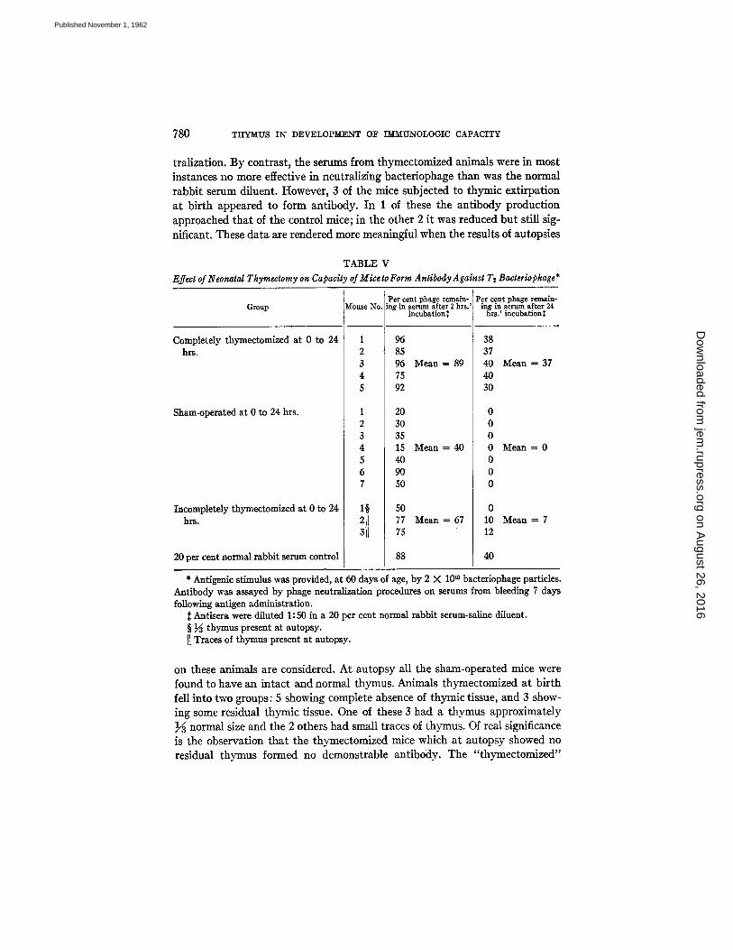
tral ization. By contrast , the serums from thymectomized animals were in most instances no more effective in neutral izing bacter iophage than was the normal rabbi t serum diluent. However, 3 of the mice subjected to thymic ext i rpat ion a t b i r th appeared to form ant ibody. In 1 of these the an t ibody product ion approached tha t of the control mice; in the other 2 i t was reduced bu t still sig- nificant. These da t a are rendered more meaningful when the results of autopsies
TABLE V Effect of Neonatal Thymectomy on Capacity of Mice to Form Antibody A gainst T2 Bacteriophage*
Group
Completely thymectomized at 0 to 24 hrs.
Sham-operated at 0 to 24 hrs.
Incompletely thymectomized at 0 to 24 hrs.
20 per cent normal rabbit serum control
Per cent phage remain- [ng in serum after 2 hrs.'
incubation:~
96 85 96 Mean = 89 75 92
20 30 35 15 Mean = 40 40 90 50
50 77 Mean = 67 75
88
Per cent phage remain- ing in serum after 24
hrs.' incubatlon$
38 37 40 Mean = 37 40 30
0 0 0 0 M e a n = 0
0 0 0
0 10 Mean = 7 12
40
* Antigenic stimulus was provided, at 60 days of age, by 2 X 10 *° bacteriophage particles. Antibody was assayed by phage neutralization procedures on serums from bleeding 7 days following antigen administration.
Antisera were diluted 1: 50 in a 20 per cent normal rabbit serum-saline diluent. § ~ thymus present at autopsy. ]1 Traces of thymus present at autopsy.
on these animals are considered. A t au topsy all the sham-operated mice were found to have an in tac t and normal thymus. Animals thymectomized a t b i r th fell into two groups: 5 showing complete absence of thymic tissue, and 3 show- ing some residual thymic tissue. One of these 3 had a thymus approximate ly }6 normal size and the 2 others had small traces of thymus. Of real significance is the observation tha t the thymectomized mice which a t au topsy showed no residual thymus formed no demonstrable ant ibody. The " thymectomized"
on August 26, 2016
jem.rupress.org
Dow
nloaded from
Published November 1, 1962

GOOD, DALMASSO, MARTINEZ, ARCHER, PIERCE, PAPERMASTER 781
mouse having a residual thymus 1/~ the normal size formed antibodies about as well as did sham-operated controls, and the 2 mice with smaller residuals of thymus formed amounts of virus-neutralizing antibodies intermediate between the 2. The results of antibody production in these three groups may be com- pared on Table V.
Acceptance of tIomografts across Minor Histocompatibility Barriers in Mice . - In our initial experiments concerning the effect of neonatal thymectomy on homotransplantation immunity, inbred strains of mice similar to one another at the H-2 histocompatibility locus but differing at other genetic loci were em- ployed. Implicit in the design of these experiments was the concept that if thymectomy in the neonatal period should produce a quantitative reduction in capacity to develop transplantation immunity, it might be revealed by testing homografts between strains of animals differing from one another in weaker histocompatibility antigens (34). To this end, four inbred strains of mice were studied. These included the DBA/2, CeJ-I, and Ce strains, and F1 hybrids re- sulting from the cross between Balb/C and DBA/2 parents.
In an initial experiment DBA/2 mice were divided into two groups. Members of one group were thymectomized at 0 to 24 hours of age, and members of another group were thymecto- mized at 30 days of age. Sham-operated controls were prepared for each age group. At 35 days of age, all mice were given skin homotransplants taken from (Balb/C x DBA/2)FI hybrid donors. In these experiments, donors and recipients were of like sex and similar age, and the skin homografts were applied according to the method generally in use in this laboratory (30). The grafts were inspected at least 3 times per week for a period of at least 6 months or until they had definitely sloughed.
The results are recorded in Table VI where it will be seen that thymectomy performed on DBA/2 strain mice in the immediate neonatal period frequently prolonged survival of (Balb/C x DBA/2)FI hybrid skin grafts. By contrast, thymectomy performed in the DBA/2 mice at 30 days of age had no significant effect on survival of (Balb/C x DBA/2)F1 skin homografts.
In additional studies, C3H mice thymectomized at 30 days of age were grafted with skin from Ce mice 5 days later. The results are summarized in Table VL I t can be seen in the table that although homografts between members of these two strains are regularly rejected in the untreated mouse, C~I mice thymec- tomized at 30 days of age subsequently accepted skin grafts from Ce strain donors and retained them indefinitely. These observations, already published in preliminary form (21) established, as did those of Miller (20), that development of homotransplantation immunity is profoundly affected by extirpation of the thymus at birth in mice. Evidence was also obtained from these studies that in certain strains of mice, but not in others, extirpation of the thymus as late as 30 days of age also has some effect on immunologic capacity.
Acceptance of Tumor Homografts across the H-2 Histocompatibility Locus.-- Since it was considered likely that relatively minor manipulations of immuno-
on August 26, 2016
jem.rupress.org
Dow
nloaded from
Published November 1, 1962

782 THYMUS IN DEVELOPMENT OF IMMUNOLOGIC CAPACITY
logic potential might permit homotransplantafion between strains of mice which differ very slightly, it was deemed necessary to s tudy the effects of neo- natal thymectomy on transplantation immunity which is dependent on genetic differences at the H-2 histocompatibility locus which controls the strongest histocompatibility antigens in mice (34). To test the possibility of inhibiting homotransplantation immunity between strains of mice differing at the H-2 histocompatibility locus, mice of the CsH and A strains were employed.
TABLE VI
Effect of Thymectomy on Survival of Skin Homografis across Rdatively Weak Histocompatibility Barriers
Group
DBA/2 mice thymectomized at 0 to 24 hrs.
DBA/2 mice sham-operated at 0 to 24 hrs.
DBA/2 mice thymectomized at 30 days
DBA/2 mice sham-operated at 30 days
C3H mice thymectomized at 30 days
C3H mice sham-operated at 30 days
Source of skin graft*
(Balb/C x DBA/2)F1
(Balb/C x DBA/2)FI
(Balb/C x DBA/2)Fx
(Balb/C x DBA/2)F1
Ce
Ce
No. of mice with prolonged graft
survival~: No. of mice grafted
27/27
0/20
9/21
O/lO
19/19
O/lO
No. of grafts accepted.per-
manency No. of mice graftec
17/27
0/20
0/21
0/10
12/19
O/lO
* All animals were grafted at 35 days of age. Graft survival was considered prolonged if it was longer than the longest of the control
homografts. When (Balb/C x DBA/2)FI skin was grafted on control DBA/2 mice, the grafts survived an average of 18 days, with a range from 14 to 28 days. When Ce skin was grafted on control Call mice, the grafts survived an average of 21 days, with a range from 14 days to 5 weeks.
Call mice were thymectomized, sham-operated, or splenectomized during the first 24 hours of life. One to 2, 22, and 50 days following surgery, mice of each group were grafted in the subcutaneous tissue with mammary adenocarcinoma spontaneously arising in an A strain breeder female. Incidences of "takes" and progressive growth of the tumors to destruction and death of the recipients were recorded.
The results of these experiments are summarized in Table V I I where it will be seen tha t thymectomy in the neonatal period facilitated transplantation of mammary adenocarcinoma across the H-2 histocompatibility barrier. The age of the animal at the time of the tumor implant was a significant variable, how- ever; as shown in Table VII , 13 of 21 (62 per cent) of sham-operated control animals accepted grafts in the neonatal period, while all 11 thymectomized
on August 26, 2016
jem.rupress.org
Dow
nloaded from
Published November 1, 1962

GOOD, DALMASS% MARTINEZ, ARCHER, PIERCE, PAPERMASTER 783
animals did so, a difference which is statistically significant (P < 0.01). The high rate of tumor acceptance in the thymectomized group was more evident when grafting was done at 22 or 50 days of age, since rejection of the transplant was the nile in control sham-operated and splenectomized mice grafted at those ages. The high rate of acceptance of grafts by control animals in the newborn period apparently reflected their immunologic immaturity.
In a second set of experiments, groups of 3S-day-old CsH mice were prepared by thymec- tomy, spleneetomy, thymectomy-splenectomy, or sham-operation. Twelve and 55 days after surgery, mice of all groups received subcutaneous grafts of A strain mammary adenocareinoma.
TABLE VII
Homotransplantatlon of Mammary Adenocarcinoma Across the H-g Histocompatability Barrier in Thymectomized Mice
Group
C3H mice thymectomized at 0 to 24 hrs. C3H mice sham-operated at 0 to 24 hrs. CsH mice splenectomized at 0 to 24 hrs. CsH mice thymectomized at 35 days C3H mice sham-operated at 35 days CsH mice splenectomized at 35 days C3H mice thymectomized and splenecto-
mized at 35 days
Survival of A strain tumor homografts*
Trans- Trans- planted planted
I to 2 days 12 days PO~ PO
11/II 13/21
O/lO 0/28 o/12 0/12
* Each entry shows the number of tumor "takes"
Trans- Trans- Trans- planted planted planted 22 days 50 days 55 days PO PO PO
13/15 4/7 1/2o O/lO 1/13 0/10
o/13 0/13 o/13 O/lO
recorded when the tumor had reached 1 cm in size and was continuing to grow) over the number of animals receiving the transplants.
PO indicates postoperatively.
In no instance did this tumor take and grow to destruction of the host. These results are summarized in Table VII.
Homotransplantation of Skin across the H-2 Histocompatibility Barrier in Mice.--In our original experiments (21) it was shown that thymectomy at birth regularly permitted transplantation across weak histocompafibility barriers, but ordinarily only prolonged survival of the grafts when the two strains of mice differed according to histocompatibility genes (tI-2) controlling strong histocompatibility antigens. Autopsy studies in the earlier experiments, how- ever, often revealed minute amounts of residual thymus tissue in these animals. With perfection of the technique of neonatal thymectomy in our laboratories, it was possible to remove all of the thymus in the newborn mouse, and studies were once again carried out using strains of mice which differed at the H-2 genetic locus.
on August 26, 2016
jem.rupress.org
Dow
nloaded from
Published November 1, 1962

784 THYMUS IN DEVELOPMENT OP rM%rUNOLOGIC CAPACITY
In this set of experiments, complete thymectomy was performed in newborn mice of the CsH and DBA/2 strains. Litter mates were prepared by sham operation, subtotal thymectomy, and splenectomy. Additional C,H mice were thymectomized, or both splenectomized and thymectomized, at 35 days of age. The Call mice completely thymectomized, sham-operated, incompletely thymectomized, or splenectomized at birth were grafted with skin from adult (A x C3H)Fz donors when they reached 40 days of age. The DBA/2 mice prepared at birth were grafted with skin from adult Call donors at 40 days of age, and the C3H mice thym- ectomized, or splenectomized and thymectomized, at 35 days of age were grafted with skin from (A x CsH)Fz donors either 1 week or 2 months following operation.
The results summarized in Table VIII indicate that whereas mice sham- operated, splenectomized, or incompletely thymectomized at birth had no survival of skin homografts across this strong histocompatibility barrier, those
TABLE VIII Effect of Thym~lomy on the Capacity of Mice to Rejsa Skin Homografls across the H-2
Hiaocompogibility Barrier
Recipient strain Skin donor strain
DBA/2 C3H
CaH (A x C,H)Fz
Age at grafting
days
40
40 42 95
Thymec- tom¥ 0 to
24 hrs.
9/12~
12/14
Subtotal* thymec- tomy 0
to 24 hrs.
0/9
0/10
Sham operation 0 to 24 hrs.
0/19
0/16 I
Splenec- tomy 0 to
24 hrs.
0/8
Thyrnec- tomy
35 days
0/8 0/6
Splenec- tomy-thy- rnectomy 35 days
0/21 019
* 90 to 95 per cent of thymus removed. Each table entry shows the number of grafts accepted over the number of mice grafted
and surviving at least 35 days after grafting.
in which thymectomy was complete at birth regularly accepted skin homo- grafts even across these strong antigenic barriers.
Mice thymectomized, or subjected to both splenectomy and thymectomy, at 35 days of age, showed no increased incidence of survival of skin homografts across these strong histocompatibility barriers, whether grafted 1 week or 2 months following the operation.
Transplantation of Male Sk in Isografts on Female Mice Thymectomized in the Neonatal Period.--I t has been shown by Eichwald and Silmser (35) that in cer- tain inbred strains of mice the females will regularly reject male skin isografts, whereas male --~ male, female --~ male, and female ~ female skin isografts are always successful. This phenomenon, known as the Eichwald-Silmser phe- nomenon, has been interpreted as being an immunologic process dependent on a genetic difference between male and female conditioned by the Y chromo- some of the males of these strains of mice. Immunologic tolerance produced by
on August 26, 2016
jem.rupress.org
Dow
nloaded from
Published November 1, 1962

GOOD, DALM.ASSO, MARTINEZ, ARCHER, PIERCE, PAPERMASTER 785
intravenous injection of spleen cells into the females from the males either at birth (36, 37) or in larger doses later in fife (38, 39) argues strongly for the opera- tion of immunologic processes in the rejection of male skin by females in these strains of mice.
In Table I X are presented data which indicate tha t thymectomy performed immediately after birth renders female C57B1/1 mice capable of accepting iso- grafts of male skin from mice of the same strain. By contrast, C57B1 females either thymectomized, or both thymectomized and splenectomized, at 35 days of age, do not take skin isografts when the skin is transplanted 1 week following the extirpation of these organs.
Failure of Spleen and Lymph Node Cells from Thymectomized Mice to Induce Graft versus Host Reactions.--in an effort to s tudy further the immunologic capacity of mice thymectomized at birth, an analysis was made of immunologic
TABLE IX Effect of Thymeaomy on Survival of Skin Isografls from Male to Female CiTBl/l Mice*
Groups of mice studied
Thymectomy at birtht
6/6
Sham-operationat ~ r ~
1/15
Thymectomy at 35 days§ Thymectomy and splenect. omy at 35 days§
1/12 0/7
* Each entry records the number of mice accepting grafts over the number grafted, ex- cluding any animal that survived for less than 35 days after grafting. The observation period was the remaining lifetime of the animals thymectomized at birth, and 4 mouths in the other groups.
:~ Skin grafted at 40 days of age. § Skin grafted one week after operation.
activity of their spleen and lymph node cells, using the graft versus host assay of histocompatibility of Simonsen (31).
Mice of the A, Balb/C, and C3H strains, and FI hybrids resulting from the cross between A and C~TB1, Balb/C and A, and C,H and DBA/2 strains were used. Animals of the A, Balb/C and Call strains were either thymectomized or sham-operated during the 1st day of life, with the exception of a series of C3H mice operated on at 6 days of age. Two months after the surgery, the animals were sacrificed, and suspensions of their spleen or lymph node cells were prepared for immediate injection into appropriate 8-day-old F1 hybrid recipients. A control group of the same Fx strain was injected with isologous spleen or lymph node cells at the same time. The Simonsen assay, an evaluation of splenic enlargement produced in the Fz hybrid recipients by the graft versus host reaction, was performed when the recipients were sacrificed 8 days later.
Table X reveals that the spleen and lymph node cells of 60-day-old mice thymectomized at birth were immunologically inactive and thus incapable of
on August 26, 2016
jem.rupress.org
Dow
nloaded from
Published November 1, 1962

786 THYMUS IN DEVELOPMENT OF IMMITNOLOGIC CAPACITY
producing a graft versus host reaction upon intraperitoneal injection into F1 hybrid recipients. By contrast, spleen and lymph node cells from mice sham- operated at the same time, regularly induced development of splenic enlarge- ment and other manifestations of homologous disease in the appropriate F1 recipients. I t will also be seen in the table that thymectomy carried out in CaI-I mice even as late as 6 days after birth had a marked effect on the capacity of the spleen and lymph node cells of these animals to produce a graft versus host reaction in hybrid hosts.
TABLE X Immunologic, Incompetence of Spleen and Lympk Node Calls from Tkymectomized Mice
Donor strain*
8alb/C Balb/C
28H
Prior treatment of donor
Thymectomy at 1 to 24 hrs. Sham-operated at 1 to 24 hrs.
Thymectomy at 1 to 24 hrs. Sham-operated at 1 to 24 hrs.
Thymectomy at 1 to 24 hrs. Thymectomy at 6 days Sham-operated at 1 to 24 hrs.
F1 hybrid recipients
(A x C6~B1)F1 (A x C.TB1)F1
(Balb/C x A)FI (Balb/C x A)FI
(CsI-I x DBA/2)F1 (CsH x DBA/2)FI (C~H x DBA/2)F~
Spleen index§
1.04 2.35
1.03 1.22 2 . 0 9 3.68
0.97 1.05 1.08 1.12 2.21 3.04
* The spleen and lymph node cell donors were sacrificed at approximately 60 days of age. :~ Each mouse was injected intraperitoneaUy with 10 million donor cells at 8 days of age,
and weighed and killed 8 days later. § The spleen weight for each animal was converted to a relative spleen weight (rag spleen
weight/100 gm body weight). A mean was figured for each experimental group and each control group (F1 hybrid animals of the appropriate strain injected with isologous spleen or lymph node cells) and the spleen index determined by dividing the mean of the experimental group by the mean for the appropriate control group. The size of the experimental groups varied from 8 to 18 animals in the spleen cell series, and 4 to 7 in the lymph node series.
These data indicate, then, tha t the peripheral lymphoid cells (spleen and lymph node cells) of mice thymectomized shortly after birth are immunologi- cally defective. The immunologic inactivity of these peripheral lymphoid ceils from mice thymectomized at birth was apparent even when the recipients had perfectly normal thymuses of their own, and by all criteria of growth, develop- ment, and general well-being, were capable of providing a normal environment for cell function. These observations argue strongly against the operation of essential non-specific processes which in thymectomized mice might be defective and capable of limiting the immunologic function of spleen or other lymphoid cells.
on August 26, 2016
jem.rupress.org
Dow
nloaded from
Published November 1, 1962
GOOD, DALMASS% MARTINEZ, ARCHER~ PIERCE, PAPERMASTER 787
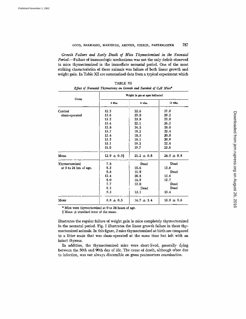
Growth Failure and Early Death of Mice Thymectomized in the Neonatal Period.--Failure of immunologic mechanisms was not the only deficit observed in mice thymectomized in the immedia te neonatal period. One of the most str iking characterist ics of these animals was failure of both l inear growth and weight gain. I n Table X I are summarized da t a from a typical experiment which
TABLE XI Effect of Neonatal Thymectomy on Growth and Survival of CsH Mice*
Group
Control sham-operated
Mean
Thymectomized at 0 to 24 hrs. of age.
Mean
4 wks.
12.5 13.6 13.2 13.4 12.8 13.7 12.4 13.5 13.1 11.0
12.9
7.8 8.3 8.8
12.4 8.0 7.7 8.2 9.3
± o.3~
Weight in gm at ages indicated
8.8 4- 0.5
8wks.
22.6 25.0 23.9 22.1 24.2 19.2 18.3 18.1 19.2 19.7
21.2 q- 0.8
Dead 15.6 11.9 20.8 14.9 12.0
Dead 13.1
14.7 4- 1.4
12 wks.
27.0 29.2 25.8 26.2 26.0 23.4 20.8 20.9 22.4 23.8
24.5 4- 0.8
Dead 13.6
Dead 15.6 12.7
Dead Dead
13.4
13.8 4- 0.6
* Mice were thymectomized at 0 to 24 hours of age. Mean 4- standard error of the mean.
i l lustrates the regular failure of weight gain in mice completely thymectomized in the neonatal period. Fig. 1 i l lustrates the l inear growth failure in these thy- mectomized animals. In this figure, 2 mice thymectomized a t b i r th are compared to a l i t ter mate tha t was sham-operated a t the same t ime bu t left with an in tac t thymus.
I n addit ion, the thymectomized mice were short-l ived, generally dying between the 50th and 90th day of life. The cause of death, al though often due to infection, was not always discernible on gross pos tmor tem examination.
on August 26, 2016
jem.rupress.org
Dow
nloaded from
Published November 1, 1962

788 THYMUS IN DEVELOPMSENT OF Y~M~UNOLOGIC CAPACITY
Production of Runt Disease in Thymectomized Fx Hybrid Mice Injected with Parental Strain Lymphoid Cells.--Because of the runting observed in thymec- tomized mice otherwise untreated, and because of some of the similarities in appearance between mice thymectomized at birth and those runted as a conse- quence of a homologous graft versus host reaction, it was of interest to determine whether mice thymectomized at birth are more susceptible to the production of
TABLE Xll Occurrence of Homologous Disease in Thymectomiced Mice Following Injection of Parent Strain
S ,leen Cdls
Recipient group
(A x CsH)FI
~A x CaH)Ft
(A x C67B1)Fx
Operation and time performed
Thymectomy at 0 to 24 hours
Sham-operation at 0 to 24 hours
Thymectomy at 0 to 24 hours
Thymectomy at 40 days Sham-operation at 40 days
Thymectomy at 40 days
Thymectomy at 40 days Sham-operation at 40
days Thymectomy at 40 days
No. of Astraincells injected
200 million~
200 million;~
None
200 millionl] 200 millionil
None
150m~lion¶ 130milUon¶
None
No. of deaths No. of anlm~ls
6/6
2/10
7/7
o/1o 0/9
o/12
9/9 8/14
0/11
Time of death (days following
injection)*
16 4- 1.6
46, 28
30 4- 2.6§
23 -4- 2.4 32 4- 1.7
* Mean 4- standard error of the mean. $ Spleen cells from 6 month old donors injected intraperitoneally 35 days following oper-
ation. § Days from 35 days of age to death. H Spleen cells from 6 month old donors injected intravenously 10 days following operation. ¶ Spleen cells from 10 to 12 month old donors injected intraperitoneally 10 days following
operation.
homologous disease than are sham-operated mice whose thymuses remain intact. The results summarized in Table X I I indicate that both the development of immunologic runt disease and the severity of the process were favored by neo- natal thymectomy. The (A x CaH)Fa animals thymectomized at birth and in- jected with parent A strain cells at 35 days of age developed runt disease more readily than did sham-operated controls, and the animals dying of the homolo- gous disease died considerably sooner in the thymectomized group studied.
Thymectomy at 40 days of age affected susceptibility of the (A x C57B1)F1
on August 26, 2016
jem.rupress.org
Dow
nloaded from
Published November 1, 1962
GOOD, DA-LM-ASS% MARTINEZ, ARCIIER, PIERCE, PAPEP~ASTER 789
hybrids to runt disease, but had no discernible effect on the incidence and severity of homologous disease in (A x C#~I)F1 animals.
These studies indicate that thymectomy at birth, and in some strain com- binations as late as 40 days of age, renders the F1 hybrid more susceptible to the damaging effect of the graft versus host reaction than are sham-operated control animals. Thymectomy carried out at 40 days of age did not interfere with the growth or apparent well-being of the F1 hybrid animals studied in this experiment.
Morphology of the Lymphoid Tissue of Thymectomized Mice.--In the mouse at birth, the thymus, an organ of endodermal-epithelial origin, is the only organ with true lymphocytes. Figs. 2, 4, and 5 illustrate the thymic structure at birth. By contrast, the spleen, lymph nodes, and gut are either myelopoietic or simple reticular in nature, and contain very few, if any, true lymphoid cells. Figs. 3, 6, and 7 illustrate this in the spleen. Structural changes are discernible during the 1st week of life, and are progressive during the next month, as the lymphoid organs increasingly show adult characteristics: typical follicular structure and well-organized, fully developed cortical-medullary architecture in the lymph nodes, and the red and white pulp characteristic of the normal spleen. Illustrated on Figs. 8 and 9 is the spleen of a 40-day-old mouse sham- operated at birth. In striking contrast are the lymphoid organs of the mouse thymectomized at birth. The lymph node and spleen are often relatively small and lack the structure and lymphoid cells observed in the mature control ani- mals. The spleen of a 40-day-old mouse thymectomized at birth is shown in Figs. 10 and 11 for comparison with the sham-operated control.
DISCUSSION
The data presented in this report indicate that in both rabbits and mice a deficiency in capacity to form circulating antibodies is produced when the animals have been completely thymectomized early enough in the neonatal period. The effect of neonatal thymectomy is somewhat more consistent and productive of greater immunologic deficiency in the mouse than in the rabbit. This result is probably due to the greater immunologic maturity of the rabbit than the mouse at birth (33).
Further, thymectomy in the early neonatal period will render mice capable of accepting skin homografts across both weak, (H-l) or (H-3), andstrong (H-2) histocompatibility barriers, and tumor homografts across strong histocompati- bility barriers, after the animals have reached maturity. Neonatal thymectomy also makes it possible for female mice of the CsTBI strain to accept male skin isografts. These findings, together with the observation reported herein that skin homotransplantation is not facilitated by neonatal thymectomy in the rabbit, indicate further that mice, being less mature immunologically at birth than are rabbits, represent more ideal animals in which to study the role of the thymus in development of immunologic potential.
on August 26, 2016
jem.rupress.org
Dow
nloaded from
Published November 1, 1962

790 THYMUS IN DEVELOP~[ENT O]~ I~I~UNOLOGIC CAPACITY
Consideration of the results reported herein and those of others indicating that thymectomy at birth in rabbits (18), mice (20, 21), and rats (40, 41) inter- feres with development of full immunologic capacity, when viewed in light of the observations of Glick et al. (16) and Mueller et a/. (17), is most provocative. The latter observers found that removal of the bursa of Fabricius in newly hatched chicks or prevention of development of this organ by hormonal treat- ment interferes with full development of immunologic potential in these ani- mals. The bursa of Fabricius in the chick, like the thymus in the mammal, is a lymphoid organ which originates as an endodermai-epithellal outpouching, is of maximal relative size in the newly hatched animal, and becomes spent and of lesser significance as the animal approaches maturity. Both of these lymphoid- epithelial organs appear to play key roles in development of full immunologic capacity in their respective hosts. Studies by Papermaster et al. (42, 43), in these laboratories, which indicate that bursectomy in the newly hatched chick does not completely abolish immunologic potential in the adult animal but rather produces a striking quantitative reduction insufficient to eliminate the homo- graft reaction, are consonant with the observations reported herein. It can be seen in our data that thymectomy in the rabbit which reduces but does not completely eliminate antibody response to serum protein antigens and bac- teriophage T2, does not permit homotransplantation in the adult animals. By contrast, thymectomy in the neonatal mouse may abolish completely the capacity to form antibody to bacteriophage T2 and permit homotransplanta- tion of skin and tumors. Histologic studies of the neonate, reported in part in this communication, may explain these observations. The mouse, at birth, has nearly all of its true lymphoid tissue limited to the thymus whereas, in the rabbit and in the chicken other organs possess lymphoid tissue at birth or hatching (44). The failure of thymectomy in newly hatched chicks to alter the immunologic potential of the maturing animal (44) probably only reflects the vigorous participation of the bursa of Fabricius in the development of full immunologic capacity.
The studies reported herein also demonstrate that in mice thymectomized at birth the cells of the peripheral lymphoid tissues, spleen and lymph nodes, lack immunologic capacity even when introduced into the intact F1 hybrid host equipped with a normal thymus, as indicated by failure to induce runt disease or Simonsen's splenic enlargement.
It is, indeed, tempting to interpret all of our findings as strong support for the postulate, perhaps first expressed by Ruth (45), and more recently elucidated by Auerbach (46), that the thymic cells are distributed centrifugally to the spleen and other "peripheral" lymphoid tissues where they play an essential role in immunologic processes. Particularly in the light of Porter and Cooper's (47) recent communication, establishing that small lymphocytes from the thoracic duct can seek out an environment in spleen, lymph nodes, and bowel
on August 26, 2016
jem.rupress.org
Dow
nloaded from
Published November 1, 1962

GOOD, DALMASSO, MARTINEZ, ARCHER, PIERCE, PAPERM:ASTER. 791
wall in which to develop into larger, immunologically competent, pyronin- ophilic cells, it seems most likely that the thymus functions as a "fertile cres- cent" from which lymphoid cells of epithelial origin are distributed centrifugally to the spleen, lymph nodes, bowel, and other lymphoreticular sites, where they settle down, perhaps mature, and function as the immunologically com- petent cells responsible for adaptive immunity. In this regard, and considering their ultimate origin in epithelium, it is tempting to consider these cells as transposed epithelial elements capable of specialized adaptive secretory ac- tivity (antibody synthesis) even in their new locations. The similarity of cytoplasmic ultrastructure of antibody-producing cells and pancreatic cells has already occasioned considerable concern (48).
Certainly other possibilities exist by which the thymus might function to play an essential role in development of full immunologic potential. Among the possibilities that occur to us, and which we feel must be investigated with our favored hypothesis expressed here, is the production by the thymus of a hormone or hormones essential to normal growth, maturation of cells, and normal function of lymphoid tissue. Still another attractive possibility is that thymic cells operate in the transmission of messages, perhaps v/a nucleic acid transfer, through the agency of a nurse cell (N~hrmutterzellen) function (49) to cells of mesenchymal origin, thus instilling immunologic capacity.
Whatever the role of the thymus and the bursa of Fabricius may be, it is clear that they have a key role in development of the immunologic system in mam- mals and chickens. Further, experiments presented here provide evidence that, whatever this function, it is not limited strictly to the neonatal period, since in C3H mice skin homografts from Ce mice will subsequently be accepted even when the animals are thymectomized as late as 30 days of age.
One means of analyzing the role of the thymus in development of immuno- logic capacity and the development of the remainder of the lymphoid tissue might be to couple the ontogenetic analysis of development of lymphoid tissue and immunologic capacity with a critical phylogenetic study. Subsequent pa- pers in this series will present the results of this method of analysis.
SUMMARY
In rabbits, complete thymectomy before the age of 5 days produced immuno- logic deficiency in the adult animals, as indicated by reduced antibody produc- tion to bovine serum albumin and bacteriophage T2. Homotransp]antation immunity was unaffected, however.
In an inbred strain of mice, complete neonatal thymectomy resulted in com- plete inability of the 60-day-old animals to form antibody to bacteriophage T2.
Inbred mice, completely thymectomized at birth, had a deficient homograft response, indicated by acceptance of skin homografts from strains differing in both the weaker and stronger (H-2) histocompatibility antigens. Tumor trans-
on August 26, 2016
jem.rupress.org
Dow
nloaded from
Published November 1, 1962

792 THYMUs IN DEVELOPMENT OF ~OLOGIC CAPACITY
plants (mammary adenocarcinoma) were also successful across the H-2 genetic barrier in mice thymectomized at birth. Neonatal thymectomy also eliminated the Eichwald-Silmser phenomenon, rendering female mice capable of accepting isografts of male skin.
Transplantation immunity in mice was also affected by later thymectomy, at 30 days of age, in certain strain combinations involving weak histocompati- bility differences.
Spleen and lymph node cells from mice thymectomized at birth or at 6 days of age, and sacrificed 2 months later, did not produce a graft versus host reaction in appropriate F1 hybrid recipients, indicating that such cells are immuno- logically inactive.
Neonatal thymectomy of F1 hybrid mice, and in one strain combination thy- mectomy at 40 days of age, produced animals with inordinate susceptibility to runt disease (homologous disease) following injection of parent strain spleen cells 35 days (neonatal surgery) and 10 days (surgery at 40 days) later.
Mice thymectomized at birth also showed growth failure and were short- lived.
Studies of newborn mice indicated that they have true lymphocytes only in the thymus, and lack such cells in the spleen, lymph nodes, and gut. In normal mice, adult lymphoid structure develops gradually, beginning during the 1st week of life and continuing for the next month. In contrast, mice thy- mectomized at birth do not develop mature lymphoid structure: the lymph nodes and spleens tend to be small and poorly organized, and show a quanti- tative deficiency in lymphoid cells.
It is our current working hypothesis that the thymus makes a major contribu- tion toward the centrifugal distribution of lymphoid cells which, in turn, is essential to the full expression of immunologic capacity.
BIBLIOGRAPHY
I. Scammon, R. E., Developmental anatomy, in Morris' Human Anatomy, (J. P. Schaeffer, editor), New York, Blakiston Division, McGraw Hill Book Company, Inc., llth edition, 1953, 1.
2. Good, R. A., Agammaglobulinemia: a provocative experiment of nature, Bull. Univ. Minnesota Hosp. and Minnesota Med. Foundation, 1954, 26, 1.
3. Good, R. A., and Varco, R. L., A clinical and experimental study of agammaglobu- linemia, J. Lancet, 1955, 75, 245.
4. Ramos, A. J., Presentation of a case, with discussion by V. Loeb, J. Am. Med. Assn., 1956, 160, 1317.
5. Gafni, J., Michaeli, D., and Heller, H., Idiopathic acquired agammaglobulinemia associated with thymoma; report of two cases and review of the literature, New England J. Med., 1960, 2~, 536.
6. Martin, C. M., Gordon, R. S., and McCullough, N. B., Acquired agammaglobu- linemia in an adult; report of a case, with clinical and experimental studies, New England J. Med., 1956, 254, 449.
on August 26, 2016
jem.rupress.org
Dow
nloaded from
Published November 1, 1962

GOOD, DALM-ASSO~ M~RTINEZ~ ARCIEER, PIERCE, PAPE~ASTER 793
7. Lambie, A. T., Burrows, B. A., and Sommers, S. C., Clinicopathologic confer- ence: refractory anemia, agammaglobulinemia, and mediastinal tumor, Am. J. Clin. Path., 1957, 27,444.
8. RStstein, J., and Good, R. A., unpublished observations. 9. MacLean, L. D., Zak, S. J., Varco, R. L., and Good, R. A., Thymic tumor and
acquired agammaglobulinemia--a clinical and experimental study of the im- mune response, Surgery, 1956, 40, 1010.
10. MacLean, L. D., Zak, S. J., Varco, R. L., and Good, R. A., The role of the thymus in antibody production: an experimental study of the immune response in thymectomized rabbits, Transplant. Bull., 1957, 4, 21.
11. Harris, T. N., Rhoads, J., and Stokes, J., Jr., A study of the role of the thymus and spleen in the formation of antibodies in the rabbit, J. Imrnunol., 1948, ~ , 27.
12. Bjg~rneboe, M., Gormsen, H., and Lundquist, F., Further experimental studies on the role of the plasma cells as antibody producers, J. Irnmunot., 1947, 55, 121.
13. Thorbecke, J. G., and Keuning, F. J., Antibody formation in vitro by haemopoi- eric organs after subcutaneous and intravenous immunization, d. Immunol., 1953, 70, 129.
14. Fagraeus, A., Antibody production in relation to the development of plasma cells: in vivo and in vitro experiments, Acta Med. Scan&, 1948, suppl. 204, 1.
15. Stoner, R. D., and Hale, W. M., Antibody production by thymus and Peyer's patches intraocular transplants, J. Irnmunol., 1955, 75, 203.
16. Glick, B., Chang, T. S., and Jaap, R. G., The bursa of Fabricius and antibody pro- duction, Poultry Sc., 1956, 35, 224.
17. Mueller, A. P., Wolfe, H. R., and Meyer, R. K., Precipitin production in chickens. XXI. Antibody production in bursectomized chickens and in chickens injected with 19-nortestosterone on the fifth day of incubation, d. Immunol., 1960, 85, 172.
18. Archer, O., and Pierce, J. C., Role of thymus in development of the immune re- sponse, Fed. Proc., 1961, 20, 26.
19. Fichtelins, K. E., LaureU, G., and Philipson, L., The influence of thymectomy on antibody formation, Acta Path. et Microbiol. Scand., 1961, 51, 81.
20. Miller, J. F. A. P., Immunological function of the thymus, Lancet, 1961, 2, 748. 21. Martinez, C., Kersey, J., Papermaster, B. W., and Good, R. A., Skin homograft
survival in thymectomized mice, Proc. Soc. Exp. Biol. and Med., 1962, 109, 193. 22. Trentin, J. J., Tolerance and homologous disease in irradiated mice protected with
homologous bone marrow, Ann. New York. Acad. So., 1958, "/3, 799. 23. BiUingham, R. E., Reactions of grafts against their hosts, Science, 1959, 130, 947. 24. Swift, H. F., Wilson, A. T., and Lancefield, R. C., Typing group A hemolytic
streptococci by M precipitin reactions in capillary pipettes, J. Exp. Med., 1943, 78, 127.
25. Farr, R. S., A quantitative immunochemical measure of the primary interaction between I* BSA and antibody, J. Infect. Dis., 1958, 103, 239.
26. Adams, M., Bacteriophages, New York, Interscience Publishers, Inc. 1959. 27. Stetson, C. A., ~lr., and Demopoulos, R., Reactions of skin homografts with spe-
cific immune sera, Ann. New York Acad. S6., 1958, 73, 687. 28. Gross, L., Effect of thymectomy on development of leukemia in C~I-I mice inocu-
on August 26, 2016
jem.rupress.org
Dow
nloaded from
Published November 1, 1962
794 THYMUS IN DEVELOPMENT OF BIMUNOLOGIC CAPACITY
lated with leukemic "passage" virus, Proc. Soc. Exp. Biol. and Med., 1959, 100, 325.
29. Dischler, W., and Rudali, G., La thymectomie totale chez le souricean nonvean-n~, Rev. Fran~. etudies din. et biol., 1961, 6, 88.
30. Martinez, C., Smith, R. T., Aust, J. B., and Good, R. A., Acquired tolerance to skin homografts in mice of different strains, Proc. Soc. Exp. Biol. and Med., 1958, 97, 736.
31. Simonsen, M., Engelbreth-Holm, J., Jensen, E., and Poulsen, H., A study of the graft-versus-host reaction in transplantation to embryos, F1 hybrids, and irra- diated animals, Ann. New York Acad. Sc., 1958, 73, 834.
32. Opstad, A. M., A methyl-green pyronin stain for plasma cells in tissue, Stain Techn., 1959, 34, 293.
33. Porter, K. A., Runt disease and tolerance in rabbits, Nature, 1960,185,789. 34. Counce, S., Smith, P., Barth, R., and Snell, G. D., Strong and weak histocompafi-
bility differences in mice and their role in the rejection of homologous tumors and skin, Ann. Surg., 1956, 144, 198.
35. Eichwald, E. J., and Silmser, C. R., Discussion of skin graft data, Transplant. Bull., 1955, 2, 148.
36. Billingham, R. E., and Silvers, W. K., Induction of tolerance of skin isografts from male donors in female mice, Science, 1958, 128,780.
37. Martinez, C., Smith, J. M., Aust, J. B., Mariani, T., and Good, R. A., Transfer of acquired tolerance to skin homografts in mice, Proc. Soc. Exp. Biol. and Med., 1958, 98, 640.
38. Mnriani, T., Martinez, C., Smith, J. M., and Good, R. A., Age factor and induc- tion of immunological tolerance to male skin isografts in female mice subse- quent to the neonatal period, Ann. New York Acad. Sc., 1960, 87, 93.
39. Mariani, T., Martinez, C., and Good, R. A., Studies of sex-linked histo-incom- patibility in inbred mice: role of age and production of immunological tolerance, Internat. Arch. Allergy and Appl. Immunol., 1960, 16, 216.
40. Waksman, B. H., Arnason, B. G., and Jankovi~, B. D., Changes in the lymphoid organs of rats thymectomized at birth, Fed. Proc., 1962, 21,274.
41. Arnason, B. G., and Jankovid, B. D., Suppression of "delayed" hypersensitivity reactions in rats thymectomized at birth, Fed. Proc., 1962, 21,274.
42. Papermaster, B. W., Friedman, D. I., and Good, R. A., Relationship of the bursa of Fabricius to immunologic responsiveness and homograft immunity in the chicken, Proc. Soc. Exp. Biol. and Med., 1962, 110, 62.
43. Papermaster, B. W., Bradley, S. G., Watson, D. W., and Good R. A., Antibody- producing capacity of adult chicken spleen cells in newly hatched chicks. A study of sources for variation in a homologous cell transfer system, J. Exp. Med., 1962, 115, 1191.
44. Papermaster, B. W., and Good, R. A., unpublished observations. 45. Ruth, R. F., Ontogeny of the blood cells, Fed. Proc., 1960, 19,579. 46. Auerbach, R., Genetic control of thymus lymphoid differentiation, Proc. Nat.
Acad. Sc., 1961, 47, 1175. 47. Porter, K. A., and Cooper, E. H., Transformation of adult allogeneic
on August 26, 2016
jem.rupress.org
Dow
nloaded from
Published November 1, 1962

GOOD, DAL~ASSO, ~ARTINEZ, ARCHER, PIERCE, PAPERMASTER 795
small lymphocytes after transfusion into newborn rats, 3". Exp. Med., 1962, 115, 997.
48. Bernard, W., and Granboulan, N., Ultrastructure of immunologically competent cells, in Cellular Aspects of Immunity, (G.E.W. Wolstenholme and M. O'Connor, editors) Boston, Little, Brown & Company, 1959, 92.
49. Ehrich, W. E., Die cellul~ren Bildungsst~tten der AntikSrper, Klln. Woch., 1955, 33, 315.
on August 26, 2016
jem.rupress.org
Dow
nloaded from
Published November 1, 1962

796 THYMUS IN D]~VEI,OPMENT OP IMMUNOLOGIC CAPACITY
EXPLANATION OF PLATES
PLATE 104
FIG. 1. A comparison, at 60 days of age, of mice thymectomized at birth and a sham- operated litter mate. Notice the runting of the thymectomized animals and the roughening of their fur. Development of diarrhea and early death characterize these mice.
FIG. 2. Low power view of the thymus in a newborn mouse. Note the development of the cortex and medulla, and the dense cellularity of the thymus at this age. Hema- toxylin and eosin. X 30.
FIG. 3. Low power view of spleen and intestine of the newborn mouse. Note the lack of development of red and white pulp in the spleen. Lymphoid tissue is virtually lacking in the spleen at this age. No lymphoid tissue is to be found in the gut at this time. Hematoxylin and eosin. X 30.
on August 26, 2016
jem.rupress.org
Dow
nloaded from
Published November 1, 1962

THE JOURNAL OF EXPERIMENTAL MEDICINE VOL. 116 PLATE 104
(Good et al.: Thymus in development of immunologic capacity)
on August 26, 2016
jem.rupress.org
Dow
nloaded from
Published November 1, 1962
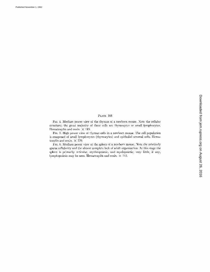
PLATE 105
FIG. 4. Medium power view of the thymus of a newborn mouse. Note the cellular structure; the great majority of these cells are thymocytes or small lymphocytes. Hematoxylin and eosin. X 115.
FI6. 5. High power view of thymus cells in a newborn mouse. The cell population is composed of small lymphocytes (thymocytes) and epithelial stromal cells. Hema- toxylin and eosin. × 770.
FIG. 6. Medium power view of the spleen of a newborn mouse. Note the relatively sparse cellularity and the almost complete lack of adult organization. At this stage the spleen is primarily reticular, erythropoietic, and myelopoietic; very little, if any, lymphopoiesis may be seen. Hematoxylin and eosin. X 115.
on August 26, 2016
jem.rupress.org
Dow
nloaded from
Published November 1, 1962

THE JOURNAL OF EXPERIMENTAL MEDICINE VOL. 116 PLATE 105
(Good et al.: Thymus in development of immunologic capacity)
on August 26, 2016
jem.rupress.org
Dow
nloaded from
Published November 1, 1962

PLATE 106
FIG. 7. High power view of a representative area of spleen from a newborn mouse. Notice that most of the cells are reticulum cells, myeloid cells, and erythropoietic cells. Very few lymphocytes are present, and no evidence of lymphopoiesis is observed. Hematoxylin and eosin. X 760.
FIG. 8. Medium power view of the spleen from a 40-day-old mouse sham-operated at birth. Note the organization into cortical and medullary portions, and the presence of follicles and lymphopoietic centers. Hematoxylin and eosin. × 75.
FIG. 9. High power view of the spleen from a 40-day-old mouse sham-operated at birth. Note that the majority of cells are small lymphocytes, although some reticulum cells and medium-sized lymphocytes are also present. Hematoxylin and eosin. × 610.
on August 26, 2016
jem.rupress.org
Dow
nloaded from
Published November 1, 1962

THE JOURNAL OF EXPERIMENTAL MEDICINE VOL. 116 PLATE 106
(Good et al.: Thymus in development of immunologic capacity)
on August 26, 2016
jem.rupress.org
Dow
nloaded from
Published November 1, 1962
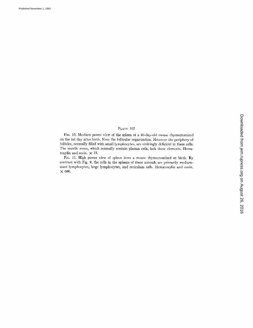
PLATE 107
Fro. 10. Medium power view of the spleen of a 40-day-old mouse thymectomized on the 1st day after birth. Note the follicular organization. However the periphery of follicles, normally filled with small lymphocytes, are strikingly deficient in these cells. The mantle zones, which normally contain plasma cells, lack these elements. Hema- toxylin and eosin. X 75.
FIG. 11. High power view of spleen from a mouse thymectomized at birth. By contrast with Fig. 9, the cells in the spleens of these animals are primarily medium- sized lymphocytes, large lymphocytes, and reticulum cells. Hematoxylin and eosin. × 600.
on August 26, 2016
jem.rupress.org
Dow
nloaded from
Published November 1, 1962

THE JOURNAL OF EXPERIMENTAL MEDICINE VOL. 116 PLATE 107
(Good et al.: Thymus in development of immunologic capacity)
on August 26, 2016
jem.rupress.org
Dow
nloaded from
Published November 1, 1962
Copyright © 2022 FDOKUMEN