Melatonin regulates glucocorticoid receptor: an answer to its antiapoptotic action in thymus
10
Melatonin regulates glucocorticoid receptor: an answer to its antiapoptotic action in thymus ROSA MARI ´ A SAINZ,* JUAN CARLOS MAYO,* RUSSEL J. REITER, ISAAC ANTOLI ´ N,* MANUEL M. ESTEBAN, ‡ AND CARMEN RODRI ´ GUEZ* ,1 *Departamento de Morfologı ´a y Biologı ´a Celular and ‡ Departamento de Biologı ´a Funcional, Facultad de Medicina, Oviedo, Spain; and ² Department of Cell and Structural Biology, University of Texas Health Science Center at San Antonio, Texas, USA ABSTRACT We have previously reported that low doses of melatonin inhibit apoptosis in both dexa- methasone-treated cultured thymocytes (standard model for the study of apoptosis) and the intact thymus. Here we elucidate the mechanism by which this agent protects thymocytes from cell death induced by glucocorticoids. Our results demonstrate an effect of melatonin on the mRNA for antioxidant enzymes in thymocytes, also showing an unexpected regulation by dexamethasone of these mRNA. Both an effect of melatonin on the general machinery of apoptosis and a possible regulation of the expression of the cell death related genes bcl-2 and p53 are shown not to be in- volved. We found melatonin to down-regulate the mRNA for the glucocorticoid receptor in thymocytes (glucocorticoids up-regulate their own receptor). The decrease by melatonin of mRNA levels for this recep- tor in IM-9 cells (where glucocorticoids down-regulate it) demonstrates that melatonin actually down-regulates glucocorticoid receptor. These findings allow us to propose the effects of melatonin on this receptor as the likely mediator of its thymocyte protection against dexamethasone-induced cell death. This effect of mel- atonin, given the oxidant properties of glucocorticoids, adds another mechanism to explain its antioxidant effects.—Sainz, R. M., Mayo, J. C., Reiter, R. J., Antolı ´n, I., Esteban, M. M., Rodrı ´guez, C. Melatonin regulates glucocorticoid receptor: an answer to its antiapoptotic action in thymus. FASEB J. 13, 1547–1556 (1999) Key Words: antioxidant enzymes z glucocorticoids z apoptosis z DNA fragmentation Melatonin is a chemical mediator produced mainly in the pineal gland, although other organs have been demonstrated to have the enzymatic ma- chinery for its synthesis. The classical effects of melatonin relate to the control of the circadian rhythms and regulation of the hypothalamo-pitu- itary-gonadal axis, but other actions have also been reported (1–10). The most recently described prop- erties of melatonin are 1) its antioxidant capability, acting both as a free radical scavenger (11, 12) and a stimulator of activity (13, 14) and mRNA levels for several antioxidant enzymes (8, 15, 16) and 2) its antiapoptotic effects in thymocytes (17, 18) and neuronal cells (16, 19, 20). The antioxidant and antiapoptotic properties suggest possible clinical ap- plications of this mediator particularly in the preven- tion of free radical damage and apoptosis in neuro- degenerative diseases (21–23). Most T cells synthesize their receptors once they are in the thymus. Thymocytes may thereafter pro- ceed in one of the following ways: they may 1) be selected positively and then mature in the thymic medulla, where they are insensitive to apoptosis- inducing factors, 2) be selected negatively and die in a typical programmed cell death process (24), or 3) remain unselected in the thymic cortex (the vast majority of them) extremely sensitive to multiple apoptosis-inducing factors (e.g., oxidative stress, DNA damaging agents, radiation, and especially glu- cocorticoids) (24). Concentrations of glucocorti- coids in a high physiological range readily induce apoptosis in nonselected thymocytes. Cohen et al. (24) proposed that these hormones are also respon- sible for the physiological apoptosis occurring in thymus with aging. The rise in thymus weight by adrenalectomy seems to support this (25). It is because of these observations that the induction of apoptosis by dexamethasone in thymocytes from the thymic cortex is a standard in vitro model for the study of apoptosis. Here we investigated the mechanism by which melatonin inhibits cell death induced by dexa- methasone in thymocytes. To do this, we analyzed the effect of this hormone on cell death induced by other agents, examining its influence on the expression of several genes related to pro- grammed cell death. We conclude that regulation of the expression of the glucocorticoid receptor 1 Correspondence: Departamento de Morfologı ´a y Biologı ´a Celular, Facultad de Medicina, Julian Claveria 33006 Oviedo, Spain. E-mail: [email protected] 1547 0892-6638/99/0013-1547/$02.25 © FASEB
Transcript of Melatonin regulates glucocorticoid receptor: an answer to its antiapoptotic action in thymus

Melatonin regulates glucocorticoid receptor: an answerto its antiapoptotic action in thymus
ROSA MARIA SAINZ,* JUAN CARLOS MAYO,* RUSSEL J. REITER,† ISAAC ANTOLIN,*MANUEL M. ESTEBAN,‡ AND CARMEN RODRIGUEZ*,1
*Departamento de Morfologıa y Biologıa Celular and ‡Departamento de Biologıa Funcional,Facultad de Medicina, Oviedo, Spain; and †Department of Cell and Structural Biology,University of Texas Health Science Center at San Antonio, Texas, USA
ABSTRACT We have previously reported that lowdoses of melatonin inhibit apoptosis in both dexa-methasone-treated cultured thymocytes (standardmodel for the study of apoptosis) and the intactthymus. Here we elucidate the mechanism by whichthis agent protects thymocytes from cell death inducedby glucocorticoids. Our results demonstrate an effectof melatonin on the mRNA for antioxidant enzymes inthymocytes, also showing an unexpected regulation bydexamethasone of these mRNA. Both an effect ofmelatonin on the general machinery of apoptosis and apossible regulation of the expression of the cell deathrelated genes bcl-2 and p53 are shown not to be in-volved. We found melatonin to down-regulate themRNA for the glucocorticoid receptor in thymocytes(glucocorticoids up-regulate their own receptor). Thedecrease by melatonin of mRNA levels for this recep-tor in IM-9 cells (where glucocorticoids down-regulateit) demonstrates that melatonin actually down-regulatesglucocorticoid receptor. These findings allow us topropose the effects of melatonin on this receptor asthe likely mediator of its thymocyte protection againstdexamethasone-induced cell death. This effect of mel-atonin, given the oxidant properties of glucocorticoids,adds another mechanism to explain its antioxidanteffects.—Sainz, R. M., Mayo, J. C., Reiter, R. J.,Antolın, I., Esteban, M. M., Rodrıguez, C. Melatoninregulates glucocorticoid receptor: an answer to itsantiapoptotic action in thymus. FASEB J. 13, 1547–1556(1999)
Key Words: antioxidant enzymes z glucocorticoids z apoptosisz DNA fragmentation
Melatonin is a chemical mediator producedmainly in the pineal gland, although other organshave been demonstrated to have the enzymatic ma-chinery for its synthesis. The classical effects ofmelatonin relate to the control of the circadianrhythms and regulation of the hypothalamo-pitu-itary-gonadal axis, but other actions have also beenreported (1–10). The most recently described prop-erties of melatonin are 1) its antioxidant capability,
acting both as a free radical scavenger (11, 12) and astimulator of activity (13, 14) and mRNA levels forseveral antioxidant enzymes (8, 15, 16) and 2) itsantiapoptotic effects in thymocytes (17, 18) andneuronal cells (16, 19, 20). The antioxidant andantiapoptotic properties suggest possible clinical ap-plications of this mediator particularly in the preven-tion of free radical damage and apoptosis in neuro-degenerative diseases (21–23).
Most T cells synthesize their receptors once theyare in the thymus. Thymocytes may thereafter pro-ceed in one of the following ways: they may 1) beselected positively and then mature in the thymicmedulla, where they are insensitive to apoptosis-inducing factors, 2) be selected negatively and die ina typical programmed cell death process (24), or 3)remain unselected in the thymic cortex (the vastmajority of them) extremely sensitive to multipleapoptosis-inducing factors (e.g., oxidative stress,DNA damaging agents, radiation, and especially glu-cocorticoids) (24). Concentrations of glucocorti-coids in a high physiological range readily induceapoptosis in nonselected thymocytes. Cohen et al.(24) proposed that these hormones are also respon-sible for the physiological apoptosis occurring inthymus with aging. The rise in thymus weight byadrenalectomy seems to support this (25). It isbecause of these observations that the induction ofapoptosis by dexamethasone in thymocytes from thethymic cortex is a standard in vitro model for thestudy of apoptosis.
Here we investigated the mechanism by whichmelatonin inhibits cell death induced by dexa-methasone in thymocytes. To do this, we analyzedthe effect of this hormone on cell death inducedby other agents, examining its influence on theexpression of several genes related to pro-grammed cell death. We conclude that regulationof the expression of the glucocorticoid receptor
1 Correspondence: Departamento de Morfologıa y BiologıaCelular, Facultad de Medicina, Julian Claveria 33006 Oviedo,Spain. E-mail: [email protected]
15470892-6638/99/0013-1547/$02.25 © FASEB

(GcR)2 gene could be the main mediator of theantiapoptotic effect of melatonin in the thymus.
MATERIALS AND METHODS
Animals
Wistar rats were maintained under controlled temperature(2062°C) in the animal room facilities. Food and water wereavailable ad libitum and photoperiodic conditions were con-stant (12:12 photoperiod, lights on at 07:00). Animals werekilled by decapitation.
Cell culture
After decapitation thymus was quickly removed and im-mersed in Hanks’ balanced salt solution (HBSS) containingglucose (1 g/l) to preserve cellular viability. Thymocytes wereobtained by pressing the thymus against a sterile stainless steelscreen. After several washes with HBSS and recovery of thecells by centrifugation at 1000 rpm for 5 min, cells werefiltered through a smaller screen. Thymocytes were main-tained in RPMI 1640 culture medium supplemented with10% fetal bovine serum (FBS), sodium bicarbonate (2 g/l),and 1% antibiotic-antimitotic mixture (100 mg/ml penicillin,100 mg/ml streptomycin, and 25 mg/ml amphotericin) at37°C and 5% CO2. Cells were counted with a NEUBAUERcamera and cultured in plastic plates (NUNC, Roskilde,Denmark).
IM-9 cells were cultured in RPMI 1640 culture mediumsupplemented with 10% FBS. Cells were grown in suspensionin T-75 flasks at 37°C and 5% CO2.
DNA electrophoresis
Cells were cultured in 35 mm plates at a density of 3 3 106
cells/ml and collected by centrifugation after scraping theplates. They were homogenized in 1 ml of homogenizationbuffer (0.1 M NaCl, 0.01 M EDTA pH 8, 0.3 M Tris-HCl pH 8,and 0.2 M sucrose) and processed as described Tilly et al.(26). To analyze DNA fragmentation, 15 mg of DNA wereelectrophoresed in 2% agarose gel in TAE buffer (2%) and0.5 mg of ethidium bromide. Gel was run at 5 v/cm, visualizedunder UV, and photographed with a POLAROID camera.
Quantification of DNA fragmentation
Cells were collected and recovered in Eppendorf tubes bycentrifugation at 13,000 3 g for 2 min. The pellet was washedin phosphate-buffered saline (PBS) and cells were lysed with400 ml of a lysis buffer (10 mM Tris pH 7.5, 1 mM EDTA, and0.2% of Triton X-100) by incubation in ice water for 20 min.High and low molecular weight DNAs were obtained asdescribed by Collota et al. (27) and quantified following acolorimetric assay described by Burton (28). Absorbance wasmeasured at 600 nm in a spectrophotometer and percentageof fragmentation was calculated as follows:
% fragmentation 5 supernatant ODr600/supernatantODr600 1 pellet ODr600
Morphometric analysis
Cells cultured at a density of 3 3 106 cells/ml were collectedand recovered by centrifugation at 1000 3 g for 5 min. Theywere washed several times in PBS and fixed in 500 ml of 3%glutaraldehyde in 0.1 M phosphate buffer, pH 7.3, for 2 h at4°C. Fixed cells were then embedded in a small volume of 1%agar, dehydrated in increasing concentrations of acetone,and embedded in SPURR resin (EMS, Fort Washington, Pa.).Semithin (1 mM) sections were obtained at four differentlevels separated by 30 mM from three blocks of each experi-mental group and stained with 0.2% toluidine blue. Apo-ptotic and nonapoptotic cells were counted in 15–20 areas ineach section at 1003. A minimum of 500 cells was counted ineach group. The percentage of apoptotic cells was calculatedin relation to the total number of cells.
RNA isolation and Northern blot analysis
Thymus tissue (400 mg) or cells cultured in 100 mm plates ata density of 50 3 106 cells/plate were used. Total RNA wasextracted according to the method described by Chomczynskiand Sacchi (29). (poly(A)1-RNA was obtained from totalRNA with a purification mRNA kit (Pharmacia Biotech,Piscataway, N.J.). After electrophoresis in a 1% agarose gel,RNA was transferred to a nylon membrane (HYBON-N1,Amersham Life Sciences, Little Chalfont, U.K.) and hybrid-ized with the following probes: a 1.6 kb HindIII/ECO RIfragment from the rat catalase cDNA clone pTZCTL (30); a0.6 kb EcoRI fragment from the rat copper-zinc superoxidedismutase (Cu-Zn SOD) cDNA clone, pUC13 (31); a 0.8 kbSalI fragment from the rat glutathione peroxidase (GPx)cDNA clone, LK 440 (32); a 2.4 kb BamHI fragment from theGcR cDNA clone pSP65 (33); a 0.85 kb ECO RI/HindIIIfragment from the human bcl-2 cDNA clone pBluescript (34);a 2 kb BamHI fragment from the mouse p53 cDNA clonepBR322 (35); and a 2.1 kb fragment from the human b-actincDNA clone, pHFBA-1 (36), which was used to normalize theremainder of the mRNA values. Autoradiographies shownbelong to a representative experiment.
Statistical analysis
Data result from three independent experiments. Results areshown as the mean 6 standard error. Statistic analysis wasperformed with analysis of variance, followed by a StudentNewman-Keuls test.
RESULTS
Melatonin effects on mRNA levels for antioxidantenzymes
Glucocorticoids (GC) have recently been found toenhance oxidative stress-induced cell death (37);this is prevented by melatonin in other systems byincreasing the levels of mRNA for antioxidantenzymes (15, 16). To determine whether the anti-apoptotic action of melatonin in dexamethasone-treated thymocytes (17) correlates with the in-crease of antioxidant enzymes, we measured thelevels of mRNAs for GPx, Cu-Zn SOD, and catalasein these cells.
2 Abbreviations: Cu-Zn SOD, copper-zinc superoxide dis-mutase; FBS, fetal bovine serum; GC, glucocorticoids; GcR,glucocorticoid receptor; GPx, glutathione peroxidase; H2O2,hydrogen peroxide; HBSS, Hanks’ balanced salt solution; IL,interleukin; PBS, phosphate-buffered saline.
1548 Vol. 13 September 1999 SAINZ ET AL.The FASEB Journal

Three groups of cells were used: one group re-ceived no treatment; a second group was treated with1027 M dexamethasone; and a third group wastreated with 1027 M dexamethasone plus 1027 Mmelatonin (preincubation of 3 h and coincubationwith the dexamethasone for 6 additional hours).Both morphometric analysis of apoptotic cells andDNA electrophoresis confirmed that melatonin pre-vented cell death induced by dexamethasone (datanot shown). Dexamethasone decreased the levels ofmRNA for GPx (almost 40%) and increased thelevels of mRNA for Cu-Zn SOD (45%) whereasmelatonin prevented both of these changes (Fig. 1A,B). No mRNA for catalase was detected.
Melatonin did not prevent apoptosis induced byetoposide
To test whether melatonin was acting on the generalmachinery of apoptosis, programmed cell death wasinduced in the same experimental model, with eto-poside causing apoptosis by inhibiting DNA topo-isomerase II (38).
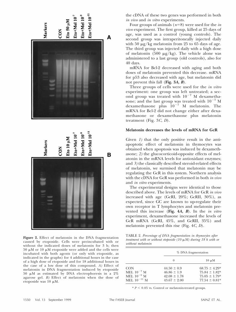
Morphometric analysis, quantification of DNAfragmentation, and DNA electrophoresis gel patternshowed no differences between the group of cellstreated with 50 mM etoposide for 4 h and cells alsotreated with 1027 or 1029 M melatonin (preincuba-tion of 3 h and coincubation with etoposide for anadditional 4 h) (Table 1 and Fig. 2A). When cellswere treated with 10 mM etoposide for 18 h with orwithout administration of melatonin (3 h pretreat-ment and coincubation for an additional 18 h), thesame results were obtained (Table 2 and Fig. 2B).
Melatonin increases mRNA levels for bcl-2 inthymus in vivo but does not affect these levels incultured thymocytes
A nuclear receptor for melatonin has been reported(39); this receptor may be involved in the regulationof several mRNAs (9, 40). To test whether melatoninwas regulating the expression of the apoptosis-in-volved genes bcl-2 and p53, Northern analysis with
Figure 1. Effects of melatonin on the variations of mRNAlevels for antioxidant enzymes induced by dexamethasone.mRNA from thymocytes cultured with no treatment (CON),1027 M dexamethasone during 6 h (DEX), or 1027 Mdexamethasone plus 1027 M melatonin (DEX1MEL) wasobtained. Autoradiography of the Northern blot performedwith the cDNAs of GPx, Cu-Zn SOD, and b-actin is shown inpanel A. The relative amount of mRNAs after normalizationwith the signal of b-actin giving the value of 100% to thecontrols is plotted in panel B. *P , 0.05 vs. CON andDEX1MEL.
TABLE 1. Percentage of DNA fragmentation and apoptotic thymocytes after treatment with or without 50 mM etoposide during 4 h withor without melatonin
% DNA fragmentation % Apoptotic cells
0 50 mM 0 50 mM
CON 28.74 6 1.3 56.38 6 1.39* 19.40 6 3.79 54.66 6 2.03*MEL 1027 M 25.48 6 1.69 53.26 6 3.28* 18.60 6 2.85 52.04 6 1.03*MEL 1029 M 26.58 6 0.66 61.17 6 1.77* 19.05 6 1.99 50.38 6 0.83*MEL 10211 M 24.04 6 1.91 50.49 6 4.28* — —
* P , 0.05 vs. control or melatonin-treated groups.
1549MELATONIN REGULATES GLUCOCORTICOID RECEPTOR
the cDNA of these two genes was performed in bothin vivo and in vitro experiments.
Four groups of animals (n58) were used for the invivo experiment. The first group, killed at 25 days ofage, was used as a control (young controls). Thesecond group was intraperitoneally injected dailywith 50 mg/kg melatonin from 25 to 65 days of age.The third group was injected daily with a high doseof melatonin (500 mg/kg). The vehicle alone wasadministered to a last group (old controls), also for40 days.
mRNA for Bcl-2 decreased with aging and bothdoses of melatonin prevented this decrease. mRNAfor p53 also decreased with age, but melatonin didnot prevent this fall (Fig. 3A, B).
Three groups of cells were used for the in vitroexperiment: one group was left untreated; a sec-ond group was treated with 1027 M dexametha-sone; and the last group was treated with 1027 Mdexamethasone plus 1027 M melatonin. ThemRNA for Bcl-2 did not change either after dexa-methasone or dexamethasone plus melatonintreatment (Fig. 3C, D).
Melatonin decreases the levels of mRNA for GcR
Given 1) that the only positive result in the anti-apoptotic effect of melatonin in thymocytes wasobtained when apoptosis was induced by dexameth-asone; 2) the glucocorticoid-opposite effects of mel-atonin in the mRNA levels for antioxidant enzymes;and 3) the classically described steroid-related effectsof melatonin, we surmised that melatonin may beregulating the GcR in this system. Northern analysiswith the cDNA for GcR was performed in both in vivoand in vitro experiments.
The experimental designs were identical to thosedescribed above. The levels of mRNA for GcR in vivoincreased with age (GcRI, 20%; GcRII, 30%), asexpected, since GC are known to up-regulate theirown receptor in T lymphocytes and melatonin pre-vented this increase (Fig. 4A, B). In the in vitroexperiment, dexamethasone increased the levels ofGcR mRNA (GcRI, 45%, and GcRII, 35%) andmelatonin prevented this rise (Fig. 4C, D).
TABLE 2. Percentage of DNA fragmentation in thymocytes aftertreatment with or without etoposide (10 mM) during 18 h with orwithout melatonin
% DNA fragmentation
0 10 mM
CON 44.50 6 0.9 68.75 6 4.29*MEL 1027 M 46.86 6 1.9 75.84 6 1.82*MEL 1029 M 42.08 6 1.78 75.05 6 1.79*MEL 10211 M 43.67 6 2.00 77.54 6 0.81*
* P , 0.05 vs. Control or melatonin-treated groups.
Figure 2. Effect of melatonin in the DNA fragmentationcaused by etoposide. Cells were preincubated with orwithout the indicated doses of melatonin for 3 h; then50 mM or 10 mM etoposide were added and the cells wereincubated with both agents (or only with etoposide, asindicated in the graphs) for 4 additional hours in the caseof a high dose of etoposide and for 18 additional hours inthe case of a low dose of this compound. A) Effect ofmelatonin in DNA fragmentation induced by etoposide50 mM as estimated by DNA electrophoresis in a 2%agarose gel. B) Effect of melatonin when the dose ofetoposide was 10 mM.
1550 Vol. 13 September 1999 SAINZ ET AL.The FASEB Journal

Melatonin has a direct effect on GcR expression
As GC up-regulate their own receptor in thymus, theeffects of melatonin seem to be opposite to theeffects of these hormones both in terms of effects onthe mRNA for antioxidant enzymes and on themRNA for GcR. To determine whether melatoninreduces the action of GC in some way or is directlyregulating its receptor expression, the effect of mel-atonin on the mRNA levels for GcR was studied inthe IM-9 cells, where this receptor is down-regulatedby its ligands (41).
As expected, dexamethasone decreased the levels ofmRNA for GcR (GcRI, 35%; GcRII, 20%). Melatonindid not have the opposite effect of dexamethasone; onthe contrary, the group treated with dexamethasoneand melatonin showed mRNA levels for GcR belowthat of the group treated only with dexamethasone(GcRI, 70%; GcRII, 60% vs. control) (Fig. 5A, B).
DISCUSSION
Thymocytic programmed cell death in vivo and invitro has long been used as a model to study apopto-
Figure 3. Effect of chronic treatment with melatonin on the expression of bcl-2 and p53 genes in the thymus of Wistar rats (A,B) and cultured thymocytes (C, D). Low (50 mg/kg) or high (500 mg/kg) melatonin doses were administered during 40 daysto young rats (25 days) and the thymus was removed after death (65 days). For the in vitro experiment, 1027 M dexamethasonewas (or was not: CON) added to the medium after preincubation with or without 1027 M melatonin for 3 h; cells were culturedwith both agents during an additional 6 h. mRNA was obtained and Northern blot with the cDNAs for bcl-2 and b-actin wasperformed. The autoradiographic signal obtained is shown in panels A, C; the relative value obtained after normalization withthe b-actin signal is represented in panels B, D, giving 100% to the values obtained in the thymus of 25 days control group orin the control group. a, b: P , 0.05 vs. all other bcl-2 groups; *P , 0.05 vs. all p53 groups.
1551MELATONIN REGULATES GLUCOCORTICOID RECEPTOR

sis. We recently found that melatonin partially pre-vents apoptosis in thymocytes in vivo (physiologicalapoptosis with aging) or in vitro (dexamethasone-induced) (17). Glucocorticoids have been related tooxidative stress. Beaver and Waring (42) reportedglutathione concentrations to decrease after dexa-methasone administration, and Behl et al. (37) haveshown that GC enhance oxidative stress-induced celldeath in neurons. Because melatonin has importantantioxidant properties (43) and its increase ofmRNA for antioxidant enzymes has been proposedas its mechanism to prevent apoptosis in othersystems (16), we studied the effect of melatonin on
these mRNAs. The present findings suggest that, forthis system, melatonin does not strictly regulateantioxidant enzymes mRNAs, but seems to exert anaction opposite to that of GC.
Our finding on the decrease of GPx and increaseof SOD by GC may well explain why these hormonesenhance the oxidative damage induced by othersubstances, as a balance in the activity of theseantioxidant enzymes is necessary to counteract cel-lular oxidative attack. Thus, the rise in SOD activitywould increase intracellular levels of hydrogen per-oxide (H2O2) as consequence of the dismutation ofthe superoxide anion radical (O2
-z). In the presence
Figure 4. Effect of chronic treatment with melatonin on the expression of GcR in the thymus of Wistar rats (A, B) and in culturedthymocytes (C, D). Low (50 mg/kg) or high (500 mg/kg) melatonin doses were administered during 40 days to young rats (25days) and the thymus was removed after death (65 days). For the in vitro experiment, 1027 M dexamethasone was added to themedium after preincubation with or without 1027 M melatonin during a period of 3 h and cells were cultured with both agentsfor an additional 6 h. A control group without treatment was also incubated. mRNA was obtained and Northern blot with cDNAsfor GcR or b-actin was performed. A, C) Autoradiographic signal; B, D) Graph after normalization with the b-actin, where 100%was given to the mRNA levels of the GcR in the young control animals or in the control group. Levels of subunits GcRI and GcRII(6.5 and 4.5 kb) are shown. a: P , 0.05 vs. all other GcRI and GcRII groups; b: P , 0.05 vs. 25 and 65 days GcRI groups. *P ,0.05 vs. GcRI and GcRII in all groups.
1552 Vol. 13 September 1999 SAINZ ET AL.The FASEB Journal

of transition metals (most often Fe21) H2O2 gener-ates the highly toxic hydroxyl radical (.OH), whichhas devastating actions within cells. When an in-crease in SOD activity occurs simultaneously with areduced GPx activity (which converts H2O2 intonontoxic products), the results can be disastrous interms of free radical damage, consistent with theresults of Peled-Kamar et al. (44). These authorsreported that the overexpression of Cu-Zn SOD intransgenic mice produces increased cell death in the
thymus after administration of lipopolysaccharides,which show a rise in the production of H2O2 andhigh levels of lipid peroxidation. The melatoninprevention of the imbalance in the mRNA for anti-oxidant enzymes caused by GC may be consideredanother facet of the antioxidant defenses of theorganism.
Melatonin has also been shown to restore Zn21
levels in the thymus of old animals (45) and toantagonize the formation of the Ca21–calmodulincomplex (6). It has been reported that DNA frag-mentation, a hallmark of programmed cell death, iscaused by a Ca21- and Mg21-dependent endonucle-ase that is inhibited by Zn21 (46); this enzyme couldwell be inactivated by melatonin. This does not seemto occur in the thymus, however. First, when mor-phometrical analysis of dexamethasone-induced ap-optotic cells in thymocytes was performed, we foundmelatonin to prevent the morphological changesassociated with programmed cell death. The fact thatthese markers precede and are independent fromDNA fragmentation (47) implies that melatonin actsat an early step of apoptosis in thymocytes. Second, ifmelatonin inhibits the Ca21- and Mg21-dependentendonuclease, it should do so regardless of themechanism used to induce apoptosis; in the presentstudy, melatonin was totally unable to prevent thefragmentation of DNA when it was induced byetoposide.
Bcl-2 and p53 are proto-oncogenes closely relatedto the process of programmed cell death (48, 49).Bcl-2 protein has been reported to inhibit all pro-cesses of apoptosis in thymocytes except the deletionof the negatively selected thymocytes (50). Melato-nin has been reported to regulate the mRNA forseveral proteins (7, 8) possibly via a nuclear receptor(9, 40). Although melatonin increased the levels ofmRNA for Bcl-2 in vivo, this was not found in vitro,suggesting that the in vivo increase may have been anindirect consequence of melatonin administrationrather than a direct effect on the expression of thisoncogene. When animals are exposed to short pho-toperiods (rising melatonin levels), thymus weightalso increases, mainly the medulla (51). Given thatbcl-2 mainly is expressed in the medulla (52), higherexpression of this oncogene after chronic melatoninadministration in vivo may have been a consequenceof greater cellularity in this area. On other hand,since melatonin did not inhibit apoptosis induced byetoposide, it seems unlikely that bcl-2 mediated thiseffect, inasmuch as bcl-2 has been reported to inhibitapoptosis induced by dexamethasone, etoposide,radiation, or exposure to antibodies (53).
Interleukin-2 (IL-2) and IL-4 mediate melatoninantiapoptotic effects in bone marrow stem cells (4).These interleukins also inhibit apoptosis induced bydexamethasone in thymocytes where IL-2 increases
Figure 5. Effect of melatonin on mRNA levels for the GcR inIM-9 cells. Cells were preincubated with 1027 M melatonin for3 h. Then 1027 M dexamethasone was added and cells wereincubated with both agents for 6 additional hours. Anothergroup without treatment was also incubated (CON). North-ern blot was performed with the cDNAs for GcR and b-actin.A) Autoradiography of the Northern blot. B) Relative valuesafter normalization with b-actin. 100% was given to themRNA levels for GcR in control groups a, b: P , 0.05 vs. otherGcRI and GcRII groups.
1553MELATONIN REGULATES GLUCOCORTICOID RECEPTOR

mRNA for Bcl-2 (54). Although we did not measureinterleukins, these are mitogenic factors in fetal andadult thymocytes (55), and we have found a decreaseof cellular proliferation in thymus after melatoninadministration (3). Nor did we find an accumulationof mRNA for Bcl-2 in thymocytes treated with mela-tonin. Bearing in mind that thymocytes only produceIL-2, we noted that our data do not support interleu-kins as mediators of the antiapoptotic effect ofmelatonin in thymocytes.
It seems that the effects of melatonin in thethymus are contrary to those of GC in terms of itseffect on both GcR and antioxidant enzymes mRNAlevels. These data suggest two possibilities: 1) mela-tonin may reduce the action of GC (by alteringbinding to the GcR, translocation of the GC-GcRcomplex into the nucleus, or binding of the GC-GcRcomplex to its response element in DNA); and 2)melatonin may regulate expression of the GcR itself.In the IM-9 cell line, GC down-regulate the expres-sion of their receptor. The fact that melatoninmagnifies the action of GC in this line, reducing thelevels of mRNA for GcR, indicates that melatonin isnot acting via any of the mechanisms mentioned inthe first item. This suggests that melatonin regulatesexpression of the GcR gene (affecting transcription,translational efficiency, or mRNA stability), causing areduction of the GcR and mediating through thisdecrease its antiapoptotic effects when apoptosis isinduced by dexamethasone in thymocytes.
Melatonin has been reported to have a nuclearreceptor (RZR-a and b/ROR-a1, a2, and a3) that isan orphan of the nuclear receptor superfamily (39).It has also been shown that this hormone bindspurified cell nuclei from thymus (56), suggesting theexistence of its nuclear receptor in this tissue.Through this receptor, melatonin has been reportedto regulate the expression of several genes (9, 40). Itis known that several nuclear receptors may bind toand activate each other’s response elements, albeitwith lower efficiency; they can also form het-erodimers (57). These and other characteristicsmake the study of the mechanisms of action ofmolecules having nuclear receptors quite complex.Melatonin is a highly soluble lipophilic moleculewith a possible nuclear receptor, which makes it acandidate as a transcription factor similar to othersteroids. Its receptor theoretically may be able tointeract with other nuclear receptors or nuclearreceptor response elements, making melatoninmechanisms of action complex and varied. Thiscomplexity may be enhanced by the fact that expres-sion of Mel 1a melatonin receptor has been reportedin T and B lymphocytes from rat thymus and spleen(58), rendering it impossible to rule out a regulationthroughout a phosphorylation cascade pathway.Hereby we demonstrate melatonin regulation of the
GcR; however, a possible regulation of the melatoninreceptor by GC and melatonin regulation of othersteroid and nuclear receptors in general should beinvestigated further.
Supported by FICYT grants PB-MAS/94–12 and PB-SAL/97–06 (C.R.). R.M.S. thanks a Health Research Supply fellow-ship (FIS) from the Spanish Ministry of Health. I.A. thanksFICYT for a postdoctoral fellowship. cDNA for GPx andCu-Zn SOD were kindly provided by Dr. Y. S. Ho (Institute ofChemical Toxicology, Wayne State University, Detroit,Mich.). We thank Dr. T. Osumi (Laboratory of Cell andMolecular Biology of Life Science, Hyogo, Japan) for catalasecDNA; Dr. M. Cleary (Department of Pathology, StanfordSchool of Medicine, Stanford University, Calif.) for bcl-2cDNA; and Dr. P. Godowsky (Department of Biochemistryand Biophysics, University of California, San Francisco) forthe cDNA for GcR. IM-9 cells were kindly provided by Dr. M.Mellado (Centro de Biologıa Molecular, Madrid, Spain).
REFERENCES
1. Hill, S. M., and Blask, D. E. (1988) Effects of the pinealhormone melatonin on the proliferation and morphologicalcharacteristics of human breast cancer cells (MCF-7) in culture.Cancer Res. 48, 6121–6126
2. Mayo, J. C., Sainz, R. M., Urıa, H., Antolın, I., Esteban, M. M.,and Rodrıguez, C. (1998) Inhibition of cell proliferation: amechanism likely to mediate the prevention of neuronal celldeath by melatonin. J. Pineal Res. 25, 12–18
3. Sainz, R. M., Mayo, J. C., Kotler, M., Urıa, H., Antolın, I., andRodriguez, C. (1998) Melatonin decreases mRNA for histoneH4 in thymus of young rats. Life Sci. 63, 1109–1117
4. Maestroni, G. J. M., Conti, A., and Lissoni, P. (1994) Colonystimulating activity and hematopoietic rescue from cancer che-motherapy compounds are induced by melatonin via endoge-nous interleukin-4. Cancer Res. 55, 4740–4743
5. Molina-Carballo, A., Munoz-Hoyos, A., Reiter, R. J., Sanchez-Forte, M., Moreno-Madrid, F., Rufo-Campos, M., Molina-Font,J. A., and Acuna-Castroviejo, D. (1997) Utility of high doses ofmelatonin as adjunctive anticonvulsant therapy in a child withsevere myoclonic epilepsy: two years’ experience. J. Pineal Res.23, 97–105
6. Benitez-King, G., Huerto-Delgadillo, L., and Anton-Tay, F.(1991) Melatonin modifies calmodulin levels in MDCK andNIE-115 cell lines and inhibits phosphodiesterase activity invitro. Brain Res. 557, 289–292
7. Rodrıguez, C., Kotler, M., Menendez-Pelaez, A., Antolın, I.,Urıa, H., and Reiter, R. J. (1994) Circadian rhythm in 5-ami-nolevulinate synthase mRNA levels in the Harderian gland ofSyrian hamster: involvement of light and pineal gland function.Endocrine 2, 863–868
8. Kotler, M., Rodriguez, C., Sainz, R. M., Antolın, I., and Menen-dez-Pelaez, A. (1998) Melatonin increases mRNA for antioxi-dant enzymes in brain cortex. J. Pineal Res. 24, 83–89
9. Steinhilber, D., Brungs, M., Werz, O., Wiesenberg, I., Daniels-son, C., Kahlen, J. P., Nayer, S., Scharder, M., and Carlberg, C.(1995) The nuclear receptor for melatonin represses 5-lipoxy-genase gen expression in human B lymphocytes. J. Biol. Chem.270, 7037–7040
10. Pozo, D., Reiter, R. J., Calvo, J. R., and Guerrero, J. M. (1994)Physiological concentrations of melatonin inhibit nitric oxidesynthase in rat cerebellum. Life Sci. 55, 455–460
11. Tan, D-X., Chen, L-D., Poeggeler, B., Manchester, L. C., andReiter, R. J. (1993) Melatonin: a potent endogenous hydroxylradical scavenger. Endocrine J. 1, 57–60
12. Hardeland, R., Reiter, R. J., Poeggeler, B., and Tan, D. X. (1993)The significance of the metabolism of the neurohormonemelatonin: antioxidative protection and formation of bioactivesubstances. Neurosci. Biobehav. Rev. 17, 347–357
1554 Vol. 13 September 1999 SAINZ ET AL.The FASEB Journal

13. Barlow-Walden, L., Reiter, R. J., Abe, M., Pablos, M., Menen-dez-Pelaez, A., and Poeggeler, B. (1995) Melatonin stimulatesbrain glutathione peroxidase activity. Neurochem. Int. 26,497–502
14. Reiter, R. J. (1997) Antioxidant actions of melatonin. Adv.Pharmacol. 38, 103–117
15. Antolın, I., Rodriguez, C., Sainz, R. M., Urıa, H., Mayo, J. C.,Kotler, M., Rodrıguez-Colunga, M. J., Tolivia, D., and Menen-dez-Pelaez, A. (1996) Neurohormone melatonin prevents celldamage. Effect of gene expression for antioxidant enzymes.FASEB J. 10, 882–890
16. Mayo, J. C., Sainz, R. M., Urıa, H., Antolin, I., Esteban, M. M.,and Rodrıguez, C. (1998) Melatonin prevents apoptosis in-duced by 6-hydroxydopamine in neuronal cells: implications forParkinson’s disease. J. Pineal Res. 24, 179–192
17. Sainz, R. M., Mayo, J. C., Urıa, H., Kotler, M., Antolın, I.,Rodrıguez, C., and Menendez-Pelaez, A. (1995) The pinealneurohormone melatonin prevents in vivo and in vitro apoptosisin thymocytes. J. Pineal Res. 19, 178–188
18. Provinciali, M., Di Stefano, G., Bulian, D., Tibaldi, A., andFabris, N. (1996) Effect of melatonin and pineal grafting onthymocyte apoptosis in aging mice. Mech. Ageing Dev. 90, 1–19
19. Cagnoli, C. M., Atabay, C., Kharlamova, E., and Maney, H.(1995) Melatonin protects neurons from singlet oxygen-in-duced apoptosis. J. Pineal Res. 20, 187–191
20. Pappolla, M. A., Sos, M., Omar, R. A., Bick, R. J., Hickson-Bick,D. L., Reiter, R. J., Efthimiopoulos, S., and Robakis, N. K. (1997)Melatonin prevents death of neuroblastoma cells exposed to theAlzheimer amyloid peptide. J. Neurosci. 17, 1683–1690
21. Simonian, N. A., and Coyle, J. T. (1996) Oxidative stress inneurodegenerative disease. Annu. Rev. Pharmacol. Toxicol. 36,83–106
22. Reiter, R. J., Guerrero, J. M., Escames, G., Pappolla, M. A., andAcuna-Castroviejo, D. (1997) Prophylactic actions of melatoninin oxidative neurotoxicity. Ann. N.Y. Acad. Sci. 825, 70–78
23. Reiter, R. J., Tang, L., Garcia, J. J., and Munoz-Hoyos, T (1997)Pharmacological actions of melatonin in free radical pathophys-iology. Life Sci. 60, 2255–2271
24. Cohen, J. J., Duke, R. C., Fadok, V. A., and Sellins, K. S. (1992)Apoptosis and programmed cell death in immunity. Annu. Rev.Immunol. 10, 267–293
25. Shortman, K., and Jackson, H. (1974) The differentiation of Tlymphocytes. I. Proliferation kinetics and the interrelationshipsof subpopulations of mouse thymus cells. Cell. Immunol. 12,230–246
26. Tilly, J. L., Kowalski, K. I., Schomberg, D. W., Hsueh, A. J. (1992)Apoptosis in atresic ovarian follicles is associated with selectivedecreases in messenger ribonucleic acid transcripts for gonad-otropin receptors and cytochrome P450 aromatase. Endocrinol-ogy 131, 1670–1676
27. Colotta, F., Polentarutri, M., Sironi, A., and Mantovani, C.(1992) Expression and involvement of c-fos and c-jun protoon-cogenes in programmed cell death induced by growth factordeprivation in lymphoid cell lines. J. Biol. Chem. 267, 18278–18283
28. Burton, K. (1956) A study of the conditions and mechanism ofthe diphenylamine reaction for the colorimetric estimation ofdeoxyribonucleic acid. Biochem. J. 62, 315–323
29. Chomczynski, P., and Sacchi, N. (1987) Single-step method ofRNA isolation by a guanidinium thiocyanate-phenol-chloroformextraction. Anal. Biochem. 162, 156–164
30. Furuta, S., Hayashi, H., Hijikata, M., Miyazawa, S., Osumi, T.,and Hashimoto, T. (1980) Complete nucleotide sequence ofcDNA and deduced amino acid sequence of rat liver catalase.Proc. Natl. Acad. Sci. USA 83, 313–317
31. Ho, Y-S., and Crapo, J. D. (1987) cDNA and deduced amino acidsequence of rat copper-zinc-containing superoxide dismutase.Nucleic Acids Res. 15, 6746–6754
32. Yoshimura, S., Takekoshi, S., Watanabe, K., Fuji-Kuriyama, Y.(1988) Determination of nucleotide sequence of cDNA codingrat glutathione peroxidase and diminished of the mRNA inselenium deficient rat liver. Biochem. Biophys. Res. Commun. 154,1024–1028
33. Miesfeld, R., Rusconi, S., Godowski, P. J., Maler, B. A., Okret,S., Wikstrom, A-Ch., Gustafsson, J-A., and Yamamoto, K. R.(1986) Genetic complementation of a glucocorticoid recep-
tor deficiency by expression of cloned receptor cDNA. Cell46, 389 –399
34. Cleary, M., Smith, S. D., and Sklar, J. (1986) Cloning andstructure analysis of cDNAs for bcl-2 and a hybrid bcl-2/immunoglobulin transcript resulting from the t(14:18) translo-cation. Cell 47, 19–28
35. Wolf, D., Harris, N., Goldfinger, N., and Rotter, V. (1985)Isolation of a full-length mouse cDNA clone coding for animmunologically distinct p53 molecule. Mol. Cell. Biol. 5, 127–132
36. Gunning, P., Ponte, P., Odayama, H., Engel, J., Blau, H., andKedes, L. (1983) Isolation and characterization of full-lengthcDNA for human alpha-beta and gamma actin have an amino-terminal cysteine that is subsequently removed. Mol. Cell. Biol.31, 787–795
37. Behl, C., Lezoualc’h, F., Trapp, T., Widmann, M., Skutella, T.,and Holsboer, F. (1997) Glucocorticoids enhance oxidativestress-induced cell death in hippocampal neurons in vitro.Endocrinology 138, 101–106
38. Walker, P. R., Smith, C., Youdale, T., Leblanc, J., Whitfield, J. F.,and Sikorska, M. (1991) Topoisomerase II-reactive chemother-apeutic drugs induce apoptosis in thymocytes. Cancer Res. 51,1078–1085
39. Becker-Andre, M., Wiesenberg, I., Schaeren-Wiemers, N., An-dre, E., Missbach, M., Saurat, J-H., and Carlberg, C. (1994)Pineal gland hormone melatonin binds and activates an orphanof the nuclear receptor superfamily. J. Biol. Chem. 269, 28531–28534
40. Schrader, M., Danielsson, C., Wiesenberg, I., and Carlberg, C.(1996) Identification of natural monomeric response elementsof the nuclear receptor RZR/ROR. They also bind COUP-TFhomodimers. J. Biol. Chem. 271, 19732–19736
41. Denton, R. R., Eisen, L. P., Elsasser, M. S., and Hermon, J. M.(1993) Differential autoregulation of glucocorticoid receptorexpression in human T and B cell line. Endocrinology 133,248–256
42. Beaver, J. P., and Waring, P. (1995) A decrease in intracellularglutathione concentration precedes the onset of apoptosis inmurine thymocytes. Eur. J. Cell Biol. 68, 47–54
43. Reiter, R. J. (1995) Oxidative processes and antioxidative de-fense mechanisms in the aging brain. FASEB J. 9, 526–533
44. Peled-Kamar, M., Lotem, J., Okon, E., Sachs, L., and Groner, Y.(1995) Thymic abnormalities and enhanced apoptosis of thy-mocytes and bone marrow cells in transgenic mice overexpress-ing Cu-Zn superoxide dismutase: implications for Down syn-drome. EMBO J. 14, 4985–4993
45. Mocchegiani, E., Bulian, D., Santarelli, L., Tibaldi, A., Muzzioli,M., Pierpaoli, W., and Fabris, N. (1994) The immuno-reconsti-tuting effect of melatonin or pineal grafting and its relation tozinc pool in aging mice. J. Neuroimmunol. 53, 189–201
46. Cohen, J. J., and Duke, R. C. (1984) Glucocorticoid activation ofa calcium-dependent endonuclease in thymocyte nuclei leads tocell death. J. Immunol. 132, 38–42
47. Cohen, G. M., Sun, X. M., Snowden, R. T., Dinsdale, D., andSkilleter, D. N. (1992) Key morphological features of apoptosismay occur in the absence of internucleosomal DNA fragmenta-tion. Biochem. J. 286, 331–334
48. Hockenberry, D. M., Oltvai, Z. N., Yin, X-M., Milliman, C. L.,and Korsmeyer, S. J. (1993) Bcl-2 functions in an antioxidantpathway to prevent apoptosis. Cell 75, 241–251
49. Enoch, T., and Norbury, B. (1995) Cellular responses to DNAdamage: cell-cycle checkpoints, apoptosis and the roles of p53and ATH. Trends Biol. Sci. 20, 427–430
50. Sentman, C. L., Shutter, J. R., Hockenbery, D., Kanagawa, O.,and Korsmeyer, S. J. (1991) Bcl-2 inhibits multiple forms ofapoptosis by not negative selection in thymocytes. Cell 67,879–888
51. Mahmoud, I., Salman, S. S., and Al-Khateeb, A. (1994) Contin-uous darkness and continuous light induce structural changesin the rat thymus. J. Anat. 185, 143–149
52. Hockenbery, D. M., Zutter, M., Hickey, W., Nahm, M., andKorsmeyer, S. J. (1991) Bcl-2 protein is topographically re-stricted in tissues characterized by apoptotic cell death. Proc.Natl. Acad. Sci. USA 88, 6961–6968
53. Korsmeyer, S. J. (1992) Bcl-2 indicates a new category ofoncogenes: regulators of cell death. Blood 89, 879–886
1555MELATONIN REGULATES GLUCOCORTICOID RECEPTOR

54. Mor, F., and Cohen, R. (1996) IL-2 rescues antigen-specific Tcells from radiation or dexamethasone-induced apoptosis: cor-relation with induction of bcl-2. J. Immunol. 156, 515–522
55. Barcena, A., Toribio, M. L., Pezzi, L., and Martinez, A. C. (1990)A role for interleukin 4 in the differentiation of mature T cellreceptor gamma/delta 1 cells from human intrathymic T cellprecursors. J. Exp. Med. 172, 439–446
56. Rafii-El-Idrisii, M., Calvo, J. R., Harmouch, A., Garcia-Maurino,S., and Guerrero J. M. (1998) Specific binding of melatonin bypurified cell nuclei from spleen and thymus of the rat. J.Neuroimmunol. 86, 190–197.
57. Tsai, M-J., and O’Malley, B. W. (1994) Molecular mechanisms ofaction of steroid/thyroid receptor superfamily members. Annu.Rev. Biochem. 63, 451–486
58. Pozo, D., Delgado, M., Fernandez-Santos, J. M., Calvo, J. R.,Gomariz, R. P., Martin-Lacave, I., Ortiz, G. G., and Guerrero, J.(1997) Expression of the Mel 1a-melatonin receptor mRNA in Tand B subsets of lymphocytes from rat thymus and spleen.FASEB J. 11, 466–473
Received for publication December 14, 1998.Revised for publication March 15, 1999.
1556 Vol. 13 September 1999 SAINZ ET AL.The FASEB Journal