Are Non-Contact Thermometers an Option in Anaesthesia? A ...

F O L I AVETERINARIA
The scientific journal of theUniversity of veterinary medicine and pharmacy in Košice — The slovak republic
issn 0015-5748
3—4LVI I • 2013

FOLIA VETERINARIA is issued by the University of Veterinary Medicine and Pharmacy in Košice (UVMP); address: Komenského 73, 041 81 K o š i c e, The Slovak Republic (tel.: +421 915 984 669, fax: +421 55 632 52 93, E-mail: [email protected]).
IČO: 397 474The journal is published quarterly in English (numbers
1—4) and distributed worldwide.Subscription rate for 1 year is 120 €. Orders are accepted
by The Department of The Scienti fic Information — The Library of The University of Vete ri nary Medicine and Pharmacy in Košice (UVIK), E-mail: [email protected]; the subscription is accepted by the State treasure.
Bank contact: State treasure, Radlinského 32, Bratislava 15, The Slovak Republic; account number: 7000072225/8180.
Issued on March 25, 2014
FOLIA VETERINARIA, vydáva Univerzita veterinárskeho lekárstva a farmácie v Košiciach (UVLF), Komenského 73, 041 81 K o š i c e, Slovenská republika (tel.: 0915 984 669, fax: 055/632 52 93, E-mail: [email protected]).
IČO: 397 474Časopis vychádza kvartálne (č. 1–4) a je distribuovaný celo-
svetove.Ročné predplatné 120 €. Objednávky prijíma Ústav vedeckých
infor mácií a knižnice Univerzity veterinárskeho lekárstva a farmácie v Ko ši ciach (UVIK), E-mail: [email protected]; predplatné Štátna pokladnica (na nižšie uvedené číslo účtu).
Bankové spojenie: Štátna pokladnica, Radlinského 32, Bratislava 15; číslo účtu: 7000072225/8180.
Dátum vydania: 25.3.2014
Tlač: Univerzita veterinárskeho lekárstva a farmácie Komenského 73, 041 81 KošiceSadzba: Sapfo publishers, Szakkayho 1, 040 01 Košice
EV 3485/09
E D I T O R I A L B O A R D
Editor in Chief : Emil P i l i p č i n e cExecutive Editor : Jaroslav L e g á t hMembers : Baumgartner, W. (Vienna), Bíreš, J. (Košice), Buczek, J. (Lublin), Campo, M. S. (Glasgow), Cigánková, V.
(Košice), Cudlín, J. (Prague), Dianovský, J. (Košice), Huszenicza, Gy. (Budapest), Korim, P. (Košice), Kottferová, J. (Košice), Kováč, G. (Košice), Levkut, M. (Košice), Máté, D. (Košice), Mojžišová, J. (Košice), Pistl, J. (Košice), Pliešovský J. (Bratislava), Pogačnik, M. (Ljubljana), Šucman, E. (Brno), Totolian, A. A. (Saint Petersburg), Vajda, V. (Košice), Valocký, I. (Košice), Vargová, M. (Košice), Večerek, V. (Brno), Vilček, Š. (Košice)
For basic information about the journal seeInternet home pages: www.uvm.sk; www.uvlf.sk
Indexed and abstractedin AGRIS, CAB, EBSCO

F O L I A V E T E R I N A R I A, 57, 3—4, 2013
C O N T E N T S
MAžENSký, D., PETROVOVá, E., LuPTákOVá, L.: ARTERIAL ARRANGEMENT OF THE DORSAL SURFACEOF THE THORACOLUMBAR SPINAL CORD IN RABBITS ............................................................................................................................................ 127
MAžENSký, D., PETROVOVá, E., SuPukA, P., SuPukOVá, A.: SEGMENTAL ARTERIES SUPPLYING THE THORACOLUMBAR SPINAL CORD IN THE RABBIT ........................................................................................................................................... 131
SIhELSká, Z., VáCZI, P., ČONkOVá, E., hOLODA, E., PISTL, J., BADLík, M.: LABORATORY DIAGNOSTIC METHODSFOR THE IDENTIFICATION OF MALASSEZIA SPECIES (A REVIEW) ....................................................................................................................... 135
PERžEľOVá, V., GáL, P.: SKIN REPAIR: FROM BIOMECHANICAL, HISTOLOGICAL, AND SPECTROFLUORIMETRIC EVALUATION TO LOW-LEVEL LASER THERAPY OF INCISIONAL AND EXCISIONAL WOUNDS ................................................................. 142
FEJERČákOVá, A., VAškOVá, J., MOJžIšOVá, G., VAškO, L.: EFFECT OF AESCULUS HIPPOCASTANUM EXTRACT AND AESCIN ON SOME REACTIVE NITROGEN SPECIES ........................................................................................................................................... 149
kONVIČNá, J., kOVáČ, G., kOSTECká, Z.: A REVIEW OF GLUTATHIONE PEROXIDASE ROLE INTHE ANTIOXIDANT PROTECTION OF THE ANIMAL ORGANISM ......................................................................................................................... 154
JALČOVá, M., DVOROžňákOVá, E., huRNíkOVá, Z.: EFFECT OF HEAVY METALS ON EXPERIMENTAL TRICHINELLA SPIRALIS INFECTION IN MICE ................................................................................................................................................................ 161
SIMONOV, M. R., VLIZLO, V. V.: CONTENT OF FREE AMINO ACIDS AND SOME PARAMETERS OF THE FUNCTIONAL STATE OF THE LIVER IN BLOOD PLASMA OF HEALTHY AND KETOTIC DAIRY COWS ................................................................................. 166
VAškOVá, J., MOJžIšOVá, G., FEJERČákOVá, A., VAškO, L., PERJéSI, P.: ASSESSMENT OF SELECTED ANTIOXIDANTS AFTER FERROCENYL-CHALCONES TREATMENT OF MITOCHONDRIA .............................................................................................................. 171
MAďAROVá, M., kOžáROVá, I., TkáČIkOVá, S.: THE USE OF THE HPLC METHOD IN CONFIRMATIONOF COCCIDIOSTAT RESIDUES IN FOOD MATRICES (A REVIEW) ........................................................................................................................... 176
DANEIL, G., ASEFA ASMARE, A.: ASSESMENT ON THE WELFARE AND ITS INFLUENCE ON THE HEALTH OF CART HORSES IN COMBOLCHA DISTRICT, ETHIOPIA ......................................................................................................................................... 183
ADETuNJI, A., LAwAL, F. M.: EFFECT OF ENROFLOXACIN ON ACEPROMAZINE-KETAMINE ANAESTHESIA IN RABBITS ............................ 190
GuIDANCE FOR CONTRIBuTORS .................................................................................................................................................................................................... 197
FOLIA VETERINARIA, 57, 2013 — CONT ENTS ............................................................................................................................................................................ 201

127
FOLIA VETERINARIA, 57, 3—4: 127—130, 2013
ABSTRACT
The aim of this study was to describe the arterial blood sup-ply of the dorsal surface of the thoracolumbar spinal cord in the rabbit. The study was carried out on 10 adult New Zealand white rabbits. The arterial system of the thoracolumbar spinal cord was injected by using Batson’s corrosion casting kit No. 17. On the dorsal surface of the spinal cord, we found two irregular longitudinal arteries receiving dorsal branches of rami spinales in 70 % of the cases examined. when present, they were located in the sulcus lateralis dorsalis bilaterally. In 20 % of the cases, no longitudinal arteries were found on the dorsal surface. In 10 % of the cases, three irregular longitudinal arteries were present which received dorsal branches of the rami spinales. A third of these arteries were located in the sulcus medianus dorsalis. Along the entire thoracic and lumbar spinal regions, the dorsal branches were left-sided in 56.5 % of the cases and right-sided in 43.5 %. The rabbit is often used as an experimental model for the study of spinal cord injury. According to the high degree of irregularity or the absence of the arteries in the thoracic section, it can be concluded that there is a high risk of possible ischemic damage in the thoracic part of the rabbit spinal cord.
key words: dorsal branch; dorsal spinal artery; rabbit; spinal cord
ARTERIAL ARRANGEMENT OF ThE DORSAL SuRFACE OF ThE ThORACOLuMBAR SPINAL CORD IN RABBITS
Maženský, D.1, Petrovová, E.1, Luptáková, L.2
1Department of Anatomy, Histology and Physiology, 2Department of Biology and GeneticsUniversity of Veterinary Medicine and Pharmacy, Komenského 73, 041 81 Košice
The Slovak Republic
INTRODuCTION
The most frequent adversely affected part of the human spinal cord is the thoracolumbar segment [11]. Injury has been known to occur during various surgical procedures in this particular segment [15]. Although numerous studies dealing with the arterial blood sup-ply of the spinal cord have been published, complications still oc-cur due to spinal cord injury, leading to fatal consequences, such as paraplegia or paraparesis [5]. The complications caused by the spinal cord injury have been observed in several animals [2], [3], [9].
The rabbit is often used as an experimental model for the study of spinal cord injury. We focused on spinal arteries supplying the dorsal part of the spinal cord in the thoracolumbar area.
MATERIALS AND METhODS
The study was carried out on 10 adult rabbits (age 140 days). We used New Zealand white rabbits (breed HY+) of both sexes (female n = 5; male n = 5) with an average weight of 2.5—3 kg in an accredited experimental laboratory of the University of Veteri-nary Medicine and Pharmacy in Kosice. The animals were kept in cages under standard conditions (temperature 15—20 °C, relative humidity 45 %, 12-hour light period), and fed with a granular feed mixture (O-10 NORM TYP). Drinking water was available to all

128
animals ad libitum. The animals were injected intravenously with heparin (50 000 IU.kg–1) 30 min before they were sacrificed by the intravenous injection of embutramide (T-61, 0.3 ml.kg–1).
Immediately after killing, the vascular network was perfused with a physiological solution. During manual injection through the ascending aorta, the right atrium of the heart was opened in order to lower the pressure in the vessels to ensure an optimal injection distribution. Batson’s corrosion casting kit No. 17 using a volume of 50 ml (Dione, Česke Budějovice, Czech Republic) was used as the casting medium. After polymerisation of the medium, 10 % formaldehyde was injected into the vertebral canal between the last lumbar vertebra and the sacrum, and between the last cervical and first thoracic vertebra to fix the spinal cord. After 1-week fixation, the vertebral canal was opened by removing the vertebral arches in the thoracic, lumbar and sacral spinal regions. The prepared spinal cord was fixed in 10 % formalin. This study was carried under au-thority decision No. 2647/07-221/5.
RESuLTS
The rami spinales of the arteriae intercostales dorsales and arteriae lumbales enter the vertebral canal at the level of intervertebral foramena in association with the respec-tive spinal nerve roots. The rami spinales after entering the vertebral canal send to the spinal cord, ventral and dorsal branches. The ventral branches enter the ventral spinal ar-tery. On the dorsal surface, we found two irregular longitu-dinal arteries receiving dorsal branches of the rami spinales in 70 % of the cases (Fig. 1). If they were present, they were located in the sulcus lateralis dorsalis bilaterally. In 20 % of the cases, no longitudinal arteries were present on the dorsal surface (Fig. 2). In 10 % of the cases, three irregular longi-tudinal arteries were present, receiving dorsal branches of the rami spinales. A third of these arteries was located in the sulcus medianus dorsalis (Fig. 3). The frequency of the oc-currence of individual dorsal branches is shown in Table 1. When the two irregular dorsal spinal arteries are present, they are formed only by fusion of the small cranial and cau-dal branches arising from the dorsal branches. They formed irregular loops between each other on the same and on the opposite sides. Among the dorsal branches observed in the thoracic region, 60.5 % of them were in the left side and 39.5 % in the right side. In the lumbar region, the observed dorsal branches were left-sided in 52.5 % of cases and right-sided in 47.5 % of the cases. Along the entire thoracic and lumbar spinal regions, the branches were left-sided in 56.5 % of the cases and right-sided in 43.5 % of the cases; this may be related to the left-sided localization of the aorta.
DISCuSSION
Until now, published works dealing with the arterial sup-ply of the spinal cord in the rabbit were concentrated only on the ventral branches entering the arteria spinalis ventralis [10, 16]. The dorsal branches of the rami spinales enter the arteriae spinalis dorsales if they are present in a large num-
Table 1. Frequency of occurrence of the dorsal branches of arterial spinal branches in the thoracolumbar region of the spinal cord
Level
Frequency of occurrence of arterial spinal branches (%)
Right Left
Th1 40 50
Th 2 70 40
Th 3 50 70
Th 4 70 90
Th 5 40 100
Th 6 30 60
Th 7 100 60
Th 8 60 100
Th 9 0 70
Th 10 60 20
Th 11 50 80
Th 12 0 80
Th 13 30 100
L 1 50 40
L 2 40 80
L 3 60 60
L 4 30 40
L 5 50 50
L 6 60 50
L − lumbar segment of the spinal cord;
Th − thoracic segment of the spinal cord
ber. On the left side also, they occurred in a large number. These branches supplied the corresponding sections of the dorsal surface of the rabbit spinal cord. Therefore, the rabbit is an appropriate animal for experimentally induced spinal cord ischemia, and experiments with the rabbit are helpful to prevent the occurrence of spinal cord ischaemia in man. It is concluded that the high irregularity or the absence of the segmental arteries in the thoracic section, causes high risk of ischaemic damage in the thoracic part of the rabbit spinal cord.
In comparison with other laboratory animals, the rabbit spinal cord segment is each supplied with one corresponding radicular artery with minimal or no collateral bloodstream

129
Fig. 1. Presence of two irregular longitudinal dorsal spinal arteries1 — dorsal spinal artery. Dorsal view. Magn. ×12.5
Fig. 3. Dorsal branches of the rami spinales forming three irregular longitudinal dorsal spinal arteries1— dorsal spinal artery; 2 — dorsal branch of ramus spinalis. Dorsal view. Magn. ×12.5
Fig. 2. Absence of longitudinal dorsal spinal arteries1— dorsal branch of ramus spinalis. Dorsal view. Magn. ×12.5

130
[13]. In the study of the spinal cord ischemic injury, dogs, rats, pigs and mice have been used as experimental mod-els. High variability in the density of the arteries that form the spinal arterial ring and in the rami spinales originating from the radicular arteries were found in the dogs [12]. The blood supply of the rat spinal cord has been probably most profusely documented, but the results have been often very different [4], [14], [16], [18]. Two dorsal spinal arteries in the rat were regularly found [8], but in albino Wistar rats they were described as less constant [18]. In pigs, the varia-tions and the presence of extrasegmental arteries of the spi-nal cord blood supply have been described [17]. Also, in the mouse, spinal cord blood supply was partially described [7]. The disadvantage of experimental work with rats and mice is the small size of the animal and demanding compliance with the standard experimental conditions, such as blood pres-sure and temperature [13].
Either no or two to three irregular dorsal spinal arteries were located on the dorsal surface (in humans the posterior spinal arteries). The posterior spinal arteries in humans are normally continuous cranial to caudal and supply the pos-terior third of the spinal cord [6]. In dogs, a pair of dorsal spinal arteries on each side of the dorsal surface of the spinal cord has been described. Each pair was composed of a larger calibre lateral dorsal spinal artery and a thinner medial dor-sal spinal artery [12]. In rats, two (much less constant) dor-sal spinal arteries that formed irregular loops between each other were described [18]. The dorsal spinal arteries in mice were found in the number of two in some studies [7] or it was described as a single artery [1].
CONCLuSIONS
Until the spinal cord blood supply in the species of ex-perimental animals is not described in detail, it will be very difficult to determine the appropriate species for experi-ments in this field. Variations in arterial arrangement can produce biased or erroneous results in studies of spinal cord injuries.
ACkNOwLEDGEMENTS
The present study was carried out within the framework of the proj-ect VEGA MŠ SR No. 1/0111/13 of the Slovak Ministry of Education.
REFERENCES
1. Bilgen, M., Axl-hafez, B., 2006: Comparison of spinal vasculature in mouse and rat: investigations using MR angiogra-phy. Neuroanatomy, 5, 12—16.
2. Blisard, k. S., Follis, F., wong, R., Miller, k. B., wernly, J. A., Seremin, O. u., 1995: Degeneration of axons in the cortico-spinal tract secondary to spinal cord ischemia in rats. Paraplegia, 33, 136—140.
3. Bower, T. C., Murray, M. J., Gloviczki, P., Yaksh, T. L., hollier, L. h., Pairolero, P. C., 1989: Effects of thoracic aortic oc-clusion on cerebrospinal fluid drainage on regional spinal cord blood flow in dogs: correlation with neurologic outcome. J. Vasc. Surg., 9, 135—144.
4. Brightman, M. w., 1956: Comparative anatomy of spinal cord vasculature. Anat. Rec., 124, 264.
5. Cambria, R. P., Clouse, w. D., Davison, J. k., Dunn, P. F., Corey, M., Dorer, D., 2002: Thoracoabdominal aneurysm repair: results with 337 operations performed over a 15-year interval. Ann. Surg., 236, 471—479.
6. Cheshire, w. P., Santos, C. C., Massey, E. w., howard, J. F., 1996: Spinal cord infarction: etiology and outcome. Neurol-ogy, 47, 321—330.
7. Lang-Lazdunski, L., Matsushita, k., hirt, L., waeber, C., Vonsattel, J. P., Moskowitz, M. A., Dietrich, w. D., 2000: Spinal cord ischemia. Development of a model in the mouse. Stroke, 31, 208—213.
8. Lazorthes, G., Gouaze, A., Zadeh, J. O., Santini, J. J., Lazorthes, Y., Burdin, P., 1971: Arterial vascularisation of the spi-nal cord: recent studies of the anastomotic substitution pathways. J. Neurosurg., 35, 253—262.
9. Mackey, M. E., wu, Y., hu, R., De Maro, J. A., Jacquin, M. F., kanellopoulos, G. k., hsu, C. Y., kouchoukos, N. T., 1997: Cell death suggestive of apoptosis after spinal cord ischemia in rab-bits. Stroke, 28, 2012—2017.
10. Mazensky, D., Radonak, J., Danko, J., Petrovova, E., Frankovicova, M., 2011: Anatomical study of the blood supply to the spinal cord in the rabbit. Spinal Cord, 49, 525—528.
11. Morishita, k., Murakami, G., Fujisawa, Y., kawaharada, N., Fukada, J., Saito, T., Abe, T., 2003: Anatomical study of blood supply to the spinal cord. Ann. Thorac. Surg., 76, 1967—1971.
12. Pais, D., Casal, D., Arantes, M., Casimiro, M., O’Neill, J. G., 2007: Spinal cord arteries in Canis familiaris and their varia-tions: implications in experimental procedures. Braz. J. Morphol. Sci., 24, 224—228.
13. Radonak, J., 2006: Spinal Cord Protection in Operations of Thoracoabdominal Aortic Aneurysm. CompuGraph, Kosice, 105—115.
14. Schievink, w. I., Luyendijk, w., Los, J. A., 1988: Does the artery of Adamkiewicz exist in the albino rat? J. Anat., 161, 95−101.
15. Shajmi, M. F., Maziak, D. E., Shajmi, F. M., Ginsberg, R. J., Pon, R., 2003: Circulation of the spinal cord: an important consid-eration for thoracic surgeon. Ann. Thorac. Surg., 76, 315—321.
16. Soutoul, J. h., Gouaz’e, A., Castaing, J., 1964: Les arte’resde la moelleepiniere des animaux d’experimentation. III. — etude com-parative durat, cobaye, lapin, chat, chien, orang-outang, chimpanze, avecl’homme et lefoetus. Pathol. Biol., 12, 950—962.
17. Strauch, J. T., Spielvogel, D., Lauten, A., Zhang, N., Shi-ang, h., weisz, D., Bodian, C. A., Griepp, R. B., 2003: Importance of extrasegmental vessels for spinal cord blood supply in a chronic porcine model. Eur. J. Cardiothorac. Surg., 24, 817—824.
18. woollam, D. h. M., Millen, J. w., 1955: The arterial supply of the spinal cord and its significance. J. Neurol. Neurosurg. Psychia-try, 18, 97—102.
Received August 19, 2013

131
ABSTRACT The aim of this study was to describe the anatomical varia-
tions in segmental branches arising from the descending aorta which supply the thoracolumbar spinal cord in rabbits. The study was carried out on twenty adult New Zealand white rab-bits. we prepared corrosion casts of the arterial system of the thoracolumbar spinal cord. Spofacryl was used as a casting me-dium. There were 12 intercostal arteries and 1 costoabdominal artery. In 70 % of the cases 9 pairs were present, in 20 % of the cases 8 pairs, and in 10 % of the cases 10 pairs were found arising from the thoracic aorta. The paired lumbar arteries were present in 6 pairs in 90 % of the cases and in 5 pairs in 10 % of the cases. we found also variations in the level of the origin of the segmen-tal arteries. Based on our results, we concluded that there is a high variability of the blood supply in the thoracolumbar part of the spinal cord in rabbits. Laboratory animals such as the rabbit are frequently used in studies of spinal cord ischemic damage. Variations in arterial arrangements can produce biased or erro-neous results in studies of spinal cord injuries.
key words: anatomical variation; dorsal intercostal artery; lumbar artery; thoracolumbar spinal cord
SEGMENTAL ARTERIES SuPPLYING ThE ThORACOLuMBAR SPINAL CORD IN ThE RABBIT
Maženský, D.1, Petrovová, E.1, Supuka, P.2, Supuková, A.3
1Department of Anatomy, Histology and Physiology2Institute of Nutrition, Dietetics and Feed Production
University of Veterinary Medicine and Pharmacy, Komenského 73, 041 81 Košice3Institute of Experimental Medicine, Faculty of Medicine, UPJŠ Košice,
Trieda SNP 1, 041 01 KošiceThe Slovak Republic
INTRODuCTION
Rabbits are often used as experimental models for the study of spinal cord injuries and the thoracolumbar area frequently serves as the site for the study of spinal cord ischemia [2], [8]. Until re-cently, published studies have dealt only partially with the variabil-ity of the spinal cord vascular system in the rabbit [5].
The aim of this study is to describe the arterial supply of the spi-nal cord of rabbits; particularly in the thoracolumbar region where surgical procedures may be associated with a high risk of serious neurological damage.
MATERIALS AND METhODS
The study was carried out on 20 adult rabbits (age 140 days). We used New Zealand white rabbits (breed HY+) of both sexes (female n = 10; male n = 10) with an average weight of 2.5—3 kg in an accredited experimental laboratory of the University of Veteri-nary Medicine and Pharmacy in Kosice. The animals were kept in cages under standard conditions (temperature 15—20 °C, relative humidity 45 %, 12-hour light period), and fed with a granular feed mixture (O-10 NORM TYP). Drinking water was available for all
FOLIA VETERINARIA, 57, 3—4: 131—134, 2013

132
animals ad libitum. The animals were injected intravenously with heparin (50 000 IU.kg–1) 30 min before they were sacrificed by an intravenous injection of embutramide (T-61, 0.3 ml.kg–1). Immedi-ately after death, the vascular network was perfused with a physi-ological solution. During the manual injection through the cannula inserted into the ascending aorta via the left ventricle, the right atrium of the heart was opened in order to lower the pressure in the vessels to ensure a well perfused injection. Spofacryl (polymethyl-methacrylate, SpofaDental, Czech Republic) in a volume of 35 ml was used as the casting medium. The maceration was carried out in 2—4 % KOH solution for a period of 5 days at 60—70 °C. This study was carried out under the authority of decision No. 2647/07-221/5.
Fig. 2. Origin of arteriae intercostales dorsales at the same level as Fig. 1(1) aorta thoracica; (2) arteriae intercostales dorsales sinistrae; (3) arteriae intercostales dorsales dextrae.
Macroscopic image, dorsal view
RESuLTS
The thoracic part of the spinal cord was supplied by rami spinales. They were branches from the segmental arteriae in-tercostale sdorsales. There were 12 dorsal intercostal arteries and 1 costoabdominal artery (the number of thoracic verte-brae was 13 in all cases). In 70 % of the cases 9 paired dorsal branches arising from the dorsal surface of the aorta tho-racica were present. In 20 % of the cases 8 pairs, and in 10 % of the cases 10 pairs were present arising from the dorsal surface of the aorta thoracica. The remaining first 3—5 pairs branched from the arteria intercostalis suprema. In 60 % of the cases, the left-sided arteries originated more cranially
Fig. 1. More cranially located origin of the left sided arteriae intercostales dorsales than the right-sided. (1) aorta thoracica; (2) arteriae intercostales dorsales sinistrae; (3) arteriae intercostales dorsales dextrae.
Macroscopic image, dorsal view

133
Fig. 4. Origin of arteriae lumbales by means of a common trunk. The first two pairs originated as independent branches(1) aorta abdominalis; (2) arteriae lumbales with independent origin; (3) arteriae lumbales originating by means of a common trunk.
Macroscopic image, lateral view
Fig. 3. Origin of arteriae lumbales by means of a common trunk(1) aorta abdominalis; (2) arteriae lumbales.
Macroscopic image, lateral view
than the right-sided (Fig. 1). In 20 % of the cases, the right and left-sided arteries originated at the same level (Fig. 2). In 10 % of the cases, the first 9 pairs originated at the same level and in the remaining pairs, the left-sided arteries originated more cranially than the right-sided. In 10 % of the cases, in the first 8 pairs, the right-sided arteries originated more cra-nially than the left-sided and in the remaining pairs, both the right- and left-sided arteries originated at the same level.
The paired arteriae lumbales originated from the dorsal surface of the aorta abdominalis. Their rami spinales sup-plied the lumbar spinal cord. They were present in 6 pairs in 90 % of the cases and in 5 pairs in 10 % of the cases (the number of lumbar vertebrae was 6 in all cases). The remain-
ing last pair branched from the arteria sacralis mediana. The right and left-sided arteriae lumbales originated by means of a common trunk in 60 % of the cases (Fig. 3). In 30 % of the cases, the first 2 pairs were found as independent branches with their origin at the same level (Fig. 4) and the remaining 4 pairs originated by means of a common trunk. In 10 % of the cases, the left-sided arteries of the first 2 pairs originated more cranially than the right-sided and the last 4 pairs origi-nated from the aorta abdominalis by means of a common trunk.

134
DISCuSSION
Similar to other species of farm animals, the blood sup-ply to the thoracic section of rabbit spinal cord occurs via arteriae intercostales dorsales. The origin of these arteries branching from the thoracic aorta in laboratory animals varies depending upon the species. The first 3—5 intercostal spaces in rabbits are supplied with blood by arteriae inter-costales dorsales, which branch off of the arteria intercostalis suprema; first 5 in guinea pigs, first 2 in rabbits and first 3 in mice and hamsters [6]. Rami spinales, which supply the spinal cord, originate from these arteries. These arteries pass through foramina intervertebralia [7]. The remaining por-tion of the thoracic spinal cord is supplied by the arteriae in-tercostales dorsales, the branches of the aorta thoracica. Mice and hamsters have 9 arteriae intercostales dorsales, rabbits and hamsters 7 [7] and rats 8 [4]. According to our obser-vations, arteriae intercostales dorsales originated irregularly from aorta thoracica. Only in 20 % of the cases, do they orig-inate from aorta thoracica at the same level. In the majority of corrosive casts, the left arteriae intercostales dorsales origi-nated more cranially than the right ones.
The lumbar part of the spinal cord is supplied by means of the rami spinales arising from the arteriae lumbales. These arteries originate from the aorta abdominalis. They are pres-ent in 6 pairs in rabbits and in 5 pairs in the rat, guinea pig, mouse and hamster [6]. In these species, the arteriae lum-bales are present as independent branches arising from the aorta abdominalis, but in the rabbit we found the origin of the segmental arteriae lumbales at the same level by means of a common trunk.
CONCLuSIONS
The organization with regard to the origin of segmen-tal arteriae intercostales dorsales and arteriae lumbales has a very important role during operations of thoracoabdominal aneurysms [1]. Correctly performed re-implantation of seg-
mental arteries decreases the risk of spinal cord ischemia, which can also lead to the paraplegia [3]. Until now, only the origin of these segmental arteries as paired branches arising from the dorsal surface of the descending aorta have been described [6], [7].
ACkNOwLEDGEMENTS
The present study was carried out within the framework of the project VEGA No. 1/0111/13 of the Slovak Ministry of Education.
REFERENCES
1. David, N., Roux, N., Douvrin, F., Clavier, E., Bessou, J. P., Plissonnier, D., 2012: Aortic aneurysm surgery: long-term pa-tency of the reimplanted intercostal arteries. Ann. Vasc. Surg., 26, 839—844.
2. De Girolami, u., Zivin, J. A., 1982:Neuropathology of ex-perimental spinal cord ischemia in the rabbit. J. Neuropathol. Exp. Neurol., 41, 129—149.
3. de haan, P., kalkman, C. J., de Mol, B. A., ubags, L. h., Veldman, D. J., Jacobs, M. J., 1997: Efficacy of transcranial motor-evoked myogenic potentials to detect spinal cord ischemia during operations for thoracoabdominal aneurysms. J. Thorac. Cardiovasc. Surg., 113, 87—100.
4. hebel, R., Stromberg, M. w., 1989: Anatomy and Embryl-ogy of the Laboratoty Rat. BioMed, Verlag, Wörthsee, 97—112.
5. Mazensky, D., Radonak, J., Danko, J., Petrovova, E., Frankovicova, M., 2011: Anatomical study of the blood supply to the spinal cord in the rabbit. Spinal Cord, 49, 525—528.
6. Nejedlý, k., 1965: Biology and Anatomy System of Labora-tory Animals (In Czech). SPN, Prague, 460—467.
7. Popesko, P., Rajtová, V., horák, J., 1990: Anatomic Atlas of Small Laboratory Animals (In Slovak). Príroda, Bratislava, 67—71.
8. Vacanti, F. X., kwun, B. D., 1996: Vascular occlusion pro-duced over 24 hours increases spinal cord tolerance to occlusion. J. Surg. Res., 62, 29—31.
Received, August 19, 2013

135
FOLIA VETERINARIA, 57, 3—4: 135—141, 2013
ABSTRACT
The taxonomy and nomenclature of the genus Malassezia was controversial for decades until 1990. Only three species of this genus were known: M. pachydermatis, M. furfur and M. sympo-dialis. Since the advent of newer molecular methods, new spe-cies of Malassezia have been discovered. The diagnostics based on phenotypic and genotypic identification of Malassezia is the main focus of this article. The phenotypic identification includes: a microscopic survey; cultivation on selective culti-vating media; and biochemical examinations. Nowadays, PCR methods, fingerprinting methods, DNA sequence analysis and restriction analysis of PCR amplicons are used for the genotypic identification.
key words: cultivation; identification; incidence; Malassezia spp.; molecular methods; PCR
INTRODuCTION
Yeasts belonging to the genus Malassezia are usually members of the normal microflora of the human skin and inhabit the skin of a variety of animal species. All of them are lipophilic yeasts. However, these yeasts are associated with many dermatological disorders of the human skin, such as; atopic dermatitis, dandruff, folliculitis, pityriasis versicolor (PV) or seborrheic dermatitis [31], and intravascular catheter-acquired infections [49]. They have par-
LABORATORY DIAGNOSTIC METhODS FOR ThE IDENTIFICATION OF MALASSEZIA SPECIES
A REVIEw
Sihelská, Z.1, Váczi, P.2, Čonková, E.2, holoda, E.1, Pistl, J.1, Badlík, M.1
1Department of Microbiology and Immunology2Department of Pharmacology and Toxicology
university of Veterinary Medicine and Pharmacy in košice, komenského 73, 041 81 košiceThe Slovak Republic
ticipated in different skin disorders in animals, especially otitis ex-terna and dermatitis [23].
Currently, the genus Malassezia includes 14 species; M. globosa, M. restricta, M. slooffiae, M. obtusa, M. furfur, M. sympodialis, M. ja-ponica, M. yamatoensis, M. dermatis, M. pachydermatis, M. caprae, M. equina, M. nana and M. cuniculi, of which, the last four have only been isolated from domestic animals [14]. M. pachydermatis is commonly present on the skin and mucosa in dogs and cats but it is not a member of the normal human microbiota. M. pachydermatis has also been isolated from ears and from healthy or lesional skin of canines and numerous other animals [42]. M. pachydermatis and M. furfur can cause life-threatening fungaemia and deep mycoses in immunocompromised patients and preterm neonates [42], [43], [52]. M. furfur has been isolated from the scalp, face, dandruff, arms, legs, urine, blood, hair, nails, eyes and the nasal cavity in hu-mans and occasionally from cats, dogs, horses, cows (healthy or with otitis) and bats [7].
M. dermatis has been detected in patients with atopic derma-titis as an associated pathogen of skin lesions [58]. M. japonica has been observed from healthy human skin and from the skin of atopic dermatitis patients [59]. M. obtusa is a rare species, which is mainly isolated from healthy human skin. The species has oc-casionally been detected in animals such as dogs with otitis [22] or healthy horses and goats [24]. M. globosa has been found in healthy and diseased human skin (PV, seborrheic dermatitis and atopic dermatitis) [21]. This species has occasionally been isolated from animal skin such as cats [9], horses and domestic ruminants [24].
M. restricta occurs mainly on the head, including scalp, neck,

136
Table 1. Differential phenotypic diagnostics of various Malassezia species, modified according to Cabañes [14]
Species/ phenotype characteristics
M. f
urfu
r
M. o
btus
a
M. p
achy
derm
atis
M. j
apon
ica
M. s
looffi
ae
M. g
lobo
sa
M. y
amat
oens
is
M. r
estr
icta
M. n
ana
M. s
ympo
dial
is
M. e
qiun
a
M. c
apra
e
M. d
erm
atis
M. c
unic
uli
Growth at 37 °C + –/w + + + –/w + v + + w –/w + +
Growth at 40 °C + – + – + – – – v + – – + +
Util
izat
ion
ofTw
eens
20 + – – – +/w – + – +/– – – – + –
40 + – + w + – + – + + w w/– + –
60 + – + + + – + – + + w w + –
80 + – + – – – + – w + w w + –
Cremophor EL +/– – + ? – – ? – – –/w – – w/+ –
Catalase reaction +/– + +/w + + + + – + + + + + +
β-GT –/w + +/– ? – – ? – – + – +/– ? +
w — weak; v — variable; ? — not specified; β-GT — β-glucosidase test
face and ears [3]. M. yamatoensis is an infrequent species, which has been reported in patients with atopic or seborrheic dermatitis but seldom from healthy individuals [57]. M. sympodialis is an in-habitant of healthy human skin. It occurs especially on the back and chest, but also at other body areas, e.g. the ear canal [54]. M. sympo-dialis has also been isolated from human PV [32] and occasionally from healthy feline skin [20], goats [24] and cows [25]. M. slooffiae is found in low frequency on healthy or lesioned human skin, usu-ally in association with M. sympodialis, M. furfur, M. globosa or M. restricta [32]. M. slooffiae is also commonly isolated from ani-mal skin, from pigs [35], cats [11] cows [25] and goats [61].
M. nana has been found in healthy cats [11], cats with otitis ex-terna, healthy cows or those with otitis [38]. M. equina was isolated from healthy anal skin of horses and from the skin of cows in Spain [13]. M. caprae so far, has been found on the healthy skin of goats and horses [13]. M. cuniculi has been found on healthy skin of the external ear canal of a rabbit in Spain [14].
The main focus of this article is the description of the labora-tory diagnostics of the members of the genus Malassezia, based on phenotypic and genotypic identification.
IDENTIFICATION OF MALASSEZIA SPP.
The identification of Malassezia spp. is complicated by the diffi-cult isolation and cultivation which is needed. Cultivation require-ments differ by the type of species [37].
Several approaches have been used to routinely identify Malas-sezia species. These include phenotypic and genotypic identifica-
tion. Phenotypic identification (biochemical and morphological characteristics) (Table 1) include: assessing of the growth on cul-tivating media; growth with different Tween nonionic detergents as the sole lipid supplement; the presence of catalase; temperature requirements [34]; the presence of β-glucosidase revealed by the splitting of esculin; selective growth with cremophor EL; and cell morphology [50]. Malassezia cells are unipolar, with size varying from 1.5 to 10 µm. They are usually round or ovoid in shape and bud on a broad or narrow base [58]. In contrast to other Malasse-zia spp., M. globosa cells are typically spherical, with buds growing from a narrow base [3]. Morphologically these species cannot be distinguished, therefore the phenotypic identification is necessary. M. pachydermatis is a lipophilic, but not a lipid-dependent specie. It grows on Sabouraud’s dextrose agar [10]. The other 13 Malassezia species have lipophilic character and therefore many culture media are supplemented with various lipid sources [2]. The following me-dia are the most used; Dixon’s agar medium [62], Modified Dixon’s agar medium [32], Leeming andNotman agar medium [47], Modi-fied CHROM agar Candida [44], and Modified CHROMO agar [45].
The main physiological tests applied for identification were based on the catalase reaction and the ability of species to utilize different Tweens [20], [40], [60], [80], [34].
Since the recognition of other lipid dependent species, mo-lecular techniques (Table 2) are needed for better diagnostics. For instance, 13.8 % of isolates identified by phenotypic means were found to be misidentified after molecular reidentification using sequence analysis of the D1/D2 domains of the large subunit ri-bosomal rDNA (LSU rDNA) and the ITS1 and ITS2 regions [36].

137
epidemiological purposes has also been investigated [17]. Repre-sentatives of all known species at that time were investigated by 20 decamer primers. All species can be discriminated, but a simple analysis was complicated by the presence of significant intraspe-cific variation [4]. Similar results were obtained in epidemiological studies of isolates from the skin, where M. sympodialis showed the greatest homogeneity [19].
In 55 isolates of M. Pachydermatis, which were obtained from various domestic animals and body parts, RAPD discriminated four genotypes, one of them occurred in various animals (cat, horse, goat, pig) and another three were observed only in the exter-nal ear canal of dogs. Different genotypes could occur on a single animal or even at a single body site [18]. Other studies on M. pachy-dermatis also demonstrated genetic heterogeneity [40]. Duarte and Hamdan [26] showed genetic heterogeneity between RAPD pro-files of M. furfur and M. slooffiae isolates from human and cattle and between M. pachydermatis isolates from dogs and cattle, sug-gesting that molecular differences of Malassezia isolates could be caused by new host adaptations. The authors suggested that RAPD is a useful epidemiological tool.
The most distinguished benefits of the RAPD are; flexibility, fa-cility of technical realization, and high separatory capability. This method does not require the figure of nucleotide sequence of DNA target. RAPD-PCR is an efficient, sensitive and suitable mean of distinguishing closely related strains because of low levels of mis-classification and high levels of specificity. The important of the RAPD typing method have been demonstrated in several studies of Malassezia genera and other fungi that are poorly characterized al-lowing a rapid evaluation of genetic diversity of these species [29].
Amplified fragment length polymorphism analysis (AFLP)AFLP is a widely used multilocus DNA typing method. It is
based on the digestion of genomic DNA with two restriction en-zymes, ligation of adapters to the ends of the restriction fragments and PCR amplification using selective primers that are comple-mentary to the restriction half sites and are elongated with one to three selective nucleotides. AFLP has been successfully applied to understand the genetic relationships among isolates of Malassezia [13]. Using AFLP all species recognized could be separated [8] and in some species distinct genotypes could be differentiated [36]. AFLP genotyping and RAPD have proved to be useful methods for subtyping and discriminating between Malassezia strains [60]. This method is not sufficiently reliable for the identification and discrimination of clinically isolated yeasts [51].
DNA SEquENCE ANALYSIS
Ribosomal DNA sequencesFor the identification of fungal microorganisms including
clinically relevant yeasts, the ribosomal RNA (rRNA) genes (also referred to as rDNA, but this also includes the IGS regions) have been often used. Fungal rRNA genes occur in tandem repeats, with each repeat encoding 18S (SSU-small subunit), 5.8S, 26(28)S (LSU-large subunit) and 5S rRNA genes (Fig. 1). Two spacer regions ex-ist in each repeat, namely the Internal Transcribed Spacer (ITS) regions located between SSU and LSU and the InterGenic Spacer (IGS) region located between LSU and SSU. The four subunits have
Table 2. Molecular methods that have been applied to detect and identify Malassezia species [12]
Fingerprinting methodsPFGE-Pulsed field gel electrophoresis RAPD-Random amplified polymorphic DNA AFLP-Amplified fragment length polymorphism DGGE-Denaturing gradient gel electrophoresis
DNA sequence analysisRibosomal DNA analysis (D1/D2 region LSU-Large subunit — rDNA)Analysis ITS (Internal transcribed spacer regions) rDNA Analysis IGS (Intergenic spacer regions) rDNA
Restriction analysis of PCR ampliconsRFLP-Restriction fragment length polymorphismtFLP-Terminal fragment length polymorphismChitin synthase gene sequence analysis
DESCRIPTION OF ThE MOLECuLAR METhODS FOR DETECTION OF MALASSEZIA SPECIES
FINGERPRINTING METhODS
Pulsed field gel electrophoresis (PFGE)Electrophoretic karyotyping using PFGE demonstrates hetero-
geneity in chromosomal patterns among Malassezia yeasts [41]. Analysis of M. pachydermatis isolates demonstrated that the karyo-types of M. pachydermatis were similar to each other and contained five chromosomes (number of chromosomes: M. globosa-8, M. ob-tusa-6, M. restricta-9, M. slooffiae-7, M. sympodialis-7, M. furfur type І-7/8, M. furfur type ІІ-10/11) [5]. No variation in karyotype was detected within individual species, with the exception of two distinct types for M. furfur, rendering the approach useful for iden-tification [4]. Karyotypes of; M. caprae, M. dermatis, M. equina, M. japonica, M. nana, M. cuniculi and M. yamatoensis, have not been analyzed. Another remarkable feature of the Malasssezia genomes is their small size, ranging from 6.4 to 14 Mb [64]. Al-though useful, PFGE is time consuming to perform, requires large amounts of high molecular weight genomic DNA for analysis, and is thus less suited as a routine diagnostic tool [16].
Denaturing gradient gel electrophoresis (DGGE)Using this method, it is possible to perform the separation of
the PCR fragments of the same size, but belonging to different spe-cies and differing in their sequence. It is based on the differences in mobility of partially denatured double-stranded DNA in a denatur-ing acrylamide gel [53]. By the DGGE (SSU region), seven Malas-sezia species (M. furfur, M. globosa, M. restricta, M. sympodialis, M. pachydermatis, M. slooffiae and M. obtusa) could be distinguished. In addition, no intraspecific variation was observed. This method is useful for the identification of Malassezia isolates, but the clinical use may be hindered by technical demands [60]. DGGE is suitable especially for the analysis of the clinical samples that may include more different species [6].
Random amplified polymorphic DNA analysis (RAPD)RAPD typing of Malassezia isolates has been used in various
studies. The use of RAPD typing for taxonomic and henceforth

138
Fig. 1. rDNA gene complex [63]The rRNA gene complex consists of multiple copies of the small subunit or 18S, the 5.8S region flanked
by the ITS regions 1 and 2, the large subunit or 28S and the ITS 1 and 2 that flank the 5S rDNA
more or less constant lengths regardless of the species concerned as follows: SSU — 1800 bp, 5.8S — 160 bp, LSU — 3500 bp and 5S — 120 bp. The rRNA genes can be compared among phylogenetically distant species because the sequences are highly conserved, but phylogenetically closely related species or isolates can be compared as well, because of the presence of more variable stretches of DNA, such as the ITS and IGS spacers. At present, sequences of the rRNA genes and ITS regions of almost all human and animal pathogenic fungi have been deposited in the GenBank [7].
DNA sequencing of ITS1 of ribosomal DNA in Malassezia spp. was performed to type the species and the strains, because the se-quencing is comparatively quick and specific. ITS1 is located be-tween 18S and 5.8S rDNA and is 162—266 bp long. Eight species were identified; M. furfur, M. globosa, M. restricta, M. sympodialis, M. pachydermatis, M. slooffiae, M. obtusa [48] and M. nana [39]. Intraspecific variants have also been discovered within some spe-cies (M. furfur, M. sympodialis, M. slooffiae and M. pachydermatis) based on sequence variations ranging from 1.7 to 2.8 % [48].
Using direct DNA sequencing of LSU of rDNA, the same eight Malassezia species were identified [33].
Sequence analysis of IGS1 has not been employed widely for species identification, although sequence variation in this spacer, which is located between LSU and 5S of the rRNA gene, has been employed to classify M. globosa and M. restricta variants according to their occurrence on seborrhoeic, atopic or healthy skin [55, 56].
RESTRICTION ANALYSIS OF PCR AMPLICONS
Restriction fragment length polymorphism (RFLP)Enzyme digestion of PCR amplicons has shown to be useful for
the differentiation of Malassezia species. Primers ITS1, ITS3, ITS4 were used to amplify the ITS region and primers Malup and Mal-down were used to amplify the LSU rRNA. PCR amplification of the ITS region with primers ITS1 and ITS4 readily distinguished M. sym-podialis from other Malassezia species by its smaller amplified frag-ment (700 bp product). The other seven species produced with prim-ers ITS1 and ITS4, the 800 bp product. Five of the seven Malassezia species (M. globosa, M. restricta, M. slooffiae, M. obtusa, M. furfur, M. sympodialis and M. pachydermatis) could thus be distinguished using the combination of the LSU and ITS regions and restriction endonucleases (LSU with AvaI and ITS with EcoRI and NcoI) [38].
A PCR-RFLP method targeted toward 26S rDNA and with two restriction enzymes, CfoI and BstF51 was developed to iden-tify eleven Malassezia species: M. globosa, M. restricta, M. slooffiae, M. obtusa, M. furfur, M. sympodialis, M. japonica, M. yamatoensis, M. dermatis, M. pachydermatis and M. nana [51].
Distinction of seven Malassezia species (M. furfur, M. globo-sa, M. restricta, M. sympodialis, M. pachydermatis, M. obtusa and M. slooffiae) was achievable upon identifying polymorphisms fol-lowing the digestion of the ITS 3/4 amplicon by AluI and HinfI [28].
The restriction endonucleases AluI, BanI and MspA1I were se-lected for producing distinct RFLP patterns and were used for the digestion of the PCR products of eleven Malassezia species (M. glo-bosa, M. restricta, M. slooffiae, M. obtusa, M. furfur, M. sympodialis, M. japonica, M. yamatoensis, M. dermatis, M. pachydermatis and M. nana) [27].
Terminal fragment length polymorphism analysis (tFLP)The tFLP method uses only three different primer sets, mini-
mizing the potential bias related to amplification efficiency. The first set of PCR primers were selectively designed to span the 18S gene through the 28S gene (including ITS1, ITS2 regions and the 5.8S gene) of the fungal rRNA gene. The second two sets of PCR primers were designed to amplify either the ITS1 region or the ITS2 region, in which differences in lengths among Malassezia species have been observed. This second step provides additional amplification to in-crease sensitivity and produces two fragments associated with each Malassezia species. The results from this study also show that tFLP analysis is capable of reproducibly assessing the Malassezia species present in complex mixtures and human scalp samples. Important-ly, it is specific for fungi and is sufficiently sensitive to allow direct assessments of human scalp swab specimens without the need for prior cultivation. Because clinical assessments can be made without prior cultivation, the results are free from culture bias and the turn-around time for analysis is significantly reduced [30].
Chitin synthase gene sequence analysisSequencing of the chs-2 gene has also allowed the differentia-
tion of seven Malassezia spp., in spite of the high similarity (95 %) in sequence among them [46]. In addition, sequence analysis of chs-2 indicated that the clinical isolates of M. pachydermatis from cats and dogs constituted four distinct genetic types (A, B, C and D), which were linked to skin lesions or otitis [1]. Recently, a mul-

139
tilocus approach, employing the sequencing of the chs-2 gene, ITS1 and LSU, has been applied to M. pachydermatis. Using this approach, three major M. pachydermatis genotypes (A, B and C) were defined [15]. Interestingly, two of these genotypes were asso-ciated with skin lessions and high phospholipase activity, whereas the third was isolated from healthy skin and had a low phospholi-pase activity [17]. Although the multilocus sequencing applied to M. pachydermatis provides exciting prospects for population ge-netic and epidemiological investigations, this approach has not yet been employed for studying Malassezia from humans or animals other than dogs [16].
CONCLuSION
At present we know of up to 14 species of Malassezia and a large number of strains. These can be divided into three groups according to the host organism: Malassezia occur-ring only in animals (M. caprae, M. equina, M. cuniculi and M. nana); Malassezia occurring primarily in humans (M. dermatis, M. japonica, M. obtusa, M. restricta, M. yama-toensis); and Malassezia occurring in animals and humans (M. furfur, M. globosa, M. slooffiae, M. sympodialis and M. pachydermatis). The diagnosis of Malassezia comprises phenotypic and genotypic identification. The phenotypic examination is not sufficiently sensitive and therefore the genotypic diagnosis is necessary for exact identification. In the selection of a method for genotypic identification: the technical complexity of a method; diagnostic options without prior cultivation (directly from the samples); time-consuming economic demands; and ultimately the ease of interpretation of the results, play important roles.
ACkNOwLEDGEMENT
This publication is the result of the project from the Research and Development Support Agency APVV 0357-07 and the project No. 26220220152 implementation supported by the Research & De-velopment Operational Programme funded by the ERDF.
REFERENCES
1. Aizawa, T., kano, R., Nakamura, Y., watanabe, S., hasegawa, A., 2001: The genetic diversity of clinical isolates of Malassezia pachydermatis from dogs and cats. Med. Mycol., 39, 329—333.
2. Ashbee, h. R., 2007: Update on the genus Malassezia. Med. Mycol., 45, 287—303.
3. Aspiroz, C., Moreno, L. A., Rezusta, A., Rubio, C., 1999: Differentiation of three biotypes of Malassezia species on human normal skin. Correspondence with M. globosa, M. sympodialis and M. restricta. Mycopathologia, 145, 69—74.
4. Boekhout, T., kamp, M., Guého, E., 1998: Molecular typ-ing of Malassezia species with PFGE and RAPD. Med. Mycol., 36, 365—372.
5. Boekhout, T., Renting, M., Scheffers, w. A., Bosboom, R.,
1993: The use of karyotyping in the systematics of yeasts. Antonie van Leeuwenhoek, 63, 157—163.
6. Boekhout, T., Guého, E., 2003: Basidiomycetous yeasts. In Pathogenic Fungi of Humans and Animals, 2nd edn., Marcel Dekker, New York, 537—542.
7. Boekhout, T., Mayser, P., Guého-kellermann, E., Velegra-ki, A., 2010: Malassezia and the Skin. Springer, Berlin, 319 pp.
8. Bond, R., Anthony, R. M., Dodd, M., Lloyd, D. h., 1996: Isolation of Malassezia sympodialis from feline skin. J. Med. Vet. Mycol., 34, 145—147.
9. Bond, R., howell, S. A., haywood, P. J., Lloyd, D. h., 1997: Isolation of Malassezia sympodialis and Malassezia globosa from healthy pet cats. Vet. Rec., 141, 200—201.
10. Bond, R., Lloyd, D. h., 1996: Comparison of media and conditions of incubation for the quantitative culture of Malassezia pachydermatis from canine skin. Res. Vet. Sci., 61, 273—274.
11. Bond, R., Stevens, k., Perrins, N., Ahman, S., 2008: Car-riage of Malassezia spp. yeasts in Cornish Rex, Devon Rex and Do-mestic short-haired cats: a cross-sectional survey. Vet. Dermatol., 19, 299—304.
12. Brakhage, A. A., Zipfel, P. F., 2008: The Mycota, Human and Animal Relationships. Springer, Berlin, 296 pp.
13. Cabanes, F. J., Theelen, B., Castellá, G. Boekhout T., 2007: Two new lipid-dependent Malassezia species from domestic animals. FEMS Yeast Res., 7, 1064—1076.
14. Cabañes, F. J., Vega, S., Castellá, G., 2011: Malassezia cu-niculi sp. nov., a novel yeast species isolated from rabbit skin. Med. Mycol., 49, 40—48.
15. Cafarchia, C., Gasser, R. B., Latrofa, M. S., Parisi, A., Campbell, B. E., Otranto, D., 2008: Genetic variants of Malassezia pachydermatis from canine skin: body distribution and phospholi-pase activity. FEMS Yeast Res., 8, 451—459.
16. Cafarchia, C., Gasser, R. B., Figueredo, L. A., Latrofa, M. S., Otranto, D., 2011: Advances in the identification of Malas-sezia. Molecular and Cellular Probes, 25, 1—7.
17. Cafarchia, C., Latrofa, S. M., Testini, G., Parisi, A., Guil-lot, J., Gasser, R. B. et al., 2007: Molecular characterization of Mal-assezia isolates from dogs using three distinct genetic markers in nuclear DNA. Molecular and Cellular Probes, 21, 229—238.
18. Castellá, G., hernández, J. J., Cabanes, F. L., 2005: Ge-netic typing of Malassezia pachydermatis from different domestic animals. Vet. Microbiol., 108, 291—296.
19. Celis, A. M., Cepero de García, M. C., 2005: Genetic poly-mophisms of Malassezia spp. yeast isolates from individuals with and without dermatological lessions. Biomedica, 25, 481—487.
20. Colombo, S., Nardoni, S., Cornegliani, L., Mancianti, F., 2007: Prevalence of Malassezia spp. yeasts in feline nail folds: a cy-tological and mycological study. Vet. Dermatol., 18, 278—283.
21. Crespo, E. V., Ojeda, M. A., Vera, C. A., Crespo, E. A., Sanches, F. F., Guého, E., 1999: Mycology of pityriasis versicolor. J. Mycol. Med., 9, 143—148.
22. Crespo, M. J., Abarca, M. L., Cabañes, F. J., 2000: Atypical lipid-dependent Malassezia species isolated from dogs with otitis externa. J. Clin. Microbiol., 38, 2383—2385.
23. Crespo, M. J., Abarca, M. L., Cabañes, F. J., 2002a: Occur-rence of Malassezia spp. in the external ear canals of dogs and cats with and without otitis externa. Med. Mycol., 40, 115—121.
24. Crespo, M. J., Abarca, M. L., Cabañes, F. J., 2002b: Occur-

140
rence of Malassezia spp. in horses and domestic ruminants. Myco-ses, 45, 333—337.
25. Duarte, E. P., Melo, M. M., hahn, R. C., hamdan, J. S., 1999: Prevalence of Malassezia spp. in the ears of asymptomatic cattle and cattle with otitis in Brazil. Med. Mycol., 37, 159—162.
26. Duarte, E. R., hamdan, J. S., 2009: RAPD differentiation of Malassezia spp. from cattle, dogs and humans. Mycoses, 53, 48—56.
27. Gaitanis, G., Velegraki, A., 2006: Verifiable single nucle-otide polymorphisms of the internal transcribed spacer 2 region for the identification of 11 Malassezia species. J. Dermatol. Sci., 43, 214—217.
28. Gaitanis, G., Velegraki, A., Frangoulis, E., Mitroussia, A., Tsigonia, A., Tzimogianni, A. et al., 2002: Identification of Malassezia species from patient skin scales by PCR-RFLP. Clin. Mi-crobiol. Infect., 8, 162—173.
29. Gandra, R. F., Simao, R. C. G., Matsumoto, F. E., Silva, B. C. M., Ruiz, L. S., Silva, E. G. et al., 2006: Genotyping by RAPD-PCR analyses of Malassezia furfur strains from pityriasis versicolor and seborrhoeic dermatitis patients. Mycopathologia, 162, 273—280.
30. Gemmer, Ch. M., DeAngelis, Y. M., Theelen, B., Boek-hout, T., Dawson, T. L., 2002: Fast, noninvasive method for mo-lecular detection and differentation of Malassezia yeast species on human skin and application of the method to dandruff microbiol-ogy. J. Clin. Microbiol., 40, 3350—3357.
31. Guého, E., Boekhout, T., Ashbee, h. R., Guillot, J., Van Belkum, A., Faergemann, J., 1998: The role of Malassezia species in the ecology of human skin and as pathogens. Med. Mycol., 36, 220—229.
32. Guého, E., Midgley, G., Guillot, J., 1996: The genus Mal-assezia with description of four new species. Antonie van Leeuwen-hoek, 69, 337—355.
33. Guillot, J., Deville, M., Berthelemy, M., Provost, F., Guého, E., 2000: A single PCR-restriction endonuclease analysis for rapid identification of Malassezia species. Lett. Appl. Microbiol., 31, 400—403.
34. Guillot, J., Guého, E., Lesourd, M., Midgley, G., Chevri-er, G., Dupont, B., 1996: Identification of Malassezia species. A practical approach. J. Mycol. Med., 6, 103—110.
35. Guillot, J., Guého, E., Mialot, M., Chermette, R., 1998: Importance des levures du genre Malassezia en dermatologie vété-rinaire. Point. Vét., 29, 21—31.
36. Gupta, A. k., Boekhout, T., Theelen, B., Summerbell, R., Batra, R., 2004: Identification and typing of Malassezia spe-cies by amplified fragment length polymorphism (AFLP) and se-quence analysis of the internal transcribed spacer (ITS) and large subunit (LSU) regions of ribosomal DNA. J. Clin. Microbiol., 42, 4253—4260.
37. Gupta, A. k., kohli, Y., Summerbell, R., Faergemann, J., 2001: Quantitative culture of Malassezia species from different body sites of individuals with or without dermatoses. Med. Mycol., 39, 243—251.
38. Gupta, A. k., kohli, Y., Summerbell, R. C., 2000: Molecu-lar differentiation of seven Malassezia species. J. Clin. Microbiol., 38, 1869—1875.
39. hirai, A., kano, R., Makimura, k., Duarte, E. R., ham-dan, J. S., Lachance, M. A. et al, 2004: Malassezia nana sp. nov,
a novel lipid-dependent yeast species isolated from animals. Int. J. Syst. Evol. Microbiol., 54, 623—627.
40. hossain, h., Landgraf, V., weiss, R., Mann, M., hayat-pour, J., Chakraborty, M. et al., 2007: Genetic and biochemical characterization of Malassezia pachydermatis with particular atten-tion to pigment-producing subgroups. Med. Mycol., 45, 41—49.
41. howell, S. A., quin, C., Midgley, G., 1993: Karyotype of oval forms of Malassezia furfur. Mycoses, 36, 263—266.
42. Chen, T. A., hill, P. B., 2005: The biology of Malassezia organisms and their ability to induce immune responses and skin disease. Vet. Dermatol., 16, 4—26.
43. Chryssanthou, E., Broberger, u., Petrini, B., 2001: Mal-assezia pachydermatis fungaemia in a neonatal intensive care unit. Acta Paediatr., 90, 323—327.
44. kaneko, T., Makimura, k., Onozaki, M., ueda, k., Ya-mada, Y., Nishiyama, Y. et al. 2005: Vital growth factors of Malas-sezia species on modified CHROM agar Candida. Med. Mycol., 43, 699—704.
45. kaneko, T., Makimura, k., Sugita, T., Yamaguchi, h., 2006: Tween 40-based precipitate production observed on modi-fied chromogenic agar and development of biological identification kit for Malassezia species. Med. Mycol., 44, 227—231.
46. kano, R., Aizawa, T., Nakamura, Y., watanabe, S., hasegawa, A., 1999: Chitin synthase 2 gene sequence of Malas-sezia species. Microbiol. Immunol., 43, 813—815.
47. Leeming, J. P., Notman, F. h., 1987: Improved methods for isolation and enumeration of Malassezia furfur from human skin. J. Clin. Microbiol,. 25, 2017—2019.
48. Makimura, k., Tamura, Y., kudo, M., uchida, k., Saito, h., Yamaguchi, h., 2000: Species identification and strain typing of Malassezia species stock strains and clinical isolates based on the DNA sequences of nuclear ribosomal internal transcribed spacer 1 regions. J. Med. Microbiol., 49, 29—35.
49. Marcon, M. J., Powell, D. A., 1992: Human infections due to Malassezia spp. J. Clin. Microbiol., 5, 101—119.
50. Mayser, P., haze, P., Papavassilis, C., Pickel, M., Gru-ender, k., Guého, E., 1997: Differentiation of Malassezia species: selectivity of cremophor EL, castor oil and ricinoleic acid for M. furfur. Br. J. Dermatol., 137, 208—213.
51. Mirhendi, h., Makimura, k., Zomorodian, k., Yamada, T., Sugita, T., Yamaguchi, h., 2005: A simple PCR-RFLP method for identification of 11 Malassezia species. J. Microbiol. Methods, 61, 281—284.
52. Morrison, V. A., weisdorf, D. J., 2000: The spectrum of Malassezia infections in the bone marrow transplant population. Bone Marrov Transplant. , 26, 645—648.
53. Muyzer, G., Smalla, k., 1998: Application of denaturing gradient gel electrophoresis (DGGE) and temperature gradient gel electrophoresis (TGGE) in microbial ecology. Antonie van Leeu-wenhoek, 73, 127—141.
54. Simmons, R. B., Guého, E., 1990: A new species of Malas-sezia. Mycol. Res., 94, 1146—1149.
55. Sugita, T., kodama, M., Saito, M., Ito, T., kato, Y., Tsuboi, R., 2003: Sequence diversity of the intergenic spacer region of the rRNA gene of Malassezia globosa colonizing the skin of patients with atopic dermatitis and healthy individuals. J. Clin. Microbiol., 41, 3022.
56. Sugita, T., Tajima, M., Amaya, M., Tsuboi, R., Nishikawa, A., 2004: Genotype analysis of Malassezia restricta as the major cu-

141
taneous flora in patients with atopic dermatitis and healthy sub-jects. Microbiol. Immunol., 48,755—759.
57. Sugita, T., Tajima, M., Takashima, M., Amaya, M., Saito, M., Tsuboi, R., Nishikawa, A., 2004: A new yeast, Malassezia yam-atoensis, isolated from a patient with seborrheic dermatitis, and its distribution in patients and healthy subjects. Microbiol. Immunol., 48, 579—583.
58. Sugita, T., Takashima, M., Shinoda, T., Suto, h., unno, T., Tsuboi, R. et al., 2002: New yeast species, Malassezia dermatis, isolated from patients with atopic dermatitis. J. Clin. Microbiol., 40, 1363—1367.
59. Sugita, T., Masako, T., Minako, k., Ryoji, T., Nishikawa, A., 2003: Description of a new yeast species, Malassezia japonica, and it’s detection in patients with atopic dermatitis and healthy sub-jects. J. Clin. Microbiol., 41, 4695—4699.
60. Theelen, B., Silverstri, M., Guého, E., Belkum, A., Boek-hout, T., 2001: Identification and typing of Malassezia yeasts using amplified fragment length polymorphism (AFLP), random ampli-
fied polymorphic DNA (RAPD) and denaturing gradient gel elec-trophoresis (DGGE). FEMS Yeast Res., 1, 79—86.
61. uzal, F. A., Paulson, D., Eigenheer, A. L., walker, R. L., 2007: Malassezia slooffiae-associated dermatitis in a goat. Vet. Der-matol., 18, 348—352.
62. Van Abbe, N. J., 1964: The investigation of dandruff. J. Soc. Cosmet. Chem., 15, 609—630.
63. white, T. J., Bruns, T., Lee, S., Taylor, J. w., 1990: Ampli-fication and direct sequencing of fungal ribosomal RNA genes for phylogenetics. In: Innis, M. A., Gelfand, D. H., Sninsky, J. J., White, T. J., ed., PCR protocols: A guide to methods and applications. Aca-demic Press, San Diego, 315—322.
64. Xu, J., Saunders, C. w., hu, P., Grant, R. A., Boekhout, T., kuramae, E. E. et al., 2007: Comparative genomics of dandruff-associated Malassezia species reveals convergent and divergent virulence traits with plant and human fungal pathogens. Proc. Nat. Acad. Sc., USA, 104, 18730—18735.
Received August 29, 2013

142
FOLIA VETERINARIA, 57, 3—4: 142—148, 2013
ABSTRACT
The use of a simple and reproducible model represents a ba-sic step in an attempt to objectively evaluate the effects of ex-ternal factors on the wound healing process. In general, there exist two basic models of wound healing: incisional (sutured) and excisional (open). Presented here are our previously pub-lished critical review papers of both models of healing which are compared to other studies. Our detailed biomechanical in-vestigations of incisional wound healing, have shown that on the skin regions with minimal strength, there may not be any difference in removing the skin sutures two, three, four, and/or five days following skin closure. Furthermore, an intradermal running suture is more appropriate for experimental use in rats when compared to simple interrupted cutaneous sutures. The spectrofluorimetric studies have shown that fluorescence spec-troscopy might be found useful in non-invasive monitoring of selected redox parameters during the early stages of healing and it also allows for the visualization of ischemic areas of the skin. By evaluating the effects of low-level laser therapy (LLLT) on skin wound healing, it was found that lasers modulate open and sutured wound healing differently. The main difference between primary and open/secondary wound healing is in the amount of granulation tissue (GT) formation. whereas, a primary wound heals without GT formation, a secondary wound needs new tis-sue development which includes extensive GT formation. Ac-cordingly, we concluded that LLLT should be wound-type (inci-sion vs. excision) specific.
SkIN REPAIR: FROM BIOMEChANICAL, hISTOLOGICAL, AND SPECTROFLuORIMETRIC EVALuATION TO LOw-LEVEL LASER
ThERAPY OF INCISIONAL AND EXCISIONAL wOuNDS
Peržeľová, V.1, Gál, P.2
1Department of Pathological Anatomy, University of Veterinary Medicine and Pharmacy, Košice2Department for Biomedical Research, East-Slovak Institute of Cardiovascular Diseases, Košice
The Slovak Republic
key words: light therapy; optical diagnostics; tensile strength; wound healing
INTRODuCTION
Due to the expected increase in the occurrence of metabolic, cardiovascular, and oncologic diseases, it may be estimated that the mortality associated with impaired wound healing will increase. In 2003, the health services in the USA spent over 9 billion USD for the treatment of complications related to impaired wound healing [3]. Many factors, such as, diabetes mellitus, AIDS, bad nutrition, as well as chronic application of corticoids, and/or infections, may negatively affect the process of wound repair. In addition, the repair process may differ due to individual biological variations, leading to different outcomes. Stagnation of granulation tissue formation, delayed epithelization, wound dehiscence, creation of hypertrophic and keloid scars, etc., are the most serious complications. Hence, it is necessary to search for new alternatives which either promote wound healing or improve the early diagnostics in post-surgical pe-riods and after traumas. Therefore, a number of experimental stud-ies have dealt with the development of new therapeutic approaches that have the potential to improve skin wound healing, in both healthy and unhealthy individuals [3], [12], [13], [34], [40], [47].
One of many possibilities of how to improve wound healing, is the use of low-level laser therapy (LLLT). LLLT belongs to a group of photochemical interactions of low-energy laser radiation with tissues and cells of living organisms. It has already been shown that

143
laser radiation affects cell proliferation, differentiation, and the production of the extracellular matrix (ECM). At the present time, however, there is no general agreement about the exact molecular mechanisms of how LLLT influences these biological processes. In numerous clinical and experimental studies it has been proven that low-energy laser light reduces pain, accelerates wound healing and positively influences inflammatory, proliferative, and maturation phases [22], [26], [35], [43], [48]. On the other hand, it has also been shown that the healing after LLLT was not accelerated [8], [28], [29], [41], which contributed to a general skepticism and pre-vents its common use in clinical practice.
Basic models of wound healingThe use of a simple and reproducible model is a basic require-
ment for an objective statement of the effects of different external factors on wound repair [18]. In general, there exist two basic mod-els (Fig. 1) of skin wound healing, i.e. incisional and excisional [10]. Whereas the incisional (sutured) skin healing model is preferred for wound tensile strength measurement [9], the excisional (open) model is more appropriate for histological, biomechanical, and mo-lecular evaluation due to a significantly higher amount of new tis-sue that is formed during the healing process.
On the other hand, in the case of skin incisions, the selection of the appropriate suturing technique represents the principal ques-tion. Traditional skin suture techniques are the most commonly used methods of wound closure worldwide. Techniques which ap-pose wound edges evenly, result in a more rapid repair process and better cosmetic outcome. These methods have been proven to be safe and effective. In general, simple interrupted percutaneous su-ture knotting is the most frequently used method of wound closure [1], [38], [47]. However, continuous intradermal running suture has many advantages in experimental, pediatric, plastic, and veterinary surgery. Intradermal suture reduces the risk of self-induced trauma, since there is no protruding material for the patient to lick or scratch and may be found more appropriate for the experimental use in rats [15]. Of note, the numbers of stitches, their placement and tying represent the main limitation of this wound closure technique.
Rat skin as A model to study wound REPAIRThe rat skin represents one of the most common models used
in experimental studies concerning wound healing. Since the rat skin contains three layers such as epidermis, dermis, and striated muscle (panniculus carnosus) (Fig. 2), it is a useful model to study the healing of three different tissue types. Nevertheless, most pub-lished papers evaluate the effect of treatment only on the epidermis and dermis. The omission of the muscle layer is; however, under-standable due to its absence in humans and other frequently used animal models, such as porcine skin.
Only the epidermis is able to completely regenerate. A wound healing of the injured dermis runs in three phases, such as, inflam-mation, proliferation, and maturation [4]. These phases are not strictly separated from each other, their processes freely blend to-gether. The inflammatory phase is characteristic by a demarcation line formation, consisting mainly of neutrophils, that separates ne-crotic from vital tissue. During the proliferation phase, fibroblasts and endothelial cells form granulation tissue. Finally, the newly formed tissue loses cells and the remaining collagen becomes or-ganized and forms a scar.
In the healing process of injured striated muscle there are two concurrent processes [23]. The first process is the differentiation of new myofibres from satellite cells. After the injury, activated satel-lite cells differentiate into myoblasts and fuse with each other into multinucleated myotubes. Myonuclei are located centrally in the myotubes and they are also known as centronucleated cells [20]. The second process of muscle healing is the formation of the granulation and scar tissue, which serve as a scaffold for regenerating myofibres. This process is very similar to the healing of the injured dermis.
wound tensile strength measurementBiomechanical studies have shown some clinical and/or ex-
perimental importance. Skin sutures can be removed as soon as the wound is fixed enough to no longer need mechanical support (su-tures). Therefore, the tensile strength (TS) of wounds is objective and the preferred method for wound healing evaluation is often used in numerous experimental studies [9], [11]. Most of the pub-lished papers evaluating the biomechanics have focused only on certain time intervals during the maturation phase of healing [1], [2], [38], [39]. However, the most significant changes occur dur-ing the first week of wound healing [34]. In our previous study, we showed that on the body region with minimal strength, there may not be any difference in removing the skin sutures two, three or four days after surgery [13]. Early sutures resection would assure a uniquely better cosmetic result [6].
histological assessmentIt may be suggested that due to the different evaluation tech-
niques and models used, similar works have sometimes resulted in different outcomes. Hence, the use of a standardized and reproduc-ible model is preferable in order to obtain objective information of the experiments. Numerous studies have been done only in a de-scriptive manner, making a qualitative assessment of the effects of various external factors on the repair processes [11], [40] difficult to evaluate. However, by using this method it is not possible to pre-cisely evaluate the range of changes induced by the tested therapies. Therefore, for the objective evaluation of the effects of various ex-ternal factors on wound healing, semi-quantitative or quantitative assessments are needed allowing statistical comparison between experimentally treated and control wounds.
It has been shown that there are only quantitative differences between primary sutured and open wound healing. Therefore, for the semi-quantitative histological evaluation of incisional wound healing, a 4-point scale [52] was used, whereas the excisional wound healing was scored by using a 5-point scale [14]. Such scale expan-sion allows more precise quantification of the occurred changes.
Fluorescence spectroscopy and wound healingFluorescence spectroscopy is a type of spectroscopy which ana-
lyzes fluorescence from a sample. It involves using a beam of light of certain wavelength and energy that excites the electrons in the studied molecules and causes them to emit light of a longer wave-length and lower energy [45].
The parameters that reflect blood circulation and oxygen deliv-ery in healing tissues have been considered to be the most impor-tant variables in wound healing stratification [21]. It is well docu-mented that an increase of oxygen tension reflects an enhanced flavin adenine dinucleotide (FAD) and decreases the reduced nico-

144
Fig. 1. Basic in vivo models of skin wound healing
Fig. 2. histology of rat skin. E — epidermis; D — dermis; PC — panniculus carnosus;
Fig. 3. Skin wounds at one week after surgeryLeft — control untreated wound with poor presence of collagen. Right — laser treated wound with granulation tissue rich on new collagen
(E — epidermis; D — dermis; GT — granulation tissue)
tinamide adenine dinucleotide (NADH) concentration; whereas an inadequate oxygen supply elicits the opposite changes in these cofactors [31]. Similarly, increasing the intensity of anaerobic me-tabolism and lactate production in oxygen insufficiency also leads to a decrease of interstitial pH. Diagnostic techniques based on op-tical spectroscopy have the potential to monitor biochemical, met-abolic, and morphological changes of different tissues. The main advantages of those methods, such as non-invasivity and speed of measurement, have been confirmed in numerous studies [42].
Our spectrofluorimetric study for the first time presented a non-invasive model to investigate the dynamics of selected redox parameters during the early phases of skin wound healing using fluorescence spectroscopy [32]. However, the basic limitation of this study was that it was possible to measure only relative changes and not absolute values of selected redox parameters such as pH, FAD, and NADH. Particularly, the fluorescence spectra in FAD and NADH bands have been shown to be an effective way of monitor-ing the skin flap ischemic area visualizations [33].

145
Low-level laser therapy (LLLT) and wound healingLaser (Light Amplification by Stimulated Emission of Radia-
tion) is a device which emits electromagnetic radiation through a process called stimulated emission [17]. Laser light is usually co-herent and monochromatic. Low-level laser therapy (LLLT) is a medical technique in which exposure to laser light might modulate cellular function leading to beneficial clinical effects. Rationales for suggesting particular combinations of wavelength, power density, and dose for the treatment of different conditions are still being discussed [7]. Nevertheless, the exact mechanism of action is still unknown, but it is generally accepted that the mechanism is photo-biochemical rather than heat-related. An in vitro experiment [24] has shown that all tested laser light wavelengths, such as 633, 670, and 820 nm, similarly oxidized cytochrome c-oxidase in HeLa cells. Accordingly, it may be suggested that the molecular mechanism of LLLT action is based on the increase of oxidative metabolism in mitochondria [24].
The LLLT experiments varied in the parameters used for laser radiation, wound models, and treatment protocol [5], [11], [19], [26], [30]. For instance, the assessment of the effect of GaAIAs laser of 904 nm wavelength at 33 J.cm–2 on days 3, 7, and 14 using an exci-sional model showed that LLLT accelerated the inflammatory pro-cess, epithelization, and positively influenced the collagen deposi-tion in the wounds of both steroid treated and steroid untreated rats [40]. Similarly, increased wound tensile strength following LLLT at 830 nm and 5 J.cm–2 in diabetic mice was shown [47]. However, in this experiment only two specific days of the maturation phase of healing (11th and 23rd day) were evaluated. In our previous studies [12], [13] we found an accelerated process of both inflammation and proliferation which is in agreement with previously published morphological studies [5], [34]. Moreover, in LLLT (670 nm, dose 5 J.cm–2) treated wounds, an increase in the amount of collagen (Fig. 3) and improved epithelization were shown [27] and these results corresponded with the results of experiments where laser radiation of an InGaAlP (685 nm, dose 2.5 J.cm–2) laser was used for the healing improvement [46]. The beneficial effects of treatment
with the 980 nm GaAlAs diode laser at 18 J.cm–2 on wound heal-ing were demonstrated in diabetic mice [25]. On the other hand, treatment at 36 J.cm–2 had rather adverse effects and decreased the healing rate.
In contrast to those results, in numerous studies, evidence has been provided on the inefficiency of LLLT [37], [41], [44]. In an exper-imental in vivo study, trying to positively affect the healing of burns in rats after using laser irradiation at 635 and 690 nm (1.5 J.cm–2), no macroscopic and microscopic differences among treated and control groups were observed [44]. Similarly, no significant differ-ences in epithelization and contraction of skin wounds in horses after LLLT using a GaAlAs laser at 2 J.cm–2 were demonstrated [41]. In addition, no significantly beneficial effects on the blood micro-circulation in wound healing in the laser treated (HeNe laser at 1.5 J.cm–2) groups in comparison with controls were demonstrated.
One of the reasons for the negative effect of LLLT in numer-ous studies may be related to the use of extremely low doses [49]. On the other hand, it has also been shown that power densities above 20 mW.cm–2 temporarily inhibit fibroblasts metabolism [50]. However, results from our previous study showed that a dose of 30 J.cm–2 (670 nm) and power density of 25 mW.cm–2 would be capable of reducing inflammation without compromising fibro-blast metabolism [13]. The high variability of the presented results from different studies indicates the need of further detailed assess-ment of LLLT parameters (wavelength, power density and dose) to achieve the best possible results in promoting the healing process in patients.
DISCuSSION
The relationships between low-level laser therapy and its effect on different models of skin wound healing are still poorly understood. Nevertheless, it has been well demon-strated that both red and infra-red lasers are able to improve wound healing using selected parameters.
Fig. 4. Comparison of the time courses of basic skin wound healing phases in humans and ratsIR — inflammation rat; IH — inflammation human; PR — proliferation rat;
PH — proliferation human; MR — maturation rat; MH — maturation human

146
Previously, an inverse relationship between wavelength and intensity in red lasers (670 nm vs. 685 nm) has been described at the histological level [11]. In this study, LLLT was found more effective when combining higher intensity with shorter wavelengths or lower intensity with longer wavelengths. The suggestion regarding the inverse relationship of LLLT pa-rameters was partially supported in our biomechanical study (635 nm vs. 670 nm) by using an incisional sutured wound model [51]. However, our observation at 635 nm was observed in their study at 670 nm [11] while their observation at 685 nm [11] was recorded in our study at 670 nm. In addition, in our studies, realized with the excisional model, it was shown that LLLT at both tested wavelengths (635 nm vs. 670 nm) demon-strated similar power density dependent effects [16], [27].
The main difference between incisional sutured and ex-cisional wound healing is in the amount of granulation tis-sue that is formed during the proliferative phase of healing. Whereas a primary wound heals either without granulation tissue or with a minimal amount of granulation tissue, a secondary wound needs new tissue development which in-cludes extensive granulation tissue formation [13], [14]. Ac-cordingly, we suggested in our previous study that an inci-sional sutured skin wound should be treated differently than an open wound [36]. In this context, it may be suggested that modern therapy should be wound-type specific.
CONCLuSION
In conclusion, the results from previously published studies showed that the healing of rat epidermis, dermis and striated muscle is, faster than but comparable to, the healing of human skin (Fig. 4) and striated muscle. Hence, rats may be considered as a fair model for basic research of skin repair and its common use by many other authors is well-founded.
The results summarized in this review may be found helpful for a better understanding of selected processes oc-curring during skin wound healing. In addition, results pub-lished by our research group in the field of optical diagnos-tics and low-level laser therapy should have the potential to improve medical technologies and finally patients’ care in veterinary and human medicine. Based on previously ob-served differences during the treatment of excisions and in-cisions, it may be suggested that wound therapy should differ for each type of wound.
REFERENCES
1. Allendorf, J. D., Bessler, M., huang, J., kayton, M. L., Laird, D., Nowygrod, R., Treat, M. R., 1997: Helium-neon la-ser irradiation at fluences of 1, 2, and 4 J.cm–2 failed to accelerate wound healing as assessed by both wound contracture rate and ten-sile strength. Lasers Surg. Med., 20, 340—345.
2. Andreassen, T. T., Oxlund, h., 1987: The influence of experimental diabetes and insulin treatments on the biochemical properties of rat skin incisional wounds. Acta Chirurgica Scandi-navica, 153, 405—409.
3. Ashcroft, G. S., Mills, S. J., Lei, k. Gibbons, L., Jeong, M. J., Taniguchi, M., Burow, M., horan, M. A., wahl, S. M., Na-kayama, T., 2003: Estrogen modulates cutaneous wound healing by downregulating macrophage migration inhibitory factor. J. Clin. Invest., 111, 1309—1318.
4. Barbul, A., Regan, M. C., 1993: Biology of wound heal-ing. Surgical Basic Science. Fischer, J. A. (Ed.). St. Louis, Mosby-Yearbook, 68—88.
5. Bisht, D., Mehrotra, R., Singh, P. A., Atri, S. C., kumar, A., 1999: Effect of helium-neon laser on wound healing. Indian J. Exp. Biol., 37, 187—189.
6. Burkitt, h. G., quick, C. R. G., Gatt, D., 1990: Essential Surgery. Churchill Livingstone, Edinburgh, 771 pp.
7. Carroll, J. D. 2009: Low level laser/photobiomodulation dosimetry. What are the thresholds and what are the limits ? Photo-medicine and Laser Surgery., 27, 155—209.
8. Damante, C. A., Greghi, S. L., Sant’Ana, A. C., Passanezi, E., Taga, R. 2004: Histomorphometric study of the healing of hu-man oral mucosa after gingivoplasty and low-level laser therapy. Lasers Surg. Med., 35, 377—384.
9. Davidson, J. M., 1998: Animal models for wound repair. Arch. Dermatol. Res., 290 Suppl. S1—11.
10. Dorsett-Martin, w. A., 2004: Rat models of skin wound healing: a review. Wound Repair and Regeneration, 12, 591—599.
11. do Nascimento, P. M., Pinheiro, A. L., Salgado, M. A., Ra-malho, L. M., 2004: A preliminary report on the effect of laser ther-apy on the healing of cutaneous surgical wounds as a consequence of an inversely proportional relationship between wavelength and intensity: histological study in rats. Photomedicine and Laser Sur-gery, 22, 513—518.
12. Gál, P., kilík, R., špaková, T., Pataky, F., Sabo, J., Pomfy, M., Longauer, F., hudák, R., 2005: He-Ne laser irradiation accel-erates inflammatory phase and epithelisation of skin wound heal-ing in rats. Biologia, 60, 691—696.
13. Gál, P., Vidinský, B., Toporcer, T., Mokrý, M., Mozeš, š., Longauer, F., Sabo, J., 2006: Histological assessment of the effect of laser irradiation on skin wound healing in rats. Photomedicine and Laser Surgery, 24, 480—488.
14. Gál, P., kilík, R., Mokrý, M., Vidinský, B., Vasilenko, T., Mozeš, S. et al., 2008: Simple method of open skin wound healing model in corticosteroid-treated and diabetic rats: standardization of semi-quantitative and quantitative histological assessments. Vet. Med., 53, 652—659.
15. Gál, P., Toporcer, T., Vidinský, B., hudák, R., živčák, J., Sabo, J., 2009: Simple interrupted percutaneous suture versus in-tradermal running suture for wound tensile strength measurement in rats: a technical note. Eur. Surg. Res., 43, 61—65.
16. Gál. P., Mokrý, M., Vidinský, B., kilík, R., Depta, F., harakaľová, M. et al., 2009: Effect of equal daily doses achieved by different power densities of low-level laser therapy at 635 nm on open skin wound healing in normal and corticosteroid-treated rats. Lasers in Medical Science, 24, 539—547.
17. Gordon, G. R., 1959: The LASER, light amplification by stimulated emission of radiation. In Franken, P. A., Sands, R. h. (Eds.). The Ann Arbor Conference on Optical Pumping, University of Michigan, 15—18 June, 128 pp.

147
18. Gottrup, F., Agren, M. S., karlsmark, T., 2000: Models for use in wound healing research: a survey focusing on in vitro and in vivo adult soft tissue. Wound Repair and Regeneration, 8, 83—96.
19. Guzzardella, G. A., Fini, M., Torricelli, P., Giavaresi, G., Giardino, R., 2002: Laser stimulation on bone defect healing: an in vitro study. Lasers in Medical Science, 17, 216—220.
20. hurme, T., kalimo, h., Lehto, M., Järvinen, M., 1991: Healing of skeletal muscle injury. An ultrastructural and immuno-histochemical study. Med. Sci. Sports. Exerc., 23, 801—810.
21. hunt, T. k., hopf, h., 2002: Quantification and stratifica-tion: wound research in the future. International Journal of Lower Extremity Wounds., 1, 68—71.
22. Iijima, k., Shimovama, N., Shimovama, M., Yamamoto, T., Shimizu, T., Mizuguchi, T., 1989: Effect of repeated irradiation of low-power He-Ne laser in pain relief from postherpetic neural-gia. The Clinical Journal of Pain, 5, 271—274.
23. Järvinen, T. A., Järvinen, T. L., kaariainen, M., kalimo, h., Järvinen, M., 2005: Muscle injuries: biology and treatment. Am. J. Sports Med., 33, 745—764.
24. karu, T. I., Afanasyeva, N. I., kolyakov, S. F., Pyatibrat, L. V., welser, L., 2001: Changes in absorbance of monolayer of liv-ing cells induced by laser radiation at 633, 670 and 820 nm. IEEE Journal of Quantum Electronics, 7, 982—988.
25. kawalec, J. S., hetherington, V. J., Pfennigwerth, T. C., Dockery, D. S., Dolce, M., 2004: Effect of a diode laser on wound healing by using diabetic and nondiabetic mice. Journal of Foot and Ankle Surgery, 43, 214—220.
26. kreisler, M. B., haj, h. A., Noroozi, N., willershausen, B., 2004: Efficacy of low level laser therapy in reducing postopera-tive pain after endodontic surgery — a randomized double blind clinical study. Int. J. Oral Maxillofac. Surg., 33, 38—41.
27. Lacjaková, k., Bobrov, N., Poláková, M., Slezák, M., Vi-dová, M., Vasilenko, T. et al., 2010: Effects of equal daily doses delivered by different power densities of low-level laser therapy at 670 nm on open skin wound healing in normal and corticosteroid-treated rats: a brief report. Lasers in Medical Science, 25, 761—766.
28. Lagan, k. M., Clements, B. A., McDonough, S., Baxter, G. D., 2001: Low intensity laser therapy (830 nm) in the manage-ment of minor postsurgical wounds: a controlled clinical study. La-sers Surg. Med., 28, 27—32.
29. Lundeberg, T., Malm, M., 1991: Low-power HeNe laser treatment of venous leg ulcers. Ann. Plast. Surg, 27, 537—539.
30. Maiya, G. A., kumar, P., Rao, L., 2005: Effect of low in-tensity helium-neon (He-Ne) laser irradiation on diabetic wound healing dynamics. Photomedicine and Laser Surgery, 23, 187—190.
31. Mironov, S. L., Richter, D. w., 2001: Oscillations and hy-poxic changes of mitochondrial variables in neurons of the brain-stem respiratory centre of mice. J. Physiol., 533, 227—236.
32. Mokrý, M., Gál, P., Vidinský, B., kušnír, J., Dubayová, k., Mozeš, S., Sabo, J., 2006: In vivo monitoring the changes of inter-stitial pH and FAD/NADH ratio by fluorescence spectroscopy in healing skin wounds. Photochem. Photobiol., 82, 793—797.
33. Mokrý, M., Gál, P., harakalová, M., hutnanová, Z., kus-nír, J., Mozes, S., Sabo, J., 2007: Experimental study on predicting skin flap necrosis by fluorescence in the FAD and NADH bands during surgery. Photochem. Photobiol., 83, 1193—1196.
34. Medrado, A. R., Pugliese, L. S., Reis, S. R. A., Andrade, Z. A., 2003: Influence of low level laser therapy on wound healing
and its biological action upon myofibroblasts. Lasers Surg. Med., 32, 239—244.
35. Nakaji, S., Shiroto, C., Yodono, M., umeda, T., Liu, q., 2005: Retrospective study of adjunctive diode laser therapy for pain attenuation in 662 patients: detailed analysis by questionnaire. Pho-tomedicine and Laser Surgery, 23, 60—65.
36. Novotný, M., Vasilenko, T., Varinská, L., Smetana, k. Jr., Szabo, P., šarišský, M. et al., 2011: ER-α agonist induces conver-sion of fibroblasts into myofibroblasts, while ER-β agonist increas-es ECM production and wound tensile strength of healing skin wounds in ovariectomised rats. Exp. Dermatol., 20, 703—708.
37. Núñez, S. C., Nogueira, G. E., Ribeiro, M. S., Garcez, A. S., Lage-Marques, J. L., 2004: He-Ne laser effects on blood mi-crocirculation during wound healing: a method of in vivo study through laser Doppler flowmetry. Lasers Surg. Med. 35, 363—368.
38. Oxlund, h., Christensen, h., Seyer-hansen, M., Andre-assen, T. T., 1996: Collagen deposition and mechanical strength of colon anastomoses and skin incisional wounds of rats. J. Surg. Res., 66, 25—30.
39. Paul, R. G., Tarlton, J. F., Purslow, P. P., Sims, T. J., wat-kins, P., Marshall, F. et al., 1997: Biomechanical and biochemical study of a standardized wound healing model. Int. J. Biochem. Cell. Biol., 29, 211—220.
40. Pessoa, E. S., Melhado, R. M., Theodoro, L. h., Garcia, V. G., 2004: A histological assessment of the influence of low-in-tensity laser therapy on wound healing in steroid-treated animals. Photomedicine and Laser Surgery, 22, 199−204.
41. Petersen, S. L., Botes, C., Olivier, A., Guthrie, A. J., 1999: The effect of low level laser therapy (LLLT) on wound healing in horses. Equine Vet. J., 31, 228—231.
42. Ramanujam, N., 2000: Fluorescence spectroscopy of neo-plastic and non-neoplastic tissues. Neoplasia., 2, 89—117.
43. Schindl, A., heinze, G., Schindl, M., Pernerstorfer-Schon, h., Schindl, L., 2002: Systemic effects of low-intensity laser irradiation on skin microcirculation in patients with diabetic mi-croangiopathy. Microvasc. Res., 64, 240—246.
44. Schlager, A., Oehler, k., huebner, k. u., Schmuth, M., Spoetl, L., 2000: Healing of burns after treatment with 670 nm low-power laser light. Plast. Reconstr. Surg., 105, 1635—1639.
45. Sharma, A., Schulman, S. G., 1999: Introduction to Fluo-rescence Spectroscopy. Wiley interscience. New York, 173 pp.
46. Silva, J. C. E., Lacava, Z. G. M., kuckelhaus, S., Silva, L. P., Neto, L. F. M., Sauro, E. E., Todesco, A. C., 2004: Evaluation of the use of low level laser and photosensitizer drugs in healing. Lasers Surg. Med., 34, 451—457.
47. Stadler, I., Lanzafame, R. J., Evans, R., Narayan, V., Dai-ley, B., Buehner, N., Naim, J. O., 2001: 830 nm irradiation increas-es the wound tensile strength in a diabetic murine model. Lasers Surg. Med., 28, 220—226.
48. Toida, M., watanabe, F., Goto, k., Shibata, T., 2003: Use-fulness of low-level laser for control of painful stomatitis in patients with hand-foot-and-mouth disease. J. Clin. Laser Med. Surg., 21, 363—367.
49. Tuner, J., hode, L., 2002: Low-Level Laser Therapy − Clini-cal Practice and Scientific Background. Prima Books AB, Gränges-berg, Sweden, 570 pp.
50. Van Breugel, h. h., Bar, P. R., 1992: Power density and exposure time of He-Ne laser irradiation are more important than

148
total energy dose in photobiomodulation of human fibroblasts in vitro. Lasers Surg. Med., 12, 528—537.
51. Vasilenko, T., Slezák, M., kováč, I., Bottková, Z., Jakubčo, J., kostelníková, M. et al., 2010: The effect of equal daily dose achieved by different power densities of low-level laser therapy at 635 and 670 nm on wound tensile strength in rats: a short report. Photomedicine and Laser Surgery, 2, 281—283.
52. Vidinský, B., Gál, P., Toporcer, T., Longauer, F., Len-hardt, ľ., Bobrov, N., Sabo, J., 2006: Histological study of the first seven days of skin wound healing in rats. Acta Veterinaria, Brno, 75, 197—202.
Received August 29, 2013

149
ABSTRACT
Reactive nitrogen oxide species, such as highly reactive hy-droxyl, peroxyl or hydroperoxyl radicals, peroxynitrite or hy-pochlorite, are able to initiate the chain reaction of lipid per-oxidation and cause a spectrum of human diseases. Some plant-derived chemicals exhibit very good antioxidant properties, mostly attributed to their high polyphenolic content. The activi-ty of such investigated compounds is attributed mainly to the in-duction of nitric oxide production, a diffusible free radical. we investigated the ability of dry horse chestnut extract (Aesculus hippocastanum) and its main component aescin (escin) at differ-ent concentrations to counteract nitric oxide oxidation products and to scavenge peroxynitrite radicals in vitro. Aescin showed a better ability to entrap nitrites than the dry horse chestnut extract. Both extracts showed very low effect towards peroxyni-trite. At lower concentrations, they even induced peroxynitrite formation. This knowledge may help explain the side effects, but also the persistence of the disease, despite treatment.
key words: aescin (escin); horse chestnut; nitric oxide; per-oxynitrite
INTRODuCTION
“The biology of nitric oxide (NO) has received enormous at-tention over the past decade due to the ability of this diatomic
FOLIA VETERINARIA, 57, 3—4: 149—153, 2013
EFFECT OF AESCULUS HIPPOCASTANUM EXTRACT AND AESCIN ON SOME REACTIVE NITROGEN SPECIES
Fejerčáková, A.1, Vašková, J.1, Mojžišová, G.2, Vaško, L.1
1Department of Medical and Clinical Biochemistry and LABMED2Department of Experimental Medicine
Faculty of Medicine, Pavol Jozef šafárik university in košice, tr. SNP 1, 040 66 košice The Slovak Republic
molecule to mediate a vast number of physiological processes, such as regulation of vascular tone and neuronal function” [9]. However, at high concentrations of NO (micromolar), the production of re-active nitrogen oxide species (RNOS) becomes significant. Nitric oxide reacts rapidly with superoxide radicals (O2
.-) to form per-oxynitrite (ONO2
-), which is, in itself cytotoxic and, readily decom-poses into the highly reactive and toxic hydroxyl radical (.OH) and nitrogen dioxide (NO2
.) [9]. ONO2- is much more reactive than NO
or O2.-, which causes diverse chemical reactions in biological sys-
tems, including nitrosylation reactions which can modify protein structure and inhibit their normal function [6]. Venotonic drugs (horse chestnut and aescin) have been used in chronic venous in-sufficiency and also in the treatment of diabetic retinopathy [4]. Aescin, the main active compound in horse chestnut seeds, is re-sponsible for most of its medicinal properties. Its effects could be due to the well-known ability of aescin to enhance cellular perme-ability to calcium which, on one hand, would increase endothelial nitric oxide synthase (eNOS) activity and NO production, and on the other hand, would lead to vascular smooth muscle contraction. Endothelial protection could contribute to the therapeutic use-fulness of aescin, while arterial vasoconstriction could represent a limiting side effect in susceptible patients [3]. However, even a short-term increase in NO can lead to chronic illness, presumably by initiating a biochemical vicious cycle recently called the NO/ONO2
- cycle [8]. Therefore, an arterial vasoconstriction is not the only possible side effect that could be expected. Moreover, aescin acted as an inhibitor of cell proliferation and an inducer of apopto-sis in carcinoma cells [10].

150
Due to the fact that NO affects several regulation processes even at nanomole concentrations and the resultant substances are capable of modulating the NO production, their overproduction can lead to negative consequences from nitrosative and oxidative stress conditions in biological systems. Thus, the aim of our study was to evaluate the basic ability of the dry chestnut extract and aescin to convert NO to nitrite and scavenge or release ONO2
- in a dose dependent manner in vitro.
MATERIALS AND METhODS
The horse chestnut (Aesculus hippocastanum L.) dry extract (HCE, 18 to 22 % aescin) and the saponin beta-aescin (E) were a gift from CALENDULA a. s. (Slovak Republic). The ability to convert NO was estimated indirectly by using the Griess reaction [7], which involves the spectrophotometric measurement of nitric oxide stable decomposition products, i.e. the nitrites and nitrates. NO can be readily oxidized to NO2, so that the basic Griess reaction is used as an indirect assay for NO. NO2 can be further oxidized to NO3 which does not form the azo dye, but the kinetics of NO2 oxidation is rela-tively slow and is therefore, considered to be insignificant [5]. In the aqueous phase, free of biological material, NO exclusively autoxi-dizes to nitrite [11], so that only the nitrite determination was per-formed without the need to reduce nitrates prior to the assay. In the Griess reaction, nitrite reacts with sulphanilic acid under acidic con-ditions to form a diazonium ion which couples to N-(1-naphthyl)ethylenediamine dihydrochloride (NEDD) to form a readily water-soluble azo dye which is detected at 546 nm. The reaction mixture, containing sodium nitroprusside (10 mmol.l–1, 2 ml), phosphate buffer solution (PBS, 1 ml, pH 7.4), and the extracts of different con-centrations (5, 25, 50, 75, 100 μg.ml–1) was incubated at 25 °C for 150 min in the dark. After that, the sulphanilic acid reagent (0.1 % in 5 % H3PO4) was added and the mixture was allowed to stand for 8 min, to complete the diazotization. Then, 0.3 ml of NEDD (0.01 %) was added, mixed and allowed to stand for another 8 min in the darkness at 25 °C, until a pink colored chromophore was formed.
Peroxynitrite (ONO2-) was prepared by mixing 1 volume of
cooled H2O2 (0.7 mol.l–1) in HCl (0.6 mol.l–1) with 1 volume of cooled potassium nitrite (0.6 mol.l–1), and then 1 volume of cooled NaOH (1.2 mol.l–1) was added to the mixture, as prescribed by B e c k m a n et al. [1]. The reaction mixture was then left to stand overnight under –20 °C. The ONO2
- yield was incubated with the tested plant extracts (diluted in PBS with pH 7.4) in final concen-trations, as mentioned above, for 15 minutes and the absorbance of the sample was recorded at 302 nm.
All experiments were performed in triplicate. The percentage inhibition of the reactive nitrogen species generation was evaluated by comparing the absorbance values of the control and experimen-tal tubes and was calculated according to the following equation:
(%) = [(Acontrol – Asample)/Acontrol] × 100
RESuLTS AND DISCuSSION
NO has been implicated in many important physiologi-cal roles, such as: cytotoxic mediation of the immune system;
regulation of vasomotor tone in the cardiovascular system; and as a neurotransmitter in the central nervous system [12]. The effects of the compounds, particularly of aescin, are re-lated to the production of NO through the enhancement of the cellular permeability to calcium, which would increase eNOS activity [3]. The actual induction and increase in ni-tric oxide synthase (NOS) activity in the body under certain conditions may not only lead to increased production of NO, but also to the formation of O2
.- [8], which rapidly reacts with NO to form ONO2
- and thus create conditions leading to the expression of adverse effects, as well as disease chronicity. It is, therefore, important to know the basic effects of the ex-tracts against these forms of reactive species, which would help to explain the mechanisms supporting the clinical in-dications and also, the possible suppression of undesirable symptoms.
Activities of compounds connected with NO can be as-sessed indirectly by measuring the NO oxidation products by the Griess reaction. Nitrite and nitrate are stable metab-olites of NO, with nitrate being by far the major oxidative metabolites of NO. Because nitrate itself is biologically com-pletely inactive, in contrast to nitrite, it was more convenient to monitor the behaviour of extracts in the aqueous phase free of biological material, where NO exclusively autoxidizes to nitrite [11].
The incubation of a solution of sodium nitroprusside in a phosphate buffer resulted in a linear concentration depen-dent upon nitrites production, which were reduced by the tested substances (Fig. 1). Both substances counteracted NO oxidation end products, although aescin showed a higher scavenging capacity. The suppression of the released NO may be partially attributed to direct NO trapping, as the tested substances decreased the amount of nitrite generated by the decomposition of sodium nitroprusside in vitro. The percent-age of NO trapped by aescin increased from 5—50 µg.ml–1 (32 %, 37 % and 41 %, respectively) whereas higher concentra-tions decreased its ability. Horse chestnut showed about twice lower ability to counteract NO end products when compared to aescin.
Although NO is a free radical, it is remarkably nonreactive toward biomolecules. The reaction of NO with O2 or O2
.- gen-erates RNOS (especially the oxidant product ONO2
-) which are highly labile and capable of modifying a wider range of biomolecules than NO itself [9]. Both extracts had no signifi-cant effect on ONO2
- scavenging (Fig. 2). While HCE showed very low non concentration dependence on ONO2
- scaveng-ing (3 %); on the contrary, aescin potentiated the release of ONO2
- at concentration lower than 25 µg.ml–1 (Fig. 2). In the case of the whole horse chestnut extract, this may not be a serious finding. It was shown that even the lowest concentra-tion of A. hippocastanum extract (20 μg.m l–1) is still capable of counteracting O2
.- and.OH and thus preventing oxidative and nitrosative stress [2]. The ONO2
- formation is thus ac-tually hindered by the O2
.- uptake. However, in the case of aescin, the concentration-dependent increase in ONO2
- is a significant finding. Most importantly, because ONO2
- is ac-tive in oxidizing tetrahydrobiopterin, a cofactor of NOS, NOS deprived of cofactors becomes uncoupled, producing

151
Fig. 1. Effect of horse chestnut extract and aescin (escin) on nitrite, the nitric oxide oxidation product
O2.- in place of NO [8]. The critical NO concentration is cell
and tissue specific as well as process dependent. The ob-served conditions of ONO2
- formation after the induction of NOS by aescin in biological systems indicate that instead of treatment, this would lead to disease chronicity as a result of inadequate dosages. Moreover, the production of ONO2
- can have pathological consequences because of strong oxidative nitrosylation of molecular targets.
CONCLuSIONS
According to the present knowledge about the effects of extracts of horse chestnut in vivo, and the low ability of ONO2
- uptake, as detected in vitro, the low ability to even induce ONO2
- (in the case of aescin), may form the basis of certain types of therapeutic efficiency; especially when cy-totoxic effects against tumour cells are requested, but also of documented side effects at the treatment of venous insuf-ficiency.

152
ACkNOwLEDGEMENT
The study was supported by the projects VEGA No. 1/1236/12, 1/0751/12 and 18/GSD/2012.
REFERENCES 1. Beckman, J. S., Beckman, T. w., Chen, J., Marshall, P. A.,
Freeman, B. A., 1990: Apparent hydroxyl radical production by
Fig. 2. Effect of horse chestnut extract and aescin (escin) on the exogenous peroxynitrite
peroxynitrite: implication for endothelial injury from nitric oxide and superoxide. PNAS, 87, 1620—1624.
2. Braga, P. C., Marabini, L., wang, Y. Y., Lattuada, N., Calò, R., Bertelli, A. et al., 2012: Characterisation of the antioxidant ef-fects of A. hippocastanum L. bark extract on the basis of radical scav-enging activity, the chemiluminescence of human neutrophil bursts and lipoperoxidation assay. Eur. Rev. Med. Pharmacol. Sci., 16, 1—9.
3. Carrasco, O. F., Vidrio, h., 2007: Endothelium protectant and contractile effects of the antivaricose principle aescin in rat aorta. Vascul. Pharmacol., 47, 68—73.

153
4. Farsa, O., šablatura, M., 2008: An alternative synthesis of calcium dobesilate, simple senous insufficiency drug, suitable for use in medicinal or organic chemistry practical courses. Chemistry, 17, 281—285.
5. Ivanov, V. M., 2004: The 125th anniversary of the Griess reagent. J. Anal. Chem., 59, 1002—1005.
6. klatt, P., Lamas, S., 2000: Regulation of protein function by S-glutathiolation in response to oxidative and nitrosative stress. Eur. J. Biochem., 16, 4928—4944.
7. Miranda, k. M., Espey M. G., wink, D. A., 2001: A rapid simple spectrophotometric method for simultaneous detection of nitrate and nitrite. Nitric Oxide, 5, 62—71.
8. Pall, M. L., 2007: Nitric oxide synthase partial uncoupling as a key switching mechanism for the NO/ONOO-cycle. Med. Hy-potheses, 69, 821—825.
9. Ridnour, L. A., Thomas, D. D., Mancardi, D., Espey, M. G., Miranda, k. M., Paolocci, N. et al., 2004: The chemistry of nitrosative stress induced by nitric oxide and reactive nitrogen
oxide species. Putting prospective on stressful biological situations. Biol. Chem., 385, 1—10.
10. Tan, S. M., Li, F., Rajendran, P., kumar, A. P., hui, k. M., Sethi, G., 2010: Identification of β-Aescin as a novel inhibitor of signal transducer and activator of transcription 3/Janus-activated kinase 2 signalling pathway that suppress proliferation and induces apoptosis in human hepatocellular carcinoma cells. J. Pharm. Exp. Ther., 334, 285—293.
11. Tsikas, D., 2007: Analysis of nitrite and nitrate in biologi-cal fluids by assays based on the Griess reaction: Appraisal of the Griess reaction in the L-arginine/nitric oxide area of research. J. Chromatography B, 851, 5170.
12. Tsuchiya, k., kirima, k., Yoshizumi, M., houchi h., Tamaki T., Mason R. P., 2002: The role of thiol and nitrosothiol compounds in the nitric oxide-forming reactions of the iron-N-methyl-D-glucamine dithiocarbamate complex. Biochem. J., 367, 771—779.
Received October 7, 2013

154
ABSTRACT
The selenoproteins are the most efficient endogenous com-ponents of the antioxidant defence system against reactive oxy-gen metabolites and their toxic products. Glutathione peroxi-dase (EC 1.11.1.9, GPx) was the first identified and is the most abundant selenoprotein in mammals. This review considers its; structure, distribution, biological function, and the effects of selenium and vitamin E concentrations, on the activity of GPx. Four glutathione peroxidase enzymes have been found in dif-ferent cell fractions and tissues of the body (cytosolic glutathi-one peroxidase, GPx-1; gastrointestinal glutathione peroxidase, GPx-2; plasma glutathione peroxidase, GPx-3; and phospholipid hydroperoxide glutathione peroxidase, and GPx-4). Epididymal secretory glutathione peroxidase (GPx-5) and Se-independent GPx occupy a special position in the GPx family. GPx-5 is a se-creted enzyme with the absence of the selenocysteine residue at the active site. Selenium-independent GPx is the glutathione S-transferase which can catalyze the reduction of organic hy-droperoxides, but not hydrogen peroxide. The functions of the GPxs were originally studied by changing dietary selenium lev-els. Selenium from organic sources is more efficiently incorpo-rated into tissues than those from inorganic ones. The body’s se-lenium status is assessed by the direct measurement of selenium concentration in blood and tissues, or indirectly by measuring the activity of GPx. The nutrition is one of the important factors which, besides selenium, influence enzyme activity. The effects of selenium deficiency in farm animals, which has been linked to
many health problems mainly in young animals, also have been known to reduce the productive and reproductive abilities of these animals. The deficiency of selenium may also result in im-mune and endocrine disorders, especially thyroid dysfunction.
key words: antioxidants; glutathione peroxidase; selenium; selenoproteins; vitamin E
INTRODuCTION
Reactive oxygen and nitrogen species are constantly produced during normal aerobic metabolism. The high reactivity and limited period of existence of free radicals (e. g. superoxide, nitric oxide and hydroxyl) and other reactive species (e. g. hydrogen peroxide, hypochlorous acid and peroxynitrite) play a major role in physi-ological and pathological processes. The imbalance between the production of reactive oxygen species and the defensive ability of biological systems to detoxify these reactive intermediates causes oxidative stress that can be an important mediator of damage to essential components of the cell (proteins, phospholipids of cell membranes, and DNA). Normally, the body is protected against re-active oxygen metabolites and their toxic products by a wide range of known defensive antioxidant mechanisms, such as: 1. preven-tion of radicals’ formation (metal-binding proteins, superoxide dismutase, glutathione peroxidase, and catalase); 2. prevention and restriction of chain reaction formation and propagation of free radical reactions (vitamin A, E, C, carotenoids, ubiquinol, gluta-
A REVIEw OF GLuTAThIONE PEROXIDASE ROLE IN ThE ANTIOXIDANT PROTECTION OF ThE ANIMAL ORGANISM
konvičná, J.1, kováč, G.1, kostecká, Z.2
1 Clinic for Ruminants,2Department of Chemistry, Biochemistry and Biophysics,
university of Veterinary Medicine and Pharmacy, komenského 73, 041 81 košiceThe Slovak Republic
FOLIA VETERINARIA, 57, 3—4: 154—160, 2013
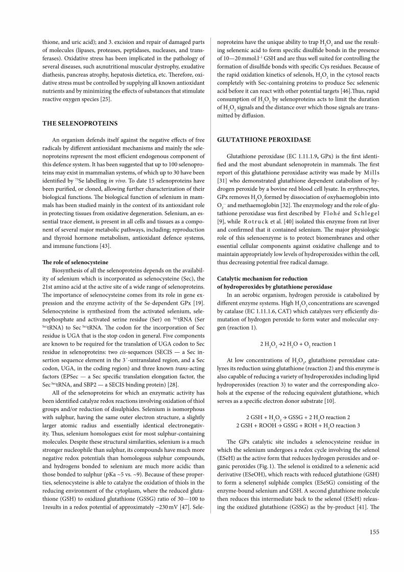
155
thione, and uric acid); and 3. excision and repair of damaged parts of molecules (lipases, proteases, peptidases, nucleases, and trans-ferases). Oxidative stress has been implicated in the pathology of several diseases, such as;nutritional muscular dystrophy, exudative diathesis, pancreas atrophy, hepatosis dietetica, etc. Therefore, oxi-dative stress must be controlled by supplying all known antioxidant nutrients and by minimizing the effects of substances that stimulate reactive oxygen species [25].
ThE SELENOPROTEINS
An organism defends itself against the negative effects of free radicals by different antioxidant mechanisms and mainly the sele-noproteins represent the most efficient endogenous component of this defence system. It has been suggested that up to 100 selenopro-teins may exist in mammalian systems, of which up to 30 have been identified by 75Se labelling in vivo. To date 15 selenoproteins have been purified, or cloned, allowing further characterization of their biological functions. The biological function of selenium in mam-mals has been studied mainly in the context of its antioxidant role in protecting tissues from oxidative degeneration. Selenium, an es-sential trace element, is present in all cells and tissues as a compo-nent of several major metabolic pathways, including; reproduction and thyroid hormone metabolism, antioxidant defence systems, and immune functions [43].
The role of selenocysteineBiosynthesis of all the selenoproteins depends on the availabil-
ity of selenium which is incorporated as selenocysteine (Sec), the 21st amino acid at the active site of a wide range of selenoproteins. The importance of selenocysteine comes from its role in gene ex-pression and the enzyme activity of the Se-dependent GPx [19]. Selenocysteine is synthesized from the activated selenium, sele-nophosphate and activated serine residue (Ser) on SectRNA (Ser
SectRNA) to Sec SectRNA. The codon for the incorporation of Sec residue is UGA that is the stop codon in general. Five components are known to be required for the translation of UGA codon to Sec residue in selenoproteins: two cis-sequences (SECIS — a Sec in-sertion sequence element in the 3´-untranslated region, and a Sec codon, UGA, in the coding region) and three known trans-acting factors (EPSec — a Sec specific translation elongation factor, the Sec SectRNA, and SBP2 — a SECIS binding protein) [28].
All of the selenoproteins for which an enzymatic activity has been identified catalyze redox reactions involving oxidation of thiol groups and/or reduction of disulphides. Selenium is isomorphous with sulphur, having the same outer electron structure, a slightly larger atomic radius and essentially identical electronegativ-ity. Thus, selenium homologues exist for most sulphur-containing molecules. Despite these structural similarities, selenium is a much stronger nucleophile than sulphur, its compounds have much more negative redox potentials than homologous sulphur compounds, and hydrogens bonded to selenium are much more acidic than those bonded to sulphur (pKa ~5 vs. ~9). Because of these proper-ties, selenocysteine is able to catalyze the oxidation of thiols in the reducing environment of the cytoplasm, where the reduced gluta-thione (GSH) to oxidized glutathione (GSSG) ratio of 30—100 to 1 results in a redox potential of approximately −230 mV [47]. Sele-
noproteins have the unique ability to trap H2O2 and use the result-ing selenenic acid to form specific disulfide bonds in the presence of 10—20 mmol.l–1 GSH and are thus well suited for controlling the formation of disulfide bonds with specific Cys residues. Because of the rapid oxidation kinetics of selenols, H2O2 in the cytosol reacts completely with Sec-containing proteins to produce Sec selenenic acid before it can react with other potential targets [46].Thus, rapid consumption of H2O2 by selenoproteins acts to limit the duration of H2O2 signals and the distance over which those signals are trans-mitted by diffusion.
GLuTAThIONE PEROXIDASE
Glutathione peroxidase (EC 1.11.1.9, GPx) is the first identi-fied and the most abundant selenoprotein in mammals. The first report of this glutathione peroxidase activity was made by M i l l s [31] who demonstrated glutathione dependent catabolism of hy-drogen peroxide by a bovine red blood cell lysate. In erythrocytes, GPx removes H2O2 formed by dissociation of oxyhaemoglobin into O2
.− and methaemoglobin [32]. The enzymology and the role of glu-tathione peroxidase was first described by F l o h é and S c h l e g e l [9], while R o t r u c k et al. [40] isolated this enzyme from rat liver and confirmed that it contained selenium. The major physiologic role of this selenoenzyme is to protect biomembranes and other essential cellular components against oxidative challenge and to maintain appropriately low levels of hydroperoxides within the cell, thus decreasing potential free radical damage.
Catalytic mechanism for reduction of hydroperoxides by glutathione peroxidase
In an aerobic organism, hydrogen peroxide is catabolized by different enzyme systems. High H2O2 concentrations are scavenged by catalase (EC 1.11.1.6, CAT) which catalyzes very efficiently dis-mutation of hydrogen peroxide to form water and molecular oxy-gen (reaction 1).
2 H2O2 →2 H2O + O2 reaction 1
At low concentrations of H2O2, glutathione peroxidase cata-lyzes its reduction using glutathione (reaction 2) and this enzyme is also capable of reducing a variety of hydroperoxides including lipid hydroperoxides (reaction 3) to water and the corresponding alco-hols at the expense of the reducing equivalent glutathione, which serves as a specific electron donor substrate [10].
2 GSH + H2O2 → GSSG + 2 H2O reaction 22 GSH + ROOH → GSSG + ROH + H2O reaction 3
The GPx catalytic site includes a selenocysteine residue in which the selenium undergoes a redox cycle involving the selenol (ESeH) as the active form that reduces hydrogen peroxides and or-ganic peroxides (Fig. 1). The selenol is oxidized to a selenenic acid derivative (ESeOH), which reacts with reduced glutathione (GSH) to form a selenenyl sulphide complex (ESeSG) consisting of the enzyme-bound selenium and GSH. A second glutathione molecule then reduces this intermediate back to the selenol (ESeH) releas-ing the oxidized glutathione (GSSG) as the by-product [41]. The

156
oxidized glutathione is reduced by the flavin adenine dinucleotide-dependent enzyme, glutathione reductase (EC 1.8.1.7, GR), to the thiol form GSH to complete the cycle. NADPH+H+ reduces FAD present in GR to produce a transient FADH- anion quickly break-ing a disulfide bond (reaction 4). The oxidation of NADPH+H+ to NADP+ is accompanied by a decrease in absorbance at 340 nm [35].
GSSG + NADPH + H+ → 2 GSH + NADP+ reaction 4
So, for each GSSG, two reduced GSH antioxidant molecules are produced, scavenging reactive oxygen species in the cell. In cells ex-posed to high levels of oxidative stress, like red blood cells, up to 10% of the glucose consumption may be directed to the pentose phos-phate pathway (PPP) for the production of the NADPH+H+ needed for this reaction. In erythrocytes, if the PPP does not function, then the oxidative stress will lead to lysis, and eventually anaemia.
Glutathione (GSH, γ-glutamylcysteinylglycine) is the major endogenous antioxidant produced by the cells, participating di-rectly as a free-radical scavenger. The ratio of reduced glutathione to oxidized glutathione within cells is often used as a measure of cellular antioxidant status. When cells are exposed to increased lev-els of oxidative stress, GSSG will accumulate and the ratio of GSH to GSSG will decrease. Therefore, the determination of the GSH/GSSG ratio and the quantification of GSSG are useful indicators of oxidative stress in cells and tissues [27]. Glutathione is able to re-generate the most important antioxidants, vitamins C and E back to their active forms. The intracellular content of glutathione depends
on environmental factors and functions as a balance between its utilization and synthesis.
ThE GLuTAThIONE PEROXIDASE FAMILY
Four structurally and genetically different forms of selenium-containing GPx have been functionally described and exist in dif-ferent tissues or parts of the cell: cytosolic glutathione peroxidase (cellular, classical, cGPx, GPx-1), gastrointestinal glutathione per-oxidase (giGPx, GPx-2), plasma glutathione peroxidase (pGPx, extracellular eGPx, GPx-3) and phospholipid hydroperoxide glu-tathione peroxidase (phGPx, GPx-4). Although GPx-1, GPx-2, and GPx-3 are homotetramers, GPx-4 is a monomer with a molecular size smaller than the subunits of the other glutathione peroxidases [5]. The small size and its hydrophobic surface have been impli-cated in its ability to react with complex lipids in membranes.
The role of the individual GPx isoenzymesAll glutathione peroxidases reduce hydrogen peroxide and
alkyl hydroperoxides at the expense of glutathione with their dif-ferent specificities for the hydroperoxide substrate. Whereas cyto-solic glutathione peroxidase (GPx-1) reduces hydrogen peroxide and some organic hydroperoxides (cumene hydroperoxide, t-butyl hydroperoxide), phospholipid hydroperoxide glutathione peroxi-dase (GPx-4) and to some extent plasma glutathione peroxidase (GPx-3) also reduce hydroperoxides of more complex lipids (phos-
Fig. 1. Catalytic mechanism for reduction of hydroperoxides by glutathione peroxidase(ESeH — selenol; ESeOH — selenenic acid; ESeSG — selenyl sulphide complex;
GSH — reduced glutathione; GSSG — oxidized glutathione; GR — glutathione reductase)

157
phatidylcholine hydroperoxide) [11], [45], [48]. GPx-4, however, efficiently reduces hydroperoxy groups of thymine, lipoproteins, and cholesterol esters and is unique in acting on hydroperoxides integrated in membranes [44].
Gastrointestinal glutathione peroxidase (GPx-2) is the closest homologue of GPx-1. It has approximately 65 % of the amino acid sequence identity and 60 % of the nucleotide sequence identity with GPx-1[6]. This enzyme represents a major barrier against hydro-peroxides originating from nutrition. During feed production and storage, some unsaturated lipids are oxidized and they can cause health-related problems, including decreased growth, impaired re-productive function and immune competence [20]. These oxidized lipids can react with transition metals, already present in the feed or as feed supplements, producing free radicals which can react with natural or synthetic antioxidants present in the feed. GPx-2 pre-vents them, in this way, entering systemic circulation.
The mRNA for GPx-3 is found predominantly in the kidney, in particular the epithelial cells of the proximal tubules [3] and therefore it shows a significant diagnostic value in kidney diseases. GPx-3 may have a specific antioxidant function in renal tubules or extracellular fluids. In the case of low concentration of glutathione, B j ö r n s t e d et al. [4] have suggested other thiols such as thioredoxin and glutaredoxin systems which can also act as electron donors and support an antioxidant role for plasma GPx. Investigating selenium deficiency in humans, C o h e n et al. [7] found that after selenium supplementation of Se-deficient individuals, plasma GPx-3 activity returned to normal values within 4—5 weeks, whereas 3—4 months were required for the red blood cell enzyme to reach this effect.
Phospholipid glutathione peroxidase decreases lipid hydroper-oxide levels, and thus inhibits the lipoxygenases that metabolise arachidonic acid to generate intermediates that mediate signals for increasing cell proliferation and inhibiting apoptosis. In par-ticular, it inhibits 5-lipoxygenase and reduces the production of 5-hydroxyeicosatetraenoic acid, which is known to stimulate the proliferation of prostate cancer cells [12]. GPx-4 has an unusual tissue distribution. In the rat testes it is 15 times more active than in the liver and kidneys, and 25 times more active than in the heart and lungs [2]. Recently, G o d e a s et al. [13] have proposed an ad-ditional role for GPx-4 in the maturation of spermatozoa via the metabolism of hydroperoxides and sperm thiol oxidation. Phos-pholipid GSH-Px may also be involved in the regulation of leukot-riene biosynthesis.
Epididymal secretory glutathione peroxidase (GPx-5) occupies a special position in the GPx family and it has been demonstrated in rats, mice, pigs, monkeys and humans. The peculiarity of this GPx form is the absence of the selenocysteine residue at the active site. It is replaced by a cysteine amino acid residue. The purified protein contains no selenium, and its GPx activity towards hydrogen perox-ide or organic peroxides is less than 0.1 % of that of GPx-1. This low activity, coupled with very low glutathione concentrations in the ep-ididymal fluids, has led to the suggestion that this protein protects sperm by binding organic hydroperoxide which would otherwise induce an acrosome reaction [8].
The glutathione S-transferases (EC 2.5.1.18, GSTs), one of the most abundant liver proteins, play an important role in protection against oxidative stress by mechanisms involving conjugation of al-dehydes and other potentially oxidative compounds. This group of enzymes has the primary role in xenobiotic detoxification metabo-
lism by catalyzing the transfer of reduced glutathione to various substrates containing a reactive electrophilic centre, to form a polar S-glutathionylated product for reducing oxidative stress. In addi-tion to conjugation reactions, the glutathione S-transferases can also function as antioxidants to reduce organic hydroperoxides, but not hydrogen peroxide. Therefore, they are sometimes referred to as enzymes with the glutathione peroxidase activity independent of selenium (Se-independent GPxs). Using hydrogen peroxide as sub-strate, the activity of Se-independent GPx is not measured because this enzyme cannot catalyze the reduction of hydrogen peroxide and thus this activity is only against organic hydroperoxides with a much higher Km than Se-dependent GPx [16]. Selenium deficien-cy causes an increase in hepatic glutathione S-transferase activity. In rat liver, glutathione S-transferase is present in the cytosol, mi-tochondria, and microsomes. Its GPx activity in the microsomes appears to be lower than that in the other sites. Rat liver glutathi-one S-transferase activity is 50 to 100 % higher in Se-deficient male rat liver cytosol than in the control. L a w r e n c e and B u r k [24] reported the presence of both Se-dependent and Se-independent GPx activities in pig livers. Approximately 67 % of the total liver GPx activity of the pig was Se-independent, whereas the data of M e y e r et al. [29] suggested that 45 to 60 % was Se-independent. The amount of Se-independent GPx activity was fairly consistent at the different dietary Se levels, the relative percentage of Se-in-dependent activity was greatest for those fed 0.1 ppm Se because of the lower Se-dependent GPx activity. These results suggest that the increase in total liver GPx activity largely reflected the increase of the Se-dependent form of the enzyme, as there was no further increase in activity at higher dietary Se levels.
ThE EFFECT OF SELENIuM AND VITAMIN E CONCENTRATION ON ThE ACTIVITY OF GLuTAThIONE PEROXIDASE
Selenium is an essential micronutrient element in diets of vari-ous animal species, which is required for maintenance of health, growth and biochemical-physiological functions, such as: biologi-cal antioxidation, immune function, reproduction and thyroid hor-mone metabolism. Selenium can be found in all the cells and tis-sues of the body but its levels in blood and tissues are very much in-fluenced by dietary selenium form and intake. It is also well known that selenium from organic sources is more efficiently incorporated into tissue than inorganic sources of selenium [38]. The body’s se-lenium status is assessed by direct measurement of selenium con-centration in blood and tissues, or indirectly by measuring the ac-tivity of glutathione peroxidase. Since selenium is incorporated by erythrocyte glutathione peroxidase during erythropoiesis, the glu-tathione peroxidase enzyme is considered to be a suitable indicator of biologically active selenium and thus an indicator of long-term supplementation of an organism with selenium. The direct deter-mination of the selenium content in the blood reveals the short-term selenium status of the body [37]. These data were confirmed by K o v á č and S a n k a r i [23], who found the highest correlation between whole blood selenium concentrations and erythrocyte GPx activities (r = 0.992), and between selenium concentration and GPx activities in the whole blood (r = 0.981). K o n v i č n á et al. [21] also observed corresponding GPx activity in the range of 569.32—

158
691.90 µkat.l–1, with the use of the most common reference sele-nium value (70.30—100.32 μg.l–1) in the whole blood of dairy cows, and the correlation analysis showed the degree of dependence be-tween the measured indices (r = 0.892; y = 0.0031x – 0.8749). In the study of P a v l a t a et al. [37], the dependence of GPx activity on the selenium concentration in the blood of sheep was found and the activity values of GPx were determined for use in the diagnosis of sufficient selenium status in sheep in the Czech Republic (GPx ac-tivity greater than 600 μkat.l–1 of whole blood). L u d v i k o v a et al. [26] defined for the practical establishment of selenium deficiency in horses the marginal level of GPx activity as 200—300 μkat.l–1 in whole blood, when the deficiency level for selenium is set as 75—100 μg.l–1 of Se in the blood. M i s u r o v a et al. [34] found that a selenium concentration of 100 μg.l–1 corresponds with GPx activity 700 μkat.l–1 in the blood of adult goats and 720 μkat.l–1 in the blood of newborn kids.
Selenium deficiency causes various diseases; the most frequent are muscle dystrophy in calves, young cattle after turning to pas-ture, lambs, foals, poultry, hepatosis dietetica in pigs, exudative diathesis, and encephalomalacia in poultry. Moreover, various re-production disorders occur (infertility, degeneration of seminifer-ous tubules), reduced libido in sheep, disorders of ovarian cycle, and increased occurrence of infertility [22]. Selenium deficiency has also been linked to many health problems in young animals such as: increased neonatal mortality; decreased sucking reflex; weakness; and higher occurrence of infectious diseases. Selenium, which is essential as a trace element for intrauterine development of calves, passes in considerable amounts through the placental barrier to the foetus, thus reducing the body pool of selenium in a pregnant female. This is why in late pregnancy, when foetuses de-velop and increase their weights most intensively, cows have much lower serum selenium concentrations compared to non-pregnant heifers and cows [42]. High-producing transition cows are highly susceptible to oxidative stress, which A i t k e n et al. [1] report to contribute to mastitis pathogenesis by modifying the expression of genes coding for proinflammatory factors. The same authors report that during the dry period, the activity of glutathione peroxidase is relatively low, but increases considerably during early lactation, when it serves a protective role in the proinflammatory state of the mammary gland. As shown by M i r a n d a et al. [33], the reduction in the number of mammary epithelial cells with advancing lacta-tion is at least in part caused by oxidative stress, which is accompa-nied by the loss of contact and cuboidal structure of cells, and the decrease in their viability as a result of apoptosis stimulation.
Vitamin E, the primary lipid-soluble antioxidant in cell mem-branes, is part of the body’s intracellular defence against the adverse effects of reactive oxygen species and free radicals that initiate oxi-dation of unsaturated phospholipids and critical thiol groups of pro-teins. Muscle damage and muscular dystrophy are common signs of both vitamin E and selenium deficiency. The more metabolically active tissues (such as skeletal and smooth muscles and liver) have a greater potential for oxidative tissue damage if the vitamin E supply is limiting. Erythrocytes and capillary walls are also susceptible to damage in animals with marginal vitamin E status. The protection of cell membranes and other cellular components of immune cells against lipid peroxidation is probably the most important mecha-nism of vitamin E in the immune response. Vitamin E and seleni-um enhance host defences by improving phagocytic cell function.
Both vitamin E and GPx protect phagocytic cells and surrounding tissues from oxidative attack by free radicals produced by the respi-ratory burst of neutrophils and macrophages during phagocytosis [36]. H o g a n et al. [17] reported that vitamin E supplementation of the diet, increased the intracellular kill of Staphylococcus aureus and Escherichia coli bacteria by neutrophils. Cows supplemented with 2.01 g vitamin E per day, in the presence of adequate selenium (0.3 ppm) starting four weeks prior to calving through eight weeks postpartum, had increased neutrophil and macrophage functions and reduced somatic cell counts compared to controls [39]. Supple-mental vitamin E was shown to specifically stimulate the phagocy-tosis of Staphylococcus aureus by bovine neutrophils. Both vitamin E and selenium increased neutrophil chemotaxis and superoxide production in these studies. M i l a d et al. [30] suggested that vita-min E and selenium supply led to a significant rise in whole blood GPx activity and differences in phagocytic functions of leukocytes and neutrophils in sheep. Vitamin E also supports the immune function by its effects on arachidonic acid metabolism and subse-quent synthesis of prostaglandins, thromboxanes and leukotrienes. The production of these compounds increases under stress condi-tions. Thromboxane and interleukin-2 appear to exert a negative feedback effect on leukocyte function [14]. In dairy cattle, supple-mentation with selenium, or both selenium and vitamin E, reduced the incidence of retained placenta in herds where the prevalence of retained placenta was high, or when selenium or vitamin E were marginal in the diet [18]. Supplemental vitamin E-selenium has also been reported to reduce metritis, cystic ovaries and the time of uterine involution in cows with metritis [15]. Z i g o et al. [49]observed the effect of parenteral application of selenium and vita-min E on the health of mammary glands in dairy cows during the peripartum period. It is one of the ways of how to prevent their deficit related to supplied feed rations and ensure the adequate con-centration of Se and activity of GPx in the blood and thus increase the natural resistance of dairy cows to intramammary infections.
CONCLuSION
Glutathione peroxidase is an important part of the anti-oxidant defence system and the existence of four Se-depen-dent GPxs in different cell compartments immediately im-plies specific functions for each of the isozymes. GPx scav-enges most of the endogenously generated H2O2 in the cells and so it has a primary physiological importance in pro-tecting the erythrocyte haemoglobin from oxidative break-down. Since the erythrocyte has a high GSH concentration and adequate mechanisms for maintaining the glutathione in the reduced state, this GPx system provides an explana-tion for the remarkable stability of haemoglobin in the intact erythrocyte. GPx plays a key role in modulating the GSH/GSSG ratio and indirectly affects the NADP/ NADPH+H+ quotient of the cell. The enzyme may thereby regulate mul-tiple cellular functions such as: cell division, pentose phos-phate pathway, gluconeogenesis, mitochondrial oxidation of α-oxo-acids and others. Glutathione peroxidase also reduces lipid hydroperoxides, potentially harmful prooxidants that may promote peroxidation of polyunsaturated fatty acids in phospholipids in biological membranes.

159
The presence of selenium in the active sites of GPx en-zyme indicates an important biological role for this trace element in the normal functioning of living cells. This bio-logical function of selenium in mammals has been studied mainly in the context of its antioxidant role in protecting tissue from oxidative degeneration. Selenium enables the functioning of the immune system and prevents develop-ment of malignant diseases. Se-dependent GPx inhibited the initiating and promoting stages of chemical carcinogenesis. In some diseases of farm animals, the effects of selenium de-ficiency reduced their productive and reproductive abilities. Deficiencies of selenium result in biological dysfunction as-sociated with protein or membrane damage. There are sev-eral factors abrogating the activity of the enzyme. Some of these are individual internal factors, resulting in a significant variation in the enzyme activity in different organs, body weight, age groups, sex and endocrine regulation. However, environmental factors also have definite effects on enzyme action. Nutrition is one of the most essential factors, as fat content and fatty acid composition of feed, or trace element intake as well as vitamin status of the animal, play a crucial role in normal enzyme activity. Selenium supplementation is required to overcome the deficiency symptoms and the bio-availability of the same depends upon the nature of supple-ments used. It is generally found that organic selenium com-pounds have substantially greater bioavailability than that of inorganic selenium and that selenium from organic sources is more efficiently incorporated into tissue than inorganic sources of selenium.
ACkNOwLEDGEMENTS
This study was supported by VEGA Scientific Grant No. 1/0592/12 from the Ministry of Education and by Slovak Research and Development Agency under contract No. APVV-0475-10.
REFERENCES
1. Aitken, S. L., karcher, E. L., Rezamand, P., Gandy, J. C., Van de haar, M. J., Capuco, A. V., Sordillo, L. M., 2009: Evalua-tion of antioxidant and proinflammatory gene expression in bovine mammary tissue during the periparturient period. J. Dairy Sci., 92, 589—598.
2. Arthur, J. R., Bermano, G., Mitchell, J. h., hesketh, J. E., 1996: Regulation of selenoprotein gene expression and thyroid hormone metabolism. Biochem. Soc. Trans., 24, 384—388.
3. Avissar, N., Ornt, D. B., Yagil, Y., horowitz, S., watkins, R. h., kerl, E. A., 1994: Human kidney proximal tubules are the main source of plasma glutathione peroxidase. Am. J. Phys., 266, 367—375.
4. Björnstedt, M., Xue, J., huang, w., Akesson, B., hol-mgren, A., 1994: The thioredoxin and glutaredoxin systems are efficient electron donors to human plasma glutathione peroxidase. J. Biol. Chem., 269, 29382—29384.
5. Brigelius-Flohé, R., Aumann, k. D., Blöcker, h., Gross, G., kiess, M., klöppel, k. D. et al., 1994: Phospholipid-hydroper-
oxide glutathione peroxidase. Genomic DNA, cDNA, and deduced amino acid sequence. J. Biol. Chem., 269, 7342—7348.
6. Chu, F. F., Doroshow, J. h., Esworthy, R. S., 1993: Expres-sion, characterization, and tissue distribution of a new cellular selenium dependent glutathione peroxidase, GSH-Px-GI. J. Biol. Chem., 268, 2571—2576.
7. Cohen, h. J., Chovaniec, M. E., Mistretta, D., Baker, S. S., 1985: Selenium repletion and glutathione peroxidase--differential effects on plasma and red blood cell enzyme activity. Am. J. Clin. Nutr., 41, 735—747.
8. Drevet, J. R., 2006: The antioxidant glutathione peroxidase family and spermatozoa: a complex story. Mol. Cell. Endocrinol., 250, 70—79.
9. Flohé, L., Schlegel, w., 1971: Glutathione-peroxidase, IV Hoppe-Seyler’s 2. Physiol. Chem., 352, 1401—1410.
10. Forstrom, J. w., Zakowski, J. J., Tappel, A. L., 1978: Iden-tification of the catalytic site of rat liver glutathione peroxidase as selenocysteine. Biochemistry, 17, 2639—2644.
11. Forstrom, J. w., Stults, F. h., Tappel, A. L., 1979: Rat liver cytosolic glutathione peroxidase: reactivity with linoleic acid hy-droperoxide and cumene hydroperoxide. Arch. Biochem. Biophys., 193, 51—55.
12. Ghosh, J., Myers, C. E., 1998: Inhibition of arachidonate 5-lipoxygenase triggers massive apoptosis in human prostate can-cer cells. Proceedings of the National Academy of Sciences USA,95, 13182—13187.
13. Godeas, C., Tramer, F., Micali, F., Roveri, A., Maiorino, M., Nisii, C., Sandri, G., Panfili, E., 1996: Phospholipid hydroper-oxide glutathione peroxidase (PHGPx) in rat testis nuclei is bound to chromatin. Biochem. Mol. Med., 59, 118—124.
14. hadden, J. w., 1987: Neuroendocrine modulation of the thymus-dependent immune system. Ann. NY. Acad. Sci., 496, 39—58.
15. harrison, J. h., hancock, D. D., St. Pierre, N., Conrad, h. R., harvey, w. R., 1986: Effect of prepartum selenium treatment of uterine involution in the dairy cow. J. Dairy Sci., 69, 1421—1425.
16. hayes, J. D., McLellan, L. I., 1999: Glutathione and gluta-thione-dependent enzymes represent a coordinately regulated de-fence against oxidative stress. Free Radic. Res., 31, 273—300.
17. hogan, J. S., weiss, w. P., Todhunter, D. A., Smith, k. L., Schoenberger, P. S., 1992: Bovine neutrophil responses to paren-teral vitamin E. J. Dairy Sci., 75, 399—405.
18. hurley, w. L., Doane, R. M., 1989: Recent developments in the roles of vitamins and minerals in reproduction. J. Dairy Sci., 72, 784—804.
19. Johansson, L. G., Gafvelin, G., Arner, E. S. J., 2005: Sele-nocysteine in proteins-properties and biotechnological use. Bio-chim. Biophys. Acta, 1726, 1—13.
20. kanazawa, k., Ashida, h., 1998: Catabolic fate of dietary trilinoleoylglycerol hydroperoxides in rat gastrointestines. Bio-chem. Biophys. Acta, 1393, 336—348.
21. konvičná, J., kováč, G., Jesenská, M., hiščáková, M., Nagy, O., Seidel, h., Link, R., 2004: Relationships between con-centrations of selenium and glutathione peroxidase activity in the whole blood of dairy cows. Proceedings of the 5th Middle-European Buiatrics Congress, Budapest, Hungary, 579—583.
22. kováč, G. et al., 2001: Cattle diseases (In Slovak), M & M publishing house, Prešov, 874 pp.
23. kováč, G., Sankari, S., 1988: Glutathione peroxidase ac-

160
tivity, selenium concentration in the blood and the masticatory muscle of cattle. Folia Veterinaria, 32, 79—94.
24. Lawrence, R. A., Burk, R. F., 1976: Glutathione peroxidase activity in selenium deficient rat liver. Biochem. Biophys. Res. Com-mun., 71, 952—958.
25. Lim, P. S., Cheng, Y. M., wei, Y. h., 2002: Increase in oxi-dative damage to lipids and proteins in skeletal muscle of uremic patients. Free Radic. Res., 36, 295—301.
26. Ludvikova, E., Pavlata, L., Vyskocil, M., Jahn, P., 2005: Selenium status of horses in the Czech Republic. Acta Veterinaria Brno, 74, 369—375.
27. Mari, M., Morales, A., Colell, A., Garcia-Ruiz, C., Fer-nandez-Checa, J. C., 2009: Mitochondrial glutathione, a key sur-vival antioxidant. Antioxid. Redox Signal., 11, 2685—2700.
28. Mehta, A., Rebsch, C. M., kinzy, S. A., Fletcher, J. E., Co-peland, P. R., 2004: Efficiency of mammalian selenocysteine incor-poration. J. Biol. Chem., 279, 37852—37859.
29. Meyer, w. R., Mahan, D. C., Moxon, A. L., 1981: Value of dietary selenium and vitamin E for weanling swine as measured by performance and tissue selenium and glutathione peroxidase ac-tivities. J. Anim. Sci., 52, 302—311.
30. Milad, k., Rácz, O., šipulová, A., Bajová, V., kováč, G., 2001: Effect of vitamin E and selenium on blood glutathione per-oxidase activity and some immunological parameters in sheep. Vet. Med. Czech., 46, 1—5.
31. Mills, G. C., 1957: Hemoglobin catabolism. I. Glutathione peroxidase, an erythrocyte enzyme which protects hemoglobin from oxidative breakdown. J. Biol. Chem., 229, 189—197.
32. Mills, G. C., 1959: The purification and properties of gluta-thione peroxidase of erythrocytes. J. Biol. Chem., 234, 502—506.
33. Miranda, S., wang, Y. J., Purdie, N. G., Osborne, V. R., Coomber, B. L., Cant, J. P., 2009: Selenomethionine stimulates expression of glutathione peroxidase 1 and 3 and growth of bo-vine mammary epithelial cells in primary culture. J. Dairy Sci, 92, 2670—2683.
34. Misurova, L., Pavlata, L., Pechova, A., Dvorak, R., 2009: Selenium metabolism in goats — maternal transfer of selenium to newborn kids. Veterinarni Medicina, 54, 125–130.
35. Paglia, D. E., Valentine, w. N., 1967: Studies on the quan-titative and qualitative characterization of erythrocyte glutathione peroxidase. J. Lab. Clin. Med., 70, 158—169.
36. Pathania, V., Syal, N., Pathak, C. M., khanduja, k. L., 1998: Changes in rat alveolar macrophageal antioxidant defence and reactive oxygen species release by high dietary vitamin E. J. Nutr. Sci. Vitaminol., 44, 491—502.
37. Pavlata, L., Pechová, A., Illek, J., 2000: Direct and indirect assessment of selenium status in cattle — a comparison. Acta Vet. Brno, 69, 281—287.
38. Pisarčíková, J., konvičná, J., Petrovič, V., kováč, G., 2011: Selenium as an essential element for living organisms (In Slovak). Infovet, 5—6, 251—255.
39. Politis, I., hidiroglou, N., white, J. h., Gilmore, J. A., williams, S. N., Scherf, h. Frigg, M.,1996: Effect of vitamin E on mammary and blood leukocyte function, with emphasis on chemo-taxis in periparturient dairy cows. Am. J. Vet. Res., 57, 468—471.
40. Rotruck, J. T., Pope, A. L., Ganther, h. E., Swanson, A. B., hafeman, D. G., hoekstra, w. G., 1973: Selenium biochemical role as a component of glutathione peroxidase. Science, 179, 588—590.
51. Roy, G., Sarma, B. k., Phadnis, P. P., Mugesh, G., 2005: Selenium-containing enzymes in mammals: Chemical perspec-tives. J. Chem. Sci., 117, 287—303.
42. Slavík, P., Illek, J., Rajmon, R., Zelený, T., Jílek, F., 2008: Selenium dynamics in the blood of beef cows and calves fed diets supplemented with organic and inorganic selenium sources and the effect on reproduction. Acta Vet. Brno, 77, 11—15.
43. Surai, P. F., 2006: Selenium in ruminant nutrition. In: Su-rai, P.F. (eds.): Selenium in Nutrition and Health, Nottingham Uni-versity Press, Nottingham, United Kingdom, 487—587.
44. Thomas, J. P., Maiorino, M., ursini, F., Girotti, A. w., 1990: Protective action of phospholipid hydroperoxide glutathione peroxidase against membrane-damaging lipid peroxidation. J. Biol. Chem., 265, 454—461.
45. ursini, F., Maiorino, M., Gregolin, C., 1985: The seleno-enzyme phospholipid hydroperoxide glutathione peroxidase. Bio-chim. Biophys. Acta, 839, 62—70.
46. winterbourn, C. C., hampton, M. B., 2008: Thiol chem-istry and specificity in redox signaling. Free Radic. Biol. Med., 45, 549—561.
47. wouters, M. A., Fan, S. w., haworth, N. L., 2010: Disul-fides as redox switches: from molecular mechanisms to functional significance. Antioxid. Redox Signal, 12, 53—91.
48. Yamamoto, Y., Takahashi, k., 1993: Glutathione peroxi-dase isolated from plasma reduces phospholipid hydroperoxides. Arch. Biochem. Biophys., 305, 541—545.
49. Zigo, F., Vasiľ, M., Elečko, J., Farkašová, Z., Chripková, M., 2012: Effect of parenteral application of selenium and vita-min E on health of mammary gland in dairy cows during the peri-partum period. Folia Veterinaria, 56, Supplementum III, 21—22.
Received October 8, 2013

161
ABSTRACT
Exposure of hosts to heavy metals can weaken their im-munocompetence and increase their susceptibility to parasitic infections. Mice were chronically intoxicated with lead (Pb), cadmium (Cd), or mercury (hg) and subsequently infected with Trichinella spiralis larvae. Our aim was to evaluate the effect of heavy metals on the intensity of parasitic infections. Lead did not affect parasite numbers in the intestine, but during the mus-cle phase of the infection, it did increase the numbers of larvae. Cadmium significantly reduced the intensity of intestinal infec-tions and also the larva infestations of mice. Mercury intoxica-tion prolonged the intestinal phase until day 20 post infection (p. i.), which may be caused by debilitation of the intoxicated an-imals. The numbers of muscle larvae in the hg-intoxicated mice, in relation to their weight, were the highest of the heavy metals tested, resulting in the heaviest parasite burden.
key words: cadmium; heavy metals; lead; mercury; mouse; Trichinella spiralis
INTRODuCTION
Parasites have long attracted ecologists’ attention because they can serve as potential indicators of environmental quality by de-termining the risk rate of toxic bio-accumulation in a host. Even though small mammals (due to their high abundance and wide
distribution) are preferably being used as bio-indicators of envi-ronmental contamination, their parasites can accumulate con-taminants in their bodies in higher concentrations than their hosts. However, very little is known about the effects of their accumula-tion capacity for heavy metals on the hosts and the effects on the course of parasitic infections in a heavy metal polluted environ-ments and particularly how they affect a host’s health.
For the purposes of environmental evaluations, the most suitable entities are the parasites with great geographic range and numerous hosts, such as Trichinella spiralis nematodes, who have cosmopolitan distribution in moderate and tropical climate zones [19]. The main hosts of Trichinella are domestic pigs (Sus scrofa domestica) and wild boars (Sus scrofa scrofa), followed by synantropic animals like rats, dogs, cats and certain wild carnivores, such as bears, red foxes and coyotes [4], [3], [20]. Horses are considered unusual hosts, due to their herbivorous diet, however, their infections are highly pathogenic to humans who consume horse meat. T. spiralis is the causative agent of trichinellosis, characterized by enteritis (caused by adult worms), inflammation and degenerative changes in the skeletal muscles (caused by the larvae) [9]. The pathology of trichinellosis is essentially a reaction to an initial inflammatory response during the intestinal phase and the subsequent allergic and inflammatory responses dur-ing the larval migration and invasion of the host’s muscle [17]. Small mammals play a significant role in the transmission of this helminthic zoonosis. In the potentially contaminated environment they present a large host population that becomes an easy prey when weakened by parasitic infection or by heavy metals, thus increasing the risk of infection and concentration of toxic substances in the predators.
FOLIA VETERINARIA, 57, 3—4: 161—165, 2013
EFFECT OF hEAVY METALS ON EXPERIMENTAL TRICHINELLA SPIRALIS INFECTION IN MICE
Jalčová, M., Dvorožňáková, E., hurníková, Z.
Institute of Parasitology, Slovak Academy of Sciences, hlinkova 3, 040 01 košiceThe Slovak Republic

162
Table 1. Experimental scheme
Group(number of mice)
Intoxication(daily, from day 0 to the end of the experiments)
Infection(per os, on day 30 of experiments)
1 (n = 21) control 500 T. Spiralis larvae/mouse
2 (n = 21) Pb(CH3COO)2.3H2O1 mg.l–1 drinking water ad libitum
500 T. Spiralis larvae/mouse
3 (n = 21)Pb(CH3COO)2.3H2O100 mg.l-1drinking water ad libitum
500 T. Spiralis larvae/mouse
4 (n = 21) CdCl2 100 mg.l–1drinking water ad libitum 500 T. Spiralis larvae/mouse
5 (n = 21) CdCl2 200 mg.l–1drinking water ad libitum 500 T. Spiralis larvae/mouse
6 (n = 21) HgCl2 0.2 mg.kg–1 body weight (b. w.), subcutaneously 500 T. Spiralis larvae/mouse
7 (n = 21) HgCl2 1.0 mg.kg–1 b. w., subcutaneously 500 T. Spiralis larvae/mouse
The aim of this study was to determine the intensity of T. spira-lis infections in the host organism intoxicated with lead, cadmium and mercury. The data may elucidate the role of heavy metals in parasitic infections relating to bio-indicative value of its hosts − small mammals in the ecosystems contaminated with heavy metals.
MATERIALS AND METhODS
The experiments were performed on 147 BALB/c male mice, divided to 7 groups with 21 animals in each, weighing 20−22 g. The animals were kept under standard conditions (at 20—21 °C, 50—60 % relative humidity) and fed ad libitum using a commercial diet. The mice, except for Group 1, were intoxicated with different doses of heavy metals in the drinking water (Pb, Cd) or subcutaneously with Hg, from day 0 to the end of the experiment and subsequently infected with the same dose of T. Spiralis larvae on day 30 of the experiments (Table 1).
Determination of intestinal worm burdensThe intestinal phase of the infection was investigated on days 5,
10, 15 and 20 post infection (p. i.). The small intestine was cut into 5−10 cm long pieces, placed into a sieve and incubated in conical Pilsner glasses in 0.9 % saline at 37 °C overnight. After incubation, the gut pieces were discarded and the sediment was examined un-der a stereomicroscope at 60 x magnification.
Isolation of muscle larvaeThe muscle phase of infection was examined on days 20 and
30 p. i. Whole eviscerated carcasses were minced and artificially di-gested (1 % pepsin, 1 % HCl for 4 h at 37 °C; both Sigma-Aldrich, Germany) according to K a p e l and G a m b l e [10]. The samples were allowed to settle for 20 min before the supernatant was dis-carded and the sediment was poured through a 180 µm sieve into a conical glass and washed with tap water. The sediment was finally transferred to a gridded Petri dish and counted using a stereomi-croscope at 40× magnification. Depending on the density of larvae, either a sub-sample or the whole sample was counted.
Statistic evaluation was carried out by one-way ANOVA meth-od and the differences were considered significant at P < 0.05.
RESuLTS AND DISCuSSION
The animals were intoxicated with different doses of heavy metals throughout the whole experiment (60 days) and infected with T. spiralis after being intoxicated for 30 days. During the intestinal phase, the adults were isolated from the small intestine on days 5, 10 and 15 p. i.; no para-sites were recovered from the small intestine on day 20 p. i. in all groups except for the groups intoxicated with mercury (Fig. 1). The group with the higher mercury exposure (1 mg.kg–1 b. w.) displayed the highest increase in the number of parasites in the small intestine on day 10 p. i., compared to Group 1. The groups with Hg exposure had the highest num-ber of parasites on days 5 and 10 p. i. The mercury-exposed and T. Spiralis infected mice suffered from cachexia, which may have affected the host parasite load.
Compared to non-intoxicated mice, the exposure to lead did not affect the intensity of the parasitic infection. On day 15 p. i., mice intoxicated with 100 mg.l–1 of lead acetate had lower numbers of parasites than the non-intoxicated mice, though the differences were insignificant.
The parasite load was reduced in mice exposed to cadmi-um and a number of parasites in the small intestine in these mice was significantly lower (P < 0.05) at the lower cadmium dose of 100 mg.l–1on days 5 and 10 p. i.
No significant differences in larva burden were observed when assessing the muscle phase of the infections (Fig. 2). The muscle larvae were found on day 20 p. i.; on day 30 p. i. a higher number of larvae were found in the groups subjected to lead and cadmium intoxication at a dose of 100 mg.l-1. A reduced number of adults in the mice intoxicated with cadmium (200 mg.l–1) resulted in a lower number of muscle larvae, though the differences were statistically insignifi-cant. Mercury intoxication on day 20 p. i. resulted in a very

163
Table 2. Index of reproductive capacity (RCI) in mice intoxicated with heavy metals (Pb, Cd, Hg) and infected with T. spiralis
G−1 G−2 G−3 G−4 G−5 G−6 G−7
Day p. i. T. spiralis1 Pb
+T. spiralis
100 Pb+
T. spiralis
100 Cd+
T. spiralis
200 Cd+
T. spiralis
0.2 Hg+
T. spiralis
1.0 Hg+
T. spiralis
20 9.00 7.37 9.13 7.43 5.70 0.08 2.39
30 16.80 9.57 40.17 36.30 2.47 17.38 7.04
Fig. 1. The intensity of parasitic infection in the intestinal phase of trichinellosis in mice intoxicated with heavy metals (Pb, Cd, hg) on days 5, 10, 15 and 20
Fig. 2. The intensity of parasitic infection in the muscle phase of trichinellosis in mice intoxicated with heavy metals (Pb, Cd, hg) on days 20 and 30

164
low number of Trichinella muscle larvae, but their number on day 30 p. i. increased to a comparable value (intoxica-tion with 0.2 mg.kg–1 b. w.) or half-value (intoxication with 1.0 mg.kg–1 b. w.).
A common biological characteristic used to evaluate Trichinella species infectivity is the Reproductive Capacity Index (RCI) — the ratio of the total number of larvae recov-ered from an animal to the number of larvae administered to it. The first is the product of 4 components: the number of females established; their fecundity; their reproductive life span; and the survival of muscle larvae. At the muscle phase of T. Spiralis infection, RCI was decreased and markedly re-duced by the high intoxication with Cd and Hg (Table 2).
The observed differences in parasite numbers could be ex-plained by the different heavy metal toxicity. Exposure to Pb ions greatly increases rodent susceptibility to parasitic infec-tions [5]. Pb can weaken the resistance and increase the mor-tality risk in experimental animals and reduce the antibody production [1], [6]. Cadmium has pro-inflammatory proper-ties [14]; already developed inflammation in mice intoxicat-ed with cadmium significantly reduced the number of adult worms. A higher cadmium dose had probably a disturbing ef-fect on their fertility. The effect of heavy metals on mammal re-production has been confirmed by several studies. P a c e et al. [18] reported that exposure to Pb have resulted in a reduced number of testicular macrophages, relating to a significant de-cline in fertility. The authors suggested that testicular macro-phages could be one of the main targets of lead and exposure to even low doses in neonatal and adolescent stages can affect reproductive functions in adult male mice.
Heavy metal toxic effects on helminths has not yet been studied in detail; a single study [2] on the intestinal fish par-asites reported the increased occurrence of morphological malformations in the reproductive organs in Proteocephalus percae tapeworms, parasitising in perch in heavy metal con-taminated environment (the Ružín water reservoir). Though we did not focus on determining the disturbing effect of heavy metals on Trichinella reproduction, a higher dose of cadmium significantly reduced the number of muscle larvae when compared to the other experimental groups. Cadmium can disturb the hormonal balance in mammals, e. g. alter-ing the concentration of pituitary gland hormones, such as prolactin, adrenocorticotropine hormone, growth hormone, luteinizing hormone, and follicle stimulating hormone [13]. In helminths, cadmium can also affect their metabolism and internal organ structure.
Exposure to Hg enhanced the sensitivity of organisms to infection due to the inhibition of cell and humoral responses [15]. On the other hand, in vitro studies with inorganic mer-cury referred to a stimulation of lymphatic system activity and humoral immunity and an increase in a number of T and B spleen cells in mercury susceptible mice strains [8], [16]. An acute or chronic mercury intoxication, even its sub-toxic doses, disabled the immune system that is manifested by increased susceptibility to autoimmune disorders or im-munosuppression [7], [11], [22].
Environmental contamination can in some cases in-crease the effects of parasitism, e.g. increase population
density of suitable intermediate hosts and definitive hosts, or negatively affect the host defence mechanisms, thus in-creasing the host susceptibility (intoxication with Pb) [12]. However, the pollutants can decrease parasitism in such a way that infected hosts suffer more from environmental load (intoxication with Hg) than do infection-free hosts, parasites are more susceptible to pollution than their hosts, or pollu-tion lowers the abundance of essential hosts and interme-diate hosts below a threshold for sustained transmission of the parasites. The effect of contaminants may vary among respective parasitic species, and developmental stages [21]. In our experiments, Hg activated adult worms in the small intestine, but reduced the number of muscle larvae.
Many studies have focused on parasite-induced stress in relation to environmental contamination on a physiologi-cal level. Such stress is often the first step to weaken the im-mune response. For instance, fish suffering from parasitic infection are weaker and more susceptible to environmental stress [22]. The unfavourable effects of parasites on the host’s health is often camouflaged by an acute toxic effect of the pollutants.
In mice intoxicated with heavy metals and subsequently infected with T. spiralis, we observed differences in an inten-sity of parasitic infection depending on the dose and type of heavy metal. Exposure to cadmium inhibited the devel-opment of parasitic infection as early as during the muscle phase. The lead affected the parasite burden in hosts insigni-ficantly; it rather increased the intensity of infection during the muscle phase. Mercury intoxication prolonged the intes-tinal phase and the larvae remained in the small intestines till day 20 p. i., but that might have been influenced by the poor condition of the mice. Our data refer to the different effect of tested heavy metals on the hosts and intensity of parasitic infection, as well as increased risk of trichinello-sis via small rodents in the ecosystems contaminated with heavy metals.
ACkNOwLEDGEMENTS
This study was supported by the Slovak VEGA agency, grant No. 2/0093/11,and by the Project “INFEKTZOON — Centre of Excel-lence For Animal Infections and Zoonoses (ITMS-26220120002)”,on the basis of support from Operational Programme Research and De-velopment, funded by the European Regional Development Fund. The experimental protocols complied with the current Slovak ethics legislation.
REFERENCES
1. Bussolaro, D., Filipak, F., Gargioni, R., Fernandes, L. C., Randi, M. A. F., Pelletier, E., OliveiraRibeiro, C. A., 2008: The immune response of peritoneal macrophages due to exposure to inorganic lead in the house mouse Mus musculus. Toxicol. In Vitro, 22, 254—260.
2. Brázová, T., Torres, J., Eira, C., hanzelová, V., Miklisová, D., šalamún, P., 2012: Perch and its parasites are heavy metal bio-

165
monitors in a freshwater environment: The case study of the Ružín water reservoir, Slovakia. Sensor, 12, 3068—3081.
3. Campbell, w. C., 1983: Epidemiology I. Modes of trans-mission. In Campbell, w. C. (Ed.): Trichinella and Trichi-nosis. Ple-num Press, New York (U. S. A.), 425—444.
4. Dick, T. A., Pozio, E., 2001: Trichinella spp. and Trichinel-losis. In Samuel, w. M., Pybus, M. J., kocan, A. A.: Parasitic Dis-eases of Wild mammals. Iowa State University Press, Ames, Iowa, 380—396.
5. Flohé, S. B., Bruggemann, J., herder, C., Goebel, C., kolb, h., 2002: Enhanced proinflammatory response to endotoxin after priming of macrophages with lead ions. J. Leuc. Biol., 71, 417—424.
6. Gidlow, D. A., 2004: Lead toxicity. Occup. Med., 54, 76—81. 7. hansson, M., Abedi-Valugerdi, M., 2004: Mercuric chlo-
ride induces a strong immune activation, but does not accelerate the development of dermal fibrosis in tight skin mice. Scand. J. Im-munol., 59, 469—477.
8. Johansson, u., hansson-Georgiadis, h., hultman, P., 1998: The genotype determines the B cell response in mercury-treated mice. Int. Arch. Allergy. Immunol., 116, 295—305.
9. Jurášek, V., Dubinský, P., 1993: Veterinary Parasitology (In Slovak). Príroda a.s., Bratislava, 275—277.
10. kapel, C. M. O., Gamble, h. R., 2000: Infectivity, persis-tence, and antibody response to domestic and sylvatic Trichinella spp. in experimentally infected pigs. Int. J. Parasitol., 30, 215—221.
11. kazantzis, G., 2002: Mercury exposure and early effects: Anoverview, Med. Law, 93, 139—147.
12. Lafferty, k. D., kuris, A. M., 1999: How environmental stress affects the impacts of parasites. Limnol. Oceanogr., 44, 925—931.
13. Lafuente, A., Cano, P., Esquifino, A. I., 2003: Are cad-mium effects on plasma gonadotropins, prolactin, ACTH, GH and TSH level, dose dependent ? Biometals, 16, 243—250.
14. Lag, M., Rodionov, D., Ovrevik, J., Bakke, O., Schwarze, P. E., Refsnes M., 2010: Cadmium-induced inflammatory respons-es in cells relevant for lung toxicity: Expression and release of cyto-kines in fibroblast, epithelial cells and macrophages. Toxicol. Lett., 193, 252—260.
15. Lawrence, D. A., 1995: Posited mechanisms of metal im-munotoxicity. Hum. Exp. Toxicol., 14, 114—116.
16. Lofteniu, A., Ekstrand, J., Moller, E., 1999: HgCl(2)-induced human lymphocyte activation in vitro: a superantigenic mechanism ? Int. Arch. Allergy. Immunol., 299, 63—70.
17. Nováková, E., kubíčková, T., Ondriska, F., 2006: Medical Parasitology (In Slovak). PRO Banská Bystrica, 55—56.
18. Pace, B. M., Lawrence, D. A., Behr, M. J., Parsons, P. J., Dias, J. A., 2005: Neonatal lead exposure changes quality of sperm and number of macrophages in testes of BALB/c mice. Toxicology, 210, 245—256.
19. Pozio, E., 2005: The broad spectrum of Trichinella hosts: from cold- to warm-blooded animals. Vet. Parasitol., 132, 3—11.
20. Pozio, E., La Rosa, G., 2000: Trichinella murrelli n. sp.: eti-ological agent of sylvatic trichinellosis in temperate areas of North America. J. Parasitol., 86, 134—139.
21. Rosenberg, D. M., Resh, V. h., 1993: Freshwater Biomoni-toring and Benthic Macroinvertebrates. Chapman a Hall, New York, USA, 1—9.
22. Sures, B., 2004: Environmental parasitology: relevancy of parasites in monitoring environmental pollution. Trends in Parasi-tology., 20, 170—177.
23. Zheng, Y., Monestier, M., 2003: Inhibitory signal override increases susceptibility to mercury-induced autoimmunity. J. Im-munol., 171, 1596—1601.
Received, October 21, 2013

166
ABSTRACT
Metabolic disorders are one of the most important problems in modern cattle production. One of the diseases, which appears to be a major obstacle for an increase in milk productivity is ke-tosis of dairy cows. Investigations into the changes of the amino acids occurring in the plasma of ketotic cows, allows one to get an insight into the dynamics of this metabolic imbalance, which has significant diagnostic and prognostic value. Therefore, character-izing such changes in the plasma content of free amino acids in ketotic cows are reported in this paper. The study was conducted on 17 holstein cows in their second to fourth lactations with milk yields above 8 000 kg in previous lactations. Nine of these cows were clinically healthy, with negative tests for ketone bodies in their urine, and eight of the cows had positive tests, which also re-vealed hepatic impairment. Blood samples were taken in March, 2013, two or three weeks after calving. The blood was withdrawn from the jugular vein before the morning feeding. This research has shown that in ketotic cows, the plasma level of ketogenic, aromatic and sulphur-containing amino acids were significantly increased. In addition, the content of glycogenic amino acids and branched-chain amino acids were decreasing, leading to an in-crease in the ratio of glycogenic/ketogenic amino acids from 4.16 to 6.02. The content of amino acids used in the antioxidant sys-tem of the organism decreased on average by 40%, except for the methionine and taurine levels which significantly increased. The ratio between the essential and nonessential amino acids in the plasma of cows affected with ketosis decreased by more than 8 %.
FOLIA VETERINARIA, 57, 3—4: 166—170, 2013
CONTENT OF FREE AMINO ACIDS AND SOME PARAMETERS OF ThE FuNCTIONAL STATE OF ThE LIVER IN BLOOD PLASMA OF hEALThY
AND kETOTIC DAIRY COwS
Simonov, M. R., Vlizlo, V. V.
Institute of Animal Biology of the National Academy of Agrarian Sciences of ukraine, Lvivukraine
keywords: amino acids; calving; cows; hepatic pathology; ketosis
INTRODuCTION
Ketosis in cows is a severe pathologic process with complex symptoms and signs in which disorders of carbohydrate, lipids and protein oxidation occur. Impairment of functions of the liver, pan-creas, other organs and tissues are observed in sick animals [10], [16], [19], [20]. Most commonly the disease is reported in high-yield dairy cows during the first two months of lactation or at the peak of milk production. Amino acids play very important roles in the pathogenesis of ketosis, since they participate in the synthesis of the majority of endogenous biologically active substances, struc-tural proteins, enzymes, some of the hormones, nitrogenous bases, and neurotransmitters [13], [14]. Exogenous amino acids entering the organisms and undergoing oxidation are involved in the energy supply and are subjected to numerous transformations. The metab-olism of these substances are controlled by different biochemical and physiological mechanisms, which maintain relatively constant concentrations of amino acids in the blood and tissues. Therefore, the concentration of free amino acids and their derivatives in phys-iological liquids and tissues may serve as an index of homeostasis, and the pattern of the formation of amino acid reserves in organ-isms objectively reflect the state of the metabolic balance. The con-centration of free amino acids and their derivatives, as such, serve as a control factor for many key metabolic steps. Investigations of

167
the changes in the plasma content of free amino acids provides the possibility to acquire an insight into the essence of metabolic im-balance in a variety of diseases. Such an approach may contribute to revealing factors of significant diagnostic and prognostic relevance. Reasoning from this fact, the objective of our study was to inves-tigate and to compare the plasma content of free amino acids in healthy and ketotic dairy cows.
MATERIALS AND METhODS
This study was performed in March, 2013, on seventeen Hol-stein cows in their second to fourth lactations with a milk yield above 8 000 kg in previous lactations. Animals were kept on a farm in similar conditions and received the same diet.
Clinical examinations of the cows were performed and the con-tent of ketone bodies in their urine was detected with diagnostic strips (Ketophan, Pliva). During the examinations it was revealed that some animals (n = 8) were lying up, revealed rapid weight loss and milk yield, they looked depressed, and some of them had mus-cular tremor. After contact of the indicator strips with the urine, the colour turned violet, indicating the presence of ketone bodies. During the laboratory investigations, the diagnosis was confirmed by using fluorometric determinations of beta-hydroxybutyrate (BHBA) levels in the blood and milk samples. Animals with positive results (the presence of ketone bodies in the urine) were relegated into a separate group. In order to obtain plasma, blood samples were withdrawn two or three weeks after calving, from the jugular vein before the morning feeding, into sterile tubes with heparin and im-mediately centrifuged at 3 000 rpm. The plasma was frozen at –20 °C until analyses were done (up to three month following).
In order to obtain serum, venous blood was withdrawn into sterile plastic tubes, which were left at room temperature for clot formation. After that, the blood clots were separated from the tube walls with a thin glass rod, settled blood serum was transferred into new sterile tubes and centrifuged for 15 minutes at 1 000 rpm. This blood serum was again transferred into new sterile tubes and stored in the refrigerator at a temperature from 4 to 6 °С up to two days until blood tests were performed. The content of free amino acids was determined in the plasma using the ion-exchange techniques on the amino acid analyser Biotronik LC 6001 (Germany). The serum was used for the determination of: AST and GGT activity (Reitman & Frankel's method and kinetic colorimetric method, re-spectively [18]); levels of albumin (bromocresol green colorimetric method); and bilirubin (colorimetric method by J e n d r a s s i k and G r o f [15]), using a biochemical analyser Humalyzer 2000 (Ger-many) and test-systems manufactured by Human (Germany).
The results were processed statistically in Excel, arithmetical mean value (x) and the standard error of the mean (SEM) was cal-culated as well as the significance of the difference between two variation series.
RESuLTS AND DISCuSSION
The investigation of the amino acid content in the plasma of healthy and sick cows revealed a range of distinctions (Ta-ble 1). In particular, we observed a significantly lower alanine
(by 24.9 %; P < 0.001), arginine (by 20.1 %; P < 0.001) and as-paragines content (by 28.6 %; P < 0.05) in the plasma of sick cows in comparison with the healthy ones, which provided evidence of an increase in the utilization of these amino acids. For example, in the case when at the beginning of lactation, there was a low glucose content, amino acids which enter the citric acid cycle or convert into pyruvate, may directly trans-form into glucose. Consequently, amino acid carbon residues comprise from 15 to 35 % in the process of gluconeogenesis [6]. Besides, the decrease in arginine level may be explained by its usage in antioxidant protection of the organism. Some available literature data [2], [11], [17] suggest that some ami-no acids are able to decrease the damaging and pathological effects caused by various oxidative influences. For instance, there is the proven ability of arginine and citrulline to deacti-vate superoxide anion radicals, which leads to normalization of myocardial activity under the influence of oxidative agents. The data obtained show a lower level of not only arginine, but also citrulline by 45.5 % (P < 0.01).
Table 1. Plasma content of free amino acids in healthy
and ketotic cows, Means ± SEM
Amino acid [μmol.l–1]
Healthyn = 9
Ketoticn = 8
P <
Alanine 157.7 ± 9.63 118.4 ± 4.93 0.001
Arginine 58.3 ± 2.16 46.6 ± 2.58 0.001
Asparagine 25.2 ± 1.93 18.0 ± 0.98 0.05
Aspartate 9.4 ± 1.21 11.1 ± 1.18 0.5
Valine 120.1 ± 4.58 101.5 ± 5.42 0.05
Histidine 36.4 ± 3.96 21.0 ± 1.31 0.001
Glycine 402.6 ± 24.35 287.7 ± 8.21 0.01
Glutamate 62.8 ± 3.57 61.5 ± 5.20 0.5
Glutamine 102.3 ± 5.71 204.6 ± 10.77 0.001
Isoleucine 70.5 ± 6.22 66.7 ± 9.68 0.5
Carnosine 14.7 ± 2.93 10.0 ± 1.97 0.5
Leucine 48.6 ± 2.30 79.2 ± 12.98 0.001
Lysine 57.3 ± 7.63 59.8 ± 6.04 0.5
Methionine 16.8 ± 1.48 24.2 ± 1.42 0.01
Proline 135.5 ± 5.21 114.5 ± 22.76 0.1
Serine 56.9 ± 5.74 65.7 ± 8.93 0.5
Taurine 17.7 ± 1.35 27.02 ± 2.49 0.05
Tyrosine 27.6 ± 2.05 31.1 ± 5.64 0.5
Threonine 45.4 ± 3.85 37.9 ± 9.90 0.5
Tryptophan 38.6 ± 2.95 56.8 ± 4.40 0.01
Phenylalanine 24.3 ± 0.53 29.8 ± 2.64 0.05
Phosphoserine 15.9 ± 5.42 24.2 ± 2.33 0.5
Cysteine 6.64 ± 0.37 1.67 ± 0.73 0.001
Citrulline 77.4 ± 6.30 42.2 ± 5.78 0.01
1-methylhistidine 9.2 ± 3.97 10.9 ± 0.93 0.5
3-methylhistidine 6.0 ± 1.03 28.4 ± 2.06 0.001

168
In contrast with the glycogenic amino acids, the plasma content of ketogenic amino acids in ketotic cows was high-er. Particularly, the content of phenylalanine was higher by 18.5 % (P < 0.05), tyrosine by 11.3 %, leucine by 38.6 % (P < 0.001) and tryptophan by 32 % (P < 0.01). On the one hand, the higher level of amino acids may be related to activation of gluconeogenesis, but then, such changes may be associ-ated with the peculiarities of the degradation of different amino acids.
Aromatic and sulphur-containing amino acids decom-pose only in the liver. In particular, phenylalanine, trypto-phan, tyrosine and methionine belong to this group. There-fore, the higher level of these amino acids maybe explained by liver injury, due to the lowering of the concentration of intracellular enzymes (which inactivate the above men-tioned amino acids) [14].
We have established that in sick cows the serum level of albumin was lower (by 25.1 %; P < 0.01; Fig. 1) and the lev-el of total bilirubin was higher almost four-fold (P < 0.001;
Fig. 2) in comparison with healthy cows. The activities of AST (more than four-fold; P < 0.001; Fig. 3) and GGT (three-fold; P < 0.001; Fig. 4) were also higher. According to Table 1, in sick animals, the plasma levels of not only phenylalanine and tyro-sine were higher, but also of methionine (by 30.6 %; P < 0.01).
Disorders of the degradation of aromatic and sulphur-containing amino acids are the cause of excessive formation of endotoxins, such as phenol, mercaptans, and indole, which exacerbate the pathological process. Nevertheless, an in-creased content of methionine also has a positive aspect, be-cause this amino acid plays a significant role in the system of antioxidant protection. The antioxidant effect of methionine is explained by the fact that it is a good source of sulphur, which inactivates free radicals and enhances the utilization of lipids, preventing their deposition in the liver and vascular walls. The synthesis of taurine and cysteine, which is a pre-cursor of glutathione, depends on the level of methionine [5], [8], [9]. The results show significantly (P < 0.05) higher taurine levels (by 34.5 %)in the plasma of ketotic cows, which is probably associated with an increase in the activity of com-
Fig. 1. Serum albumin concentration in cows Fig. 2. Serum levels of total bilirubin in cows
Fig. 3. Serum activity of aspartate aminotransferase in cows Fig. 4. Serum activity of gamma-glutamyltransferase in cows

169
pensatory mechanisms in animal organisms. There are also available data [8] concerning the positive role of proline and histidine in the antioxidant defence of organisms. Proline is an effective quencher of singlet oxygen; it enhances preven-tion of cell damage and death under oxidative stress. Histidine has the ability to trap peroxyl radicals, prevents protein car-boxylation and formation of protein cross-links. These effects are probably determined by physico-chemical properties, re-lated to their ability to react with active forms of oxygen. As it can be seen from the results, in sick animals, the content of proline and histidine was lower by 15.5 % and by 42.3 % (P < 0.001) respectively in comparison with healthy cows. Be-sides, thelevelofcysteinewas lower by 74.8 % (P < 0.001) and glycine by 28.5 % (P < 0.01), which might be explained by the usage of these amino acids in the synthesis of glutathione, which, in turn, is utilized for organism detoxification.
This research has shown that the level of branched-chain amino acids (valine and isoleucine) was lowered (by 15.5 %; P < 0.05 and 5.4 %, respectively), except for leucine, which also belongs to branched-chain amino acids, which exerts a ketogenic action. These three essential amino acids are de-graded mostly in muscle tissue and play a significant role in energy metabolism, particularly in the synthesis and de-position of glycogen. Consequently, an imbalance in amino acid content in the blood occurs in ketotic cows, which is an unfavourable factor in the course of the disease.
Besides the changes in plasma amino acid content, we should also draw attention to the significantly higher levels of glutamine (by 50 %; P < 0.001) and 3-methylhistidine (by 78.9 %; P < 0.001) levels. These increased glutamine levels may contribute to the growth of glucocorticoidal activity, which leads to an increase of glutamine synthetase activity, the enzyme which catalyzes the formation of glutamine from glutamic acid and ammonia. High level of 3-methylhistidine may be considered as a marker of catabolism intensification in muscle tissue. It is an amino acid specific for contractile proteins (actin and myosin). During degradation of these proteins the given amino acid enters the blood circulation, but due to the absence of specific tRNA, it is not used for pro-tein synthesis, nor metabolized and in an unchanged form is excreted in the urine [1], [3], [4], [7], [12], [21]. These prop-erties make the determination of 3-methylhistidine content, an informative indicator of muscle protein catabolism.
The investigation of the ratio between glycogenic ami-no acids (alanine, arginine, asparagine, valine, histidine, glycine, glutamine, methionine, proline, serine, threonine, cysteine) and amino acids with ketogenic (leucine, lysine) or mixed action (tyrosine, tryptophan, phenylalanine) be-comes also noteworthy. Thus, in the plasma of ketotic cows, this ratio is higher by 30.9 % (4.16 vs. 6.02), and the ratio between essential and nonessential amino acids is lower by more than 8 % (from 1.81 vs. 1.66).
CONCLuSIONS
Ketotic cows exhibit an imbalance of amino acid content in their blood. This is an unfavourable factor in the course
of the disease. Particularly, in comparison with healthy animals, in ketotic cows, the content of glycogenic acids is significantly lower, and the level of ketogenic amino acids is higher, which in turn causes an increase in the ratio of glycogenic/ketogenic amino acids from 4.16 to 6.02.
The content of amino acids, which are used in the anti-oxidant defence of organisms (arginine, citrulline, proline, histidine, cysteine and glycine),were lower, on average by 40 %, except for methionine and taurine, the content of which was significantly higher.
Impairment of hepatic function in ketotic cows causes elevation of the levels of aromatic and sulphur-containing amino acids and lowering of the content of branched-chain amino acids. The ratio of essential and nonessential amino acids in the plasma of cows affected with ketosis was lower by more than 8 % (1.81 vs. 1.66).
REFERENCES
1. Akamatsu, h., Saitoh, Y., Serizawa, M., 2007: Changes of serum 3-methylhistidine concentration and energy-associated me-tabolites in dairy cows with ketosis. J. Vet. Med. Sci., 69, 1091—1093.
2. Annison, E. F. Bryden, w. L., 1999: Perspectives on rumi-nant nutrition and metabolism. J. Nutr. Res. Rev., 12, 147—177.
3. Bos, C., Gaudichon, C., Tomé, D., 2000: Nutritional and physiological criteria in the assessment of milk protein quality for humans. J. Am. Coll. Nutr., 19, 191—205.
4. Chae, L. J., Zheng, Y., Geumsoo, k., Levine, R. L., 2011: Methionine sulphoxide reductase A is a stereospecific methionine oxidase. In Proceedings of the National Academy of Sciences USA, 108, 409—412.
5. Chen, C., Dickman, M. B., 2005: Proline suppresses apop-tosis in the fungal pathogen Colletotrichum trifolii. Proc. Natl. Acad. Sci. USA, 102, 3459—3464.
6. D’Mello, J. P. F., 2003: Amino Acids in Animal Nutrition. Edinburgh, UK, СABI Publishing, 526 pp.
7. Drift, S. G., houweling, M., Schonewille, J. T., Tielens, A. G., Jorritsma, R., 2012: Protein and fat mobilization and as-sociations with serum β-hydroxybutyrate concentrations in dairy cows. J. Dairy Sci., 95, 4911—4920.
8. El-hattab, A. w., Emrick, L. T., Craigen, w. J., Scaglia, F., 2012: Citrulline and arginine utility in treating nitricoxide deficien-cy in mitochondrial disorders. J. Mol. Genet. Metab., 6, 399—406.
9. Erdmann, k., Grosser, N., Schrzder, h., 2005: L-methio-nine reduces oxidant stress in endothelial cells: role of haeme oxy-genase-1, ferritin, and nitric oxide. The AAPS Journal, 7, 18—19.
10. Gonzalez, C. I., Rosendo, O., 2013: Assessment of fatty liver syndrome and its predisposing factors in a dairy herd from Venezuela. ISRN Vet. Sci., 2013, 82—89.
11. Gou, L., Zhang, L., Yin, C., Jia, G., Yin, X., Zhuang, X. et al., 2011: Protective effect of L-citrulline against acute gastric mucosal lesions induced by ischemia-reperfusion in rats. J. Physiol. Pharmacol., 89, 317—327.
12. houweling, M., van der Drift, R., Jorritsma, A. G. M., 2012: Tielens technical note: Quantification of plasma 1- and3-methylhistidine in dairy cows by high-performance liquid chroma-tography — tandem mass spectrometry. J. Dairy Sc., 95, 3125—3130.

170
13. huzzey, J. M., Nydam, D. V., Grant, R. J., Overton, T. R., 2011: Associations of prepartum plasma cortisol, haptoglobin, fecal cortisol metabolites, and nonesterified fatty acids with postpartum health status in Holstein dairy cows. J. Dairy Sci., 94, 5878—5889.
14. hyde, R., Taylor, P., hundal, h., 2003: Amino acid trans-porters: roles in amino acid sensing and signalling in animal cells. J. Biochem., 373, 1—18.
15. Jendrassik, L., Gróf, P., 1938: Vereinfachte photome-trische Methoden zur Bestimmung des Blut bilirubins. Biochem. Zeitschrift, 297, 82—89.
16. Loor, J. J.,Everts, R. E., Bionaz, M., Dann, h. M., Morin, D. E., Oliveira, R. et al., 2007: Nutrition-induced ketosis alters metabolic and signalling gene networks in liver of periparturient dairy cows. Physiol. Genomics, 32, 105—116.
17. Noris, M., Todeschini, M., Cassis, P., Pasta, F., Cappel-lini, A., Bonazzola, S. et al., 2004: L-arginine depletion in pre-eclampsia orients nitric oxide synthase toward oxidant species. J. Hyperten., 43, 614—622.
18. Reitman, S., Frankel, S., 1957: A colorimetric method for the determination of serum glutamic oxalacetic and glutamic pyru-vic transaminases. Am.J. Clin. Pathol., 28, 56—63.
19. Steen, A., 2001: Field study of dairy cows with reduced ap-petite in early lactation: clinical examinations, blood and rumen fluid analyses. Acta Vet. Scand., 42, 219—228.
20. Stojević, Z., Piršljin, J., Milinković-Tur, S., Zdelar-Tuk, M., Ljubić, B., 2005: Activities of AST, ALT and GGT in clinically healthy dairy cows during lactation and in the dry period. Veteri-narski Arhiv, 75, 67—73.
21. Therkildsen, M., 2005: Muscle protein degradation in bull calves with compensatory growth. Livest. Prod. Sci., 98, 205—218.
Received October 10, 2013

171
ABSTRACT
Ferrocene related organometallic compounds are promising compounds for the design of novel drugs as they may exhibit much greater activity in living systems than the original compound without the ferrocene moiety. we investigated the activities of en-zymatic and non-enzymatic antioxidant levels of five ferrocenyl derivatives at a final concentration of 0.1 and 0.01 μg.ml-1 in mi-tochondria (the smallest, sufficiently closed system for the study of oxidative damage in vitro). The highest ability to influence the redox state was demonstrated by the ferrocenyl chalcone, 1,1´-bis [(E)-(2-indanonyl)-methylidene] ferrocene, which decreased glu-tathione peroxidase activities and caused glutathione depletion, when used in a final concentration of 0.01 μg.ml-1. Similarly, a greater ability to cause oxidative damage contributing to retrograde re-dox signalling from the mitochondria to the cytosol and nucleus, could result from the treatment with 1,1´-bis [(E)-(2-tetralonyl)-methylidene] ferrocene. however, further investigations of their biological activity are necessary.
key words: antioxidant; ferrocene; chalcone; metallocene
INTRODuCTION
Ferrocene (Fc), as an effective bioisostere, is becoming an ap-plicable platform for drug design by virtue of its redox properties, high lipophilicity and three dimensional metallocene unit. There-
fore, ferrocene moieties may lead to some positive changes in selec-tivity toward biological targets compared to phenyl or alkyl groups (15). As aromatic compounds, they show interesting redox proper-ties and chemical stability, and several of their derivatives may also affect their relative safety in many mammalian species. Many ferro-cenyl compounds display interesting cytotoxic, antitumor, antima-larial, antifungal and DNA cleaving activities [17]. Ferrocene can act; as one electron donor undergoing oxidation to the ferricenium ion (Fe3+) or, as a source of reactive oxygen species (ROS) that has a direct influence on its activity [12], [16]. The combination of a fer-rocenyl moiety with heterocyclic structures may increase biological activities or create new medicinal properties.
Chalcones are naturally occurring derivatives of the parent compound 1,3-diphenyl-2-propen-1-one and belong to the flavo-noid family of organic compounds. Chalcones have been reported to have a broad range of biological activities, such as: antimalarial, antibacterial, antitumour, antihyperglycemic and anti-HIV. A fer-rocenyl chalcone (Fc-chalcone) derivative is formed when either ring A or B is replaced with a ferrocenyl group [1]. Series A and B compounds differ significantly in their physicochemical proper-ties. The series A compounds have been characterized by; a higher resistance to oxidation and generally lower lipophilicity, but more polarized carbonyl bonds, than in series B [17]. When ferrocene is ring B, free radical generation should be greater, since ferrocene is more readily oxidized to ferricenium. Since the oxidizability of ferrocene can largely influence the oxidizing properties of Fc-chal-cones, there is evidence supporting the information to what extent they are able to scavenge radicals, and to what extent they are able
FOLIA VETERINARIA, 57, 3—4: 171—175, 2013
ASSESSMENT OF SELECTED ANTIOXIDANTS AFTER FERROCENYL-ChALCONES TREATMENT OF MITOChONDRIA
Vašková, J.1, Mojžišová, G.2, Fejerčáková, A.1
Vaško, L.1, Perjési, P.3
1Department of Medical and Clinical Biochemistry and LABMED,
2Department of Experimental Medicine, Faculty of Medicine,
Pavol Jozef šafárik university in košice, tr. SNP 1, 040 66 košiceThe Slovak Republic
3Institute of Pharmaceutical Chemistry, Faculty of Medicine, university of Pécshungary

172
to donate electrons to other suitable acceptors with the formation of free radicals and subsequent biological response.
We investigated the effects of five ferrocenyl derivatives (of which four were ferrocenyl chalcones) at two concentrations which most influenced the viability of cancer cell lines, on the activity of enzymatic antioxidants and levels of non-enzymatic antioxidants in mitochondria (which are the smallest and sufficiently closed sys-tem for the study of the potential effects on cellular metabolism in vitro).
MATERIALS AND METhODS
Compounds; A: (E)-3-(ferrocenemethylene)-4-chromanone; B: (E)-2-(ferrocenemethylene) -cyclohexanone; C: 1,1´-bis [(E)-(2-indanonyl)-methylidene] ferrocene; D: 1,1´-bis [(E)-(2-tetralonyl)-methylidene] errocene; and E: 2-cyano-3-ferrocenylacrylonitrile; were synthesized and purified as described previously (4, 13). Their structures were characterized by IR and NMR spectroscopy. The purity of the compounds was checked using thin layer chroma-tography and gas chromatography. All other chemicals used were of analytical grade and, if not stated otherwise, purchased from Sigma — Aldrich or Serva (Heidelberg, Germany). Liver mito-chondria were isolated using the method of F e r n á n d e z - V i z a r a et al. (5). Measurements were carried out in a respiratory medi-um (80 mmol.l-1 KCl, 300 mmol.l–1 KH2PO4, 300 mmol.l–1 K2HPO4, 15 mmol.l–1 TRIS-HCl, 6 mmol.l–1 MgCl2, and 0.78 mmol.l–1 EDTA) at pH 7.4. The tested compounds were added in a final concentra-tion of 0.1 µg.ml–1 and 0.01 µg.ml–1. The activity of glutathione re-ductase (GR; EC 1.6.4.2) was measured according to a modified method described by C a r l b e r g and M a n n e r v i k [2]; that of glutathione peroxidase (GPx, EC 1.11.1.9) was measured as de-scribed by F l o h e and G u n z l e r [6] and that of superoxide dis-mutase (SOD, EC 1.15.1.1) by means of the SOD-Assay Kit-WST (Fluka, Japan), following the user manual provided. The reduced glutathione (GSH) content was measured by the modified method of F l o r e a n i et al. [7] using Ellman’s reagent. All the measured parameters were calculated per mg or g of mitochondrial protein, determined by using the bicinchoninic acid assay.
The results were expressed as the mean ±SD of three indepen-dent measurements. The significance of differences between groups was determined using an unpaired Student t-test and were consid-ered significant at: * — P < 0.05; ** — P < 0.01; *** — P < 0.001.
RESuLTS AND DISCuSSION
The production of ROS by mammalian mitochondria is important because it underlies pathology caused by oxida-tive damage and contributes to retrograde redox signalling from the organelle to the cytosol and nucleus [10]. Mito-chondria generate approximately 2—3 nmol of O2
.– per mg of protein, the ubiquitous presence of which, indicates it is to be the most important physiological source of this radical in living organisms [9]. Therefore, it is important to under-stand O2
.– production within isolated mitochondria under conditions that mimic those that may arise in vivo under physiological or pathological conditions. A constraint of us-
ing isolated mitochondria is that the system is complicated and difficult to manipulate [3]. O2
.– undergoes a dismutation reaction to H2O2 spontaneously, or is accelerated by about four orders by SOD, in the mitochondrial matrix by Mn-SOD, and in the intermembrane space by Cu, Zn-SOD [8]. The activity of SOD was decreased (compared to the control) in the mitochondria with higher concentrations of the test-ed compounds, significantly by the treatment with the first three ferrocenyl chalcone compounds, and also in group E (Fig. 1). Treatment with a lower concentration of the tested substances led contrariwise to a significant increase in the activity of SOD compared to the control, except for com-pound B which demonstrated a significantly lower activity than the control.
SOD contributes significantly to protecting the organ-ism from the toxic effects of O2
.– but its activities produce H2O2 from O2
.–, both at the inter-membrane space and in the mitochondrial matrix. H2O2 diffuses rapidly through membranes, and the release of H2O2 from the mitochondria to the cytosol reflects the balance between H2O2 production and consumption reactions; with the latter mainly involving the reduction of H2O2 to H2O via GPx. The activities of GPx significantly decreased compared with the control under the treatment with both concentrations (Fig. 2), with the sole ex-ception of the treatment with compound E at the final con-centration of 0.1 μg.ml–1. However, the differences were not significant at the highest level (*** — P < 0.001) when com-pared to the control. Considering the inability of O2
.– to cross the membranes and the decreased requirements on its dis-mutation according to the measured SOD activities (Fig. 1, using concentration 0.1 μg.ml–1), a decrease in GPx activ-ity (Fig. 2, using concentration 0.1 μg.ml–1) suggested lower peroxide formation and thus lower need for its elimination. However, at relatively high concentrations of H2O2 and other hydroperoxides, peroxidases undergo substrate inactivation by the modification of the prosthetic haem group [11]. This is still necessary to consider if we want to explain the high activity of SOD and also significantly reduced activity of GPx (Fig. 1 and 2, using the concentration of 0.01 μg.ml–1).
The activity of GPx is affected by the presence of another antioxidant enzyme, GR, which continuously recycles the oxidized glutathione to the reduced state. In our experi-ments, GR activities were not significantly changed com-pared to the controls (Fig. 3). Exposed thiols are present in mitochondria at a high concentration [14]. The regeneration of GSH from glutathione disulphide (GSSG) trapped inside the mitochondria requires GR, which harnesses the more negative reduction potential of NADPH in the process. GR continually recycles the oxidized thiol back to its reduced form, since the potential for GSH regeneration within the mitochondrial matrix is proportionately responsible for the constant degradation of ROS. Another point to be consid-ered is that mitochondrial GSH is not synthesized in the matrix but is imported from the cytoplasmic compartment [14]. Thus, a variation in the mitochondrial matrix GSH ra-tio could not be balanced by the import. However, in the case of mitochondria, GSH reflected a significant decline in the response to a change in the GSH/GSSG ratio only un-

173
Fig. 1. Superoxide dismutase activities in mitochondria after treatment with the tested compoundsA: (E)-3-(ferrocenemethylene)-4-chromanone; B: (E)-2-(ferrocenemethylene) -cyclohexanone; C: 1,1´-bis [(E)-(2-indanonyl)-methy-lidene] ferrocene; D: 1,1´-bis [(E)-(2-tetralonyl)-methylidene] ferrocene: E: 2-cyano-3-ferrocenylacrylonitrile) at the final concentra-
tions 0.1 µg.ml-1 and 0.01 µg.ml-1
* — Indicates significance of differences; ** — P < 0.01; *** — P < 0.001
Fig. 2. Glutathione peroxidase activities in mitochondria after treatment with the tested compounds A: (E)-3-(ferrocenemethylene)-4-chromanone; B: (E)-2-(ferrocenemethylene) -cyclohexanone; C: 1,1´-bis [(E)-(2-indanonyl)-methy-lidene] ferrocene; D: 1,1´-bis [(E)-(2-tetralonyl)-methylidene]ferrocene, E: 2-cyano-3-ferrocenylacrylonitrile with final concentra-
tions 0.1 µg.ml-1 and 0.01 µg.ml-1
* — Indicates significance of differences; * — P < 0.05; ** — P < 0.01)

174
Fig. 4. Levels of reduced glutathione in mitochondria after treatment with the tested compoundsA: (E)-3-(ferrocenemethylene)-4-chromanone; B: (E)-2-(ferrocenemethylene) -cyclohexanone; C: 1,1´-bis [(E)-(2-indanonyl)-methy-lidene] ferrocene; D: 1,1´-bis [(E)-(2-tetralonyl)-methylidene] ferrocene, E: 2-cyano-3-ferrocenylacrylonitrile, at final concentrations
0.1 µg.ml-1 and 0.01 µg.ml-1. *Indicates significance of difference * — P < 0.05; ** — P < 0.01)
Fig. 3. Glutathione reductase activities in mitochondria after treatment with the tested compoundsA: (E)-3-(ferrocenemethylene)-4-chromanone; B: (E)-2-(ferrocenemethylene) -cyclohexanone; C: 1,1´-bis [(E)-(2-indanonyl)-methy-lidene] errocene; D: 1,1´-bis [(E)-(2-tetralonyl)-methylidene]ferrocene; E: 2-cyano-3-ferrocenylacrylonitrile at final concentrations
0.1 µg.ml-1 and 0.01 µg.ml-1

175
der treatment with compound C in the final concentration of 0.01 μg.ml–1 (Fig. 4). Treatment with the higher concen-tration (0.1 μg.ml–1) led in most cases (A, B, E) to increased levels of reduced glutathione compared to the control.
Ferrocene related organometallic compounds are known and are promising compounds for the design of novel drugs. By attaching organic groups, which creates a conjugated or non-conjugated system, they may exhibit much greater ac-tivity in living systems than the original compounds without the ferrocene moiety. Therefore, it would be very interest-ing to observe their effect on redox homeostasis through the activities of primary antioxidant enzymes and levels of re-duced glutathione in the mitochondria. The results indicate that at a concentration of 0.01 μg.ml–1, there was a significant increase in SOD activity compared to the control (except with compound B), which itself indicates a high production of superoxide radicals, and thus oxidative stress conditions. Increased dismutation leads to increased formation of per-oxides, but a high level is likely to cause inhibition of the catalytic activity of GPx and therefore no significant needs of GR activity on the feedback reduction of oxidized gluta-thione. GSH is a strong reductant and is able to react not only with peroxides, which can explain the decrease in the measured levels not only when comparing the two final con-centrations but especially with compound C, where its level was significantly reduced.
CONCLuSIONS
We observed the most significant effect of concomitant antioxidant enzyme activities together with GSH levels in the case of C derivative, suggesting that 1,1´-bis [(E)-(2-indanonyl)-methylidene] ferrocene demonstrated the high-est ability to influence the redox state of cells. The ability to cause oxidative damage, contributing to retrograde redox signalling from the mitochondria to the cytosol and nu-cleus, was expected for the treatment with 1,1´-bis [(E)-(2-tetralonyl)-methylidene] ferrocene. However, additional analysis of the pro-oxidant, antioxidant and biological ac-tivities of this compound is necessary. The low effective con-centration of the compound is very promising.
ACkNOwLEDGEMENT
The study was supported by VEGA grants 1/1236/12 and 1/0751/12.
REFERENCES 1. Attar, S., O’Brien, Z., Alhaddad, h., Golden, M. L.,
Calderón-urrea, A., 2011: Ferrocenyl chalcones versus organic chalcones: A comparative study of their nematocidal activity. Bioor. Med. Chem., 19, 2055—2073.
2. Carlberg, I., Mannervik, B., 1985: Glutathione reductase. Methods Enzymol., 113, 484—485.
3. Cochemé, h. M., Murphy, M. P., 2008: Complex I is the major site of mitochondrial superoxide production by paraquat. J. Biol. Chem., 283, 1786—1798.
4. Dimmock, J. R., kandepu, N. M., Nazarali, A. J., kow-alchuk, T. P., Motaganahalli, N., quail, J. w. et al., 1999: Con-formational and quantitative structure-activity relationship study of cytotoxic 2-arylidenebenzocycloalkanones. J. Med. Chem., 42, 1358—1366.
5. Fernández-Vizzara, E., Ferrín, G., Pérez-Martos, A., Fernández-Silva, P., Zeviani, M. et al., 2010: Isolation of mito-chondria for biogenetical studies: An update. Mitochondrion, 10, 253—262.
6. Flohe, L., Gunzler, w. A., 1984: Assay of glutathione per-oxidase. Methods Enzymol., 105, 114−121.
7. Floreani, M., Petrone, M., Debetto, P., Palatini, P., 1997: A comparison between different methods for the determination of reduced and oxidized glutathione in mammalian tissues. Free Radic. Res., 26, 449−455.
8. han, D., Antunes, F., Canalli, R., Rettori, D., Cadenas, E., 2003: Voltage-dependent anion channels control the release of the superoxide anion from mitochondria to cytosol. J. Biol. Chem., 278, 5557—5563.
9. Inoue, M., Sato, E. F., Nishikawa, M., Park, A. M., kira, I., Imada, k. et al., 2003: Mitochondrial generation of reactive oxy-gen species and its role in aerobic life. Curr. Med. Chem., 10, 2495—2505.
10. Murphy, M. P., 2009: How mitochondria produce reactive oxygen species. Biochem. J., 417, 1—13.
11. Olorunniji, F., Iniaghe, M. O., Adebayo, J. O., Malomo, S. O., Adediran, S. A., 2009: Mechanism-based inhibition of my-eloperoxidase by hydrogen peroxide: enhancement of inactivation rate by organic donor substrates. Open Enzyme Inhib. J., 2, 28—35.
12. Osella, D., Ferrali, M., Zanello, P., Laschi, F., Fontani, M., Nervi, M. et al., 2000: On the mechanism of the antitumour activ-ity of ferrocenium derivatives. Inorg. Chim. Acta, 306, 42—48.
13. Perjési, P., Nusser, T., Tarczay, G., Sohár, P., 1999: E-2-Benzylidenebenzocycloalkanones. Synthesis, stereostructure and NMR spectroscopic investigation. J. Mol. Struct., 479: 13—19.
14. Schafer, F. R., Buettner, G. R., 2001: Redox environment of the cell as viewed through the redox state of the glutathione disul-phide/glutathione couple. Free Radic. Biol. Med., 30, 1191—1212.
15. Schatzschneider, u., Metzler-Nolte, N., 2006: New Prin-ciples in Medicinal Organometallic Chemistry. Angew. Chem. Int. Ed., 45, 1504—1507.
16. Top, S., Vessieres, A., Laclerq, G., quivy, J., Tang, J., Vaissermann, J. et al., 2003: Synthesis, biochemical properties and molecular modelling studies of organometallic specific estrogen re-ceptor modulators (SERMs), the ferrocifens and hydroferrocifens. Evidence for an antiproliferative effect of hydroxyferrocifens on both hormone-dependent and hormone-independent breast can-cer cell lines. Chem. Eur. J., 9, 5223—5236.
17. wu, X., Tiekink, E. R. T., kostetski, I., kocherginsky, N., Tan, A. L. C., khoo, S. B. et al., 2006: Antiplasmodial activity of ferrocenyl chalcones: Investigations into the role of ferrocene. Eur. J. Pharm. Sci. 27, 175—187.
Received December 2, 2013

176
FOLIA VETERINARIA, 57, 3—4: 176—182, 2013
ABSTRACT
high Performance Liquid Chromatography (hPLC) is the analytical separation method most frequently used in the de-tection of residues of pharmacologically active substances in food matrices. It is also utilized to identify coccidiostats in feed and the residues of food. hPLC can be used for both screening and confirmation. These screening and confirmation methods have been validated due to the progress in the field of analytical chemistry. The requirements of the confirmation analysis and ensuring the quality of results are currently met only by liquid chromatography using the procedures consisting of appropri-ate combinations of cleaning, chromatographic separation, and spectrometry detection.
key words: coccidiostats; confirmation; development; hPLC
COCCIDIOSIS AND COCCIDIOSTATS
Food producing animals are often affected by the parasitic disease, coccidiosis [40]. Coccidiosis is a severe infectious disease caused by the microscopic parasite of the Eimeria species affecting many types of farm animals, especially poultry, in which it causes severe intestinal damage [32], [37], [46]. Even small damage to the intestinal wall can lead to the deteriorated growth of an animal, re-duced feed conversion, poor absorption of nutrients from the feed, dehydration, and loss of blood, thus reducing the viability of an
ThE uSE OF ThE hPLC METhOD IN CONFIRMATION OF COCCIDIOSTAT RESIDuES IN FOOD MATRICES
A REVIEw
Maďarová, M.1, kožárová, I.1, Tkáčiková, S.2
1university of Veterinary Medicine and Pharmacy, komenského 73, 041 81 košice, 2Department of Medical and Clinical Biophysics, Faculty of Medicine
P. J. Safarik university, Trieda SNP 1, 040 11 košiceThe Slovak Republic
animal [37], [46], [60]. Due to intensive poultry breeding in small concentrated areas in humid and warm environments, this disease spreads very rapidly [37]. The acute form of coccidiosis can cause high mortality, which is one of the main problems in poultry pro-duction [11].
In modern poultry production, it is very important to use coc-cidiostats for the prevention and therapy of coccidiosis. Coccidio-stats can be divided into two main groups, i.e. chemical (synthetic) and ionophore (biosynthetic). Synthetic coccidiostats include a wide range of compounds with various chemical structures which are used as feed additives in animal nutrition (halofuginone, ro-benidine, nicarbazin, diclazuril, decoquinate, and other), or are in-tended for the therapy of coccidiosis (sulphonamides). Biosynthet-ic coccidiostats are the substances containing the polyether group and are naturally produced by the fermentation of microorganisms such as Streptomyces species and Actinomadura species (monensin, narasin, salinomycin, lasalocid, maduramycin, and semduramycin) (Table 1). According to the current applicable legislation, they are only used as feed additives for animal nutrition for coccidiosis pre-vention [11].
RISkS RESuLTING FROM ThE uSE OF COCCIDIOSTATS AND ThE LEGAL ASPECTS
Because of the frequent and long use of coccidiostats in poultry breeding, there is a high risk of residues in food matrices [37]. The careful administration of coccidiostats to poultry is critical to en-

177
sure reduced residues of these substances in animal products made from poultry, in order to minimization general health risks [42]. The residues of coccidiostats can be present in the food of animal origin due to: an improper dose of coccidiostats; improper admin-istration of the medicated feed during certain periods of time; fail-ure to adhere to the withdrawal period; or due to the cross-contam-ination in feed production while using the same production line (37). If the total exposure in animals does not exceed the allowable daily limit for each coccidiostat, then the contents of each coccid-iostats residue in edible animal tissues remain sufficiently low. The most frequently detected coccidiostats in poultry products include: robenidine, nikarbazin, diclazuril, and lasalocid. The major reasons for the presence of their residues are the improper use of coccidio-stats and cross-contamination of the feed [23].
Biosynthetic coccidiostats are well-known for their cardiovas-cular effects. The majority of these effects have been characterized for monensin, as a model substance for the entire group of the above mentioned coccidiostats. Their action is based on increasing the coronary blood flow with the subsequent dilatation of the coro-nary arteries [27]. Biosynthetic coccidiostats are also the cause of muscular degeneration and have a toxic effect on the nervous sys-tem that leads to neuropathy, manifested by myelin degeneration and ataxia [38]. The adverse effects of synthetic coccidiostats on humans as consumers have not been described, except for residues of sulphonamides that can cause severe damage to the consumer’s health. Sulphonamides are well-known for their negative effects on the thyroid in connection with the development of thyroidal carci-noma [42].
REGuLATIONS APPLYING TO COCCIDIOSTATS
For the purpose of human health protection, the following reg-ulations apply to coccidiostats:
Regulation (EC) No. 1831/2003 of the European Parliament and of the Council on Additives for Use in Animal Nutrition. The regulation currently authorises eleven coccidiostats as the supple-mentary substances intended for broilers, turkeys, and rabbits (mo-nensin, narasin, salinomycin, lasalocid, maduramycin, semdura-mycin, robenidine, halofuginone, nikarbazin, diclazuril, and deco-quinate) [53].
Regulation of the Government of the Slovak Republic No. 320/2003 Coll. on Monitoring of Certain Substances and their Residues in Live Animals and in Products of Animal Origin. The governmental regulation determines the obligatory inspection of coccidiostat residues in beef cattle, sheep, goats, swine, equidae, poultry, in eggs, in rabbit meat, game, in animals from farm breed-ing and in feed [51].
Commission Regulation (EU) No. 37/2010 on pharmacologi-cally active substances and their classification regarding maximum residue limits in foodstuffs of animal origin. The applicable word-ing of the regulation determines the maximum limits for residues of; amprolium, diclazuril, decoquinate, halofuginone, monensin, lasalocid, sulphonamides, and toltrazuril in milk, meat, liver, kid-neys, eggs, skin, and fat. The maximum residue limits have been determined in compliance with the generally acknowledged prin-ciples for safety assessment, considering the toxicological risks, environmental pollution, as well as microbiological and pharmaco-logical effects of the residues [17].
Commission Regulation (EC) No. 124/2009, setting maximum levels for the presence of coccidiostats or histomonostats in food resulting from the unavoidable carry-over of these substances in non-target feed. The applicable wording of the regulation deter-mines the maximum contents of; monensin, narasin, salinomycin, lasalocid, maduramycin, semduramycin, robenidine, halofuginone, nicarbazin, diclazuril, and decoquinate in milk, liver, kidneys, eggs, skin, fat, and in other food products [16].
Commission Decision No. 2002/657/EC, implementing Coun-cil Directive 96/23/EC concerning the performance of analytical methods and the interpretation of results. The decision stipulates the rules for analytical methods to be used in the testing of the of-ficial samples and especially the general rules for interpretation of analytic results of official control of these samples [15].
ChROMATOGRAPhIC SEPARATION TEChNIquES uSED IN CONFIRMATION OF COCCIDIOSTAT RESIDuES
With regard to the presence of coccidiostat residues in the food of animal origin, it is necessary to have certain techniques estab-lished in practice for their identification and quantification. Litera-ture sources describe plenty of methods for the analysis of coccid-iostat residues in food matrices, such as liver, meat, eggs [25], [41], [43], [50], [60], [61] and milk [48].
Certified laboratories perform the routine inspection of coc-cidiostats which requires sensitive and reliable methods. The ma-jority of the methods developed for the identification of coccidio-stat residues in food matrices are based on the liquid chromatog-raphy (LC) with spectrophotometry or fluorescence detection, but also with the mass spectrometry, especially while using the tandem mass spectrometry, enabling sensitive identification and quantifi-cation of the target compound [11].
ThIN LAYER ChROMATOGRAPhY (TLC)
In the early 1970’s, the thin layer chromatography (TLC) was used for the qualitative detection of banned substances (thyreostat-
Table 1. Overview of synthetic and biosynthetic coccidiostats
Synthetic coccidiostats Biosynthetic coccidiostats
Halofuginone Monensin
Robenidine Narasin
Nicarbazin Salinomycin
Diclazuril Lasalocid
Decoquinate Maduramycin
and others Semduramycin

178
ics and some anabolics) [20]. This technique was also the first fast semi-quantitative analytical method developed also for identifica-tion of coccidiostat residues which was based on the division of individual substances between the ascending mobile phase and the stationary phase of the thin layer. In the TLC method, identification of analytes is carried out either directly under the ultraviolet(UV) radiation emitting lamp (UV lamp), or by spraying a plate with a suitable reagent which forms, together with analytes, products vis-ible with the naked eye.
One of the ways how to increase the sensitivity and specific nature of this method is to combine it with the microbiological detection (TLC/B), i.e. with the bacterial strain (59). Microbiological detection is generally used to identify the antimicrobial activity of pharmaco-logically active substances. The detection is carried out directly on the chromatographic plate placed on the surface of agar containing the respective bacterial strain that is incubated under the prescribed conditions. The separation and microbial detection is thus carried out on the same plate. The visualisation of zones is usually carried out using the dehydrogenase-active detection reagents [13].
In 1967, D o n o h o et al. [22] first described the identification of residues of biosynthetic coccidiostats using the TLC with mi-crobiological detection using the bacterial strain Bacillus subtilis. V a n d e r k o p et al. [65] gradually enhanced this method by im-proving the sample preparation, the sample cleaning process, agar, and solvents. Coccidiostat residues can be identified using also other bacterial strains, such as Bacillus stearothermophilus, which was used to identify residues of salinomycin [34], or residues of monensin, salinomycin, and lasalocid [66]. The available literature indicates that the TLC was used in a smaller extent also for the identification of synthetic coccidiostats, for example robenidine in the feed [8].
A s u k a b e et al. [3] detected monensin, lasalocid, and sali-nomycin using the high-performance thin layer chromatography (HPTLC). The HPTLC principle is the same as in the TLC. The dif-ferences are: for example, in the thickness of the chromatographic layer of the plate; in the number of theoretical compartments on a plate; in the volume of the sample dose; etc. The HPTLC was suc-cessfully used for the qualitative and quantitative detection of resi-dues of sulphonamides in food matrices, e. g. meat.
The application of this TLC method in practice, while identify-ing coccidiostat residues, is less frequently used, due to the extended use of other procedures based on the use of high-performance liq-uid chromatography as the separation technique [64]. At present, the TLC method is used exclusively for the screening purposes [69].
hIGh-PERFORMANCE LIquID ChROMATOGRAPhY wIThSPECTROPhOTOMETRIC DETECTION (hPLC-uV)AND FLuORESCENCE DETECTION (hPLC-FLD)
History shows that the largest number of confirmation methods described for the analysis of coccidiostat residues in food matrices and in feed, was represented by the analytical separation method, the high-performance liquid chromatography (HPLC). The spe-cific nature of this method, however, depends on the detector used which also affects the selectivity and sensitivity of the method [63].
The use of the UV detector in the analysis of residues is limited,
as this detector is only suitable for identification of those analytes which show absorbance within the wave length range from 180—600 nm. Similarly, the use of FLD detector is limited by the ability of analytes to excite at the specific wave length and then emit the radiation of a specific wave length [29], [59].
Some analytes cannot be detected by absorbance in the UV area or fluorescence without further adjustment, e. g. de-rivatization (insertion of a suitable chromophore/fluorophore into their structure). Thus, it requiresthe reaction with a selected agent, such as;9-antryldiazomethane (ADAM), dansyl chloride, 2,4-dini-trophenylhydrazine, 1-(bromoacetyl) pyrene, vanillin, or dimeth-ylaminobenzaldehyde. If the reaction runs before the injection into the HPLC system, it is called the pre-column derivatization; if it runs after the chromatographic separation on the column, it is called the post-column derivatization [59]. The chromophore is a functional group in the substance molecule that absorbs the UV ra-diation in the area; for example, a molecule with conjugated double bond, carbonyl, amino and aromatic group.
Identification of some coccidiostat residues, e. g. halofuginone, robenidine, nicarbazin, diclazuril, and lasalocid, using the HPLC-UV technique, can be carried out directly without any chemical analyte pre-adjustment. The identification of biosynthetic coccid-iostats cannot be carried out directly, as there is no chromophore in their structure. The HPLC-FLD technique can be used without the chemical pre-adjustment of analyte only in lasalocid identification. Other coccidiostatics are only detectable using this technique after they are derivatised [59], [63].
The HPLC with the UV detection has been used since the mid-dle of the 1970’s, compared to the HPLC with the FLD detection, the use of which began a little later [20]. The use of the HPLC with the UV detection method is described in the identification of: di-clazuril in feed [21]; nikarbazin in tissues, eggs, and in feed [24]; monensin, narasin, and salinomycin in tissues [31]; nikarbazin in feed [26]; monensin in poultry tissues [43]; and robenidine in tis-sues [73]. Robenidine identification in feed was described also by the method of the high-performance liquid chromatography with the diode array detector (HPLC-DAD) which also provides the information on the spectrum of analyte in the UV area, which is important for coccidiostats identification [39]. The HPLC with the FLD detection was developed for: lasalocid in liver [70]; monensin, narasin, and salinomycin in liver [45]; monensin, salinomycin, and lasalocid in feed [2]; and robenidine in tissues [14].
GAS ChROMATOGRAPhY wITh MASS SPECTROMETRY (GC-MS)
In the 1990’s, the gas chromatography with mass spectrometry (GC–MS) appeared on the market for a reasonable price [20]. Al-though the gas chromatography is able to separate the compounds with a wide range of chemical and physical properties, for the coc-cidiostat residues, it lacks the high resolution efficiency [54]. One disadvantage of this method is that in the case of identification of coccidiostats, it is time consuming due to difficult sample adjust-ment before the analyse [57], and considering the fact that it works at high temperatures, this method is less suitable for the analysis of coccidiostat residues due to their low volatility and thermal insta-bility [27], [59].

179
The GC-MS method was used to identify both, synthetic and biosynthetic coccidiostats, such as halofuginone in the feed [1], la-salocid in the liver [71], monensin in the meat [58], and diclazuril in the feed [5].
LIquID ChROMATOGRAPhY wITh MASS SPECTROMETRY (LC-MS)
Since the late 1990’s, the liquid chromatography with the mass spectrometry (LC–MS) belongs to the standard equipment in labo-ratories [20]. The efficiency of the mass spectrometry lies in the fact that by means of identification of precursor ions and products of its fragmentation, it provides the information on the analyte struc-ture [33]. The most frequently used mass analyser, is the simple quadrupole that represents a standard analyser for both, gas and liquid chromatography, with sufficient resolution (i. e. the analyser’s ability to provide differentiated signals for ions of similar mass and charge — m/z). It became frequently used for analyses thanks to its simplicity and low price.
The review of the available literature indicates that the LC-MS method was used to detect residues of biosynthetic coccidiostats in the food of animal origin [7], [19], [33], nikarbazin in tissues [44], [47], and in eggs [6], robenidine in feed [39], [47], diclazuril in plasma [18] etc.
S c h n e i d e r et al. [55] was the first team to apply the LC-MS electrospray ionization technique to identify the residues of biosyn-thetic coccidiostats in the liver of broiler chickens.
Today, the mass spectrometry represents the basic confirma-tion method in certified laboratories, and in a combination with the liquid chromatography it is a suitable option for identification and quantification of coccidiostat residues in food and in feed [4], [12].
LIquID ChROMATOGRAPhY wITh TANDEM MASS SPECTROMETRY (LC-MS/MS)
By concurrent engaging of several mass analysers, a highly sensitive and selective tandem mass analyser is formed [59]. For analyte quantification, the so-called triple quadrupole (QQQ) is most frequently used. It consists of two mass analysers of the quad-rupole type and the collision cell, and it is also the most frequently used mass spectrometer for identification of coccidiostat residues in food matrices, such as milk, muscles, liver, kidneys, eggs, and in feed. In certified laboratories, however, residue analyses are gradu-ally carried out preferably using other mass analysers, such as the ion trap (IT) that appeared sometimes in 1990 and later the high-resolution mass spectrometers, such as the time-of-flight analyser (TOF) [4]. Innovations in the field of liquid chromatography are represented by the combinations of quadrupole either with the ion trap, or with the time-of-flight analyser. Also well-known, is the combination of quadrupole, where the collision cell and the third mass analyser are replaced with the ion trap and then followed by the time-of-flight analyzer (QITTOF) [30].
For identification of biosynthetic coccidiostats (monen-sin, narasin, salinomycin, and lasalocid) in the feed for poultry, W a n g et al. [68] used the matrix-assisted laser desorption/ioniza-tion technique combined with the time-of-flight mass spectrom-
etry (MALDI-TOF-MS). Other well-known studies describing the identification of synthetic and biosynthetic coccidiostats by using the LC-MS/MS technique are: 28, 10, 48, 49, 52, 56, 60, 62, 67 and many others.
Newer more sophisticated techniques are nowadays repre-sented by linear ion traps (LIT), orbitrap (electrostatic orbital trap), or the Fourier transform ion cyclotron resonance; but since they do not belong to equipments with acceptable prices, they are not widely used in common practice [35].
Costs efficiency of analytic procedures represent an important issue for all laboratories performing the residues analyses. The growth of capital expenses is conditioned and/or determined by: the use of systems enabling automation and shortening of analytic procedures; initial expensive equipment (TOF, Orbitrap, and oth-ers); testing a large number of samples per day; and other such fac-tors. The alternative is to maximise the number of analytes identi-fied by a single analytic procedure. This procedure, despite a high price, is definitely possible with the LC-MS/MS technique [72].
uLTRA PERFORMANCE LIquID ChROMATOGRAPhY wITh TANDEM MASS SPECTROMETRY (uPLC-MS/MS)
The latest developments in the field of liquid chromatography are focused on: the development of new columns with a reduced size of the stationary phase; lower diameters; as well as, the use of higher pressures in the system. These developments significantly improve: the separation efficiency; shortens the duration of the analysis; improves the detection limits; reduces the consumption of solvents; and significantly reduces the elution times, which sig-nificantly increases the number of analysed samples per day. An ex-ample of this is the ultra performance liquid chromatography with tandem mass spectrometry (UPLC-MS/MS). This method is based on the principle of using the stationary phase consisting of particles smaller than 2.5 µm. Also well-known, is the combination of the UPLC and the TOF mass analyser for multi-residue analysis, pro-viding the absolute and unequivocal confirmation of positive find-ings of coccidiostat residues in animal products [9], [36], [37], [74].
CONCLuSIONS
Laboratories performing certified inspection of residues must face the increasing number of stricter requirements and criteria applying to the performance of analytic methods as defined by the currently applicable legislation. The pur-pose of the legal requirements is to eliminate the risk result-ing from the presence of residues in food matrices and pro-vide the consumer with safe and full-value food. The highest priority is the development of accurate methods of residue identification in order to prevent residues entering the food chain and thus to protect the consumers’ health.
History shows that the constant technical innovations and improvement of the performance of analytic methods used for the identification of coccidiostat residues in the food of animal origin and in feed, leads to the selection of the most suitable and appropriate methods. An example of

180
such advancement include the sensitive and reliable LC-MS/MS technique that identifies analytes on the basis of their molecule mass and structure. Nevertheless, it is necessary to realise that despite the significant improvements in this area, the extant analytic separation methods must be constantly improved.
ACkNOwLEDGEMENT
The article was elaborated with the support by the VEGA Grant of the Ministry of Education of the Slovak Republic No. 1/0939/12.
REFERENCES
1. Anderson, A., Christopher, D. h., woodhouse, R. N., 1979: Analysis of the anti-coccidial drug, halofuginone, in chicken feed using gas-liquid chromatography and high-performance liq-uid chromatography. J. Chromatogr. A, 168, 471—480.
2. Asukabe, h., Murata, h., harada, k., Suzuki, M., Oka h., Ikai, Y., 1994: Improvement of chemical analysis of antibiotics. Simultaneous determination of three polyether antibiotics in feeds using high-performance liquid chromatography with fluorescence detection. J. Agricult. Food Chem., 42, 112—117.
3. Asukabe, h., Yoneyama, h., Mori, Y., harada, k. I., Su-zuki, M., Oka, h., 1987: Improvement of chemical analysis of an-tibiotics: XI. Simultaneous fluorodensitometric determination of polyether antibiotics. J. Chromatogr. A, 396, 261—271.
4. Bizec, B. L., Pinel, G., Antignac, J. P., 2009: Options for veterinary drug analysis using mass spectrometry. J. Chromatogr. A, 1216, 8016—8034.
5. Blanchflower, w. J., hughes, P. J., Cannavan, A., kennedy, D. G., 1994: Determination of diclazuril in avian feed and premix-es with gas chromatography/mass spectrometry. J. AOAC Int., 77, 1061—1065.
6. Blanchflower, w. J., hughes, P. J., kennedy, D. G., 1997: Determination of nicarbazin in eggs by liquid chromatography-atmospheric pressure chemical ionization mass spectrometry. J. AOAC Int., 80, 1177—1182.
7. Blanchflower, w. J., kennedy, D. G., 1995: Determination of lasalocid in eggs using liquid chromatography-electrospray mass spectrometry. Analyst, 120, 1129—1132.
8. Bories, G. F., 1975: Simple determination of the coccidio-stat robenidine in poultry feed. Analyst, 100, 567—569.
9. Broakaert, N., Van Peteghem, C., Daeseleire, E., Stick-er, D., Van Poucke, C., 2011: Development and validation of an UPLC-MS/MS method for the determination of ionophoric and synthetic coccidiostats in vegetables. Anal. Bioanal. Chem., 401, 3335—3344.
10. Cha, J. M., Yang, S., Carlson, k. h., 2005: Rapid analysis of trace levels of antibiotic polyether ionophores in surface water by solid-phase extraction and liquid chromatography with ion trap tandem mass spectrometric detection. J. Chromatogr. A, 1065, 187—198.
11. Chico, I., Rúbies, A, Centrich, F., Copangó, R., Prat, M. D., Grandos, M., 2013: Use of gel permeation chromatography for clean-up in the analysis of coccidiostats in eggs by liquid chro-
matography-tandem mass spectrometry. Anal. Bioanal. Chem., 405, 4777—4786.
12. Chico, J., Rúbies, A., Centrich, F., Companyó, R., Prat, M. D., Granados, M., 2008: High-throughput multiclass method for antibiotic residue analysis by liquid chromatography-tandem mass spectrometry. J. Chromatogr. A, 1213, 189—199.
13. Choma, I. M., Grzelak, E. M., 2011: Bioautography detection in thin-layer chromatography. J. Chromatogr. A, 1218, 2684—2691.
14. Cohen, h., Armstrong, F., Campbell, h., 1995: Sensitive fluorescence detection of robenidine by derivatization with dansyl chloride and high-performance liquid chromatography. J. Chro-matogr. A, 694, 407—413.
15. Commission Decision 2002/657/EC of 12 August 2002 implementing Council Directive 96/23/EC concerning the perfor-mance of analytical methods and the interpretation of results. Of-ficial Journal of the European Communities, L221, 8—28.
16. Commission Regulation (EC) No. 124/2009 of 10 Febru-ary 2009setting maximum levels for the presence of coccidiostats or histomonostats in food resulting from unavoidable carry-over of these substances in non target feed. Official Journal of the European Communities, L 40/7, 2009, 1—5.
17. Commission Regulation (Eu) No. 37/2010 of 22 December 2009 on pharmacologically active substances and their classification regarding maximum residue limits in foodstuffs of animal origin. Of-ficial Journal of the European Communities, L15, 2010, 1—72.
18. Croubels, S., Cherlet, M., De Backer, P., 2002: Quantita-tive analysis of diclazuril in animal plasma by liquid chromatogra-phy/electrospray ionization mass spectrometry. Rapid Communica-tions in Mass Spectrometry, 16, 1463—1469.
19. Davis, A. L., harris, J. A., Russell, Ch. A. L., wilkins, J. P. G., 1999: Investigations by HPLC-electrospray mass spectrom-etry and NMR spectroscopy into the isomeration of salinomycin. Analyst, 124, 251—256.
20. De Brabander, h. F., Noppe, h., Verheyden, k., Vanden Bussche, J., wille, k., Okerman, L. et al., 2009: Residue analysis: Future trends from a historical perspective. J. Chromatogr. A, 1216, 7964—7976.
21. De kock, J., De Smet, M., Sneyers, R., 1992: Determina-tion of diclazuril in animal feed by liquid chromatography. J. Chro-matogr., 606, 141—146.
22. Donoho, A. L., kline, R. M., 1967: Monensin, a biologically active compound VII. Thin-layer bioautographic assay for monen-sin in chick tissues. Antimicrob. Agents Chemother., 7, 763—766.
23. Dorne, J. L. C. M., Fernández-Cruz, M. L., Bertelsen, u., Renshaw, R. w., Peltonen, k., Anadon, A. et al., 2013: Risk assess-ment of coccidostatics during feed cross-contamination: Animal and human health aspects. Toxicol. Appl. Pharmacol., 270, 196—208.
24. Draisci, R., Lucentini, L., Boria, P., Lucarelli, C. J., 1995: Micro high-performance liquid chromatography for the determi-nation of nicarbazin in chicken tissues, eggs, poultry feed and litter. J. Chromatogr. A, 697, 407—414.
25. Dubois, M., Pierret, G., Delahaut, P. h., 2004: Efficient and sensitive detection of residues of nine coccidiostats in egg and muscle by liquid chromatography–electrospray tandem mass spec-trometry. J. Chromatogr. B, 813, 181—189.
26. Dusi, G., Fagionato, E., Gamba, V., Baiguera, A., 2000: Determination of nicarbazin and clopidol in poultry feeds by liquid chromatography. J. Chromatogr. A, 882, 7984.

181
27. Elliott, C. T., kennedy, D. G., Mc Caughey, w. J., 1998: Methods for the detection of polyether ionophore residues in poul-try. Analyst, 123, 45—56.
28. Galarini, R., Fioroni, L., Moretti, S., Pettinacci, L., Dusi, G., 2011: Development and validation of a multi-residue liquid chromatography–tandem mass spectrometry confirmatory meth-od for eleven coccidiostats in eggs. Anal. Chim. Acta,700, 167—176.
29. Garaj, J., Bustin, D., hladký, Z., 1987: Analytical Chemis-try (In Slovak). SNTL Alfa, Bratislava, 121—129.
30. Gentili, A., Perret, D., Marchese, S., 2005: Liquid chro-matography-tandem mass spectrometry for performing confirma-tory analysis of veterinary drugs in animal-food products. Trends in Analytical Chemistry, 24, 704—733.
31. Gerhardt, G., Salisbury, C. D. C., Cambell, h. M., 1995: Determination of ionophores in the tissues of food animals by liq-uid chromatography. Food Addit. Contam., 12, 73—737.
32. Goldová, M., 2002: Coccidiosis in poultry. Slovak Veteri-nary Journal, 6, 30—32.
33. harris, J. A., Charlotte-Russell, A. L., John-wilkins, P. G., 1998: The characterisation of polyether ionophore veterinary drugs by HPLC-electrospray MS. Analyst, 123, 2625—2628.
34. heil, k., Fleur, P., Cieleszky, S., 1984: Thin-layer bioau-tographic assay for the detection of salinomycin sodium in rabbit tissues. J. Agric. Food Chem., 32, 997—998.
35. http://holcapek.upce.cz/teaching/03_HmotnostniAnaly-zatory.pdf(Mass Analysers)
36. http://www.chromatographyonline.com 37. huet, A. C., Bienenmann-Ploum, M., Vincent, u., Dela-
haut, P., 2013: Screening methods and recent developments in the detection of anticoccidials. Anal.Bioanal. Chem., DOI 10.1007/s00216-013-7035-6.
38. kart, A., Bilgili, A., 2008: Ionophore antibiotics: toxicity, mode of action and neurotoxic aspect of carboxylic ionophores. Journal of Animal and Veterinary Advances, 7, 748—751.
39. kot-wasik, A., wasik, A., 2005: Determination of robeni-dine in animal feeds by liquid chromatography coupled with diode-array detection and mass spectrometry after accelerated solvent ex-traction. Anal. Chim. Acta, 543, 46–51.
40. kožárová, I., Mačanga, J., Goldová, M., Major, P., Tkáčiková, S., 2011: Detection of maduramycin residues in the tissues of chickens and pheasants by the screening test for antibi-otic residues (STAR). Food Additives and Contaminants: Part A, 28, 608—618.
41. kožárová, I., Máté, D., hussein, k., Raschmanová, k., Marcinčák, S., Jevinová, P., 2004: High-performance liquid chro-matographic determination of sulphadimidine residues in eggs. Acta Veterinaria (Beograd), 54, 427—435.
42. kožárová, I., Máté, D., Pipová, M., Laciaková, A., Je-vinová, P., 2003: Residues of anticoccidials in animal poultry prod-ucts. In Proceedings of the international conference Risk Factors of Food Chain III., Nitra, SPU, 70—71.
43. kožárová, I., Tkáčiková, S., Máté, D., Cabadaj, R., hus-sein, k., Neuschl, J., Zaborskiene, G., 2002: An evaluation of the high-performance liquid chromatography method for determining monensin residues in the tissues of broiler chickens. Folia Veteri-naria, 46, 189—193.
44. Leadbetter, M. G., Matusik, J. E., 1993: Liquid chromato-graphic determination and liquid chromatographic-thermospray
mass spectrometric confirmation of nicarbazin in chicken tissues: interlaboratory study. J. AOAC Int., 76, 420—423.
45. Martinéz, E. E., Shimoda, w., 1986: Liquid chromato-graphic determination of multiresidue fluorescent derivatives of ionophore compounds monensin, salinomycin, narasin and lasalo-cid in beef liver tissues. J. Assoc. Off. Anal. Chem., 69, 637—641.
46. Moloney, M., Clarke, L., O’Mahony, J., Gadaj, A., O’kennedy, R., Danaher, M., 2012: Determination of 20 coccid-iostats in egg and avian muscle tissue using ultra high performance liquid chromatography-tandem mass spectrometry. J. Chromatogr. A, 1253, 94—104.
47. Mortier, L., Daeseleire, E., Delahaut, P. h., 2003: Simulta-neous detection of five coccidiostats in eggs by liquid chromatogra-phy tandem-mass spectrometry. Anal. Chim. Acta, 483, 27—37.
48. Nebot, C., Iglesias, A., Regal, P., Miranda, J., Cepeda, A., Fente, C., 2012: Development of a multi-class method for the identification and quantification of residues of antibiotics, coc-cidiostats and corticosteroids in milk by liquid chromatography tandem-mass spectrometry. International Dairy Journal, 22, 78—85.
49. Olejnik, M., Szprengier-Juszkiewicz, T., Jedziniak, P., 2009: Multi-residue confirmatory method for the determination of twelve coccidiostats in chicken liver using liquid chromatography tandem mass spectrometry. J. Chromatogr. A, 1216, 8141—8148.
50. Olejnik, M., Szprengier-Juszkiewicz, T., Jedziniak, P., 2009: Multi-residue Confirmatory Method for the Determination of Twelve Coccidiostats in Chicken Liver Using Liquid Chromatogra-phy Tandem Mass Spectrometry. Department of Pharmacology and Toxicology, National Veterinary Research Institute, Partyzantow 57, 24—100.
51. Ordinance of the Government of the Slovak Republic No. 320/2003 Coll. of 9 July 2003 on monitoring of certain sub-stances and residues thereof in live animals and animal products. Coll. No. 320/2003, Part 145, 2541—2585.
52. Peters, R. J. B., Bolck, Y. J. C., Rutgers, P., Stolker, A. A. M., Nielen, M. w. F., 2009: Multi-residue screening of veterinary drugs in eggs, fish and meat using high-resolution liquid chromatography accurate mass time-of-flight mass spectrometry. J. Chromatogr. A, 1216, 8206—8216.
53. Regulation (EC) No 1831/2003 of the European Parlia-ment and of the Council of 22 September 2003 on additives for use in animal nutrition. Official Journal of the European Communities, L268, 29—43.
54. Rokka, M., Jestoi, M., Eerola, S., 2007: Liquid chroma-tography with conventional detection. In Picó, Y.: Food Toxicants Analysis: Techniques, Strategies and Developments, Elsevier, Nether-lands, 475—508.
55. Schneider, R. P., Lynch, M. J., Ericson, J. F., Founda, h. G., 1991: Electrospray ionisation mass spectrometry of semduramycin and other polyether ionophores. Anal. Chem., 63, 1789—1794.
56. Shao, B., wu, X., Zhang, J., Duan, h., Chu, X., wu, Y., 2009: Development of a rapid LC–MS–MS method for multi-class determination of 14 coccidiostat residues in eggs and chicken. Chromatographia, 69, 1083—1088.
57. Stolker, A. A. M., Brinkman, u. A. T. h., 2005: Analytical strategies for residue analysis of veterinary drugs and growth-promot-ing agents in food-producing animals. J. Chromatogr. A, 1067, 15—53.
58. Takatsuki, k., Suzuki, S., ushizawa, I., 1986: Liquid chro-matographic determination of monensin in chicken tissues with

182
fluorometric detection and confirmation by gas chromatography-mass spectrometry. J. Assoc. Off. Anal. Chem., 69, 443—448.
59. Tkáčiková, S., 2010: Use of the HPLC Method in Identifica-tion of Levels of Residues of Selected Modern Anticoccidials in Food Chain. Dissertation Thesis, UVMP Košice, 126 pp.
60. Tkáčiková, S., kožárová, I., Mačanga, J., Levkut, M., 2012: Determination of lasalocid residues in the tissues of broiler chickens by liquid chromatography tandem mass spectrometry. Food Additives and Contaminants, Part A, 29, 761—769.
61. Tkáčiková, S., kožárová, I., Máté, D., 2010: Liquid chro-matography tandem mass spectrometry determination of madura-mycin residues in the tissues of broiler chickens. Food Additives and Contaminants, Part A, 27, 1226−1232.
62. Tkáčiková, S., kožárová, I., Máté, D., 2010: Use of the LC/MS/MS technique in identification of residues of maduramycin in tissue of broiler chickens. In XIth International Conference ACP 2010: Current situation and prospective of analytical chemistry in practice. Chemistry Letters, 104, 16, 497—502.
63. Toldrá, F., Reig, M., 2006: Methods for rapid detection of chemical and veterinary drug residues in animal foods. Trends Food Sci. Technol., 17, 482—489.
64. Van Poucke, L. S. G., Depourcq, G. C. I., Van Peteghem, C. h., 1991: A quantitative method for the detection of sulphon-amide residues in meat and milk samples with a high-performance thin-layer chromatographic method. J. Chromatogr., 29, 423—427.
65. Vanderkop, R. A., MacNeil, J. D., 1989: Thin-layer chro-matography/bioautography method for the detection of monensin in poultry tissues. J. AOAC Int., 72,735—738.
66. Vanderkop, R. A., MacNeil, J. D., 1990: Separation and detection monensin, lasalocid and salinomycin by thin-layer chro-matography/bioautography. J. Chromatogr., 508, 386—390.
67. Villalba, M., Moyano, E., Galceran, M. T., 2009: Fast liq-uid chromatography/multiple-stage mass spectrometry of coccid-iostats. Rapid Comm. Mass Spectrom.,23, 1255—1263.
68. wang, J., Sporns, P., 2000: MALDI-TOF-MS Quantifi-cation of coccidiostats in poultry feeds. J. Agric. Food Chem., 48, 2807—2811.
69. wang, S., Zhang, h. Y., wang, L., Duan, Z. J., kenne-dy, I., 2006: Analysis of sulphonamide residues in edible animal products: A review. Food Addit. Contam., 23, 362—384.
70. weiss, G., Felicito, N. R., kaykaty, M., Chen, G., Caru-so, A., hargroves, E. et al., 1983: Tissue residue regulatory method for the determination of lasalocid sodium in cattle liver using high-performance liquid chromatography with fluorometric detection. J. Agric. Food Chem., 31, 75—78.
71. weiss, G., kaykaty, M., Miwa, B., 1983: A pyrolysis gas chromatographic-mass spectrometric confirmatory method for lasalocid sodium in bovine liver. J. Agric. Food Chem., 31, 78—81.
72. Yamada, R., kozono, M., Ohmori, T., Morimatsu, F., kitayama, M., 2006: Simultaneous determination of residual vet-erinary drugs in bovine, porcine and chicken muscle using liquid chromatography coupled with electrospray ionization tandem mass spectrometry. Biosci. Biotechnol. Biochem., 70, 54—65.
73. Yeom, h., Yang, D. h., Suh, J. h., Eom, h. Y., kim, u., kim, J. et al., 2013: Determination of robenidine residues in chick-en muscle by high performance liquid chromatography with ultra-violet detection. Archives of Pharmacal Research, 36, 359—365.
74. Zhao, X. T., Lin, q. B., Song, h., Pan, Y. L., wang, X., 2011: Development of an immunoaffinity chromatography purifi-cation and ultra performance liquid chromatography tandem mass spectrometry method for determination of 12 sulphonamides in beef and milk. J. Agric. Food Chem., 59, 9800—9805.
Received December 18, 2013

183
ABSTRACT
A cross-sectional study on randomly selected 390 uncastrated cart horses was conducted from October 2011 to April 2012 to as-sess their major health and welfare problems in and around the town of Combolcha, Ethiopia. The welfare aspect of cart horses was determined in the study area by direct assessment, which in-cluded the incidence of lameness, injuries, and other diseases of cart horses in relationship to their body condition, age, and be-haviour. Focus group discussions and proportional pulling tech-niques were also used to generate further information. The ani-mals were allotted into three age groups; 0—6 years old (3.3 %), 6—11 years old (49.4 %), and 11—15 years old (46.9 %) and their body condition was determined to be; thin (7.4 %), fair (56.7 %), good (32.6 %), and fat (3.3 %). The overall prevalence of injury was 32.1 %. The prevalence of external injuries was found in animals; under 6 years old (61.5 %), between 11—15 years of age (31.1 %), and between 6 to 11 years of age (30.9 %). In addition, more exter-nal injuries were registered in thin horses (P < 0.05) compared to horses having fair or good body condition. The least incidence of injury was recorded in fat horses. Significantly higher number of cases of injuries was reported in cart horses out of work, followed by cart horses transporting people and cart horses transporting goods (85.7 %, 32.6 %, and 21.2 %, respectively; (X2 = 12.142; df = 2; P < 0.05). Further, an increased (P < 0.01) incidence of external in-juries were observed in alert and not friendly horses. Improper harnesses, overworking, and overloading probably caused the injuries. The cart horses out of work were horses that could no
FOLIA VETERINARIA, 57, 3—4: 183—189, 2013
ASSESMENT ON ThE wELFARE AND ITS INFLuENCE ON ThE hEALTh OF CART hORSES IN COMBOLChA DISTRICT, EThIOPIA
Daneil, G.1, Asefa Asmare, A.2
1wollo university, P. O. Box, 208, Dessie2woliata Sodo university, P. O. Box 137, Sodo
Ethiopia
longer work due to severe injury and other serious health prob-lems. They were abandoned by their owners and were straying in and around the town of Combolcha. The overall prevalence of lameness was 18.3 %. Similar to the incidence of external injuries, the highest prevalence of lameness (P = 0.780) occurred in cart horses below 6 years old. In this study, animals with the fair body condition score, showed a high incidence of lameness (22.2 %; P = 0.075). Further, cart horses out of work had a high incidence of lameness also (28.6 %; P = 0.461), followed by cart horses used for goods transportation (23.1 %) and cart horses used for people transportation (17.2 %; P = 0.795). In addition, depressed horses had a high prevalence of lameness. Lameness found in this study most probably was caused by shoeing abnormalities in addition to the malconformation of the hoof. Despite the animals being af-fected by lameness, they were obliged to continue to work. In focus group discussions, respondents reported wounds as the primary health problem and the cause of the wounds as due to overloading of the cart horses. The study revealed that many cart horses work-ing in the study area are experiencing multiple welfare problems.
key words: age; animal welfare; behaviour; body condition; injury; lameness
INTRODuCTION
Horses are important draught animals for both the developed and developing countries of the world. They play an important role

184
in socio-economics by being used for farming, cart pulling, trans-portation, sporting and leisure activities. They are also important sources of food in certain countries. The American horse council estimated that horses have a direct impact on the economy of the United States of over $39 billion and an indirect impact of over $102 billion. Horses used to be, and still are, an important use of transportation and they do not pollute the earth [13].
According to a report compiled by the Food and Agricultural Or-ganization of the United Nations [6], there are about 58 million horses in the world. Ethiopia is placed 8th in the world, and first in Africa, with its 1,655,385 horse population. Horses are efficient users of low quality, high fibre food and can tolerate up to 30 % dehydration [21]. Compared to other production animals, they require lesser quantity of food and have the ability to conserve more energy. They drink infrequently and irregularly. This makes horses a suitable adaptable animal for harsh environments and difficult working conditions [17].
Horses provide a life line for rural and urban poor communi-ties in Ethiopia. The report by the Brooke [1], an international char-ity, which works to improve the welfare of working equines around the world, indicated that horses are crucial to Ethiopian communi-ties in rural and urban settings. This study revealed, working horses can reduce the burden for women while at the same time offering house holds an opportunity to generate income and improve food security by helping to reduce poverty [14]. According to B i f a and W o l d e m e s k e l [2], horses are important animals in the farming and transport systems of Ethiopia. As pack animals, they transport harvest from the fields to the homestead. They also transport ag-ricultural products and fuel to the market and building materials and other commodities from the market to the villages. For rural traders and urban cart owners, horses are the sole means of income to sustain their extended families. The average net return from horse ownership and use has been estimated to be the equivalent of $330 per year in Ethiopia [2]. Moreover, horses play a key role in job creation in the supply of; chains for carts, harnesses, health services, animal feed, hoof/shoeing, and other provisions.
The cart is a common form of short to mid distance transporta-tion for people and goods in many small towns and cities in Ethio-pia. A two wheel cart is pulled by one horse. Cart pulling horses are suffering from the burden of stress. While experiencing severe stress, they can succumb to disease [10]. It is known that animal health is a key component of animal welfare and the active involve-ment of veterinary services and veterinarians is important for im-proving animal welfare [12]. Despite cart horses’ important socio-economic contributions, their welfare is often neglected and receive very low attention. Little interest has been given to research on wel-fare issues of the cart horses compared to donkeys in Ethiopia.
Therefore, the purpose of this study was to assess the welfare problems and their influence on the health of cart horses in and around Combolcha, Ethiopia.
MATERIALS AND METhODS
A cross sectional study was conducted on randomly selected 390 uncastrated cart horses residing in peri-urban and urban areas of the town of Combolcha, between October 2011 and April 2012. The sample size for this study was calculated based on the formula developed by T h r u s f i e l d [19].
1.962 × p × (1 – p)n = d2
Where n = sample size p = expected prevalence = 50 % = 0.5 d = desired level of precision = 5 % = 0.05
1.962 × 0.5 × (1 – 0.5)n = = 384 0.052
Since there was no study previously carried out on cart horses and their welfare in the study area, 50 % prevalence was used to calculate the sample size.
The welfare aspect of cart horses was determined in the study area by direct assessment, which included the incidence of lame-ness, injuries, and other diseases in association with body condition, age, and behaviour. The cart horses body condition was determined based on the method developed by C a r o l l and H u n t i n g t o n [4] as cited by P r i t c h a r d et al. [16]. The lameness was determined by observing their gait and the result was categorized as low grade, moderate, high grade and immobile to each lame cart horse based on the criteria formulated by D y s o n [5]. The cart horses were physically examined for grossly visible skin/tissue injuries measur-ing greater than or equal to 5 cm located on any part of the animal body. Injuries had to be active, with ongoing tissue damage, with or without blood/exudates/pus, abscess formation, or any second-ary bacterial complication as stated by B i f f a and W o l d e m e s- k e l [2]. The age of the animals was estimated by birth records ob-tained from the owners and dentition characteristics. Accordingly, the cart horses were allotted into four age groups: Group 1 con-sisted of animals 0—6 years old; Group 2, 6—11 year old; Group 3, 11—15 years old; and Group 4, > 15 years old. The behaviour of the study animals was investigated by close observation and contact with the animals, both at work and during rest. Their behaviour was characterized as; anxious, friendly, alert and friendly, alert and not friendly, or depressed [2]. Other diseases manifested by the study of the cart horses were diagnosed in the veterinary clinic and recorded as well.
Focus group discussions were held with local communities, cart horse owners, and clients for the indirect assessment of the welfare of the cart horses. The discussions were focused on: the role of the cart horses, management constraints, cart horses health problems, handling of cart horses by cartmen while working and the reaction of the community, government or non-governmental organizations (NGOs) to the mishandling of the cart horses. Information gath-ered from the focus group discussions were given a rank.
The data collected from the study area was entered into MS ex-cel spread sheets and analysed using STATA 7.0 and SPSS 15.0 sta-tistical software. The statistical analysis used included the compari-son of proportions, an X2 test and a test of agreement [19]. The Chi square test was applied to test if any statistical association existed between the welfare of the cart horses and the risk factors, such as; age, body condition, and type of work the horses performed.

185
RESuLTS
During the study period, the animals were clinically ex-amined for the presence of any gross external lesions, lame-ness, and other diseases. The body condition of the animals was scored as well as, their age, and their behaviour. All of the study cart horses were male. Of the study cart horses, 3.3 % were under the age of 6 years, whereas 49.4 % were be-tween 6—11 years and 46.9 % between 11—15 years of age. There were no cart horses above 15 years of age (Table 1.).
Table 1. Age grouping of cart horses
Age [year]
Number of animals
Percentage [%]
0—6 13 3.3
6—11 194 49.4
11—15 183 46.9
> 15 0 0
A study of the type of work revealed that 84.4 % of the horses were used for people transport, 13.3 % for transport of goods, and 1.8 % were found to be out of work (Table 2).
Table 2. Type of work the study animals were engaged in
Type of work
Number of animals
Percentage[%]
Transporting people 331 84.4
Transporting goods 52 13.3
Out of work 7 1.8
Of the 390 cart horses examined, the body conditions of 7.4 %, 56.7 %, 32.6 %, and 3.3 % were found to be thin, fair, good, and fat, respectively (Table 3.).
Table 3. Body condition score of the study animals
Body condition score
Number of animals
Percentage [%]
Thin 29 7.4
Fair 221 56.7
Good 127 32.6
Fat 13 3.3
Very fat 0 0
The highest number of horses (66.9 %) demonstrated alert and not friendly behaviour followed by depressed (14.9 %), anxious (8.7 %), friendly (8.2 %), and alert and friendly behaviour (1.3 %) (Table 4.).
Table 4. Observed behaviour of the study animals
BehaviourNumber
of animalsPercentage
[%]
Friendly 32 8.2
Alert and friendly 5 1.3
Alert and not friendly 261 66.9
Depressed 58 14.9
Anxious 34 8.7
Of the cart horses, a total of 32.1 % (n = 125) had injuries on different parts of their bodies (Fig. 1).
Injuries on the cart horses were more frequently ob-served on girth (12.8 %; n = 50), shoulder (3.30 %; n = 16), and mouth/bit (4.9 %; n = 19) compared with other body parts (Fig. 1). The association of the prevalence of external injuries on cart horses with age, body condition, work type, and behaviour is illustrated in Table 5. An increased preva-lence of external injuries was found in animals under 6 years old (61.5 %) compared to animals between 11—16 years of age (31.1 %) and 6—11 years old (30.9 %) in a descending order. According to this study, there was no significant dif-ference in the prevalence of external injuries among different age groups of animals (X2 = 5.371, df = 3, p = 0.068).
More external injuries were registered in thin horses compared to horses having fair or good body condition (58.6 %, 33.9 %, and 24.4 %, respectively). The least incidence of injuries was recorded in fat horses (15.4 %). The body con-dition was found to significantly influence the prevalence of external injuries (X2 = 14.824, df = 4, P < 0.05). High cases of injuries were reported on cart horses out of work followed by cart horses transporting people and cart horses trans-porting goods (85.7 %, 32.6 %, and 21.2 %, respectively). In the present study, significant variation in the prevalence of external injury on cart horses performing different type of work was encountered (X2 = 12.142, df = 2, P < 0.05). Signifi-cantly higher (P < 0.01) incidence of external injuries was observed in alert and not friendly horses comparing to de-pressed, friendly, and anxious horses (100 %, 51.7 %, 40.6 %, and 38.2 %, respectively). The least prevalence of injuries was recorded in alert and friendly cart horses (24.5 %). The be-haviour of the cart horses was found to be highly associated with the prevalence of injury (X2 = 29.392).
The overall prevalence of lameness among the study ani-mals (n = 390) was 18.3 % (n = 71). From the affected animals, 36.6 % had high grade, 28.2 % moderate, and 33.8 % low grade lameness, while 1.4 % of them were immobile (Table 6.).
The association of the prevalence of lameness in cart horses with age, body condition, work type and behaviour is indicated in Table 7.
The lowest prevalence of lameness occurred in cart horses below 6 years of age (3.0 %; n = 13) followed by, 6—11 years old (33.0 %; n = 194), and 11—15 years old (35.0 %; n = 183). There was no significant difference among different age groups in the prevalence of lameness. On the other hand,

186
Fig. 1. Prevalence of injuries on different body parts of cart horses
Fig. 2. Prevalence of other diseases in the cart horses
animals with fair body condition score showed high inci-dence of lameness (22.2 %) compared to thin (20.7 %), good (11.8 %), and fat (7.7 %) in decreasing order. The Chi-square value of 6.907 indicated an association of body condition and lameness. However, the prevalence of lameness did not differ significantly among animals with different body con-dition score.
Cart horses out of work had a high incidence of lame-ness (28.6 %), followed by cart horses used for goods trans-
portation (23.1 %) and cart horses used for people trans-portation (17.2 %). The alert and not friendly animals had a high prevalence of lameness (24.1 %), followed by anxious (20.6 %), alert and friendly (20 %), depressed (17.2 %), and friendly (12.5 %). The incidence of lameness by behaviour and work type was not significant.
A total of 90 (23.1 %) sick study cart horses were exam-ined in Combolcha town veterinary clinic. Of which, 27 (30 %) were affected with indigestion, 22 (24.4 %) with colic,

187
Table 6. Prevalence of lameness in the study animals
Degree of lameness
Number of animals
Prevalence in %
High 26 36.6
Moderate 20 28.2
Low 24 33.8
Immobile 1 1.4
Table 5. Prevalence of external injuries by age, body conditions (BCS), type of work, and behaviour
Factor Factor categoriesPrevalence
[%]X2 = value df P value
Age (year) 0—6 (n = 13) 61.5 5.371 2 0.068
6—11 (n = 194) 30.9
11—15 (n = 183) 31.1
BCS Thin (n = 29) 58.6 14.824 3 0.02
Fair (n = 221) 33.9
Good (n = 127) 24.4
Fat (n = 13) 15.4
Work type Transport people (n =331) 32.6 12.142 2 0.002
Transport goods (n = 52) 21.2
Out of work (n = 7) 85.7
Behaviour Friendly (n = 32) 40.6 29.392 4 0.00
Alert and friendly (n = 261) 24.5
Alert and not friendly (n = 5) 100
Anxious (n = 34) 38.2
Depressed (n = 58) 51.7
df = degree of freedom; X2 = Chi square; significant at P < 0.05
21 (23.3 %) with epizootic lymphangitis, 16 (17.8 %) with pneumonia, and 4 (4.4 %) with tick infestation (Fig. 2).
In the focus group discussions, it was found that inad-equate feed supply to cart horses was a major management constraint (15.3 % of respondents), followed by poor veteri-nary health service (14.5 % of respondents), and inappropri-ate harnessing (12.7 % of respondents). Respondents further reported wound as the primary health problem of the cart horses (16.8 % of respondents) and cause of the wounds were overloading cart horses (11.1 % of the respondents) followed by the beating of the cart horses by the cart drivers (10.7 % of respondents).
DISCuSSION
Cart horses are the most important animals for transpor-tation of people and their goods both in urban and rural ar-eas of developing countries [7]. In the peri-urban and urban areas of Ethiopia, horses mostly pull carts. They are impor-tant and the sole means of livelihood for many families. They work in unfriendly environmental conditions, including in-tense heat, difficult terrain, and inappropriate harnesses [9]. Despite their important socio-economic contributions, cart horses are often treated harshly; suffer from overloading and overworking coupled with poor shelter, malnutrition, and poor health condition.
In this investigation, 32.1 % (n = 125) of the study cart horses (n = 390) had incidences of external injuries on dif-ferent part of their bodies. The incidence of injuries in this study was lower than the prevalence of injuries indicated by B i f f a and W o l d e m e s k e l [2]. Injuries in the study cart horses were more frequently observed on the girth (12.8 %), shoulder (4.9 %), and mouth/bit (4.1 %), compared with other body parts. The injuries on the girths were probably caused by improper harnesses. Harnesses of carts in the study area were mostly made up of strips of old car tyres. De-spite their endurance, strips of car tyres were inflexible and had sharp edges. Thus, due to their constant friction with the girth of the animals, they might cut into the skin and caused injury. The shoulder injuries were probably caused by iron or

188
wood made saddles firmly tied to the body without adequate protection underneath, coupled with overloading and over-working. The saddle might constantly rub and irritated the shoulder of the animal causing injury. The cart horses usu-ally transported four people being overloaded in a trip and worked for 6 to 7 hours long a day. In agreement with our study, Y i l m a et al. [20] and B i f f a and W o l d e m e s k e l [2] encountered injuries in equines caused by improper har-nesses and saddles. P e a r s o n [15] reported that overweight and different types of load/work contributed to high causes of back sores in the donkeys of central Ethiopia. F r e d and P a s c a l [8] also reported that donkeys in Kenya developed extensive sores and wounds due to overworking.
Contrary to the findings of B i f f a and W o l d e m e s k e l [2], higher prevalences of injuries were registered in horses less than 6 years of age compared to older horses. Signifi-cantly (P < 0.05) more external injuries were registered in thin horses compared to horses having fair or good general body conditions. The least incidence of injury was recorded in fat horses. This may be because thin horses are not toler-ant to hardship conditions and injuries persist longer due to weak immune reactions. In addition, where the horse is in poor body condition and lacks the layer of subcutaneous fat, there will be a greater prevalence of sores due to ill fitting or badly made harnesses [3], [9]. In addition, significantly higher (P < 0.05) cases of injuries were reported in cart hors-es out of work, followed by cart horses transporting people
and cart horses transporting goods, in decreasing order. The cart horses out of work were horses that can no longer work due to severe injury and other severe health problems. They were abandoned by their owners and were straying on the roads in and around Combolcha. Also, significantly higher (P < 0.01) incidence of external injuries was observed in alert and not friendly horses, compared to depressed, friendly, and anxious horses. Because of working under difficult con-ditions, cart horses were unable to express natural behav-iour. When they become aggressive, they may inflict injury to themselves. Forceful and reckless usage of the bit to lead and brake cart horses probably caused injuries at the com-missures of the mouth. N a w a z et al. [11] supported this finding.
Similar to the prevalence of injuries, a high incidence of lameness was recorded in young animals less than six years of age. Thin horses showed an insignificantly higher preva-lence of lameness followed by horses that had fair body con-dition. Further, depressed animals had a high prevalence of lameness compared to anxious, alert and not friendly, alert and friendly, and friendly cart horses in descending order. In the study area, cart horses were shoed with rubber taken from old car tyre. The hoof/shoeing was performed by low skilled farriers who did not understand hoof balance and care. Therefore, lameness found in this study most probably was caused by hoof/shoeing abnormalities in addition to the possible malconformation of a hoof. This finding was sup-
Table 7. Prevalence of lameness in association with age, body condition, type of work and behaviour of study cart horses
FactorsFactor categories
[n]Prevalence
[%]X2-value df p-value
Age (year) 0—6 (13) 3.0 0.497 2 0.780
6—11 (194) 33.0
11—15 (183) 35.0
BCS Thin (29) 20.7 6.907 3 0.075
Fair (221) 22.2
Good (127) 11.8
Fat (13) 7.7
Work typeTransport people
(331)17.2 1.549 2 0.461
Transport goods (52) 23.1
Out of work (7) 28.6
Behaviour Friendly (32) 12.5 2.378 4 0.795
Alert and friendly (261)
20.0
Alert and not friendly (5)
24.1
Anxious (34) 20.6
Depressed (58) 17.2

189
ported by the study of T a d i c h [18]. Despite the animals being affected by lameness, they were obliged to continue to work. Because these animals were not moving fast to the desire of the cart drivers, they were often whipped and ex-posed to further injury and severe stress. The lame animals in this study were obliged to work because if they stopped working, the cart owners and their families might lose their livelihood.
The inadequate provision of water and feed to cart hors-es was manifested by low body condition scores. Sixty four point one percent (64.1 %) of the study animals scored thin to fair body condition. In addition, indicators of poor clini-cal health of the study animals were recorded on visits to the veterinary clinic including; inappetence, colic, pneumonia, lymphangitis, and parasitic infestations.
Respondents from the focus group discussions gave im-portance to the provision of adequate feed and water to the cart horses, usage of appropriate harnesses and carts, provi-sion of adequate veterinary service, and avoidance of whip-ping and overworking to improve the welfare of cart horses.
CONCLuSIONS
The lack of knowledge of animal welfare and the out-come of poor welfare and the poverty of cart horse’s owners leads to multiple welfare and health problems of cart horses. Cart horses were routinely exposed to stressful situations by; overworking, overloading, working in hot weather, and diffi-cult terrain without adequate water and feed supply. Further, the welfare and health of most cart horses was significantly compromised by the injuries caused by improper harnesses and hoof/shoeing. Direct physical abuse of the cart horses was often observed. Even though the horses were lame and/or sick with other diseases, they were obliged to continue to work without any treatment. Moreover, when the cart horses were deemed unable to work because of disease and exhaus-tion, they were abandoned by their owners and left to stray on the roads in the suburbs of Combolcha town.
REFERENCES
1. Admassu, B., Shiferaw, Y., 2011: Donkeys, horses and mules — their contribution to people’s livelihoods in Ethiopia. The Brooke, Adis Ababa, Ethiopia, 72 pp. /www.thebrooke.org/.
2. Biffa, D., woldemeskel, M., 2006: Causes and factors as-sociated with occurrence of external injuries in working equines in Ethiopia. Int. J. Appl. Res. Vet. Med., 4, 1—7.
3. Bradbury, h., Bubear, A. J., 2001: ILPH International Training on Making Saddlery and Harness Manual. International League for the Protection of Horses, 20—25.
4. Carroll, C. L., huntington, P. J., 1988: Body condition scoring and weight estimation of horses. Equine Vet. J., 20, 41—45.
5. Dayson, J. S., 1996: History and Approach to Physical Ex-amination; Grading of Lameness. Centre for Equine Studies, Ani-mal Health Trust, Lanwades Park, Kentfold, Newmarket, Suffolk, United Kingdom, 134—137.
6. Food and Agricultural Organization (FAO), 2002: Glob-al Livestock Production and Health Atlas. Veterinaria Italiana, 43, 745–751. http://www.fao.org/ag/aga/glipha/index.jsp .
7. Fielding, D., 1991: The Number and Distribution of Equines in the World. University of Edinburgh, Centre for Tropical Veteri-nary Medicine, Scotland, UK, 62—66.
8. Fred, O., Pascal, k., 2006: Extension Approaches to Im-proving the Welfare of Working Equines. Kenya Network for Dis-semination of Agricultural Technologies (KENDAT), Nairobi, Ke-nya, 1—28.
9. International League for the Protection of horses (ILPh), 1999: Overseas Training Reports. ILPH, 1998—1999.
10. Moberg, G. P., 1985: Biological Response to Stress: Key to Assessment of Animal Wellbeing. American Physiological Society, Bethesda, Maryland, USA, 27—49.
11. Nawaz, S., Shah, Z., Gondal, J. I., habib, M., Shaw, A., 2007: The influence of cart and bit characteristics on presence, size and severity of lip lesions in draught equines in Mardan-Pakistan. In Pearson, R. A.: The Future for Working Equines. The Donkey Sanctuary, Sidmouth, Devon, EX10 ONU, 181—188.
12. Office International Des Epizootics (OIE), 2008: Putting OIE standards to work. In The 2nd OIE Conference on Animal Wel-fare, Cairo Egypt. www.oie.com.
13. Owen, E., kitalyi, A., Jayasuriya, N., Smith, T., 2005: Live-stock Development and Wealth Creation: Improving the Husbandry of Animals Kept by Resource Poor People in Developing Countries. Nottingham University, Nottingham, UK, 490—492.
14. PACE-Ethiopia, 2003: Experience and the way on commu-nity based animal health service delivery in Ethiopia. In Proceed-ings of a workshop held at the Queen of Sheba Hotel, March 6−7, Addis Ababa, Ethiopia, 6—10.
15. Pearson, R. A., 1998: Draught animals and their manage-ment: the future in rain-fed agriculture. Annals of the Arid Zone, 37, 233—251.
1 6. Pritchard, J. C., Lindberg, A. C., Main, D. C. J., whay, h. R., 2005: Assessment of the welfare of working horses, mules, and donkeys, using health and behaviour parameters. Prev. Vet. Med., 69, 265—283.
17. Swai, E. S., Bwanga, S. J. R., 2008: Donkey keeping in Northern Tanzania: socio-economic roles and reported husbandry and health constraints. Livest. Res. Rural Dev., 20, 67—74.
18. Tadich, T., 2008: Husbandry and welfare aspects urban draught horses in the South of Chile. Arch. Med. Vet., 40, 267—273.
19. Thrusfield, M., 2005: Veterinary Epidemiology. 2nd edn., BlackWell WissenChaft Verlag, Berlin, Germany, 187—284.
20. Yilma, J. M., Feseha, G., Svendsen, E. D., Mohammed, A., 1991: Health problems of working donkeys in Debre-Zeit and Me-nagesha regions of Ethiopia. In Fielding, D.: Donkeys, Mules and Horses in Tropical Agricultural Development. CTVM. Edinburgh, 151—155.
21. Yousef, M. k., 1991: Physiological responses of the donkey to heat stress. In Fielding, D.: Donkeys, Mules and Horses in Tropi-cal Agricultural Development. CTVM, Edinburgh, 96—97.
Received January 13, 2014

190
FOLIA VETERINARIA, 57, 3—4: 190—195, 2013
ABSTRACT
Time to induction, duration of recumbency and time to standing, as well as changes in heart rate (hR), respiratory fre-quency (fR) and rectal temperature (RT) over a period of 120 min were determined in 12 healthy rabbits, after intramuscular (IM) injection of 5 mg.kg–1 acepromazine, followed 30 min later with concurrent IM injections of 75 mg.kg–1 ketamine and the antibi-otic, 10 mg.kg–1 enrofloxacin (AkE). Two weeks later, the rabbits were similarly injected with acepromazine and ketamine (Ak) but without enrofloxacin to serve as a control. The time to induc-tion (1.8 ± 0.3 min) with AkE rabbits was significantly (P < 0.05) shorter than that with the Ak value of 4.4 ± 0.8 min, while the duration of recumbency (149.0 ± 5.6 min) and time to standing (20.2 ± 6.5 min) with AkE rabbits were significantly (P < 0.05) longer than their respective Ak values of 139.4 ± 4.9 min and 13.6 ± 2.0 min. with the AkE, the mean hR ranged from 164.0 ± 7.2 to 192.7 ± 16.1 beats.min–1 as against the control range of 176.6 ± 4.2 to 206.6 ± 5.6 beats.min–1; the mean fR ranged from 26.7 ± 3.2 to 56.7 ± 10.7 breaths.min–1 as against the control range of 28.6 ± 7.9 to 37.0 ± 4.1 breaths.min–1; and the mean RT ranged from 38.1 ± 0.3 to 39.6 ± 0.6 °C as against the control range of 39.3 ± 0.2 to 39.7 ± 0.2°C. In all treatment groups, the recorded changes in physiological parameters fell within the normal range of values for awake rabbits. It was concluded that the concurrent administration of enrofloxacin and acepromazine-ketamine an-aesthesia in clinically healthy rabbits produced shorter time to in-duction, but longer duration of recumbency and time to standing
EFFECT OF ENROFLOXACIN ON ACEPROMAZINE-kETAMINE ANAESThESIA IN RABBITS
Adetunji, A.1, Lawal, F. M.2
1Department of Veterinary Surgery and Reproduction,university of Ibadan, Ibadan
2Department of Veterinary Surgery and Radiology, usmanu Danfodiyo university, Sokoto Nigeria
than the corresponding control values. Significantly lower mean heart rate and rectal temperature as well as higher respiratory fre-quency were also recorded compared to the control values.
key words: acepromazine; anaesthesia; concurrent; enro-floxacin; ketamine; rabbits
INTRODuCTION
Ketamine is frequently used to provide chemical restraint in animals [17], [18], [29], [42]. It is a useful injectable anaesthetic agent because of its: wide margin of safety; compatibility with most other anaesthetic agents; the possibility of administering it by the intramuscular route where venepuncture proves difficult to achieve in the awake animal; and its sympathomimetic properties causing an increase in heart rate, cardiac output and blood pressure [18], [20], [29]. However, when used alone, ketamine produces poor muscle relaxation, hypertonus, persistent pain reflex responses, and violent recovery from anaesthesia [17], [18], [20], [29], [42]. In order to counteract these side effects of ketamine, sedatives in-cluding an alpha2 adrenergic agonist (xylazine or medetomidine), acepromazine or a benzodiazepine (diazepam, midazolam, or zo-lazepam) have been used in combination with it [3], [29], [32]. In-deed, ketamine-based combinations are currently the most popular in rabbit anaesthesia [3], [11], [24], [34].
Enrofloxacin is a synthetic chemotherapeutic agent of the fluo-roquinolone family. The drug has a broad-spectrum antibacterial

191
activity and it has been licensed for use in the treatment of bacterial infections in the rabbit [22], [36]. Therefore, in difficult practice situations where perioperative bacterial culture and sensitivity test-ing is not practical, enrofloxacin may be administered to rabbits for its broad-spectrum antibacterial activity to prevent or treat infections. Occasionally, rabbits on current enrofloxacin medica-tion may be scheduled for clinical procedures necessitating the use of anaesthetics, thus providing the opportunity for an antimicro-bial-anaesthetic interaction to occur. Such drug-drug interaction could lead to alteration in the actions or frank toxicity of the drugs involved [9], [33]. In one study, in cats pretreated with chloram-phenicol, A d e t u n j i et al. [1] reported that xylazine-ketamine an-aesthesia was associated with longer duration of analgesia, sleeping and standing times. Also in rabbits with concurrent administration of enrofloxacin and xylazine-ketamine anaesthesia, A d e t u n j i and L a w a l [2] reported shorter times to induction and standing, but a longer duration of recumbency. However, the effect of con-current administration of enrofloxacin on acepromazine-ketamine anaesthesia has yet to be reported in the rabbit.
The aim of this study, therefore, was to determine the effects, if any, of the concurrent administration of enrofloxacin (E) on acepromazine-ketamine (AK) anaesthesia in rabbits, using selected anaesthetic indices, as well as heart rate (HR), respiratory frequency (fR) and rectal temperature (RT) responses, to assess the degree of depression of both life support systems, in the course of the trials.
MATERIALS AND METhODS
Animals Twelve adult New Zealand white and American Chinchilla
cross rabbits (six bucks and six does) whose body weight ranged from 1.1 to 1.5 kg (1.3 ± 0.1 kg, mean ± SD), were used for this study. They were obtained from the experimental animal unit of our Fac-ulty of Veterinary Medicine. The animals were maintained in in-dividual cages with dimensions of 60 × 60 × 60 cm, for at least four weeks before the experiments for acclimation and constant human handling. They were fed ad libitum with commercial pellet food and fresh water was provided free choice in their cages.
Study design The Institutional Animal Care and Use Committee of our Fac-
ulty approved this study. Two series of experiments were carried out on the 12 experimental rabbits at two weeks intervals. The first series being the treatment consisted of the intramuscular (IM) ad-ministration of acepromazine, ketamine and enrofloxacin (AKE), while the second series (control) were similarly treated but with-out the enrofloxacin injection (AK). Selected anaesthetic indices were calculated for both groups. Some physiological parameters of the rabbits were also measured immediately before the concurrent administration of ketamine and enrofloxacin, and subsequently at 10 min interval until the effect of the drugs abated.
Experimental procedure Food and water were allowed up to the time of the trials. Each
rabbit was weighed using a special weighing balance with a reading range of 0.0 to 5.0 kg. Each rabbit in the AKE series was premedi-cated with IM injection of 5 mg.kg–1 of acepromazine (Berkuce(R)
10 mg.ml–1, Berk Pharmaceutical Ltd, England), followed 30 min lat-er by concurrent IM injections of 75 mg.kg–1 of ketamine (Ketmin(R) 50 mg.ml–1, Laborate Pharmaceutical, India) and 10 mg.kg–1 of en-rofloxacin (Conflox(R) Vet 50 mg.ml–1, Concept Pharmaceutical Ltd, India) into either thigh [13], [22].
Rabbits in the control series (AK) were similarly treated but without the enrofloxacin injection. The anaesthetized rabbits were placed on their right lateral recumbency on a foam padded wooden table.
Calculated anaesthetic indices In these trials, the following selected anaesthetic indices were
calculated:1. Time to induction: time interval (in min) between the IM
injection of ketamine and the loss of righting reflex by the rabbit. 2. Duration of recumbency: time interval (in min) between
the loss of righting reflex and assumption of sternal posture by the rabbit.
3. Time to standing: time interval (in min) between the as-sumption of sternal and standing postures by the rabbit.
Measured physiological parameters The baseline HR, fR, and RT were measured immediately be-
fore concurrent administration of ketamine and enrofloxacin (time 0 min), and subsequently at 10 min interval until the effects of the drugs abated. The HR in beats.min–1 was evaluated with the aid of a precordial stethoscope. The fR in breaths.min–1 was determined by counting the costo-abdominal movements of the anaesthetized rabbits. The RT (°C ) was measured using a digital thermometer inserted into the rectum.
Statistical analysis The data were expressed as the mean ± SD of 12 rabbits. The
mean values of the selected anaesthetic indices of both the AKE and AK groups were compared using the student’s t-test for paired data. The means of measured physiological parameters of the AKE were also compared with the AK values using analysis of variance (ANOVA) for repeated measures followed by Tukey multiple com-parisons test as post-test. A value of P < 0.05 was accepted as statis-tically significant. The NCSS 2004 statistical package was used (10).
RESuLTS
Calculated anaesthetic indices The time to induction (1.8 ± 0.3 min) with AKE rab-
bits was significantly (P < 0.05) shorter than with AK value of 4.4 ± 0.8 min, while the duration of recumbency (149.0 ± 5.6 min) and time to standing (20.2 ± 6.5 min) with AKE rabbits were significantly (P < 0.05) longer than their respective AK values of 139.4 ± 4.9 min and 13.6 ± 2.0 min.
Measured physiological parameters The mean HR, fR , and RT for the AKE and AK series were
as shown in Tables 1, 2 and 3 respectively. The mean HR with the AKE ranged from 164.0 ± 7.2 to 192.7 ± 16.1 beats.min-1 as against the control range of 176.6 ± 4.2 to 206.6 ± 5.6 beats.min–1. Apart from the 50th min time interval, the AKE rabbits had

192
Table 1. Heart rate responses of rabbits to intramuscular administration of either acepromazine-ketamine
(AK) or acepromazine-ketamine-enrofloxacin (AKE)
Time interval [min]
HR [beats.min–1]
AK AKE
0a 198.2 ± 4.4 192.7 ± 16.1
10 206.6 ± 5.6+ 185.3 ± 19.4*
20 189.5 ± 2.5+ 188.7 ± 13.7
30 189.2 ± 7.3+ 184.0 ± 6.1*
40 180.0 ± 3.9+ 177.3 ± 6.6+
50 176.6 ± 4.2+ 180.0 ± 3.3*
60 186.1 ± 4.8+ 172.7 ± 6.2*+
70 188.3 ± 6.4+ 180.7 ± 8.91*
80 186.1 ± 5.9+ 178.0 ± 7.4*
90 190.5 ± 3.4+ 182.0 ± 9.6*
100 199.3 ± 3.7 182.0 ± 9.6*
110 200.0 ± 7.4 176.0 ± 8.5*+
120 200.0 ± 6.2 164.0 ± 7.2*+
Data are expressed as mean ± SD of 12 rabbitsAK: IM injection of 5 mg.kg–1 acepromazine, followed 30 min later by IM injection of 75 mg.kg–1 ketamine; AKE: IM injection of 5 mg.kg–1 acepromazine, followed 30 min later by concurrent IM injections of
75 mg.kg–1 ketamine and 10 mg.kg–1 enrofloxacin
a — Baseline data obtained immediately before concurrent administration of ketamine and enrofloxacin;
* — P < 0.05 versus control; + — P < 0.05 versus baseline data
Table 2. Respiratory frequency responses of rabbits to intramuscular administration of either acepromazine-ketamine
(AK) or acepromazine-ketamine-enrofloxacin (AKE)
Time interval (min)
fR
[beats.min–1]
AK AKE
0a 32.0 ± 10.4 30.3 ± 2.9
10 28.8 ± 4.5 26.7 ± 3.2
20 29.7 ± 5.9 28.7 ± 4.6
30 30.6 ± 8.5 32.3 ± 4.0
40 28.6 ± 7.9 38.3 ± 4.1*
50 31.1 ± 3.6 48.0 ± 5.8*+
60 35.2 ± 4.6 50.0 ± 6.8*+
70 32.7 ± 2.7 56.7 ± 10.7*+
80 37.0 ± 4.1 46.0 ± 7.0*+
90 33.1 ± 2.7 49.3 ± 5.9*+
100 35.3 ± 3.1 53.0 ± 10.9*+
110 35.7 ± 4.6 48.7 ± 7.7*+
120 35.2 ± 3.9 44.7 ± 5.0*+
Data are expressed as mean ± SD of 12 rabbitsAK: IM injection of 5 mg.kg–1 acepromazine, followed 30 min later by IM injection of 75 mg.kg–1 ketamine; AKE: IM injection of 5 mg.kg–1 acepromazine, followed 30 min later by concurrent IM injections of
75 mg.kg–1 ketamine and 10 mg.kg–1 enrofloxacin
a — Baseline data obtained immediately before concurrent administration of ketamine and enrofloxacin; * — P < 0.05 versus control;
+ — P < 0.05 versus baseline data
significantly (P < 0.05) lower mean HR than the correspond-ing control values throughout the trial period. Whereas the mean HR for AKE fell significantly from the baseline val-ue at 40, 60, 110 and 120 min time intervals, the mean HR for AK fell significantly in the first 90 min of the trial and returns to the baseline value after 100th min time interval (Table 1). The mean fR with the AKE ranged from 26.7 ± 3.2 to 56.7 ± 10.7 breaths.min–1 as against the control range of 28.6 ± 7.9 to 37.0 ± 4.1 breaths.min–1. Although they main-tained similar mean fR values up to the 30th min time inter-val, thereafter, AKE rabbits had significantly higher mean fR than the corresponding control values until the end of the trial period. In addition, the mean fR with the AKE increased significantly more than the baseline value from the 40th min time interval until the end of the trial, while the AK rabbits maintained similar mean fR values throughout the trial period (Table 2). The mean RT with the AKE ranged
from 38.1 ± 0.3 to 39.6 ± 0.6 °C as against the control range of 39.3 ± 0.2 to 39.7 ± 0.2 °C. Conversely, at the 20 min time in-terval, the mean RT of the AKE rabbits fell significantly from the baseline value and between the control values until the end of the trial, whereas AK rabbits maintained their mean RT throughout the trial period, except at the 40th min time interval (Table 3). However, in both groups, recorded chang-es in physiological parameters fell within the normal ranges of HR (130—325 beats.min–1), fR (32—60 breaths.min-1), and RT (38.5—40.0 °C ) for awake rabbits (21, 23).
DISCuSSION
The results of this study showed that concurrent admin-istration of enrofloxacin and acepromazine-ketamine anaes-thesia in clinically healthy rabbits produced shorter time to

193
induction, but longer duration of recumbency and time to standing; with significant differences in heart rate, respira-tory frequency and rectal temperature when compared with the control values.
The experimental rabbits in this study were not deprived of food and water before anaesthetic induction as recom-mended by F l e c k n e l l [14]. This is because rabbits are not known to vomit during either induction of, or recovery from, anaesthesia, so that there is no risk of aspiration by the an-aesthetized rabbits. Furthermore, withholding food for any length of time would predispose the rabbit to the develop-ment of hypoglycaemia or trigger digestive disturbances that could result in endotoxaemia [14]. Also, deprivation of water from rabbits is known to cause preanaesthetic dehydration that is often difficult to control [14]. The intramuscular doses of acepromazine (5 mg.kg–1) and ketamine (75 mg.kg–1) em-ployed in this study were those recommended by F l e c k n e l [13] for clinical use in rabbits and the dosing interval of
Table 3. Rectal temperature responses of rabbits to intramuscular administration of either acepromazine-ketamine
(AK) or acepromazine-ketamine-enrofloxacin (AKE)
Time interval [min]
RT [°C]
AK AKE
0a 39.3 ± 0.2 39.6 ± 0.6
10 39.4 ± 0.2 39.3 ± 0.4
20 39.5 ± 0.2 39.0 ± 0.4*+
30 39.5 ± 0.2 39.0 ± 0.3*+
40 39.7 ± 0.2+ 38.9 ± 0.4*+
50 39.6 ± 0.2 38.9 ± 0.3*+
60 39.6 ± 0.2 38.9 ± 0.4*+
70 39.6 ± 0.2 38.8 ± 0.5*+
80 39.4 ± 0.4 38.7 ± 0.4*+
90 39. 4 ± 0.1 38.2 ± 0.4*+
100 39.4 ± 0.2 38.1 ± 0.3*+
110 39.5 ± 0.2 38.4 ± 0.4*+
120 39.3 ± 0.2 38.2 ± 0.4*+
Data are expressed as mean ± SD of 12 rabbitsAK: IM injection of 5 mg.kg–1 acepromazine, followed 30 min later by IM injection of 75 mg.kg–1 ketamine; AKE: IM injection of 5 mg.kg–1 acepromazine, followed 30 min later by concurrent IM injections of
75 mg.kg–1 ketamine and 1 mg.kg–1 enrofloxacin
a — Baseline data obtained immediately before concurrent administration of ketamine and enrofloxacin;
* — P < 0.05 versus control; + — P < 0.05 versus baseline data
30 min were those recommended by C l i f o r d [7] for rab-bits. It is recognised that drug doses are preferably tailored to the need of the patient in real clinical practice. For the same reason, a fixed dose of 10 mg.kg-1 of enrofloxacin [22], [36] was employed in this study. Although the subcutaneous route of administration of the antimicrobial agent was rec-ommended by these authors in the rabbit, the intramuscular route was preferred in this study for prophylaxis at the time of anaesthesia in order to achieve high blood level of enro-floxacin at the time of surgery.
The anaesthetized rabbits were not tracheally intubated because unlike in dogs, it is difficult to establish in rabbits, due to their long and narrow oral cavity [18], [21]. Fur-thermore, this mimics the current clinical practice where endotracheal intubation is not routinely carried out in rab-bits [15]. The fact that no respiratory complications were re-corded in the course of the trials suggests that endotracheal intubation is not crucial to maintaining an open airway in the anaesthetized rabbits.
In this study, the finding that the mean time to induc-tion of the AKE-treated rabbits was shorter than the control value indicates a more rapid uptake of drugs from the IM site of injection. The mechanism by which this was achieved is not clear. Enrofloxacin is known to be rapidly absorbed from both oral and parenteral sites of administration [36]. It is probable that the antimicrobial agent crosses the blood-brain barrier in small amounts to cause some depression of the central nervous system, leading to the rapid onset of anaesthesia in the anaesthetized rabbits. This is likely to be true, since 6-10 percent of enrofloxacin present in the serum is reportedly found in the cerebrospinal fluid [36].
This difference in the induction times between the treat-ment and control groups, however, should not be of clini-cal concern. The longer duration of recumbency and time to standing associated with AKE-treated rabbits than the con-trol values, suggested that the antimicrobial agent has some potentiating effects on acepromazine-ketamine anaesthesia in the rabbit. In dogs and cats, ketamine and enrofloxacin are both known to be renally excreted in part and hepatically metabolised [16], [18], [29], [37]. If this is equally true of rabbits, it may be that both agents use similar renal excre-tion and hepatic metabolism mechanisms that compete for the same receptor site, leading to the prolonged period of elimination of the drugs or their active metabolites.
The time to induction of 4.4 ± 0.8 min associated with AK in this study conforms with the time to the loss of righting reflex (5.1 min) in a similar acepromazine (2 mg.kg-1) and ketamine (60 mg.kg–1) anaesthesia reported by A m a r p a l et al. [3]. Also, this finding agrees with the previous similar study involving concurrent intramuscular administration of enrofloxacin (10 mg.kg–1) and xylazine (5 mg.kg–1)-ketamine (35 mg.kg–1) anaesthesia which produced shorter time to in-duction (1.7 ± 0.1 min) but longer duration of recumbency (70.0 ± 6.0 min) than the control values of 3.4 ± 0.3 min and 62.0 ± 1.5 min respectively [2].
The mean HR in the AK group was mildly depressed for the first 50 min and proceeded to stabilized, except for a significant increase in HR at 10 min time intervals. This

194
observation was also reported in rabbits [3], cats [8], [39] and sheep [5]. V e r s t e g e n et al. [41], and I n g w e r s e n et al. [26] did not find any significant changes in the mean HR after the administration of AK in cats, except an increase at the first 10 min time interval. Acepromazine-ketamine has also been reported to cause a significant increase in the HR in cats [40] and dogs [12]. These changes were due to ket-amine sympathomimetic effect and possible vagolytic action [30], [31], [40], [42]. The apparent sparing effect of ketamine on the cardiovascular system, secondary to its sympathomi-metic activity, was negated by acepromazine’s sympatholytic properties as observed with AK group in this study. Although enrofloxacin alone is not known to have impact on the car-diovascular system [4], when combined with acepromazine-ketamine, it potentiates the cardiovascular depressive effect of acepromazine as shown in this study, especially towards the end of the trial. The decreased mean HR obtained with AKE throughout the trial period compared with the AK val-ues still falls within the normal range accepted for awake rab-bits [21], [23]. Thus, the drug induced decreased HR appears to be of no clinical significance in clinically healthy rabbits.
The increase in the mean respiratory frequency of the AKE group, from the 40 min time interval until the end of the study compared with the control values, is worthy of note. The mechanism for this could not be determined from this study. On its own, acepromazine at clinical doses has very little effect on respiration [19], [38]. The decrease in the respiratory frequency of the AK group in the first 40 min of our study can be attributed to the respiratory depressant properties of ketamine [19], [27], [29]. The decline in respi-ratory frequency was also reported in rabbits [25] and cats [8], [26] administered with acepromazine-ketamine. Enro-floxacin administered alone at therapeutic, or even excessive doses, does not exert significant effects on the vital param-eters [4] and thus there is no expected side effect of acute nature in the clinics [6].
The significant difference in the mean RT with the AKE group than the AK group from 20th min time interval until the end of the study is interesting. All phenothiazine deriva-tives including acepromazine are known to cause a fall in body temperature, partly due to increased heat loss through dilated cutaneous vessels, and partly through resetting of thermoregulatory mechanisms [19], [28], [35], whereas ket-amine is known to cause hyperthermia especially in cats [37]. In this study, the mean RT with AK group increased slightly for the first 40 min, but not significantly different from the baseline value and later stabilized until the end of the trial. This showed that the hypothermic effect of acepromazine counteracted the hyperthermic effect of ketamine. This be-comes obvious when enrofloxacin potentiates the hypother-mic effect of acepromazine, as observed in the AKE group until the end of the study.
In conclusion, the concurrent administration of enro-floxacin and acepromazine-ketamine anaesthesia in clinical-ly healthy rabbits produced a shorter time to induction, but longer duration of recumbency and time to standing, than the corresponding control values. Significantly lower mean heart rate and rectal temperature, as well as, higher respi-
ratory frequency were also recorded compared to the con-trol values. Therefore the use of this combination requires prolonged nursing care and caution in a cardiopulmonary compromised, hypovolaemic or critically ill patients, and also there is a need for body temperature monitoring and control in the AKE rabbits subjected to surgical exposure of the major body cavities, especially in a long duration proce-dure lasting more than one hour. However, it remains a good anaesthetic regimen in the healthy rabbit.
REFERENCES
1. Adetunji, A., Ajadi, R. A., Akerele, T. O., 2002: Effect of chloramphenicol on xylazine/ ketamine anaesthesia in cats. Trop. Vet., 20, 231—237.
2. Adetunji, A., Lawal, F. M., 2010: Effect of concurrent ad-ministration of enrofloxacin on xylazine/ketamine anaesthesia in rabbits. In Proc. 47th Ann. Nat. Cong. Nig. Vet. Med. Assoc., Makur-di, 77—79.
3. Amarpal, kinjavdekar, P., Aithal, h. P., Pawde, A. M., Singh, J., udehiya, R., 2010: Evaluation of xylazine, acepromazine and medetomidine with ketamine for general anaesthesia in rab-bits. Scand. J. Lab. Anim., 37, 223—229.
4. Babaahmady, E., khosravi, A., 2011: Toxicology of baytril (enrofloxacin). Review. Afr. J. Pharm. Pharmacol., 5, 2042−2045.
5. Baniadam, A., Afshar, F. S., Balani, M. R. B., 2007: Cardio-pulmonary effects of acepromazine-ketamine administration in the sheep. Bull. Vet. Inst. Pulawy, 51, 93—96.
6. Boothe, D. M., 1994: Enrofloxacin revisited. Vet. Med., 8, 744—753.
7. Clifford, D. h., 1984: Preanaesthesia, anaesthesia, analgesia and euthanasia. In Fox, J. C., Coliun, B. J., Loew, F. M. (eds): Labo-ratory Animal Medicine, Academic Press, New York, 527 pp.
8. Colby, E. D., Sanford T. D., 1981: Blood pressure and heart and respiratory rates of cats under ketamine/xylazine, ketamine/acepromazine anesthesia. Fel. Pract., 11, 19—24.
9. Davis, L. E., 1979: Important interactions of antimicrobial drugs. J. Am. Vet. Med. Assoc., 175, 729—730.
10. Dawson, B., Trapp, R. G., 2004: Basic and Clinical Biosta-tistics. 4th edn., Lange Medical Publication/ McGraw-Hill, New York, 416 pp.
11. Dupras, J., Vachen, P., Cuvelliez, S., Blais, D., 2001: Anaesthesia of the New Zealand rabbit using the combination of tiletamine-zolazepam and ketamine-midazolam with or without xylazine. Can. Vet. J., 42, 455—460.
12. Farver, T. B., haskins, S. C., Patz, J. D., 1986: Cardiopul-monary effects of acepromazine and of the subsequent administra-tion of ketamine in the dog. Am. J. Vet. Res., 47, 631—635.
13. Flecknell, P. A., 1987: Laboratory Animal Anaesthesia. A Practical Introduction for Research Workers and Technicians. Aca-demic Press, London, 98—100.
14. Flecknell, P. A., 1991: Anaesthesia and postoperative care in small mammals. In Pract., 12, 181—198.
15. Flecknell, P. A., Cruz, L. J., Lillies, J. h., whelan, G., 1996: Induction of anaesthesia with halothane and isoflurane in rabbits – a comparison of the use of a face mask or anaesthetic chamber. Lab. Anim., 30, 67—74.

195
16. Gardiner, h., 2009: Administration seeks to restrict antibi-otics in livestock. The New York Times, A18.
17. Green, C. J., knight, J., Precious, S., Simpkins, S., 1981: Ketamine alone and combined with diazepam or xylazine in labo-ratory animals: a 10-year experience. Lab. Anim., 15, 163—170.
18. hall, L. w., Clarke, k. w., Trim, C. M., 2001a: Injectable anaesthetic agents. In hall, L. w., Clarke, k. w., Trim, C. M. (eds): Veterinary Anaesthesia, 10th ed., Saunders, W. B., London, 113—131.
19. hall, L. w., Clarke, k. w., Trim, C. M., 2001b: Principles of sedation, analgesia and premedication. In hall, L. w., Clarke, k. w., Trim, C. M. (eds): Veterinary Anaesthesia, 10th ed., Saunders, W. B., London, 75—112.
20. harcourt-Brown, F., 2002a: Anaesthesia and analgesia. In harcourt-Brown, F. (ed): Textbook of Rabbit Medicine, Butter-worth Heinemann, Oxford, 121—139.
21. harcourt-Brown, F., 2002b: The rabbit consultation and clinical techniques. In harcourt-Brown, F. (ed): Textbook of Rabbit Medicine, Butterworth Heinemann, Oxford, 52—93.
22. harcourt-Brown F., 2002c: Therapeutics. In harcourt-Brown, F. (ed): Textbook of Rabbit Medicine, Butterworth Heine-mann, Oxford, 94—120.
23. harkness, J. E., wagner, J. E., 1989: The Biology and Medi-cine of Rabbits and Rodents. 3rd edn., Lea and Febiger, Philadelphia, 241 pp.
24. hedenqvist, P., Roughan, J. V., Orr, h. E., Antunes, L. M., 2001: Assessment of ketamine/medetomidine anaesthesia in the New Zealand White rabbit. Vet. Anaesth. Analg., 28, 18—25.
25. hobbs, B. A., Rolhall, T. G., Sprenkel, T. L., Anthony, k. L., 1991: Comparison of several combinations for anesthesia in rabbits. Am. J. Vet. Res., 52, 669—674.
26. Ingwersen, w., Allen, D. G., Dyson, D. h., Pascoe, P. J., O’Grady, M. R., 1988: Cardiopulmonary effects of a ketamine hy-drochloride/acepromazine combination in healthy cats. Can. J. Vet. Res., 52, 1—4.
27. kul, M., koc, Y., Alkan, F., Ogurtan, Z., 2000: The effect of xylazine-ketamine and diazepam-ketamine on arterial blood pres-sure and blood gases in dogs. Online J. Vet. Res., 4, 124—132.
28. Lemke, k. A., 2007: Anticholinergics and sedatives. In Tranquilli, w. J., Thurmon, J. C., Grimm, k. A. (eds): Lumb and Jones’ Veterinary Anesthesia and Analgesia. 4th edn., Blackwell Pub-lishing, Iowa, 203—239.
29. Lin, h. C., 2007: Dissociative anesthetics. In Tranquilli, w. J., Thurmon, J. C., Grimm, k. A. (eds): Lumb and Jones’ Vet-erinary Anesthesia and Analgesia. 4th edn., Blackwell Publishing, Iowa, 301—353.
30. McCarthy, T. C., 1976: The phencyclidine anesthetics. Their effects on central nervous, cardiovascular and respiratory function. Vet. Anaesth., 3, 49—52.
31. Muir, w. w., 1977: Anesthesia and the heart. J. Am. Vet. Med. Assoc., 171, 92—97.
32. Muir, w. w., 1985: Cyclohexamine drug mixtures. The pharmacology of ketamine and ketamine drug combinations. In Proc. 2nd. Intl. Cong. Vet. Anes., Sacramento, California, 5—14.
33. Murad, F., Gilman, A. G., 1985: Drug interactions. In Gil-man, A. G., Goodman, L. S., Roll, T. w. (eds): Goodman and Gil-man’s The Pharmacological basis of Therapeutics, 7th edn., Macmil-lan Publishing Co., New York, 1734—1750.
34. Orr, h. E., Roughan, J. V., Flecknell, P. A., 2005: Assess-ment of ketamine and medetomidine anaesthesia in the domestic rabbit. Vet. Anaesth. Analg., 32, 271—279.
35. Plumb, D. C., 2002a: Acepromazine. In Plumb, D. C. (ed): Veterinary Drug Handbook. 4th edn., Iowa State University Press, Ames, 2—5.
36. Plumb, D. C., 2002b: Enrofloxacin. In Plumb, D. C. (ed): Veterinary Drug Handbook. 4th edn., Iowa State University Press, Ames, 323—326.
37. Plumb, D. C., 2002c: Ketamine. In Plumb, D. C. (ed): Veterinary Drug Handbook. 4th edn., Iowa State University Press, Ames, 481—485.
38. Popovich, N. A., Mullane, J. E., Yhap, E. O., 1972: Effects of acepromazine maleate on certain cardiorespiratory responses in dogs. Am. J. Vet. Res., 33, 1819—1824.
39. Sanford, T. D., Colby, E. D., 1982: Feline anesthesia in-duced by ketamine/acepromazine and ketamine/xylazine. Fel. Pract., 12, 16—24.
40. Tranquilli, w. J., Thurmon, J. C., Speiser, J. R., Benson, G. J., Olson, w. A., 1988: Butorphanol as a preanesthetic in cats: its effects on two common intramuscular regimens. Vet. Med., 83, 848—854.
41. Verstegen, J., Fargetton, X., Donnay, L., Ectors, E., 1991: An evaluation of medetomidine-ketamine and other drug combi-nations for anesthesia in cats. Vet. Rec., 128, 32—35.
42. wright, M., 1982: Pharmacologic effects of ketamine and its use in veterinary medicine. J. Am. Vet. Med. Assoc., 180, 1462—1471.
Received January 14, 2014

197
GuIDANCE FOR CONTRIBuTORS
FOLIA VETERINARIA, 57, 3—4: 197—200, 2013
All articles, which conform to the Uniform Requirements for Manuscripts submitted to Biomedical Journals, will be reviewed.
The house style, which differs in respect of References, is set out below.
Folia Veterinaria is issued quarterly and distributed world-wide. Original research papers on all aspects of veterinary science together with clinical case studies, notes and short communica-tions, review articles, and correspondence are welcomed on the un-derstanding that the manuscript, in part or in whole, has not been published or submitted for publication elsewhere. Each author is responsible for the originality of the work and the correctness of its content. In the case of joint authorship, it will be assumed that all the authors agree to the submission. Manuscripts are consid-ered on the proviso that consent to publication has been given by the head(s) of department(s) where this is required. Papers will be subject to peer review and if accepted for publication, they become the copyright property of Folia Veterinaria. In return for the assign-ment of copyright, the author will be given twenty off prints of the article, and will have no restriction placed on his/her personal free-dom to use the information in the article in publications elsewhere. Manuscripts not recommended for publication will be returned.
Manuscripts. Papers should be written in English and the spelling should follow the preferred form in The New Shorter Ox-ford English Dictionary (1993). Three copies of the text (preference: CD ROM, secondrate: disc-form) and one copy of the photographs and illustrations, tables or graphs (in digital form) should be sent to the Executive Editor, Folia Veterinaria, The University of Veterinary Medicine, Komenského 73, 041 81 Košice, The Slovak Republic.
The text should be double-spaced and typewritten on one side of A4 paper, 30 lines, 60 strokes, with margins of at least 25 mm.
Where papers cited are “in press”, copies should accompany the manuscript submitted. The editor reserves the right to make liter-ary corrections: texts will be returned to the author(s) for major rectification, in line with the recommendations of the referees.
Authorship. In accordance with the criteria for authorship recommended by the International Committee of Medical Journal Editors (JAMA 1997; 277, 927—934; Can. Med. Assoc. J. 1997; 156:
270—7 and 1997; 156: 571—574) each author must have (a) par-ticipated substantially in the conception and execution of the work, (b) contributed significantly to the drafting and/or revision of the manuscript, and (c) agreed with the final version, in order to accept public responsibility for the article. In cases of multiple authorship, authors should provide a description of what each contributed. This information may be published. The order of authorship on the by-line should be a joint decision of the co-authors. Authors should be prepared to explain this order.
Acknowledgements. Those who have given technical assis-tance, or moral or financial support, or supplied equipment or materials, or engaged in translation or general supervision, etc. should be recognized in the Acknowledgements (cf also McNab, S. M. Coping with Clutter in a Scientific Paper. European Science Editing, 1992; 45: 8)
Conflict of Interest. If a study evaluates a pharmaceutical product, a medical or scientific device, or any other commercial manufacture, the authors must disclose, in a confidential covering letter to the editor, any and every financial interest (e. g. employ-ment, consultancy, share-holding, board membership, etc.) they may have in the company that manufactures the product discussed or in a rival firm and/or commodity.
References. Only the work used should be mentioned. At the end, the references should be listed in alphabetical order by the first author’s surname. List the first six authors followed by et al. Refer-ences should be set out thus.
Journals: Surname(s) and initial(s) of the author(s), year of publication, full title of the paper, title of the journal (in italics), volume, and relevant pages (See examples below). The issue num-ber should be quoted in parentheses only if the pagination of the journal is by issue rather than by volume.
Books: Surname(s) and initial(s) of the author(s) and/or editor(s), year of publication, full title of the book and edition (if not the first), publisher and place of publication, and pages.

198
These references should then be numbered. In the text, these numbers are used instead of names and dates for citations, e. g. “All space-flight embryos… showed normal embryogenesis [3], [6] and post-hatch development [5].” Only if the writer’s name is a necessary part of the sentence should it be used, e. g. “J o n e s [7] discovered that…”. If the date is essential, it too should form part of the text, e. g. “Then in 1997 J o n e s [7] made a breakthrough.” This alphabetical — numerical style for references is to make the text flow: to separate the science from the customary clusters of nominal citations.
EXAMPLES
Ahlborg, B., Ekelund, L. C., Nilsson, C. G., 1968: Effect of potassium-magnesium aspartate on the capacity of prolonged ex-ercise in man. Acta Physiol. Scand., 74, 238—245.
Black, h., Duganzich, D., 1995: A field evaluation of two vac-cines against ovine pneumonic pasteurellosis. New Zeal. Vet. J., 43, 60—63.
Brown, L. w., Johnson, E. M., 1989: Enzymatic evidence of alkaline phophatase. In Caster, A. R.: Enzymology. Plenum Press, New York, 99—101.
Ikuta, k., Shibata, N., Blake, J. S., Dahl, M. V., Nelson, R. D., hisamichi, k. et al., 1997: NMR study of the galactomannaus of Trichophyton mentagrophytes and Trichophyton rubrum. Biochem. J., 323, 297—305.
Language Style. Be prepared to use the first person (“I” or “We”), but do not overuse it. (e. g. “We studied 24 Slovak Merino ewes.”)
The excessive use of the passive voice is a principal cause of dullness in scientific writing. Use it sparingly, and prefer the active voice (“We conclude that…”) to the passive (“It can be concluded that…”) whenever justifiable.
Use the past tense for reporting observations, completed ac-tions, and specific results (“We observed no significant changes.”)
Use the present tense or the present perfect for generalizations and generalized discussion. (“This suggests that…”)
Employ the specialist vocabulary of your discipline(s), but do not allow this technical jargon to turn into gobbledegook. “The dynamic development of biological sciences has… had a positive influence on the current knowledge of the activated mechanisms… in the case of human and animal organisms” can be rendered suc-cinctly as “The rapid growth of biological science has enabled us to understand the functions of human and animal bodies better.” Convoluted and roundabout expression does not impress and may well irritate the reader.
Remember that many readers will not be native-speakers of English. If you are an ESL (English as Second Language) author, apply the principles of English style and syntax when writing, and be mindful that the correct word order is important in English sen-tences. Make sure that your sentences are sentences: do not lose control of their structure.
Be simple and concise; where possible use verbs instead of abstract nouns. Break up long noun clusters and “stacked modi-fiers” (strings of adjectives before nouns without clues about which modifies which). Logically ordered and lucidly expressed ideas will make your meaning clear: obfuscation will not assist the reader.
Avoid “dictionary” and “computer English” — transverbation based upon an incorrect choice of words in a dictionary or word bank. (One computer produced this: “Natural immunity is not bound on antecedent individual skill by your leave pathogen and him close non-pathogenic microorganism”).
units of Measurement. Measurements of length, height, weight, and volume should be reported in metric units.
Temperatures should be given in degrees Celsius; blood pres-sures in millimetres of mercury.
All haematological and clinical chemistry measurements should be recorded in the metric system or in the terms of the In-ternational System of Units (SI).
Abbreviations and Symbols. Use only standard abbreviations. Avoid abbreviations in the title and abstract. Abbreviations and ac-ronyms should be used only if they are repeated frequently. The full term for which an abbreviation stands should precede its first use in the text unless it is a standard unit of measurement, e. g. positron emission tomography (PET).
Numerals and Dates. Whole numbers from one to ten should be written as words in the text, not as numerals, e. g. “Experiments were carried out on four male Rhine geese…” Numerals should be used for numbers above ten, except in the titles of papers and at the beginning of sentences, where they must appear as words. Dates in the text should be written as follows: 29 September 2000.
Nomenclature and Terminology. Medicines must be shown by there generic name followed by the proprietary name and man-ufacturer in parentheses when they are first mentioned, e. g. Apra-mycin (Apralan 200; Elanco, Austria).
Authors should respect international rules of nomenclature. For animal species and organisms, the recommendations of the International Code of Zoological Nomenclature, London 1999, should be observed. Linnaean names should be used for plant spe-cies. Anatomical terminology should agree with the nomenclature published in the Nomina Anatomica Veterinaria 4th edn. (1994) ed. Habel, R. E., Frewein, J., and Sack, W. O., World Association of Vet-erinary Anatomists, Zurich and Ithaca, New York.
Latin terms and other non-English words should be italicised in the manuscript. Use the British Standard 2979:1958 for the trans-literations of Cyrillic characters in the references as well as the text.
Photographs and Illustrations. These should be on separate sheets, each with a label pasted on its back, bearing the author’s name, the figure number, and an arrow indicating the top of the figure. Black-and-white photographs should be clear and sharp, suitable for reproduction. Photomicrographs must state the magni-fication and stain technique. Illustrations should be drawn in black ink on white paper in a form suitable for photographic reproduc-tion. The main objects, changes, and findings should be shown by an arrow or some other symbol explained in the legend.
Graphs and Tables should contain essential data not given in the text. In the journal, graphs will have an overall width of no more than 8.5 cm and be drawn on pages 17.5 cm wide. The size of the letters in legends should suit these dimensions. Computer-

199
drawn graphs are preferred-laser-printed on diamond paper. Sta-tistics (with tables of parameters) must be enclosed. Captions for figures, including graphs and photographs, and all legends should be subscribed. Tables should be typed with double-spacing on sep-arate sheets of paper and numbered consecutively in the order of their citation in the text. Within each table, lines should separate only the headings from the body of the table, and the body of the table from any totals, averages, etc. Titles for tables should be su-perscribed, and explanatory matter placed in footnotes, using the symbols and sequence recommended. Number explanatory texts to graphs, figures, and tables with Roman letters or Arabic numer-als. Each such text should be enclosed on a separate sheet of paper and placed in order. The author should indicate with pencil in the left-hand margin of the manuscript where the supporting material should be inserted.
Ethical Considerations. When reporting experiments on ani-mals indicate whether the Institution’s or Research Council’s Guide for, or any national law on, the care and use of laboratory animals were followed. Manuscripts should describe the measures taken to minimize or eliminate pain and distress in animals during experi-ments and procedures. If the editors deem that animals have been subjected to suffering unjustified by the scientific value of the infor-mation sought, they will reject the paper on ethical grounds.
The journal encourages integrity in science. Questionable and fraudulent claims will not be entertained.
Experimental hazards. Authors should draw attention to any dangers involved in carrying out their experiments, and should de-tail the precautions taken to guard against such hazards.
Statistics. Describe statistical methods with enough detail to enable a knowledgeable reader with access to the original data to verify the results reported. When possible, quantify findings and present them with appropriate indicators of measurement error or uncertainty. Discuss the eligibility of experimental subjects. Give details about randomisation. (Cf. the statistical guidelines for au-thors in The Australian Veterinary Journal Vol. 76, No. 12, Decem-ber 1998, p. 828.)
Types of Papers. Please state clearly which category of paper is being submitted. (If an author believes that his/her article or short communication is of outstanding topicality and importance, he/she should indicate this in a covering letter. It may merit fast-track publications.)
Standard Full Length Papers. Full papers should be concise. They should not exceed 12 pages (A4) including tables, graphs, il-lustrations, photographs, and references.
The Title Page. The papers should be headed with the full title, which should accurately and concisely describe the topic in no more than two lines. The surname(s) and initials of the author(s) and the name and place(s) of their employment should follow this. (If the work was carried out in an institution other than the place of employment, this should be noted in the body of the text.) Ac-knowledgements (see above) should be typed in a separate section, separated by a reasonable space. A short title (the running head) of
no more than forty characters (counting letters and spaces) should be included at the foot of the page. Each manuscript should be the-matically complete: serialization is discouraged.
The Abstract. The second page should carry an abstract, which should be self-contained and not exceed 250 words. It should brief-ly incorporate the purpose and relevance to veterinary science of the work, basic procedures, the main findings, and principal con-clusions. It should emphasize new and important aspects of the study or observations.
key words. Key words should be listed below the abstract, from which they are separated by a one-line space. They should consist of three to ten words in alphabetical order, written in lower case and separated by semi-colons.
The Introduction. State the purpose of the article and sum-marize the rationale for the study or observation. Give only strictly pertinent references and do not include data or conclusions from the work being reported.
Material and Methods. Describe your selection of observa-tional or experimental subjects (including controls) clearly. Iden-tify the age, sex, state of health, and other important characteristics of the subjects.
Identify the methods, apparatus (with the manufacturer’s name and address in parentheses), and procedures in sufficient detail for other workers to reproduce the experiment. Quote established methods, including statistical methods; provide references and brief descriptions for methods that have been published but are not well known; describe new or substantially modified methods in full; give reasons for using them, and evaluate their limitations. Precisely identify all drugs and chemicals used, including generic name, dose, and route of administration.
Results. These should be as succinct as possible and presented
in a logical sequence in the text, with graphs and tables. Emphasize or summarize only the important observations in the text. Do not duplicate in the text all the data in the graphs and tables.
Discussion. Emphasize the new and important aspects of the study and the conclusions that follow from them. Do not repeat in detail data or other material given in the Introduction or the Re-sults sections. Include in the Discussion section the implications of the findings and the limitations, together with their significance for future research. Relate the observations to other relevant studies.
Link the conclusions with the aims of the study, but avoid un-qualified statements and conclusions not completely supported by the data. Avoid claiming priority and alluding to work that has not been completed. Recommendations, when appropriate, may be in-cluded.
Notes and Short Communications. Such manuscripts should have the same form as full papers, but are much shorter. Separate headings are needed only for the Acknowledgements, Key Words, Abstract, Main Text, and References. These scripts fall under the following main headings and should be marked accordingly.

200
Technical Notes. Such notes should record a new method, technique, or procedure of interest to veterinary scientists. They should include the reason(s) for the new procedure, a comparison of results obtained by the new method with those from other meth-ods, together with a discussion of the advantages and disadvantages of the new technique. A technical note should not exceed six print-ed pages, including figures and tables.
Research Communications. These are short articles, no more than four printed pages, which should introduce novel and signifi-cant findings to the commonwealth of veterinarians.
Observations. Research of this kind contributes to knowledge, but not to the advancement of ideas or the development of con-cepts. In some cases, these papers underpin what may seem obvi-ous, with statistical data. Such communications should not exceed four type-set pages.
Current Issues. Papers that deal with issues of topical interest to veterinary scientists will be considered. Issues may include items on environmental concerns, legislative proposals, etc.
Review Articles. These should provide a substantial survey, with an appropriate historical perspective, of the literature on some aspect of veterinary medicine. Alternatively, such articles may re-view a topic of veterinary interest, which may not come within the normal purview of many veterinarians (e.g. Asefa Asmare, A., 2000: The Camel,…, Folia Veterinaria, 44, 4, 215—221). Authors submitting review manuscripts should include a section describing the methods used for locating, selecting, extracting, and synthesiz-ing data. These methods should be summarized in the abstract.
Book. Reviews may be submitted. They should bring a new text to the readership and evaluate it.
Letters to the Editor. These are items of scientific correspon-dence, designed to offer readers the chance to discuss or comment on published material and for authors to advance new ideas. Should a letter be polemical, a reply or replies for simultaneous publication may be sought from interested parties.
Editorial Board

201
No. 1
OLOYE, A. A., OYEYEMI, M. O., OLA-DAVIES, O. E., AJAYI, O. L., AJAYI, A. R.: VITAL REPRODUCTIVE INDICES IN NON GRAVID FEMALE RABBITS TREATED WITH CRUDE ETHANOL EXTRACT OF SPONDIAS MOMBIN ...................................................................................................................... 5
SOETAN, k. O., AJIBADE, T. O.: EFFECTS OF THREE VARIETIES OF LABLAB PURPUREUS SEEDS ON SOME HAEMATOLOGICAL AND BIOCHEMICAL PARAMETERS AND HISTOLOGY OF VITAL ORGANS OF RATS ......................................................................................................................................................................... 10
SMITkA, P., TóThOVá, CS., CIBEREJ, J., NOVOTNý, J., BíREš, J., SMARžIk, M., ZALEhA, P.: SERUM CONCENTRATION OF C-REACTIVE PROTEIN AND HAPTOGLOBIN IN WILD BOARS (SUS SCROFA L.) .......................................................................................................................................................................................... 18
ČAPkOVIČOVá, A., FAIXOVá, Z.: ALTERNATIVES TO ANTIBIOTICS IN CHICKS (A REVIEW) ............................................................................................................................................................................................................ 21
REOThIA, A., DEVI, J., ShARMA, M., SARMA, k.: STUDY OF SOME SERUM ENZYME ACTIVITY IN RAMBOUILLET SHEEP DURING SEASONAL MIGRATION TO ALPINE PASTURE OF NORTH WESTERN HIMALAYAN REGION (JAMMU) ...................................................................................................................................................... 26
MAžENSký, D., kREšákOVá, L., PETROVOVá, E., SuPukA, P.: THE CONNECTION BETWEEN THE INTERNAL VENOUS VERTEBRAL SYSTEM AND THE CRANIAL VENA CAVA IN RABBITS ............................................................................... 34
SABOL, F., VIDINSký, B., SOLáR, P., PILáTOVá, M., PERžEľOVá, V., MOJžIš, J., GáL, P., LENhARDT, ľ.:CANNABINOID RECEPTOR 2 AGONIST (JWH-133) INHIBITS PROLIFERATION OF CEM AND JURKAT-T LEUKEMIC CELLS: A SHORT PRELIMINARY IN VITRO INVESTIGATION ............................................................................... 38
MAžENSký, D., PETROVOVá, E., LuPTákOVá, L.: VASCULAR VARIATIONS OF THE RABBIT KIDNEYSAND THEIR CLINICAL SIGNIFICANCE ............................................................................................................................................................................... 41
hERMANOVSká, L., huRNíkOVá, Z., OBERhAuSEROVá, k., ČuRLík, J., BRuNČák, J.,
DVOROžňákOVá, E.: SARCOCYSTOSIS IN MALLARDS IN THE SLOVAK REPUBLIC ......................................................................................................... 45
MuDROň, P., REhAGE, J.: ABDOMINAL SURGERY AND LIPID PEROXIDATION IN DAIRY COWS WITH ABOMASAL DISPLACEMENT ................................................................................................................................................... 49
No. 2
žILINČík, M., hALáN, M.: MONITORING OF SEASONAL CHANGES IN BLOOD PARAMETERSOF SNAKES LAMPROPELTIS TRIANGULUM SINALOE ..................................................................................................................................................... 61
BRATIČákOVá, M., SMRČO, P., BEČáROVá, L.: THE INFLUENCE OF CATIONIC PEPTIDES AND DIETARY NUCLEOTIDES ON PARAMETERS OF NON-SPECIFIC IMMUNITY IN DOGS ........................................................................................................ 65
F O L I A V E T E R I N A R I A, 57, 2013
C O N T E N T S

202
BuČkOVá J., BEČáROVá L., VOJTEk, B.: FACTORS AFFECTING THE EFFECTIVENESS OF VACCINATION OF DOGS AND CATS .................................................................................................................................................................................................................. 68
VáGó, T., kOČIšOVá, A.: STUDY OF EPIZOOTIOLOGICAL ASPECTS OF FASCIOLOIDOSIS IN SOUTH-WEST SLOVAKIA .................................................................................................................................................................................................. 71
PRužINSká, k., kOČIšOVá, A.: THE EFFECTIVENESS OF DIFFERENT TYPES AND FORMSOF ECTOPARASITE CONTROL PREPARATIONS IN DOGS ............................................................................................................................................ 74
POšIVák, J., IZSákOVá, E., NOVOTNý, F., VALOCký, I., BOLDIžáR, M., POšIVákOVá, T.: COMPARISON OF SELECTED METABOLIC PARAMETERS IN OBESE AND NON-OBESE HORSES ................................................................................................ 77
JuRíČkOVá, Z., FIALkOVIČOVá, M.: BACTERIAL DISEASES OF DOGS AND CATS .................................................................................................................................................................................................................. 80
ČORNEJOVá, T., DIČákOVá, Z., LOJANOVá I., BYSTRICký, P.: COMPARISON OF LEVELS OF BIOGENIC AMINES IN SOME TRADITIONAL FOODS FROM VARIOUS COUNTRIES ................................................................................... 83
kuBACkOVá, J., kOžáROVá, I.: RAPID SCREENING OF RESIDUES OF ANTIMICROBIALS IN TISSUES OF FOOD PRODUCING ANIMALS BY PREMI®TEST AND TOTAL ANTIBIOTICS TEST — COMPARATIVE STUDY ................................... 86
šPAkOVá, M., BEňOVá, k., šPALkOVá, M.: THE EFFECT OF WOLLY FOXGLOVE AND COMMON FOXGLOVEON ARTEMIA FRANCISCANA .................................................................................................................................................................................................. 88
STREČANSká, A., BEňOVá, k., ČIPákOVá, A., RENČkO, A.: CONTENT OF RADIONUCLIDES IN ROCKS ............................................................................................................................................................................................ 91
TóThOVá, M., BEňOVá, k., ČIPákOVá, A.: CONTAMINATION OF SELECTED HERBS WITH RADIONUCLIDES IN SLOVAKIA ............................................................................................................................................................................... 94
uhELákOVá, k., SüLI, J., SOBEkOVá, A.: FACTORS AFFECTING MAILLARD REACTIONS ........................................................................................................................................................................................................... 97
ROhAľOVá, V., SOBEkOVá, A.: PHYSICO-CHEMICAL CHARACTERISTICS OF VARIOUS SNAKE VENOMS .............................................................................................................................................................................................. 101
DOBRíkOVá, I., kOSTECká, Z., hEINOVá, D.: INFLUENCE OF SOYBEAN DIET ON AMINOPEPTIDASEACTIVITY OF HOUSEFLY LARVAE ...................................................................................................................................................................................... 104
NEZBEDOVá, Z., DIČákOVá, Z., DuDRIkOVá, E.: HISTAMINE AND HISTAMINE INTOLERANCE ....................................................................................................................................................................................... 107
huSEBø, C.: TREATMENT OF CORNEAL ULCER BY USE OF HORSE AMNIOTIC MEMBRANE ................................................................................................................................................................................... 110
MELZER, R., kOČIšOVá, A.: PARADILEPIS SCOLECINA (RUDOLPHI, 1819) TAPEWORMOF CORMORANTS (PHALACROCORAX CARBO) IN SLOVAKIA ................................................................................................................................ 114
GORDON, M., hALáN, M.: PREVALENCE OF FLUKE WORMS IN RUMINANTS IN THE WEST OF IRELAND ................................................................................................................................................................................................... 117
BuTLER, M., hALáN, M.: PINWORMS (OXYURIDA) OF LIZARDS KEPT IN TERRARIUMS IN SLOVAKIA ............................................................................................................................................................................................ 120

203
No. 3—4
MAžENSký, D., PETROVOVá, E., LuPTákOVá, L.: ARTERIAL ARRANGEMENT OF THE DORSAL SURFACEOF THE THORACOLUMBAR SPINAL CORD IN RABBITS ............................................................................................................................................ 127
MAžENSký, D., PETROVOVá, E., SuPukA, P., SuPukOVá, A.: SEGMENTAL ARTERIES SUPPLYING THE THORACOLUMBAR SPINAL CORD IN THE RABBIT ........................................................................................................................................... 131
SIhELSká, Z., VáCZI, P., ČONkOVá, E., hOLODA, E., PISTL, J., BADLík, M.: LABORATORY DIAGNOSTIC METHODSFOR THE IDENTIFICATION OF MALASSEZIA SPECIES (A REVIEW) ....................................................................................................................... 135
PERžEľOVá, V., GáL, P.: SKIN REPAIR: FROM BIOMECHANICAL, HISTOLOGICAL, AND SPECTROFLUORIMETRIC EVALUATION TO LOW-LEVEL LASER THERAPY OF INCISIONAL AND EXCISIONAL WOUNDS ................................................................. 142
FEJERČákOVá, A., VAškOVá, J., MOJžIšOVá, G., VAškO, L.: EFFECT OF AESCULUS HIPPOCASTANUM EXTRACT AND AESCIN ON SOME REACTIVE NITROGEN SPECIES ........................................................................................................................................... 149
kONVIČNá, J., kOVáČ, G., kOSTECká, Z.: A REVIEW OF GLUTATHIONE PEROXIDASE ROLE INTHE ANTIOXIDANT PROTECTION OF THE ANIMAL ORGANISM ......................................................................................................................... 154
JALČOVá, M., DVOROžňákOVá, E., huRNíkOVá, Z.: EFFECT OF HEAVY METALS ON EXPERIMENTAL TRICHINELLA SPIRALIS INFECTION IN MICE ................................................................................................................................................................ 161
SIMONOV, M. R., VLIZLO, V. V.: CONTENT OF FREE AMINO ACIDS AND SOME PARAMETERS OF THE FUNCTIONAL STATE OF THE LIVER IN BLOOD PLASMA OF HEALTHY AND KETOTIC DAIRY COWS ................................................................................. 166
VAškOVá, J., MOJžIšOVá, G., FEJERČákOVá, A., VAškO, L., PERJéSI, P.: ASSESSMENT OF SELECTED ANTIOXIDANTS AFTER FERROCENYL-CHALCONES TREATMENT OF MITOCHONDRIA .............................................................................................................. 171
MAďAROVá, M., kOžáROVá, I., TkáČIkOVá, S.: THE USE OF THE HPLC METHOD IN CONFIRMATIONOF COCCIDIOSTAT RESIDUES IN FOOD MATRICES (A REVIEW) ........................................................................................................................... 176
DANEIL, G., ASEFA ASMARE, A.: ASSESMENT ON THE WELFARE AND ITS INFLUENCE ON THE HEALTH OF CART HORSES IN COMBOLCHA DISTRICT, ETHIOPIA ......................................................................................................................................... 183
ADETuNJI, A., LAwAL, F. M.: EFFECT OF ENROFLOXACIN ON ACEPROMAZINE-KETAMINE ANAESTHESIA IN RABBITS ............................ 190
GuIDANCE FOR CONTRIBuTORS .................................................................................................................................................................................................... 197
FOLIA VETERINARIA, 57, 2013 — CONT ENTS ............................................................................................................................................................................ 201
Copyright © 2022 FDOKUMEN