Forebrain HCN1 Channels Contribute to Hypnotic Actions of Ketamine
11
Anesthesiology, V 118 • No 4 785 April 2013 ABSTRACT Background: Ketamine is a commonly used anesthetic, but the mechanistic basis for its clinically relevant actions remains to be determined. e authors previously showed that HCN1 channels are inhibited by ketamine and dem- onstrated that global HCN1 knockout mice are twofold less sensitive to hypnotic actions of ketamine. Although that work identified HCN1 channels as a viable molecular tar- get for ketamine, it did not determine the relevant neural substrate. Methods: To localize the brain region responsible for HCN1-mediated hypnotic actions of ketamine, the authors used a conditional knockout strategy to delete HCN1 chan- nels selectively in excitatory cells of the mouse forebrain. A combination of molecular, immunohistochemical, and cellular electrophysiologic approaches was used to verify conditional HCN1 deletion; a loss-of-righting reflex assay served to ascertain effects of forebrain HCN1 channel abla- tion on hypnotic actions of ketamine. Results: In conditional knockout mice, HCN1 channels were selectively deleted in cortex and hippocampus, with expression retained in cerebellum. In cortical pyramidal neurons from forebrain-selective HCN1 knockout mice, effects of ketamine on HCN1-dependent membrane properties were absent; notably, ketamine was unable to evoke membrane hyperpo- larization or enhance synaptic inputs. Finally, the EC 50 for ketamine-induced loss-of-righting reflex was shifted to signifi- cantly higher concentrations (by approximately 31%). Conclusions: ese data indicate that forebrain principal cells represent a relevant neural substrate for HCN1-mediated hyp- notic actions of ketamine. e authors suggest that ketamine inhibition of HCN1 shifts cortical neuron electroresponsive properties to contribute to ketamine-induced hypnosis. Forebrain HCN1 Channels Contribute to Hypnotic Actions of Ketamine Cheng Zhou, M.S.,* Jennifer E. Douglas, B.A.,† Natasha N. Kumar, Ph.D.,‡ Shaofang Shu, B.S.,§ Douglas A. Bayliss, Ph.D.,ǁ Xiangdong Chen, M.D., Ph.D.# * Ph.D. Student, # Professor, Department of Anesthesiology, Lab- oratory of Anesthesia & CCM, Translational Neuroscience Center, State Key Laboratory of Biotherapy of Cancer, West China Hospi- tal of Sichuan University, Chengdu, Sichuan, People’s Republic of China. † Undergraduate Neuroscience Student, ‡ Research Associ- ate, § Lab Technician, ǁ Professor, Department of Pharmacology and Anesthesiology, University of Virginia, Charlottesville, Virginia. Received from the Department of Anesthesiology, Laboratory of Anesthesia & CCM, Translational Neuroscience Center, State Key Laboratory of Biotherapy of Cancer, West China Hospital of Sichuan University, Chengdu, Sichuan, People’s Republic of China. Submit- ted for publication May 1, 2012. Accepted for publication January 3, 2013. Supported by GM66181 (to Dr. Bayliss) from the National Institutes of Health, Bethesda, Maryland; and grant 81171274 (to Dr. Chen) from the National Natural Science Foundation of China, Bei- jing, People’s Republic of China; the 2009 Young Investigator Award Program (to Dr. Chen) from the National Alliance for Research on Schizophrenia and Depression, Great Neck, New York; and the Fundamental Research Funds for the Central Universities (to Dr. Chen) from the Ministry of Education of China, Beijing, People’s Republic of China. Address correspondence to Dr. Chen: Department of Anesthe- siology, Translational Neuroscience Center, West China Hospital of Sichuan University, Chengdu, Sichuan, People’s Republic of China. [email protected]. Information on purchasing reprints may be found at www.anesthesiology.org or on the masthead page at the beginning of this issue. ANESTHESIOLOGY’S articles are made freely accessible to all readers, for personal use only, 6 months from the cover date of the issue. What We Already Know about This Topic • HCN1 channels are inhibited by ketamine, but the neural sub- strate for this blockade and a full understanding of the mecha- nistic basis for ketamine’s clinically relevant actions remain to be determined What This Article Tells Us That Is New • Using a conditional knockout strategy to selectively delete HCN1, a relevant neural substrate for ketamine’s clinical ac- tion, HCN1-mediated hypnotic actions of ketamine was found in forebrain principal (excitatory) cells, consistent with the view that ketamine inhibition of HCN1 alters cortical neuron electro- responsive properties Copyright © 2013, the American Society of Anesthesiologists, Inc. Lippincott Williams & Wilkins. Anesthesiology 2013; 118:785–95 Presented at the American Society of Anesthesiologists Annual Meeting, October 2012. ◆ This article is accompanied by an Editorial View. Please see: Forman SA: The expanding genetic toolkit for exploring mechanisms of general anesthesia. ANESTHESIOLOGY 2013; 118:769–71. PERIOPERATIVE MEDICINE
Transcript of Forebrain HCN1 Channels Contribute to Hypnotic Actions of Ketamine

Anesthesiology, V 118 • No 4 785 April 2013
ABSTRACT
Background: Ketamine is a commonly used anesthetic, but the mechanistic basis for its clinically relevant actions remains to be determined. The authors previously showed that HCN1 channels are inhibited by ketamine and dem-onstrated that global HCN1 knockout mice are twofold less sensitive to hypnotic actions of ketamine. Although that work identified HCN1 channels as a viable molecular tar-get for ketamine, it did not determine the relevant neural substrate.Methods: To localize the brain region responsible for HCN1-mediated hypnotic actions of ketamine, the authors used a conditional knockout strategy to delete HCN1 chan-nels selectively in excitatory cells of the mouse forebrain. A combination of molecular, immunohistochemical, and
cellular electrophysiologic approaches was used to verify conditional HCN1 deletion; a loss-of-righting reflex assay served to ascertain effects of forebrain HCN1 channel abla-tion on hypnotic actions of ketamine.Results: In conditional knockout mice, HCN1 channels were selectively deleted in cortex and hippocampus, with expression retained in cerebellum. In cortical pyramidal neurons from forebrain-selective HCN1 knockout mice, effects of ketamine on HCN1-dependent membrane properties were absent; notably, ketamine was unable to evoke membrane hyperpo-larization or enhance synaptic inputs. Finally, the EC50 for ketamine-induced loss-of-righting reflex was shifted to signifi-cantly higher concentrations (by approximately 31%).Conclusions: These data indicate that forebrain principal cells represent a relevant neural substrate for HCN1-mediated hyp-notic actions of ketamine. The authors suggest that ketamine inhibition of HCN1 shifts cortical neuron electroresponsive properties to contribute to ketamine-induced hypnosis.
Forebrain HCN1 Channels Contribute to Hypnotic Actions of Ketamine
Cheng Zhou, M.S.,* Jennifer E. Douglas, B.A.,† Natasha N. Kumar, Ph.D.,‡ Shaofang Shu, B.S.,§ Douglas A. Bayliss, Ph.D.,ǁ Xiangdong Chen, M.D., Ph.D.#
* Ph.D. Student, # Professor, Department of Anesthesiology, Lab-oratory of Anesthesia & CCM, Translational Neuroscience Center, State Key Laboratory of Biotherapy of Cancer, West China Hospi-tal of Sichuan University, Chengdu, Sichuan, People’s Republic of China. † Undergraduate Neuroscience Student, ‡ Research Associ-ate, § Lab Technician, ǁ Professor, Department of Pharmacology and Anesthesiology, University of Virginia, Charlottesville, Virginia.
Received from the Department of Anesthesiology, Laboratory of Anesthesia & CCM, Translational Neuroscience Center, State Key Laboratory of Biotherapy of Cancer, West China Hospital of Sichuan University, Chengdu, Sichuan, People’s Republic of China. Submit-ted for publication May 1, 2012. Accepted for publication January 3, 2013. Supported by GM66181 (to Dr. Bayliss) from the National Institutes of Health, Bethesda, Maryland; and grant 81171274 (to Dr. Chen) from the National Natural Science Foundation of China, Bei-jing, People’s Republic of China; the 2009 Young Investigator Award Program (to Dr. Chen) from the National Alliance for Research on Schizophrenia and Depression, Great Neck, New York; and the Fundamental Research Funds for the Central Universities (to Dr. Chen) from the Ministry of Education of China, Beijing, People’s Republic of China.
Address correspondence to Dr. Chen: Department of Anesthe-siology, Translational Neuroscience Center, West China Hospital of Sichuan University, Chengdu, Sichuan, People’s Republic of China. [email protected]. Information on purchasing reprints may be found at www.anesthesiology.org or on the masthead page at the beginning of this issue. Anesthesiology’s articles are made freely accessible to all readers, for personal use only, 6 months from the cover date of the issue.
What We Already Know about This Topic
• HCN1 channels are inhibited by ketamine, but the neural sub-strate for this blockade and a full understanding of the mecha-nistic basis for ketamine’s clinically relevant actions remain to be determined
What This Article Tells Us That Is New
• Using a conditional knockout strategy to selectively delete HCN1, a relevant neural substrate for ketamine’s clinical ac-tion, HCN1-mediated hypnotic actions of ketamine was found in forebrain principal (excitatory) cells, consistent with the view that ketamine inhibition of HCN1 alters cortical neuron electro-responsive properties
Ketamine Hypnosis and Forebrain HCN1 Channels
Zhou et al.
April
4
Frank
2013
Copyright © 2013, the American Society of Anesthesiologists, Inc. Lippincott Williams & Wilkins. Anesthesiology 2013; 118:785–95
Anesthesiology
2013
118
785
95
PERIOPERATIVE MEDICINE
Presented at the American Society of Anesthesiologists Annual Meeting, October 2012.
This article is accompanied by an Editorial View. Please see: Forman SA: The expanding genetic toolkit for exploring mechanisms of general anesthesia. Anesthesiology 2013; 118:769–71.
PeRIoPeRATIve MedICINe

Anesthesiology 2013; 118:785-95 786 Zhou et al.
Ketamine Hypnosis and Forebrain HCN1 Channels
A NEsTHETIC compounds represent some of the most useful drugs in the clinical arsenal. Despite extensive
clinical experience and intensive experimental scrutiny, the molecular and neural mechanisms by which most of these drugs mediate their important actions remain elusive.1,2 Prevailing ideas hold that contributions of individual molecular or neuronal targets depend on the particular anesthetic compound and the specific clinical endpoint examined.1-3 This implies that understanding actions of any anesthetic drug demands information regarding how modulation of a given target within a defined neural system can yield each clinically relevant outcome.2,3 In this work, we use conditional knockout mice to test whether inhibition of HCN1 channels specifically in forebrain principal cells contributes to ketamine-induced hypnosis.
In our earlier work, we made the unexpected observa-tion that ketamine inhibited recombinant HCN1 channels at clinically relevant concentrations via a decrease in maxi-mal current amplitude and a hyperpolarizing shift in the V½ of channel activation.4 These inhibitory effects of ketamine were stereoselective, with greater potency of channel inhibi-tion obtained with the s-(+)-ketamine isomer that is also the more potent anesthetic, and subunit selective, modulating HCN1-containing homomeric or HCN1–HCN2 hetero-meric channels but not HCN2 homomeric channels. We validated this action of ketamine on an HCN1-mediated native neuronal hyperpolarization-activated cationic current (Ih) by showing that ketamine-mediated inhibition of neu-ronal Ih was absent in HCN1−/− mice. We also demonstrated that mice with global deletion of HCN1 were approximately twofold less sensitive to ketamine-induced hypnosis, provid-ing strong evidence that HCN1 channels represent a rele-vant target for this anesthetic endpoint.4
A role for HCN1 channels in constraining oscillatory activity in forebrain pyramidal neurons is predicted based on their depolarizing influence on membrane potential and by virtue of their ability to shunt excitatory synaptic inputs.5–7 Indeed, we found that inhibition of HCN1 chan-nels by ketamine led to membrane hyperpolarization and improved dendritosomatic synaptic transfer in cortical pyra-midal neurons.4 Thus, insofar as these two effects—mem-brane hyperpolarization and increased synaptic efficacy—are associated with an enhanced propensity for coherent cortical rhythms,8,9 such as those accompanying ketamine-induced hypnosis,10 we proposed that deletion of this ketamine target in cortical neurons could contribute to the diminished hyp-notic actions of ketamine we observed in HCN1−/− mice.4
Because HCN1 subunits were deleted globally in the ket-amine-resistant HCN1 knockout mice used for our previous work, we sought here to use conditional knockout strategies to better localize the neural substrate for HCN1-dependent actions of ketamine. To this end, we took advantage of a line of mice in which HCN1 channels were deleted only from forebrain principal cells, and we focused on the hyp-notic endpoint because that was strongly affected in global
knockouts.4 Insofar as we find that forebrain-selective HCN1 knockout mice largely recapitulated the decreased sensitivity to ketamine-induced hypnosis observed previously in global HCN1 knockout animals, these new results reinforce the idea that inhibition of forebrain HCN1 channels can con-tribute to hypnotic actions of anesthetics.
Materials and MethodsAnimalsWe used a Cre-loxP strategy to generate forebrain-selective deletion of HCN1 channels, essentially as described previ-ously.6,11 In this system, genetic engineering is performed in mice to incorporate two so-called loxP sites flanking a region of the targeted gene (i.e., to “flox” the gene); when mice bear-ing the floxed gene are crossed to a separate group of mice that express the bacteriophage Cre-recombinase, the enzyme will excise the region between the loxP sites, inactivating the gene.12 For this work, we obtained mice with loxP sites flanking the fourth exon of the HCN1 gene, the region that encodes the channel pore domain and s6 transmembrane segment, from Dr. Eric Kandel (Columbia University, New York, NY).6,11 We also obtained the R1ag#5 line of transgenic mice from Dr. scott Zeitlin (University of Virginia, Charlot-tesville, VA).13,14 In these mice, Cre-recombinase is expressed selectively in forebrain principal cells under the control of the CaMKIIα-promoter,13,14 providing the opportunity to restrict Cre-mediated gene inactivation only to those cells. The CaMK-Cre mice were crossed with HCN1f/+ mice and the progeny intercrossed to obtain breeding lines in which CaMKCre:HCN1f/f mice were mated with HCN1f/f mice, yielding littermates homozygous for floxed HCN1 alleles that were either Cre+ (forebrain-selective knockouts) or Cre− (control). All procedures involving animals were approved by the University of Virginia Animal Care and Use Commit-tee (Charlottesville, VA).
ImmunohistochemistryTo confirm HCN1 channel deletion from HCN1−/− mice and from the forebrain of CaMKCre:HCN1ff mice, HCN1 immunohistochemistry was performed concurrently on tissue obtained from HCN1+/+, HCN1−/−, HCN1f/f, and CaMKCre:HCN1f/f mice (n = 3). Mice were anesthetized with an intramuscular injection of ketamine (ketamine, 200 mg/kg; xylazine, 14 mg/kg) and perfused transcardially with phosphate-buffered saline followed by 4% paraformaldehyde/0.1 M phosphate buffer. Brains were removed and postfixed in the same solution overnight. Coronal brain sections (30 μm) including the cerebellum, hippocampus, and cortex were cut on a vibrating microtome (VT1000s; Leica Microsystems, Wetzlar, Germany) and stored in cryoprotectant (50% phosphate-buffered saline, 30% ethylene glycol, and 20% glycerol) until they were used for immunohistochemistry. Free-floating sections were washed in 0.1 M Tris-buffered saline before incubation for

Anesthesiology 2013; 118:785-95 787 Zhou et al.
PeRIoPeRATIve MedICINe
30 min in 1% H2O2 and again washed in buffered saline. After incubating sections in blocking reagent (Basic M.O.M Immunodetection Kit; Vector Laboratories, Burlingame, CA) for 1 h, tissue was incubated overnight at 4°C in mouse anti-HCN1 (1:1000; clone N70/28; UC Davis/NIH NeuroMab Facility, Davis, CA). Thereafter, sections were incubated for 1 h in M.O.M. biotinylated anti-mouse IgG reagent followed by 1 h with avidin–biotin–peroxidase complex (Vectastain ABC Elite Kit; Vector Laboratories), before visualization with 3,3′-diaminobenzidine (DAB peroxidase substrate kit; Vector Laboratories). The 3,3′-diaminobenzidine reaction was quenched by washing in 0.1 M phosphate buffer. sections were then mounted onto gelatinized slides, dried, dehydrated in ethanol, cleared in xylene, and cover-slipped using DPX mounting media (VWR International, Radnor, PA). Images were obtained on a Zeiss Fs microscope (Carl Zeiss Microscopy, LLC, Thornwood, NY) equipped with a PixelFly digital camera (Cooke Corp., Romulus, MI) using IPLab software (scanalytics; spectra services Inc., Ontario, NY).
Quantitative Real-Time Polymerase Chain ReactionHCN1 expression levels were determined in Cre− floxed con-trol mice (HCN1f/f) and CaMKCre-expressing HCN1f/f mice (CaMKCre:HCN1f/f) by using quantitative real-time poly-merase chain reaction (qRT-PCR), as described previously.15 The cortex, hippocampus, and cerebellum were microdissected for RNA isolation from adult mice (with five of more animals per genotype). Total RNA was extracted from all tissues using TRIzol reagent (Invitrogen, Carlsbad, CA) following the man-ufacturer’s instructions. An equal quantity of RNA from each sample was reverse transcribed using superscript II (Invitro-gen) enzyme. The resulting complementary DNA was assayed to quantify the relative abundance of various mRNA species using the sYBR Green Real-Time polymerase chain reaction kit (Qiagen, Valencia, CA) according to the manufacturer’s protocol. The qRT-PCR was performed from each sample in quadruplicate, using an ICycler (Bio-Rad Laboratories, Her-cules, CA), a previously validated HCN1 primer set (upper, AGG TTA ATC AGA TAC ATA CACC; lower, GAG TGC GTA GGA ATA TTG TTTT; 231-bp amplicon) and PCR conditions (3 min at 95°C; 40 cycles: 10 s, 95°C; 40 s, 60°C; 40 s, 72°C) that yielded greater than or equal to 97% efficiency.15 We used cyclophilin as an internal standard (upper, GGC TCT TGA AAT GGA CCC TTC; lower, CAG CCA ATG CTT GAT CAT ATT CTT; 91-bp amplicon), and control samples with no added template were included with every experiment. Each animal contributed a single data point, and qRT-PCR data were analyzed using a modification of the so-called ΔΔCt normalization procedure to obtain HCN1 mRNA levels for each genotype, relative to cyclophilin.16
Electrophysiologic Recordings from Mouse Cortical Pyramidal NeuronsMice of both sexes (14–22 days old) were anesthetized (ketamine, 200 mg/kg; xylazine, 14 mg/kg, intramuscular
injection) and transverse brain slices prepared as described previously.4,15,17 slices were submerged in a recording cham-ber on a Zeiss Axioskop Fs microscope and visualized with Nomarski optics; pyramidal cells were targeted for record-ing based on location in the slice and characteristic size and shape. For voltage clamp recording, pipettes (2–4 MΩ) were filled with KCH3O3s, 120 mM; NaCl, 4 mM; MgCl2, 1 mM; CaCl2, 0.5 mM; HEPEs, 10 mM; EGTA, 10 mM; MgATP, 3 mM; and GTP-Tris, 0.3 mM (pH 7.2); for current clamp, pipette solution contained KCl, 17.5 mM; potassium gluco-nate, 122.5 mM; HEPEs, 10 mM; EGTA, 0.2 mM; NaCl, 9 mM; MgCl2, 1 mM; MgATP, 3 mM; and GTP-Tris, 0.3 mM
(pH 7.2). Recordings were obtained with an Axopatch 200B amplifier (Axon Instruments, Inc., Union City, CA) at room temperature (~24°C) in standard bath solution containing NaCl, 140 mM; KCl, 3 mM; HEPEs, 10 mM; CaCl2, 2 mM; MgCl2, 2 mM; and glucose, 10 mM; we routinely added tetro-dotoxin (0.5 µM; Alomone Labs, Jerusalem, Israel), BaCl2 (200 µM), and bicuculline/strychnine (both at 30 µM; sigma, st. Louis, MO) to the bath, except where noted. In some experiments, ZD-7288 was used to block Ih (50 µM; Tocris Cookson, Bristol, United Kingdom). Ketamine (Ketaset; Fort Dodge Animal Health, Fort Dodge, IA) was diluted into the bath solution at the indicated concentrations.
The properties of HCN channel currents and neuronal Ih were determined from whole-cell voltage clamp experiments, essentially as described.18 Under current clamp, we measured input resistance and the magnitude of depolarizing “sag” from voltage response to hyperpolarizing current injection. We evoked excitatory postsynaptic potentials (EPsPs) in cor-tical pyramidal neurons using a pipette that was placed in the superficial layers of the cortex and connected to a stimula-tor (Grass s48; Grass Technologies, West Warwick, RI) via a stimulus isolation unit (Grass sIU5; Grass Technologies); the pipette was filled with standard bath solution and synaptic responses were evoked by applying 40-Hz, 5- to 10-V pulses (corresponding to 2–10 μA). The EPsP summation was quantified as the ratio of the amplitudes of the last and first evoked EPsPs in the train (EPsP5:EPsP1).4,15 All data are corrected (−8 mV) for a measured liquid junction potential.
Analysis of Anesthetic Action in MiceBolus injections of ketamine (5–20 mg/kg) were admin-istered via the tail vein of each mouse (2–4 months old). Each animal was injected with only a single concentration of the drug on any given day; some animals received multiple doses (on different days, always separated by at least 1 week), but no individual animal contributed more than one data point for any given dose. Hypnosis was established by loss-of-righting reflex (LORR) that occurred within 10 s after completion of the injection and that persisted for at least 10 s thereafter19; we also measured the latency to regain the righting reflex.19
To assay ketamine effects on gross sensorimotor function, we used RotaRod (Med Associates Inc., Georgia, VT) and

Anesthesiology 2013; 118:785-95 788 Zhou et al.
Ketamine Hypnosis and Forebrain HCN1 Channels
tail flick assays, essentially as described.20 For motor function, mice were placed on a rod rotating at constant acceleration (5–40 rpm), and the length of time the animal was able to stay on the rod was recorded (Med Associates Inc., Georgia, VT), under control conditions and then immediately after receiving a subanesthetic dose of ketamine (2.5 mg/kg, IV). To measure sensitivity to painful stimuli, mice were lightly restrained in a commercially available apparatus (Tail-Flick Unit 7360; UgoBasile, Comerio, Italy) while a radiant heat source (intensity and cutoff time were set at 90 and 12 s) was directed onto the tail; the time required for the mice to remove their tails from the heat source was recorded, before and after treatment with ketamine (20 mg/kg, IV, at 30-s intervals for 5 min)
Data Acquisition and AnalysisData were analyzed statistically with sigmaPlot 11.0 (systat software, Inc., san Jose, CA). Results are presented as mean ± sEM. Comparisons of mean values were performed using two-way ANOVA, with post hoc pairwise comparisons used the Tukey correction of the t test. For evaluation of neuro-nal electroresponsive properties, genotype and drug treat-ment (ketamine, ZD7288) represented principal factors, with a repeated measures design allowing comparisons for each cell under different conditions. For behavioral effects of ketamine analyzed by two-way ANOVA with genotype and dose as principal factors (fig. 5), each observation was treated as an independent data point because not all individual ani-mals were assessed at each dose. Quantal dose–response data of hypnotic effects of ketamine were fitted using logistic equations to obtain EC50 values for hypnosis in Cre+ and Cre− HCN1f/f mice, and those values were analyzed statisti-cally using GraphPad Prism 5.0 (GraphPad software, Inc., La Jolla, CA) by ANOVA. Differences in mean values were considered significant for values of P < 0.05.
ResultsTo test the role of forebrain HCN1 expression in anesthetic actions of ketamine, we crossed HCN1f/f mice with a line of CaMKCre transgenic mice that express Cre-recombinase under the control of the CaMKIIα-promoter (see Materials and Methods for details of this conditional knockout strat-egy).13,14 Of relevance here, CaMKIIα is expressed only in excitatory (i.e., principal) cells of the forebrain, with expres-sion progressively increasing from barely detectable levels at birth to approximately 10-fold higher levels by postnatal day 21 and a further approximately 2.5-fold increase by post-natal day 90.21 Thus, this construct drives Cre expression selectively to pyramidal neurons of the hippocampus and neocortex in the postnatal period,13,14 where it is expected to inactivate the HCN1 channel gene. It is worth noting that forebrain-selective HCN1 knockout mice were described previously to have enhanced spatial memory but otherwise normal sensorimotor function.6,11
We first verified forebrain-specific deletion of HCN1 and loss of HCN1-dependent membrane properties in cortical pyramidal cells from CaMKCre:HCN1f/f mice, including ketamine modulation. After confirming HCN1 deletion and absence of ketamine actions on HCN1 channels in those cells, we tested the sensitivity of CaMKCre:HCN1f/f mice to ketamine-induced hypnosis and effects on sensorimotor function.
HCN1 Expression Is Selectively Deleted from Forebrain of CaMKCre:HCN1f/f MiceTo demonstrate that the HCN1 channel gene was deleted selectively from forebrain neurons in CaMKCre:HCN1f/f mice, we performed immunohistochemistry and qRT-PCR to detect HCN1 expression. As shown in figure 1A, immu-nostaining for HCN1 was observed in the cortex, hippocam-pus, and cerebellum from mice that were homozygous for either wild-type (HCN1+/+) or floxed (HCN1f/f) alleles at the HCN1 locus; note the prominent immunostaining in apical dendrites from cortical pyramidal neurons and in the molec-ular layer of the hippocampal CA1 region (see arrows) (stra-tum lacunosum-moleculare). In tissue from global HCN1 knockout mice (HCN1−/−), HCN1 expression was unde-tectable in all brain areas. However, consistent with fore-brain selective knockout expected for CaMKCre:HCN1f/f mice,6 HCN1 staining was largely absent from cortex and hippocampus but retained in cerebellum. Corroborating these immunohistochemical results, our qRT-PCR analysis revealed that HCN1 mRNA was reduced by more than 90% in cortex and hippocampus of CaMKCre:HCN1f/f mice (decreased by 93.6% and 96.8%, respectively), with no dif-ference in HCN1 transcript levels in cerebellum (fig. 1B). Together, these data confirm forebrain-specific deletion of HCN1 in CaMKCre:HCN1f/f mice.
Ih Is Reduced and Ketamine Effects Are Diminished in Cortical Pyramidal Neurons from CaMKCre:HCN1f/f MiceTo verify deletion of HCN1 channel subunits functionally, we determined whether HCN1-dependent membrane properties and HCN1-dependent ketamine actions were diminished in cortical pyramidal neurons of CaMKCre:HCN1f/f mice.
In cortical pyramidal neurons, Ih is primarily mediated by HCN1 and HCN2 channels, with HCN1 subunits confer-ring relatively faster activation kinetics and more depolarized voltage-dependence in homomeric HCN1 channels or het-eromeric HCN1–HCN2 channels17,22; homomeric HCN2 channels activate with slower kinetics and at more hyperpo-larized potentials.17,22 Accordingly, as shown in the voltage clamp recordings of figure 2, Ih was smaller (Ih amplitude: 316.5 ± 42.6 pA for Cre− vs. 102.0 ± 13.4 pA for Cre+) and V½ of current activation was more negative (V½: −92.6 ± 1.6 mV for Cre− vs. −101.2 ± 1.6 mV for Cre+) in pyramidal neu-rons from CaMKCre:HCN1f/f mice than in those same cells from control HCN1f/f mice (n = 5 and 7 for Cre− and Cre+, P < 0.05 for each variable). In addition, the residual Ih in

Anesthesiology 2013; 118:785-95 789 Zhou et al.
PeRIoPeRATIve MedICINe
pyramidal neurons from CaMKCre:HCN1f/f mice showed slower activation kinetics (τfast at −118 mV: 106.4 ± 24.0 ms for Cre− vs. 358.3 ± 50.0 ms for Cre+; data not shown). These results are consistent with deletion of HCN1 from pyrami-dal neurons of CaMKCre:HCN1f/f mice, leaving a residual Ih provided primarily by homomeric HCN2 channels.
We previously found that ketamine inhibits recombinant HCN channels that contain an HCN1 subunit in either
HCN1 homomeric or HCN1–HCN2 heteromeric conformations; by contrast, ketamine had no appreciable effect on HCN2 homomeric channels.4 This subunit-selective action of ketamine was also apparent in its effects on native Ih in cortical pyramidal neurons, which express both HCN1 and HCN2: ketamine inhibited Ih in pyramidal neurons from wild-type mice but not from global HCN1 knockout mice.4 Likewise, as shown in figure 2, we found
Fig. 1. HCN1 is selectively deleted from forebrain neurons in CaMKCre:HCN1f/f mice. (A) HCN1 channel expression was as-sessed by immunohistochemistry in the cortex, hippocampus, and cerebellum from wild-type (HCN1+/+) or floxed (HCN1f/f) mice, from global HCN1 knockout mice (HCN1−/−) and from floxed mice expressing Cre-recombinase under the CaMKIIα promoter (CaMKCre:HCN1f/f). The characteristic HCN1 expression in apical dendrites of the cortex and in stratum lacunosum-moleculare of CA1 in the hippocampus (arrows) that is typical of wild-type mice was also present in HCN1f/f mice, but was absent in both HCN1−/− and CaMKCre:HCN1f/f mice. The insets highlight dendritic staining, at 4× magnification. Note that HCN1 expression was preserved in the cerebellum of all mice except the global knockout mice. (B) Quantitative real-time polymerase chain reaction (qPCR) shows that HCN1 transcript levels were reduced by over 90% in both the cortex and hippocampus from CaMKCre:HCN1f/f mice by comparison with HCN1f/f mice, but HCN1 expression in the cerebellum was not different (n = 5, P < 0.05 by two-way ANOVA, * HCN1f/f:cre vs. HCN1f/f).

Anesthesiology 2013; 118:785-95 790 Zhou et al.
Ketamine Hypnosis and Forebrain HCN1 Channels
that ketamine inhibited Ih in cortical pyramidal neurons from control HCN1f/f mice by evoking a hyperpolarizing shift in voltage dependence of activation (ΔV½: −8.4 ± 1.7 mV, from −92.6 ± 1.6 mV to −101.0 ± 1.3 mV) together with a decrease in maximal current amplitude (% inhibition: 27.6 ± 4.5%, from 316.5 ± 42.6 pA to 228.0 ± 32.8 pA; n = 5, P < 0.05 for both variables), whereas ketamine had essentially no effect on the smaller and slower residual Ih in cortical pyramidal neurons from CaMKCre:HCN1f/f mice (ΔV½: −1.8 ± 1.0 mV, from −101.2 ± 1.6 mV to −103.0 ± 1.3 mV; % inhibition: 2.9 ± 4.2%, from 102.0 ± 13.4 pA to 97.5 ± 11.8 pA, n = 7, P < 0.05) (fig. 2). After ketamine treatment, ZD7288 completely blocked all remaining Ih in cells from
both genotypes. Together, these data indicate that the ketamine-sensitive HCN1 subunit was effectively deleted in cortical pyramidal neurons from CaMKCre:HCN1f/f mice, leaving a residual Ih that is unaffected by ketamine.
It is known that Ih contributes a resting inward current that provides a depolarizing membrane potential bias in cortical pyramidal neurons; in addition, during a step hyperpolarization, voltage-dependent activation of Ih produces a depolarizing “sag” in the membrane potential trajectory.5,22 Thus, as shown in figure 3, and as expected for deletion of HCN1 channels, cortical pyramidal neurons from CaMKCre:HCN1f/f mice presented with a more hyperpolarized resting membrane potential (resting
Fig. 2. Ih is diminished and the effects of ketamine on Ih are reduced in cortical pyramidal neurons from CaMKCre:HCN1f/f mice. (A) Sample voltage clamp recordings of Ih in cortical pyramidal neurons from HCN1f/f and CaMKCre:HCN1f/f mice under control conditions (above), during exposure to ketamine (20 µM, center) and following treatment with the Ih blocker ZD-7288 (50 µM, below). (B) Averaged data ± SE depicting Ih amplitude (at −118 mV) and V½ of activation of Ih in cortical pyramidal neurons from HCN1f/f mice and CaMKCre:HCN1f/f mice. These data show that Ih was smaller with a more hyperpolarized V½ in cortical pyramidal neurons from CaMKCre:HCN1f/f mice; they also reveal that the ketamine-induced decrease in current amplitude and hyperpolarizing shift in V½ observed in cells from HCN1f/f mice were absent in CaMKCre:HCN1f/f mice. Note that ZD7288 com-pletely blocked Ih in cells from both genotypes; thus, amplitude and V½ data after ZD7288 are not presented in B (n = 5 and 7, P < 0.05, * ketamine vs. control; ‡ HCN1f/f:cre vs. HCN1f/f).

Anesthesiology 2013; 118:785-95 791 Zhou et al.
PeRIoPeRATIve MedICINe
membrane potential: −70.4 ± 1.1 mV for Cre− vs. −78.1 ± 0.7 mV for Cre+), greater input resistance (RN: 164.1 ± 8.9 MΩ for Cre− vs. 244.3 ± 29.1 MΩ for Cre+), and diminished sag (sag at −90 mV: −3.0 ± 0.3 mV for Cre− vs. −0.8 ± 0.2 mV for Cre+), by comparison with pyramidal neurons from control HCN1f/f mice (n = 5 for Cre− and Cre+, P < 0.05 for each variable). Moreover, whereas ketamine caused membrane hyperpolarization (resting membrane potential: from −70.4 ± 1.1 mV to −73.1 ± 0.9 mV, n = 5, P < 0.05), increased input resistance (RN: from 164.1 ± 8.9 MΩ to 180.5 ± 11.1 MΩ, n = 5, P < 0.05), and reduced depolarizing sag (from −3.0 ± 0.3 mV to 1.7 ± 0.2 mV at −90 mV; n = 5, P < 0.05) in pyramidal neurons from control HCN1f/f mice, these actions of ketamine were occluded in pyramidal neurons from CaMKCre:HCN1f/f mice (resting membrane potential: from −78.1 ± 0.7 mV to −78.3 ± 0.8 mV; RN: from 244.3 ± 29.1 MΩ to 248.9 ± 27.4 MΩ; sag: from −0.8 ± 0.2 mV to −0.6 ± 0.2 mV at −90 mV; n = 5, P > 0.05 for all variables). Again, blocking the remaining Ih with ZD7288 eliminated all genotype-dependent differences in resting membrane potential, RN, or sag. Overall, these data are consistent with removal of the ketamine-sensitive HCN1 subunit in cortical pyramidal neurons from CaMKCre:HCN1f/f mice.
Ketamine Modulation of Synaptic Properties Is Abolished in Cortical Pyramidal Neurons of CaMKCre:HCN1f/f MiceIn cortical pyramidal neurons, HCN1 is most prominently expressed in distal dendritic domains (e.g., see fig. 1A) and the corresponding dendritic Ih provides a major current shunt that serves to dampen synaptic inputs onto those cells.7 We therefore examined whether selective HCN1 deletion in cortical pyramidal neurons was associated with improved dendritosomatic synaptic transfer.
To evaluate effects of HCN1 deletion on basal and ketamine-modulated synaptic properties, we used a measure of synaptic summation that is known to be influenced by dendritic Ih.
7 For this, EPsPs were evoked in pyramidal neurons by 40-Hz extracellular stimulation of the superficial cortex (fig. 4A), and EPsP temporal summation was calculated as the ratio of the final evoked EPsP (EPsP5) relative to the first EPsP (EPsP5/EPsP1) (fig. 4B); the degree of summation is most clearly evident in the aligned and normalized traces (see lower panels of fig. 4A). Note that temporal summation of evoked EPsPs was enhanced after HCN1 deletion (EPsP5/EPsP1 ratio: 1.9 ± 0.2 for Cre− vs. 3.1 ± 0.2 for Cre+, n = 5, P < 0.05). Importantly, whereas ketamine increased EPsP temporal summation by approximately 28% (from 1.9 ± 0.2 to 2.5 ± 0.2, n = 5, P <
Fig. 3. HCN1 deletion from cortical pyramidal neurons of CaMKCre:HCN1 f/f mice alters basal membrane properties and occludes ketamine modulation. (A) Sample current clamp recordings from cortical pyramidal neurons from HCN1f/f and CaMKCre:HCN1f/f mice under control conditions (above) and during exposure to ketamine (20 µM, center) and ZD-7288 (50 µM, below). The depo-larizing membrane sag observed during membrane hyperpolarization, a phenomenon attributed to Ih, is indicated by the arrow. Tetrodotoxin was not applied in these experiments. (B) Averaged data ± SE show that membrane potential was more hyperpolar-ized, RN was higher, and sag was reduced in cortical pyramidal neurons from CaMKCre:HCN1f/f mice. Ketamine hyperpolarized membrane potential, increased RN, and decreased sag only in cells from HCN1f/f mice (n = 5 and 5, P < 0.05, * ketamine vs. control; ‡ HCN1f/f:cre vs. HCN1f/f).

Anesthesiology 2013; 118:785-95 792 Zhou et al.
Ketamine Hypnosis and Forebrain HCN1 Channels
0.05) in HCN1f/f mice, this effect of ketamine was totally eliminated in pyramidal neurons of CaMKCre:HCN1f/f mice (from 3.1 ± 0.2 to 3.2 ± 0.2, n = 5, P > 0.05).
CaMKCre:HCN1f/f Mice Are Significantly Less Sensitive to Hypnotic Actions of KetamineWe previously suggested that HCN1-containing channels may be important molecular substrates for hypnotic anesthetic actions of ketamine4 because those channels are inhibited by ketamine and because the associated synaptic enhancement could promote cortical synchrony8,9,23 like that observed during anesthetic-induced hypnosis.10 Indeed, we showed that HCN1−/− mice showed a marked decrease
in sensitivity to ketamine-induced LORR, an established behavioral surrogate in mice for hypnotic anesthetic actions.1,2 However, those earlier experiments using global knockout mice could not localize that effect to deletion of HCN1 channels within forebrain neurons.4 Here, we examined whether altered ketamine sensitivity was also observed in CaMKCre:HCN1f/f mice, in which HCN1 subunits were deleted only from forebrain neurons. As shown in figure 5A, the EC50 that produced LORR was significantly higher in CaMKCre:HCN1f/f mice (by approximately 31%; 10.4 ± 0.4 mg/kg for Cre− vs. 13.7 ± 0.3 mg/kg for Cre+, n = 31 and 52, P < 0.0001); there was no overlap of the 95% CIs for the estimated EC50 values between genotypes (9.6–11.3 mg/kg for Cre− vs. 13.0–14.3 mg/kg for Cre+). Also consistent with decreased sensitivity to ketamine, the duration of LORR evoked by ketamine was reduced in CaMKCre:HCN1f/f mice, by comparison with control HCN1f/f mice (fig. 5B) (F1,211 = 10.4, P < 0.002). In contrast to these differences in ketamine-induced LORR, we found no genotype-dependent differences in ketamine actions on gross sensorimotor function. In a tail-flick assay to assess responses to noxious sensory stimulation (fig. 5C), HCN1f/f and CaMKCre:HCN1f/f mice displayed essentially identical latencies to remove their tails from a painful heat source under control conditions, and both showed a similar, transient increase in latency following high-dose ketamine administration (20 mg/kg). In a RotaRod assay to assess motor function (fig. 5D), both control and conditional HCN1 knockout mice were able to remain on an accelerating rod for equal durations initially, and both genotypes showed comparable decreases in duration following a subanesthetic dose of ketamine (2.5 mg/kg). Note that at higher ketamine doses, even those where all animals retained their righting reflex (i.e., 5 mg/kg), the mice were too incapacitated to remain on the RotaRod for more than a few seconds. In sum, these data indicate that actions of ketamine on behaviors that involve spinal nociceptive reflexes or cerebellar function were unaffected in mice with forebrain-selective deletion of HCN1, as expected, whereas those mice were significantly less sensitive to ketamine-induced hypnosis.
discussionIn this article, we have shown that selective deletion of HCN1 subunits from forebrain principal cells in mice leads to decreased sensitivity to hypnotic actions of ketamine, addressing both the molecular and the neural bases for this anesthetic action. That is, this work provides additional support for our earlier proposition that HCN1-containing channels represent a behaviorally relevant molecular target of ketamine.4 It also further restricts the potential neural sub-strate for HCN1-mediated hypnotic actions of ketamine to forebrain principal neurons. In this latter respect, we found that ketamine-mediated inhibition of HCN1-containing channels in cortical pyramidal neurons enhances dendrito-somatic synaptic integration, an effect that would support
Fig. 4. Ketamine fails to enhance EPSP temporal summation in cortical pyramidal neurons from CaMKCre:HCN1 f/f mice. (A) Sample voltage traces show EPSP recordings in cortical pyramidal neuron from HCN1f/f (left) and CaMKCre:HCN1f/f mice (right) in response to 40-Hz stimulation under control conditions and during exposure to ketamine (20 µM). (Lower panels) EPSPs were aligned to initial membrane potential and normalized to the amplitude of the first EPSP in the train to highlight the effects of ketamine on temporal summation. (B) Averaged EPSP summation ratio (EPSP5/EPSP1) for HCN1f/f and CaMKCre:HCN1f/f mice under the indicated conditions. Ketamine enhanced EPSP summation in cortical neurons from HCN1f/f animals but not from CaMKCre:HCN1f/f mice (* n = 5 and 5, P < 0.05 by two-way repeated measures ANOVA). Tetrodotoxin was not applied in these experiments. EPSP = excitatory postsynaptic potential.

Anesthesiology 2013; 118:785-95 793 Zhou et al.
PeRIoPeRATIve MedICINe
the synaptically mediated slow cortical rhythms that accom-pany the hypnotic state induced by ketamine.8–10,23 Thus, these data remain consistent with the hypothesis that cortical pyramidal neurons represent a plausible neural substrate for HCN1 contributions to ketamine-induced hypnosis.
As described by others, a number of criteria should be met for a molecular target to qualify as relevant for an anes-thetic action.1,24 These include the following: (1) the target is modulated by the anesthetic at clinically relevant concen-trations and with a stereoselectivity that matches the in vivo actions of the drug; (2) the target is expressed in a specific anatomical location where its modulation could conceivably mediate the specific behavioral effects of the anesthetic (i.e.,
plausibility); and (3) elimination of the target (or disrupt-ing its modulation by the anesthetic) diminishes the abil-ity of the drug to achieve a specific anesthetic endpoint in vivo. With respect to pharmacology, we previously found a stereoselective, subunit-specific inhibition of HCN1 chan-nels by clinically appropriate concentrations of ketamine4; we showed that racemic ketamine shifts the V½ of HCN1 activation with an EC50 of approximately 8 µM, whereas the s-(+)-enantiomer does so with an EC50 of approximately 4 µM,4 consistent with an approximately twofold greater anes-thetic potency of s-(+)-ketamine.25,26 Importantly, we also demonstrated by global elimination of HCN1 channels that HCN1 knockout mice were strikingly less sensitive to the
Fig. 5. Deletion of HCN1 channels from forebrain reduced sensitivity of mice to hypnotic actions of ketamine. (A and B) Mice were injected with incrementing concentrations of ketamine (5–20 mg/kg, IV), and the fraction of HCN1f/f and CaMKCre:HCN1f/f mice that failed to right themselves (loss-of-righting reflex [LORR]) was determined as a measure of hypnosis. CaMKCre:HCN1f/f mice were less sensitive to hypnotic effects of ketamine, as indicated by increased EC50 for ketamine-induced LORR (A: F1,328 = 33.2, P < 0.0001 by ANOVA) and reduced duration of the LORR (B: F1,211 = 10.4, P < 0.002 by two-way ANOVA). (C) The latency for mice to remove their tail from a radiant heat source was essentially identical in HCN1f/f and CaMKCre:HCN1f/f mice under control conditions, and ketamine (20 mg/kg, IV) evoked a similar transient increase in latency in both sets of mice. (D) The dura-tion that HCN1f/f and CaMKCre:HCN1f/f mice were able to stay on an accelerating RotaRod (Med Associates Inc., Georgia, VT) was not different before or immediately after treatment with a subanesthetic dose of ketamine (2.5 mg/kg, IV) (n = 31 and 52, P < 0.05, * HCN1f/f:cre vs. HCN1f/f).

Anesthesiology 2013; 118:785-95 794 Zhou et al.
Ketamine Hypnosis and Forebrain HCN1 Channels
hypnotic effects of ketamine4; this did not appear to repre-sent a nonspecific effect of HCN1 deletion because sensi-tivity of the mice to the anesthetic effects of etomidate was unchanged.4 The current study confirms and extends that previous work in terms of plausibility by showing that this relative resistance to ketamine-induced hypnosis was largely recapitulated in mice with deletion of HCN1 channels restricted to forebrain principal cells, a brain region relevant for anesthetic-induced hypnosis. Thus, the accumulated evi-dence available to this point is consistent with the idea that forebrain HCN1 channels indeed contribute to ketamine-induced hypnosis.
Mechanistically, we favor the idea that the relevant forebrain cells are cortical pyramidal neurons, because inhibition of dendritic HCN1 channels in those cells causes membrane hyperpolarization with enhanced synaptic efficacy and thus yields conditions conducive to coherent cortical activity.4,8,9 It should be noted that Cre expression is not limited only to cortical pyramidal neurons in this line of mice. However, those cells represent the predominant site of HCN1 expression in the forebrain,22 and this convergence of both Cre and HCN1 expression thus favors the idea that pyramidal neurons are the likely site of action in this mouse model. Nevertheless, further refinement of the relevant neural substrate, especially between pyramidal neurons of the hippocampus and neocortex, will require mouse lines with even more restricted Cre expression.
As was seen with global HCN1−/− mice, ketamine remained capable of inducing hypnosis in CaMKCre-HCN1f/f mice, albeit at higher concentrations. Thus, other molecular targets must also be able to contribute to ketamine-induced hypnosis. In this respect, it is important to emphasize that ketamine is well known to be a potent modulator of N-methyl-D-aspartic acid (NMDA) receptor channels.1,26 However, at present there remains no evidence from NMDA receptor knockout mice to support a role for NMDA receptor modulation in anesthetic actions of ketamine. Moreover, MK801 is a more potent and selective NMDA receptor blocker than ketamine but is actually a poor anesthetic.27,28 Thus, despite widespread recognition of NMDA receptors as a ketamine target, the contributions of NMDA receptor antagonism to specific anesthetic actions of ketamine remain to be delineated. It is possible that inhibitory effects on NMDA receptors underlie other nonanesthetic central actions of ketamine (e.g., antidepressant or psychotomimetic)26,29–31; it is also possible that modulation by ketamine of other molecular targets (e.g., activation of tonic γ-aminobutyric acid type A receptors containing α6 or δ subunits) may contribute to its other anesthetic actions.32–34
In conclusion, these results indicate that forebrain prin-cipal cells represent a relevant neural substrate for HCN1-mediated hypnotic actions of ketamine. As knowledge of the molecular determinants for ketamine inhibition of HCN1 channels becomes available, it may be possible to develop
knock-in mice that express ketamine-insensitive mutant HCN1 channels, preferably in more restricted neuronal populations, to even more rigorously define the molecular and neuronal targets for this specific clinical endpoint of ketamine.
References 1. Franks NP: Molecular targets underlying general anaesthesia.
Br J Pharmacol 2006; 147: S72–81 2. Franks NP: General anaesthesia: From molecular targets to
neuronal pathways of sleep and arousal. Nat Rev Neurosci 2008; 9:370–86
3. Grasshoff C, Rudolph U, Antkowiak B: Molecular and sys-temic mechanisms of general anaesthesia: The ‘multi-site and multiple mechanisms’ concept. Curr Opin Anaesthesiol 2005; 18:386–91
4. Chen X, Shu S, Bayliss DA: HCN1 channel subunits are a molecular substrate for hypnotic actions of ketamine. J Neurosci 2009; 29:600–9
5. Pape HC: Queer current and pacemaker: The hyperpolariza-tion-activated cation current in neurons. Annu Rev Physiol 1996; 58:299–327
6. Nolan MF, Malleret G, Dudman JT, Buhl DL, Santoro B, Gibbs E, Vronskaya S, Buzsáki G, Siegelbaum SA, Kandel ER, Morozov A: A behavioral role for dendritic integration: HCN1 channels constrain spatial memory and plasticity at inputs to distal dendrites of CA1 pyramidal neurons. Cell 2004; 119:719–32
7. Magee JC: Dendritic integration of excitatory synaptic input. Nat Rev Neurosci 2000; 1:181–90
8. Hill S, Tononi G: Modeling sleep and wakefulness in the thalamocortical system. J Neurophysiol 2005; 93:1671–98
9. Bazhenov M, Timofeev I, Steriade M, Sejnowski TJ: Computational models of thalamocortical augmenting responses. J Neurosci 1998; 18:6444–65
10. Amzica F, Steriade M: Electrophysiological correlates of sleep delta waves. Electroencephalogr Clin Neurophysiol 1998; 107:69–83
11. Nolan MF, Malleret G, Lee KH, Gibbs E, Dudman JT, Santoro B, Yin D, Thompson RF, Siegelbaum SA, Kandel ER, Morozov A: The hyperpolarization-activated HCN1 channel is impor-tant for motor learning and neuronal integration by cerebel-lar Purkinje cells. Cell 2003; 115:551–64
12. Rossant J, Nagy A: Genome engineering: The new mouse genetics. Nat Med 1995; 1:592–4
13. Dragatsis I, Zeitlin S: CaMKIIalpha-Cre transgene expres-sion and recombination patterns in the mouse brain. Genesis 2000; 26:133–5
14. Dragatsis I, Levine MS, Zeitlin S: Inactivation of Hdh in the brain and testis results in progressive neurodegeneration and sterility in mice. Nat Genet 2000; 26:300–6
15. Chen X, Shu S, Kennedy DP, Willcox SC, Bayliss DA: Subunit-specific effects of isoflurane on neuronal Ih in HCN1 knock-out mice. J Neurophysiol 2009; 101:129–40
16. Pfaffl MW: A new mathematical model for relative quantifica-tion in real-time RT-PCR. Nucleic Acids Res 2001; 29:e45
17. Chen X, Shu S, Bayliss DA: Suppression of ih contributes to propofol-induced inhibition of mouse cortical pyramidal neurons. J Neurophysiol 2005; 94:3872–83
18. Chen X, Sirois JE, Lei Q, Talley EM, Lynch C III, Bayliss DA: HCN subunit-specific and cAMP-modulated effects of anes-thetics on neuronal pacemaker currents. J Neurosci 2005; 25:5803–14
19. Garfield JM, Bukusoglu C: Propofol and ethanol produce additive hypnotic and anesthetic effects in the mouse. Anesth Analg 1996; 83:156–61
Anesthesiology 2013; 118:785-95 795 Zhou et al.
PeRIoPeRATIve MedICINe
20. Crawley JN: Behavioral phenotyping strategies for mutant mice. Neuron 2008; 57:809–18
21. Colbran RJ: Regulation and role of brain calcium/calmodulin-dependent protein kinase II. Neurochem Int 1992; 21:469–97
22. Santoro B, Chen S, Luthi A, Pavlidis P, Shumyatsky GP, Tibbs GR, Siegelbaum SA: Molecular and functional heterogene-ity of hyperpolarization-activated pacemaker channels in the mouse CNS. J Neurosci 2000; 20:5264–75
23. Antkowiak B: In vitro networks: Cortical mechanisms of anaesthetic action. Br J Anaesth 2002; 89:102–11
24. Hemmings HC Jr, Akabas MH, Goldstein PA, Trudell JR, Orser BA, Harrison NL: Emerging molecular mechanisms of gen-eral anesthetic action. Trends Pharmacol Sci 2005; 26:503–10
25. Franks NP, Lieb WR: Where do general anaesthetics act? Nature 1978; 274:339–42
26. Kohrs R, Durieux ME: Ketamine: Teaching an old drug new tricks. Anesth Analg 1998; 87:1186–93
27. Irifune M, Katayama S, Takarada T, Shimizu Y, Endo C, Takata T, Morita K, Dohi T, Sato T, Kawahara M: MK-801 enhances gabaculine-induced loss of the righting reflex in mice, but not immobility. Can J Anaesth 2007; 54:998–1005
28. Stabernack C, Sonner JM, Laster M, Zhang Y, Xing YL, Sharma M, Eger EI: Spinal N-Methyl-D-aspartate receptors may
contribute to the immobilizing action of isoflurane. Anesth Analg 2003; 96:102–7
29. Berman RM, Cappiello A, Anand A, Oren DA, Heninger GR, Charney DS, Krystal JH: Antidepressant effects of ket-amine in depressed patients. Biol Psychiatry 2000; 47: 351–4
30. Zarate CA Jr, Singh JB, Carlson PJ, Brutsche NE, Ameli R, Luckenbaugh DA, Charney DS, Manji HK: A randomized trial of an N-methyl-D-aspartate antagonist in treatment-resistant major depression. Arch Gen Psychiatry 2006; 63: 856–64
31. Wolff K, Winstock AR: Ketamine: From medicine to misuse. CNS Drugs 2006; 20:199–218
32. Hevers W, Hadley SH, Lüddens H, Amin J: Ketamine, but not phencyclidine, selectively modulates cerebellar GABA(A) receptors containing alpha6 and delta subunits. J Neurosci 2008; 28:5383–93
33. Rudolph U, Antkowiak B: Molecular and neuronal substrates for general anaesthetics. Nat Rev Neurosci 2004; 5:709–20
34. Schnoebel R, Wolff M, Peters SC, Bräu ME, Scholz A, Hempelmann G, Olschewski H, Olschewski A: Ketamine impairs excitability in superficial dorsal horn neurones by blocking sodium and voltage-gated potassium currents. Br J Pharmacol 2005; 146:826–33
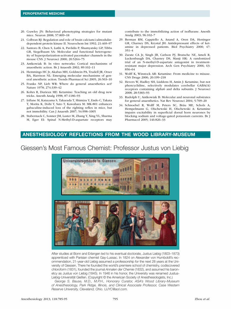
Giessen’s Most Famous Chemist: Professor Justus von Liebig
After studies at Bonn and Erlangen led to his eventual doctorate, Justus Liebig (1803–1873) apprenticed with Parisian chemist Gay-Lussac. In 1824 on Alexander von Humboldt’s rec-ommendation, 21-year-old Liebig assumed a professorship for the next 28 years at the Uni-versity of Giessen. There he founded the world’s premiere school of chemistry, codiscovered chloroform (1831), founded the journal Annalen der Chemie (1832), and assumed his baron-etcy as Justus von Liebig (1845). In 1946 in his honor, the University was renamed Justus-Liebig-Universität Gießen. (Copyright © the American Society of Anesthesiologists, Inc.)
George S. Bause, M.D., M.P.H., Honorary Curator, ASA’s Wood Library-Museum of Anesthesiology, Park Ridge, Illinois, and Clinical Associate Professor, Case Western Reserve University, Cleveland, Ohio. [email protected].
ANeSTHeSIoLoGY ReFLeCTIoNS FRoM THe Wood LIBRARY-MUSeUM