Forebrain HCN1 Channels Contribute to Hypnotic Actions of Ketamine
Upload
grenoble-univCategory
view
0download
0
Accepted Manuscript
Title: Beneficial effects of a ketamine/atropine combination insoman-poisoned rats under a neutral thermal environment
Author: Laure Barbier Frederic Canini Celine Giroud ClaireBeaup Annie Foquin Renaud Maury Josiane Denis AndrePeinnequin Frederic Dorandeu
PII: S0161-813X(15)00110-2DOI: http://dx.doi.org/doi:10.1016/j.neuro.2015.07.003Reference: NEUTOX 1840
To appear in: NEUTOX
Received date: 27-3-2015Revised date: 30-6-2015Accepted date: 12-7-2015
Please cite this article as: Barbier L, Canini F, Giroud C, Beaup C, FoquinA, Maury R, Denis J, Peinnequin A, Dorandeu F, Beneficial effects of aketamine/atropine combination in soman-poisoned rats under a neutral thermalenvironment, Neurotoxicology (2015), http://dx.doi.org/10.1016/j.neuro.2015.07.003
This is a PDF file of an unedited manuscript that has been accepted for publication.As a service to our customers we are providing this early version of the manuscript.The manuscript will undergo copyediting, typesetting, and review of the resulting proofbefore it is published in its final form. Please note that during the production processerrors may be discovered which could affect the content, and all legal disclaimers thatapply to the journal pertain.
Page 1 of 35
Accep
ted
Man
uscr
ipt
The present work demonstrates that ketamine/atropine combination given after soman
intoxication, in a warm environment
- does not induce deleterious effects
- presents some beneficial biochemical effects in animals experiencing convulsions
*Highlights (for review)

Page 2 of 35
Accep
ted
Man
uscr
ipt
1
1
Title page 2
Beneficial effects of a ketamine/atropine combination in soman-3
poisoned rats under a neutral thermal environment 4
Laure Barbier a
; Frédéric Canini b,e
; Céline Giroud a ; Claire Beaup
a ; Annie Foquin
a ; 5
Renaud Maury b
; Josiane Denis c ; André Peinnequin
d and Frédéric Dorandeu
a,e. 6
a Département de Toxicologie et risques chimiques,
b Département neurosciences et 7
contraintes opérationnelles, c Laboratoire d’analyses biologiques,
d Unité de biologie 8
moléculaire – IRBA BP73 91223 Brétigny sur Orge cedex, France 9
e Ecole du Val-de-Grâce, 1 place Alphonse Laveran, 75230, Paris, France. 10
11
12
Corresponding author 13
Laure Barbier, PhD 14
Département de Toxicologie et Risques Chimiques 15
Institut de Recherche Biomédicale des Armées 16
BP 73 17
91223 Brétigny sur Orge cedex 18
France 19
Tel.: (33) 178 65 13 49 20
E-mail: [email protected] 21
*Manuscript

Page 3 of 35
Accep
ted
Man
uscr
ipt
2
E-mail address of authors 1
Laure Barbier, PhD ; corresponding author 2
E-mail: [email protected] 3
Frédéric Canini, MD, PhD 4
E-mail: [email protected] 5
Ms Céline Giroud 6
E-mail: [email protected] 7
Ms Claire Beaup 8
E-mail: [email protected] 9
Ms Annie Foquin 10
E-mail: [email protected] 11
Mr Renaud Maury 12
E-mail: [email protected] 13
Ms Josiane Denis 14
E-mail: [email protected] 15
André Peinnequin, MD, PhD 16
E-mail: [email protected] 17
Frédéric Dorandeu, PharmD, PhD 18
E-mail: [email protected] 19
20
21

Page 4 of 35
Accep
ted
Man
uscr
ipt
3
Abstract 1
Exposure to organophosphorus (OP) compounds, such as pesticides and the chemical 2
warfare agents (soman and sarin), respectively represents a major health problem and a threat 3
for civilian and military communities. OP poisoning may induce seizures, status epilepticus 4
and even brain lesions if untreated. We recently proved that a combination of atropine sulfate 5
and ketamine, a glutamatergic antagonist, was effective as an anticonvulsant and 6
neuroprotectant in mice and guinea-pigs exposed to soman. Since OP exposure may also 7
occur in conditions of heat strain due to climate, wearing of protective gears or physical 8
exercise, we previously demonstrated that ketamine/atropine association may be used in a hot 9
environment without detrimental effects. In the present study, we assess soman toxicity and 10
evaluate the effects of the ketamine/atropine combination on soman toxicity in a warm 11
thermoneutral environment. Male Wistar rats, exposed to 31°C (easily reached under 12
protective equipments), were intoxicated by soman and treated with an anesthetic dose of 13
ketamine combined with atropine sulfate. Body core temperature and spontaneous locomotor 14
activity were continuously monitored using telemetry. At the end of the warm exposure, 15
blood chemistry and brain mRNA expression of some specific genes were measured. In 16
soman-intoxicated animals, metabolic and genic modifications were related to convulsions 17
rather than to soman intoxication by itself. In the warm environment, ketamine/atropine 18
combination did not produce any side-effect on the assessed variables. Furthermore, the 19
ketamine/atropine combination exhibited beneficial therapeutic effects on soman-intoxicated 20
rats such as a limitation of convulsion-induced hyperthermia and of the increase in some 21
blood chemistry markers. 22
23
Key words 24
Soman, Seizures, Thermoregulation, Rat, Ketamine, Atropine sulfate 25
List of abbreviations 26
AS: atropine sulfate 27
AUC: area under curve 28
C+/C
- : presence or absence of convulsions after soman intoxication 29
CxF: frontal cortex 30
Hpc: hippocampus 31
KET: ketamine 32

Page 5 of 35
Accep
ted
Man
uscr
ipt
4
OP: organophosphorus compounds 1
RSE: refractory status epilepticus 2
SE: status epilepticus 3
SLA: spontaneous locomotor activity 4
Ta: ambient temperature 5
Tabd: abdominal temperature 6
7
8
9

Page 6 of 35
Accep
ted
Man
uscr
ipt
5
Introduction 1
Organophosphorus (OP) compounds are worldwide used in high amounts as pesticides 2
in agriculture, thus exposing farmers to their deleterious effects. They are also commonly 3
used in suicide attempts with a high mortality rate (Bouchard et al., 2006, Eddleston et al., 4
2004, Eddleston et al., 2007, Gunnell et al., 2007, Quiros-Alcala et al., 2011). Another group 5
of OP, the highly lethal nerve agents, such as soman or sarin, still represent a credible threat 6
in military operations as well as for terrorist acts as demonstrated by the Tokyo subway 7
attacks in 1995 (Yanagisawa et al., 2006). 8
The main action of OP is the irreversible inhibition of both central and peripheral 9
cholinesterases (ChE), resulting in acetylcholine accumulation and therefore uncontrolled 10
activation of cholinergic synapses. Such cholinergic activation explains the acute clinical 11
signs of peripheral and central origin such as salivation, miosis, bronchoconstriction, 12
convulsive seizures and even death due to cardiorespiratory failure of peripheral and central 13
origins (McDonough and Shih, 1997). Epileptic seizures may evolve into status epilepticus 14
(SE) depending on the type and dose of toxicant. The first-line treatment of OP poisoning 15
consists of the injection of a muscarinic antagonist (e.g., atropine sulfate) injection combined 16
with an oxime acting as a peripheral ChE re-activator (e.g. pralidoxime methylsulfate, 17
Contrathion
). The administration of an anticonvulsant (a benzodiazepine or a prodrug like 18
prodiazepam Avizafone
) is currently indicated in the early phase of OP poisoning in order to 19
control seizures and SE. Approximately thirty minutes after the initial seizures, SE becomes 20
refractory to benzodiazepine (RSE) (Chen and Wasterlain, 2006). Only a few compounds are 21
presently able to stop OP-induced RSE. Previous works have demonstrated the beneficial 22
effect of atropine sulfate combined with N-methyl-D-aspartate (NMDA) antagonists such as 23
MK-801 (Braitman and Sparenborg, 1989), TCP (Carpentier et al., 1994) or GK11 (Lallement 24
et al., 1999) to control seizures after soman intoxication. However, none of these drugs can be 25
considered for clinical use because of severe side effects (MK-801, TCP) or because of an 26
insufficient clinical efficacy that stopped its development (GK-11, Chenoweth et al., 2014). 27
Ketamine thus appears as a valuable alternative in the delayed treatment of soman 28
intoxication as it is the only injectable NMDA antagonist currently licensed for human and 29
animal use. Ketamine is worldwide used as an anesthetics, especially for military surgery and 30
emergency care and can be administered by different routes. Although ketamine 31
administration may be accompanied by psychological side effects, such as dissociative states 32
and hallucinations (Sinner and Graf, 2008), it does not commonly induce respiratory 33

Page 7 of 35
Accep
ted
Man
uscr
ipt
6
depression and has little effect on the cardiovascular system in humans (for review, see 1
Dorandeu et al., 2013b and the papers published in the dedicated special issue). Furthermore 2
the combination of ketamine and atropine sulfate exhibits an effective anticonvulsant and 3
neuroprotectant effect in guinea-pigs (Dorandeu et al., 2005, Dorandeu et al., 2007), mice 4
(Dhote et al., 2012) and rats (Testylier et al., unpublished results), even when given one hour 5
after the beginning of seizures, viz. during the soman induced-RSE. Ketamine also appears as 6
an interesting treatment option for non-toxic RSE (Dorandeu et al., 2013b). 7
The thermal environmental context in which OP intoxication may take place is 8
important to consider for two reasons : (i) thermoregulation may be altered by OP intoxication 9
and its treatment and (ii) the body core temperature may have an influence on OP toxicity. 10
The deleterious interactions may occur in conditions of increased body temperature due to 11
physical exercises, warm climate, the wearing of protective suits or any combination of these. 12
Both cholinergic and glutamatergic modulation, as observed during OP poisoning, may alter 13
thermoregulatory processes and therefore interact with the environmental strain. First, any 14
modulation of the cholinergic tone could alter thermoregulation since cholinergic pathways 15
are involved in temperature regulation. Indeed atropine was shown to induce an impairment 16
of thermoregulation in man (Kolka et al., 1984, Kolka et al., 1987), rats (Matthew et al., 1989, 17
Matthew, 1991, Matthew et al., 1994) and monkeys (Avlonitou and Elizondo, 1988). 18
Thermoregulation is also one of the many regulatory systems affected by OP intoxication 19
(Gordon, 1993, 1994b) although consequences of OP exposition on thermoregulatory system 20
revealed contradictory results. Indeed some studies demonstrated that soman injection was 21
followed by a transient hypothermia in anesthetized (Meeter et al., 1971), restrained (Meeter, 22
1969) or exposed to cool environment (Maickel et al., 1990) rats and mice (Clement, 1993). 23
This response may be explained by the overstimulation of heat pathways due to the sudden 24
rise in acetylcholine in the central nervous system resulting to heat loss (Clement and Erhardt, 25
1994, Meeter, 1971). In other studies, an increase in heat production was observed in rats 26
after soman intoxication and such heat production might have been be further enhanced by 27
convulsions themselves (Clement, 1993). Secondly, some glutamate NMDA receptors 28
inhibitors are also known to strongly interfere with thermoregulation such as MK-801 (Canini 29
et al., 2002). However at difference with MK-801, we could show that ketamine, even for an 30
anesthetic dose, had no deleterious effect on thermoregulation system and may counteract 31
atropine sulfate side-effects in rats submitted to heat (38°C) (Barbier et al., 2012). Finally, 32
although described extensively for some chemicals such as amphetamines that display an 33

Page 8 of 35
Accep
ted
Man
uscr
ipt
7
increased toxicity in a hot environment (Dias da Silva et al., 2013), the relationships between 1
ambient temperature and OP toxicity have rarely been investigated. Indeed only one study 2
showed that soman toxicity in rats was increased during exposure to either cold or hot 3
environments (Wheeler, 1989). 4
The combination of ketamine with atropine representing an efficient treatment of 5
soman-induced RSE poisoning, we became interested in the potential effects that such a 6
therapeutic combination may provoke when administered to a poisoned individual in 7
conditions of increased body temperature. This study thus complements our previous data 8
(Barbier et al., 2012) obtained without poisoning. Indeed intentional or accidental OP 9
poisoning mostly occurs in countries where the average environmental temperature is 10
elevated and, even in milder climates, civilian or military personnel may have to wear 11
protective overgarments. The present work was designed with one major aims, viz. to 12
evaluate the effects of a ketamine/atropine association in poisoned rats exposed to a 13
temperature (31°C) mimicking thermal conditions that may be faced by soldiers wearing NBC 14
suits. However, our paradigm could also bring some insights on the toxicity of soman in an 15
ambient temperature (Ta) close to the upper thermolysis threshold (Gordon and Fogelson, 16
1993, Gordon, 1994a), at the frontier between thermoneutrality and warm environment in 17
order to best observe any impairment of thermoregulation. 18
2. Material and methods 19
2.1. Animals 20
Fifty-eight adult male Wistar rats (Janvier Laboratories, Genest St Isle, France) were 21
involved in this study (Barbier et al., 2012, Michel et al., 2007). They were accustomed to 22
laboratory conditions during at least one week. Animals were housed in a controlled 23
environment (Ta=22 ± 1°C, 30 ± 10% relative humidity (r.h.), 12 h dark/light cycle with light 24
on at 07:30 a.m.) at two per cage until surgery and individually thereafter. Food and water 25
were given ad libitum. Animal care procedures were approved by the Institutional Animal 26
Care and Research Advisory Committee of the IRBA-CRSSA in accordance with the 27
applicable European Union and institutional guidelines for the care and use of laboratory 28
animals and relevant French legislation. All efforts were done to minimize animals’ suffering. 29
2.2. Drugs 30
Soman (> 97% pure as assessed by gas chromatography) was supplied by the Centre 31

Page 9 of 35
Accep
ted
Man
uscr
ipt
8
DGA Maîtrise NRBC (Vert-le-Petit, France). The solution was freshly prepared by diluting 1
the initial concentrated solution (2 mg.mL-1
in isopropanol) in ice-cold 0.9% (w/v) saline. 2
Sodium pentobarbital and Extencilline® were purchased from Sanofi-Synthelabo (Le Plessis-3
Robinson, France), Isoflurane from Belmont Laboratories (Paris, France) and Finadyne® from 4
Schering-Plough Laboratories (Segre, France). Ketamine hydrochloride was obtained from 5
Panpharma (Fougères, France) and atropine sulfate from Sigma Chemicals (L’Isle d’Abeau 6
Chesnes, France). 7
According to previous experiments conducted in rats, soman was administered 8
subcutaneously (s.c) at 60 µg.kg-1
(Maickel et al., 1990) corresponding to 2/3 LD50 in male 9
Wistar rat (Blanchet et al., 1994). This dosage was chosen after a preliminary study carried 10
out at Ta=31°C in which two rats were challenged with 70 µg.kg-1
, s.c and two other animals 11
with 60 µg.kg-1
, s.c. In both cases, rats showed convulsions and died, the higher survival time 12
(5 h) being observed for the 60 µg.kg-1
dose. In our paradigm, soman was thus injected to this 13
dosage under a volume of 200 µL as previously described (Carpentier et al., 2010). 14
Atropine sulfate was dissolved in 0.9% sterile saline and injected by the 15
intraperitoneal (i.p) route at 5 mg.kg-1
, a dose previously used as a medication against soman 16
poisoning in male Wistar rats (Carpentier et al., 2004) and in our previous study (Barbier et 17
al., 2012). 18
Ketamine (Panpharma®; 5% solution) was diluted in sterile saline immediately prior 19
to i.p. injection. The dose of ketamine was chosen based on what might be required for the 20
treatment of OP-induced SE/RSE: 75 mg.kg-1
, an anesthetic dose for rats. In other species, an 21
anesthetic dose of ketamine combined with atropine sulfate was indeed proven to be 22
protective against OP-induced brain lesions in guinea-pigs (Dorandeu et al., 2005, Dorandeu 23
et al., 2007) and mice (Dhote et al., 2012). Ketamine used at 75 mg.kg-1
was also suggested to 24
reduce the side effects of atropine sulfate in heat-exposed rats (Barbier et al., 2012). This 25
dosage was thus considered in this study but at difference to the other studies from our 26
laboratory on the treatment of soman-induced SE/RSE, these drugs were administered only 27
once. 28
2.3. Surgical procedures 29
The body core temperature (Tabd) and spontaneous locomotor activity (SLA) were 30
continuously recorded using a Physiotel® TA10TAF40 transmitter (Data Sciences, St Paul, 31

Page 10 of 35
Accep
ted
Man
uscr
ipt
9
MN, USA) driven by a computer-based data acquisition system (Dataquest® IV, Data 1
Sciences). The telemetric device was implanted under deep anesthesia (pentobarbital sodium, 2
60 mg.kg-1
, i.p) according to a procedure previously described (Barbier et al., 2012). Rats 3
were given 10 days for recovery from surgery. They were weighed and handled daily in order 4
to verify their recovery from surgery and to reduce as much as possible their stress. 5
2.4. Experimental design 6
Rats were randomly distributed into four experimental groups depending on whether 7
they were poisoned or not and treated or not (Table 1): 8
(i) the “CONTROL” group injected with saline instead of soman or instead of 9
ketamine/atropine treatment (n=10); 10
(ii) the “KETAS” group receiving saline instead of soman and then therapeutic 11
ketamine/atropine mixture (n=10); 12
(iii) the “SOMAN” group challenged with soman and injected with saline 13
instead of ketamine/atropine association (n=17). According to convulsions 14
development or not, this group was divided in 2 sub-groups respectively 15
called SOMAN C+ and SOMAN C-; 16
(iv) the “SOMAN/KETAS” group constituted from rats poisoned with soman 17
and then treated with ketamine/atropine (n=13). Again, this group was 18
divided in 2 sub-groups according to the development of convulsions or not. 19
On the day of the investigation, animals were weighed, placed in a new cage without 20
bedding and left in a climatic chamber (Ta=31°C, r.h.=30 ± 10%) for 5 hours with access to 21
water but not food. An exposure to 31°C, a temperature too low to induce heatstroke (Sharma 22
et al., 1998), was chosen to mimic conditions that may be faced by soldiers operating under 23
NBC protective suits. This temperature was also previously used to study soman toxicity in 24
rats (Wheeler, 1989). Two hours after the beginning of heat exposure, animals received either 25
soman or saline, s.c. All the intoxications were performed between 9:00 am and 11:30 am to 26
reduce as much as possible the circadian variations of cholinergic parameters (Elsmore, 27
1981). After soman injection, signs and symptoms, including hypersalivation and 28
development of convulsions, were continuously monitored. Although our current paradigm 29
did not include electroencephalographic recordings, we previously showed that there was a 30
close relationship between the occurrence of long-lasting convulsions and the development of 31

Page 11 of 35
Accep
ted
Man
uscr
ipt
10
SE in mice (Dhote et al., 2012, Testylier et al., 2007). Seventy-five minutes after challenge 1
(after ca. 1 h of convulsions, see results), rats were treated with the combination of ketamine 2
(75 mg.kg-1
, i.p., 1.5 mL.kg-1
) and atropine sulfate (5 mg.kg-1
, i.p.). The total duration of 3
exposure to the warm environment was 5 hours long. 4
At the end of heat exposure, rats were immediately transferred into another room to be 5
anesthetized under isoflurane (5% into 100% O2). Blood was taken by intra-cardiac puncture 6
and collected into heparin-coated tubes to determine various biochemical parameters. The left 7
hippocampus (Hpc) and frontal cortex (Cxf) were dissected out and placed on RNAlater® 8
(Ambion) for quantification of gene expression using reverse transcription (RT) and 9
quantitative real-time polymerase chain reaction (qPCR) (Barbier et al., 2012). 10
2.5. Physiological variables 11
The Tabd and SLA values were acquired each minute in each rat for a 10 s-period. SLA 12
was calculated as the variations in the transmitted power signal correlated to changes in rat 13
position. The experimental time course was separated into 4 periods (figure 1): (i) the 14
“Baseline” which consists in the 60 min spent in the laboratory room (Ta=22°C) immediately 15
preceding the warm exposure; (ii) the “Heat baseline” period represented by the first 2 hours 16
of warm exposure; (iii) the “Post-Soman” period represented by the 75 minutes following 17
soman (or vehicle) injection, including the first 15-minutes usually without convulsions and 18
the following hour when convulsions may occur depending on the animals; (iv) the “Post-19
treatment” period constituted by the 105 minutes following the injection of treatment (or 20
vehicle). 21
Physiological data were then analysed using two distinct methods. The Tabd and SLA 22
time course were based on 15-min averaged values throughout the investigation (Figure 1 23
and Table 3). Four remarkable values were considered for Tabd statistical analysis: i) the Tabd 24
mean value of the “Baseline”, ii) the mean value of the 15-min epoch immediately preceding 25
soman intoxication (or vehicle) was assumed to be representative of the “Heat baseline” 26
period; (iii) the mean value of the 15-min epoch immediately preceding treatment injection 27
(or vehicle) was assumed to be representative of the “Post-Soman” period; (iv) the mean 28
value of the 15-min epoch placed at the end of the investigation was assumed to represent the 29
“Post-treatment” period. The same analysis was carried out for SLA changes but the mean 30
value was replaced by the area under the curve (AUC) calculated for each of the animal in 31

Page 12 of 35
Accep
ted
Man
uscr
ipt
11
each time period (Figure 1 and Table 3) (Matthews et al., 1990). 1
2.6. Blood chemistry 2
Different blood parameters were measured because of their interest for either the 3
evaluation of the impact of the temperature or the consequence of soman poisoning. 4
Blood ChE activity determination was carried out using a specific kit (Roche 5
Diagnostics®, Meylan, France). ChE activity levels were considered as a marker of the 6
importance of soman poisoning, high blood ChE inhibition being related to initiation of 7
convulsions (Carpentier et al., 2010). 8
Dehydration was evaluated from the hematocrit. The plasma corticosterone 9
concentration was determined using a specific radioimmunoassay kit (Coat-A-Count Rat 10
Corticosterone, Diagnostics Products Corporation, USA). Serum urea, creatinine, proteins, 11
lactate, glucose, ASAT, ALAT, CK, LDH, Na+, K
+, Cl
- and triglycerides were assayed on 12
Hitachi 912 (Roche Diagnostics®, Meylan, France) using either the colorimetric method with 13
Roche™ reagents (Roche Diagnostics®, Meylan, France) or a specific kit for glycerol 14
(Biocontrol system, Lyon, France). All analyses were performed according to the 15
manufacturer’s instructions. 16
2.7. Gene expression 17
At the end of the experiment both Hpc (59 ± 1 mg, mean ± Standard Error of the Mean 18
(SEM) of 50 values) and CxF (85 ± 3 mg, mean ± SEM of 50 values) were removed. tRNA 19
extraction and RT-qPCR were performed as previously described (Barbier et al., 2012). In 20
this work, two different ARBP primers were used: one toward the 5’ end and the second 21
toward the 3’ end of the mRNA sequence. The ratio of amplicons obtained thus reflected RT 22
efficiency (Pugniere et al., 2011). Selected forward (F) and reverse (R) primer sequences and 23
their relative annealing temperature are listed in Table 2 for each structure. Normalization 24
was performed by calculating the geometric mean of three internal validated control genes 25
(acidic ribosomal phosphoprotein, Arbp; peptidylpropyl isomerase A, Ppia and hypoxanthine 26
guanine phosphoribosyl transferase, Hprt) (Vandesompele et al., 2002). The 0.072 (Hpc) and 27
0.092 (CxF) pairwise variations of these three genes were below the threshold (0.150) that 28
requires the inclusion of an additional normalization gene. 29
30

Page 13 of 35
Accep
ted
Man
uscr
ipt
12
2.8. Statistical analysis 1
All values were expressed as mean ± SEM. Statistical analysis was performed with 2
Statistica® 10.0 Software (Statsoft-France, Maison-Alfort, France) using non-parametric tests. 3
In order to evaluate consequences of the treatment given after intoxication in the heat, all the 4
groups were considered and a Kruskal-Wallis ANOVA by ranks was performed followed by 5
ad-hoc pairwise comparisons with the Bonferonni correction. 6
Statistical analysis focused on three major questions: (i) the consequences of soman-7
intoxication were evaluated by comparing SOMAN with CONTROL group; (ii) the effects of 8
convulsions were considered by comparing SOMAN C+ and SOMAN C- sub-groups and (iii) 9
the beneficial effects of treatment in rats developing convulsions comparing SOMAN 10
C+/KETAS and SOMAN C+ sub-groups. For the SLA changes, as previously mentioned, 11
individual AUC were calculated for the different periods of the experiment and then 12
statistically compared (Matthews et al., 1990) using the same approach as for other 13
parameters. 14
3. Results 15
3.1. Effects of soman challenge 16
A subcutaneous (s.c) injection of 60 µg.kg-1
soman led to convulsions after 16.7 ± 2.2 17
min in 66% of the intoxicated rats (25 out of 38 rats). The development of convulsions seems 18
to be a key factor impacting a lot of the studied parameters (either because it has a direct 19
effect on the parameter or because it reflects a more severe poisoning), Table 1 thus presents 20
separately the sub-groups created post-hoc based on whether convulsions appeared or not. 21
Among intoxicated animals, 8 rats died 132.1 ± 10.5 min after intoxication (data not shown). 22
They all belong to the groups developing convulsions and treatment did not seem efficient to 23
alleviate them (SOMAN C+: n=5 and SOMAN C+/KETAS: n=3; data not shown). Cause of 24
death was probably heatstroke (Tabd values were 41.2±0.2 and 40.3±0.3 for SOMAN C+ and 25
SOMAN C+/KETAS groups, respectively). In the following results, animals dying before the 26
end of the experiment were discarded from statistical analysis (n=30 intoxicated animals). 27
3.2. Effect of poisoning and treatment on thermoregulation (Figure 1A and Table 3) 28
During Baseline, the six experimental groups exhibited similar average Tabd (Table 3) 29
and time course (Figure 1A). Exposure to Ta=31°C induced a transient and moderate increase 30

Page 14 of 35
Accep
ted
Man
uscr
ipt
13
in Tabd in all groups. Just before soman intoxication, Tabd was similar among the six 1
experimental groups and was also comparable to that observed in Baseline. 2
The temperature response was different after soman intoxication whether convulsions 3
developed or not. Animals experiencing convulsions exhibited higher Tabd than CONTROL (p 4
≤ 0.01, SOMAN C+ vs CONTROL and SOMAN C+/KETAS vs CONTROL). Administration 5
of ketamine combined with atropine sulfate in rats previously treated with saline induced a 6
reversible and moderate increase in Tabd. The same was observed in rats suffering from a 7
peripheral intoxication by soman but not experiencing convulsions. However in both cases, no 8
statistical difference was observed with CONTROL animals at the end of the experiment. At 9
the end of the investigation, the hyperthermia observed in animals developing convulsions 10
was only limited by the administration of ketamine and atropine sulfate (p ≤ 0.001, SOMAN 11
C+ vs CONTROL; p ≤ 0.05, SOMAN C+/KETAS vs CONTROL, and p ≤ 0.01, SOMAN 12
C+/KETAS vs SOMAN C+). 13
3.3. Effect of poisoning and treatment on behaviour (Figure 1B and Table 3) 14
During Baseline, no difference in SLA was observed among the six experimental 15
groups. Exposure to the warm ambiance was followed by a strong, but transient, increase in 16
rat locomotion to the same extent whatever the groups. After soman intoxication, animals 17
developing convulsions exhibited a steady decrease in locomotion which persisted until the 18
end of the “post-soman” period, just before treatment administration (p ≤ 0.01, SOMAN C+ 19
vs CONTROL and p ≤ 0.05, SOMAN C+/KETAS vs CONTROL). 20
As expected, the anesthetic dose of ketamine induced a decrease in SLA but at the end 21
of the investigation, all rats exhibited the same behaviour. This is with the notable exception 22
of those belonging to the SOMAN C-/KETAS group which developed hyperlocomotion that 23
persisted until the end of the experiment (p ≤ 0.01, SOMAN C-/KETAS vs CONTROL). 24
3.4. Effect of poisoning and treatment on blood chemistry (Tables 4 and 5) 25
When compared with CONTROL animals, soman-intoxicated rats exhibited a 26
dramatic inhibition in blood ChE activity (-80% 3 hours after intoxication; p ≤ 0.01 SOMAN 27
vs CONTROL) without significant difference appeared between rats developing convulsions 28
or not (Table 4). Whatever the first injection (soman or saline), ketamine combined with 29
atropine sulfate did not modify blood ChE activity. 30
Page 15 of 35
Accep
ted
Man
uscr
ipt
14
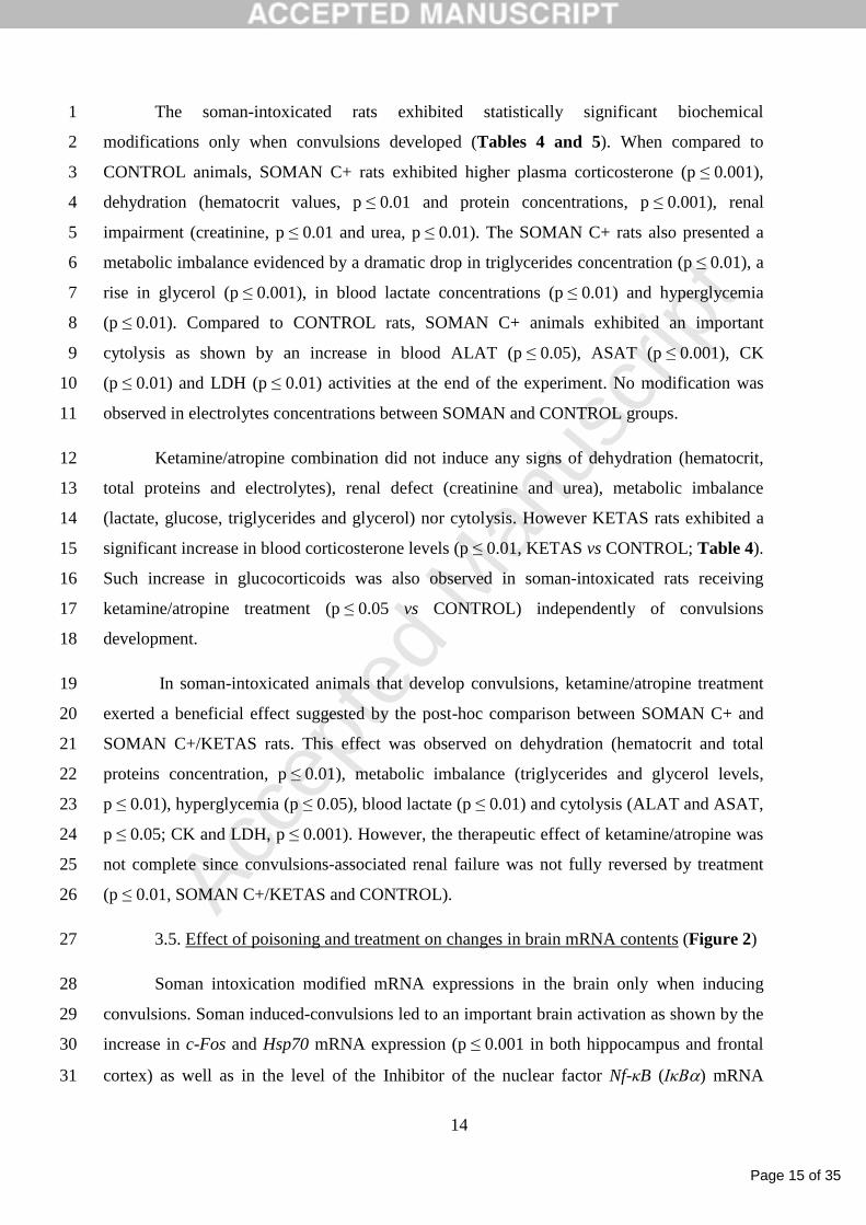
The soman-intoxicated rats exhibited statistically significant biochemical 1
modifications only when convulsions developed (Tables 4 and 5). When compared to 2
CONTROL animals, SOMAN C+ rats exhibited higher plasma corticosterone (p ≤ 0.001), 3
dehydration (hematocrit values, p ≤ 0.01 and protein concentrations, p ≤ 0.001), renal 4
impairment (creatinine, p ≤ 0.01 and urea, p ≤ 0.01). The SOMAN C+ rats also presented a 5
metabolic imbalance evidenced by a dramatic drop in triglycerides concentration (p ≤ 0.01), a 6
rise in glycerol (p ≤ 0.001), in blood lactate concentrations (p ≤ 0.01) and hyperglycemia 7
(p ≤ 0.01). Compared to CONTROL rats, SOMAN C+ animals exhibited an important 8
cytolysis as shown by an increase in blood ALAT (p ≤ 0.05), ASAT (p ≤ 0.001), CK 9
(p ≤ 0.01) and LDH (p ≤ 0.01) activities at the end of the experiment. No modification was 10
observed in electrolytes concentrations between SOMAN and CONTROL groups. 11
Ketamine/atropine combination did not induce any signs of dehydration (hematocrit, 12
total proteins and electrolytes), renal defect (creatinine and urea), metabolic imbalance 13
(lactate, glucose, triglycerides and glycerol) nor cytolysis. However KETAS rats exhibited a 14
significant increase in blood corticosterone levels (p ≤ 0.01, KETAS vs CONTROL; Table 4). 15
Such increase in glucocorticoids was also observed in soman-intoxicated rats receiving 16
ketamine/atropine treatment (p ≤ 0.05 vs CONTROL) independently of convulsions 17
development. 18
In soman-intoxicated animals that develop convulsions, ketamine/atropine treatment 19
exerted a beneficial effect suggested by the post-hoc comparison between SOMAN C+ and 20
SOMAN C+/KETAS rats. This effect was observed on dehydration (hematocrit and total 21
proteins concentration, p ≤ 0.01), metabolic imbalance (triglycerides and glycerol levels, 22
p ≤ 0.01), hyperglycemia (p ≤ 0.05), blood lactate (p ≤ 0.01) and cytolysis (ALAT and ASAT, 23
p ≤ 0.05; CK and LDH, p ≤ 0.001). However, the therapeutic effect of ketamine/atropine was 24
not complete since convulsions-associated renal failure was not fully reversed by treatment 25
(p ≤ 0.01, SOMAN C+/KETAS and CONTROL). 26
3.5. Effect of poisoning and treatment on changes in brain mRNA contents (Figure 2) 27
Soman intoxication modified mRNA expressions in the brain only when inducing 28
convulsions. Soman induced-convulsions led to an important brain activation as shown by the 29
increase in c-Fos and Hsp70 mRNA expression (p ≤ 0.001 in both hippocampus and frontal 30
cortex) as well as in the level of the Inhibitor of the nuclear factor Nf-κB (IκB) mRNA 31

Page 16 of 35
Accep
ted
Man
uscr
ipt
15
(p ≤ 0.05 in both hippocampus and frontal cortex) and Bdnf mRNA levels (in both 1
hippocampus and frontal cortex, p ≤ 0.001). Conversely, there was no significant modification 2
of the mRNA contents of its receptor TrkB. 3
Treatment by ketamine/atropine did not modify the selected brain mRNA genes 4
expression neither in non-intoxicated rats nor in soman-injected animals with or without 5
convulsions. 6
4. Discussion 7
Ketamine in association with atropine may be an interesting treatment combination for 8
RSE induced by nerve agent poisoning. We designed our experiment in order to try and 9
obtain some answers to one key operational question: could a warm environment modify the 10
interest of this combination when given to poisoned individuals? Our major finding is that 11
soman intoxication led to physiological modifications and changes in blood chemistry and 12
brain mRNA expression only when convulsions had developed. Some of these changes were 13
limited by ketamine/atropine administration whereas the use of this combination did not 14
induce any apparent side effects. 15
Although not the core question, our paradigm gave us a large body of data on the 16
impact of a warm environment on the deleterious effects of soman intoxication. An 17
interaction is suggested by the apparently increased prevalence of convulsions after 18
intoxication. For a dose of 60 µg.kg-1
, we observed
66% long-lasting convulsions, a 19
proportion comparable to that observed (58%) in rat kept at room temperature (Ta=22°C) and 20
intoxicated with a higher dose of soman (70 µg.kg-1
, Carpentier et al., 2010). However only a 21
group of animals exposed to 60 µg.kg-1
soman at room temperature would permit a definite 22
answer. 23
Several authors have reported altered toxicity of ChE inhibitors depending on the 24
temperature. During exposure to Ta=23°C, Maickel et al. (1990) reported that 60 µg.kg-1
of 25
soman induced less than 10% mortality. An increased mortality was observed when rats were 26
challenged at temperatures below or higher than 23°C (Wheeler, 1989). At 31°C, this author 27
described the LD10 being between 60 and 70 µg.kg-1
. In our paradigm, a higher mortality rate 28
was observed after intoxication (21%, 8 out of 38 animals) but we have to take into account 29
experimental differences (soman batch, strains and age of rats) before drawing conclusions. 30
Moreover all these investigations were conducted in a range of temperature that did not really 31

Page 17 of 35
Accep
ted
Man
uscr
ipt
16
challenge thermoregulatory processes (Gordon, 1994a). Whether soman is more toxic in a 1
moderately hot environment thus remains to be demonstrated. 2
The deleterious activity of soman in a warm environment may also be related to its 3
activity on the thermoregulatory processes at central and peripheral levels (Clark and Clark, 4
1980, Coudray-Lucas et al., 1981). For Ta comprised between 23 and 25°C, soman 5
intoxication led to hypothermia two hours after injection (Maickel et al., 1990), suggesting an 6
increased peripheral thermolysis. In our work, at Ta=31°C, soman-intoxicated rats that did not 7
develop convulsions exhibited the same thermoregulatory response. Although slightly more 8
pronounced for Soman C-, the tendency was also observed for CONTROL animals 9
preventing to clearly advocate an effect of soman. Conversely, soman-intoxicated animals 10
experiencing convulsions exhibited hyperthermia as previously reported (Wheeler, 1989). A 11
convulsion-linked increase in peripheral muscle activity may lead to an exacerbate heat 12
production and therefore an increase in core temperature, a known fact in epileptic patients 13
(Simon, 2006). For unclear reasons, hypothermia was also reported in mice even at soman 14
doses that induced convulsions (Clement, 1993). 15
Soman and ketamine/atropine combination had different effects on the spontaneous 16
locomotion of the animals. Before poisoning and treatment, animals exhibited an increase in 17
locomotion which occurred immediately after the beginning of heat exposure in all groups. 18
Such an effect was previously reported in rats submitted to 36°C (Chuang and Lin, 1994), 19
38°C (Barbier et al., 2012) and to 40°C (Michel et al., 2010) and may be related to the 20
exposure to a new environment. This hyperlocomotion may also be linked to a stress reaction 21
during which animals tried to escape to this sudden increase in temperature. Animals that 22
were treated with ketamine/atropine (Soman C+/KETAS or KETAS) showed a decrease in 23
locomotion that lasted until the end of the experiment. Although the obvious reason might 24
have been the use of an anesthetic dose of ketamine (Barbier et al., 2012), this is not 25
supported by the observation that in the Soman C-/KETAS group, ketamine/atropine 26
combination oppositely led to an increase in locomotion at the end of the investigation. The 27
reason for this behaviour is not easily found as the hypothesis of atropine causing the same 28
transient hyperlocomotion we showed at 38°C (Barbier et al., 2012) is negated by what we 29
reported here from the KETAS group. 30
At the end of the investigation, soman had induced a dramatic decrease (ca. 80%) in 31
blood ChE activity with no apparent link between the extent of the inhibition and the 32
Page 18 of 35
Accep
ted
Man
uscr
ipt
17
occurrence of convulsions in accordance to some earlier work of our team (Baille et al., 2001, 1
Carpentier et al., 2010). Our results are also in line with those from Maickel et al. (1990) who 2
reported a 88% blood ChE activity inhibition 8 hours after intoxication (60 µg.kg-1
) when 3
animals were kept at 23-25°C. As expected, the possible interaction between temperature and 4
severity of the poisoning we mentioned earlier is thus not simply linked to a clear 5
modification in peripheral inhibition. 6
A strong stress reaction was evidenced by the dramatic increase in blood 7
corticosterone levels only in soman-intoxicated rats experiencing convulsions. This signs a 8
profound central impact of soman. In the same way, lithium-pilocarpine-induced SE is 9
accompanied by a robust increase in blood corticosterone concentrations (Mazarati et al., 10
2009). Inter-relations between stress and seizures are well known (Haut et al., 2003, Lai and 11
Trimble, 1997, Sawyer and Escayg, 2010) although the details of such interaction remain 12
elusive. 13
Hyperglycemia was observed in soman-intoxicated rats exhibiting signs of toxicity 14
such as chewing, salivation, muscle fasciculations or tremors (Fletcher et al., 1988, Jovic, 15
1974, Maickel et al., 1990, Rahimi and Abdollahi, 2007). Conversely, we and others (Fletcher 16
et al., 1988) observed hyperglycemia only in animals developing convulsions. It thus appears 17
to be related to a certain level of brain activation during convulsions (Simon, 2006) and due to 18
a stress reaction through the sympathetic activation together with glucocorticoids release 19
(Drouet et al., 2012). Similarly, the decrease in triglycerides (Pohanka, 2011 and our study) 20
together with an increase in glycerol levels could be explained by the stress reaction (Ricart-21
Jane et al., 2002) and the lipolysis due to sympathetic activation. The same could stand for 22
enzymes such as CK, ASAT and ALAT which activity can be increased in case of 23
psychological stressor exposure (Sanchez et al., 2002). Altogether, this suggests that soman-24
intoxicated animals experiencing convulsions exhibited a higher stress level than non-25
convulsive rats. However cytolysis may also result from the intense muscle activity associated 26
with convulsions and some direct organ toxicity of soman as reported for other OP (Dorandeu 27
et al., 2008, Marrs, 1993). Some levels of tissue degradation may partly explain the renal 28
failure (Gupta et al., 2010, Mishra and Dave, 2013), evidenced by the 2-fold increase in 29
creatinine concentrations (Palm and Lundblad, 2005). Such an effect had already been 30
reported after soman intoxication in rats (Pohanka, 2011). 31
Soman-intoxicated animals experiencing convulsions also exhibited a strong 32

Page 19 of 35
Accep
ted
Man
uscr
ipt
18
homeostatic imbalance. Convulsions are usually associated with blood lactic acidosis during 1
seizures in man (Osnes and Hermansen, 1972) and in animals exposed to OP (e.g rats, 2
(Husain et al., 1987) or baboons (Anzueto et al., 1986)). The strong increase in blood lactate 3
we observed in our paradigm is congruent with these reports. A high dehydration, evidenced 4
by the increase in hematocrit and total blood proteins, is also observed in soman-poisoned 5
animals with convulsions. This is not usually observed during OP poisoning at room 6
temperature and thus suggests that poisoned animals, especially if experiencing convulsions, 7
may be then more prone to heatstroke. 8
Soman-intoxicated animals that developed convulsions also presented an important 9
brain activation as suggested by the higher c-Fos and Hsp70 mRNA expressions, in 10
accordance with previous work (Baille et al., 1997). With the limitations of a simple mRNA 11
study, brain inflammation was also suggested by a reduction in the content of IkBa mRNA 12
that can be used to evaluate NFkB gene activation (Barbier et al., 2009), well in line with the 13
description of the various inflammatory pathways that are modified following soman 14
poisoning (Dhote et al., 2007, Dillman et al., 2009, Williams et al., 2003), such changes being 15
confirmed by the increase in some of the related coded proteins in several brain areas (Dhote 16
et al., 2012). Conversely, little is known regarding the modulation of the brain neurotrophin 17
expression following soman intoxication. The activation of the neurotrophin/Trk signaling 18
had been reported in the hippocampus of soman-intoxicated rats developing convulsions 19
(Dillman et al., 2009). In our study, we observed a strong increase in Bdnf mRNA expression 20
without changes in TrkB expression in both brain areas studied. This result would need to be 21
matched by changes in the protein level before any conclusion can be drawn. 22
Very importantly, the present study clearly demonstrates the absence of significant 23
deleterious impact and even the therapeutic benefit of the combination of ketamine and 24
atropine sulfate in soman-intoxicated animals in a warm environment. When the combination 25
was given to convulsing rats, its anticonvulsivant properties (for review, see Dorandeu et al., 26
2013a) were also probably the cause of the limitation of convulsion-associated hyperthermia. 27
Conversely, in non-poisoned rats the therapeutic combination led to a transient increase in 28
core temperature that could either be due to the anesthetic dose of ketamine or to atropine 29
sulfate (Barbier et al., 2012). Indeed, during heat exposure, atropine was shown to impair 30
thermoregulation (Kolka et al., 1987, Matthew et al., 1986) and lead to heat stress, a side-31
effect counteracted when atropine was associated with an anesthetic dose of ketamine 32

Page 20 of 35
Accep
ted
Man
uscr
ipt
19
(Barbier et al., 2012). Finally, a lot of convulsion-related biochemical modifications were 1
counteracted by this single treatment injection. This is not surprising given the fact that ample 2
evidence of the anticonvulsant and neuroprotectant efficacy of this therapeutic association in 3
soman-intoxicated guinea-pigs (Dorandeu et al., 2005, Dorandeu et al., 2007) and mice 4
(Dhote et al., 2012, Fauvelle et al., 2012) have been previously published. At brain level 5
however, the single administration of ketamine/atropine appeared not to have been able to 6
abate the increase in mRNA gene expression observed in animals with convulsions, totally in 7
line with the above mentioned previous work advocating for repeated injection of the 8
combination for a better efficacy. A histopathological study of the brain neuroprotection 9
afforded by the combination was not considered here but would be required for a more 10
complete assessment. 11
5. Conclusions 12
All in all, the present work clearly confirms that a ketamine/atropine combination 13
given during soman intoxication in a warm environment does not induce deleterious effects 14
and even seems to bring some beneficial therapeutic effects as it could either normalize the 15
blood parameters or at least block their modifications linked to the occurrence of seizures. 16
Our results also stress the important impact of convulsions that may superimpose their effects 17
to those of warm exposure. Further experiments are needed to get better insight into the 18
possible interactions between poisoning and conditions that may occur in the context of 19
military operations. 20
6. Conflict of interest 21
The authors declare that there are no conflicts of interest. 22
7. Acknowledgments 23
This work was supported by the French Military Health Service and by grants from the 24
Direction Générale de l’Armement (DGA; research grant to F. Dorandeu). The authors wish 25
to thank Dr Christophe Pierard for his excellent advice on manuscript redaction. We are 26
indebted to Mr Hervé Chaussard for his help in animal technical assistance. We would like to 27
thank technical staff of the medical analyses laboratory at IRBA-CRSSA for the biochemical 28

Page 21 of 35
Accep
ted
Man
uscr
ipt
20
determinations. 1
8. References 2
Anzueto A, Berdine GG, Moore GT, Gleiser C, Johnson D, White CD, Johanson WG, Jr. 3
Pathophysiology of soman intoxication in primates. Toxicol Appl Pharmacol 1986; 4
86:56-68. 5
Avlonitou E, Elizondo R. Effects of atropine and pyridostigmine in heat-stressed patas 6
monkeys. Aviat Space Environ Med 1988; 59:544-8. 7
Baille V, Lallement G, Carpentier P, Foquin A, Pernot-Marino I, Rondouin G. c-fos antisense 8
oligonucleotide prevents delayed induction of hsp70 mRNA after soman-induced 9
seizures. Neuroreport 1997; 8:1819-22. 10
Baille V, Dorandeu F, Carpentier P, Bizot JC, Filliat P, Four E, Denis J, Lallement G. Acute 11
exposure to a low or mild dose of soman: biochemical, behavioral and 12
histopathological effects. Pharmacol Biochem Behav 2001; 69:561-9. 13
Barbier L, Diserbo M, Lamproglou I, Amourette C, Peinnequin A, Fauquette W. Repeated 14
stress in combination with pyridostigmine Part II: changes in cerebral gene expression. 15
Behav Brain Res 2009; 197:292-300. 16
Barbier L, Dorandeu F, Giroud C, Beaup C, Foquin A, Maury R, Alonso A, Peinnequin A, 17
Canini F. Ketamine does not impair heat tolerance in rats. Eur J Pharmacol 2012; 18
691:77-85. 19
Blanchet G, Carpentier P, Lallement G, Sentenac-Roumanou H. Prevention and treatment of 20
status epilepticus induced by soman. Ann Pharm Fr 1994; 52:11-24. 21
Bouchard M, Carrier G, Brunet RC, Dumas P, Noisel N. Biological monitoring of exposure to 22
organophosphorus insecticides in a group of horticultural greenhouse workers. Ann 23
Occup Hyg 2006; 50:505-15. 24
Braitman DJ, Sparenborg S. MK-801 protects against seizures induced by the cholinesterase 25
inhibitor soman. Brain Res Bull 1989; 23:145-48. 26
Canini F, Simler N, Bourdon L. MK801 impairs thermoregulation in the heat. Can J Physiol 27
Pharmacol 2002; 80:226-32. 28
Carpentier P, Foquin-Tarricone A, Bodjarian N, Rondouin G, Lerner-Natoli M, Kamenka JM, 29
Blanchet G, Denoyer M, Lallement G. Anticonvulsant and antilethal effects of the 30
phencyclidine derivative TCP in soman poisoning. Neurotoxicology 1994; 15:837-51. 31
Carpentier P, Foquin A, Lallement G, Dorandeu F. Flunarizine: A possible adjuvant 32
medication against soman poisoning? Drug and Chemical Toxicology 2004; 27:213-33
31. 34
Carpentier P, Pouyatos B, Dorandeu F, Campo P, Baille V, Foquin A, Job A. Prediction of 35
soman-induced cerebral damage by distortion product otoacoustic emissions. 36
Toxicology 2010; 277:38-48. 37
Chen JW, Wasterlain CG. Status epilepticus: pathophysiology and management in adults. 38
Lancet Neurol 2006; 5:246-56. 39
Chenoweth JA, Gerona RR, Ford JB, Sutter ME, Rose JS, Albertson TE, Clarke SO, Owen 40
KP. Altered mental status and end organ damage associated with the use of 41
Gacyclidine : a case series. J Med Toxicol 2014; 11:115-20. 42
Chuang JI, Lin MT. Responses to cold, heat, and pain increase locomotion in rats and are 43
attenuated by pinealectomy. Physiol Behav 1994; 55:583-6. 44
Clark WG, Clark YL. Changes in body temperature after administration of acetylcholine, 45
histamine, morphine, prostaglandins and related agents. Neurosci Biobehav Rev 1980; 46
4:175-240. 47
Page 22 of 35
Accep
ted
Man
uscr
ipt
21
Clement JG. Pharmacological nature of soman-induced hypothermia in mice. Pharmacol 1
Biochem Behav 1993; 44:689-702. 2
Clement JG, Erhardt N. In vitro oxime-induced reactivation of various molecular forms of 3
soman-inhibited acetylcholinesterase in striated muscle from rat, monkey and human. 4
Arch Toxicol 1994; 68:648-55. 5
Coudray-Lucas C, Prioux-Guyonneau M, Tassel A, Coq HM, Cohen Y, Wepierre J. Influence 6
of intoxication by anticholinesterase agents on core temperature in rats: relationships 7
between hypothermia and acetylcholinesterase inhibition in different brain areas. Acta 8
Pharmacol Toxicol 1981; 49:215-22. 9
Dhote F, Peinnequin A, Carpentier P, Baille V, Delacour C, Foquin A, Lallement G, 10
Dorandeu F. Prolonged inflammatory gene response following soman-induced 11
seizures in mice. Toxicology 2007; 238:166-76. 12
Dhote F, Carpentier P, Barbier L, Peinnequin A, Baille V, Pernot F, Testylier G, Beaup C, 13
Foquin A, Dorandeu F. Combinations of ketamine and atropine are neuroprotective 14
and reduce neuroinflammation after a toxic status epilepticus in mice. Toxicol Appl 15
Pharmacol 2012; 259:195-209. 16
Dias da Silva D, Silva E, Carmo H. Cytotoxic effects of amphetamine mixture in primary 17
hepatocytes are severely aggravated under hyperthermic conditions. Toxicol in vitro 18
2013; 27:1670-8. 19
Dillman JF, 3rd, Phillips CS, Kniffin DM, Tompkins CP, Hamilton TA, Kan RK. Gene 20
expression profiling of rat hippocampus following exposure to the 21
acetylcholinesterase inhibitor soman. Chem Res Toxicol 2009; 22:633-8. 22
Dorandeu F, Carpentier P, Baubichon D, Four E, Bernabe D, Burckhart MF, Lallement G. 23
Efficacy of the ketamine-atropine combination in the delayed treatment of soman-24
induced status epilepticus. Brain Res 2005; 1051:164-75. 25
Dorandeu F, Baille V, Mikler J, Testylier G, Lallement G, Sawyer T, Carpentier P. Protective 26
effects of S(+) ketamine and atropine against lethality and brain damage during 27
soman-induced status epilepticus in guinea-pigs. Toxicology 2007; 234:185-93. 28
Dorandeu F, Foquin A, Briot F, Delacour C, Denis J, Alonso A, Froment MT, Renault F, 29
Lallement G, Masson P. An unexpected plasma cholinesterase activity rebound after 30
challenge with a high dose of the nerve agent VX. Toxicology 2008; 248:151-7. 31
Dorandeu F, Barbier L, Dhote F, Testylier G, Carpentier P. Ketamine combinations for the 32
field treatment of soman-induced self-sustaining status epilepticus. Review of current 33
data and perspectives. Chem Biol Interact 2013a; 203:154-9. 34
Dorandeu F, Dhote F, Barbier L, Baccus B, Testylier G. Treatment of status epilepticus with 35
ketamine, are we there yet? CNS Neurosci Ther 2013b; 19:411-27. 36
Drouet JB, Fauvelle F, Batandier C, Peinnequin A, Alonso A, Fidier N, Maury R, Poulet L, 37
Buguet A, Cespuglio R, Canini F. Metyrapone effects on systemic and cerebral energy 38
metabolism. Eur J Pharmacol 2012; 682:92-8. 39
Eddleston M, Dawson A, Karalliedde L, Dissanayake W, Hittarage A, Azher S, Buckley NA. 40
Early management after self-poisoning with an organophosphorus or carbamate 41
pesticide - a treatment protocol for junior doctors. Crit Care 2004; 8:R391-7. 42
Eddleston M, Udayakumara N, Adhikari S, de Silva D, Sheriff MH, Waidyaratne DL. The 43
importance of poisoning vs. road traffic injuries as a cause of death in rural Sri Lanka. 44
PLoS One 2007; 2:e599. 45
Elsmore TF. Circadian susceptibility to Soman poisoning. Fundam Appl Toxicol 1981; 1:238-46
41. 47
Fauvelle F, Carpentier P, Dorandeu F, Foquin A, Testylier G. Prediction of neuroprotective 48
treatment efficiency using a HRMAS NMR-based statistical model of refractory status 49
epilepticus on mouse: a metabolomic approach supported by histology. J Proteome 50

Page 23 of 35
Accep
ted
Man
uscr
ipt
22
Res 2012; 11:3782-95. 1
Fletcher HP, Akbar WJ, Peoples RW, Spratto GR. Effect of acute soman on selected 2
endocrine parameters and blood glucose in rats. Fundam Appl Toxicol 1988; 11:580-3
6. 4
Gordon CJ. Acute and delayed effects of diisopropyl fluorophosphate on body temperature, 5
heart rate, and motor activity in the awake, unrestrained rat. J Toxicol Environ Health 6
1993; 39:247-60. 7
Gordon CJ, Fogelson L. Relationship between serum cholinesterase activity and the change in 8
body temperature and motor activity in the rat: a dose-response study of diisopropyl 9
fluorophosphate. Neurotoxicol Teratol 1993; 15:21-5. 10
Gordon CJ. Thermoregulation in laboratory mammals and humans exposed to 11
anticholinesterase agents. Neurotoxicol Teratol 1994a; 16:427-53. 12
Gordon CJ. Thermoregulatory effects of chlorpyrifos in the rat: long-term changes in 13
cholinergic and noradrenergic sensitivity. Neurotoxicol Teratol 1994b; 16:1-9. 14
Gunnell D, Fernando R, Hewagama M, Priyangika WD, Konradsen F, Eddleston M. The 15
impact of pesticide regulations on suicide in Sri Lanka. Int J Epidemiol 2007; 16
36:1235-42. 17
Gupta P, Singh VP, Chatterjee S, Agarwal AK. Acute renal failure resulting from 18
rhabdomyolysis following a seizure. Singapore Med J 2010; 51:79-80. 19
Haut SR, Vouyiouklis M, Shinnar S. Stress and epilepsy: a patient perception survey. 20
Epilepsy behav 2003; 4:511-4. 21
Husain K, Mirza MA, Matin MA. Convulsions as the etiology of lactic acidosis in acute 22
diazinon toxicity in rats. Toxicology letters 1987; 37:257-61. 23
Jovic RC. Correlation between signs of toxicity and some biochemical changes in rats 24
poisoned by soman. Eur J Pharmacol 1974; 25:159-64. 25
Kolka MA, Levine L, Cadarette BS, Rock PB, Sawka MN, Pandolf KB. Effects of heat 26
acclimation on atropine-impaired thermoregulation. Aviat Space Environ Med 1984; 27
55:1107-10. 28
Kolka MA, Stephenson LA, Bruttig SP, Cadarette BS, Gonzalez RR. Human 29
thermoregulation after atropine and/or pralidoxime administration. Aviat Space 30
Environ Med 1987; 58:545-9. 31
Lai CW, Trimble MR. Stress and epilepsy. J Epilepsy 1997; 10:177-86. 32
Lallement G, Baubichon D, Clarencon D, Galonnier M, Peoc'h M, Carpentier P. Review of 33
the value of gacyclidine (GK-11) as adjuvant medication to conventional treatments of 34
organophosphate poisoning: primate experiments mimicking various scenarios of 35
military or terrorist attack by soman. Neurotoxicology 1999; 20:675-84. 36
Maickel RP, Kinney DR, Ryker ND, Nichols MB. Effects of environmental temperature on 37
hypothermia and neuroendocrine changes induced by soman. Fundam Appl Toxicol 38
1990; 14:696-705. 39
Marrs TC. Organophosphate poisoning. Pharmacol Ther 1993; 58:51-66. 40
Matthew CB, Hubbard RW, Francesconi R, Szlyk PC. An atropinized heat-stressed rat model: 41
dose response effects and pharmacokinetics. Aviat Space Environ Med 1986; 57:659-42
63. 43
Matthew CB, Hubbard RW, Francesconi RP. Atropine, diazepam, and physostigmine: 44
thermoregulatory effects in the heat-stressed rat. Life Sci 1989; 44:1921-7. 45
Matthew CB. Anticholinergics: effects on thermoregulation and performance in rats. Neurosci 46
Biobehav Rev 1991; 15:141-6. 47
Matthew CB, Glenn JF, Bowers WD, Jr., Navara DK. Cholinergic drug interactions and heat 48
tolerance. Life Sci 1994; 54:1237-45. 49
Matthews JN, Altman DG, Campbell MJ, Royston P. Analysis of serial measurements in 50
Page 24 of 35
Accep
ted
Man
uscr
ipt
23
medical research. BMJ 1990; 300:230-5. 1
Mazarati AM, Shin D, Kwon YS, Bragin A, Pineda E, Tio D, Taylor AN, Sankar R. Elevated 2
plasma corticosterone level and depressive behavior in experimental temporal lobe 3
epilepsy. Neurobiol Dis 2009; 34:457-61. 4
McDonough JH, Jr., Shih TM. Neuropharmacological mechanisms of nerve agent-induced 5
seizure and neuropathology. Neurosci Biobehav Rev 1997; 21:559-79. 6
Meeter E. The mode of action of cholinesterase inhibitors on the temperature regulation of the 7
rat. Arch Int Pharmacodyn Ther 1969; 182:416-9. 8
Meeter E. The effect of atropine on the hypothermia and the shift in set-point for heat release 9
evoked by a cholinesterase inhibitor in the rat. Proc K Ned Akad Wet C 1971; 74:105-10
12. 11
Meeter E, Wolthuis OL, van Benthem RM. The anticholinesterase hypothermia in the rat: its 12
practical application in the study of the central effectiveness of oximes. Bull World 13
Health Organ 1971; 44:251-7. 14
Michel V, Peinnequin A, Alonso A, Buguet A, Cespuglio R, Canini F. Decreased heat 15
tolerance is associated with hypothalamo-pituitary-adrenocortical axis impairment. 16
Neuroscience 2007; 147:522-31. 17
Michel V, Peinnequin A, Alonso A, Fidier N, Maury R, Drouet JB, Buguet A, Cespuglio R, 18
Canini F. The relationship between locomotion and heat tolerance in heat exposed 19
rats. Behav Brain Res 2010; 211:41-7. 20
Mishra A, Dave N. Acute renal failure due to rhabdomyolysis following a seizure. Journal of 21
family medicine and primary care 2013; 3:86-7. 22
Osnes JB, Hermansen L. Acid-base balance after maximal exercise of short duration. J Appl 23
Physiol 1972; 32:59-63. 24
Palm M, Lundblad A. Creatinine concentration in plasma from dog, rat, and mouse: a 25
comparison of 3 different methods. Vet Clin Pathol 2005; 34:232-6. 26
Pohanka M. Cholinesterases, a target of pharmacology and toxicology. Biomed Pap Med Fac 27
Univ Palacky Olomouc Czech Repub 2011; 155:219-29. 28
Pugniere P, Banzet S, Chaillou T, Mouret C, Peinnequin A. Pitfalls of reverse transcription 29
quantitative polymerase chain reaction standardization: Volume-related inhibitors of 30
reverse transcription. Anal Biochem 2011; 415:151-7. 31
Quiros-Alcala L, Bradman A, Nishioka M, Harnly ME, Hubbard A, McKone TE, Ferber J, 32
Eskenazi B. Pesticides in house dust from urban and farmworker households in 33
California: an observational measurement study. Environ Health 2011; 10:19. 34
Rahimi R, Abdollahi M. A review on the mechanisms involved in hyperglycemia induced by 35
organophosphorus pesticides. Pesticide Biochemistry and Physiology 2007; 88:115-36
21. 37
Ricart-Jane D, Rodriguez-Sureda V, Benavides A, Peinado-Onsurbe J, Lopez-Tejero MD, 38
Llobera M. Immobilization stress alters intermediate metabolism and circulating 39
lipoproteins in the rat. Metabolism 2002; 51:925-31. 40
Sanchez, Arnau A, Pareja M, Poch E, Ramirez I, Soley M. Acute stress-induced tissue injury 41
in mice: differences between emotional and social stress. Cell stress chaperones 2002; 42
7:36-46. 43
Sawyer NT, Escayg A. Stress and epilepsy: multiple models, multiple outcomes. Journal of 44
clinical neurophysiology 2010; 27:445-52. 45
Sharma HS, Westman J, and Nyberg F. Pathophysiology of brain edema and cell changes 46
following hyperthermic brain injury. In: Sharma HS and Westman J editors. Progress in brain 47
research. Elsevier Science; 1998. p. 351-442. 48
Simon RP. Physiologic responses to status epilepticus. In: Wasterlain CG and Treiman DM 49
editors. Status epilepticus Mechanisms and management. London: The MIT Press; 50

Page 25 of 35
Accep
ted
Man
uscr
ipt
24
2006. p. 149-161. 1
Sinner B, Graf BM. Ketamine. Handb Exp Pharmacol 2008; 313-33. 2
Testylier G, Lahrech H, Montigon O, Foquin A, Delacour C, Bernabe D, Segebarth C, 3
Dorandeu F, Carpentier P. Cerebral edema induced in mice by a convulsive dose of 4
soman. Evaluation through diffusion-weighted magnetic resonance imaging and 5
histology. Toxicol Appl Pharmacol 2007; 220:125-37. 6
Vandesompele J, De Preter K, Pattyn F, Poppe B, Van Roy N, De Paepe A, Speleman F. 7
Accurate normalization of real-time quantitative RT-PCR data by geometric averaging 8
of multiple internal control genes. Genome Biol 2002; 3:RESEARCH0034. 9
Wheeler TG. Soman toxicity during and after exposure to different environmental 10
temperatures. J Toxicol Environ Health 1989; 26:349-60. 11
Williams AJ, Berti R, Yao C, Price RA, Velarde LC, Koplovitz I, Schultz SM, Tortella FC, 12
Dave JR. Central neuro-inflammatory gene response following soman exposure in the 13
rat. Neurosci Lett 2003; 349:147-50. 14
Yanagisawa N, Morita H, Nakajima T. Sarin experiences in Japan: acute toxicity and long-15
term effects. J Neurol Sci 2006; 249:76-85. 16
17
18

Page 26 of 35
Accep
ted
Man
uscr
ipt
25
Legends 1
Figure 1: Time course of Tabd (A) and SLA (B) in CONTROL (n=10), KETAS (n=10), 2
SOMAN C-(n=8), SOMAN C-/KETAS (n=5), SOMAN C+ (n=8) and SOMAN C+/KETAS 3
(n=9) rat groups during the 300-min experimental procedure. Values are expressed as the 4
mean ± SEM. AU: arbitrary unit. 5
Figure 2: Relative quantity, in arbitrary units (AU), of c-Fos, Hsp70, IκB, Bdnf and TrkB 6
mRNAs levels in the hippocampus (black bar) and frontal cortex (grey bar) in CONTROL 7
(n=10), KETAS (n=10), SOMAN C- (n=8), SOMAN C+ (n=8), SOMAN C-/KETAS (n=5) 8
and SOMAN C+/KETAS (n=9) rat groups at the end of the experiment. Values are expressed 9
as the mean ± SEM. Comparisons between all different groups were done using Kruskal-10
Wallis test followed by multiple comparisons with the Bonferonni correction : *, p ≤ 0.05 ; 11
***, p ≤ 0.001 vs CONTROL. 12
Table 1: Experimental subgroups were determined post-hoc whether animals showed 13
convulsions or not to facilitate the analysis of the consequences of convulsions. Body weight 14
was expressed in g and presented as mean ± SEM. NA: not applicable. 15
Table 2: Accession number, forward (F) and reverse (R) primer sequences used for 16
quantitative PCR assays in hippocampus and frontal cortex. 17
Table 3: Values of abdominal temperature (Tabd) and spontaneous locomotor activity (SLA) 18
during the baseline period (before warm exposure), after heat exposure (heat baseline), after 19
soman or vehicle injection and after treatment or saline administration in CONTROL (n=10), 20
KETAS (n=10), SOMAN C- (n=8), SOMAN C+ (n=8), SOMAN C-/KETAS (n=5) and 21
SOMAN C+/KETAS (n=9) rat groups. Values are expressed as a mean of the last 15 minutes 22
of each period ± SEM. 23

Page 27 of 35
Accep
ted
Man
uscr
ipt
26
Table 4: Blood ChE activity (U.L-1); Plasma corticosterone (ng.mL-1), hematocrit (%), 1
proteins (g.L-1), creatinine (µmol.L-1), urea (mmol.L-1), glucose (mmol.L-1) and serum 2
triglycerides (mmol.L-1), lactate (mmol.L-1) and glycerol (µmol.L-1) in CONTROL, 3
KETAS, SOMAN C-, SOMAN C+, SOMAN C-/KETAS and SOMAN C+/KETAS rat 4
groups at the end of the experiment. 5
Table 5: Plasma enzyme activities (U.L-1) of ASAT, ALAT, CK, LDH and electrolytes 6
concentrations (mmol.L-1) in CONTROL, KETAS, SOMAN C-, SOMAN C+, SOMAN C-7
/KETAS and SOMAN C+/KETAS rat groups at the end of the experiment.8

Page 28 of 35
Accep
ted
Man
uscr
ipt
27
Table 1:
Group name Injection Treatment Sub-group name Convulsions Body weight (g)
CONTROL
Saline
Saline
NA
NA
374 ± 7 (n=10)
KETAS
Saline
Ketamine-atropine sulfate
NA
NA
371 ± 5 (n=10)
SOMAN
Soman
Saline
SOMAN C+
YES
374 ± 11 (n=8)
Soman
Saline
SOMAN C-
NO
370 ± 7 (n=8)
SOMAN/KETAS
Soman
Ketamine-atropine sulfate
SOMAN C+/KETAS
YES
371 ± 7 (n=9)
Soman
Ketamine-atropine sulfate
SOMAN C-/KETAS
NO
367 ± 6 (n=5)

Page 29 of 35
Accep
ted
Man
uscr
ipt
28
Table 2:
Gene Accession
number 5’3’- primer sequence
Product size
(bp)
Annealing temperature
in hippocampus (°C)
Annealing temperature
in frontal cortex (°C)
Arbp 3’ set NM_022402 F CCTGCACACTCGCTTCCTAGAG 73 57 57
R CAACAGTCGGGTAGCCAATCTG
Arbp 5’ set NM_022402 F GGCGACCTGGAAGTCCAACTA 117 57 55
R CATGCGGATCTGCTGCATCT
Ppia NM_017101 F GGCAAATGCTGGACCAAACAC 92 56 56
R CTTCCCAAAGACCACATGCTTG
Hprt NM_012583 F CTCATGGACTGATTATGGACAGGAC 123 58 60
R GCAGGTCAGCAAAGAACTTATAGCC
Hsp70 NM_031971 F ACCATCGAGGAGGTGGATTAGAGG 77 58 60
R ACCAGCAGCCATCAAGAGTCTGTC
Bdnf NM_012513 F TTACCTCTTGGGGTTAGGAGAAGTC 87 55 55
R TCACTAGGGAAATGGGCTTAACAC
TrkB NM_012731 F CGGAACTGCTTGGTAGGAGAGAAC 115 57 57
R CGGATGGGCAACATTGTGTG
IκB XM_343065 F AGCTGACCCTGGAAAATCTTCAG 115 55 57
R CCTCCAAACACACAGTCATCGTAG
c-Fos NM_022197 F CGGAGAATCCGAAGGGAAAG 136 55 55
R TGGCAATCTCGGTCTGCAAC
Note : Arbp acidic ribosomal phosphoprotein, Ppia peptidylpropyl isomerase A, Hprt hypoxanthine guanine phosphoribosyl transferase, Hsp70
Heat Shock Protein 70, Bdnf Brain Derived Neurotrophic Factor, Ntrk2 also known as TrkB Tropomyosin-related kinase B, IκB, also known as
NFκB-IA, Inhibitor of the nuclear factor NF-κB, c-Fos the immediate early gene c-Fos.

Page 30 of 35
Accep
ted
Man
uscr
ipt
29
Table 3:
Tabd SLA (AUC)
Baseline (22°C)
Heat baseline (31°C)
Post-soman (31°C)
Post-treatment (final value at 31°C)
Baseline (22°C)
Heat baseline (31°C)
Post-soman (31°C)
Post-treatment (final period at 31°C)
CONTROL
37.7±0.1
38.0±0.0
37..3±0.0
36.8±0.2
342.6±80.7
1830.3±204.4
605.0±91.7
428.6±51.5
KETAS
37.5±0.0 37.9±0.0 37.5±0.0 37.8±0.2 232.1±44.4 1286.6±108.4 451.9±42.4 227.9±19.1
SOMAN C-
37.7±0.1 37.8±0.1 37.1±0.1 36.7±0.2 265.8±67.1 1169.5±121.7 389.8±23.1 307.6±57.7
SOMAN C+
37.5±0.0 38.1±0.0 38.8±0.1** 40..9±0.1
*** 300.0±40.9 1204.5±141.5 202.4±34.8 ** 174.8±57.8
SOMAN C-/KETAS
37.8±0.0 38.0±0.0 37.3±0.1 37..3±0.3 223.0±23.1 1460.2±133.2 611.6±91.5 617.0±39.1**
SOMAN C+/KETAS
37.6±0.0 38.0±0.0 38.7±0.0 ** 39.0±0.1
* / $$ 322.0±66.0 1952.0±523.5 279.3±51.9 * 367.9±40.0
Concerning spontaneous locomotor activity, for each different period, the area under curve (AUC) was calculated in order to statistically
compare the different groups. Comparisons between all different groups were done using Kruskal-Wallis test followed by multiple comparisons
with the Bonferonni correction : *, p ≤ 0.05; **, p ≤ 0.01; ***, p ≤ 0.001 vs CONTROL and $$, p ≤ 0.01 SOMAN C+/KETAS vs SOMAN C+.

Page 31 of 35
Accep
ted
Man
uscr
ipt
30
Table 4:
CONTROL
n=10
KETAS
n=10
SOMAN C-
n=8
SOMAN C+
n=8
SOMAN C-/KETAS
n=5
SOMAN C+/KETAS n=9
ChE 2517.5±69.2
2568.5±57.6 538.3±11.1 **
569.9±21.8 **
529.0±17.7 **
560.6±22.7 **
Corticosterone 154.7±25.2 567.8±15.8 **
276.9±75.3 773.2±75.3 ***
549.1±40.6 * 554.9±59.9
*
Hematocrit 45.3±0.5 44.3±0.5 46.9±0.5 54.3±1.4 **
44.8±0.4 46.0±0.8 $$
Total proteins 55.5±0.7 56.6±0.7 59.4±1.6 65.2±1.2 ***
56.6±0.9 56.4±1.0 $$
Creatinine 22.1±1.1 28.2±1.5 23.1±0.7 45.5±5.9 **
26.0±1.3 46.0±7.0 **
Urea 4.9±0.1 4.9±0.1 5.0±0.2 9.0±0.8 **
4.0±0.2 8.8±0.8 **
Triglycerides 1.4±0.1 1.4±0.1 1.6±0.1 0.4±0.0 **
1.3±0.2 1.3±0.1 $$
Glycerol 97.2±12.6 133.3±8.5 126.3±19.4 254.4±18.0 ***
124.8±21.0 184.4±15.3 $$
Lactate 2.2±0.2 1.5±0.1 2.6±0.4 12.8±1.6 **
1.5±0.2 1.9±0.2 $$
Glucose 11.1±0.2 11.0±0.2 12.7±0.9 16.5±0.6 **
12.4±0.6 12.0±0.2 $
Values are expressed as the mean ± SEM. Comparisons between all different groups were done using Kruskal-Wallis test followed by multiple
pairwise comparisons with the Bonferonni correction : soman intoxicated or treated animals vs CONTROL (*, p ≤ 0.05 ; **, p ≤ 0.01 ; ***,
p ≤ 0.001) and SOMAN C+/KETAS vs SOMAN C+ rats ($, p ≤ 0.05 ; $$, p ≤ 0.01 ; $$$, p ≤ 0.001).

Page 32 of 35
Accep
ted
Man
uscr
ipt
31
Table 5:
Control
n=10
KETAS
n=10
Soman C-
n=8
Soman C+
n=8
Soman C-/KETAS
n=5
Soman C+/KETAS
n=9
ALAT 46.6±2.7 45.7±2.0
42.7±1.7 75.4±8.8* 46.4±2.7 51.2±4.1
$
ASAT 88.6±4.8 91.0±6.2
91.8±2.8 334.0±44.3***
100.3±7.7 101.6±2.6$
CK 340.1±60.8 400.0±96.5
363.6±35.4 1373.5±57.8**
427.0±105.7 310.3±37.6$$$
LDH 346.4±45.2 291.0±42.4
362.6±38.8 1420.6±83.3**
319.6±39.6 495.8±60.6$$$
Cl- 101.7±0.5 104.7±0.6
104.5±0.9 102.8±1.4 103.7±1.1 103.5±1.1
K+ 4.5±0.1 4.3±0.1
4.5±0.3 5.0±0.6 3.7±0.1 4.3±0.1
Na+ 143.3±0.4 145.9±0.5
143.2±0.7 148.1±1.9 145.0±0.7 144.2±0.9
Values are expressed as the mean ± SEM of n values. Comparisons between all different groups were done using Kruskal-Wallis test
followed by multiple pairwise comparisons with the Bonferonni correction: soman intoxicated or treated animals vs Control (*, p ≤ 0.05 ; **,
p ≤ 0.01 ; ***, p ≤ 0.001) and Soman C+/KETAS vs Soman C+ rats ($, p ≤ 0.05 ; $$, p ≤ 0.01; $$$, p ≤ 0.001).

Page 33 of 35
Accep
ted
Man
uscr
ipt
Figure 1

Page 34 of 35
Accep
ted
Man
uscr
ipt
Figure 2a

Page 35 of 35
Accep
ted
Man
uscr
ipt
Figure 2b
Copyright © 2022 FDOKUMEN