Nocebo: Opioid free general anaesthesia? - PublicationsList.org
Upload
khangminh22Category
view
0download
0
DEVELOPING ANAESTHESIA TEXTBOOK
Dr. David Pescod MBBS FANZCA
Version 1.6 2007

2
Pescod, David, 1959-. Developing anaesthesia: guidelines for anaesthesia in Developing countres. Includes index. ISBN 0 9586452 5 6. 1. Anaesthesia – Developing countries – Handbooks, manuals, Etc. I. Title. 617.96091724
For the latest version and associated resources please access www.developinganaesthesia.org
Creative Commons
Developing Nations 2.0 You are free to copy, distribute, display, and perform the work and to make derivative works Under the following conditions: Developing Nations. You may exercise the above freedoms in developing nations only. Attribution. You must give the original author credit. • For any reuse or distribution, you must make clear to others the license terms of this work. • Any of these conditions can be waived if you get permission from the copyright holder. Your fair use and other rights are in no way affected by the above. This is a human-readable summary of the Legal Code (the full license).
Disclaimer http://creativecommons.org/licenses/disclaimer-popup?lang=en

3
www.developinganaesthesia.org
Welcome to www.developinganaesthesia.org. This website has been created to promote the advancement of anaesthetic practice and to empower anaesthetists in countries with limited resources. The site also hopes to foster the growth of an online community of anaesthetists thoughout the world.
A web-based resource has significant advantages. The information provided can remain current and be tailored to the requirements of the community. Hard copy texts may be expensive, difficult to access and inappropriate to the delivering of anaesthesia outside of tertiary institutions. The majority of journals have similar limitations.
developinganaesthesia.org is a free, up to date resource, specifically designed to address these
problems.
The authors envisage the website will have five principle functions, though the dynamic nature of web publishing will allow the evolution of the site as directed by the anaesthesia community.
1. Continuing Education developinganaesthesia.org will provide an anaesthetic educational resource for anaesthetists. The site contains a textbook, articles, case studies and links. With time the site will contain power point and video presentations.
2. Anaesthetic Training developinganaesthesia.org will provide an anaesthetic educational resource for anaesthetic trainees. The site will contain lecture notes for physiology, pharmacology, equipment, monitoring and statistics.
3. Teach the Teacher
developinganaesthesia.org will provide a resource to aid anaesthetists in educational methods.
4. Peer-reviewed Publication developinganaesthesia.org will provide a venue for peer-reviewed publication online at no cost
to authors or readers. All submitted material (case studies, articles, audits etc) is welcomed and encouraged.
5. Discussion Forums
developinganaesthesia.org has an open forum for discussion, exchange of ideas/experience and seeking advice. A panel of anaesthetists with experience in delivering anaesthesia and teaching in developing countries will moderate the forum but colleges in similar countries may provide the most relevant advice.
Success and the growth of www.developinganaesthesia.org will depend on feedback from the anaesthetic community it serves. Please have a look at the site and register as a user, there is no cost. Registration allows you to participate in forum discussions, submit your own articles and comments and in doing so help foster community growth.

4
CONTENTS
CHAPTER ONE: PREOPERATIVE MANAGEMENT
1. PREOPERATIVE ASSESSMENT 9 2. PREOPERATIVE INVESTIGATIONS 13 3. PREMEDICATION 14 4. PREOPERATIVE FASTING 15 5. AIRWAY ASSESSMENT 17 6. CARDIOVASCULAR DISEASE 19
Ischaemic heart disease Valvular heart disease Hypertension
7. PERIOPERATIVE BETA BLOCKADE 23 8. RESPIRATORY DISEASE 25
Respiratory infections Asthma Chonic obstructive airway disease
9. SMOKING 27 10. STEROID SUPPLEMENTATION 28 11. RENAL DISEASE 29
Acute renal failure Chonic renal failure
12. LIVER DISEASE 32 13. DIABETES 34 14. EMERGENCY SURGERY 36
CHAPTER TWO: GENERAL ANAESTHESIA
15. CHECKING THE EQUIPMENT 38 16. BREATHING SYSTEMS 43
Circle system Mapleson breathing systems
17. DRAWOVER ANAESTHESIA 47

5
18. INDUCTION OF ANAESTHESIA 51
Intravenous induction Inhalation induction Patient positioning
19. AIRWAY MANAGEMENT 55 20. RAPID SEQUENCE INDUCTION 72 21. INHALATION ANAESTHETIC AGENTS 75
Diethyl ether Halothane Trichloroethylene Enflurane Sevoflurane Methoxyflurane Cyclopropane Nitrous oxide
22. INTRAVENOUS INDUCTION AGENTS 81
Thiopentone Propofol Ketamine
23. BENZODIAZEPINES 86 24. NEUROMUSCULAR BLOCKADE 87
Non-depolarisning • Tubocurarine Gallamine • Alcuronium Atracurium • Pancuronium Vecuronium • Cisatracurium Rocuronium • Mivacurium Pipecuronium • Fazidinium Metocurine
Depolarising
• Suxamethonium
CHAPTER THEE: PAEDIATRIC ANAESTHESIA
25. PAEDIATRIC ANATOMY, PHYSIOLOGY & PHARMACOLOGY 93 26. ANAESTHESIA FOR INFANTS AND CHILDREN 97
Preoperative assessment Premedication Parents in induction room Induction of anaesthesia
27. PAEDIATRIC ANAESTHETIC EQUIPMENT 101 28. CAUDAL EPIDURAL ANAESTHESIA 103

6
CHAPTER FOUR: OBSTETRICS AND GYNAECOLOGY
29. LABOUR ANALGESIA 106 Epidural anaesthesia for labour
30. CAESAREAN SECTION 111
General anaesthesia
31. SPINAL ANAESTHESIA FOR OBSTETRIC PATIENTS 113 32. RESUSCITATION OF THE NEWBORN INFANT 116 33. OBSTETRIC HAEMORRHAGE 120
Placenta praevia Placental abruption Uterine rupture Retained placenta Uterine atony Ectopic pregnancy
34. PRE-ECLAMPSIA 122
CHAPTER FIVE: REGIONAL ANAESTHESIA
35. SPINAL ANAESTHESIA 125 36. COMPLICATIONS OF SPINAL ANAESTHESIA 132 37. INTRAVENOUS REGIONAL ANAESTHESIA 136 38. LOCAL ANAESTHETIC TOXICITY 138
CHAPTER SIX: POST ANAESTHETIC CARE UNIT (RECOVERY)
39. POST ANAESTHETIC CARE UNIT (RECOVERY) 141
40. COMPLICATIONS 143 Cardiovascular complications Respiratory complications Central nervous system complications
41. PAIN MAMAGEMENT 148

7
CHAPTER SEVEN: CRITICAL INCIDENT MANAGEMENT
42. CRISIS IN ANAESTHESIA 154 43. HAEMORRHAGE 156 44. BLOOD TRANSFUSION 160 45. HYPOTENSION 166 46. HYPERTENSION 168 47. CARDIAC ARRHYTHMIAS 169 48. SINUS BRADYCARDIA 170 49. TACHYARRHYTHMIAS 171 50. PERIOPERATIVE MYOCARDIAL ISCHAEMIA 173 51. ACUTE CORONARY SYNDROME 175 52. CARDIAC ARREST 177 53. PAEDIATRIC ARREST 180 54. HYPOXAEMIA 182 55. HIGH AIRWAY PRESSURE 184 56. LARYNGOSPASM 187 57. ANAPHYLAXIS 188 58. MALIGNANT HYPERTHERMIA 190 59. HYPERNATRAEMIA 192 60. HYPONATRAEMIA 193 61. HYPERKALAEMIA 194 62. HYPOKALAEMIA 196
CHAPTER EIGHT: QUALITY ASSURANCE AND IMPROVEMENT
63. QUALITY ASSURANCE 197
INDEX 199 CREATIVE COMMONS LICENSE 207

8
The word anaesthesia is derived from the Greek language, meaning “without sensation”. Modern anaesthesia is safe. In countries that have extensive anaesthetic resources, the risk of dying is one in 100,000 to 500,000. The risk of death has decreased to one-tenth of what it was thirty years ago. Safety has improved with better knowledge of pharmacology and physiology, and advances in drugs, investigations, monitoring and education. The complexity and expense of providing anaesthesia has escalated.
When resources (personnel, equipment, drugs and funding) are limited, an anaesthetist with good clinical skills and a thorough knowledge of physiology, pharmacology, equipment and how disease will affect the patient, can provide safe and effective anaesthesia.
All anaesthetists must pay careful attention to detail. There must be thorough preoperative assessment and planning for anaesthesia. The anaesthetist should anticipate problems and have a secondary anaesthetic plan to deal with these problems. They must also be well trained in treating unanticipated emergencies.
Good clinical skills of history taking and examination can approximate the accuracy of complex investigations. There are simple “bedside tests” of respiratory and cardiovascular function that can predict intra-operative problems and postoperative recovery.
All appropriate anaesthetic monitoring should be used when available. Increasing complexity of monitoring can improve patient safety but continuous close observation of the patient and basic monitoring will provide a safe anaesthetic and detect adverse events.
With advances in drugs and equipment the intricacy of delivering anaesthesia has increased, but when resources are limited an anaesthetist who is thoroughly familiar with an appropriate anaesthetic technique can provide a safe and effective anaesthetic service.
This text aims to provide clinical guidance for anaesthetic trainees and anaesthetists who are providing anaesthesia with limited resources.
Acknowledgements The author wishes to thank the Australian Society of Anaesthetists and the World Federation of Societies of Anaesthesiologists who have funded several teaching programmes in Mongolia, which inspired the creation of a textbook for developing countries, the anaesthetic staff of the Northern Hospital Melbourne Victoria Australia and Jeanette Thirlwell, Emeritus Consultant Children’s Hospital Westmead (Sydney), who have provided constructive criticism and proof reading, and DAN Asia-Pacific who have given advice and invaluable assistance with publication. The views expressed in this publication are those of the author alone. Every effort has been made to trace and acknowledge copyright. However should any infringement have occurred, the authors tender their apologies and invite copyright owners to contact them.

9
1. PREOPERATIVE ASSESSMENT
Every patient should be seen by the anaesthetist before surgery. The anaesthetist must determine if the patient is ill, if the illness increases the chance that the surgery/anaesthesia may adversely affect the patient’s health and if the illness can be improved before surgery. The anaesthetist should also ask about the past medical history, past anaesthetic history, family history, examine the patient and assess the patient’s airway. With this knowledge the anaesthetist can decide if the patient needs medical treatment before the surgery, when the surgery can be done, what sort of anaesthetic to give and how to look after the patient after surgery.
Medical History
The anaesthetist must take a medical history. This history includes why the patient is having the surgery and also any serious illness, in particular heart disease (including ischaemic heart disease, cardiac failure and valvular disease), respiratory disease (including asthma and smoking), diabetes, kidney disease and reflux oesophagitis. The anaesthetist should also ask about medications, allergies and determine the patient’s exercise tolerance. The patient’s exercise tolerance gives a good indication of the chance that the patient’s health will be poorly affected by surgery/anaesthesia. If the patient is unable to climb a flight of stairs then they are at increased risk.
Medications
Drugs of special significance to anaesthesia include anticoagulants, steroids and diabetic treatment. As a general rule, with the exception of these drugs, it is best not to stop any drugs before surgery.
Allergy and Drug Reactions
The anaesthetist must ask the patient about unusual, unexpected or unpleasant reactions to drugs. True allergic reactions are uncommon but any drug that has caused a skin reaction, facial or oral swelling, shortness of breath, choking, wheezing or hypotension should be considered to have caused an allergic response and must be avoided.
Anaesthetic History
The anaesthetist should read any old anaesthetic notes. Good anaesthetic notes will include responses to drugs, ease of mask ventilation and endotracheal intubation and any anaesthetic complications. Patients should be asked about their prior anaesthetics.
Family History
The anaesthetist should ask if anyone in the family has had a bad reaction to anaesthesia.

10
Smoking and Alcohol
Patients should be encouraged to stop smoking and alcohol before surgery.
Physical Examination
The anaesthetist must perform a physical examination. This examination must pay special attention to the patient’s airway, cardiovascular and respiratory systems. Every patient’s airway must be assessed to determine how difficult it will be to mask ventilate and intubate. This assessment includes measuring mouth opening, neck flexion and extension and the distance from the mandible to the thyroid cartilage and looking in the mouth. Cardiovascular examination is particularly concerned with determining the hydration status of the patient (heart rate, blood pressure, postural drop, any signs of dehydration), signs of cardiac failure and cardiac valve abnormalities. Patients who have a low blood pressure and tachycardia must have intravenous fluid resuscitation before commencing surgery/anaesthesia. Respiratory examination should look for signs of upper airway obstruction, bronchospasm or infection. At this stage the anaesthetist may have diagnosed several problems that require further investigation and treatment before surgery.
Documentation
The preoperative assessment should be documented, ideally on a preoperative assessment form.
ASA classification
It is useful to assign an ASA (American Society of Anesthiologists) classification.
ASA 1: a normal healthy person ASA 2: a patient with mild systemic disease ASA 3: a patient with severe systemic disease limiting activity but not incapacitating ASA 4: a patient with incapacitating systemic disease that is a constant theat to life ASA 5: an extremely ill patient who is not expected to live 24 hours with or without an operation

11
Recommendation
The anaesthetist must decide: If the patient’s condition can be improved by further treatment. How urgent the surgery is. If surgery can be delayed until the patient is in the best possible condition. What the best anaesthetic technique for the patient and planned surgery is. How to care for the patient after surgery (especially pain control).
Finally, the anaesthetist must discuss the anaesthetic with the patient and answer any questions.

12

13
2. PREOPERATIVE INVESTIGATIONS
Often it is not necessary to order routine investigations. The decision will depend on the patient’s age, general health, medications and proposed operation.
Recommendation
Healthy patients less than 40 years of age may require no routine investigations. Healthy patients between 40 and 60 years of age may require no investigations or may need an electrocardiogram, full blood examination and renal function tests depending on the extent of surgery. Healthy patients older than 60 years of age are more likely to need an electrocardiogram, full blood examination, renal function tests and, with major surgery a chest X-ray.
For patients who are not healthy, preoperative investigations will depend on the patient’s history and examination.
Recommendation
Full blood examination (haemoglobin or haematocrit, white cell count, platelet count): anaemia, pallor, jaundice, malignancy, blood loss, infection, cardiac/renal/hepatic disease and major surgery. Renal function test (sodium, potassium, urea, creatinine): cardiac/renal/hepatic disease, diuretics, infection, diabetes, hypertension and dehydration. Electrocardiogram: cardiac/respiratory disease, hypertension, diabetes and atypical abdominal pain. Blood glucose: diabetes, steroid treatment and glycosuria. Chest X-ray: respiratory/cardiac disease, heavy smoking and TB exposure. Liver function tests (bilirubin, ALT, AST): cardiac/hepatic disease, jaundice, severe infection, alcohol abuse and biliary surgery. Thyroid function tests: check within 1 month of thyroid surgery. Patients with a very low TSH should not have surgery. APPT: heparin, liver disease and major surgery. INR: warfarin, liver disease, jaundice and major surgery. INR & APPT: bleeding tendency, septicaemia and severe pre-eclampsia. Blood group and cross match: major surgery with anticipated blood loss generation less than 15%.
14
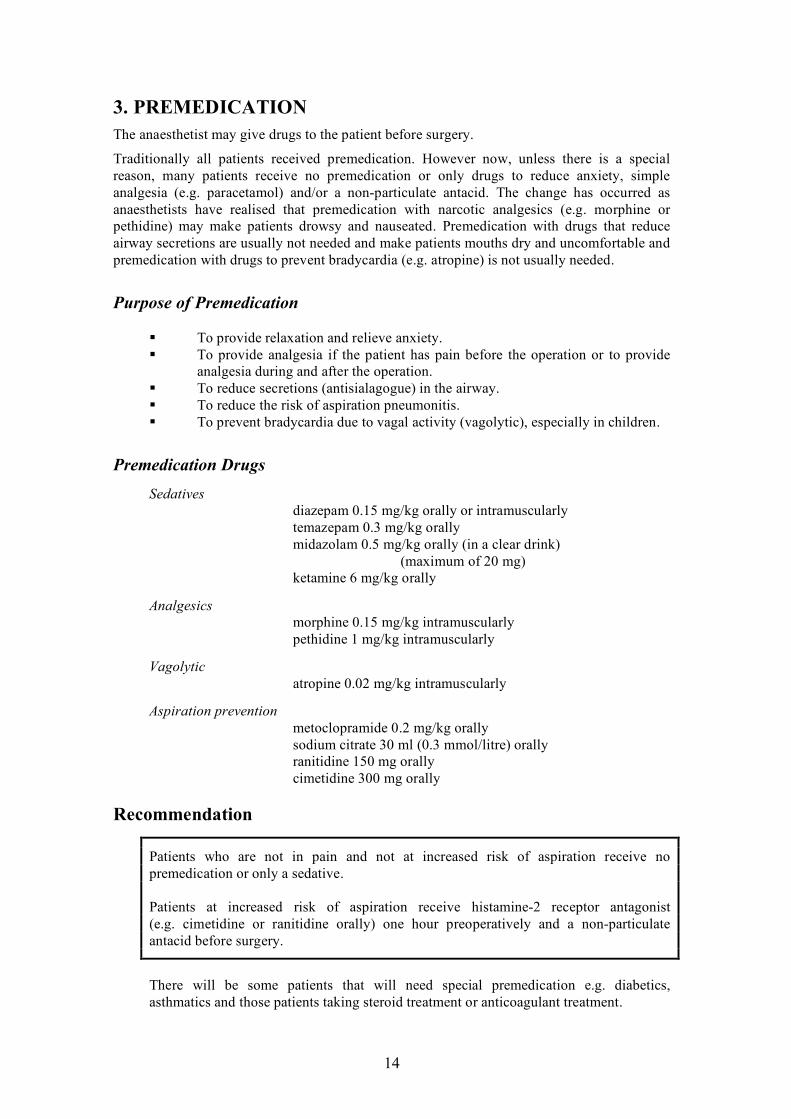
3. PREMEDICATION
The anaesthetist may give drugs to the patient before surgery.
Traditionally all patients received premedication. However now, unless there is a special reason, many patients receive no premedication or only drugs to reduce anxiety, simple analgesia (e.g. paracetamol) and/or a non-particulate antacid. The change has occurred as anaesthetists have realised that premedication with narcotic analgesics (e.g. morphine or pethidine) may make patients drowsy and nauseated. Premedication with drugs that reduce airway secretions are usually not needed and make patients mouths dry and uncomfortable and premedication with drugs to prevent bradycardia (e.g. atropine) is not usually needed.
Purpose of Premedication
To provide relaxation and relieve anxiety. To provide analgesia if the patient has pain before the operation or to provide
analgesia during and after the operation. To reduce secretions (antisialagogue) in the airway. To reduce the risk of aspiration pneumonitis. To prevent bradycardia due to vagal activity (vagolytic), especially in children.
Premedication Drugs
Sedatives diazepam 0.15 mg/kg orally or intramuscularly temazepam 0.3 mg/kg orally midazolam 0.5 mg/kg orally (in a clear drink) (maximum of 20 mg) ketamine 6 mg/kg orally
Analgesics morphine 0.15 mg/kg intramuscularly pethidine 1 mg/kg intramuscularly
Vagolytic atropine 0.02 mg/kg intramuscularly
Aspiration prevention metoclopramide 0.2 mg/kg orally sodium citrate 30 ml (0.3 mmol/litre) orally ranitidine 150 mg orally cimetidine 300 mg orally
Recommendation
Patients who are not in pain and not at increased risk of aspiration receive no premedication or only a sedative. Patients at increased risk of aspiration receive histamine-2 receptor antagonist (e.g. cimetidine or ranitidine orally) one hour preoperatively and a non-particulate antacid before surgery.
There will be some patients that will need special premedication e.g. diabetics, asthmatics and those patients taking steroid treatment or anticoagulant treatment.

15
4. PREOPERATIVE FASTING
All patients must fast, if possible, before surgery.
Physiology
With the onset of anaesthesia, protective airway reflexes are diminished and patients are at risk of regurgitation and inhaling (aspirating) their stomach contents. The aim of fasting is to minimize the risk of aspiration. However the anaesthetist should also consider patient comfort in the preoperative period and minimise any potential significant physiological changes that may occur from prolonged fasting. As gastric secretion is continuous at 6 ml/kg/h and 1 ml/kg/h of saliva is swallowed, the stomach is never truly empty. These volumes and the speed at which the stomach empties food and liquid will change with diseases, emotion, pain and hunger. It is important to remember that a patient who is in pain and/or sustained an injury soon after eating may still have a full stomach even with prolonged fasting, and should be treated as at risk of aspiration. This is common in children.
Preoperative Assessment
The preoperative assessment must try to identify those patients with an increased risk of aspiration. The anaesthetist should ask about a history of gastroesophageal reflux disease, dysphagia, gastrointestinal motility disorders, metabolic disorders (e.g. diabetes), obesity, pregnancy and drugs (e.g. morphine) that may increase the risk of regurgitation and pulmonary aspiration. The anaesthetist must be aware of surgical conditions such as intra-abdominal infective/inflammatory disorders (e.g. appendicitis) and obstructive disorders (e.g. bowel cancer) that will also increase the risk of regurgitation and aspiration. Finally the anaesthetist must consider the fasting time. If the anaesthetist believes the patient to be at an increased risk of regurgitation and aspiration then they will need to alter their anaesthetic management (e.g. rapid sequence induction and intubation of the trachea). The risk of aspiration can be reduced by fasting, emptying the stomach (nasogastric tube or causing vomiting), reducing stomach acidity (non-particulate antacid, histamine-2 receptor antagonists) and increasing the speed of emptying of the stomach (metoclopramide). Nasogastric tubes and inducing vomiting are unpleasant for the patient and are not routinely done. Nasogastric tubes may be appropriate for patients with an ileus.
Fasting time
The fasting times for clear fluids and solids are different. Solids are emptied from the stomach at a much slower rate than clear fluids. Aspiration of solids can cause obstruction of airways and potentially greater morbidity and mortality. There are also differences in stomach emptying between breast milk, cow’s milk and formula. Gastric emptying is much slower for formula compared with breast milk. It should be treated as a solid.

16
Recommendations for Fasting Times
For elective surgery Preoperative fasting solids and non-human milk: 6 hours Preoperative fasting infant formula: 6 hours Preoperative fasting breast milk: 4 hours Preoperative fasting clear fluids: 2 hours All patients must be allowed to take most of their usual medications before surgery with 30 ml of water.
Recommendations for Drug Treatment
(There are many drugs that affect stomach emptying)
The routine preoperative use of gastrointestinal stimulants (e.g. metoclopramide) for reducing gastric volume in patients who are not at increased risk of aspiration is not recommended. The routine preoperative use of histamine-2 receptor antagonists that block gastric acid secretion (e.g. cimetidine or ranitidine) in patients who are not at increased risk of aspiration is not recommended. If antacids are given preoperatively to reduce gastric acidity, then only non-particulate antacids should be used. These drugs should be used in patients who are at risk of aspiration.

17
5. AIRWAY ASSESSMENT
One in a hundred tracheal intubations may be difficult. By taking a history and performing an examination, the anaesthetist may identify those patients that may be difficult to intubate.
Preoperative Assessment
Intubation may be difficult because the patient has reduced mouth opening (e.g. osteoarthitis, trauma, rheumatoid arthitis, infection), reduced neck flexion/extension (e.g. osteoarthitis, trauma, rheumatoid arthitis, ankylosing spondylitis), lesions in the oral cavity (e.g. swelling, infections or tumours of larynx, pharynx, tongue) or congenital facial abnormalities. Intubation may also be difficult in patients who are obese or have large breasts.
Anaesthetic History
The anaesthetist’s preoperative history should determine if the patient has had problems with an anaesthetic in the past. The anaesthetist must look at the patient’s old anaesthetic notes to see if there have been problems with intubation during previous anaesthetics. (If the anaesthetist has a problem with intubation or any part of the anaesthetic they must write a clear account of that problem to warn other anaesthetists). The anaesthetist should also ask about a history of arthitis in the neck, infections or tumours in the mouth, trauma to the neck or mouth, loose teeth and dentures and also ask about any symptoms of airway obstruction such as hoarse voice, stridor, wheezing and airway obstruction with changes in the patient’s position.
Physical Examination
The physical examination is very important. The anaesthetist should assess the patient’s mouth opening, cervical spine mobility, teeth, thyromental distance, and mouth cavity. The anaesthetist must perform a complete airway assessment for every patient. The patient should be able to open their mouth more than thee fingers breadth. They should be able to touch their chin to their chest and also extend their neck backwards. Large front teeth will make intubation more difficult and bad teeth may be damaged or lost during intubation. If the thyromental distance (the distance between the lower border of the mandible to the thyroid notch) is less than four fingerbreadths, there may be difficulty seeing the glottis.
Mallampati Classification
The mouth cavity should be assessed by sitting the patient upright with the head in a normal position, mouth open as wide as possible and tongue poking out. The airway can then be given a Mallampati score depending on how much of the oral cavity can be seen.

18
(Class 1:soft palate, uvula, fauces and pillars; class2: soft palate, uvula, fauces; class3: only soft palate and class 4: soft palate not visible).
If the patient has a Mallampati class 1 airway and no other airway problems, most intubations will be easy. If the patient has a Mallampati class 4 airway then intubation may be difficult. Patients with more than one airway abnormality are more likely to have a difficult intubation. For example, an obese patient with a short neck, reduced movement in the cervical spine and reduced thyromental distance, or a patient with large upper teeth, small mouth and small mandible.
Laboratory Investigations
In most patients a good history and examination will warn the anaesthetist of a difficult airway, and investigations are not required. Chest and cervical spine neck X-rays can reveal tracheal deviation or narrowing. Cervical spine X-rays are very important in trauma patients. Indirect laryngoscopy can show lesions of the pharynx and larynx. Arterial blood gases can show the severity of the patient’s respiratory disease.
Conclusions
Anticipation of a difficult airway will help the anaesthetist to best manage the airway and avoid disasters. If the anaesthetist anticipates a difficult airway they must plan how to manage the airway. They should also plan what they would do if the first plan is not successful.
If the anaesthetist does not assess the patient’s airway, they will not be prepared to manage the patient who is difficult to intubate. If the patient’s airway is managed badly the patient may suffer severe complications or death.
A difficult airway cannot always be predicted. The anaesthetist must always be prepared to manage an unexpected difficult airway.

19
6. CARDIOVASCULAR DISEASE
ISCHAEMIC HEART DISEASE
Assessing patients with coronary artery disease who are having non-cardiac surgery is difficult. The purpose of the preoperative evaluation is
• to identify patients who would benefit from further cardiac testing, • to decide if the risk can be reduced and • to decide if the non-cardiac surgery is so urgent that it should be carried out
rapidly despite the risk.
In hospitals that have assess to all investigations and all medical and surgical treatments, preoperative management would depend on clinical assessment and preoperative testing (for example: exercise electrocardiogram, dipyridamole-thallium scan, left ventricular ejection fraction, dobutamine stress echocardiogram, transthoracic echocardiogram and coronary angiogram). The patient may then proceed to further treatment including coronary artery surgery, angioplasty or maximal medical treatment of the ischaemic heart disease.
In hospitals that do not have access to all investigations and treatment, patients may still be effectively managed by clinical assessment alone. History and examination of the patient are key elements of preoperative risk assessment. The anaesthetist must determine the patient’s risk factors, the surgical risk factors and the overall fitness (functional capacity) of the patient.
Patient Risk Factors
Patient risk factors should be subdivided into major, intermediate and minor. Major patient risk factors are markers of unstable coronary artery disease and include myocardial infarction within 6 weeks, unstable or severe angina, ongoing chest pain after myocardial infarction, clinical ischaemia and uncontrolled congestive heart failure, clinical ischaemia and arrhythmias (high grade AV block or SVT with uncontrolled ventricular rate) or coronary artery bypass operation within 6 weeks. These patients should not have elective operations until they are investigated and treated. Only emergency procedures should be considered. Intermediate patient risk factors are markers of stable coronary artery disease and include myocardial infarction longer than 6 weeks ago but less than 3 months ago, stable angina, diabetes and controlled congestive cardiac failure. Minor patient risk factors are markers of coronary artery disease but not of increased perioperative risk. They include a family history of coronary artery disease, uncontrolled hypertension, hypercholesterolaemia, electrocardiogram abnormalities (arrhythmia, left ventricular hypertrophy, bundle branch block) and patients who have had a previous myocardial infarction more than 3 months ago and are asymptomatic without treatment.

20
Functional Capacity
The patient’s general health (exercise tolerance or functional capacity) will provide the anaesthetist with a good estimate of perioperative risk. Patients with vascular disease who can exercise to 85% of their estimated maximal heart rate (220 minus age) have a low risk of perioperative cardiac complications. Climbing stairs is a simple test of perioperative cardiac risk. Patients who cannot climb one flight of stairs are at increased risk of cardiovascular complications.
Surgical Risk Factors
Surgery can also be considered as low, intermediate or high risk. Low risk surgery includes endoscopic, breast, skin, limb, eye and plastic surgery. Intermediate risk surgery includes minor vascular, minor abdominal and thoracic, neurosurgery, ENT and orthopaedic surgery. High-risk surgery includes emergency intermediate risk surgery, aortic and major vascular, thoracic and prolonged surgery.
Management
The anaesthetist must take a history and perform an examination and assess the patient risk factors, surgical risk and the patient’s functional capacity. With this knowledge the anaesthetist can estimate the patient’s risk of perioperative cardiac complications. If the patient is at high risk and the operation is elective, the patient should not have the surgery. If the surgery is urgent and the patient is at an increased risk then the anaesthetist must ensure that the patient has the best available care. High risk patients with high risk surgery and poor exercise tolerance may need coronary angiography and coronary artery bypass operation before the non-cardiac surgery. It is very important that the anaesthetist always avoids events that will increase the risk of perioperative cardiac complications such as hypothermia, extreme anaemia, hypotension, tachycardia and postoperative pain. This can easily be achieved. Perioperative beta-blockade may also be of benefit.
VALVULAR HEART DISEASE
Patients with valvular heart disease will have abnormal cardiac function. They must have a full preoperative assessment. As with ischaemic heart disease, the patient’s exercise tolerance is a good indicator of the severity of the heart disease. All patients with valvular heart disease need antibiotic treatment to prevent bacterial endocarditis.

21
Mitral Stenosis
Mitral stenosis is usually due to rheumatic fever. Mitral stenosis prevents left ventricular filling, which results in decreased cardiac output. Left atrial emptying is decreased, which results in left atrial enlargement and increased pulmonary artery pressures to maintain cardiac output. These patients may develop pulmonary oedema, cardiac failure and atrial fibrillation. The main symptom of mitral stenosis is dyspnoea. Patients with atrial fibrillation, dyspnoea at rest and who wake at night short of breath (paroxysmal nocturnal dyspnoea) are at increased risk. The anaesthetist should avoid myocardial depressants, tachycardia (which reduces ventricular filling time), hypovolaemia and hypotension and increased pulmonary vascular resistance (e.g. due to hypoxia, pain or hypercarbia). The anaesthetist should aim for a slow sinus rhythm, normal intravascular volume, normal cardiac contractility and normal systemic vascular resistance.
If regional anaesthesia is used, epidural anaesthesia maybe safer than spinal anaesthesia. The anaesthetist must avoid hypotension.
Mitral Regurgitation
50% of mitral regurgitation is due to rheumatic fever. As the left ventricle contracts some of the blood flows backwards into the left atrium. The regurgitant flow will increase with increased systemic vascular resistance and bradycardia. Most patients with chonic mitral regurgitation are well for many years without evidence of heart failure. Dyspnoea and pulmonary oedema are signs of severe mitral regurgitation. The anaesthetist should avoid myocardial depressants, hypovolaemia, bradycardia and increased systemic vascular resistance. They should aim for a normal or increased heart rate, decreased systemic vascular resistance and normal cardiac contractility and intravascular volume. Regional anaesthesia is well tolerated.
Aortic Stenosis
Aortic stenosis may be congenital or acquired. It is a chonic condition with symptoms only occurring when the stenosis is severe. The main symptoms of aortic stenosis are dyspnoea, angina and syncope. Once symptoms develop, the patient’s life expectancy may be less than 5 years and these patients should not have elective surgery. The anaesthetist must maintain sinus rhythm. Atrial contraction is vital to maintaining adequate ventricular filling. The heart rate should be normal. Tachycardia and bradycardia will both reduce coronary blood flow. The systemic vascular resistance should be kept normal. An increase in systemic vascular resistance will further reduce cardiac output and a reduction in systemic vascular resistance may reduce coronary blood flow. Myocardial depressants must be avoided. Regional anaesthesia can cause dangerous changes in systemic vascular resistance and heart rate. However, epidural anaesthesia may be tolerated if performed slowly with careful monitoring and treatment of blood pressure and heart rate.
22
Aortic Regurgitation
Patients with aortic regurgitation may not have symptoms for many years. They may develop signs and symptoms of left ventricular failure. The anaesthetist should avoid bradycardia as this increases the time for backwards flow. They should also avoid increased peripheral resistance and myocardial depressants. They should aim to maintain an increased heart rate, adequate intravascular volume and decreased systemic vascular resistance.
Regional anaesthesia is well tolerated in patients with chonic aortic regurgitation.
HYPERTENSION
It is important that all antihypertensive medication is continued and that the patient is fully assessed for signs and symptoms of the complications of chonic hypertension. Organ damage from hypertension presents a greater risk than hypertension itself.
The management of patients with hypertension has changed over the last decades. Hypertension is defined by the World Health Organisation as a diastolic blood pressure greater than 95 mmHg and a systolic pressure greater than 160 mmHg. Chonic hypertension may cause renal failure, cardiac failure, stroke and myocardial infarction. Ideally all patients with hypertension should be treated before surgery. However, there is little evidence for an association between systolic pressures of less than 180 mmHg or diastolic pressures less than 110 mmHg and perioperative complications though the anaesthetist must be aware that the patient may have large swings in blood pressure. Intra-operative arterial pressure should be maintained within 20% of the preoperative arterial pressure.

23
7. PERIOPERATIVE BETA BLOCKADE
Previous controlled studies with nitrates, calcium channel blockers, clonidine and digoxin have not demonstrated protection from myocardial ischaemia intra- or postoperatively. Recent studies suggest that giving beta-blockers perioperatively may reduce the risk of cardiac complications and death in patients having major non-cardiac surgery. The greatest benefit would seem to be for those patients at high risk of perioperative cardiac complications having major surgery.
Contraindications
Beta-blockade should not be used in patients who have a resting heart rate less than 60 beats/minute or who have asthma requiring regular treatment.
Choice of Beta-blocker
If possible, beta-1 selective beta-blockers should be used. Non-selective beta-blockers are more likely to produce respiratory complications such as bronchospasm. At this stage no evidence suggests any particular beta-1 blocker is better.
Management
The beta-blocker should be started as soon as possible before the surgery in high-risk patients (even up to a month before) so that the dose can be changed to achieve a resting heart rate of 50 to 60 beats/minute. Even if the anaesthetist is unable to start beta blockade in the weeks before surgery, there may still be a benefit in giving a beta-blocker on induction of anaesthesia. The beta-blocker should be given in small doses to avoid a fall in blood pressure of greater than 20%. The beta-blocker should be continued after surgery at least as long as the patient remains in hospital.
High Risk Factors
Patient risk factors for perioperative myocardial infarction include: • previous myocardial infarction or angina, • diabetes, • major surgery (intraabdominal, intrathoracic, vascular), • congestive heart failure, • renal impairment due to vascular disease or diabetes and • poor exercise tolerance (unable to walk up 2 flights of stairs or 400 metres on flat ground).

24
Recommendation
Giving beta-blockers perioperatively may reduce the risk of cardiac complications and death in patients having major non-cardiac surgery. High-risk patients are those with 3 or more of the above risk factors or myocardial infarction within the previous 6 months or angina increasing in severity or of recent onset. A cardiologist should review them before surgery. Low to moderate risk patients have only 1 or 2 of the above risk factors present and should be treated with beta-blockers at least one week before major surgery aiming for a resting heart rate of less than 60 bpm.

25
8. RESPIRATORY DISEASE
Respiratory disease often occurs in patients presenting for anaesthesia and surgery. Common respiratory diseases include asthma, chonic obstructive lung disease, upper respiratory tract infections, tuberculosis and smoking. General anaesthesia will have several effects on the patient’s respiratory function including a decrease in lung volume and a decreased respiratory rate response to hypoxia and hypercarbia. Respiratory function will be further decreased by poorly treated postoperative pain.
Preoperative Assessment
The anaesthetist must take a full history, examination and order relevant investigations. Respiratory function testing is useful in predicting which patients may not survive a pneumonectomy but is less reliable in predicting postoperative pulmonary complications for other surgical procedures. The anaesthetist may need to rely on clinical findings. The history and examination may reveal important information and conditions which are significant risk factors including dyspnoea, cough and sputum production, recent chest infection, haemoptysis, wheezing, smoking, obesity and pulmonary complications from previous surgery. An increase in the patient’s respiratory rate, especially above 25 breaths each minute, is associated with an increase in postoperative pulmonary complications. Bacterial and even viral respiratory infections will have an adverse effect on respiratory function, increasing airflow obstruction for up to 5 weeks after the infection. Wheezing is usually reversible and should be treated with bronchodilators however the anaesthetist must also check and treat for non-respiratory causes of wheezing such as cardiac failure. Smoking should be ceased. Patients who are not short of breath at rest and who can climb more than two flights of stairs are unlikely to develop postoperative pulmonary complications. The anaesthetist must treat any potentially reversible respiratory disease before surgery. They should encourage the patient to stop smoking, treat acute bacterial infections, humidify inhaled gases, encourage chest physiotherapy and treat bronchospasm and right heart failure.
Respiratory Infections
90% of upper respiratory tract infections are likely to be viral. If bacterial infection is suspected the patient should be treated with antibiotics prior to surgery. Even viral infections will increase the risk of laryngospasm and bronchospasm and it is wise to delay surgery if possible for 5 weeks. A careful history and examination looking for fever, cough, shortness of breath and lethargy will allow the anaesthetist to assess the severity of the infection.

26
Tuberculosis
Tuberculosis increases the risk to the patient and medical staff. Early pulmonary tuberculosis may be asymptomatic. Cough, haemoptysis, chest pain and shortness of breath occur late in the disease. Patients with tuberculosis must have a careful history and examination taken. The anaesthetist must also be aware of non-pulmonary symptoms. Tuberculosis can affect many organs including the central nervous system, kidney and bone marrow. Hyponatraemia may occur with pulmonary tuberculosis. If time allows, active tuberculosis must be treated before any surgery.
Asthma
A careful history, examination and simple investigations will allow the anaesthetist to determine how severe a patient’s asthma usually is and if the patient’s asthma could be improved before surgery. As with all anaesthetics, the urgency of the surgery needs to be balanced against the severity of a patient’s disease. To establish how severe a patient’s asthma usually is, the anaesthetist needs to know how often the patient has asthma attacks, what medication they are taking, how often they take the medication and what their best exercise tolerance is. If the patient’s asthma is currently worse than usual they should be treated prior to surgery with increased bronchodilators and/or a course of oral steroids. All asthmatics may benefit from nebulised salbutamol immediately before anaesthesia. The anaesthetist should avoid histamine-releasing drugs and if possible avoid endotracheal intubation, which can precipitate bronchospasm.
Chonic Obstructive Pulmonary Disease (COPD)
Chonic obstructive pulmonary disease increases the risk of hypoxaemia, hypercarbia, bronchospasm and postoperative pulmonary complications. The anaesthetist should ask about cough, sputum production, shortness of breath, exercise tolerance, smoking and recent chest infections. The chest X-ray may be normal in early disease. The patient should stop smoking and be treated for any chest infections. These patients may have some reversible lung disease and may benefit from preoperative bronchodilators, steroids, antibiotics and chest physiotherapy. Postoperative pain control is very important in any patient with respiratory disease.

27
9. SMOKING
Smoking is a major risk factor for perioperative complications. Cigarette smoking is a major cause of coronary heart disease and an important cause of cerebrovascular disease. It is the single most important cause of cancer mortality in the United States, accounting for 30% of all cancer deaths. Cigarette smoking also is a major cause of chonic obstructive pulmonary disease and is associated with an increased risk of pneumonia and postoperative respiratory complications. The anaesthetist must be aware of the damage that smoking can do to a patient’s health.
Recommendation
Patients who smoke should be encouraged to stop smoking at least six to eight weeks (ideally 6 months) before surgery. Even stopping smoking for 12 hours before surgery is of some benefit.
Physiological Effects
More than 4000 substances have been found in cigarette smoke. Cigarette smoke contains 2 to 6% carbon monoxide. Smoking increases the amount of carboxyhaemoglobin in the blood. The range of carboxyhaemoglobin levels in smokers is 2 to 15%. As the half-life of carboxyhaemoglobin is only four hours, 12 hours of stopping smoking will greatly reduce its levels, improve oxygen content and reverse the negative inotropic and arrhythmic effects. The polycythaemia takes several days to reverse. Nicotine increases heart rate, blood pressure and causes peripheral vasoconstriction. These effects improve after 12 to 24 hours of stopping smoking. Smoking also causes increased secretion of mucus in the lungs. The airways are narrowed and the lungs ability to remove mucus is reduced by smoking so smokers tend to become hypoxic more quickly. It takes 6 weeks before the mucus production in the lungs returns to normal. Smoking also has an adverse effect on the patient’s immune system. It takes 6 months before a patients immune system returns to normal. One year after stopping smoking there is a marked reduction in the patient’s risk of myocardial infarction.

28
10. STEROID SUPPLEMENTATION
Patients on long term or recently ceased steroid therapy may require increased doses of glucocorticoids during the stress of illness and surgery. Acute adrenal insufficiency may result in cardiovascular collapse and death. The benefit of steroid supplement must be weighed against the risk. Perioperative steroid supplementation may cause immunosuppression, delayed wound healing, hyperglycaemia and sodium and water retention. Patients receiving steroids should continue the steroid treatment.
Recommendation
If the normal prednisolone dose is less than 10 mg/day then give only the patient’s usual steroid treatment. No extra steroid cover is required. If the normal prednisolone dose is greater than 10 mg/day,
for minor surgery give the patient’s usual dose plus 25 mg of hydrocortisone at the start of the anaesthetic
for moderate surgery give the patient’s usual dose plus 25 mg of hydrocortisone at the start of the anaesthetic and 100 mg of hydrocortisone over the next 24 hours
for major surgery give the patient’s usual dose plus 25 mg of hydrocortisone at the start of the anaesthetic and 100 mg of hydrocortisone/day for 2days
Patients who have ceased receiving long-term steroids may still need to receive steroids supplements.
Recommendation
If steroid treatment was ceased less than 3 months ago then treat the patient as if they were still taking steroids for moderate and major surgery only. If steroid treatment was ceased more than 3 months ago then no perioperative steroids are indicated.
[Dexamethasone 1 mg = prednisolone 5 mg = hydrocortisone 20 mg]

29
11. RENAL DISEASE
Patients with renal disease may have many medical problems. Renal failure may be acute or chonic. All patients require careful preoperative assessment. The anaesthetist must consider how the anaesthetic might affect the renal disease and how the renal disease might affect the anaesthetic.
Acute Renal Failure
Acute renal failure usually occurs over a few days. The patient may have had normal or reduced renal function previously. The patient’s urine output may be normal, reduced or absent. Acute renal failure may be due to a decreased blood flow to the kidney (prerenal), renal disease (renal) or an obstruction in the urinary collecting system (post renal). Causes of prerenal acute renal failure include shock, hypovolaemia, cardiac failure and renal artery stenosis. If the blood flow to the kidney is quickly restored the kidney function usually returns to normal but if the poor blood flow continues there may be permanent renal damage. Causes of renal acute renal failure include glomerulonephitis, diabetes, polycystic renal disease, pyelonephitis, hypertensive vascular disease, nephotoxins and acute tubular necrosis (ATN). ATN accounts for 75% of hospital admissions for acute renal failure. Post renal acute renal failure may occur from any obstruction in the urinary collecting system like bladder tumours, renal stones or prostate disease. If the cause of the obstruction can be quickly treated then the renal function should return to normal.
The death rate from acute renal failure is high (30%) in surgical and trauma patients.
Preoperative Assessment
Patients with acute renal failure usually have decreased urine output. They have increased blood levels of urea and other substances that cause nausea, vomiting, and tiredness. They may also have increased bleeding and are at increased risk of infections. Sodium and water excretion is reduced so the patients develop oedema, hypertension, acidosis and hyperkalaemia.
Chonic Renal Failure
Chonic renal failure is irreversible and often follows acute renal failure. The most common cause is glomerulonephitis. Other causes include pyelonephitis, diabetes, polycystic renal disease, vascular disease and hypertension. These patients have many changes to their health, which are important for the anaesthetist to identify and treat if possible before surgery.

30
Preoperative Assessment
Patients with chonic renal failure may be tired, confused and finally convulsing and in coma. They may have hypertension, pericarditis and pericardial effusions (which may cause a pericardial tamponade), peripheral vascular disease and cardiac failure. They will usually have hyperkalaemia, hypermagnesaemia and hyponatraemia. Increased blood levels of parathyroid hormone may cause hypocalcaemia and hyperphosphataemia. Acidosis is common. Patients may have a normocytic normochomic anaemia caused by reduced erythopoietin production, reduced red cell survival and bone marrow depression. These patients may also have prolonged bleeding time due to decreased platelet adhesiveness. Chonic renal failure can cause both peripheral and autonomic neuropathy. The autonomic neuropathy can cause delayed gastric emptying.
The effects of drugs on the patient (pharmacokinetics) will also be changed due to changes in body water, pH, electrolytes, total protein and rates of excretion.
Anaesthetic Management
As with all patients, the anaesthetist must take a complete history and examination and look at all investigations. The anaesthetist must decide if the patient’s health can be improved before surgery, whether the surgery should be delayed and what the best anaesthetic for that patient will be. When assessing the patient the anaesthetist should take a history and examination looking for both the severity of the renal disease and the severity of the cause of the renal disease (e.g. diabetes, vascular disease, hypertension). In particular, the anaesthetist should assess cardiovascular complications, fluid and electrolyte and acid base changes. If the patient has signs or symptoms of autonomic neuropathy, the patient may be at an increased risk of aspiration of gastric contents. Chonic anaemia rarely needs transfusion. The anaesthetist should also check the drugs the patient is taking. Laboratory investigations are important. If available, ideally the patient’s sodium, potassium, chloride, bicarbonate, haemoglobin and coagulation should be tested. Patients with a sodium less than 130 mmol/l or greater than 150 mmol/l, or a potassium less than 2.5 mmol/l or greater than 5.0 mmol/l will probably need treatment before surgery because these abnormalities may cause dangerous heart arrhythmias and reduced heart function. An electrocardiogram is useful to look for signs of myocardial ischaemia, electrolyte changes and pericarditis. A chest X-ray may show signs of heart failure, pericardial effusions or pneumonia. It is also important to check the renal function. Blood urea is not a good measure of renal function, as it will change with cardiac output, diet, body size and dehydration. Blood creatinine also is not a good measure as it is affected by skeletal muscle mass and the patient’s activity level. The rate at which creatinine is excreted by the kidneys is a good measure of renal function. It can be measured by collecting the patient’s urine for 12 or 24 hours and measuring the creatinine concentration in the urine, the urine volume and the creatinine level in the blood.
Estimated creatinine clearance ml/min = (140 – age) x weight in kilograms 72 x blood creatinine mg/dl

31
Recommendation
Patients with an estimated creatinine clearance of greater than 50 ml/min can be treated as if they have normal renal function. Patients with an estimated creatinine clearance of between 30 to 50 ml/min have decreased renal function and the anaesthetist must avoid dehydration and nephotoxins. Patients with an estimated creatinine clearance of between 10 to 30 ml/min have severe renal disease and may need preoperative dialysis. Patients with an estimated creatinine clearance of less than 10 ml/min have severe renal disease and should have dialysis within 24 hours preoperatively.
Premedication
The dose of central nervous system depressant premedications should be reduced, as renal failure patients are more sensitive to them. The anaesthetist may wish to give an antacid and histamine (H-2) blocker as delayed gastric emptying and increased gastric volume are common.
Patients on dialysis must be dialysed before major surgery. Check the hydration status of the patient (weight, central venous pressure, lung fields). Patients who have not been recently dialysed may have fluid overload as well as electrolyte abnormalities.
Anaesthetic Maintenance
The anaesthetist must avoid hypovolaemia. Potassium-containing intravenous fluids should not be given. Drugs, which accumulate in renal failure, should be avoided (e.g. gallamine). Drugs that can reduce renal function (e.g. gentamicin, NSAID, radioactive dye) should not be given. The dose of induction agents may need to be reduced and should be given slowly to avoid hypotension. Renal patients are more sensitive to opioids, benzodiazepines, phenothiazines, barbiturates and propofol. These drugs should be given in reduced dosages. Suxamethonium is not contraindicated unless there is hyperkalaemia (greater than 5.5 mmol/l) or peripheral neuropathy. Atracurium and cis-atracurium are a good choice of muscle relaxants as their metabolism is generally unaffected in renal failure. Methoxyflurane can cause renal damage by increasing blood fluoride levels. Though both enflurane and sevoflurane can increase blood fluoride they have not been shown to decrease renal function. The metabolite of pethidine (nor-pethidine) may accumulate in renal failure. NSAIDs should be avoided. The anaesthetist may choose general or regional anaesthesia. (Patients for regional anaesthesia should have normal coagulation).

32
12. LIVER DISEASE
Hepatic failure may be acute or chonic. Causes of acute hepatic failure include viral hepatitis, shock, drugs (e.g. paracetamol, halothane, chloroform, chlorpromazine, phenytoin) and poisons. Causes of chonic hepatic failure include autoimmune hepatitis, viral hepatitis, drugs (e.g. methyldopa, alcohol), metabolic diseases (e.g. haemochomatosis), biliary disease and cardiac failure. When a patient has liver failure he or she will have many changes in their health, which will affect anaesthesia.
Preoperative Assessment
The liver has many functions including the production of plasma proteins, clotting factors and plasma cholinesterase. The liver is a site of gluconeogenesis, bilirubin metabolism, drug metabolism and detoxification. Depending on the severity of the liver failure other organs may be affected. Reduced detoxification of toxic waste products will cause neurological impairment. This may range from mild to marked confusion or coma. As liver failure patients also have an increased sensitivity to sedatives due to reduced liver metabolism, the dosages of sedative drugs must be reduced. The patients with impaired consciousness are also at risk of gastric aspiration especially if they also have ascites. They may need treatment to prevent aspiration and undergo rapid sequence intubation of the trachea. Patients with liver failure have decreased levels of albumin, increased levels of aldosterone and antidiuretic hormone which all lead to increased total body water (e.g. ascites, oedema, pleural effusion) but they usually have a reduced intravascular volume. Their cardiac output is usually increased as a result of decreased systemic vascular resistance. They are usually hyponatraemic, hypokalaemic and have a metabolic acidosis. There may be poor gas exchange (ventilation/perfusion mismatch) in the lungs resulting in low levels of oxygen in the blood. Patients with liver failure may also have renal failure (hepatorenal syndrome). Coagulation defects occur for several reasons. There is decreased production of clotting factors, decreased absorption of vitamin K (which is an important factor in the production of factors II, VII, IX, and X) and thombocytopenia. Hypoglycaemia may occur in liver failure. The risk of complications and death depends on the severity of the liver disease and the type of surgery. The severity of the liver disease can be estimated by a modified Child’s classification.

33
Child’s Classification
CLASS A
CLASS B
CLASS C
Albumin (g/dl)
Greater than 3.5
3.0-3.5
Less than 3.0
Bilirubin (mg/dl)
Less than 2.0
2.0-3.0
Greater than 3.0
Ascites
None
Controlled
Uncontrolled
Encephalopathy
None
Mild
Severe
INR
Less than 2.0
2.0-3.0
Greater than 3.0
Anaesthetic Management
Patients undergoing emergency surgery, or with a prothombin time greater than 2.5 times normal despite treatment, or hepatorenal syndrome or severe liver failure, are at a greater risk of death. Patients at high risk should not have elective surgery. The anaesthetist must take a full history and examination. Appropriate investigations include liver function tests, renal function test, sodium, potassium, clotting profile, chest X-ray, electrocardiogram, blood glucose and blood gases if available. They should aim to correct any complications of liver failure. In particular they should correct hypovolaemia and coagulation and electrolyte abnormalities. Care must be taken to prevent aspiration. Regional anaesthesia may be contraindicated if coagulation changes cannot be corrected. In general the effects of all drugs will be greater and prolonged, so the anaesthetist may need to reduce drug dosages.

34
13. DIABETES
Diabetic patients present the anaesthetist with two special problems. One is the control of the patient’s blood sugar in the perioperative period and the other is the long-term effect of the diabetes on their health (end-organ disease). Hypoglycaemia must be avoided. The anaesthetic management will depend on how well the diabetes is controlled, how urgent the surgery is and which type of treatment the patient is receiving for their diabetes.
Preoperative Assessment
As the major risk factors for all diabetics undergoing surgery are end-organ diseases associated with diabetes, all diabetics must have a complete preoperative assessment and treatment. This assessment should focus on heart disease, kidney disease, joint abnormalities and neuropathies.
Cardiovascular Assessment
50% of patients who are diabetic and have hypertension will have autonomic neuropathy compared to only 10% in diabetics without hypertension. All diabetic patients should be assessed for autonomic neuropathy. The presence of autonomic neuropathy may make the perioperative period more dangerous. These patients are more likely to have intra-operative or postoperative cardiorespiratory arrest, have a higher rate of painless myocardial ischaemia, may have cardiovascular instability and are more likely to have delayed emptying of stomach contents (gastroparesis) and therefore are at an increased risk of pulmonary aspiration. Symptoms of autonomic neuropathy include: lack of sweating, fall in blood pressure when standing (orthostatic hypotension), penis erectile problems, severe constipation or night-time diarrhoea. Signs of autonomic neuropathy include orthostatic hypotension (fall of 20 mmHg in systolic or 10 mmHg in diastolic blood pressure for more than 3 minutes after standing), lack of pulse rate change with breathing (less than a 5 beat/min change in pulse rate on deep inspiration) and loss of electrocardiogram R-R interval variability. Diabetic patients have an increased incidence of atherosclerosis and all its complications. Unfortunately they may have painless myocardial ischaemia so the anaesthetist cannot rely on a history of chest pain to assess the patient’s risk. The anaesthetist should carefully assess for myocardial ischaemia in all diabetics who are obese, physically inactive, over 55 years of age or who have chonically elevated blood glucose (greater than 11 mmol/l). In diabetic patients, the risk of coronary artery disease is two to four times higher than the general population. A good indication of the severity of ischaemic heart disease is the patient’s exercise tolerance. If the patient can climb a flight of stairs, walk up a hill or run a short distance then their risk is probably low. An electrocardiogram may not show signs of myocardial ischaemia.
Renal Assessment
Blood testing of the kidney may show reduced function.

35
Musculo-Skeletal Assessment
Diabetics can have stiffness of the alanto-occipital joint making intubation of the trachea difficult. All diabetic patients need careful assessment of their airway.
Preoperative Management
The better the control of a patient’s diabetes the lower the perioperative risk. Poorly treated diabetics having elective surgery should be postponed until their diabetic treatment is improved. Adequacy of treatment can be assessed by history and investigation. Those patients who are still symptomatic or who have a raised glycosylated haemoglobin (HbA1C) are likely to have poor diabetic control. Glycosylated haemoglobin gives the best evidence of blood glucose control over the previous 1 to 2 months. If the HbA1C is greater than 9%, the patient’s diabetic control is inadequate.
Recommendation
If the patient’s diabetes is well treated (asymptomatic, HbA1C less than 9%):
Diet control only. No change in treatment. Oral hypoglycaemic. All oral hypoglycaemics should be omitted on the day of surgery. Insulin. Arrange for the patient to be first on the morning or afternoon operating list. If on the morning list then omit the morning insulin and monitor the blood glucose level. If on an afternoon operating list the patient should have a light early morning breakfast with half their normal insulin dose. Blood glucose should be measured at least every second hour until the patient is eating and drinking. All patients should have a blood sugar measurement on admission to the hospital. If the blood sugar level (BSL) is less than 5 mmol/l or greater than 10 mmol/l the patient needs further treatment.
If the patient’s diabetes is poorly treated:
If the surgery is elective the case should be cancelled and the patient’s treatment of the diabetes improved. The case can be rebooked in 4 to 6 weeks. If the surgery is urgent and the patient is treated with only oral hypoglycaemics then stop all hypoglycaemic tablets, begin 6 hourly Actrapid insulin injections according to a sliding scale and give 5% dextrose (80 ml/h) when fasting. Measure the BSL every 2 hours and hourly intra-operatively. If the surgery is urgent and the patient is treated with insulin, then stop the normal insulin, begin 6 hourly Actrapid insulin injections according to a sliding scale and give 5% dextrose (80 ml/h) when fasting. Measure the BSL every 2 hours before surgery and at least hourly intra-operatively.

36
14. EMERGENCY SURGERY
Patients for emergency surgery are at a high risk of perioperative complications. The anaesthetist must carefully assess the patient by history, examination and investigations in the time available before surgery is required. The anaesthetist must try to resuscitate (airway management, ventilation and intravenous fluid) the patient before surgery but for extreme emergencies they may need to resuscitate and anaesthetise at the same time. The anaesthetist must balance the urgency of the surgery and the need for preoperative assessment and treatment.
Risk
The anaesthetist must be aware of problems related to inadequate preparation of the patient. The patient may not be starved and therefore at risk of aspiration. These patients require a rapid sequence induction. Coexisting medical problems such as diabetes, asthma and ischaemic heart disease may be poorly treated. There may not be enough time to properly investigate the patient and order blood cross matching. Patients for emergency surgery often have severe changes in their health that must be assessed and if time allows, treated before surgery. Some examples include severe dehydration with severe electrolyte changes due to intestinal obstruction and vomiting/diarrhoea, severe hypovolaemia and anaemia from haemorrhage. There may be septic shock from untreated infections or there may be a damaged/obstructed airway in the trauma patient.
Choice of Anaesthesia
The choice of anaesthesia will depend on the type of surgery, the experience of the anaesthetist, the equipment available, the time available and the condition of the patient. Hypovolaemia and a full stomach are two common but deadly problems in emergency anaesthesia that the anaesthetist must be aware of when they plan the type of anaesthesia. If appropriate to the surgery required, regional anaesthesia of a limb or local anaesthesia may be the safest choice of anaesthesia. Spinal/epidural anaesthesia will reduce the risk of aspiration, however, hypovolaemia must always be corrected before spinal/epidural anaesthesia. These emergency patients must have a normal blood pressure and no tachycardia. There should be no postural drop in blood pressure and adequate urine output. The anaesthetist must ensure that a patient with burns has been given enough intravenous fluid. A burnt patient will need at least 4ml/kg times the percentage of body burnt, in the first 24 hours to replace fluid loss. For example, a 70 kg man with 30 percent burns will need at least (70 x 4 x 30) 8.4 litres in the first 24 hours. Usually half of the calculated fluid loss is given over the first 8 hours and the remainder over the next 16 hours. The patient will also need their daily maintenance fluid. General anaesthesia may be safer for patients with untreated hypovolaemia but they should receive reduced doses of almost all anaesthetic drugs except muscle relaxants.

37
Induction agents especially need to be given very carefully as these may cause cardiovascular collapse from vasodilatation in the hypovolaemic patient. If general anaesthesia is chosen then the anaesthetist must prevent aspiration of gastric contents. The non-fasted patient must have a rapid sequence induction and intubation of the trachea.
The anaesthetist should choose the type of anaesthetic depending on his or her experience and training, their assessment of the patient, the equipment and drugs available and the needs of the surgeon. The anaesthetist must try to treat patient problems caused by the emergency and other medical problems before giving an anaesthetic if time allows.

38
15. CHECKING THE EQUIPMENT
It is the responsibility of the anaesthetist to check all anaesthetic equipment and drugs before giving an anaesthetic. There must always be alternative equipment to ventilate the patient’s lungs if the anaesthetic machine or oxygen supply fails. A self-inflating resuscitation bag does not need a source of oxygen. It should be available whenever an anaesthetic is given.
Airway Equipment
An alternative method of ventilating the patient must always be available. Ideally the anaesthetist would have at least two laryngoscopes of different sizes. The light should be checked. Oropharyngeal (and nasopharyngeal) airways should be available in different sizes. A flexible stylet and gum elastic bougies are excellent aids for intubation. The anaesthetist should have several different sized masks and an appropriate sized endotracheal tube (plus one size smaller and one bigger) available. A laryngeal mask may be used as the airway or as an excellent alternative airway if endotracheal intubation is difficult (secondary plan). Emergency airway equipment (e.g. laryngeal masks, intubating laryngeal masks, percutaneous tracheostomy, fibreoptic laryngoscopes) should be kept together in a labelled container in a central area.
Suctioning
Suction equipment should be available. It consists of a pump to generate a vacuum, a reservoir and tubing. The reservoir must be large enough to hold the aspirated fluid but not too large. (The larger the reservoir the longer it will take to achieve a vacuum). The minimal flow rate should be 35 l/min of air and generate at least 600 mmHg (80 kPa) negative pressure. Suction may be powered by electricity, compressed gas or by hand/foot.
Continuous Flow Anaesthetic Machine (Boyle’s machine) The anaesthetic machine can be considered in thee parts: high pressure (pipeline, cylinders, pressure gauges and regulators), low pressure (oxygen failure alarm, antihypoxic device, flowmeters, vaporisers, pressure release valve, and common gas outlet) and the breathing system.
Cylinders and Pipelines
Cylinder and pipeline gases are too highly pressurised (5,000 kPa to 14,000 kPa) for safe flow regulation. Regulators are used to decrease the pressure to a safe level. Pressurised gases must never be connected directly to the breathing system. (1 atmosphere = 760 mmHG = 98 kPa = 14 psi. 1 psi =6.9 kPa).

39
Cylinders should be checked regularly for faults. Full and empty cylinders should be kept separately. Cylinders must be handled carefully. They are heavy and oxygen cylinders are a fire risk. Different gases are supplied at different pressures. Oxygen is stored at 14,000 kPa. A standard D cylinder contains 400 litres, an E cylinder 680 litre and an F cylinder 1400 litres. The gauge pressure on an oxygen cylinder will decrease at a rate proportional to the amount of oxygen used. When half the contents of a cylinder are used, the gauge pressure will be half of the original pressure. A second oxygen cylinder must always be available and checked. Oxygen is available as “industrial” or “medical” grade. The same process is used to produce both grades of oxygen and it is safe to use “industrial” grade oxygen if “medical” grade oxygen is unavailable. Nitrous oxide cylinders are filled with liquid nitrous oxide. The gauge pressure of a nitrous oxide cylinder will not change as the nitrous oxide is used until all the liquid is depleted. Once the gauge pressure of a nitrous oxide cylinder starts to fall the cylinder is nearly empty. A full C cylinder of nitrous oxide contains 450 litres, a D cylinder 900 litres, an E cylinder 1800 litres and an F cylinder 3600 litres. In order to ensure that the correct cylinder is attached to the yoke of the anaesthetic machine a series of pins on the machine yoke is made to fit an identical pattern of indentations on the cylinder. This is a pin-index system.
Flow Meters
Gases from the cylinders and pipeline pass though flow meters. The flow meters are made for a specific gas. They are not interchangeable. Flow meters have a spindle valve in the base to control flow and a bobbin or a ball in a vertical tube. The bobbin should spin. After the gases pass though the flow meters the different gases are joined together. Oxygen is added last to reduce the chance of giving a hypoxic mixture. New anaesthetic machines link the flow of nitrous oxide to the flow of oxygen to prevent less than 25% oxygen being given (anti-hypoxic device). Anaesthetic machines without an anti-hypoxic device should have an oxygen analyser.
Oxygen Failure Alarm
The anaesthetic machine should have an oxygen failure warning device. An anaesthetist should not use an anaesthetic machine that does not have an oxygen failure warning device or a broken device. If there is no alternative the anaesthetist must check the oxygen gauge pressure every 5 minutes. The cylinder must be changed when the cylinder pressure is less than quarter full. There are a variety of alarms. Older models depend on batteries to power a red light and nitrous oxide to power a whistle (Bosun oxygen failure alarm). The anaesthetist must check that the batteries are working. Other devices do not rely on batteries and will shut off the nitrous oxide. Some have a reserve supply of oxygen.

40
Vaporisers
A horizontal pipe (back bar) on the anaesthetic machine connects the flow meters to a common gas outlet. The breathing systems are connected to the common gas outlet. Vaporisers are usually mounted on the back bar. Some older vaporisers may be free-standing and are connected to the common gas outlet. The anaesthetist must check that the vaporisers are connected in the correct direction. Vaporisers are made for a specific volatile anaesthetic agent. Filling a vaporiser with the incorrect volatile anaesthetic agent will produce the wrong concentration. Some vaporisers have a special filling system to ensure that they are filled with the correct agent. If a vaporiser does become contaminated with the incorrect agent it should be emptied, washed out several times with the correct agent and then blown though with oxygen or air until all smell has been eliminated. On some anaesthetic machines it is possible to connect more than one vaporiser to the back bar. Newer anaesthetic machines have a mechanism to prevent more than one vaporiser being turned on at the same time. Turning more than one vaporiser on at the same time will produce dangerous concentrations of volatile anaesthetic gases. The vaporisers made for the back bar are for use with compressed gas. They have a high internal resistance. They must not be used for drawover anaesthesia. The anaesthetist must check that the vaporiser is filled with the correct agent, correctly fitted to the back bar and that it easily turns on and off. The vaporiser should be left in the off position. (A Boyle’s bottle should have both the lever and the plunger pulled up. Check that filling ports are closed). Vaporisers must never be tilted or turned upside down. This will produce dangerous concentrations of the agent when it is turned on.
Oxygen Flush/Pressure Relief Valve
At the end of the back bar there may be an emergency oxygen flow button (oxygen flush) and a pressure relief valve. Anaesthetic machines should have an emergency high flow rate (20 to 35 litres/min) supply of oxygen that bypasses the flow meters and the vaporisers. The anaesthetist should check the oxygen flush by pressing the spring-loaded button. The pressure relief valve is located downstream from the flow meters and the vaporiser. It protects the anaesthetic machine and vaporisers from high pressures. It does not protect the patient.

41
Oxygen and N2O flow from cylinders and or wall outlet though flowmeters, along the backbar, though calibrated vaporiser and then via the machine common gas outlet to the breathing system. (Reproduced by permission of Datex·Ohmeda).

42
Checking the Anaesthetic Machine
Always have an alternative resuscitation device (e.g. self-inflating bag).
Check that cylinders are full and attached to the anaesthetic machine. There must always be a reserve supply of oxygen. Never use a machine if there is no reserve supply of oxygen.
Turn off all cylinders.
Turn on all flow meters. There should be no flow. Check the flow meters for cracks.
Turn on the oxygen cylinder. There should only be flow in the oxygen flow meter. The bobbin should spin. Repeat with each oxygen cylinder. Set the oxygen flow to 4 litres/min.
Turn on the nitrous oxide cylinder. Check that there is flow in the nitrous oxide flow meter (the bobbin should spin) and that the oxygen flow meter is still at 4 litres/min.
Turn off the oxygen supply and push the oxygen flush button. The oxygen failure alarm should sound.
Turn on the oxygen cylinder again. The oxygen failure alarm should go off.
Check that all vaporisers are full and correctly fitted. The controls should operate thoughout their full range without sticking. Turn off the vaporisers.
If the anaesthetic machine is fitted with a pressure relief valve it should be tested by occluding the common gas outlet whilst gas is flowing. (Never do this test if a
pressure relief valve is not fitted).
Attach the breathing system. Check that it has been correctly assembled. Close the APL valve, occlude the end and fill with gas. Squeeze the reservoir bag to ensure there are no leaks.
Open the valve and ensure the breathing system empties.
Check all airway equipment, suction equipment and drugs.

43
16. BREATHING SYSTEMS
An ideal breathing system should be safe and simple. It should be able to be used for spontaneous and controlled ventilation. The system would be lightweight, not bulky or complicated and efficient. It should protect the patient against barotrauma. Breathing systems include the circle system (with carbon dioxide reabsorption) and “Mapleson” systems.
Respiratory Physiology
The volume of air inspired during normal breathing is called the tidal volume (6 to 10 ml/kg). The minute ventilation (MV) is the tidal volume (TV) times the respiratory rate (RR). The normal adult minute ventilation is 80 ml/kg/min. Some of the tidal volume air does not enter the alveoli (where it gives up oxygen and takes up carbon dioxide). It remains in the oropharynx, trachea and larger airways. This volume of air is called the anatomical dead space (DS). The normal dead space is about 30% of the tidal volume. The alveolar ventilation (AV) is the amount of air that is involved in gas exchange each minute. It is equal to the (TV – DS) x RR. Expired air contains 5% carbon dioxide and reduced oxygen (16%). If the patient breathes in his expired air (re-breathing) he will be breathing high concentrations of carbon dioxide and low concentrations of oxygen.
Circle System
Circle systems use less gas and volatile agent, conserve heat and moisture and are suitable for spontaneous ventilation and intermittent positive pressure ventilation (controlled ventilation or IPPV). They can be used a with very low fresh gas flow (FGF) of less than 1 litre/minute. They must only be used with a very low fresh gas flow if the anaesthetist can check the inspired oxygen concentration, there is a carbon dioxide absorber and the inspired oxygen concentration is greater than 40%. A circle system is larger, more complex (10 connections) and requires a carbon dioxide absorber. The circle system consists of seven parts: the fresh gas flow, inspiratory and expiratory valves, inspiratory and expiratory tubing, a Y piece connector, reservoir bag, overflow or airway pressure limiting (APL) valve and the carbon dioxide absorbent container. There are several different ways of arranging the parts. To prevent rebreathing, the fresh gas flow must not enter between the expiratory valve and the patient, the overflow valve must not be located between the patient and the inspiratory valve, and the inspiratory and expiratory valves must be located between the patient and the reservoir bag on both the inspiratory and expiratory limbs of the circuit. The fresh gas flow enters the inspiratory limb of the circle and passes though the inspiratory valve to the patient. Exhaled gas passes along the expiratory limb though the expiratory valve to a carbon dioxide absorber and back to the patient. There are several common carbon dioxide absorbents (e.g. soda lime). In general, they contain a hydroxide that reacts with carbon dioxide. Heat and water are produced as by-products. They contain a chemical indicator which changes colour when the soda lime is

44
exhausted. The anaesthetist must know which chemical indicator is used. Different chemical indicators change to different colours. A circle system can be used without soda lime but re-breathing and carbon dioxide retention can occur. The risk of re-breathing depends on the arrangement of the parts, the fresh gas flow and the ventilation. To prevent re-breathing the fresh gas flow should be 60 ml/kg/min and ventilate at thee times normal minute ventilation, or set the fresh gas flow to alveolar ventilation and ventilate at thee times normal minute ventilation.
Vaporisers can be placed in their usual position on the back bar (vaporiser out of circuit VOC) or can rarely be placed in the circle breathing system (vaporiser in circuit VIC). Vaporisers made to work with compressed gas (plenum) or drawover vaporisers must never be placed in the circuit. Gas expired from the patient will contain some volatile anaesthetic agent. If this is allowed to recirculate though the vaporiser it will continue to increase the volatile concentration above the concentration which has been selected on the vaporiser. Vaporisers should only be placed in circuit if they are made to be used in a circle breathing system and agent concentration monitoring is available. Trichloroethylene must not be used with carbon dioxide absorbers due to production of toxic products.
Mapleson Breathing Systems
The Mapleson breathing systems have no valves to direct gases to and from the patient. There is no carbon dioxide absorber. The fresh gas flow must wash out the expired carbon dioxide in the breathing system. The parts of a Mapleson breathing system are a reservoir bag, tubing, fresh gas flow, APL valve and patient connector. The Mapleson breathing systems are simple and inexpensive. They require high fresh gas flow to prevent re-breathing and the fresh gas flow rate may need to be altered when changing from spontaneous to controlled ventilation. They do not conserve heat or moisture. The Mapleson A, B and C breathing systems have the APL valve close to the patient where it may be difficult to access. The Mapleson E and F breathing systems are difficult to scavenge. If there is a fall in fresh gas flow with the Mapleson breathing systems there is a risk of re-breathing. There are different ways of arranging the parts. The Mapleson A (Magill) breathing system is efficient for spontaneous ventilation. Fresh gas flow should equal minute ventilation. It is inefficient for controlled ventilation. Fresh gas flow must be 2 to 3 times minute ventilation to prevent re-breathing. The Mapleson B and C breathing systems are rarely used for anaesthesia. They are used for resuscitation. Fresh gas flow for controlled ventilation should be 2 to 2.5 times minute ventilation. The Mapleson D breathing system is inefficient for spontaneous ventilation. The flow rate should be 150 to 250 ml/kg/min. It is efficient for controlled ventilation. Fresh gas flow should be 70 ml/kg/min. The Mapleson E (Ayres T piece) breathing system is used in children because it has a very low resistance and minimal dead space. The reservoir limb should be larger than the tidal volume and fresh gas flow should be 2 to 3 times minute ventilation.

45
The Mapleson F (Jackson Rees modification of the Ayres T piece) breathing system is a Mapleson E breathing system with an open bag attached to the expiratory limb. The bag allows easy controlled ventilation and visual assessment of spontaneous ventilation. Fresh gas flow should be 2 to 3 times minute ventilation.
MAPLESON A
MAPLESON B
MAPLESON C
MAPLESON D
MAPLESON E
MAPLESON F

46
Basic circle breathing system. (Reproduced by permission of Datex·Ohmeda, Madison, Wisconsin).

47
17. DRAWOVER ANAESTHESIA
Drawover anaesthesia is simple. The equipment is robust, versatile, easily maintained, relatively inexpensive, portable and does not need a pressurised gas supply, regulators or flow meters. In many parts of the world a regular supply of compressed gas is not available. The drawover vaporisers are less complex and have basic temperature compensation. Drawover equipment is designed to provide anaesthesia without requiring a supply of compressed gas. In drawover systems the carrier gas (air or air/oxygen) is drawn though the vaporiser (adding the vapour from the liquid) either by the patient’s own respiratory efforts or by a self-inflating bag or manual bellows with a one-way valve placed downstream from the vaporiser. (Supplemental oxygen is administered via a T-piece connection mounted on the intake port of the vaporiser). Drawover systems operate at less than, or at ambient pressure, and flow though the system is “intermittent”, varying with different phases of inspiration and ceasing in expiration. A one-way valve prevents reverse flow in the circuit. This is different to plenum anaesthesia in which a carrier gas (compressed gas) is pushed though the vaporiser at a constant rate (continuous flow). In plenum systems the carrier gas and vapour is then collected in a breathing system with a reservoir bag or bellows. Plenum systems are more technically complex and need a well-regulated, constant, positive pressure gas supply. If the compressed gas supply ends, so does the anaesthetic. They require a more sophisticated anaesthetic machine (e.g. Boyles machine).
Supplemental Oxygen
The 21% oxygen in air is diluted by the addition of vapour in the vaporiser, allowing a potentially “hypoxic mixture” to be delivered to the patient. This is a theoretical problem rather than a practical one, as the vapour concentration is small, and it is unlikely that the inspired oxygen concentration would fall below 18%. It is important to consider the respiratory physiological effects of general anaesthesia that tend to reduce ventilation and increase shunting of blood within the lung (V/Q mismatch). Therefore hypoxia becomes a clinical problem with inhalation agents that decrease ventilation (e.g. halothane, isoflurane, enflurane) with spontaneous ventilation (SV) in air and supplemental oxygen is required. The problem is reduced, but not abolished when applying intermittent positive pressure ventilation (IPPV). Ether can be used in air (without supplemental oxygen), though for IPPV when used without oxygen in air with spontaneous respiration, some patients may become hypoxic. In drawover systems supplemental oxygen is administered via a T-piece connection mounted on the intake port of the vaporiser. To maximise the inspired oxygen concentration a “reservoir tube” is attached to the T-piece. A one metre length of tubing with an internal volume of 415 ml allows an inspired oxygen concentration of at least 30% with a flow rate of 1.0 l/min, and 60% at 4 l/min, at normal adult ventilation. With higher respiratory rates and/or tidal volumes, the inspired oxygen concentration falls due to increased air dilution.
Breathing System
The drawover vaporiser is connected by 22 mm tubing to a self-inflating bag or bellows. This is then connected by tubing to the patient’s airway device. The breathing system must contain at least two valves to make the gas flow in the correct direction. There

48
should be one valve at the patient end to ensure that expired gas passes to the atmosphere. Another valve is needed to prevent gas flowing back up into the vaporiser rather than down to the patient. The PAC vaporiser has a built in valve and the self-inflating bag is mounted on a T-piece limb.
Drawover Vaporisers
The volume of carrier gas passing though the vaporiser is determined by the patient’s tidal volume and respiratory rate. A proportion of the carrier gas is allowed to enter the vaporiser chamber and the remainder flows though a bypass channel. The gas flows then combine. The ratio of the flows and the saturated vapour pressure of the inhalation agent will determine the final concentration. Increasing the area of the vaporising chamber by inserting wicks will improve vaporisation but also increase airflow resistance. The ideal drawover vaporiser needs to have low internal resistance to gas flow to allow easy spontaneous ventilation, while the vapour output should be constant for a given dial setting over a wide range of minute volumes and ambient temperatures. Plenum vaporisers have a constant driving pressure and predictable flow rates. They will operate effectively with increased internal complexity and resistance. Modern plenum vaporisers still have performance limitations at extremes of flow rate and temperature, but they are generally more accurate than drawover vaporisers.
Temperature Compensation
As vapour is liberated, the temperature of the liquid volatile agent falls due to the latent heat of vaporisation. This causes a fall in the saturated vapour pressure and lowers the output of the vaporiser. Temperature compensation is managed in two basic ways. The first is to provide a large heat-sink of conductive material (water bath or mass of metal), the dimensions of which are limited by size and portability. Heat is conducted from the heat-sink to the volatile liquid to minimise the fall in temperature. The second method is to vary the vapour chamber output with temperature, so that more carrier gas is allowed to pass though the vapour chamber as the temperature falls, and less as it rises. This is achieved by bimetallic strips and/or ether filled bellows in plenum vaporisers, but they cause an increase in the internal resistance. Some drawover vaporisers have basic thermo-compensation devices (EMO, PAC). Drawover vaporisers theoretically should not be used as a plenum vaporiser, as the output may not be the same as the setting. Most plenum vaporisers cannot be used for drawover anaesthesia because their internal resistance is too high. If a drawover vaporiser needs filling during an anaesthetic, the vaporiser must be turned to the zero position before opening the filling port. If the vaporiser is left “on” and the filling port opened, air will be drawn into the vaporising chamber and a dangerously high concentration of inhalation agent can be delivered to the patient.
EMO
EMO (Epstein Macintosh Oxford) is designed for use with ether and must not be used with halothane. The temperature compensation device of the EMO vaporiser is a sealed canister containing liquid ether attached to a spindle, automated by opposing springs. The

49
thermo-compensation valve is automatic and can be seen though a small window on top of the vaporiser. When the temperature of the vaporiser is within its working range (10 to 30 ºC) a black ring is visible in the window. If the vaporiser overheats a red ring also appears. If the vaporiser is too cold the black ring disappears and only the aluminium disc is visible. The metal disc will also be visible if the thermo-compensation device breaks. The vaporiser should not be used if it is too hot or cold. The splitting system comprises two concentric brass cylinders with holes, one of which rotates with the dial setter, thus altering the overall ratio between vapour chamber and bypass flow. The pointer may stick after prolonged use due to a build-up of sticky deposits around the brass cylinders. These can be removed and cleaned. A setting gauge is available from Penlon to position the splitting device correctly. Alternatively a 0.1 inch (2.6 mm, 8 French gauge, 12 Stubs needle gauge) wire can be used. To calibrate the dial properly, the central screw should be loosened and the dial placed in the 6% position. The setting gauge is placed in the aperture, though the temperature compensator portal, and the screw is tightened until the gauge is lightly gripped. The vaporising chamber sits in a water bath that acts as a heat sink. (New vaporisers will have an empty water bath and must be filled before use). The chamber can be emptied for transport. In plenum mode the EMO only begins to perform reasonably accurately with flow rates around 10 l/min.
OMV
The OMV (Oxford Miniature Vaporiser) is the most portable and versatile drawover vaporiser, but its size does create performance limitations. The original model contained only 20 ml of volatile agent. Newer models contain 50 ml but this can empty rapidly when in use. It is suitable for a number of agents. A different dial is attached to the OMV for each agent. A pointer that is moved over the scale controls the concentration of the agent. A build-up of thymol (the preservative in halothane) can cause the pointer to stick. A temporary repair is to fill the OMV with some ether and move the pointer until it is free. The OMV must be emptied of ether and blown dry before adding another agent. The OMV has basic thermal compensation made up of a reservoir of glycol within a metal heat-sink. Metal mesh wicks increase the output without significantly increasing internal resistance. It suffers a reduction in vapour pressure at lower temperatures, with a maximum output varying from 2 to 4% with halothane between 0 and 30 degrees Celsius. It is common to use 2 OMVs in series to increase the output, as is standard in the Triservice apparatus, which was originally used with trichloroethylene in one vaporiser and halothane in the other. The OMV can operate as a plenum vaporiser. Output reflects the dial setting at 25 ºC, in either continuous or drawover use, but falls dramatically at 15 ºC and rises steeply when above 35 ºC. It is reasonably accurate over a wide range of flow rates and tidal volumes and, in particular, performs well at small tidal volumes. With continuous flow it is best to keep the fresh gas flow above 4 l/min. The OMV should not be used in a circle system. It is efficient and can produce very high concentrations.

50
PAC
The PAC (Portable Anaesthesia Complete. Now called TEC) was originally released as a series of individual vaporisers designed for specific volatile agents. A multi-agent version, the Ohmeda Universal PAC is now also available. It may be used with halothane, isoflurane, enflurane and ether. The PAC vaporisers have automatic bimetallic strip thermo-compensation. Unfortunately the output is less accurate at small tidal volumes, or when used as a plenum vaporiser with gas flows below 2 to 4 l/min. Therefore it is not as useful for paediatric anaesthesia.
Self-Inflating Bag/Bellows
Self-inflating bag/bellows allow controlled ventilation. The Oxford inflating bellows (OIB) comes with the EMO system. The bellows sit vertically with a residual internal volume maintained by a spring. This allows movement of the bellows during spontaneous respiration providing a useful indicator of breathing. All self-inflating bags have a one-way valve upstream of the bag to prevent gas flowing back to the vaporiser. The OIB also has a one-way valve located downstream from the bellows. The OIB was originally designed for use with a simple spring-loaded valve (e.g. Heidbrink valve). This arrangement works well for spontaneous ventilation, but is less satisfactory for IPPV as the Heidbrink valve must be constantly re-adjusted. Because the Heidbrink valve has no mechanism to prevent the patient’s expired gas from flowing backwards the OIB has the valve downstream. Non-rebreathing valves (e.g. Laerdal, Ambu) can be used effectively at the patient end of the drawover circuit to facilitate IPPV, and are equally suitable for SV. These non-rebreathing valves will prevent expired gas flowing backwards. The anaesthetist must be careful with this adaptation of the OIB because unless the downstream valve on the OIB is disabled with the magnet provided, the OIB is prone to jam. When the OIB jams the patient cannot exhale as an air lock develops between the non-rebreathing valve and the OIB valve. The patient must be disconnected to allow exhalation. The problem is more common with IPPV, but may occur with SV. When in use the magnet holds the distal OIB flap valve in the open position and stops the air lock developing. Some anaesthetists remove the downstream valve to prevent this problem. A simpler, single flap valve bellows called the Penlon Bellows Unit has been developed to prevent this problem and avoid confusion concerning when the magnet should and should not be used. Remember, when using modern valves use the magnet. The tap on the side of the OIB is intended for connection to supplemental oxygen when using the bellows for resuscitation. However, during anaesthesia it is preferable to leave this closed and supply oxygen upstream of the vaporiser. Adding oxygen at the bellows dilutes the anaesthetic vapour. With IPPV the OIB is operated by a rocking motion rather than direct up and down. This creates less fatigue and produces less variability in tidal volume. The bellows should not be lifted to its maximum capacity. This would produce a tidal volume of 2 litres. If the bellows is pushed down too hard a clip will engage and lock the bellows. [The majority of this section has been reproduced from the excellent article in World Anaesthesia, Update in Anaesthesia Issue 15 (200) article 6 by Dr Scott Simpson and Dr Iain Wilson.]

51
18. INDUCTION OF ANAESTHESIA
The aim of general anaesthesia is to maintain the health and safety of the patient, produce amnesia and analgesia, and provide optimal surgical conditions. Induction of anaesthesia produces an unconscious patient. Reflexes are depressed. The patient is entirely dependent on the anaesthetist for their safety. Most complications occur during induction and extubation. Problems include hypotension, arrhythmias, hypoventilation, apnoea, hypoxia, aspiration, laryngospasm and adverse drug reactions.
Preoperative Assessment
All patients must have a complete preoperative assessment (history, health, airway, fasting, premedication and consent). Even emergency cases can be assessed while resuscitating the patient and preparing for the anaesthetic. The choice of anaesthesia will depend on the patient’s medical condition, patient’s preference, surgery required, drugs and equipment available, and the experience and preference of the anaesthetist.
Anaesthetic Plan
After assessing the patient, the anaesthetist must decide on a plan for the anaesthetic. The anaesthetist must also plan for anticipated problems with the anaesthetic (secondary plan). For example, if a nerve block fails the anaesthetist must be prepared to provide general anaesthesia. Finally, the anaesthetist must be prepared for any major complication that is not anticipated such as failed intubation, anaphylaxis, severe hypotension or arrhythmias. Before inducing anaesthesia the anaesthetist must check equipment, drugs, staff and monitoring. Anaesthesia must never be started before all preparation is complete.
Operating Room
The operating room should be warm and quiet. The patient should be positioned lying down on a firm operating table at a height that is comfortable for the anaesthetist. It should be possible to quickly tilt the operating table head down if required. The patient’s head should be resting on a low firm pillow. The anaesthetist should have an assistant who is trained to help. The assistant should have no other duties. The assistant’s only job should be to assist the anaesthetist.
Monitoring
Before inducing anaesthesia the anaesthetist should attach any monitoring. All patients must have their respiratory rate, blood pressure and pulse recorded by the anaesthetist at least every five minutes. The anaesthetist can also monitor the skin colour (cyanosis, anaemia), sweating and dilatation of the eyes, peripheral blood supply (capillary return), temperature, blood loss and urine output. Monitoring machines (e.g. ECG, pulse oximeter) should be used when available. Standard monitoring in a major teaching hospital would include the continuous presence of the anaesthetist, ECG, non-invasive blood pressure, pulse oximeter, end tidal carbon dioxide, temperature, inspired and expired oxygen concentration, inspired and expired inhalation agent concentration,

52
MAC, degree of neuromuscular blockade, respiratory rate, airway pressure as well as ventilator settings. More complicated patients may have central venous pressure and invasive arterial pressure or other monitors.
Drugs
All drugs that the anaesthetist plans to use should be drawn up before inducing anaesthesia and carefully labelled. For difficult cases, or if the assistant is inexperienced, the anaesthetist may draw up some emergency drugs such as suxamethonium, atropine and a vasopressor before starting the anaesthetic. These emergency drugs must be very clearly labelled and kept away from the anaesthetic drugs. It may be wise to label emergency drugs with a different colour (e.g. red).
Intravenous Induction
Intravenous induction of anaesthesia is fast, pleasant for the patient and easy. After pre-oxygenation the induction agent should be given slowly until the patient can no longer keep his/her eyes open and the eyelash reflex is lost. (The dose of intravenous induction agent may be greatly reduced for patients who have lost large amounts of blood. The dose should also be reduced in the elderly). The patient will become apnoeic and their airway will become obstructed. The anaesthetist must have airway equipment and be skilled in airway management. People who are not skilled with airway management must not use intravenous induction agents.
Inhalational Induction
Inhalational induction of anaesthesia will maintain spontaneous ventilation. Patients with a difficult or unstable airway may be safely managed with inhalation induction of anaesthesia. If the patient’s airway starts to become obstructed, the anaesthetist can decide if they need to stop the anaesthetic and give 100% oxygen before complete obstruction of the airway occurs. Inhalational induction of anaesthesia does not protect the patient against the risk of aspiration of gastric contents. Inhalational induction can be performed by having the patient breathe a low concentration of the inhalation agent (e.g. halothane, sevoflurane, ether) and then increasing the concentration by 0.5% every 4 to 5 breaths until the required induction concentration is reached. Once the patient is asleep the anaesthetist must remember to turn the inhalation agent concentration down to the correct maintenance concentration. Anaesthesia can also be induced by using a single breath. The anaesthetist needs to fill the breathing system and reservoir bag with 4 to 5% halothane or 5 to 8% sevoflurane. Ideally the anaesthetist needs a 5-litre reservoir bag. The patient is instructed to blow all the air out of their lungs. The mask is placed over the patient’s face and they inhale as deeply as possible and hold their breath for as long as possible. (It is wise to practice a few times with the patient using oxygen to ensure the patient understands). Another method is to have the patient take thee large breaths of 4 to 5% halothane or 5 to 8% sevoflurane, holding the last breath for at least 10 seconds and then breathe normally. Surgical anaesthesia as judged by central pupils is usually obtained in about 4 minutes.

53
General care of the anaesthetised (unconscious) patient. Once unconscious, from whatever cause (e.g. anaesthesia, head injury, critical illness or trauma) the anaesthetist is responsible for the total care of the patient.
Positioning
Position the patient gently and carefully. Joints, tendons and muscle may be damaged by over extension. Be very careful with the elderly who may have osteoporotic bones and less muscle bulk. Patients with rheumatoid arthitis may have cervical spine instability. Aggressive movement of the cervical spine may cause spinal cord injury. The table and arm boards must be padded to prevent pressure sores. Tourniquets can cause nerve damage. Always record the application time of tourniquets. Two hours is a common maximum recommendation. Arm tourniquets should be inflated to the systolic blood pressure plus 50 mmHg (plus 100 mmHg for intravenous regional anaesthesia) and leg tourniquets to 2 times systolic blood pressure. Be careful in the elderly, diabetic and patients with peripheral vascular disease. Incorrect positioning and overextension can cause peripheral nerve damage. Nerves can be stretched or compressed. The damage may be temporary (recovery in 6 weeks) or permanent.
Supine Position
The brachial plexus is usually stretched by shoulder abduction and extension with supination. The stretch is made worse if both shoulders are abducted. The shoulders should not be abducted more than 90 degrees and not extended. The soft tissues in the axilla should be loose. If both arms are abducted keep the head facing forwards. If only one arm is abducted face the head towards that arm. The ulnar nerve may be compressed between the humerus and the operating table or trapped in the cubital tunnel by acute flexion of the elbow. The radial nerve can be damaged if the patient’s arm is hanging over the side of the operating table. The legs should lie flat and uncrossed in the supine position. Pressure on the eye can cause arterial haemorrhage and retinal ischaemia. The eyes should be gently taped shut to avoid corneal abrasions. The supraorbital nerve can be compressed by a tight facemask. This will cause photophobia, pain in the eye and numbness of the forehead.
Head Down (Trendelenberg)
The patient is supine with a head down tilt. This has a number of physiological effects including increased venous return, increased intracranial pressure and increased intraocular pressure. The contents of the abdomen displace the diaphagm reducing lung compliance and functional residual capacity, especially in obese patients. This may cause hypoxia. There is increased intragastric pressure and an increased risk of regurgitation of gastric contents.
Head Up (Reverse Trendelenburg).
Reduced venous return may lead to a fall in cardiac output and blood pressure.

54
Face Down (Prone)
This position can be especially dangerous. Disconnections or accidents can occur while turning the patient and the anaesthetist has limited access to the airway. A sufficient number of people are required to turn a patient prone. The anaesthetist must control the head and co-ordinate the turning team. It is usually easier to anaesthetise the patient on their bed and then turn them prone onto the operating table. At the end of the operation the patient is turned supine onto their bed then extubated. A well secured armoured endotracheal tube is most suitable. Chest wall and abdominal movement during respiration may be reduced. Supports (e.g. pillows) should be placed under the iliac crests and shoulders. The face and eyes must be carefully padded. Be certain that the neck is not over-extended or rotated. The axillary nerve may be stretched when the shoulders are extended and arms placed above the head.
Legs Up (Lithotomy)
The common peroneal nerve may be compressed between the lithotomy pole and the fibular head. Both legs should be moved together to avoid strain on the pelvic ligaments. Two people should move the legs. One hand should be behind the knee to prevent hyperextension injuries. The lithotomy position is often combined with the Trendelenburg position.
Anaesthetised patients cannot protect themselves from trauma or burns. Objects should not be unnecessarily passed over the patient. Hot liquid and equipment must be kept away. The diathermy plate must be correctly applied and the site checked after surgery.

55
19. AIRWAY MANAGEMENT
The priorities of basic life support are airway (A), breathing (B) and circulation (C) Anaesthetists must be very skilled at airway management. All patients must have a careful preoperative assessment of their airway. The anaesthetist will then decide on a plan to manage the patient’s airway. The anaesthetist must also decide what alternative action they will take if the airway is difficult to manage. Finally, the anaesthetist must be prepared and skilled in managing a patient who cannot be intubated or ventilated. 28% of deaths related to anaesthesia occur because the anaesthetist was unable to mask ventilate or intubate. Careful preoperative evaluation and planning will prevent morbidity and mortality.
The Unconscious Patient
The unconscious patient or any severely ill patient lying on their back may have an obstructed airway. The first step in basic life support is to keep the airway clear. Look, listen and feel. Look to see if the chest is rising and falling with respiration. Partial or complete obstruction makes the diaphagm muscle work harder. The abdomen will continue to move but there will be less rise of the chest (paradoxical movement) and there will be indrawing of the spaces between the ribs and above the collar bones during inspiration. Listen for airway noises (stridor). A partially obstructed airway may have noises on inspiration or expiration. A completely obstructed airway may be silent. Feel for breaths at the mouth and nose. Clear the airway. Do not try to clear the airway without looking. Sweeping a finger “blindly” in the airway may push the obstruction further in. Turn the patient on their side and check the airway is clear. In the anaesthetised patient or patients who cannot be turned onto their side, the airway is kept open by extending the neck and pulling the jaw forwards (jaw thust). The tongue will be lifted forward by the genioglossus muscle that is attached to the base of the point of the jaw. If the airway remains obstructed place an artificial airway (e.g. oropharyngeal airway, nasopharyngeal airway, laryngeal mask or endotracheal tube). Continually monitor the patient. Look, listen and feel. Use a stethoscope to check there is air entry and that it is bilateral. If available use pulse oximetry. Cyanosis may be an unreliable sign of hypoxaemia. Skin pigmentation, room lighting and the experience of the observer can affect it. Cyanosis occurs when there is 5 g/dl of unoxygenated blood. An anaemic patient may be severely hypoxic without showing cyanosis. A patient with haemoglobin of 15 g/dl would become cyanotic at a SaO2 of 78% (PaO2 44 mmHg). A patient with haemoglobin of 9 g/dl would show cyanosis at a SaO2 of 63% (PaO2 33 mmHg). A patient with haemoglobin less than 9 g/dl would be likely to die before showing cyanosis. Changes in heart rate and blood pressure are late signs of hypoxia.

56
Artificial Airways
Oropharyngeal airways (Guedel) are hollow curved plastic devices with a rigid flange. When correctly placed the curved portion holds the tongue clear of the posterior oropharyngeal wall and the flange sits against the lips. The correct sized oropharyngeal airway will reach from the angle of the mouth to the ear. The wrong size oral airway may worsen obstruction. If the airway is too short it may push the tongue down, if the airway is too long it may lie against the epiglottis. The airway is inserted “upside down” (with the concave surface facing up) until the tip is beyond the end of the tongue. It is then rotated 180 degrees. The anaesthetist may need to continue to extend the neck and pull the jaw forwards to maintain a clear airway. A cuffed oropharyngeal airway with a 15 mm connector for attachment to a breathing system is available. They may not be tolerated if the patient has an intact gag reflex. Nasopharyngeal airways are smooth non-cuffed tubes with a flange to prevent pushing them completely into the nose. Nasopharyngeal airways avoid damage to the teeth and can be inserted if the mouth cannot be opened, but they may cause the nose to bleed which may cause further obstruction. They are well tolerated by awake or sedated patients with an intact gag reflex. An un-cuffed endotracheal tube with a safety pin though one end may be used as a nasopharyngeal airway. The correct size nasopharyngeal airway will reach from the tip of the nose to the tragus of the ear. They must be lubricated before insertion. Gently insert along the floor of the nostril, perpendicular to the face (never upwards towards the cribriform plate). If there is resistance to insertion, gently rotate the nasal airway, try the other nostril or use a smaller tube. The laryngeal mask is a spoon-shaped mask attached at 30 degrees to a connecting tube. When correctly placed it forms a low pressure seal around the laryngeal inlet. The laryngeal mask provides a more secure airway than mask ventilation, allows the anaesthetist to attend to other tasks and avoids many of the complications of endotracheal intubation. It is simple to use. Though aspiration is uncommon, the laryngeal mask does not protect against aspiration. An air leak will occur with positive pressure ventilation greater than 15 to 20 cm H-20. The laryngeal mask is available in sizes 1 (less than 5 kg), 1.5 (5 to 10 kg), 2 (10 to 20 kg), 2.5 (20 to 30 kg), 3 (30 to 50 kg), 4 (small adult), 5 (large adult). The laryngeal mask is best used for routine general anaesthesia without muscle relaxation or as an emergency airway device when unable to intubate or ventilate. There are modified laryngeal masks for positive pressure ventilation (Proseal ®) and intubation (Fastrach ®). The Proseal has a larger wedge shaped mask that creates a better seal, allowing the proseal to be used for positive pressure ventilation. The Proseal also has a drainage tube, which will direct regurgitated contents away from the laryngeal inlet. The Fastrach is a rigid laryngeal mask, which will direct an endotracheal tube centrally and anteriorly towards the laryngeal inlet. An endotracheal tube can be passed though a laryngeal mask but the success is much greater using a Fastrach. [Laryngeal mask size/endotracheal size: 1/3.5, 2/4.5, 3/5.0, 4/6.0, and 5/7.5] The laryngeal mask is inserted blindly into the pharynx. There are several ways of inserting a laryngeal mask. The first technique is based on how food is swallowed. The patient is placed in the sniffing position (head extended on the neck and neck flexed on the chest) and the mouth opened. The tip of a deflated and lubricated laryngeal mask is placed against the hard palate. The index finger is placed at the join of the cuff and connecting tube, and the laryngeal mask is pushed around the curve of the hard palate until resistance is felt. The cuff should be inflated without holding the tube. If correctly

57
placed, the laryngeal mask will rise about 1.5 cm. The longitudinal black line along the tube should be in the midline against the upper lip. Other methods include placing the laryngeal mask in “upside down” (inlet facing up) and rotating the tube 180 degrees after passing passed the tongue, like an oropharyngeal airway. A bite-block, usually folded gauze, is inserted in the mouth to protect the laryngeal mask from being bitten.
Mask ventilation is used for preoxygenation, short operations (when there is no risk of aspiration) and for resuscitation. This is the most important skill an anaesthetist has. Patients do not die because they can’t be intubated; they die because they can’t be ventilated. The correct sized mask will fit around the bridge of the nose and over the cheeks and mouth. The mask is usually held with the left hand but with difficult mask ventilation the anaesthetist may need to hold the mask with both hands while an assistant ventilates the patient. An oropharyngeal or nasopharyngeal airway may help maintain a clear airway. The mask may be placed over the bridge of the nose and rolled forward into place. The thumb and index fingers are on the neck of the mask. The third and fourth fingers are placed along the mandible. These fingers should not push into the soft tissues of the neck. The little finger is placed at the angle of the jaw. The jaw should be pulled forward into the mask. The mask should not be pushed down onto the jaw. This would cause the neck to flex at the head and obstruct the airway. If there is a leak from one side of the mask the anaesthetist can roll the mask slightly to one side or ask the assistant to push the cheek up into the mask. A mask does not protect against regurgitation and aspiration.

58
Oropharyngeal Airway
Nasopharyngeal Airway
Oropharyngeal Airway The oropharyngeal airway is inserted facing “backwards”. Once the tip of the airway is beyond the
base of the tongue, the airway is rotated 180 degrees and fully inserted.
If the airway is still obstructed, the anaesthetist may need to pull the jaw forwards.

59
Insertion of a Laryngeal Mask The index finger is placed at the junction of the mask and the shaft. The lubricated laryngeal mask
is pushed back against the roof of the mouth and down into the oropharynx. Once placed, the mask is inflated.
Blind Intubation With a Fastrach Once a lubricated fastrach laryngeal mask is inserted, a well-lubricated fastrach endotracheal tube
can be blindly passed though the laryngeal inlet.

60
ENDOTRACHEAL INTUBATION
The first attempt at intubation is usually the best attempt. The anaesthetist must be prepared before attempting intubation. Drugs and equipment are checked. The assistant is ready. The patient is carefully positioned. The anaesthetist must be ready to deal with a difficult intubation at any time. The trachea may be intubated with the patient awake, anaesthetised and breathing spontaneously or anaesthetised and paralysed. Maintaining spontaneous respiration is safer if the anaesthetist believes that airway management may be difficult. Endotracheal intubation needs to be learnt and practised. In an emergency the airway must be clear and the patient must be ventilated. Consider alternative airway management (e.g. laryngeal mask, oropharyngeal airway and mask ventilation) if not skilled in intubation.
Indications
Endotracheal intubation may be required in several conditions including respiratory arrest, respiratory failure, airway obstruction, reduced conscious state (Glasgow coma score less than 8), protection from aspiration, suctioning of the trachea and bronchi, drug administration, prolonged ventilation, inhalation injury, unstable mid-face fracture, large flail segment and anaesthesia.
Preoperative Assessment
All patients for anaesthesia must have careful airway assessment regardless of the planned anaesthetic technique. Patients receiving local anaesthesia may have complications that require the anaesthetist to intubate. Mask or laryngeal mask ventilation may be inadequate and the patient may need intubation. Surgery may become more complicated/extensive requiring intubation. The anaesthetist must have a plan for the management of the patient’s airway and a secondary plan to manage problems with ventilation. The anaesthetist must always be prepared to establish an emergency airway. Patients must never be given a muscle relaxant unless the anaesthetist is certain of being able to ventilate them.
Equipment
Correct equipment includes two working laryngoscopes and a selection of blades, a variety of endotracheal tubes, introducers for endotracheal tubes (rigid stylets and flexible bougies), Magill forceps, oral and nasal airways, facemasks, suction, alternative airway (e.g. laryngeal mask) and an emergency airway (e.g. cricothyroid puncture kit).
Positioning
Position the patient in the sniffing position. Successful direct laryngoscopy requires alignment of the oral, pharyngeal and laryngeal axes. The neck should be flexed 25 to 35 degrees at the chest and extended at the head (atlanto-occiptal joint). This can be achieved by elevating the head about 10 cm with a firm pillow or pads beneath the occiput with the shoulders remaining on the table. The occiput of children less than 2 years of age naturally extends the head. They may not need a pillow.
Apply monitoring to the patient if available.

61
Endotracheal tube
Select the endotracheal tube and laryngoscope. Modern endotracheal tubes are available in sizes from 2.5 mm internal diameter (I.D.) to 9.0 mm I.D. in 0.5 mm increments. Adult tubes have a high volume low-pressure cuff to seal the trachea. The cuff should not be inflated to a pressure of more than 25 mmHg. This is the perfusion pressure in the tracheal mucosa. Inflation of the cuff to more than 25 mmHg may cause tracheal mucosa ischaemia. The pressure in a red rubber endotracheal tube often exceeds 25 mmHg. The narrowest part of the trachea is the cricoid cartilage in children. Un-cuffed tubes should be used before puberty. The larger the endotracheal tube the lower the airway resistance and the less chance that there will be herniation of the cuff from over inflation but the smaller the tube the less chance of sore thoat and the easier the intubation. Airway resistance does not increase significantly unless the tube is less than a size 6.0 mm I.D. The endotracheal tube should have a hole cut in the wall opposite the bevelled end (Murphy eye). This allows gas to flow even if the bevelled end is obstructed.
Laryngoscopes
There are many different types of laryngoscopes (including McCoy, Bell, Miller and Polio). The standard rigid laryngoscope consists of a detachable blade with a removable bulb (or fibreoptic light) that attaches to a battery-containing handle. The blade has a flange on the left side for displacing the tongue. The blade may be curved (e.g. Macintosh) or straight (e.g. Miller). The curved blade may present more room in the mouth as the blade matches the curve of the oropharynx. The straight blade may be better when mouth opening is vertically limited or the larynx is anterior. The anaesthetist should be trained in the use of both blades. When laryngoscopy is difficult with one blade, the other blade may be useful. Laryngoscope blades are available in different lengths. Adults usually need a size #3 or #4 Macintosh blade, children less than 8 years of age a size #2 Macintosh blade and term infants a size #1 Miller blade and premature infants a size #0 Miller blade. When choosing the size of the endotracheal tube and length of the laryngoscope blade remember that it is easier to intubate with a small tube and long blade than a large tube and a short blade. All patients should be pre-oxygenated for at least thee minutes.

62
Endotracheal Tubes
Laryngoscopes From the top: Bell scope (which contains a prism), 135 degree handle and the McCoy
laryngoscope with an adjustable tip.
63
Laryngoscopy
Laryngoscopy is performed with the laryngoscope held in the left hand. The blade is inserted into the right side of the mouth. Be careful not to pinch the lips or knock the incisor teeth. The assistant can help by pulling the lower lip out of the way. Be careful that the assistant does not flex the head while pulling the lip. At the tonsillar pillars sweep the tongue to the left and identify the uvula. Advance the laryngoscope blade slowly down the midline over the base of the tongue until the epiglottis is seen. A common mistake is to insert the blade too far down and into the oesophagus. If unsure, withdraw the laryngoscope slowly and the epiglottis may fall into view. A curved blade should have its tip in the vallecula. The tip of a straight blade is placed over the epiglottis. Exposure of the laryngeal inlet is improved by lifting the laryngoscope in the direction of the handle. Do not use the blade as a lever on the teeth and gums. The laryngoscope should only be moved in the direction of the handle, not back towards the anaesthetist. Insert the endotracheal tube from the right corner of the mouth. Rotating the tube 90 degrees clockwise may improve the view of the larynx. The tip of the endotracheal tube may be difficult to push pass the arytenoids and the base of the laryngeal inlet. Rotating the tube 90 degrees anti-clockwise may help pass the tube though the laryngeal inlet. If only the base of the larynx can be seen or the endotracheal tube cannot be positioned anteriorly to pass though the larynx, the assistant can help by pressing on the cricoid. The assistant should push the cricoid backwards, upwards and to the right (BURP). Alternatively the anaesthetist can push the cricoid until there is a good view of the larynx then have the assistant hold the cricoid in that position. The rigid stylet and flexible bougie are excellent intubation aids. The rigid stylet is placed in the endotracheal tube and the tube is bent into a more useful shape. Usually the curve at the distal end is increased. The stylet should not extend beyond the end of the endotracheal tube. It could cause tracheal trauma. The flexible bougie (gum elastic bougie) should be 60 cm long with a J shape at the distal tip. It should be soft and flexible to prevent trauma to the trachea. The flexible bougie is used as a guide for the endotracheal tube. Perform laryngoscopy. At least the tip of the epiglottis must be seen but ideally the arytenoids should also be seen. BURP may help improve the view. Pass the flexible bougie behind the mid point of the epiglottis with the J tip facing anteriorly. Gently push the flexible bougie in an anterior direction. If successful, “clicks” may be heard as the tip passes over the tracheal rings. A tracheal ring or the carina will stop the flexible bougie. If there are no clicks and the passage of the flexible bougie is not stopped, then the bougie may be in the oesophagus. If in doubt remove the bougie, ventilate the patient with 100% oxygen and try again. Hold the bougie firmly at the level of the mouth. Pass the endotracheal tube over the bougie until the proximal end of the bougie emerges. Have the assistant hold the proximal end of the bougie firmly. Maintain laryngoscopy. Gently push the endotracheal tube down the bougie. The tip of the tube may be stopped by the arytenoids. Turning the tube 90 degrees anticlockwise may help.

64
Endotracheal Intubation The patient’s head is placed in the “sniffing position” with the neck flexed on the trunk and the head extended on the neck. The laryngoscope is gently inserted with the left hand into the right
side of the mouth. The tongue is swept to the right side of the mouth as the laryngoscope is pushed to the centre. The laryngoscope is gently inserted. The uvula is seen first then the
epiglottis. A common error with laryngoscopy is to insert the laryngoscope too far without identifying oropharyngeal structures. This usually results with the tip of the laryngoscope in
the oesophagus. If the laryngoscope is slowly removed the epiglottis will fall into view.
The anaesthetist should only apply force in the direction of the handle
The anaesthetist must never pull back on the handle. This will not improve the view and will damage teeth.

65
Gum Elastic Bougie
The gum elastic bougie is placed in the trachea with direct laryngoscopy. The anaesthetist may feel clicks as the tip of the bogie passes over the tracheal rings. It is not possible to fully insert
the gum elastic bougie in the trachea. If the bougie can be inserted fully, it is probably in the oesophagus.
Once the bougie is positioned in the trachea the endotracheal tube is slid along the bougie and into the trachea. The anaesthetist should maintain laryngoscopy. It may be necessary to rotate
the endotracheal tube to help it pass though the cords.
The gum elastic bougie is then removed and tracheal placement of the endotracheal tube confirmed.

66
Nasotracheal Intubation
Nasotracheal intubation may be required for intraoral surgery. It is contraindicated for patients with a fractured base of skull, fractured nose and coagulopathy. The patient will require a smaller endotracheal tube (6.0 to 7.0 mm). Spraying the nose with a vasoconstrictor and warming the tube in hot water will reduce the incidence of bleeding. The anaesthetist should check which nostril is patent. The right nostril is preferred, as the bevel of the endotracheal tube will face the flat septum. This is less likely to cause trauma. Once in the pharynx, a Magill forceps may be needed to direct the tube though the laryngeal inlet.
Oesophageal Intubation
Always confirm that the endotracheal tube is in the trachea. If in doubt pull it out and mask ventilate with 100% oxygen before trying again. The cuff should be placed just below the cords. In an adult the marking on the tube at the lip is usually between 20 and 24 cm. The best way to assess the tube position is to see the endotracheal tube pass though the cords and check for the presence of end-tidal carbon dioxide. Look for symmetrical chest movement and listen for breath sounds on both sides of the chest. Check that there are no breath sounds over the stomach. Look for vapour condensation on the inside of the tube with exhalation.
Oesophageal Detector
Unrecognised oesophageal intubation will cause gastric dilatation, aspiration, hypoxia and death. When capnography is not available an oesophageal detector device is a simple way of detecting oesophageal intubation. Oesophageal detector devices work by aspirating air. If the endotracheal tube is in the trachea, air is easily aspirated. (The trachea is a rigid structure and will not collapse). It is difficult to aspirate air if the endotracheal tube is in the oesophagus, as it will collapse. A 60 ml catheter tip syringe can be connected to an endotracheal tube connector by a short length of rubber tubing. If the tube is in the oesophagus there will be resistance with aspiration and the plunger will return to its original position when released. Aspiration of 30 ml of air is a good sign that the tube is in the trachea. It is not 100% accurate. Some false results can occur. If there is distension of the oesophagus or stomach with air, or if the joins of the oesophageal detector device are not airtight, there may be aspiration of air. Bronchial intubation, bronchospasm, tracheal compression, obesity and chonic obstructive airways disease may cause resistance to aspiration of air.
Failed Intubation
Always have a plan for a failed intubation. Don’t waste time trying to intubate, instead mask ventilate, re-evaluate and try again. If pulse oximetry is available stop attempting intubation if the SaO2 falls below 90% and do not try again until the SaO2 is greater than 95%. No attempt at intubation should be longer than 30 seconds.

67
If the patient is at risk of regurgitation and aspiration, maintain the cricoid pressure. Ideally the patient at risk of aspiration should have been given suxamethonium and will begin to breathe within 3 to 5 minutes. If the patient becomes hypoxic before the suxamethonium stops working, The anaesthetist should be give gentle mask ventilation whilst maintaining the cricoid pressure. If the patient at risk of aspiration has unfortunately been given a long-acting muscle relaxant, they will need cricoid pressure and ventilation by mask until the muscle relaxant can be safely reversed. If the first attempt at intubation (less than 30 seconds) fails, mask ventilate the patient. (Always make sure the patient is oxygenated). Reassess the patient’s position and the equipment. Is the patient in the sniffing position? Is it the correct size laryngoscope? Would BURP help? Would an intubation aid help? Can you call for help? Try to intubate again. If intubation fails thee times consider abandoning endotracheal intubation. If endotracheal intubation is abandoned the anaesthetist must decide how to manage the airway and decide if the surgery should continue. Alternative airway management to endotracheal intubation includes mask ventilation, laryngeal mask, Proseal and Fastrach. Neither will protect against regurgitation (though the Proseal laryngeal mask offers some protection). Laryngeal masks have the advantage that the anaesthetist can attempt to pass an endotracheal tube blindly though the laryngeal mask. The anaesthetist must also be aware that because of repeated attempts at intubation the airway may become more difficult to manage. Airway bleeding and oedema may make mask ventilation more difficult. If there are surgical complications it may be difficult for the anaesthetist to manage both an unsecured airway and the surgical complication. Most surgery can be delayed at least a few hours while the patient is allowed to wake up and an alternative plan made. Reassess the need for intubation. Regional anaesthesia may be possible for some surgery. However the anaesthetist must be aware that rare complications from regional anaesthesia (e.g. convulsion, high spinal) may require intubation.
Awake Intubation
If the surgery cannot be done under regional anaesthesia the anaesthetist must decide on a plan for airway management. With more airway aids and the help of a more experienced anaesthetist it may be possible to attempt endotracheal intubation again. Alternative airway management includes awake intubation or awake surgical or percutaneous tracheostomy. The aim of awake intubation is to anaesthetise the upper airway using local anaesthesia in order to pass an endotracheal tube. An awake intubation should be performed as an elective procedure. It is not a good choice of emergency airway management when intubation fails. It is a good choice when intubation is assessed preoperatively as being difficult. Awake intubation can be performed with a flexible fibreoptic bronchoscope or using direct laryngoscopy. The endotracheal tube may be placed though the nose or the mouth. The nasal route may be less stimulating. The patient must be carefully prepared. The patient must be co-operative. They will need a full explanation of the procedure. Premedication with intramuscular atropine will dry up oral secretions and improve visibility but may be uncomfortable for the patient. The patient may require minimal amounts of sedation but the anaesthetist must be aware that an awake intubation is

68
performed because the airway may be difficult. If the anaesthetist gives too much sedation the airway may become obstructed and intubation may be impossible. It is safer to take more time than give more sedation. The local anaesthetic can be given topically or by nerve blocks. Remember not to exceed the recommended maximum dose. Lignocaine can be “sprayed as you go”. Laryngoscopy must be performed very slowly. Lignocaine is sprayed over the exposed mucosa and time is allowed for it to work before advancing. The patient will cough when lignocaine is sprayed on the cords and down the trachea. Alternatively lignocaine may be nebulised (5 ml of 4%). The patient will often also need some extra sprays. The nose may be anaesthetised with sprays of lignocaine with adrenaline or with cocaine (4% up to 2 mg/kg). It is important to use a vasoconstrictor to reduce the chance of nasal haemorrhage. The endotracheal tube must be well lubricated. The airway may be anaesthetised by gargling 4% lignocaine (or a glossopharyngeal nerve block) and then performing superior laryngeal nerve blocks and a trans-tracheal injection of local anaesthetic. A superior laryngeal nerve block will anaesthetise the supra-glottic structures: 2 to 3 ml of lignocaine is injected between the greater cornu of the hyoid bone and the thyroid cartilage. A trans-tracheal injection of 3 ml of 2 % lignocaine with a 23-gauge needle though the cricothyroid membrane will anaesthetise the trachea. The needle must be quickly removed because most patients will cough vigorously. The glossopharyngeal nerves may be blocked by 5 ml of 1% lignocaine injected into the area where the base of the tongue touches the palatoglossal fold. The needle must be aspirated to avoid intravascular injection. Gargling with lignocaine followed by a spray of 10% lignocaine is equally effective. If the laryngeal structures are easily seen during awake laryngoscopy it may be safe to induce general anaesthesia and intubate the patient. Remember that some patients may depend on the normal muscle tone in the upper airway to maintain a patent airway and with induction of anaesthesia the structure of the upper airway may change making intubation difficult. For example it may be possible to view the larynx with awake laryngoscopy in a patient with a large intra-oral tumour but with induction of anaesthesia and loss of airway muscle tone the tumour may move to completely obstruct the larynx. Awake blind nasal intubation can be attempted after adequate local anaesthesia of the airway. A well-lubricated endotracheal tube is passed though the nose into the hypopharynx. The anaesthetist listens for breath sounds at the end of the tube. The tube is advanced slowly towards the maximum breath sounds. If breath sounds disappear the tube has passed into the oesophagus. The tube is passed into the larynx during inspiration or the patient is asked to pant which will maintain the cords in an open position.
Extubation
Extubation of patients that were difficult to intubate must be done with care because they may need to be re-intubated. The patient should be fully awake, reversed and receive 100% oxygen before removing the tube. Emergency airway management equipment should be prepared. If in doubt, insert a bougie or a guide wire though the endotracheal tube and extubate the patient. The endotracheal tube may then be re-inserted over the bougie if the patient needs re-intubation.

69
CAN’T INTUBATE – CAN’T VENTILATE!
Good preparation and careful assessment should prevent the anaesthetist ever having this nightmare. The anaesthetist must rapidly establish an emergency airway (cricothyroidotomy).
Cricothyroidotomy
With a cricothyroidotomy the patient can only be oxygenated. Carbon dioxide cannot be removed and respiratory acidosis will occur. A cricothyroidotomy should be converted to a tracheostomy as soon as possible. It is not possible to breathe spontaneously though a cricothyroidotomy. Use a large intravenous cannula attached to a high-pressure oxygen source to ventilate the patient. A 12 or 14 gauge intravenous cannula is connected to a 10 ml syringe half full with liquid (e.g. saline) and introduced though the cricothyroid membrane until air is aspirated. Placing saline in the syringe makes it easier to see the air being aspirated. One hand can hold the trachea between the thumb and index finger. Once air is aspirated the cannula is advanced off the needle. The syringe with saline should be re-connected to the cannula and aspirated to confirm the cannula has entered the trachea. The cannula can then be connected to an oxygen source by several methods. It is wise to have a cricothyroidotomy kit prepared for each operating room. Time may be wasted trying to remember how to attach the oxygen. A 3 mm endotracheal tube adaptor will connect directly to the cannula or a 7.5 mm endotracheal tube connector will attach to the barrel of a 3 ml syringe that will attach to the intravenous cannula. Another excellent alternative is to attach a thee-way tap to the intravenous cannula. The correct size oxygen tubing will connect to the opposite side of the thee-way tap. The tap is turned to open all thee channels. The oxygen supply is turned up to 12 to 15 litre/minute. The anaesthetist can then intermittently obstruct the third channel of the thee-way tap with a finger. This will direct oxygen into the lungs. This method of connection ensures the anaesthetist’s hand remains in contact with the cannula and patient. There is less chance of the emergency airway being lost. This method also delivers high volumes of oxygen. The anaesthetist should also attempt to prevent obstruction of the patient’s airway (jaw thust, neck extension, artificial airway). The gas delivered to the patient will not be able to be expired if the patient’s airway is totally obstructed. This will cause barotrauma.

70
Emergency Cricothyroidotomy
The endotracheal connector of a 7.5 endotracheal tube will fit into the barrel of a 3 ml syringe which can be connected to the intravenous catheter.
The endotracheal connector of a 3.5 mm endotracheal tube will fit directly into the intravenous catheter.

71
Another excellent alternative is to attach a thee-way tap to the intravenous cannula. The correct size oxygen tubing will connect to the opposite side of the thee-way tap. The tap is
turned to open all thee channels. The oxygen supply is turned up to 12 to 15 litres/minute. The anaesthetist can then intermittently obstruct the third channel of the thee-way tap with a finger.
This will direct oxygen into the lungs. This method of connection ensures the anaesthetist’s hand remains in contact with the cannula and patient. There is less chance of the emergency
airway being lost. This method also delivers high volumes of oxygen.
The anaesthetist must have a plan of action if there is abnormal ventilation or inadequate oxygenation in an intubated patient.
Check the oxygen supply.
Detach the patient from the ventilator and manually ventilate. If the patient improves there is a problem with the anaesthetic machine. If ventilation or
oxygenation remains abnormal then there is a problem with the airway or patient.
Check the position of the endotracheal tube. Ensure that it is in the trachea. Ensure that it is not endobronchial. Listen to both sides of the chest. Make sure it is not kinked. Inspect it for obstructions. Pass a suction catheter down the tube. If a suction catheter passes easily it is unlikely to be obstructed. If the
catheter does not pass easily deflate the cuff. The cuff may have herniated over the end of the tube. If in doubt, pull it out and mask ventilate. Replace the endotracheal tube.
Check the patient. There are many patient causes of abnormal ventilation or inadequate oxygenation
including bronchospasm (asthma, anaphylaxis), pneumothorax, aspiration and cardiac failure.

72
20. RAPID SEQUENCE INDUCTION.
Ideally all patients should be fasted before anaesthesia. If the stomach is not empty when consciousness is lost, the patient may regurgitate gastric material that may be aspirated into the lungs causing a chemical and infective pneumonitis as well as airway obstruction (Mendelson’s syndrome). Based on animal studies, as little as 25 ml of gastric fluid with a pH less than 2.5 can cause severe pneumonitis.
Aspiration Pneumonitis
The more acidic the gastric fluid, the greater the lung damage. The acid causes a loss of surfactant. Pneumonitis develops within hours. The patient will become dyspnoeic, tachypnoeic, tachycardic, hypoxic and may develop bronchospasm. X-ray changes occur within 8 to 24 hours. Treatment includes oxygen, bronchodilators and physiotherapy. Bronchoscopy can help remove large particles. If secondary bacterial infection occurs the patient will require antibiotics. In non-hospital patients, anaerobic organisms predominate and penicillin may be the appropriate choice of antibiotic. In hospitalised patients both anaerobic and aerobic organisms are commonly found and these patients may require penicillin plus gentamicin plus metronidazole. Corticosteroids are not of proven benefit in any aspiration syndrome.
Predisposing Patient Factors
Factors predisposing to aspiration include a full stomach, hiatus hernia, reflux oesophagitis, gastrointestinal obstruction, gastrointestinal bleeding, oesophageal achalasia, oesophageal strictures, nasogastric tube, ineffective laryngeal reflexes, ileus, intra-abdominal infection, trauma, anxiety, pain, pregnancy, obesity and some drugs (e.g. opioids). The anaesthetist must identify the patients at risk of aspiration of gastric contents and undertake action to reduce the risk of aspiration.
Recommendations
1. If possible, delay anaesthesia/surgery until the patient has had adequate fasting time. However the patient must not be put at risk by delaying urgent surgery. Some patients will not empty their stomach even after 6 hours of fasting and must be treated as non-fasted.
2. Attempt to reduce the volume and acidity of the stomach contents. Empty the
stomach with a nasogastric tube. Remember that a nasogastric tube is unlikely to completely empty the stomach. The nasogastric tube should be removed immediately before anaesthesia. H-2 antagonists (ranitidine, cimetidine) 2 hours preoperatively and non-particulate antacids (sodium citrate 30 ml) 30 minutes preoperatively will reduce acidity. Metoclopramide can increase the lower oesophageal sphincter tone and increase stomach emptying.
3. Consider the best type of anaesthesia. Local anaesthetic techniques without
sedation will maintain laryngeal reflexes and will protect against aspiration. If general anaesthesia is required, the lungs must be protected by a cuffed endotracheal tube (before the age of puberty an uncuffed endotracheal should be used). The best method for intubation is a “rapid sequence induction” (RSI) using preoxygenation and cricoid pressure.

73
Rapid Sequence Induction
Rapid sequence induction requires careful preparation and good assistance. The anaesthetist must assess the patient’s airway. It may be wise to choose a different anaesthetic technique if the anaesthetist believes that intubation may be difficult.
Recommended technique of Rapid Sequence Induction
1. Prepare all equipment and drugs.
Oxygen, suction, anaesthetic machine, anaesthetic circuit, masks, endotracheal tubes, laryngoscopes, tilting operating table, induction agent, suxamethonium. Ensure you have an assistant.
2. Insert an intravenous cannula.
3. Demonstrate the position for cricoid pressure to the assistant. The cricoid
pressure must not be released until the anaesthetist is certain that the endotracheal tube is correctly placed within the trachea and that the cuff is inflated. There must be no air leak.
4. Preoxygenate the patient. The lungs contain approximately 79% nitrogen and 21%
oxygen. At the end of a normal expiration there is a volume of air remaining in the lungs called the functional residual capacity (FRC). The FRC has several physiological functions including acting as an oxygen reserve on which the patient depends when they are not breathing. The normal FRC is 30 ml/kg in the adult. If the patient breaths 100% oxygen for 3 minutes most of the nitrogen in the FRC will be replaced with oxygen. Ideally this allows the patient up to 7 minutes of apnoea before becoming hypoxic.
5. Administer the induction agent and as soon as consciousness is lost ask the
assistant to apply cricoid pressure (Sellick’s manoeuvre). The assistant must not release the cricoid pressure until instructed by the anaesthetist. The cricoid is the only complete ring of cartilage in the larynx and trachea. When firm backward pressure is applied to the cricoid, it will compress the oesophagus between the cricoid and a vertebral body preventing any regurgitated gastric fluid from entering the pharynx. Cricoid pressure requires 2 to 4 kg of pressure. If the cricoid pressure is incorrectly applied it may hinder intubation. If intubation is unexpectedly difficult the anaesthetist must check that the cricoid pressure is not pushing the larynx to one side. The anaesthetist should move the assistant’s hand to the correct position but the cricoid pressure must not be released. If intubation is impossible, the cricoid pressure must be maintained.
6. Administer suxamethonium (1.5 mg/kg) (scoline, succinylcholine).
Suxamethonium is the only muscle relaxant that should be used for a rapid sequence induction. It has an ultra short duration of action (3 to 5 minutes). Preoxygenation may provide up to 7 minutes reserve of oxygen. If intubation is impossible the patient should have return of spontaneous respiration before they become hypoxic. If the patient does become hypoxic before the return of spontaneous respiration the anaesthetist must give gentle mask ventilation. The cricoid pressure must not be released.

74
If suxamethonium is not available or contraindicated then the anaesthetist may need to use a local anaesthetic technique or consider using an inhalation induction with the patient in a head down position on the left side. Once the patient is deeply anaesthetised they are intubated whilst still in the lateral position. Any regurgitated material should drain away from the airway.
7. Prepare to intubate. Once the suxamethonium has been given, the anaesthetist should keep the facemask in place but must not attempt to ventilate the patient manually, unless the patient becomes hypoxic, as some of the oxygen may be forced into the stomach, increasing intragastric pressure and increasing the risk of aspiration. As soon as the suxamethonium is effective the anaesthetist must intubate the patient, inflate the endotracheal tube cuff, check the position and check that there is no air leak.
8. Release cricoid pressure only when the patient is intubated.
9. Extubate the patient when fully awake and on their side. The patient is at risk of
aspiration when recovering from the anaesthesia. They should be turned onto their side to allow any stomach contents that are regurgitated to drain away from their airway. The anaesthetist should remove any regurgitated gastric contents with gentle suction. The patient must be fully awake and capable of protecting their own airway before the endotracheal cuff is deflated and the tube removed.

75
21. INHALATIONAL ANAESTHETIC AGENTS
Inhalational anaesthesia forms the basis of most general anaesthetics. In Western countries where ultra-short acting intravenous anaesthetic drugs and computer delivery systems are available, some anaesthetists favour total intravenous anaesthesia (TIVA).
Inhalational anaesthetic agents include nitrous oxide and the volatile agents such as ether, chloroform, halothane, isoflurane, enflurane, methoxyflurane, sevoflurane, desflurane, cyclopropane and trichloroethylene (trilene). All the current agents may trigger malignant hyperthermia. There are two methods to deliver volatile anaesthetic drugs: drawover and continuous flow. In a drawover system the carrier gases (air or air enriched with oxygen) is drawn though a low resistance vaporiser by the patient’s inspiratory effort (or manual ventilation with a self-inflating bag). The drawover apparatus is robust, compact, portable, cheap and not dependent on compressed gases. During anaesthesia with continuous flow compressed gases at high-pressure pass though regulators that reduce the gas pressure, flow meters and then though a vaporiser. Continuos flow apparatus is dependent on a supply of compressed gas. If the supply of compressed gas fails, the anaesthetic fails. Anaesthesia can be induced intravenously and maintained with a volatile anaesthetic agent or volatile anaesthetic agent can be used for both induction and maintenance of anaesthesia. The patient can breathe spontaneously or may be paralysed with muscle relaxants. When muscle relaxants are used, the concentration of volatile anaesthetic agents should be reduced. Spontaneous ventilation with a volatile anaesthetic agent has greater safety. The patient will adjust his or her own dose. If the anaesthetic is “light” the patient will increase their respiratory rate and deepen their anaesthetic. If the anaesthetic is “heavy”, respiratory depression will occur and the patient will inhale less anaesthetic. Inhalational induction of anaesthesia may be a good option for patients who may be difficult to intubate. Ether, which is both an anaesthetic and analgesic, may be used as the only anaesthetic drug or combined with other drugs. Halothane and similar volatile anaesthetics are not analgesics.
Pharmacokinetics
An inhaled anaesthetic agent will first enter the lungs, then the blood. The circulation will carry the agent to all the organs of the body including the brain. It is the partial pressure of the anaesthetic agent in the brain that will cause anaesthesia. There are many factors that determine the speed of onset of an inhaled anaesthetic agent including the inspired concentration, alveolar ventilation, solubility, and cardiac output. The higher the inspired concentration of the agent the more rapid the rise in the partial pressure in the brain. Agents with a low boiling point will evaporate easily (are more volatile) and therefore can be delivered in higer concentrations. Ether has a boiling point of 35 degrees Celsius and could produce a maximum concentration of 56%. Trichloroethylene has a boiling point of 87 degrees Celsius and could only be given at a maximum concentration of 8%. Another way of expressing volatility is the saturated vapour pressure (SVP). The SVP is the pressure exerted by the vapour phase of an agent

76
when in equilibrium with the liquid phase. The SVP of ether is 425 mmHg (59 kPa). The SVP of halothane is 243 mmHg (32 kPa). The SVP of trichloroethylene is 60 mmHg (8 kPa). The higher the alveolar ventilation, the more inhaled anaesthetic agent will be taken into the lungs and the quicker the rise in the partial pressure of the agent in the brain. The greater the solubility of the gas in the blood, the slower the rise in the partial pressure of the agent in the brain and therefore the slower the onset of anaesthesia. A very soluble agent such as ether will dissolve in large quantities in blood before the brain levels rise enough to cause anaesthesia. More soluble agents will also have longer recovery. The solubility of an agent is called its blood-gas partition coefficient. The blood-gas coefficient is the ratio of the amount dissolved in blood to the amount in the same volume of gas in contact with that blood. Ether is very soluble and has a blood gas coefficient of 12. Halothane with a blood gas coefficient of 2.3 has a much more rapid onset of anaesthesia. Trichloroethylene has high solubility with a blood gas coefficient of 9. A high cardiac output will cause more agent to dissolve in the blood and organs other than the brain, thus delaying the onset of anaesthesia. Inhalation agents also vary in their potency. The minimum alveolar concentration (MAC) is used to express the potency of inhalation agents. The MAC is the minimum alveolar concentration of an agent required to prevent a response to a skin incision in 50% of patients. The lower the MAC the more potent the agent. The MAC of an agent may be reduced (potency increased) by many factors including combining other central nervous system depressants, hypothermia, severe hypotension and extremes of age. The MAC of an agent can be increased (potency reduced) by factors such as hyperthermia, hyperthyroidism and alcoholism. Trichloroethylene has high potency (MAC 0.17%), halothane has a lower potency (MAC 0.75%) and ether has an even lower potency (MAC 1.92%). The anaesthetist can predict the behaviour of a volatile anaesthetic agent by the SVP, blood gas coefficient and MAC. Ether is highly soluble (Blood-gas coefficient 12) and will have a slow onset. With a MAC of 1.92% it has low potency but fortunately has an SVP of 425 mmHg, which means that it can be given in high concentrations. Trichloroethylene is potent (MAC 0.17%) but is a weak anaesthetic because vaporisers cannot produce high enough concentrations because the volatility is very low (SVP 60 mmHg). It has a high blood solubility (blood-gas coefficient 9) so has a slow onset. Halothane is volatile (SVP 243 mmHg) so adequate concentrations can be delivered by a vaporiser. The solubility is low (blood gas coefficient 2.3), allowing rapid induction and recovery. The effect of volatile anaesthetic agents on other organs is usually similar, however, there are some important differences.
Diethyl ether (Ether).
Ether is an inexpensive, colourless agent made from sugar cane with a strong irritant smell. It was used in the “first anaesthetic” (W.T.G. Morton, Boston, 16 October 1846). Ether has some significant advantages. It is both an anaesthetic and analgesic. Unlike other volatile agents, ether stimulates cardiac output (maintaining blood pressure) and respiration. (Ether is safe to use for spontaneous respiration without additional oxygen for most patients and is an excellent inhalation agent where oxygen is unavailable). Very

77
high concentrations of ether may cause direct myocardial depression. Ether does not relax the uterus like halothane and some other volatile agents but gives good abdominal muscle relaxation. It is a good bronchodilator. 10 to 15% is metabolised. It should be stored in a cool dark place. Though ether can be used a sole anaesthetic agent, as it is both an anaesthetic and analgesic, it has several properties that make it less than ideal. Inhalational induction by ether is very difficult because it has an unpleasant smell, is very slow, causes marked secretions (requiring atropine premedication), bronchial irritation, breath holding and coughing. Ether may cause postoperative nausea and vomiting (PONV) and recovery is slow. It is also flammable in air and explosive in oxygen and nitrous oxide. Intravenous induction or using halothane for induction and then changing over to ether may overcome problems with ether inhalational induction. For intravenous induction the patient should be premedicated with atropine, pre-oxygenated, induced with thiopentone and intubated after receiving a muscle relaxant. Ether in air is delivered by intermittent positive pressure ventilation (IPPV) at 10 to 15% for about 2 to 8 minutes, then reduced to 4 to 8% depending on the patient’s need (sick patients may only require 2%). Patients who are spontaneously breathing after suxamethonium will require a higher maintenance concentration of ether (6 to 8%). Stop the ether well before the end of the operation to avoid prolonged recovery. Ether is flammable in air and explosive in oxygen and nitrous oxide. The safest practice is to not use ether with diathermy. The ether vapour is flammable within the patient (airway, lung or stomach) and within 30 cm of the anaesthetic circuit. No sources of ignition are permitted within 30 cm of this zone of risk. Scavenging must always be carried out if possible. If diathermy must be used with ether, oxygen must be turned off well beforehand.
Halothane
Halothane is sweet smelling, non-irritant, non-flammable and induces anaesthesia more quickly than ether. If planning an inhalational anaesthetic, halothane may be used for induction to avoid the problems with ether and then change over to ether. Halothane inhalational induction may be a good choice of induction especially for children and difficult intubations. Halothane is not an analgesic. It cannot be used as the sole anaesthetic agent and patients must receive analgesia. Halothane must be combined with intravenous analgesia or may be used in combination with trichloroethylene, a good analgesic but poor anaesthetic. (Two vaporisers are connected. Trichloroethylene is delivered at 0.5% to 1% to provide analgesia and the concentration of halothane is varied to maintain anaesthesia). Never connect any vaporiser containing halothane to the inlet port of an EMO (Epstein, Macintosh, Oxford) vaporiser. Halothane will corrode the vaporiser. It is safe to connect a halothane vaporiser (such as an OMV) to the outlet port of the EMO. The halothane vaporiser must be nearest the patient. Turn the trichloroethylene off a few minutes before the end of the operation as it has a slow recovery. Halothane is potent and overdose is easy. It must always be given though a calibrated vaporiser. Using a vaporiser not made for halothane will give an incorrect concentration. If halothane is put into a vaporiser calibrated for a more volatile or potent agent, the effect will be a lower concentration. If halothane is put into a vaporiser calibrated for a

78
less volatile or potent agent, the effect will be a higher concentration. Vaporisers must be serviced regularly. Untrained staff must not use halothane. Halothane will cause dose-dependent respiratory depression resulting in hypoxia. Halothane produces dose dependent increases in the rate of breathing. 1 MAC of halothane will approximately double the respiratory rate. Tidal volume is decreased. The patient will have rapid shallow breathing. The increase in the rate of breathing is insufficient to offset the reduction in tidal volume, causing a reduction in minute ventilation and elevation of arterial carbon dioxide (PaCO2). Halothane depresses the ventilatory response to arterial hypoxia that is normally mediated by the carotid bodies. 1.1 MAC will produce 100% depression. Oxygen must be provided for halothane anaesthesia. Halothane produces dose dependent cardiovascular depression. 1 MAC of halothane can cause a 20% reduction in blood pressure as a consequence of decreases in myocardial contractility and cardiac output (decrease in stroke volume). Peripheral vascular resistance is not significantly altered by halothane. Halothane slows conduction of cardiac impulses though the atrioventricular node and the His-Purkinje system. A junctional rhythm causing a fall in blood pressure is common. Halothane also reduces the dose of adrenaline (epinephine) required to produce ventricular arrhythmias. The dose of submucosally injected adrenaline necessary to produce ventricular arrhythmias in 50% of patients receiving 1.25 MAC of halothane is 2.1 micrograms/kg. It is likely that cardiac dysrhythmias due to adrenaline will persist until the halothane concentration is less than 0.5%. Injection of adrenaline by the surgeon may be dangerous and the doses, and the patient, need to be carefully monitored. Halothane will cause uterine relaxation. This may be useful to help manual removal of the placenta but can cause increased uterine haemorrhage when given in concentrations above 0.8%. 0.5 MAC of halothane with 50% nitrous oxide will ensure amnesia during caesarean section and has no effect on the foetus and does not increase uterine bleeding. Postoperative shivering may occur. Halothane increases cerebral blood flow (and an increase in intracranial pressure) but a reduction in cerebral oxygen requirement. Halothane hepatitis is extremely rare (1:30,000). Volatile anaesthetics can trigger malignant hyperthermia. Inhalational induction requires the gradual increase of inspired concentration up to 3%. A maintenance dose is 1 to 2% for spontaneously breathing patients and 0.5 to 1% during IPPV. Recovery is quick.
Trichloroethylene
Trichloroethylene is a colourless, non-irritant, safe agent that is decomposed by light. It maintains cardiac output and provides good analgesia but it cannot be used as a sole anaesthetic agent. Trichloroethylene has a SVP of 60 mmHg so it is impossible to deliver a high enough concentration to cause anaesthesia. A blood/gas coefficient of 9 means that induction and recovery is slow (turn off at least 10 minutes before the end of anaesthesia). Higher concentrations of trichloroethylene can cause arrhythmias and adrenaline should not be administered with trichloroethylene. Trichloroethylene causes an increase in respiratory rate but a decrease in tidal volume so that PaCO2 rises and

79
PaO2 falls in spontaneously breathing patients. It is a poor muscle relaxant and causes more PONV than halothane. Trichloroethylene must never be used in a circle system with soda lime as the toxic compounds phosgene and carbon monoxide are produced. Trichloroethylene is an excellent agent to use as background analgesia. Initial dose is 0.5 to 1%, reducing to 0.2 to 0.5%.
Enflurane
Enflurane is similar to halothane. It is a colourless volatile liquid with a SVP of 175 mmHg, blood gas coefficient of 1.9 and MAC of 1.7. Enflurane causes less sensitisation of the heart to adrenaline than halothane and a greater fall in blood pressure but a similar fall in cardiac output. The rise in cerebral blood flow (and intracranial pressure) is less than with halothane but enflurane can produce epileptic waveforms on EEG, especially above 2 MAC and if PaCO2 is less than 30 mmHg. 20% of enflurane is metabolised, producing fluoride ions. Peak fluoride ion concentration after prolonged enflurane administration (2.5 MAC hours) may reach 20 microM/l (1/3 the level considered to be toxic). Halothane is a much superior agent for inhalational induction.
Isoflurane
Isoflurane has a SVP of 250 mmHg, blood gas coefficient of 1.4 and MAC of 1.15. It has fast recovery but is a very difficult agent to use for inhalation induction because of its irritating bad smell. Isoflurane does not sensitise the heart to adrenaline. It causes a greater fall in blood pressure than halothane but minimal fall in cardiac output. Isoflurane is a more potent coronary artery vasodilator than halothane or enflurane in animals and patients with coronary artery disease.
Sevoflurane
Sevoflurane has a SVP of 160 mmHg, blood gas coefficient of 0.6 and MAC of 2.0. It is sweet smelling and non-irritant. These features make it an excellent induction agent with rapid onset and recovery.
Methoxyflurane
Methoxyflurane is a potent (MAC 0.2) anaesthetic and powerful analgesic but with very slow onset and recovery (blood gas coefficient 13). The very low SVP (23 mmHg) of methoxyflurane made it difficult to vaporise. The metabolism of methoxyflurane releases fluoride ions that can cause high output renal failure.

80
Cyclopropane
Cyclopropane has fast onset and recovery (blood gas coefficient 0.45). It causes marked respiratory depression and ventricular arrhythmias are common with hypercapnia, hypoxaemia and atropine or adrenaline administration. Nausea and vomiting are common. It is explosive in oxygen and air.
Nitrous Oxide
Nitrous oxide is a colourless, sweet smelling, non-irritant and non-flammable gas. It is a fair analgesic and has minimal cardiovascular and respiratory effects. Nitrous oxide has a rapid onset and recovery (blood gas coefficient 0.47) but is a very poor anaesthetic (MAC 104%). There are several disadvantages. Nitrous oxide is relatively expensive, does not produce muscle relaxation, increases cerebral blood flow and may increase pulmonary vascular resistance. Nitrous oxide has a 35-fold greater blood gas coefficient than nitrogen (0.013). For every molecule of nitrogen removed from airspace, 35 molecules of nitrous oxide will pass in. During anaesthesia, nitrous oxide diffuses into any body cavity, which contains air. This includes the middle ear, gut and a pneumothorax. 70% nitrous oxide will double the size of a pneumothorax in 10 minutes. It must not be given to a patient with an untreated pneumothorax. Nitrous oxide can cause diffusion hypoxia. (At the end of an operation nitrous oxide rapidly leaves the blood and passes out though the lungs. This can dilute the oxygen in the lungs). All patients should receive oxygen at the end of the anaesthetic. Nitrous oxide can be used as a simple analgesic for mild to moderate pain. It may be used in combination with oxygen and another volatile anaesthetic for the maintenance of anaesthesia.
Agent Blood Gas
Coefficient SVP mmHg
(kPa) BP MAC
Ether
12 425 (59) 35 1.9
Halothane
2.4 243 (32) 50 0.76
Enflurane
1.9 175 (24) 56 1.68
Isoflurane
1.4 250 (33) 49 1.3
Sevoflurane
0.69 160 (21) 58 2.4
Nitrous oxide
0.47 -88 105
Desflurane
0.42 673(88) 23 6
Trichloroethylene
9 60 (8) 87 0.17
Methoxyflurane
13 23 (3) 105 0.2
Cyclopropane
0.45 -33 9.2

81
22. INTRAVENOUS INDUCTION AGENTS
Intravenous induction of anaesthesia is a safe, reliable, pleasant method of inducing anaesthesia. However, intravenous induction can be very dangerous in some patients. Most intravenous induction agents will cause apnoea and relaxation of airway muscles resulting in airway obstruction. Intravenous induction agents should be used with extreme caution for patients whose airway may be difficult to manage. For these patients inhalation induction, awake intubation or ketamine may be safer. Intravenous induction agents must not be given by individuals who are not skilled in airway control. Ketamine will mildly depress respiratory rate and tidal volume. It has a minimal effect on responsiveness to hypercarbia and will maintain protective laryngeal reflexes longer than with other intravenous induction agents. Muscle tone is usually well maintained. However, even with ketamine a safe airway is not guaranteed as airway obstruction can still occur and there is still a danger of aspiration of gastric contents. Most intravenous induction agents except ketamine will cause a fall in blood pressure. The healthy patient will compensate by vasoconstriction and an increase in their heart rate. However, some patients cannot compensate (hypovolaemia, high spinal) and intravenous induction agents can cause severe hypotension. Hypovolaemia should be treated before anaesthesia. Common intravenous induction agents include thiopentone, methohexitone, propofol and ketamine. Propanidid and althesin are not available now because of the high rate of allergy to these drugs. Benzodiazepiness and opioids could be used as intravenous induction agents but have a very long duration of action.
THIOPENTONE (thiopental or pentothal)
Duration of Action
Barbiturate induction agents include thiopentone, thiamylal and methohexitone. These thee drugs are very similar. All are ultra-short acting, causing unconsciousness (by a complex reaction with GABA receptors) within 1 to 2 minutes. They have a short duration of action (5 to 8 minutes) because they rapidly leave the brain due to redistribution to other body organs (fat, muscle). Metabolism is very slow (only 10 to 20% metabolised by the liver each hour). Patients given repeated doses or an infusion of thiopentone would be unconscious for a prolonged period of time (several hours to days). Repeated doses of thiopentone should not be given to prolong anaesthesia.
Dosage
Thiopentone is prepared by dissolving a yellowish powder in sterile water to provide a 2.5% solution. This should be used within 24 hours. Thiopentone should not be mixed with other drugs, as it may form a cloudy solution. The usual dose of thiopentone is 3 to 5 mg/kg in adults (methohexitone comes as a 1% solution and the average dose is 1 mg/kg). The dose of thiopentone should be reduced with age greater than 65 years (2 to 3 mg/kg). Pregnancy, renal failure and liver failure have no significant affect on the induction dose of thiopentone. Opioid premedication

82
can reduce the amount of thiopentone needed by 50%. The loss of the eyelash reflex is a good guide to loss of consciousness. To avoid severe cardiac and respiratory depression in patients who are hypovolaemic, the dose and speed of administration of thiopentone must be greatly reduced. Ideally the anaesthetist should give 50 mg to 100 mg (2 to 4 ml of 2.5% thiopentone) doses till the patient is asleep. The anaesthetist should check the patient’s blood pressure, pulse and conscious state between each dose.
Placental Transfer
Thiopentone freely crosses the placenta. Maximal foetal blood thiopentone levels occur within 3 minutes of administering thiopentone to the mother.
Central Nervous System
Thiopentone will cause sleep and unconsciousness. (Methohexitone may also cause involuntary movements and hiccoughs). It is a very poor analgesic and in sub-anaesthetic doses will cause painful stimuli to be more painful (hyperalgesia). Cerebral metabolic rate is decreased by 50%. Cerebral blood flow is reduced by 50% and intracranial pressure is reduced. Cerebral perfusion is not compromised because intracranial pressure decreases more than mean arterial pressure. Thiopentone is an appropriate induction agent for neurosurgical patients.
Respiratory System
Barbiturate induction drugs cause central nervous system and respiratory depression. The extent of the respiratory depression depends on the dose, rate of injection and type of barbiturate and is potentiated by opioids. Patients given an anaesthetic dose of thiopentone usually take 2 or 3 big breaths, and then become apnoeic. There is rarely bronchospasm or laryngospasm after barbiturate induction of anaesthesia however unless very large doses of barbiturates are used, laryngeal and tracheal reflexes remain intact. If the patient is only lightly anaesthetised and the airway is stimulated (e.g. oropharyngeal airway, suctioning, laryngeal mask or endotracheal tube) the patient may develop laryngospasm. Because propofol causes a greater depression of laryngeal reflexes, laryngospasm is less common with propofol than thiopentone. Thiopentone is safe to use in treated asthmatic patients.
Cardiovascular System
The main cardiovascular effect of barbiturate induction is dilation of veins (venodilation) causing pooling of the blood in the periphery. Normally patients compensate for the venodilation by increased sympathetic nervous system activity with an increase in heart rate and vasoconstriction. In healthy patients, 5 mg/kg of thiopentone will cause a transient drop in blood pressure of 10 to 20 mmHg compensated for by a rise in heart rate of 15 to 20 beats per minute. The barorecptor reflex is only slightly depressed by barbiturate induction drugs. Barbiturate induction agents must be used with extreme care in patients who cannot compensate by increasing their sympathetic nervous system activity (high spinal, severe hypovolaemia) or in whom an increased heart rate or pooling of blood (decreased preload) would be dangerous.

83
Thiopentone has no significant effect on the liver or kidneys and is safe to use in renal and liver failure. It has no effect on the neuromuscular junction.
Contraindications
Thiopentone must not be used if the patient is allergic (1:30,000) or has porphyria. It must be avoided or used with extreme care if the patient has an obstructed airway, uncompensated heart disease (mitral stenosis, aortic stenosis, constrictive pericarditis, cardiac tamponade) or severe shock.
Complications
Intravenous administration of thiopentone is painless (methohexitone may cause mild pain). If the patient complains of pain on injection, the needle is probably not in a vein and injection should be immediately stopped. Subcutaneous injection of thiopentone or methohexitone will cause pain and redness. The area should be infiltrated with lignocaine. Intra-arterial injection of thiopentone or methohexitone will cause severe pain, spasm of peripheral arteries and can cause gangrene and loss of fingers or hand. If intra-arterial injection is suspected (severe pain, blanching of the extremity followed by cyanosis) the anaesthetist must immediately stop injecting, leave the needle in the artery and inject normal saline to dilute the thiopentone. Give lignocaine/procaine to treat the pain, a vasodilator (papaverine 40 mg, tolazoline 40 mg, phentolamine 2 to 5 mg) to reduce arterial spasm and heparin to reduce thombosis. Brachial plexus block and stellate ganglion block (before giving heparin) have been used to help vasodilatation.
PROPOFOL
Propofol is an intravenous induction agent very similar to thiopentone but has some important advantages, however it is more expensive. It is a white solution. The usual dose is 2 to 2.5 mg/kg. This should be reduced in the elderly and hypovolaemic patient. Unfortunately pain on injection of propofol is common. Mixing propofol with 1 ml of lignocaine and injecting into a large vein reduces the incidence of pain. Intra-arterial injection does not cause intense pain and vasospasm. Allergy to propofol has been reported but it is much less common (1 in 60,000) than allergy to thiopentone. Like thiopentone, propofol will cause a fall in blood pressure, respiratory depression, and reduces intracranial pressure. Propofol causes a greater depression of laryngeal reflexes than thiopentone. Laryngospasm is less common. Propofol has the same onset time as thiopentone but has a quicker recovery time (4 to 8 minutes) and does not rely on redistribution for recovery. Propofol can be given as repeated doses or as an infusion.
Total Intravenous Anaesthesia
Propofol may be used as part of total intravenous anaesthesia (TIVA) where all drugs are administered as infusions by electronic syringe pumps. These syringe pumps are “target controlled.” The patient’s weight and the desired blood concentration (propofol induction 4 to 8 micrograms/ml, maintenance 3 to 6 micrograms/ml) of the drug are

84
entered into the syringe pump and the pump automatically delivers the correct infusion rate. An alternative to target controlled pumps is to follow an “infusion recipe”. One such recipe is to administer propofol (along with 66% nitrous oxide) at 10 mg/kg/h for 10 minutes, then at 8 mg/kg/h for 10 minutes then at 6 mg/kg/h. These rates should be adjusted according to clinical signs (blood pressure, heart rate).
KETAMINE
Ketamine is an unusual intravenous induction agent. It has hypnotic (sleep producing), analgesic and amnesic (short term memory loss) effects. Unlike other intravenous induction agents it causes a trance like anaesthesia (dissociative anaesthesia). The patient’s eyes may remain open and there may be movement of their limbs but the patient will not respond to pain. Ketamine has some important advantages compared to other intravenous induction agents including bronchodilatation, minimal respiratory depression, cardiovascular stimulation and analgesia.
Dosage
Ketamine is available in concentrations of 10 mg/ml, 50 mg/ml and 100 mg/ml. Ketamine may be given intravenously (1 to 2 mg/kg), intramuscularly (5 to 10 mg/kg) or as an intravenous infusion (1 mg/min) for adults. The higher doses of ketamine will cause a lot of salivation and patients may need to be given atropine (10 to 20 micrograms/kg). Sub-anaesthetic dose of 0.2 to 0.5 mg/kg intravenously provides excellent analgesia without significant respiratory or cardiovascular changes. Oral ketamine (6 mg/kg) may be used for children. Intravenous ketamine causes anaesthesia in 2 to 3 minutes and may last for 10 to 20 minutes. Intramuscular ketamine causes anaesthesia in 3 to 5 minutes and provides 15 to 30 minutes of surgical anaesthesia. Repeated doses of ketamine (quarter the intravenous dose or half of the intramuscular dose) can be given to prolong the anaesthesia when the patient shows signs of pain. Patients may require premedication to reduce the incidence of bad dreams and “emergence delirium”. Adults can be given diazepam 0.15 mg/kg orally one hour preoperatively or intravenous diazepam 0.1 mg/kg or midazolam at induction. Children can be given promethazine 0.5 mg/kg or midazolam 0.3 mg/kg orally one hour before surgery.
Respiratory System
Ketamine usually maintains airway muscle tone and causes only a mild decrease in respiratory rate and tidal volume. (Apnoea can occur if ketamine is given rapidly). Patients usually will breathe adequately without assistance from the anaesthetist. However, ketamine does not guarantee an unobstructed airway or protection from aspiration. All patients given ketamine should be given oxygen. Ketamine is a good bronchodilator and is a useful drug for severe asthmatics. (An infusion of ketamine 0.5 to 2.5 mg/kg/h has been used to treat severe asthma). Ketamine will increase salivation.

85
Cardiovascular System
Ketamine stimulates the sympathetic nervous system causing an increase in heart rate and blood pressure. The systolic pressure usually increases 20 to 40 mmHg over 3 to 5 minutes then returns to normal over the next 10 to 20 minutes. The heart rate may increase by 20%. Occasionally ketamine can cause a marked increase in blood pressure. All patients given ketamine must have their blood pressure checked. Ketamine is a good choice of induction agent for the hypovolaemic patient. Ketamine should be used with caution in patients with severe hypertension or ischaemic heart disease. As ketamine increases blood pressure and also increases intracranial pressure, it should not be used in pre-eclampsia.
Central Nervous System
Ketamine will increase intracranial pressure, increase cerebral blood flow by up to 60% and increase cerebral oxygen consumption. It is not a good choice of anaesthesia for neurosurgery. Ketamine increases intraocular pressure and causes nystagmus. It is not a good choice of anaesthesia for ophthalmic surgery. Ketamine is an oxytocic. It will increase muscle tone and can cause spontaneous movements. Ketamine does not cause histamine release. Ketamine will produce unpleasant dreams (5 to 30%). They are more common in adults, females and with dosages greater than 2 mg/kg. On recovery the patient may be restless and agitated (emergence delirium). Giving benzodiazepines for premedication or during induction can reduce the incidence of unpleasant dreams and emergence delirium. A patient may continue to experience unpleasant dreams for 24 hours after ketamine has been given.

86
23. BENZODIAZEPINES
Benzodiazepines include diazepam, midazolam, nitrazepam, clonazepam and lorazepam. Their actions include sedation, amnesia, reduced anxiety and an anticonvulsant action. They are not analgesics and must not be given to quieten a patient postoperatively who is in pain. They must also be used with great care for patients who are agitated after surgery. The anaesthetist must exclude other causes of agitation, especially hypoxia. Benzodiazepines will cause dose-dependent respiratory depression. They will reduce both the tidal volume and respiratory rate. Benzodiazepines are very rarely required postoperatively unless they are used to treat convulsions.
Diazepam
Diazepam is insoluble in water. When dissolved in ethylene glycol it causes pain on intravenous injection and thombophlebitis. This is not a problem with the emulsion of diazepam in soya bean oil. Diazepam has been used for sedation, as an anticonvulsant and for premedication. It is rapidly absorbed orally with peak concentrations in adults within 1 hour (premedication 0.1 to 0.2 mg/kg). Intramuscular injection is very painful and absorption may be unpredictable so it should be avoided. The anticonvulsant dose of diazepam is 0.25 mg/kg intravenously.
Midazolam
Midazolam is a water-soluble benzodiazepine. Compared to diazepam, it is two to thee times as potent. Midazolam is rapidly absorbed from the gastrointestinal tract but about 50% undergoes liver metabolism before entering the circulation (first pass effect). Midazolam is a very good drug for premedication in children. It may be given orally 30 minutes before surgery (0.5 mg/kg up to 10 mg), mixed with apple juice or any clear fluid. Midazolam can be given intranasally (0.3 mg/kg) or intra-muscularly (0.1 mg/kg). Doses of 1 to 2 mg (up to 0.1 mg/kg) are used for sedation in adults. Compared to diazepam, midazolam produces a more rapid onset with greater amnesia and less postoperative sedation.
Flumazenil
Flumazenil is a benzodiazepine antagonist. It will reverse the sedation and respiratory depression however it has a short duration (1 to 2 hours) of action and a patient may need repeated doses (0.2 mg intravenously repeated slowly up to 1 to 2 mg) if re-sedation occurs.

87
24. NEUROMUSCULAR BLOCKADE (muscle relaxants)
Neuromuscular blocking drugs produce skeletal muscle relaxation, which allows easier intubation of the trachea, mechanical ventilation and improved operating conditions. Always remember that a neuromuscular blocking drug is not an anaesthetic agent. They have no effect on consciousness and must never be given to a conscious patient. Neuromuscular blocking drugs will stop the patient’s breathing. They must never be given to a patient unless the doctor is certain they can ventilate the patient (either by mask or endotracheal intubation). Always give reversal drugs (neostigmine and atropine). Never attempt to reverse neuromuscular blocking drugs before there is evidence of return of muscle tone and breathing or wait at least 20 minutes after the last dose of muscle relaxant. The anaesthetist must never extubate a patient until they are certain that the paralysis has been reversed and the patient has adequate muscle strength to protect their airway and breathe.
Mechanism of Action
Activation of a motor nerve causes release of acetylcholine (Ach) from the nerve ending. The nerve ending and adjacent muscle are called the motor end-plate. Electrical depolarisation of the motor nerve causes increase permeability of calcium ions, which causes release of Ach. The space between the nerve ending and the muscle is called the synaptic cleft. The Ach crosses from the nerve ending to receptors on the muscle. Activation of these Ach receptors causes muscle contraction. In general, depolarising muscle relaxants (suxamethonium) are used for paralysis of rapid onset and short duration. The non-depolarising muscle relaxants are used for prolonged paralysis when rapid intubation of the trachea is not required.
Non-depolarising
Non-depolarising muscle relaxant drugs compete with Ach to bind with muscle Ach receptors. They therefore block the action of Ach and prevent depolarisation of the muscle. Non-depolarising muscle relaxant drugs include alcuronium, atracurium, cisatracurium, fazadinium, gallamine, mivacurium, pancuronium, pipecuronium, rocuronium, tubocurarine and vecuronium. The selection of a non-depolarising muscle relaxant drug will depend on many factors including availability, expense, time needed to intubate the trachea, expected duration of surgery, patient’s health, drug side-effects and method of metabolism. Different muscle groups differ in their sensitivity to muscle relaxants. The muscles of the eye are the most sensitive. The muscles of the limbs, intercostals and abdomen are the next most sensitive. The diaphagm is the least sensitive. Non-depolarising muscle relaxing drugs need reversal of their action by an anticholinesterase (plus atropine to control the side-effects of the anticholinesterase).

88
Depolarising
Depolarising muscle relaxants (suxamethonium or succinylcholine or scoline) mimic the action of Ach. They bind to the Ach receptors and cause brief irregular muscle contractions (fasciculations) followed by a brief period of relaxation. Depolarising muscle relaxants do not require reversal. They are rapidly metabolised. Suxamethonium is hydrolysed by plasma cholinesterase to choline and succinylmonocholine (a depolarising agent with about 1/20 the potency of suxamethonium), and then converted to choline and succinic acid.
Reversal of Action of Non-Depolarising Muscle Relaxant Drugs
All non-depolarising muscle relaxing drugs should be reversed (anticholinesterase and atropine) at the end of an operation. The anaesthetist should not attempt to reverse the non-depolarising muscle relaxing drugs before evidence of return of muscle function (peripheral nerve stimulator or patient attempting to breathe). Most non-depolarising muscle relaxing drugs (except mivacurium) must not be reversed for at least 20 minutes after the last dose. The anaesthetist must never extubate a patient until they are certain that the paralysis has been reversed and that the patient has adequate muscle strength to protect their airway and breathe. Ideally the anaesthetist can monitor muscle function with a peripheral nerve stimulator. If a peripheral nerve stimulator is not available the anaesthetist must use clinical signs of adequate reversal. Rapid shallow breaths are not evidence of adequate muscle strength. Even with a tidal volume of 5 ml/kg the patient may still have 80% of Ach receptors blocked. A tidal volume of 5 ml/kg is a poor sign of adequate muscle strength. Signs of adequate muscle strength include a vital capacity of 20 ml/kg, head lift for 5 seconds and normal handgrip. Unfortunately all require some patient cooperation. The head lift should be done with the patient lying flat and must be unassisted. Handgrip needs to be tested before anaesthesia.
Anticholinesterases
Common anticholinesterases include edrophonium (0.5 to 1.0 mg/kg), neostigmine (0.03 to 0.06 mg/kg) and pyridostigmine (0.25 mg/kg). Anticholinesterases increase the concentration of Ach by inhibiting acetylcholinesterase, which normally metabolises Ach. Anticholinesterases will increase Ach concentrations at both the motor end plate (reversing non-depolarising muscle relaxant drugs) and at the muscarinic receptors of the vagus nerve. Increasing Ach at the vagus nerve can cause severe salivation, bradycardia and bronchoconstriction. To prevent this complication atropine (0.02 mg/kg) must always be given with the anticholinesterase. Neostigmine is the most frequently used reversal agent. Different countries have different standard concentrations of neostigmine and atropine. In some countries adults are given 1.2 mg atropine and 2.5 mg neostigmine.

89
Non-Depolarising Muscle Relaxants
Tubocurarine (1935) Intubating dose 0.3 to 0.5 mg/kg, duration of action 30 to 40 minutes, supplementary doses 0.1 to 0.15 mg/kg. The onset of action is about 3 to 5 minutes. Tubocurarine commonly causes histamine release and ganglion blockade, causing vasodilatation and hypotension. Occasionally causes bronchospasm. Severe anaphylaxis is very rare. 30% excreted unchanged in the urine, the reminder is metabolised in the liver.
Gallamine (1948)
Intubating dose 1.0 to 2.0 mg/kg, duration of action 20 to 30 minutes, supplementary doses 0.5 mg/kg. The onset of action is about 2 to 3 minutes. Gallamine mostly excreted by the kidneys and should be avoided in renal failure. Increases the heart rate by 20 to 30 beats/minute due to vagal inhibition. Anaphylaxis is rare. Gallamine is potentiated by alkalosis and antagonised by acidosis.
Alcuronium (1961)
Intubating dose 0.2 to 0.3 mg/kg, duration of action 20 to 40 minutes, supplementary doses 0.05 to 0.1 mg/kg. The onset of action is about 3 minutes. It may cause some histamine release and has caused anaphylaxis. Most is excreted unchanged in the urine but some is metabolised by the liver. Alcuronium should be stored at less than 25 degrees Celsius.
Atracurium (1980)
Intubating dose 0.3 to 0.6 mg/kg, duration of action 20 to 40 minutes, supplementary doses 0.1 to 0.2 mg/kg. Intubation is possible after about 2 minutes. Has been given by intravenous infusion at 0.3 to 0.6 mg/kg/h. Atracurium may cause histamine release and has caused anaphylaxis. At body temperature and pH it undergoes spontaneous “breakdown” (Hofmann elimination) to laudanosine. Up to 50% may also be hydrolysed by blood esters. This makes atracurium very predictable and may be the drug of choice for patients with renal or liver failure. In high doses laudanosine can cause convulsions in animals. However, even after prolonged infusions of atracurium in humans, laudanosine concentrations are much less than those that produce convulsions. It needs to be stored at 2 to 8 degrees Celsius. Activity decreases by a few percent per month if stored at room temperature.
Pancuronium (1967)
Intubating dose 0.1 mg/kg, duration of action 30 to 60 minutes, supplementary doses 0.01 to 0.02 mg/kg. The onset of action is about 3 minutes. Pancuronium may cause an increase in heart rate, blood pressure and cardiac output. Traditionally used in shocked patients. It rarely causes histamine release. Elimination may be prolonged in renal and liver failure.
Vecuronium (1983)
Intubating dose 0.08 to 0.1 mg/kg, duration of action 20 to 30 minutes, supplementary doses 0.03 to 0.05 mg/kg. The onset of action is about 2 to 3 minutes. Vecuronium has minimal effect on blood pressure or pulse rate and does not cause histamine release.

90
Cisatracurium (1995) Intubating dose 0.1 to 0.2 mg/kg, duration of action 30 to 40 minutes, supplementary doses 0.03 mg/kg. The onset of its action is about 2 minutes. Cisatracurium has been given as an intravenous infusion at 0.06 – 0.18 mg/kg/h. It is derived from atracurium (stereoisomer) but causes less histamine release. Storage and metabolism is the same as for atracurium.
Rocuronium (1994)
Intubating dose 0.6 mg/kg, duration of action 30 to 40 minutes, supplementary doses 0.15 mg/kg. The onset of action is about 1 minute. Rocuronium is similar to vecuronium but may cause some tachycardia.
Mivacurium (1993)
Intubating dose 0.07 to 0.25 mg/kg, duration of action 10 to 20 minutes, supplementary doses 0.1 mg/kg. The onset of action is about 3 minutes. Histamine release may occur especially in high dosages and if given rapidly. Mivacurium is metabolised by plasma cholinesterase. The rapid recovery of muscle power may make reversal unnecessary.
Pipecuronium
Intubating dose 0.07 to 0.08 mg/kg, duration of action 90 to 120 minutes. A more potent form of pancuronium. The onset of action is about 3 minutes.
Fazidinium
Onset within 1 minute lasts 40 to 60 minutes but causes marked vagal blockade.
Metocurine (dimethyl tubocurarine chloride/bromide). More potent and longer acting (90 to 120 minutes) form of tubocurarine. The intubating dose is 0.2 to 0.4 mg/kg. The onset of action is about 3 minutes.
Depolarising Muscle Relaxants Suxamethonium (1951)
Depolarising muscle relaxants include suxamethonium and decamethonium. Suxamethonium (succinylcholine or Scoline) is a depolarising muscle relaxant introduced in 1951. It is usually available as the chloride salt but may be presented as a bromide or iodide salt. All types of suxamethonium are destroyed by alkali and must not be mixed with thiopentone. Suxamethonium chloride must be stored at 4 degrees Celsius. Suxamethonium bromide may be available as a powder, which can be stored for longer. Structurally suxamethonium is two acetylcholine molecules joined together.
Indications
Suxamethonium will produce the best intubating conditions in the shortest time and has the shortest duration of action. However suxamethonium has several associated adverse effects that can limit or even contraindicate its use. Suxamethonium may be the ideal muscle relaxant for very short operations (e.g. bronchoscopy). 1 mg/kg will produce paralysis for 3 to 5 minutes. The muscle

91
paralysis can be continued with intermittent doses of about 25% of the initial dose. The total dose must not exceed 4 to 6 mg/kg, or recovery may be very slow (phase 2 block). Bradycardia is common after the second dose, but may occur after the first dose, especially in children. This can be prevented by prior treatment with atropine. The anaesthetist must ensure that the patient continues to receive an anaesthetic drug as well as the suxamethonium. Suxamethonium may be the ideal muscle relaxant for a difficult intubation. However if the anaesthetist is not skilled in difficult intubation or does not have the equipment necessary, he/she should never give any muscle relaxant. Other types of anaesthesia must be used, e.g. local anaesthetic, spinal anaesthetic or awake intubation. Suxamethonium is the only choice of muscle relaxation for a difficult intubation because it has a rapid onset of action (30 seconds) and a short duration of action. Some of the new non-depolarising muscle relaxants (rocuronium) also have a rapid onset of action but they cannot be used instead of suxamethonium because they have a long duration of action. If the anaesthetist has the skill and equipment to use suxamethonium for a difficult intubation they must have the patient breathe 100% oxygen for at least thee minutes (pre-oxygenation) before inducing anaesthesia and giving suxamethonium. This will replace all the nitrogen in the patient’s lung with oxygen and may allow the healthy patient to be apnoeic for 5 to 7 minutes without becoming hypoxic. If the anaesthetist is unable to intubate the trachea, the patient should begin to breathe before the oxygen supply in the lungs is consumed (duration of action of suxamethonium is 3 to 5 minutes). This will only occur if the patient is healthy, correctly pre-oxygenated and the muscle relaxant has a very short duration of action. Suxamethonium may be the ideal muscle relaxant for patients at risk of aspiration of gastric contents. As with the difficult intubation, patients at risk of aspiration must be correctly pre-oxygenated. They also must have cricoid pressure (An assistant pushes the cricoid cartilage of the larynx backwards which compresses the oesophagus. Cricoid pressure prevents passive regurgitation of gastric contents).
Dose
Intravenous: 1 to 1.5 mg/kg plus intermittent doses of 25% of the initial dose (total dose must not exceed 4 to 6 mg/kg). Intramuscular: 2 to 3 mg/kg. (In an emergency suxamethonium may be given intramuscularly but the onset of action is slower and less predictable).
Adverse Effects and Contraindications
Prolonged paralysis may be caused by excessive dosage (greater than 4 mg/kg) or reduced metabolism by cholinesterase. Cholinesterase activity may be reduced because there is reduced production (liver failure, starvation, carcinoma, hypothyroidism), inhibition of cholinesterase by other drugs (nerve gas, insecticides, ecothiopate) or inherited abnormal cholinesterase. Approximately 1 in 2,800 patients will have an inherited homozygous abnormal cholinesterase. These patients may have a normal response to suxamethonium or an increase in the duration of action up to 4 hours.

92
Bradycardia may occur after the second dose of suxamethonium but may occur after the first dose, especially in children. Raised intragastric pressure of 15 to 20 mmHg but the lower oesophageal tone is also raised so that the patient is not at an increased risk of aspiration. Raised intracranial and intraocular pressure is transient after suxamethonium. There is an increase within 1 minute but the pressure has returned to normal within 6 minutes. Suxamethonium is usually avoided in patients with penetrating eye injuries however the anaesthetist must consider the risk of aspiration in the non-fasted patient. Coughing and vomiting can cause the loss of intraocular contents. Muscle pain in the muscles of the neck, back and abdomen may occur after suxamethonium, especially in young adults. Hyperkalaemia. Plasma potassium increases usually by 0.5 to 1 mmol/l in normal patients. A greater rise in potassium (enough to cause cardiac arrest) may occur in patients with unhealed third degree burns, spinal cord injury, muscle atrophy and severe intraabdominal sepsis. Suxamethonium is best avoided 48 hours after the injury and for the next 2 years. Malignant hyperthermia and anaphylaxis can be triggered by suxamethonium.

93
25. PAEDIATRIC ANATOMY PHYSIOLOGY & PHARMACOLOGY
Children, especially neonates and children weighing less than about 15 kg, differ markedly from adults. There are differences in size, anatomy, physiology, pharmacology and psychology.
Respiratory Anatomy and Physiology
Babies have a relatively larger head with a prominent occiput. The head needs to be stabilised for intubation. The neck is short and the tongue large. The airway is prone to obstruction. The relatively large head with little hair leads to greater heat loss. The head should be covered. Infants and neonates breathe mainly though their noses. Their nostrils are small and easily obstructed. The larynx is more anterior and is situated at a higher level relative to the cervical vertebrae (C3 to C4 at birth) compared to an adult (C6). The epiglottis is relatively longer, leaflike and U shaped. The inexperienced anaesthetist may find the baby more difficult to intubate. The trachea is short and the right main bronchus is angled less than the left. Right main bronchus intubations are more likely. With most infants, if the 10 cm mark on the endotracheal tube is at the gums, the tip of the tube will be just above the carina. In older children the length of the endotracheal tube may be estimated by (age/2) + 12 cm. Always listen to both lungs to check that the endotracheal tube is not in one lung. Because the length of the trachea is short, a small movement of the tube may move it to the wrong position. The tube should be secured to the maxilla rather than the mandible, which is mobile. The narrowest part of the upper airway is the cricoid ring in the pre-pubertal child. After puberty, the narrowest part of the airway is at the level of the vocal cords. One of the most serious complications of endotracheal intubation is mucosal oedema and post extubation stridor due to pressure from the external surface of the endotracheal tube. The diameter of the trachea in the newborn is 4 to 5 mm. Just 1 mm of oedema can cause serious harm. Children before puberty should have an uncuffed tube and there should be a slight air leak with positive pressure ventilation. It is important to select the correct size endotracheal tube. Paediatric endotracheal size and age
Premature 2.5 - 3.0 mm Neonate – 6 months 3.0 - 3.5 mm 6 months – 1 year 3.5 mm – 4.0 mm Greater than 1 year (Age/4) + 4
Their ribs are more horizontal and any increase in the volume of the thorax is due to downward movement of the diaphagm. A distended abdomen or surgical retraction can easily reduce ventilation.

94
Oxygen consumption in neonates may be greater than 6 ml/kg/min, i.e twice the oxygen consumption of adults. In infancy a gradual change towards the adult rate (3.5 ml/kg) occurs. A higher oxygen consumption means that neonates and infants will rapidly consume their oxygen reserves and become cyanotic if they are apnoeic. The anaesthetist must be skilled at maintaining a clear airway and intubation. Attempts at intubation must not exceed 30 seconds. Higher oxygen consumption leads to a higher carbon dioxide production, which requires increased ventilation to remove it. The increased ventilation is mainly achieved by a higher respiratory rate (newborn 35 to 40 breaths/minute). The tidal volume/kg is similar for adults and children. Peripheral airways are narrower and airway resistance is relatively higher in babies. In the newborn or the pre-term baby the brain control of respiration is immature. Pre- mature and ex-premature babies up to 52 weeks post conceptual age are at risk of apnoea after general anaesthesia. They must be very closely observed for at least 24 hours.
Cardiovascular Anatomy and Physiology
Cardiac output in the neonate might be 200 to 400 ml/kg/min compared to 70 to 80 ml/kg/min in the adult because of the higher metabolic rate and oxygen requirement in the neonate. Stroke volume is relatively fixed in the newborn due to the poorly compliant ventricular muscle. Stroke volume in the newborn is 5 to 7 ml/kg compared to 1 to 2 ml/kg in adults. Therefore, an increase in cardiac output is achieved by an increase in heart rate. The newborn’s resting heart rate is much higher than that of the adult (130 to 140/min in the neonate, 70/min in the adult) and it is not until about the age of ten that it reaches adult rates. Blood pressure is lower in children than adults because of low peripheral resistance. Blood volume in the neonate is about 80 ml/kg compared to 70 ml/kg in the adult. The sympathetic nervous system is not well developed. Infants can easily become bradycardic. Atropine premedication will reduce the incidence of bradycardia and reduce secretions. (Intravenous or intramuscular dose is 0.01 to 0.02 mg/kg). Maximum dose should be less than 0.06 mg/kg. Haemoglobin at birth is high (18 g/dl) and falls to a low at 3 to 6 months of about 11 g/dl. The change is due to a decrease in foetal haemoglobin. Foetal haemoglobin is not able to deliver oxygen to the tissues as efficiently as adult haemoglobin. A haemoglobin of less than 13 g/dl in the newborn and less than 10 g/dl in the first 6 months of life may be significant. Paediatric Cardiovascular Parameters.
Age Weight (kg) Heart Rate Blood Pressure Newborn 3.5 120 80/40 3 months 6.0 140 95/55 6 months 7.5 140 95/55
1 year 10 125 95/65 3 year 14 100 100/60 7 year 22 90 100/70 10 year 30 80 105/70 14 year 50 80 120/70

95
Renal System and Fluid Balance
Neonates have a greater total body water (70 to 75% of body weight) compared to adults (60% of body weight). There is a larger extracellular compartment (ECF) and smaller intracellular compartment (ICF). By the first year of age the proportions are the same as for adults (ECF 45%, ICF 55% of total body water). The increased metabolic rate of infants results in a faster turnover of extracellular fluid. An interruption of the normal fluid intake can therefore rapidly lead to dehydration and the anaesthetist must take care with fluid management. The anaesthetist must estimate replacement fluid, maintenance fluid and ongoing fluid losses. Estimating Maintenance Fluid Requirements Newborn first 24 hours 3 ml/kg/h Newborn day 1 to 7 5 ml/kg/h Infant 4 ml/kg/h for the first 10kg
adding 2 ml/kg/h for the second 10kg and 1 ml/kg/h for each kg over 20 kg.
[for example a 16 kg child needs (10 kg x 4 ml) + (6 kg x 2 ml) = 52 ml/h or 1248 ml/day] Remember that the maintenance fluid volume will need to be reduced (70% maintenance) in many unwell children: ie children with suspected neurological [meningitis and encephalitis] and respiratory [bronchiolitis and pneumonia] disease. Well children 0.45% NaCl with 5% glucose (+/- 20 mmol KCl/l) Unwell children 0.9% NaCl Remember that most children in hospital should receive oral fluids and nutrition.
Children who are dehydrated preoperatively need fluid replacement before surgery. The anaesthetist must assess the degree of dehydration. Children have a relatively small blood volume. A 5kg infant will have a blood volume of only 400 ml. Blood loss of only 40 ml is a 10% decrease in blood volume and 80 ml a 20% loss of blood volume. A soaked swab will contain at least 5 ml and a small pack at least 20 ml of blood. Urine output should be at least 0.5 ml/kg/h. The neonate has decreased glomerular filtration and tubular function. The ability to excrete a fluid load is initially poor but this function rapidly increases in the first month of life. The ability to produce concentrated urine is also initially poor and improves rapidly in the first two months reaching adult levels by two years of age.

96
Temperature
The newborn is at a greater risk of cooling when exposed to a cold environment because the ratio of body surface area to body weight is double that of older patients. Skin and subcutaneous fat is thinner, providing less insulation and leading to greater heat loss. Heat production is low and the ability to shiver is not well developed. Temperature regulation is immature. The environmental temperature range in which oxygen consumption is minimal (thermoneutral range) is narrow. A decrease in environmental temperature of two degrees Celsius may double the oxygen consumption of a newborn. Infants must be kept warm. The operating theatre should be heated and the infant kept covered. Try to warm intravenous fluids.
Hepatic Physiology
Liver metabolism may be poor in the newborn but develops rapidly in the first few weeks. Drugs such as opioids, benzodiazepines and barbiturates may not be metabolised as rapidly in neonates.
Paediatric Pharmacology
The differences in physiology of the infant will alter the effect of some drugs. All opioids and central nervous system depressants must be given with caution in neonates unless the patient is being ventilated and closely monitored. Morphine clearance in neonates is one quarter that of adults so that the elimination half time will be four times that of adults. The immature respiratory centre makes the neonate more sensitive to the respiratory depressive effects of morphine. The proportion of cardiac output going to the brain is greater in the neonate than in older children. The dose of intravenous induction agents should be reduced in neonates. Decreased renal and liver function results in certain drugs being excreted more slowly. The dosing interval should be increased to avoid toxicity. Neonates and infants require a greater dose suxamethonium (2 mg/kg) than adults (1 mg/kg). The MAC of inhalational agents is greater in the young and decreases with increasing age, however neonates require lower concentrations than infants do. There may be nearly a 30% greater anaesthetic requirement for inhalation agents but there is a smaller margin of safety between adequate anaesthesia and cardiovascular and respiratory depression in infants compared with adults. Both induction and recovery from inhalation agents is more rapid in children than adults.

97
26. ANAESTHESIA FOR INFANTS AND CHILDREN
The anaesthetist must access the child and prepare for anaesthesia and must also gain the trust of the child and the parents. The anaesthetist should explain to the child where appropriate, and the parents what will happen, especially preoperative fasting, the method of induction of anaesthesia and what to expect after the operation.
Preoperative Assessment
The preoperative assessment is the same for children as for adults. It is very important to assess the severity of any upper respiratory tract infection, which is very common in preschool children. Elective surgery should be cancelled if the child is unwell with a high fever and has signs and symptoms of a respiratory tract infection. These children are at risk of laryngospasm, bronchospasm and hypoxia. If the upper respiratory tract infection is mild, then the anaesthetist should decide if the surgery should be delayed. The anaesthetist must consider the size and type of surgery. The child must be weighed. Drugs need to be given accurately based on weight. As with adults, the child needs assessment of their heart and lungs. The airway should be assessed. Children often have loose teeth. The anaesthetist should ask about previous anaesthetics and about a family history of anaesthetic problems.
Premedication
Premedication may be useful to produce preoperative sedation. Injections should be avoided and medications should be given orally if possible. Sedative drugs include benzodiazepines (e.g. diazepam, midazolam), chloral hydrate and antihistamines (e.g. trimeprazine, promethazine). Choral hydrate (40 mg/kg) has been used safely and effectively for many years but has a bitter taste. Midazolam may be given orally (0.5 mg/kg up to a maximum of 20 mg) or intranasal (0.3 mg/kg) and produces sedation within 30 minutes. Midazolam can rarely cause respiratory depression. Occasionally it may make the child hyperactive. For oral administration it should be mixed with a small amount of clear sweet liquid (e.g. apple juice). Though injections are best avoided, midazolam may also be given intravenously (0.1 to 0.2 mg/kg or intramuscularly 0.5 mg/kg). Paracetamol is very effective in providing analgesia and can be given orally or rectally. It should be given at least 30 minutes before the operation. The initial maximum dose is 30 mg/kg. Further doses should be 15 mg/kg every 4 hours. The rectal dose is 30 mg/kg initially then 20 mg/kg every 6 hours. The maximum daily dose during the first two days is 90 mg/kg. After two days the maximum daily dose should not exceed 60 mg/kg. Ibuprofen is a non-steroidal anti-inflammatory drug, which may be given orally (5 to 10 mg/kg every 4 to 8 hours). Ketamine may also be used as premedication orally (5 mg/kg). The intramuscular dose is 2 to 4 mg/kg. Opioids are best avoided as premedication unless the child is in severe pain.

98
Fasting
Small children and babies are more likely to become distressed with fasting. Clear fluids up to two hours preoperatively reduces anxiety and may decrease gastric volume. Neonates and infants may have breast milk up to four hours preoperatively. Fasting time for cow’s milk, solids and formula should be six hours. It is important not to fast children and babies for too long, as they have a smaller glycogen store which puts them at risk of hypoglycaemia. Premature infants cannot maintain adequate blood sugar for any period of fasting. Severe hypoglycaemia can result in apnoea, convulsions and brain damage. Premature infants should have intravenous dextrose whilst they are fasting.
Parents in the Induction Room
Parents in the induction room may be of benefit as they may reduce the level of anxiety in the child. Children, especially between 10 months and 6 years may have separation anxiety. Some parents, however, are very anxious and may make the child more anxious. Some parents are not happy to be present at induction of anaesthesia. Parents who are anxious or fearful will make their children anxious and fearful. Careful explanation to the parents during the preoperative assessment can reduce fear. The anaesthetist giving the anaesthetic must decide if they are happy to have a parent present. If a parent is accompanying their child, they must be told of what to expect and they must leave when told to. Someone must be available to take the parent out of the induction room.
Inhalational Induction
Inhalational induction is easily achieved with halothane or sevoflurane with oxygen. Nitrous oxide can be added. The child is encouraged to breathe 70% nitrous oxide (if available) and oxygen for a few breaths before adding the volatile anaesthetic agent. If the child is unhappy with a mask, the gas can be given with a cupped hand held away from the face. The hand is gradually placed on the face and finally replaced with a mask. The child can be encouraged to “blow up the balloon“ (inflate the reservoir bag) or told to blow hard to blow the smell away. The volatile agent is gradually introduced 0.5% every few breaths. Wiping the inside of the mask with a food flavouring like chocolate essence can hide the smell of the volatile anaesthetic agent. Once anaesthesia is obtained an intravenous cannula is inserted.
Intravenous Induction
Intravenous induction has become easier with the introduction of topical local anaesthetic agents (e.g. EMLA). This local anaesthetic cream is put on the skin over a vein and covered with an occlusive dressing 30 minutes before surgery. Thiopentone (3 to 5 mg/kg), propofol (1 to 3 mg/kg) and ketamine (1 to 2 mg/kg) are all suitable for intravenous induction of paediatric anaesthesia. Ketamine may also be given intramuscularly (5 to 10 mg/kg). Thiopentone and propofol will cause hypotension and respiratory depression and apnoea. If these drugs are used the anaesthetist must be skilled at paediatric airway management. Ketamine will maintain the blood pressure and spontaneous respiration though this does not guarantee that the airway will not obstruct. The anaesthetist still needs experience with airway management. Ketamine will provide analgesia. It may be given for maintenance of anaesthesia (2 mg/kg/h).

99
Muscle Relaxants
Neonates and infants require a greater dose suxamethonium (2 mg/kg) than adults (1mg/kg). Children less than 6 months should be given atropine before the suxamethonium to avoid bradycardia. Atropine should be given before any second dose of suxamethonium for all children. Non-depolarising muscle relaxants should be reversed at the end of surgery. Add 2.5 mg of neostigmine to 1 mg of atropine in the same syringe. Dilute this to 5 ml and give 1 ml/10 kg (or give neostigmine 0.05 to 0.07 mg/kg with atropine 20 micrograms/kg).
Inhalational Agents
All the volatile inhalational agents and nitrous oxide can be used in paediatric anaesthesia. Halothane and sevoflurane are both suitable for inhalation induction of anaesthesia. Infants and children are very sensitive to the cardiac depression produced by halothane. The anaesthetist must take care not to give an overdose.
Analgesia
Children need intra-operative and postoperative analgesia. Propofol, thiopentone and the inhalation agents are not analgesics. Morphine (and pethidine) can be given orally, rectally, intravenously, intramuscularly or subcutaneously. All may cause respiratory depression, nausea and vomiting. It is best to give a smaller dose and check the sedation score and pain score. Further small doses can be given if needed. Intravenous administration is the most effective and rapid route. It is also the route with the greatest potential for overdose and acute respiratory depression. The anaesthetist must check the child’s weight and age. Morphine and pethidine both need dilution before intravenous administration. There is always a risk of incorrect calculation. The anaesthetist must check the dilution and the dose/weight. [Morphine 10 mg/ml and pethidine 100 mg/ml can be diluted in 100 ml of normal saline giving a final concentration of morphine 100 microgram/ml and pethidine 1mg/ml. The usual intravenous bolus dose with this dilution would be 0.2 to 0.5 ml/kg. Ideally this bolus dose volume should be placed in a burette and given over 5 minutes.] Before giving an intravenous bolus the anaesthetist should check the patient’s pain score and sedation score. The child should be monitored every 5 minutes for 20 minutes (blood pressure, pulse rate, respiratory rate, sedation score and pain score). Oxygen, resuscitation equipment and naloxone should be available. The morphine dose (age over 6 months) is
0.02 to 0.05 mg/kg intravenously 0.5 mg/kg orally every 4 hours 0.1 to 0.2 mg/kg intramuscularly every 4 hours 0.1 to 0.2 mg/kg subcutaneously every 4 hours.
The dose of opioids can be reduced by also using paracetamol, non-steroidal anti-inflammatory drugs and regional anaesthesia.

100
The efficacy of paracetamol and non-steroidal anti-inflammatory drugs are often underestimated. When given at the correct dosage and regularly these drugs can provide excellent analgesia. Postoperative pain is best anticipated and pretreated. Paracetamol (20 to 30 mg/kg orally) should be given 30 minutes before surgery or with induction (20 to 30 mg/kg rectally). Postoperatively paracetamol may be given at dosages of 15mg/kg every 4 to 6 hours strictly up to a maximum of 90 mg/kg/day for the first two days and then as required.

101
27. PAEDIATRIC ANAESTHETIC EQUIPMENT
Paediatric patients can deteriorate rapidly during anaesthesia. The anaesthetist must check and prepare all equipment and drugs before starting the anaesthetic. They should have a plan for the anaesthetic and be prepared for complications.
Paediatric Airway Equipment
Paediatric breathing equipment must have a small deadspace and low resistance to breathing. Laryngeal masks are available in a number of sizes. They are useful in paediatric anaesthesia. Low deadspace face masks are generally used (Rendell-Baker). A clear mask allows the anaesthetist to check the child’s colour during anaesthesia. There are a number of different breathing systems suitable for use in paediatric anaesthesia. The Mapleson E (Ayres’ T piece) is valveless, low resistance, simple and lightweight. It has a small dead space. The volume of the expiratory limb should be greater than the patient’s tidal volume. It is very suitable for children less than 20 kg. Fresh gas flows of 2 to 3 times minute volume should be used to prevent re-breathing during spontaneous ventilation or a minimum of 6 litres/min. For controlled ventilation a flow rate of 1000 ml + 100 ml/kg should prevent re-breathing. For children above 20 kg, adult breathing systems are suitable for both spontaneous and controlled ventilation. A circle breathing system can be used safely for controlled ventilation in children heavier than 10 kg if the deadspace is reduced by using smaller tubing, Y piece and connectors. Many adult ventilators cannot be safely used for paediatric patients. They cannot reliably deliver the small tidal volumes and rapid respiratory rates required. Laryngoscopes are available in a wide range of sizes. In babies a straight blade or one with only a slight curve at the tip may be easier to use. Endotracheal tubes should be uncuffed. The size may be estimated by age/4 + 4. There should always be an endotracheal tube one size larger and smaller available.
Intravenous Cannula
The intravenous cannula may be easier to insert if they are first flushed with normal saline. This makes the “flash back” of blood more obvious. Intravenous cannula must be carefully taped. The anaesthetist may wish to immobilise the limb, by gently wrapping it to a board, to prevent the cannula from being removed. The anaesthetist should use a paediatric intravenous line (60 drops/ml) if available. Lines should have a burette filled with only the amount of fluid the anaesthetist wishes to give. If a burette is not available the anaesthetist must take care not to give too much intravenous fluid.

102
Drugs
The correct dose of drugs should be calculated and only that dose should be drawn up. Having the full adult dose of a drug in a syringe could lead to a large overdose. All syringes must be labelled. If the anaesthetist wishes to have emergency drugs (e.g.suxamethonium, atropine) ready, these syringes should be carefully labelled and stored away from the other anaesthetic drugs. A different colour label (red) makes the emergency drugs easier to identify.
Monitoring
Standard monitoring includes close, continuous observation by the anaesthetist. A precordial or oesophageal stethoscope can be used to assess breath sounds, heart rate, rhythm and the intensity of heart sounds. The precordial stethoscope should be firmly taped on the chest wall over the apex of the heart. Accurate blood pressure measurement requires the correct size cuff. The cuff should cover at least two thirds of the upper arm and the inflatable bag should almost encircle the arm. If the cuff is too small, a reading that is falsely high may be obtained. If the cuff is too large the reading may be falsely low. Temperature monitoring is very important in children. The operating theatre should be heated. The patient should be kept covered as much as possible. Children have a relatively larger head and will lose more heat from their head than adults. Urine output should be at least 0.5 ml/kg/h. More advanced monitors increase safety. These include pulse oximetry, end tidal carbon dioxide, ECG and intra-arterial and central venous pressure monitoring.
Rendall Baker Masks, paediatric laryngoscope blades and laryngeal mask
Acknowledgment I would like to thank Dr. Robert MacDougall and Dr. Ken Brownhill from the Royal Children’s Hospital, Victoria, Australia for their advice and guidance on paediatric anaesthesia.

103
28. CAUDAL EPIDURAL ANAESTHESIA
Caudal anaesthesia has been used since 1901 until Page described the lumbar approach in 1921. It is suitable for anaesthesia and analgesia below the umbilicus in adult and paediatric patients, obstetric analgesia and chonic pain problems. In adults caudal anaesthesia may be used alone. In children, caudal anaesthesia is usually combined with sedation or general anaesthesia. In labour, as the pain of the first stage of labour is transmitted by T10 to L1, caudal anaesthesia is unlikely to be useful as a sole technique of analgesia. However, it is excellent for the second stage or instrumental deliveries. Care must be taken that the foetal head does not lie close to the site of injection, as there have been at least four case reports of direct injection of local anaesthetic into the foetus.
Contraindications
Caudal anaesthesia should not be performed if there is infection near the site of injection, coagulopathy or congenital abnormalities of the lower spine or meninges, or if the patient refuses the technique.
Anatomy
The sacrum is a triangular bone that consists of the five fused sacral vertebrae (S1 to S5). It joins above with the fifth lumbar vertebra and below with the coccyx. The back (dorsal) surface is convex and irregular with important prominences representing fused elements of the sacral vertebrae. The sacral hiatus is a defect at the lower end on the posterior wall from the failure of fusion of S5 and/or S4. The thick fibrous posterior sacroccygeal ligament covers it. Unfortunately there is considerable variation in the anatomy of the sacrum. Frequently bony landmarks are obscured to a degree by asymmetric bony growth and by overlying fibrous or fatty tissues. Distorted anatomy is less common in the younger patients and rare in children. The sacral canal is a continuation of the lumbar spinal canal, which terminates at the sacral hiatus. The sacral canal has an average volume of 30 to 34 ml in the adult. It contains (1) the terminal part of the dural sac, ending between S1 and S3, (2) the filum terminale which exits though the sacral hiatus and attaches to the back of the coccyx, (3) epidural fat which is variable in nature and sacral epidural veins which generally end at S4 and (4) the five sacral nerves and coccygeal nerves making up the cauda equina. The sacral nerves give rise to the posterior cutaneous nerve of thigh, perforating cutaneous nerve, pudendal nerve, anococcygeal nerve, pelvic splanchnic nerves and muscular branches. They provide total sensory input from the vagina, anorectal region, floor of the perineum, anal and bladder sphincters, uretha, scrotal skin, vulva (except the far most anterior margin) and penis (except the base) along with a narrow band of skin extending from the posterior aspect of the gluteal region to the plantar and lateral surface of the foot.

104
Caudal Anaesthesia
The patient should be fasted and all appropriate equipment and drugs for treating complications of epidural anaesthesia (e.g. intravascular injection, total spinal) available. The anaesthetist must be prepared to ventilate the patient, and treat fitting and hypotension. An intravenous cannula must always be inserted before performing caudal anaesthesia. The procedure must be performed with a strict aseptic technique. The skin should be cleaned with an antiseptic and the anaesthetist must wear gloves. Caudal anaesthesia may be performed with the patient lying face down or on their side. Usually the patient is placed in the Simms position (on their side with the upper leg fully flexed and lower leg partially flexed). This helps to part the buttocks. Finding the bony landmarks is the key to success. The sacral hiatus may be identified by feeling the tip of the coccyx and the moving the finger towards the head about 4 to 5 cm in the adult. It is important to keep the finger in the midline. Sagging of the buttocks may cause confusion in confirming the midline. It may be helpful to have an assistant hold the upper buttock up. Once over the sacral hiatus, the prominent sacral cornua can be felt for on each side by rocking the palpating finger. Once identified, a needle is inserted at about 45 degrees to the skin though the sacrococcygeal ligament, often with a distinct pop. After perforating the sacrococcygeal ligament the needle should be depressed towards the skin to align the needle approximately with the long axis of the canal and inserted a further 1 cm. The needle should not be inserted more than 2 cm into the caudal space. If the needle is inserted further than 2 cm it may enter a blood vessel or the spinal space. Intravascular injection may cause local anaesthetic toxicity, and intraspinal injection may cause a total spinal. The needle should be aspirated looking for CSF or blood. It may be useful to turn the needle 90 degrees and aspirate again. A negative aspiration does not always exclude the needle being in a vessel or in the spinal space. The anaesthetist must always be aware that the needle may be in the wrong place and give a test dose and never give the full dose more quickly than 10 ml/30 seconds. There should be no resistance to injection. A small amount (4 ml) of local anaesthetic should be injected (test dose). The anaesthetist must look for signs of intravascular injection (arrhythmias, tingling around the mouth, hypotension). The test dose should not produce a lump beneath the skin. This would show that the needle is not in the caudal space but was beneath the skin. If the test dose is normal then the whole dose may be given slowly.
Suggested Local Anaesthetic Dosage for Caudal Anaesthesia
Both lignocaine 1% and bupivacaine 0.25% (or ropivacaine 0.75%) are commonly used for caudal anaesthesia. The anaesthetist must not give more than the maximum amount allowed of 2 mg/kg bupivacaine or 4 mg/kg lignocaine. There are various factors that are known (age, weight, height and speed of injection) and unknown (size of caudal space 12 to 65 ml in adult, size and patency of anterior sacral foramina, amount of bony distortion, presence of septa and amount and nature of soft tissues), which may explain the various dosage regimes that have been suggested. In children Lignocaine 1% at 0.1 ml/segment/year + 0.1 ml/segment or Bupivacaine 0.25% at 0.5 ml/kg for lumbosacral block, 1 ml/kg for thoracolumbar block and 1.25 ml/kg for a mid thoracic block produce reliable blocks.

105
In adults 20 ml of 2% lignocaine with adrenaline or 0.5% bupivacaine with adrenaline (5 micrograms per ml) will spread approximately 9 segments (T9 to L5) 10 ml of 2% lignocaine with adrenaline or 0.5% bupivacaine with adrenaline will spread approximately 7 segments (T11 to L5) 5 ml of 2% lignocaine with adrenaline or 0.5% bupivacaine with adrenaline will spread approximately 4 segments (L1 to L5).
Complications of Caudal Anaesthesia
• Failure • Intravenous injection – the needle should not be inserted more than 1 cm and
sacral epidural vein puncture excluded by negative aspiration. Intravascular injection can cause fitting and/or cardio-respiratory arrest.
• Dural puncture – should be excluded by negative aspiration for CSF. Injection into
the CSF may cause a total spinal. The anaesthetist must be skilled at paediatric airway management. Dural puncture may occur in 1:2000 to 1:3000 cases.
• Foetal injection • Urinary retention – occurs occasionally in the postoperative period. The incidence
is only increased if opioids are administered into the caudal space. • Leg weakness • Neurological complication – very rare. • Infection – superficial and deep abscesses may rarely occur.

106
29. LABOUR ANALGESIA
It is estimated that about two thirds of normal healthy pregnant women suffer severe intolerable pain during labour and only 2% describe little or no discomfort. It is always the mother’s decision as to whether she will have any treatment for labour pain, but this can only be done in an informed fashion if she is educated about her pain control options. The pain of childbirth is often rated by women as being the most painful experience of their lives. It is frequently severe but due to the large emotional experience of pain, each woman's experience of labour pain is unique. Analgesic options must therefore be varied to allow for such a wide variation in the pain experienced. The most appropriate time to discuss the options for pain relief is before the woman goes into labour. There needs to be a degree of flexibility so that as the painful experience of labour progresses, the woman is allowed to exercise further options.
Physiological Effect of Labour Pain
As with any sort of acute pain, a stress response is mounted to severe pain in labour. The woman will experience anxiety and fear; she may become pale and sweaty and hyperventilate. The hyperventilation can lead to giddiness, fatigue and circumoral tingling as well as to uterine vasoconstriction in response to a low carbon dioxide concentration. The autonomic response to pain will lead to an increase in the cardiac workload with tachycardia and vasoconstriction. Adrenaline release leads to hypertension and acidosis. There is delayed gastric emptying that may lead to nausea and vomiting. The progress of labour may be impaired due to severe pain as a result of inefficient contractions.
Relieving Labour Pain
Labour pain may be thought of as two different sorts of pain. The first stage of labour involves uterine contractions, and cervical dilatation and effacement. This causes autonomically mediated pain, which is poorly localised and often referred to the back, abdomen and upper thighs. The nerve impulses are transmitted to the spinal cord via visceral afferents (C and A-delta fibres) entering from T10 to L1 spinal segments. The second stage of labour is defined as the period after complete cervical dilatation until delivery of the foetus. The pain of second stage is due to stretching of the vagina and perineum and is somatic. It is better localised and is transmitted via the pudendal nerves to the spinal cord (S2 to S4). There are many different ways of treating labour pain. The relief of labour pain ranges from the non-pharmacological to systemic opioids to regional anaesthesia. Non-pharmacological methods are generally learned beforehand during antenatal classes and have a large role to play in the early part of labour and in conjunction with pharmacological methods. They include: psychological preparation of the parturient and her partner, having a support person present thoughout labour, positioning and movement, relaxation and breathing techniques, massage, heat and cold, imagery, hypnosis and transcutaneous electrical nerve stimulation (TENS).

107
1. Relaxation and Breathing Technique
The term “psychoprophylaxis” means to prevent pain though psychological methods, and this will require a combination of antenatal instruction and the use of coping methods during labour. The basis of psychoprophylaxis is the belief that pain of labour can be suppressed by reorganisation of cerebral cortical activity. The expectant mother is taught to respond to the beginning of a contraction by immediately taking a deep “cleansing breath”, gently exhaling, and then breathing in a shallow pattern until the contraction ends as well as focusing on a specific object. It is claimed that by using this technique mothers experience 30% less pain in labour and that the incidence of forceps delivery is reduced.
2. Positioning and Movement
Pain relief requirements may be decreased again by up to 30% if the mother is mobile during labour. Changing to a more comfortable position may be of great benefit as long as lying flat on the back (aorto-caval compression) is avoided.
3. Heat, Cold Showering and Massage
Are all harmless techniques that may provide additional comfort.
4. Hypnosis
It is claimed that the hypnotic trance achieves analgesia, shortens labour and that the acid-base status of the neonate is better at birth. In reality only about 25% of patients in labour, with the hypnotherapist present, can be hypnotised so that pain appreciation is adequately reduced. Usually hypnotic conditioning begins with sessions obtaining a greater degree of trance until a level of analgesia is acquired. The failure rate for self-hypnosis by pre-hypnotic suggestion is very high. Hypnosis is not without complications. Side-effects include anxiety, and even frank psychosis.
5. Acupuncture
The success rate of acupuncture is relatively low i.e. less than 25%.
6. Transcutaneous Electrical Nerve Stimulation (TENS)
The gate theory of pain proposes that stimulation of large myelinated A-ß nerve fibres will close the gate (i.e. increase the pain modulating function of the substantia gelatinosa). Pain sensation from A-delta and C nerve fibres may thus be altered or blocked. TENS is thought to affect A-ß fibres (although others suggest that the endogenous opioid system is responsible for TENS). Regardless of the aetiology, TENS has been reported to produce pain relief in 20 to 25% of mothers and to be of slight benefit in up to 60%.

108
7. Nitrous Oxide
Nitrous oxide is an analgesic. The exact mechanism of action is unknown. About 50% of women find it effective for labour. For it to achieve its peak analgesic effects, it is necessary to start breathing it 45 seconds before a contraction, which is very difficult to time. Its onset of action is 15 seconds and the elimination is rapid as it is not very soluble in blood. A concentration of 50% is required to produce worthwhile analgesia. The side-effects include a feeling of disorientation or confusion and possibly nausea. Because it is completely eliminated via the lungs without being metabolised, there are no effects on the foetus. Unfortunately it is difficult to time effectively when in labour and so about 30% of women have no relief from nitrous oxide.
8. Opioids
Women in labour are commonly prescribed pethidine 1 to 1.5 mg/kg intramuscularly 4 hourly prn. This alone is effective in about 60% of patients. The dose is usually timed to be at least thee hours before delivery to avoid foetal respiratory depression. Patient controlled analgesia narcotics have also been used with patients receiving 15 to 25 µg bolus of fentanyl with a 5-minute lockout.
9. Epidural Analgesia
Epidural anaesthesia can provide complete analgesia for labour and delivery as well as for caesarean section; however, epidural anaesthesia requires a greater level of skill for the anaesthetist and nursing staff. Epidural anaesthesia may cause hypotension, delayed progress of labour and headache. Extremely rare complications include total spinal, epidural haematoma, epidural abscess and neurological damage.
10. Combined Spinal Epidural Analgesia (CSE)
The indications for the use a combined spinal epidural include: • very early labour in women who wish to ambulate • late in labour for multiparous women • operative or instrumental delivery where epidural analgesia is
indicated postoperatively
EPIDURAL ANAESTHESIA FOR LABOUR
Epidurals are the most effective and consistently reliable way of relieving childbirth pain. An epidural will provide conduction anaesthesia of the spinal nerves and the spinal cord. (neuraxial block) The aim is to provide analgesia by blocking the A-delta and C fibres of the spinal segments involved in the transmission of labour pain. However, because spinal nerves transmit motor, autonomic and other sensory impulses, they will also be blocked if a large enough dose of local anaesthetic is applied to them.

109
Epidural Anaesthesia
The conduct of epidural analgesia for labour requires the operator to explain the procedure and gain consent for the procedure. A skilled assistant should be in attendance during the insertion and after the block has been established. The assistant should help to position the patient and perform 5 minutely observations of maternal blood pressure and heart rate, height of the block and foetal heart rate for 20 minutes after a top-up or the establishment of the epidural block. Where an epidural infusion is in use in labour and the block is stable, observations can be performed half-hourly with continuous CTG monitoring. Intravenous access is established before the conduct of the epidural. A fluid bolus of at least 500 ml of crystalloid is given. Resuscitation drugs and equipment should be immediately available and checked. After positioning the patient in the lateral or sitting position, the skin is prepared with antiseptic solution. The correct spinal level for epidural insertion is identified (usually L3/L4 or L4/L5) and local anaesthetic is infiltrated into the skin and subcutaneous tissues. An 18 or 16 guage Tuohy needle is inserted with the bevel directed cephalad. A loss of resistance technique is used to identify the epidural space and a 20 guage catheter is fed so that 3 to 4 cm remain in the epidural space. The catheter can then be tested with a 3 ml dose of local anaesthetic (generally 2% lignocaine) to ensure that it is correctly positioned. The total dose of local anaesthetic (for example 8 to 12 ml of 0.25% bupivacaine) is then given in increments until the correct block height is attained. (T10 upper level for first stage of labour) This may take up to 20 minutes with longer-acting local anaesthetics such as bupivacaine or ropivacaine. An infusion of weak local anaesthetic with opioid (for example 0.125% bupivacaine with fentanyl 2 ug/ml at 6 to 12 ml/h) is commenced to provide ongoing analgesia during the labour. Further top ups of the catheter may be given for breakthough pain.
Complications
The side-effects of the epidural depend largely on the dose of local anaesthetic used. A loss of sensation is inevitable and some degree of motor block can be expected. This generally means the patient cannot walk, will require a urinary catheter and may require a lift-out forceps delivery. The autonomic blockade will produce vasodilatation and may create hypotension. If the block extends to the T1 to T4 fibres, then bradycardia may also occur. Shivering is very common. The cause is not clear, but there may be a degree of heat loss (although the women often do not complain of feeling cold) and it is more common with larger doses of local anaesthetic. The complications of epidural analgesia range from the more common but mild to the rare and catastrophic. Accidental dural puncture is usually recognized when it occurs by the free flow of CSF though the needle or catheter. The incidence is roughly 1 in 300 epidural insertions. When it is recognized, there are usually no serious complications. However, 80% of the women will develop a post dural puncture headache, some of which will require an epidural blood patch. If a large dose of local anaesthetic has been administered into the subarachnoid space, then this will cause a high spinal block and will lead to refractory

110
hypotension and a loss of consciousness requiring intubation and ventilation until the block wears off. Local anaesthetic toxicity is another potentially severe complication. If injected intravenously, the large dose used to establish an epidural block may cause fitting and loss of consciousness. If a large dose of bupivacaine is injected intravenously into an epidural vein, cardiac toxicity will occur. Epidural infection leading to abscess or epidural haematomata will cause compression of the spinal cord leading to paraplegia if the mass is not compressed within 6 hours. This is rare and difficult to quantify. Neural injury due to parturition (obstetric palsy - often a foot drop or obturator nerve palsy from a difficult forceps delivery) occurs in one in 3000 deliveries. These are temporary and resolve within 6 weeks. Similarly, nerve root injury from needling of the epidural space may occur and are mostly temporary. Backache occurs in up to 50% of women who have had a baby regardless of whether or not they have received an epidural. Most of this is related to changes in posture, relaxation of the pelvic joints and childbirth itself. Bruising and tenderness over the insertion site however is common.

111
30. CAESAREAN SECTION
The choice of anaesthesia for caesarean section depends on the experience of the anaesthetist, the wishes of the mother, the urgency of the procedure and the health of the mother and foetus. The anaesthetist must understand the physiological changes of pregnancy, avoid aortocaval compression, avoid neonatal depression and be aware that difficult tracheal intubation and aspiration of gastric contents with general anaesthesia are major causes of maternal morbidity and mortality. Preoperatively the anaesthetist must perform a full preoperative assessment with particular attention to assessment of the airway for possible difficult intubation, contraindications to regional anaesthesia, the reason for the caesarean section and determine if the patient is hypovolaemic. The average blood loss from caesarean section is 600 to 700 ml.
Choice of Anaesthesia
The advantages of regional anaesthesia (spinal or epidural) include an awake mother, minimal newborn depression, reduced blood loss and avoiding the risks of general anaesthesia. General anaesthesia may be necessary when regional anaesthesia is contraindicated (maternal preference, coagulopathy, infection, raised intracranial pressure), there is severe foetal distress or maternal haemorrhage. General anaesthesia has the advantages of less hypotension in the hypovolaemic patient, better control of the airway and ventilation and rapid onset. However there are potential problems including aspiration of gastric contents, failed intubation, difficult mask ventilation, uterine atony, neonatal depression and maternal awareness.
Aspiration Risk
The anaesthetist must try to reduce the risk of aspiration of gastric contents in all patients having a caesarean section (general anaesthesia and regional anaesthesia). The patient should be fasted if possible. For elective caesarean sections, an oral H-2 receptor (ranitidine or cimetidine) should be given the night before and two hours before surgery. For emergency caesarean section, a H-2 receptor antagonist may be given as soon as the decision to operate is made. All patients should receive a non-particulate oral antacid such as sodium citrate, within 1 hour of the start of anaesthesia. All patients should be positioned with a lateral tilt to reduce aortocaval compression and receive oxygen if available.
Regional Anaesthesia
Spinal anaesthesia is a simple, rapid and reliable technique if there is no contraindication. The anaesthetist must be aware that spinal anaesthesia may be dangerous if the mother has untreated hypovolaemia or large blood loss. Epidural anaesthesia is an alternative technique. It has a slower onset than spinal anaesthesia (20 minutes) and the anaesthesia may not be as effective but the dose of epidural anaesthetic can be titrated and repeated if required. The epidural can also be used for postoperative analgesia. A dose of 15 to 20 ml of 3% chloroprocaine or 0.5% bupivacaine or 2% lignocaine with adrenaline 1:200,000 is usually effective. The anaesthetist should inject 5 ml of local anaesthetic each 5 minutes and assess the level of the block. Giving increments of local anaesthetic will avoid hypotension and a high block.

112
General Anaesthesia
General anaesthesia may be the technique of choice for emergency caesarean section, when regional anaesthesia is refused or contraindicated, or when large blood loss is expected. It allows rapid anaesthesia, control of the patient’s airway and less hypotension. However, the risk of aspiration is increased and general anaesthesia may cause foetal depression. There is also a risk of awareness, and failure to intubate remains a major cause of maternal morbidity and mortality. If general anaesthesia is chosen, the patient must breathe 100% oxygen for 3 minutes immediately before the induction of anaesthesia. Position the patient with a lateral tilt to avoid aorto-caval compression. The anaesthetist must use a rapid sequence induction with cricoid pressure, intravenous thiopentone 4 to 5 mg/kg or propofol 2 to 2.5 mg/kg and succinylcholine (suxamethonium) 1.5 mg/kg. The cricoid pressure should be maintained until the trachea is intubated. The mother is ventilated with 50% mixture of oxygen and nitrous oxide with low amounts of an inhalation agent (enflurane 1%, isoflurane 0.75% or halothane 0.5%). Anaesthetic requirements are decreased during pregnancy. In animal experiments the minimum alveolar concentration (MAC) of halothane is reduced by 25 to 40%. High doses of inhalation agents can cause increased uterine bleeding. Low doses of inhalation agents do not increase uterine bleeding or neonatal depression and will reduce maternal awareness. Using 50% nitrous oxide without a volatile inhalation agent will cause awareness in more than 20% of mothers. Muscle relaxation may be achieved with a short-acting non-depolarising agent or repeated doses of suxamethonium. After delivery of the baby the anaesthetist can give the mother an intravenous opioid (pethidine 50 to 100 mg or morphine 5 to 10 mg). Most anaesthetic agents apart from the muscle relaxants will cross the placenta and can cause neonatal depression. 5 international units (IU) of oxytocin should be given intravenously immediately after the delivery of the baby. It must be given slowly. One side-effect of oxytocin is relaxation of vascular smooth muscle that will cause a fall in diastolic and systolic blood pressure, and a reflex tachycardia. Hypovolaemic patients may have a serious fall in blood pressure. At the end of anaesthesia, remember that the mother is still at risk of aspiration of gastric contents. She must be awake and in a lateral position before the endotracheal tube is removed.

113
31. SPINAL ANAESTHESIA FOR OBSTETRIC PATIENTS
There are several important issues to consider when preparing anaesthesia for obstetric patients, including the physiological changes of pregnancy, the effect of anaesthesia and drugs on the newborn, and the risks and benefit of different anaesthetic techniques for the mother.
Advantages
Peripheral nerve blocks (pudendal and paracervical) are satisfactory for some obstetric procedures but spinal or epidural anaesthesia provide the best conditions for all obstetric procedures. Spinal anaesthesia for caesarean section has several benefits compared to general anaesthesia, however spinal anaesthesia must be performed with care in the obstetric patient. The main advantage of spinal anaesthesia is that the mother remains awake. This means she does not require endotracheal intubation (which is more difficult in the obstetric patient and has a higher rate of failure), and she can protect herself against aspiration of gastric contents (Mendelson’s syndrome). Spinal anaesthesia also means the mother can see her baby immediately; there is less blood loss than with general anaesthesia and reduced postoperative morbidity including fatigue, depression, fever and cough. Studies of the advantages to the newborn are conflicting. Some studies have shown no difference between general and spinal anaesthesia while other studies have shown better newborn heart rate, less respiratory depression and better APGAR scores.
Physiological Changes of Pregnancy
Respiratory system
There are several normal changes of physiology in the obstetric patient that have major implications for anaesthesia. Oxygen consumption increases during pregnancy by approximately 20% at term. This increase is compensated for by an increase in ventilation of 50% however the upward movement of the diaphagm by the uterus reduces the functional residual capacity. The increase in oxygen consumption and decrease in oxygen storage means that the mother can rapidly become hypoxic. Obesity, lying down and the lithotomy position increases the risk of rapid hypoxia. If the anaesthetist chooses general anaesthesia for caesarean section then the mother is at risk of developing hypoxia. There are also changes in the mother’s airway that may make intubation more difficult including swelling of the airway, and large breasts. Difficulties with endotracheal intubation occur more commonly in obstetric patients (1:300) than in general surgical patients (1:3000). Inability to secure an airway is the leading cause of anaesthetic related maternal death.
Gastrointestinal System
During pregnancy the secretion of gastric acid increases and, in the last months of pregnancy, gastric emptying is delayed. (The enlarging uterus displaces the pylorus of the stomach). Labour pain will also delay gastric emptying. Non-pregnant patients will usually empty their stomachs of food within 6 hours, however a labouring patients may not empty her stomach for 8 to 24 hours. As pregnancy progresses, the lower oesophageal sphincter becomes less efficient at preventing oesophageal reflux. All these changes increase the risk of respiratory aspiration of gastric contents.

114
Cardiovascular System
There are many cardiovascular changes with pregnancy. Of concern to the anaesthetist planning spinal anaesthesia is aorto-caval compression. After 28 weeks the pregnant uterus will obstruct the inferior vena cava when the mother is supine. Most mothers (90%) compensate for the vena caval obstruction by increased vasoconstriction and increased heart rate. With spinal or epidural anaesthesia the blockade of sympathetic nerves will reduce the mother’s ability to compensate for aorto-caval compression. The mother will become hypotensive. The supine position must be avoided in all obstetric patients with epidural or spinal anaesthesia. These patients must be cared for in the lateral position or with a minimum of 15 degrees of left lateral tilt. Uterine blood flow is largely pressure dependent so maternal hypotension must be treated immediately. The lateral tilt should be increased, intravenous fluids given and vasoconstrictors given if the blood pressure remains low (less than 100 mmHg systolic). Ephedrine is recommended, as it is less likely to constrict uterine vessels. However, as the uterine vessels become less sensitive to vasoconstrictors in late pregnancy and as uterine blood flow is largely pressure dependent, metaraminol or phenylephine may be considered as an alternative if ephedrine is ineffective. In some patients right lateral tilt is more effective. The whole patient may be tilted or a wedge placed under the patient’s hip to tilt the pelvis and abdomen. Another consequence of the pregnant uterus compressing the inferior vena cava is that blood returning to the heart from the lower limbs is diverted in part though the epidural veins. This has two effects. It reduces the volume of the epidural and spinal space, which in part explains why obstetric patients need less local anaesthetic for spinal and epidural anaesthesia. (With pregnancy there is also an increase in sensitivity of nerve fibres to local anaesthetics). It also increases the risk of epidural haematoma.
Local Anaesthetic Alternatives
Caesarean section (anaesthesia should extend to T6) 0.5% bupivacaine plain / heavy 2.5 ml or 0.5% bupivacaine plain / heavy 2.2 to 2.5 ml and 10 to 20 µg of fentanyl or 2% lignocaine 2.0 to 2.5 ml or 5% heavy lignocaine 1.4 to 1.6 ml. Forceps delivery Lift out (low) forceps: 1.5 ml of plain or 0.5% heavy bupivacaine. High or rotational forceps: 2.5 ml of plain or 0.5% heavy bupivacaine. (Heavy is the same as hyperbaric).

115
Recommended Technique for Spinal Anaesthesia for Caesarean Section
1. Preoperative visit. Explain the spinal anaesthetic to the mother, perform a full preoperative assessment especially checking the patient’s airway.
2. Premedication. Give a non-particulate antacid (e.g. sodium citrate) when
leaving the ward. Ideally a H-2 antagonist (e.g. ranitidine or cimetidine) should also be given orally 2 hours prior to surgery.
3. Check the anaesthetic machine and resuscitation equipment and drugs. Check
that suction is available. Check the oxygen delivery system. Prepare emergency drugs and equipment (ephedrine, suxamethonium, thiopentone, laryngoscopes, endotracheal tubes).
4. Transport the patient to the operating theatre in the lateral position. 5. Check the mother’s heart rate and blood pressure and foetal heart rate. 6. Place a large intravenous cannula and give 500 to1000 ml of intravenous
fluid. 7. Perform the spinal. Use the smallest needle possible. A non-cutting point
will produce fewer headaches. It may be easier to perform the spinal with the mother sitting up.
8. Position the mother supine with at least 15 degrees of left lateral tilt and
administer oxygen though a face mask. 9. Monitor the mother’s blood pressure and heart rate. 10. Treat hypotension with further lateral tilt, intravenous fluids and 10 mg
intravenous ephedrine. Repeat ephedrine if required. Consider using metaraminol 0.5 mg if not responding.
11. After delivery of the baby, 5.0 international units of syntocinon should be
given by slow intravenous injection. The recommended dose of syntocinon is 5.0 international units by slow intravenous administration. It can cause hypotension tachycardia and arrhythmias. Syntocinon can cause cardiac arrest in severely hypovolaemic patients.

116
32. RESUSCITATION OF THE NEWBORN INFANT
The normal newborn does not require resuscitation after a normal birth and will begin to breathe within a few seconds of birth and quickly establish regular breathing. The first breath of the newborn is important to establish normal respiratory and cardiovascular function. The newborn that does not breathe spontaneously within one minute is abnormal. After birth the baby should be placed on a dry, warm towel, placed under a heater and dried. (It is essential to conserve the baby’s body heat during a difficult resuscitation). Gently aspirating the mouth and nose should clear the baby’s airway. If suction is not available the baby should be maintained with the head down to allow drainage of secretions. Suction is not necessary if the baby has been born vaginally and is vigorous and crying. Most newborns that do not cry will begin breathing after gentle stimulation by drying. Simple airway management can prevent hypoxia. Always check the equipment before the baby is born.
Predicting the Need for Resuscitation
Often the need for resuscitation can be predicted. Certain obstetric situations may warn the anaesthetist that the newborn may need resuscitation including: • prolonged labour, cephalopelvic disproportion, breech delivery, shoulder dystocia,
difficult forceps delivery, prolapsed umbilical cord • maternal haemorrhage, placenta praevia, maternal infection, maternal diabetes • foetal distress, prematurity, meconium liquor • opioids or other respiratory depressant drugs given close to the time of delivery
Assessment of the Newborn
The clinical condition of the infant will indicate what resuscitation is needed. The anaesthetist must make a rapid assessment of the newborn within the first 30 to 60 seconds to assess the urgency of the situation. There are four questions the anaesthetist must answer.
1. Does the baby respond to stimulation? 2. Is the baby breathing? (absent, irregular, regular) 3. Is the heart rate above or below 100? (listen to the heart or feel the base of
the umbilical cord) 4. Is the baby active or floppy? 5. Is the baby pale, cyanosed or pink?

117
Most newborns will respond to the stimulation of birth with movement of all limbs, breathing and a heart rate over 100/min. If theses responses are absent or weak the newborn should be stimulated by gentle drying only. After initial respiratoty efforts the newborn’s breathing may pause for a few seconds before establishing respiration sufficient to maintain its heart rate greater than 100/min.
Resuscitation
The baby may be: Normal: active baby with regular breathing and heart rate above 100 bpm and pink. (Apgar 8 – 10). These babies require no treatment other than drying and keeping warm. Mild depression: occasionally breathes, heart rate above 100 bpm and good muscle tone. (Apgar 7 – 8). These babies need oxygen by facemask and ventilation by bag and mask (40 to 60 breathes per minute) if breathing does not become regular. Moderate depression: absent or irregular breathing, fair muscle tone and heart rate above 100 bpm. (Apgar 3 – 6) These babies need bag-and-mask ventilation but be prepared to intubate if the heart rate slows or the baby does not become pink and active within thee minutes. Severe depression: no respiratory or spontaneous movement, limp and pale with heart rate less than 100 bpm. A heart rate below 100/min is a serious sign. These babies need immediate positive pressure ventilation until the heart rate is greater than 100/min. If breathing remains inadequate and the heart rate falls below 60/min assess the adequacy of ventilation and improve if possible. Start heart compressions at a ratio of 3:1 with 90 compressions and 30 inflations/minute. If the heart rate does not improve after 30 to 60 seconds of ventilation and heart compression give adrenaline 0.1 to 0.3 ml/kg of 1:10,000 intravenously followed by a small flush. Volume expansion (10 ml/kg) should be comsidered if there is suspected blood loss, the child appears shocked or if not responding to resuscitation efforts. Shock: if there is acute foetal blood loss, rapid replacement of the blood volume by syringe into the umbilical vein can be life saving. Use O Rh –ve blood, blood cross matched for the mother, freshly collected maternal blood or any fluid in an emergency. Give 10 to 20 ml/kg.
Airway
The most important action for resuscitation of the newborn is to obtain a clear airway and administer oxygen. Tilting the head into a neutral position and lifting the jaw upwards can clear the newborn airway. The mouth can be cleared of secretions by gentle suctioning. Aggressive suctioning must be avoided as it can cause laryngospasm and vagal bradycardia. Intrapartum suctioning (before delivery of the shoulders) makes no difference to outcome of babies with meconium staine liquor. If pharyngeal suctioning is required, it should be performed with a suction source of less than 100 mmHg and should not exceed more than 5 seconds or be inserted more than 5 cms.

118
If the amniotic fluid contains thick meconium and the infant has weak or absent respiration and decreased muscle tone, sucking meconium from the mouth and pharynx should be carried out immediately under direct laryngoscopy and if needed followed up by endotracheal intubation and suctioning of the trachea. Self-inflating resuscitation bags or facemask T piece resuscitators must have a safety pressure release system (20 to 30 cmH20). An advantage of self-inflating resuscitation bags is that they do not need an oxygen source. (If oxygen is available it must be used. To optimise oxygenation, the self-inflating bag should have an oxygen reservoir attached. Oxygen flow should be at least 15 l/min). Some newborns may need endotracheal intubation. Attempts at intubation must not be longer than 30 seconds.
Endotracheal
Tube Size Birth Weight Gestation
weeks Depth of insertion
(from upper lip cm)
2.5 <1000 g <28
6.5 to 7
3.0 1000 to 2000 g 28 to 34 7 to 8 3.0/3.5 2000 – 3000 g 34 to 38
8 to 9
3.5/4.0 >3000 g >38 > 9
(an endotracheal tube 0.5 size bigger and smaller should be available).
Drugs
Adrenaline may be needed if the heart rate is less than 60/min. (0.1 to 0.3 ml/kg of 1:10,000). There is insuffiecient evidence to support endotracheal adrenaline, however if used, adrenaline should be given at 30 to 100 µg/kg. Naloxone is an opioid antagonist. If the infant is depressed from maternal morphine or pethidine give 0.01 mg/kg. Intramuscular injection is usually adequate. Sodium bicarbonate and glucose may be given when the baby is severely depressed and resuscitation is prolonged. The use of sodium bicarbonate remains controversial. 8.4% sodium bicarbonate can be given intravenously (1 mg/kg). This dose should be diluted 1:1 with dextrose or water to make a 4.2% solution and injected slowly over 1 to 2 minutes. Glucose should only be given to patients with known hypoglycaemia (less than 2 mmol/l).
Cardiac Compressions
The best method of cardiac compression in the newborn is to place both thumbs over the lower half of the sternum with the hands encircling the body and the fingers supporting the back. The sternum is compressed 2 to 3 cm at a rate of 120/minutes. Alternative the lower half of the sternum can be compressed with the index and middle finger. This allows the anaesthetist to use only one hand.

119
Venous Access
Drugs may be given by a peripheral vein, umbilical vein or down the endotracheal tube. Peripheral venous access can be very difficult in the shocked newborn. Only adrenaline should be administered by the endotracheal route and this is not supported by evidence. Naloxone may be given intramuscularly but only after establishment of adequate assisted ventilation and peripheral circulation. Intraoosseous routes are not usually used in newborns because of the availability of the umbilical vein and the fragility of the newborns bones. Umbilical vein catheterisation is not difficult but there are potential complications. Insertion of an umbilical vein catheter should occur under sterile conditions. Having cleaned the umbilical stump a cord can be lightly tied around it. This will be tightened after the umbilical catheter is inserted. The cord should be cut leaving at least 2 cm. The umbilicus contains 2 arteries and 1 vein. The vein is usually the large thin walled structure found at 12 o’clock. This should be dilated gently. A sterile 3.5 or 5 French catheter is inserted 2 to 4 cm beyond the abdominal wall (Long term umbilical catheters must be carefully positioned using X-ray). It should advance without any resistance and be gently aspirated for blood. (Sometimes blood cannot be aspirated from a correctly placed catheter because the vein is collapsing. Flush the catheter with 2 ml of normal saline and aspirate more gently). Tighten the cord around the base of the umbilical stump and suture the catheter to the base of the cord.
APGAR SCORE: points are awarded for each of five criteria.
Score
Sign
0
1
2
Heart rate
Absent
<100
>100
Respiratory effort
Absent
Slow irregular
Good crying
Colour
Blue pale
Body pink limbs blue
Pink
Muscle tone
Limp
Some flexion
Active movements
Reflex irritability (catheter in nose)
Absent
Grimace
Cough or sneeze
For example, a newborn with a heart rate over 100 beats per minute, that is making slow irregular respiratory effort and is active and blue with absent reflex would have an APGAR score of 5.

120
33. OBSTETRIC HAEMORRHAGE
Obstetric haemorrhage can occur before birth (antepartum) or after birth (post partum). Causes of antepartum haemorrhage include placenta praevia, placental abruption and uterine rupture. Causes of post partum haemorrhage include uterine atony, retained placenta, placenta accreta and birth trauma. Obstetric haemorrhage can cause maternal death. The anaesthetist must carefully assess the degree of blood loss and attempt to resuscitate the patient before anaesthesia. Blood loss may be external and also internal (concealed). The anaesthetist must also be aware of the physiological changes of pregnancy that will affect the anaesthetic. Obstetric patients are at a greater risk of aspiration and difficult intubation.
Placenta Praevia
Placenta praevia occurs when the placenta lies near the internal opening of the uterus (internal os). The risk of this occurring in a normal pregnancy is 0.25% and the incidence increases if the patient has had a previous caesarean section. The patient may complain of painless bleeding. The diagnosis is made with ultrasound. One third of women who have vaginal bleeding in late pregnancy will have placenta praevia. Ultrasound can also show how much of the internal os is covered by the placenta. With mild placenta praevia the placenta is low in the uterus but does not reach the internal os or just reaches the edge of the internal os and the mother may delivery vaginally. With severe placenta praevia the placenta covers the internal os and the mother needs a caesarean section because the foetus will compress the placenta with vaginal delivery, obstructing its blood supply and causing maternal haemorrhage. Traditionally, general anaesthesia has been used for caesarean section for placenta praevia. However, for elective, low risk placenta praevia spinal anaesthesia may be used. For emergency caesarean section and high-risk placenta praevia, it is safer to use general anaesthesia. It is difficult to manage an awake patient and treat severe haemorrhage at the same time. All patients with placenta praevia must have two large size intravenous cannulas and blood available because the surgeon may need to cut though the placenta to deliver the baby. The anaesthetist must try to treat any hypovolaemia before giving the anaesthesia. In severe haemorrhage the dose of thiopentone must be reduced (usually less than 100 mg). Ketamine (0.5 to 1.0 mg/kg) may be a good choice for induction of anaesthesia. The placenta can invade the wall of the uterus (placenta accreta, placenta increta and placenta percreta). This occurs in 0.04% of all pregnancies and in 5 to 9% of mothers with placenta praevia. The risk is greater in women with placenta praevia who have had a previous caesarean section. In patients with placenta percreta and accreta massive blood loss can occur (2000 to 5000 ml). About 20% of these patients will develop coagulopathies. At least 30% will need a caesarean hysterectomy to stop the bleeding.

121
Placental Abruption
Placental abruption (abrupto placentae) is bleeding behind the placenta causing partial separation of the placenta from the uterine wall. It usually causes painful frequent uterine contractions and vaginal bleeding. Placental abruption is more common in women who have had several pregnancies, abdominal trauma during pregnancy, have an abnormal uterus or who have had a previous placental abruption. It can be mild, moderate or severe. The amount of blood loss from the vagina is less than the total amount of blood loss, as some blood will remain behind the placenta (concealed haemorrhage). As much as 4000 ml of blood can be in the uterus. The anaesthetist must perform a careful examination to estimate the total blood loss. 10% of patients will develop disseminated intravascular coagulopathy (DIC) with low amounts of fibrinogen, platelets and factors V and V11. If possible, all patients should have their coagulation tested. A bedside test of coagulation is to place 5 ml of blood into a glass test tube, shake gently and allow to stand. A coagulation defect is present if a clot does not form within 6 minutes. The anaesthetic management of placental abruption depends on the severity of haemorrhage and the health of the mother and foetus. If the abruption is severe, the anaesthetist must use general anaesthesia with rapid sequence induction. Hypovolaemia and abnormal coagulation make spinal or epidural anaesthesia dangerous.
Uterine Rupture
Management requires treatment of severe haemorrhage, emergency laparotomy and may require caesarean hysterectomy.
Retained Placenta
Retained placenta is when all or part of the placenta fails to deliver. It happens in 1% of all vaginal deliveries. Haemorrhage occurs because the uterus cannot contract. If there has been a lot of bleeding and the patient shows signs and symptoms of hypovolaemia, spinal or epidural anaesthesia may not be suitable and may cause severe hypotension. General anaesthesia must be performed with a rapid sequence induction to prevent aspiration of gastric contents.
Uterine Atony
Uterine atony occurs in 2 to 5% of all vaginal deliveries. It ranges from mild to severe. A completely atonic uterus can bleed 2 litres of blood in less than 5 minutes. The anaesthetist must treat the blood loss, give intravenous oxytocin and monitor the patient. The obstetrician can try to treat the atonic uterus by massage of the uterus, placing packs in the uterus and giving ergot or prostaglandin f2α. The anaesthetist must be aware that prostaglandin f2α can cause bronchospasm, hypotension and hypertension. Oxytocin is a vasodilator and must be given slowly and carefully if the mother is hypovolaemic. If the patient continues to bleed she may need an emergency laparotomy for hysterectomy or ligation of the internal iliac arteries.
ECTOPIC PREGNANCY Patients with ectopic pregnancy may have severe blood loss. The anaesthetist must assess the amount of blood loss and attempt to treat the hypovolaemia before surgery.

122
34. PRE-ECLAMPSIA
Pre-eclampsia is hypertension (greater than 140/90 mmHg), proteinuria (greater than 0.3 g/l/day) and oedema occurring after 20 weeks of pregnancy and usually resolving within 48 hours of delivery. It occurs in 1 to 4% of pregnancies. There is a greater risk of developing pre eclampsia if the mother has chonic renal failure, twin pregnancy, is over 40 years old, is diabetic or has a family history of pre-eclampsia. Pre-eclampsia rarely occurs before the 20th week of pregnancy and is usually associated with hydatidiform mole, with multiple pregnancy or with foetal triploidy. Patients who develop pre-eclampsia early in pregnancy pose problems for foetal viability and tend to exhibit more maternal features of pre-eclampsia.
Patients with pre-eclampsia may have other symptoms including hyperreflexia and low platelet count. Up to one third of patients have thombocytopenia. Severe pre-eclampsia can cause disseminated intravascular coagulopathy, pulmonary oedema, cardiac failure, renal failure, hepatic and splenic infarcts, cerebral haemorrhage, convulsions(eclampsia), the HELLP syndrome (Haemolysis, Elevated Liver enzymes, Low Platelets) and death. Severe pre-eclampsia is a systolic blood pressure greater than 160 mmHg, or diastolic blood pressure greater than 110 mmHg or urine output less than 500 ml/24 h, or urine protein greater than 5 g/24 h with or without headache, visual disturbances and epigastric pain.
Management of Pre-eclampsia
The management of pre-eclampsia is to deliver the baby and treat the symptoms. Mild pre-eclampsia can be treated with oral anti-hypertensives such as labetolol or alpha methyldopa and bed rest so that the pregnancy can progress until the foetus is more mature. Severe pre-eclampsia needs to be treated definitively with delivery of the baby and placenta. The mother’s condition must be stabilised so that the anaesthetic can be performed safely. This will involve controlling the blood pressure, ensuring adequate intravascular filling, checking that the coagulation is normal, and prevention of eclampsia. The mother’s blood pressure, urine output, conscious state and tendon reflexes must be frequently checked. Ideally the anaesthetist would arrange blood tests for liver function test, full blood count, serum magnesium level and clotting.
Hypertension
Hypertension should be treated with an intravenous anti-hypertensive such as hydralazine or labetolol. The aim is to keep the mean arterial pressure between 100 and 140 mmHg. It is important to maintain placental perfusion. Hydralazine can be given as bolus injections of 5 to 10 mg every 15 minutes or as an infusion of 2 to 4 mg/h.

123
Fluid Resuscitation
Pre-eclampsia causes a reduced intravascular volume and the mother will need intravenous fluid replacement. The anaesthetist must assess the severity of dehydration (is the patient thirsty, is the urine output less than 30 ml/h, is the tongue dry, is the central venous pressure low?). Intravenous fluid replacement should be guided by monitoring of the central venous pressure and urine output (with a urinary catheter). The patient should have at least 1ml/kg of urine output each hour. The central venous pressure should be 2 to 4 cmH2O. It is important not to give too much intravenous fluid as the patient may develop pulmonary oedema due to leaky capillaries. Most patients will need 1 litre of intravenous fluid rapidly followed by 1 litre over the next hour. If the urine output is still less than 30 ml/h the patient may need another 500 ml over half an hour until urine output is normal or the central venous pressure is greater than 4 cmH2O. If the central venous pressure is greater than 4 cmH2O and the urine output is still low, then the patient may need a diuretic such as frusemide.
Coagulopathy
Ideally the patient’s coagulation and platelet concentration should be tested. Pre eclampsia can cause a rapid fall in platelet count. Spinal and epidural anaesthesia should not be performed if the platelet count is less than 100,000.
Convulsion control
The anaesthetist must assess the risk of the patient having a convulsion. Hyper-reflexia, headache, visual changes and high blood pressure all indicate that the patient may fit. Giving an anti-epileptic drug such as diazepam or phenytoin may prevent convulsions. Magnesium sulphate is the best drug. It will cause vasodilatation and also cause central nervous system depression. Magnesium sulphate is given as an intravenous bolus of 2 to 4 g over 15 minutes, then as an intravenous infusion of 1 to 3 g/h. Ideally magnesium blood levels should be monitored. An alternative regimen (described by Lucas, Leveno & Cunningham in 1995) is to give 10 g of magnesium sulphate intramuscularly followed by 5 g intramuscularly every 4 hours until 24 hours post delivery. The aim is to maintain magnesium levels at 4 to 8 mEq/l (2 to 4 mmol/l). (Deep tendon reflexes diminish at 10 mEq/l or 5 mmol/l and respiratory paralysis and heart block occur at 15 mEq/l or 7.5 mmol/l. If blood magnesium levels cannot be monitored then the patient must have frequent observation of their tendon reflexes, respiratory rate and heart rate. If depression of reflexes occurs, stop the infusion until the reflexes return). Magnesium will also increase the patient’s sensitivity to depolarising and non-depolarising muscle relaxants. The anaesthetist will need to reduce the dose of muscle relaxants (to 30% of the predicted dose) if the patient needs a general anaesthetic. Calcium gluconate is the antidote for magnesium sulphate. Diazepam is still widely used but magnesium sulphate is the preferred agent. The anaesthetist may be required to provide labour analgesia, provide anaesthesia for caesarean section or be involved in the medical management of the pre-eclamptic patient. They must ensure that patient has been optimally treated prior to an anaesthetic. If coagulation is normal, an epidural will provide good labour analgesia and help control the blood pressure.

124
Choice of Anaesthetic
The choice of anaesthetic technique for caesarean section will depend on the health of the mother, health of the foetus and the technical ability of the anaesthetist. (It is safer to use a familiar technique). General anaesthesia may avoid the hypotension that can occur with spinal anaesthesia and is safer with thombocytopenia, but pre-eclamptic patients may be very difficult to intubate. There may be severe oedema of the airway. The anaesthetist must assess the pre-eclamptic patient’s airway with extreme care and always be prepared for a difficult or impossible intubation. The anaesthetist must also be aware that the pre-eclamptic patient may have exaggerated cardiovascular responses (hypertension, tachycardia) to intubation and extubation. Spinal/epidural anaesthesia should only be performed if the patient’s coagulation is normal. The platelet count should ideally be above 100,000. Spinal anaesthesia should not cause a severe drop in blood pressure if the patient’s blood pressure is controlled and they have had adequate fluid resuscitation.
Post Delivery Care
The anaesthetist must be aware that the patient remains at risk from pre-eclampsia for up to 48 hours after delivery. More than 50% of convulsions and pulmonary complications occur in the post partum period.

125
35. SPINAL ANAESTHESIA
Successful spinal anaesthesia depends on careful positioning of the patient and a good knowledge of the anatomy of the vertebral column.
Anatomy
The space that contains the cerebrospinal fluid has several names. It is called the subarachnoid, dural or spinal space.
Vertebral Column The vertebral column consists of 33 vertebrae (7 cervical, 12 thoracic, 5 lumbar, 5 sacral and 4 coccygeal) and has four curves. The cervical and lumbar curves face forwards and the thoracic and sacral curves face backwards. These curves will affect how far the local anaesthetic spreads. When the patient is lying supine the low points of the vertebral column are at T5 and S2 and the high points at C5 and L5. Each vertebral body is connected to adjacent vertebral bodies by several ligaments. The supraspinous ligament runs between the tips of the spinal processes. The interspinous ligament runs between the spinous processes and the ligamentum flavum connects the anterior surfaces of the lamina. The ligamentum flavum is a very important ligament for identifying the spinal and epidural space. It is a very tough ligament and when the epidural or spinal needle enters it the anaesthetist should feel an increase in resistance to advancing the needle. It is this increase in resistance that warns the anaesthetist that they are about to enter the epidural space and then the subarachnoid space. Deep to the ligamentum flavum is the epidural space, which contains fat, blood vessels and the spinal nerves that cross it. The epidural space is widest posteriorly. Its width varies, ranging from 1 to 1.5 mm at C5 to 5 to 6 mm at the level of L2. The anterior and posterior longitudinal ligaments connect the vertebral bodies together.
Spinal Cord The spinal cord is contained in the subarachnoid space, surrounded by cerebrospinal fluid. There are 31 pairs of spinal nerves. The spinal cord usually ends at the lower border of L1 in adults and L3 in children. There is an increased risk of damaging the spinal cord if spinal anaesthesia is attempted above these levels. An important landmark to identify is the line joining the top of the iliac crests. This line passes though either the spinous process of L4 or though the space between L4 and L5 (L4/L5 interspinous space).
Positioning
Correct positioning of the patient is very important for successful spinal anaesthesia. If the vertebral column is tilted or rotated it will make spinal anaesthesia more difficult. The anaesthetist should ensure that the patient is correctly positioned. It is important to have an assistant to help maintain the correct position.

126
Usually the patient is positioned either lying on his/her side or sitting up. Lying on the side may be more comfortable for the patient and is safer for patients who have been premedicated, but it is easier to correctly position the patient sitting up. A patient lying on this/her side should be placed on the edge of the table with the knees pulled up to their chest and the chin down on the chest. The anaesthetist must check that the vertebral column remains parallel to the table and that the patient’s body is perpendicular to the table. If the patient is allowed to roll either forwards or backwards this will make spinal anaesthesia more difficult. There is a difference in the shape of the male and female body. The spinal column of patients lying on their side is rarely truly horizontal. The male is usually wider at the shoulders than the hips so the vertebral column slopes up towards the head. The female is wider at the hips than the shoulders so the vertebral column slopes down towards the head. With obese patients, folds of fat may hang down making it difficult to identify the midline. It is easier to position the patient correctly in the sitting position and identify the midline. The anaesthetist must check that the patient’s back is parallel to the bed, that the shoulders are at the same height and that the patient is not rotated to the left or right. The patient preparation for spinal anaesthesia should be the same as for general anaesthesia. The patient should have a preoperative assessment, be fasted, have intravenous fluids running, monitoring and all appropriate equipment and drugs for securing the airway should be checked. The patient’s blood pressure should be checked before performing spinal anaesthesia.
Intravenous Fluid Preloading
Giving large amounts of intravenous fluid before spinal anaesthesia is not effective in preventing hypotension but the anaesthetist must correct any hypovolaemia. Performing spinal anaesthesia on a patient who is hypovolaemic is very dangerous.
Spinal Needle
The anaesthetist should choose the smaller gauge or a rounded non-cutting (pencil-point) needle to reduce the incidence of post spinal headache. Pencil-point needles may reduce the incidence of postdural spinal headache to less than 1%.
Spinal Anaesthesia
Spinal anaesthesia must be performed as an aseptic technique. The anaesthetist must at least wear gloves and must clean the patient’s back with an antiseptic solution. The anaeesthetist should feel for a suitable interspinous space remembering that the line between the tops of the iliac crests passes though the L4 spinous process or L4/L5 interspinous space. The anaesthetist may have to press hard to feel the spinous processes in the obese patient. A small amount of local anaesthetic is injected at the selected interspinous space to anaesthetise the skin and subcutaneous tissue. The spinal needle is inserted (though an introducing needle if appropriate) with the stylet in the needle. It is important to insert the spinal needle in the middle or lower half of the interspinous space and keep the needle in the midline.

127
The spinal needle should be angled slightly towards the head (cephalad) and advanced slowly. When the needle enters the ligamentum flavum the anaesthetist will feel an increase in resistance followed by a loss of resistance as the epidural space is entered. Another loss of resistance may be felt as the dura is pierced. The stylet is removed and cerebrospinal fluid should flow. If the spinal needle strikes bone at a shallow depth it is likely that it has hit the spinous process of the vertebra above. The spinal needle should be removed and inserted 1cm lower. If the needle strike bone at a greater depth then it is likely that it has hit the vertebral body of the vertebra below and the needle should be removed and inserted with the needle angled slightly more towards the patient’s head. When correctly inserted, the spinal needle should be carefully held in place. The needle is best immobilised by resting the back of the non-dominant hand firmly against the patient’s back, holding the hub of the spinal needle between the thumb and index finger. If the patient moves, the anaesthetist’s hand and the spinal needle will move with the patient. The syringe containing the local anaesthetic should be firmly attached to the spinal needle. It is wise to gently aspirate some cerebrospinal fluid into the syringe to check that the spinal needle is in the correct position.
Spread of Local Anaesthetic
Local anaesthetics are either heavier (hyperbaric), lighter (hypobaric) or have the same specific gravity (isobaric) as cerebrospinal fluid (CSF). Hyperbaric solutions tend to spread down from the level of injection due to gravity and it may be easier to predict the spread of the local anaesthetic. Isobaric solutions may be made hyperbaric by adding dextrose. Baricity is the ratio of the density of the local anaesthetic to the density of CSF. More than 20 factors affect where and how far a local anaesthetic will spread in the CSF, but not all are important. The patient’s weight, age, sex, concentration of local anaesthetic, addition of vasoconstrictors, direction of the bevel of the needle, rate of injection and barbotage have no significant affect on the spread of local anaesthetic. Rapid injection and barbotage may make the spread less predictable. (Barbotage means to inject some of the local anaesthetic then aspirate some CSF back into the syringe several times during the injection). Slow injection without barbotage produces the most reliable results. Factors that do have a significant effect include the level of injection, dose of local anaesthetic, position of patient during injection, position of patient after injection and the baricity of the local anaesthetic (hyperbaric, isobaric or hypobaric). The volume of the local anaesthetic has a minor effect and only extremes of patient height will have an affect (e.g. paediatric). An increase in intra-abdominal pressure (e.g. pregnancy) will increase the spread of local anaesthetic. The effect of concentration, dose and volume of a local anaesthetic has been studied. The level of anaesthesia will be higher if the patient is given a larger dose (mg). Patients given the same dose (mg) but in a larger volume will have the same level of anaesthesia. The total dose is more important than the volume or concentration of local anaesthetic in determining the spread of local anaesthetic in the CSF. The most important factors affecting the spread of spinal anaesthetic solutions, and the factors that the anaesthetist can change, are the baricity of the local anaesthetic and the dose of local anaesthetic, the level of injection and the position of the patient during the injection and immediately afterwards. For example if a lumbar spinal anaesthetic is

128
performed with the patient sitting up using a hyperbaric solution and the patient remains sitting up for several minutes then the local anaesthetic will only block the sacral nerves (saddle block). This spinal anaesthetic will not affect the patient’s blood pressure and is suitable for all operations on the perineum.
Suggested dosage of local anaesthetics:
L4
Saddle block
T10
T4 -6
Duration
Hours
Hyperbaric Bupivacaine 0.5%
5 – 10 mg (1 – 2 ml)
10 – 15 mg (2 – 3 ml)
10 – 20 mg (2 – 4 ml)
1.5 – 2.5
Isobaric Bupivacaine 0.5%
5 – 10 mg
(1 – 2 ml)
10 – 15 mg (2 – 3 ml)
10 – 20 mg (2 – 4 ml)
1.5 – 2.5
Hyperbaric Lignocaine 5%
25 – 50 mg (0.5 – 1 ml)
50 – 75 mg (1 – 1.5 ml)
75 – 100 mg (1.5 – 2 ml)
1 – 1.5
Lignocaine 2%
25 – 50 mg (1.25 – 2.5 ml)
50 – 75 mg (2.5 – 3.75 ml)
75 – 100 mg (3.75 – 5 ml)
1 – 1.5
HyperbaricAmethocaine
(Tetracaine) 0.5%
4 – 6 mg
(0.8 – 1.2 ml)
8 – 12 mg
(1.6 – 2.4 ml)
14 – 16 mg
(2.8 – 3.2 ml)
1.5 – 2.5
Hyperbaric Cinchocaine 0.5%
4 – 6 mg (0.8 – 1.2 ml)
6 – 8 mg (1.2 – 1.6 ml)
10 –12 mg (2 – 2.4 ml)
2 – 3h
Vasoconstrictors / Additives
The affect on the duration of spinal anaesthesia by the addition of a vasoconstrictor depends on the local anaesthetic used. Vasoconstrictors prolong the duration of tetracaine and prolong the duration of lignocaine anaesthesia in the lumbar region but have little effect on bupivacaine. The addition of a vasoconstrictor to the local anaesthetic does not increase the risk of spinal cord ischaemia. The addition of opioid improves the quality and duration of analgesia but also increases risk. It is safe to add 10 to 20 µg of fentanyl for caesarean section. Many patients remain comfortable for 24 hours after a single spinal (intrathecal) dose of morphine (0.1 to 0.3 mg) however patients receiving intraspinal morphine are at risk of early (within 2 hours) and late (within 6 to 12 hours) respiratory depression. Patients should not receive a long acting intraspinal opioid unless there is a trained nurse present postoperatively who can keep a constant check on the patient. Intraspinal morphine can also cause severe itching, severe nausea and vomiting and urinary retention. Ketamine, midazolam, neostigmine and clonidine have all been used in spinal anaesthesia, however these drugs are not recommended.

129
Physiological Changes with Spinal Anaesthesia
Spinal anaesthesia is the temporary blockage of nerve transmission in the subarachnoid space produced by the injection of a local anaesthetic into the cerebrospinal fluid. It provides safe and reliable anaesthesia for surgery with minimal equipment and drugs. There are thee types of nerve: motor, sensory and autonomic. Motor nerves control movement and sensory nerves transmit touch and pain. Autonomic nerves regulate non-voluntary body functions and are divided into parasympathetic and sympathetic nerves. Parasympathetic nerves arise from the brain and from the sacral part of the spinal cord. They increase gastrointestinal activity, and reduce arousal and cardiovascular activity. Sympathetic nerves arise from thoracic and lumbar parts of the spinal cord. They increase arousal, cardiovascular activity and constrict blood vessels. The smaller sympathetic nerves are more easily blocked than the larger sensory nerves that, in turn, are more easily blocked than motor nerves.
Cardiovascular Physiology
Spinal anaesthesia produces important physiological changes. The most important physiological changes involve the cardiovascular system. Initially these changes are the result of blocking sympathetic nerves. The magnitude of the cardiovascular changes depends on the level of the spinal anaesthesia. Sympathetic blockade causes vasodilatation below the level of the block. If the spinal block only involves sacral nerves (a saddle block suitable for surgery on the perineum) there will be no drop in blood pressure because sympathetic nerves arise from T1 to L3. If the spinal block is extended to T1 to involve all sympathetic nerves there will be a marked drop in blood pressure. Dilatation of arteries will cause a 15% reduction in total peripheral vascular resistance, but the main cause of the fall in blood pressure is dilatation of veins causing a reduction in blood returning to the heart (preload). Hypovolaemic patients are at great risk of hypotension unless they are resuscitated before attempting spinal anaesthesia. Raising the patient’s legs, intravenous fluids and vasoconstrictors can treat hypotension. In the obstetric patient, the anaesthetist must avoid aortocaval compression by always positioning the patient with at least 15 degrees of lateral tilt. If the cardiac sympathetic nerves (T1 to T4) are blocked the patient will also become bradycardic. Myocardial oxygen supply decreases by up to 48% but myocardial oxygen demand is reduced by up to 53% so that oxygen supply is still greater than demand. Myocardial oxygen demand decreases because the total peripheral resistance decreases so the heart does not need to contract as hard, heart rate decreases and preload decreases so the amount of blood pumped by the heart decreases. The ability of the heart to contract is not affected by spinal anaesthesia. Cerebral blood flow is kept constant unless the mean arterial pressure falls below 50 mmHg. Renal blood flow, like cerebral blood flow is kept constant over a wide range of blood pressures. Renal blood flow will only decrease if the mean arterial pressure is less than 50 mmHg. Blood flow to the liver will decrease in proportion to the fall in blood pressure.

130
Respiratory Physiology
Spinal anaesthesia has little effect on respiratory function. Arterial blood gases are not changed in patients with high spinals breathing room air. A high thoracic spinal anaesthetic will cause paralysis of the intercostal muscles. Resting tidal volume and maximum inspiratory volume is not affected. Arterial blood gases will remain normal. Maximum breathing capacity, maximum expiratory volume and the ability to cough will be reduced. With spinal anaesthesia, the patient remains awake, reducing the risk of airway obstruction and aspiration.
Miscellaneous
Urinary retention may occur. The bowel will contract. Blood loss may be reduced and deep venous thombosis may be less common. It is suitable for diabetic patients as there is little risk of unrecognised hypoglycaemia in an awake patient.

131
Suggested minimum skin levels for spinal anaesthesia Operative site Level
Lower legs T12
Hip T10
Uterus T10
Bladder, prostate T10
Testis, ovaries T8
Lower abdominal T6
Other Intraabdominal T4

132
36. COMPLICATIONS OF SPINAL ANAESTHSIA
Spinal anaesthesia can safely provide excellent operating conditions for surgery below the umbilicus. It may also be used, with skill, for some upper abdominal surgery. However the anaesthetist must be aware that spinal anaesthesia will block sympathetic nerves, resulting in vasodilatation, which may cause a large fall in the patient’s blood pressure and a decrease in heart rate. A “high” spinal may make the patient unconscious and stop breathing (apnoea). The anaesthetist must be skilled at resuscitation and airway management. Obstetric patients must be treated with care.
Contraindications
Spinal anaesthesia is not appropriate for all patients. It is contraindicated in patients who have clotting disorders, infection at the site of lumbar puncture or raised intracranial pressure. Patients must not be hypovolaemic. They should have a normal blood pressure, no postural blood pressure drop and no tachycardia. Spinal anaesthesia may not be appropriate for patients with septicaemia, anatomical deformities of the back or neurological disease. The anaesthetist must be careful providing spinal anaesthesia for a patient with a difficult airway. It is wise to avoid attempting to intubate these patients but a total spinal or a surgical complication may require the anaesthetist to immediately intubate the patient. The choice of anaesthesia for patients with a difficult airway is difficult. Spinal anaesthesia should only be performed if all resuscitation drugs and airway management equipment are available. All patients must be monitored. The blood pressure, pulse and respiratory rate should be checked every five minutes. The blood pressure can fall rapidly, especially in patients who have a decreased blood volume. Before performing a spinal anaesthetic the anaesthetist must learn how to treat any complications of spinal anaesthesia. The main complications are failure of the anaesthesia, hypotension, nausea and vomiting, shivering, headache, backache, neurological damage and total spinal.
Failure to Perform the Spinal Aanaesthetic
Failure can occur for several reasons. If the spinal needle always strikes bone the patient’s position should be checked. The anaesthetist must ensure that there is maximal lumbar flexion, that the spine is not rotated and that the needle is placed in the midline. If the spinal needle enters the subarachnoid space but no CSF appears then the anaesthetist should wait at least 30 seconds, then rotate the needle 90 degrees and wait again. If there is still no CSF the anaesthetist may aspirate the needle. If there is still no CSF flow then the spinal will need to be repeated.

133
Blood in the CSF
If blood flows from the needle the anaesthetist should wait a short time to see if the blood clears. If blood continues to flow from the needle, it should be removed and replaced at a level above or below.
Pain on Injection
If the patient complains of pain while injecting the local anaesthetic the anaesthetist must stop injecting and reposition the spinal needle.
No Spinal Block
If there is no spinal block (the patient has normal sensation and muscle power) after 10 minutes then the anaesthetist may repeat the spinal with the same dose of local anaesthetic.
Unilateral / Inadequate Block
If the block is one-sided or not high enough then the patient’s position can be changed to help improve the block. Hyperbaric local anaesthetics will flow downwards and hypobaric local anaesthetics will flow upwards. It is important to test the extent of the spinal block before allowing the surgeon to operate. Using loss of temperature is easy. A swab soaked in alcohol can be touched to the patient’s arm to show the patient how cold it is and then touched from the legs up, on both sides of the body, until the swab feels cold to the patient.
Hypotension
Spinal anaesthesia will block sympathetic nerves, which will cause vasodilatation and hypotension. The higher the block, the greater the hypotension it will produce. Patients may have other symptoms before the blood pressure falls and the anaesthetist must check the patient for pallor, sweating, nausea or feeling unwell. Intravenous fluids must be administered to all patients who receive spinal anaesthesia. Hypovolaemia must be corrected before starting the spinal anaesthetic. Hypotension is also more likely in obstetric patients when aortocaval compression may occur. Administering intravenous fluid before spinal anaesthesia may not be effective in preventing hypotension however it is wise to treat any dehydration due to fasting with 500 to 1000 ml of fluid. If the blood pressure does not increase with fluid administration or if the hypotension is severe the anaesthetist must give a vasoconstrictor. The choice of intravenous vasoconstrictor drugs includes ephedrine 5 to 10 mg, methoxamine 2 mg, phenylephine 0.5 mg, metaraminol 0.5 to 1 mg or adrenaline 0.05 mg. The anaesthetist may need to give repeated doses. Sympathetic nerves to the heart, which increase heart rate, come from the thoracic levels T1 to T4. If the spinal anaesthesia blocks these nerves, then the patient will be hypotensive (due to the vasodilatation) and bradycardia. These patients may also require atropine as well as intravenous fluids and vasoconstrictors.

134
Nausea and Vomiting
Nausea and vomiting are usually symptoms of hypotension and will resolve when the hypotension is treated.
Backache
Mild backache is common after spinal anaesthesia. This is self-limiting .
Headache
Headache after spinal anaesthesia (post-dural puncture headache PDPH) is more common in women and in younger patients. Also the larger the size of the spinal needle the more frequent the incidence. A 16 gauge needle will cause headache in 75% of patients, a 20 gauge in 15% and a 25 gauge in 1 to 3%. The headache is thought to be due to leakage of cerebrospinal fluid causing the stretching of meningeal vessels and nerves. Spinal headache may be mild to severe, is located at the front or back of the head and may involve the neck and upper shoulders. The severity of the headache changes with position, becoming worse when standing and less when lying down. Coughing, sneezing and vomiting increases the headache. Patients may also have nausea, loss of appetite, ringing in the ears, deafness, blurred vision and photophobia. Onset is usually 24 to 48 hours after the spinal anaesthetic. The headache will eventually resolve but may take weeks. Using a smaller gauge spinal needle will reduce the incidence of spinal headache. If available, a pencil-point spinal needle rather than a cutting-point spinal needle will further reduce the incidence. Post spinal headache can be treated with bed rest, intravenous and oral fluids, caffeine-containing drinks like coffee, tea or Coca Cola ® and analgesics. The anaesthetist may wish to treat severe post dural puncture headache with an epidural blood patch. 70% of patients will be treated successfully by one epidural blood patch and 90% by a second patch. An epidural needle is inserted into the epidural space and 15 to 20 ml of the patients own blood taken at the time is injected into the epidural space. Often the patient complains of back discomfort. The blood must be taken and given in a way that avoids all risk of infection.
Total Spinal
Total spinal is a very rare but life-theatening complication. It usually occurs when an epidural dose of local anaesthetic is mistakenly injected into the spinal space. The patient becomes very hypotensive and bradycardic as all sympathetic nerves are blocked. They will require large volumes of intravenous fluid and repeated doses of vasoconstrictors. Atropine can be used to treat bradycardia. As the height of the spinal block increases the patient will develop weakness and tingling of the arms and hands. This indicates that the spinal block has reached the lower cervical nerves. As the intercostal nerves are blocked the patient will have difficulty taking a deep breath. When the phenic nerves (C345) are blocked the patient will stop breathing. The anaesthetist must always monitor the patient. Marked hypotension, bradycardia and weakness in the hands are warning signs that the patient may develop a total spinal. If these signs occur the anaesthetist must treat the hypotension and bradycardia and prepare to establish a clear airway and ventilate with oxygen. Obstetric and non-fasted patients are at risk of aspiration and need endotracheal intubation. A total spinal may not relax the jaw and the patient may require suxamethonium for intubation.

135
Permanent Neurological Complications
Permanent neurological complications are extremely rare. They may occur from damage to the spinal cord or spinal nerves by the needle, injection of the wrong drug, infection (meningitis or epidural abscess) or damage to an epidural vein causing an epidural haematoma that compresses the spinal cord. If the anaesthetist only performs a spinal as a sterile procedure then the risk of infection is extremely low. An epidural haematoma is only likely to happen in patients with abnormal coagulation (platelet count less than 100,000 or INR greater than 1.3). Patients with abnormal clotting must not have spinal or epidural anaesthesia.

136
37. INTRAVENOUS REGIONAL ANAETHESIA (IVRA)
Intravenous regional anaesthesia (Bier’s block) is a method of producing analgesia of the distal part of a limb by intravenous injection, while the circulation to the limb is occluded. It was first described by Bier in 1908. It is suitable for any procedure on the arm below the elbow or on the leg below the knee that will be completed within 60 minutes (though operations of 6 hours duration have been described). Intravenous regional anaesthesia is reliable, easily performed (no specific anatomical knowledge is required), safe, has a rapid onset (5 to 10 minutes), controllable duration of action (governed by the time the tourniquet is kept inflated), controllable extent of analgesia, rapid recovery, good motor blockade and no risk of infection. A tourniquet must be used which introduces several disadvantages including tourniquet pain, being unable to produce analgesia of an entire limb, and the duration of surgery being limited by the time an arterial tourniquet is safe. There is a risk of toxicity of local anaesthetic if the tourniquet suddenly deflates soon after the local anaesthetic has been injected. Local anaesthetic toxicity mainly affects the central nervous system and cardiovascular system. Because of the toxicity of bupivacaine, it should never be used for IVRA.
Contraindications
Intravenous regional anaesthesia must not be used in diseases for which tourniquets cannot be safely used, for example, severe Raynaud’s or homozygous sickle cell disease. It should be used with caution on limbs which have sustained crush injuries where a further period of hypoxia may theaten viable tissue or where there are extensive infections of the limb.
Intravenous Regional Anaesthesia
The patient should be fasted and the anaesthetist must check that there are no contraindications to IVRA or a local anaesthetic allergy. Before performing IVRA the patient’s blood pressure should be measured and an intravenous cannula inserted in a distal vein of the other arm in case of any complications. An intravenous cannula should be inserted into the distal end of the limb to be anaesthetised. A pneumatic tourniquet is applied to the upper part of the limb. The cuff size should be 20% wider than the limb diameter (12 to 14 cm wide for the average adult limb). The tourniquet should never be placed on the forearm or lower leg, as adequate arterial compression cannot be obtained. (Ideally a tourniquet with a double cuff is used. The upper cuff is inflated first and IRVA performed. Once anaesthesia is established the lower cuff may be inflated over the now anaesthetised arm and the upper cuff deflated.) A better block may be obtained if the limb is exsanguinated before the tourniquet is inflated. The usual method is to wrap a bandage snugly up the limb starting, where possible, just proximal to the needle. If the patient is unable to tolerate this, elevation of

137
the limb for 30 seconds while applying firm digital pressure on the brachial (or femoral) artery is acceptable. The tourniquet is inflated to a pressure 50 mmHg greater than the patient’s systolic blood pressure. Disappearance of a distal pulse will confirm an adequately high inflation pressure. As the local anaesthetic is slowly injected, the skin usually becomes mottled and the limb may start to feel hot. If sufficient analgesia is not present by 5 to 10 minutes, a further bolus of local anaesthetic may be required. The cuff must remain inflated for a minimum of 20 minutes. At the completion of surgery the tourniquet is deflated and normal sensation quickly returns. At this time adverse reactions may occur. The patient should be warned about transient generalised paraesthesia and sometimes ringing in the ears (tinnitus).
Local Anaesthetic Agents
The drug of choice for IVRA is prilocaine (low toxicity, largest therapeutic index). Cloroprocaine is the least toxic local anaesthetic, however it has a high incidence of thombophlebitis. Lignocaine is an acceptable alternative however patients may experience a greater incident of transient tinnitus and general paraesthesia. If prilocaine is not available, then lignocaine is a very safe alternative. Using 0.5% lignocaine in a dose of 2.5 mg/kg and releasing the tourniquet after only 5 minutes, the maximum levels of lignocaine in venous blood do not exceed 2 µg/ml. (The venous blood level of lignocaine that causes convulsions is 10 µg/ml). The clinical profile of prilocaine is similar to lignocaine. It has a relatively rapid onset, intermediate duration of action and profound depth of conduction blockade. It causes significantly less vasodilatation than lignocaine so can be used without adrenaline. (In general, the duration of action of lignocaine with adrenaline is equal to plain prilocaine). The main advantage of prilocaine over lignocaine is its significantly decreased potential for producing systemic toxic reactions. Prilocaine is approximately 40% less toxic than lignocaine. The major deterrent to the use of prilocaine is related to the formation of methaemoglobin. In general, doses of prilocaine of 600 mg are required before the development of clinically significant levels of methaemoglobin. The formation of methaemoglobin is believed to be related to the degradation of prilocaine in the liver to O-toluidine, which is responsible for the oxidation of haemoglobin to methaemoglobin. The methaemoblobin is spontaneously reversible or may be treated by intravenous methylene blue (1 mg/kg). Bupivacaine is contraindicated. Adrenaline containing solutions must be avoided in IVRA.
Dosage
A suitable dose for anaesthesia of the arm is 40 ml of 0.5% prilocaine (or 0.5% lignocaine). The maximum recommended dose for a 60 to 70 kg patient is 400 mg prilocaine or 250 mg lignocaine.

138
38. LOCAL ANAESTHETIC TOXICITY
Local anaesthetics are drugs that produce reversible blockade of nerve impulse conduction. They act directly on specific receptors on sodium channels inhibiting sodium ion influx. The first local anaesthetic discovered was cocaine. This local anaesthetic is present in large amounts in the leaves of a tree growing in the Andes Mountains and was first used by Koller in 1884 to produce local anaesthesia of the eye. Different nerves have different sensitivity to local anaesthetics. Usually a patient will develop a sympathetic block with peripheral vasodilatation and increased skin temperature followed by loss of pain and temperature sensation. This is followed by loss of proprioception, then loss of touch and pressure sensation and finally motor paralysis. Local anaesthetics may cause systemic toxicity involving mainly the cardiovascular and central nervous system, local tissue damage, allergy, addiction and methaemaglobinaemia. The amount of local anaesthetic absorbed depends on the dose given, the blood supply to the area injected, the presence of adrenaline (epinephine) in the solution, and the physical and chemical properties of the drug. There is more absorption of local anaesthetic from intercostal nerve blocks than from brachial plexus nerve blocks. The addition of adrenaline (1:200,000) will reduce absorption by about 50%. Different local anaesthetics are more likely to cause systemic toxicity. The safest local anaesthetics are the esters, chlorprocaine and procaine. From least toxic too most toxic the local anaesthetics can be ranked: chlorprocaine, procaine, prilocaine, lignocaine, mepivacaine (carbocaine), etidocaine, bupivacaine, tetracaine (amethocaine), dibucaine (cinchocaine) and cocaine.
Central Nervous System Toxicity
The stronger the local anaesthetic, the greater the central nervous system toxicity. Lignocaine, procaine and prilocaine cause central nervous system toxicity when plasma concentrations reach about 5 to 10 microgram/ml. Bupivacaine and etidocaine cause central nervous system toxicity at about 1.5 microgram/ml. The severity of signs and symptoms of central nervous system toxicity increase with the severity of toxicity. Early signs include numbness of the tongue and light-headedness. Increasing toxicity will cause visual and auditory disturbances, muscular twitching and tremors of the face, hands and feet. Severe toxicity will cause unconsciousness, convulsions (tonic-clonic) and coma. At lower levels of toxicity, the local anaesthetics cause blockade of inhibitory pathways in the cerebral cortex causing the initial excitatory signs and symptoms. With higher levels of toxicity both inhibitory and excitatory pathways are blocked. The acid-base status of the patient can change the central nervous system toxicity of the local anaesthetic agent. The higher the PCO2, the lower the dose needed to cause convulsions. If the PCO2 is elevated from 25 to 40 mmHg to 65 to 80 mmHg then the dose required to produce convulsions is halved for various local anaesthetics.
Cardiovascular Toxicity
The cardiovascular system is more resistant than the central nervous system to local anaesthetic toxicity. Experimentally, sheep need seven times more lignocaine to cause cardiovascular collapse compared to that needed to cause convulsions .

139
Toxicity can occur though altered cardiac conduction, reduced force of contraction of the ventricles and peripheral vascular smooth muscle relaxation. Local anaesthetics block conduction of nerve impulses by a direct action on sodium channels. At low concentrations of local anaesthetics, the blockade of cardiac sodium channels may prevent or treat cardiac arrhythmias. (Lignocaine is used to treat ventricular arrhythmias). However, higher doses of local anaesthetics will cause cardiac arrest. The cardiovascular toxicity of bupivacaine appears to differ from lignocaine. Rapid intravenous administration of bupivacaine will cause fatal ventricular fibrillation. Pregnant patients are more sensitive to bupivacaine toxicity. Cardiac resuscitation is more difficult with bupivacaine toxicity, and acidosis and hypoxia potentiate the cardiotoxicity of bupivacaine. The mechanism by which local anaesthetics reduce the force of contraction of the heart (contractility) is unknown. Low doses of local anaesthetics can cause vasoconstriction but as the dose increases they cause vasodilatation. Cocaine is the only local anaesthetic to cause vasoconstriction at all blood concentrations. Low blood levels of local anaesthetics produce no change in blood pressure or heart rate. Higher blood levels will cause an increase in cardiac output, blood pressure and heart rate directly related to the convulsions of central nervous system toxicity. Higher doses will cause a transient and reversible fall in blood pressure. A further increase in dosage and blood levels will cause marked vasodilatation; depressed heart contractility and severe bradycardia that will lead to cardiac arrest. Some local anaesthetics (bupivacaine and to a lesser extent ropivacaine) can also cause ventricular fibrillation.
Treatment of Systemic Toxicity
At the first sign of local anaesthetic toxicity the anaesthetist must stop giving the local anaesthetic. They must monitor the patient’s conscious state, blood pressure and heart rate. Oxygen should be administered. If convulsions occur, an anticonvulsant should be given (diazepam 5 to 10 mg or midazolam 1 to 2 mg or thiopentone 50 to 100 mg). The anaesthetist must ensure that the patient has a patent airway and is breathing. If required, the anaesthetist may need to “bag and mask” or intubate the patient. The anaesthetist must prevent a rise in arterial carbon dioxide levels (hypercarbia) as this will increase the local anaesthetic toxicity. If cardiac arrest occurs, the patient will need cardiopulmonary resuscitation.
Allergic Reactions
Allergic reactions to local anaesthetics are rare. Ester local anaesthetics (chlorprocaine, procaine, tetracaine and cocaine) are more likely to cause an allergic reaction. Allergic reactions to amide local anaesthetics are extremely rare. The preservative in some local anaesthetics may also cause allergic reactions.
Methaemoglobinaemia
Methaemoglobinaemia is a side-effect of large dosages of prilocaine. Usually in excess of 600 mg. The formation of methaemoglobinaemia is due to a breakdown product of prilocaine, O-toludine. O-toludine oxidises haemoglobin to methaemoglobin. Usually the methaemoglobinaemia is not of clinical significance and spontaneously resolves. It may be treated with methylene blue.
Addiction - Cocaine can become a drug of addiction.

140
LOCAL ANAESTHETICS
Local Anaesthetic
Duration
Maximum
Dose
LIGNOCAINE
1 – 2 h
3 mg/kg plain 6 mg/kg with
adrenaline
Most versatile
PRILOCAINE
1 – 2 h
600 mg
5 – 8 mg/kg
Methaemoglobinaemia
MEPIVACAINE
1 – 3 h
5 mg/kg
BUPIVACAINE
2 – 4 h
2 m/kg
Cardiotoxicity
ROPIVACAINE
2 – 4 h
3.5 mg/kg
S enantiomer of bupivacaine. less
cardiotoxicity
ETIDOCAINE
2 – 4 h
2 mg/kg
DIBUCAINE
2 – 4 h
2 mg/kg
PROCAINE
1 h
12 mg/kg
CHLORO-PROCAINE
1 h
15 mg/kg
Spinal may be associated
with sensory/motor deficits
TETRACAINE
1 h
1.5 mg/kg
COCAINE
1 h
3 mg/kg
Vasoconstriction, addiction
BENZOCAINE
1 h
Topical

141
39. POST ANAESTHETIC CARE UNIT/RECOVERY
Recovery from anaesthesia is the time from the end of surgery to when the patient is alert and physiologically stable. The anaesthetist is responsible for the care of the patient while he or she are recovering from the effects of anaesthesia. For most patients, recovery from anaesthesia is uneventful however, complications in the immediate postoperative period can be sudden and life-theatening. All patients should be nursed in a post anaesthetic care unit (PACU) or “recovery” before returning to a ward bed. The PACU must be located close to the operating theatres so that the anaesthetist can quickly attend and be staffed by trained nursing staff. All patients will benefit from supplemental oxygen.
Functions of PACU
The main functions of a PACU are to
1. monitor the patient’s vital signs more closely than is possible on the ward, 2. optimise the patient’s analgesia, 3. quickly detect and treat early complications after surgery and anaesthesia.
Transport
Patients should be transported from the operating theatre on their side by the anaesthetist. Regurgitation of gastric contents while the patient is supine is more likely to result in pulmonary aspiration. Giving oxygen though a facemask can prevent hypoxia.
Admission
The anaesthetist must “hand over” the patient to the nurse who will care for the patient in the PACU. The anaesthetist should tell the nurse about the patient’s pre-existing illnesses, the operation, the anaesthetic, the fluid balance and any intra-operative problems. The anaesthetist should inform the nurse of any postoperative orders including analgesia and intravenous fluid treatment. Anaesthetists must not leave the patient until they are certain that the patient is stable. They should wait until the first set of observations is performed.

142
Monitoring
Close observation of the patient’s respiratory, cardiovascular and conscious state is most important. Observations should be performed and recorded every five minutes.
• Respiratory system and airway:
rate, depth and character of respiration. Oxygen saturation monitoring if available.
• Cardiovascular system:
pulse rate and rhythm, blood pressure, bleeding (drain tubes, drainage bottles and dressings).
• Central nervous system: conscious state, sedation score, pain score. • Renal system: urine output. • Miscellaneous: temperature.
Standard Care
The patient may require management of the airway, pain, nausea and vomiting, temperature and circulation. Some patients may develop postoperative complications that will need immediate recognition and treatment. The incidence of complications varies but has been estimated to occur in approximately 5% of patients. Airway: All patients will benefit from oxygen therapy. Respiratory complications are the most common complication in PACU. Patients may require suctioning of their airway. An obstructed airway must be made patent by performing a “jaw thust” (backward tilt of the head with anterior displacement of the mandible), inserting a nasopharyngeal or an oropharyngeal airway, manually assisting ventilation or by intubating the trachea. Pain: All patients should be asked about the amount of pain they are experiencing and have the pain treated. Patients should not be returned to the ward until their pain is well controlled. Patients given opioids in recovery should remain in PACU for at least another 30 minutes. Nausea and vomiting: 10 to 50% of patients will have postoperative nausea and vomiting. This will depend on the patient’s age, sex, anaesthetic and type of surgery. Antiemetics should be given. The anaesthetist must be careful to exclude other causes of nausea including hypotension and pain.
Discharge Criteria
Discharge criteria. The patient should have a stable circulation, patent airway and adequate respiratory function, be conscious, have well controlled pain (pain score less than or equal to 3) and not be hypothermic. Nausea and vomiting should be treated.

143
40. POST ANAESTHETIC CARE UNIT COMPLICATIONS
The incidence of post anaesthetic/surgery complications varies but has been estimated to occur in approximately 5% of patients. The most common complications are nausea and vomiting, hypotension, hypertension, arrhythmias, altered conscious state and respiratory depression. All patients should be observed in a post anaesthetic care unit till they are well enough to return to the ward.
CARDIOVASCULAR COMPLICATIONS
Hypotension
The accuracy of the blood pressure should be checked. In all cases the patient’s airway must be assessed and oxygen administered. Check the patient’s pulse. Bradycardia and tachycardia can cause hypotension. Obtain an ECG. Bradycardia may need up to 3 mg of atropine in 0.5 mg doses. Raising the patient’s legs will improve venous return.
Hypovolaemia is the most common cause of hypotension in the PACU. Intravenous fluid (250 to 1000 ml) should be given whilst the anaesthetist reviews the patient’s history, anaesthetic and surgical history and examines the patient. Remember to check drains and dressings for blood loss. If the hypotension is not corrected by adequate volume replacement the anaesthetist must look for other causes of hypotension. Other causes of hypotension include decreased vascular tone, decreased venous return due to mechanical forces and decreased myocardial activity. There are several causes of decreased vascular tone including anaesthetic agents, spinal/epidural anaesthesia, anaphylaxis and infection. These patients need fluid replacement and drug treatment with alpha-receptor agonists such as metaraminol, epinephine (adrenaline) or norepinephine (noradrenaline). Decreasing the force of contraction of the heart (decreased inotropy) will cause hypotension. There are many causes of decreased inotropy including myocardial ischaemia, myocardial infarction, arrhythmias, cardiac failure, drugs, infection and hypothyroidism. The anaesthetist must examine the patient for signs and symptoms and treat any cause. A 12 lead ECG and basic laboratory investigations may help in the diagnosis. Mechanical causes of decreased venous return are unusual and include pneumothorax and pericardial tamponade. These patients will have signs of hypovolaemia (hypotension, tachycardia, dry mucous membranes, thirst, decreased urine output, decreased conscious state) and signs of obstruction to venous return (jugular vein distention, elevated central venous pressure, decreased breath and heart sounds). They also need volume replacement. The cause of the mechanical obstruction must be treated.
Hypertension
When hypertension occurs it is often caused by pain, hypercapnia, hypoxia, full urinary bladder or excessive intravenous fluid administration. Hypertension may also occur in patients with pre-existing hypertension especially if they have not received their usual anti-hypertensive medications.

144
The anaesthetist must check the accuracy of the blood pressure reading, administer oxygen, assess the patient’s airway, review the patient’s medical and surgical history and examine the patient. Management aims at treating the cause and the hypertension. There are many drugs that are suitable for treating hypertension in the PACU including beta-adrenergic blockers, calcium channel blockers, hydralazine and nitrates.
Dysrhythmias
The causes of dysrhythmias include hypoxia, hypercarbia, pain, electrolyte imbalance (especially hypokalaemia), drugs acid-base imbalance and myocardial ischaemia. The anaesthetist must assess the patient’s airway, give oxygen and check the patient’s blood pressure. Patients who are hypotensive, hypoxic or have signs of myocardial ischaemia need immediate treatment. The most common dysrhythmias in PACU are sinus tachycardia, sinus bradycardia, premature ventricular beats, ventricular tachycardia and supraventricular tachycardia. Sinus tachycardia may be caused by pain, hypoxia, hypercarbia, hypovolaemia, infection, cardiac failure or pulmonary embolism. The anaesthetist must treat the cause of the sinus tachycardia. Sinus bradycardia can occur from a high spinal block, drugs and vagal stimulation. The anaesthetist should administer oxygen and check the blood pressure. Patients with bradycardia and hypotension need immediate treatment with atropine or adrenaline and intravenous fluid replacement depending on the severity. Supraventricular tachycardia (SVT) has many causes including hypoxia, hypercarbia, acid base disturbances, electrolyte abnormalities, hyperthyroidism and valvular heart disease. The supraventricular tachycardia may arise from the sino-atrial node, atrium or atrio-ventricular node. The anaesthetist must assess the patient’s airway, administer oxygen and check the blood pressure. If the patient is not hypotensive the anaesthetist should try to diagnose the SVT. Diagnosis is made easier by slowing the ventricular rate (carotid body massage, adenosine 3 to 6 mg intravenously). If hypotension is severe the anaesthetist should give a vasopressor and consider immediate cardioversion. Atrial flutter can be treated with esmolol 10 mg intravenously, digoxin 0.25 mg intravenously or cardioversion 10 to 25 J if necessary. Atrial fibrillation can be treated with digoxin 0.5mg intravenously, verapamil 2.5 to 5 mg intravenously, esmolol 10mg intravenously or cardioversion 100 to 200 J. Re-entry tachycardia (including Wolff-Parkinson-White syndrome) can be treated with adenosine 3 to 6 mg intravenously or verapamil 2.5 to 5 mg intravenously up to a total of 20 mg. Avoid using calcium channel blocking drugs and beta blocking drugs together as they can cause severe hypotension. Ventricular tachycardia has many causes including hypoxia, myocardial ischaemia, acidosis and hypokalaemia. The anaesthetist must assess the patient’s airway, administer oxygen and check the blood pressure. If the patient is hypotensive they need immediate cardioversion. Stable ventricular tachycardia can be treated with lignocaine 1.5 mg/kg intravenously, followed by an infusion at 1 to 4 mg/min.

145
Myocardial Ischaemia
The anaesthetist must correct any imbalance between myocardial oxygen demand and myocardial oxygen supply. Common causes include hypoxaemia, anaemia, tachycardia, hypotension and hypertension and must be treated.
RESPIRATORY COMPLICATIONS
The anaesthetist must assess the patient’s airway and establish a clear upper airway. The patient may need a “jaw thust”, oropharyngeal/nasopharyngeal airway, assisted mask ventilation or endotracheal intubation. Oxygen must be given. The anaesthetist should also check and treat the patient’s blood pressure and heart rate.
Hypoxaemia
General anaesthesia has several physiological effects that continue in the postoperative period that can cause hypoventilation and hypoxaemia. General anaesthesia causes a reduction in the functional residual capacity, inhibits hypoxic pulmonary vasoconstriction, inhibits hypoxic and hypercarbic ventilatory drive and decreases respiratory muscle strength. Causes of hypoxaemia include pneumothorax, aspiration of gastric contents, bronchospasm, laryngospasm, upper airway obstruction, hypoventilation, pulmonary oedema and diffusion hypoxia. Hypoxia must be excluded before giving a sedative to calm a patient. Sedatives (e.g. benzodiazepines) are rarely needed in recovery.
Hypoventilation
Decreased ventilatory drive or weak respiratory muscles can cause hypoventilation. Hypoventilation will cause hypoxia, hypercarbia and eventually, apnoea and myocardial ischaemia. The anaesthetist must assess the patient’s airway breathing and circulation. The anaesthetic history, medical history and examination of the patient may diagnose the cause. Decreased ventilatory drive can be caused by anaesthetic agents (inhalation agents, narcotics, benzodiazepines). Naloxone will reverse hypoventilation due to opioids. Doses of 100 micrograms will treat the hypoventilation in 1 to 2 minutes and last 30 to 60 minutes. Naloxone has several side-effects including tachycardia, hypertension, pulmonary oedema and pain. Doses of flumazenil, 0.2 mg will reverse the hypoventilation caused by benzodiazepines. Weak respiratory muscles may be from pre-existing respiratory disease or inadequate reversal of neuromuscular blockade. In most cases neuromuscular blockers should be reversed (neostigmine 2.5 mg plus atropine 1.2 mg). Neuromuscular blockade should be monitored during the operation with a peripheral nerve stimulator if available. Clinical signs of inadequate reversal include hypoxia, shallow breathing, generalised twitching and patient distress. If adequately recovered from neuromuscular blocking drugs, the patient should be able to lift their head off the pillow for 5 seconds.

146
Upper Airway Obstruction
The anaesthetist must immediately treat upper airway obstruction and give oxygen. Patients will show signs of inadequate respiration, intercostal and suprasternal retraction and abnormal chest and abdominal movement. Patients may require a nasopharyngeal/oropharyngeal airway, assisted manual ventilation or endotracheal intubation. Upper airway obstruction can be caused by incomplete recovery from anaesthesia, laryngospasm, foreign body or airway oedema. The most common cause of upper airway obstruction is pharyngeal obstruction from a sagging tongue in the unconscious patient. This is most effectively treated by tilting the patient’s head backwards and moving the jaw forwards or moving the patient to a lateral position.
CENTRAL NERVOUS SYSTEM COMPLICATIONS
Failure to Regain Consciousness
The anaesthetist must check that the patient has a clear airway, is breathing and has an adequate blood pressure and heart rate. Oxygen should be given. The most frequent cause of delayed awaking is the persistent effect of anaesthesia. The anaesthetist must assess the patient and anaesthetic history. Drugs such as naloxone and flumazenil will reverse some of the sedative effects of anaesthesia. Other causes include decreased cerebral perfusion, hypoxaemia, metabolic causes such as hypoglycaemia, sepsis, severe hypothermia, hyponatraemia and other electrolyte, and acid base disturbances. Cerebrovascular accidents and raised intracranial pressure are rare causes of delayed awakening.
Emergence Delirium
Delirium is more common in the elderly and patients with a history of alcohol abuse and dementia but there are several other causes including hypoxaemia, acidosis, hypoglycaemia, sepsis, raised intracranial pressure, hyponatraemia and severe pain.
Postoperative Nausea and Vomiting
Postoperative nausea and vomiting (PONV) is common after surgery. Untreated, at least one third of patients who undergo surgery will have postoperative nausea and vomiting. Numerous pathophysical mechanisms are known to cause nausea and vomiting. Nausea and vomiting may be caused by visceral stimulation though dopamine and serotonin, by vestibular and central nervous system stimulation though histamine and acetylcholine, and by chemoreceptor trigger zone stimulation though dopamine and serotonin. Volatile anaesthetics, nitrous oxide and opioids increase the incidence of PONV. Using multi-modal analgesia can reduce opioid doses. Intra-operative intravenous fluids can reduce postoperative nausea and vomiting. 4 mg of ondansetron (or a similar serotonin antagonist), 4 mg of dexamethasone, 1.25 mg of droperidol and total intravenous anaesthesia all reduce the relative risk of PONV to a similar extent (approximately 26 percent). These interventions all act independently

147
of one another. Metoclopramide is probably ineffective prophylactically. Dexamethasone should be given at the start of anaesthesia. Because the relative risk reduction is similar the anaesthetist should choose the least expensive and safest option first. Prevention of PONV will provide the greatest absolute risk reduction in patients with the greatest risk of PONV. The most important risk factors are female, non-smoker and a history of motion sickness and PONV and the use of postoperative opioids. Patients at high risk (3 or 4 risk factors) may benefit from a combination of interventions. Patients at moderate risk (2 risk factors) may benefit from a single intervention. Prophylaxis is rarely indicated for patients at low risk. An antiemetic that has not been used prophylactically should be chosen for the treatment of PONV.
Postoperative Hypothermia and Shivering
Hypothermia causes shivering and increases metabolic rate, cardiac output and oxygen requirements (up to 500%). Patients who shiver should receive oxygen and be warmed. 25 to 50 mg of intravenous pethidine is usually effective for non-hypothermic shivering.

148
41. PAIN MANAGEMENT
Pain control is critical to prevent physiological and psychological problems. Pain causes an increase in the sympathetic response leading to increases in the heart rate, cardiac work and oxygen consumption. Uncontrolled pain can cause cardiac ischaemia. Pain prevents the patient from being active, which may cause slow circulation, deep venous thombosis and pulmonary embolus. Operations, especially on the thorax and upper abdomen will cause poor respiratory ventilation. The patient will be unable to cough, leading to atelectasis and pneumonia. Pain can slow gut movement and may cause gastric ulceration. Untreated pain may also lead to anxiety, agitation, urinary retention, nausea and vomiting and chonic pain syndromes. The anaesthetist must ensure perioperative pain is eliminated or reduced in every patient with a minimum of side-effects.
Measurement
Pain is a subjective sensation. However it should be quantified in order to estimate how effective the pain management is. Pain should be measured both at rest and during activity (for example taking two large breaths). This is because increasing activity is one of the goals of pain management. One of the easiest scales to record pain is the Visual Analogue Scale (VAS). This is a 10cm ruler. The patient indicates where their pain lies on the scale. More commonly, a verbal report of a patient’s pain is recorded. Zero is no pain and 10 is the maximum pain ever experienced by that person. The anaesthetist should aim to provide analgesia so that a patient can move freely in the bed with a pain score less than 3.
Visual analogue Scale (VAS)
No pain Worst pain 0 1 2 3 4 5 6 7 8 9 10
The patient’s pain should be assessed in the recovery room and the patient should not be sent to the ward until the patient’s pain is less than 3. On the ward, the nurses should continue to assess the patient’s pain. Ideally, an anaesthetist should be available to check each patient with moderate to severe pain each day and to educate hospital staff in pain management. If a patient has unexpected intense pain associated with a change in their vital signs (e.g. hypotension, tachycardia, fever) the patient may have a complication of the surgery and should be reviewed by the surgical team.

149
There is a paediatric pain assessment tool called the Wong-Baker faces scale.
From Wong D.L., Hockenberry-Eaton M., Wilson D., Winkelstein M.L., Schwartz P.: Wong's Essentials of Pediatric Nursing, ed. 6, St. Louis, 2001, p. 1301. Copyrighted by Mosby, Inc. Reprinted by permission.
Pathophysiology
Following injury, there is increased responsiveness around the injured area. This is known as peripheral sensitisation. Pain receptors (A delta and C nerve fibres) are stimulated causing the sensation of pain. When there is tissue damage or prolonged stimulation, the pain receptors become more responsive and the original stimulus is amplified. The inflammatory response due to tissue damage is the release of intracellular contents from damaged cells and inflammatory cells such as macrophages, lymphocytes and mast cells. Nerves also produce peptides that enhance the activity of the sympathetic nerves, causing vasodilatation and the leaking of plasma proteins into the tissues. The combination of these peptides and chemical mediators such as substance P and products of arachidonic acid metabolism, further sensitise the pain receptors. Some of these chemicals make good targets for pain relief. For example, non-steroidal anti-inflammatory drugs (NSAID) are a useful component of drug treatment for acute pain. They block the effects of arachidonic acid production. If the initial injury is extensive or prolonged, there is an increase in pain transmission activity at the level of the spinal cord. This is called central sensitisation. Clinically, the patient will describe pain to light touch in the affected area. This is called allodynia. Also, the patient will show hyperalgesia, which is more pain than would be expected from a painful stimulus. The painful area may also be larger than expected for the type of injury. With time, there are more permanent anatomical and functional changes in the nervous system. Central sensitisation indicates that the pain treatment should become more complex. Chonic pain is a result of central sensitisation. Pain transmission can be altered at several points. For example, peripheral (NSAID, local anaesthetics, morphine), spinal cord (ketamine, morphine, local anaesthetics) or central nervous system (morphine, benzodiazepines).

150
Pain Management
The choice of pain management will depend on the patient, the surgery, the anaesthetist and the available equipment, drugs and staff.
Operations on the thorax and upper abdomen may be more painful than operations on the lower abdomen, which, in turn, are more painful than operations on the limbs. However any operation involving the body, large joint, deep tissues or a large area should be treated as being moderately to severely painful. Patients have different expectations of pain after surgery and pain thesholds. Always ask the patient if they have pain. Some patients may suffer in silence. Patients who are scared or anxious may have more pain. Patients may have had poor pain treatment in the past. During the preoperative assessment the anaesthetist should warn the patient of what pain to expect and tell the patient how the pain can be treated. The current practice in pain management is to combine more than one analgesic agent. This is known as a multi-modal approach. The aim is to reduce the doses and side-effects of each drug and treat the pain at more than one part of the pain pathway. Treating moderate to severe pain with only one analgesic may not be effective. (There are many reasons why treating pain only with intramuscular morphine or pethidine may be ineffective. For example, the effect of morphine varies between patients, side-effects may limit the dose, doctors and nurses may have fears about respiratory depression, there may be reduced blood supply to the muscle and there may be a delay in giving the dose). Pain is harmful. The anaesthetist must try to reduce pain with all available drugs. The main problems with acute postoperative pain management are a failure to regularly assess the patient’s pain (VAS), a failure to understand the variability between patients, a failure to use adequate doses of opioids (morphine) and a failure to use a multimodal approach. More complex techniques of analgesia, for example epidural opioids and patient controlled analgesia, can give better patient satisfaction but simple analgesic techniques, for example regular opioid, paracetamol and NSAID, does not cause an increase in morbidity or mortality. A good pain service needs education of patients, nurses and doctors. Guidelines should be written.
Preoperative Pain Management
Patients with pain should be treated preoperatively. Oral analgesics can be given with a small amount of water to patients who are fasting. Alternatively, for more severe pain, the patient may receive an intramuscular or intravenous opioid. All adults can be given an oral loading dose of paracetamol (at least 1 g). Children may be best given a loading dose (30 mg/kg) intra-operatively in suppository form or as an oral premedication (20 mg/kg).

151
Intra-operative Management
Multi-modal analgesia is the best approach for moderate to severe pain. Single analgesics are adequate for minor surgery only. A good combination of drugs for adults is local anaesthetic (infiltration or regional blocks), paracetamol (1 g oral premedication or intra-operative suppository), NSAID (injectable or suppository) and intravenous opioid. Inhalation induction agents (e.g. halothane or sevoflurane) and intravenous induction agents (e.g. thiopentone, propofol) are not analgesics. Antihistamines and benzodiazepines will aid sedation but are not analgesics.
Postoperative Management
In recovery, patients should receive small doses of opioids until pain control is adequate. For example, 2 mg morphine IV every 5 minutes. The same drugs used intraopertaively for adult pain control may be continued postoperatively. Again, multi-modal management is required until pain is minimal. For example, an open cholecystectomy may require thee analgesics for up to one week after surgery. Pain must be assessed. Analgesics need to be given by an appropriate route (oral, subcutaneous, intramuscular, intravenous, rectal, sublingual, transdermal or epidural). Paracetamol can be continued 1 g four times a day (oral or rectal) as well as NSAID (e.g. diclofenac 50 mg oral/rectal thee times a day) for up to a week. An opioid (e.g. morphine) should be given (IV/IMI or patient controlled analgesia PCA) until the pain score is consistently less than 3. This analgesic regimen is enhanced by the addition of regional blockade. Morphine (and pethidine) is the most important analgesic in the perioperative period. It is usually required in 90% of operations. The side-effects of nausea and vomiting are easily managed with antiemetics such as antihistamines. The risk of respiratory depression is low.
Analgesic Drugs
Paracetamol (acetaminophen) should be given preoperatively and postoperatively to patients. It enhances the action of NSAID and opioids. Adults may be given 1 gram orally or rectally up to 6 g per day. After 2 days the maximum dose should be reduced to 4 g per day. Children may receive a loading dose of 20 to 30 mg/kg, then maintenance of 15 mg/kg up to a maximum of 90 mg/kg/day. Neonates should not receive more than 60 mg/kg/day. Paracetamol should be given initially as a regular dose during the first 48 hours after surgery, rather than on demand. Large doses may cause liver toxicity. Hepatocellular necrosis may occur if more than about 7.5 g are taken. Patients may be asymptomatic for 24 hours. Early symptoms include nausea and vomiting, anorexia and abdominal pain. Liver damage becomes maximal in about 3 to 4 days.
Non-Steroidal Anti-Inflammatory Drugs (NSAID) act by decreasing inflammatory mediators at the site of tissue injury. They help reduce the amount of opioid required. Side-effects include gastritis and ulceration, decreased renal function (especially if associated with hypovolaemia, nephotoxic antibiotics, elderly and renal impairment)

152
and decreased platelet function. The maximum adult oral dose is indomethacin 200 mg/day, diclofenac 150 mg/day, ibuprofen 1600 mg/day and naproxen 1000 mg/day. The maximum adult intravenous dose is ketorolac 90 mg/day (less than 65 years), 60 mg/day (older than 65 years), parecoxib 40 mg/day (20 mg/day in the elderly). The maximum adult rectal dose is indomethacin 100 mg/day. NSAIDs should be given initially as a regular dose rather than on demand. Opioids (morphine) are the best analgesia for moderate to severe postoperative pain. They have side-effects that are dose dependent. The dose (and side-effects) can be reduced by multi-modal analgesia with regular doses of paracetamol and/or NSAIDs. Morphine is the agent of choice in most situations. Pethidine is an alternative for patients with a true allergy or excessive nausea and vomiting. Pethidine is metabolised to nor-pethidine which is capable of causing convulsions. Codeine produces its analgesic effect by being metabolised to morphine. Some patients (5 to 10%) do not metabolise codeine to morphine. It is important to realise that the opioid dose is more closely related to age than to weight. Also there is an 8 to 10 fold variation in opiate requirements in people of the same age and weight.
Side-effects include nausea and vomiting, pruritus, constipation, urinary retention, sedation, hallucinations, constricted pupils and allergy (very rare). Addiction is not a problem when opioids are used to treat acute pain after surgery. Morphine has little direct effect on the cardiovascular system. Relieving pain may cause a small fall in the blood pressure. Significant hypotension after giving morphine is usually due to other causes such as hypovolaemia. The most dangerous side-effect is respiratory depression. Respiratory depression is unlikely unless the dosage is high or the patient frail. The sedation score is the best indicator of early respiratory depression. A reduced respiratory rate is a late sign. All patients who are given morphine must be observed for sedation and respiratory depression.
Sedation Score
1. awake 2. sedated/asleep, easily aroused 3. sedated/asleep, hard to rouse 4. unrousable
Patients with a sedation score of 3 or more or respiratory rate of less than 8 breaths per minute should be given oxygen, naloxone 200 micrograms intravenously and be carefully observed. Patients with a sedation score of 2 and a respiratory rate greater than 8 breaths per minute should have their morphine dose reduced. Morphine can be given orally, sublingual, rectally, subcutaneously, intramuscularly, intravenously or injected into the epidural or subarachnoid space. Regular oral, rectal, sublingual, intramuscular, intravenous or subcutaneous plus “on demand doses” for break though pain is an effective technique for postoperative pain control. Epidural/subarachnoid and patient controlled analgesia dosing of morphine

153
may produce better patient satisfaction but requires more equipment, staffing and experience. Intravenous morphine has a quick onset. Peak analgesic effect occurs within 15 minutes. Patients need close observation (5 minutely for 30 minutes) so should only be used when individual nursing (1:1 or 1:2) is available e.g. recovery or intensive care. Intravenous morphine is the analgesia of choice for the control of moderate to severe pain in recovery. Patients should have their loading doses of paracetamol and /or NSAID plus intravenous opioids during anaesthesia. They should receive 1 to 2 mg intravenously every 5 minutes in PACU until the pain score is less than 3. Patients need close observation for 30 minutes after the last intravenous dose of morphine. Intramuscular or subcutaneous morphine is easy to administer, cheap and requires no special training. It has a slower onset of action. Peak effect occurs in 30 minutes. Intramuscular/subcutaneous morphine orders need to be adjusted for each patient. It is best to write a range of doses and time intervals. A 2 hour dosing interval is usually more appropriate than 4 hours. Remember there is a marked variation between patients in morphine requirement, and age is more important than weight.
Age
Intramuscular or Subcutaneous
Morphine mg
Intramuscular Pethidine
mg
20 – 39
7.5 – 12.5
75 – 125
40 – 59
5 – 10
50 – 100
60 – 69
2.5 – 7.5
25 – 75
70 – 85
2.5 – 5
25 – 50
>85
2 – 3
20 – 30
If the patient’s pain score is checked and documented and the patient is reviewed intramuscular/subcutaneous morphine can be successfully used plus regular paracetamol and/or NSAIDS.
The anaesthetist should aim for a sedation score of 1, a respiratory rate above 8 breaths per minute and a pain score of less than 3 at rest and with coughing.
Acknowledgement I would like to thank Dr. Charlotte Johnstone for her advice and help with preparation of this chapter.

154
42. CRISIS IN ANAESTHESIA
A crisis rarely occurs during an anaesthetic. Mishaps that do occur include hypotension, hypovolaemia, hypoventilation, hypoxia, airway obstruction, aspiration, drug overdose, equipment failure, inadequate preparation, inexperience, inadequate vigilance and inadequate treatment of anaesthetic problems. A life-theatening crisis may appear to occur suddenly during an anaesthetic but usually they develop over time with one or more problems becoming more severe until the patient is at risk. Problems may happen with the patient (e.g. myocardial ischaemia), surgery (e.g. blood loss), anaesthesia (e.g. endotracheal tube disconnection) and with equipment (e.g. anaesthetic machine). A good anaesthetist can detect and correct a problem early to prevent it from becoming a crisis.
Prevention
The anaesthetist may prevent a crisis during an anaesthetic by always doing a complete preoperative assessment of each patient, planning the anaesthetic and checking all equipment. If the anaesthetist is uncertain how to anaesthetise a patient or believes that it is unsafe to anaesthetise a patient they must discuss their concerns with other anaesthetists and the surgeon before giving the anaesthetic. In some cases the patient may be too ill to safely have an anaesthetic.
Crisis Management
The anaesthetist must have a plan to treat any crisis. Remember that common problems happen commonly but rare problems may also be life theatening. With any anaesthetic crisis the anaesthetist must take command. They should call for help early rather than late. The anaesthetist must use all the available people in theatre. In an emergency anaesthetists cannot do everything themselves. They must decide what needs to be done and who needs to do it. They must communicate well. In a crisis the anaesthetist should state their commands clearly and directly to a person. That person should repeat what the anaesthetist has asked, to make sure that they have clearly heard what the anaesthetist needs. During a crisis the anaesthetist must repeatedly assess and re-evaluate the crisis. They must ask themselves did my action have an effect, is the problem getting better or worse, are there side-effects from my actions, are there any new problems and was my first diagnosis correct?
Recognise the problem early
Call for help
Start immediate treatment
Re – evaluate
Diagnose the underlying cause
Begin definitive treatment

155
Errors of Crisis Management
The anaesthetist must avoid errors of crisis management. It is easy to believe that everything will be all right and take no action despite a problem occurring. It is also easy to believe that your first diagnosis is correct and fail to reassess the crisis, and change your diagnosis even when the problem is becoming worse. Finally, it is easy to be hesitant to start treatment even though you know there is a problem. The anaesthetist must also be aware of his or her own attitude to a crisis. Some attitudes can make a crisis worse. They must not be antiauthority and believe that policies are for someone else. They should follow the rules, the rules are usually right. They should not be impulsive, think first. They must not be arrogant and overconfident. A crisis can happen to anyone. Taking chances is foolish. Plan. The anaesthetist must not be timid. When a crisis happens they must act.
IN EVERY CRISIS
CALL FOR HELP ENSURE ADEQUATE OXYGENATION AT ALL COSTS SUPPORT THE CIRCULATION (check the blood pressure and pulse) IF THERE IS NO BLOOD PRESSURE AND PULSE START CPR TREAT THE MOST CRITICAL PROBLEM FIRST

156
43. HAEMORRHAGE
The anaesthetist must always attempt to treat haemorrhage before giving an anaesthetic. Patients who need immediate surgery may need to have the haemorrhage treated at the same time as performing anaesthesia. These patients must be anaesthetised with extreme care. Blood loss causes a reduction in blood volume, which causes a decrease in venous return, which causes a decrease in cardiac output and blood pressure. The fall in blood pressure activates baroreceptors, which increases sympathetic activity, causing tachycardia and peripheral vasoconstriction. Both general anaesthesia and spinal/epidural anaesthesia will reduce the sympathetic activity causing a fall in blood pressure. This can be severe. Spinal/epidural anaesthesia is best avoided in patients who have a large untreated blood loss. General anaesthetic induction drugs (e.g. thiopentone, propofol) must be given in small doses. Ketamine is a good alternative induction agent in patients with large blood loss. It will maintain the patients sympathetic activity.
Estimating Blood Loss
The anaesthetist must estimate the amount of blood loss and attempt to correct the hypovolaemia. Early volume replacement is essential. Blood loss can be estimated by observing wounds, dressings and drain tubes and by the patient’s clinical condition. The blood volume of an adult is 70 ml/kg, of a child is 80 ml/kg and of a neonate 90 ml/kg. A healthy adult can loose 500 ml of blood without any clinical effect. With more blood loss the patient will develop signs and symptoms. The diastolic blood pressure changes before the systolic pressure due to active arterial vasoconstriction. Young fit adults can vasoconstrict so intensely that they can maintain a normal systolic blood pressure even after 1500 to 2000 ml of blood loss. The anaesthetist must not rely only on the blood pressure as an indicator of the degree of blood loss. Similarly the anaesthetist must not use the systolic blood pressure as the only indicator of adequate fluid resuscitation. Other clinical signs and symptoms must also be assessed. The anaesthetist must also assess the heart rate, respiratory rate, urine output, skin colour and temperature, conscious state, capillary refill and postural hypotension. (Capillary refill is assessed by squeezing the finger nail bed and observing how long it takes for the circulation to return. Normally it is less than 2 seconds).

157
CLASS 1
CLASS 2
CLASS 3
CLASS 4
Blood Loss
(adult)
750ml
750 – 1500 ml
1500 – 2000 ml
>2000 ml
Blood Loss %
<15%
15 – 30%
30 – 40%
>40%
Systolic Blood Pressure
Normal
Normal
Reduced
Very Low
Diastolic Blood
Pressure
Normal
Raised
Reduced
Very Low
Pulse
100
100 – 120
120 – 140
weak
>140
Capillary Refill
Normal
>2sec
>2sec
Undetectable
Respiratory Rate
Normal
20 – 30/min
30 –40/min
>40/min
Urine Output
>30 ml/h
20 – 30 ml/h
10 - 15 ml/h
0 – 10 ml/h
Skin
Normal
Pale
Pale
Pale Cold
Conscious State
Alert Thirsty
Anxious
Thirsty
Anxious
or Drowsy
Drowsy
Confused Unconscious
The anaesthetist must take care in assessing patients who have significant medical disease and who are very young or very old. These patients may become hypotensive after relatively little blood loss.
Goals of Treatment
The treatment of blood loss aims to achieve an adequate blood volume and an adequate concentration of haemoglobin. It is not necessary to return the patient’s haemoglobin concentration to normal but it is essential to return the patient’s blood volume to normal. Blood transfusion is associated with potential risks and is rarely indicated if the haemoglobin concentration is greater than 100 g/litre (10 g/dl) and is almost always indicated if the haemoglobin concentration is less than 60 g/litre ( 6g/dL) in adults.

158
Choice of Intravenous Fluid
The choice of intravenous fluids will often be determined by what is available. Blood is the best volume expander and oxygen carrier but it takes time to crossmatch and is often in short supply. Colloids will correct hypovolaemia more quickly than crystalloids and will maintain intravascular oncotic pressure. Crystalloids require larger volumes to correct hypovolaemia but are equally effective as colloids and are cheaper. Crystalloids should be given at thee times the estimated blood loss as they rapidly distribute between the circulation and extracellular fluid. In adults, blood loss of up to 20% (1 litre) can be safely treated with crystalloid or colloid. Check the haematocrit or haemoglobin and consider giving packed red blood cells. Blood loss of 20% to 50% of blood volume (1 to 2.5 litres) may need a blood transfusion. Give packed red blood cells, check the haematocrit or haemoglobin and coagulation. Monitor the patient’s temperature and consider giving clotting factor replacement. Blood loss of more than 50% of blood volume (more than 2.5 litres) will need packed red blood cells and clotting factors. Consider giving a platelet transfusion. Check coagulation, temperature and electrolytes. If there is an abnormal response to blood replacement consider ongoing concealed bleeding, cardiac tamponade, tension pneumothorax, pulmonary embolism, neurogenic, cardiogenic and septic shock. One unit of blood usually increases the haematocrit by 3 to 5%. The anaesthetist must consider giving blood earlier in children and especially in neonates. It is wise to consider blood transfusions for greater than 10% blood loss.
Risks of Blood Transfusion
The greater the blood transfusion the greater the risk of complications. A massive blood transfusion in an adult may be considered as more than 10 units within 6 hours or more than 5 units in 1 hour or more than one blood volume within 24 hours. These patients are at risk of complications. The potential risks of blood transfusions include coagulopathy (decreased platelets, factor V and V111 and disseminated intravascular coagulopathy), decreased oxygen delivery, hypothermia, hypocalcaemia, hyperkalaemia, metabolic acidosis, hypervolaemia, infection (hepatitis, HIV, malaria, syphilis, CMV), microaggregates and immunological reactions. The anaesthetist can reduce blood loss by local infiltration with vasoconstrictors, tourniquets and positioning the bleeding site above the level of the heart. Deliberate hypotension will reduce bleeding but is dangerous with anaemic and hypovolaemic patients. Aim to keep the blood pressure within 20% of normal.

159
Management of Haemorrhagic Shock
When managing haemorrhagic shock the anaesthetist must remember the ABC of resuscitation (Airway, Breathing and Circulation). Give oxygen, intubate the patient if required, and control external bleeding by elevating the bleeding point and direct firm pressure. In massive bleeding, insert at least two large intravenous cannulae (preferably into different limbs), take blood for an urgent blood cross match, administer intravenous fluids, and monitor the patient’s response to fluid resuscitation (blood pressure, pulse rate, conscious state and urine output). Give boluses of fluid (200 to 500 mls) until the blood pressure and pulse rate are near normal. Perform blood investigations (coagulation, electrolytes, haematocrit/haemoglobin). Be prepared to transfuse blood and correct coagulation problems.
Vascular Access
Peripheral percutaneous cannulation is the procedure of choice. Alternative vascular access includes surgical cut-down, central venous cannulation, femoral vein cannulation and interosseous needle. Cut downs require surgical expertise, take longer and have complication rates similar to femoral vein and cental vein cannulation. Central vein cannulation has few complications when performed by an experienced anaesthetist. Life theatening complications include haematoma, haemo/pneumothorax, cardiac tamponade, air embolism and arrhythmias. Femoral vein cannulation has less immediate complications and can be performed at the same time as airway management. Intraosseous needles can be used in all age groups but are most successful in children less than 6 years old. The needle is inserted in the upper third of the tibia with the point directed downwards (away from the epiphyseal plate).

160
44. BLOOD TRANSFUSION
Blood should only be given after careful clinical and laboratory assessment. It can be life saving or prevent significant morbidity, but there is often a shortage of blood and a blood transfusion can cause complications. There are other techniques to reduce blood loss and minimise the patient’s need for transfusion (e.g. positioning, haemodilution, vasoconstrictors, tourniquet, and blood pressure control). The anaesthetist must consider the patient’s age, haematocrit (Hct), ongoing blood loss, rate of blood loss, cardiac and respiratory disease, and availability and efficiency of screening of blood before giving blood. Having considered the risks and benefits the anaesthetist should record their decision and reason for giving blood in the patient’s chart.
Blood Transfusion Therapy
Blood is transfused to correct anaemia, thombocytopenia or coagulopathy.
Anaemia
The main reason for transfusion of red blood cells is to maintain the oxygen carrying capacity of the blood. Oxygen (O2) is carried in the blood in two ways. A very small amount is dissolved in blood (0.003 ml/dL of blood/mmHg) and the majority is bound to haemoglobin (Hb). When 100% saturated, each gram of haemoglobin contains 1.34 ml of oxygen. Arterial blood is usually 97% saturated. Therefore, arterial blood contains 19.8ml of oxygen per decilitre (0.29 ml dissolved and 19.5 ml bound to haemoglobin). The quantity of oxygen made available to the body’s tissues each minute (oxygen delivery) is equal to the cardiac output times the amount of oxygen in the blood. (The quantity of oxygen dissolved in the blood is tiny and not available to the tissues so may be excluded from calculations.) Oxygen delivery (ml O2/min) = Cardiac output (litre/min) x Hb (g/litre) x 1.34 (ml O2/g of Hb) x % saturation. [In the normal adult this is 5000 ml blood/min x 15 g/l x 1.34 x 0.97 = approximately 1000 ml O2/min]. If the patient’s cardiac function is normal and the circulating blood volume is kept normal (normovolaemia) then acute falls in haematocrit to 20% or 25% are well tolerated. Oxygen delivery is maintained by a 2.5 fold increase in cardiac output. An increase in heart rate, stroke volume and a decrease in blood viscosity increase cardiac output. It is essential that the patient be kept normovolaemic. A patient who has blood loss and is allowed to become hypovolaemic will suffer significant complications. A healthy adult may tolerate a loss of 30% of their blood volume (Hb 7 to 8 g/dL or haematocrit 21 to 24%) if normovolaemia is maintained. A less healthy patient may only tolerate a 20% loss of blood volume, and a patient with poor health may only tolerate a 10% loss. Patients who are unable to increase their cardiac output, who have decreased respiratory function, who have limitation of flow to vital organs (e.g. coronary artery disease) or pre-existing anaemia will not tolerate a large fall in haematocrit. They must remain normovolaemic and will need an earlier blood transfusion.

161
The anaesthetist should decide preoperatively how much blood loss is acceptable before giving a blood transfusion. Transfusion is rarely indicated if the Hb is greater than 10 g/dL and is almost always indicated if the Hb is less than 6 g/dL, especially if the anaemia is acute. To maintain normovolaemia, intravenous crystalloids need to be given at 3 times the estimated blood los, and colloids given in an amount equal to the volume of blood lost. 5% dextrose produces little effect on blood volume. The anaesthetist must estimate the blood lost. Maintenance intravenous fluid should be at least 5mls/kg/h for an adult.
One unit of packed red blood cells (Hct 70%, volume 250 ml) will usually raise the haematocrit of the adult patient by 2 to 3%.
Thombocytopenia
Spontaneous bleeding is unusual with platelet counts above 20,000/ml. Platelet count of above 50,000/ml is preferred for surgery. One unit of platelets increases platelet count by 5,000 to 10,000/ml.
Coagulopathy
Blood usually coagulates appropriately when coagulation factor concentrations are at least 20 to 30% of normal and when fibrinogen levels are greater than 75 mg/dL. Replacement of an entire blood volume usually reduces coagulation factors to approximately one third of normal. Fresh frozen plasma in a dose of 10 to 15 ml/kg generally increases plasma coagulation factors to 30% of normal. Fresh frozen plasma should be used for massive transfusion with active bleeding; urgent reversal of warfarin or to treat inherited or acquired coagulopathy.
Estimating Blood Loss
A preoperative Hb or Hct should be taken. Patients with preoperative anaemia should be investigated and treated before elective surgery. Oral iron (ferrous sulphate 200 mg thee times a day for an adult and 15 mg/kg/day for a child) will raise the haemoglobin level by about 2 g/dL within thee weeks in a patient with iron deficiency anaemia. The decision to give a blood transfusion will depend on the patient’s health and the percentage of the patient’s blood volume that is lost. (Neonates and small infants need only lose a small volume of blood to lose 20% of their blood volume). The blood volume of a neonate is 90 ml/kg, children 80 ml/kg and an adult 70 ml/kg. The anaesthetist must estimate the amount of blood loss and monitor for signs of blood loss. Measure the amount of blood in suction bottles. (Remember to subtract the volume of irrigation fluid). Estimate the amount of blood on surgical drapes and the floor and estimate the amount of blood in swabs and packs. Swabs and packs can be weighed and the dry weight subtracted. 1ml of blood weighs about 1 gram. (The small approximately 4 inch swabs hold about 5 ml of blood. The small packs hold about 20 ml and the large packs about 50 ml). The anaesthetist must continually assess the blood loss because many of the signs of blood loss will not be apparent under general anaesthesia (restlessness, confusion,

162
sweating, thirst). There are many causes of hypotension but hypovolaemia is a very common cause.
The patient must be kept normovolaemic. Preoperative and intra-operative fluid and blood loss must be replaced. The patient should not be hypotensive or tachycardic. Urine output should be greater than 0.5 to 1 ml/kg/h.
When to Transfuse
The benefit of transfusion must outweigh the risk of transfusion for the patient. There should be specific clinical or laboratory indicators for the transfusion. The decision to transfuse blood can be made in two ways. Calculate the patient’s blood volume and decide on the percentage of the blood volume that can be safely lost, depending on the clinical condition of the patient and provided normovolaemia is maintained. Alternatively the anaesthetist can decide on the lowest acceptable Hb or Hct that is safe for the patient and calculate the allowable blood loss before requiring transfusion. Blood loss up to the allowable volume must be replaced with crystalloid or colloid to maintain normovolaemia. Blood loss greater than the calculated allowable loss will need to be replaced with blood. The allowable blood loss can be approximately calculated.
Allowable blood loss = blood volume x (preop Hb – lowest acceptable Hb)/ preop Hb
The volume of blood to transfuse can be estimated
Blood volume to transfuse = (Hct desired – Hct present) X blood volume/Hct transfused blood
Minimising Blood Transfusion
Good anaesthetic and surgical technique can reduce blood loss. The patient can be positioned with the operative site above the level of the heart. A tourniquet (inflated 100 to 150 mmHg above the systolic blood pressure) can be used on the operative limb. Vasoconstrictors can be infiltrated along the incision. Good surgical technique should stop bleeding points. The anaesthetist must avoid hypertension. Ensure that the patient is adequately anaesthetised. Avoid coughing, straining and patient manoeuvres that increase venous pressure. Avoid hypercarbia that will cause vasodilatation. Use regional anaesthesia (spinal or epidural) where appropriate. Avoid hypothermia and give adequate analgesia. Controlled hypotension reduces blood loss but if performed poorly can cause significant morbidity. Hypotension may cause organ ischaemia, particularly heart, liver, kidneys, brain and spinal cord. The patient must be normovolaemic. Deliberate hypotension is considered too dangerous by some anaesthetists.

163
Preoperative donation. Units of the patient’s own blood can be collected every 5 to 7 days up to 35 days before the surgery. The blood must be tested, labelled and stored. The patient is given oral iron supplements. Normovolaemic haemodilution is appropriate if the surgical blood loss is expected to be greater than 20% of blood volume. The patient should have a haemoglobin greater than 10 g/dL. A volume of blood is removed immediately prior to surgery into a blood donation bag, labelled and stored at room temperature for reinfusion within 6 hours. At the same time the patient is kept normovolaemic by the infusion of crystalloid or colloid. This will haemodilute the patient. The anaesthetist should aim for a Hct of about 30%. Blood loss during the surgery will contain fewer red blood cells. The collected blood is reinfused, preferably after surgery. This fresh blood will contain near full concentrations of platelets and coagulation factors. Efficacy is greatest when substantial haemodilution is followed by significant blood loss.
Blood recovery is the aseptic collection of blood from the wound or body cavity and its reinfusion to the patient. The blood is anti-coagulated, washed, filtered and stored. Systems may be automatic (cell saver) or in the simplest form blood is collected with a small bowl or low pressure suction and filtered though at least 8 layers of sterile gauze into a sterile bottle containing anticoagulants
Complications of Blood Transfusion
Blood should be kept refrigerated. It can be stored for 35 to 42 days depending on which anti-coagulant/preservative solution is used. Blood at room temperature should be used within 4 hours. After 4 hours the blood should be discarded. Whole blood consists of approximately 450 ml of blood with 65 grams of haemoglobin. Packed red blood cells contain about 200 ml with a haematocrit of 80 percent. A massive blood transfusion in an adult may be considered as more than 10 units within 6 hours or more than 5 units in 1 hour or more than one blood volume within 24 hours. These patients are at risk of complications. There are several possible complications though it is often the underlying cause of the haemorrhage and the end result of major haemorrhage that cause complications rather than the transfusion itself. Massive transfusion of refrigerated blood will cause hypothermia. Hypothermia will increase oxygen consumption and increase bleeding. A blood warmer should be used. There will be dilution of platelets and coagulation factors. Stored blood has no platelet activity after 24 hours and there is progressive loss of coagulation factors, especially factors V and V111. Platelets should be given if the platelet count falls below 50,000 and there are signs of microvascular bleeding or if the platelet count falls below 20,000. Fresh frozen plasma (10 to 15 ml/kg) should be given to correct bleeding due to reduced coagulation factors. Citrate toxicity and hypocalcaemia is rare. Citrate is usually rapidly metabolised to bicarbonate (which neutralises the acid load of transfusion). Hypocalcaemia in combination with hypothermia and acidosis may cause decreased cardiac output and arrhythmias. Hyperkalaemia is rarely of clinical significance. Acidosis is usually the result of inadequate resuscitation rather than the transfusion. Disseminated intravascular coagulation (DIC) may occur with massive transfusion.

164
Complications may also occur with routine blood transfusion. These may be immune or non-immune. Non-immune complications include infection, hypervolaemia, iron overload and hypothermia. Numerous different viruses, bacteria and protozoa can be transmitted though blood transfusion (including hepatitis B, hepatitis C, HIV, CMV, syphilis, malaria and Chagas’ disease). The efficiency with which different countries reduce the risk of transmission though screening donors for risk factors and laboratory tests varies. Iron overload will only occur with chonic transfusions.
Transfusion Reactions
Immune complications include acute haemolytic transfusion reactions, delayed haemolysis and febrile non-haemolytic reactions. Acute haemolytic reactions may occur in 1 in 6,000 to 1 in 30,000 transfusions. Most are due to clerical errors. The most common cause of severe transfusion reactions is the patient being given the wrong blood. Even a small volume (10 to 50 ml) of the incorrect blood can cause a severe reaction. Patients may complain of chest pain, flank pain, headache, dyspnoea, chills and fevers. They show signs of anxiety and agitation. Under general anaesthesia there are fewer signs (fever, hypotension, tachycardia, bleeding, haemoglobinuria). Transfusion reactions occur during or shortly after transfusion.
Treatment of Transfusion Reaction
• Stop the transfusion. • Send the unused donor blood and a fresh sample of the patient’s blood for re
cross matching. • Send blood samples for free Hb, haptoglobin, Coombs test and DIC screening if
available. • Replace the infusion set with normal saline. • Be prepared to maintain the blood pressure and oxygenation. Give 100%
oxygen. Give intravenous fluids. Give adrenaline • Preserve renal function. Monitor the urine output. Maintain normovolaemia. • Be alert for DIC.

165
Pre-Transfusion Check List
Before giving blood always check
The identity of the patient against notes and transfusion form. Is it the correct
patient? The label on the blood and the transfusion form. Is it the correct blood? The donor blood group and the patient blood group. Is the blood compatible? The expiry date on the blood.

166
45. INTRA-OPERATIVE HYPOTENSION
Hypotension is a fall in blood pressure of more than 20% below the preoperative blood pressure or a mean arterial pressure of less than 60 mmHg. Hypotension is an unintended event that often occurs during anaesthesia. The anaesthetist must treat the hypotension, diagnose the cause and treat the cause at the same time. The most common causes of hypotension include a relative overdose of anaesthetic agents, hypovolaemia and epidural/spinal anaesthesia.
Prevention
All patients must be assessed before anaesthesia. Patients who are hypovolaemic must receive intravenous fluids before induction of anaesthesia. The dose of anaesthetic agents must be adjusted depending on the health, age and weight of the patient.
Management
The anaesthetist should aim to keep the patient’s blood pressure within 20% of the normal preoperative blood pressure. If hypotension occurs they must ensure that the patient is oxygenated and ventilating. Increase the inspired concentration of oxygen if the patient is poorly oxygenated or if the hypotension is severe. Hypoxaemia will cause hypotension. Check the blood pressure and look at the ECG. Both bradycardia and tachycardia can cause hypotension. Arrhythmias and myocardial ischaemia will cause hypotension. Decrease the anaesthetic agents. A relative overdose of anaesthetic agent is a common cause of hypotension. Increase the circulating blood volume by giving intravenous fluids (10 to 20 ml/kg). Check the response to the intravenous fluid. Hypovolaemia is a very common cause of hypotension. If hypovolaemia is the cause, continue giving intravenous fluids until the heart rate and blood pressure return to normal. Assess the blood loss, urine output, capillary return and intravenous fluids given. Give a vasopressor (e.g. ephedrine, epinephine, metaraminol). If hypotension is severe notify the surgeon and call for help. If the initial assessment and treatment does not treat the hypotension, continue to try to restore the blood pressure to normal with intravenous fluids and vasopressors, ensuring that the patient is oxygenated and ventilated.

167
Consider less common causes of intra-operative hypotension:
1. mechanical obstruction of venous return by surgical instruments or aortocaval compression in obstetric patients.
2. pericardial tamponade.
3. pulmonary embolus.
4. valvular heart disease.
5. increased intrathoracic pressure: tension pneumothorax.
6. anaphylaxis.
7. severe sepsis.
8. cardiac failure.
9. endocrine abnormalities (for example, addisonian crisis, hypothyroidism
or hypogylcaemia).
10. myocardial ischaemia/infarction

168
46. INTRAOPERTIVE HYPERTENSION
Hypertension is a rise in blood pressure of more than 20% above the preoperative blood pressure. The common causes of intra-operative hypertension are relatively light anaesthesia/pain or pre-existing hypertension but there are other causes of intra-operative hypertension that must be excluded by the anaesthetist including hypoxaemia, hypercarbia, unintended administration of a vasopressor, drug interactions, pre eclampsia, raised intracranial pressure, phaeochomocytoma, volume overload and a full bladder.
Prevention
Check that hypertension is treated adequately preoperatively and that all anti-hypertensive medications are continued before surgery. Elective surgery should be postponed if the patient has severe hypertension (diastolic greater than 110 mmHg). The anaesthetist should anticipate times of high surgical stimulus and increase the depth of anaesthesia. Avoid fluid overload and ensure that the patient is oxygenated and ventilated at all times. Give antihypertensive drugs in small doses and monitor the response.
Management
Check the blood pressure. Ensure that the patient is oxygenated and ventilating. Assess the depth of anaesthesia and check for new surgical stimulus. If the hypertension is due to light anaesthesia, increase the depth of anaesthesia (increase the concentration of volatile anaesthetic or give a further dose of narcotic). If treatment is needed, consider beta blockade, nitroprusside infusion (0.1 to 0.3 micrograms/kg/min), calcium channel blockade (verapamil 2.5 mg intravenous doses, nifedipine 10mg sublingual) or hydralazine 5 mg intravenously. Review the intravenous fluid management, preoperative history and check for a distended bladder.

169
47. CARDIAC ARRHYTHMIAS
Anaesthetists will often see patients with perioperative arrhythmias. There are thee basic questions that the anaesthetist must quickly answer:
• What is the rhythm? • What is the cause? • What is the urgency?
What is the Rhythm?
There are two basic rhythms: bradycardia and tachycardia. The anaesthetist should obtain an ECG. The anaesthetist should check if there are P waves and if so, what their relationship to the QRS complex. Is the QRS complex normal? Is the rhythm regular?
What is the Cause?
Perioperative arrhythmias usually occur in patients with some structural heart disease and a precipitating event. The precipitating event that initiates the arrhythmia may be: ischaemia, sympathetic stimulation, drugs, electrolyte disturbance, hypoxia or hypercarbia. The anaesthetist must always ensure that the patient is adequately oxygenated and ventilating.
What is the Urgency?
The anaesthetist must assess the urgency of the situation. All arrhythmias are significant but if the patient is well oxygenated, ventilating and has a stable blood pressure then the anaesthetist has time to look for a cause and treat the cause. Some arrhythmias are life theatening and need immediate treatment.
A life theatening bradycardia needs immediate treatment with drugs and/or pacing. A life theatening tachycardia needs immediate treatment with electrical cardioversion/defibrillation. An unstable bradycardia needs the cause identified and treatment with drugs. An unstable tachycardia needs the cause identified and treatment with drugs.

170
48. SINUS BRADYCARDIA
Sinus bradycardia is a heart rate less than 60 beat per minute in an adult. Common causes include increased vagal tone (traction on the eye or peritoneum, laparoscopy), drugs (narcotics, beta adrenergic blockers, calcium channel blockers, halothane, repeated doses of suxamethonium), hypoxaemia, hypothermia, hypothyroidism, disease of the sinus node (sick sinus syndrome), high spinal or epidural block and congenital heart block. Sinus bradycardia may be well tolerated if it develops slowly. Sinus bradycardia that occurs suddenly may cause symptoms. With all patients, the anaesthetist must check that the patient is receiving oxygen and is ventilating well. Bradycardia is common in hypoxaemic arrest. Verify the bradycardia and assess its haemodynamic significance. (Check the blood pressure and feel a peripheral pulse)
Management
If the sinus bradycardia is not associated with any symptoms, monitor the patient closely. Look for and treat any cause of sinus bradycardia. Bradycardia during spinal or epidural anaesthesia should be treated even if the patient is asymptomatic. If the sinus bradycardia is associated with minor symptoms (small decrease in blood pressure, nausea, vomiting, mild change in conscious state), treat the bradycardia and the cause. Initial drug treatment is atropine 0.5 mg repeated doses to a total of 3 mg. Other alternative drugs include adrenaline (epinephine), isoprenaline and ephedrine. If the sinus bradycardia is associated with severe symptoms (severe hypotension, loss of consciousness, seizures), call for help, ensure the patient is receiving 100% oxygen, is ventilating well and that all anaesthetic drugs are turned off. Give adrenaline (epinephine) 0.1 mg repeated doses. If the bradycardia fails to respond to repeated adrenaline doses, consider giving isoprenaline or using transcutaneous cardiac pacing if available. Sinus bradycardia due to a first-degree block or mobitz type 1 second-degree block is rarely symptomatic. With a mobitz type one block there is a progressive increase in the delay between the P and the QRS complex, until a QRS complex is missed. A mobitz type 2 second degree block is usually caused by myocardial infarction or chonic degeneration of the A-V conduction system and can progress unexpectedly to a third degree block. With a mobitz type 2 block there is intermittent failure of AV conduction with the loss of a QRS complex, without a progressive increase in the delay between the P and QRS complex. With a third degree block there is total failure of the AV conduction. This is an unstable rhythm that is associated with severe bradycardia and periods of ventricular asystole.
Sick sinus syndrome shows alternating bradycardia and tachycardia. There may be periods of severe bradycardia or sinus arrest which may alternate with periods of supraventricular tachycardia (SVT) or AF. It usually occurs in elderly patients with ischaemic heart disease and may be precipitated by anaesthesia. Treatment requires cardiac pacing.

171
49. TACHYARRHYTHMIAS
Life theatening tachycardia needs electrical cardioversion regardless of the cause or type of arrhythmia. Antiarrhythmic drugs are useful if the patient has a stable blood pressure, if cardioversion has failed or after successful cardioversion to stabilize the rhythm. The patient usually has structural heart disease and a precipitating event has initiated the arrhythmia. These events include, hypoxia, hypercarbia, electrolyte disturbance, myocardial ischaemia and drug toxicity. The anaesthetist must ensure that the patient is oxygenated and adequately ventilating.
Atrial Fibrillation
Atrial fibrillation (AF) is the most common perioperative tachyarrhythmia. The atrial rate is usually 350 to 600 beats/minute with a variable ventricular rate. Patients who have had atrial fibrillation for more than two days are at risk of emboli if they have cardioversion, and should be anticoagulated before cardioversion. Patients with atrial fibrillation who have a low blood pressure may need synchonised cardioversion (100 to 200J). Patients with a stable blood pressure and rapid AF need drug treatment to control their heart rate. If they have poor left ventricular function they may require amiodarone 5 to 7 mg/kg over 30 minutes followed by an infusion at 50 mg/h or digoxin 15 micrograms/kg over one hour. If the patient has good left ventricular function the rate can be controlled with amiodarone, digoxin, beta-blockers or verapamil. Asymptomatic patients may require no treatment. Often spontaneous atrial fibrillation will spontaneously revert within 24 hours.
Atrial Flutter
Atrial flutter is usually a regular rhythm with an atrial rate of 250 to 350 beats/minute and is often resistant to drug treatment and needs cardioversion (50J).
Supraventricular Tachycardia
Most patients with a wide complex QRS tachycardia have ventricular tachycardia (VT). (Patients with SVT and a right bundle branch block will have a wide complex tachycardia). Most patients with a narrow complex QRS tachycardia have supraventricular tachycardia (SVT). It is very important to try and diagnose the difference between SVT and VT as the treatment of each arrhythmia is different and VT may progress to ventricular fibrillation (VF) and death. SVT is less dangerous.

172
Life theatening tachycardia needs electrical cardioversion Non-life theatening wide complex tachyarrhythmia is best treated with amiodarone (150 mg over 10 minutes then 1 mg/min for 6 hours) or lignocaine (1 to 1.5 mg/kg dose then 1 to 4 mg/min infusion). Non-life theatening narrow complex tachyarrhythmia is best treated with adenosine (6 mg), amiodarone or digoxin.
Antiarrhythmic Drugs
Adenosine is the drug of choice for AV nodal or AV re-entry tachycardia. It will revert the arrhythmia in more than 90% of cases. If an initial dose of 6 mg is ineffective a second dose of 12 mg may be given. It should be given rapidly into a large vein and flushed with saline. It may cause bronchospasm in asthmatics. Verapamil is better than beta-blockers. (1mg/minute up to a maximum of 10m). It should not be used if the patient has sinus node abnormalities, 2nd or 3rd degree heart block, VT or AF associated with Wolf Parkinson White syndrome (verapamil will increase the ventricular response). Amiodarone can be used for both SVT and VT, though adenosine or verapamil are better for SVT. Beta-blockers don not revert AF or atrial flutter but will slow the ventricular rate.
173
50. PERIOPERATIVE MYOCARDIAL ISCHAEMIA
In a conscious patient, myocardial ischaemia usually causes chest pain and/or shortness of breath (dyspnoea). In an anaesthetised patient, myocardial ischaemia is usually recognised by changes in the ECG. Myocardial ischaemia occurs when myocardial oxygen demand exceeds myocardial oxygen supply. Myocardial oxygen demand depends mainly on ventricular wall tension, heart rate and contractility. Myocardial oxygen supply depends mainly on coronary blood flow and arterial oxygen content.
Prevention
The anaesthetist must identify which patients are at risk of myocardial ischaemia and avoid and treat perioperative events that worsen the balance between myocardial oxygen supply and demand. Patients with a high risk of myocardial ischaemia include those with unstable coronary artery disease, recent myocardial infarction, untreated congestive cardiac failure, severe valvular disease and symptomatic ventricular arrhythmias or supraventricular arrhythmias with a rapid ventricular rate. Patients with intermediate risk include mild angina, previous myocardial infarction, treated heart failure and diabetes. Patients at low risk include old age, abnormal ECG and uncontrolled hypertension. High and moderate risk surgery includes vascular, thoracic, carotid, abdominal, major orthopaedic and emergency surgery. Myocardial oxygen supply must be maintained to meet demand. Myocardial oxygen supply will be reduced by reducing coronary blood flow (tachycardia, hypotension) and by reducing arterial oxygen content (anaemia, hypoxaemia). Myocardial oxygen demand will be increased by increased wall tension (hypertension, hypervolaemia), tachycardia and increased contractility.
Treatment
The anaesthetist must check that the patient is oxygenated and ventilating. Give 100% oxygen. The blood pressure and heart rate must be assessed. Treat any precipitating event. Myocardial oxygen demand must be reduced. Tachycardia is the most important determinant of increased myocardial oxygen demand. Deepen the anaesthesia if appropriate. Reduce the heart rate with a beta-blocker. (Intravenous repeated doses of esmolol 0.25 to 0.5 mg/kg, labetolol 5 to 10 mg or propranolol 0.25 to 1 mg). Aim for a heart rate of 50 to 60/minute. Treat hypertension. Nitrates will reduce preload (wall tension) by venodilation, thus reducing myocardial oxygen demand. (Sublingual nitroglycerine 0.3 mg. Intravenous nitroglycerine 10 micrograms per minute infusion, increasing by 10 micrograms every 3 to 5 minutes, until there is a reduction in symptoms or hypotension).

174
Ensure adequate coronary perfusion by treating bradycardia and hypotension. Use inotropic drugs with care as they may increase myocardial oxygen demand. Aspirin 160 to 325 mg should be given unless there is a contraindication. Inform the surgeon and discuss completing the surgery as soon as possible. If possible, transfer the patient to a high dependency ward for postoperative management. If the myocardial ischaemia fails to respond to treatment it is important to re-evaluate the patient. They may have an acute coronary syndrome. Patients with reversible ST segment changes or T wave inversion should be treated as angina. Those with non-reversible ST segment elevation should be investigated for possible myocardial infarction and evaluated for reperfusion by thombolysis as soon as possible (if available). Postoperatively patients should be monitored for ischaemia. The risk of ischaemia may be reduced by postoperative oxygen, maintaining the blood volume, avoiding anaemia, continuing beta blockade, aspirin and excellent pain management. Untreated myocardial ischaemia can cause myocardial infarction, arrhythmias and cardiac arrest.

175
51. ACUTE CORONARY SYNDROMES
Acute coronary syndrome includes unstable angina, non-ST segment elevation (non-Q wave) myocardial infarction and ST segment elevation (Q wave) myocardial infarction. Q wave infarction involves the entire thickness of the myocardial wall; non-Q wave infarction involves only the subendocardial portion of the myocardial wall. These syndromes are associated with an increased risk of death. Acute coronary syndromes are commonly caused by platelet aggregation and thombus formation at a site where an atherosclerotic plaque in an epicardial artery has ruptured. The changes in blood pressure and heart rate that may occur in the perioperative period may predispose to plaque rupture and the hypercoagulabe state may predispose to thombus formation.
Diagnosis
The diagnosis of acute coronary syndrome includes: • Angina unresponsive to nitrate treatment • ST segment elevation • T wave inversion • New onset left bundle branch block • Biochemical markers (may be normal during the first 6 hours)
Management
If the anaesthetist suspects the patient has an acute coronary syndrome they must: • Declare an emergency and call for help. • Give 100% oxygen and check that the patient is ventilating. • Check the blood pressure and pulse rate and optimise the blood pressure and
pulse (as for myocardial ischaemia). • Terminate the surgery as soon as possible. • Give aspirin and beta-blockers if there are no contraindications. • Obtain a 12 lead ECG and blood test for biochemical markers. • Organise an urgent consultation with a cardiologist to evaluate the patient for
reperfusion (if available). • Be prepared to treat cardiac arrest or arrhythmias.

176
Ideally patients should have a 12 lead ECG and blood tests for biochemical markers of myocardial cellular damage (Troponin, CK-MB). If there is ST segment elevation unresponsive to medical treatment or elevation of the biochemical markers the optimal treatment may be reperfusion by thombolysis, percutaneous transvenous coronary angioplasty or coronary artery bypass grafting. Thombolysis is contraindicated after recent surgery because of the risk of bleeding.
If reperfusion is not available then the patient’s blood pressure and heart rate must be controlled. They should receive oxygen and be nursed in a high dependency/coronary care unit. They will benefit from aspirin and beta-blockers that both reduce the risk of myocardial infarction and the risk of death after a myocardial infarction.
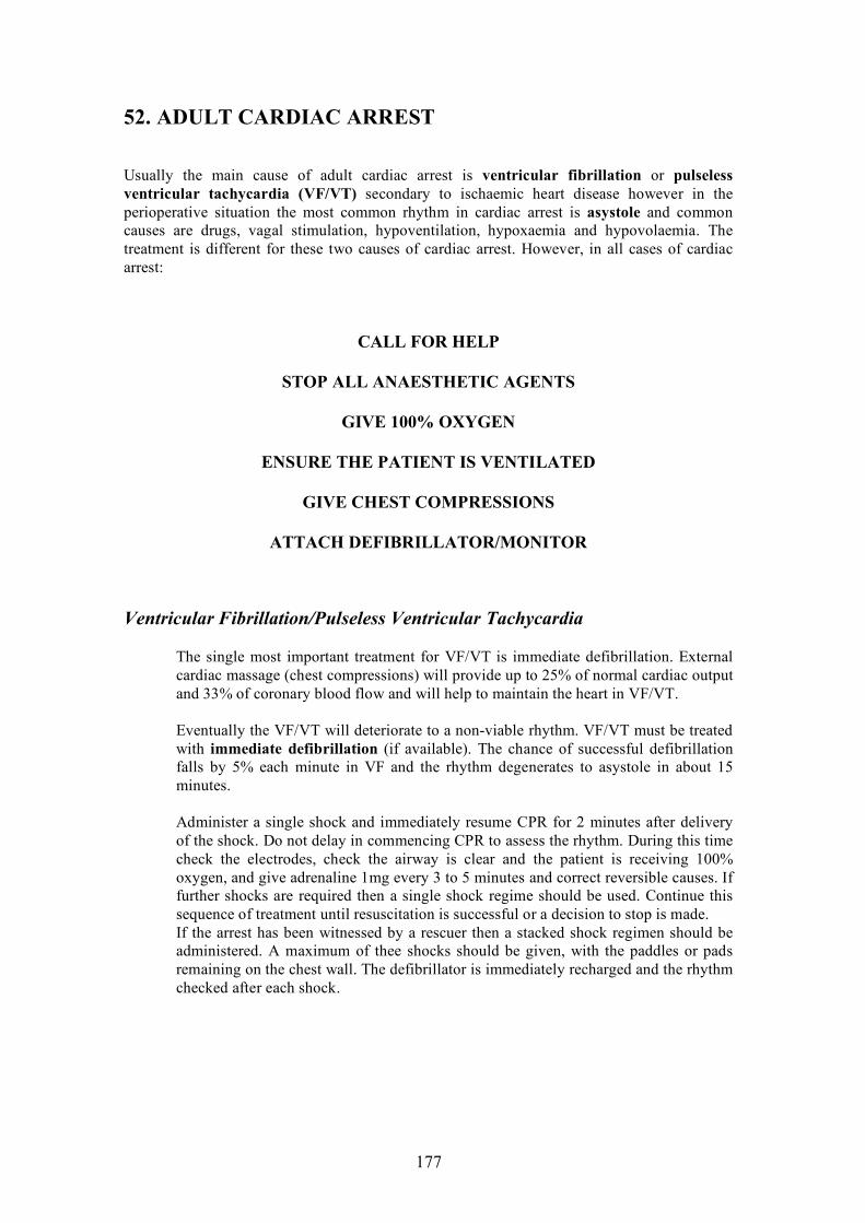
177
52. ADULT CARDIAC ARREST
Usually the main cause of adult cardiac arrest is ventricular fibrillation or pulseless ventricular tachycardia (VF/VT) secondary to ischaemic heart disease however in the perioperative situation the most common rhythm in cardiac arrest is asystole and common causes are drugs, vagal stimulation, hypoventilation, hypoxaemia and hypovolaemia. The treatment is different for these two causes of cardiac arrest. However, in all cases of cardiac arrest:
CALL FOR HELP
STOP ALL ANAESTHETIC AGENTS
GIVE 100% OXYGEN
ENSURE THE PATIENT IS VENTILATED
GIVE CHEST COMPRESSIONS
ATTACH DEFIBRILLATOR/MONITOR
Ventricular Fibrillation/Pulseless Ventricular Tachycardia
The single most important treatment for VF/VT is immediate defibrillation. External cardiac massage (chest compressions) will provide up to 25% of normal cardiac output and 33% of coronary blood flow and will help to maintain the heart in VF/VT. Eventually the VF/VT will deteriorate to a non-viable rhythm. VF/VT must be treated with immediate defibrillation (if available). The chance of successful defibrillation falls by 5% each minute in VF and the rhythm degenerates to asystole in about 15 minutes. Administer a single shock and immediately resume CPR for 2 minutes after delivery of the shock. Do not delay in commencing CPR to assess the rhythm. During this time check the electrodes, check the airway is clear and the patient is receiving 100% oxygen, and give adrenaline 1mg every 3 to 5 minutes and correct reversible causes. If further shocks are required then a single shock regime should be used. Continue this sequence of treatment until resuscitation is successful or a decision to stop is made. If the arrest has been witnessed by a rescuer then a stacked shock regimen should be administered. A maximum of thee shocks should be given, with the paddles or pads remaining on the chest wall. The defibrillator is immediately recharged and the rhythm checked after each shock.

178
Asystole or Pulseless Electrical Activity (PEA)
If PEA is the initial rhythm there is a chance that there is a treatable cause. The anaesthetist must check that VF/VT is not present, commence CPR, give adrenaline 1mg every 3 minutes and immediately think about the cause of the PEA/Asystole. The most common cause is hypovolaemia. The other causes are: Hypovolaemia Hypoxaemia Hypo or hyperkalaemia, hypermagnesaemia, hypercalcaemia Hypothermia, hyperthermia Tension pneumothorax Tamponade (trauma, renal failure, malignancy) Thomboembolic, pulmonary embolus Toxicity (anaphylaxis, drug overdose)

179
ADULT CARDIORESPIRATORY ARREST Apply defibrillator pads/ecg
What is the rhythm? VF or VT Asystole or EMD
Defibrillate Single shock Biphasic 200J Monophasic 360J
Immediate CPR Intubate IV access
Immediate CPR Intubate IV access
Give drugs Check rhythm Defibrillate 360 J x 3
Give drugs Continue CPR
Recheck rhythm Correct reversible cause Continue CPR
Recheck rhythm Correct reversible cause Continue CPR
Reversible causes Hypoxia Hypovolaemia Hyperinflation Hypo/hyperkalaemia Tension pneumothorax Tamponade Toxins/drug overdose/anaphylaxis Thomboembolism
Drugs for VT/VF 1. Adrenaline 1mg. Repeat every 3 minutes Give drug IV Flush with 20 ml Continue CPR 30 sec Check rhythm
Indications for Potassium
1. Hypokalaemia Calcium
1. Hypocalcaemia 2. Hyperkalaemia 3. Massive transfusion 4. Ca antagonist overdose
Bicarbonate
1. TCA overdose 2. Preexisting acidosis 3. Hyperkalaemia
Magnesium 1. Polymorphic VT
Drugs for asystole & EMD 1. Adrenaline 1 mg Repeat every 3 minutes 2. Atropine 2 mg to 3 mg Give drug IV Flush with 20 ml Continue CPR 30 sec Check rhythm

180
53. PAEDIATRIC CARDIAC ARREST
Paediatric arrest usually originates from a primary respiratory event causing hypoxia, hypercarbia and acidosis that result in bradyarrhythmias and eventually asystole. (The majority of adult arrests are due to a primary cardiac event). Primary cardiac arrest in children is rare. The difference in the cause of the arrest is very important. As most paediatric arrests are due to a respiratory event, there is a longer pre arrest phase allowing time for recognition and treatment of the primary event before death. In paediatric life suppor, recognition and aggressive treatment of primary pulmonary events will improve survival.
Management
The immediate response to a paediatric arrest must be to clear the airway and ventilate the patient. Causes of paediatric arrest include sudden infant death syndrome, trauma, near-drowning, upper airway obstruction, respiratory infections, congenital diseases and severe sepsis.
Airway
A clear airway is essential. The first action of the anaesthetist must be to clear the airway. A simple head tilt and lifting the jaw forward will prevent obstruction by the tongue. The anaesthetist must check for a foreign body obstructing the airway. Foreign bodies should be removed carefully under direct vision.
Breathing
A clear airway and adequate ventilation are the first priorities of paediatric resuscitation. If the child is not breathing the anaesthetist must ventilate the child before performing any other procedure. Breathing can be assessed by looking for chest movement, feeling for air movement and listening for breath sounds. If there is no chest movement then there is no ventilation. Always check that the airway is clear. The tidal volume should be enough to raise the child’s chest. Excessive tidal volumes can cause gastric distension and regurgitation of gastric content. Expired air resuscitation will deliver 16% oxygen. Ventilation with high-inspired oxygen concentrations should be established as soon as possible. A self-inflating resuscitation bag with an oxygen reservoir can provide up to 90% oxygen concentration. (Self-inflating resuscitation bags can function without an oxygen source unlike an anaesthetic breathing system). Endotracheal intubation should be considered as an “elective” procedure. (Endotracheal size equals age/4 plus 4) The anaesthetist should only have a couple of attempts of less than 30 seconds. Self-inflating bag and mask ventilation is usually adequate.

181
Circulation
Drugs can be given via intravenous, intraosseous routes or via the endotracheal tube. Drugs given by a peripheral route should be followed by a fluid flush to speed the onset of action. Several drugs can be given by the tracheal method, if no other method is available, including lignocaine, atropine, naloxone and adrenaline. There is little research as to their efficiency. The speed of onset is the same for peripheral intravenous and intraosseous administered drugs. All drugs, including blood, can be given by the intrasseous route. The anaesthetist can use a special short bevel needle, 18 gauge intravenous needle or spinal needle. The needle is inserted 1 to 2 cm below and medial to the tibial tuberosity on the medial surface of the tibia. It should be inserted at 75 degrees, directed towards the feet to avoid the epiphyseal plate. The anaesthetist feels for a loss of resistance as the needle enters the medullary cavity. When the needle is in the marrow cavity it should remain upright without support, bone marrow can be aspirated and there is free flow of drugs and fluids.
Paediatric Cardiorespiratory arrest.
Commence basic CPR with a compression to ventilation ratio of 30:2. Attach defibrillator/ECG monitor and assess the rhythm. Shockable VT/ pulseless VT should receive on DC shock of 2 J/kg followed by immediate CPR for 2 minutes and reassess Further single shocks should be delivered at 4 J/kg. During CPR, check electrodes, attempt intravenous access, correct reversible causes, consider intubation, vasopressor (adrenaline 10 µg/kg every 3 minutes), antiarrhythmics (amiodarone 5 mg/kg, lignocaine 1 mg/kg, magnesium 0.1-0.2 mmol/kg for torsades de pointes) and atropine 20 µg/kg for bradycardia. For a witnessed arrest, give up to 3 stacked shocks (2,4,4 j/kg) at the first defibrillation attempt. Non shockable PEA/asystole requires adrenaline 10 µg/kg and CPR plus the same assessment and management during CPR as for shockable rhythms.

182
54. HYPOXAEMIA (hypoxia)
Hypoxaemia is an oxygen saturation less than 90% or arterial oxygen of less than 60 mmHg, or a fall in oxygen saturation of more than 5%. Anaesthesia will reduce the body’s cardiac and respiratory response to hypoxaemia, making the clinical detection of hypoxaemia even more difficult. Only the late signs of hypoxaemia may occur including: bradycardia, tachycardia, arrhythmias, hypotension and cardiac arrest.
Cyanosis
Patient oxygenation (SaO2) is usually measured with an oxygen saturation monitor (pulse oximeter). Clinically, hypoxaemia is detected by looking for cyanosis. This can be difficult to detect and is much less accurate than an oxygen saturation monitor. Cyanosis can be detectable if there is more than 5 g/100ml of reduced haemoglobin (SaO2 of only less than 85% or arterial oxygen of 40 to 45 mmHg). Peripheral cyanosis (finger tips) can occur from just poor peripheral blood flow without true hypoxaemia. Central cyanosis (tongue) is a true sign of hypoxaemia. Hypoxaemia may rapidly cause death and is an emergency. The anaesthetist should use a saturation monitor at all times.
Causes of Hypoxia
There are several causes of hypoxaemia including: Decreased inspired oxygen due to empty oxygen supply, low oxygen flow rate, breathing system disconnected or broken, obstructed breathing system or airway or endotracheal tube in the wrong place (oesophagus/endobronchial). Decreased ventilation due to decreased conscious state, high spinal, increased airway resistance (asthma, upper airway obstruction, endobronchial intubation), wrong ventilator settings (low tidal volume, low respiratory rate) and pneumothorax. Lung disease Decreased cardiac output Cardiac shunt Increased oxygen requirement due to severe infection, thyrotoxicosis

183
Prevention
Always check the anaesthetic machine, oxygen supply and oxygen failure alarms before giving any anaesthetic or sedation. Always have a spare supply of oxygen and an alternative method to ventilate a patient (e.g. self inflating resuscitation bag). Always use an oxygen saturation monitor if available. Always monitor the patient for signs of cyanosis.
Management
Increase the inspired oxygen concentration. Check the oxygen pressure gauges and flow meters. Check that ventilation is adequate. (Check end-tidal CO2 if available). If hypoxia occurs soon after endotracheal intubation immediately check that the endotracheal tube is in the trachea and not the oesophagus. Hand ventilate the patient, with large tidal volumes, to assess lung compliance and exclude leaks or obstructions in the breathing system. Check the endotracheal tube is not blocked or dislodged. Listen to the chest for equal air entry and bronchospasm. Exclude a pneumothorax. Check for low cardiac output. Check the blood pressure and heart rate. Consider lung problems: aspiration, pulmonary oedema, consolidation, atelectasis and pulmonary embolism.

184
55. HIGH AIRWAY PRESSURE
An increase in the pressure needed to ventilate a patient is not uncommon but inspiratory pressures greater than 40 cmH2O must be treated as abnormal. High airway pressure can present in several different ways including problems with ventilating the patient (poor chest expansion, decreased breath sounds, decrease tidal volume), hypoxia, hypercarbia and tachycardia.
Causes of High Airway Pressure
Ventilation may be difficult because of a problem with one of three sites:
• anaesthetic equipment (ventilator, anaesthetic breathing system) • airway device (endotracheal tube, laryngeal mask, face mask) • the patient.
The anaesthetist should immediately look for obvious causes. Airway pressure may be high immediately after intubation, when neuromuscular blockade has decreased and if the airway is kinked.
Management
If there is no obvious cause, the anaesthetist should have a systematic approach to the diagnosis of high airway pressure: Gas supply: Check the oxygen supply, increase the concentration of oxygen. Breathing circuit: Check the common gas outlet and hose connections. If available, hand-ventilate the patient with a self-inflating resuscitation bag. If the problem is with the breathing circuit or anaesthetic machine then ventilation should be easy with the resuscitation bag. If the problem is with the airway or the patient, then ventilation will continue to be difficult. Airway: Check the airway. Make sure that it is not kinked or obstructed. Pass a suction catheter down the airway and apply suction to clear any secretions. If the catheter passes easily then the airway is unlikely to be obstructed. If the endotracheal tube is obstructed, deflate the cuff and try to pass the suction catheter again. A herniation of the endotracheal tube cuff can cause obstruction of the tube. If the suction catheter still will not pass freely, remove the endotracheal tube, mask ventilate and reintubate. Lungs: Look for bilateral chest expansion and listen to both sides of the chest. If breath sounds are only on one side consider endobronchial intubation (withdraw the endotracheal tube 2 cm and reassess) or pneumothorax (check the heart rate and blood pressure, feel to see if the trachea is central and percuss the chest). If wheezes are present, consider bronchospasm, aspiration or pulmonary oedema.
The surgical procedure or the position of the patient may also make ventilation difficult.

185
Pneumothorax
A pneumothorax may occur for many reasons including insertion of intercostal nerve blocks or placing a central venous catheter. It can happen spontaneously or because of chest trauma or high ventilation pressure during general anaesthesia. The awake patient may complain of dyspnoea, chest pain, and be tachypnoeic and hypoxic. In the anaesthetised patient, it can be very difficult to diagnose a pneumothorax. The patient may be hypoxic and have raised inspiratory airway pressures. A large pneumothorax or a tension pneumothorax will cause hypotension, tachycardia and may cause death. On examination the patient may have reduced or absent breath sounds on one side, increased resonance to percussion, tracheal deviation or subcutaneous emphysema. The anaesthetist must always consider a pneumothorax in their diagnosis, especially if the patient is at increased risk (central venous catheter inserted, chest trauma, asthma, high airway pressure). A pneumothorax may be present with signs and symptoms similar to several other problems, including aspiration of gastric contents, endobronchial intubation, a blocked endotracheal tube and bronchospasm. An erect chest x-ray will help with the diagnosis (a pneumothorax can be very difficult to see on a supine chest x-ray).
Management
Always ensure that the patient is well oxygenated and ventilating. Turn off the nitrous oxide and give 100% oxygen (70% nitrous oxide will rapidly increase the size of a pneumothorax by 100% in 10 minutes). Check the blood pressure and pulse rate. If the blood pressure is low and there is no other cause for a low blood pressure, treat the patient as if they have a tension pneumothorax. A tension pneumothorax can rapidly cause death and must be treated as an emergency. Inform the surgeon and call for help. Treat the low blood pressure with intravenous fluids and vasopressor drugs. Insert a large intravenous catheter into the pleural space to aspirate the pneumothorax. The intravenous catheter should be placed in the second intercostal space above the rib in line with the middle of the clavicle, to avoid damaging the intercostal nerves and blood vessels. A chest tube must be inserted following insertion of an intravenous catheter.

186
Bronchospasm
Bronchospasm will cause wheezing (greater on expiration) and increased airway pressures. There are several causes of intra-operative bronchospasm including:
• Mechanical irritation of the airway (secretions, intubation, oropharyngeal airway, suctioning).
• Chemical irritation of the airway (some inhalational anaesthetics, smoke).
• Drugs (histamine release from drugs like morphine or atracurium, beta adrenergic blocking agents).
• Aspiration of gastric contents.
Anaesthesia should be delayed for elective surgery if the patient has an upper respiratory tract infection or an acute episode of asthma.
Management
Ensure the patient is oxygenated and ventilating. Check the blood pressure and pulse rate.
Increase the oxygen concentration if the patient is hypoxic.
Check for other causes of increased airway pressure such as endobronchial intubation, blocked endotracheal tube, pulmonary oedema, aspiration of gastric contents, pneumothorax or anaphylaxis.
Remove any airway irritant.
Increase the depth of anaesthesia. Halothane and other inhalation agents are good bronchodilators.
Give beta-adrenergic agents, anticholinergics and steroids.
Salbutamol
Salbutamol (Ventolin) may be given by inhalation or injection (intramuscular, subcutaneous or intravenously). Side-effects include tachycardia, tremor, nausea, headache and lactic acidosis.
Inhalation: continuous nebulisation (5 mg/2.5 ml) or 8 puffs (100 micrograms/puff).
Intravenous bolus (500 micrograms/ml): 200 to 300 micrograms over 10 minutes. Repeat after 15 minutes if needed.
Intravenous infusion add 5mg (5 ml of 5 mg/ml) to 45 ml of normal saline (equals 100 micrograms/ml). Start at 5 micrograms/min (3 ml/h) and increase by 1 microgram/min up to a maximum of 10 microgram/min.
Ipratropium
Ipratropium 1ml nebulised every 2 to 4 hours. Dexamethasone
Dexamethasone 4mg intravenously every 8 hours.

187
56. LARYNGOSPASM
Laryngospasm is the reflex closure of the vocal cords. It is caused by irritation of the airway (e.g. secretions, blood, vomit and laryngoscopy) or in response to other stimulation (e.g. peripheral pain) during light anaesthesia. Mild laryngospasm is incomplete closure of the vocal cords and the patient will have stridor. Severe laryngospasm is the complete closure of the vocal cords and there may be no airway noise because the patient’s airway is completely obstructed. Laryngospasm will cause hypoxia, hypercarbia, acidosis and initially tachycardia and hypertension. If not treated the patient will become hypotensive, bradycardic and develop ventricular arrhythmias leading to cardiac arrest.
Prevention
To prevent laryngospasm, the anaesthetist should ensure an adequate depth of anaesthesia before laryngoscopy, extubate patients either fully awake or in a deeper plane of anaesthesia and should clear the airway with gentle suctioning before extubation.
Management
The anaesthetist must remain with the patient in the recovery room until they are certain that the patient has a clear airway. If laryngospasm occurs, remove the stimulus. If the patient is still anaesthetised, deepen the anaesthetic. Give 100% oxygen. Tilt the head back and pull the jaw forwards. If the patient does not improve give continuous positive airway pressure (CPAP) by applying a facemask tightly and tightening the expiratory valve of the breathing system. This will often relieve the laryngospasm. If the laryngospasm does not improve give suxamethonium (0.1 to 0.2 mg/kg intravenously or 0.2 to 0.6 mg/kg intramuscularly) and continue to mask ventilate the patient with 100% oxygen. Intubate if necessary.

188
57. ANAPHYLAXIS
Anaphylaxis is a severe life theatening allergic reaction. It is initiated by antigen binding to immunoglobin E (IgE) antibodies. (Anaphylactoid reactions are clinically similar but are not initiated by IgE). Common drugs that can cause anaphylaxis include some antibiotics, protamine, some neuromuscular blocking agents and blood products. The anaphylaxis usually occurs immediately but may be delayed for 2 to 15 minutes.
Assessment
The anaesthetised patient is often covered with drapes and unconscious so some of the signs and symptoms of anaphylaxis may not occur. Approximately:
• 75% Hypotension (due to peripheral vasodilation and increased capillary permeability), tachycardia, hypotensive shock. (Hypotension may be the only sign of anaphylaxis in the anaesthetised patient).
• 70% Urticaria, flushing, rash • 50% Bronchospasm, stridor, dyspnoea, airway oedema, respiratory
arrest
Prevention
The anaesthetist must ask the patient about allergies during the preoperative visit. Drugs known to cause anaphylaxis (e.g. antibiotics) should be given as a “test dose” first when appropriate. The drug should be diluted to 20 ml and then 2 ml given as a test. The anaesthetist should wait several minutes and then if there is no signs of anaphylaxis give the remainder of the drug. As anaphylaxis and a relative overdose of induction agents will both cause hypotension, it is wise not to give drugs that can cause anaphylaxis (e.g. antibiotics) at the same time as induction agents.
Management
Stop giving the drug that may have caused the anaphylaxis. Call for help, inform the surgeon. Ensure the patient is oxygenated and ventilating. Give 100% oxygen. Intubate the patient if necessary. Remember that anaphylaxis will cause airway oedema, and that the patient’s breathing and intubation may become much more difficult with time. If concerned about the patient’s airway, intubate early. Treat the hypovolaemia with intravenous fluids. The patient may need many litres of intravenous fluids.

189
Mild anaphylaxis may only need 0.5 to 1.0 mg of intramuscular adrenaline (epinephine). Severe anaphylaxis will need repeated intravenous doses of 100 ug of adrenaline to treat the hypotensive shock. Once the blood pressure is treated the patient may need an adrenaline infusion for 24 to 48 hours. Severe anaphylaxis will cause cardiorespiratory arrest. Be prepared to start cardiopulmonary resuscitation if the patient arrests. Histamine-1 blockers (diphenhydramine intravenously 50 mg) and corticosteroids (dexamethasone intravenously 20 mg, methylprednisolone intravenously 100 mg) may be useful. Ideally, blood testing for tryptase should be done at the time of the anaphylaxis and the patient should return in 6 weeks time for skin testing for allergy.

190
58. MALIGNANT HYPERTHERMIA (Malignant Hyperpyrexia)
Malignant hyperthermia, was first described in Australia in 1960. It is a life theatening inherited (autosomal dominant) disorder of skeletal muscle. The incidence is 1:5,000 to 1:200,000. Exposure to triggering agents (suxamethonium and volatile anaesthetics) causes a reduction in calcium reuptake by the sarcoplasmic reticulum, which causes sustained muscle contraction. Malignant hyperthermia usually occurs in the operating room but can occur up to 11 hours postoperatively. It can occur in patients who have had normal anaesthetics in the past.
Assessment
Symptoms of malignant hyperthermia may include: Sustained muscle contraction. Masseter spasm may be an early sign of malignant hyperthermia but not all patients with masseter spasm will have malignant hyperthermia. Muscle breakdown causing hyperkalaemia and myoglobinaemia. Hyperkalaemia above 6.5 mmol/l will cause ECG changes including tall peaked T waves, prolonged PR interval, loss of P waves and widening of the QRS complexes. Hyperkalaemia will cause ventricular arrhythmias including ventricular fibrillation and cardiac arrest. Increased metabolism causing increased oxygen consumption and carbon dioxide production. Spontaneously breathing patients will hyperventilate. Increased temperature of up to 1°C every 5 minutes. The patient’s temperature may reach 45°C. The rise in temperature is often a late sign of malignant hyperthermia. Tachycardia Metabolic acidosis. Complications of malignant hyperthermia include disseminated intravascular coagulopathy, acute tubular necrosis and death. Other conditions that cause unexplained tachycardia, increased metabolism or increased temperature may appear like malignant hyperthermia. These include light anaesthesia, infection, hyperthyroidism, phaeochomocytoma and drug reactions (neuroleptic malignant syndrome, cocaine overdose).
Prevention
The anaesthetist must always take a careful family anaesthetic history. As malignant hyperthermia is an inherited disorder, other members of the patient’s family may have had an unexplained death during anaesthesia. If at all suspicious avoid the triggering agents suxamethonium and volatile anaesthetic agents. For patients known to have malignant hyperthermia the anaesthetist must avoid the triggering agents. The anaesthetic machine should have the vaporiser removed, the anaesthetic tubing and bag and new soda lime should be new. The anaesthetic machine should be flushed with 100% oxygen (10 L/min) for at least 10 minutes. Patients suspected of having malignant hyperthermia can be diagnosed by a muscle biopsy test.

191
Management
Malignant hyperthermia is a life theatening disorder that can develop rapidly and requires multiple treatment tasks. Dantrolene is the only drug that can reverse the effect of malignant hyperthermia. Call for help. Stop the volatile anaesthetics (anaesthesia can be maintained by intravenous propofol and opioid). Hyperventilate the patient with 100% oxygen. Check the ECG for signs of hyperkalaemia (hyperkalaemia can be treated with an intravenous infusion of 100 ml of 10% dextrose with 10 units of insulin over 30 minutes or sodium bicarbonate 50 to 150 mEq. Calcium gluconate 10mls of 10% may be given for severe hyperkalaemia). Check the temperature. If the temperature rises above 38°C try to cool the patient (surface cooling with cool water or ice, cold intravenous fluids, gastric lavage with cold water). Check the urine output. Patients are at risk of acute tubular necrosis. Try to keep the urine output greater than 2 ml/kg/h with intravenous fluids and frusemide. Be prepared to treat ventricular arrhythmias (procainamide 3 mg/kg or lignocaine). Dantrolene should be administered as soon as malignant hyperthermia is suspected. Unfortunately the drug is expensive and has a short shelf life. The dantrolene dose is 2.5 mg/kg repeated every 10 minutes if needed up to 10 mg/kg.

192
59. HYPERNATRAEMIA
Hypernatraemia is a serum sodium greater than 145 mmol/l.
Causes
Common causes include dehydration states (e.g. diarrhoea and vomiting), hyperosmolar states (such as hyperglycaemia, diabetic ketoacidosis or uraemia) and diabetes insipidus (e.g. from meningitis, cerebral TB or renal failure).
Clinical Signs and Symptoms The clinical signs and symptoms will depend on the severity of the hypernatraemia and the speed of onset. Hypernatraemia usually produces symptoms if the serum sodium exceeds 160 mmol/l. Patients may be weak, irritable, drowsy or confused. The may have fevers, seizures or be in a coma.
Management
Elective surgery should be delayed for patients who are symptomatic or have a serum sodium greater than 150 mmol/l. They should be investigated and treated. Hypernatraemia may be hypervolaemic or hypovolaemic. Hypervolaemic hypernatraemia is due to sodium excess. Causes include treatment with hypertonic saline or sodium bicarbonate and mineralocorticoid excess (e.g. aldosteronism, Cushings syndrome). The kidneys should be allowed to slowly excrete the excess sodium. Fluid should be given as hypotonic solutions (e.g. 5%dextrose or oral fluids). Diuretics will also increase sodium excretion. Dialysis is rarely required. Hypovolaemic hypernatraemia is more common and is due to water loss exceeding sodium loss (e.g. diarrhoea, vomiting, sweating, osmotic diuretics) or inadequate water intake (e.g. unconsciousness) Diabetes insipidus causes the kidneys to excrete excess amounts of water. The hypovolaemia should be rapidly treated before slowly treating the hypernatraemia. Once the blood pressure and pulse are normal the free water deficit should be replaced with hyponatraemic solutions (0.45% N/saline, 4% dextrose & 1/5 N/saline, 5% dextrose). The serum sodium must be checked frequently. It must be reduced slowly (less than 2 mmol/l/h). Rapid reduction of serum sodium can cause cerebral oedema, seizures and death. The water deficit can be calculated. Total body water = 0.6 x body weight (kg). Current body water = normal sodium divided by current sodium x total body water Water deficit = normal total body water minus current body water or. calculated water deficit = body weight (kg) x 0.6 x ([Na – 140]/140)

193
60. HYPONATRAEMIA
Hyponatraemia is a serum sodium less than 135 mmol/l.
Causes
There are many causes of hyponatraemia. Excessive intake of water (e.g. excessive drinking, intravenous administration of sodium poor fluids and absorption of irrigation solutions) and reduced excretion of water (syndrome of inappropriate antidiuretic hormone secretion) will cause hyponatraemia. Other causes include cardiac, renal and liver failure, diuretic treatment, oxytocin, diarrhoea and vomiting and pancreatitis.
Clinical Signs and Symptoms
The patient’s symptoms will depend on the severity and speed of onset of the hyponatraemia. Usually patients do not have symptoms until the serum sodium is less than 125 mmol/l. Moderate or gradual onset hyponatraemia can cause confusion, headache, muscle cramps, anorexia and nausea. Severe or rapid hyponatraemia may cause seizures and coma.
Management
The urgency of treatment is determined by the conscious state of the patient. Unless there are neurological symptoms, treatment should only be to stop any intravenous 5% dextrose and restrict oral fluid intake. The majority of hyponatreamic patients will improve with conservative treatment. Rapid correction of hyponatraemia can cause cerebral demyelination. In severe hyponatraemia, where serum sodium is less than 115 mmol/l and the conscious state is affected, treatment is an emergency and the patient should be in an intensive care ward. There should be restriction of water and administration of concentrated sodium chloride. The aim is to rapidly raise (50 mmol/h) the serum sodium by 5 mmol/l and then to very slowly raise the serum sodium to 120 to 125 mmol/l over 48 hours. Correction of the hyponatraemia must be slow. Only patients with severe symptomatic hyponatraemia should have an initial rapid small rise in sodium concentration. Total sodium deficit assuming distribution thoughout the total body water = (125 minus measured sodium) x 60% of body weight.

194
61. HYPERKALAEMIA
Hyperkalaemia is a serum potassium (K) greater than 5.5 mmol/l.
Causes
It can occur from increased intake (oral or intravenous), inadequate excretion (including renal failure, adrenal insufficiency, potassium sparing diuretics) or from a shift of potassium from the tissues to the plasma (such as extensive tissue trauma, respiratory and metabolic acidosis or malignant hyperthermia). Acidosis increases K 0.5 mmol/l for every 0.1 increase in pH.
Clinical Signs and Symptoms
Patients may have no symptoms of hyperkalaemia. Clinical effects are seen with a rapid rise or a high level (greater than 6.5 mmol/l). If serum K rises very rapidly the first clinical effect may be ventricular fibrillation or asystole. ECG changes usually do not occur until serum K is greater than 6.5 mmol/l. There is first, prolongation of the PR interval, followed by peaked T waves, widened QRS, decreased R wave height, loss of ST segment and loss of P wave. Patients can develop bradycardia, complete heart block, ventricular fibrillation or asystole. Severe hyperkalaemia can cause peripheral weakness.
Management
Mild hyperkalaemia (serum potassium less than 6.0 mmol/l) The surgery for elective patients should be delayed and the patient should be investigated for a cause of the hyperkalaemia. They should be given cation exchange resins (e.g. resonium) by rectal or oral routes. Emergency patients should be given cation exchange resins. The anaesthetist should be careful with giving suxamethonium, which can cause a further rise in serum potassium. Stop any potassium containing fluids. The patient’s potassium should be monitored (blood analysis and ECG). Moderate to severe hyperkalaemia (serum potassium greater than 6.0 mmol/l) Moderate to severe hyperkalaemia must be corrected before surgery. Check for ECG changes or signs of peripheral weakness. If present give 5 to 10 ml of 10% Calcium chloride. Check for acidosis (arterial blood gas). If present give 50 mmol of 8.4% sodium bicarbonate.

195
Give 50 ml of 50% dextrose (or 25 grams of dextrose) plus 10 units of insulin. Then give an infusion of insulin at 5 units per hour plus 10% dextrose at 100ml/hour. (Adjust the 10% dextrose rate to maintain a blood glucose of 5 to 8 mmol/l). The serum potassium and blood glucose must be measured frequently. If an infusion pump is not available continue to give 50 ml of 50% dextrose with 10 units of insulin each hour until the serum potassium has returned to normal. Check for renal failure. If the renal function is normal then increase the urine output by giving 20 mg of frusemide plus intravenous normal saline. If renal failure is present start resoniumA 15 mg oral or 30 mg PR.
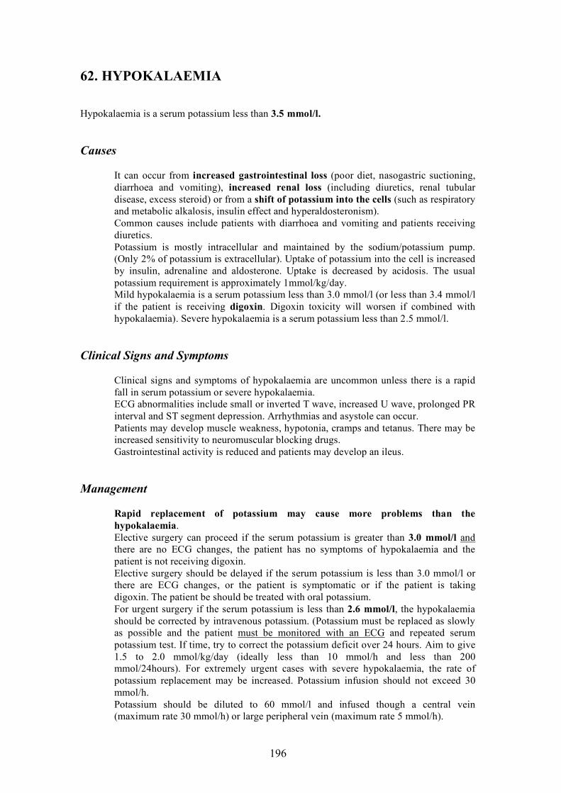
196
62. HYPOKALAEMIA
Hypokalaemia is a serum potassium less than 3.5 mmol/l.
Causes
It can occur from increased gastrointestinal loss (poor diet, nasogastric suctioning, diarrhoea and vomiting), increased renal loss (including diuretics, renal tubular disease, excess steroid) or from a shift of potassium into the cells (such as respiratory and metabolic alkalosis, insulin effect and hyperaldosteronism). Common causes include patients with diarrhoea and vomiting and patients receiving diuretics. Potassium is mostly intracellular and maintained by the sodium/potassium pump. (Only 2% of potassium is extracellular). Uptake of potassium into the cell is increased by insulin, adrenaline and aldosterone. Uptake is decreased by acidosis. The usual potassium requirement is approximately 1mmol/kg/day. Mild hypokalaemia is a serum potassium less than 3.0 mmol/l (or less than 3.4 mmol/l if the patient is receiving digoxin. Digoxin toxicity will worsen if combined with hypokalaemia). Severe hypokalaemia is a serum potassium less than 2.5 mmol/l.
Clinical Signs and Symptoms
Clinical signs and symptoms of hypokalaemia are uncommon unless there is a rapid fall in serum potassium or severe hypokalaemia. ECG abnormalities include small or inverted T wave, increased U wave, prolonged PR interval and ST segment depression. Arrhythmias and asystole can occur. Patients may develop muscle weakness, hypotonia, cramps and tetanus. There may be increased sensitivity to neuromuscular blocking drugs. Gastrointestinal activity is reduced and patients may develop an ileus.
Management
Rapid replacement of potassium may cause more problems than the hypokalaemia. Elective surgery can proceed if the serum potassium is greater than 3.0 mmol/l and there are no ECG changes, the patient has no symptoms of hypokalaemia and the patient is not receiving digoxin. Elective surgery should be delayed if the serum potassium is less than 3.0 mmol/l or there are ECG changes, or the patient is symptomatic or if the patient is taking digoxin. The patient be should be treated with oral potassium. For urgent surgery if the serum potassium is less than 2.6 mmol/l, the hypokalaemia should be corrected by intravenous potassium. (Potassium must be replaced as slowly as possible and the patient must be monitored with an ECG and repeated serum potassium test. If time, try to correct the potassium deficit over 24 hours. Aim to give 1.5 to 2.0 mmol/kg/day (ideally less than 10 mmol/h and less than 200 mmol/24hours). For extremely urgent cases with severe hypokalaemia, the rate of potassium replacement may be increased. Potassium infusion should not exceed 30 mmol/h. Potassium should be diluted to 60 mmol/l and infused though a central vein (maximum rate 30 mmol/h) or large peripheral vein (maximum rate 5 mmol/h).

197
63. QUALITY ASSURANCE AND IMPROVEMENT
An anaesthetic department and an individual anaesthetist should try to deliver the best health care with the fewest complications as soon as possible, given the resources available (equipment, personnel, funding). Quality assurance can be defined as an organised process that assesses and evaluates health services to improve quality of care. All departments of anaesthesia and individual anaesthetists should participate in quality assurance. Without a quality assurance programme, anaesthetic related incidents might be thought of as isolated events and not seen to be a recurring problem, and anaesthetic departments and individual anaesthetists cannot be compared to practice guidelines and standards of care. A quality assurance programme should evaluate safety, provider competence, accessibility, efficiency and effectiveness of care.
Standards of Care and Practice Guidelines
These are written documents which have been produced by the anaesthetic college or department. Standards of care should provide the absolute minimum requirements for patient care (e.g. monitoring, preoperative assessment, post- anaesthetic care units, anaesthetic record, checking equipment). These should be documented and readily available. Practice guidelines provide recommendations of management (e.g. conscious sedation, supervision of trainees, anaesthesia away from the operating room, anaesthetic assistants, infection control). These should be valid, reliable, clinically applicable, flexible, clear, scheduled for review and documented.
Auditing
An anaesthetic department and individual anaesthetists need to know what their recent or current standard of care is so that they can identify abnormal events and evaluate future care. Retrospective and prospective audits should be performed on specific areas of anaesthetic care. The audits may be regular events evaluating standards of care or practice guidelines (e.g. chart review, postoperative pain, and preoperative assessment) or occur because of a specific incident (e.g. equipment failure, wrong drug given).
Incident Reporting
All members of the anaesthetic department should be encouraged to report adverse events or events that may have caused an adverse outcome and interesting cases. Anaesthetists work in isolation from each other. An incident may seem very infrequent to an individual anaesthetist but may be occurring frequently in the department due to an error in the system. Up to 80% of anaesthetic incidents are due to human error but they occur within a complex system and can be avoided by changing the system. An incident may also be very rare. Rare incidents are very difficult to manage. If a rare incident is reported and discussed it will prepare other anaesthetists.

198
An anaesthetic department should meet regularly to discuss reported incidents. They must not just document the incident, blame the anaesthetist and say it should never happen again. The incident should be evaluated without blame, the cause identified and action taken to prevent it occurring again.
Sentinel Events
Some anaesthetic events should always be reported by the anaesthetist or others (e.g. death, operation on the wrong patient or body part, haemolytic blood transfusion reaction and cardiac or respiratory arrest). Staff should be educated and a list of these events should be displayed in the operating theatre.
Education
An anaesthetic department should have regular (e.g. weekly) meetings. All members should be encouraged to present interesting cases and reviews of journal articles. Some of these meetings should be dedicated to quality assurance (e.g. once a month). The anaesthetic department must encourage peer review, incident reporting and audits. Departments should document their own standards of care and practice guidelines (or use those of their college). Incident and sentinel event reporting forms should be available. The senior members of a department should lead by example and report all incidents and interesting cases. No one should be blamed. Quality of care can only be improved by evaluating the current quality.

199
INDEX
Acetylcholine 87 Acupuncture 107 Acute coronary syndrome 175 Acute renal failure 29
Adenosine 144, 172 Adrenal insufficiency 28 Airway assessment 17 Airway obstruction 146 Alcuronium 89 Allergy 9, 188 Allodynia 149 Alveolar ventilation 43 Amiodarone 171 Amnesia 86 Anaemia 160 Anaesthetic plan 51 Analgesia 148, 151 Analgesia intra-operative 150 Analgesia paediatric 99, 149 Analgesia postoperative 151 Analgesia preoperative 150 Anaphylaxis 188 Angina 173 Antacid 14 Anticholinesterase 88 Anticonvulsant 86, 139 Anxiolytic 86 Aortic regurgitation 21 Aortic stenosis 21 Apgar 117,119 Appt 13 Arrhythmias 165 ASA classification 10 Aspiration 15, 16, 36, 56, 60, 72, 91, 120 Asthma 26 Asystole 178 Atracurium 89 Atrial fibrillation 21, 144, 171 Atrial flutter 144, 171 Atropine 88 Autonomic neuropathy 30, 34 Axillary nerve 54 Ayres t piece 44, 101
Backache 110 Barbotage 127 Bell laryngoscope 62 Benzodiazepines 86 Beta-blocker 23 Biers block 136 Blood glucose 13 Blood loss estimation 156, 161

200
Blood transfusion 160 Blood transfusion minimise 162 Blood transfusion risks 158, 163 Blood/gas coefficient 76, 80 Blood/gas solubility 76 Bougie 63 Boyles machine 38 Brachial plexus 53 Breathing systems 43 Bronchospasm 186 Bundle branch block 19, 171, 175 Burns 36 BURP 63
Caesarean section 111 Caesarean section epidural anaesthesia 111 Caesarean section general anaesthesia 112 Caesarean section spinal anaesthesia 113 Carbon dioxide absorber 44 Carbon monoxide 27, 79 Cardiac arrest adult 177 Cardiac arrest paediatric 180 Caudal anaesthesia 103 Caudal anaesthesia complications 105 Central sensitisation 149 Childs classification 33 Chonic renal failure 29 Circle system 43 Cisatracurium 90 Citrate toxicity 163 Coagulopathy 161 Codeine 152 Combined spinal epidural analgesia CSE 108 Congestive heart failure 19, 173 COPD 26 Coronary artery bypass 19 Coronary artery disease 19 Creatinine clearance 30 Cricoid pressure 67, 73 Cricothyroidotomy 69 Crisis management 154 Cross match 13 Cyanosis 55, 182 Cyclopropane 80 Cylinders 38 Dantrolene 191 Dead space 43, 101 Defibrillation 169, 177, 179 Depolarising muscle relaxant 88 Dexamethasone 28 Diabetes 34 Dialysis 31 Diazepam 14, 86 Diclofenac 151

201
Diffusion hypoxia 80 Discharge 142 Disseminated intravascular coagulation 121, 163 Drawover 47, 75 Dysrhythmias 144 Ectopic pregnancy 121 Electrocardiogram 13, 30 Emergence delirium 84,146 Emergency 36 EMO 48, 77 Endorphonium 91 Endotracheal tube 60 Endotracheal tube paediatric 93, 101 Enflurane 79 Epidural anaesthesia 108 Epidural anaesthesia complications 109 Epidural haematoma 109 Equipment 38 Equipment paediatric 101 Ether 76 Extubation 68 Fasting 15, 72 Fasting paediatric 98 Fastrach 56, 59 Fazidinum 90 Flow meter 39 Fluid resuscitation 123, 156 Flumazenil 86, 145 Functional capacity 20 Functional residual capacity 73, 113, 145
Gallamine 89 Gastric secretion 15 Gastroparesis 34 Glomerulonephitis 29 Glossopharyngeal nerve 68 Glycosylated haemoglobin 35 Haemorrhage 156 Haemorrhagic shock 158 Halothane 77 Handgrip 88 HbA1C 35 Heidbrink valve 50 HELP 122 Hepatitis 32, 78, 158 Hepatorenal syndrome 32 High airway pressure 184 Histamine antagonist 141 History anaesthetic 9 History family 9 History medical 9 Hydralazine 122

202
Hydrocortisone 28 Hypercholesterolaemia 19 Hyperkalaemia 29, 92,190,194 Hypermagnesaemia 30, 178 Hypernatraemia 192 Hyperphosphataemia 30 Hypertension 22, 143 Hypertension intra-operative 168 Hypnosis 107 Hypocalcaemia 30, 163 Hypokalaemia 196 Hyponatraemia 193 Hypotension 114, 126, 133, 143 Hypotension intra-operative 166 Hypothermia 146, 147, 158, 163 Hypoventilation 145 Hypoxaemia 145, 182 Ibuprofen 97, 152 Immunosuppression 28 Indomethacin 152 Induction 51 Induction inhalational 52 Induction intravenous 52, 81 Inhalational anaesthetics 75 Inhalational induction paediatric 99 INR 13 Inspired concentration 75 Insulin 35, 195 Intravenous induction paediatric 98 Intravenous regional anaesthesia 136 Intubation 17, 60 Intubation awake 67 Intubation failed 66 Intubation nasotracheal 66 Intubation oesophageal 66 Ipratropium 186 Iron overload 164 Ischaemic heart disease 19 Isoflurane 79
Jackson Res 45 Jaw thust 55, 117 Ketamine 81, 84 Ketorolac 152 Kidney disease See renal Labetolol 122, 173 Labour analgesia 106 Laryngeal mask 56 Laryngoscope 61 Laryngoscopy 63 Laryngospasm 187

203
Lithotomy position 54 Liver disease 32 Liver function tests 13 Local anaesthetic toxicity 138 MAC 76 Macintosh laryngoscope 61 Magill breathing system 44 Magnesium sulphate 123 Malignant hyperthermia 75, 78, 92, 190 Mallampati 17 Mapleson systems 43 Mask ventilation 57 Maximal heart rate 20 Mccoy laryngoscope 61, 62 Meconium 116, 117 Medications 9 Methaemoglobinaemia 137, 139 Methoxyflurane 31, 79 Metoclopramide 14, 72, 147 Metocurine 90 Midazolam 14, 84, 86, 97 Miller laryngoscope 61 Minimum alveolar concentration MAC 76 Mitral regurgitation 21 Mitral stenosis 21 Mivacurium 90 Mobitz 170 Morphine 14, 96, 128, 149 Morphine paediatric 99 Morton W.T.G. 76 Motor end plate 87 Muscle relaxants 87 Myocardial infarction 19, 175 Myocardial ischaemia 23, 145 Myocardial ischaemia perioperative 173
Naloxone 118, 145, 152 Naproxen 152 Nasopharyngeal airway 56, 58 Neostigmine 88 Nephotoxin 29 Nerve damage 53 Neuromuscular blockade 87 Nicotine 27 Nitrous oxide 39, 80, 108 Non-depolarising muscle relaxant 87 Non-steroidal anti-inflammatory drugs 151 Oesophageal detector 66 Oesophageal stethoscope 102 OMV 49, 77 Opioids 81, 96, 97, 150 Oropharyngeal airway 56, 58 Orthostatic hypotension 34

204
Oxford inflating bellows 50 Oxygen delivery 160 Oxygen failure alarm 39 Oxygen flush 40 Oxytocic 112 PAC 50 Paediatric anaesthesia 93 Paediatric anatomy 93 Paediatric pharmacology 96 Paediatric physiology 93 Pain management 148 Pain transmission 149 Pancuronium 89 Papaverine 83 Paracetamol 100, 150 Parecoxib 152 Penlon bellows unit 50 Pentothal See thiopentone Pericardial effusion 30 Pericarditis 30 Peripheral neuropathy 31 Peripheral sensitisation 149 Pethidine 14, 99, 108, 147, 150 Phentolamine 83 Phosgene 79 Pipecuronium 90 Pipeline 38 Placenta praevia 120 Placental abruption 121 Plenum 47, 48 Pneumothorax 80, 185 Polio laryngoscope 61 Porphyria 83 Positioning 53, 125 Post anaesthetic care unit PACU 141 Post anaesthetic complications Postoperative analgesia 143 Postoperative nausea vomiting PONV 142 Practice guidelines 197 Prednisolone 28 Pre-eclampsia 122 Pregnancy physiological changes 113 Premedication 14 Premedication paediatric 97 Preoperative assessment 9 Preoperative assessment paediatric 97 Preoperative investigations 13 Preoxygenation 72, 73 Pressure care 53 Pressure relief valve 40, 42 Pre-transfusion check 165 Prilocaine 137 Prone position 53 Propofol 83

205
Proseal 56 Pulseless electrical activity PEA 178 Pyelonephitis 29 Pyridostigmine 88 Quality assurance 197 Radial nerve 53 Rapid sequence induction 72 Re-breathing 44 Recovery 141 Regurgitation 15, 67 Relaxation 107 Renal artery stenosis 29 Renal disease 29 Renal function test 13 Respiratory depression 82, 108, 143 Respiratory disease 25 Respiratory infection 25 Resuscitation newborn 116 Retained placenta 121 Reversal 87, 88, 145 Rocuronium 90 Sacral canal 103 Sacrum 103 Salbutamol 26, 186 Saturated vapour pressure 75 Scoline apnoea 91 Secondary plan 38, 60 Sedation score 152 Seizure 122 Self inflating bag 50 Sentinel events 198 Sevoflurane 79 Shivering 147 Sick sinus syndrome 170 Simms position 104 Sinus bradycardia 144, 170 Sinus tachycardia 144 Smoking 10, 25, 27 Spinal anaesthesia 113, 125 Spinal anaesthesia complications 132 Spinal anaesthesia contraindications 132 Spinal anaesthesia dosage 128 Spinal anaesthesia physiological changes 129 Spinal needle 126 Stairs 20 Standard of care 197 Steroid 28, 72 Stridor 17, 55, 187 Stylet 63 Substance P 149 Suctioning 38, 117 Superior laryngeal nerve 68
206
Supine position 53 Supraorbital nerve 53 Supraventricular tachycardia 144, 170 Suxamethonium 31, 67, 73, 88, 90 Tachyarrhythmias 171 Thermoneutral range 96 Thiopentone 81 Third degree heart block 170 Thombocytopenia 160 Thyroid function test 13 Thyromental distance 17 TIVA 87 Tolazoline 83 Total spinal 104, 132, 134 Tuohy needle 109 Tourniquet 53, 136 Transcutaneous electrical nerve stimulation TENS
107
Transfusion reactions 164 Transtracheal injection 68 Trendelenberg position 53 Trichloroethylene 78 Tuberculosis 26 Tubocurarine 89 Ulnar nerve 53 Umbilical vein catheter 118 Unilateral block 133 Uterine atony 121 Uterine rupture 121 Valvular heart disease 20 Vaporiser 40 Vascular access 159 Vecuronium 89 Verapamil 144, 168, 172 Vertebral column 125 VIC 44 Visual analogue scale VAS 148 VOC 44 Water deficit 192 Wheeze 25 Wolff Parkinson White syndrome 144 Wong-Baker faces scale 149 Xray cervical 18 Xray chest 13
Copyright © 2022 FDOKUMEN