Plexus brachialis anaesthesia: Optimising clinical aspects
122
Plexus brachialis anaesthesia: Optimising clinical aspects Thesis for the degree of Philosophiae Doctor (PhD) Cand. med. Anne Holmberg Institute of clinical medicine, Faculty of medicine University of Oslo Oslo, Norway & Department of Anaesthesiology Division of Emergencies and Critical Care Oslo University Hospital Oslo, Norway 2020
-
Upload
khangminh22 -
Category
Documents
-
view
4 -
download
0
Transcript of Plexus brachialis anaesthesia: Optimising clinical aspects

Plexus brachialis anaesthesia:
Optimising clinical aspects
Thesis for the degree of Philosophiae Doctor (PhD)
Cand. med. Anne Holmberg
Institute of clinical medicine, Faculty of medicine
University of Oslo
Oslo, Norway
&
Department of Anaesthesiology
Division of Emergencies and Critical Care
Oslo University Hospital
Oslo, Norway
2020

© Anne Holmberg, 2021 Series of dissertations submitted to the Faculty of Medicine, University of Oslo ISBN 978-82-8377-814-4 All rights reserved. No part of this publication may be reproduced or transmitted, in any form or by any means, without permission. Cover: Hanne Baadsgaard Utigard. Print production: Reprosentralen, University of Oslo.

3
Table of contents 1. Acknowledgements 6
2. Synopsis 9
3. Norsk vitenskapelig sammendrag (Norwegian summary) 11
4. List of original papers 14
5. Abbreviations 15
6. Introduction 17
7. Background 19
7.1 Brief history 19
7.1.1 History of local anaesthetics 19
7.1.2 History of brachial plexus blocks 20
7.2 Lateral sagittal infraclavicular blocks 21
7.3 Clinical use of lateral sagittal infraclavicular blocks 23
7.4 Local anaesthetics and adjuvants in peripheral nerve blocks 24
7.4.1 Ropivacaine 25
7.4.2 Lidocaine 26
7.4.3 Dexamethasone 26
7.4.4 Adrenaline 28
7.5 Pain and peripheral nerve blocks 28
7.6 Preemptive and preventive analgesia 30
7.7 Volar plate fixations of distal radius fractures 32
7.8 Skin microcirculation of the upper limb 33
7.9 Changes in microcirculation induced by brachial plexus blocks 34
7.10 Microcirculation and pain 35
8. Aims and hypothesis 37
9. Materials and methods 39
9.1 Study designs 39
9.2 Study settings 42
9.3 Study populations 42
9.3.1 Study 1 and 2 42
9.3.2 Study 3 43
9.4 Study prosedures. 43

4
9.4.1 Inclusion 43
9.4.2 Brachial plexus blocks 45
9.4.3 Block assessment 46
9.4.4 Perioperative procedures study 1 and 2 47
9.4.5 Surgical procedure study 1 and 2 48
9.4.6 Postoperative analgesic procedures study 1 and 2 48
9.4.7 Randomisation and blinding 49
9.4.8 Follow up after discharge 50
9.4.9 Data registration and security 51
9.5 Outcomes 51
9.5.1 Study 1 51
9.5.2 Study 2 52
9.5.3 Study 3 53
9.6 Outcome measures 53
9.6.1 Pain 53
9.6.2 Microcirculation 54
9.6.3 Non-‐invasive continuous haemodynamic measurements 56
9.6.4 Other outcomes 56
9.7 Statistical analyses 57
10. Results 58
10.1 Study 1 58
10.2 Study 2 59
10.3 Study 1 and 2 combined 61
10.4 Study 3 62
11. Ethical considerations and approvals 64
12. Methodological considerations 67
12.1 Study design 68
12.2 Study populations 69
12.3 Outcome measures 70
12.3.1 Block duration 70
12.3.2 Pain 71
12.3.3 Microcirculation 75
12.3.4 Non-‐invasive haemodynamic measurements 78
12.4 Statistical analysis 79

5
12.4.1 General considerations 79
12.4.2 Analysis of continuous data 79
12.4.3 Analysis of categorical data 81
12.4.4 Sample size calculations 81
12.4.5 Overall evaluation of the statistical methods 83
13. Discussion of results 86
13.1 General discussion 87
13.1.1 Study 1 87
13.1.2 Study 2 89
13.1.3 Study 3 91
13.1.4 Overall discussion 93
13.2 Main strengths and limitations of the studies 95
13.2.1 Study 1 95
13.2.2 Study 2 96
13.2.3 Study 3 98
13.3 Clinical implications 99
14. Conclusions 102
14.1 Paper 1 (specific research question A) 102
14.2 Paper 2 (specific research question B) 102
14.3 Paper 3 (specific research question C) 102
14.4 Overall conclusions 102
15. Future area of research/interest 104
16. References 106
17. Errata list 117
18. Reprint of papers I-‐III 118


6
1. Acknowledgements
The work presented in this thesis was carried out at the Department of
Anaesthesiology at Oslo University Hospital during the period from 2011 to 2020. I
would like to thank the University of Oslo for the opportunity to participate in their
PhD program, the Department of Anaesthesiology at Oslo University Hospital for time
and financial support, and the South Eastern Norway Regional Health Authority for a
research grant.
Most of all, I would like to thank my three outstanding supervisors for introducing me
to the scientific world, their academic inspiration and for the wonderful opportunity to
write a PhD thesis. You have all patiently given valuable supervision and support
throughout this whole project. Johan Ræder, I feel fortunate to have had you as my
principal supervisor. You are close to a human medical encyclopaedia. You have
given constructive help and feedback on all my manuscripts in a polite manner. Thank
you for your persistent effort, and your ability to solve problems when the going gets
though. Axel Sauter, you must be among the most positive, flexible and hardworking
co-supervisors ever. With your extensive experience in peripheral nerve blocks, you
have provided invaluable support. I am also extremely impressed and grateful for the
endless hours you spend counting capillaries with me when we both would have
preferred to enjoy beautiful summer evenings. Your enthusiasm made me never give
up. Last but not least, many thanks to my co-supervisor Øivind Klaastad. I am proud
to have learned ultrasound guided lateral sagittal infraclavicular block from the very
master. With your accuracy, you helped me optimise my clinical skills performing the
blocks. You were the first one to suggest I should start on a PhD project, and have
helped, encouraged and supported me whenever needed.
This work would not have been possible without Tomas Drægni. With all your
knowledge and experience, you have helped me making questionnaires and designed
all the databases in this project. Patiently you have called patient after patient, even
late at night or during weekends when needed. I really appreciate all the hours you
have spent plotting and controlling all our data.

7
Also, many thanks to all my co-authors, Karin Toska, Ai-Van Thu Ho, Dmitry
Fernand, Torjus Wester, Sondre Hassellund, Fredrik Ottesen, Anders Nordby and
Allan Gulestøl. You have all invested considerable expertise, time and effort to make
this project possible.
I am extremely grateful for all the help from the anaesthetic and post-operative care
nurses at Legevakta. You helped me find potential patients, registered data, and took
responsibility for blinding procedures. You even took care of my newborn baby-girl to
make it possible for me to include patients in the project. You are all amazing. I am
also thankful to all the orthopaedic surgeons and anaesthesiologists who helped during
the studies.
I really appreciate the flexibility and support from my clinical department. Special
thanks to Kristin Sem Thagaard, Anne Bøen and Ingrid Elise Hoff for making it
possible to combine clinical research with part time clinical work, and for time,
facilitation and support needed to finish the project.
I would also like to thank my everyday colleges, for an inspiring work environment
and important professional discussions. Special thanks to Anne Siri Johnsen, Marlin
Comelon, Anne Kristin Hæg and Pia Wikborg for inspiration, valuable advice and
helpful feedback during this project.
Thanks to all the patients and volunteers that took part in my studies. I really
appreciate the time and effort you offered for the studies.
I wish to thank my friends and family. My fantastic neighbour, Astrid Kvale, offering
language support. My mother Ingebjørg and father Rune, who has provided constant
support and love, both during this project and throughout my whole life. My two
sisters, Marte and Hege and their families, for their encouragement, help and joy.
Finally, I want to say thank you to my closest family. To the love of my life, Torgrim,
for always being there. Our two beautiful children Ingebjørg and Erling, for bringing
endless love and happiness into my life. All three of you have offered me inspiration,

8
help, care and lots of positive distractions while writing this thesis. You mean the
world to me.
Oslo, August 2020


9
2. Synopsis
Brachial plexus blocks are used for analgesia, anaesthesia and in special situations to
improve peripheral circulation in the upper extremity. The aim of this thesis is to
evaluate the clinical and physiological benefits of the lateral sagittal infraclavicular
approach, and modify the use in dedicated areas to improve its analgesic and
circulatory benefits and avoid undue harm. We hypothesised that proper modifications
of timing and adjuvants of infraclavicular brachial plexus blocks may have a positive
impact on postoperative clinical outcome and peripheral circulation.
Lateral sagittal infraclavicular blocks are frequently used to provide anaesthesia and
analgesia for volar plate fixations of distal radius fractures. Severe acute postoperative
pain and long-lasting pain are common after this type of surgery. Infraclavicular
blocks are also used to improve circulation after for example replantation surgery after
traumatic amputations and different vascular procedures. Skin microcirculation
consists of two different components, the subpapillary blood flow important for
thermoregulation, and the nutritive blood flow responsible for oxygenation and
nutrition of peripheral cells. The effect of brachial plexus blocks and adrenaline
adjuvant on these two different components is not fully explored.
In study 1, we evaluated the preemptive effect of infraclavicular blocks in patients
with distal radius fractures scheduled for volar plate surgery. We found a small, but
significant, improvement in early postoperative pain with pre-operative (i.e.
preemptive) blocks compared with postoperative blocks. Mean (SD) time to first
rescue analgesic after emergence from general anaesthesia was 544 (217) min after
pre-operative blocks compared with 343 (316) min after postoperative blocks
(p=0.015). Pre-operative blocks resulted in reduced postoperative pain scores, fewer
patients requiring rescue analgesia during the first 4 hours after surgery, and less
analgesic consumption at day seven after surgery. However, a pre-operative block did
not attenuate strong pain during block resolution and did not seem to have an impact
on the high incidence of minor persistent pain.

10
In study 2, we compared the effect of single (oral etoricoxib) and double (oral
etoricoxib and intravenous dexamethasone) anti-inflammatory prophylaxis in patients
with distal radius fractures scheduled for volar plate surgery with brachial plexus
anaesthesia. We found that intravenous dexamethasone improved early postoperative
analgesia. Median (IQR[range]) worst pain score during the first 24 hours, as assessed
by verbal numeric rating scale (0-10), was 4(2-6[0-7]) in the patients receiving both
dexamethasone and etoricoxib, compared with 8(5-8[2-10]) in the patients receiving
etoricoxib only (p<0.001). Adding intravenous dexamethasone to oral etoricoxib and
paracetamol before start of surgery also resulted in increased block duration, shorter
duration of moderate-severe pain and reduced rescue analgesic consumption from 8-24
hours after surgery. Perioperative intravenous dexamethasone may also reduce the
development of chronic pain.
In study 3, we evaluated the effect of lidocaine infraclavicular blocks with or without
adrenaline on peripheral microcirculation using laser Doppler Fluxmetry, capillary
video microscopy and temperature measurements. It was a cross-over study in healthy
volunteers. We found substantially increased subpapillary blood flow 30 minutes after
lidocaine brachial plexus blocks, from median (IQR[range]) 8.5(4.4-13.5[2.9-28.2]) to
162.7(111.0-197.8[9.5-206.7]) arbitrary units with adrenaline (p=0.017), and from
6.9(5.3-28.5[1.8-42.1] to 133.7(16.5-216.7[1.0-445.0] arbitrary units without
adrenaline (p=0.036). Nutritive blood flow (functional capillary density), measured at
the dorsal side of the hand, decreased in the blocked extremity when adrenaline was
used as adjuvant, from median (IQR[range]) 45(36-52[26-59]) to 38(29-41[26-42])
capillaries/mm2 (p=0.028), whereas no significant change occurred without adrenaline.
In conclusion, to improve efficacy for management of acute and possibly also long-
lasting pain after volar plate surgery, brachial plexus block should be performed pre-
incisional rather than postoperatively and be combined with NSAIDs and intravenous
dexamethasone. When used to increase the microcirculation and oxygenation of
peripheral cells, the best approach may be to use a block without adrenaline adjuvant.

11
3. Norsk vitenskapelig sammendrag (Norwegian summary)
Plexus brachialis blokader brukes som smertelindring, bedøvelse og i enkelte
situasjoner for å bedre perifer sirkulasjon i overekstremiteten. I denne avhandlingen
ønsker vi å se nærmere på kliniske og fysiologiske effekter av lateral sagittal
infraklavikulær blokade og justere bruken innen noen områder for å utnytte blokadens
analgetiske potensial og sirkulatoriske effekter bedre. Hovedhypotesen vår er at bedre
tilpasset bruk av infraklavikulær blokade, både med tanke på tidspunktet den settes på
og bruk av tilsetninger, kan forbedre både postoperativt klinisk resultat og perifer
sirkulasjon.
Lateral sagittal infraklavikulær blokade brukes hyppig både til anestesi og som
smertelindring i forbindelse med fiksasjon av distale radiusfrakturer med volar plate.
Mange opplever sterke postoperative smerter etter denne typen operasjoner, og endel
utvikler også kroniske plager. Plexus brachialis blokader brukes også for å øke perifer
sirkulasjon etter blant annet replantasjonskirurgi etter traumatiske amputasjoner og ved
forskjellig vaskulære prosedyrer. Hudens mikrosirkulasjon består av to viktige deler,
den subpapillære sirkulasjonen som er viktig for temperaturreguleringen, og den
nutritive sirkulasjonen som er viktig for ernæring og oksygenering av perifere celler.
Effekten av plexus brachialis blokader og eventuelle tilsetninger (som for eksempel
adrenalin) på den subpapillære og nutritive sirkulasjonen, er ikke fullstendig
undersøkt.
I den første studien inkluderte vi pasienter med distale radiusfrakturer planlagt fiksert
med volar plate for å se om infraklavikulære blokader har en preemptiv effekt av
klinisk betydning. Vi fant en liten, men signifikant, bedring i akutt postoperativ smerte
med preoperativ (dvs preemptiv) infraklavikulær plexus brachialis blokade
sammenliknet med postoperativ blokade. Gjennomsnittlig (SD) tid til første analgetika
ved behov var 544 (217) min etter preoperative blokader sammenliknet med 343 (316)
min etter postoperative blokader (p=0.015). Preoperative blokader førte også til lavere
postoperativ smerte, til at færre pasienter hadde behov for smertestillende de første 4

12
timene etter operasjonen og til at færre pasienter brukte smertestillende en uke etter
operasjonen. Preoperativ blokade hadde ingen effekt på den sterke smerten mange av
pasientene opplevde når blokaden gikk ut. Vi fant heller ingen forskjeller i
forekomsten av mild smerte etter 6 mnd.
I den neste studien sammenlignet vi effekten av dobbel anti-inflammatorisk profylakse
(etorikoksib og deksametason) med enkel anti-inflammatorisk profylakse (etorikoksib)
hos pasienter med distale radiusfrakturer planlagt fiksert med volar plate. Vi fant at
intravenøs deksametason førte til betydelig forlenget effekt av blokaden og mindre
postoperative smerter. Median(IQR[range]) høyeste smertescore de første 24 timene
målt med en verbal numerisk smerteskala (0-10), var 4(2-6[0-7]) hos pasientene som
fikk både etorikoksib og deksametason sammenliknet med 8(5-8[2-10]) hos pasientene
som bare fikk etorikoksib (p<0.001). Både varighet av moderat-sterk smerte og bruk
av analgetika fra 8-24 timer var betydelig redusert etter en enkeltdose intravenøs
deksametason. En perioperativ dose med intravenøs deksametason hadde muligens
også en positiv effekt med tanke på å redusere forekomsten av kroniske smerter.
I den tredje studien så vi på hvordan lidokain blokader med og uten adrenalintilsetning
påvirker den subpapillære og nutritive sirkulasjonen ved hjelp av laser Doppler,
kapillær videomikroskopi og temperaturmålinger. Dette var en eksperimentell studie
med friske frivillige. Vi fant en betydelig økt subpapillær sirkulasjon de første 30
minuttene etter infraklavikulær blokade med lidokain, fra median(IQR[range]) 8.5
(4.4-13.5[2.9-28.2]) til 162.7(111.0-197.8[9.5-206.7]) arbitrære enheter med adrenalin
(p=0.017), og fra 6.9(5.3-28.5[1.8-42.1] til 133.7(16.5-216.7[1.0-445.0] arbitrære
enheter uten adrenalin (p=0.036). Den nutritive sirkulasjonen (funksjonell
kapillærtetthet), målt på håndens dorsalside, sank i blokkert ekstremitet etter blokader
med adrenalin, fra median(IQR[range]) 45(36-52[26-59]) til 38(29-41[26-42])
kapillærer/mm2 (p=0.028), mens vi ikke fant noen forskjell uten adrenalin.
Vi konkluderte med at en preoperativ infraklavikulær plexus brachialis blokade
kombinert med både etorikoksib og deksametason gir best effekt med tanke på å
redusere postoperative smerter etter fiksering av distale radiusfrakturer med volar

13
plate. Når blokaden brukes for å øke mikrosirkulasjonen og oksygeneringen av
perifere celler, kan det være best å benytte en blokade uten adrenalin.
Vi håper resultatene i denne avhandlingen vil være et bidrag til videre forbedring i
klinisk bruk av infraklavikulær plexus brachialis blokade.


14
4. List of original papers
This thesis is based on the following scientific papers:
Paper 1:
Holmberg A, Sauter AR, Klaastad O, Draegni T, Raeder JC: Pre-operative brachial
plexus block compared with an identical block performed at the end of surgery: a
prospective, double-blind, randomised clinical trial. Anaesthesia 2017; 72: 967-977
Paper 2:
Holmberg A, Hassellund SS, Drægni T, Nordby A, Ottesen FS, Gulestøl A, Ræder J:
Analgesic effect of intravenous dexamethasone after volar plate surgery for distal
radius fractures in brachial plexus block anaesthesia. A prospective, double-blind,
randomised clinical trial.
[published online ahead of print, 2020 May 30]. Anaesthesia. 2020;
doi:10.1111/anae.15111
Paper 3:
Holmberg A, Ho AV, Fernand D, Toska K, Wester T, Klaastad Ø, Drægni T, Sauter
AR: Microcirculation and haemodynamics after infraclavicular brachial plexus block
using adrenaline as an adjuvant to lidocaine: a randomised, double-blind, crossover
study in healthy volunteers. Anaesthesia 2019; 74: 1389-96.


15
5. Abbreviations
ASA: American Society of Anesthesiologist
AU: Arbitrary units
AVAs: Arteriovenous anastomosis
CRF: Case Registration Form
CRP: C-reactive protein
FCD: Functional capillary density
HR: Heart rate
Hz: Hertz
IQR: Interquartile range
LDF: Laser Doppler Flow
LSIB: Lateral sagittal infraclavicular nerve block
ms: millisecond
NRS: numerical rating scale
NSAIDs: non-steroidal anti-inflammatory drugs
PONV: Postoperative nausea and vomiting
PRWHE: Patient Rated Wrist and Hand evaluation
SD: standard deviation
US: Ultrasound
USG: Ultrasound guided

16
VAS: Visual analogue scale
VNRS: Verbal numeric rating scale
VRS: verbal rating scales

17
6. Introduction
Brachial plexus blocks have undergone several exciting advances and gained increased
popularity in the last decades (1). The introduction of ultrasound guidance has
improved the success rate, efficacy, ease of performance and safety, resulting in nerve
blocks being used more routinely in anaesthesia, analgesia and to improve peripheral
circulation. Infraclavicular brachial plexus blocks are widely used for analgesia and
anaesthesia of the elbow, forearm and hand and with the intention to improve
peripheral circulation. The overall goal of this thesis is to evaluate the clinical and
physiological benefits of this method further and modify the use in dedicated areas to
improve its benefits, and avoid undue harm. We hypothesised that modifications of
timing and adjuvants in brachial plexus block will improve both postoperative clinical
outcome and peripheral circulation.
Both acute and long-lasting pain is a major problem after volar plate surgery for distal
radius fractures (2). A previous study in this patient group showed superior
postoperative analgesia and less chronic pain in patients who received infraclavicular
brachial plexus block compared with general anaesthesia during surgery (3). Brachial
plexus blocks are therefore used for surgical anaesthesia to enhance patient comfort
and recovery after volar plate surgery for distal radius fractures.
A successful infraclavicular brachial plexus block results in a complete block of
nociceptive nerve impulses from a surgical field of the distal upper arm, the forearm
and the hand. The potential benefits of perioperative regional anaesthesia may extend
beyond acute pain relief. It is unclear if the timing of brachial plexus blocks, before or
after surgery, affects the incidence of acute and long-lasting pain after surgery. In
every day practice, it is an on-going discussion if we should take the time to perform
nerve blocks prior to the operation, or rather wait till after the operation to potentially
gain a few extra hours with an effective nerve block. In study 1 we try to explore this
area of interest.

18
One of the main challenges of brachial plexus blocks in acute pain management is the
abrupt termination of the analgesic effect after single-injection techniques. This is
particularly problematic in ambulatory surgery when the patients are at home when the
nerve block wears off. A prolonged analgesic effect of brachial plexus block after
surgery may reduce both rebound pain and opioid consumption in the postoperative
period (4). This is important in light of the current focus on eliminating unnecessary
perioperative use of opioids. In study 2, we try to evaluate the effect of single
(etoricoxib) and double (etoricoxib and dexamethasone) anti-inflammatory
prophylaxis after volar plate surgery in brachial plexus anaesthesia. We aim to
ascertain if the addition of a single dose of intravenous dexamethasone (to oral
etoricoxib) can reduce the patient`s total pain burden (both acute and long-lasting
pain) and opioid consumption after volar plate surgery.
Sympathetic blocks may play an important role in the treatment of several medical
diseases with alterations of skin microcirculation. In our hospital, which is the national
unit for reconstructive surgery of the upper extremity, we use brachial plexus blocks to
optimise circulation during and after reconstruction surgery due to traumatic
amputations. The effect of brachial plexus blocks and adjuvants on the two different
but important components of peripheral microcirculation, the sub-papillary blood flow
and the nutritive blood flow, is not fully explored. In study 3, we want to investigate
skin microcirculation and haemodynamic changes after brachial plexus blocks, with a
focus on the use of adrenaline as an adjuvant.
With this PhD thesis focusing on different clinical aspects of infraclavicular plexus
block, we hope to add relevant knowledge and understanding and further improve its
clinical use.

19
7. Background
7.1 Brief history
Brachial plexus blocks have been used as an anaesthetic technique for more than a
century. It has gradually developed from open surgical techniques, to percutaneous
techniques based on landmarks, further to the use of nerve stimulator to confirm
needle placement, and finally to ultrasound-guided techniques. While brachial plexus
block previously was associated with a significant failure rate and thus not used
routinely in many clinics, the development of the method and the introduction of
ultrasound guidance has improved the success rate to make the nerve blocks a reliable
and popular form of anaesthesia.
7.1.1 History of local anaesthetics
The foundation of all regional anaesthesia was the isolation of cocaine alkaloid from
cocoa leafs by Albert Niemann (5). The invention was the basis for his PhD thesis
published in 1860. Before that, the properties of cocoa leafs had been utilized for
various purposes for millennia. The Incas in the Andes used cocoa leafs for religious,
social and medical purposes (6). The Italian explorer, Amerigo Vespucci described
local people chewing cocoa leafs when he reached the coast of Venezuela in 1499 (6).
In 1855, the German chemist Friedrich Gaedcke isolated red crystals, “erythroxylum,”
from the cocoa leaf, and reported its ability to anaesthetise the tongue (7). The latter
was also described by Albert Niemann in his PhD thesis (8). A few years later, in
1884, Carl Koller introduced cocaine as the first effective local anaesthetic (9). The
introduction led to revolutionary changes in anaesthesia, even though cocaine was a
toxic substance with an addictive potential and had a rather short duration of the local
anaesthetic action (10, 11).
After the introduction of cocaine, several attempts were made to develop a more ideal
local anaesthetic with fewer side effects. The progress accelerated after Willstätters
determination of the chemical structure of cocaine in 1898 (8). Several agents were

20
developed, including “Stovaine” in 1904, but none of them turned out to be a suitable
local anaesthetic (11). The use of additives to improve the effect of local anaesthetics
was also investigated. Heinrich Braun was the first to report adrenalines ability to
prolong the local anaesthetic effect of cocaine in 1903 (12). The first suitable local
anaesthetic agent was procaine (Novocaine®), patented by Einhorn in 1904 (8). It
quickly became the standard local anaesthesia. However, the anaesthetic effects of
procaine were weak, there were major problems with allergic reactions, and high
concentrations of adrenaline was required (8).
First after World War II, alternatives to procaine became available. Lidocaine
(Xylocaine®) was developed by Nils Löfgren and Bengt Lundquist in 1943 and
released both with and without adrenaline adjuvant in January 1948 (13). With an
efficient and almost non-toxic local anaesthetic drug available, local and regional
anaesthesia rapidly became more popular (8, 13). In the years to follow, the
development of local anaesthetics accelerated. HC Marks and MI Rubin developed
chloroprocaine in 1949. Bupivacaine and mepivacaine were synthesised by Bo Af
Ekenstam and released in 1957 (14). The longer duration of action of bupivacaine
made it possible to conduct long acting blocks. Several other local anaesthetics have
been developed after bupivacaine, including prilocaine by Löfgren and Tegner in 1969
and articaine by Winther in 1972. Ropivacaine was synthesized in 1957 by Ekenstam,
but first introduced into clinical practise in 1996 (14, 15) as a less cardiac toxic
alternative to bupivacaine. Levobupivacaine was introduced in 1999, and is considered
to be less toxic than bupivacaine with a small increase in sensory block duration (16).
Cocaine is the only naturally occurring local anaesthetic substance available today. All
others are synthetically derived.
7.1.2 History of brachial plexus blocks
Shortly after Carl Koller introduced cocaine as the first effective local anaesthetic in
1884, Halsted and Hall began infiltrating cocaine into the brachial plexus to perform
painless operations on the upper limb at the outpatient department of the Roosevelt
Hospital in New York (8, 17). In 1911, both Kulenkampff and Hirschel described

21
different percutaneous techniques of brachial plexus blocks (18). The use of electrical
nerve stimulation was first described by Georg Perthes in 1912 (19). Fifty years later,
Greenblatt and Denson revived the method by introducing a small battery-operated
nerve stimulator (20). This development greatly improved the success rates of brachial
plexus blocks and facilitated for reduced volume and dosage of local anaesthetic
agents (20, 21). In the years to follow, improvements of local anaesthetics and nerve
block equipment, as well as studies on the anatomy, contributed to the development of
more effective nerve blocks. Winnie and Collins studied the anatomy of the brachial
plexus and suggested an accurate percutaneous location for a single injection
subclavian perivascular technique in 1964 (22). In 1989 ultrasound was used for the
first time to visualize the local anaesthetic spread during an axillary block procedure
(23). In the early nineties, more ultrasound-guided nerve block techniques were
performed after Kapral had published the first description of an ultrasonic guidance of
the injection cannula in 1994 (24, 25). Advances in ultrasound technology with small
and mobile ultrasound units with improved image resolution in an affordable price
range made peripheral nerve blocks increasingly popular. The use of ultrasound
guidance led to improved success rates and reduced performance time during block
procedures.
7.2 Lateral sagittal infraclavicular blocks
The brachial plexus is a network of nerves originating from C5-T1. Various
contributions may also come from C4 and T2. The network of nerves begins as spinal
nerve roots, which merges to form the three trunks. The trunks then split to form the
six divisions, and further reorganises into three cords before they give rise to the five
terminal nerve branches (26).

22
Figure 1: Brachial plexus with roots, trunks, divisions and cords and various approaches for brachial plexus
blocks. (Illustration by Jennifer Gentry. Reproduced from Upper extremity regional anesthesia: essentials of
our current understanding, 2008. Neal JM, Gerancher JC, Hebl JR et al. Reg Anesth Pain Med.
2009;34(2):134-70, Copyright 2009 by American Society of Regional Anesthesia and Pain Medicine, with
permission from BMJ Publishing Group Ltd (26)).
The infraclavicular brachial plexus block was first described by Bazy in 1917 (27). In
1973, P. Prithvi Raj described a new infraclavicular approach using the peripheral
nerve stimulator (28), and the technique was further modified by Sims in 1977 (29).
Øivind Klaastad at our Department of Anaesthesiology at the Oslo University Hospital
optimised the infraclavicular block technique further based on magnetic resonance
imaging studies. He described the highly successful clinical method of lateral sagittal
infraclavicular block (LSIB) in 2004 (30). LSIB was introduced as a nerve-stimulator
guided technique, but ultrasound guidance rapidly became more popular.
When using ultrasound in the infraclavicular region, the lateral, medial and posterior
cords may be located as round hyper echoic structures close to the axillary artery.
Local anaesthetic spread reaching all three cords, or surrounding the artery from 3 to
11 o`clock (if all the cords cannot be identified), are considered sufficient for a
successful LSIB (31). The method is easy to perform, precise and has a low risk for

23
adverse events or complications (30). Ultrasound guided LSIB has a success rate of at
least 95% (32, 33).
Image 1: Ultrasound image of the brachial plexus in the infraclavicular region. A cannula can be visualised in
the 7 o`clock position to the axillary artery. Authors own photo.
Peripheral nerve blocks are considered a safe anaesthetic technique with few
complications (34). Persistent postoperative neuropathy due to the nerve block is a
serious complication with an overall incidence of approximately 0.22% (35).
Complications that can be associated to infraclavicular blocks are vessel puncture,
pneumothorax, nerve injury, and local anaesthetic toxicity.
7.3 Clinical use of lateral sagittal infraclavicular blocks
The infraclavicular brachial plexus block is widely used to provide analgesia and
anaesthesia for the elbow, forearm and hand. It is also used with the intention to
improve peripheral circulation after microvascular procedures, reconstruction surgery
after traumatic amputation, in treatment of vasospasm induced by Raynaud disease,
and in treatment of peripheral embolism (36-39).
Evidence suggests that the use of peripheral nerve blocks as sole anaesthesia technique
during a surgical procedure reduces both operating room time and length of hospital

24
stay (40-42). The patient may be awake during the procedure and not subjected to the
potential side effects of general anaesthesia, such as hypotension, cognitive
dysfunction and respiratory impairment. Compared with general anaesthesia, regional
anaesthesia offers the benefits of better pain protection and less need of opioids
postoperatively, with subsequent reduced incidence of nausea or vomiting as well as
more rapid mobilisation (43, 44). This allows for earlier discharge of the patient after
ambulatory surgery (45).
Peripheral nerve blocks are also used with the intention to improve tissue perfusion
after free flap and replantation surgery. Although sparse evidence for improved
clinical outcome, there are studies supporting the use after digital replacements, and
promising reports for more proximal limb replantation (46-48).
7.4 Local anaesthetics and adjuvants in peripheral nerve blocks
Several different local anaesthetics are available. They typically contain a hydrophilic
tertiary amine group linked to a lipophilic ester or amide (49), and are accordingly
classified as either ester or amide local anaesthetics (50). Local anaesthetics in the
amine group are more commonly used, due to a lower risk of allergic reactions and
systemic toxicity (49). The primary mode of local anaesthetic action is by a reversible
inhibition of sodium influx in nerve fibres (51). However, local anaesthetics have a
wide range of effects, as they inhibit sodium, potassium, and calcium ion channels,
alpha-adrenoceptors, and phosphatidylinositol signalling (49). More lipophilic local
anaesthetics are more potent as the molecules are more likely to remain in the lipid
rich environment of the axonal membrane where the sodium channels are present (49,
50).
The quality of a nerve block depends on the choice of local anaesthetic, the
concentration on site, the amount of the nerve exposed to the agent and the
characteristics of the nerve. Smaller nerve fibres are easier to block than larger fibres
(a shorter length of the axon needs to be blocked to halt the conduction completely)

25
and myelinated fibres are more easily blocked than unmyelinated (local anaesthetics
pool near the axonal membrane) (50). Thin unmyelinated C-fibres are most resistant to
local anaesthetics.
The onset-time and duration of the block depends on several factors; the choice of
local anaesthetic, how close to the nerve the local anaesthetic is deposited, the volume,
concentration and lipid solubility of local anaesthetic, the physical characteristics of
the tissue surrounding the nerve, and the actual pH value at the injection site (50).
Adjuvants are commonly added to peripheral nerve blocks to increase a desired effect
of the blockade. The most commonly used adjuvants are adrenaline, clonidine,
dexmedetomidine, dexamethasone, and buprenorphine. However, only adrenaline has
been officially approved for perineural administration (1). In this thesis we seek to
further explore the effects of adrenaline on both microcirculation and haemodynamic
parameters.
Intravenously administered drugs may also affect nerve blocks. Dexamethasone has
been used perineurally as well as intravenously to improve the effects of peripheral
nerve blocks (52). The effect of intravenous dexamethasone on infraclavicular brachial
plexus blocks used during volar plate surgery is one of the major topics of this thesis.
The characteristics of the local anaesthetics and adjuvants used in this thesis are
summarised in the following paragraphs.
7.4.1 Ropivacaine
Ropivacaine is a long-acting amide local anaesthetic drug metabolized mainly in the
liver by the cytochrome P450 system. It has a lower cardiotoxic potential than
bupivacaine (53). High concentrations (5mg/ml and higher) result in a profound motor
and sensory blockade and are therefore commonly used in peripheral nerve blocks
during surgical procedures. Lower concentrations (2.0 mg/ml and lower) result in a
sufficient sensory blockade with a limited motoric effect and are best suited for
postoperative pain relive. Onset-time and duration depends on the volume and

26
concentration as well as site of administration. Ropivacaine has an intrinsic
vasoconstrictive property and is little affected by the use of vasoconstrictors as an
adjuvant (such as adrenaline).
The onset-time for infraclavicular brachial plexus block with ropivacaine 7.5 mg/ml is
about 14 min for sensory block and 20 min for motor block, with a sensory block
duration of about 9-11.5 hours, and motor block duration 7-10 hours (54). The
minimum effective volume for infraclavicular blocks with ropivacaine 7.5 mg/ml
sufficient for surgery distal to the elbow in 95% of the patients has been estimated to
31 ml (55). A high block success rate is also reported with smaller volumes, i.e. 20 ml,
in one study (56).
7.4.2 Lidocaine
Lidocaine is an amide local anaesthetic drug with intermediate duration of action,
mainly metabolised in the liver by the cytochrome P450 system (49). It may be used
with or without the addition of adrenaline. Lidocaine induces vasodilatation at high
concentrations commonly used in clinical practice and accelerates the transfer of
locally injected adrenaline to the blood (57). The use of low concentrations may
induce vasoconstriction. Onset-time for brachial plexus blocks (25-30 ml of lidocaine
20 mg/ml with adrenaline 5 µg/ml added) is about 20 min (58). In a Canadian study,
the minimum effective volume of lidocaine 15 mg/ml with adrenaline 5 µg/ml for
infraclavicular blocks was estimated to be 35 ml (59). High success rates are also
reported with smaller volumes, i.e. 16 ml of lidocaine 20 mg/ml in one study (60).
7.4.3 Dexamethasone
Dexamethasone is a corticosteroid with predominantly glucocorticoid effects (only
minimal mineralocorticoid effects), high potency and long duration of action. The
molecules are lipophilic and cross the blood-brain barrier. Dexamethasone is
metabolized in the liver and mainly excreted through the urine. It is frequently used to
reduce both acute and long-lasting postoperative pain during operations in both
general and regional anaesthesia, for example in abdominal surgery, gynaecologic

27
surgery and spine surgery (61, 62). It has an anti-inflammatory effect by inhibition of
prostaglandin synthesis and reduces tissue oedema by decreasing vascular
permeability (63). Corticosteroids may also inhibit the initiation of neuropathic pain
by reducing spontaneous discharge of the membrane potential in the injured nerve as
demonstrated in animal models (63, 64). Improved postoperative analgesic effect
when dexamethasone was added to a nonsteroidal anti-inflammatory drug (rofecoxib)
is previously shown before breast surgery (65).
Both intravenous and perineural dexamethasone prolong duration and analgesic effect
of peripheral nerve blocks (66-69). Interestingly, both intravenous and perineural
administration of doses more than 8 mg seem to result in similar prolongation of
duration of blocks from ropivacain (66, 68, 70). At lower doses, i.e. 5 mg or less,
perineural administration seems to be more effective (68, 71-73). The effect of
perineural dexamethasone appears to be dose dependent up to a ceiling of 4 mg to
reduce postoperative need of analgesic (1, 71, 74). The mechanism behind this
observation is not fully understood (69), but prolonged duration of block may by itself
reduce rebound pain scores (4). An earlier study with a single 10 mg dose of
intravenous dexamethasone resulted in an increase in interscalene block duration of 8-
9 hours (66).
The litterature lacks safety data on perineural use of dexamethasone. The benefits over
intravenous dexamethasone, which is considered safe and well established, seems
marginal (62, 75). When dexamethasone is added to ropivacaine a crystallisation
reaction may occur due to the elevated pH of dexamethasone (1, 76). When
administrating intravenous dexamethasone (0.1 mg/kg) it is important to be aware that
it causes an increase in blood glucose levels by 1.5 mmol in both diabetic and non-
diabetic patients (1).
Even though there are published several studies on dexamethasone and peripheral
nerve blocks the last few years, several questions remain unanswered. The complete
mechanisms behind the effects of dexamethasone on peripheral nerve blocks are not
fully understood. Whether or not dexamethasone reduces the highest pain score, the
28
duration of high pain scores, and the development of long-lasting pain after surgery
are not fully explored. Neither is the ideal dose of intravenous dexamethasone for
optimising the effect of nerve blocks.
7.4.4 Adrenaline
Adrenaline is commonly added as an adjuvant to local anaesthetics in peripheral nerve
blocks. It is a vasoconstrictor that prolongs block duration, increases the density of the
block, serves as a marker of intravascular injection, and reduces the peak plasma level
of local anaesthetics by up to 50% with subsequent reduction in toxicity (1, 77, 78).
Adrenaline may also affect haemodynamics and microcirculation. Systemic effects of
adrenaline include increased heart rate, contractility and arterial blood pressure.
Locally, adrenaline causes vasoconstriction in the skin through an alfa-adrenergic
effect, whereas binding to beta-adrenergic receptors in arterioles in skeletal muscles
induce vasodilation (79).
The recommended dose of adrenaline added in a nerve block is not known, but it has
been suggested that doses higher than 5 µg/ml may result in more systemic effects.
One study on the effect of adrenaline comparing high and low doses and its
hemodynamic effects (blood pressure and heart rate) concluded that low dose
adrenaline offered more stable haemodynamics and similar block quality (80). The
microcirculation may also be affected by adrenaline adjuvant in brachial plexus
blocks, but there is a lack of studies describing how adrenaline adjuvant affects the
different parts of the microcirculation.
7.5 Pain and peripheral nerve blocks
Pain is a subjective experience and is usually associated with actual or potential tissue
damage. The different aspects of pain include: intensity, frequency/temporal
characteristics, location, affect, quality, and pain interference (impact on life,
emotional impact). A persons attitude, beliefs and personality have a strong impact on
the pain experience (81).

29
It is common to distinguish between acute pain as pain that resolves quickly and
chronic pain as long-lasting pain. Some authors define acute pain to last less than 30
days and chronic pain to be pain lasting more than 3, 6 or 12 months (81). Yet, the
distinction between acute and chronic pain can be difficult. Acute pain may also be
defined as the normal predicted physiological response to an underlying cause (81). It
is sometimes specified to be the initial phase of a nociceptive cascade triggered by
tissue injury (81). It may last for less than a month, but can also last for more than 6
months (81). Chronic pain is long-lasting, and may continue even after the tissue
injury that caused the initial pain is completely healed. Some patients suffer from
chronic pain without any previous injury or damage.
Acute pain may quickly transform into more long-lasting pain. Short periods of acute
pain can trigger long-term remodelling and sensitisation, and thus the development of
chronic pain (81). Already within the first hours of injury, the biological and
physiological foundation for long-term persistent pain may be in place (81).
The actual cause of pain is often a tissue destruction that activates nociceptors and
initiates a local inflammatory response. Multiple mediators and immune cells maintain
the local inflammation. Sensitised nociceptors at the site of tissue injury are affected
by multiple inflammatory mediators, neurotransmitters and growth factors (81). The
different pain qualities are associated with different causes, sources and types of pain.
Different nociceptors and fibres underlie different pain sensations. The two main types
of nociceptors are the myelinated A-delta fibres responsible for localised ”sharp,”
”stinging,” and ”shooting” pain, and the unmyelinated C-fibres responsible for less
localised dull pain sensations (82). Pharmacological treatment of pain affects both
central and peripheral pain mechanisms.
Optimal pain treatment after surgery can improve clinical outcome, while a significant
stress response may impair recovery (81). Theoretically, sufficient postoperative pain
treatment may also prevent chronic pain by allowing the patients to do exercises
important for their recovery.

30
The local anaesthetics in a nerve block efficiently stop transmission of nociceptive
pain provoking impulses from a surgical site (83). However, the potential anti-
inflammatory effects on a remote surgical site seem to be minor or moderate (84). This
may explain why brachial plexus blocks often result in strong rebound pain when the
blocks resolves (2, 85).
7.6 Preemptive and preventive analgesia
Preemptive analgesia: The concept of preemptive analgesia is based on the
assumption that an anti-nociceptive treatment administered prior to the surgical trauma
is more effective than the same treatment administered after the surgical trauma (86).
The activation of pain reducing mechanisms before the start of the surgical trauma
counteracts central sensitisation (87-89).
Preventive analgesia: The concept is simply to provide analgesia before the patients
report pain, i.e. prophylactic analgesia as opposed to treating pain upon demand.
Preventive encompasses all perioperative efforts to decrease postoperative pain and
analgesic consumption (86). It includes placebo treatment and multimodal treatment,
and can be given at any time in the perioperative period (both pre-operatively, during
the operation and postoperatively) before the patient is expected to report pain.
Both preemptive and preventive analgesia are old concepts in anaesthesia. George
Washington Crile (1864-1943) postulated that despite unconsciousness, the tissue
trauma of surgery was sending painful signals to the brain and that these signals were
being processed (90). In 1887 he used cocaine for regional anaesthesia to reduce
nociceptive activity during surgery to prevent shock in his surgical patients (before,
during and after the surgical trauma). He recommended a multimodal approach
combining drugs, regional blocks and general anaesthesia (86). He found that the site,
duration, and intensity of a stimulus made a difference in postoperative outcome and
postulated that “the more complete the surgical anaesthesia, the less physiologic
disturbance would be observed during recovery” (90).

31
Today, we know from basic pain physiology that continuous nociceptive stimulation
results in an increase in pain sensation, partly as a result of physiological changes in
the wounded area (peripheral sensitisation) and partly because of changes in the
impulse transmission in the medullary dorsal horn and higher brain areas (central
sensitisation). Preventive analgesia is used with the intention to decrease peripheral
and central sensitisation as well as hyperalgesia. Regional anaesthesia may reduce
long-lasting pain after surgery by decreasing pain sensitisation and by decreasing
intraoperative opioid use and subsequently opioid induced hyperalgesia (91). The
combination of preemptive analgesia before the surgical trauma and a multimodal
analgesic regime (preventive analgesia) is probably the most effective method to
reduce postoperative and long-lasting pain after operations (92).
Whether the incidence and severity of postoperative long-lasting pain is reduced by
the use of regional anaesthesia is controversial. Some reports have been promising,
especially with epidural analgesia and paravertebral blocks, while the documentation
for local anaesthesia, ketamine and NSAIDs are uncertain (43, 93-96). However, some
studies have shown a significantly improved pain control for a prolonged period of
time after the primary effect of the local anaesthesia has vanished (3, 97).
Even more contentious is whether a regional nerve block is more effective in the
postoperative phase when performed before the start of surgery compared with after
surgery. Some studies and reviews are in favour of pre-operative administration (98,
99), whereas numerous studies could not demonstrate such benefits (100-103). In a
study of interscalene brachial plexus block using lidocaine for shoulder surgery,
Haltiavaara et al. showed no benefits in applying the block before the start of surgery
compared with a postoperative block (104). This study was criticized because
lidocaine may be too short acting to have an effect on nociceptive pain mechanisms
that occur following the surgical intervention (105). Proximal nerve blocks do not
reduce local inflammation or release of potent proteins from damaged cells in the
wound area. Release of these substances into the general circulation has an impact on
the systemic stress response as well as on cellular processes in the medullary spinal
cord, and may potentially result in more pain (92, 101).

32
In every day practice, there is on-going discussions if we should take the time to
perform our nerve blocks prior to the operation, or rather wait until after the operation
to potentially get a few hours extra with an effective nerve block. When a brachial
plexus block is performed before surgery, nociceptive impulses may be more
effectively reduced than by general anaesthesia alone. On the other hand, when the
nerve block is performed after surgery, several extra hours of good postoperative
analgesia may be expected, compared with a block performed 1-2 hours earlier, before
start of surgery.
7.7 Volar plate fixations of distal radius fractures
Radius fractures are one of the most common fractures in humans. An earlier study has
estimated that a 50 year old white woman in the US or Northern Europe has a 15%
lifetime risk of a radius fracture (106). Volar plate fixations of displaced radius
fractures are every-day procedures in orthopaedic surgery. Many patients experience
pain after these operations, both severe acute postoperative pain and long-lasting pain
(2). Long-lasting pain after volar plate surgery may be a combined result of prolonged
pain after the fracture per-se and the pain from the surgical procedure. In a large series
of non-operated radius fractures, 9.8% had stiffness after one year and 9.5% had
dystrophia or chronic regional pain syndrome (107).
Brachial plexus blocks provide excellent postoperative analgesia in the immediate
postoperative period after volar plate surgery. A previous study in this patient group
supports the long-term benefits of less persistent pain after surgery during
infraclavicular nerve blocks compared with general anaesthesia (3), while others find
no such connection (2, 108). On the downside, strong rebound pain at block resolution
12-24 hours postoperatively can be a relevant problem (2, 85, 109).
Good pain treatment may improve postoperative outcome. Therefore, it is important to
explore methods to reduce both postoperative pain (including the strong pain at block
resolution) and impede the development of long-lasting pain in this large patient
group.

33
7.8 Skin microcirculation of the upper limb
Human microcirculation includes 99% of the blood vessels in adults. It consists of a
network of arterioles, capillaries and venules between the arterial and venous part of
the circulation (110). 8-10% of the total blood flow in the body can be found in the
skin. Temperature regulation is the major task of the cutaneous vascular supply, but
the microcirculation is also essential for the transfer of nutrients and oxygen to the
tissues according to their needs, elimination of waste products, and to prevent
variations of hydrostatic pressure at the level of the capillaries (111, 112).
Skin blood flow is complex with regional variations across the body for the
thermoregulatory and nutritional vascular systems. It consists of horizontal vascular
plexuses parallel to the skin surface that communicate through arterioles and venules
(113). Skin microcirculation consists of two different components which differ in their
importance on perfusion of tissue cells: the subpapillary blood flow which is
controlled by synchronous opening or closing of arterio-venous anastomosis for
thermoregulation, and the superficial nutritive blood flow in papillary capillaries which
supplies oxygen and nutrients for epithelial cell proliferation (114). The nutritive
blood flow compromises about 20% of the skin blood flow, the rest of the blood flow
is functional (113, 115).
Arterio-venous anastomosis are direct connections between small arteries and small
veins in the dermis, that bypasses the capillary network (116). They are short vessels
with a thick muscular wall, and provide a low-resistance connection between arteries
and veins when they are open. They are innervated by the autonomic nervous system
and important for temperature regulation in humans. To reduce heat loss and ensure
deep cutaneous circulation, they can restrict the blood flow through the superficial
plexuses. Humans have numerous arterio-venous anastomosis in the glabrous (non-
hairy) skin of the palmar side of the hands, while they are not present in the non-
glabrous (hairy) skin on the dorsal side of the hand (116).

34
Several factors influence the human skin microcirculation, including temperature,
arterial pressure, physical and mental activity, age, feeding, menstrual cycle, stress,
medication, smoking and different pathological changes (111).
7.9 Changes in microcirculation induced by brachial plexus blocks
Both a sympathetic blockade and any systemic uptake of local anaesthetics or
adjuvants may induce changes in microcirculation and haemodynamics.
Peripheral nerve blocks decrease the intensity of sympathetic nervous signals
transmitted by noradrenaline to blood vessels in the arm, both in muscles and skin. A
blockade of vasoconstrictive sympathetic nervous impulses to blood vessels, is
expected to increase both local nutritive blood flow to skin and muscle, as well as
increase the blood flow through arterio-venous anastomoses (117). A previous study
has shown that a block of the sympathetic nerve fibres innervating the arteriovenous
anastomosis in the glabrous (non-hairy) skin increases blood flow in the palms and
fingers (39). As far as we know, no studies have been done to confirm this theory after
brachial plexus blocks.
Laser Doppler was used in several studies to investigate changes in skin perfusion
after brachial plexus blocks, all of them reporting alterations in circulations. Laser
Doppler measurements have shown arterial vasodilatation, increase in blood flow
velocity, and increase in blood flow through the ipsilateral brachial artery after axillary
brachial plexus blocks (118). Landsverk et al observed alterations in the oscillatory
components of the flowmetry signal in the blocked arm and the contra-lateral arm.
However, blood flow was not significantly increased in this study (119). Interestingly,
the group measured skin perfusion on the volar side of the hand where arteriovenous
anastomoses are not present. Lethipalo and colleagues observed increased skin
perfusion in the index finger after interscalene brachial plexus block (120).
Comparing brachial plexus blocks with and without adrenaline, McGregor et al. found
a greater increase in blood flow to the arm and a higher temperature rise when adding

35
adrenaline to an axillary brachial plexus block (121). The authors suggested that these
findings were caused by the systemic effect of adrenaline inducing a rise in cardiac
output.
The effect of brachial plexus blocks and adrenaline adjuvant on the two different
vascular entities of human skin microcirculation, subpapillary blood flow and nutritive
blood flow, has not to our knowledge been studied in detail so far.
7.10 Microcirculation and pain
Reduced microcirculation can cause pain and microcirculatory changes may affect
pain. Several non-injury pain conditions are due to compromised microcirculation.
Pain may increase the risk of vasospasms. A number of pain conditions are treated
with techniques to improve microcirculation, for example different types of tendinitis,
Raynaud disease, ischemic pain and muscular pain. The analgesic effects of
acupuncture are believed partly to be due to increased microcirculation (122, 123).
During an acute inflammatory reaction, several microcirculatory changes occur. First,
the arterioles go through a transient vasoconstriction, then a vasodilation that results in
increased blood flow. The following increase in vascular permeability leads to reduced
blood flow, stasis and interstitial oedema (111).
Vascular disturbances affecting the microcirculation are believed to be part of the
mechanism behind complex regional pain syndrome, although the complete
pathophysiology behind this syndrome is not fully understood (124). At least two
different suggestions of the mechanism behind the pain and oedema are suggested: a
high capillary filtration capacity possible due to an imbalance between the sympathetic
constrictor tone of post-capillary and pre-capillary vessels, or a local inflammatory
reaction causing microcirculatory alterations(125).
Peripheral nerve blocks are believed to only have limited effect in the treatment of
ischemic pain, because ischemic pain is mediated by thick A-beta fibres resistant to
local anaesthetics (surgical pain are mediated through unmyelinated C-fibres and

36
myelinated A-delta fibres). However, brachial plexus blocks are known to increase
peripheral blood flow. A possible improvement in microcirculation due to brachial
plexus blocks can theoretically reduce both inflammatory and ischemic reactions, and
subsequently have the potential to reduce pain.

37
8. Aims and hypothesis
The general aim of this thesis is to evaluate the clinical and physiological benefits of
infraclavicular brachial plexus block and modify the use in dedicated areas to improve
its benefits and avoid undue harm.
Overall hypothesis of the thesis:
1: Proper modifications of timing and adjuvants of infraclavicular brachial plexus
block may have a positive impact on postoperative clinical outcome and peripheral
circulation (addressed in papers 1, 2 and 3).
2: Modification of preemptive and preventive non-opioid analgesia will reduce pain
intensity, duration of moderate to severe pain, and total opioid requirement
postoperatively (addressed in papers 1 and 2).
3: Enforced anti-inflammatory prophylaxis will reduce pain after volar plate surgery.
Double prophylaxis with etoricoxib and dexamethasone will result in less pain
compared with single prophylaxis with etoricoxib (addressed in paper 2).
4: Brachial plexus block will improve peripheral circulation, including both
subpapillary and nutritive blood flow. The addition of adrenaline to a lidocaine
infraclavicular brachial plexus block may further modify both local and systemic
blood flow (addressed in paper 3).
The specific research questions were as follows:
A) Does a pre-operative infraclavicular brachial plexus block have a significant
preemptive effect, resulting in better and more long-lasting postoperative analgesia
compared with an identical block performed at the end of surgery? (Study 1)
B) Does a single pre-incisional intravenous dose of dexamethasone reduce both acute
and long-lasting postoperative pain after surgery performed under infraclavicular
brachial plexus anaesthesia with both paracetamol and etoricoxib as premedication?
(Study 2)

38
C) Do infraclavicular brachial plexus blocks with or without adrenaline added cause
alterations in both subpapillary and nutritive blood flow and systemic cardiovascular
variables? (Study 3)

39
9. Materials and methods
9.1 Study designs
To answer our specific research questions, we decided to do two clinical studies on
patients and one study in healthy volunteers.
A) Does a pre-operative infraclavicular brachial plexus block have a significant
preemptive effect, resulting in better and more long-lasting postoperative analgesia
compared with an identical block performed at the end of surgery?
To answer this question we designed an interventional, prospective, double-blind,
randomised clinical study with a parallel design comparing two groups (Study 1).
B) Does a single pre-incisional intravenous dose of dexamethasone reduce both acute
and long-lasting postoperative pain after surgery performed under infraclavicular
brachial plexus anaesthesia with both paracetamol and etoricoxib as premedication?
We aimed to explore if increased inflammatory prophylaxis would reduce acute and
long-lasting pain after volar plate surgery, and how dexamethasone influences on the
brachial plexus block. To answer these questions, we designed a clinical study with a
prospective, double-blind, parallel, randomised design comparing two groups (Study
2).
C) Do infraclavicular brachial plexus blocks with or without adrenaline added cause
alterations in both subpapillary and nutritive blood flow and systemic cardiovascular
variables?
To answer this, we designed a randomised double-blind study in healthy volunteers.
We chose the crossover design to limit the number of volunteers needed (Study 3).

40
Tabl
e 1:
Ove
rvie
w o
f stu
dies
incl
uded
in th
is th
esis
Stud
y 1
2 3
Pape
r tit
le
Pre-
oper
ativ
e br
achi
al p
lexu
s blo
ck
com
pare
d w
ith a
n id
entic
al b
lock
per
form
ed
at th
e en
d of
surg
ery:
a p
rosp
ectiv
e, d
oubl
e-bl
ind,
rand
omis
ed c
linic
al tr
ial.
Ana
lges
ic e
ffec
t of i
ntra
veno
us d
exam
etha
sone
afte
r vo
lar p
late
surg
ery
for d
ista
l rad
ius f
ract
ures
in b
rach
ial
plex
us b
lock
ana
esth
esia
. A p
rosp
ectiv
e, d
oubl
e-bl
ind,
ra
ndom
ised
clin
ical
tria
l.
Mic
roci
rcul
atio
n an
d ha
emod
ynam
ics a
fter
infr
acla
vicu
lar b
rach
ial p
lexu
s blo
ck u
sing
ad
rena
line
as a
n ad
juva
nt to
lido
cain
e: a
ra
ndom
ised
, dou
ble-
blin
d, c
ross
over
stud
y in
he
alth
y vo
lunt
eers
.
Stud
y de
sign
Pr
ospe
ctiv
e, d
oubl
e-bl
ind,
rand
omis
ed
cont
rolle
d cl
inic
al tr
ial.
Pros
pect
ive,
dou
ble-
blin
d, ra
ndom
ised
con
trolle
d cl
inic
al
trial
. R
ando
mis
ed, d
oubl
e-bl
ind,
cro
ssov
er st
udy
in
heal
thy
volu
ntee
rs.
Stud
y po
pula
tion
52 p
atie
nts a
ged
18-7
0 sc
hedu
led
for v
olar
pl
ate
surg
ery
for d
ista
l rad
ius f
ract
ure.
53
pat
ient
s age
d 18
-65
sche
dule
d fo
r vol
ar p
late
surg
ery
for d
ista
l rad
ius f
ract
ure.
12
hea
lthy
adul
t mal
e vo
lunt
eers
.
Stud
y pe
riod
20
11-2
014
2017
-201
8 20
14
Inte
rven
tion
Infr
acla
vicu
lar b
rach
ial p
lexu
s blo
ck b
efor
e or
afte
r sur
gery
. In
trave
nous
dex
amet
haso
ne o
r pla
cebo
at s
tart
of su
rger
y.
Lido
cain
e br
achi
al p
lexu
s blo
ck w
ith o
r with
out
adre
nalin
e ad
ded.
Aim
To
com
pare
bra
chia
l ple
xus b
lock
s pe
rfor
med
bef
ore
and
afte
r sur
gery
with
re
gard
to a
cute
and
long
-last
ing
post
oper
ativ
e pa
in a
fter v
olar
pla
te su
rger
y.
To e
valu
ate
the
effe
ct o
f int
rave
nous
dex
amet
haso
ne o
n br
achi
al p
lexu
s blo
ck, a
cute
pos
tope
rativ
e pa
in a
nd lo
ng-
last
ing
pain
afte
r vol
ar p
late
surg
ery.
To in
vest
igat
e th
e ef
fect
of a
dren
alin
e w
hen
adde
d to
lido
cain
e in
bra
chia
l ple
xus b
lock
on
both
subp
apill
ary
and
nutri
tive
bloo
d flo
w, a
s w
ell a
s on
gene
ral s
yste
mic
car
diov
ascu
lar
varia
bles
.
Prim
ary
endp
oint
Ti
me
until
firs
t res
cue
opio
id a
nalg
esia
afte
r em
erge
nce
from
ana
esth
esia
. H
ighe
st p
ain
scor
e du
ring
the
first
24
hour
s afte
r sur
gery
. A
ltera
tions
in su
bpap
illar
y bl
ood
flow
afte
r ad
rena
line
adju
vant
.

41
Seco
ndar
y en
dpoi
nts
Post
oper
ativ
e pa
in a
nd a
nalg
esic
co
nsum
ptio
n at
30
min
, 60
min
, 120
min
, 4
hour
s, 8
hour
s, 24
hou
rs, 7
day
s and
6
mon
ths a
fter s
urge
ry. I
ncid
ence
of
post
oper
ativ
e si
de e
ffec
ts. O
vera
ll sa
tisfa
ctio
n an
d qu
ality
of l
ife m
easu
res.
Cha
nges
in p
lasm
a st
ress
med
iato
rs.
Post
oper
ativ
e pa
in a
nd a
nalg
esic
con
sum
ptio
n at
30
min
, 60
min
, 120
min
, 4 h
ours
, 8 h
ours
, 24
hour
s, 36
hou
rs, 3
da
ys, 7
day
s, 6
wee
ks, 6
mon
ths a
nd 1
yea
r afte
r sur
gery
. In
cide
nce
of p
osto
pera
tive
side
eff
ects
. Ove
rall
satis
fact
ion
and
qual
ity o
f life
mea
sure
s. B
lock
dur
atio
n.
Firs
t tim
e w
ith p
ain
scor
e le
ss th
an 4
afte
r blo
ck
reso
lutio
n. P
RW
HE
scor
e af
ter 6
wee
ks.
Alte
ratio
ns in
nut
ritiv
e bl
ood
flow
afte
r ad
rena
line
adju
vant
. Cha
nges
in sy
stem
ic
card
iova
scul
ar v
aria
bles
. Cha
ract
eris
e th
e tim
e co
urse
of c
hang
es in
blo
od fl
ow to
the
bloc
ked
arm
and
con
trol a
rm a
s wel
l as t
he c
hara
cter
istic
s of
blo
ck w
ith o
r with
out a
dren
alin
e ad
juva
nt.
Mai
n da
ta
mat
eria
l V
NR
S sc
ores
Ana
lges
ic c
onsu
mpt
ion
in m
g.
Like
rt sc
ale
on q
ualit
y of
life
mea
sure
s.
Tim
e in
hou
rs a
nd m
inut
es.
VN
RS
scor
es.
Ana
lges
ic c
onsu
mpt
ion
in m
g.
Like
rt sc
ale
on q
ualit
y of
life
mea
sure
s.
Tim
e in
hou
rs a
nd m
inut
es.
Lase
r Dop
pler
mea
sure
men
ts. D
ata
from
vid
eo
mic
rosc
opy.
Tem
pera
ture
mea
sure
men
ts.
Con
tinuo
us h
aem
odyn
amic
mea
sure
men
ts fr
om
Nex
fin m
easu
rem
ent d
evic
e an
d H
R fr
om a
thre
e le
ad e
lect
roca
rdio
gram
.
Stat
istic
al
anal
ysis
Pa
ram
etric
and
non
-par
amet
ric m
etho
ds.
Two-
sam
ple
t-tes
t. M
ann-
Whi
tney
U-te
st.
Pear
son
chi-s
quar
ed te
st. F
ishe
r exa
ct te
st.
Kap
lan-
Mei
er c
urve
.
Non
-par
amet
ric m
etho
ds. M
ann-
Whi
tney
U-te
st. P
ears
on
chi-s
quar
ed te
st. F
ishe
r exa
ct te
st.
Non
-par
amet
ric m
etho
ds. W
ilcox
on ra
nk su
m
test
. Pea
rson
chi
-squ
ared
test
.

42
9.2 Study settings
The studies for paper 1 and 2 were conducted at the orthopaedic unit in Storgata, Oslo
University Hospital. The orthopaedic unit in Storgata takes care of ambulatory
orthopaedic surgery in ASA 1 and 2 patients (also stable ASA 3 patients) and planned
surgery in ASA 1 and 2 patients.
The study for paper 3 took place at Oslo University Hospital, Rikshospitalet. The
experimental runs were conducted in an operating theatre with controlled air
temperature. Some pre- and post-experimental observations were obtained in the
department for research and development.
9.3 Study populations
9.3.1 Study 1 and 2
These two studies were clinical studies on healthy adult patients (ASA 1 and 2) able to
communicate in a Scandinavian language scheduled for volar plate surgery of distal
radius fractures. All patients included had to be able to follow up and cooperate on the
study.
Common exclusion criteria for the two studies included pregnancy or current
breastfeeding, body mass index under 18 kg/m2 or greater than 35 kg/m2, and
contraindications for: brachial plexus block, ropivacaine or oxycodone. Patients with
severe psychiatric disorders, known nerve injury or nerve compression syndrome (e.g.
carpal tunnel syndrome) of the upper limb, chronic pain or regular use of non-steroidal
anti-inflammatory drugs, steroids, opioids, or other types of analgesics before the
radius fracture, were also excluded from the study.
In study 1, we excluded patients with contraindications for general anaesthesia with a
laryngeal mask as all patients in this study received general anaesthesia.

43
In study 2, we excluded patients with contraindications for dexamethasone or
etoricoxib, immunocompromised patients and patients with diabetes mellitus due to
risk of adverse events. Patients using drugs with sedative effect on a regular basis were
also excluded as these drugs may interact with oxycodone, and the patients potentially
have a greater risk of developing opioid dependence. We decided only to screen
patients scheduled for operation 1-20 days after their fracture trauma to get a
reasonably homogenous group of patients.
9.3.2 Study 3
To get a homogenous group with as little risk for adverse advents as possible, the
study was conducted on healthy volunteers (ASA 1-2) aged 18 – 60, with body mass
index 18-35 kg/m2, and without known nerve injury or nerve compression syndrome of
the upper limb, diabetes mellitus, known reduced immune response or any
contraindications to lidocaine or adrenaline. We only included males, because
women’s menstrual cycle affects microcirculation (126). As smoking, feeding, and
medication may influence on microcirculation, all volunteers were non-smoking, had
fasted at least 6 hours, and were instructed not to consume coffee, black tea or garlic
12 hours prior to the experiments and no medication the last seven days before the
experiments (111).
9.4 Study prosedures.
9.4.1 Inclusion
Study 1:
According to the presence of the first author (or Ketil Spook the first few months of
the study), patients scheduled for volar plate fixations at the orthopaedic unit in
Storgata were screened for eligibility to participate in the study during the period from
November 2011 to December 2014. One-hundred-and-twenty-three patients were
assessed for eligibility to participate, 71 were not included (42 did not meet the
inclusion criteria, 26 refused to participate, three were not included due to pre-

44
operative logistic problems). Fifty-two patients were included and randomised after
oral and written informed consent. One patient was not studied after randomisation
due to change of operation technique to percutaneous pinning instead of volar plate
surgery. For five patients, data on the primary outcome was not complete. Forty-six
patients were analysed for the primary outcome.
Study 2:
Patients aged 18-65 years scheduled for volar plate fixations at the orthopaedic unit in
Storgata were screened for eligibility to participate during the period from January
2017 to April 2018. A total of 162 patients were screened, 109 of them were not
included (31 did not meet the inclusion criteria, 66 due to our exclusion criteria, five
refused to participate, six due to no study doctor present for inclusion, and one due to a
pre-operative logistic problem). A total of 53 patients were included and randomised
after oral and written informed consent. Six patients dropped out after inclusion for
different reasons. One patient was not able to answer any of our questions after
discharge from hospital on the day of surgery or the day after. Another had the
operation postponed after inclusion. Unfortunately, no study doctor was present during
the block procedure on the day of the operation, hence the patient was excluded from
the study. One patient had the operation technique changed to percutaneous pinning
instead of volar plate surgery. In three patients, the surgeon discovered antebrachii
fractures after inclusion. Hence, they did not fill the inclusion criteria. These three
patients were followed up and received postoperative care according to the protocol,
but were excluded them from the published data to present clean material with only
volar plate surgery for distal radius fractures. Forty-seven patients were analysed for
the primary outcome, 23 in the dexamethasone group and 24 in the placebo group.
Study 3:
The twelve volunteers participating in this study were recruited by poster
advertisements and included after oral and written informed consent. The experimental
runs took place in March and April 2014. Due to the crossover design in the study, all
volunteers received two brachial plexus blocks with a two weeks washout period

45
between the blocks. An incorrect group allocation occurred in one volunteer who
mistakenly received a block without adrenaline twice. We therefore had no paired data
from this participant, and excluded the dataset from the analysis.
9.4.2 Brachial plexus blocks
To diminish the risk of complications after brachial plexus blocks, all the blocks were
performed by doctors well experienced with ultrasound guided infraclavicular blocks.
The blocks were performed under aseptic conditions with standard monitoring
(electrocardiogram, pulse oximetry and non-invasive blood pressure) and an
intravenous line established.
Lateral sagittal infraclavicular brachial plexus blocks were performed ultrasound-
guided in a standardised way in all three studies (31, 33). A 22-gauge, 80mm
SonoPlex® Stim cannula (Pajunk® GmbH, Geisingen, Germany) was placed under
ultrasound guidance, in-plane in a central position to the plexus, cranio-posterior and
close to the axillary artery under sterile conditions (31). Start of block was defined as
the time when the injecting needle was withdrawn (‘needle out’).
In study 1, Anne Holmberg and Ketil Spook performed all blocks, using a SonoSite S-
Nerve ultrasound unit (SonoSite, Bothell, WA, USA) with a C11, 5–8 MHz,
broadband curved array probe. Because the patients received the blocks under general
anaesthesia, nerve stimulation and injection pressure monitoring were used in addition
to ultrasound to minimise the risk of intraneural injection. A volume of 0.5 ml/kg
ropivacaine 0.75% was injected.
In study 2, all blocks were performed by one of the authors (Anne Holmberg, Anders
Nordby, Fredrik Ottesen, Allan Gulestøl or Johan Ræder) using a SonoSite Edge 2
ultrasound unit (SonoSite, Bothell, WA, USA) with a C11x, 5–8 MHz, broadband
curved array probe or a HFL38, 13-6 MHz linear probe. All patients were awake
during the block procedure. They received 0.4 ml/kg ropivacaine 0.75%, with a
minimum volume of 25 ml and a maximum volume of 40 ml.

46
In study 3, Axel Sauter performed all blocks using a Phillips HD 11 XE ultrasound
unit (Philips Medical Systems, Bothell, WA, USA) with a C8-5, 5-8 MHz, curved
linear transducer. All patients were awake during the block procedure. A volume of
0.4 ml/kg lidocaine 1.5% was injected, either with or without adrenaline 5 µg/ml.
We chose different local anaesthetic solutions in the clinical and experimental studies.
In study 1 and 2 we used ropivacaine because we wanted a long lasting agent, and
because it is the most frequently used local anaesthetic solution for volar plate surgery
in our hospital. In the study on microcirculation (Study 3), we chose lidocaine to get a
short-acting agent with low toxicity when performing infraclavicular blocks in
volunteers.
9.4.3 Block assessment
We used the same block assessment and definition of block success in all three studies.
Sensory testing included the musculocutaneous, radial, median, ulnar, and medial
antebrachial cutaneous nerve. The blocks were assessed by temperature testing,
repeatedly touching the skin on predefined positions in the sensory area of the nerves.
A sensory block was considered successful when the participant had either no
sensation for cold or touch (anaesthesia) or no sensation for cold (analgesia). Motor
testing included the musculocutaneous, radial, median and ulnar nerve. A motor block
was considered successful when the subjects had less than 50% of normal muscle
power as estimated by the examiner.
The blocks were assessed at predefined times in all studies. In study 1 (where the
patients got their blocks under general anaesthesia before or after surgery), the blocks
were assessed at 30 min and 2 hours after surgery. In study 2, the blocks were assessed
preoperatively 30 min after the block procedure and 30 min after surgery. In study 3,
the effect of the blocks was assessed after all measurements were completed, about 60
min after local anaesthetics injection.
In study 1, we used time to first analgesic to estimate sensory block duration. In study
2 and 3, we designed a simple test the participants could easily perform themselves at

47
home. They were asked to report the first time they were able to hold a cup (or another
object) and lift it to the face area with the blocked extremity. Hence in study 2, where
our aim was to evaluate pain, we used the duration of motor blockade as a surrogate
for the endpoint of interest, nociceptive blockade.
9.4.4 Perioperative procedures study 1 and 2
Study 1
All patients received paracetamol 2 g as oral premedication.
Standard monitoring during surgery included ECG, pulse oximetry, non-invasive
blood pressure and capnography. Surgery was performed under general anaesthesia
with propofol and remifentanil target controlled infusions (127-129). Propofol target
controlled infusion was kept at an effect site target of 2.0-2.5 µg/ml, adjusted to
maintain a bispectral index (BIS monitor XP 2000, SW 3.12, Aspect Medical Systems,
Natick, MA, USA) of between 45 and 55. The remifentanil target controlled infusion
was adjusted to maintain the systolic blood pressure between 85 and 120 mmHg. The
patients’ lungs were ventilated through a laryngeal mask whilst maintaining
normocarbia.
Blood samples for plasma leukocyte counts, blood sugar, C-reactive protein (CRP) and
cortisol levels were taken after induction of anaesthesia, just after surgery, and two
hours after emergence from general anaesthesia.
In case of post operative nausea or vomiting (PONV), Metoclopramide 10 mg
intravenously was given, supplemented by rescue ondansetron 4 mg intravenously
when needed.
Study 2
The patients were tested for pressure pain threshold (defined as when a non-painful
increasing pressure stimulus turned into a painful pressure sensation) with a pressure
algometer (Algometer type II, Sonomedic production AB, Sollentuna, Sweden) before
premedication and block procedure.

48
All patients received paracetamol 2 g and etoricoxib 120 mg (i.e. 90 mg if weight
under 70 kg) as oral premedication.
The dexamethasone group received 4 ml of dexamethasone 4 mg/ml slowly
intravenously (DexaGalen®, Galen Pharma GmbH, Kiel, Germany), a total of 16 mg,
at start of surgery. Patients in the placebo Group received 4 ml of NaCl 0,9%
(Natriumklorid 9 mg/ml, Fresenius Kabi, Halden, Norway).
Surgery was performed in brachial plexus block with or without intravenous sedation,
either propofol target control infusion (Schnider effect site model) (130) or midazolam
bolus with maintenance of non-assisted spontaneous ventilation, at the discretion of
the anaesthesiologist. In case of pain, either from tourniquet or from the operation site,
intravenous oxycodone (OxyNorm®) was given.
9.4.5 Surgical procedure study 1 and 2
The surgical procedure was performed through a standardised flexor radialis carpi-
approach while using a tourniquet. Pronator quadratus was detached from radial and
distal origin. After reduction, the fracture was fixed by a volar locking plate. The
tourniquet was released, and surgical haemostasis performed. The wound was covered
with soft dressing. The fractures were considered stable after surgery and early
mobilization was allowed.
9.4.6 Postoperative analgesic procedures study 1 and 2
Study 1
In the post-anaesthesia care unit, the patients were given rapid release oxycodone
(OxyNorm®) 1 mg intravenously or 5 mg orally if a rescue analgesic was required due
to patient request or a numeric rating scale score above 3.
After discharge, all patients received a standardised per oral multimodal analgesic
regime, according to verbal and written instructions. Paracetamol 1 g x 4 was
prescribed as long as they felt pain from surgery. Controlled release oxycodone
(OxyContin®) 20 mg was prescribed as further baseline medication every 12 hours,

49
three doses in total (10 mg if weight less than 70 kg or age above 60 years). First dose
of controlled release oxycodone was given in the evening after the operation if pain
was present, otherwise it was introduced the next morning. Oral rapid release
oxycodone (OxyNorm®) 5 mg was taken as needed on top of baseline medication
during the first 48 hours. If pain medication was needed beyond day two, oral
paracetamol was recommended as a first line drug, on demand combined with codeine.
Study 2
After discharge, all patients received a standardised per oral multimodal analgesic
regime, according to verbal and written instructions. Paracetamol 1 g x 4 was
prescribed as long as they felt pain from surgery. Etoricoxib (Arcoxia®) 120 mg x1
was given on postoperative day 1 and 2, (90 mg if weight was less than 70 kg).
Controlled release oxycodone (OxyContin®) 20 mg was prescribed as further baseline
medication every 12 hours, three doses in total (15 mg if weight less than 70 kg). The
first dose was given 8 hours after brachial plexus block. Oral rapid release oxycodone
(OxyNorm®) 5 mg was taken as needed on top of baseline medication during the first
48 hours. If pain medication was needed beyond day two, oral paracetamol was
recommended as a first line drug, on demand combined with tramadol or codeine.
9.4.7 Randomisation and blinding
The patients in study 1 and 2 were randomly allocated to two groups using a
computer-generated list of random numbers. Codes for two group allocations were
made and placed in sealed envelopes before the start of the study by a person not
involved with the patient handling. In study 3, the sequence of blocks, i.e. if adrenaline
was added in the first or the second session, was randomised by computer-generated
codes and packed in sealed envelopes by a person not involved in the experiments.
Study 1:
The doctor performing the block opened the envelope after induction of anaesthesia.
Patients in the pre-operative block group received an ultrasound-guided lateral sagittal
infraclavicular brachial plexus block just after induction of general anaesthesia, 20 min

50
before application of an upper arm tourniquet and the start of surgery. Patients
allocated to the postoperative block group received an identical block just after the end
of the surgical procedure. These patients were then kept anaesthetised for 20 min
before emergence of anaesthesia (and removal of the laryngeal mask), in order to
allow the brachial plexus block to be effective, and ensure blinding of the
postoperative observation personnel. No one present in the operating theatre took part
in postoperative care or observations of the patients.
Study 2:
To ensure double blinding for all data, two nurses not involved in the patient handling
opened the sealed envelopes after patient inclusion and prepared syringes with
dexamethasone or NaCl. The syringes were then handed over to the anaesthetic nurse,
who injected the content slowly (to avoid perineal irritation) before application of the
tourniquet. The patients, all investigators and all hospital staff involved in the patients
were blinded to group allocation.
Study 3:
An investigator not taking part in the conduction of the experiments opened the sealed
envelopes and prepared two 20 ml syringes containing the local anaesthetic solution
(lidocaine with or without adrenaline). The volunteers and all investigators present at
the experimental sessions were blinded to group allocation. All nerve blocks were
tested by a doctor (Øivind Klaastad) who was blinded to group allocation and not
present during the experimental runs.
9.4.8 Follow up after discharge
In the first two studies, the patients were followed up at different time intervals up to
one year after the operation (6 months in study 1, 12 months in study 2).
We decided to use phone-calls for our follow up at 8 hours, 3 days, 7 days, 6 months
and 12 months. At discharge from hospital, all patients received a folder containing
general information about the study and all the questions we were to ask during the
phone calls so they could prepare. Before every scheduled phone interview, we sent a

51
text message to agree on an appropriate time. When we did not reach the patients as
agreed, we would try to contact them again at least twice and specified their
opportunity to leave the study. At 6 weeks after the operation, the patients had a
scheduled appointment with the surgeon. The patients got the choice of handing in a
questionnaire at that appointment, or participating in a phone interview. To try to
ensure the questions were asked equally to all patients, two investigators conducted
almost all the phone interviews.
In study 3, all patients were called the day after the experimental runs to make sure the
block had completely resolved, to get the time of block resolution (some volunteers
went home with a remaining block), to register adverse events, and to answer
questions they might have had about the study.
9.4.9 Data registration and security
All data in our studies were handled according to the Norwegian laws and guidelines
for research. The data in all studies was registered in a paper Case Registration Form
(CRF) and stored in ring binders securely stored in locked cabinets. The de-identified
data was then entered into an Access CRF database, further analysed by IBM SPSS
(SPSS Inc., Chicago, IL, USA) (different versions for the different studies), and stored
on a secured platform in Oslo University hospital, separately from identifiable
personal patient information. The data was only accessible for the main investigators
of the studies.
9.5 Outcomes
All outcomes in all studies were predefined.
9.5.1 Study 1
Our primary aim was to evaluate whether a pre-operative infraclavicular brachial
plexus block has a significant preemptive effect, resulting in better and/or more long-
lasting postoperative analgesia compared with an identical block performed at the end
of volar plate surgery.

52
The primary outcome was time until first rescue opioid analgesia after emergence
from anaesthesia. Secondary outcomes were postoperative pain scores (VNRS) and
analgesic consumption at 30 min, 60 min, 120 min, 4 hours, 8 hours, 24 hours, 7 days
and 6 months after emergence of anaesthesia. Other secondary outcomes were
incidence of postoperative side effects (nausea, vomiting, sedation and pruritus),
overall satisfaction and quality of life measures (sleep quality, working capacity,
impact on social life and cognitive function) as well as changes in plasma stress
mediators (white cell count, glucose, CRP and cortisol levels). Unfortunately, we were
not able to look at changes in lactate levels due to lack of analysis facilities on site.
9.5.2 Study 2
Our primary aim was to evaluate the effect of double anti-inflammatory prophylaxis
with oral etoricoxib and intravenous dexamethasone compared with single prophylaxis
with oral etoricoxib on acute postoperative pain and long-lasting pain after volar plate
surgery in brachial plexus anaesthesia.
The primary outcome was the difference between the two groups (dexamethasone
group and placebo group) in highest pain score (VNRS) during the first 24 hours after
surgery. Major secondary outcomes were: difference in highest pain scores during the
first 36 hours after surgery, postoperative pain scores (VNRS) and analgesic
consumption at 30 min, 60 min, 120 min, 4 hours, 8 hours, 24 hours, 36 hours, 3 days,
7 days, 6 weeks, 6 months and one year after end of surgery. Other secondary
outcomes were differences between the two groups in block duration, differences
between the two groups to first time with VNRS score less than 4 after block
resolution, incidence of postoperative side effects (nausea, vomiting, sedation and
itching) as well as overall satisfaction and quality of life measures (sleep quality,
working capacity, impact on social life). Cognitive function (Likert scale) was
evaluated as memory and concentration problems may be a consequence of long
lasting pain). We looked at differences in PRWHE (Patient Rated Wrist and Hand
evaluation) score after six weeks. We also asked about pain frequency, activities
causing pain, and some questions about different qualities of pain that could indicate

53
development of neuropathic pain.
9.5.3 Study 3
The aim of this exploratory study was to investigate if infraclavicular brachial plexus
blocks with or without adrenaline adjuvant cause alterations in subpapillary and
nutritive blood flow and systemic cardiovascular variables.
Our primary outcome was differences in subpapillary blood flow between lidocaine
infraclavicular blocks with or without adrenaline added, measured by laser Doppler.
Other major outcomes were differences in nutritive blood flow measured with
capillary video microscopy, temperature differences and differences in systemic
cardiovascular variables after lidocaine brachial plexus blocks with or without
adrenaline. We also wanted to characterise the time course of changes in blood flow to
the blocked arm and control arm as well as the characteristics of block with or without
adrenaline adjuvant.
9.6 Outcome measures
9.6.1 Pain
We used several methods for evaluating pain in our studies. We used a 0-10 verbal
numeric rating scale (VNRS) to evaluate pain intensity (for both acute and long-lasting
postoperative pain). We registered both pain at rest and during movement at
predefined points in time. We also asked for recall rating of average and worst pain
scores over different time periods. A Likert scale with five categorical ratings (no
pain, mild pain, moderate pain, strong pain and very strong pain) was used in addition
to VNRS score on some endpoints.
Both total analgesic consumption and rescue opioid consumption were recorded first
week after the operation as well as last week before every follow up, and used it as an
indicator of pain in our studies. We also registered time to rescue opioid consumption
and duration of moderate to severe pain (VNRS score above 3).

54
To evaluate pain impact, we used a Likert scale with five categories (no impact, minor
impact, medium impact, high impact and very high impact). To evaluate different pain
qualities, we used dichotomous scales (yes-no).
9.6.2 Microcirculation
In study 3, we used a combination of laser Doppler measurements, capillary video
microscopy and temperature measurements to evaluate skin microcirculation. All
methods are discussed in depth in the methodological considerations chapter of this
thesis.
Laser Doppler measurements
Laser Doppler Fluxmetry (LDF) (MBF3D, Moor Instruments, Devon, UK) was used
to measure continuous subpapillary skin blood flow from the left and right hypothenar.
During the measurements, a low-energy monochromatic laser beam is reflected from
red blood cells 1 – 2 mm below the skin surface (131). By analysing the spectrum of
the reflected light, the concentration of moving blood cells and their average velocity
can be calculated. This permits measurement of blood flow mainly in the subpapillary
vessels (including the arteriovenous anastomoses) where the blood flow is primarily
serving a temperature-regulating purpose (114).
The laser Doppler probes were fixed to the skin with double-sided tape. The noise-
limiting filter of the instrument was set at its highest level (21 kHz), and the emitted
wavelength was 820 mm. The flux output signal was filtered with a time constant of
0.1 second and sent to the recording computer. The sampling frequency was 2 Hz.
Capillary video microscopy
Capillary video microscopy with a few micrometres penetration depth was used to
assess the nutritive circulation of the skin (132). This permits direct assessments of
both the functional density and the flow velocity of visible capillaries (110).
Functional capillary density (FCD) is a dynamic measurement (measured in videos
because all capillaries cannot be clearly seen in a static image) that indicates the
quality of tissue perfusion (133, 134).

55
Capillary video microscopy was performed twice with a handheld digital microscope
(GT700 UV, Firefly, Belmont, MA, USA), first after 20 minutes of acclimatisation
and again 30 minutes after the brachial plexus block, by one of the investigators (Anne
Holmberg). Baby skin care oil (Sebamed®, Boppard, Germany) was applied on the
skin before examination. The microscope was then placed between the first and second
metacarp on the dorsal side of both hands. The hand of the extremity to be blocked
was examined first. Five video sequences were recorded for each examination, each
video lasting 15 seconds. The recorded sequences were subsequently analysed using
the software QuickTime Player 10.4 for MAC (Apple Inc., Cupertino, CA, USA) on a
ME32C colour display unit (Samsung electronics, Surrey, UK). Xscope software for
MAC (the Iconfactory, Greeensboro, NC, USA) was used to create a grid dividing the
screen into 16 equal squares. Capillaries were counted in the four central squares.
When artefact occurred in any of the selected squares, four alternative adjacent squares
were randomly chosen. Average numbers from the capillary counts of two
investigators (Anne Holmberg and Axel Sauter) were used for analyses of functional
capillary density (average number of counted papillary loops per mm2).
A third investigator (Torjus Wester) with broad experience in capillary flow
assessment examined the flow velocity. The flow was classified into five different
categories: 0) No flow; 1) Sluggish flow; 2) Continuous low flow; 3) Continuous high
flow; 4) Brisk flow (135).
Temperature measurements
We attached thermocouples to the digital pulp of finger 1, 2, 4, and 5 on both hands,
excluding the middle finger with the blood pressure cuff. The temperatures were
continuously sampled at 1-minute intervals with a multi-channel Hydra Series II data
logger (Fluke, Everett, WA, USA) and transferred for storage to a recording computer.
The mean temperature of the four fingers on each hand was calculated for each time
bin of 1 minute, and the median response from all subjects was calculated.

56
9.6.3 Non-invasive continuous haemodynamic measurements
In study 3, heart rate (HR) was calculated over each R-R interval of a three lead
electrocardiogram (ECG). Blood pressure was recorded continuously and non-
invasively by the volume clamp method through a finger pneumonic cuff placed on the
third finger of both hands, using two Nexfin measurement devices (Nexfin®, BMEYE
B.V., Amsterdam, the Netherlands) (136). The measurements were performed on both
sides to have comparable situations on the blocked extremity and on the control arm.
Only the recording from the dominant hand, which was not used for plexus block, was
reported. During the experiments, both hands were placed on the arm boards of the
operation table at the height of the right atrium. The instantaneous blood pressure
curve was transferred to a recording computer where beat-to-beat mean arterial
pressure (MAP) was calculated by integration over each RR-interval.
The automatic self-calibration of the Nexfin disturbed minor parts of the recordings.
After the experiments, the MAP values measured during these calibration periods (3-5
heart beats) were replaced by values obtained by interpolation from the MAP values
on the beats immediately before and after calibration. This was done with a custom-
made computer program coded in MATLAB (MathWorks inc, Natick, MD, USA).
In order to calculate an average time course of the cardiovascular responses to the
brachial block, the median value from all subjects was calculated in each separate time
bin of 0.1 second for the cardiovascular variables.
9.6.4 Other outcomes
For the evaluation of postoperative sedation, nausea, vomiting and itching we used a
five rating categorical scale (non, little, medium, much, very much). Categorical scales
were also used to evaluate amount of sleep, sleep quality and functional capacity.
Amount of sleep was categorised as less than usual, usual or more than usual. Sleep
quality was categorised as OK, pain/uneasy and nightmares. Functional ability was
categorised as in bed, able to sit up, and up and going.

57
Dichotomous scales (yes-no) were used for different measures for work ability and use
of analgesics.
9.7 Statistical analyses
To avoid errors in our data material, two investigators checked all access case
registration forms and excel files for accuracy before transferring them to SPSS.
In our datasets, there are both continuous and categorical variables. The VNRS scores
are not truly continuous, but a discrete variable given numerical values with many
levels. It is therefore treated as continuous in our analysis. We analysed our continous
data using both parametric and non-parametric methods. P-values smaller than 0.05
were considered statistically significant in all studies.
The data was checked for normal distribution and independent samples t test were
used for two group comparison for the parametric data in study 1. This includes the
primary outcome (time to first rescue analgesic) and some of the secondary outcomes
(duration of surgery and general anaesthesia, dosis of ropivacaine, propofol,
remifentanil and oxycodone). This data was presented as median (SD).
Mann-Whitney U test was used for two group comparison of the non-parametric data
in study 1 and 2. Because of the relatively small sample size we decided to treat all our
postoperative pain scores as non-parametric. In study 2, we used non-parametric
methods on all other outcomes as well. As a result of the small sample size in study 3,
Wilcoxon rank-sum test for paired samples was used for all continuous data. We
presented the median values with interquartile range and range.
For categorical data, Pearson chi-squared test was used in all our studies, and Fisher
exact test when any of the expected scores was under 5. Pearson Chi-squared test with
Yates`s continuity correction was used to evaluate block success in study 3.

58
10. Results
10.1 Study 1
The aim of this double-blind study was to evaluate whether preventive analgesia with
a pre-operative ultrasound-guided infraclavicular brachial plexus block results in better
postoperative analgesia compared with an identical postoperative block. We
hypothesised that time to first rescue analgesia would be longer in patients receiving
block before surgery compared with after.
Time to first rescue analgesic after emergence from general anaesthesia was
significantly longer in the pre-operative block group (Table 2). Postoperative pain was
stronger and more patients needed rescue analgesia during the first four hours after
surgery in the postoperative block group (Table 2). More patients in the postoperative
group still used analgesics at day seven (Table 2), but there were no significant
differences in opioid consumption. Pain at block resolution was described as very
strong in 27 (63%) of the patients (groups pooled). After 6 months, 26 patients (76%)
had episodes of mild pain, with no significant differences in pain scores between the
two groups.
We conclude that a pre-operative ultrasound-guided infraclavicular block provides
longer and better analgesia in the acute postoperative period when compared with an
identical postoperative block in patients undergoing volar plate hand surgery.

59
Table 2: Major outcomes in study 1. Patients receiving pre-operative compared with postoperative brachial plexus blocks. Verbal Numeric Rating scale (VNRS) 0-10. Values are median (IQR[range])), number (proportion) or mean (SD). N Pre-operative group Postoperative group P -
value Pain at rest Preoperative 24/26 2.0(0.0-3.8[0.0-8.0]) 1.5(1.0-2.3[0.0-6.0]) 0.606 30 min 25/26 0.0(0.0-0.0[0.0-8.0]) 0.0(0.0-6.0[0.0-8.0]) 0.001 1 hour 24/26 0.0 (0.0-0.0[0.0-6.0]) 0.0(0.0-4.0[0.0-7.0]) 0.003 2 hours 25/25 0.0 (0.0-0.0[0.0-4.0]) 0.0 (0.0-2.0[0.0-3.0]) 0.030 4 hours 25/25 0.0 (0.0-0.0[0.0-2.0]) 0.0 (0.0-1.0[0.0-3.0]) 0.031 Number (%) with VNRS above 3 first 4 hours
25/26 1(4%) 10 (39%) 0.003
Number (%) with need of analgesics first 4 hours
24/25 2 (8%) 11 (44%) 0.005
Mean (SD) time (min) to first opioid rescue
22/24 544 (217) 343 (216) 0.015
Worst pain score 8-24 hours 25/26 8.0 (5.0-9.0[0.0-10.0]) 8.0 (6.3-9.0[0.0-10.0]) 0.787 Day 1-7 23/24 5.0 (3.0-7.0[0.0-10.0]) 5.0 (3.0-6.8 [0.0-9.0]) 0.592 Number (%) with need of analgesics after 7 days
22/24 8 (36%) 16 (67%) 0.040
6 months: Number (%) of patients with pain
17/17 15 (88%)
11 (65%)
0.225
Average pain last week 15/12 2.0 (1.0-3.0[0.0-5.0]) 1.0(0.0-1.8[0.0-4.0]) 0.053 Worst pain last week 15/12 4.0(2.0-5.0[0.0-6.0]) 3.0 (1.3-4.8[0.0-5.0]) 0.457
10.2 Study 2
The aim of this double-blind study was to evaluate the effect of a single intravenous
dose of dexamethasone on acute and chronic pain. The primary endpoint was highest
pain score first 24 hours after surgery.
The worst pain score during the first 24 hours after surgery was significantly lower in
the patients receiving intravenous dexamethasone compared with the control group
(Table 3). Also average pain score and rescue opioid consumption (5(0-10[0-35]) mg
versus 10(5-15[0-50]) mg (p=0.037)) were lower in the dexamethasone group from 8-
24 hours after surgery. Brachial plexus block duration was 69% longer in the

60
dexamethasone group (Table 3). Two patients (9%) in the dexamethasone group
compared with 12 (50%) in the placebo group experienced worst pain score of 8-10
during the first 36 hours (p=0.002). After three and seven days there were no
significant differences in pain scores or opioid consumption. At six months, 27
patients (57%) reported episodes of pain at the site of surgery, with lower average pain
score in the dexamethasone group, but no significant differences between the two
groups in worst pain score in the last week (Table 3). At one year, 10 patients (42%) in
the placebo group reported long-lasting pain, (i.e. VNRS pain score above 0, last week
at one year) compared to 2 (10%) in the dexamethasone group (p=0.015). Among
them, five patients (42%) had experienced pain score above 3 in the last week.
We conclude that intravenous dexamethasone improves postoperative analgesia when
added to etoricoxib, and may also be hypothesised to improve clinical outcome after 6
and 12 months.

61
Table 3: Major outcomes in study 2. Patients receiving dexamethasone compared with placebo during hand surgery. Verbal Numeric Rating scale (VNRS) 0-10. Values are median (IQR[range])) or number (proportion). N Dexamethasone
group Placebo group
P -value
Average pain 8-24 hours 22/24 2(1-4[0-5]) 5(3-6[0-8]) 0.001 24-36 hours 20/23 3(1-4[0-5]) 1(0-2 [0-4]) 0.035 36-72 hours 23/24 2(1-2[0-4]) 1(1-2 [0-4]) 0.292 day 3-7 23/23 1(1-2[0-5]) 1(1-2[0-5]) 0.706 Worst pain score 8-24 hours 23/24 4(2-6[0-7]) 8(5-8[2-10]) <0.001 24-36 hours 19/23 4(2-6[0-9]) 3(1-4[0-7]) 0.176 8-36 hours 23/24 5(3-7[0-9]) 8(5-8[2-10]) 0.005 36-72 hours 23/23 4(2-5[0-7]) 2(2-5[0-7]) 0.363 day 3-7 23/24 4(2-5[0-8]) 3(2-4[0-7]) 0.764 first week in total 23/24 6 (4-7[0-9]) 8(5-8[2-10]) 0.024 Duration of brachial plexus block (hours)*
23/24 21.5(19.1-23.4[12.9-24.1])
12.7(11.9-15.3[7.4-26.6])
<0.001
Time from block resolution to first pain less than VNRS 4 (hours)**
23/20 0.9(0.0-3.3[-0.8-6.8]) 6.6(1.3-11.0[-8.0-14.0])
0.001
6 months: Number (%) of patients with pain at rest 23/24 1(4%) 4(17%) 0.348 Pain score above 0 last week 23/24 11(48%) 16(67%) 0.192 Average pain last week 23/24 0 (0-0[0-1]) 0 (0-1[0-3]) 0.036 Worst pain last week 23/24 2 (0-3[0-7]) 3 (0-5[0-7]) 0.218 12 months: Number (%) of patients with pain at rest 21/24 0(0%) 5(21%) 0.051 Pain score above 0 last week 21/24 2(10%) 10(42%) 0.015 Average pain last week 21/24 0 (0-0[0-1]) 0 (0-1[0-4]) 0.056 Worst pain last week 21/24 0 (0-0[0-4]) 0 (0-2[0-7]) 0.018 * Time to first time after the block the patient succeeded in holding an object and lifting it to the facial area with the blocked extremity ** For those who never had VNRS 4, length of block
10.3 Study 1 and 2 combined
There were small differences in the inclusion criteria and the postoperative pain
regimes in study 1 and 2, thus the results of the two studies cannot be directly
compared. However, a comparison may be done as a hypothesis for future studies.

62
Comparing the two studies, preventive anti-inflammatory prophylaxis with etoricoxib
may reduce postoperative opioid consumption (Table 4). The addition of
dexamethasone does not seem to reduce opioid consumption further, but seems to
reduce postoperative pain scores (Table 4). Dexamethasone may also reduce pain after
6 months.
Table 4: Comparison of the results in study 1 and 2 with regard to the effect of no anti-inflammatory prophylaxis, single anti-inflammatory prophylaxis (etoricoxib) and double anti-inflammatory prophylaxis (etoricoxib and dexamethasone). Paracetamol
(Study 1) Paracetamol and etoricoxib (Study
2)
Paracetamol, etoricoxib and dexamethasone
(Study 2) Median
(range) Mean Median
(range) Mean Median
(range) Mean
Pain at rest (VNRS) 8 hours 0(0-5) 0.3 0(0-9) 1.0 0(0-0) 0 24 hours 3(0-6) 3.1 2(0-6) 2.4 2(0-6) 2.1 1 week 1(0-5) 1.3 1(0-4) 1.0 1(0-5) 1.1 Worst pain score (VNRS)
8-24 hours 8(0-10) 6.6 7.5(2-10) 6.7 4(0-7) 4.1 24 hours-7 days 5.5(0-10) 5.4 3.5(0-7) 3.8 5(0-9) 4.6 Rescue oxycodone (mg) First 24 hours 20(0-82) 21.9 10(0-50) 12.5 5(0-35) 7.6 First week 25(0-
220) 31.6 15(0-50) 17.5 15(0-50) 17.0
At 6 months (VNRS) Worst pain last week 3(0-5) 2.9 3(0-7) 2.8 1.5(0-7) 2.1 Average pain last week 0(0-4) 1.1 0(0-3) 0.6 0(0-1) 0.1 Do you ever feel pain or a different feeling in the operated arm? (% yes)
75,8% 75% 78,3%
10.4 Study 3
In this study we evaluated the effect of adrenaline when added to lidocaine in
infraclavicular brachial plexus blocks on human skin microcirculation (nutritive and
subpapillary blood flow) and systemic cardiovascular variables.
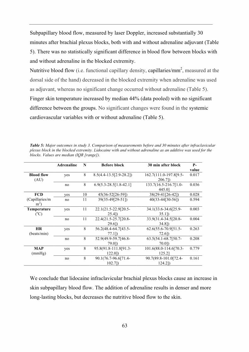
63
Subpapillary blood flow, measured by laser Doppler, increased substantially 30
minutes after brachial plexus blocks, both with and without adrenaline adjuvant (Table
5). There was no statistically significant difference in blood flow between blocks with
and without adrenaline in the blocked extremity.
Nutritive blood flow (i.e. functional capillary density, capillaries/mm2, measured at the
dorsal side of the hand) decreased in the blocked extremity when adrenaline was used
as adjuvant, whereas no significant change occurred without adrenaline (Table 5).
Finger skin temperature increased by median 44% (data pooled) with no significant
difference between the groups. No significant changes were found in the systemic
cardiovascular variables with or without adrenaline (Table 5).
Table 5: Major outcomes in study 3. Comparison of measurements before and 30 minutes after infraclavicular plexus block in the blocked extremity. Lidocaine with and without adrenaline as an additive was used for the blocks. Values are median (IQR [range]).
Adrenaline N Before block 30 min after block P-value
Blood flow (AU)
yes 8 8.5(4.4-13.5[2.9-28.2]) 162.7(111.0-197.8[9.5-206.7])
0.017
no 8 6.9(5.3-28.5[1.8-42.1] 133.7(16.5-216.7[1.0-445.0]
0.036
FCD (Capillaries/m
m2)
yes 10 45(36-52[26-59]) 38(29-41[26-42]) 0.028 no 11 39(35-49[29-51]) 40(33-44[30-56]) 0.594
Temperature (oC)
yes 11 22.1(21.5-22.9[20.5-25.4])
34.1(33.6-34.6[25.9-35.1])
0.003
no 11 22.4(21.5-25.7[20.8-29.6])
33.9(31.4-34.5[20.8-34.8])
0.004
HR (beats/min)
yes 8 56.2(48.4-64.7[43.5-77.1])
62.6(55.6-70.9[51.5-72.6])
0.263
no 8 52.9(49.9-59.7[46.8-79.0])
63.5(54.1-68.7[50.7-70.0])
0.208
MAP (mmHg)
yes 8 95.8(91.8-111.8[91.3-122.0])
101.6(88.0-114.6[70.3-125.2]
0.779
no 8 90.1(76.7-96.6[71.4-102.7])
90.7(89.8-101.0[72.4-124.2])
0.161
We conclude that lidocaine infraclavicular brachial plexus blocks cause an increase in
skin subpapillary blood flow. The addition of adrenaline results in denser and more
long-lasting blocks, but decreases the nutritive blood flow to the skin.

64
11. Ethical considerations and approvals
The studies were reviewed and approved by the Regional Committee for Medical
Research Ethics of South Eastern Norway with the permission numbers 2011/476a
(study 1), 2016/500 (Study 2) and 2012/2189b (study 3). Study 2 and study 3 were
also approved by the Norwegian Medicines Agency, and registered in the EU Clinical
Trials Register, EudraCT number 2016-000684-16 (study 2) and 2012-005651-17
(study 3). Study 1 and 2 were reported to the international Clinical Trials registry of
planned studies, identifier number NCT01740141 (study 1) and NCT03011905 (study
2). All studies were conducted in adherence to guidelines for Good Clinical Practice
and all manuscripts adhere to the CONSORT guidelines.
All participants in the three studies signed a written informed consent prior to
inclusion after thorough oral and written information. They were also informed about
the possibility to withdraw from participation at any time before, during or after the
study. The participants in all the studies received an ultrasound guided infraclavicular
brachial plexus block (175). All participants were healthy (ASA 1-2) with no increased
risk for nerve damage because of cardiovascular disease, diabetes or pre-existing
neurological disorders. The risk for serious complications after peripheral nerve blocks
is considered to be low (34).
Study 1 and 2 were clinical studies on patients. The patients received no benefits from
taking part in the studies apart from the frequent phone calls (which could be
considered both a benefit or a hassle) and the possibility to contact a study doctor
whenever needed.
Study 1:
The patients received brachial plexus block during general anaesthesia. This might
have put them at increased risk for nerve injury because they would not be able to feel
pain in case of the needle touching the nerve cords. To avoid nerve damage, we
therefore used triple monitoring with ultrasound, injection pressure monitors (to keep
injection pressure under 15 psi) and electrical nerve stimulation (no motor response on

65
a current setting of 0,5 mA, 0,1 ms impulse duration, and a frequency of 2 Hz, if motor
response was elicited with 0.5 mA or below, the needle was withdrawn until the
response disappeared) (176).
Study 2:
Brachial plexus block is the standard anaesthetic method for all patients scheduled for
volar plate surgery in our hospital, thus the patients in the study received standard
anaesthesia for the surgery. Half of the patients in the study received dexamethasone.
Dexamethasone is a well-known medication licenced for intravenous use. Although
not licenced to treat pain in Norway, it is frequently used to reduce pain after for
example abdominal, gynaecological and spine surgery, and considered to be safe in the
doses used in our study (61, 62). The patients randomised to the dexamethasone group
could possibly receive better pain treatment than standard treatment in our department,
as we did not use dexamethasone for volar plate surgery before or during the study.
Study 3:
All medication used in this study were registered in Norway and used within approved
indication. Even though brachial plexus block is considered safe and the risk of serious
complications are low, the use of brachial plexus block in volunteers needs an ethnical
consideration. A possible damage to a nerve in a healthy volunteer would not be
acceptable. We used healthy volunteers to get a homogenous group with as little risk
for adverse events as possible. This enabled us to diminish confounders caused by
factors like pre-existing conditions influencing on human microcirculation, and tissue
damage as well as aberrant anatomy in patients. It also made it possible to standardise
the conditions during the experimental runs to avoid disturbance to our measurements.
We used the crossover design to limit the amount of volunteers needed. To avoid
nerve damage, Axel Sauter, who is extremely experienced, performed all blocks in the
healthy volunteers. The three cords were localised using ultrasound, and the needle
was approached in safe distance from the cords. The volunteers did not get any
sedation, to be able to report to us if they felt any discomfort or signs of intraneural
placement of the needle during the procedure.

66
The volunteers got a gift card for entering the study. The value of the gift card was
supposed to be large enough to compensate for all the hours used during the
experimental runs as well as the time they had an anaesthetised arm after the
experiments. Still, we did not want the compensation to be too large, not to make the
compensation the main reason for attending the experiments.
All volunteers got thorough information about how to take care of the blocked
extremity after they left the study site to avoid any damage to the arm. They were
asked to contact us if they had any questions or worries after the experiments. In
addition, we contacted all participants on the day after the experiments to make sure
the blocks had worn off and they felt their normal state.

67
12. Methodological considerations
When conducting a study, it is important to plan how to evaluate the different
measures or methods in advance. The validity, reliability and utility have to be
considered (82). The validity of a method refers to the appropriateness,
meaningfulness and usefulness of a measure for a specific purpose (i.e. if the method
measures what is supposed to be measured). The reliability of a method refers to the
extent to which the score from a test is free from measurement errors, in other words
how precise the data is. The utility of a measure is a more practical issue. Although it
would be interesting, it is impossible to design a study measuring pain in all possible
activities a person could engage in, and few patients would participate in a study
where they would have to report pain every hour for several weeks (82).
There will always be errors in measurements (i.e. deviations of the true values), no
matter how careful one is. It is common to distinguish between random errors and
systematic errors. Random errors are unknown and unpredictable, and cannot be
eliminated from an experiment. By increasing the sample size, the effect of random
errors can be minimised. Systematic errors influence the accuracy of the data and
include selection bias, information bias and confounding. They can often be avoided.
When measuring physical data it is important to consider several sources of
measurement errors. In our three studies, we may have biological variation between
individuals, day-to-day variation within an individual, observer variation, laboratory
variation, instrument variation, and measurement uncertainty. We tried to adjust our
methods to limit these variations. All investigators were well trained in the methods
used. The measurements were recorded under controlled conditions. We used multiple
measures for some of our endpoints. I will discuss these aspects further in the
following sections.

68
12.1 Study design
All our studies were randomised and double-blinded. The randomisation ensures a fair
starting point while the double-blinding prevents observers to be biased during the trial
period.
The randomisation is important in order to get two similar and comparable groups with
limited systematic errors. It minimises allocation bias (i.e. differences in how the
participants are allocated to treatment) and balances the groups with respect to both
known and unknown confounding factors (137). A good randomisation and an
adequate sample size limits confounders, resulting in reduced need to adjust for them
in the analysis. Significant differences between groups regarding possible confounders
in a randomised study are due to chance or flawed randomisation.
Despite randomisation, we detected a few variations in our baseline characteristics in
the two clinical studies. There may also be undetected confounders in our materials. A
larger sample size could possibly have reduced variations between the study groups in
our materials. However, we do not believe the baseline variations have influenced our
major conclusions. This is further discussed in the “main strengths and limitations”
section under the relevant study.
A double-blinded design removes subjective expectations regarding the effect (138).
In a clinical study, the investigators may wish to show a treatment effect. The
participants may be more likely to report pain if they know they are in the placebo
group. We used double-blinded design in all our studies to minimise detection bias.
In study 3, we used a cross-over design to allow for a smaller sample size. The cross-
over design may reduce the variance in the data compared to analysing two separate
groups. The variation within an individual is usually smaller than between individuals
(parallel groups).
All our studies were mainly quantitative, but studies 1 and 2 also have a few
qualitative aspects. To better explore the patients` experiences during the operation
and the postoperative period, as well as how the fractures actually affected their

69
everyday life, both in short-term and long-term, a qualitative design might have been
an interesting addition to our quantitative studies. However, as the main purpose of
this thesis was to find out if specific modifications of brachial plexus block had clear-
cut objective effects, we concluded that a quantitative approach would be most
appropriate.
12.2 Study populations
Study 1 and 2 were conducted on patients scheduled for volar plate surgery after distal
radius fractures. This surgery is relatively standardised and infraclavicular blocks are
frequently used for this procedure. The patient group is large and quite homogenous.
Both strong pain in the postoperative period and long-lasting pain after the injury or
operation are common after this type of surgery. Thus, we thought it was highly
relevant to evaluate measures to reduce pain in this patient group.
In study 1 and 2, we aimed for wide inclusion criteria to cover the relevant population.
At the same time, we wanted to limit the possibility for complications from the
interventions among our study patients. Therefore, patients with increased risk of
complications were excluded. Overall, we think the included population reflects the
relevant target population and thus ensures a high external validity of the studies.
In our study on microcirculation, we decided to use volunteers instead of patients
because several factors may influence on the human microcirculation including the
pathological condition for which the patients are to have surgery. Several factors are
mentioned under “Evaluation of human skin microcirculation”. These factors are
easier to control in an interventional setting in healthy volunteers than in patients
before and during an operation. The equipment used to measure microcirculation in
our study might also have been in conflict with the surgical field.
A challenge with our selection of relatively young, healthy male volunteers is that they
may not be representable for the patient groups where brachial plexus blocks are used
to improve microcirculation. These patients will often have conditions that impact
their microcirculation. Thus, the external validity of the study is questionable.

70
12.3 Outcome measures
12.3.1 Block duration
To estimate block duration, we had to find a surrogate for frequent sensory and motor
testing of the nerve blocks, since most of our participants were at home at block
resolution. We used different criteria in the three different studies.
In study 1, we used time to first analgesic to estimate the duration of sensory block. A
challenge with this definition may be if the patients do not need analgesics or try to
avoid analgesics after the operation. However, as all patients in this study experienced
significant pain and need for extra analgesia after the operation, we consider this
definition to represent a reliable clinical surrogate of block duration.
This definition was not used in study 2 and 3 because several patients in study 2 did
not experience pain requiring rescue analgesics, and the volunteers in study 3 did not
use analgesics at all. Instead, they were asked to report the first time after the block
when they were able to perform a simple test, to hold a cup (or another object) and lift
it to the mouth with the blocked arm.
Hence, in study 2, we used the duration of motor blockade as a surrogate for the
endpoint of interest, nociceptive blockade. It could be argued that our test of motor
blockade will result in an overestimation of block duration as patients may hesitate to
try this manoeuvre shortly after block resolution. However, the patients were
instructed to check this frequently when they started to feel the nerve block wear off,
and if pain should be the cause of delayed registration of block resolution, this should
be more evident in the control group. Also in this study, occurrence and start of any
pain may be considered as an adjunctive, surrogate measure of block duration,
applicable in the majority of cases.
In study 3 where we used a short acting local anaesthetic in the blocks, we could have
asked the volunteers to stay in the hospital until the block wore off and performed
frequent testing to get a better estimate of block duration. Because block duration was

71
not an important endpoint in this study, we chose not to spend the volunteers’ time and
resources on this.
12.3.2 Pain
General considerations
The main aim of study 1 and 2 was to evaluate pain. Unfortunately, a complete
objective method for measuring pain does not exist, as pain is a subjective experience
based on nociception in the postoperative setting. The subjective experience of pain
includes several different aspects: intensity, temporal characteristics (i.e. variability,
frequency and duration), location, quality, affect (i.e. general unpleasantness, fear,
anger, sadness, frustration, hopelessness), impact (i.e. impact on life, emotional
impact), and pain interference (i.e. how the pain interferes with day-to day
functioning). When evaluating pain in clinical studies, it is important to decide which
aspects to focus on, and to have an opinion on factors that may influence the different
aspects. It is essential to balance the investigator`s need for a thorough assessment
against the need for minimal assessment burden for the patient (82). It is also
important to be aware of factors that may influence the patient’s response when asked
about pain. This includes the setting (i.e. if the patient is in hospital, at home, alone or
among other people), the person administrating the measure, the patients experiences
and feelings (i.e. upset or fatigued), cultural factors, the patients pain expectations, and
motivational factors (i.e. to get more medication) (82).
Methods for evaluating pain
There are several methods for evaluating pain in clinical trials. A high number of
different pain scales and pain questionnaires exist. I will discuss a few that could have
been relevant in our studies.
Visual analogue scales (VAS), numerical rating scales (NRS) and categorical verbal
rating scales are all pain scales used for evaluating pain intensity. They are considered
equivalent, adequately valid and reliable to assess pain, and to be sensitive to detect
treatment effects (139, 140). For both VAS and 0-10 NRS, score changes between 30-

72
35% appear to indicate a meaningful change in pain to patients across patient
populations (82, 140).
Comparing the three, VAS has shown higher failure rates than NRS and categorical
verbal rating scales (82). This may be because it is more abstract, it uses an instrument
(i.e. a ruler, written paper etc.) and because increased age and opioid intake is
associated with greater difficulty completing VAS measures (82, 140). Categorical
verbal rating scales may have advantages for some patient groups. For example,
children or patients with cognitive impairment may find it easier to differentiate
between some categorical levels than the 11 levels of pain on a NRS scale (140).
NRS score 0-10 is the most commonly used scoring system in clinical studies to
evaluate pain intensity. When used for verbal reporting of the patient, it is often called
VNRS (Verbal Numeric Rating Scale). It has a high validity and reliability and is easy
to understand for most healthy patients. It does not require any instruments or paper-
work to perform, and it can be easily reported over the phone or on a questionnaire.
The optimal clinical cut points between mild, moderate and severe pain are not clear-
cut (141), but a common differentiation is to use the following: A NRS score of 1-3
indicates mild pain, a score of 4-6 implies moderate pain, whereas a score of 7-10
indicates severe pain (142). A 0-10 scale is considered precise enough to detect
treatment effects and to show changes over time (82). Recall ratings of pains scores
over a period of time may also be used. An earlier study has shown that recall ratings
are valid indicators of pain which reflects average pain well (143). However, when
recording pain scores it is important to differentiate on what the patient what is
questioned about: Pain just now or pain during a defined period? Worst pain or
average pain? Pain at rest or pain during provocation or movement?
We have used a combination of different VNRS ratings, as the relevance may be for
more than one modality such as resting pain which is important for sleep and rest,
provocation pain which is important for mobilisation and physiotherapy.
The use of pain intensity as the only outcome measure has been questioned because
treating pain strictly based on pain intensity scales (e.g. any value of 4 or higher)

73
might overestimate treatment requirements of the individual patients (144). This is
especially important in light of the opioid crisis. However, patients in a study should
not be subjected to undue pain and extra suffering just for the purpose of
demonstrating pain differences. Then, the use of rescue analgesia may conceal
differences, which would otherwise have been evident.
Thus, rescue opioid consumption and total opioid consumption are often used as an
indirect measure of pain in clinical studies. The use of analgesic consumption for
evaluating analgesic efficiency of an intervention is criticised for being an uncertain
method with a very large variability found between patients (145). There is often a
large spread in total doses of rescue opioids with a skewed distribution. However, as
patient comfort has an ethical priority in clinical studies, differences between
treatment groups in opioid rescue medication may be more sensitive than differences
in measured pain.
The other aspects of pain (apart from pain intensity 0-10) may be more complicated to
evaluate. There are a lot of different validated pain questionnaires available.
The McGill Pain Questionnaire (MPQ), a multiple item scale consisting of 78 words
describing pain, is used to assess pain quality and pain affect (146). It also exists in a
short form (SF-MPQ). The draw back with this questionnaire is that it does not allow
for evaluating the effect of a treatment on specific pain qualities. The Leeds
Assessment of Neuropathic Symptoms and Signs (LANSS) questionnaire can be used
to distinguish neuropathic from nociceptive pain (147). The questionnaire requires a
clinician to examine the patient and is often considered difficult to administer. There is
also a Self-report LANSS including 5 pain quality items and a sensory test that can be
self administered by the patient. Pain Quality Assessment Scale (PQAS) is a 21-item
scoring system intended to be comprehensive enough to capture the majority of a
patient`s pain experience, yet brief enough to minimise assessment burden (148). Brief
Pain inventory Pain interference scale (BPI) is used to assess pain interference (i.e. the
extent to which pain has interfered with general activity, mood, walking ability,
normal work, relations with other people, sleep and enjoyment of life)(149).

74
Overall evaluation of our pain measurements
The major outcomes in both our pain studies were related to pain intensity. In addition,
we wanted to do a brief evaluation on the temporal characteristics of the pain, pain
interference and pain quality for long-lasting pain.
We chose to use VNRS (verbal numeric rating scale) 0-10 because it is easy to apply,
associated with good compliance and can be administered through telephone
interviews. Categorical scales, total time with moderate-severe pain, rescue- and total
analgesic consumption were used to add to the total evaluation of pain.
A challenge with the VNRS scale is that a rating of for example 3 out of 10 will rarely
be exactly the same in two different patients. The Mankoski pain scale (with numbers
and corresponding descriptions of pain) seeks to ensure equal understanding of the
numerical pain values of patients and investigators, and might have been an
alternative, but more cumbersome option in our studies (150).
We could have used diary-based measures to get more information about pain impact
and pain quality. We chose to use categorical scales because they require less effort
from both the patients and the investigators, and diary-based methods may result in
more missing data. The use of existing questionnaires, could have evaluated the
different aspects of long lasting pain better and added interesting aspects to the studies.
However, it might have made the study more complicated and time consuming for the
participants, resulting in fewer patients agreeing to take part in or completing the
studies.
In retrospect, we could have asked the important questions: Do you have bothersome
pain? Are you satisfied with the postoperative pain treatment? These questions may be
considered more important than the actual pain intensity on a 0-10 VNRS scale.
Another issue we should have considered is the fact that increased activity may be
accompanied by increased pain. A number of our patients reported highest pain score
in the last week during push-ups in the gym after 6 months and 1 year. The total pain

75
burden for these patients was probably low, even though they reported a high worst
pain score.
The strengths about the way in which we assessed pain in our studies are the use of
well-established pain scales for our main outcomes combined with multiple other pain
and quality of life measures. This made it possible to cover several different aspects of
pain and get a fairly good impression of the patients’ total situation without a too high
assessment burden for the patients.
12.3.3 Microcirculation
General considerations
When evaluating skin microcirculation, it is important to consider factors that may
have an impact on the microcirculation and the measurement methods. This includes
the position of the subject, temperature, arterial pressure, physical and mental activity,
stress, age, gender, menstrual cycle, feeding, medication, smoking, black tea, coffee,
garlic, alcohol, inflammatory reactions, and different pathological changes (111, 115).
During our experiments, we tried to eliminate as many as possible of these factors. We
kept a constant room temperature of 21 degrees Celsius, played soothing music in the
room during the experiments to try to make the volunteers as relaxed and comfortable
as possible, and made sure the volunteers rested for at least 20 min before the
measurements.
There are several ways to investigate skin microcirculation, but unfortunately there is
not a perfect method available. The optimal method for evaluating skin
microcirculation would be a non-invasive, safe, easy, exact, fast and reproducible
method that does not affect skin blood flow. The output data should preferably be in
an absolute variable with easy data analysis (preferably automatic) to simplify the
interpretation of the data (151). New technologies and technical improvements of
existing methods have given us more reliable measurement tools over the years.
Several different non-invasive methods are used to evaluate the skin microcirculation
of the upper extremity. Some of them provide indirect measures of the

76
microcirculation (i.e. assess tissue oxygenation as a surrogate for microcirculatory
function), other methods are direct measurements of the microcirculation (152).
Methods for evaluating skin microcirculation
Available methods include laser Doppler flowmetry, Doppler ultrasonography with or
without intravenous contrast, different capillary microscopy techniques (i.e. traditional
capillaroscopy, fluorescence capillaroscopy, videocapillaroscopy and orthogonal
polarisation spectral imaging), transcutaneous oximetry, temperature measurements,
thermography, photoplethysmography, and laser speckle contrast imaging.
We chose to use a combination of laser Doppler measurements, capillary video
microscopy and temperature measurements because we had experience with the
methods as well as equipment and local expertise available.
Laser Doppler fluxmetry has been available since the 1970s and is frequently used to
evaluate microcirculation (116). It determines dynamic changes in skin
microcirculation and enables a real-time, sensitive, continuous, reproducible, non-
invasive and easy to apply assessment of blood flow, uninfluenced by the underlying
blood flow in skeletal muscles (115, 153). It is a semi quantitative assessment of
microvascular blood cell flow (flux) expressed in arbitrary units (AU). During normal
conditions, about 85% of the laser Doppler signals originates from the deeper layers
(subpapillary blood flow), whereas about 15% originates from the superficial layers
(nutritive blood flow) (114). In pathological conditions with reduced capillary density,
even more than 85% of the laser Doppler signal may originate from the deeper part of
microcirculation (114). Disadvantages of the method includes its sensitivity to motion
artefacts and unstable test conditions, the way the anatomical positioning of the probe
influences the laser Doppler flux signal, and the lack of absolute perfusion values
measured (115, 151). Another downside is the large amount of raw data to be
processed (151).
Capillary video microscopy provides direct visualisation of the microcirculation and is
a reliable method used to assess the nutritive circulation of the skin (151, 154).
Compared with traditional capillary microscopy, capillary video microscopy provides

77
better magnifying power, and the possibility of storing pictures and off-line analysis
(110). Disadvantages with this method is its sensitivity to motion (results in movement
artefacts) and pressure (may disturb blood flow), the operator dependent output, and
the time consuming off-line analysis (151). A real time automatic digital image
analysis would probably be better. An entirely automated system for analysing
microcirculation videos to reduce human interaction and computation time is
developed, but not yet commonly used (133).
Temperature measurement provides an indirect measure of tissue perfusion. The skin
temperature reflects the status of the underlying circulation, and changes in
temperature reflect changes in the underlying thermoregulatory microcirculation.
There are other methods we also considered using which could have added interesting
information to our experiments. Doppler ultrasonography is a promising and highly
available non-invasive method (most ultrasound machines have ultrasound Doppler
functions) (111). A major disadvantage with the method is variability from one
examiner to another, especially among beginners. As we did not have experience in
using the method on evaluating microcirculation, we chose not to use it. We also
highly considered using the simple, non-invasive and indirect method of transcutan
oxygen measurement. The method provides information on the body`s ability to
deliver oxygen to the tissues through assessment of the amount of oxygen that diffuses
from the capillaries through the epidermis to the electrode (110). It does not require
expertise, is not complicated and almost free for examiner-dependent errors.
Unfortunately, we had no equipment available and a budget not allowing us to procure
this equipment.
Overall evaluation of our microcirculatory measurements
A major strength about the way we assessed microcirculation in our study is the
combination of laser Doppler flowmetry, capillary microscopy and temperature
measurements. This made it possible to examine both the nutritive and the deeper
subpapillary microcirculation, which differ in their importance on perfusion on tissue

78
cells. The use of both direct and indirect measurements is also an advantage with our
combination of methods.
We used both laser Doppler and capillary microscopy, which are both sensitive to
motion (results in motion artefacts). Therefore, we cannot exclude motion artefacts in
our data, even though our volunteers were relaxed in the supine position. Disturbed
vessel blood flow might also have affected our capillary microscopy measurements
because the method is sensitive to pressure. To get as reliable results as possible, the
operator used a gentle touch with the microscope and recorded 5 video sequences for
each examination.
Due to limited precision of the instruments and a small sample size in study 3, there
may be random errors in our data. We may also have some systematic errors due to
insufficient calibration of equipment (although all equipment was calibrated) and
environmental factors during the experiments. Because we used multiple analysing
methods and analysed differences (due to the paired measurements), this should not be
a major source of errors. In addition, a few volunteers were not included in the some
of the analysis in study 3 due to technical errors or functional failures with the
equipment during the recordings. This might have resulted in loss of power in the
study.
12.3.4 Non-invasive haemodynamic measurements
The volume clamp method used in this study is a fairly reliable method based on
photoplethysmographic technology (155, 156). The method may overestimate brachial
pulse pressure in young healthy subjects as our volunteers, and is contraindicated in
patients with severe finger hypoperfusion (157). As we looked at changes in
haemodynamics, small inaccuracies in the absolute values should not represent a
problem.

79
12.4 Statistical analysis
12.4.1 General considerations
We present p-values for most of our comparisons in addition to the standard deviation,
the interquartile range and range, or the number and proportion. The p-value is the
probability of the observed result or a more extreme result to occur assuming that the
0-hypothesis is true. P-values and confidence intervals are different ways of presenting
the same information and are seen as measures of how trustworthy the claimed effect
of a study-measure is (158). The p-value is a continuous measure, the smaller the p-
value is, the more evidence we have against the 0-hypothesis (158). A significance
level of 0.05 (or 0.01) is often chosen as a cut off level for rejecting the 0-hypothesis.
The use of hypothesis testing and p-values have been widely criticised the last few
years due to misuse and misinterpretation (159-161).
The interpretation of a p-value depends on the analysing process. A statistically
significant result does not always imply a clinically significant result. It is possible to
obtain small p-values for unimportant differences and large p-values for large
differences depending on the sample size and variance in the material. The difference
between a “statistically significant” p-value of 0.049 and a “non-significant” p-value
of 0.051 is minor. Hence, it is important to report the observed effect size in addition
to p-value (158). Some authors suggest to interpret p-values as continuous indices
against the 0-hypothesis, or to report the probability for no real effect in addition to the
p-value (160, 162).
12.4.2 Analysis of continuous data
A continuous variable is a measurable scale variable. It is numeric and can take any
value between two numbers. The intervals between the values are equally spaced.
Standard deviation is a measure of reliability. By indicating the spread for continuous
data, it estimates the amount of variability in the population (163). Standard error is a
measure of the amount of variability in the sample mean, depending on both the

80
standard deviation and the sample size (163). The confidence interval is a range of
plausible values for the population mean, given the sample mean and its standard
error. A 95% confidence interval can be estimated as 1.96 x the standard error on
either side of the mean (163). The interquartile range indicates the spread of the
middle 50% of the distribution. The range is the difference between the largest and
smallest values in the sample.
Parametric methods
The two-sample t-test is one of the most frequently applied tests in statistics used to
compare mean outcomes of continuous data in two separate treatment groups (164). It
is based on the assumptions that all observations are independent, the data is normally
distributed, and variance or standard deviations are equal in the groups. For matched
samples, the paired t-test is widely used requiring the difference for each pair is
normally distributed (164).
Non-parametric methods
When the data is not normally distributed, the standard parametric methods described
above should normally not be used. Non-parametric methods are often used, although
data transformation often can make the data suitable for parametric methods (log
transformation for data skewed to the right, and square transformation for data skewed
to the left). While parametric tests are based on the observed values, most non-
parametric tests are based on rank-sums.
The Wilcoxon rank sum test and the Mann-Whitney U test are used for comparing two
independent samples and are less sensitive to outliers than the two-sample t-test (165).
The two tests give exactly the same results, but use slightly different test statistics.
They are based on the assumptions of independence within and between groups, as
well as the same distribution in both groups. The tests rank all values as if they were
from the same group. The sums of the ranks are assumed approximately normally
distributed, so normal approximation can be used for the tests.

81
The Sign test and the Wilcoxon signed rank test are commonly used for non-
parametric paired measurements (two outcomes measured on the same individual
under different treatment) (165, 166). Both tests assume independent observations.
The Sign test has no assumptions about the distribution. The Wilcoxon signed rank
test assumes symmetrical distributions, and also takes the strength of the difference
into account, not just if they are positive or negative. It is slightly harder to get
significant results using the Sign test, because of lesser assumptions.
12.4.3 Analysis of categorical data
A categorical variable is a countable variable with two or more categories. There is no
intrinsic order on the levels of the categories. Examples are house colour or gender.
To analyse categorical data, the Pearson chi-squared test for independence may be
used. It is valid provided all the expected values are at least 5 (167). When chi-squared
tests are applied on larger tables (than the 2x2 table), the test is valid when less than
20% of the numbers are under 5, and none of the expected numbers are less than 1.
The Pearson chi-square test is known to be more precise in studies with high numbers
of expected counts. The Pearson chi-squared test with Yates´s continuity correction is
sometimes used to prevent overestimation of stastical significanse in small datasets
(when any of the expected values are less than 5). The method is critizised because it
tends to overcorrect and may result in type 2 errors (168). The use of Fisher exact test
is recommended when the expected numbers are less than 5 or the sample size is
small. This test is also sometimes critizised for being too conservative (168). To get
less conservative results, the use of a mid-p adjustment method (by subtracting half of
the point probability from the p-values) could be preferable (169, 170).
12.4.4 Sample size calculations
Conclusions in scientific studies depend on the sample size. It is important to estimate
the sample size when planning a new study. The sample size has to be large enough to
detect an effect. A large sample size will equally distribute all factors (confounders) in
the treatment groups (except the treatment influencing the outcome). A small sample

82
size reduces the chance of getting significant results as well as the generalisability of
the study.
When calculating sample sizes, it is essential to consider the minimum effect of
interest, and make sure it is clinically relevant. It is also important to decide on a level
of significance and power. With a significance level of 5%, there is a 5% chance of
discovering difference when there really is no difference between the groups. The
power of the study is the probability to find a difference between the groups if there
really is a difference.
There are two main errors when testing hypothesis. Type 1 error is to conclude there is
a difference, when in reality there is not (false positive, reject a true 0-hypothesis)
(158). It correlates with the significance level. Type 2 error is not to discover a true
effect (false negative, fail to reject a false 0-hypothesis). A higher power makes type 2
errors less likely.
The smaller the effect size is, the more data is needed to get a high power. Continuous
measurements yield more power than categorical, and may reduce the required sample
size. Use of paired measurements is another option to reduce the required sample size.
The power and sample size have to be weighted against whether the participants entail
a risk, how many participants it is possible to include in a given time, the cost of
including participants, and the total workload with the study.
In our studies, we calculated our sample sizes based on the primary outcomes. In study
1 and 2 (in patients), we decided on a power of 0.95, to also make a negative result
valid. The studies were not powered to find statistically significant differences after 6
and 12 months. To find differences in long-lasting pain, we would have needed a
much higher number of patients. In study 3, we decided on a lower power of 0.80 (the
usual minimum for a study with “good” power), because of the potential hassle and
risk for the volunteers.

83
Due to the small sample sizes in our studies, we may have some random errors caused
by random variation in the two study groups. Type 2 errors are a problem with small
sample sizes.
12.4.5 Overall evaluation of the statistical methods
We had one main outcome in each study, and several secondary outcomes. A
challenge with several outcomes is the fact that more tests increase the risk of finding
chance associations (171). With a p-value of 0.05, one in 20 tests will appear to be
significant when it is really coincidental (172). Corrections for multiple analyses with
p-value adjustments reduce the risk for type 1 errors, but results in greatly reduced
significance values and increase the chance for type 2 errors (171, 172). The
Bonferroni method is the classical method of adjusting p-values by dividing the p-
value with the number of tests performed. This correction is criticised for being too
conservative. We did not adjust for multiple testing in our studies, but each study had
one predefined primary outcome with quite large effect size. Our secondary outcomes
were also predefined and related to the primary aim of the studies. Because we
evaluated several secondary outcomes, we may have statistical significance by chance
on some of the variables with a low level of significance (i.e. 0.01-0.05 range).
However, our major conclusions build on the primary outcomes and the combination
of several predefined secondary outcomes. It is therefore unlikely that a potential type
2 error in some of the secondary outcomes has affected our main conclusions.
We could probably have used more parametric methods in our analysis, but chose to
use mainly non-parametric tests. A concern is that this may have resulted in too
conservative results and subsequently type 2 errors. The parametric t-test is robust for
moderate departures from normality. Because of our randomised design with a similar
number of observations in each group, most distributions not extremely skewed, and
few extreme observations, the t-test could be considered valid (173). It is difficult to
balance the risk reporting a non-existent difference and the risk of not reporting a true
difference. Weighting the two, we decided rather to be too conservative than too
positive in our conclusions.

84
Relative risk and odds ratio may be used to compare outcome between two groups. We
could have used odds ratio to compare the risk of several events in our studies, like
presence of pain and back at work after 6 and 12 months. We used frequency
distributions and Pearson chi squared/Fisher Exact tests because we think it presented
the results satisfactorily and some readers might find them easier to comprehend.
Our pain scores were mainly treated as continuous variables, but for some analysis we
categorized the VNRS scores before analysing them. This includes number of patients
with pain scores less than 4 and number of patients experiencing worst pain scores 8-
10 in the first 36 hours. The categorization of continuous variables is disputed and
raises two important questions: how many categories do we want and where should we
put the cut off between the categories. The categorization can lead to loss of
information and may increase the risk of a false positive result (type 1 error). We used
the categorization in addition to presenting the analysis of the continuous variables,
with similar results. The categorization has therefore not changed the major
conclusions of the papers, but rather served to simplify the interpretations of the
results.
A few patients were not studied after being included in the studies. This decision was
taken before unblinding due to reasons accounted for in the inclusion section in this
thesis. Most questionable is probably the exclusion of the three patients due to
antebrachii fractures in study 2. This can be considered a violation to the intention-to-
treat principle which implies all patients should be analysed in their original group as
if they have received the planned intervention and regardless of their adherence to the
entry criteria (174). It may also violate the principles of randomisation. Another
strategy is per-protocol analysis which implies analysis of only those patients who
strictly adheres to the protocol and estimates the true efficacy of an intervention (174).
A concern about per-protocol analysis is that they may cause selection biases because
of the reasons for the exclusion of the patients. We chose to exclude the patients from
the published data to present clean material with only volar plate surgery for distal
radius fractures. The number of excluded patients in our material was low compared to
the total number of patients. In addition, we performed control analysis on all major

85
outcomes with these patients included, with no major differences from the published
results. We therefore consider a possible bias created by exclusion of patients not to be
relevant. All other patients in the materials were analysed according to their original
group, regardless of how they followed up the study, according to the intention-to-treat
concept. This was to maintain comparability between the groups, to try to eliminate
bias and to reflect clinical practise (174).
Overall, we think the statistical analyses used in our studies were adequate to answer
our research questions satisfactorily. Our sample sizes in study 1 and 2 were estimated
to detect differences in acute pain, but too small to achieve appropriate statistical
power for long-lasting pain. Hence our findings should be corroborated with new
studies. The latter also applies to the results from study 3 considering its exploratory
nature. We could have defended using more parametric methods instead of non-
parametric methods, and chi-squared tests instead of exact tests, but chose to be more
conservative and therefore may have type 2 errors in our materials. We also could
have corrected for multiple outcomes in our analysis in all studies, but chose to look at
the effect sizes and build our major conclusions on the combination of several
outcomes.

86
13. Discussion of resultsTable 6: Summary of articles in this thesis
Study 1 Study 2 Study 3
Aim To compare brachial plexus blocks performed before and after surgery with regard to acute and long-lasting postoperative pain after volar plate surgery
To evaluate the effect of intravenous dexamethasone on brachial plexus block and acute and long-lasting postoperative pain after volar plate surgery
To investigate the effect of adrenaline when added to lidocaine in brachial plexus blocks on human skin microcirculation, both subpapillary blood flow and nutritive blood flow
Methods 52 patients scheduled for volar plate surgery were included in a randomised controlled clinical trial.
53 patients scheduled for volar plate surgery were included in a randomised controlled clinical trial.
12 healthy male volunteers were included in a randomised controlled trial with a crossover set-up.
Main findings Optimal efficacy for management of acute postoperative pain is more likely to be obtained with pre-operative rather than postoperative nerve blockade.
Intravenous dexamethasone increases duration of brachial plexus blocks and improves postoperative analgesia when added to etoricoxib. It may also improve clinical outcome after 6 and 12 months.
Lidocaine infraclavicular brachial plexus blocks increase subpapillary blood flow in the skin. The addition of adrenaline causes denser and more long-lasting blocks, but decreases the nutritive blood flow.
Main strengths The randomised double-blinded design. High success rates of blocks.
The randomised double-blinded design. Uniform patient population. High success rates of blocks. Low dropout range at 6 and 12 months.
The randomised double-blinded design. The use of several different methods to evaluate the two major aspects of microcirculation.
Main limitations
We did not classify type of radius fracture or register if the patients had associated soft tissue injuries. High dropout range at 6 months. The blocks in the postoperative group might still have been developing at emergence of anaesthesia 20 min after blocks procedure.
The use of motor blockade to estimate block duration. Our sample size was determined to find differences in worst pain score first 24 hours after surgery, the sample size was too small to achieve appropriate statistical power for long-lasting pain.
Our homogenous group of volunteers differs from the usual patients in a clinical setting. Randomisation error in one subject. The acclimatisation period of 20 min might have been too short in some subjects.

87
13.1 General discussion
13.1.1 Study 1
The specific aim of this study was to explore the preemptive effect of infraclavicular
brachial plexus blocks. We investigated if a pre-operative infraclavicular brachial
plexus block results in better and more long-lasting postoperative analgesia compared
with an identical block performed at the end of surgery. We concluded that optimal
efficacy for management of acute postoperative pain is more likely to be obtained with
pre-operative rather than postoperative nerve blockade. Pre-operative blocks resulted
in less pain during the first postoperative hours and less analgesic consumption 1 week
after surgery, but had no impact on the high incidence of mild long-lasting pain.
The preemptive effect of infraclavicular brachial plexus blocks demonstrated in this
study is important, and to our knowledge not previously demonstrated after peripheral
nerve blocks. Time to first rescue analgesic after emergence from general anaesthesia
was significantly longer in the pre-operative block group. The patients in the pre-
operative block group also reported significantly less pain and rescue analgesic
consumption first 4 hours after surgery. In addition, fewer patients in this group used
paracetamol analgesics after one week. The latter supports that the effect of a pre-
operative block significantly outlasts the duration of the block per se. To perform the
nerve blocks pre-operatively rather than postoperatively is simple, it does not require
extra resources and is easy to incorporate in a clinical setting to improve perioperative
treatment. Also, when blocks are used for upper extremity surgery, the clinical routine
is to apply them pre-operatively for the benefit of per-operative anaesthetic effect
without the need for general anaesthesia.
Another important effect of pre-operative nerve blocks is the reduced need of opioids
during surgery. Per-operative use of remifentanil may cause hyperalgesia, which is
related to the dose and duration of administration (177, 178). The patients in the
postoperative group received substantially more remifentanil during surgery that the
pre-operative block group in this study. Opioid induced hyperalgesia could partly
explain the differences in initial postoperative pain. But the difference after 7 days is

88
difficult to explain by less hyperalgesia, and is probably due to improved analgesia in
the pre-operative block group.
A pre-operative infraclavicular block could potentially result in less surgical trauma
for the patients due to more relaxation in their forearm muscles, permitting improved
access to the fracture and less need for dissection and trauma to the neighbouring
muscles. However, we registered a similar decrease in systemic serum stress response
parameters (i.e. cortisol, leucocyte count, C-reactive protein and glucose values) in the
two groups during surgery, as well as a similar increase 2 hours postoperatively. Thus,
we assume the surgical trauma in the two groups to be similar.
In this study we demonstrated severe pain at block resolution approximately 10 hours
postoperative in the majority of our patients, with no significant difference between
the two groups. The abrupt termination of the analgesic effect of single-injection
techniques is one of the main challenges of brachial plexus blocks in acute pain
management. Rebound pain after peripheral nerve blocks are demonstrated in several
other studies (2, 4, 85, 109, 179, 180). Severe pain at block resolution is particularly
problematic in ambulatory surgery, like volar plate fixations, because it can cause
considerable discomfort and fear in the patients and may increase the need for
unplanned medical assistance after discharge (109).
The patients in the study were instructed to take analgesics as soon as they started to
feel mild pain after surgery. Because peripheral nerve blocks resolve rather quickly,
this might have been too late to achieve adequate effect of the analgesics before
complete emergence of the blocks. The patients in this study did not receive any anti-
inflammatory protection or multimodal analgesic regimen perioperatively. Thus, once
the analgesic effects on the antinociceptive afferent nerves disappeared, the patients
could have been in a state of strong local and central sensitisation from systemic
inflammatory mediators. The local anaesthetics in an infraclavicular brachial plexus
block efficiently stops transmission of nociceptive pain provoking impulses from a
surgical site (83), but the potential anti-inflammatory effect on a remote surgical site
seems to be minor or moderate (84). Further, when a tourniquet is applied during

89
surgery, there is no access of systemic analgesics to the surgical site, and there is a
local build-up of acidity stimulating peripheral nociceptors (181). When the tourniquet
is released at end of surgery, potent circulating mediators from the arm and wound
areas are expected to be released into general circulation.
In this study, 76% reported episodes of mild pain 6 months after surgery with no
significant difference between the two groups. Long-lasting mild pain has also been
demonstrated after volar plate surgery in other studies (182, 183). Unfortunately, we
do not know how many patients had bothersome pain in everyday life, or during which
activities they experienced the worst pain scores after 6 months. However, pain at 6
months did have an impact on work ability for about half of our patients and on social
life for about 20% of the patients, while it did not influence quality of sleep or
cognitive function for the majority of the patients.
13.1.2 Study 2
With this study, we wanted to address the problem with severe pain at block resolution
demonstrated in the first study. Our specific aim was to explore the effect of a single
pre-incisional intravenous dose of dexamethasone on acute and long-lasting
postoperative pain. In contrast to the first study, all patients in this study received anti-
inflammatory prophylaxis with etoricoxib, which was supposed to be circulating in the
area of surgery before application of tourniquet and start of surgery.
We found that adding intravenous dexamethasone to oral etoricoxib and paracetamol
before start of volar plate hand surgery resulted in more long-lasting brachial plexus
blocks, less pain upon block resolution, and shorter duration of moderate-severe pain.
Further, at six months after surgery, average pain scores were lower and we found less
need for adjustments at work in the dexamethasone group. At one year after surgery,
less patients in the dexamethasone group experienced long-lasting pain, 2 versus 10
patients in the placebo group.
The increased block duration in the dexamethasone group in this study is important
and in agreement with earlier publications (66-69). The possible more pronounced

90
prolongation in our study might be explained by a higher dose of dexamethasone.
From a clinical point of view there is considerable benefit for the patient not to wake
up during the first night after surgery with strong postoperative pain, but rather have
the experience of a dimishing block and need of rescue medication while being awake
the next morning.
Our primary endpoint in this study was worst pain score first 24 hours after surgery.
The increase in block duration after intravenous dexamethasone to median 21.5 hours
could make the value of this endpoint discussable. However, median block duration
after end of surgery was 19 hours, and only three blocks lasted longer than 24 hours
after surgery. Additionally, we found a similar reduction in worst pain score first 36
hours after surgery. Retrospectively, this could probably have been a better primary
endpoint. Possibly more important than the reduction in worst pain score first 36 hours
after surgery, is the considerably reduced duration of moderate-severe pain (VNRS
above 3) with pre-incisional dexamethasone.
An interesting result in this study was the minor, but significant positive effects of
adding dexamethasone on some of the outcomes after 6 and 12 months. This is, to our
knowledge, not previously reported after volar plate surgery or other surgical
interventions, although perioperative measures to reduce the surgical inflammatory
response have been hypothesised to be a target to reduce chronic pain after surgery
(62, 184-186). Unfortunately, our study was not appropriately powered to conclude on
differences in long-lasting pain. This is an interesting area for future studies.
Several mechanisms may explain our findings. Both the anti-inflammatory effect and
the analgetic effect of systemic dexamethasone during 1-2 days after administration is
well known (62, 187). The reason why dexamethasone prolongs nerve block duration
remains unclear. One possible explanation for the prolonged nerve block duration
could be a systemic effect of dexamethasone on neurons. Another possible explanation
is the general analgesic effect of dexamethasone which results in less pain at block
resolution, and therefore a subjective feeling of longer duration of the block. The latter

91
explanation seems less likely, since we also found prolonged motor block duration in
the dexamethasone group.
The prolonged nerve block duration in the dexamethasone group may partly explain
our results of lower rebound pain scores in this group, as nociceptive input and pain
generally declines consistently during the hours after surgery (4). As the
dexamethasone group also experienced shorter duration of strong pain after operation
in our study, it may also be a result of less sensitisation of the central nervous system
(81). The latter could also be hypotetized to be a part of the explanation for less
longlasting pain in the dexamethasone group.
13.1.3 Study 3
The specific aim of this study was to explore the circulatory changes caused by
infraclavicular brachial plexus blocks. We investigated changes in both subpapillary
and nutritive skin blood flow as well as changes in systemic vascular variables after
lidocaine blocks with and without adrenaline added.
We found a substantial increase in subpapillary blood flow in the blocked arm after
infraclavicular brachial plexus blocks. In contrast, the sympathetic block did not
increase blood flow to the nutritive capillaries in the non-glabrous skin on the dorsal
side of the hand where arteriovenous anastomoses are not present. When adrenaline
was used as an adjuvant, the nutritive skin blood flow decreased. Compared with the
control arm, nutritive blood flow to the skin was lower after blocks with and without
adrenaline. The use of adrenaline had no significant effect on cardiovascular variables,
but resulted in denser and longer lasting nerve blocks.
Increased blood flow after brachial plexus blocks has been reported in previous studies
(118, 120, 188). Use of peripheral nerve blocks is also reported to be beneficial in
some clinical settings to prevent tissue ischemia (46-48). The new perspective in this
study is the possible reduction in nutritive blood flow. Whether this is an effect of
adrenaline or simply a result of the denser blocks in the adrenaline group is not clear.
It is also not clear if the nutritive skin blood flow is representable of the nutritive

92
microcirculation in other microvascular beds, like in skeletal muscles, because of the
complexity of microcirculation with regional variations across the body.
The observation of possible reduced nutritive blood flow might be important in several
clinical settings. Optimised peripheral circulation may lead to better wound healing
and improved outcome after surgery. This is especially important after free-flap
surgery, reconstruction surgery, or other types of surgery where the nutritive
circulation is important. A possible decreased nutritive blood flow to the skin after
brachial plexus blocks may question the use of nerve blocks to improve peripheral
microcirculation after some procedures. Our results may lead to the suggestion that
adrenaline adjuvant is to be recommended when a strong block for surgery or pain
relief is required, whereas a block without adrenaline may be recommended when the
purpose of the block is to increase the microcirculation and oxygenation of peripheral
cells to improve function and survival of tissue. Both these aspects should be
considered when brachial plexus blocks are performed to enhance vascular
microcirculation.
The reduced nutritive skin blood flow after infraclavicular blocks with adrenaline
adjuvant could be explained by a steal effect caused by the increase in blood flow to
the arteriovenous anastomosis in the palms and fingers (116). Circulating adrenaline
may also cause direct vasoconstriction in nutritive blood flow to the skin. The
vasoconstrictive effect might increase when there is a steal effect in the arteriovenous
anastomosis due to the sympathetic blockade.
Adrenaline is known to prolong block duration due to vasoconstriction at the injection
site leading to slower removal of local anaesthetics from the paraneural compartment
and an enhanced diffusion of local anaesthetics into the nerve fibres (78). We found
denser and more long-lasting blocks when adrenaline was added to lidocaine. Similar
observations on block quality are also reported in other studies (77, 80). The longer
duration and better quality of the adrenaline supplemented blocks in our study may
explain the greater changes in subpapillary and nutritive skin blood flow. Better block

93
quality after addition of adrenaline might also cause a stronger steal effect to the
nutritive skin perfusion compared with the blocks without adrenaline.
In our study, we observed no systemic effect of adrenaline that can explain our results.
The systemic effect of adrenaline might depend on the total dose of adrenaline and the
blood circulation near the injection site. Our findings are in contrast to other studies on
peripheral nerve blocks (axillary brachial plexus blocks and paracervical blocks)
where significant changes in haemodynamics were found when adrenaline was used as
an adjuvant to lidocaine or bupivacaine in similar doses as in our study (80, 189).
13.1.4 Overall discussion
The general aim of this thesis was to evaluate the clinical and physiological benefits of
infraclavicular brachial plexus block in some dedicated areas to improve its benefits
and avoid undue harm. The findings in our three studies may add knowledge to
improve the clinical use of brachial plexus blocks.
Study 1 and 2 demonstrate the importance of a multimodal pre-operative pain
treatment. Infraclavicular brachial plexus blocks have a preemptive effect worth
considering. Anti-inflammatory prophylaxis with etoricoxib may reduce opioid
requirements postoperatively. The addition of dexamethasone further reduces pain
intensity and shortens the duration of moderate to severe postoperative pain. To
achieve a good clinical outcome after fracture surgery, sufficient pain relief is
important. It enables the patients to do exercises needed for optimal rehabilitation. Our
studies show that peripheral nerve blocks combined with anti-inflammatory
prophylaxis including dexamethasone are important parts of a multimodal
perioperative analgesic regime.
Whereas no patients in study 1 received anti-inflammatory prophylaxis, all patients in
study 2 received etoricoxib. When looking at the results from the study 1 and 2, we
may speculate that the anti-inflammatory effect of etoricoxib is an important reason
for less opioid consumption in study 2. This could be a relevant topic for further

94
studies. However, anti-inflammatory prophylaxis with etoricoxib alone did not seem to
reduce the high pain scores at block resolution.
The inflammatory reaction is a major cause of postoperative pain. An infraclavicular
brachial plexus block has no direct anti-inflammatory effect in the wounded area.
Nevertheless, it may be argued that a brachial plexus block may have a small anti-
inflammatory effect due to its ability to increase peripheral circulation and thus
potentially induce increased “washing out” of inflammatory mediators. However,
when the surgery is performed with tourniquet applied, the effect of increased
peripheral circulation after pre-operative brachial plexus block will be diminished. In
study 1, we found better postoperative analgesia when the infraclavicular block was
performed before compared to after surgery. This is probably mainly due to the
preemptive effect of the blockade, counteracting peripheral and central sensitisation,
and not due to the possible anti-inflammatory effect of increased peripheral
circulation.
The exact mechanism behind why a perioperative single dose of intravenous or
perineural dexamethasone increases block duration and reduces postoperative pain is
not known. The systemic effect was demonstrated in study 2. Whether or not
perineural dexamethasone also has a local action is unclear. There are several possible
explanations for a local action. Dexamethasone may decrease the absorption of local
anaesthetics through vasoconstriction due to local action on glucocorticosteroid
receptors (190). It may also supress transmission of pain signals in c-fibres by
reducing spontaneous discharge of the membrane potential of the nerve cells (191). It
could also be through a potential neurotoxic effect causing a transient nerve injury. An
interesting study with bilateral saphenous nerve blocks, with the addition of
dexamethasone on one side, aimed to answer the question if the mechanism of
dexamethasone on nerve block duration is solely systemic, or if dexamethasone has
any local perineural effect. The study only found an inconsistent and modest block
prolongation on the side with local dexamethasone of minor clinical importance (192).
Hence the local effect of dexamethasone seems to be minor.

95
Our studies support that brachial plexus blocks have the potential to improve clinical
outcome after operations by reducing acute and possibly also long-lasting
postoperative pain. When the block is used to improve peripheral microcirculation, a
block without adrenaline may be recommended.
13.2 Main strengths and limitations of the studies
13.2.1 Study 1
There are several limitations in this study that need to be addressed. We did not
classify the types of fracture, apart from all patients being in need of a volar plate
fixation. Nor did we record if the patients had associated soft tissue injuries. Thus,
there could be differences between the two groups in fracture severity. Still, because of
randomisation, we assume the two groups to be similar with regards to severity of the
fractures and soft tissue injuries.
Despite randomisation, we have some small variations in our baseline characteristics
in pre-operative risk factors for pain that need to be addressed. The duration of surgery
tended to be slightly longer in the pre-operative group. This could be an indication of
more complicated surgery in the pre-operative group. There were also more cigarette
smokers in the pre-operative group, 15 (60%) compared with 4 (15%) in the
postoperative group. Smoking may affect both acute and chronic pain conditions, and
animal studies have demonstrated increased sensitivity to pain stimuli with nicotine
withdrawal (193, 194). These possible confounders may have influenced on our results
in terms of more pain in the pre-operative group. Therefore, they should not have
affected our main conclusion of less postoperative pain in the pre-operative block
group.
The 20 min period from the application of infraclavicular blocks to emergence of
anaesthesia in the postoperative group might have been too short to make all blocks
work sufficiently before the patients were awake. Our clinical experience is that the
blocks may continue to develop during the first hour after application. Yet, previous
studies have shown that successful infraclavicular blocks can be obtained at 20 min

96
(54, 195). We decided to keep the patients in general anaesthesia for the minimal time
needed for a sufficient block, 20 min, to minimise the extra time used in the operating
theatre in a busy day care unit. We tested the sensory blocks 30 min after emergence
from general anaesthesia (i.e. 50 min after block procedure in the postoperative group)
to allow the patients to recover from anaesthesia and be able to cooperate in order to
perform the test procedure. All but one block in each group were successful at this
time according to our predefined criteria.
We registered actual pain at 30 min, 1, 2, 8 and 24 hours after surgery, and the worst
pain score experienced during first 24 hours. The data obtained from these points in
time might not be representable for the patients average pain experience first 24 hours.
The duration of medium to strong postoperative pain is another important measure we
did not register in this study.
Due to our study design, we did not include enough patients for appropriate statistical
power on our endpoints after 6 months. In addition, several patients were lost to follow
up at the 6-months interview. With only 17 patients left in each group at the 6 months
interview, the potential to find statistically significant differences between the two
groups was reduced.
The study also has several strengths. The topic is relevant and of clear clinical
importance. Other strengths include the randomised double-blind design, the high
success rate of the blocks, the registration of systemic serum stress response markers
and the use of rebound pain as outcome.
13.2.2 Study 2
This study also has several limitations. Despite randomisation, more patients in the
placebo group had injuries in the dominant hand. This may influence the incidence of
both pain and the ability to work unimpeded, as this hand is used more than the other.
A smaller number of women in the dexamethasone group may have affected the
outcome since women are reported to experience more pain (196). However, women
are also reported to have longer duration of nerve blocks (68). A slightly higher pain

97
catastrophizing incidence and higher expectation of pain in the placebo group could
also explain a higher pain intensity in that group (197). Otherwise, the two groups
were similar in preoperative risk factors of postoperative pain, including pain
threshold measured before the operation. The differences in worst pain and block
duration between our two study groups were large, robust and strongly significant.
Hence, these outcomes can not be explained by minor differences between the two
groups in confounding factors.
Another potential limitation may be that some of the nerve blocks might have resolved
in the middle of the night when the patients were asleep, with subsequent over-
estimation of block duration in patients with low or no pain. However, as 81% of our
patients (i.e. both groups pooled) were in need of rescue opioid shortly after resolution
of the block, we do not consider this aspect to be of any major importance.
To estimate block duration, we used the duration of motor blockade as a surrogate for
the endpoint of nociceptive blockade. Because the patients were at home when the
nerve blocks wore off, we decided to use a simple test, which the patients could easily
perform themselves. End of block duration was therefore defined as time to lift a pen
to the face area. It may be argued that this will be an overestimation as patients may
hesitate to try this manoeuvre shortly after block resolution. However, the patients
were instructed to check this frequently when they started to feel the nerve block wear
off, and if pain should be the cause of delayed registration of block resolution, this
should be more evident in the control group.
Some may argue the use of anti-inflammatory prophylaxis with etoricoxib is a
limitation of the study, and that a study with only dexamethasone as anti-inflammatory
prophylaxis compared with placebo would have been a cleaner and better design.
However, etoricoxib was introduced as a standard premedication before volar plate
surgery in our department prior to study 2. We wanted to find out if dexamethasone
has an additional anti-inflammatory effect to etoricoxib worth considering. It could
also be considered unethical and make recruitment more difficult if we had offered our
study patients an analgesic regime inferior to the standard regime in our department.

98
Our sample size was determined to find differences in highest pain score during the
first 24 hours after surgery. We did not include enough patients to achieve appropriate
statistical power on some of our secondary outcomes. To evaluate the true incidence of
long-lasting pain after such operations, a higher number of patients would be needed,
both because of lower incidence of pain and less difference in pain scores after 6
months and one year. As we evaluated several variables, it is possible to have
statistical significance by chance on some of the variables with a low level of
significance (i.e. 0.01-0.05 range) at 6 and 12 months.
We found no side effects from dexamethasone in our study, and did not study blood
glucose levels, which are reported to be transiently elevated in many previous studies
(74, 198, 199). However, serious side-effects related to dexamethasone, such as
delayed wound healing or wound infection, are very rare and were not reported in the
study of more than 18000 orthopaedic surgery patients by Vuorinen et al (200).
The strengths of our study include the clinically relevant topic, the randomised double-
blind design, the uniform patient population, the high success rate of blocks, and the
low dropout rate at 6 and 12 months.
13.2.3 Study 3
A limitation of this study is the selection of participants that may not be representable
for the typical patient in a clinical setting. All our participants were predominantly
young, healthy and non-smoking males. We did not include women because female
reproductive hormones may influence on the non-noradrenergic mechanisms of
vasoconstriction (126). Older patients may have age related alterations in skin blood
flow. Therefore, the results may not apply in women or in older patients. Our results
apply to infraclavicular blocks with lidocaine 15 mg/ml using adrenaline 5 µg/ml as an
adjuvant with an injection volume of 0.4 ml/kg. The use of other local anaesthetics,
dosages, and adrenaline concentrations could lead to different results.
Further, an acclimatisation period of 20 minutes in a temperature controlled operation
theatre might have been too short to ensure stable baseline circulation measurements

99
in some of the volunteers. This may have affected the measurements in the control arm
as well as the anaesthetised arm during the course of our registrations.
Because of a small sample size in our study, we might have type 2 errors in our
statistical calculations.
The short block duration in the blocks without adrenaline might have affected the
capillary video microscopy measurements in some of our subjects. Median (IQR
[range]) block duration was only 67(62-72[30-93]) min in these blocks. The time from
needle out until all measurements were completed was about 30-35 min. Hence, the
effect of the blocks was probably about to wear off in some of our volunteers during
the microscopy session at 30 min after nerve block. Therefore, we might have
underestimated changes in nutritive blood flow after nerve blocks without adrenaline.
Due to technical failure with the Hydra Series II data logger, we had to replace it and
used a C28 K type thermometer with temperature measurements every 5 minutes on
two volunteers on day two. A Hydra Series III data logger was used on one volunteer
on day two. Even though all the equipment was calibrated, the different measuring
methods might have given us minor inaccuracies in the temperature results. However,
because our temperature results were robust and highly significant, this has probably
not affected our main conclusions.
The major strengths of this study are the novelty, its potential clinical relevance, the
randomised double-blinded design and the use of several different methods to evaluate
the different aspects of microcirculation.
13.3 Clinical implications
With the studies in this thesis, we have tried to evaluate different clinically relevant
outcomes after infraclavicular brachial plexus blocks to help improve its clinical use.
All strategies we have evaluated are easy to incorporate in a clinical practise.
Study 1 and 2 clearly demonstrate the importance of preventive analgesia. The results
highly support the use of pre-operative brachial plexus block combined with pre-

100
incisional intravenous dexamethasone and perioperative NSAIDs in patients
undergoing volar plate fixations for distal radius fractures unless there are
contraindications.
Both study 1 and several other studies have demonstrated one of the major clinical
challenges of using single injection brachial plexus blocks: the strong rebound pain at
block resolution (2, 85, 109). Several strategies may be used to try to diminish this
problem: use of continuous block catheters; use of adjuvants to local anaesthetics to
prolong block effect; or more aggressive preemptive analgesic regimens. In study 2,
we found both reduced worst pain scores and reduced duration of moderate–severe
pain after block resolution (to a satisfactory level for most patients) after a
combination of oral etoricoxib and intravenous dexamethasone. The simplicity of this
treatment, which is considered safe and is well established, compared with time-
consuming nerve block catheter techniques or unlicensed use of perineural adjuvants
like dexamethasone, favours the use of intravenous dexamethasone in a clinical setting
(62, 75, 201).
In light of the current focus of eliminating unnecessary perioperative opioid
consumption, the possible reduced opioid consumption in patients receiving anti-
inflammatory prophylaxis is important. Also, the potential positive effects of a single
intravenous dose of dexamethasone on long-lasting pain after 6 and 12 months is a
novel and interesting finding that should be further investigated.
Study 3 may have important implications when brachial plexus blocks are used to
improve peripheral circulation. We found increased subpapillary blood flow after
brachial plexus blocks. This is also reported in previous studies (118, 120). The new
and important perspective in this study is the effect on nutritive skin blood flow. A
significant reduction in nutritive skin blood flow was found after blocks with
adrenaline adjuvant, which enhance the block density and duration. Compared with the
control arm, nutritive blood flow to the skin was lower both after blocks with and
without adrenaline. A possible decreased nutritive circulation after brachial plexus
blocks may question the use of nerve blocks to improve peripheral microcirculation

101
after surgery where the nutritive circulation is important. According to our results, it
may be beneficial to avoid adrenaline adjuvant if the purpose of the nerve block is to
increase the microcirculation and oxygenation of peripheral cells. Further studies are
needed to confirm these findings.

102
14. Conclusions
14.1 Paper 1 (specific research question A)
A pre-operative brachial plexus block leads to a small, but significant improvement in
early postoperative pain compared with postoperative administration of the block, and
possibly a lower analgesic consumption up to one week after surgery. However, a pre-
operative block does not attenuate strong pain during block resolution and does not
seem to have an impact on the high incidence of minor persistent pain.
14.2 Paper 2 (specific research question B)
The addition of intravenous dexamethasone to oral paracetamol and etoricoxib
provides longer duration of brachial plexus blocks, improved acute postoperative
analgesia, and may also reduce the development of chronic pain after volar plate hand
surgery.
14.3 Paper 3 (specific research question C)
Subpapillary skin blood flow increases after lidocaine infraclavicular brachial plexus
blocks whereas nutritive blood flow to the non-glabrous skin does not increase.
Adrenaline as additive did not have a significant effect on cardiovascular variables, but
resulted in denser nerve blocks and a significant decrease in nutritive skin blood flow.
14.4 Overall conclusions
To obtain the optimal efficacy for management of both acute and long-lasting
postoperative pain and reduce total opioid requirement after volar plate surgery for
distal radius fractures, brachial plexus block should be performed pre-incisionally
rather than postoperatively and be combined with NSAIDs and intravenous

103
dexamethasone. When a brachial plexus block is performed for the purpose of
increasing the microcirculation and oxygenation of peripheral cells, the best approach
may be to use a block without adrenaline added.

104
15. Future area of research/interest
Intravenous dexamethasone demonstrated clear positive tendencies for preventing
long-term pain in our study. Further studies with focus on possible long-term pain
reducing effects of dexamethasone and other interventions would be an area of
interest. A thorough evaluation of the affective qualities of long lasting pain (fear,
anger, sadness, frustration, feelings and hopelessness), as well as an evaluation of the
other aspects of long-lasting pain would also be of interest.
Even though intravenous use of dexamethasone are considered safe and well
established with low incidence of side effects (62), many clinicians are still concerned
about possible adverse effects like wound infections, delayed wound healing and
increased levels of blood glucose. The literature is scarce on safety data on perineural
use of dexamethasone and its benefits over intravenous use (75). Large studies
exploring possible side effects of both intravenous and especially perineural use of
dexamethasone would be interesting and important. Such studies would require a high
number of included patients. Also, studies on repeated doses of dexamethasone will be
of interest in order to elucidate any potential enhanced prolongation effect versus any
increased risk of side effects.
Another area of interest is to find the optimal dose of dexamethasone for pain relief
after volar plate surgery and other operations. Is there a major difference between 4
mg, 8 mg or 16 mg regarding the reduction in both acute and long-lasting pain? What
are the median effective dose (ED50) and the dose required to achieve the desired
effect in 95% of the patients (ED95)? Studies with a similar perioperative design as
our study, or with an increasing or repetitive dose of glucocorticoid would be
interesting. The latter have been reported beneficial on pain in a study of orthopedic
hip surgery (202).
More studies evaluating the effect of the combination of several agents on both
peripheral nerve blocks and postoperative pain would be interesting. A Korean group
recently published a study where the intravenous combination of dexmedetomidine 1

105
µg/kg and dexamethasone 0.11 mg/kg substantially increased the duration of analgesia
after interscalene brachial plexus blocks for shoulder arthroscopy from 10.9 hours to
66.3 hours (203).
An area of growing interest is to evaluate pre-operatively individual patients risk
factors for high levels of postoperative pain. Such mapping of risk factors may
enhance the precision of pain-prophylactic measures, by providing extra analgesia for
those at high risk, while the low risk patients will not be exposed to drugs that they do
not need.
Regarding circulatory changes after brachial plexus block, our finding of reduced
nutritive blood flow to the non-glabrous skin after blockades with adrenaline adjuvant
needs to be confirmed. Compelling studies comparing the nutritive microcirculation in
different microvascular beds, to evaluate the use of skin microcirculation as a
biomarker for the microcirculation in general, would be interesting. Studies on
women, where female reproductive hormones may influence on the non-noradrenergic
mechanisms of vasoconstriction, would be important and interesting (126). Also
studies on older patients regarding both microcirculatory and hemodynamic changes
are needed because of age related alterations in skin blood flow and age related
haemodynamic changes. Studies to compare our results with lidocaine and adrenaline
with other local anaesthetics, to see if they cause similar changes in microcirculation,
would also be interesting.

106
16. References
1. Albrecht E, Chin KJ. Advances in regional anaesthesia and acute pain management: a narrative review. Anaesthesia. 2020;75 Suppl 1:e101-e10. 2. Galos DK, Taormina DP, Crespo A, Ding DY, Sapienza A, Jain S, et al. Does Brachial Plexus Blockade Result in Improved Pain Scores After Distal Radius Fracture Fixation? A Randomized Trial. Clin Orthop Relat Res. 2016;474(5):1247-54. 3. Egol KA, Soojian MG, Walsh M, Katz J, Rosenberg AD, Paksima N. Regional Anesthesia Improves Outcome After Distal Radius Fracture Fixation Over General Anesthesia. J Orthop Trauma. 2012;26(9):545-9. 4. Williams BA, Bottegal MT, Kentor ML, Irrgang JJ, Williams JP. Rebound pain scores as a function of femoral nerve block duration after anterior cruciate ligament reconstruction: Retrospective analysis of a prospective, randomized clinical trial. Region Anesth Pain M. 2007;32(3):186-92. 5. Ball C, Westhorpe R. Local anaesthesia--Freud, Koller and cocaine. Anaesth Intensive Care. 2003;31(3):249. 6. Karch SB. A brief history of cocaine. Boca Raton, Fla.: CRC Press; 1998. 202 p. p. 7. Lopez-Valverde A, de Vicente J, Martinez-Dominguez L, de Diego RG. Local anaesthesia through the action of cocaine, the oral mucosa and the Vienna group. Br Dent J. 2014;217(1):41-3. 8. Calatayud J, Gonzalez A. History of the development and evolution of local anesthesia since the coca leaf. Anesthesiology. 2003;98(6):1503-8. 9. Ball C, Westhorpe R. Local anaesthesia--before cocaine. Anaesth Intensive Care. 2003;31(1):3. 10. Zimmer M. History of anaesthesia: early forms of local anaesthesia. Eur J Anaesthesiol. 2014;31(1):1-12. 11. Ball C, Westhorpe R. Local anaesthesia after cocaine. Anaesth Intensive Care. 2004;32(2):157. 12. Ball CM, Featherstone PJ. The early history of adrenaline. Anaesth Intensive Care. 2017;45(3):279-81. 13. Ball C, Westhorpe R. Local anaesthetics--the introduction of xylocaine into clinical practice. Anaesth Intensive Care. 2004;32(6):733. 14. Ball C, Westhorpe R. Local anaesthesia--after xylocaine. Anaesth Intensive Care. 2005;33(1):3. 15. Hansen TG. Ropivacaine: a pharmacological review. Expert Rev Neurother. 2004;4(5):781-91. 16. McLeod GA, Burke D. Levobupivacaine. Anaesthesia. 2001;56(4):331-41. 17. Lopez-Valverde A, De Vicente J, Cutando A. The surgeons Halsted and Hall, cocaine and the discovery of dental anaesthesia by nerve blocking. Br Dent J. 2011;211(10):485-7. 18. Wordliczek J, Jakowicka-Wordliczek J, Zajaczkowska R. Henryk Hilarowicz: The Forgotten Polish Inventor and Pioneer of the Interscalene Brachial Plexus Block. J Anesth Hist. 2018;4(1):11-7. 19. Goerig M, Agarwal K. Georg Perthes--the man behind the technique of nerve-tracer technology. Reg Anesth Pain Med. 2000;25(3):296-301. 20. Greenblatt GM, Denson JS. Needle nerve stimulatorlocator: nerve blocks with a new instrument for locating nerves. Anesth Analg. 1962;41:599-602. 21. Bruce BG, Green A, Blaine TA, Wesner LV. Brachial plexus blocks for upper extremity orthopaedic surgery. J Am Acad Orthop Surg. 2012;20(1):38-47.

107
22. Winnie AP, Collins VJ. The Subclavian Perivascular Technique of Brachial Plexus Anesthesia. Anesthesiology. 1964;25:353-63. 23. Ting PL, Sivagnanaratnam V. Ultrasonographic study of the spread of local anaesthetic during axillary brachial plexus block. Br J Anaesth. 1989;63(3):326-9. 24. Kapral S, Krafft P, Eibenberger K, Fitzgerald R, Gosch M, Weinstabl C. Ultrasound-guided supraclavicular approach for regional anesthesia of the brachial plexus. Anesth Analg. 1994;78(3):507-13. 25. Marhofer P, Schrogendorfer K, Koinig H, Kapral S, Weinstabl C, Mayer N. Ultrasonographic guidance improves sensory block and onset time of three-in-one blocks. Anesth Analg. 1997;85(4):854-7. 26. Neal JM, Gerancher JC, Hebl JR, Ilfeld BM, McCartney CJ, Franco CD, et al. Upper extremity regional anesthesia: essentials of our current understanding, 2008. Reg Anesth Pain Med. 2009;34(2):134-70. 27. Deleuze A, Gentili ME, Marret E, Lamonerie L, Bonnet F. A comparison of a single-stimulation lateral infraclavicular plexus block with a triple-stimulation axillary block. Reg Anesth Pain Med. 2003;28(2):89-94. 28. Raj PP, Montgomery SJ, Nettles D, Jenkins MT. Infraclavicular brachial plexus block--a new approach. Anesth Analg. 1973;52(6):897-904. 29. Sims JK. A modification of landmarks for infraclavicular approach to brachial plexus block. Anesth Analg. 1977;56(4):554-5. 30. Klaastad O, Smith HJ, Smedby O, Winther-Larssen EH, Brodal P, Breivik H, et al. A novel infraclavicular brachial plexus block: the lateral and sagittal technique, developed by magnetic resonance imaging studies. Anesth Analg. 2004;98(1):252-6. 31. Sauter AR, Smith HJ, Stubhaug A, Dodgson MS, Klaastad O. Use of magnetic resonance imaging to define the anatomical location closest to all three cords of the infraclavicular brachial plexus. Anesth Analg. 2006;103(6):1574-6. 32. Koscielniak-Nielsen ZJ, Rasmussen H, Hesselbjerg L, Gurkan Y, Belhage B. Clinical evaluation of the lateral sagittal infraclavicular block developed by MRI studies. Reg Anesth Pain Med. 2005;30(4):329-34. 33. Sauter AR, Dodgson MS, Stubhaug A, Halstensen AM, Klaastad O. Electrical nerve stimulation or ultrasound guidance for lateral sagittal infraclavicular blocks: A randomize, controlled, observer-blinded, comparative study. Anesth Analg. 2008;106(6):1910-5. 34. Auroy Y, Benhamou D, Bargues L, Ecoffey C, Falissard B, Mercier F, et al. Major complications of regional anesthesia in France - The SOS Regional Anesthesia Hotline Service. Anesthesiology. 2002;97(5):1274-80. 35. Watts SA, Sharma DJ. Long-term neurological complications associated with surgery and peripheral nerve blockade: outcomes after 1065 consecutive blocks. Anaesth Intensive Care. 2007;35(1):24-31. 36. Berger A, Tizian C, Zenz M. Continuous plexus blockade for improved circulation in microvascular surgery. Ann Plast Surg. 1985;14(1):16-9. 37. Aguirre J, Del Moral A, Cobo I, Borgeat A, Blumenthal S. The role of continuous peripheral nerve blocks. Anesthesiol Res Pract. 2012;2012:560879. 38. Szili-Torok T, Paprika D, Peto Z, Babik B, Bari F, Barzo P, et al. Effect of axillary brachial plexus blockade on baroreflex-induced skin vasomotor responses: assessing the effectiveness of sympathetic blockade. Acta Anaesthesiol Scand. 2002;46(7):815-20. 39. Hermanns H, Werdehausen R, Hollmann MW, Stevens MF. Assessment of skin temperature during regional anaesthesia-What the anaesthesiologist should know. Acta Anaesthesiol Scand. 2018;62(9):1280-9.

108
40. Gebhard RE, Al-Samsam T, Greger J, Khan A, Chelly JE. Distal nerve blocks at the wrist for outpatient carpal tunnel surgery offer intraoperative cardiovascular stability and reduce discharge time. Anesth Analg. 2002;95(2):351-5. 41. Hadzic A, Arliss J, Kerimoglu B, Karaca PE, Yufa M, Claudio RE, et al. A comparison of infraclavicular nerve block versus general anesthesia for hand and wrist day-case surgeries. Anesthesiology. 2004;101(1):127-32. 42. Pavlin DJ, Chen C, Penaloza DA, Polissar NL, Buckley FP. Pain as a factor complicating recovery and discharge after ambulatory surgery. Anesth Analg. 2002;95(3):627-34. 43. Dahl V, Reder JC, Erno PE, Kovdal A. Pre-emptive effect of pre-incisional versus post-incisional infiltration of local anaesthesia on children undergoing hernioplasty. Acta Anaesthesiologica Scandinavica. 1996;40(7):847-51. 44. Borgeat A, Ekatodramis G, Schenker CA. Postoperative nausea and vomiting in regional anesthesia: a review. Anesthesiology. 2003;98(2):530-47. 45. Chan VW, Peng PW, Kaszas Z, Middleton WJ, Muni R, Anastakis DG, et al. A comparative study of general anesthesia, intravenous regional anesthesia, and axillary block for outpatient hand surgery: clinical outcome and cost analysis. Anesth Analg. 2001;93(5):1181-4. 46. Kurt E, Ozturk S, Isik S, Zor F. Continuous brachial plexus blockade for digital replantations and toe-to-hand transfers. Annals of plastic surgery. 2005;54(1):24-7. 47. Su HH, Lui PW, Yu CL, Liew CS, Lin CH, Lin YT, et al. The effects of continuous axillary brachial plexus block with ropivacaine infusion on skin temperature and survival of crushed fingers after microsurgical replantation. Chang Gung Med J. 2005;28(8):567-74. 48. Stevens GJ, Durning SJ, Stevens SD, Lowery DR. Regional Anesthesia in the Setting of Arm Replantation: A Case Report. A A Pract. 2018;11(2):38-40. 49. Aronson JK, Dukes MNG. Meyler's side effects of drugs : the international encyclopedia of adverse drug reactions and interactions. 15th ed. Amsterdam ; Boston: Elsevier; 2006. 50. Hadzic A, New York School of Regional Anesthesia. Hadzic's peripheral nerve blocks and anatomy for ultrasound-guided regional anesthesia. 2nd ed. New York: McGraw-Hill Professional; 2012. xvi, 704 p. p. 51. Kuthiala G, Chaudhary G. Ropivacaine: A review of its pharmacology and clinical use. Indian J Anaesth. 2011;55(2):104-10. 52. Chong MA, Berbenetz NM, Lin C, Singh S. Perineural Versus Intravenous Dexamethasone as an Adjuvant for Peripheral Nerve Blocks: A Systematic Review and Meta-Analysis. Reg Anesth Pain Med. 2017;42(3):319-26. 53. Janzen PR, Vipond AJ, Bush DJ, Hopkins PM. A comparison of 1% prilocaine with 0.5% ropivacaine for outpatient-based surgery under axillary brachial plexus block. Anesth Analg. 2001;93(1):187-91. 54. Piangatelli C, De Angelis C, Pecora L, Recanatini F, Cerchiara P, Testasecca D. Levobupivacaine and ropivacaine in the infraclavicular brachial plexus block. Minerva Anestesiol. 2006;72(4):217-21. 55. Flohr-Madsen S, Ytrebo LM, Kregnes S, Wilsgaard T, Klaastad O. Minimum effective volume of ropivacaine 7.5 mg/ml for an ultrasound-guided infraclavicular brachial plexus block. Acta Anaesthesiol Scand. 2013;57(4):495-501. 56. Vazin M, Jensen K, Kristensen DL, Hjort M, Tanggaard K, Karmakar MK, et al. Low-Volume Brachial Plexus Block Providing Surgical Anesthesia for Distal Arm Surgery Comparing Supraclavicular, Infraclavicular, and Axillary Approach: A Randomized Observer Blind Trial. Biomed Res Int. 2016;2016:7094121.

109
57. Ueda W, Hirakawa M, Mori K. Acceleration of epinephrine absorption by lidocaine. Anesthesiology. 1985;63(6):717-20. 58. Fredrickson MJ, Patel A, Young S, Chinchanwala S. Speed of onset of 'corner pocket supraclavicular' and infraclavicular ultrasound guided brachial plexus block: a randomised observer-blinded comparison. Anaesthesia. 2009;64(7):738-44. 59. Tran DQ, Dugani S, Dyachenko A, Correa JA, Finlayson RJ. Minimum effective volume of lidocaine for ultrasound-guided infraclavicular block. Reg Anesth Pain Med. 2011;36(2):190-4. 60. Sandhu NS, Bahniwal CS, Capan LM. Feasibility of an infraclavicular block with a reduced volume of lidocaine with sonographic guidance. J Ultrasound Med. 2006;25(1):51-6. 61. Shahraki A, Feizi A, Jabalameli M, Nouri S. The effect of intravenous Dexamethasone on post-cesarean section pain and vital signs: A double-blind randomized clinical trial. J Res Pharm Pract. 2013;Jul;(2(3)):99-104. 62. De Oliveira GS, Jr., Almeida MD, Benzon HT, McCarthy RJ. Perioperative single dose systemic dexamethasone for postoperative pain: a meta-analysis of randomized controlled trials. Anesthesiology. 2011;115(3):575-88. 63. Watanabe S, Bruera E. Corticosteroids as adjuvant analgesics. J Pain Symptom Manage. 1994;9(7):442-5. 64. Mensah-Nyagan AG, Meyer L, Schaeffer V, Kibaly C, Patte-Mensah C. Evidence for a key role of steroids in the modulation of pain. Psychoneuroendocrinology. 2009;34 Suppl 1:S169-77. 65. Hval K, Thagaard KS, Schlichting E, Raeder J. The prolonged postoperative analgesic effect when dexamethasone is added to a nonsteroidal antiinflammatory drug (rofecoxib) before breast surgery. Anesth Analg. 2007;105(2):481-6. 66. Desmet M, Braems H, Reynvoet M, Plasschaert S, Van Cauwelaert J, Pottel H, et al. I.V. and perineural dexamethasone are equivalent in increasing the analgesic duration of a single-shot interscalene block with ropivacaine for shoulder surgery: a prospective, randomized, placebo-controlled study. Br J Anaesth. 2013;111(3):445-52. 67. Kirksey MA, Haskins SC, Cheng J, Liu SS. Local Anesthetic Peripheral Nerve Block Adjuvants for Prolongation of Analgesia: A Systematic Qualitative Review. PLoS One. 2015;10(9):e0137312. 68. Schnepper GD, Kightlinger BI, Jiang Y, Wolf BJ, Bolin ED, Wilson SH. A Retrospective Study Evaluating the Effect of Low Doses of Perineural Dexamethasone on Ropivacaine Brachial Plexus Peripheral Nerve Block Analgesic Duration. Pain Med. 2018;19(7):1485-93. 69. Baeriswyl M, Kirkham KR, Jacot-Guillarmod A, Albrecht E. Efficacy of perineural vs systemic dexamethasone to prolong analgesia after peripheral nerve block: a systematic review and meta-analysis. Br J Anaesth. 2017;119(2):183-91. 70. Rosenfeld DM, Ivancic MG, Hattrup SJ, Renfree KJ, Watkins AR, Hentz JG, et al. Perineural versus intravenous dexamethasone as adjuncts to local anaesthetic brachial plexus block for shoulder surgery. Anaesthesia. 2016;71(4):380-8. 71. Woo JH, Kim YJ, Kim DY, Cho S. Dose-dependency of dexamethasone on the analgesic effect of interscalene block for arthroscopic shoulder surgery using ropivacaine 0.5%: A randomised controlled trial. Eur J Anaesthesiol. 2015;32(9):650-5. 72. Chun EH, Kim YJ, Woo JH. Which is your choice for prolonging the analgesic duration of single-shot interscalene brachial blocks for arthroscopic shoulder surgery? intravenous dexamethasone 5 mg vs. perineural dexamethasone 5 mg randomized, controlled, clinical trial. Medicine (Baltimore). 2016;95(23):e3828.

110
73. Kawanishi R, Yamamoto K, Tobetto Y, Nomura K, Kato M, Go R, et al. Perineural but not systemic low-dose dexamethasone prolongs the duration of interscalene block with ropivacaine: a prospective randomized trial. Local Reg Anesth. 2014;7:5-9. 74. Albrecht E, Reynvoet M, Fournier N, Desmet M. Dose-response relationship of perineural dexamethasone for interscalene brachial plexus block: a randomised, controlled, triple-blind trial. Anaesthesia. 2019;74(8):1001-8. 75. Hewson D, Bedforth N, McCartney C, Hardman J. Dexamethasone and peripheral nerve blocks: back to basic (science). Br J Anaesth. 2019;122(4):411-2. 76. Watkins TW, Dupre S, Coucher JR. Ropivacaine and dexamethasone: a potentially dangerous combination for therapeutic pain injections. J Med Imaging Radiat Oncol. 2015;59(5):571-7. 77. Tschopp C, Tramer MR, Schneider A, Zaarour M, Elia N. Benefit and Harm of Adding Epinephrine to a Local Anesthetic for Neuraxial and Locoregional Anesthesia: A Meta-analysis of Randomized Controlled Trials With Trial Sequential Analyses. Anesth Analg. 2018;127(1):228-39. 78. Sinnott CJ, Cogswell IL, Johnson A, Strichartz GR. On the mechanism by which epinephrine potentiates lidocaine's peripheral nerve block. Anesthesiology. 2003;98(1):181-8. 79. Rang HP. Rang and Dale`s pharmacology. 8th ed. ed. London, UK: Elsevier Churchill Livingstone; 2016. 80. Dogru K, Duygulu F, Yildiz K, Kotanoglu MS, Madenoglu H, Boyaci A. Hemodynamic and blockade effects of high/low epinephrine doses during axillary brachial plexus blockade with lidocaine 1.5%: A randomized, double-blinded study. Reg Anesth Pain Med. 2003;28(5):401-5. 81. Carr DB, Goudas LC. Acute pain. Lancet. 1999;353(9169):2051-8. 82. Jensen MP. Measurement of Pain. In: Fishman SM, Ballantyne JC, Rathmell JP, editors. Bonica`s Management of pain. Philadelphia, PA: Lippincott Williams and Wilkins; 2010. p. 251-70. 83. Cruz FF, Rocco PR, Pelosi P. Anti-inflammatory properties of anesthetic agents. Crit Care. 2017;21(1):67. 84. Perniola A, Magnuson A, Axelsson K, Gupta A. Intraperitoneal local anesthetics have predominant local analgesic effect: a randomized, double-blind study. Anesthesiology. 2014;121(2):352-61. 85. Lavand'homme P. Rebound pain after regional anesthesia in the ambulatory patient. Curr Opin Anaesthesiol. 2018;31(6):679-84. 86. Vadivelu N, Mitra S, Schermer E, Kodumudi V, Kaye AD, Urman RD. Preventive analgesia for postoperative pain control: a broader concept. Local Reg Anesth. 2014;7:17-22. 87. Woolf CJ. Central sensitization: Implications for the diagnosis and treatment of pain. Pain. 2011;152(3):S2-S15. 88. Wilder-Smith OH. Pre-emptive analgesia and surgical pain. Prog Brain Res. 2000;129:505-24. 89. Wilder-Smith O. Changes in sensory processing after surgical nociception. Curr Rev Pain. 2000;4:234-41. 90. Tetzlaff JE, Lautsenheiser F, Estafanous FG. Dr. George Crile--early contributions to the theoretic basis for twenty-first century pain medicine. Reg Anesth Pain Med. 2004;29(6):600-5. 91. Rivat C, Bollag L, Richebe P. Mechanisms of regional anaesthesia protection against hyperalgesia and pain chronicization. Curr Opin Anaesthesiol. 2013;26(5):621-5. 92. Vadivelu N, Mitra S, Schermer E, Kodumuli V, Kaye AD, Urman RD. Preventive analgesia for postoperative pain control: A broader concept. Local and Regional Anesthesia. 2014;7(1):17-22.

111
93. Lavand'homme P, De Kock M, Waterloos H. Intraoperative epidural analgesia combined with ketamine provides effective preventive analgesia in patients undergoing major digestive surgery. Anesthesiology. 2005;103(4):813-20. 94. Mathisen LC, Aasbo V, Raeder J. Lack of pre-emptive analgesic effect of (R)-ketamine in laparoscopic cholecystectomy. Acta Anaesthesiologica Scandinavica. 1999;43(2):220-4. 95. Ong CK, Lirk P, Seymour RA, Jenkins BJ. The efficacy of preemptive analgesia for acute postoperative pain management: a meta-analysis. Anesth Analg. 2005;100(3):757-73. 96. Andreae MH, Andreae DA. Regional anaesthesia to prevent chronic pain after surgery: a Cochrane systematic review and meta-analysis. Brit J Anaesth. 2013;111(5):711-20. 97. Aasbo V, Thuen A, Raeder J. Improved long-lasting postoperative analgesia, recovery function and patient satisfaction after inguinal hernia repair with inguinal field block compared with general anesthesia. Acta Anaesthesiologica Scandinavica. 2002;46(6):674-8. 98. Ong CKS, Lirk P, Seymour RA, Jenkins BJ. The efficacy of preemptive analgesia for acute postoperative pain management: A meta-analysis. Anesth Analg. 2005;100(3):757-73. 99. Ejlersen E, Andersen H, Eliasen K, Mogensen T. Preoperative or postoperative local anesthesia. Effect on postoperative pain treatment. Ugeskr Laeger. 1993;155:4079-82. 100. Moiniche S, Kehlet H, Dahl JB. A qualitative and quantitative systematic review of preemptive analgesia for postoperative pain relief - The role of timing of analgesia. Anesthesiology. 2002;96(3):725-41. 101. Dahl JB, Kehlet H. Preventive analgesia. Curr Opin Anesthesio. 2011;24(3):331-8. 102. Jaeger P, Grevstad U, Henningsen MH, Gottschau B, Mathiesen O, Dahl JB. Effect of adductor-canal-blockade on established, severe post-operative pain after total knee arthroplasty: a randomised study. Acta Anaesthesiol Scand. 2012;56(8):1013-9. 103. Dahl V, Ræder JC, Ernø PE, Kovdal A. Pre-emptive effekt of pre-incisional versus post-incisional infiltration of local anaesthesia on children undergoing hernioplasty. Acta Anaesthesiologica Scandinavica. 1996;40:847-51. 104. Haltiavaara KM, Laitinen JO, Kaukinen S, Viljakka TJ, Laippala PJ, Luukkaala TH. Failure of interscalene brachial plexus blockade to produce pre-emptive analgesia after shoulder surgery. Eur J Anaesth. 2003;20(1):72-3. 105. Blumenthal S, Nadig M, Borgeat A. Pre-emptive analgesia produced by interscalene blockade. What failed: the block or the methods? Eur J Anaesth. 2003;20(11):933-4. 106. Handoll HH, Madhok R, Dodds C. Anaesthesia for treating distal radial fracture in adults. Cochrane Database Syst Rev. 2002(3):CD003320. 107. DeGeorge BR, Jr., Van Houten HK, Mwangi R, Sangaralingham LR, Larson AN, Kakar S. Outcomes and Complications in the Management of Distal Radial Fractures in the Elderly. J Bone Joint Surg Am. 2019. 108. McCartney CJ, Brull R, Chan VW, Katz J, Abbas S, Graham B, et al. Early but no long-term benefit of regional compared with general anesthesia for ambulatory hand surgery. Anesthesiology. 2004;101(2):461-7. 109. Sunderland S, Yarnold CH, Head SJ, Osborn JA, Purssell A, Peel JK, et al. Regional Versus General Anesthesia and the Incidence of Unplanned Health Care Resource Utilization for Postoperative Pain After Wrist Fracture Surgery Results From a Retrospective Quality Improvement Project. Region Anesth Pain M. 2016;41(1):22-7. 110. Neubauer-Geryk J, Hoffmann M, Wielicka M, Piec K, Kozera G, Brzezinski M, et al. Current methods for the assessment of skin microcirculation: Part 1. Postepy Dermatol Alergol. 2019;36(3):247-54. 111. Attila Tamas-Szora MAS, Alexandru F. Badea, Iulian Opincariu and Radu I. Badea. Noninvasive Evaluation of Microcirculation under Normal and Pathological Conditions Using

112
Contrast-Enhanced Ultrasonography (CEUS). In: Lenasi H, editor. Microcirculation Revisted - From Molecules to Clinical Practice: IntechOpen; 2016. 112. Awan ZA, Wester T, Kvernebo K. Human microvascular imaging: a review of skin and tongue videomicroscopy techniques and analysing variables. Clin Physiol Funct Imaging. 2010;30(2):79-88. 113. Braverman IM. The cutaneous microcirculation. J Investig Dermatol Symp Proc. 2000;5(1):3-9. 114. Gschwandtner ME, Ambrozy E, Fasching S, Willfort A, Schneider B, Bohler K, et al. Microcirculation in venous ulcers and the surrounding skin: findings with capillary microscopy and a laser Doppler imager. Eur J Clin Invest. 1999;29(8):708-16. 115. Lenasi H. Assessment of Human Skin Microcirculation and Its Endothelial Function Using Laser Doppler Flowmetry. In: Erondu OF, editor. Medical Imaging: IntechOpen; 2011. 116. Walloe L. Arterio-venous anastomoses in the human skin and their role in temperature control. Temperature (Austin). 2016;3(1):92-103. 117. Robertson D, Biaggioni I. Primer on the autonomic nervous system. 3rd ed. Amsterdam ; Boston: Elsevier/AP; 2012. xxv, 703, p. p. 118. Li J, Karmakar MK, Li X, Kwok WH, Ngan Kee WD. Regional hemodynamic changes after an axillary brachial plexus block: a pulsed-wave Doppler ultrasound study. Reg Anesth Pain Med. 2012;37(1):111-8. 119. Landsverk SA, Kvandal P, Kjelstrup T, Benko U, Bernjak A, Stefanovska A, et al. Human skin microcirculation after brachial plexus block evaluated by wavelet transform of the laser Doppler flowmetry signal. Anesthesiology. 2006;105(3):478-84. 120. Lehtipalo S, Winso O, Koskinen LO, Johansson G, Biber B. Cutaneous sympathetic vasoconstrictor reflexes for the evaluation of interscalene brachial plexus block. Acta Anaesthesiol Scand. 2000;44(8):946-52. 121. McGregor AD, Jones WK, Perlman D. Blood flow in the arm under brachial plexus anaesthesia. J Hand Surg Br. 1985;10(1):21-4. 122. Sandberg M, Lundeberg T, Lindberg LG, Gerdle B. Effects of acupuncture on skin and muscle blood flow in healthy subjects. Eur J Appl Physiol. 2003;90(1-2):114-9. 123. Min S, Lee H, Kim SY, Park JY, Chae Y, Lee H, et al. Local changes in microcirculation and the analgesic effects of acupuncture: a laser Doppler perfusion imaging study. J Altern Complement Med. 2015;21(1):46-52. 124. Wasner G. Vasomotor disturbances in complex regional pain syndrome--a review. Pain Med. 2010;11(8):1267-73. 125. Schurmann M, Zaspel J, Gradl G, Wipfel A, Christ F. Assessment of the peripheral microcirculation using computer-assisted venous congestion plethysmography in post-traumatic complex regional pain syndrome type I. J Vasc Res. 2001;38(5):453-61. 126. Stephens DP, Aoki K, Kosiba WA, Johnson JM. Nonnoradrenergic mechanism of reflex cutaneous vasoconstriction in men. Am J Physiol Heart Circ Physiol. 2001;280(4):H1496-504. 127. Struys MMRF, De Smet T, Depoorter B, Versichelen LFM, Mortier EP, Dumortier FJE, et al. Comparison of plasma compartment versus two methods for effect compartment-controlled target-controlled infusion for propofol. Anesthesiology. 2000;92(2):399-406. 128. Marsh B, White M, Morton N, Kenny GNC. Pharmacokinetic Model Driven Infusion of Propofol in Children. Brit J Anaesth. 1991;67(1):41-8. 129. Minto CF, Schnider TW, Egan TD, Youngs E, Lemmens HJM, Gambus PL, et al. Influence of age and gender on the pharmacokinetics and pharmacodynamics of remifentanil .1. Model development. Anesthesiology. 1997;86(1):10-23. 130. Schnider TW, Minto CF, Shafer SL, Gambus PL, Andresen C, Goodale DB, et al. The influence of age on propofol pharmacodynamics. Anesthesiology. 1999;90(6):1502-16.

113
131. Lossius K, Eriksen M. Spontaneous flow waves detected by laser Doppler in human skin. Microvasc Res. 1995;50(1):94-104. 132. Ambrozy E, Waczulikova I, Willfort-Ehringer A, Ehringer H, Koppensteiner R, Gschwandtner ME. Microcirculation in mixed arterial/venous ulcers and the surrounding skin: clinical study using a laser Doppler perfusion imager and capillary microscopy. Wound repair and regeneration : official publication of the Wound Healing Society [and] the European Tissue Repair Society. 2009;17(1):19-24. 133. Demir SU, Hakimzadeh R, Hargraves RH, Ward KR, Myer EV, Najarian K. An automated method for analysis of microcirculation videos for accurate assessment of tissue perfusion. BMC Med Imaging. 2012;12:37. 134. Nolte D, Zeintl H, Steinbauer M, Pickelmann S, Messmer K. Functional capillary density: an indicator of tissue perfusion? Int J Microcirc Clin Exp. 1995;15(5):244-9. 135. Wester T, Haggblad E, Awan ZA, Barratt-Due A, Kvernebo M, Halvorsen PS, et al. Assessments of skin and tongue microcirculation reveals major changes in porcine sepsis. Clin Physiol Funct Imaging. 2011;31(2):151-8. 136. Bogert LW, van Lieshout JJ. Non-invasive pulsatile arterial pressure and stroke volume changes from the human finger. Exp Physiol. 2005;90(4):437-46. 137. Kang M, Ragan BG, Park JH. Issues in outcomes research: an overview of randomization techniques for clinical trials. J Athl Train. 2008;43(2):215-21. 138. Sedgwick P. Why randomise in clinical trials? BMJ. 2012;345:e5584. 139. Caraceni A, Cherny N, Fainsinger R, Kaasa S, Poulain P, Radbruch L, et al. Pain measurement tools and methods in clinical research in palliative care: recommendations of an Expert Working Group of the European Association of Palliative Care. J Pain Symptom Manage. 2002;23(3):239-55. 140. Dworkin RH, Turk DC, Farrar JT, Haythornthwaite JA, Jensen MP, Katz NP, et al. Core outcome measures for chronic pain clinical trials: IMMPACT recommendations. Pain. 2005;113(1-2):9-19. 141. Krebs EE, Carey TS, Weinberger M. Accuracy of the pain numeric rating scale as a screening test in primary care. J Gen Intern Med. 2007;22(10):1453-8. 142. Jensen MP, Smith DG, Ehde DM, Robinsin LR. Pain site and the effects of amputation pain: further clarification of the meaning of mild, moderate, and severe pain. Pain. 2001;91(3):317-22. 143. Bolton JE. Accuracy of recall of usual pain intensity in back pain patients. Pain. 1999;83(3):533-9. 144. Pogatzki-Zahn E, Schnabel K, Kaiser U. Patient-reported outcome measures for acute and chronic pain: current knowledge and future directions. Curr Opin Anaesthesiol. 2019;32(5):616-22. 145. Moore RA, Mhuircheartaigh RJ, Derry S, McQuay HJ. Mean analgesic consumption is inappropriate for testing analgesic efficacy in post-operative pain: analysis and alternative suggestion. Eur J Anaesthesiol. 2011;28(6):427-32. 146. Hawker GA, Mian S, Kendzerska T, French M. Measures of adult pain: Visual Analog Scale for Pain (VAS Pain), Numeric Rating Scale for Pain (NRS Pain), McGill Pain Questionnaire (MPQ), Short-Form McGill Pain Questionnaire (SF-MPQ), Chronic Pain Grade Scale (CPGS), Short Form-36 Bodily Pain Scale (SF-36 BPS), and Measure of Intermittent and Constant Osteoarthritis Pain (ICOAP). Arthritis Care Res (Hoboken). 2011;63 Suppl 11:S240-52. 147. Bennett M. The LANSS Pain Scale: the Leeds assessment of neuropathic symptoms and signs. Pain. 2001;92(1-2):147-57. 148. Jensen MP, Lin CP, Kupper AE, Galer BS, Gammaitoni AR. Cognitive testing and revision of the pain quality assessment scale. Clin J Pain. 2013;29(5):400-10.

114
149. Hand BN, Velozo CA, Krause JS. Measuring the interference of pain on daily life in persons with spinal cord injury: A Rasch-validated subset of items from the Brief Pain Inventory interference scale. Aust Occup Ther J. 2018;65(5):405-11. 150. Douglas ME, Randleman ML, DeLane AM, Palmer GA. Determining pain scale preference in a veteran population experiencing chronic pain. Pain Manag Nurs. 2014;15(3):625-31. 151. Eriksson S, Nilsson J, Sturesson C. Non-invasive imaging of microcirculation: a technology review. Med Devices (Auckl). 2014;7:445-52. 152. Charlton M, Sims M, Coats T, Thompson JP. The microcirculation and its measurement in sepsis. J Intensive Care Soc. 2017;18(3):221-7. 153. Saumet JL, Kellogg DL, Jr., Taylor WF, Johnson JM. Cutaneous laser-Doppler flowmetry: influence of underlying muscle blood flow. J Appl Physiol (1985). 1988;65(1):478-81. 154. Lamah M, Chaudhry H, Mortimer PS, Dormandy JA. Repeatability of intravital capillaroscopic measurement of capillary density. Int J Microcirc Clin Exp. 1996;16(1):23-9. 155. Monnet X, Picard F, Lidzborski E, Mesnil M, Duranteau J, Richard C, et al. The estimation of cardiac output by the Nexfin device is of poor reliability for tracking the effects of a fluid challenge. Crit Care. 2012;16(5):R212. 156. Hofhuizen CM, Lemson J, Hemelaar AE, Settels JJ, Schraa O, Singh SK, et al. Continuous non-invasive finger arterial pressure monitoring reflects intra-arterial pressure changes in children undergoing cardiac surgery. Br J Anaesth. 2010;105(4):493-500. 157. de Wilde RB, de Wit F, Geerts BF, van Vliet AL, Aarts LP, Vuyk J, et al. Non-invasive continuous arterial pressure and pulse pressure variation measured with Nexfin((R)) in patients following major upper abdominal surgery: a comparative study. Anaesthesia. 2016;71(7):788-97. 158. Murtaugh PA. In defense of P values. Ecology. 2014;95(3):611-7. 159. Colquhoun D. A response to critiques of 'The reproducibility of research and the misinterpretation of p-values'. R Soc Open Sci. 2019;6(11):190819. 160. Colquhoun D. An investigation of the false discovery rate and the misinterpretation of p-values. R Soc Open Sci. 2014;1(3):140216. 161. Greenland S, Senn SJ, Rothman KJ, Carlin JB, Poole C, Goodman SN, et al. Statistical tests, P values, confidence intervals, and power: a guide to misinterpretations. Eur J Epidemiol. 2016;31(4):337-50. 162. Amrhein V, Greenland S. Remove, rather than redefine, statistical significance. Nat Hum Behav. 2018;2(1):4. 163. Altman DG, Bland JM. Standard deviations and standard errors. BMJ. 2005;331(7521):903. 164. Xu M, Fralick D, Zheng JZ, Wang B, Tu XM, Feng C. The Differences and Similarities Between Two-Sample T-Test and Paired T-Test. Shanghai Arch Psychiatry. 2017;29(3):184-8. 165. Laake P, Skovlund E. Statistisk analyse av kontinuerlige data. In: Laake P, Olsen BR, Benestad HB, editors. Forskning i medisin og biofag: Gyldendal Norsk Forlag; 2013. p. 351-90. 166. Grzegorzewski P, Spiewak M. The sign test and the signed-rank test for interval-valued data. Int J Intell Syst. 2019;34:2122-50. 167. McHugh ML. The chi-square test of independence. Biochem Med (Zagreb). 2013;23(2):143-9. 168. Hess AS, Hess JR. Understanding tests of the association of categorical variables: the Pearson chi-square test and Fisher's exact test. Transfusion. 2017;57(4):877-9.

115
169. Lydersen S, Fagerland MW, Laake P. Recommended tests for association in 2 x 2 tables. Stat Med. 2009;28(7):1159-75. 170. Rubin-Delanchy P, Heard NA, Lawson DJ. Meta-Analysis of Mid-p-Values: Some New Results based on the Convex Order. J Am Stat Assoc. 2019;114(527):1105-12. 171. Ranganathan P, Pramesh CS, Buyse M. Common pitfalls in statistical analysis: The perils of multiple testing. Perspect Clin Res. 2016;7(2):106-7. 172. Feise RJ. Do multiple outcome measures require p-value adjustment? BMC Med Res Methodol. 2002;2:8. 173. Aalen OO, Frigessi A, Moger TA, Scheel I, Skovlund E, Veierød MB. Statistiske metoder i medisin og helsefag. Oslo: Gyldendal Akademisk; 2006. 174. Ranganathan P, Pramesh CS, Aggarwal R. Common pitfalls in statistical analysis: Intention-to-treat versus per-protocol analysis. Perspect Clin Res. 2016;7(3):144-6. 175. Hadzic A, Sala-Blanch X, Xu D. Ultrasound guidance may reduce but not eliminate complications of peripheral nerve blocks. Anesthesiology. 2008;108(4):557-8. 176. Gadsden J, McCally C, Hadzic A. Monitoring during peripheral nerve blockade. Curr Opin Anaesthesiol. 2010;23(5):656-61. 177. Lenz H, Raeder J, Draegni T, Heyerdahl F, Schmelz M, Stubhaug A. Effects of COX inhibition on experimental pain and hyperalgesia during and after remifentanil infusion in humans. Pain. 2011;152(6):1289-97. 178. Kim SH, Stoicea N, Soghomonyan S, Bergese SD. lntraoperative use of remifentanil and opioid induced hyperalgesia/acute opioid tolerance: systematic review. Front Pharmacol. 2014;5. 179. Goldstein RY, Montero N, Jain SK, Egol KA, Tejwani NC. Efficacy of Popliteal Block in Postoperative Pain Control After Ankle Fracture Fixation: A Prospective Randomized Study. J Orthop Trauma. 2012;26(10):557-61. 180. DeMarco JR, Componovo R, Barfield WR, Liles L, Nietert P. Efficacy of Augmenting a Subacromial Continuous-Infusion Pump With a Preoperative Interscalene Block in Outpatient Arthroscopic Shoulder Surgery: A Prospective, Randomized, Blinded, and Placebo-Controlled Study. Arthroscopy. 2011;27(5):603-10. 181. Reeh PW, Steen KH. Tissue acidosis in nociception and pain. Prog Brain Res. 1996;113:143-51. 182. Buijze GA, Ring D. Clinical impact of United versus nonunited fractures of the proximal half of the ulnar styloid following volar plate fixation of the distal radius. J Hand Surg Am. 2010;35(2):223-7. 183. Kim JK, Park SD. Outcomes after volar plate fixation of low-grade open and closed distal radius fractures are similar. Clin Orthop Relat Res. 2013;471(6):2030-5. 184. de Oliveira GS, Jr., Bialek JM, Turan A, McCarthy RJ, Sessler DI. Perioperative Dexamethasone and the Development of Chronic Postmastectomy Pain: A Single-Center Observational Cohort Study. Reg Anesth Pain Med. 2015;40(5):539-44. 185. Nielsen RV, Siegel H, Fomsgaard JS, Andersen JD, Martusevicius R, Mathiesen O, et al. Preoperative dexamethasone reduces acute but not sustained pain after lumbar disk surgery: a randomized, blinded, placebo-controlled trial. Pain. 2015;156(12):2538-44. 186. Pehora C, Pearson AM, Kaushal A, Crawford MW, Johnston B. Dexamethasone as an adjuvant to peripheral nerve block. Cochrane Database Syst Rev. 2017;11:CD011770. 187. Paiva-Oliveira JG, Bastos PR, Cury Pontes ER, da Silva JC, Delgado JA, Oshiro-Filho NT. Comparison of the anti-inflammatory effect of dexamethasone and ketorolac in the extractions of third molars. Oral Maxillofac Surg. 2016;20(2):123-33. 188. Wenger A, Amr A, Schaller HE, Rothenberger J. Skin Perfusion Changes within 12 h after Axillary Plexus Block. Eur Surg Res. 2017;58(5-6):227-34.

116
189. Sjoeen GH, Falk RS, Hauge TH, Langesaeter E. Haemodynamic stability after paracervical block: A randomized, controlled, double-blind study comparing bupivacaine-adrenaline with bupivacaine. Acta Anaesthesiol Scand. 2019;63(3):373-80. 190. Shishido H, Kikuchi S, Heckman H, Myers RR. Dexamethasone decreases blood flow in normal nerves and dorsal root ganglia. Spine (Phila Pa 1976). 2002;27(6):581-6. 191. Johansson A, Hao J, Sjolund B. Local corticosteroid application blocks transmission in normal nociceptive C-fibres. Acta Anaesthesiol Scand. 1990;34(5):335-8. 192. Jaeger P, Grevstad U, Koscielniak-Nielsen ZJ, Sauter AR, Sorensen JK, Dahl JB. Does dexamethasone have a perineural mechanism of action? A paired, blinded, randomized, controlled study in healthy volunteers. Br J Anaesth. 2016;117(5):635-41. 193. Schmidt BL, Tambeli CH, Gear RW, Levine JD. Nicotine withdrawal hyperalgesia and opioid-mediated analgesia depend on nicotine receptors in nucleus accumbens. Neuroscience. 2001;106(1):129-36. 194. Shi Y, Weingarten TN, Mantilla CB, Hooten WM, Warner DO. Smoking and pain: pathophysiology and clinical implications. Anesthesiology. 2010;113(4):977-92. 195. Deleuze A, Gentili ME, Marret E, Lamonerie L, Bonnet F. A comparison of a single-stimulation lateral infraclavicular plexus block with a triple-stimulation axillary block. Region Anesth Pain M. 2003;28(2):89-94. 196. Ruau D, Liu LY, Clark JD, Angst MS, Butte AJ. Sex differences in reported pain across 11,000 patients captured in electronic medical records. J Pain. 2012;13(3):228-34. 197. Quartana PJ, Campbell CM, Edwards RR. Pain catastrophizing: a critical review. Expert Rev Neurother. 2009;9(5):745-58. 198. Tien M, Gan TJ, Dhakal I, White WD, Olufolabi AJ, Fink R, et al. The effect of anti-emetic doses of dexamethasone on postoperative blood glucose levels in non-diabetic and diabetic patients: a prospective randomised controlled study. Anaesthesia. 2016;71(9):1037-43. 199. Ottens TH, Nijsten MW, Hofland J, Dieleman JM, Hoekstra M, van Dijk D, et al. Effect of high-dose dexamethasone on perioperative lactate levels and glucose control: a randomized controlled trial. Crit Care. 2015;19:41. 200. Vuorinen MA, Palanne RA, Makinen TJ, Leskinen JT, Huhtala H, Huotari KA. Infection safety of dexamethasone in total hip and total knee arthroplasty: a study of eighteen thousand, eight hundred and seventy two operations. Int Orthop. 2019;43(8):1787-92. 201. Auyong DB, Cantor DA, Green C, Hanson NA. The Effect of Fixation Technique on Continuous Interscalene Nerve Block Catheter Success: A Randomized, Double-Blind Trial. Anesth Analg. 2017;124(3):959-65. 202. Lei Y, Huang Q, Xu B, Zhang S, Cao G, Pei F. Multiple Low-Dose Dexamethasone Further Improves Clinical Outcomes Following Total Hip Arthroplasty. J Arthroplasty. 2018;33(5):1426-31. 203. Kang RA, Jeong JS, Yoo JC, Lee JH, Gwak MS, Choi SJ, et al. Improvement in postoperative pain control by combined use of intravenous dexamethasone with intravenous dexmedetomidine after interscalene brachial plexus block for arthroscopic shoulder surgery: A randomised controlled trial. Eur J Anaesthesiol. 2019;36(5):360-8.

117
17. Errata list
Name of candidate: Anne Holmberg
Title of thesis: Plexus brachialis anaesthesia: Optimising clinical aspects
The originally referenced article by Suresh K “An overview of randomization techniques: An unbiased assessment of outcome in clinical research” published in Journal of Human Reproductive Science in 2011 (was reference number 137) is retracted owing to duplicity of the text. It is therefore replaced with a new reference by Kang et al (a new reference number 137), ”Issues in outcomes research: an overview of randomization techniques for clinical trials” published in Journal of Athletic Training in 2008, covering the same topic. By an editing error, some of the references appear in the wrong format in the original manuscript. They have been changed to the correct format.
By an editing error, the references 175 and 176 appear out of sequence early in the manuscript (after reference 136 on pages 64 and 65). However, all given numbers of references in the text are corresponding to the correct numbers in the reference list. Therefore no changes have been made to the original manuscript.
The original reference number 27 and 195 refers to the same article. No change has been made to the original manuscript.
No changes have been made to the original text of the thesis, only corrections to the reference list. All changes made are summarised in the table below:
Page Reference number
Type of correction
Correction Explanation
68 137 Change of reference
A new reference number 137
Original reference retracted
107, 108, 109, 111, 116
30, 40, 42, 65, 95, 189
Corrected the format of the references
Removed ”table of contents” after the page numbers in the reference or included correct page numbers.
Wrong format of references