
Are Non-Contact Thermometers an Option in Anaesthesia? A ...
Upload
khangminh22Category
view
0download
0
4Monitoring During Anaesthesia and Recovery
C Mendonca
To ensure optimum patient safety certain core standards of monitoring should be usedduring anaesthesia and recovery. It provides information that facilitates earlyrecognition and management of critical incidents. Experienced and vigilantanaesthetist is the most important monitor. However it is impossible to prevent alladverse events, as human error is inevitable. Appropriate monitoring gives an earlywarning and it reduces the consequences of critical incidents.
Association of Anaesthetists of Great Britain and Ireland has produced guidelines forminimum standards of monitoring during anaesthesia. An anaesthetist withappropriate experience should be present throughout general anaesthesia. Anaesthetistshould make frequent observation of clinical parameters, parameters from themonitoring equipment and should undertake appropriate timely interventions. Allanaesthetic equipment should be checked before use according to the manufacturer’sinstructions. Alarm limits should be set at appropriate values for individual patients.All alarms should be enabled during anaesthesia and recovery.
During general anaesthesia inspired oxygen concentration, peripheral oxygensaturation (pulse oximetry), expired carbon dioxide concentration (capnograph) andelectrocardiogram (ECG) are monitored continuously. Blood pressure and vapourconcentration should also be monitored. In a ventilated patient ventilatory parameterssuch as tidal volume, minute ventilation and airway pressures should be monitored.Core temperature, invasive blood pressure, central venous pressure, cardiac output,neuromuscular block, blood loss and urine out put monitoring is dictated by the natureof surgery and physical status of the patient. The following monitoring should beestablished during induction of anaesthesia.
• Pulse oximeter• Blood pressure• ECG• Capnograph
During local anaesthesia and sedation continuous ECG, pulse oximetry, non-invasiveblood pressure and respiration should be monitored. In addition a verbal contactshould be maintained throughout the procedure.
Warwick Medical School- Handbook of Anaesthesia 2006 1
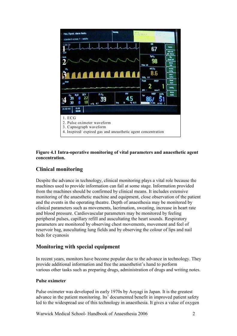
123
4
1. ECG 2. Pulse oximeter waveform 3. Capnograph waveform 4. Inspired/ expired gas and aneasthetic agent concentration
Figure 4.1 Intra-operative monitoring of vital parameters and anaesthetic agentconcentration.
Clinical monitoring
Despite the advance in technology, clinical monitoring plays a vital role because themachines used to provide information can fail at some stage. Information providedfrom the machines should be confirmed by clinical means. It includes extensivemonitoring of the anaesthetic machine and equipment, close observation of the patientand the events in the operating theatre. Depth of anaesthesia may be monitored byclinical parameters such as movements, lacrimation, sweating, increase in heart rateand blood pressure. Cardiovascular parameters may be monitored by feelingperipheral pulses, capillary refill and auscultating the heart sounds. Respiratoryparameters are monitored by observing chest movements, movement and feel ofreservoir bag, auscultating lung fields and by observing the colour of lips and nailbeds for cyanosis
Monitoring with special equipment
In recent years, monitors have become popular due to the advance in technology. Theyprovide additional information and free the anaesthetist’s hand to perform various other tasks such as preparing drugs, administration of drugs and writing notes.
Pulse oximeter
Pulse oximeter was developed in early 1970s by Aoyagi in Japan. It is the greatestadvance in the patient monitoring. Its’ documented benefit in improved patient safetyled to the widespread use of this technology in anaesthesia. It gives a value of oxygen
Warwick Medical School- Handbook of Anaesthesia 2006 2

saturation of the haemoglobin in the arterial blood. It is a simple, reliable andcontinuous non invasive method of detecting hypoxaemia.
Oxygen saturation
Pulse rate
Pulse oximeter probe
Figure 4.2 Pulse oximeter
Pulse oximeter probe consists of two light emitting diodes and a photo-detector. Oneof the photo-detector emits light at red region (660 nm wave length) and other atinfrared region (940 nm wavelength). De-oxyhaemoglobin (reduced Hb) absorbsmaximum light at red region (660 nm wavelength). At 940 nm absorbance of oxy-haemoglobin is greater than de-oxy haemoglobin. There is a microprocessor in themain unit with an inbuilt algorithm in which the ratio of absorption at red region toinfra-red region corresponds to an empirically found saturation value. It produces awaveform of pulsatile flow. It also displays the heart rate derived from the pulse waveform. Pulse oximeters are accurate in the range of saturations of 70 – 100% (+/-2%).
In the following situations pulse oximeter may not be accurate.o Presence of abnormal haemoglobins such as carboxy haemoglobin and
methaemoglobin.o Anaemia (below 8g/dl)o Dyes like methylene blue leads to false low readingo Reduced peripheral circulation due to vasoconstriction (hypovolaemia,
hypotension, cold) or peripheral vascular disease results inaccurate reading.o Venous congestion may result in low readings.o Bright ambient light can affect the accuracy of pulse oximeter.o Motion artefacts such as shivering or seizure activity can result in inaccurate
reading. o Presence of nail varnish may cause falsely low readings.
Electrocardiogram (ECG)
ECG is a surface recording of the electrical activity of the myocardium. It is recordedby connecting various electrodes through which electrical potentials are measured.ECG provides information on heart rate, rhythm and some indication of myocardialischaemia. It doesn’t provide any indication about the adequacy of circulation.
Warwick Medical School- Handbook of Anaesthesia 2006 3

ECG monitoring system consists of following three componentso Skin electrodes detect the electrical activity of the heart.o An amplifier to boost the ECG signal.o An oscilloscope displays the amplified signal.
ECG is recorded at a speed of 25 mm per second and at a standardisation of 1 cmheight representing one mV of amplitude. On a standard ECG recording paper widthof a small square (1 mm) represents 0.04 seconds and a large square (5 mm) 0.2seconds. Five big squares represent a time scale of 1 second and 300 big squaresrepresent a time scale of 60 seconds or one minute. Hence if the rhythm is regular,heart rate can be calculated by dividing 300 by number of big squares within onecardiac cycle (between two consecutive R waves).
The lead system: There are 12 conventional leads, 6 in frontal plane (I, II, III, aVR,aVL, aVF) and 6 in horizontal plane (V1-V6). The heart is situated in the centre of theelectrical field which it generates. The electrical intensity diminishes as the distanceincreases from the centre. The lead axes from three standard leads (lead I, II & III)form a triangle known as an Einthoven triangle. In routine practice, monitors withthree limb leads are used. Three electrodes are placed as follows
o one on the left arm (LA),usually colour coded as yellowo one on the right arm (RA), usually colour coded as redo and one on the left leg (LL), usually colour coded as green or black.
For convenience, during intra-operative monitoring left leg electrode is often placedover the left side of chest, near the apex beat.
- + - -
+ +
Figure 4.3 Standard lead axes forming the Einthoven triangle.
Lead I measures the potential difference between the right arm electrode and the leftarm electrode. Lead II is derived from negative electrode on the right arm and positiveelectrode on the left leg, measures the potential difference between right arm and leftleg electrode. It is usually the best lead for detecting rhythm disturbances. Lead IIImeasures the potential difference between the left arm and left leg. For detectingischaemic changes, ST segment should be monitored in appropriate leads. The STsegment changes in lead V1-V4 usually monitor the left anterior descending arteryterritory, V4-V6 circumflex artery and lead II, III, aVF monitor the right coronaryartery territory. When only bipolar leads are used then a modified V5 lead may beused for detecting ischaemia. CM5 is a modified V5 lead where the right arm
Warwick Medical School- Handbook of Anaesthesia 2006 4
RA LA
LL

electrode of lead I is placed over the manubrium sternum, left arm electrode is placedover the left anterior axillary line in the 5th intercostal space and ground electrode isplaced on the left shoulder.
Table 4.1 Electrode positions for limb leads, augmented leads and modified CM5lead.
Lead RA LA LLI RA (negative) LA (positive) groundII RA (negative) ground LL (positive)III Ground LA (negative) LL (positive)aVR RA ground groundaVL ground LA groundaVF ground ground LLCM5 manubrium V5 ground
In a normal ECG P wave represents the atrial depolarisation, QRS complexventricular depolarisation and T wave represents ventricular repolarisation. The P-Rinterval represents the time taken for the depolarisation to travel from SA node to theventricles (via AV node and bundle of His-Purkinje system). QRS duration representsthe time taken for depolarisation to travel through bundle of His-Purkinje system andventricular muscles.
o Normal P-R interval is 0.12 -0.2 seconds.o Normal QRS duration is less than or equal to 0.1 seconds.o ST segment depression more than 2 mm represents myocardial ischaemia.
and ST segment is elevated in myocardial infarction or myocarditis.
Continuous ECG monitoring should be used for all cases during anaesthesia andsedation. Knowledge of ECG is essential for appropriate diagnosis of peri-operativearrhythmias and ischaemic changes.
Blood pressure
Blood pressure is an indirect measure of blood flow and function of circulatorysystem. It can be measured non-invasively using a cuff and manometer or automatedoscillometric method and invasively by placing a catheter in the peripheral artery.
Non-invasive technique of measuring blood pressure
An aneroid manometer or mercury type manometers (sphygmomanometer) can beused for manual measurement of blood pressure. Mercury sphygmomanometers needregular maintenance with suitable safety procedures in place for using mercury.Automated devices are increasingly used in current clinical practice. The cuff isinflated to a pressure above the expected systolic pressure and then slowly deflated ata rate of 2-3 mmHg per beat. At systolic pressure peripheral pulse appears which canbe detected by palpation of radial or brachial artery. On auscultation over the brachial
Warwick Medical School- Handbook of Anaesthesia 2006 5

artery characteristic sounds (described by Korotkoff) can be heard. These sounds aredescribed in 5 phases. Phase I begins with the appearance of sounds and correspondsto systolic pressure; phase II, it becomes louder; in phase III there is a rise in volume;phase IV is muffling of sound and phase V is total disappearance of sounds. Phase Vis accepted as diastolic pressure.
For accurate reading appropriately sized pneumatic cuff should be used. The maximalocclusion pressure under the cuff is proportional to the inflation pressure and cuffwidth. A too narrow cuff over estimates the blood pressure and a too large cuff underestimates the blood pressure. The width of the cuff should be 20% greater than thediameter of the arm and it should cover two-thirds of the upper arm. The arm shouldbe supported at heart level.
Figure 4.4 Automated device for measuring blood pressure
In an automated device (figure 4.4), a pressure transducer measures the pressure andoscillations. A microprocessor controls the inflation, deflation and display ofnumerical value.
Disadvantages of non-invasive technique.
o Inaccurate in the presence of arrhythmias.o Not possible to have continuous measurement.o Not reliable in extremes of BP (underestimates when too high and vice
versa). o Pressure effects when used for prolonged time and frequent reading resulting
in petechiae, nerve palsy.
Warwick Medical School- Handbook of Anaesthesia 2006 6

Invasive technique of measuring blood pressure
The technique of invasive pressure measurement involves placing a cannula in theperipheral artery. Radial, dorsalis pedis, brachial and femoral arteries are commonlyused. The system includes a cannula placed in the artery connected to a transducer( figure 4.5). In the transducer mechanical energy of movement of diaphragm due toarterial pulsations is converted in to an electrical energy and displayed as bloodpressure reading on the monitor. The cannula is continuously flushed with heparinisednormal saline to prevent clotting.
Arterial pressure wave form
Transducer
Cannula in the radial artery
Figure 4.5 Arterial cannula (radial artery) and transducer system.
Indications for direct arterial blood pressure measurement
o Cases where rapid blood pressure changes is anticipated as in cardiovasculardisease, major blood loss, cardiac surgery, intracranial surgery and inducedhypotension
o Need for frequent arterial blood gas analysiso Cases where non-invasive blood pressure may be inaccurate: arrhythmias,
morbidly obese patient
Disadvantages of invasive technique
o Arterial obstruction and distal ischaemia can occur due to thrombus,haematoma
o Bleedingo Infectiono Accidental injection of drugs.
Warwick Medical School- Handbook of Anaesthesia 2006 7

Capnography
It is the measurement of carbon dioxide concentration (ETCO2) in each breath of therespiratory cycle. Capnograph displays a wave form (Figure 4.1) from which therespiratory rate and ETCO2 can be measured. Monitoring expired carbon dioxideconcentration provides useful information about respiratory system, cardiovascularsystem, metabolism and integrity of the breathing system. During induction ofanaesthesia it is used to confirm the placement of tracheal tube. The contour andpattern of the waveform also provides additional information regarding re-breathing,airway obstruction and wearing off of neuromuscular block.
Time
Car
bon
diox
ide
E1
2
3
4
II = inspirations E = experation
Figure 4.6 Normal capnographic trace
1. Inspiratory phase.2. Upslope phase; onset of expiration.3. Expiratory Plateau. ETCO2 is the reading taken at the end of this phase.4. Inspiratory down stroke; inspiration begins.
In healthy patients at normal physiological parameters, ETCO2 approximates to thepartial pressure of arterial carbon dioxide (PaCO2). In normal lungs the partialpressure ETCO2 is about 0.5 to 0.8 kPa less than the PaCO2.
Hyperventilation, reduced perfusion of alveoli due to reduced cardiac output, reducedmetabolism or pulmonary embolism leads to decrease in the ETCO2. Increasedmetabolism, fever, re-breathing and hypoventilation results in high ETCO2.
Infrared analysers are most commonly used to measure end-tidal carbon dioxideconcentration. Gas molecules having two or more dissimilar atoms absorb infraredlight. CO2 has a strong absorption band at a wavelength of 4.26 µm. The amount ofinfrared radiation absorbed is proportional to the carbon dioxide concentration.
Warwick Medical School- Handbook of Anaesthesia 2006 8

Monitoring of anaesthetic agent concentration
Most often general anaesthesia is maintained using inhalational (volatile) anaestheticagents. Inspired and expired concentration of anaesthetic agent should be continuouslymonitored. Again infrared analysers are commonly used for measuring anaestheticconcentration.
Monitoring inspired oxygen concentration
In view of avoiding hypoxia it is important to continuously monitor inspired orexpired oxygen concentration. Oxygen concentration in a gas mixture can bemeasured using paramagnetic technique, or a fuel cell. Partial pressure of oxygen inthe blood can be measured using a Clark electrode.
Monitoring depth of anaesthesia
Depth of anaesthesia can be monitored using electro encephalogram (EEG) whichinvolves surface recording of the electrical activities from the cerebral cortex. Inpractice the information is obtained from EEG is difficult to interpret and the patternof EEG is also affected by various pathophysiological events like hypoxia,hypotension and hypercarbia. Recently Bispectral index (BIS) monitor has been usedas depth of anaesthesia monitor. This monitor generates a number called bispectral index on a continuous scale of 0 to100.100 represents normal cortical activity and 0 represents no cortical activity.BIS values of 40-60 imply adequate depth of anaesthesia.
Monitoring neuromuscular function
This helps to assess the onset of neuromuscular block, depth of neuromuscular blockand adequacy of recovery from neuromuscular block. Most often a peripheral nervestimulator is used. The principle involves transcutaneous electrical stimulation near anerve and assessment of muscle response by visual inspection or by palpation of themuscle.
Warwick Medical School- Handbook of Anaesthesia 2006 9

Figure 4.7 Peripheral nerve stimulator
Monitoring temperature
Body temperature usually decreases during intra-operative period. There is heat lossfrom radiation, convection, evaporation and respiration. General anaesthesia depressesthermoregulatory centre and most of the anaesthetic agents produce vasodilatation,facilitating heat loss. Other factors such as infusion of cold i.v. fluids, exposure ofbody cavity, low room temperature all ca further increase heat loss. Temperatureshould be monitored during major surgery and in all cases where heat loss isanticipated.
Figure 4.8 Tympanic thermometer
Temperature can be measured using non-electrical thermometer such as mercurythermometer and electrical thermometers such as thermister, thermocouple andresistance thermometer. Due to the limitations and risks involved with mercurythermometer, electrical thermometers are preferred. Temperature probes that are basedon the principle of thermocouple are used clinical practice to measure temperature.Most commonly used sites for temperature measurement are nasopharynx, andtympanic membrane.
Measurement of central venous pressure, cardiac output and pulmonary artery wedgepressure are sometimes used for major surgeries and in patients with cardiovasculardisease. Central venous pressure is monitored by placing a long catheter in the internaljugular vein or subclavian vein. Balloon-tipped pulmonary artery catheter is used toassess left atrial pressure and pulmonary artery pressure. Cardiac output can bemeasured using oesophageal Doppler or thermodilution techniques.
Further reading
Warwick Medical School- Handbook of Anaesthesia 2006 10

Recommendations for monitoring during anaesthesia and recovery. Association ofAnaesthetists of Great Britain and Ireland (AAGBI), 2000.
Fearnley SJ. Pulse oximetry. Anaesthesia Update,1995 (issue 5). http:// www.nda.ox.ac.uk
McFadyen JG. Capnography. Anaesthesia Update, 2000 (issue 11). http:// www.nda.ox.ac.uk
Nimo R & Smith G. Anaesthesia 2ndEdn. Oxford: Blackwell Scientific publications, 1994.
Warwick Medical School- Handbook of Anaesthesia 2006 11
Copyright © 2022 FDOKUMEN