
AUSTRALASIAN ANAESTHESIA 2011 - ANZCA
222
AUSTRALASIAN ANAESTHESIA 2011
-
Upload
khangminh22 -
Category
Documents
-
view
1 -
download
0
Transcript of AUSTRALASIAN ANAESTHESIA 2011 - ANZCA

AUSTRALASIAN ANAESTHESIA 2011

AUSTRALASIAN ANAESTHESIA 2011Invited papers and selected continuing education lectures
Editor: Richard RileyDepartment of Anaesthesia and Pain MedicineRoyal Perth HospitalPharmacology and Anaesthesiology Unit, School of Medicine and PharmacologyUniversity of Western Australia

Published in 2012 by:Australian and New Zealand College of Anaesthetists630 St Kilda RoadMelbourne VIC 3004
ISBN 978-0-9775174-7-3
ISSN 1032-2515
Requests to reproduce original material should be addressed to the Publisher.
Printed by: Snap West Melbourne 673 Spencer Street, West Melbourne, VIC, 3003

ContentsComplex regional pain syndrome (CRPS), a brief review 1 Will Howard
The flimsy framework of methodology in the acute pain literature – shaky structure in need of repair? 7 Mark Reeves
Management of opioid side effects – a personal view 11 Paul de Souza
Pethidine: the case for its withdrawal 17 Gavin Pattullo and Ross MacPherson
Neuraxial Block and Septicaemia 27 Girish Palnitkar and Thomas Bruessel
The disappointing spinal – A review of the potential aetiologies for failure of spinal anaesthesia 33 Steven Koh
Anaesthetic Management of Acute Spinal Cord Injury 43 Neil Dooney and Armagan Dagal
Anaesthesia for instrumented spinal surgery 49 Lars P. Wang and Andrew Nicolas Miles
Reappraisal of Adult Airway Management 57 Keith B. Greenland
Cricoid Pressure: Is there any evidence? 67 Christopher Acott
Trends in Paediatric Tracheal Tubes 73 Martyn Lethbridge
Jet Ventilation and Anaesthesia – A practical guide to understanding jet ventilation and its 79 current applications in clinical anaesthetic practice Sasanka S Dhara
Care of the potential lung transplant donor – optimisation, prevention of decline and future prospects 93 Ian James Smith
The Last Frontier: Donation after Cardiac Death (DCD) reaches Western Australia 103 David Simes
Smoking and surgery: time to clear the air 115 Ashley Webb
The Management of Adult Jehovah’s Witnesses in Anaesthesia and Critical Care 125 Anne-Marie Welsh
Intravenous Iron in Surgery and Obstetrics 135 Roger Browning
Sickle Cell Disease in Australia – a phantom menace? 145 Carmen P Owusu-Ansah and Peter L Mulrooney
Oxytocin: A guide for Anaesthetists 157 Celine Andrea Baber
Heart Disease in Pregnancy and Labour 163 James B Sartain
Perioperative Transthoracic Echocardiography in Australasia: Current Position and Future Directions 171 John Faris and Colin Royse
Cardiac output monitoring in non-cardiac surgery: how and why? 179 Philip Peyton
Teamwork: hard facts, soft skills. 189 Jennifer Weller
Innovations in continuing medical education in the age of Net 2.0 197 M. Jim Yen, Mel Herbert and Stuart P. Swadron
What next for anaesthesia in Australia? 203 Richard Halliwell
Leadership in Anaesthetic Departments: A Surgeon’s View 209 Mohamed Khadra

Faculty and Regional Sub-EditorsDr Robyn CampbellModbury Hospital, Adelaide
Dr Robyn Campbell Faculty of Pain Medicine, Modbury Hospital, Adelaide
Professor Thomas Bruessel Australian Capital Territory, Canberra Hospital, Canberra
Dr Doug Campbell New Zealand, Auckland Hospital, Auckland
Dr Simon Morphett Tasmania, Royal Hobart Hospital, Hobart
Dr Richard Riley Western Australia, Royal Perth Hospital, Perth
Dr Sean McManus Queensland, Cairns Base Hospital, Cairns
Dr Gerald Toh South Australia, Royal Adelaide Hospital, Adelaide
Professor David Story Victoria, Austin Health, Melbourne
Dr Sharon Tivey New South Wales, St George Hospital, Sydney

AUSTRALASIAN ANAESTHESIA 2011


Preface
Welcome to the 2011 edition of Australasian Anaesthesia. This marks the first edition wherein our Intensive Care colleagues have officially departed and we are sad to see them go. However, this does not mean the end of articles with a focus on Intensive Care and this issue of Australasian Anaesthesia has two authors who are Intensive Care Physicians. Indeed, it will be our pleasure to continue to provide a vehicle for topics of mutual interest.
Perhaps I should mention again that articles from this book are published on the website of ANZCA and that there is often some bonus material located there; such as video files or brochures. The authors have generously allowed their articles to be distributed in this way to maximise the educational impact of their work.
Finally, this issue of the Blue Book once again provides a diverse range of topics for your interest. I thank the authors, the regional editors and Katherine Goodwin for their work and support. It always surprises me that our countries produce outstanding clinicians who are willing to share their experiences, knowledge and perspectives with us. I hope you have the opportunity to thank those authors personally when you can and also consider writing yourself for a future edition.
Richard Riley


Complex regional pain syndrome (CRPS), a brief review 1
Complex regional pain syndrome (CRPS), a brief reviewDR WILL HOWARD FFPMANZCA, FFANZCA, DIP MED (PAIN MANAGEMENT)
Dr Howard became a fellow of ANZCA in 1987 and a fellow of the Faculty of Pain Medicine (ANZCA) in 2000. He has worked in the chronic pain service at Austin Health, Victoria, since 1992 and his interest in complex regional pain syndrome dates from then. He is the Director of the Chronic Pain Service at Austin Hospital.
INTRODUCTIONI was surprised when a colleague, with a well deserved reputation for his erudition, expressed his ignorance of the term CRPS. I should have masked my surprise better as that event transmogrified into a request to write this article.
The term complex regional pain syndrome was adopted by the International Association of the Study of Pain (IASP) in 1994.1 The rationale of its adoption was that other terms (see table 1) implied a pathophysiology which was not necessarily occurring. For example, reflex sympathetic dystrophy (RSD) was widely used but the condition is not a reflex and the role of the sympathetic nervous system is contentious (see later – pathophysiology); another example is Sudeck’s atrophy which describes the abnormalities on plain X-ray which occur only in some patients.
Commentators have made the point that when a condition has many names, the profusion of terms reflects the confusion surrounding the condition. CRPS is intended to be a neutral term which states what we know about the condition: that it is complex pathology, affecting a region, and pain is (usually) a feature. The IASP proposed two types: CRPS II where the condition followed traumatic damage to a peripheral nerve and CRPS I where it did not. The rationale for this distinction was that intense burning pain was a feature of the condition after nerve trauma; previously it had been called causalgia – derived from the Greek words for burning and pain. Patients with causalgia due to traumatic nerve injuries sustained in the American Civil War were the source of the original description by Silas Mitchell, an American surgeon. Pain very much occurs in CRPS I and it may be burning; however common alternative descriptors include cold and deep aching.
CRPS is often an enduring and wretched condition which blights the lives of those affected.1-8 The pain can be spontaneous, or evoked by everyday activities such as a change in temperature when opening a refrigerator door or having a shower, light touch such as the brushing of clothes over skin, or the most minor motor activity such as using a keyboard or standing. Anyone can be affected. In the past decade I have treated three anaesthetists with CRPS all of whom have had to cease ‘hands on’ anaesthesia practice.
Anaesthetists may encounter a patient with CRPS either where the diagnosis is established and the patient is presenting for surgery, or in the context of postoperative pain management when a patient has unexpectedly severe pain.
EPIDEMIOLOGY AND ONSETCRPS is more prevalent in Caucasians. It can occur at any age but its incidence seems highest between 20 and 50 years. In adults, females are more frequently affected than males. Studies are occurring to identify the genetic causes of CRPS but at this time there is insufficient data to draw wide conclusions. Based on studies of the incidence of CRPS in Europe and the USA, in Australia one would expect 1,000 to 5,000 new cases each year. The onset of CRPS is usually associated with an event of tissue damage, commonly of musculo-skeletal tissue but sometimes visceral, e.g. after stroke; occasionally no event can be identified. Frequently the event is minor e.g. a sprain, surgery for carpal tunnel release; it can even follow a venepuncture or a bee sting. Colles fracture is a well-recognised preceding event. CRPS usually occurs in the extremities; in adults the upper limb is much more commonly affected than the lower limb; in children the lower limb is more frequently affected. In the past there was a view that CRPS was a psychological disease: this view has no credence these days. It has been replaced by recognition that those affected by persisting manifestations of this unpleasant disease would be expected to show psychological sequelae due to ongoing pain and loss of function.
DIAGNOSISCRPS is a syndrome comprising pain, oedema, abnormalities of the vasomotor, sudomotor (sweating), and motor systems, and trophic changes. The severity of each of these manifestations varies from one patient to another. Generally diagnosis is clinical. Investigations may be supportive of the diagnosis: for example bone scan may show characteristic abnormalities of blood flow; plain X-ray, CT or MRI may show abnormalities of bone mineralisation or oedema; however such changes are usually where the condition has been or could have been diagnosed clinically. In 2006 a consensus group of Dutch experts concluded that “ ... additional tests were not required” (my emphasis).1 There is not a widely available test to support the diagnosis where the situation is unclear. This lack of certainty can result in contention when considering clinical management, when interpreting trials, and in reaching medicolegal outcomes. A number of proposals have been made regarding what variables must be satisfied to satisfy diagnostic criteria; it has been proposed that criteria for research are more stringent than those for clinical use. See table 2.

2 Australasian Anaesthesia 2011
CLINICAL COURSEThe clinical course is variable and unpredictable. Most patients present with pain which is regional and spreading to adjacent regions – e.g. pain affecting all of a hand spreading at times proximally to the forearm, or pain affecting the knee and spreading towards the ankle. With time the pain may spread further. The pain is present both spontaneously and in response to provocations such as using the limb, changes in temperature, and stress and anger. Often the pain is worse at night; significant disturbance of sleep is common. Swelling may be mild or extreme; commonly it fluctuates, again this can be both spontaneous or in response to the provocations which cause pain. In the early stage of CRPS the affected part may be warm, red and sweaty; later it is likely to be blue and cold; however some patients do not experience the warm phase; and others initially fluctuate between the two with cold manifestations prevailing later. Swelling is usually more marked in the early stage. Variable motor manifestations occur – stiffness, weakness, tremor and impaired co-ordination; their severity tends to mirror the course of the disease. Increased growth of hair and of the nails may be seen.
In some patients – perhaps many patients – spontaneous resolution occurs; in a very small proportion there is an apparently inexorable progression to severe dystrophy with a wasted shiny cold and contractured limb; many patients endure a persisting fairly stable condition of moderate pain, swelling and stiffness.
PATHOPHYSIOLOGYThe pathophysiology of both the onset and the maintenance of CRPS have not been defined; it may be that the clinical entity, with its variable manifestations, is in fact the outcome of more than one pathophysiology. Amongst the intriguing pieces of evidence are the following. Multiple groups have reported increased amounts of proinflammatory cytokines (tumour necrosis factor alpha (TNF-alpha), interleukin-1beta and interleukin-6 ) and of neuropeptides (substance P, calcitonin gene-related peptide (CGRP)) in tissue fluid from affected regions, or in plasma, or in CSF. Abnormal or excessively sustained release of these agents after tissue damage might lead to CRPS. These cytokines and neuropeptides can cause pain, swelling, vasodilatation, sweating, increased hair growth and osteoclastic activity – all phenomena seen in CRPS. Even in CRPS in which there is not injury to a major peripheral nerve, i.e. CRPS-I, reduced density of C-fibres and Adelta-fibres has been reported by more than one research group. In the past it was considered that excessive activity of the sympathetic nervous system (SNS) was contributory: however evidence suggests that, in the affected region at least, there is reduced SNS outflow; however there may be sympatho-afferent coupling, i.e. increased expression of adrenergic receptors on nociceptors and their afferent fibres and at their cell bodies in the dorsal root ganglia (as is known to occur after nerve injury) so that the region is more sensitive to catecholamines despite reduced SNS activity. There appears to be a lack of data, in humans at least, whether central sensitisation occurs at the spinal cord. However there is evidence from multiple groups of altered neural processing in the brain (see below – graded motor imagery).
PREVENTIONA high quality study of 416 patients in a double-blind prospective multicenter trial by Zollinger et al.2 found that vitamin C in a dose of 500 mg or more per day reduced the prevalence of CRPS after wrist fracture from around 10% to less than 2%. Treatment was continued for 50 days. It has been speculated that the efficacy of vitamin C was due to its anti-oxidant activity.
There is no robust evidence to guide, but expert opinion has recommended that patients with a history of CRPS should avoid surgery on the affected part; if surgery is being undertaken, whether on the affected limb or other body extremity, regional blockade might be preventative. The role of perioperative steroids or of ketamine is unclear; in my opinion, their potential benefit would be expected to outweigh potential harm.
TREATMENTThere is a dearth of quality evidence regarding treatment of CRPS; where RCTs have been performed the studies have tended to be small and/ or to be awaiting replication by other researchers. The lack of trials is perhaps due to the variability of clinical presentations, the need for multiple modalities of treatment, the variability in response to treatment with some patients seeming to improve spontaneously and others not improving regardless of an array of treatments. Thus clinicians must turn to guidelines drawn from a consensus of experts.
Allied Health As summarised succinctly by the Dutch clinician guidelines in 20061 “The key to recovery seems to be in properly adjusted movement and in learning to reintegrate the affected limb to everyday activity.” Thus physiotherapists and occupational therapists have the key roles in managing CRPS. In the majority of cases standard treatment, judiciously modified and often protracted, suffices. Experience and judgement is required because activity of the affected part can both mitigate and aggravate CRPS: the challenge is to achieve sufficient use to turn off the CRPS process but not so much as to cause a flare of CRPS activity. Some patients have moderate or severe CRPS from early on, or fail to respond to lengthy treatment: these patients require additional medical input but always the principle is for the medical treatment to be facilitating activity of the affected part as this seems to be the key to reversing the CRPS.
In addition to physical and medical measures, psychological support is often appropriate, to assist patients to adjust to live with a significantly painful condition, and to develop strategies and implement behavioural changes to optimise their situation. Many patients will have been let down badly by their medical experience: they will have had surgery or other treatment which has failed, and often their complaints of pain will have been met with scepticism.

Complex regional pain syndrome (CRPS), a brief review 3
Medical treatmentsAmongst medications with RCTs to support their use are: steroids, free-radical scavengers (dimethyl sulfoxide cream, N-acetyl cysteine), biphosphonates. Evidence for calcitonin has been mixed. Agents used for neuropathic pain – opioids, gabapentinoids, and antidepressants blocking re-uptake of noradrenaline (amitriptyline, venlafaxine, duloxetine) - have been used for CRPS. Recently infusions of ketamine have become quite widely used. The infusion is often at doses causing actual or potential sedation; hence patients must be hospitalised; there is uncertainty regarding optimal dose, duration, and frequency of treatments.
SYMPATHETIC BLOCKSympathetic blockade (stellate ganglion block and lumbar sympathetic block) has been used extensively. There continues to be a lack of evidence to support its use: the most recent Cochrane review could find only one RCT of adequate quality and that was for permanent sympathectomy. However sympathetic block continues to be recommended in consensus guidelines. It is unclear whether permanent sympathectomy (i.e. ablation of the relevant ganglia) is better than repeated temporary blockade by local anaesthetic; if the latter is undertaken there is uncertainty regarding the optimal interval between blocks and the optimal duration of treatment. Long-lasting ablation can be achieved surgically or by percutaneous delivery of heat or neurotoxic chemical. Intravenous regional guanethidine (Bier’s block) has been demonstrated not to be effective and it seems to be little used these days. Epidural block will provide both sympathetic and somatic block; whether somatic block is advantageous is unclear; generally epidural treatment will require hospitalisation; duration of the infusion will be limited by apprehension regarding infection.
Due to the severe pain and impaired function of this condition, major interventions are sometimes undertaken. Usually they are reserved for the most severe cases. Their cost, the required expertise, and need for ongoing supervision have tended to limit their use to compensable patients or well-resourced public clinics. There has been a small RCT supporting the use of spinal cord stimulators (SCS) which showed a modest decrease in pain intensity, a modest improvement in quality of life but no change in function (Kemmler 2000)3. The hardware for a spinal cord stimulator plus one one lead costs approximately $25,000; in the past the implanted stimulator had to be replaced when the battery was exhausted after several years, a recurring cost of about $18,000. Intrathecal pumps infusing an opioid and sometimes other agents such as clonidine or baclofen are occasionally used; their use tends to be limited to the lower limb because treatment of the upper limb necessitates higher positioning of the intrathecal catheter or higher infusion rates or both and thus side-effects are more problematic. Such pumps necessitate ongoing involvement with the patient to provide refills of the pump and monitor for complications of the intrathecal device and intrathecal medications, e.g. granulomas causing neurologic compromise and hormonal suppression secondary to chronic opioid use.
The past decade has seen the introduction of graded motor imagery (GMI). This technique was originally investigated for use to relieve phantom limb pain. It uses techniques which retrain neural circuits in the brain. Research by an Australian physiotherapist, Lorimer Moseley, has demonstrated that three components of retraining should be undertaken and that the order in which patients perform them is important. In the first phase patients practise ‘lateral recognition’ which requires them to distinguish whether a limb (hand or foot) is left or right when images are rapidly presented for a number of minutes, using flash cards or electronic means. Patients with CRPS have impeded recognition of the affected side – manifested by a time lag or higher rate of inaccuracy or both. Improvement requires frequent and sustained practice. Once lateral recognition approaches normality, the patient is encouraged to undertake phase two, ‘imagined movements’ in which movements of the affected part is imagined. When the patient is able do imagined movements without causing aggravation of the CRPS – pain and swelling- they progress to phase three ‘mirror movements’ in which the patient observes movements of the contralateral limb in a mirror so that it is perceived by their brain to be movements of the affected limb. Remarkably this process of neural retraining directed at brain circuitry can reduce or turn off the manifestations of CRPS in the periphery. Further evidence of the role of brain neural processing has been the fascinating observation that CRPS is associated with changes in the somatotopic representation of the affected limb: representation shrinks and shifts to a more proximal part of the cortical somatotopic map. For example if the hand is affected, its representation becomes smaller and is located closer to the shoulder and the face, and thus tactile stimulation of the hand may be perceived in the shoulder or the face or both. These changes reverse when treatment is successful – a stunning example of neuroplasticity.
SUMMARYCRPS remains an enigmatic disease. Its pathophysiology is unclear but there is increasing acceptance of the roles of: firstly an interaction between peripheral nerves, peripheral neuropeptides and cytokines; and secondly reversible changes in brain neural patterns. CRPS is particularly likely after injury to the distal upper or lower limb but there appears to be a high rate of natural resolution. In those with severe or persistent symptoms, CRPS is disabling and often very distressing. The key to turning off CRPS is believed to be actual or simulated use of the limb; the primary objective of medical treatments should be to facilitate function. CRPS has a range of medical treatments including neuropathic medications and various procedures; the evidence for most treatments of CRPS is weak. However there is strong evidence for the prophylactic use of vitamin C to prevent CRPS after wrist fractures.There are a number of recent very good reviews available for further reading.4,5,6

4 Australasian Anaesthesia 2011
Table 1. Previous (anachronistic) names for complex regional pain syndrome
Reflex sympathetic dystrophy Causalgia
Sudeck’s atrophy Algodystrophy
Sudeck’s osteodystrophy Shoulder-hand syndrome
Table 2. Diagnosis of CRPS9
Clinical use: 1 or more symptoms from 3 or more categories and one or more signs from 2 or more categories – sensitivity 0.85, specificity 0.6
Research use: 1 or more symptoms from all categories and one or more signs from 2 or more categories – sensitivity 0.70, specificity 0.96
Sensory abnormalities Spontaneous painMechanical hyperalgesiaThermal hyperalgesiaDeep somatic hyperalgesia
Vascular abnormalities VasodilatationAsymmetric skin temperaturesSkin colour changes
Oedema or sweating abnormalities SwellingHyperhidrosis
Motor or trophic changes WeaknessTremorDystoniaImpaired co-ordinationNail or hair changesSkin atrophyJoint stiffnessSoft tissue changes
REFERENCES1. http://pdver.atcomputing.nl/pdf/CRPS_I_Guidelines.pdf Guidelines complex regional pain syndrome type 1
Netherlands Society of Rehabilitation Specialists and Netherlands Society of Anaesthesiologists.
2. Zollinger P, Tuinebreijer W, Breederveld R, Kries R Can vitamin C prevent complex regional pain syndrome in patients with wrist fractures? J Bone Joint Surg am 2007;1424-1431.
3. Kemmler MA, De Wet HC et al The effect of spinal cord stimulation in patients with reflex sympathetic dystrophy: two years’ follow-up of the randomized control trial Am Neurol 2004;55:13-18.
4. Marinus J, Moseley L, Birklein F, et al Clinical features and pathophysiology of complex regional pain syndrome www.thelancet.com/neurology2011;10:637-648.
5. Bruehl S An Update on the Pathophysiology of Complex Regional Pain Syndrome Anesthesiology 2010; 113: 713-725.
6. Maihofner C, Seifert F, Markovic K Complex regional pain syndromes: new pathophysiological concepts and therapies European J Neurology 2010; 17: 649-660.
7. Moseley GL.Graded motor imagery for pathologic pain: a randomized controlled trial. Neurology. 2006;67: 2129-34.
8. Daly AE, Bialocerkowski AE. Does evidence support physiotherapy management of adult Complex Regional Pain Syndrome Type One? A systematic review. Eur J Pain. 2009; 13: 339-53.
9. Baron R, Janig W Complex regional pain syndromes-how do we escape the diagnostic trap? Lancet 2004;364:1739-1741.

5


The flimsy framework of methodology in the acute pain literature – shaky structure in need of repair? 7
The flimsy framework of methodology in the acute pain literature – shaky structure in need of repair?MARK REEVES, MBBS, FANZCA, PGDIPBIOSTAT.
Senior Clinical Research Fellow, University of Tasmania and Visiting Medical Officer, North West Regional Hospital, Burnie Tasmania
Dr Reeves has been an anaesthetist in rural public and private practice in north-west Tasmania for 10 years. He is a Primary examiner and Councillor for ANZCA. His research interests include acute pain and statistics.
INTRODUCTIONThe revelation that a respected author had been fraudulently altering data in his clinical trials sent shockwaves through the anesthesia research community.1,2 Scott Reuben’s particular field of research had been acute and chronic pain medicine. Whilst his motivations are not public knowledge the undeniable possibility is that he falsified data to get positive results and that he wanted positive results because these would be more easily published.
PUBLICATION BIAS IN THE ACUTE PAIN LITERATUREPublication bias is alive and well, as evidenced by the simple fact that the vast majority of published trials in acute pain do have a positive result.3 A negative trial tends to attract the obvious criticism of “Type 2 error” – the treatment effect was not statistically significant because there were not enough subjects in the groups to reliably detect a difference between them. Thus, the trial does not add to our knowledge if the treatment was novel and does not stand as a refutation if the trial was a repeat of work previously published. Ironically, no such criticism appears to be leveled at positive trials. One rarely reads a clinical trial with a discussion of “Type 1 error” – the possibility that the finding of a treatment effect was merely a chance occurrence and the treatment has, in reality, no discernable effect.
At first blush, this may not appear to be a major problem, other than to give the impression that the weight of published evidence favours a specific intervention. However, when these trials are collated in a meta-analysis, any Type 1 error can be immediately conflated as it is difficult to source the unpublished negative trials. Now the apparent treatment effect has narrower confidence limits and increased “significance”. Perhaps the more dangerous interpretation is that an intervention may now have the imprimatur of a meta-analysis and be considered “Level 1” evidence. To my thinking this is bizarre, as the included trials are not necessarily a cross-section of all trials in the area but just those that have been published. Also, those performing the analysis rarely have equipoise as they would have to be interested in an area to wish to perform such an analysis. Equally they can hardly be blinded to the provenance of a paper when determining methodological quality as the outcome is available and the literature is often familiar to any researcher with an interest in that area. I have had the honour of having a single clinical trial (4) examined in three separate meta-analyses, all examining the impact of ketamine on postoperative analgesia. In one it received a quality score of 5/55, in a second 3/5 (using the same system)6 and in the third it was excluded due to “methodological flaws”7. I cannot believe my experience is isolated. Meta-analyses have an important place in the literature, but mostly in the area of hypothesis generation (if positive) or to refute treatments (if negative).
CHOICE OF ENDPOINTS IN THE ACUTE PAIN LITERATUREThe most frequent type of clinical trial on acute postoperative pain in the major anaesthesia journals is a comparison of one analgesic strategy to another. The commonest overall design is the comparison of an analgesic regime or strategy against placebo, measuring the endpoints of opioid requirements and pain scores between the groups, usually after major surgery. The theory is undeniably attractive – an intervention leads to reduced opioid requirements (and possibly therefore reduced opioid-related side-effects) or reduced pain scores (or even both). If only the former then this is because every participant is seeking a specific pain score and those in the treatment group require less opioid to achieve this. If only the latter, then somehow the intervention enhances the quality of the analgesia, even if not the requirement for opioid.
The particular attraction of these endpoints – and these are the ones on which virtually all such trials are predicated in the a priori power analysis – is that the variables are continuous, ratio data and can thus be compared with parametric analyses such as Students t-test or analysis of variance (ANOVA). Or are they?

8 Australasian Anaesthesia 2011
ANALYSIS OF RESULTS IN THE ACUTE PAIN LITERATURE1. Pain scoresThere has been much debate over whether pain scores can be treated as ratio (continuous data) or should be considered ordinal. Wong-Baker faces and verbal rating scales are clearly ordinal (The data falls into discrete groups which nonetheless can be ranked – severe is greater than moderate etc.) In contrast, the visual analogue scale (VAS) has some obvious features of continuous data in that there are limitless possibilities between 0 and 100mm, although practically these are measured to the nearest mm. There has been some work that suggests that these are true ratio data for both moderate and severe pain8,9 however the pitfall remains that although 40 is half of 80 for each participant in a trial, 40 in one person is not necessarily the same as 40 in another – the measuring instruments (patients) do not all measure the same thing the same way, so the data is not truly ratio and continuous. Another common scale is the verbal numerical rating scale (VNRS)– the answer to the question “On a scale of 0-10, where zero is no pain and 10 is the worst pain imaginable, what number do you give your pain?” This is a practical and convenient scale, widely used outside of research; however from a statistical point of view it is not truly continuous and all the same caveats apply as to the VAS.
Perhaps more crucially, the assumptions in applying a t-test or ANOVA on data gathered in a trial include a requirement that the data need to be drawn from a normal distribution. Pain scores are almost never normally distributed. If patients are receiving ethically appropriate amounts of analgesia their pain scores will always cluster around the mid- to low-range, especially after 24 hours postoperatively, so the data will always be skewed. It is common practice to show the results of statistical tests designed to infer evidence of “non-normality” in results such as the Shapiro –Wilks test. When the test result has a probability value of greater than 0.05, this is taken as evidence that the data is consistent with that from a normal distribution. Indeed, this is very likely with a sample size of less than 50, but lack of proof of non-normality is not necessarily proof of normality. In a recent unpublished analysis of the MASTER trial dataset of over 900 patients10, no set of pain scores in either control group or epidural group were normally distributed at any time point postoperatively, either at rest or on movement. Admittedly these data were all from ASA3 (or sicker) patients having major surgery, so the inference may not be applicable to the wider population.
2. Opioid consumptionMorphine consumption using patient-controlled analgesia (PCA) is the other attractive endpoint for analysis. The theory underlying its use in clinical trials is that all patients will self-administer morphine to a specific level of comfort, especially at rest and might also pre-emptively use the PCA prior to mobilisation or physiotherapy. It could be a continuous variable but in reality the possible “values” that could be used are constrained by bolus size, lockout intervals and the necessity for sleep. Actual utilisation of the PCA will also depend on preoperative PCA education as well as reinforcement by nursing staff and the Acute Pain Service (hopefully). Perhaps more importantly is the influence of age on postoperative morphine requirement, especially in the first 24 hours. This was demonstrated by Macintyre et al11, and has been reinforced by other work (although there appears to be some racial variation). If age is as influential as Macintyre suggests (first 24 hour morphine use = [100-age] mg) then the age variability of the surgical population in the study might be more influential than the treatment itself – especially given that age is not a normally distributed variable. Simulation work3 suggests that studies would require larger sample sizes than are currently being used to account for such an effect and that age should be accounted for in analyses, much like the use of cardiac index rather than output when comparing two patients.
APPLYING THE PAIN LITERATURE TO OUR PATIENTSWhen we are having a discussion about pain management options with patients it is important to be able to describe the risks and benefits of each appropriate technique and to be able to put it into context for that patient. In the MASTER trial, the median pain score at rest on the first day was 1/10 in the control group and 0/10 in the epidural group; on coughing the figures were 5/10 and 4/10 respectively. This unimpressive improvement, although statistically significant, needs to be weighed against the risk of minor and major complications when choosing a strategy, along with other potential benefits. Perhaps a more convincing endpoint is the number of patients with severe pain recorded on the first day (pain score ≥7/10): 40% in the control group, 25% in the epidural group. Cynics may argue that pain outcomes were not the primary endpoint of the trial and that other trials have had more positive results. I would reply that the MASTER analgesia outcomes are more “real-world” precisely because they were not the primary focus of the trial and occurred outside the rather artificial constructs of most analgesic trials.
NEW MODELS FOR ACUTE PAIN RESEARCHAll in all, the careful reader of the pain literature might notice that the efficacy of many interventions is surprisingly modest, and that the endpoints of efficacy that we choose may not be those most relevant to the patients needs. Some of the most intriguing work comes from the unlikely sources of large studies whose primary aims were not centred around analgesic outcomes. Many anaesthetists reduced their use of nitrous oxide after the results of the ENIGMA trial were published.12 At that time, there was no inkling of the surprising results that would be found 2 years later in a post hoc follow up of 423 patients in the Hong Kong cohort.13 It was found that 11% of patients reported continuing pain, and 6.6% had severe pain (defined as VAS >5). The rate of chronic post-surgical pain in the group randomised to receive nitrous oxide) was less than half that of the group who received a nitrous oxide-free anaesthetic (odds ratio 0.26; 95%CI 0.07-0.89).

Validation of new endpoints is urgently needed: episodes of severe pain, constant moderate pain, development of persistent pain, intolerable side-effects, functional disability, length of stay and satisfaction are all worthy of investigation and have been variously included in studies, although usually as secondary endpoints. These outcomes are non-parametric (frequency counts, ordinal scales, categories, etc). Trials comparing different strategies on outcomes such as these will need far greater numbers than the most frequent types of studies populating the current literature, nearly all of which have less than 100 participants.14 This will be challenging and will probably require multi-centre approaches. In Australasia we have the capacity to do this kind of research, as the achievements of the Trials Group at the Australia and New Zealand College of Anaesthetists have shown since the MASTER trial. So, if someone taps you on the shoulder to be involved in a trial on postoperative pain, please don’t scoff and assume that we already have most of the answers we need. We’ve barely scratched the surface.
REFERENCES1. Shafer SL. Notice of retraction. Anesth Analg. 2009; 108(4):1350.
2. Website: http://en.wikipedia.org/wiki/Scott_Reuben accessed 31/5/11
3. Reeves M. The influence of age on sample size calculation in acute pain trials using morphine consumption as an end point. Anesth Analg. 2010; 110(4):1186-90
4. Reeves M, Lindholm DE, Myles PS, Fletcher H, Hunt JO. Adding ketamine to morphine for patient-controlled analgesia after major abdominal surgery: a double-blinded, randomized controlled trial. Anesth Analg. 2001; 93(1):116-20.
5. Subramaniam K, Subramaniam B, Steinbrook RA. Ketamine as adjuvant analgesic to opioids: a quantitative and qualitative systematic review. Anesth Analg. 2004; 99(2):482-95.
6. Carstensen M, Møller AM. Adding ketamine to morphine for intravenous patient-controlled analgesia for acute postoperative pain: a qualitative review of randomized trials. Br J Anaesth. 2010;104(4):401-6.
7. Bell RF, Dahl JB, Moore RA, Kalso E. Peri-operative ketamine for acute post-operative pain: a quantitative and qualitative systematic review (Cochrane review). Acta Anaesthesiol Scand. 2005; 49(10):1405-28.
8. Myles PS, Troedel S, Boquest M, Reeves M. The pain visual analog scale: is it linear or nonlinear? Anesth Analg. 1999; 89(6):1517-20.
9. Myles PS, Urquhart N. The linearity of the visual analogue scale in patients with severe acute pain. Anaesth Intensive Care. 2005; 33(1):54-8.
10. Rigg JR, Jamrozik K, Myles PS, Silbert BS, Peyton PJ, Parsons RW, Collins KS; MASTER Anaesthesia Trial Study Group. Epidural anaesthesia and analgesia and outcome of major surgery: a randomised trial. Lancet. 2002; 359(9314):1276-82.
11. Macintyre PE, Jarvis DA. Age is the best predictor of postoperative morphine requirements. Pain. 1996; 64(2): 357-64.
12. Myles PS, Leslie K, Chan MT, Forbes A, Paech MJ, Peyton P, Silbert BS, Pascoe E; ENIGMA Trial Group. Avoidance of nitrous oxide for patients undergoing major surgery: a randomized controlled trial. Anesthesiology. 2007; 107(2):221-31.
13. Chan M, Wan A, Leslie K, Myles P. Chronic post-surgical pain in the ENIGMA Trial. Australian Society of Anaesthetists’ National Scientific Congress, Melbourne 2010.
14. Reeves MD. Increase in quality, but not quantity, of clinical trials in acute pain: 1992 versus 2007. Anesth Analg. 2009; 109(5):1656-8.
The flimsy framework of methodology in the acute pain literature – shaky structure in need of repair? 9


Management of opioid side effects – a personal view 11
Management of opioid side effects – a personal viewPAUL DE SOUZA, BSC(MED), MB BS, MPH, PHD, FRACP
Professor of Medical Oncology, University of Western Sydney School of Medicine, Conjoint Professor, University of NSW, Department of Medical Oncology, Liverpool Hospital, Liverpool, NSW
Professor Paul de Souza is a clinical academic medical oncologist at the University of Western Sydney, and is based at Liverpool and Campbelltown Hospitals in Sydney. He has an interest in translational cancer research and clinical trials, and runs a laboratory focusing on cancer cell biology and drug development. Prior to his appointment at UWS, he had a busy clinical practice at St George Public Hospital.
INTRODUCTIONThere are many published reviews on the pharmacological management of pain1,2,3,4, particularly in relation to cancer pain, but very few on the side-effects of opioids. There are also recent reviews on the role of specific opioids in the management of pain5,6. This review will focus on the management of side effects from opioids in the cancer patient, from a personal perspective. It is hoped that this review will assist anaesthetists in developing an appropriate management plan for management of opioid side effects in their patients in both acute and chronic settings. Chronic non-cancer pain is becoming increasingly frequent, as is the use of narcotics for its management7, which will bring with it challenges of its own, including issues such as psychological management of the patient and addiction, but these will not be reviewed here. The perioperative management of chronic pain8,9,10, and the opioid – dependent patient11 are considered elsewhere.
There is very little published research on management of side effects from opioids, and a dearth of therapeutic or comparative trials. Opioid-induced side effects are common, and well-known to practising doctors. Nausea, constipation, sedation, hallucinations and dry mouth are arguably more common than a group which includes itch, myoclonus, urine retention, and respiratory depression.12 Management is largely based on personal experience, anecdote, consensus panels13 and institutional guidelines.
MANAGEMENT PRINCIPLESPrevention of opioid side effects, if possible, is perhaps the key to effective management. Since many side effects of narcotics are well known and predictable, it makes sense to start pharmacological treatment at the same time as opioids are prescribed, much in the same manner as potassium supplements are administered concurrently with the initiation of frusemide. Further, drugs should initially be administered regularly. Depending on the patient’s response to treatment and whether dose titration is necessary, pro re nata (PRN) orders may then be contemplated. Specific side effects should be treated promptly if there is a clear relationship to the opioid, but in the cancer patient, there may be multiple contributing factors to consider.
Depending on the risk/benefit ratio, the onset of side effects, and whether pain is adequately controlled in a given patient, reduction of opioid dose is often effective in helping to manage side effects. This can be a useful strategy in a patient who may also have co-analgesics introduced as part of their overall pain management plan. For instance, paracetamol added to oxycodone is often worthwhile, and doses of oxycodone may be reduced at the same time paracetamol doses are increased, providing of course pain remains under control. Palliative care physicians tend to use other complementary drugs for analgesia if the aim is not to increase narcotic dose in a given patient. Antiepileptic medications such as gabapentin are frequently prescribed. Other adjunct treatments (antidepressants, steroids) are commonly used as a means of minimising opioid doses.
Of course, the best management plan for pain involves treating the underlying cause. For patients with cancer, this may involve surgery (for example as a palliative treatment for bowel obstruction), radiation, chemotherapy or even antibiotics if there is a suspicion of an infective cause.
If, after some time, opioid dose reduction or administration of narcotic-sparing agents are not successful in reducing side effects, opioid rotation is sometimes used, though there does not appear to be good evidence to support this practice.14 This clearly requires a longer term plan and frequent monitoring of the patient, and may be best performed in an inpatient or hospice setting.
Other issues such as polypharmacy can be a problem, especially in palliative care. However, in this setting, acute management of opioid side effects will probably remain largely pharmacological. If more chronic management of opioid side effects is required, strategies such as dietary advice and exercise play an important part (and are highly recommended for the management of constipation, for example).
NAUSEAThis is usually a self-limiting symptom, lasting days to around a week, mostly due to initiation of narcotics or a change (increase) in dose. Exclusion of other causes for nausea would be prudent (consider liver metastases, brain metastases, constipation, bowel obstruction).
Prevention or treatment of nausea often involves regular administration of prochlorperazine, metoclopramide, haloperidol or steroids. Other more specialised agents such as cisapride are occasionally useful.
Occasionally, patients complain of nausea when they are really referring to something else (eg. reflux, epigastric discomfort). A careful history is helpful. In my experience, nausea from opioids fluctuates in intensity and usually responds to simple anti-emetics. Truly constant nausea is very unusual and is a potential sign of mislabelled nausea. Another consideration is the possibility of changing causes for nausea; nausea that improves with medication, then recrudesces several days later without an accompanying change in opioid or analgesic dosage is a clue that another cause may be at play.

12 Australasian Anaesthesia 2011
CONSTIPATIONThis is probably the most frequently encountered side effect of opioids, occurring in 72% of cancer patients on morphine in one study.15,16 Accurate measurements of incidence of constipation are probably not possible, but from best estimates, constipation occurs in approximately 10% of the general population, 20% of those over 65 years of age, 50% of those with cancer, 70% of patients with advanced cancer, rising to 90% or more in those with advanced cancer being treated with opioids. Strictly speaking, the term “opioid bowel dysfunction”17 is perhaps more accurate and often encountered in the literature, but the definition is problematic, and varies from publication to publication. Perhaps the most practical definition for the patient is the experience of less frequent bowel motions or the sensation that more frequent bowel movements are required.
In cancer patients, there are contributing factors to be considered: cancer – related causes, medication related and those due to other causes. For example, bed rest, pelvic cancers, bowel obstruction, hypercalcaemia and dehydration are not uncommon in a cancer patient population. Some drugs such as chemotherapy agents (eg. vincristine), some antiemetics such as 5HT3 antagonists, antispasmodics, anticholinergics, diuretics and iron supplements can all contribute to constipation. Other conditions such as hypothyrodism, autonomic neuropathy and diabetes may also exacerbate constipation from other causes.
The pathophysiology of constipation in patients with cancer treated with opioids is not fully understood, but involves inhibition of opioid receptors in the myenteric plexus, where the circular smooth muscles are more affected than longitudinal muscles18. Delayed gastric emptying can occur, and reduced peristalsis throughout the gastrointestinal tract is thought to be common. Decreased mucosal secretion has been noted, and when this occurs in addition to increased rectal tone, it is not surprising that opioid bowel dysfunction occurs almost universally. The implication of an increased rectal tone is that some patients with stomas may only require stool softeners (without bowel stimulants) to relieve their constipation.
The principles of management of constipation include exclusion of bowel obstruction, finding and treating the underlying cause if relevant and treating the contributing factors. I find something as simple as intravenous fluids for 24-48hrs, or encouraging patients to be as active as possible, preferably walking around the ward, can be very helpful. If pharmacological treatment is required, then the combination of stool softeners and stimulants is a useful start (eg. sennosides with docusate). Table 1 lists a suggested starting dose equivalent of laxatives for a given level of opioid dose. There is no published literature except for Mancini et al19 who calculated a laxative/opioid ratio for morphine of around 0.15 in a study of 49 patients admitted acutely to a hospice, based on the designation of 100mg docusate plus 8mg senna as one laxative unit (containing more docusate than one Coloxyl with Senna tablet in Australia). Using their calculations, one might predict a laxative dose of around 2 tablets twice a day for an opioid dose of 60mg per 24hrs, not dissimilar to Table 1. It is important to note that the dose of laxatives needs to be titrated according to individual response. Like nausea, constipation is best managed using laxatives on a preventative basis: I usually recommend starting a laxative with, or even before, the first dose of opioid, since most laxatives require 4-24hrs to work.
In the event of constipation not responding to preventative laxatives in about 2-3 days, Table 2 lists a suggested escalation plan involving agents delivered per rectum. In general, oral laxatives are preferred before rectal administration, though sometimes both are required. Oils (paraffin, olive oil) can be administered via the oral route or as enemas, and are also occasionally useful in severe impaction, though manual evacuation is more reliable. Oils are not recommended for chronic use. In patients in whom I am interested in taking an aggressive approach to constipation, overcorrection of constipation may be acceptable before dose reduction of laxatives.
In practice, I like to use abdominal X-rays if patients have been constipated for some time, in order to rule out bowel obstruction, though this is not always recommended in the literature. Most radiologists however, do not comment on constipation or faecal loading, and reports generally focus on ruling out acute abdominal events such as bowel obstruction. As a result, it is always best to visualise the X-rays directly. Also, overflow incontinence is perhaps under-recognised, and without a careful history, one can be fooled by the apparent clearing of chronic constipation.
SEDATIONSedation is a common problem, but tends to be mild, occurs early and at increases of opioid dose20. Tolerance typically develops in about 2-3 days. For this reason, and because continual changes in doses of narcotics are unhelpful, any change in dose of opioids is best done only every 2-3 days, in my view, unless of course it becomes clear that pain is not adequately controlled. In this situation however, sedation is not usually an issue, and the main problem is inadequate analgesia. Occasionally, sedation fluctuates, in which case strategies could include small doses of methylphenidate, possibly modafinil or even a small dose of steroids. If sedation is accompanied by respiratory depression, then reducing the dose of opioid is the best way to manage the problem. Naloxone can be titrated to effect if used judiciously, but in cancer patients in whom pain control has been an issue, naloxone is best avoided altogether in view of the potential to cause severe rebound pain.

HALLUCINATIONSNon-opioid causes for hallucinations are perhaps more common than narcotics in cancer patients.20 Cerebral metastases, particularly if hallucinations in the setting of advanced disease are the first presentation, sepsis, hypoxia, and hypercalcaemia are not uncommon causes. Assuming these have been excluded, management includes dose reduction of opioids, small doses of haloperidol (eg. 0.5mg bd), or risperidone (0.5-1.0mg bd). For more prolonged episodes of hallucinations or delirium, palliative care physicians sometimes prescribe higher doses of haloperidol, risperidone, olanzapine or chlorpromazine.
DRY MOUTHThis is a common, though generally mild, side effect associated with opioids. Symptomatic treatment is all that is usually required if it is a feature in the patient’s complaints.
OTHER SIDE EFFECTSItch is occasionally an issue, and has been estimated to occur in around 1% of patients treated with opioids, rising to 40% with the use of spinal opioids, suggesting a spinal opioid receptor mechanism.21 The cause is unclear, and may be dose related. There is some evidence that opioids cause some histamine release from mast cells (though this has not been demonstrated for fentanyl and sufentanil, though curiously, itching still can occur with the use of these drugs). Treatment is again symptomatic, with a trial of antihistamines worthwhile. Physical treatments such as cool compresses and moisturisers may be helpful.
Myoclonus may be more frequent with chronic use, and the risk appears to increase in the presence of spinal cord lesions in patients.22 It may also be related to reduced clearance of opioid metabolites, though there is no good evidence in this regard. In the presence of persistent myoclonus, opioid rotation (morphine, hydromorphone, oxycodone, fentanyl, methadone) has been suggested.22 If this approach fails, gabapentin23 lorazepam or clonazepam may be useful.
NEW DRUGS AND APPROACHESIn the last decade, a number of novel drugs have been developed that target the most common side effect of opioids: constipation. Recent meta-analyses24,25 suggest that both methylnaltrexone and alvimopam have Level 1 evidence to show that they are better than placebo in treating constipation and post-operative ileus.Methylnaltrexone is a peripheral mu-opioid receptor antagonist that does not cross the blood brain barrier in appreciable amounts and as a result, does not antagonise opioid induced central nervous system effects such as sedation.26,27 In Australia, it is approved on the pharmaceutical benefits scheme. A subcutaneous dose results in a bowel movement around 4 hours later, but occasionally a second dose may be required. Given its apparent ability to “rescue” patients with constipation, it is generally not used as first line treatment, and tends to be reserved for patients who fail standard laxatives.
Alvimopam is also a peripheral mu-opioid receptor antagonist that does not cross the blood brain barrier in appreciable amounts.27 It was approved by the American Federal Drug Agency (FDA) in 2008 for use for post-operative ileus.28 For this indication, it is commenced prior to surgery and administered twice a day for a week. The drug is well-tolerated.
Phase III trials of an oxycodone/naloxone combination show that significantly less constipation occurs with its use than oxycodone.29,30 The rationale behind this is the pharmacological advantage of giving naloxone orally, which has low bioavailability and which exclusively inhibits gut opioid receptors. However, side effects from the oxycodone component can occur, not unexpectedly.
Tapentadol is a mu-opioid receptor activator and noradrenaline reuptake blocker that acts in the central nervous system.31 It is useful in a variety of pain conditions, including back pain and osteoarthritis.32 It has already been FDA approved and appears to cause less constipation than oxycodone in clinical trials.
PHARMACOGENETICSAlthough in its infancy, the study of pharmacogenetics of opioids appears to be taking shape. It may be possible therefore, to identify patients who have a higher risk of severe or unusual reactions to opioids prior to the development of these adverse events.33 For instance, genetic polymorphisms of cytochrome P4502D6 may help to determine rapid metabolisers of codeine (into morphine, more side effects) from those who are poor metabolisers (more pain), (see Kadiev et al, 2008 for review).34
CONCLUSIONSAlthough opioids are extremely useful in the management of pain syndromes in many different diseases, patients commonly experience side effects. Fortunately, most of these are either mild or manageable. The incidence of these adverse events can only be estimated from large clinical trials, but these patient populations are unlikely to represent the “real world” where patients have generally poorer performance status, or may have multiple co-morbidities which increase the risk of side effects. If we assume recent pharmacogenetic work is a reasonable guide, then approximately 10% of patients have unusual or more severe side effects than most patients, while 10% of patients could be expected to have poor pain control with standard opioid and adjunct treatment approaches. It would seem that these patients may benefit most from the novel drugs recently developed, which of course would also be suitable for patients in whom side effects of opioids such as constipation are difficult to manage.
Management of opioid side effects – a personal view 13

14 Australasian Anaesthesia 2011
Table 1.
Suggested starting laxative dosing equivalents for given levels of morphine requirements. Laxatives should be commenced with, or before, the first dose of opioid for optimum prevention of constipation. The overall aim is not necessarily to produce normal bowel movements, but something close. In patients in whom constipation is a potential issue (eg. recent hemorrhoidal bleeding, previous severe constipation), looser bowel movements may be preferable.
Morphine equivalents per 24hrs
Examples Laxative equivalents
10-30mg codeine containing medications16mg tdscodeine containing medications 30mg bdoxycodone 5mg tds
docusate with sennosides 2 tablets bd OR macrogol sachets 1 bd-tds
40-60mg oxycodone sustained release 20mg bd docusate with sennosides 2 tablets bd
80-100mg morphine sc 5mg q4h (=30mg, converting to about 60-90mg oral morphine equivalents per 24h)
docusate with sennosides 2 tablets tds WITH macrogol sachets 2 bd
>120mg As for >80mg, but sometimes requiring
Table 2.
Suggested escalation strategy for constipation not responding to preventative laxatives. Clearly, the choice to use any of these agents must be guided by patient preference and the clinical situation. At some stage, consultation with gastroenterologists, colorectal surgeons or palliative care physicians might be prudent.
Level Agent If inadequate response, consider:
1 sennosides and docusate add macrogol sachets
2 Level 1 add lactulose
3 Level 2 suppositories (eg. bisacodyl, glycerine), particularly if the rectum or left colon
4 Level 3 enemas, sodium picosulphate
Impaction manual evacuation, oils
REFERENCES1. CLEARY, J.F. (2007). The pharmacologic management of cancer pain. J Palliat Med, 10, 1369.
2. DAVIS, M.P., LASHEEN, W. & GAMIER, P. (2007). Practical guide to opioids and their complications in managing cancer pain. What oncologists need to know. Oncology (Williston Park), 21, 1229.
3. DRONEY, J. & RILEY, J. (2009). Recent advances in the use of opioids for cancer pain. J Pain Res, 2, 135.
4. AHLBECK, K. (2011). Opioids: a two-faced Janus. Curr Med Res Opin, 27, 439.
5. PIGNI, A., BRUNELLI, C. & CARACENI, A. (2011). The role of hydromorphone in cancer pain treatment: a systematic review. Palliat Med, 25, 471.
6. KING, S.J., REID, C., FORBES, K. & HANKS, G. (2011). A systematic review of oxycodone in the management of cancer pain. Palliat Med, 25, 454.
7. CHAN, B.K., TAM, L.K., WAT, C.Y., CHUNG, Y.F., TSUI, S.L. & CHEUNG, C.W. (2011). Opioids in chronic non-cancer pain. Expert Opin Pharmacother, 12, 705.
8. HADI, I., MORLEY-FORSTER, P.K., DAIN, S., HORRILL, K. & MOULIN, D.E. (2006). Brief review: perioperative management of the patient with chronic non-cancer pain. Can J Anaesth, 53, 1190.
9. ROZEN, D. & GRASS, G.W. (2005). Perioperative and intraoperative pain and anesthetic care of the chronic pain and cancer pain patient receiving chronic opioid therapy. Pain Pract, 5, 18.
10. RICHEBE, P. & BEAULIEU, P. (2009). Perioperative pain management in the patient treated with opioids: continuing professional development. Can J Anaesth, 56, 969.

11. HARRIS, J.D. (2008). Management of expected and unexpected opioid-related side effects. Clin J Pain, 24 Suppl 10, S8.
12. MCNICOL, E. (2008). Opioid side effects and their treatment in patients with chronic cancer and noncancer pain. J Pain Palliat Care Pharmacother, 22, 270.
13. CHERNY, N., RIPAMONTI, C., PEREIRA, J., DAVIS, C., FALLON, M., MCQUAY, H., MERCADANTE, S., PASTERNAK, G. & VENTAFRIDDA, V. (2001). Strategies to manage the adverse effects of oral morphine: an evidence-based report. J Clin Oncol, 19, 2542.
14. QUIGLEY, C. (2004). Opioid switching to improve pain relief and drug tolerability. Cochrane Database Syst Rev, CD004847.
15. DRONEY, J., ROSS, J., GRETTON, S., WELSH, K., SATO, H. & RILEY, J. (2008). Constipation in cancer patients on morphine. Support Care Cancer, 16, 453.
16. ROSTI, G., GATTI, A., COSTANTINI, A., SABATO, A.F. & ZUCCO, F. (2010). Opioid-related bowel dysfunction: prevalence and identification of predictive factors in a large sample of Italian patients on chronic treatment. Eur Rev Med Pharmacol Sci, 14, 1045.
17. HOLZER, P., AHMEDZAI, S.H., NIEDERLE, N., LEYENDECKER, P., HOPP, M., BOSSE, B., SPOHR, I. & REIMER, K. (2009). Opioid-induced bowel dysfunction in cancer-related pain: causes, consequences, and a novel approach for its management. J Opioid Manag, 5, 145.
18. DAVIS, M.P. (2005). The opioid bowel syndrome: a review of pathophysiology and treatment. J Opioid Manag, 1, 153.
19. MANCINI, I.L., HANSON, J., NEUMANN, C.M. & BRUERA, E.D. (2000). Opioid type and other clinical predictors of laxative dose in advanced cancer patients: a retrospective study. J Palliat Med, 3, 49.
20. VELLA-BRINCAT, J. & MACLEOD, A.D. (2007). Adverse effects of opioids on the central nervous systems of palliative care patients. J Pain Palliat Care Pharmacother, 21, 15.
21. BALLANTYNE, J.C., LOACH, A.B. & CARR, D.B. (1988). Itching after epidural and spinal opiates. Pain, 33, 149.
22. MERCADANTE, S. (1998). Pathophysiology and treatment of opioid-related myoclonus in cancer patients. Pain, 74, 5.
23. MERCADANTE, S., VILLARI, P. & FULFARO, F. (2001). Gabapentin for opioid-related myoclonus in cancer patients. Support Care Cancer, 9, 205.
24. MCNICOL, E.D., BOYCE, D., SCHUMANN, R. & CARR, D.B. (2008). Mu-opioid antagonists for opioid-induced bowel dysfunction. Cochrane Database Syst Rev, CD006332.
25. BECKER, G., GALANDI, D. & BLUM, H.E. (2007). Peripherally acting opioid antagonists in the treatment of opiate-related constipation: a systematic review. J Pain Symptom Manage, 34, 547.
26. DEIBERT, P., XANDER, C., BLUM, H.E. & BECKER, G. (2010). Methylnaltrexone: the evidence for its use in the management of opioid-induced constipation. Core Evid, 4, 247.
27. THOMAS, J. (2008). Opioid-induced bowel dysfunction. J Pain Symptom Manage, 35, 103.
28. OBOKHARE, I.D., CHAMPAGNE, B., STEIN, S.L., KRPATA, D. & DELANEY, C.P. (2011). The effect of alvimopan on recovery after laparoscopic segmental colectomy. Dis Colon Rectum, 54, 743.
29. SIMPSON, K., LEYENDECKER, P., HOPP, M., MULLER-LISSNER, S., LOWENSTEIN, O., DE ANDRES, J., TROY FERRARONS, J., BOSSE, B., KRAIN, B., NICHOLS, T., KREMERS, W. & REIMER, K. (2008). Fixed-ratio combination oxycodone/naloxone compared with oxycodone alone for the relief of opioid-induced constipation in moderate-to-severe noncancer pain. Curr Med Res Opin, 24, 3503.
30. LOWENSTEIN, O., LEYENDECKER, P., HOPP, M., SCHUTTER, U., ROGERS, P.D., UHL, R., BOND, S., KREMERS, W., NICHOLS, T., KRAIN, B. & REIMER, K. (2009). Combined prolonged-release oxycodone and naloxone improves bowel function in patients receiving opioids for moderate-to-severe non-malignant chronic pain: a randomised controlled trial. Expert Opin Pharmacother, 10, 531.
31. VADIVELU, N., TIMCHENKO, A., HUANG, Y. & SINATRA, R. (2011). Tapentadol extended-release for treatment of chronic pain: a review. J Pain Res, 4, 211.
32. CANDIOTTI, K.A. & GITLIN, M.C. (2010). Review of the effect of opioid-related side effects on the undertreatment of moderate to severe chronic non-cancer pain: tapentadol, a step toward a solution? Curr Med Res Opin, 26, 1677.
33. STAMER, U.M. & STUBER, F. (2007). The pharmacogenetics of analgesia. Expert Opin Pharmacother, 8, 2235.
34. KADIEV, E., PATEL, V., RAD, P., THANKACHAN, L., TRAM, A., WEINLEIN, M., WOODFIN, K., RAFFA, R.B. & NAGAR, S. (2008). Role of pharmacogenetics in variable response to drugs: focus on opioids. Expert Opin Drug Metab Toxicol, 4, 77.
Management of opioid side effects – a personal view 15


Pethidine: the case for its withdrawalGAVIN PATTULLO, BMEDSCI, MBBS (HONS), FANZCA, FFPMANZCA
Department of Anaesthesia and Pain Management,Royal North Shore Hospital, St Leonards NSW 2065
Dr Pattullo is a Senior Staff Specialist Anaesthetist and Specialist Pain Medicine Physician. He recently completed a 2 year Fellowship in Anaesthesia in Toronto, Canada where he took a keen interest in ultrasound guided regional anaesthesia. As the Director of the Acute Pain Service his interests are in the evidence-based application of contemporaneous pain management.
ROSS MACPHERSON, MSC, PHD, FANZCA
Department of Anaesthesia and Pain Management,Royal North Shore Hospital, St Leonards NSW 2065
Dr MacPherson is a Senior Staff Specialist and Clinical Associate Professor in the Department of Anaesthesia and Pain Management at Royal North Shore Hospital, Sydney. Apart from clinical anaesthesia, Ross is also interested in peri-operative assessment and is a member of the Hospital’s Acute Pain Management team. He is also currently Chairman of the Primary Examination Committee.
INTRODUCTIONSeventy years after pethidine became available it is time to critically challenge its continued use. Numerous drugs of similar vintage have long since been eliminated from anaesthetic practice. There are notably other opioids even older than pethidine still in widespread use – such as hydromorphone and morphine. However, in contrast to these two examples, only with pethidine is it now appreciated that use brings an excess of worrying side effects. Experts in the fields of anaesthesia, pain management and addiction medicine calling for the curbing of pethidine use have overwhelmed the weak or even absent arguments in support of its ongoing use.1-3 This calls into question the appropriateness of continued pethidine use.
Pethidine can be viewed as having lived somewhat of a charmed life. It was born in 1939 originally out of the search for an agent with atropine-like activity. Serendipity intervened with pethidine being found to possess analgesic qualities. Much of the promotion of pethidine in its early years is ascribed by historians to the need for Allied countries to have ready access to an alternative opioid analgesic, since Germany dominated morphine manufacture at the time. Good fortune continued with pethidine arriving at a time when both an exciting new opioid was longed for and one that was hoped to be devoid of typical opioid side effects was keenly sought, and it must be remembered that pethidine was essentially the first new opioid on the scene for the last two thousand years! The promotion of pethidine as being a better choice in a number of key areas such as labour and biliary disease was often conducted with disregard to the evidence at the time, or was accompanied by an unfair demonisation of morphine. The oft-encountered belief of pethidine’s absence of an emetogenic effect spread by rumour and an almost mystical reverence.
ADVANTAGES OF PETHIDINE (?)The use of pethidine brings to patients a unique set of harms without any clear benefit over other opioids. An extensive discussion of the harms of pethidine will be presented. There are however a number of areas in which pethidine is commonly held to offer benefit, being a combination of those stated in texts or mentioned by colleagues; the evidence base (or rather the lack of evidence) in support of pethidine will be reviewed.
Reduction in Nausea and Vomiting Perhaps the best way to answer this assertion is to analyse patient controlled analgesia (PCA) studies comparing outcomes for the use of pethidine with the commonly available alternatives. PCA studies allow the simplest means to control for differences in analgesic potency otherwise complicating the interpretation of single-dose studies, and are also more likely to reflect opioid-induced side effects in the purest sense, being more remote on a time basis to the muddying influence of postoperative nausea and vomiting. There have been a number of studies comparing the PCA use of pethidine with alternatives of morphine, fentanyl or oxymorphone.4-6 None of these reported a statistically significant difference in nausea and vomiting rates between pethidine and the alternatives available in Australia of fentanyl or morphine. Woodhouse et al’s study comparing fentanyl, morphine and pethidine found patients who were blinded to treatment allocation did not report greater satisfaction with the use of one opioid over another. Similarly, a blinded medical observer was unable to identify a discernable difference between opioid treatment groups.7
It is worthy of comment these PCA studies were not designed to have nausea and vomiting rates as their primary endpoint, and were therefore underpowered to demonstrate significant differences in the occurrence of these adverse events. However, this lack of evidence of such a benefit becomes salient when we broaden the view to weighing up purported benefits of pethidine against the known harm.
There is a suspicion of interindividual susceptibility to the emetogenic effect of a given opioid. This is often based on the observation in PCA managed patients who have a resolution of nausea and vomiting when the opioid is changed to an alternative. Which of the myriad possibilities is responsible for this, including simply a resolution with time, is unclear.8
Pethidine: the case for its withdrawal 17

18 Australasian Anaesthesia 2011
Much of anaesthetist-administered pethidine is provided intraoperatively with the justification of lowering nausea and vomiting rates. In this circumstance what is being observed is postoperative nausea and vomiting (PONV). On the topic of PONV the anaesthetic literature provides us with quite a deal of information as to which factors impact PONV rates. Leslie et al have identified use of nitrous oxide and a longer duration of anaesthesia as increasing the observed incidence of PONV.9 Use of bispectral index monitoring was associated with a reduction in PONV, apportioned to the lessening in anaesthesia exposure afforded by its use. From Gan et al’s Consensus Guidelines for managing PONV, similar anaesthetist-modifiable factors such as use of nitrous oxide and use of volatile anaesthetic are cited as being positive causative factors for PONV.10 These guidelines add a third contributor to PONV: use of intraoperative and postoperative opioids. In this review, the preferential benefit of one opioid over another to reduce PONV was not identified. The appropriate conclusion from the anaesthetic literature is that the contribution to PONV by the opioids is a class effect with no one opioid consistently offering an advantage over another.
Anaesthetists may be presented with a patient reporting a history of being PONV-free only when pethidine is administered - a claim that must at times be viewed with caution, as there may be innumerable alternative reasons why this may have been so. Even the same anaesthetic administered on different days to the same patient can have widely different outcomes. When the evidence is explored it becomes evident far more substantive reductions in a patient’s PONV risk can be achieved by an emphasis on eliminating the factors known to promote PONV, rather than by the preferential use of a drug that has little evidence in its support.
If a benefit in nausea and vomiting rates does exist with pethidine, but has not yet been demonstrated by the research available, then this might be expected to have become evident through other means. There might have been a noticeable spike in PONV rates coinciding with a hospital’s sudden withdrawal of pethidine. In 2006 Royal North Shore Hospital (Sydney, NSW) undertook just such a withdrawal of pethidine, however no rise in PONV frequency was noted.
Sphincter of Oddi Dysfunction It has been accepted dogma taught to generations of medical students that only pethidine should be used for analgesia in patients with biliary disease, such as acute cholecystitis or pancreatitis. Any doctor who was foolhardy enough to administer morphine to such patients, it was said, would witness a calamitous worsening of the patient’s pain. This was taught on the basis pethidine did not possess sphincter of Oddi (SO) stimulating activity, whereas morphine did.
In order to challenge this second justification for pethidine it is informative to first ask: what was the origin of the belief in the SO` sparing actions of pethidine? To answer, it is necessary to take quite a step back in time.
In the 1930s, an open cholecystectomy involving operative exploration of the common bile duct (CBD) would generally mandate the placement of a T-tube to allow percutaneous drainage of bile fluid. The presence of a T-tube meant measurement of bile duct pressure was a relatively simple matter of connecting a fluid filled column. Using this methodology, administration of morphine to the patient was noted to lead to a rise in bile duct pressure and this rise often, but not always, was limited by the prior administration of atropine.11 Then in 1939 came the synthesis and marketing of pethidine, a drug designed to have similar structure activity to atropine. These two events were linked and soon pethidine was being promoted as the analgesic of choice for biliary tree pathology – but based largely on theory.
As early as 1947 Gaensler et al published work questioning the theoretical benefit of pethidine.12 T-tube based studies demonstrated pethidine was also capable of producing substantative rises in CBD pressures. Although this was generally less than that seen with morphine, in 10% of patients this was noted to be greater than the response to morphine. The authors concluded, based on their findings: preference should not be given to pethidine over morphine for patients with biliary pathology. In fact, they went on to state morphine could be preferred because it seemed to provide better analgesia to patients suffering biliary pain than pethidine was able.
Despite the publication of such early work challenging pethidine’s role as the analgesic panacea for biliary disease, the belief was perpetuated in major textbooks of the late 20th century.
In 1984 came perhaps the most notable publication in the anaesthesia literature on the biliary pressure response to different opioids. Using similar T-tube pressure methodology, morphine was observed to cause a greater rise in CBD pressure than pethidine, however this was still much less than the increase in pressure seen following fentanyl administration.13 This finding of fentanyl being the most potent provocateur of CBD pressure rise presented a challenge to the prevailing clinical practice and so the understanding of CBD clinicopathology, fentanyl being the predominant intraoperative opioid utilised for cholecystectomy in many centres, as well as being the recommended agent for pain management of pancreatitis sufferers admitted to hospital requiring a PCA. For both these scenarios and based on this evidence, use of fentanyl should have resulted in noticeable presentations of biliary-type pain. The explanation for this perplexing paradox lies in one, or a combination of two possibilities. First, the threshold for pain due to biliary distension varies from person to person. Secondly, it is not so much the distension that is thought to lead to pain, but rather it is the exaggerated relaxation phase following an opioid-induced overstimulated contraction that leads to pain (J. Kellow, personal communication, June 2011). Therefore, the emphasis on measuring CBD pressure elevations may have been misleading all along. Increased CBD pressure may serve only as a marker of opioid activity and is not the cause of the pain per se.

Rather than focusing on CBD pressure it becomes more informative to closely observe the most active site where opioid smooth muscle stimulation is believed to occur: at the sphincter of Oddi. This is undertaken experimentally by placing a pressure-transducing catheter within the lumen of the CBD at the level of the SO at the time of endoscopic retrograde cholangiopancreatocography. Studies undertaken in such a way result in an equal number reporting a response of the SO to morphine as to pethidine.14-17 The studies do not consistently find a sustained increase in the basal pressure as might be expected. Rather, they demonstrate an increase in the phasic firing of SO contractions produced by both pethidine and morphine equally (measured as contractions per minute). The mechanism by which this increase in contraction rate leads to the coincident rise in CBD pressure observed by earlier researchers lies in the way in which the SO functions. The SO not only has a valve-like function, but also acts as a pump. When this pump is stimulated beyond its maximal efficiency rate, it begins to fail in both a forwards and backwards direction, much in the same way the left ventricle fails in times of tachycardia.
The stimulating effect of the opioids on the biliary tree smooth muscle entirely mirrors their effect on the rest of the gastrointestinal tract. This is seen most profoundly in a clinical sense for the small intestine and is contrary to what is believed by many to be the effect of the opioids on bowel contractions, which is in fact stimulatory rather than inhibitory. Administration of morphine leads to an increase in the phasic firing of the migrating motor complex18, however the resulting contractions of the small intestine now become discoordinated. This leads to loss of normal coordinated peristalsis and rather than luminal contents being propelled toward the rectum, they are merely squeezed to and fro, leading to constipation.
There is no one opioid known to consistently lead to less constipation in all patients. Drawing parallels to this, the preferential use of pethidine in patients with biliary disease has little evidence to support it. This argument is further supported by reviews of the topic: “Reevaluation of this preference of meperidine (pethidine) over morphine shows that it is the medical equivalent of an urban legend.”19 and “No studies or evidence exist to indicate that morphine is contraindicated for use in acute pancreatitis.”20
Labour AnalgesiaPethidine comprehensively dominates as the injectable opioid for labour analgesia. Finding reason for this status quo requires a look back in time of several decades, and again reveals a practice based on dogma rather than science.21
The initiating paper thought to have thrust pethidine forward as the agent of choice in labour analgesia was published in 1949 by Little and Tovell.22 They conducted a retrospective audit of associated factors for neonatal asphyxia cases reported in Indiana, USA. Morphine use in the labouring mother was reported to be associated with an incidence of neonatal asphyxia of 3.1%, whereas with pethidine the incidence was reduced to 2.8%. This reduction in complications was ascribed to pharmacological advantage of pethidine over morphine. Only in more recent years has an alternative hypothesis for the difference been put forward: bias due to use of non-equivalent drug doses. The brand-new agent of pethidine was innovatively presented in a solution making it easy to measure out a precise dose (and this was indeed one of the marketed advantages). Morphine dosing in comparison was antiquated; being cumbersome and inaccurate due to presentation in a solid pelletised form, termed a ‘grain’. The standard dosing of morphine for labour analgesia at the time was a quarter of a grain, or approximately 16 mg – a far more generous dose than we would administer today and greater than the equipotent recommended dose of pethidine.
Evidence based medicine has now caught up with pethidine’s sanctified place in labour. A recent Cochrane Review of parenteral opioids for maternal pain relief in labour analysed 54 comparative studies of opioid use in labour.23 The review concluded there was insufficient evidence to advocate the preferential use of any one opioid in labour when measured as effectiveness of pain relief or by occurrence of side effects. Favourable comparisons were highlighted with the use of pethidine alternatives of fentanyl24, morphine25,26 and even remifentanil PCA27 for labour analgesia.
From research of a non-comparative design, and therefore not included in the mentioned Cochrane Review, comes reason to be concerned for pethidine use in labour. Pethidine, owing to a high lipid solubility has been demonstrated to readily cross the placenta into the fetal circulation. High pethidine levels in umbilical blood of up to two-thirds the level seen in analgesed adults have been recorded after single-dose pethidine administration to the mother.28 Greater neonatal depression has been correlated with higher pethidine blood concentrations in the newborn.29 Research has also focused on the effect of the nor-pethidine metabolite on the neonate. Kuhnert et al reported a correlation between poorer neonatal behaviour scores and a prolongation of the drug delivery interval, defined as the interval between pethidine administration and delivery of the baby.30 A longer drug delivery interval results in greater transfer and accumulation of nor-pethidine into the fetus and this is the mechanism thought to underlie these findings. Although this is a complex area with ongoing research awaited, we should be aware of these concerns nonetheless.
The concern for placental transfer does not exist with morphine. There has been only limited research conducted on morphine use in labouring mothers – so pervasive is the lingering fear of morphine use resulting from Little and Tovell’s work – but what has been conducted demonstrates a reassuringly minor placental transfer of morphine. Gerdin et al found at the time of delivery, morphine could only be detected in 8% of umbilical cord vein samples after administration of morphine to the mother during labour.31
Implementation of present day knowledge has seen pethidine successfully withdrawn from the labour floor and replaced with morphine in at least two major teaching hospitals of Sydney (Westmead and RNSH).
Pethidine: the case for its withdrawal 19

20 Australasian Anaesthesia 2011
Epidural Pethidine for Post Caesarean Analgesia Another area in which pethidine use is commonly encountered in Australian practice is via the epidural route to patients recovering from caesarean section. There have been a small number of papers published on this topic.32 Paech et al demonstrated an advantage of the epidural administration of pethidine over the intravenous route by improving analgesia, and at the same time reducing both side effects and total dose requirements.33
A valid alternative to the use of pethidine in this scenario is morphine. Of the comparative studies between these two agents, Roseag and Lindsay’s 1994 publication reporting a favourable side effect profile with pethidine is often cited by clinicians as providing justification for pethidine’s preferential use.34,35 Patients administered a dose of 3 mg epidural morphine had the symptoms of pruritus and nausea recorded at a rate 3 times that for those provided with pethidine via patient controlled epidural analgesia. The high recorded incidences for nausea of up to 56% and pruritus up to 84% are seemingly disconcordant with the lower rates observed by experienced obstetric anaesthetists who regularly administer epidural morphine. Contemporaneous studies of epidural morphine in the obstetric patient (post vaginal delivery, morphine 2.5 mg epidural) report rates for nausea and pruritus of only 9% and 12% respectively – a clinically tolerable rate which is only modestly increased compared to placebo (5% and 6%, respectively).36
Therapeutic advantage from neuraxial opioids can be optimised by paying heed to the highly suggestive nature of itch as a symptom and its powerful nocebo (I shall harm) properties.37 The so called ‘sabotaging’ of care, through use of poorly chosen or negative wording, can therefore be prevented when anaesthetists avoid drawing undue attention of patients to the itch induced by neuraxial opioid administration.
A number of benefits arise from the use of epidural morphine over pethidine post caesarean section. Morphine not only provides superior analgesia at rest but importantly also improves dynamic pain relief.35 This reduction in pain with activity is of high value to mums and their wish to participate in the care of their newborn without being limited by pain. Epidural morphine allows earlier de-medicalisation of patients encouraging earlier mobilisation and so most closely emulates the concepts of modern enhanced recovery after surgery programs. These factors likely contribute to the higher satisfaction rates reported by mothers receiving morphine compared with those given pethidine.35 Finally, in contrast to pethidine, epidural morphine allows immediate decatheterisation of the epidural space. This allays the concern held by many obstetric anaesthetists for development of frank bacterial infection posed by the additive effect of duration of catheterisation upon the frequent presence of bacterial colonisation of labour epidural catheters.38
Postoperative Shivering The incidence of postoperative shivering has declined in recent years. Traditionally, this condition has been treated by the administration of low dose (25 mg) pethidine, and there is no doubt that this is effective.39
However, this effect is not unique to pethidine, and in fact any opioid will provide similar relief. Alternatives to pethidine that have been studied include clonidine, alfentanil and tramadol. Tramadol is often preferred as the first-line alternative to pethidine, owing to the lessened risk of hypotension, sedation or respiratory depression that can bedevil clonidine or alfentanil use. Tramadol dosed at 1 mg/kg IV is equally effective as pethidine once shivering is established40,41 or when administered intraoperatively to hypothermic patients to prevent shivering during recovery from general anaesthesia.42
Allergy to Morphine True immune mediated allergy to morphine is rare. Most commonly when morphine appears in the allergy box of a patient’s chart it represents a simple adverse effect of the drug. Simple urticaria and erythema in the skin overlying the injected vein is a demonstration of the pharmacodynamic action of morphine and is not believed to represent an allergy as such. This can be bothersome but is generally harmless.
If for whatever reason, morphine is to be avoided then it is worth noting the days of the simple dichotomy of choice between morphine and pethidine are long past. There are a number of alternative opioids available for use in Australia. For the purposes of this paper’s message, a brief discussion on each is provided:1. Fentanyl Many clinicians are able to provide effective pain relief with fentanyl. In order to achieve this, it is necessary to
be familiar with the pharmacological challenges of fentanyl. To maintain an effect site concentration of fentanyl in the therapeutic range it is necessary to push the pharmacokinetics beyond one determined by the very short distribution half-life (t1/2 alpha) to one determined by the much longer elimination half-life (t1/2 beta). This is accomplished by a generous intraoperative loading dose (+/- 100 – 300 mcg per hour) and/or by the provision of an appropriately sized PCA bolus dose of for example, 20 – 30 mcg five minutely for an average adult. The pharmacodynamic challenges arise because although fentanyl has profound antinociceptive properties; it can be a poor analgesic, in the broader sense, for some patients. This is likely due to the weak anxiolytic properties of fentanyl (particularly when compared with morphine) and so patients do not experience a relief from their suffering to such a great extent with the use of fentanyl. Clinicians may have encountered this in their own patients who report an ineffectiveness and poor satisfaction with fentanyl. A dramatic and rapid improvement is often witnessed when such patients are switched to an alternative opioid, like morphine, possessing greater anxiolytic properties.

2. Oxycodone. Injectable oxycodone has been available in Australia since 2007. It is simple to administer (having similar numerical
dosages to morphine), is as effective as morphine, and has a tolerable side effect profile. Clinician preference for the use of injectable oxycodone for their patients is increasingly encountered. Use carries the advantage of simplicity of dose conversion since oral oxycodone, in immediate release or extended release forms, is a favoured agent for step-down analgesia.
3. Hydromorphone. Use in Australia of injectable hydromorphone tends to be limited to chronic and cancer pain and is infrequently
administered in the acute setting. The latter is for no particular reason, other than perhaps simply a lack of familiarity or perceived need. Hydromorphone use in the perioperative period is commonly encountered in North American hospitals. The main downside to hydromorphone is unfamiliarity and so potential for confusion with morphine, leading to drug administration errors. This was the topic of a recent NSW Department of Health Safety Alert. Orders for hydromorphone are recommended to include use of TALLman lettering (i.e. HYDROmorphone) as well as addition of the commercial name Dilaudid, placed in brackets immediately next to the order.
DISADVANTAGES OF PETHIDINE USEMuch has already been published on the complications from pethidine use. The following discussion will aim to summarise that which is already well known and provide an extended discussion on issues of current interest to anaesthetists.
Pethidine Seizures The seizure provoking activity of pethidine is unique as it is the only opioid for which this side effect is encountered at the doses prescribed for acute pain. Pethidine’s predominate metabolite nor-pethidine is a neuroexcitant, lowering the seizure threshold. At low blood levels nor-pethidine leads to nervousness, tremor and twitching. As nor-pethidine accumulates and blood levels rise the seizure threshold is eventually reached. It is critical to appreciate that even following the recommended dosage guidelines (upper limit 600 – 1200 mg per day) does not provide surety of avoiding nor-pethidine toxicity. Seizures have been reported with doses as low as 540 mg/d.43 In order to prevent seizures from nor-pethidine accumulation, pethidine should be avoided in: repeated doses, PCA delivery or in renal impairment.44
Drug Incompatibilities Concomitant use of pethidine with tricyclic anti-depressants, monoamine oxidase inhibitors and tramadol has resulted in serotonin syndrome, a condition capable of causing seizures and rarely, death, due to the additive effect of pethidine’s uptake inhibition of a number of neurotransmitters, predominantly serotonin. Patients medicated with these agents often have an unfavourable co-existence of the psychological conditions that would also make them attracted to pethidine use, making for a dangerous mix.
AddictionIt is in the area of addictive potential that pethidine so strikingly stands apart from the alternative opioids. This is particularly noticeable when compared to the phenanthrene family of opioids of oxycodone, hydromorphone and morphine, which all have a similar mid-range addiction potential.45,46 There is increasing awareness and much media publicity of oxycodone misuse. The rapid rise in rates of oxycodone misuse is not thought to result from any greater addictiveness possessed by oxycodone, but rather from the marked increase in prescribing of oxycodone for non-cancer pain, combined with the large milligram content (e.g. 80 mg) of pharmaceutical-grade purity found in the extended release formulation often prescribed. OxyContin subsequently has become an ideal agent for diversion and supply to the recreational drug market.
Pethidine’s two structurally related synthetic phenylpiperidine opioids of fentanyl and alfentanil have been purported to also possess high addictive potentials, based largely on their pharmacokinetic properties allowing rapid entry into the CNS. Where these two agents diverge from pethidine, and hence why there have not been calls for the removal of these agents, is in the lack of translation of this potential into a high number of reported addiction cases in the general community. This may be due to availability issues, for example: fentanyl and alfentanil are not prescribed by general practitioners, whereas pethidine often is, making for a more readily accessible supply source. The exception to this is fentanyl misuse by operating theatre personnel, who, largely on account of their access to a reliable supply source, demonstrate a preference for fentanyl (see below for further discussion).
Our knowledge of the powerful addictive qualities of pethidine stems from a multitude of sources. On an individual basis each piece of information may be argued away, but when drawn together they build a consistent and concerning argument.
Pethidine: the case for its withdrawal 21

22 Australasian Anaesthesia 2011
Pethidine HighFor many patients, administration of pethidine produces a profound and complex “high” and far more so than is experienced with the alternative opioids.47 This euphoric high is in addition intensely reinforcing with a greater potential to develop an uncontrollable urge to recreate the experience, leading to addiction. Generally, addiction risk increases with repeated dosing. Pethidine however is so intensely reinforcing that concern is held by addiction medicine specialists for even a brief exposure to pethidine (as brief as a single dose) because it will conceivably, in individuals with an addictive personality, lead to a highly vulnerable state for opioid addiction (S. Jurd, personal communication, 25 August 2011).
Similarities between cocaine and pethidine The stimulant effects of pethidine observed in humans have long been compared to those seen with cocaine.48 This led to the proposal for a non-opioid receptor mediated mechanism to explain at least some of pethidine’s actions. This is supported firstly by experimental research demonstrating incomplete reversal of pethidine actions, including nociception, following administration of opioid antagonists. Further support for a non-opioid receptor involvement lies in research with trained monkeys capable of discriminating cocaine from saline, whereby the simultaneous administration of pethidine and the mu-antagonist naltrexone led to a response in the monkeys identical to that seen with cocaine.49
The cocaine and pethidine molecules share a number of structural features, including a piperidine ring, which are thought responsible for their similar actions. Notably, pethidine and cocaine have comparably high potencies for dopamine uptake inhibition in rat brains. Morphine was found to be devoid of such actions. Dopamine is heavily implicated in the neurobiology of addiction and so pethidine’s actions to inhibit dopamine uptake to a similar degree as cocaine may explain its strong addictive potential.50
Lessons learnt from use in chronic painPethidine is not recommended for use in chronic pain states owing to the high likelihood of developing addiction.51
Of all the patients who present to pain clinics, it is those who have been maintained on pethidine that are notoriously more difficult to manage. Weaning of pethidine injections is far more difficult than for alternative opioids and additionally is rarely accomplished without inpatient admission and close supervision of dosing. Pain medicine specialists left to deal with such patients do so needlessly since the patients could – and should – have been managed with a less addictive agent in the first instance. The anaesthetists who are frequently the initial prescribers of pethidine are rarely involved in the subsequent management of the patient many weeks or months later and so remain unaware of the harm caused by their actions.
Expert OpinionAddiction medicine is a field in which there is not the ability to conduct large randomised controlled trials or Framingham-styled longitudinal studies of the type we have grown accustomed to in other fields of medicine and so we are heavily reliant on expert opinion. This expert opinion is overwhelmingly to avoid the use of pethidine, owing to the greater risk of addiction. Some anaesthetists express their own counter opinion of pethidine use posing no greater risk of addiction than any other opioid based on their absent recollections of addiction seen in their own patients following many years of pethidine prescribing. Such assertions are challengeable on the basis of naivety. Given that the interaction between anaesthetist and patient is usually relatively brief and at best episodic, it can not be expected that anaesthetists would consistently conduct a thorough assessment of their patient’s psychosocial history capable of uncovering the intimately personal nature of addictive behaviour toward opioids.
Medical profession misuseDoctors detected to be self-administering opioids display a preference for the misuse of pethidine over morphine. In the often-cited review of Cadman and Bell, pethidine was the main opioid abused by 84% of doctors disciplined by the New South Wales Department of Health.52 Self-administering doctors brought to the attention of the authorities stand the greatest chance of successful rehabilitation, however the associated mortality rate is still high at 13%. Far more concerning is the doctors who do not receive treatment for their addiction. For these doctors, particularly anaesthetists, the anecdotal experience is of a significant number to have their first presentation of opioid abuse in the form of a fatality (G Knoblanche, personal communication, May 2011). Up until the 1980s, pethidine was the abusing anaesthetist’s drug of choice, reflecting that of the medical and nursing professions as a whole.53,54 Then, with the introduction of fentanyl came a change in behaviour of anaesthetists, with fentanyl replacing pethidine as the most common opioid of abuse by anaesthetists.55
This change in preference to fentanyl away from pethidine by anaesthetists is ascribed to a number of reasons. Firstly, the increased awareness and vigilance for aberrant pethidine prescribing makes diversion without drawing suspicion quite difficult. Secondly, fentanyl is presented in ampoules containing very high doses and is indeed often administered in very high doses. Diversion of even small proportional quantities therefore can readily go unnoticed, but are still of sufficient potency to provide reward to the abuser. For example, the intentional diversion of 100 mcg out of a 500 mcg ampoule intended for a patient will have barely detectable consequences for the patient whilst providing a substantive effect for the abusing anaesthetist. Finally, an anaesthetist under the influence of fentanyl is capable of a higher degree of function than if using pethidine.56 The present-day preference away from pethidine by anaesthetists does not now justify an attitude of complacency. Instead it should be viewed as evidence of the effectiveness of strategies to date, but strategies that need to be enhanced in order to reduce the rate of pethidine abuse still further.
If we know our medical colleagues have a preference for the misuse of pethidine over the equally effective alternative opioids then it becomes an abrogation of our duty of care to tolerate the availability of pethidine. We are obliged to eliminate the hazardous occupational exposure of our anaesthetic colleagues in the operating theatre, and we need to extend our concern to nursing staff in the operating room, the general wards and on the labour floor. In 2006, out of concern for the personal misuse of pethidine by general practitioners, the Australian Medical Association and Pharmaceutical Benefits Advisory Committee collaborated to enact just such a strategy, and had pethidine removed from the Doctor’s Bag supply carried by general practitioners.
The withdrawal of pethidine is a simple and effective means of minimising harm from the occupational exposure of doctors to the most addictive of opioids, particularly because there are suitable alternatives to pethidine. Concern for fentanyl exposure on the other hand, would be somewhat impractical to address solely by advocating its withdrawal since it is the cornerstone of many general and neuraxial anaesthesia practices. Continued vigilance and education will provide the greatest protection from fentanyl misuse.57
SUMMARYA critical examination of the evidence fails to find support for the commonly held beliefs as to the benefits of pethidine use. In stark contrast is the significant evidence for harm resulting from pethidine use.
While evidence based medicine has provided, and will continue to provide real guidance for all doctors in therapeutic decision making, it must be acknowledged that our own personal experience with different medications is a powerful influence on patient management. Many anaesthetists have used pethidine for decades, possibly without any (known) complications, and will find articles such as these somewhat unpalatable. However, it must be acknowledged that the evidence against the continued use of pethidine is now so clear, and incontrovertible, and has been disseminated in so many forums, that should any patient experience any untoward effects due to pethidine use, it will be difficult to sustain a defence based on the credo that “it has been my usual practice…”.
In many Australian states inroads have been made by major teaching hospitals to completely withdraw, or severely restrict use of pethidine. Hospitals regarded for their expertise in pain management that have removed pethidine from their formularies include Royal Adelaide in 1993 and Royal North Shore in 2006. Time will hopefully see the private sector following in a similarly educated manner. Keys to the success of a hospital’s withdrawal of pethidine are the involvement of the Drug Committee as well as a transition period of tolerated use in extenuating circumstances. For example, at the authors’ hospital when pethidine was withdrawn, provision was made to allow the use of pethidine on an Individual Patient Use (IPU) basis – largely to appease a minority who were adamant practice was not possible without pethidine. In the 5 years during which access to pethidine was available under IPU, there were reassuringly no applications for pethidine.
Figures from the International Narcotics Control Board for 2004 place Australia in a desirable position on the international ranking of pethidine prescribing.58 On a per head of population per annum basis, Australia consumes 9 mg and is well behind our Canadian counterparts at the head of the table with 36 mg as well as the United States with 19 mg. Data extracted by the New South Wales Department of Health tallied a total use of pethidine for that state of over 14 kg for the year of 2010.59 Of this total: 4.5 kg is accounted for by administration in labour, assuming one in two of the approximating 90 000 labouring mothers for that year was administered a 100 mg ampoule of pethidine (or part thereof). Whilst on an international basis Australian anesthetists have demonstrated a commendably enlightened practice, the substantative ongoing use (at least from NSW data) indicates there is still further work to be done. This may lie particularly in educating our colleagues in the other medical professions to whom much of the continued prescribing of pethidine is apportioned.
CONCLUSIONIn 2011 we have reached the point where the case has been made for the appropriate and effective use of opioids to manage pain, particularly in the acute setting. We need to continue these efforts to further refine our practice, ensuring the agents used are as safe and effective as possible.
The seductive simplicity to the change in practice necessary to eliminate pethidine is that it is completely painless. Clinicians will not suffer and more importantly nor will our patients.
Pethidine: the case for its withdrawal 23

24 Australasian Anaesthesia 2011
REFERENCES1. Shipton E. Should New Zealand sign up to the pethidine protocol? The NZ Medical Journal 2006;116:1875.
2. MacPherson R. Strategy to eliminate pethidine use in hospitals. Journal of Pharmacy Practice and Research 2008;38:88-9.
3. Latta KS, Ginsberg B, Barkin RL. Meperidine: a critical review. Am J Ther 2002;9:53-68.
4. Sinatra RS, Lodge K, Sibert K, Chung KS, Chung JH, Parker A, Jr., Harrison DM. A comparison of morphine, meperidine, and oxymorphone as utilized in patient-controlled analgesia following cesarean delivery. Anesthesiology 1989;70:585-90.
5. Plummer JL, Owen H, Ilsley AH, Inglis S. Morphine patient-controlled analgesia is superior to meperidine patient-controlled analgesia for postoperative pain. Anesth Analg 1997;84:794-9.
6. Stanley G, Appadu B, Mead M, Rowbotham DJ. Dose requirements, efficacy and side effects of morphine and pethidine delivered by patient-controlled analgesia after gynaecological surgery. Br J Anaesth 1996;76:484-6.
7. Woodhouse A, Hobbes AF, Mather LE, Gibson M. A comparison of morphine, pethidine and fentanyl in the postsurgical patient-controlled analgesia environment. Pain 1996;64:115-21.
8. Woodhouse A, Ward ME, Mather LE. Intra-subject variability in post-operative patient-controlled analgesia (PCA): is the patient equally satisfied with morphine, pethidine and fentanyl? Pain 1999;80:545-53.
9. Leslie K, Myles PS, Chan MT, Paech MJ, Peyton P, Forbes A, McKenzie D. Risk factors for severe postoperative nausea and vomiting in a randomized trial of nitrous oxide-based vs nitrous oxide-free anaesthesia. Br J Anaesth 2008;101:498-505.
10. Gan TJ, Meyer T, Apfel CC, Chung F, Davis PJ, Eubanks S, Kovac A, Philip BK, Sessler DI, Temo J, Tramer MR, Watcha M. Consensus guidelines for managing postoperative nausea and vomiting. Anesth Analg 2003;97:62-71.
11. Best RR, Barr HJ. The administration of morphine and antispasmodics in biliary colic. Annals of Surgery 1943;117:207-15.
12. Gaensler E, McGowan JM, Henderson FF. A comparative study of the action of demerol and opium alkaloids in relation to biliary spasm. Surgery 1947;22:211-20.
13. Radnay PA, Duncalf D, Novakovic M, Lesser ML. Common bile duct pressure changes after fentanyl, morphine, meperidine, butorphanol, and naloxone. Anesth Analg 1984;63:441-4.
14. Helm JF, Venu RP, Geenen JE, Hogan WJ, Dodds WJ, Toouli J, Arndorfer RC. Effects of morphine on the human sphincter of Oddi. Gut 1988;29:1402-7.
15. Thune A, Baker RA, Saccone GT, Owen H, Toouli J. Differing effects of pethidine and morphine on human sphincter of Oddi motility. Br J Surg 1990;77:992-5.
16. Elta GH, Barnett JL. Meperidine need not be proscribed during sphincter of Oddi manometry. Gastrointest Endosc 1994;40:7-9.
17. Sherman S, Gottlieb K, Uzer MF, Smith MT, Khusro QE, Earle DT, Brunelle RL, Hawes RH, Lehman GA. Effects of meperidine on the pancreatic and biliary sphincter. Gastrointest Endosc 1996;44:239-42.
18. Borody TJ, Quigley EM, Phillips SF, Wienbeck M, Tucker RL, Haddad A, Zinsmeister AR. Effects of morphine and atropine on motility and transit in the human ileum. Gastroenterology 1985;89:562-70.
19. Lee F, Cundiff D. Meperidine vs morphine in pancreatitis and cholecystitis. Arch Intern Med 1998;158:2399.
20. Thompson DR. Narcotic analgesic effects on the sphincter of Oddi: a review of the data and therapeutic implications in treating pancreatitis. Am J Gastroenterol 2001;96:1266-72.
21. Aly EE, Shilling RS. Are we willing to change? Anaesthesia 2000;55:419-20.
22. Little D, Tovell R. The role of analgesia and anesthesia in the production of asphyxia neonatorium. Journal of the Indiana State Medical Association 1949;42:201-10.
23. Ullman R, Smith LA, Burns E, Mori R, Dowswell T. Parenteral opioids for maternal pain relief in labour. Cochrane Database Syst Rev 2010:CD007396.
24. Rayburn WF, Smith CV, Parriott JE, Woods RE. Randomized comparison of meperidine and fentanyl during labor. Obstet Gynecol 1989;74:604-6.
25. Prasertsawat OP, Herabutya Y, Chaturachinda K. Obstetric analgesia: comparison between tramadol, morphine and pethidine. Current Therapeutic Research, Clinical and Experimental 1986;40:1022-8.
26. Sliom CM. Analgesia during labour: a comparison between dihydrocodeine and pethidine. S Afr Med J 1970;44:317-9.
27. Volikas I, Male D. A comparison of pethidine and remifentanil patient-controlled analgesia in labour. Int J Obstet Anesth 2001;10:86-90.
28. Tomson G, Garle RI, Thalme B, Nisell H, Nylund L, Rane A. Maternal kinetics and transplacental passage of pethidine during labour. Br J Clin Pharmacol 1982;13:653-9.

29. Belfrage P, Boreus LO, Hartvig P, Irestedt L, Raabe N. Neonatal depression after obstetrical analgesia with pethidine. The role of the injection-delivery time interval and of the plasma concentrations of pethidine and norpethidine. Acta Obstet Gynecol Scand 1981;60:43-9.
30. Kuhnert BR, Linn PL, Kennard MJ, Kuhnert PM. Effects of low doses of meperidine on neonatal behavior. Anesth Analg 1985;64:335-42.
31. Gerdin E, Salmonson T, Lindberg B, Rane A. Maternal kinetics of morphine during labour. J Perinat Med 1990;18:479-87.
32. Ngan Kee WD. Epidural pethidine: pharmacology and clinical experience. Anaesth Intensive Care 1998;26:247-55.
33. Paech MJ, Moore JS, Evans SF. Meperidine for patient-controlled analgesia after cesarean section. Intravenous versus epidural administration. Anesthesiology 1994;80:1268-76.
34. Fanshawe MP. A comparison of patient controlled epidural pethidine versus single dose epidural morphine for analgesia after caesarean section. Anaesth Intensive Care 1999;27:610-4.
35. Rosaeg OP, Lindsay MP. Epidural opioid analgesia after caesarean section: a comparison of patient-controlled analgesia with meperidine and single bolus injection of morphine. Can J Anaesth 1994;41:1063-8.
36. Macarthur A, Imarengiaye C, Tureanu L, Downey K. A randomized, double-blind, placebo-controlled trial of epidural morphine analgesia after vaginal delivery. Anesth Analg 2010;110:159-64.
37. van Laarhoven AI, Vogelaar ML, Wilder-Smith OH, van Riel PL, van de Kerkhof PC, Kraaimaat FW, Evers AW. Induction of nocebo and placebo effects on itch and pain by verbal suggestions. Pain 2011;152:1486-94
38. Siddiqui N, Freidman Z, McGeer A, Carvalho JC, Davies S. Should gowning be the standard practise for epidural anesthesia. Poster session presented at Canadian Anesthesiologists’ Society, Toronto, Canada 2011.
39. Kranke P, Eberhart LH, Roewer N, Tramer MR. Pharmacological treatment of postoperative shivering: a quantitative systematic review of randomized controlled trials. Anesth Analg 2002;94:453-60.
40. de Witte J, Deloof T, de Veylder J, Housmans PR. Tramadol in the treatment of postanesthetic shivering. Acta Anaesthesiol Scand 1997;41:506-10.
41. Bhatnagar S, Saxena A, Kannan TR, Punj J, Panigrahi M, Mishra S. Tramadol for postoperative shivering: a double-blind comparison with pethidine. Anaesth Intensive Care 2001;29:149-54.
42. Mohta M, Kumari N, Tyagi A, Sethi AK, Agarwal D, Singh M. Tramadol for prevention of postanaesthetic shivering: a randomised double-blind comparison with pethidine. Anaesthesia 2009;64:141-6.
43. Kaiko RF, Foley KM, Grabinski PY, Heidrich G, Rogers AG, Inturrisi CE, Reidenberg MM. Central nervous system excitatory effects of meperidine in cancer patients. Ann Neurol 1983;13:180-5.
44. Hubbard GP, Wolfe KR. Meperidine misuse in a patient with sphincter of Oddi dysfunction. Ann Pharmacother 2003;37:534-7.
45. Kalso E. How different is oxycodone from morphine?[comment]. Pain 2007;132:227-8.
46. Murray A, Hagen NA. Hydromorphone. J Pain Symptom Manage 2005;29:S57-66.
47. Polonio P, Lisbon MB. Pethidine addiction. Lancet 1947;249:592-4.
48. Himmelsbach C. Further studies of the addiction liability of demerol. Journal of Pharmacology and Experimental Theraputics 1943;79:5-.
49. Izenwasser S, Newman AH, Cox BM, Katz JL. The cocaine-like behavioral effects of meperidine are mediated by activity at the dopamine transporter. Eur J Pharmacol 1996;297:9-17.
50. Daglish MR, Williams TM, Wilson SJ, Taylor LG, Eap CB, Augsburger M, Giroud C, Brooks DJ, Myles JS, Grasby P, Lingford-Hughes AR, Nutt DJ. Brain dopamine response in human opioid addiction. Br J Psychiatry 2008;193:65-72.
51. Molloy A. Does pethidine still have a place in therapy? Australian Presciber 2002;25:12-3.
52. Cadman M, Bell J. Doctors detected self-administering opioids in New South Wales, 1985-1994: characteristics and outcomes. Med J Aust 1998;169:419-21.
53. Ward CF, Ward GC, Saidman LJ. Drug abuse in anesthesia training programs. A survey: 1970 through 1980. JAMA 1983;250:922-5.
54. Isbell H, White WM. Clinical characteristics of addictions. Am J Med 1953;14:558-65.
55. Bryson EO, Levine A. One approach to the return to residency for anesthesia residents recovering from opioid addiction. J Clin Anesth 2008;20:397-400.
56. Bryson EO. Should anesthesia residents with a history of substance abuse be allowed to continue training in clinical anesthesia? The results of a survey of anesthesia residency program directors. J Clin Anesth 2009;21:508-13.
57. Bryson EO, Silverstein JH. Addiction and substance abuse in anesthesiology. Anesthesiology 2008;109:905-17.
58. University of Wisconsin Pain and Policy Studies Group. Availability of morphine and pethidine in the world and Africa. http://wwwpainpolicywiscedu/publicat/monograp/africa06pdf 2006.
59. NDS Transaction Data, Office of Chemical Safety, Australian Government on Health and Aging. Unpublished data 2011.
Pethidine: the case for its withdrawal 25


Neuraxial Block and Septicaemia 27
Neuraxial Block and Septicaemia DR GIRISH PALNITKAR, B MED SC, MBBS, FANZCA
Department of Anaesthesia, The Canberra Hospital, Garran ACT 2605
Dr Palnitkar is a VMO anaesthetist currently working in Canberra. Having begun his training in Launceston, he moved to Canberra and, having finished his training, he completed fellowships in Melbourne and Cambridge, UK, before returning to the ACT. He has wide-ranging interests including cardiothoracic anaesthesia, transoesophageal echocardiography, ENT and eyes but prefers cycling and researching red wine.
PROF THOMAS BRUESSEL, DR MED, FANZCA
Australian National University Medical School, Canberra
Prof Bruessel is the Chair of Anaesthesia at the Australian National University and Director of the Department of Anaesthesia & Pain Management at The Canberra Hospital. With his increasing role in management and administration his focus of interest has changed from cardiac- and neuroanaesthesia to improvement of various areas of acute perioperative patient care.
INTRODUCTIONNeuraxial blockade (NB) is a commonly used anaesthetic technique. It is an anaesthetic technique with many advantages including attenuating the stress response of surgery; it provides optimal analgesia; a reduction in thromboembolic phenomena; and reduced postoperative pulmonary complications. However, like any procedure, there are certain risks. Although rare, infectious complications may include arachnoiditis, meningitis, and abscess formation. Should they occur, these complications result in significant morbidity and potential mortality. It is for this reason that traditional teaching considers fever and sepsis to be contraindications to NB. 1 However, results of new evidence make this an area of contention. This review will provide an historical background justifying conventional teaching and then outline the results of recent literature that challenges conventional wisdom.
Infectious complications as a result of neuraxial blockade can be devastating. Infectious complications may include meningitis, abscess formation and sepsis. The incidence of infectious complications is rare, with a wide variety of figures reflecting the various risk factors in local practice and patient population.
TYPES OF INFECTIOUS COMPLICATIONS ArachnoiditisThere is very little data regarding incidence of arachnoiditis post NB. The data comprises of isolated case reports only. As such, this will no longer be considered.
MeningitisMeningitis has long been considered a risk of dural puncture.2 The first reports of meningitis as a result of lumbar puncture were by Weed et al in 1919.3 Wegeforth and Latham also observed that 5 out of 6 bacteremic patients who subsequently underwent lumbar puncture developed meningitis.4 However, this observational study was flawed in that it was conducted at an army barracks (under less than ideal asepsis) where a meningitis outbreak was prevalent amongst the community. More recently, Carp and Bailey conducted a study where they inoculated 40 rats with a gram negative bacillus and then subjected them to a lumbar cisternal puncture with a 26 gauge needle. 5 Twenty rats were given peri-procedural antibiotics; twenty were not. Of the rats who did not receive antibiotics, twelve developed meningitis. Examining the literature, it is evident that the risk of meningitis post lumbar puncture ranges from 1.8-15%. However, the studies are all small in number (largest with 1089 patients) and compare different population groups.
Meningitis post spinal anaesthesia has rarely been reported. Kilpatrick and Girgis retrospectively reviewed the records of all patients admitted with meningitis to a hospital in Cairo, Egypt between 1975-1980.6 During this period, 1429 patients were admitted with meningitis. 17 of these patients had a history of recent spinal anaesthesia. Onset of symptoms ranged from 2-30 days (mean=9) after NB. Ten out of the 17 patients had positive CSF cultures (P. aeruginosa =8; S. aureus =1; S. mitis =1). The lack of a positive CSF culture in the remaining 7 patients was attributed to the fact that antibiotics had been commenced before admission.
Kane retrospectively reviewed 65 000 spinal anaesthetics and found 3 cases of meningitis.7 Holloway et al reported one case of meningitis in 42 000 combined spinal epidurals (CSE) performed in the UK.8 The Swedish National Survey estimated the incidence of meningitis post spinal anaesthesia as 1:53 000 (a total of 29 patients).9 The patients who developed meningitis were fit and healthy, undergoing minor surgical procedures. The chief symptom was headache (present in all cases). Interestingly, the classic triad of fever, headache and nuchal rigidity were present in only 14 patients. Twelve (12) patients had positive CSF cultures. Eleven (11) of these grew alpha-haemolytic streptococci. The remaining culture grew S. aureus. Moen et al. did highlight that the point that in individual cases of meningitis post spinal anaesthesia, the organism had been isolated (serotyping) to originate from the upper respiratory tract of the anaesthetist.9

28 Australasian Anaesthesia 2011
These figures highlight an interesting discrepancy between pure lumbar punctures and spinal anaesthesia. It has been postulated that the lower incidence of meningitis with spinal anaesthesia compared to lumbar punctures may be due to the fact that people undergoing spinal anaesthesia are generally not infected (whereas those undergoing LP generally have a source of infection) and also the bacteriostatic properties of local anaesthetics.2 Another issue is that post spinal meningitis is probably under-diagnosed. As mentioned, the most common symptom (an isolated headache) may be confused with a post-dural puncture headache.
Abscess FormationLike meningitis, the incidence of epidural abscess is low.10 Most appear as individual case reports or in retrospective reviews. Based on current data, it appears that most epidural abscesses are due to infections of skin, soft tissue, spine or haematogenous spread rather than due to catheter placement. The retrospective review of Baker et al reported epidural abscess to account for 2-12 cases per 100 000 admissions to tertiary hospitals.11 The most commonly identified organisms were S. aureus (57%), streptococci (18%) and Gram negative bacilli (13%). Out of the 39 cases reported, only 1 was attributed to epidural catheter placement. Similarly, Ericsson et al reported 10 cases of epidural abscess occurring at one teaching hospital over 10year period.12 Of the 10 cases, only 1 was attributed to epidural catheter placement. The remainder were due to repeated lumbar puncture in patients with meningitis (2 cases), a paravertebral injection (1 case) and spontaneous (6 cases).
When reviewing the literature limited to neuraxial blockade, there is equal paucity of data. The incidence appears to be dependent on a variety of factors including population sampled (eg obstetric versus surgical), duration that catheter was in situ and whether the patient was immuno-compromised. The incidence quoted ranges from 1:1930 (Wang et al)13 to 1:100000 (Aromaa et al).14 Analysis of the patients who developed epidural abscess has led to identifying various risk factors that may increase its likelihood. These include poor aseptic technique, immuno-compromised patients (including diabetics, patients on steroids, alcoholism, cancer), multiple attempts at insertion, type of surgery (urology and gynaecology more common due to risk of bacteraemia) and traumatic insertion.15 Although a theoretical risk, the reports of blood patch causing infectious complications appear to be limited to superficial abscess (Collis et al).16 Pre-existing sepsis is regarded as a risk factor despite there being very little evidence to support this.
As mentioned, despite the lack of data, pre-existing sepsis is regarded as a relative contraindication to neuraxial blockade. In fact the current best evidence is that pre-existing sepsis does not increase the likelihood of infectious complications. Two studies that showed this involved women with chorioamnionitis.
Bader et al investigated the use of regional anaesthesia in patients with chorioamnionitis.17 Out of 10047 women, 319 were identified as having chorioamnionitis based on the presence of 2 or more of the following: pyrexia (>38˚ Celsius) on 2 or more occasions, maternal leukocytosis (white cell count > 20 000/µL), tachycardia (pulse>120/min), foul smelling amniotic fluid and uterine tenderness. Of the 319 women identified, 100 had blood cultures taken, of which 8 were consistent with a bacteraemia. 293 of the 319 women had some form of neuraxial blockade – 43 of these women had peri-procedural antibiotics. None of the 319 women, including those with a documented bacteraemia, developed infectious complications.
Goodman et al also reported similar results.18 They retrospectively reviewed the records of 531 paturients who had some form of neuraxial blockade and were then later identified as having chorioamnionitis. Of the 531 women, 146 had blood cultures taken. Thirteen of these were positive. Antibiotics were given prior to the block in 123 patients whilst one-third of patients had no antibiotics during the entire peripartum period. Like reported by Bader and colleagues, there were no infectious complications identified in any of the patients.
Another paper that supports this conclusion is a retrospective audit conducted at a single centre over a ten-year period.19 Forty-six epidurals were inserted in children who had a pneumonic empyema that required thoracotomy/decortication. Of the 46 patients, 23 exhibited signs of sepsis (temperature >38˚ C and white cell count > 14 000); 21 had a leukocytosis but no pyrexia; 2 had a normal white cell count and were afebrile. These patients were followed up for three years post discharge. None of the patients developed any infectious complications, even at long term follow up. Whilst it may be argued that with 46 patients, it would be difficult to detect even one epidural abscess (given its low incidence), the results are nevertheless compelling.
A retrospective review by Steffen et al highlighted the fact that bacterial colonisation does not translate into infectious complications.20 Steffen et al performed a retrospective study of 502 epidurals inserted for abdominal, thoracic or trauma surgery. A standardised aseptic technique and post insertion dressing was employed. There was a daily monitoring of the puncture site. The catheter tips were removed in a sterile fashion and cultured. The average catheterisation duration was 5 days. What was surprising was that despite an aseptic technique and dressing, there was a unexpectedly high bacterial colonisation rate (5.8%). The predominant bacterium cultured was S. epidermidis (76%). The patients were all followed up for 6 months post insertion. No patient developed infectious complications despite the apparently high colonisation rate.

Recently, the results of a UK survey regarding epidural analgesia in patients with sepsis undergoing laparotomy were published.22 This was a nation wide survey to 304 anaesthetic departments within the United Kingdom. The survey consisted of questions as to whether a policy existed for the use of epidural analgesia in sepsis and questions related to two vignettes. The response rate was 67% (211 departments). Only five hospitals (2%) had a policy regarding the use of epidurals in the face of sepsis. This perhaps highlights the fact that many people still hold conventional teachings to be true and thus do not require a policy. One hundred and fifty two (82%) of the 185 respondents who routinely use an epidural for a laparotomy reported that they would do so in a patient with a suspected small bowel perforation but no signs of a systemic inflammatory response syndrome (SIRS). In patients with clearly defined SIRS, forty-nine respondents (27%) said that they would use epidural analgesia. The main reason cited for not using an epidural was fear of epidural abscess (116 respondents) followed by fear of haemodynamic instability (102 respondents) and contraindication to potential use of activated protein C in management of sepsis (23 respondents). Although no consensus was made, it did highlight the changing opinion regarding the use of epidurals in the face of sepsis amongst anaesthetists in the United Kingdom.
RECOMMENDATIONSThe notion that neuraxial instrumentation is contraindicated during sepsis is controversial. The perceived increased risk of infective complications is based on case series and older studies (of limited relevance). Whilst there is a theoretical increase in risk, this is not seen when large retrospective studies are reviewed. The incidence of infective complications remains low. Certainly, a high bacterial colonisation rate does not translate to a high infectious complication rate. This then suggests that other factors also play a role in determining the likelihood of developing infectious complications, such as diabetes, steroid therapy, alcoholism and cancer. The evidence that neuraxial infection in the face of sepsis is limited, but may indicate that there is not an increased risk. As such the following recommendations may be made:
1. Infectious complications as a result of neuraxial blockade are rare but potentially serious.
2. The rate of infection must be minimised by adhering to strict aseptic technique (i.e. gown/gloves/mask/hat/drapes/alcohol based antiseptic).22
3. The use of aseptic technique does not eliminate the risk of infectious complications.
4. Rather than viewing sepsis as an absolute contraindication to neuraxial blockade (traditional view), it should be viewed as a relative contraindication.
5. Each case should be reviewed on its merits. A risk-benefit analysis needs to be performed. If there is a compelling reason to perform neuraxial blockade in the face of sepsis, it should be carried out using periprocedural antibiotics.
6. The patient will need to be monitored closely postoperatively for signs of infection.
Neuraxial Block and Septicaemia 29

30 Australasian Anaesthesia 2011
REFERENCES1. Eissa D, Carton EG, Buggy DJ. Anaesthetic management of patients with severe sepsis. BJA 2010;105:734-43.
2. Wedel DJ, Horlocker TT. Regional Anaesthesia in the Febrile or Infected Patient. Reg Anes Pain Med 2006;31: 324-33.
3. Weed LH, Wegeforth P, Ayer JB, Felton LD. The production of meningitis by release of cerebrospinal fluid during an experimental septicemia. JAMA 1919;72:190-93.
4. Wegeforth P, Latham JR. Lumbar puncture as a factor in the causation of meningitis. Am J Med Sci 1919;158: 183-202.
5. Carp H, Bailey S. The association between meningitis and dural puncture in bacteremic rats. Anesthesiology 1992;76:739-42.
6. Kilpatrick M, Girgis N. Meningitis-A complication of spinal anesthesia. Anesth Analg 1983;62:513-5.
7. Kane RE. Neurologic deficits following epidural or spinal anaesthesia. Anesth Analg; 1981 60:150-61.
8. Holloway J, Seed PT, O’Sullivan G, Reynolds F. Paraesthesia and nerve damage following combined spinal epidural and spinal anaesthesia: a pilot survey. Int J Obstet Anesth 2000;9:151-5.
9. Moen V, Dahlgren N, Irestedt L. Severe neurological complications after central neuraxial blockades in Sweden 1990-1999. Anesthesiology 2004;101:950-59.
10. Grewal S, Hocking G, Wildsmith JAW; Epidural Abscess; BJA; 2006; 96;292-302.
11. Baker AS, Ojemann RG, Swartz MN, Richardson EP Jr. Spinal Epidural Abscess; NEJM; 1975; 293; 463-8.
12. Ericsson M et al; Spinal epidural abscesses in adults: review and report of iatrogenic cases; Scand J Infect Dis; 1990; 22; 249-57.
13. Wang LP, Hauerberg J, Schmidt JF. Incidence of spinal epidural abscess after epidural analgesia: a national 1-year survey. Anesthesiology 1999;91:1928-36.
14. Aromaa U, Lahdensuu M, Cozanitis DA. Severe complications associated with epidural and spinal anaesthesias in Finland 1987-1993. A study based on patient insurance claims. Acta Anaesthesiol Scand 1997;41:445-52.
15. Horlocker TT, Wedel DJ. Infectious complications of regional anesthesia; Best Prac Research Clin Anesth 2008;22:451-75.
16. Collis RE, Harries SE. Subdural Abscess and infected blood patch complicating regional anaesthesia for labour. Int J Obstet Anesth 2005;14:246-51.
17. Bader AM, et al. Regional anesthesia in women with chorioamnionitis. Reg Anesth 1992;17:84-6.
18. Goodman EJ, et al. Safety of spinal and epidural anesthesia in parturients with chorioamnionitis. Reg Anesth 1996;21:436-41.
19. Kotze A, et al. Audit of epidural analgesia in children undergoing thoracotomy for decortication of empyema. BJA 2007;98:662-6.
20. Steffen P, et al. Bacterial contamination of epidural catheters: microbiological examination of 502 epidural catheters used for postoperative analgesia. J Clin Anesth 2004; 16:92-7.
21. Nightingale J, Burmeister L, Hopkins D. A National Survey of the use of epidural analgesia in patients with sepsis undergoing laparotomy. Anaesthesia 2011;66:311-12.
22. Hebl JR. The importance and implications of aseptic techniques during regional anesthesia. Reg Anesth Pain Med 2006;31:311-23.

31


The disappointing spinal 33
The disappointing spinal – A review of the potential aetiologies for failure of spinal anaesthesiaSTEVEN KOH BSC(MED)(HONS) MB BS MBA FANZCA
Visiting anaesthetist, Lismore Base Hospital, Lismore 2480 NSWPO box 111, Tugun, QLD 4224Email: [email protected]
Dr Koh is a visiting anaesthetist at Lismore Base Hospital, nsw and is also involved in private practice on the Gold Coast. Despite the occasional failures in his spinal anaesthetics, he retains an interest in regional anaesthesia.
THE DISAPPOINTING SPINALThe conundrum of the failed spinal anaesthetic has plagued practitioners since the inception of subarachnoid blockade as an anaesthetic technique. As early as 1898 and the first forays into experimentation with spinal blockade, Bier and Hilderbrandt encountered the vagaries of inadequate spinal anaesthesia.1 Bier and Hilderbrandt injected spinal cocaine into each other, testing the effects by applying, inter alia, a burning cigar to the legs, traction on the testicles, blows to the shins with an iron hammer, and insertion of a needle through the tissues of the leg to the femur. Although successful in Hilderbrandt, in Bier’s case, through poor fit of the Pravaz syringe onto the needle, the cocaine injectate was lost together with a large amount of cerebrospinal fluid (csf). Along with the later development of a post dural puncture headache confining Bier to bed for 9 days, ‘no insensibility was achieved’ and ‘small incisions and needle puncture everywhere elicited pain’.1
The efficacy of modern spinal anaesthesia is in general very good with a reliable block developing in the majority of cases. However, failure of spinal block is not infrequently seen. Rates vary in the literature, and range from as low as less than 1%2 to as high as 17%.3 Most practitioners would consider this latter rate unacceptably high and indeed, examination of these cases deemed most failures ‘avoidable’. Furthermore, the definition of what constitutes a ‘spinal failure’ will influence estimates. Complete absence of motor and sensory block is a much less common occurrence. Seen more often is a block that is inadequate either in its extent (height), its quality (density), or its duration. More familiarly therefore, are the rates for complete spinal failure (0.5%) and inadequate anaesthesia requiring supplementation (4.1%) which have recently been reported.4
The aim of this article is to present some of the aetiologies underlying failure of spinal anaesthesia. Much of the causality is speculative in nature and the basis of further investigation. This article discusses the aspects of failure with regards to:• Technique • Anatomical variants• Pathology• Local anaesthetic maldistribution• Local anaesthetic resistance• And chemical failure
TECHNIQUEIt is hard to overemphasise the importance of attention to meticulous technique in order to maximise the success of spinal blockade. Poor technique is possibly the reason for the majority of spinal failures. Not surprisingly, the factors most significant in predicting successful spinal anaesthesia are the experience of the practitioner, the quality of the patient’s landmarks, and the ability to adequately position the patient.5 Lee and Atkinson, those great authorities on subarachnoid block, perspicaciously comment on ‘the spinal that does not take: all experienced workers have encountered this occasionally even though accepted procedure has apparently been followed. Reflection, however, usually discloses some flaw in technique’.6 Notwithstanding aseptic technique, the tenets of getting the right drug, into the right space, in the right dose will always apply.
Individual elements of technique are not the focus of this article however a couple of points warrant mention. The increased use of pencil point spinal needles has improved the incidence of post dural puncture headache and requirement for epidural blood patching7 but may predispose to inadequate anaesthesia.8,9 This may arise because of the location of the spinal needle distal aperture, which occurs some distance away from the needle point. Potentially, the aperture can straddle the dura, a position in which csf may flow out of the needle and be able to be aspirated, but can result in injectate spilling into the epidural space partially or in its entirety. Advancing the spinal needle a further 1-2mm after csf appearance, has been advocated as a way of minimising this complication.
A dural flap may occur during puncture of the dura, this flap producing a one-way, valve-like effect during the procedure.10 The flap occurs with the spinal needle again in the position of straddling the dura. The flap moves towards the operator with aspiration, allowing csf flow into the spinal needle and confirming apparent correct placement. Conversely, as the injectate is delivered the flap moves away from the aperture, sealing off its distal end and forcing local anaesthetic into the epidural space or possibly the subdural space, rather than the intrathecal compartment. This mechanism may explain instances of both totally failed spinal and the development of a patchy block representing epidural and subdural injection respectively.

34 Australasian Anaesthesia 2011
ANATOMICAL VARIANTSAnatomical variants have long been touted as the cause of failed and inadequate spinal anaesthesia. Although difficult to ascribe to individual cases post hoc, numerous studies have demonstrated that the anatomy of the intrathecal compartment is not simply a cylindrical structure allowing unimpeded flow of csf and introduced agents.
Subarachnoid trabeculaeIntrathecal subarachnoid trabeculae have been described and are formed from arachnoid trabecula cells surrounding extracellular collagen. Arachnoid trabeculae appear to form a loose and indeed random arrangement, bridging the subarachnoid space between the more tightly organised ‘arachnoid barrier cell’ layer and the pia mater.11 However, they may form more organised and extensive sheets and under examination the collagen content of the spinal arachnoid trabeculae (or reticular layer) is often more substantial than that of its cranial counterpart.12 Furthermore, the arachnoid barrier cell layer may at times come into close apposition with the pia mater, the arachnoid trabeculae being compacted in between, and the subarachnoid space become all but obliterated. As a result of these morphological characteristics, the free flow of exogenous substances introduced into the csf may be impeded resulting in a suboptimal block.
‘subdural’ blockCertain authors refute the notion of a true potential subdural space.11 Histologically, the meninges, from outside in, consist of the superficial periosteal dural layer, a meningeal dural layer, a dural border cell layer, arachnoid barrier cell layer, arachnoid trabeculae, and ultimately the pia mater. The so called ‘subdural space’ has been argued to be an artifactual space formed within the dural border cell layer and therefore, ‘intradural’. This comes about because of the existence of many cellular interconnections between the arachnoid (barrier cell layer) and the dura (border cells). Histological evidence of blood between the cells of the dural border cells in de novo and experimental ‘subdural’ haematomas seems to confirm an intradural locale. The dural border cell layer represents a weak layer of the meninges due to the paucity of intercellular connections, enlarged extracellular spaces, and lack of extracellular collagen.11,12 Further, the existence of dural border cells lining the capsules of subdural haematomas lends credence to the fact that the subdural space forms as a result of tearing and disruption of cells within the dural border cell layer. This lack of a definitive true potential subdural space may explain why ‘subdural injection’ of local anaesthetic creates a block that is often patchy and high relative to dose, as the ‘subdural space’ is artificially and pathologically created rather than opened up.
SeptaeOther anatomical variants have also been described in the literature including the existence of a posterior septum of the cord formed from the arachnoid trabecula. In a study of human cadaver cords, the posterior septum was described as progressing from a number of filaments and strands in the cervical area to becoming more extensive caudad to form a finely-woven, fenestrated mesh in the lumbar region of the cord.18 The posterior septum was noted to occupy the majority of the posterior subarachnoid space and arose perpendicularly from the pia cord between the posterior nerve rootlets and even beyond their origins. Although largely fenestrated it is possible that individual differences in the density of openings may limit the spread of local anaesthetic throughout the subarachnoid space. The presence of a ‘septum posticum’ has been demonstrated and was seen to occupy the midline in the lower thoracic and upper lumbar region dividing the subarachnoid space into two sections.19 In a clinical correlate, complete unilateral anaesthesia was described following spinal injection in a 26 year old woman undergoing caesarean section.20 Despite an uncomplicated procedure with easy identification and aspiration of csf, sensory and motor blockade was achieved purely on the right side to the T2 level with no demonstrable block on the left. This persisted into the recovery period after general anaesthesia was performed. The author postulated that the existence of an imperforate posterior septum or thickening of the dorsolateral membrane surrounding posterior nerve roots might have been responsible.
Csf volumeMany explicit patient and injectate factors have been postulated to affect the spread of local anaesthetics, however none have been shown to explain more than 50% of the variability in patient response. However, one small study demonstrated that both block height and sensory block duration were highly correlated with csf volume (r=0.91 and 0.83 respectively).13 The diluent effect of the csf on local anaesthetics injected into the intrathecal space and the limitation of spread of effective concentrations of these agents is a plausible and attractive explanation for this finding. This correlation was supported in a further study wherein removal of 5ml of csf prior to subarachnoid block led to a higher block14, and a number of case reports have confirmed the presence of a large csf volume in instances of failed spinal anaesthesia.15,16
Individual csf volumes can vary by a factor of three, and this large interindividual variability makes prediction of extent and duration an inexact science.17 Unfortunately, correlation between csf volume (and therefore spinal effect and necessary local anaesthetic dose) and any phenotypic patient characteristic is poor, with body mass index being the best correlate identified (r=0.40). Increasing body mass index appears to negatively correlate with csf volume, obese subjects having less csf. This finding may explain in part why obese and pregnant patients are generally more susceptible to the effects of subarachnoid blockade at same dose compared to their lean counterparts.

Nerve rootsA further facet of human anatomy that may have a bearing on efficacy is the target nerve roots themselves and the ease with which local anaesthetics can diffuse into them. Another cadaveric study demonstrated that the posterior nerve roots exhibit significant variability in diameter between individuals, with the posterior L5 nerve root area ranging from 2.33-7.71mm2.21 Anterior nerve roots are generally half as large in diameter, and nerve root diameters are smaller in the thoracic region and largest in the lumbosacral region. This may explain the enhanced effect of epidurals in the thoracic region compared to lumbar placement. Despite the dorsal nerve roots being larger, stranding into individual components was pronounced and would serve to increase the surface area to volume ratio of nerve roots and facilitate anaesthetic action. It is therefore possible that diffusion characteristics of nerve roots may themselves affect spinal anaesthesia, and poor effect be partly explained by large diameter posterior nerve roots that resist stranding.
DepositionThe existence of natural curvatures within the vertebral canal can contribute to inadequate spinal blockade. This is a particular problem with solutions designed to be hyperbaric as their spread will be influenced by gravity and thus patient positioning and site of injection. The lumbar lordosis of the spine typically occurs at the L4 level in men and women22 and may be imagined as the apex down which hyperbaric solutions will flow. Depending on which side of the apex the injection occurs, anaesthetic solution, in the absence of further patient positioning, will be encouraged to flow either more cephalad or more caudad and thereby affect block efficacy. It would seem sensible to aim to inject hyperbaric local anaesthetic solutions no lower than the L3/4 interspace in order to promote cephalad spread, unless a ‘saddle block’ is intended or patient positioning is to follow.
Changes in curvatureAlterations of these natural curves may influence the effectiveness of the spinal block. Interindividual differences in the site of the lumbar apex appear to be small but should be considered as a potential problem. The lowest point of the thoracic spinal canal will influence the number of thoracic segments blocked. Typically this occurs at the body of the T8 vertebra. In contrast to the lumbar apex however, the lowest thoracic level of the spinal canal does exhibit greater individual variability and ranges over the T7-T9 vertebral bodies.22 This may explain the differences in cephalad spread amongst patients in which the same dose is used. Of greater import is the effect of the gravid status on the vertebral curvatures. MRI studies have demonstrated that in the supine position with left tilt, the pregnant patient near term has a lumbar apex in the L4/5 position and the lowest point of the thoracic spine was more cephalad (at T6-7) compared to non-pregnant women.23 These findings could also account for the increased efficacy of spinal anaesthesia in pregnant women and the potential problem of inadequate cephalad extent with hyperbaric solutions when too low an injection site is used.
PATHOLOGYDural ectasia or ballooning of the lumbosacral dural sac is commonly found in conditions such as Marfan’s syndrome and is a postulated cause of spinal failure. The incidence of dural ectasia in Marfan’s syndrome is estimated to be 63-92%24,25 and, along with ectopia lentis and aortic dilatation, is one of the major manifestations of the syndrome. The presence of dural ectasia is not associated with aortic dilatation as such but is associated with back pain, and the degree of ectasia with the severity of the pain. Similar in effect to the presence of a large csf volume, dural ectasia has been described in two case reports.26 In these two obstetric cases, continuous spinal anaesthesia failed to produce a satisfactory block for elective caesarean section despite large doses of bupivacaine, the block being either patchy or inadequate in its cephalad extent, and reversion to general anaesthesia was required. Postoperative computed tomography demonstrated dural ectasia in both cases. The authors concluded that dural ectasia resulting in a large csf volume was the cause of an unsatisfactory block.
Tarlov cysts are an increasingly recognised anatomical variant most likely due to the more widespread use of magnetic resonance imaging. These cysts are extradural outpouchings of the meninges encasing the posterior nerve root sheaths and occur most frequently in the lumbosacral region. They may occur either de novo or as a result of surgery or trauma. Their estimated incidence is between 4.6%27 and 9% and although generally asymptomatic they may give rise to pain and neurological symptoms of parasthesia, bowel, and bladder disturbance. They communicate with the intrathecal compartment and as a result contain csf. Enlargement of the cyst is often due to increasing volumes of csf within them and the cause of the mass effect and symptoms. Furthermore, they may eventually lose communication with the intrathecal space. Their existence may be another explanation for failure to achieve satisfactory spinal anaesthesia. Entry of the spinal needle into a Tarlov cyst would lead to aspiration of csf and deposition of local anaesthetic into the cyst rather than the intrathecal space. The spinal anaesthetic effect will depend on how much of the solution is able to distribute to the appropriate neural structures from the cyst. If the cyst has separated entirely from the intrathecal space, a complete failure of spinal block will predictably occur. Although a postulated cause of spinal failure, to this author’s knowledge Tarlov cysts have not been described in patients following failure of spinal anaesthesia.
The disappointing spinal 35

36 Australasian Anaesthesia 2011
Pathology associated with previous surgery, spinal stenosis, and damage to nerve roots may also be contributory. Despite adherence to good technique and apparent uncomplicated administration, spinal blockade may fail due to inability of spread and/or diffusion into neural structures. A published account of inadequate spinal anaesthesia after two apparently uncomplicated spinal punctures occurred in a male patient who had previously received intrathecal chemotherapy.28 The authors suggested chemotherapy-induced nerve root changes may have been responsible. Pathological conditions of the spinal cord itself, such as syringomyelia, may render subarachnoid block unsuccessful and have also been reported.29 Adhesions between nerve roots or between the nerve root and the arachnoid membrane have been demonstrated by spinaloscopy and may hinder local anaesthetic action.19 Diabetes has also been suggested as a potential cause of failed subdural anaesthesia resulting from glycosylation of nerve roots in a manner similar to the well-recognised damage of autonomic and peripheral nerves in this condition.
LOCAL ANAESTHETIC MALDISTRIBUTIONSpinal anaesthesia relies on the appropriate concentration of local anaesthetic acting on the appropriate neural structures. Introduction of a spinal needle, demonstration of free flow of csf, and the ability to aspirate confirms injection into the csf milieu. However, what concentration of local anaesthetic acts on nerve structures is indeterminate. As has been suggested in other reports16, csf volume, and presumably thereby local anaesthetic concentrations, play an important role in subarachnoid efficacy. One study reported on the csf concentrations of bupivacaine in 20 patients following failed spinal injection and preceding a repeat injection.30 Of these 20 patients, 60% had concentrations greater than 73µg/ml, this arbitrary threshold being the 5th percentile of csf concentrations producing effective block in another study by Ruppen and colleagues.31 Of the 6 patients with a completely failed spinal, one had bupivacaine csf concentrations in excess of this threshold [106µg/ml]. Amongst the 14 patients with incomplete spinal anaesthesia, eleven were above this threshold with one demonstrating a csf bupivacaine concentration of 1020µg/ml!
The authors of this study conjectured that given 60% of patients with either incomplete or totally failed spinal anaesthesia had csf bupivacaine concentrations above the threshold where one would expect satisfactory anaesthesia, maldistribution of local anaesthetic was a possible major factor. In particular, the patient with a concentration of ≈1mg/ml after an injectate of 17.5mg of bupivacaine, would presuppose a csf volume of 17.5ml, well below the volumes estimated in previous studies,13 and unlikely given the expectation of a high block with such a small csf volume. This particular case strongly suggests maldistibution and non-uniform spread of local anaesthetic was responsible.
Certainly anatomical considerations as alluded to above, may hinder effective spread of local anaesthetic throughout the intrathecal compartment, and compartmentalisation would explain seemingly adequate csf concentrations with inadequate block or even the absence of any block. Models of the intrathecal compartment have demonstrated this as a potential issue.32
Ruppen’s particular study involved bupivacaine concentration estimations in 60 patients subjected to a 2nd diagnostic lumbar puncture, all of whom had had a successful initial spinal block.31 He demonstrated highly variable concentrations with no correlation between concentration and block extent at particular time points. Whilst interpretation of their results is limited by methodology, the highly variable concentrations suggest non-uniform spread of local anaesthetic within the csf may be a normal occurrence. Multiple factors dictating this spread have been elaborated.33 Fortunately, the factors most significant are largely under the control of the anaesthetist, these being local anaesthetic baricity, patient positioning following deposition, and dosage.
Aside from the development of a less than optimal block, the clinical significance of maldistribution of local anaesthetic within the subarachnoid space is the possibility of neurotoxicity associated with high concentrations. This potential is exacerbated with repeat injections since, if maldistribution is responsible and compartmentalisation has resulted in high concentrations in restricted csf regions, a repeated injection could undergo the same process and elevate concentrations further. The potential for neurotoxicity is thus augmented.
There is a longstanding concern within the anaesthetic community over neurotoxicity of local anaesthetic solutions. In vitro and animal research has demonstrated this toxicity convincingly34,35,36 and many solutions have been implicated. The local anaesthetic agent used, the concentration and dose, and the time of exposure to the anaesthetic agent all appear significant. A higher concentration and dosage (presumably through a concentration effect) are more neurotoxic, whilst lignocaine, particularly at high concentration, may be worse than bupivacaine (although equipotent studies on neurotoxicity are lacking). Hyperbaricity may predispose to pooling and worsen concentration-associated neurotoxicity.
In the clinical literature there are numerous reports of significant neurotoxicity predominantly involving high concentrations of lignocaine37,38,39,40 through a continuous spinal catheter technique. A catheter may predispose to both the use of higher doses than a single shot technique, and continuous or repeated delivery of local anaesthetic that undergoes maldistribution as a practitioner attempts to achieve a satisfactory block. However, cauda equina syndrome has also been reported in cases involving single shot techniques using 5% hyperbaric lignocaine41 and following a repeat single shot of dibucaine.42 Conus medullaris injury has been described following tetracaine and, subsequently, lignocaine spinal anaesthesia43, and numerous cases of severe neurologic deficits have been published involving multiple older spinal agents, tetracaine included.44 In a prospective study of complications related to regional anaesthesia, 12 cases of cauda equina syndrome were identified following uncomplicated subarachnoid block.45 Nine of these had received hyperbaric lignocaine, and 3 had received hyperbaric bupivacaine. These latter three however, had only transient neurological deficits.

Many of such reported cases have demonstrated clinical evidence of local anaesthetic maldistribution with a restricted block. In the instance of a failed spinal if a repeat injection is to be is performed, it would seem prudent to limit the total dose of local anaesthetic to that which is reasonable for a single injection. Furthermore, testing of the sacral dermatomes in the circumstance of a ‘totally’ failed block may give an insight into the problem of maldistribution and the potential for neurotoxicity with repeat injection.
LOCAL ANAESTHETIC RESISTANCELocal anaesthetic resistance is an intriguing postulated cause of spinal failure. Despite the lack of definitive molecular or genetic evidence, there are multiple anecdotal reports within the literature that suggest resistance exists as a true phenomenon.
Case reports within the literature vary in the local anaesthetic used, techniques, and attempts to redress failure. Reports where patients have demonstrated failure to one type of anaesthetic and success subsequently with another type and/or skin anaesthesia to the original local anaesthetic are more tenuous for complete resistance. This variable response may suggest either technical failure, a selective resistance, or possibly a site concentration problem as previously elaborated.
One report described the case of a 77 year old man who underwent continuous spinal anaesthesia for cystoscopy with an end-hole catheter inserted without incident.46 Despite being able to freely aspirate csf and injection of a total of 125mg of hyperbaric lignocaine in two aliquots over 25 minutes, the patient failed to develop any demonstrable block. Csf was aspirated from the catheter following lignocaine injection and subsequent failure, and analysis demonstrated concentrations too high to measure. 0.75% bupivacaine however was able to provide an adequate block at a conventional dose. The patient returned a second time for a transurethral resection of the prostate and again underwent continuous spinal anaesthesia. Lignocaine was used as before, and again failed to result in any block, with successful anaesthesia achieved with bupivacaine. Interestingly, subcutaneous lignocaine from the same vial resulted in skin anaesthesia. The authors concluded that the most likely explanation was the existence of local anaesthetic resistance to lignocaine. However this conclusion would need to be explained by failure within the same class of local anaesthetic since bupivacaine was used as the ‘rescue’ drug after lignocaine. Additionally, their conclusion does not explain why skin anaesthesia was achieved.
Another author published a case report of a 48 year old male who underwent continuous spinal anaesthesia on two occasions with intrathecal tetracaine.47 Despite apparent successful insertion of the catheter, no sensory or motor blockade resulted. On the second occasion following failure of the spinal anaesthetic, radiographic contrast was injected through the catheter and demonstrated dispersion of the contrast throughout the csf. Subcutaneous tetracaine was effective in producing anaesthesia, with the author concluding that anaesthetic resistance was excluded by such means. However, as demonstrated by the previous case, complete failure of spinal anaesthesia has been reported where the same anaesthetic agent produced skin anaesthesia.
A case report was published of a 55 year old female who presented with lower limb and perianal numbness and an abnormal magnetic resonance imaging study suggestive of transverse myelitis.48 Attempts at lumbar puncture were hampered by inability to produce skin anaesthesia with up to 15ml of 1% lignocaine being used. The procedure was abandoned and performed under general anaesthesia. On further questioning the patient gave a history of failure of local anaesthesia with dental treatments and a similar problem with her father.
Reports of the failure of patients to achieve demonstrable subarachnoid block using multiple forms of local anaesthetics on multiple occasions and in multiple settings, is more convincing for true resistance to local anaesthetics. Reports of such failure are summarised:
In a series of 71 patients, hyperbaric 5% lignocaine was used in 30 patients and in five of these there was complete absence of motor or sensory block.49 In four of these five, csf lignocaine levels where measured and found to be at high enough concentration to warrant a successful block. Successful anaesthesia was achieved when 0.75% bupivacaine was used in four patients. However, one patient failed to develop any subarachnoid block with neither hyperbaric lignocaine (csf concentrations adequate when tested) nor 0.75% bupivacaine and also gave a history of multiple regional anaesthetic failures including failed brachial plexus and wrist blocks, and multiple failures of local anaesthetics used for dental restorations.
In a further case report a female parturient failed to achieve any sensory or motor block with a single shot spinal anaesthetic for a caesarean section despite evidence of technical proficiency.50 The local anaesthetic agent used was 0.75% bupivacaine. Interestingly, she also failed to develop skin anaesthesia with 1% lignocaine sourced from two different lots preventing attempts at a combined spinal-epidural technique as originally planned. General anaesthesia was performed and no block was discernable on wakening. Subsequent history revealed the same problems with her previous caesarean in which attempts to achieve skin anaesthesia were unsuccessful, the regional block failed, and reversion to general anaesthesia was necessary. Furthermore, the patient related repeated failures of local anaesthetics for intravenous line placement, and a series of dental procedures despite three different types of anaesthetics being used. A published letter responded to this relating a case of a failed spinal in a patient having a history of repeated failures of local anaesthetics for dental and dermatological procedures.51
The disappointing spinal 37

38 Australasian Anaesthesia 2011
The incidence of resistance to local anaesthetics was investigated in a pilot screening project.52 This investigator had noted a significant number of patients in whom local anaesthetics were ineffective after well-performed pain procedures. Patients with a history of poor response to local anaesthetic injections were tested with a panel of lignocaine, bupivacaine, and mepivacaine using skin infiltration. Of 1198 patients seen over a one month period in the clinic, 250 were tested after a suggestive history. 7.5% of these patients were found to be hypoaesthetic to only mepivacaine, 3.8% to lignocaine, and the remainder hypoaesthetic to all agents, or bupivacaine. This pilot study seemed to suggest a relatively high incidence of resistance to local anaesthetics in a pain clinic population. Interestingly, other published reports suggest a high incidence of poor local anaesthetic efficacy in those with red hair with the melanocortin-1 receptor gene variant.53.54
Local anaesthetics exert their action on the neural sodium channel and a channel mutation has been suggested as the underlying aberration. The sodium channel site of local anaesthetic action is purported to be the alpha subunit which is comprised of four homologous domains, each in turn comprised of six helical transmembrane units. Mutations in the transmembrane segment s6 of domain iv resulted in near abolition of the effect of local anaesthetic, confirming the significance of this location in local anaesthetic efficacy as well as demonstrating the possibility of anaesthetic resistance due to receptor mutations.55 However, local anaesthetic activity is more complex with other sites of the alpha subunit seeming to be important in local anaesthetic activity. Mutations of the batrachotoxin binding site at s6 of domain i reduce local anaesthetic affinity,56 whilst other investigators have shown enhanced affinity with disparate mutations.57 Conversely, mutations of other domains have been shown to affect batrachotoxin binding but leave local anaesthetic activity intact.58
Recently, human mutations involving the TTX-sensitive voltage-gated sodium channel subtype Na1.7 have been implicated in the condition primary hereditary erythromelalgia.59,60 In this condition, patients experience a painful neuropathy characterised by intense burning pain and erythema of the peripheries. The Na1.7 subtype is expressed at high levels in human dorsal root and sympathetic ganglia and the genetic mutations are multiple. The analgesic response to treatment with lignocaine infusions in these patients is variable61 and has been explained on the basis of the different mutations which may variably affect the lignocaine binding site. One study on the N395K mutation, which affects the local anaesthetic binding site of the sodium channel, demonstrated reduced lignocaine inhibition in vitro.62 This is significant in that the association of a human mutation of a sodium channel subtype (demonstrated in vitro to render lignocaine less effective) in a population of patients in whom treatment with lignocaine is recognised to be variably effective, raises the very real possibility that genomic mutations affecting the local anaesthestic binding site may lead to functional local anaesthetic resistance.
As the cause of spinal failure, local anaesthetic resistance must of itself be extraordinarily rare. Our understanding of the normal sodium channel and the mechanisms of local anaesthetic efficacy are still in their infancy. However, further study seems warranted in delineating the phenomenon of resistance given anecdotal reports and the evidence of channel mutations able to influence local anaesthetic molecular binding.
CHEMICAL FAILUREChemical failure is a commonly touted aetiology of failed spinal anaesthesia. In the circumstance of a technically straightforward procedure that results in a totally failed spinal or a block that is unsatisfactory, it is compelling to ascribe blame to the injected material. So much so that many authors have published their experiences of such failure.63,64,65,66 However problems with the local anaesthetic, after assays have been conducted on such samples, have never been identified by the manufacturer. AstraZeneca examined 562 such samples within a 6 year period to 2007 and all were found to be within the product’s specifications.10 Furthermore, the majority of case reports published in the literature have demonstrated an effect of the local anaesthetic, albeit an unsatisfactory one, and possibly chemical failure would be more likely to result in a totally failed spinal anaesthetic. Given the multiple potential causes of spinal failure presented in this review and the repeated negative testing on ‘failed’ samples, the reality of chemical failure as a significant cause may be questionable.
CONCLUSIONAs a specialty, anaesthetists have grown accustomed to spinal anaesthesia as an effective and reliable means of rendering surgery possible. It is often with considerable surprise that a practitioner discovers the subarachnoid block to have failed, partially or in its entirety. Hopefully, these relatively rare occurrences can be made more easily explained by the host of potential problems elaborated in this review. And whilst the aetiologies presented are mostly speculative and difficult to prove, perhaps the mystery of the failed spinal may be more acceptable, knowing there are a myriad potential ways to have led to the disappointing spinal.

REFERENCES1. Bier a: versuche uber cocainisirung des ruckenmarkes. (experiments on the cocainization of the spinal cord)
deutsche zeitschrift fur chirurgie 1899; 51: 361-369.
2. Harten jm, boyne i, hannah p, varveris d, brown a. Effects of a height and weight adjusted dose of local anaesthetic for spinal anaesthesia for elective caesarean section. Anaesthesia 2005; 60: 348-353.
3. Levy jh, islas ja, ghia jn, turnbull c. A retrospective study of the incidence and causes of failed spinal anesthetics in a university hospital. Anesth analg 1985; 64: 705-710.
4. Sng bl, lim y, sia ath. An observational prospective cohort study of incidence and characteristics of failed spinal anaesthesia for caesarean section. Int j obstet anesth 2009; 18: 237-241.
5. De filho gr, gomes hp, da fonseca mh, hoffman jc, pederneiras sg, garcia jh. Predictors of successful neuraxial block: a prospective study. Eur j anaesthesiol 2002 19: 447-451.
6. Lee ja, Atkinson rs. Sir robert macintosh’s lumbar puncture and spinal analgesia (1978) edinburgh: churchill livingstone.
7. Vallejo mc, mandell gl, sabo d, ramanathan s. Postdural puncture headache: a randomized comparison of five spinal needles in obstetric patients. Anesth analg 2000 91: 916-920.
8. Render ca. Inadequate spinal block. Anaesthesia 1997; 52: 188.
9. Crone ll, vogel w. Failed spinal anesthesia with the sprotte needle. Anesthesiology 1991; 75: 717-718.
10. Fettes pdw, jansson jr, wildsmith jaw. Failed spinal anaesthesia: mechanisms, management, and prevention. Bja 2009; 102: 739-748.
11. Haines de. On the question of a subdural space. The anatomical record 1991; 230: 3-21.
12. Vandenabeele f, creemers j, lambrichts i. Ultrastructure of the human spinal arachnoid mater and dura mater. J. Anat. 1996; 189: 417-430.
13. Carpenter rl, hogan qh, liu ss, crane b, moore j. Lumbosacral cerebrospinal fluid volume is the primary determinant of sensory block extent and duration during spinal anesthesia. Anesthesiology 1998; 89: 24-29.
14. Jawan b, lee jh. The effect of removal of cerebrospinal fluid on cephalad spread of spinal analgesia with 0.5% plain bupivacain. Acta anaesthesiol scand 1990; 34: 452-454.
15. Hirabayashi y, fukuda h, saitoh k, inoue s, mitsuhata h, shimizu r. Failed spinal anesthesia: cause identified by mri. Can j anaesth 1996; 43: 1072-1075.
16. Spiegel je, hess p. Large intrathecal volume: a cause of true failed spinal anesthesia. J anesth 2007; 21: 399-402.
17. Hogan qh, prost r, kulier a, taylor ml, liu s, mark l. Magnetic resonance imaging of the cerebrospinal fluid volume and the influence of body habitus and abdominal pressure. Anaesthesiology 1996; 84: 1341-1349.
18. Parkinson d. Human spinal arachnoid septa, trabeculae, and “rogue strands”. The american journal of anatomy 1991; 192: 498-509.
19. Blomberg rg. Fibrous structures in the subarachnoid space: a study with spinaloscopy in autopsy subjects. Anesth analg 1995; 80: 875-890.
20. Armstrong pj. Unilateral subarachnoid anaesthesia. Anaesthesia 1989; 44: 918-919.
21. Hogan q. Size of human lower thoracic and lumbosacral nerve roots. Anesthesiology 1996; 85: 37-42.
22. Hirabayashi y, shimizu r, saitoh k, fukuda h, furuse m. Anatomical configuration of the spinal column in the supine position. I. A study using magnetic resonance imaging. Bja 1995; 75: 3-5.
23. Hirabayashi y, shimizu r, fukuda h, saitoh k, furuse m. Anatomical configuration of the spinal column in the supine position. Ii. Comparison of pregnant and non-pregnant women. Bja 1995; 75; 6-8.
24. Pyeritz re, fishman ek, bernhardt ba, siegelman ss. Dural ectasia is a common feature of the marfan syndrome. Am j hum genet 1988; 43: 725-732.
25. Fattori r, nienaber ca, descovich b, et al. Importance of dural ectasia in phenotypic assessment of marfan’s syndrome. Lancet 1999; 354: 910-913.
26. Lacassie hj, millar s, leithe lg, muir ha, montana r, poblete a, habib as. Dural ectasia: a likely cause of inadequate spinal anaesthesia in two parturients with marfan’s syndrome. Bja 2005; 94: 500-504.
27. Paulsen rd, call ga, murtagh fr. Prevalence and percutaneous drainage of cysts of the sacral nerve root sheath (Tarlov cysts). Ajr am j neuroradiol. 1994; 15: 293-297.
28. Westphal m, gotz t, booke m. Failed spinal anaesthesia after intrathecal chemotherapy. Eur j anaesthesiol 2005; 22: 233-243.
29. Adler r, lenz g. Neurological complaints after unsuccessful spinal anaesthesia as a manifestation of incipient syringomyelia. European journal of anaesthesiology 1998; 15: 103-105.
30. Steiner la, hauenstein l, ruppen w, hampl kf, seeberger md. Bupivacaine concentrations in lumbar cerebrospinal fluid in patients with failed spinal anaesthesia. British journal of anaesthesia 2009; 102: 839-844.
The disappointing spinal 39

40 Australasian Anaesthesia 2011
31. Ruppen w, steiner la, drewe j, hauenstein l, brugger s, seeberger md. Bupivacaine concentrations in the lumbar cerebrospinal fluid of patients during spinal anaesthesia. British journal of anaesthesia 2009; 102: 832-838.
32. Rigler ml, drasner k. Distributions of catheter-injected local anesthetic in a model of the subarachnoid space. Anesthesiology 1991; 75: 684-692.
33. Hocking g, wildsmith ja. Intrathecal drug spread. British journal of anaesthesia 2004; 93: 568-578.
34. Byers mr, fink br, kennedy rd, middaugh me, hendrickson ae. Effects of lidocaine on axonal morphology, microtubules, and rapid transport in rabbit vagus nerve in vitro. J neurobiol 1973; 4: 125-143.
35. Lambert la, lambert dh, strichartz gr. Irreversible conduction block in isolated nerve by high concentrations of local anesthetics. Anesthesiology 1994; 80: 1082-1093.
36. Li df, bahar m, cole g, rosen m. Neurological toxicity of the subarachnoid infusion of bupivacaine, lignocaine or 2-chloroprocaine in the rat. Bja 1985; 57: 424-429.
37. Rigler ml, drasner k, krejcie tc, yelich sj, scholnick ft, defontes j, bohner d. Cauda equina syndrome after continuous spinal anesthesia. Anesth anal 1991; 72: 275-281.
38. Drasner k. Local anesthetic neurotoxicity: clinical injury and strategies that may minimize risk. Regional anesthesia and pain medicine 2002; 27: 576-580.
39. Fda safety alert. Cauda equina syndrome associated with the use of small-bore catheters in continuous spinal anesthesia. May 29, 1992.
40. Schell rm, brauer fs, cole dj, applegate rl. Persistent sacral nerve root deficits after continuous spinal anaesthesia. Can j anaesth 1991; 38: 908-911.
41. Loo cc, irestedt l. Cauda equina syndrome after spinal anaesthesia with hyperbaric 5% lignocaine: a review of six cases of cauda equina syndrome reported to the swedish pharmaceutical insurance 1993-1997. Acta anaesthesiol scand 1999; 43: 371-379.
42. Hirabayashi y, konishi r, shimizu r. Neurologic symptom associated with a repeated injection after failed spinal anesthesia. Anesthesiology 1998; 89: 1294-1295.
43. Waters jh, watson tb, ward mg. Conus medullaris injury following both tetracaine and lidocaine spinal anesthesia. J clin anesth 1996; 8: 656-658.
44. Kane re. Neurologic deficits following epidural or spinal anesthesia. Anesth analg 1981; 60:150-161.
45. Auroy y, narchi p, messiah a, litt l, rouvier b, sarnii k. Serious complications related to regional anesthesia. Anesthesiology 1997; 87: 479-486.
46. Bevacqua bk, cleary wf. Relative resistance to intrathecal local anesthetics. Anesth analg 1994; 78: 1024-1026.
47. Weiskopf r. Unexplained failure of a continuous spinal anesthetic. Anesthesiology 1970; 33: 114-116.
48. Batas d, nejad mrg, prabhu pk. Resistance to local anaesthetics: a case report. Http://bja.oxfordjournals.org/cgi/qa-display/short/brjana_el; 1576.
49. Schmidt si, moorthy ss, dierdorf sf, anagnostou jm. A series of truly failed spinal anesthetics. J. Clin. Anesth. 1990; 2: 336-338.
50. Kavlock r, ting ph. Local anesthestic resistance in a pregnant patient with lumbosacral plexopathy. Bmc anesthesiology 2004; 4: 1-4.
51. Woolard a. Causes of local resistance. Http://www.biomedcentral.com/1471-2253/4/1/comments/comments.
52. Trescot a. Local anesthetic “resistance”. Pain physician 2003; 6: 291-293.
53. Binkley cj, beacham a, neace w, gregg rg, liem eb, sessler di. Genetic variations associated with red hair colour and fear of dental care and avoidance of dental care. Jada 2009; 140: 896-905.
54. Liem eb, joiner tv, tsueda k, sessler di. Increased sensitivity to thermal pain and reduced subcutaneous lidocaine efficacy in redheads. Anesthesiology 2005; 102: 509-514.
55. Ragsdale ds, mcphee jc, scheuer t, catterall wa. Molecular determinants of state-dependent block of na channels by local anesthetics. Science 1994; 265: 1724-1728.
56. Wang kw, quan c, wang s. Local anesthetic block of batrachotoxin-resistant muscle na channels. Molecular pharmacology 1998; 54: 389-396.
57. Nau c, wang s, strichartz gr, wang gk. Point mutations at n434 in d1-s6 of mu1 na channels modulate binding affinity and stereoselectivity of local anesthetic enantiomers. Molecular pharmacology 1999; 56: 404-413.
58. Wang s, barile m, wang gk. Disparate role of na channel d2-s6 residues in batrachotoxin and local anesthetic action. Molecular pharmacology 2001; 59: 1100-1107.
59. Yang y, wang y, li s, xu z, li h, ma l, fan j, bu d, liu b, fan z, wu g jin j, ding b, zhu x, shen y. Mutations in scn9a, encoding a sodium channel alpha subunit, in patients with primary hereditary erythermalgia. J med genet 2004; 41: 171-174.

60. Drenth jp, te morsche rh, guillet g, taieb a, kirby rl, jansen jb. Scn9a mutations define primary hereditary erythromelalgia as a neuropathic disorder of voltage gated sodium channels. J invest dermatol 2005; 124: 1333-1338.
61. Catteral wa, dib-hajj sd, meisler mh, pietrobon d. Inherited neuronal ion channelopathies: new windows on complex neurological diseases. J neurosci 2008; 28: 11768-11777.
62. Sheets pl, jackson jo, waxman sg, dib-hajj sd, cummins tr. A nav1.7 channel mutation associated with hereditary erythromelalgia contributes to neuronal hyperexcitability and displays reduced lidocaine sensitivity. J physiol 2007; 581: 1019-1031.
63. Calthorpe n. Inadequate spinal anaesthesia with 0.5% marcaine heavy (batch dk 2016). Int j obstet anaesth 2004; 13: 131.
64. Wood m, ismail f. Inadequate spinal anaesthesia with 0.5% marcaine heavy (batch 1961). Int j obstet anaesth 2003; 12: 310-311.
65. Harris rw, mcdonald p. Inadequate spinal anaesthesia with 0.5% marcaine heavy (batch dk-1961). Int j obstet anaesth 2004; 13: 130-131.
66. Smiley rm, redai i. More failed spinal anaesthetics with hyperbaric bupivacaine. Int j obstet anaesth 2004; 13: 131-134.
The disappointing spinal 41


Anaesthetic Management of Acute Spinal Cord Injury 43
Anaesthetic Management of Acute Spinal Cord InjuryDR NEIL DOONEY FANZCA, STAFF ANAESTHETIST, ROYAL ADELAIDE HOSPITAL.
Dr Dooney is a staff anaesthetist at Royal Adelaide Hospital. He obtained FANZCA in 2010 and completed his provisional fellowship year in neuroanaesthesia at Harborview Medical Centre, University of Washington, Seattle, USA.
DR ARMAGAN DAGAL FRCA, ASSISTANT PROFESSOR ANESTHESIOLOGY, UNIVERSITY OF WASHINGTON.
Dr A Dagal obtained FRCA following his anaesthesia training in Nottingham, UK. He is an assistant professor at Harborview Medical Center, University of Washington, Seattle, USA and is currently heading the spine and orthopedic anaesthesia services at the same hospital.
INTRODUCTIONSpinal cord injury (SCI) is an acquired lesion of the spinal cord from an external mechanical force that leads to temporary or permanent sensory or motor impairment, or bladder/bowel dysfunction. Consequences of SCI for the individual and society can be devastating. Between 300 and 400 new cases of SCI occur in Australia each year, the majority of these resulting from trauma. In 2007-08, transport accidents accounted for 46% of SCI cases, 28% resulted from injuries sustained from falls and 9% from aquatic related injuries.1
Anaesthetists are closely involved with the immediate resuscitation as well as perioperative care of patients with SCI. Some patients with SCI will require surgical management for spinal cord decompression (for bony misalignment, herniated disc or hematoma) and/or spinal stabilisation.
The presiding goal of perioperative management of SCI is the prevention of secondary injury through maintenance of adequate oxygenation, blood pressure support (volume replacement and cardiovascular support) and immobilisation.
CLASSIFICATIONSCI is graded according to firstly, the lowest level of the spinal cord with normal function, and second, the degree of injury. The degree of injury is described using the American Spinal Injury Association Impairment Scale (AIS). AIS grade A refers to complete loss of motor and sensory function, whilst AIS grade E refers to intact motor and sensory function. Grades B, C and D refer to progressively less severe involvement of motor and sensory pathways.2 In addition, several clinical syndromes exist, describing various types of incomplete injury. The more common syndromes include:• Central Cord Syndrome: more common in cervical spine; upper limb weakness with lower limb sparing;
reduced pain and temperature sensation• Anterior Cord Syndrome: typical with anterior spinal artery infarction; paralysis below the lesion; loss of pain
and temperature but sparing of touch, vibration and position• Brown-Sequard Syndrome: hemicordectomy; ipsilateral paralysis, segmental anaesthesia, and loss of
vibration and position sense; contralateral loss of pain and temperature• Cauda Equina Syndrome: radicular sensory changes; asymmetric lower motor neuron-type leg weakness;
sphincter dysfunction

44 Australasian Anaesthesia 2011
Figure 1. ASIA standard neurological classification of spinal cord injury chart. (American Spinal Injury Association: International Standards for Neurological Classification of Spinal Cord Injury, revised 2011; Atlanta, GA. Reprinted with permission 2011.)

AIRWAY MANAGEMENTEmergency or semi-elective airway management may be required during the hospital stay of the patient with known or suspected SCI. In Australia 53% of SCI cases involves the cervical segments. The most common cervical spine injury is located at C4–C5 level, accounting for 61% of all cervical SCI cases and 32% of all documented neurological injuries.1 Up to 10% of patients with head injury will also have a cervical SCI. As such, a high index of suspicion should be employed during airway management of the traumatically injured patient, especially with head injury.
In the normal cervical spine, direct laryngoscopy causes extension at upper cervical joints, minimal movement at C4-C7, and flexion at the cervico-thoracic junction.3,4,5 Instability involving subaxial cervical vertebrae may act as a second site for extension during laryngoscopy, leading to iatrogenic spinal cord injury.
Spinal immobilisation and Manual In Line Stabilisation (MILS) are logical choices and are recommended by current ATLS guidelines as a standard for airway intervention in patients with known or suspected cervical injury.6 The goal of MILS is to apply equal opposing forces to the head and neck, limiting movement during airway intervention. MILS reduces cervical movement better than a rigid collar during laryngoscopy, and an improved view is obtained, owing to better mouth opening.7,8 The incidence of neurological impairment due to endotracheal intubation when MILS is used has been reported to be extremely rare.9 Care should also be taken with mask ventilation. The minimum of jaw thrust and chin lift should be used to maintain the patient›s airway.10,11 Cricoid pressure remains controversial however, a cadaver model of upper cervical injury showed that cricoid pressure did not result in significant cervical movement.12
The patient presenting to theatre for operative fixation of their spine or associated injuries affords time to plan successful airway management. Patients with pre-existing cervical spinal pathology including spondylosis, rheumatoid arthritis, Klippel-Feil, ankylosing spondylitis, tumor, pre-existing cervical fusion and upper (vs. lower) cervical disease or physical obstacles such as cervical traction or halo may increase the difficulty of airway interventions.13
No single intubation technique has been proved superior to others. Patients without existing neurological impairment and acceptable radiological findings can be managed with direct laryngoscopy with MILS. If the airway is potentially difficult and the patient has an existing neurological deficit and C-spine instability, an alternative should be considered. Awake intubation has not been shown to be superior to asleep intubation.14,15,16 Videolaryngoscopes including the Airtraq (AT) (Prodol Ltd., Vizcaya, Spain), and Glidescope (GS)(Verathon Ltd., Bothell, USA) have become a viable alternate technique. Numerous studies of their use in various scenarios exist but it is difficult to draw conclusions due to the heterogeneity of the studied populations. Studies in normal subjects with either MILS or hard collar in-situ to stimulate cervical immobilisation showed that AT performed better than direct laryngoscopy (DL), but GS compared to DL yielded conflicting results. GS prolonged the intubation time for experienced laryngoscopists, but not for new learners. Studies looking at cervical motion suggested less movement occurred with AT, while GS again produced equivocal results.17
Reinforced endotracheal tubes are less likely to kink during patient positioning and also prevent tracheal compression during anterior cervical procedures. The decision to extubate postoperatively is influenced by factors including the extent of surgery, complications (e.g. recurrent laryngeal nerve injury), duration, prone positioning, blood loss and subsequent fluid resuscitation, and ease of intubation. The presence of a cuff leak in the spontaneously breathing patient has not consistently been shown to predict subsequent airway obstruction from edema. An airway exchange catheter may facilitate emergent re-intubation in the event of obstruction from airway edema. Good clinical judgment is necessary, and if concern remains, delayed extubation should be considered.
BLOOD PRESSURE MANAGEMENTTraumatic SCI may be complicated by systemic hypotension and reduced spinal cord perfusion pressure (SCPP).18 Hypotension should be avoided to prevent the worsening of secondary neurologic injury.
Spinal cord perfusion is autoregulated in a similar fashion as cerebral blood flow.19 Systemic hypotension may result either from hemorrhage from associated traumatic injuries (chest, intra-abdominal, retroperitoneal, pelvic or long bone fractures) or neurogenic shock, or a combination. Neurogenic shock is hypotension and inadequate tissue perfusion due to vasodilatation from loss of central sympathetic control.20 It is more common after cervical SCI and is usually associated with bradycardia from unopposed vagal tone. Increased blood pressure leads to improved axonal function both in motor and somatosensory tracts of the injured spinal cord.21,22 Current recommended blood pressure targets include maintenance of mean arterial pressure (MAP) at 85-90mmHg and avoiding systolic blood pressure below 90mmHg for up to five days post injury.18 These parameters should be maintained perioperatively and will require judicious use of intravenous fluids, vasopressors and inotropes. Hypotension may also interfere with neurophysiological monitoring. A retrospective review of anterior cervical fusion cases found that intraoperative deterioration of evoked potential monitoring was associated with hypotension in 1% of cases.23
Anaesthetic Management of Acute Spinal Cord Injury 45

46 Australasian Anaesthesia 2011
FLUID MANAGEMENTSeveral strategies have been used to minimize intraoperative blood loss. Elevation of the intra-abdominal pressure should be avoided. An operative table with the Jackson frame attachment is preferable, enabling the abdomen to hang free. This reduces epidural venous bleeding when compared to positioning prone on the Wilson frame.24
Antifibrinolytic agents have been shown to decrease intraoperative and total perioperative blood loss in patients undergoing spinal fusion. A randomised study showed an absolute but non-significant decrease in both total perioperative blood loss and transfusion requirements using aminocaproic acid compared to control.25 A randomised study of tranexamic acid versus placebo for spinal fusions showed significantly less perioperative blood loss compared to placebo, but no difference in the amount of blood products transfused between the two groups.26
There was no increase in thromboembolic complications.A small randomised dose escalation trial using recombinant factor VIIa (rFVIIa) in spinal fusion showed an
absolute but non significant decrease in intraoperative blood loss for the rFVIIa groups at any dose studied.27 One thromboembolic event causing death was reported in the rFVIIa group.
Studies reporting the effectiveness of cell saver in reducing the need for homologous transfusion have shown variable results. A recent systematic review of cell saver in routine elective spine surgery concluded that there is insufficient evidence in the literature to support its cost-effective use.28
Optimal fluid therapy in SCI patients remains unknown. Hypotonic crystalloids such as 5% dextrose and 0.45% normal saline however, may exacerbate cord swelling and should be avoided. Albumin use is contraindicated in patients with concurrent traumatic brain injury following report of increased mortality from the SAFE-TBI study.29,30
EVOKED POTENTIAL MONITORINGModern intraoperative neurological monitoring during spinal surgery includes evoked potential monitoring (sensory and motor) and spontaneous electromyography (EMG).
Somatosensory evoked potentials (SSEP) are elicited by stimulation over peripheral nerves and recording responses at some point along the sensory pathway, usually the somatosensory cortex. Motor evoked potential (MEP) monitoring involves transcortical stimulation over the motor cortex and recording the muscle response. EMG can detect nerve root irritation by electrode placement in the innervated muscle group.
The aim of evoked potential monitoring is the early detection of worsening spinal cord function, giving the opportunity to correct offending factors such as: patient position (e.g. neck position, shoulder position), hypotension, hypothermia, and factors related to the surgical intervention. A recent systematic review indicated that there is only low level evidence that intraoperative neuromonitoring reduces the rate of new or worsening neurologic deficits.31
Total intravenous anaesthesia (TIVA) without muscle relaxation is required for MEP monitoring. Volatile anesthetics and nitrous oxide are best avoided as they cause a dose-dependent reduction in MEP signal amplitude, commencing at low concentrations.
Volatiles suppress cortical SSEPs in a dose dependant way, especially above 0.5 MAC. TIVA provides better monitoring conditions. Volatile anesthetics may also be used for spontaneous EMG recording, provided muscle relaxants are avoided. Opioids do not impact evoked potential monitoring and ketamine has been shown to enhance evoked potential monitoring.32 Dexmedetomidine has been used as a supplement to TIVA, without detriment to evoked potential monitoring.33,34 A stable anesthesia without significant changes in blood pressure or dosing of anesthetic agents needs to be provided so that changes in evoked responses may be attributed solely to surgical technique.
CONCLUSIONThe anaesthetist plays a crucial role in the perioperative management of patients with spinal cord injury. The overall goal of anaesthetic management is the prevention of secondary injury to the spinal cord. This paper presents an overview to the assessment, and summarises evidence for successful anaesthetic management of the cord-injured patient.
REFERENCES1. Norton L. Spinal cord injury Australia 2007-2008. Australian Institute of Health and Welfare. 2010. p. 13,18.
2. Association ASI. Reference Manual of the International Standards for Neurological Classification of Spinal Cord Injury. American Spinal Injury Association 2003.
3. Lennarson PJ, Smith DW, Sawin PD, Todd MM, Sato Y, Traynelis VC. Cervical spinal motion during intubation: Efficacy of stabilization maneuvers in the setting of complete segmental instability. J Neurosurg 2001;94:265-70.
4. Lennarson PJ, Smith D, Todd MM, Carras D, Sawin PD, Brayton J, et al. Segmental cervical spine motion during orotracheal intubation of the intact and injured spine with and without external stabilization. J Neurosurg 2000;92:201-6.
5. Sawin PD, Todd MM, Traynelis VC, Farrell SB, Nader A, Sato Y, et al. Cervical spine motion with direct laryngoscopy and orotracheal intubation. An in vivo cinefluoroscopic study of subjects without cervical abnormality. Anesthesiology 1996;85:26-36.
6. American College of Surgeons CoT. Advanced Trauma Life Support Student Course Manual. Advanced Trauma Life Support Student Course Manual, 8 th ed. Chicago; 2008. p. 168.

7. Heath KJ. The effect of laryngoscopy of different cervical spine immobilisation techniques. Anaesthesia 1994;49:843-5.
8. Aoi Y, Inagawa G, Hashimoto K, Tashima H, Tsuboi S, Takahata T, et al. Airway Scope Laryngoscopy Under Manual Inline Stabilization and Cervical Collar Immobilization: A Crossover In Vivo Cinefluoroscopic Study. J Trauma 2010.
9. Manoach S, Paladino L. Manual in-line stabilization for acute airway management of suspected cervical spine injury: Historical review and current questions. Ann Emerg Med 2007;50:236-45.
10. Aprahamian C, Thompson BM, Finger WA, Darin JC. Experimental cervical spine injury model: Evaluation of airway management and splinting techniques. Ann Emerg Med 1984;13:584-7.
11. Hauswald M, Sklar DP, Tandberg D, Garcia JF. Cervical spine movement during airway management: Cinefluoroscopic appraisal in human cadavers. Am J Emerg Med 1991;9:535-538.
12. Donaldson WF 3 rd, Heil BV, Donaldson VP, Silvaggio VJ. The effect of airway maneuvers on the unstable C1-C2 segment. A cadaver study. Spine (Phila Pa 1976) 1997;22:1215-8.
13. Calder I, Calder J, Crockard HA. Difficult direct laryngoscopy in patients with cervical spine disease. Anaesthesia 1995;50:756-63.
14. Popitz MD. Anesthetic implications of chronic disease of the cervical spine. Anesth Analg 1997;84:672-83.
15. Crosby ET, Lui A. The adult cervical spine: Implications for airway management. Can J Anaesth 1990;37:77-93.
16. Suderman VS, Crosby ET, Lui A. Elective oral tracheal intubation in cervical spine-injured adults. Can J Anaesth 1991;38:785-9.
17. Cheyne D, Doyle P. Advances in laryngoscopy: rigid indirect laryngoscopy. F1000 Med Reports 2010, 2:61
18. AANS/CNS. Blood Pressure Management after Acute Spinal Cord Injury. Neurosurgery 2002;50:S58-S62.
19. Kobrine AI, Doyle TF, Rizzoli HV. Spinal cord blood flow as affected by changes in systemic arterial blood pressure. J Neurosurg 1976;44:12-5.
20. Wuermser LA, Ho CH, Chiodo AE, Priebe MM, Kirshblum SC, Scelza WM. Spinal cord injury medicine. 2. Acute care management of traumatic and nontraumatic injury. Arch Phys Med Rehabil 2007;88:S55-61.
21. Ploumis A, Yadlapalli N, Fehlings MG, Kwon BK, Vaccaro AR. A systematic review of the evidence supporting a role for vasopressor support in acute SCI. Spinal Cord 2010;48:356-62.
22. King BS, Gupta R, Narayan RK. The early assessment and intensive care unit management of patients with severe traumatic brain and spinal cord injuries. Surg Clin North Am 2000;80:855-70.
23. Lee JY, Hilibrand AS, Lim MR, Zavatsky J, Zeiller S, Schwartz DM, et al. Characterization of neurophysiologic alerts during anterior cervical spine surgery. Spine (Phila Pa 1976) 2006;31:1916-22.
24. Bess RS, Lenke LG. Blood loss minimization and blood salvage techniques for complex spinal surgery. Neurosurg Clin N Am 2006;17:227-34.
25. Urban MK, Beckman J, Gordon M, Urquhart B, Boachie-Adjei O. The efficacy of antifibrinolytics in the reduction of blood loss during complex adult reconstructive spine surgery. Spine (Phila Pa 1976) 2001;26:1152-6.
26. Wong J, El Beheiry H, Rampersaud YR, Lewis S, Ahn H, De Silva Y, et al. Tranexamic Acid reduces perioperative blood loss in adult patients having spinal fusion surgery. Anesth Analg 2008;107:1479-86.
27. Sachs B, Delacy D, Green J, Graham RS, Ramsay J, Kreisler N, et al. Recombinant activated factor VII in spinal surgery: A multicenter, randomized, double-blind, placebo-controlled, dose-escalation trial. Spine (Phila Pa 1976) 2007;32:2285-93.
28. Elgafy H, Bransford RJ, McGuire RA, Dettori JR, Fischer D. Blood loss in major spine surgery: Are there effective measures to decrease massive hemorrhage in major spine fusion surgery? Spine (Phila Pa 1976) 2010;35:S47-56.
29. Finfer S, Bellomo R, Boyce N, French J, Myburgh J, Norton R. A comparison of albumin and saline for fluid resuscitation in the intensive care unit. N Engl J Med 2004;350:2247-56.
30. Choi PT, Yip G, Quinonez LG, Cook DJ. Crystalloids vs. colloids in fluid resuscitation: A systematic review. Crit Care Med 1999;27:200-10.
31. Fehlings MG, Brodke DS, Norvell DC, Dettori JR. The evidence for intraoperative neurophysiological monitoring in spine surgery: Does it make a difference? Spine (Phila Pa 1976) 2010;35:S37-46.
32. Erb TO, Ryhult SE, Duitmann E, Hasler C, Luetschg J, Frei FJ. Improvement of motor-evoked potentials by ketamine and spatial facilitation during spinal surgery in a young child. Anesth Analg 2005;100:1634-6.
33. Anschel DJ, Aherne A, Soto RG, Carrion W, Hoegerl C, Nori P, et al. Successful intraoperative spinal cord monitoring during scoliosis surgery using a total intravenous anesthetic regimen including dexmedetomidine. J Clin Neurophysiol 2008;25:56-61.
34. Tobias JD, Goble TJ, Bates G, Anderson JT, Hoernschemeyer DG. Effects of dexmedetomidine on intraoperative motor and somatosensory evoked potential monitoring during spinal surgery in adolescents. Paediatr Anaesth 2008;18:1082-8.
Anaesthetic Management of Acute Spinal Cord Injury 47


Anaesthesia for instrumented spinal surgeryLARS P. WANG, FANZCA
Dr Lars Peter Wang, FANZCA, Dept of Anaesthesia & Pain Medicine, Royal Perth Hospital, Wellington Street Campus, Perth WA 6000.
Dr Wang has a main interest in neuroanaesthesia. Currently, he mainly works in neuroanaesthesia at several private hospitals. He also works in interventional radiology at Royal Perth Hospital.
DR ANDREW NICOLAS MILES, FRACS,
Neurosurgeon, 178 Cambridge Street, Wembley WA 6014.
Mr Miles is a consulting neurosurgeon with special interest and training in complex spinal surgery, neurosurgery for epilepsy and awake craniotomy.
INTRODUCTIONSpinal fusion is the most common neurosurgical procedure in the USA increasing from 54,000 to 350,000 annually between 1993 and 2007.1 This is an enormous increase in the number of procedures that often require cell salvage and blood transfusions, invasive cardiovascular monitoring, radiology services, ICU admissions and pain specialist management. Approximately 8000 instrumented lumbar spine procedures are performed in Australia annually. Anaesthesia for instrumented spinal surgery is most frequently discussed in association with anaesthesia for scoliosis surgery. However, the anaesthetic management of other instrumented spinal procedures is seldom mentioned in the anaesthesia literature. This review focuses on the anaesthetic management of non-urgent instrumented cervical and lumbar procedures (Table 1); complex thoracolumbar anterior-posterior juvenile scoliosis surgery is beyond the scope of this text.
Table 1. Instrumented cervical and lumber spine surgery
Cervical:• Anterior cervical fusion with cage, plate and screws.• Artificial disc insertion.• Posterior cervical fusion with lateral mass screws• Cranio-cervical fusion
Lumbar:• Anterior lumbar interbody fusion (ALIF) with cage, plate and screws,• Artificial disc insertion• Posterior lumbar interbody fusion (PLIF) with cage, pedicle screws, rods and cross links.• Posterior lumbar fusion with pedicle screws
Instrumented spinal procedures refer to spinal procedures performed with some form of stabilising metal hardware, such as pedicle screws, intervertebral rods and plates, interbody cages or artificial discs. It is well known that instrumented spinal procedures may be associated with major blood loss. It is also recognised that these procedures are associated with a high level of postoperative pain. Instrumented spinal surgery requires frequent use of an image intensifier to assess and direct the placement of pedicle screws, interbody cages and artificial discs. Proper protective screens and gowns and the presence of a competent radiographer add to the radiation safety in theatre.
CERVICAL SPINE ANATOMYThe cervical spine is separated from the hypoharynx and larynx by the prevertebral fascia. The C4 vertebral body lies just posterior to the larynx. The proximal trachea is at the C6 level. The anterior and posterior cervical spinal arteries arise from the vertebral arteries and supply most of the spinal cord. The carotid sheath surrounds the vagal nerve, carotid artery and internal jugular vein. The recurrent laryngeal nerves lie in close proximity postero-lateral to the trachea. These structures may have to be pushed aside or retracted to access the cervical spine through an anterior approach.
The most common cervical spinal procedures include one, two or three level anterior cervical fusion, artificial disc insertion and posterior lateral mass fusion involving one or more levels.
ANTERIOR CERVICAL FUSION AND ARTIFICIAL DISC INSERTIONAnterior procedures are performed with the patient supine, the head on a head ring, and the neck extended. The arms rest by the sides and are inaccessible to the anaesthetist. Surgery is most frequently performed through a supraclavicular incision on the right side.
Anaesthesia for instrumented spinal surgery 49

50 Australasian Anaesthesia 2011
Anaesthetic management:A history of connective tissue disease (rheumatoid arthritis) or previous cervical spine surgery is important to the planning of airway management and intubation. A wire re-inforced (armoured) tube can be positioned to optimise surgical access. Also, it withstands compression from retractors. Intra-arterial blood pressure monitoring is often used. This allows for careful titration of the mean arterial blood pressure and provides early information on autonomic stimulation during surgery. Finally, arterial blood gas analysis may assist in the assessment of potential postoperative airway compromise.
Postoperative airway compromise is not common but may have several causes. Recurrent laryngeal nerve dysfunction is usually unilateral. It may result in vocal cord palsy and cause postoperative hoarseness. Although anterior fusion may include several levels, artificial disc insertion usually only involves one level. Postoperative bleeding arising from the spine may form a retropharyngeal haematoma displacing and narrowing the trachea. The risk of bleeding increases with the number of levels fused. Patients having undergone fusion of three or more vertebrae generally require close postoperative observation in the intensive care or high-dependency unit.
INSTRUMENTED POSTERIOR LATERAL MASS AND CRANIO-CERVICAL FUSIONSThe patient is in the prone position, the head fixed in Mayfield skull pins, and the neck is flexed. The arms rest by the patient’s sides and are inaccessible to the anaesthetist.
Anaesthetic management:This procedure may involve C1and C2. In the event of odontoid process pathology, an unstable cervical spine should be anticipated. Awake fibreoptic intubation may be the preferred method of airway management. This may dictate the tube selection. If conventional intubation is possible, a wire re-inforced tube can be useful as the tube lumen is generally preserved and patent for ventilation and suction in the prone position with a flexed neck. Care should be taken to protect the patient’s eyes from the disinfectant solution used for the preparation of the neck. Excessive flexion of the neck in combination with the prone position may result in swelling and oedema of the tongue, which may become apparent following extubation.
Intra-arterial blood pressure measurement allows for careful monitoring of the mean arterial blood pressure and facilitates blood pressure control. Arterial blood gas analysis may assist in the assessment of the severity of any postoperative airway compromise. Intensive care admission may be considered following surgical procedures involving C1 and C2.
LUMBAR SPINE SURGERYInstrumented lumbar spine surgery includes anterior lumbar interbody fusion (ALIF), artificial lumbar disc insertion, posterior lumbar interbody fusion (PLIF) and posterior lumbar decompression and fixation.
LUMBAR SPINE ANATOMYThe distal abdominal aorta lies to the left of and anterior to the L4 vertebral body where it divides into the common iliac arteries. Above the L4 body the inferior vena cava lies slightly to the right. The internal and external iliac veins form the common iliac veins anterior to the sacroiliac joints. The common iliac veins unite on the right hand side of the L5 vertebral body. Hence, the iliac vessels must be pushed aside using retractors to expose the L4/L5 and L5/S1 discs through an anterior (abdominal) approach and so may be vulnerable during L4/5 and L5/S1 surgery. Conversely, during a PLIF procedure the postero-medial walls of the inferior vena cava and the aorta may be visible through the L4/L5 interbody space.
ANTERIOR LUMBAR INTERBODY FUSION (ALIF) AND ARTIFICIAL DISC INSERTIONThe patient is placed in the lithotomy position with the arms abducted. The operating table is placed in a slight head-down position to facilitate surgical access and the surgeon stands between the patient’s legs. Anterior lumbar spine procedures most frequently involve the L4/L5 and L5/S1 levels and are performed through an infra-umbilical midline incision. A hybrid operation indicates a combination of an artificial disc insertion at one level and an ALIF at the adjacent level. The bladder is drained by the insertion of an indwelling urinary catheter.
Anaesthetic management:Reliable large-bore intravenous access is essential. Following induction and insertion of the appropriate lines the patient is positioned in the lithotomy position with both arms abducted to nearly 90°. In the presence of peripheral vascular disease, perfusion of the lower legs may be borderline in this position. It has been suggested that the presence of a satisfactory pulse oximetry signal from a toe is evidence of adequate perfusion.2 Monitoring is applied as determined by the patient’s co-morbidities. Invasive arterial blood pressure monitoring may be useful in the event of a major bleeding. Central venous access and central venous pressure monitoring are generally not required for ALIF procedures. Bleeding may potentially occur from the iliac vessels during the initial exposure of the surgical site or towards the end of surgery when the retractor pins are removed. Cell salvage may be employed in the event of major blood loss. Postoperative intravenous fluid management is continued until bowel function has returned.

POSTERIOR LUMBAR INTERBODY FUSION (PLIF)PLIF should be considered as a major procedure. Although most PLIF procedures involve one level, they may involve from one to three or more levels. The procedures are lengthy; a one-level PLIF generally takes around 3.5-4 hrs. A three-level procedure may take around 6 hours. Posterior lumbar fusions of more than 3 levels are most frequently performed without insertion of interbody cages. There is generally a considerable and predictable blood loss often up to 500mL per vertebral level. PLIF generally involves • Posterior decompression of the spinal cord and nerve roots (extended laminectomy), • Bilateral insertion of pedicle screws above and below the level(s) involved, • Connection of the screws across the intervertebral space with “rods”,• Insertion of a “cage” to replace the intervertebral disc and • Fitting of cross-links between the rods. • Bone growth is promoted by application of bone tissue (from the laminectomy) and bone growth stimulating
agents (Infuse®, Medtronic International Ltd., Singapore, or I-Factor®, Life Healthcare™, North Ryde, NSW, Australia) into the interbody cage and along the rods in the interpedicular space.
The anaesthetic assessment of the patient should elicit information about issues that may add to patient morbidity. A history of diabetes mellitus has several implications in the setting of PLIF surgery. The risk of visual disturbances may be increased because of the diabetic retinopathy. Autonomic neuropathy may impair blood pressure regulation in the prone position. A history of previous shoulder surgery should prompt a careful examination of shoulder movement to ensure that the arms can be abducted to 90°. Preoperative investigations should include ECG, blood group and antibody screen, haemoglobin, coagulation profile, blood glucose level, electrolytes and S-creatinine.
THEATRE SETUPMost surgeons prefer to use a Jackson table (Orthopedic Systems Inc. Union City, California, USA) for extensive lumbar surgery. The Jackson table reduces the intra-abdominal pressure and hence the peri-spinal venous pressure by supporting the patient’s chest, bony pelvis and thighs.3 The patient is at high risk of hypothermia in the prone position due to increased convection and radiation from the anterior surface of the body. Although forced air warmer blankets are available that may be positioned between the patient and the operating table, most forced air warmer blankets are applied on top of the patient. The use of an upper body blanket covering the arms and upper back combined with a lower body blanket covering the legs requires two heat generators but greatly facilitates body temperature control.
ANAESTHETIC MANAGEMENTThe use of a wire reinforced tracheal tube allows for easy positioning of the tube when the patient is prone and avoids kinking. Invasive monitoring of the arterial blood pressure and central-venous pressure (CVP) is generally indicated. The use of a central-venous catheter facilitates the use of inotropes and vasopressors. CVP monitoring may also be of value in the assessment of spinal cord and ocular perfusion pressures and may be used to guide fluid and blood replacement therapy. Monitoring should further include neuromuscular function as a deep level of neuromuscular blockade facilitates the retraction of the paraspinal muscles. Hourly urine production and body temperature may be monitored using a combined urinary catheter and temperature sensor (“Foley-Temp”™, Tyco Healthcare Group LP, California USA). Once the patient is turned prone onto the operating table, positioning should ensure that the head in placed in a neutral position. Most hospitals use disposable foam head rests with cut-outs for eyes, nose and tracheal tube. The absence of external pressure on the eyes should be confirmed regularly during the anaesthetic. The table may be slightly tilted in the head-up position to reduce cerebral and ocular venous stasis. The prone position may cause a reduction in cardiac output4 and if the patient is already on an ACE inhibitor or ATII converting enzyme inhibitor, hypotension may be troublesome. Because of the vasodilatation caused by the anaesthetic agents, a low-dose infusion of an alpha-agonist (metaraminol or phenylephrine) may be useful to ensure the adequate perfusion of the spinal cord and the ocular structures.
Blood loss during PLIF may arise from decorticated bone. Troublesome venous bleeding may also occur as a result of an increased blood flow in de-compressed epidural veins. A dural tear may cause collapse of the dural sac and further bleeding from the epidural veins. Repair of a dural tear may extend the surgical time by around 30 min. The CSF leak may result in postoperative dural puncture headache and even cranial nerve palsies.5
Anaesthesia for instrumented spinal surgery 51

52 Australasian Anaesthesia 2011
MANAGEMENT OF BLEEDING AND BLOOD REPLACEMENTIntraoperative blood loss can be significant. In cases of cervical spine surgery and anterior lumbar spine surgery major bleeding may occur suddenly but infrequently. However, during PLIF surgery the blood loss occurs during a prolonged period of time and may amount to several litres. Techniques to reduce the loss of red blood cells (RBC) should be considered to reduce the number of allogeneic blood transfusions that may be required6,7,8 (Table 2).
Table 2. Blood conservation strategies during instrumented spinal surgery
• Inhibition of fibrinolysis• Acute normovolemic hemodilution• Cell salvage and autotransfusion• Hypotensive anaesthesia
Fibrinolysis inhibitors. These agents have been used to reduce blood loss during PLIFs. A recent study demonstrated good effect on blood loss of intravenous tranexamic acid (TXA).9 A dose regime of TXA of 10mg/kg bodyweight prior to skin incision followed by 1mg/kg/h has been suggested for instrumented spinal surgery.9
Acute normovolaemic haemodilution (ANH). The technique of ANH remains controversial. ANH has its supporters but others are critical of its transfusion-reducing capacity.11 Some authors have found that ANH reduces the number of patients needing allogeneic transfusion; others have found that ANH does not alter the number of patients needing transfusion but ANH does reduce the number of units transfused.
ANH may be most efficacious in terms of avoiding allogeneic blood transfusion when the blood loss is moderate.12
This is because, firstly, the volume of withdrawn blood must be in the 1000-2000mL range in order to provide a meaningful level of hemodilution12 and, secondly, the surgical blood loss occurs in addition to the volume of withdrawn blood and should therefore probably not exceed 1000mL before re-transfusion is considered. In the clinical reality, many patients present with a history of ischaemic heart disease and rarely have a preoperative haematocrit value in the 0.40-0.45 range. If the blood loss is massive, allogeneic blood may be required regardless of ANH.
Intraoperative cell-salvage (CS). This may be useful in the setting of major blood loss. The advantages of cell salvage include rapid availability, reduced immunomodulation, no risk of transmission of infectious agents and in some cases reduced costs.13
The main issues associated with cell salvage are• Depletion of clotting factors.6
• Hypothermia.• Sudden hypotension during re-infusion of salvaged blood.14,15
• Cell salvage may reduce but not eliminate the need for allogeneic blood products.16
• Logistics. CS has been recommended to reduce the use of allogeneic blood products.17 Intuitively, the technique is
appealing and justified. The technique is used in 53% of British hospitals and it is recommended by the Association of Anaesthetists in Great Britain and Ireland to reduce the need for allogeneic blood (product) transfusion for spinal surgery.18 The proviso pertaining to cell salvage is that any reduction in the use of allogeneic blood products is beneficial to the patient. The problem is that CS may be used for procedures and patients for whom the technique itself may not be beneficial. The benefits of CS in orthopaedic, vascular and obstetric surgery are well documented.6,17 The benefits of CS for instrumented spinal surgery on the other hand are not well supported in the literature.16,19 One study found that the use of cell salvage doubled the costs of blood related charges.19 The authors also found that the transfusion requirements could be satisfied by pre-donation. The cost issue is a sensitive one and only relevant for cases in which the cell salvage does not avoid the use of allogeneic blood transfusion. The mean blood loss in the study was 650mL and subsequently well within the volume that may be replaced by one unit of pre-donated blood. It is thus no surprise that CS did not have a role in this series.
Although the red blood cells (RBC) are returned to the patient, the washing procedure depletes the returned blood of clotting factors and platelets.6 Regular monitoring of haemoglobin, total blood loss and coagulation profile are required to ascertain the indication for blood products.6 A mathematic model of cell salvage suggests that a healthy patient with a hematocrit of 45 can tolerate a blood loss of 7 litres using cell salvage with 57% recovery rate and a bowl volume of 225 mL.20 In clinical practice, however, this amount of bleeding is likely to require transfusion of packed red cells and fresh frozen plasma in order to preserve cardiovascular stability and clotting factors.
Cell salvage requires anticoagulant solution to be added to the salvaged blood. The use of citrate (Acid Citrate Dextrose Anticoagulant, ACDA) binds the calcium in the collected blood.21 Following the washing of the RBCs, the blood is transferred to a collection bag and re-infused. Most centres use a filter for infusion of cell salvaged blood. Some centres use a 40micron lipid and leucocyte filter that clears the RBC solution of non-cellular components (bone and tissue fragments and fat droplets) as well as leucocytes when the RBCs are returned to the patient.22
Sudden severe hypotension following transfusion of washed RBCs has been reported.4,15 and indeed noticed by the author on several occasions. This has been ascribed to mast cell activation and the subsequent release of bradykinin caused by the filter surface.14 If re-transfusion is required urgently some authors advice that the filter be omitted if severe hypotension occurs or – alternatively – that the filtered RBCs be stored for 60 min before re-infusion.14,15,22

Hypotensive anaesthesia. The use of induced hypotension during anaesthesia has become controversial. The Brain Trauma Foundation’s guidelines for management of the central nervous system perfusion pressure are also applicable to the spinal cord. This means that the spinal cord perfusion pressure should be maintained in the 50-70mmHg range.24 The recent awareness of postoperative blindness has further weakened the arguments for hypotensive anaesthesia.25 In addition, a recent study found a significant increase of intraocular pressure from 13mmHg after induction of anaesthesia in the supine position to 40mmHg after 5 hours of spinal surgery in the prone position.26
VISUAL LOSSVisual disturbances have been quoted to occur in approximately 1 in 2000 spinal procedures.27,28 Visual loss has also been reported following cardiac surgery and, head and neck surgery.28 The aetiology remains unclear. The American Society of Anesthesiologists’ Postoperative Visual Loss (PVL) Registry identified few common traits in patients who have developed postoperative blindness.29 Eighty-two per cent of the patients sustained a blood loss in excess of 1000mL, and 94% had been anaesthetised for more than 6 hours (mean 9.8 h). Large volumes of crystalloids were used for blood replacement (9.7 l). In an earlier retrospective review of 37 cases of visual loss after spinal surgery the authors compared a subgroup of 28 patients (with complete notes) with matching historical controls. The authors found that the mean surgery time was 410 min (6h50min) with a mean blood loss of 3500mL.30 Blood pressure changes were similar in both groups and the authors conclude that although hypotension may increase the risk of PVL, moderate hypotension alone is rarely the cause. It is common-sense and generally accepted that pressure on the eyes and eyeballs should be avoided during the prone positioning of the patient.
Anaesthetic risk management with respect to PVL in prone spinal surgery should consider:• Positioning of the head in the neutral position.25,30
• The use of a soft head rest with cut-outs for eyes, nose and the tracheal tube.• Avoidance of direct pressure on the eyes.25,30
• Correction of anaemia (Hb < 80g/L).• Avoidance of prolonged arterial hypotension (SAP <90mmHg).29,30
• Balanced fluid replacement with crystalloids and colloids.25
• Where possible, ensure the patient’s head is level with or slightly higher than the torso.
PERIOPERATIVE PAIN MANAGEMENTMany patients presenting for instrumented spinal surgery may have experienced varying levels of pain. In addition to paracetamol and NSAIDs many patients may require opioids, tramadol and pregabalin or gabapentin. Some patients also use carbamazepine, valproic acid or amitriptyline for neuropathic pain. There is good evidence that a preoperative dose of gabapentin may improve pain control after instrumented spinal fusion although the optimal dose has yet to be determined.31 Pregabalin 150mg before and 12 hours after surgery has been found to reduce the postoperative morphine consumption.32
Commonly used opioids in Australia include oral oxycodone and topical fentanyl or buprenorphine for non-traumatic, degenerative back pain. These patients are therefore opioid-tolerant when they present for surgery and postoperative pain management may be challenging. Intraoperatively, short acting opioids like fentanyl, alfentanil or remifentanil may be used in combination with tramadol, clonidine and a volatile agent to maintain pain control. Surgery is very stimulating and high doses of opioids may be required. There is evidence that a small intraoperative dose of ketamine may improve postoperative analgesia in patients undergoing spinal surgery.33 Many spinal surgeons prefer to avoid COX2-inhibitors because of the possible effect on bone growth.
The postoperative pain level is generally high and an opioid-based patient-controlled analgesia (PCA) technique combined with paracetamol, tramadol and perioperative oral pregabalin.32 is generally required. Gabapentin has also been shown to improve the quality of postoperative pain relief.31 Opioid rotation describes a model of pain management that replaces the patient’s regular opioid with an equi-analgesic combination of other opioids and/or NMDA antagonists.34 If the patient has required opioids preoperatively, a PCA hydromorphone technique may provide better analgesia than morphine. Occasionally, the patient may benefit greatly from a ketamine infusion of 0.1mg/kg/h for the first 24-48 hrs in addition to the PCA device.35 Subarachnoid injection of morphine may assist in the management of postoperative pain. Doses of 100-200 mcg offer effective analgesia with a low risk of adverse effects.36 Many surgeons are reluctant to use spinal techniques for pain management. Subcutaneous infusion of local anaesthetic into the wound may reduce the requirements of systemic analgesia after lumbar fusion.37
In summary, instrumented spinal surgery is a rapidly growing branch of neurosurgery that requires a high level of specialised anaesthetic involvement to manage the challenges of patient positioning, cardiovascular control, a significant blood loss and postoperative pain.
Anaesthesia for instrumented spinal surgery 53

54 Australasian Anaesthesia 2011
REFERENCES1. Pasternak JJ, Lanier WL. Neuroanesthesiology update 2010. J Neurosurg Anesthesiol 2011; 23: 67-99
2. Urban MK. Anesthesia for orthopaedic surgery. In: Miller RD ed. Miller’s Anesthesia 7th ed. 2010, p. 2241-59. Churchill Livingstone Elsevier.
3. Nuttall GA, Horlocker TT, Santrach WC Dekutoski MB, Bryant S. Predictors of blood transfusions in spinal instrumentation and fusion surgery. Spine 2000; 25: 596-601.
4. Biais M, Barnard O, Ha JC, Degryse C, Sztark F. Abilities of pulse pressure variations and stroke volume variations to predict fluid responsiveness in the prone position during scoliosis surgery. BJA 2010; 104: 407-13
5. Lau D, Lin J, Park P. Cranial nerve III palsy resulting from intracranial hypotension caused by cerebrospinal fluid leak after paraspinal tumor resection: etiology and treatment options. Spine J 2011; 11: e10-3 (www.thespinejournal.com).
6. Ashworth A, Klein AA. Cell salvage as part of a blood conservation strategy in anaesthesia. BJA 2010; 105: 401-16.
7. Cardone D, Klein AA. Perioperative blood conservation. European J Anaesthesiology 2009; 26: 722-9.
8. Carless PA, Henry DA, Moxley AJ et al. Cell salvage for minimising perioperative allogeneic blood transfusion. Cochrane Database of Systematic Reviews. 2010, issue 4, article number CD001888.
9. (http://onlinelibrary.wiley.com/o/cochrane/clsysrev/articles/CD001886/frame.html).
10. Wong J, El Beheiry H, Rampersaud YR, Lewis S, Ahn H, De Silva Y, Abrisharmi A, Baig N, McBroom RJ, Chung F. Tranexamic acid reduces perioperative blood loss in adult patients having spinal fusion surgery. Anest Analg 2008; 107: 1479-88.
11. Cyclocapron Tranexamic acid. Product information 2010, October 8. Pfizer Australia Pty Ltd.
12. Segal JB, Blasco-Colmenares E, Norris EJ, Guallar E. Perioperative acute normovolemic hemodilution: a meta-analysis. Transfusion 2004; 44: 632-44.
13. Bryson GL, Laupacis A, Wells GA et al. Does acute normovolemic hemodilution reduce perioperative allogeneic transfusion? A meta-analysis. Anesth Analg 1998; 86: 9-15.
14. Sloan TB, Myers G, Janik DJ, Burger EM, Patel VV, Jameson LC. Intraoperative autologous transfusion of hemolyzed blood. Anest Analg 2009:109: 38-42.
15. Sreelaksmi TR, Eldridge J. Acute hypotension associated with leucocyte depletion filters during cell salvaged blood transfusion. Anaesthesia 2010; 65: 742-4.
16. Kessack LK, Hawkins N. Severe hypotension related to cell salvaged blood transfusion in obstetrics. Anaesthesia 2010; 65: 745-8.
17. Reitman CA, Watters WC, Sassard WR. The cell saver in adult spinal fusion surgery. A cost-benefit outcomes study. Spine 2004; 29: 1580-4.
18. Carless PA, Henry DA, Moxley AJ et al. Cell salvage for minimising perioperative allogeneic blood transfusion. Cochrane Database of Systematic Reviews. 2010, issue 4, article number CD001888.
19. Blood Transfusion and the anaesthetist. Intraoperative cell salvage. Association of Anaesthetists in Great Britain and Ireland, 2009.
20. Gause PR, Siska PA, Westrick ER, Zavatsky J, Irrgang JJ, Kang JD. Efficacy of intraoperative cell saver in decreasing postoperative blood transfusions in instrumented posterior lumbar fusion patients. Spine 2008; 33: 571-5.
21. Waters JH, ShinJung J, Karafa MT. A mathematical model of cell salvage efficiency. Anesth Analg 2002; 95: 1312-7.
22. Kang Y, Aggarwal S, Virji M, Pasculle AW, Lewis JH, Freeman JA, Martin LK. Clinical evaluation of autotransfusion during liver transplantation. Anesth Analg 1991; 72: 94-100.
23. Booke M, van Aken H, Storm M, Fritzsche F, Wirtz S, Hinder F, Fat elimination from autologous blood. Anesth Analg 2001; 92: 341-3.
24. Hussain S, Clyburn P. Cell-salvage induced hypotension and London buses (Editorial). Anaesthesia 2010; 6: 661-3.
25. Brain Trauma Foundation Guidelines. J Neurotrauma 2007; Suppl 1: 1-95.
26. Goepfert CE, Ifune C, Tempelhoff R. Ischemic optic neuropathy: are we any further? Curr Opin Anaesthesiol 2010; 23: 582-7.
27. Cheng MA, Todorov A, Tempelhoff R, McHugh T, Crowder CM, Lauryssen C. The effect of prone positioning on intraocular pressure in anesthetized patients. Anesthesiology 2001; 95: 1351-5.
28. Stevens WR, Maj MC, Glazer PA, Kelley SD, Bradford DS. Ophthalmic complications after spinal surgery. Spine 1997; 22: 1319-24.

29. Holy SE, Tsai JH, McAllister RK, Smith KH. Perioperative ischemic neuropathy. A case control analysis of 126,666 surgical procedures at a single institution. Anesthesiology 2009; 110: 246-53.
30. Lee LA, Roth S, Posner KL, Cheney FW, Caplan RA, Newman NJ, Domino KB. The American Society of Anesthesiologists’ postoperative visual loss registry: Analysis of 93 spine surgery cases with postoperative visual loss. Anesthesiology 2006; 105: 652-9.
31. Myers MA, Hamilton SR, Bogosian AJ, Smith CH. Visual loss as a complication of spine surgery: a review of 37 cases. Spine1997; 22: 1325-9.
32. van Elstraete AC, Tirault M, Lebrun T, Sandefo I, Bernard JC, Polin B, Vally P, Mazoit JX. The median effective dose of preemptive gabapentin on postoperative morphine consumption after posterior spinal fusion. AA 2008; 106:305-8.
33. Kim JC, Choi YS, Kim KN, Shim JK, Lee JY, Kwak YL. Effective dose of peri-operative oral pregabalin as an adjunct to multimodal analgesic regimen in lumbar spinal fusion surgery. Spine 2011; 36: 428-33.
34. Bell RF, Dahl JB, Moore RA, Kalso E. Perioperative ketamine for acute post-operative pain: a quantitative and qualitative systematic review (Cochrane review). Acta Anaesthesiol Scand 2005; 49: 1405-28.
35. Mercadante S, Arcuri E. Hyperalgesia and opioid switching. Am J Hosp Pal Med 2005; 22: 291-4.
36. Subramaniam K, Subramaniam B, Steinbrook RA. Ketamine as adjuvant analgesic to opioids: a quantitative and qualitative systematic review. Anesth Analg 2004; 99: 482-95.
37. Acute Pain Management: Scientific evidence. 3rd ed. 2010. Australian and New Zealand College of Anaesthetists and Faculty of Pain Medicine, Melbourne Vic, Australia.
38. Bianconi M, Ferraro L, Ricci R, Zanoli G, Antonelli T, Guilia B, Guberti A, Massari L. The pharmacokinetics and efficacy of ropivacaine continuous wound instillation after spine fusion surgery. Anesth Analg 2004; 98: 166-72.
Anaesthesia for instrumented spinal surgery 55


Reappraisal of Adult Airway Management 57
Reappraisal of Adult Airway ManagementDR. KEITH B. GREENLAND, M.B.B.S, M.D., F.A.N.Z.C.A., F.H.K.A.M. (ANAESTHESIOLOGY)
Dr Greenland is a private anaesthesist with Wesley Anaesthesia and Pain Management in Brisbane. He is also a part-time senior staff anaesthetist in the Department of Anaesthesia and Perioperative Medicine, Royal Brisbane & Women’s Hospital. His thesis on adult difficult airway management led him to receive a M.D. from The University of Queensland in April 2011. His other present interests include acrylic painting and travelling to S.E. Asia.
“We cannot solve our problems with the same thinking we used when we created them.” ~ Albert Einstein
INTRODUCTIONTraditionally, difficult airway management is equipment or procedure-based. That is, operators approach a difficult airway from the position “I have these skills with this equipment. Let me try to manage this difficult airway…” Such an approach manages the difficult airway from a treatment perspective. I would suggest that the approach should be diagnosis then treatment. Difficult airway is a manifestation of a “disease state”, for example retrognathia, acute epiglottitis or ankylosing spondilitis. Difficult airway management should consider these disease states as causes of an effect, the difficult airway. Unfortunately, traditional approaches focusing only on the effect of these conditions may lead to unstructured attempts using inappropriate devices.
CURRENT STATE OF AFFAIRS AND THE NEED FOR A NEW DIRECTIONOver the last seven years there have been a number of coronial enquiries into acute airway problems1-5 within Australia. The underlying cause of cardio-respiratory arrest in these cases was hypoxia. The resuscitative measures used were unlikely to be successful because re-oxygenating was not achieved early in resuscitation. Unfortunately, these cases are probably only a small fraction of those difficult airways, which have led to patient morbidity and mortality.6,7
There have been several surveys8-13 conducted in various countries assessing difficult airway management, training and equipment availability. These surveys have revealed regional differences in selection of technique and equipment.
A reduction in working hours for all trainees has taken place over recent years in an effort to improve patient safety. This has led to a fall in clinical exposure. The long term impact of this on patient safety has not been assessed. This limitation of the clinical training is compounded by the increasing use of supraglottic devices and the associated reduction in bag-mask ventilation and tracheal intubation.14-16 These factors have lead to a reduced exposure to difficult airway management during the professional life of many anesthetists.17 Acquisition of this skill is now largely confined to the various workshops including Advanced Trauma Life Support (ATLS), Early Management of Surgical Trauma (EMST) and other difficult airway simulations.
Are we adequately skilled and prepared to manage a difficult airway if required? The transference of skills learned from manikin-based workshops to the clinical domain may be problematic.18 Despite these workshops, there are often repetitive attempts at tracheal intubation or use of a supraglottic airway without a structured and logical approach in the clinical setting.
Many review articles and textbooks on airway management provide a list of causes for difficult airways and then a list of devices or procedures for dealing with a “generic” difficult airway scenario. It is difficult to rely on current airway management research because methodologies for normal airway patients are homogeneous while those for difficult airways are often heterogeneous.19 Also there is a paucity of randomised controlled studies with many publications only describing limited numbers or case studies.The main goals for improving difficult airway management are to focus on the diagnosis20 and to develop of clinically relevant algorithms using a limited number of “user-friendly” airway devices and manoeuvres appropriate for the diagnosis.
KEY PROPOSALS FOR A MORE STRUCTURED APPROACH BASED ON THE TWO-CURVE THEORY AND THE THREE COLUMN MODELThe treatment of any medical condition starts with diagnosis followed by management. A concern with difficult airway management is that the occasional operator is not aware how the airway expert subconsciously forms this diagnosis and management plan. Therefore, a standardised format or algorithm must be established for teaching the occasional operator. To understand how difficult airways occur, a model may be produced that classifies them and supports a clearer understanding of the airway assessment process.
THE TWO-CURVE THEORYThe sniffing position has been taught since Sir Ivan Magill’s description in 1936 as the optimal position for direct laryngoscopy. The Three Axes Alignment Theory based on Bannister and Macbeth’s work21 in 1944 described the alignment of the oral, pharyngeal and laryngeal axes. The laryngoscope blade was the only device available to the anaesthetist at that time.
Adnet and co-workers contested the validity of the sniffing position and the Three Axes Alignment Theory in a series of papers.22-25 Their initial study22 examined eight healthy volunteers with normal airways using magnetic resonance imaging (MRI). They concluded that alignment of the three axes was impossible.

58 Australasian Anaesthesia 2011
Figure 1 shows a selection of the original diagrams from Bannister and Macbeth’s article (figure 1A), the MRI scans from Adnet’s article22 (figure 1B) and the diagrams illustrating the three axes theory in Miller’s “Anesthesia” textbook26 (figure 1C). Comparing these diagrams, there is obvious disagreement where these axes should be drawn. A more scientific explanation for this critical point in airway management should be available.
A new theory is now timely. I have called this proposal the Two-Curve Theory.27,28 This theory not only supports the sniffing position for direct laryngoscopy but also provides understanding of how other devices (e.g. video laryngoscopes and optical stylets) function and, just as important, why they may fail. This is critical in difficult airway management when each technique attempted takes time and multiple failures may lead to patient morbidity or mortality.1-5,29
Figure 1. Diagrams illustrating the Three Axes Alignment Theory from A) Bannister and Macbeth (1944)21, B) Adnet and co-workers (2001)22 and C) Miller’s “Anesthesia” (2000)26 (head and neck positioning: upper figures in neutral, middle figures in extension and lower figures in sniffing).
The Three Column Model allows classification of the anatomical variations occurring with head and neck posi-tioning during direct laryngoscopy by considering the airway passage as two curves (figure 2A).27 In a normal patient extension of the head and neck leads to flattening of the Primary Curve with little effect on the Second-ary Curve (figure 2B). Head elevation leads to flattening of the Secondary Curve with little effect on the Primary Curve (figure 2C). Combination of head elevation and head and neck extension (i.e. the sniffing position) (figure 2D) causes flattening of both curves. Insertion of a laryngoscope and elevation of the mandible with compres-sion of the submandibular space further flattens the Primary Curve improving direct laryngoscopy.
Neutral position
Extension position
Sniffing position

Figure 2. Airway passage (solid curved line) superimposed over MRI scan showing Primary and Secondary Curves. The line of vision (dashed line) is drawn from top front incisors to glottis. Dotted line is the laryngeal vestibule axis27
THREE COLUMN MODEL FOR DIRECT LARYNGOSCOPYEssentially there are two phases of laryngoscopy, static and dynamic. Firstly, the patient is placed in position. Ideally, this is the sniffing position but some situations may preclude it such as manual in-line stabilisation of the neck. The Posterior Column (i.e. the cervical spine) largely governs this static positioning.The next step is the dynamic phase. The patient’s mouth is opened; the laryngoscope is inserted into the oral cavity and the mandible is elevated antero-caudally. The submandibular tissues are also elevated and compressed within the submandibular space to provide a line of sight from the operator to the glottis. The mandible and submandibular tissues compose the Anterior Column. Finally, the Middle Column is the airway passage.
AIRWAY ASSESSMENT BASED ON THREE COLUMN MODEL OF DIRECT LARYNGOSCOPY30In the author’s view, airway assessment has previously lacked a logical structure. One way to correct this problem is by use of the Three Column Model for Direct Laryngoscopy.20 Tests used to predict a difficult airway usually assess one of the three columns described in this model: the Anterior, Middle or Posterior Columns.
Reappraisal of Adult Airway Management 59

60 Australasian Anaesthesia 2011
ASSESSMENT OF THE ANTERIOR COLUMN:This may be divided into the following:• Volume of the submandibular space• Compliance of the submandibular tissues• Tethering of the inverted triangular pyramid (Anterior Column) at one or more of its apices:
– One or both Temporomandibular joints (TMJs) – mechanical “locking” of the TMJ (e.g. zygomatic fracture involving TMJ)
– Hyoid – bilateral calcification of the stylohyoid ligament
VOLUME OF THE SUBMANDIBULAR SPACEAbsolute reduction in volume –The volume of the Anterior Column is assessed by measuring the three sides of the inverted triangular pyramid (TMJ-incisor, incisor-hyoid and TMJ-TMJ).• TMJ-incisor distance correlates with short mandibular length or retrognathia• Reduction in the incisor-hyoid distance is related to short thyromental distance• Reduced TMJ-TMJ distance is associated with a narrow palate
Relative reduction in volume –• Large tongue (e.g. macroglossia that occurs in acromegaly)• Prominent upper front incisors requiring more mandibular protrusion during direct laryngoscopy
COMPLIANCE OF THE SUBMANDIBULAR TISSUESAssessment of submandibular compliance remains largely qualitative based on a history of conditions that may predispose to low compliance. These include previous radiotherapy to the submandibular area, neck masses, haemorrhage or infection involving the submandibular space and severe burns to the neck and jaw. No convenient objective measurement of submandibular compliance has so far been reported.
ASSESSMENT OF THE MIDDLE COLUMN:Assessment of the Middle Column or airway passage for conditions, such as laryngeal tumours and lingual tonsillar hypertrophy,31 follows a well-established pathway including 1) an adequate history and physical examination, 2) imaging of the airway passage and 3) nasopharyngoscopy. Reliable clinical assessment of the Middle Column remains elusive when the airway proves unexpectedly difficult to manage, and continues to demand a high degree of expertise. The effect of changes in tone of the airway musculature and the dynamic effects of positive pressure ventilation are difficult to accurately predict during anaesthesia.
ASSESSMENT OF THE POSTERIOR COLUMN:Measurement of the range of movement of the cervical spine and the ability of the patient to achieve a sniffing position assesses the Posterior Column. Evaluation of neck and head movement was described by Wilson and co-workers.32,33 The subject fully extends the head and neck. A pencil is placed flat on the forehead and the patient is asked to fully flex while the observer measures the change of angle in reference to a fixed point. This is then divided into greater or less than 80°.
SUMMARY OF AIRWAY ASSESSMENT BASED ON THE THREE COLUMN MODEL FOR DIRECT LARYNGOSCOPYAirway assessment studies have shown that the more individual tests performed, the better the chance of difficult airway prediction.34-36 Importantly, when tests assessing Anterior, Middle and Posterior Columns (figure 3) are all included, the correlation with difficult intubation is closest.34,35,37 The relationship of the three columns is shown in figure 3. Notably, the Middle Column is shown between the Anterior and Posterior because changes in the latter two have a direct effect on the airway passage. This relationship is discussed further in the next sections where the combination of the Theory and the Model is considered in the clinical setting.

Figure 3. History, physical examination and investigations for airway assessment based on the Three Column Model for Direct Laryngoscopy (elements of Model in clear boxes, airway assessment tests in shaded boxes)
– full explanation of this figure is found in the original publication in Anaesthesia and Intensive Care30
TMJ – temporomandibular joint
Reappraisal of Adult Airway Management 61
Model for Direct
Posterior
Absolute
Anterior
Middle
Relative
Volume of submandibular
Limited occipito-atlanto-
axial complex
Restricted movement of
Compliance of submandibular
Range of movement
Range of movement of stylo-hyoid
Airway
Dynamic
Static
Short Incisor-hyoid (short thyromental)
Prominent upper
Low compliance of submandibular
Limited mouth
Poor mandibular protrusion in front
Naso-
Short TMJ-TMJ distance
Large tongue (Mallampati
Airway problems History of current
Airway imaging X-ray, CT Scan,
Short TMJ-incisor distance

62 Australasian Anaesthesia 2011
COMBINING THE TWO-CURVE THEORY AND THREE COLUMN MODELFigure 4 shows how both the Anterior and Posterior Columns of the Three Column Model for Direct Laryngoscopy influence the airway passage (Middle Column) configuration, that is the Two-Curves. Changes in the Posterior Column have a direct effect on the Primary and Secondary Curves – extension: flattening Primary Curve, head lift: flattening Secondary Curve.
In contrast, the effect of the Anterior Column on the Primary and Secondary Curves is the result of both pathology of the column and the impact different airway devices have on it. For instance, variations in the Anterior Column that occur in retrognathia and macroglossia will affect the shape of the airway curves. In addition, the effect of airway devices on the curves may vary from little or none in the case of fibreoptic bronchoscopy and Glidescope videolaryngoscopeTM to marked flattening of the Primary Curve with the C-Mac video laryngoscopeTM.
Finally, intrinsic lesions of the airway passage (or Middle Column), such as foreign bodies and airway tumours, may distort the Primary and Secondary Curves.
Figure 4. Both the anterior (triangle) and posterior (black line) columns of the Model for Direct Laryngoscopy influence the shape of the airway passage configuration (black dotted line) as described in the Two-Curve Theory.
EXAMPLES OF AIRWAY MANAGEMENT BASED ON THE TWO-CURVES THEORY AND THE THREE COLUMN MODEL
Anterior Column ProblemsAnterior Column problems are a diverse group of pathological conditions which may be divided into (i) reduced volume of the submandibular space (retrognathia/micrognathia), (ii) reduced compliance of the submandibular tissues (including Ludwig’s angina, post-radiotherapy to the submandibular space and tumour of the tongue base) and (iii) restriction of temporo-mandibular joint function.
Optimising head and neck position is essential to ensure flattening of the Secondary Curve before focusing on the Primary Curve with Anterior Column problems. With reduced volume or reduced compliance of the submandibular space, the Primary Curve is the major focus of management. There are two potential management plans for dealing with Primary Curve problems.
1) Paraglossal or retromolar insertion of a straight laryngoscope:Magill described a technique later called by Bonfil38 “homolateral retromolar intubation” which allows the operator to bypass the Primary Curve and enter the supraglottic space (rostral part of Secondary Curve). Henderson39,40 re-visited this concept, using a low profile straight blade with a paraglossal approach rather than the midline for a patient with a hypoplastic mandible40,41 and limited forward movement of the hyoid.40

2) Following the Primary Curve without displacing the submandibular tissues:Methods that manoeuvre around the Primary Curve without causing its displacement include flexible fibreoptic bronchoscopy, rigid indirect laryngoscopy devices and blind nasal intubation. Successful use of the GlideScope videolaryngoscopeTM has been described for tracheal intubation of patients with mandibular hypoplasia.42,43 Blind intubation techniques including blind nasal intubation44 and lightwands such as the TrachlightTM have been used for mandibular hypoplasia45-47 and patients with limited mouth opening.47,48
The choice should be based on the experience of the operator and the suitability of the available device or technique.
POSTERIOR COLUMN PROBLEMSElective patients with normal Anterior Column requiring manual in-line neck stabilisation (MILNS):The levered laryngoscope when activated produces a fulcrum at the base of the tongue, which increases pressure on the hyo-epiglottic ligament. This elevates the epiglottis in patients with normal Anterior Columns and flattens both the Primary and importantly, the Secondary Curve. The levered blade has been used successfully in at least three studies examining its role in patients requiring manual in-line stabilisation of the neck or wearing cervical collars49-51 where the Anterior Column is normal.
CLINICAL CONDITIONS WITH COMBINED ANTERIOR, MIDDLE AND POSTERIOR COLUMN PROBLEMSAcromegaly:Features of acromegaly that may cause difficult laryngoscopy and intubation52 include macroglossia, prognathism, cervical spine osteophyte formation with decreased range of movement, thickening of the pharyngo-laryngeal soft tissues and recurrent laryngeal nerve palsy. The problems are three-fold. Firstly, there is inability to compress the enlarged swollen tongue (Anterior Column) during direct laryngoscopy despite the increased submandibular volume created by the prognathism. Secondly, there is encroachment into the airway passage by swollen upper airway mucosa (Middle Column). Lastly, restricted neck mobility may interfere with positioning in the sniffing position. Though all patients may not exhibit all of these aspects, they do have a potential for problems with all three columns.
If there is no osteophyte formation and the difficulty is mainly due to the Anterior Column, then a straight laryngoscope blade used in conjunction with a curved-tip bougie may be effective in displacing the enlarged tongue. An airway passage restricted by thickened or redundant mucosa may, however, make the narrower straight blade less effective. It seems that an indirect laryngoscopy device, intubating laryngeal mask or fibreoptic bronchoscopy should be more reliable, but mixed results have been found with indirect laryngoscopy devices. The intubating laryngeal mask has a 47.4% failure rate.53 This appears to be related to the difficulty in matching the Primary Curve of acromegalics to the fixed curvature of a device that has an integral tracheal tube conduit. Preliminary reports52 indicate that use of a flexible bronchoscope through a Classic LMATM may provide flexibility to match the patient’s Primary Curvature. The GlideScope videolaryngoscopeTM separates the device from the tube/stylet and allows for easier manoeuvrability in the airway with high success.52
Awake fibreoptic intubation54-56 provides a useful alternative to indirect laryngoscopy methods, especially if all three aspects of the model are involved.
CONCLUSIONIn Eugene Wigner’s “The Unreasonable Effectiveness of Mathematics in the Natural Sciences”, he states that the mathematical structure of physics often leads to further advances in theory and even empirical predictions. This is a significant statement. They are interwoven and, despite their complexities, predictable. We need a similar mathematical model for airway management that will explain what we know and will predict what we should come to understand.
The Two-Curve Theory and the Three Column Model of Direct Laryngoscopy provide a basis for teaching and ongoing research in this area. The foundation stone of this approach is assessment of the airway. This may be either embarking on airway management or as an urgent reappraisal when an unexpected difficult airway is encountered. The diagnosis will assist the operator to select an airway manoeuvre that is likely to be successful and to avoid those that are not.
These two theories are based partly on experimental work and partly on “scholastic reasoning”. With time they may remain unchanged, be modified or need replacing.
“Imagination is more important than knowledge. Knowledge is limited; imagination encircles the world.” ~ Albert Einstein
Reappraisal of Adult Airway Management 63

64 Australasian Anaesthesia 2011
REFERENCES1. Office of the State Coroner – Queensland Inquest into the death of Kathryn Marnie Sabadina, From
http://www.courts.qld.gov.au/Sabadina-findings_final.pdf, Accessed June 2010.
2. Office of the State Coroner – Queensland Inquest into the death of Karl David Scholtz, From http://www.courts.qld.gov.au/OSC-Inquest-ScholzKD231107.pdf, Accessed June 2010.
3. Office of the State Coroner – South Australia Inquest into the death of Daniel Brindley Salmon, From http:// www.courts.sa.gov.au/courts/coroner/findings/findings_2006/salmon.finding.htm, Accessed June 2010.
4. Office of the State Coroner – Western Australia Inquest into the death of Richard Christopher Jankowski, From http://www.safetyandquality.health.wa.gov.au/docs/mortality_review/inquest_finding/jankowski%20finding.pdf, Accessed June 2010.
5. Office of the State Coroner – Western Australia Inquest into the death of Rachael Anne Rasmussen, From http://www.safetyandquality.health.wa.gov.au/docs/mortality_review/inquest_finding/Rasmussen_finding.pdf, Accessed June 2010.
6. Cook TM, Scott S, Mihai R Litigation related to airway and respiratory complications of anaesthesia: an analysis of claims against the NHS in England 1995-2007. Anaesthesia 2010; 65: 556-63.
7. Peterson G, Domino K, Caplan R, Posner K, Lee L, Cheney F Management of the Difficult Airway: A Closed Claims Analysis. Anesthesiology 2005; 103: 33-9.
8. Borg PA, Stuart C, Dercksen B, Eindhoven GB Anaesthetic management of the airway in The Netherlands: a postal survey. Eur J Anaesthesiol 2001; 18: 730-8.
9. Rosenblatt WH, Wagner PJ, Ovassapian A, Kain ZN Practice patterns in managing the difficult airway by anesthesiologists in the United States. Anesth Analg 1998; 87: 153-7.
10. Jenkins K, Wong DT, Correa R Management choices for the difficult airway by anesthesiologists in Canada. Can J Anaesth 2002; 49: 850-6.
11. Bokhari A, Benham SW, Popat MT Management of unanticipated difficult intubation: a survey of current practice in the Oxford region. Eur J Anaesthesiol 2004; 21: 123-7.
12. Goldmann K, Braun U Airway management practices at German university and university-affiliated teaching hospitals–equipment, techniques and training: results of a nationwide survey. Acta Anaesthesiol Scand 2006; 50: 298-305.
13. Zugai BM, Eley V, Mallitt KA, Greenland KB Practice patterns for predicted difficult airway management and access to airway equipment by anaesthetists in Queensland, Australia. Anaesth Intensive Care 2010; 38: 27-32.
14. Clarke R, Gardner A Anaesthesia trainee’s exposure to airway management in an Australian tertiary adult teaching hospital. Anaesth Intensive Care 2008; 36: 513-5.
15. Smith N, Koutantos A Airway experience of anaesthetic registrars. Anaesth Intensive Care 2008; 36: 516-9.
16. Weller J, Segal R The acquisition of airway skills by new trainee anaesthetists (editorial). Anaesth Intensive Care 2008; 36: 487-8.
17. Wong DT, Lai K, Chung FF, Ho RY Cannot intubate-cannot ventilate and difficult intubation strategies: results of a Canadian national survey. Anesth Analg 2005; 100: 1439-46.
18. Borges BCR, Boet S, Siu LW, Bruppacher HR, Naik VN, Riem N, et al. Incomplete adherence to the ASA difficult airway algorithm is unchanged after a high-fidelity simulation session. Can J Anaesth 2010; 57: 644-9
19. Mihai R, Blair E, Kay H, Cook TM A quantitative review and meta-analysis of performance of non-standard laryngoscopes and rigid fibreoptic intubation aids. Anaesthesia 2008; 63: 745-760.
20. Greenland K A proposed model of direct laryngoscopy and tracheal intubation. Anaesthesia 2008; 63: 156-61.
21. Bannister F, Macbeth R Direct laryngoscopy and tracheal intubation. Lancet 1944; 244: 651-4.
22. Adnet F, Borron SW, Dumas JL, Lapostolle F, Cupa M, Lapandry C Study of the “sniffing position” by magnetic resonance imaging. Anesthesiology 2001; 94: 83-6.
23. Adnet F, Borron SW, Lapostolle F, Lapandry C The three axis alignment theory and the “sniffing position”: perpetuation of an anatomic myth? Anesthesiology 1999; 91: 1964-5.
24. Adnet F, Baillard C, Borron SW, Denantes C, Lefebvre L, Galinski M, et al. Randomized study comparing the “sniffing position” with simple head extension for laryngoscopic view in elective surgery patients. Anesthesiology 2001; 95: 836-41.
25. Adnet F A Reconsideration of Three Axes Alignment Theory and Sniffing Position. Anesthesiology 2002; 97: 754.
26. Stone D, Gal T Airway Management, Anesthesia, 5th Edition. Edited by Miller R. Philadelphia, Churchill Livingstone, 2000, p. 1419
27. Greenland K The sniffing and extension-extension position: the need to develop the clinical relevance. Anaesthesia 2008; 63: 1013-1014.

28. Greenland KB, Edwards MJ, Hutton NJ, Challis VJ, Irwin MG, Sleigh JW Changes in airway configuration with different head and neck positions using magnetic resonance imaging of normal airways: a new concept with possible clinical applications. Br J Anaesth 2010; 105: 683-690.
29. Harmer M Independent Review on the care given to Mrs Elaine Bromiley on 29 March 2005, From www.chfg.org/resources/07_qrt04/Anonymous_Report_Verdict_and_Corrected_Timeline_Oct_07.pdf, Accessed June 2010
30. Greenland KB Airway assessment based on a three column model of direct laryngoscopy. Anaesth Intensive Care 2010; 38: 14-9.
31. Greenland KB, Cumpston PHV, Huang J Magnetic resonance scanning of the upper airway following difficult intubation reveals an unexpected lingual tonsil. Anaesth Intensive Care 2009; 37: 301-4.
32. Wilson ME, Spiegelhalter D, Robertson JA, Lesser P Predicting difficult intubation. Br. J. Anaesth. 1988; 61: 211-6.
33. Wilson ME Predicting difficult intubation. Br. J. Anaesth. 1993; 71: 333-4.
34. El-Ganzouri A, McCarthy R, Tuman K, Tanck E, Ivankovich A Preoperative Airway Assessment: Predictive Value of a Multivariate Risk Index. Anesth Analg 1996; 82: 1197-1204.
35. Rocke DA, Murray WB, Rout CC, Gouws E Relative risk analysis of factors associated with difficult intubation in obstetric anesthesia. Anesthesiology 1992; 77: 67-73.
36. Randell T Prediction of difficult intubation. Acta Anaesthesiol Scand 1996; 40: 1016-23.
37. Karkouti K, Rose DK, Wigglesworth D, Cohen MM Predicting difficult intubation: a multivariable analysis. Can J Anaesth 2000; 47: 730-9.
38. Bonfils P [Difficult intubation in Pierre-Robin children, a new method: the retromolar route]. Der Anaesthesist 1983; 32: 363-7.
39. Henderson JJ ENT Vs anaesthesia “straight” laryngoscopes. Anaesth Intensive Care 2002; 30: 250-1.
40. Henderson JJ The use of paraglossal straight blade laryngoscopy in difficult tracheal intubation. Anaesthesia 1997; 52: 552-60.
41. Semjen F, Bordes M, Cros A-M Intubation of infants with Pierre Robin syndrome: the use of the paraglossal approach combined with a gum-elastic bougie in six consecutive cases. Anaesthesia 2008; 63: 147-50.
42. Milne AD, Dower AM, Hackmann T Airway management using the pediatric GlideScope in a child with Goldenhar syndrome and atypical plasma cholinesterase. Paediatr Anaesth 2007; 17: 484-7.
43. Vitin AA, Erdman JE A difficult airway case with GlideScope-assisted fiberoptic intubation. J Clin Anesth 2007; 19: 564-5.
44. Christodoulou C, Hung O Blind Intubation Techniques, Management of the Difficult and Failed Airway. Edited by Hung OR, Murphy MF. New York, McGraw-Hill Medical, 2008, p. 168.
45. Iseki K, Watanabe K, Iwama H Use of the Trachlight for intubation in the Pierre-Robin syndrome. Anaesthesia 1997; 52: 801-2.
46. Xue FS, Yang QY, Liao X, He N, Liu HP Lightwand guided intubation in paediatric patients with a known difficult airway: a report of four cases. Anaesthesia 2008; 63: 520-5.
47. Hung OR, Pytka S, Morris I, Murphy M, Stewart RD Lightwand intubation: II--Clinical trial of a new lightwand for tracheal intubation in patients with difficult airways. Can J Anaesth 1995; 42: 826-30.
48. Favaro R, Tordiglione P, Di Lascio F, Colagiovanni D, Esposito G, Quaranta S, et al. Effective nasotracheal intubation using a modified transillumination technique. Can J Anaesth 2002; 49: 91-5.
49. Gabbott DA Laryngoscopy using the McCoy laryngoscope after application of a cervical collar. Anaesthesia 1996; 51: 812-4.
50. Laurent SC, de Melo AE, Alexander-Williams JM The use of the McCoy laryngoscope in patients with simulated cervical spine injuries. Anaesthesia 1996; 51: 74-5.
51. Uchida T, Hikawa Y, Saito Y, Yasuda K The McCoy levering laryngoscope in patients with limited neck extension. Can J Anaesth 1997; 44: 674-6.
52. Osborn I, Kramer D, Luney S The Difficult Airway in Neurosurgery, Benumof’s Airway Management: Principles and Practice, 2nd Edition. Edited by Hagberg CA. Philadelphia, Mosby Elsevier, 2007, p. 965-6.
53. Law-Koune JD, Liu N, Szekely B, Fischler M Using the intubating laryngeal mask airway for ventilation and endotracheal intubation in anesthetized and unparalyzed acromegalic patients. J Neuro Anesth 2004; 16: 11-13.
54. Ovassapian A Acromegaly - Use of fiberoptic laryngoscopy to avoid tracheostomy. Anesthesiology 1981; 54: 429-30.
55. Southwick J, Katz J Unusual airwy difficulty in acromegalic patient: Indications for tracheostomy. Anesthesiology 1979; 51: 72-3.
56. Young M, Hanson C An alternative to tracheostomy following transsphenoidal hypophysectomy in a patient with acromegaly and sleep apnea. Anesth Analg 1993; 76: 446-9.
Reappraisal of Adult Airway Management 65


Cricoid pressure: is there any evidence? 67
Cricoid Pressure: Is there any evidence?CHRISTOPHER ACOTT, MBBS, DIPDHM, FANZCA, CERTDHM, SENIOR SPECIALIST, ROYAL ADELAIDE HOSPITAL, ADELAIDE SA
Dr Acott is Senior Anaesthetist at the Royal Adelaide Hospital. He has worked in Papua New Guinea, England, South Africa, Bermuda, several Australian states and Fiji. He is one of the ‘founding fathers” of the Airway Special Interest Group. His interests include the difficult airway, head and neck anaesthesia, history of anaesthesia and medicine, marine animal envenomation, diving medicine and medicine in the 3rd World. He conducts diving medicine courses for doctors and a paramedic course for commercial divers and is involved with AusAID projects in Fiji and Papua New Guinea.
INTRODUCTIONCricoid pressure (CP) has become the ‘lynchpin’ of rapid sequence induction (RSI) to prevent aspiration during anaesthesia. There is now a growing body of literature questioning the efficacy of CP because its clinical efficacy has never been proven. There are no prospective randomised controlled clinical trials, partly because to conduct such studies would be considered unethical today and such studies would be difficult to conduct, due to the low incidence of regurgitation and pulmonary aspiration during anaesthesia.1-3
HISTORY OF CP AND ASPIRATIONSellick described the manoeuvre in 1961 but he was not the first. In 1774, Monro, and 1776, John Hunter, described a form of CP which was used to prevent gastric distension during ventilation of the lungs of ‘persons drowned and seemingly dead’. Later, in 1796, James Curry, described a similar procedure to block the oesophagus to prevent gastric dissention during positive pressure ventilation (a use of CP today). Finally, in 1961 Ruben investigated the effect of flexion and extension of the head during bag mask ventilation to prevent gastric inflation. He reported that gastric inflation was unlikely to occur if 15 to 20 centimetres of water pressure was not exceeded. Sellick’s description differs from the application of CP today: “During induction, the patient lies supine with a slight head-down tilt. The head and neck are fully extended (as in the position for tonsillectomy). This increases the anterior convexity of the cervical spine, stretching the oesophagus, and prevents its lateral displacement when pressure is applied to the cricoid … the cricoid is palpated and lightly held between the thumb and second finger; as anaesthesia begins, pressure is exerted on the cricoid cartilage mainly by the index finger”. Sellick based his recommendation of CP on 3 cases of regurgitation in 26 patients after CP was released. Sellick did not suggest what action should be taken when the application of CP would be clinically difficult. Even today there is no recommendation for this scenario. Prior to Sellick’s recommendation various anaesthetic manoeuvres had been used to prevent aspiration: a gaseous induction either supine or on the side with a 200 head down tilt; rapid intravenous induction with a 400
head up tilt; and a gaseous induction supine using nitrous oxide, oxygen, ether and carbon dioxide. There are no data on the effectiveness of these manoeuvres.2,4
Mendelson published the original data on aspiration in 1946. He surveyed 44,000 pregnancies between 1932 and 1945 and recorded 66 cases of aspiration (2 deaths in patients who aspirated solid material). The anaesthesia was provided by junior medical staff and consisted of nitrous oxide, ether, oxygen without intubation.5
ASPIRATIONThe majority of the data on aspiration concerns induction, however, aspiration is just as likely to occur at extubation where the risk factors associated with regurgitation and aspiration noted at induction are often ignored.6,7 French researchers reported an incidence of 1/14,150 but more recent data from the United States showed an incidence of 1 in 4000 in elective cases and 1 in 900 during emergency cases (the total number of patients who aspirated was 67 in 172,334, in 64% of these there were no sequelae, 20% require intermittent positive pressure ventilation and there was 1 death).8,9 There are limited data on intraoperative regurgitation and aspiration. One study quoted an incidence of 7.8% of regurgitation during anaesthesia of which 8.6% aspirated. Intraoperative aspiration was associated with exchange of the endotracheal tube and Laryngeal Mask use.2,9,10
HOW MUCH PRESSURE SHOULD BE APPLIED?The amount of pressure required has been described as firm, painful and inadequate and has varied between 20 and 44 Newtons. The early recommendation of 44 Newtons was based on two studies, however, in a study by Palmer et al 44 Newtons was shown to cause cricoid deformation, occlusion of the airway, difficulty in ventilation and vocal cord closure in over 50% of patients. In the same study Palmer et al showed that a pressure 30 Newtons could cause airway occlusion in greater than 30% of patients, however, 20 Newtons caused airway occlusion in less than 20% of patients but was uncomfortable and activated airway reflexes in awake patients. Vanner showed that a pressure of 15 Newtons was effective in the sniffing position without any adverse effects. However, it is the opinion of the author most practitioners have no idea whether they are applying insufficient or excessive pressure. 9,11

68 Australasian Anaesthesia 2011
HOW SHOULD CP BE APPLIED – SINGLE OR BIMANUALLY?There is controversary on how CP should be applied, particularly, in relation to the view obtained at laryngoscopy. Cook concluded that single-handed CP gave a better view, Yentis the opposite – that bimanual gave the better view, while Vanner et al concluded there was no difference. The author’s opinion is that single-handed CP is easier to apply while bimanual requires the assistant to concentrate solely on the application of CP.12-14
IS CP EFFECTIVE?In a study by Howells et al of a 139 anaesthetists surveyed 10% had witnessed regurgitation despite CP.15 In another survey by Morris et al 209 anaesthetists reported regurgitation in 99 patients undergoing a rapid sequence induction (RSI) and CP, 15 aspirated and 3 died.16
The application of CP requires that:• it is easily applied and taught;• the force is applied to the cricoid cartilage;• the force must be in the correct direction;• anatomically the oesophagus lies behind the cricoid cartilage;• there must be a correct amount of force; and• the force must be applied for a correct duration of time.
There are, however, several fundamental faults in the application of CP, which can be identified. Meek et al published a study of anaesthesia assistants regarding the application of CP which showed:• that CP was not easily taught prior to anaesthesia;• that there was a lack of knowledge and training on its application;• a lack of knowledge on how much force to apply; and• the majority interviewed used a poor technique.17
In a study presented at the Difficult Airway Society meeting in 2010, of 68 anaesthetic assistants showed that 32% were unable to identify the cricoid ring, 80% were unable to quantify how much pressure was needed pre induction and 71% were unable to quantify how much pressure was needed post induction.18
The effectiveness of CP has been shown to decrease with time. If the application time is greater than 2 minutes it becomes less effective due to operator fatigue.6
Anatomical variations may make CP ineffective. The cricoid cartilage may be insufficiently rigid to achieve oesophageal occlusion or the cricoid cartilage does not overly the oesophagus. A CT and MRI study of the position of the oesophagus published by Smith et al showed in 49% of patients there is a lateral displacement of the oesophagus (92% to left and 8% to right). The oesophagus starts at the inferior border of the cricoid cartilage so CP may actually be compressing the inferior hypopharynx and the cricopharyngeus muscle.19
Finally, CP is known to decrease lower oesophageal sphincter pressure and therefore encourage regurgitation.20
THE PHYSIOLOGICAL EFFECTS OF CPApplication of CP causes a haemodynamic response (an increase in heart rate and blood pressure) and a reduction in lower oesophageal sphincter pressure, accompanied by an increase in upper oesophageal sphincter pressure. An application of a pressure of 20 Newtons to an awake patient will activate upper airway reflexes provoking retching and regurgitation.2,6,7
CONTRAINDICATIONS TO CPCP should not be applied to patients who are actively vomiting because it may cause oesophageal rupture. It is also contraindicated in patients suspected of having a fractured cervical spine or laryngeal tracheal injury. It is not contraindicated in patients with a foreign body in the oesophagus but is if the foreign body is in the upper airway. It should not be applied if it can’t be applied correctly.2

COMPLICATIONS OF CPThere are limited data on reported complications of CP, these are listed in Table 1.
Table 1. Reported complications of CP
Oesophageal rupture (n=2).
Fracture of the cricoid cartilage (n=1).
Regurgitation.
Intrathyroidal bleeding (n=1).
Neck soft tissue haematoma.
Pharyngo-oesophageal trauma.
Traumatic recall due to patient awareness.
Airway difficulties.
The impact on airway management is the most important complication of CP. It not only impairs laryngoscope insertion and intubation, but also the passage of a bougie and insertion of a laryngeal mask. It can cause airway obstruction and decrease the tidal volume during bag mask ventilation; however, it can be used in cardiopulmonary resuscitation to limit gastric inflation by limiting the tidal volume achieved. If applied incorrectly CP will cause airway distortion, pharyngeal compression and misalignment of the larynx and the trachea.2
There are various recommendations proposed if CP interferes with laryngoscopy:1. if it is applied correctly then decrease the pressure and review, if the view is still inadequate release the CP;2. if it is incorrect adjust accordingly and review, if the view is still inadequate release the CP.
In the author’s opinion if the view obtained at laryngoscopy is poor then release CP and intubate, to delay intubation to adjust CP is poor clinical practice, because the purpose of a RSI is to intubate as soon as possible.
PREVENTION OF ASPIRATIONMajority of clinicians would agree that prevention of aspiration should not solely rest on the application of CP. Risk factors for pulmonary aspiration (Table 2) should be identified prior to anaesthesia and appropriate action taken. There should be a suitable pre operative fasting regime, although fasting does not guarantee an empty stomach. Some studies suggest that safety can be improved by the use of drugs, which reduce gastric volume and increase gastric pH. There has been only one study, from the Mayo Clinic, that assesses the value of using antacids in the pre operative period. 2,10
Table 2. Known risk factors for regurgitation and pulmonary aspiration
Pregnant patients greater than 20 weeks gestation.
Patients who have had a recent meal within the last 6 hours.
Patients who have suffered a recent injury.
Patients with gut obstruction or dysfunction (hiatus hernia, GORD).
Patients receiving opioid medication.
Patients with head injury and/or a depressed level of consciousness.
Obesity.
Patients with in-coordination of swallowing.
Patients with a tracheostomy.
Cricoid pressure: is there any evidence? 69

70 Australasian Anaesthesia 2011
PRESENCE OF A NASOGASTRIC TUBESellick originally proposed that a nasogastric (N/G) tube should be removed prior to the application of CP. He thought that it interfered with oesophageal compression and made the lower and upper oesophageal sphincters incompetent. This is supported by the clinical view that a N/G tube breaches the integrity of the lower oesophageal sphincter and acts as a capillary tube encouraging regurgitation, particularly in a case of upper gastrointestinal obstruction with raised intragastric pressure with the N/G occluded proximally. However, this is challenged by studies which show that the efficacy of CP is enhanced by the presence of a N/G tube because it occupies the part of the oesophagus not obliterated by CP, the raised intragastric pressure can be decreased with suction prior to the application of CP, and that this pressure reduction is maintained if the N/G tube’s proximal end is left open during induction and the application of CP.
SUMMARYThere is little evidence to support the view that the application of CP reduces the incidences of aspiration. There is a growing body of literature questioning the efficacy of CP. Effective or not it is likely to remain standard practice because its efficacy can’t be disproven and there may not be a better option.
The majority of the data on aspiration ignores post operative and intraoperative aspiration, of which there are limited data. New guidelines have been proposed for the application of CP:1. the patient should be placed with a 200 head up tilt; this will make pre oxygenation more effective and will
make intubation easier and decrease the pressure applied to prevent regurgitation; 2. is to be maintained during pre oxygenation;3. if there is a poor laryngoscopic view during intubation then CP should be released;4. should be released to allow insertion of a laryngeal mask.21
REFERENCES1. Thwaites AJ, Rice CP, Smith I. Rapid sequence Induction: a questionaaire survey of its routine conduct and
continued management during failed intubation. Anaesthesia. 1999; 54(4):376-381.
2. Brimacoombe J, Berry A. Cricoid pressure. Can J Anaesth. 1997;44(4):414-425.
3. Neillipovitz DT, Crosby ET. No evidence for decreased incidence of aspiration after rapid sequence induction. Can J Anaesth. 2007;54(9):748-764.
4. Sellick BA. Cricoid pressure to control regurgitation of stomach contents during induction of anaesthesia. Lancet. 1961; 2:404-406.
5. Mendelson CL. The aspiration of stomach contents into the lungs during obstetrical anaesthesia. American J Obs Gynae. 1946;52:191-204.
6. Warner MA, Warner ME, Weber JG. Clinical significance of pulmonary aspiration during the peri operative period. Anesthesiology. 1993; 78(1):56-62.
7. Maltby JR, Beriault MT. Science, pseudoscience and Sellick. Can J Anaesth. 2002;49(5):443-447.
8. Lienhart A, Auroy Y, Pequiqriot F et al. Survey of anaesthesia- related mortality in France. Anaesthiology. 2006; 105(6):1087-1097.
9. Vanner R. The aspiration problem. In Calder I, Pearce A. eds. Core topics in Airway management. Cambridge UK; Cambridge University Press, 2011.
10. Engelhardt T, Webster N. Pulmonary aspiration of gastric contents in anaesthetics. BJA. 1999; 83(3):453-460.
11. Palmer JHM, Ball DR. The effect of cricoid pressure on the cricoid cartilage and vocal cords: an endoscopic study in anaesthetised patients. Anaesthesia. 2000; 55(3):263-268.
12. Cook T. Cricoid pressure: Are two hands better than one? Anaesthesia. 1996; 51(4):365-368.
13. Yentis S. The effects of single handed and bimanual Cricoid pressure on the view at laryngoscopy. Anaesthesia. 1997; 52(4):332-335.
14. Vanner R, Clarke P, Moore WJ et al. Effect of neck support on the view at laryngoscopy. Anaesthesia. 1997; 52(9):896-900.
15. Howells YH, Chamney Ar, Wraight WJ. The application of Cricoid pressure: assessment and survey of its practice. Anaesthesia. 1983; 38(5):457-460.
16. Morris J, Cook TM. Rapid sequence induction: a national survey of practice. Anaesthesia. 2001; 56(11): 1090-1097.
17. Meek T, Gittins N, Duggan J. Cricoid Pressure: Knowledge and performance amongst anaesthesia assistants. Anaesthesia. 1999; 54(1):59-62.

18. Patel PN. Effect of education on the application of Cricoid pressure during Rapid sequence Induction. Difficult Airway Society Meeting, Cheltenham UK 2010.
19. Smith KJ; Dobransowski J, Yip G et al. Cricoid pressure displaces the oesophagus: an observational study using magnetic resonance imaging. Anesthesiology. 2003; 99(1):66-64.
20. Toumadre J, Chassard D, Berrada KR et al. Cricoid pressure decreases lower oesophageal sphincter tone. Anesthesiology. 1997; 86(1):7-9.
21. Vanner R. A presentation at the Difficult Airway Society Meeting, Cheltenham UK 2010.
Cricoid pressure: is there any evidence? 71


Trends in Paediatric Tracheal TubesDR MARTYN LETHBRIDGE MBBS (HONS), FANZCA, B. APP SCI (PHYSIO)
Consultant Anaesthetist, Princess Margaret Hospital for Children, Subiaco, Western Australia
Martyn Lethbridge is a Consultant Anaesthetist at Princess Margaret Hospital for Children in Perth, WA. He completed medical training at The University of Queensland, Brisbane and Anaesthetic training in Perth before undertaking a paediatric fellowship at The Royal Children’s Hospital, Melbourne in 2009.
INTRODUCTIONAnaesthetic dogma has historically recommended the compulsory use of uncuffed endotracheal tubes in children less than 8 years of age. The reasoning behind this dogma hinges on early descriptions of the paediatric airway by Eckenhoff, and was based on experience when endotracheal tube design and materials were in their infancy. Over the past 30 years there has been a shift in opinion and anaesthetists and paediatric intensivists have been using cuffed endotracheal tubes in smaller patients. The rarity of the most dreaded complication of airway instrumentation, subglottic stenosis, means that trials comparing safety between the two endotracheal tube types have to rely on surrogate markers of airway morbidity. The validity of these markers however is now being called into question. Care of the paediatric airway, whatever the tube chosen, continues to demand vigilance from the anaesthetist.
HISTORYEarly airway instrumentation, as performed by the fathers of anaesthesia, was achieved with rudimentary devices. Trendelenberg was one of the first to describe the use of a cuffed tracheostomy tube in 1869. Guedel and Waters described cuffed endotracheal tubes in 1928. In 1951 James Eckenhoff described the fundamental differences between the adult and paediatric airway and their implications for intubation.1 He described two cases of paediatric airway misadventure, including one death that occurred due to profound post-operative airway oedema. The child had had a series of intubation attempts with successively smaller endotracheal tubes that had, at autopsy, left a mucosal ulcer measuring 0.5cm and gross airway oedema. In 1965, two articles by McDonald and Stocks2, from Melbourne and Allen and Steven3 from Adelaide document long term tracheal intubation in children, thus avoiding tracheostomy. Both groups report cases of subglottic stenosis. From Eckenhoff’s description and reports such as those by the two Australian groups, came recommendations about choosing endotracheal tubes that allowed a small leak during inflation of the lungs to mitigate the risks of an oversized tube placing pressure on the mucosal surface of the cricoid ring. It should be remembered that all of these reports were occurring at a time when un-cuffed endotracheal tubes were the norm.
AIRWAY INJURYAirway injury can occur in the intubated trachea for a number of reasons. Trauma at the time of intubation, tube movement, infection (especially with MRSA), systemic hypotension, and prematurity have all been suggested as contributing factors. From endoscopic examination, Holzki suggests that the cause of airway injury is due to an oversized tube in 92% of case.4 This suggests that appropriate endotracheal tube selection is paramount to providing safe care of the paediatric patient. For more than 50 years, anaesthetic dogma limited that selection to un-cuffed tubes that permitted a leak at inflation pressures of around 20cmH2O.
CUFFED VS. UNCUFFEDConcerns about a tube leak in the presence of potential aspiration, difficulty ventilating poorly compliant lungs and increased fresh gas flow and volatile requirements lead to clinicians using cuffed tubes in certain situations. However, as recently as 2008, a survey of UK specialist paediatric hospitals and intensive care units found only 7% of anaesthetic departments and 5% of intensive care units routinely use cuffed endotracheal tubes in children less than 8 years of age.5 The earliest studies that dared examine the potential safety of cuffed tubes in paediatrics came from the intensive care literature.
In 1994, Deakers published an observational study of 288 children in a paediatric intensive care unit who were admitted with, or received either a cuffed or uncuffed endotracheal tube.6 Children were aged from less than 1 year and approximately 50% of children were in each group. However, the demographics of each group were different with the average age of the children with a cuffed endotracheal tube being 8 years and those with an uncuffed endotracheal tube being 2.5 years. Children with the cuffed ETT were intubated for longer (6.1 vs 3.7 days). Despite these differences, Deakers reports an incidence of post-extubation stridor that was not significantly different between the two groups. There was also no difference in stridor between the cuffed and uncuffed groups in age subgroups < 1 yr, 1-5 yrs and > 5 years. Deakers makes the comment that in the intensive care setting, lung compliance is often reduced and a leak around the tube can compromise ventilation. He concludes that the use of cuffed endotracheal tubes should not be limited by age but by clinical condition.
Trends in Paediatric Tracheal Tubes 73

74 Australasian Anaesthesia 2011
Khine et al. published the first randomised trial comparing the use of cuffed and uncuffed tubes in the paediatric anaesthetic setting in 1997.7 In all 488 children were recruited with an average age of 3 years in each group. Ages ranged from term neonates to 8 years. Outcome measures included the number of intubations required to achieve an appropriately sized tube, need for fresh gas flows greater than 2 L/min indicating a significant leak, atmospheric pollution with nitrous oxide, and post-extubation croup. Khine’s study allocated a cuffed tube size according to formula Age/4 + 3, and an uncuffed tube by Age/4 + 4. For children less than 1 year of age, a cuffed size 3.0 or uncuffed 4.0 was used, although at the discretion of the anaesthetist a smaller tube was chosen for 15 of the 49 children randomised to a 4.0 uncuffed tube. This suggests that in infants, a one size fits all approach to choosing an appropriate endotracheal tube is naive. Khine’s results demonstrated a correctly sized tube in 99% of patients allocated to a cuffed tube but only 77% in the uncuffed group, necessitating reintubation with a better fitting tube. Atmospheric pollution of nitrous oxide exceeding 25 ppm occurred in 37% cases with an un-cuffed tube but in no cases where a cuffed tube was used. There was no difference in the number of patients with post-extubation stridor. From these results, Khine concludes that cuffed endotracheal tubes may be used routinely in term neonates through to older children. There is, however, no breakdown of ages in Khine’s analysis so the reader is left wondering how applicable the results are to a neonatal population where most controversy exists regarding cuffed tubes.
Based on Khine’s publication, Murat reports a change in practice at a French Children’s Hospital.8 After 1997, the institution changed to the use of cuffed endo-tracheal tubes exclusively. An audit conducted in 2000 showed, 9845 anaesthetics, of which 55% were intubated. Over 3400 were under 8 years, and 900 were infants. There was no morbidity attributable to the use of cuffed tracheal tubes and when data was compared to a similar audit conducted prior to the change in tube use there was considerable reduction in the operating theatre pollution with nitrous oxide.
Another study by Newth et al in a paediatric intensive care setting reported outcomes from a 12 month period for children intubated with either cuffed or uncuffed tubes. Tube selection was at the discretion of the clinician and cuff pressure was regularly monitored to ensure a small leak at maximum inflation pressure. The outcome measures used were the need for post-extubation adrenaline to treat stridor and the rate of failed extubation necessitating reintubation. There was no significant difference in outcomes across 5 age sub-groups including neonates, however numbers in each group were not even with only 20% of neonates receiving a cuffed tube.9 Despite this, Newth makes the conclusion that traditional teaching of cuffed endotracheal tubes being contraindicated in children less than 8 years of age is “archaic”.
MORBIDITYAll studies to date utilise surrogate outcome measures of laryngeal or tracheal morbidity when comparing cuffed and uncuffed endotracheal tubes. These outcomes include post-extubation stridor, need for adrenaline nebulisers post-extubation, or need for reintubation. While these outcomes provide some measure by which comparison has been made, they differ from the more meaningful outcome of subglottic stenosis. The early descriptions of prolonged intubation in children by McDonald and Stocks2, and Allen and Stevens3 included cases of subglottic stenosis from which recommendations were made. However, improved materials and care of intubated patients has made subglottic stenosis an extremely rare outcome seen almost exclusively in small premature infants, intubated and ventilated in the neonatal intensive care for longer periods. Josef Holzki publishes an alternate point of view, stating that stridor is not necessarily present in spite of significant airway mucosal injury.10 Holzki describes endoscopic findings from children documented to have had airway trauma from intubation. Graphic pictures of airway morbidity are presented, many from children intubated with cuffed endotracheal tubes. One of his criticisms of cuffed tubes is the pressure that the ridges of an uninflated cuff can exert on the airway mucosa. Despite the compelling images, Holzki fails to produce the statistical evidence that cuffed endotracheal tubes are more likely to cause airway injury. What Holzki does raise is doubt that outcome measures used in studies are reflecting airway morbidity.
In spite of these concerns editorial opinion was shifting towards a less dogmatic view of cuffed tubes in paediatrics. In 2001, Paediatric Anaesthesia published an editorial by James who examined the arguments for and against cuffed tubes and in spite of a lack of evidence to support them being more dangerous, found there was not much to be gained by using a cuffed tube.11
ADVANTAGES OF USING CUFFED TRACHEAL TUBESThere are, however, some instances where a cuffed endotracheal tube may be advantageous. The reduced number of tube changes to achieve a satisfactory fitting tube has been demonstrated.7,16 Reduced atmospheric pollution, reduced fresh gas flows7 and improved capnography have also been shown with cuffed endotracheal tubes. James, in his editorial, raises another potential advantage. As the size of the cuffed endotracheal tube is generally one half size smaller than with an uncuffed tube, there is less potential for pressure from the round tube against the posterior portions of the larynx and cricoid. The inflated cuff will therefore tend to help the tube sit centrally in the trachea further reducing pressure on the mucosa.11

Ventilatory mechanics are also altered by a leak around the endotracheal tube. Modern ventilators monitor leak by sensing the difference between the inspiratory and expiratory volumes. In the neonatal intensive care unit using uncuffed tubes, tracheal tube leaks greater than 5% of tidal volume (VT) are present in 75% of all ventilated neonates.12 In over 40% of infants the leak was > 40% VT at some time during the period of ventilation. Since modern lung protective strategies rely on targeted tidal volumes, a significant leak makes instituting these strategies impossible. Furthermore, repetitive atelectasis and recruitment from loss of PEEP may result in mechanical stress and inflammation. Tracheal tube leak was most significant in infants with lower birth weight and smaller diameter tubes placing the most vulnerable lungs at risk of errors in measuring VT. The authors make the comment that the use of cuffed endotracheal tubes would enable better monitoring of VT and avoid the need for multiple intubations in attempts to optimise tube size. However, since the smallest available cuffed tube is 3.0 mm internal diameter there would not be that option in preterm and low birth-weight neonates.
DISADVANTAGES OF USING CUFFED TRACHEAL TUBESDisadvantages of the use of cuffed tubes include increased cost, the need for a smaller internal diameter tube which has implications for suctioning and work of breathing in the spontaneously breathing child and the need to monitor the pressure in the cuff. The ideal cuff pressure should provide a seal at inflation of the lungs but also permit perfusion of the tracheal mucosa. Mucosal perfusion pressure in small children is not well known but it has been shown that the presence of a leak at 25 cmH2O reduces the incidence of post-extubation stridor with uncuffed tubes.13 Cuff pressure needs to be monitored at regular intervals, if not continuously. In the presence of nitrous oxide, cuff pressures have been shown to increase above 25cmH2O within 12 minutes due to diffusion of nitrous oxide into the cuff.14 Another disadvantage of cuffed endotracheal tubes is the small margin of error in depth of insertion. When the cuff of some brands of tube is placed below the level of the glottis the tube tip lies perilously close to the carina.15 Based on these findings, a new endotracheal tube (Microcuff™) was developed with a shorter, low pressure-high volume cuff and a short distance from the cuff to tube tip. This design gives a wider margin of safety with respect to depth of insertion.
The Microcuff endotracheal tube was compared to uncuffed tubes of several varieties in a large multi-centre randomised controlled trial of over 2200 children across Europe.16 The study included term neonates to children aged 5 years. Outcome measures were post-extubation stridor and the number of tube exchanges required to find an appropriately sized tube. The rate of post-operative stridor was around 4.5% for both cuffed and uncuffed groups. When children were analysed in age groups 0 to <8mths, 8mths to < 18months, 18mths to < 36mths, and 36mths to < 60mth there was also no significant difference in the rate of post-extubation stridor. There was, however, a need to alter the initially chosen tube size in 2.1% of those randomised to a cuffed tube compared with 30.8% (p<0.0001) in the uncuffed group. It should be noted that the cuff pressure was kept below 20cmH2O by way of a pressure release valve.
This large well designed study prompted further shift in editorial comment, such that in 2009 Lonnqvist makes the statement that in the anaesthetic setting evidence now supports the use of appropriately designed cuffed endotracheal tubes in paediatrics provided the cuff pressure is monitored.17
SIZING CUFFED TRACHEAL TUBESSo if Anaesthetists and Intensivists are to have a similar shift in opinion, how should these cuffed tubes be sized to achieve a satisfactory fit? Weiss’s study used a 3.0 cuffed tube for term neonates to < 8mths, 3.5 for 8mths to <18mths, 4.0 for 18mths to <36mths and 4.5 for 36mths to <5 years. This regimen had a satisfactory fit in over 97% patients. For older children, the formula - Age/4 + 3.5, has been validated.18 While useful in predicting likely tube requirements, clinical judgement is paramount and any resistance to insertion should prompt downsizing of the endotracheal tube.
One area where evidence based practice is compromised is the example of the neonate intubated in theatre with a cuffed tube and returning to intensive care intubated. Evidence for longer term intubation in this sub-population is less strong, and in my own institution current practice is to return intubated neonates to intensive care with an uncuffed tube. Further work is required to assess risks and benefits of cuffed tubes in this group.
SUMMARYIf we are to assume that airway morbidity can be accurately predicted from surrogate markers such as stridor, then current evidence supports the use of cuffed endotracheal tubes in paediatrics from neonates up to older children. The risk of airway trauma seems to be determined more by the technique of intubation, the size of the tube chosen and the vigilance of monitoring for pressure on the airway mucosa. Further evidence is required in the intensive care setting, particularly with regards to neonates and small ex-premature infants.
Trends in Paediatric Tracheal Tubes 75

76 Australasian Anaesthesia 2011
REFERENCES1. Eckenhoff JE. Some anatomic considerations of the infant larynx influencing endotracheal anesthesia.
Anesthesiology 1951;12(4):401-410.
2. McDonald IH, Stocks JG. Prolonged nasotracheal intubation. BJA 1965;37:161-173.
3. Allen TH, Steven IM. Prolonged endotracheal intubation in infants and children. BMJ 1965; 37:566-573.
4. Holzki J. Laryngeal damage from tracheal intubation. Paediatric Anaesthesia 1997;7:435-437.
5. Flynn PE, Black AE, Mitchell V. The use of cuffed tracheal tubes for paediatric tracheal intubation, a survey of specialist practice in the United Kingdom. European Journal of Anaesthesiolgy 2008;25:685-688.
6. Deakers TW, Reynolds G, Stretton M, Newth CJL. Cuffed endotracheal tubes in pediatric intensive care. The Journal of Pediatrics 1994;125(1):57-62.
7. Khine HH, Corddry DH, Kettrick RG et-al. Comparison of Cuffed and Uncuffed Endotracheal Tubes in Young Children during General Anesthesia. Anesthesiology 1997;86:627-631.
8. Murat I. Cuffed tubes in children: a 3 year experience in a single institution (Letter). Paediatric Anaesthesia 2001;11:748-749.
9. Newth CJ, Rachman B, Patel N, Hammer J. The use of cuffed versus uncuffed endotracheal tubes in pediatric intensive care. The Journal of Pediatrics 2004;144:333-337.
10. Holzki J. Stridor is not a scientifically valid outcome measure for assessing airway injury. Pediatric Anesthesia 2009;19(Supp.1):180-197.
11. James I. Cuffed tubes in children. Paediatric Anaesthesia. 2001;11:259-263.
12. Mahmoud RA, Proquitte H, Fawzy N, et-al. Tracheal tube airleak in clinical practice and impact on tidal volume measurement in ventilated neonates. Neonatal Intensive Care 2011;12(2):197-202.
13. Suominen P, Taivainen T, Tuominen N, et-al. Optimally fitted tracheal tubes decrease the probability of postextubation adverse events in children undergoing general anesthesia. Pediatric Anesthesia 2006;16:641-647.
14. Felton M, Schmautz E, Delaporte-Cerceau S, et-al. Endotracheal Tube Cuff Pressure Is Unpredictable In Children. Anesth Analg 2003;97:1612-1616.
15. Weiss M, Dullenkopf A, Gysin C, et-al. Shortcomings of cuffed paediatric tracheal tubes. BJA 2004;92(1): 78-88.
16. Weiss M, Dullenkopf A, Fischer JE, et-al. Prospective randomised controlled mutli-centre trial of cuffed or uncuffed endotracheal tubes in small children. BJA 2009;103(6):867-873.
17. Lonnqvist PA. Cuffed or uncuffed tracheal tubes during anaesthesia in infants and small children: time to put the eternal discussion to rest? BJA 2009;103(6):783-785.
18. Duracher C, Schmautz E, Martinon C, et-al. Evaluation of cuffed tracheal tube size predicted using the Khine formula in children. Pediatric Anesthesia 2008;18:113-118.

77


Jet Ventilation and Anaesthesia 79
Jet Ventilation and Anaesthesia A practical guide to understanding jet ventilation and its current applications in clinical anaesthetic practiceSASANKA S DHARA, FANZCA, VISITING MEDICAL OFFICER, DEPARTMENT OF ANAESTHESIA, ROYAL HOBART HOSPITAL.
Dr Dhara is a part time VMO Anaesthetist at the Royal Hobart Hospital. His career includes a long period as Director of the Anaesthetic Department at Singapore General Hospital. Over his career he has pursued numerous interests but especially in the field of difficult airway management and particularly in anaesthesia for head and neck surgery. He has numerous publications in these areas, including the application of high frequency jet ventilation.
INTRODUCTIONJet ventilation refers to the form of mechanical ventilation in which the inspiratory gas is delivered into the trachea at high pressure (101– 405kPa) through a conduit of small diameter. In low frequency (< 1 Hz) mode it is mostly used for emergency oxygenation (such as in “can’t intubate can’t ventilate” scenarios) but with higher frequency (1–10 Hz) it has numerous applications in anaesthesia and intensive care. This review summarises the physiology, mechanics and practicalities of using jet ventilation, both low and high frequency and then looks at its application in a range of clinical anaesthetic settings. This review does not cover the use of High Frequency Jet Ventilation (HFJV) in intensive care.
HISTORYThe history of insufflating gases through tracheal cannulae to sustain life goes back several centuries.1 However, most of our current understanding of the mechanical and physiological aspects of jet ventilation and its applications in anaesthesia and intensive care practice has developed through the 1970s and 80s. The most important pioneering articles [2-10] are summarised in table 1.
Table 1. Landmark publications on transtracheal oxygenation and jet ventilation2-10
Jacoby 1951 Percutaneous, transtracheal, apnoeic oxygenation with 4-5L/min of oxygen.
Jacoby 1956 Use of the above technique for oxygenation in patients with airway obstruction.
Sanders 1967 Jet ventilation at 344 kpa (50 psi) through a rigid bronchoscope providing unhindered
Spoerel 1971 Earliest description of elective, percutaneous, trans tracheal jet ventilation (PTTJV) during anaesthesia.
Oultol 1971 Description of a dedicated jet channel in a Jako laryngoscope for ventilation during laryngoscopic procedures.
Jacobs 1972 Intermittent insufflation with a pressure of 413kpa (60psi) for resuscitation.
Smith 1973 & 1975 Report of a series of patients undergoing PTTJV during elective surgery as well
Klain & Smith 1977 Development of a fluid logic based jet ventilator capable of 60-100 breaths per minute.
MECHANISM OF ACTION OF HFJVHFJV is unusual in that it can produce adequate respiratory gas exchange even when the apparent tidal volume is a fraction of the anatomical dead space. It is thought that there are several mechanisms involved in driving gas exchange during high frequency jet ventilation, each at different levels of the respiratory anatomy having different flow profiles and resistance characteristics.11-12
Within the trachea and large airwaysThere is direct ventilation of alveoli close to main bronchi in the normal manner by convection or “bulk flow”. The high velocity stream of inspiratory flow occurring with a jet creates additional flow from entrainment in the presence of a patent proximal airway. In low frequency jet ventilation, this entrainment will form a significant part of the tidal volume. Together, the high velocity stream and entrainment cause turbulence and this leads to enhanced gas mixing.

80 Australasian Anaesthesia 2011
Within the conducting bronchial zoneAt this level, the flow becomes laminar but importantly the velocity profiles are not uniform across the entire width of the bronchial lumen due to viscous shear against the wall of the airway leading to a higher velocity flow in the centre of the lumen. Augmented dispersion, also called Taylor dispersion, can then occur as there is an interaction between this convective laminar flow and the additional movement of molecules by diffusion at the front edges of the velocity profile. It is well described in the engineering literature by somewhat complicated mathematics but the end result is a kind of amplified mixing state resulting from a synergism between the non-uniform but laminar flow and diffusion. The velocity profile can also create coaxial movement of inspiratory flow in the centre and expulsion of expiratory gases at the periphery known as streaming.
Gas exchange in alveolar (area of lowest resistance) region takes place by:i) Simple molecular diffusion, the rate of which is dependent on total cross sectional area and the concentration
gradient (Fick’s law)ii) Cardiac oscillation causing transmitted movement from the beating heart has been shown to increase
molecular diffusion in distal airways.iii) Pendelluft results from the fact that alveoli with different resistances and compliances (time constants) do not
fill or empty at the same time. This leads to movement of gas occurring between these alveoli and up to a point will lead to better gas mixing and improved gas exchange. However, if excessive, pendelluft will simply increase physiological dead space resulting in CO2 retention.These mechanisms working at different levels of the respiratory anatomy are not mutually exclusive and often
work in combination.
THE DELIVERY OF JET VENTILATIONOxygen concentration in jet ventilationManual jet ventilators are usually operated with oxygen alone but can be driven with an air/oxygen blender to control FiO2 if needed. Automatic jet ventilators need a supply of oxygen and air to start with and the chosen FiO2 is then delivered from a built in blender. The ability to manage FiO2 is crucial with the use of laser in and around the airway.
It is difficult to predict the FiO2 during supraglottic jet ventilation through laryngoscopes due to the large amount of entrainment and mixing of ambient air.
“Low frequency” jet ventilationOxygen from either a cylinder or a pipeline at supply pressures of 379 – 413 kPa can be used as a source of pressure for jet ventilation performed manually. In the Sanders type of injector the pipeline pressure is used directly and applied using a simple on/off valve operated by pressing a button3, whereas in newer equipment for manual jet ventilation e.g. ManujetTM (VBM Medizintechnik, Sulz a.N., Germany) a pressure adjustment valve and a pressure gauge are provided for selecting from a range of driving pressures, allowing adaptation for use in adult or paediatric ventilation.
In an emergency, oxygen flowmeters mounted on walls or those found on anaesthetic machines may be used to deliver oxygen through a transtracheal cannula. Numerous adaptations to enable this have been described. However, there are great variations in the output pressures of different oxygen flowmeters. If it is intended to use this setup to jet ventilate, then if practical, one should test the flowmeter beforehand to ensure that it is capable of providing the minimum working pressure of 103 kPa that is necessary to produce a true jet “breath”.13 In practice it is faster, easier and more reliable to use a purpose built manual jet ventilation apparatus and this should be made available in all areas where anaesthesia is administered or airway interventions are performed.14
As distinct from HFJV, low frequency trans-tracheal jet ventilation is in effect more a “volume ventilation” delivered through the jet cannula, using a manual jet ventilator. Observation of appropriate chest movements during respiratory cycles and exhalation of expired air are the usual indications of safe delivery of jet ventilation in this setting.
However, volume delivered by jet ventilation causes intrapulmonary pressure to rise rapidly to a point where there is a balance struck between inspiratory flow, pressure inside the expanded lungs and the rate of expiratory flow. An inadequate pathway for expiration such as can occur with upper airway obstruction mean that hyperexpansion and high airway pressure leading to barotrauma are a major risk. The decision to place a second catheter inside the trachea as an expiratory vent should be considered early if there is any doubt about adequate expiratory egress.
In addition, the pressure of the jet from the tip of a malpositioned catheter within or through the tracheal wall can cause trauma to mucus membranes and itself create surgical emphysema or pneumomediastinum.
High frequency jet ventilationSpecifically designed automatic jet ventilators are essential if HFJV is to be used. (figure1) The jet delivery in an automatic jet ventilator is controlled by a fluidic, pneumatic or solenoid mechanism making precise adjustments of ventilatory parameters possible.

Parameter settings on a jet ventilatorDriving pressure (DP)The DP is the operating pressure for the jet ventilation that may range from 103 – 405 kPa.
‘Start low, go slow’ is the appropriate approach for initiating jet ventilation. In an adult, one can start with a DP of 150 – 200 kPa, apply a few manual breaths watching chest movement, airway pressure and expiratory CO2. If satisfactory, the DP may be raised by increments till optimum ventilation is achieved.
Frequency of breaths (Respiratory Rate)Commercial automatic jet ventilators are capable of delivering jets at 1–10Hz. An initial rate of 100 –150 breaths/min (1.5 – 2.5 Hz) is commonly chosen. It is then adjusted depending on the limits of other interacting parameters and the adequacy of ventilation.
Figure 1. Controls on an automatic jet ventilator (Courtesy of ACUTRONIC Medical Systems AG, Hirzel, Switzerland)
Inspiratory to Expiratory (I:E) RatioA longer expiratory time is normally chosen with a typical 1:E ratio of 1:3 upwards for better emptying of the lungs. However, oxygenation may be affected in some patients when a longer expiratory time is coupled with a higher ventilatory frequency (> 200 breaths/min) as inspiratory time may then be too short.
End Expiratory Pressure (EEP) LimitRate dependent gas trapping is a feature during high frequency ventilation15 due to inadequate time for full expiration of gases, altered lung mechanics and sometimes, presence of partial upper airway obstruction. The EEP is an indicator of alveolar distension or put more simply, the state of FRC. More gas trapping occurs with proximal than distal jet injection.16
As with PEEP in normal ventilation, a certain amount of EEP is desirable as it is beneficial for recruitment of lung units during small volume ventilation but higher pressures may increase right ventricular afterload, decrease venous return and hence cause a drop in cardiac output.
The value for EEP limit on the ventilator is set at a similar level to the PEEP during IPPV between 5–10 cm of H2O.
AlarmsBarotrauma during HFJV is always a possibility and monitoring of airway pressures is mandatory for safe and effective operation.17,18 Automatic ventilators are equipped to detect either a jet obstruction, high peak pressure or raised end expiratory pressure. When a set limit is violated an alarm is activated with cessation of further delivery of jet till normalisation of the parameter. The pressure inside the trachea is either measured through a set of tubing with low internal volume or electronically between the jet breaths (‘pause pressure’) through the jet delivery tube.
Jet Ventilation and Anaesthesia 81

82 Australasian Anaesthesia 2011
Manipulating gas exchange during HFJVOxygenation during HFJV is determined largely by the FRC. Factors that affect FRC directly are EEP, driving pressure and inspiratory time/I:E ratio. However, oxygenation during HFJV is usually not an issue in subjects with normal lungs.
During HFJV arterial carbon dioxide tension varies inversely with minute ventilation as normal but at rates above 200/min, carbon dioxide elimination becomes determined by tidal volume alone.19 The tidal volume is in turn dependent on driving pressure and the diameter and the length of the jet conduit.
Elimination of CO2 is similar with infraglottic or supraglottic injection but the former does it with lower peak airway pressure and tidal volume.20
The following relationships between HFJV parameter settings and their effect on ventilation have been described:• There is a linear correlation between DP and peak airway pressure (Paw) and between DP and minute volume
(MV).21
• Increases in respiratory rate (frequency) have no significant effect on MV but lead to slightly lower Paw and higher EEP and lung volume.
• Increases in either DP or I: E ratio will cause an increase in MV, Paw, EEP and lung volume.22
• In animal studies, carbon dioxide elimination actually varies inversely with I:E ratio. Decreasing the I:E ratio alone may achieve lower airway pressure without affecting the gas exchange.23
The summary then of how jet ventilation is adjusted is:To reduce CO2 – increase DP, increase frequency (up to a point), or perform intermittent tidal washout. The
reverse manoeuvres will allow an increase in CO2.To increase oxygenation – increase EEP, DP or I: E ratio.
Monitoring during HFJVIn addition to minimum monitoring standards during anaesthesia, safe use of HFJV requires particular techniques and attention for the monitoring of ventilatory pressures and the efficacy of ventilation.
Monitoring ventilatory pressuresDuring HFJV peak (Paw), mean (Pmean) and end expiratory (EEP) pressures are commonly measured.
A clear expiratory pathway during jet ventilation must not be assumed, even if present initially. Expiratory obstruction may occur simply due to a change in position of the vocal cords or upper airway. Pathological conditions creating a narrowing of the airway that is proximal to the jet delivery have a similar effect. Surgical manipulation or placement of tailed cotton strips for haemostasis at the laryngeal inlet may also cause obstruction. All will be reflected by an immediate increase in airway pressures. It cannot be overemphasized that continued ventilation without adequate outlet will lead to hyperexpansion and barotrauma. Thus it is mandatory that airway pressures be monitored during jet ventilation.
Actual inspiratory pressures immediately distal to the jet are high but there is a rapid and significant pressure drop further down the airway. Monitored peak pressure during subglottic jet ventilation can be attenuated as it is commonly measured inside the trachea from a point proximal to the point of injection. However it has been suggested that the optimal point for pressure monitoring is 5–10 cm beyond the point of jet injection24 as this will be more reflective of alveolar pressure.
If monitoring pressure proximal to jet injection animal studies have shown Pmean to be similar to the mean alveolar pressure at commonly used frequencies during HFJV.25 Again, at ventilatory frequencies of up to 5 Hz, EEP has been found to be a good indicator of pulmonary overdistension.26
A number of other things that may not be immediately obvious are important to note:• The presence of high static pulmonary compliance and airway resistance (such as with emphysema) creates
more gas trapping, increasing the alveolar pressure but this may not be reflected in the proximally monitored airway pressure.
• The effective tracheal diameter should be a minimum of 4.0–4.5 mm to allow jet ventilation without gas trapping.27
• In the presence of pre-existing laryngotracheal stenoses, supralaryngeal delivery actually creates more EEP than a distal injection point and this is attributed to the increased expiratory resistance from a longer column of turbulent gas in the proximal airways.28 The degree of gas trapping increases as the diameter of the stenosis gets smaller and supralaryngeal injection should be avoided in presence of tight stenoses. Compared to supraglottic jet delivery, subglottic HFJV produces lower distal pressures and more consistent oxygen concentrations across a similar range of stenosis sizes.29 It has been suggested that supralaryngeal injection should be at a distance of 8 cm or more from the point of stenosis to prevent high intrapulmonary pressure.30

Monitoring end tidal carbon dioxide (EtCO2)With small volume ventilation hypercarbia can easily occur. During jet ventilation however, due to limited washout of lungs, the expired gas is never fully “alveolar” and so there is no true EtCO2. The methods used to accurately monitor CO2 during jet ventilation include intermittent arterial blood gas analysis, intermittent tidal breaths (see figure 2) and transcutaneous CO2 monitoring. Several studies have shown good correlation between EtCO2 with intermittent tidal breaths and arterial blood gas results.31-33
End tidal CO2 monitoring is a familiar technique using existing equipment and is easy to perform. Transcutaneous CO2 monitoring has shown good correlation to arterial blood gases34 during steady state but response times are slow and so it is inaccurate with rapidly changing CO2.35 Figure 2. examples of capnography waveforms with HFJV
Modern jet ventilators provide a display of the tidal and minute volume delivered out of the machine.
Humidification during HFJVThe prolonged use of dry gases for artificial ventilation is well known to cause damage to respiratory mucus membranes with consequent morbidity.36 Heated humidification is provided in some modern jet ventilators.
APPLICATIONS OF JET VENTILATION IN ANAESTHETIC PRACTICEThe unique features of jet ventilation include delivery of ventilation through a narrow, unobtrusive conduit, adequate gas exchange at low peak inspiratory pressure and with minimal thoracic excursion, non-interference with spontaneous ventilation and a relative absence of the circulatory impairment that may be associated with conventional positive pressure ventilation.
Jet ventilation in low frequency mode is mostly used for difficult airway management in the emergency setting while HFJV has a broad range of applications in clinical anaesthesia.
The difficult airwayIt is possible to provide emergent or prophylactic oxygenation and ventilation via a trans-tracheal cannula inserted percutaneously through the anterior neck – percutaneous transtracheal jet ventilation (PTTJV). This is of course a recommended technique for emergency oxygenation in all published, national, difficult airway management algorithms.37 This has actually been the catalyst for the development of jet ventilation3,5,7-9,38 and there are several case series of its use in both emergency and elective situations.
The 14–16G venous cannulae that have traditionally been used for trans tracheal delivery of jet ventilation can kink easily. Vessel dilators from central venous access kits are firm, pliable and resist kinking and have been successfully used for cricothyroidotomy and jet ventilation.39 The author has described the use of triple lumen central venous catheters for monitored PTTJV.40
The VBM trans-tracheal cannulae (VBM Medizintechnik, Sulz a.N., Germany) designed for adults (13G) and paediatric (15G) use, are more resistant to kinking, have extra holes at the tip, are resistant to stray laser beams and offer the option of either a Luer lock or 15 mm ID connection. They are a useful addition to a difficult airway trolley.
Jet Ventilation and Anaesthesia 83

84 Australasian Anaesthesia 2011
It is possible that supraglottic jet ventilation delivered through an LMA could provide adequate oxygenation in a critically obstructed airway.41
It is often very useful in a less urgent setting where a difficult airway has been anticipated. For example, the prophylactic use of PTTJV to ensure oxygenation during an anticipated difficult fibreoptic intubation or while teaching the use of flexible fibreoptic intubation has been described.42-43 The sight of the catheter just beyond the vocal cords has been used as a marker during difficult laryngoscopy in situations where the anatomy may be distorted [44]. It has been the author’s practice to place a trans-tracheal cannula under local anaesthesia and light sedation prior to commencement of anaesthesia or airway manipulation in situations where major difficulties are anticipated.
Laryngeal surgeryHFJV is most frequently used during surgery on or around the larynx. Having no conduit or only a very small conduit through the larynx offers optimum conditions for the surgical intervention while the airway is shared between the surgeon and the anaesthetist.
General anaesthesia during these procedures is normally provided using a total intravenous (TIVA) method and a muscle relaxant. Topical analgesia of the vallecula, epiglottis, laryngeal inlet and the vocal cords before the introduction of the rigid laryngoscope is a very useful adjunct to blunt airway reflexes.
In this setting HFJV may be achieved using a supraglottic or a subglottic jet delivery. Subglottic jet delivery may in turn be via a catheter passed trough the laryneal inlet (translaryngeal) from above or via a trans-tracheal catheter or cannula (PTTJV).
Supraglottic jet ventilationSupraglottic jet ventilation is done through an operating laryngoscope while the tip of the scope is aligned with the laryngeal inlet. Jet delivery is done either with an injector clamped into the main lumen of the scope or through a dedicated channel built into the wall of the scope. A technique delivering both a low frequency and a high frequency jet stream through dedicated channels built into a modified laryngoscope has also been reported45 and is termed “combined frequency jet ventilation”.
The main advantage of supraglottic jet ventilation is that view and access to the larynx is unhindered. However, entrainment of air is large and accurate monitoring of FiO2, airway pressure and expired CO2 is difficult. Furthermore, the large airflow not only causes a ripple movement of mucus membranes and vocal cords but can also blow blood, smoke or tissue debris down the trachea. There has been a concern expressed of tumour implantation in more distal airways. Finally, this technique may not be suitable in the presence of a compromised glottic aperture caused by stenosis, tumour or paralysed vocal cords.6,46
The proper alignment of the rigid laryngoscope is very important as a misdirected jet may cause gastric insufflation and a vagal reaction.
Subglottic methodsThe value of subglottic jet ventilation without a large tracheal tube placed through the larynx obscuring most of it from view and restricting room needed for surgical manipulation has long been appreciated (Figure 3). A range of tubes, catheters and cannulae had been reported in the literature for delivery of translaryngeal subglottic jet ventilation.47-56
The use of flexible catheters for subglottic HFJV is by far the most popular choice for endolaryngeal intervention. Whipping of the tip of a catheter during jet ventilation is a concern as it can cause trauma to the tracheal mucosa, leading to the spread of pressurized air into the mediastinum, lungs and soft tissues of the neck and upper chest area. Jet induced movement of the catheter tip is reduced or eliminated by having multiple openings at the tip, using a stiffer material and placing a minimum length of catheter inside the airway. Figure 3. The view and workspace available with a shielded 7Fr (2.33 mm O.D) catheter through the larynx of a child and a 5 mm (O.D) catheter through an adult larynx.

A cage design at the tip of the catheter to keep the catheter central inside the trachea, was first described by Benjamin and Gronow.50 A catheter with modified cage design and a second channel designed by Hunsaker (Mon- Jet TM tube Xomed-Treace, Jacksonville, FL, USA) is commonly used now.54
With the introduction of CO2 laser for endolaryngeal interventions, flammability of the jet conduits became an issue. Long metal cannulae, various coverings for the catheters like metal foils and specially designed tapes that could be soaked in water were used. The Hunsaker device has a fluoroplastic coating resistant to laser beams.
Recently use of a new device named the ‘Jockjet ‘ tube system has been described.56 The design has the jet flow encased in a segment of 4 mm teflon tube and a trailing double lumen catheter to measure distal airway pressure and to sample gas for capnography.
HFJV has been successfully employed for airway management and anaesthesia in neonates, infants and children.57-58
During subglottic ventilation one must ensure a clear expiratory pathway before the first breath is delivered. This should be managed as described below in the section on PTTJV. The technique of subglottic HFJV using a catheter may avoid tracheostomy or laryngectomy during endoscopic treatment and resection of various glottic and subglottic tumours. It has also been used successfully to deliver ventilation during tracheostomy, tracheoplasty and tracheal resection.59
Percutaneous trans-tracheal jet ventilationWhile less common than a “trans-laryngeal” catheter approach, PTTJV is often used for endolaryngeal interventions as the technique offers the best view and working space with no tubes at all in or around the laryngeal inlet (Figure 4) Figure 4. A narrow laryngeal inlet that may not have enough room for even the smallest catheter, precluding safe translaryngeal ventilation.
The percutaneous transtracheal catheter is introduced using local anaesthesia and tested while the patient is awake. After induction of general anaesthesia using TIVA an oropharyngeal or nasopharyngeal airway is inserted and the neck extended before HFJV is delivered. Driving pressure is increased once the expiratory outlet is confirmed patent. Jet ventilation is stopped transiently while the airway is taken out and the operating laryngoscope is positioned in place. (figure 5)
There are several series of case reports describing the usefulness of this technique and a low incidence of complications.60-65 Recently, a prospective audit of successful management of 50 cases with severe airway compromise and stridor has been reported [65]. It should be stressed that there is a learning curve and all reports strongly recommend that HFJV should be monitored closely and conducted in presence of experienced operators. Figure 5. PTTJV for endolaryngeal surgery
Jet Ventilation and Anaesthesia 85

86 Australasian Anaesthesia 2011
Other indications for the use of HFJVHFJV has several applications in areas other than in laryngeal surgery. A subglottic technique is usually employed for this purpose.
Minimum movement with ventilation:The movements of thoracic and abdominal viscera during HFJV are much smaller than those seen during tidal ventilation. This produces a relatively immobile target that is ideal for precision therapeutic interventions.
Extracorporeal Shock Wave Lithotripsy (ESWL) uses focused, high intensity, acoustic pulses to break up stones inside the kidney with minimal collateral damage. Most current lithotriptors have smaller focal zones with the advantage of minimizing damage to surrounding soft tissues. Unfortunately, movement of the diaphragm during spontaneous or intermittent positive pressure ventilation causes movement of the kidney and therefore the stone in and out of the acoustic ‘energy cone’ with every respiratory cycle. The shockwaves applied while the stones are out of the target zone have no effect on fragmentation leading to higher failure rates, incomplete treatment, and the need for retreatment. Energy from such ‘stray’ shock waves can also cause injury to surrounding kidney tissues.
Subglottic HFJV has been used during ESWL with great success.66-67 A simplified technique of HFJV through a supraglottic airway such as a laryngeal mask airway is certainly simple and easy for this outpatient procedure.68
For similar reasons, HFJV has also been reported as a ventilation technique for stereotactic irradiation of metastasis in the lungs, cardiac radiofrequency ablation and percutaneous radiofrequency ablation of hepatic tumours.69-71
Thoracic surgery:The low peak tracheal and transpulmonary pressures and low mean airway pressures (relaxed lungs) during HFJV make it very useful for thoracic surgery. It has been used in conjunction with a wide range of thoracic procedures. (Table 2)
Table 2. Some thoracic procedures in which HFJV has been reported as a useful ventilation technique
Anaesthesia for open thoracic surgery59,72-82
One lung ventilation
Partial lung ventilation during lung resection
Staple excision of subcentimeter lung nodules
Bronchial repair
Transthoracic oesophagectomy
TAA with tracheal and right bronchial compression
Off pump single lung transplant
Repair of coarctation
Anterior approach scoliosis surgery
Carinal resection / Tracheoplasty
Bronchial stenting
Repair of congenital tracheal stenosis
Anaesthesia for closed chest procedures83-87
Endobronchial laser treatment for tumours
Transthoracic endoscopic sympathectomy
HFJV through the FOB for identifying segmental planes
Lung lavage
Interventional fibreoptic bronchoscopy

Uninterrupted oxygenation during airway manoeuvres:The use of jet ventilation through an airway exchange device is an option that provides a way to deliver oxygen into the lungs during procedures like a trial of extubation or the re-positioning of an endotracheal tube.53,88 In this setting, the narrow internal diameter and rather long length of some exchange catheters can result in volume loss due to compression89 and yet can also be dangerous when used in conjunction with an endotracheal tube with small internal diameter (restricted expiratory outlet) or when the tip of the catheter is inside a snugly fitting distal airway (risk of barotrauma).88 The delivery of jet ventilation through the suction-biopsy channel of a flexible fibreoptic bronchoscope has been shown to provide adequate ventilation.90
Common problems seen with HFJVHypercarbia is well documented and is more usually a problem with obese patients.
Trans-tracheal cannulae have been found to be a major independent risk factor for complications in a 10 year review of different ventilation strategies in endoscopic laryngeal surgery.91 Obstruction of trans-tracheal cannulae can occur in up to 20% of cases92 even with custom made, kink-resistant varieties. Care must be taken during insertion of the cannula to aim in a 45 degree angle caudad direction as soon as the trachea is entered. In some patients with restricted space between the larynx and the jaw, a lower point of puncture at the cricotracheal junction or even between the upper rings of the trachea may help to get the angle right.
Difficulty with CO2 measurement is encountered at times with side stream CO2 monitoring as the sampling line tends to get blocked by respiratory secretions and blood. The use of preoperative anticholinergics, periodic suctioning and the use of a three way tap in the CO2 sampling line providing a route to unblock it are helpful.
Serious complications associated with HFJV that have been reported are summarized in table 3.
Table 3. Summary of reported complications associated with HFJV9, 45, 60- 65, 93-94
Exhalation difficulties
Surgical emphysema in the neck and chest
Pneumothorax
Pneumomediastinum
Occasional hypoxia (hypoventilation)
Necrotising tracheo bronchitis
Haemodynamic instability
Benign gastric distension
CONCLUSIONJet ventilation during anaesthesia has been practiced for over forty years but is not widely practiced as a technique of choice mainly due to lack of exposure. Surveys on the practice show that most complications are associated with the use of manual jet ventilation at direct pipeline pressure without monitoring. HFJV delivered with automatic jet ventilators is safer but is only available in a few centres [97,98]. All reports concur that the essential factors for successful HFJV are use of automatic jet ventilators, close monitoring and expert supervision.
ACKNOWLEDGEMENTSThe author is thankful to Dr Simon Morphett, Staff Specialist, Department of Anaesthesia at the Royal Hobart Hospital, Hobart, Tasmania for editing the manuscript and J. Hanuszewicz from the Teaching and Research Support Unit of University of Tasmania for help with the illustrations.
Jet Ventilation and Anaesthesia 87

88 Australasian Anaesthesia 2011
REFERENCES1. Hooke R. Account of experiment made by R Hooke, of preserving animals alive by blowing through their lungs
with bellows. Phil. Tr. Roy. Soc. 1667; 2:539-540.
2. Jacoby JJ, Hamelberg W, Reed JP, Gillespie B. A simple method of artificial respiration (demonstration). American Journal of Physiology 1951; 167:799.
3. Jacoby JJ, Hamelberg W, Ziegler CH, Flory FA, Jones JR. Transtracheal Resuscitation. JAMA 1956; 162(7): 625-628.
4. Sanders RD. Two ventilating attachments for bronchoscopes. Delaware medical journal 1967; 39:170-192.
5. Spoerel WE, Narayanan PS, Singh NP. Transtracheal ventilation. Br. J Anaesth 1971; 43:932-939.
6. Oulton JL, Donald DM. A ventilating laryngoscope. Anesthesiology 1971; 35(5):540-542.
7. Jacobs H. Emergency percutaneous transtracheal catheter and ventilator. The Journal of Trauma 1972; 12(1): 50-55.
8. Smith RB. Transtracheal ventilation during anaesthesia. Anesth Analg 1974; 53(2):225-228.
9. Smith RB, Schaer WB, Pfaeffle H. Percutaneous transtracheal ventilation for anaesthesia and resuscitation: a review and report of complications. Canad.Anaesth.Soc.J 1975; 22(5): 607-612.
10. Klain M & Smith RB. Fluidic technology. Anaesthesia 1976; 31:750-757.
11. Chang HK. Mechanisms of gas transport during ventilation by high-frequency oscillation. J Appl Physiol 1984; 56:553-563.
12. Sandiford TJ, Morganroth ML. High-ftrquency ventilation. Chest 1989; 96 (6): 1380-1389.
13. Fassl J, Jenny U, Nikiforov S, Murray WB, Foster. Pressures available for transtracheal jet ventilation from anaesthesia machines and wall-mounted oxygen flowmeters. Anesth Analg 2010; 110 (1): 94-100.
14. N. J. Flint, W. C. Russell and J. P. Thompson. Comparison of different methods of ventilation via cannula cricothyroidotomy in a trachea-lung model. Br J Anaesth 2009; 103: 891-5.
15. Bergman NA. Intrapulmonary gas trapping during mechanical ventilation at rapid frequencies. Anesthesiology 1972;37(6): 626-633.
16. Spackman DR, Kelloow N, White SA, Seed PT, Feneck RO. High frequency jet ventilation and gas trapping. Br J Anaesth 1999’ 83(5): 708-14.
17. Benumof JL, Gaughan SD. Concerns regarding barotraumas during jet ventilation. Anesthesiology 1992; 76:1072-1073.
18. Baer GA. Prevention of barotrauma during intratracheal jet ventilation. Anesthesia 1993; 48: 544-545.
19. Rossing TH, Slutsky AS, Lehr JL, Drinker PA, Kamm R, Drazen JM. NEJM 1981; 305 (23): 1375-1379.
20. Rouby JJ, Benhamou D, Ecoffey C, Spielvogel C, Viars P. High frequency jet ventilation: a comparative study of proximal and distal injection. Anesthesiology 1984; 61(3A): A506. Abstract.
21. Takahashi H, Takezawa J, Nishijima MK, Shindoh Y, Kim SY, Taenaka N, Shimada Y, Yoshiya I. Effects of driving preassure and respiratory rate on airway pressure and Paco2 in rabbits during high frequency jet ventilation. Critical Care Medicine 1985; 13(9): 728-732.
22. Fischler M, Seigneur F, Bourreli B, Melchior JC, Lavaud C, Vourc’h G. What changes can be expected during high frequency jet ventilation when the rate of Ventilation, the I:E ratio and the driving pressure are modified? A laboratory study. Br J Anaesth 1986; 58: 92-98.
23. Paloski WH, Barie PS, Mullins RJ, Newell JC. Effects of changing inspiratory to expiratory time ratio on carbon dioxide elimination during high frequency jet ventilation. Am Rev Respir Dis 1985;131 (1): 109-14.
24. Waterson C, Milittzer HW, Quan S, Calkins JM. Airway pressure as a measure of gas exchange during high frequency ventilation. Critical Care Medicine 1984; 12(9): 742-746.
25. Perez Fontan JJ, Heldt GP, Gregory GA. Mean airway pressure and mean alveolar pressure during high frequency jet ventilation in rabbits. J Appl Physiol 1986;61(2): 456-463.
26. Bourgain JL, Desruennes E, Cosset MF, Mamelle G, Belaiche S, Truffa-Bachi J. Measurement of end expiratory pressure during transtracheal high frequency Jet ventilation for laryngoscopy. Br J Anaesth 1990; 65: 737-743.
27. Dworkin R, Benumof J, Benumof R, Karagianes TG. The effective tracheal diameter that causes air trapping during jet ventilation. Journal of cardiothoracic anaesthesia. 1990; 4 (6): 731-736.
28. Ng A, Russell W, Harvey N, Thompson JP. Comparing methods of administering high frequency jet ventilation in a model of laryngotracheal stenosis. Anesth Analg 2002; 95:764-9.
29. Buczkowski PW, Fombon FN, Lin ES, Russell WC, Thompson JP. Air entrainment during high-frequency jet ventilation in a model of upper tracheal stenosis. Br J Anaesth. 2007; 99 (6): 891-7.

30. Ihra G, Heid A, Pernerstorfer T. Airway stenosis-related increase of pulmonary pressure during high frequency jet ventilation depends on injector’s position. Anesth Analg 2009;109 (2): 461-465.
31. Capan L, Ramanathan S, Kuntala S, Turndorf H. Arterial to end tidal CO2 gradients during spontaneous breathing, intermittent positive pressure ventilation and jet ventilation. Crit Care Med 1985; 13: 810-813.
32. Lazarevic ZD. The value of capnography during jet ventilation for suspension laryngoscopy Acta Anaesth Belg 1980; 31 (Suppl): 255-264.
33. Mihm FG, Feeley TW, Rodarte A, Ashton JPA. Monitoring high frequency jet ventilation by end tidal carbon dioxide concentrations. Anesthesiology 1982; 57(3): A87. Abstract.
34. Arai T, Hatano Y, Mori K. Transcutaneous monitoring during high frequency jet ventilation. Crit Care Med 1987; 15: 882-3.
35. Simon M, Gottschall R, Gugel M, Fritz H, Mohr S, Klein U. Comparison of transcutaneous and end tidal CO2 monitoring for rigid bronchoscopy during high frequency jet ventilation. Acta Anaesthesiol Scand 2003; 47: 861-867.
36. Doyle HJ, Napolitano AE, Lippman HR, Cooper KR, Duncan JS, Eakins K, Glauser FL. Different humidification systems for high frequency jet ventilation. Crit Care Med.1984;12 (9):815-9.
37. Heidegger T, Gerig HJ, Henderson JJ. Strategies and algorithms for management of the difficult airway. Best Practice & Research Clinical Anaesthesiology 2005; 19(4): 661-674.
38. Smith BE. Developments in the safe use of high frequency jet ventilation. Br J Anaesth. 1990; 65: 735-736.
39. Boyce JR, Peters G. Vessel dilator cricothyrotomy for transtracheal jet ventilation. Can J Anaesth 1989; 36(3): 350-353.
40. Dhara SS, Liu EHC, Tan KHS. Monitored transtracheal jet ventilation using a triple lumen central venous catheter. Anaesthesia. 2002; 57: 578-583.
41. Williams A, Patel A, Ferguson C. High frequency jet ventilation through the laryngeal mask airway in a critically obstructed airway. Anaesthesia 2008; 63: 1369-71.
42. Boucek CD, Gunnerson HB, Tullock WC. Percutaneous transtracheal high frequency jet ventilation as an aid to fibreoptic intubation. Anesthesiology 1987; 67: 247-249.
43. Gerig HJ, Schnider T, Heidegger T. Prophylactic percutaneous transtracheal catheterization in the management of patients with anticipated difficult airways: a case series. Anaesthesia 2005; 60: 801-806.
44. Chandradeva K, Palin C, Ghosh SM, Pinches SC. Percutaneous transtracheal jet ventilation as a guide to tracheal intubation in severe upper airway obstruction from supraglottic oedema. Br J Anaesth 2005; 94(5): 683-6.
45. Ihra G, Hieber C, Schabernig C, Kraincuk P, Adel S, Plochl W, Aloy A Supralaryngeal tubeless combined high frequency jet ventilation for laser surgery of the larynx and trachea. Br J Anaesth 1999; 83(6): 940-2.
46. Baer G. Complications and technical aspects of jet ventilation for endolaryngeal procedures. Acta Anaesthesiol Scand 2000; 44: 1273-4.
47. Smith RB, Babinski M, Petruscak J. A method for ventilating patients during laryngoscopy. Laryngoscope 1972; 54: 553-559.
48. Gillick JS. The inflation catheter technique of ventilation during laryngoscopy. Can Anaesth Soc J 1976; 23: 534-544.
49. Carden E, Ferguson GB, Crutchfield WM. A new silicone elastomer tube for use during microsurgery on the larynx. Ann Otol Rhinol Laryngol. 1974; 83(3): 360-4.
50. Benjamin B, Gronow D. A new tube for microlayngeal surgery. Anaesthesia and Intensive Care 1979; 7(3): 258-263.
51. Woo P, Vaughan CW. A safe, noninflammable, all metal, cuffless endotracheal venturi ventilation system for use in laser surgery. Otolaryngol Head Neck Surg 1983; 91: 497-501.
52. Dhara SS, Butler PJ. High frequency jet ventilation for microlaryngeal laser surgery. An improved technique. Anaesthesia. 1992; 47(5): 421-4.
53. Dhara SS. A multilumen catheter guide for difficult airway management. Anaesthesia 1994; 49: 974-978.
54. Hunsaker DH. Anesthesia for microlaryngeal surgery: The case for subglottic jet ventilation. Laryngoscope. 1994; 104 (Suppl. 6):1-30.
55. Klein U, Karzai W, Gottschall R, Gugel M, Bartel M.Respiratory gas monitoring during high frequency jet ventilation for tracheal resection using a double lumen jet catheter. Anesth Analg 1999; 88: 224-226.
56. Barakate M, Maver E, Wotherspoon G, Havas T. Anaesthesia for microlaryngeal and laser laryngeal surgery: impact of subglottic jet ventilation The Journal of Laryngology & Otology (2010),124, 641-645.
57. Mausser G, Friedrich G,Schwarz G Airway management and anesthesia in neonates, infants and children during endolaryngotracheal surgery Pediatric Anesthesia 2007;17: 942-947.
Jet Ventilation and Anaesthesia 89

90 Australasian Anaesthesia 2011
58. El Hammar- Vergnes F, Cros AM. High frequency jet ventilation in paediatric anaesthesia. Ann Fr Anesth Reanim. 2003; 22 (7): 671-5. (article in French)
59. Giunta F, Chiaranda M, Manani G, Giron GP. Clinical uses of high frequency jet ventilation in anaesthesia. 1989; 63:102S-106S.
60. Shikowitz MJ, Abramson AL, Liberatore L. Endolaryngeal jet ventilation: a 10- year review. Laryngoscope 1991; 101: 455-461.
61. Patel RG. Percutaneous transtracheal jet ventilation. A safe, quick and temporary way to provide oxygenation and ventilation when conventional methods are unsuccessful. Chest 1999; 116(6):1689-1694.
62. Russell WC, Maguire AM, Jones GW. Cricothyrodotomy and transtracheal high frequency jet ventilation for elective laryngeal surgery. An audit of 90 cases. Anaesth Intensive Care 2000; 28: 62-67.
63. Bourgain JL, Desruennes E, Fischler M, Ravussin P.Transtracheal high frequency jet ventilation for endoscopic airway surgery: a multicentre study. Br J Anaesth 2001; 87 (6): 870-5.
64. Gulleth Y, Spiro J. Percutaneous transtracheal jet ventilation in head and neck surgery. Arch Otolaryngol Head Neck Surg 2005;131(10): 886-90.
65. Ross-Anderson D, Ferguson C, Patel A. Transtracheal jet ventilation in 50 patients with severe airway compromise and stridor. Br J Anaesth 2011; 106 (1): 140-4.
66. Schulte amEsch J, Kochs E, Meyer WH. Use of high frequency jet ventilation in extracorporeal shockwave lithotripsy. Anaesthetist 1985; 34: 294-8.
67. Warner MA, Warner ME, Buck C, Segura J. Clinical efficacy of high frequency jet ventilation during extracorporeal shock wave lithotripsy of renal and ureteral calculi: a comparison with conventional mechanical ventilation. Journal of Urology 1988;139: 486-7.
68. Canty D. J, Dhara S. S. High frequency jet ventilation through a supraglottic airway device: a case series of patients undergoing extra corporeal shock wave lithotripsy. Anaesthesia. 2009; 64:1295-1298.
69. Fritz P, Kraus HJ et al. High frequency jet ventilation for complete target immobilization and reduction of planning target volume in stereotactic high single-dose irradiation of stage I non-small cell lung cancer and lung metastasis. Int J Radiat Onco Biol Phys 2010;78 (1):136-42.
70. Perkins PE. High frequency jet ventilation during radiofrequency ablation: a case report. AANA J 2008; 76(3): 209-12.
71. Biro P, Spahn DR, Pfammatter T. High frequency jet ventilation for minimizing breathing related liver motion during percutaneous radiofrequency ablation of multiple hepatic tumours. Br J Anaesth 2009; 102 (5): 650-3.
72. Hildebrand PJ, Prakash D, Cosgrove J, Wilson JJ, Coppel DL. High frequency jet ventilation. A method for thoracic surgery. Anaesthesia 1984;39 (11):1091-5.
73. Ng JM. Hypoxaemia during one lung ventilation: jet ventilation of the middle and lower lobes during right upper lobe sleeve resection. Anesth Analg 2005; 101: 1554-5.
74. Misiolek H, Knapik P, Swanevelder J, Wyatt R, Misiolek M. Comparison of double-lung jet ventilation and one-lung ventilation for thoracotomy. Eur J Anaesthesiol 2008; 25(1):15-21.
75. Lara-Guerra H, Kalloger SE, Powell T, Kim DW, Coxson H, Clifton JC, Finley JR, Mayo JR. Tomographic comparison of ventilation techniques for CT-guided thoracoscopic staple excision of subcentimeter lung nodules. J Invest Surg 2006;19 (3):185-91.
76. Buise M, van Bommel J, van Genderen M, Tilanus H, van Zundert A, Gommer D. Two-lung high-frequency jet ventilation as an alternative ventilation technique during transthoracic esophagectomy. J Cardiothorac Vasc Anesth. 2009; 23 (4): 509-12.
77. Koomen E, Schurink GW, Mochtar B, Jacobs MJ, Smets RJ. Repair of thoracic aortic aneurysm associated with tracheal and right mainstem bronchus compression. J Cardiothorac Vasc Anesth. 2007; 21 (1): 88-90.
78. Lohser J, Smyth CE, Yee J. High-frequency jet ventilation rescue of an off-pump single-lung transplant. J Cardiothorac Vasc Anesth. 2009; 23 (6): 846-9.
79. Cotter T, Russo P, Tobias JD. Intraoperative jet ventilation during aortic coarctation repair in an infant. J Cardiothorac Vasc Anesth. 2004;18 (2):207-9.
80. Hübner BL, Anderson BJ, Stuart C, Janssens MM. Jet ventilation for anterior paediatric scoliosis surgery. Paediatr Anaesth. 2002;12 (8): 724-8.
81. Perera ER, Vidic DM, Zivot J. Carinal resection with two high-frequency jet ventilation delivery systems. Can J Anaesth. 1993; 40(1): 59-63.
82. Ito H, Sobue K, So M, Hirate H, Azami T, Sasano H, Katsuya H. Bilateral independent high-frequency jet ventilation for intra- operative airway management of repair of congenital tracheal stenosis. Anaesth Intensive Care 2006; 34(5): 683-4.

83. Schneider M, Probst R. High frequency jet ventilation via a tracheoscope for endobronchial laser surgery. Can J Anaesth.1990; 37 (3): 372-6.
84. D’Haese J, Camu F, Noppen M, Herregodts P, Claeys MA. Total intravenous anesthesia and high-frequency jet ventilation during transthoracic endoscopic sympathectomy for treatment of essential hyperhidrosis palmaris: a new approach. J Cardiothorac Vasc Anesth. 1996; 10 (6): 767-71.
85. Okada M, Mimura T, Ikegaki J, Katoh H, Itoh H, Tsubota N. A novel video- assisted anatomic segmentectomy technique: selective segmental inflation via bronchofiberoptic jet followed by cautery cutting. J Thorac Cardiovasc Surg. 2007;133 (3):753-8.
86. Arima H, Nakamura T, Sobue K. High frequency jet ventilation through a fibreoptic bronchoscope channel during lung lavage. Anaesth Intensive Care 2005; 33: 274-276.
87. Hauntmann H, Gamarra F, Hencke M, Diehm S, Huber R. High frequency jet ventilation in interventional fibreoptic bronchoscopy. Anesth Analg 2000; 90(6): 1436-1440.
88. Benumof JL. Airway exchange catheters. Simple concept, potentially great danger. Editorial Anesthsiology 1999; 91(2): 342-344.
89. Carlon GC, Miodownik S, Ray C Jr, Kahn RC. Technical aspects and clinical implications of high frequency jet ventilation with a solenoid valve. Crit Care Med. 1981; 9(1): 47-50.
90. Satyanarayana T, Capan L, Ramanathan S, Chalon J, Turndorf H. Bronchofibrescopic Jet Ventilation. Anesth Analg 1980; 59: 350-354.
91. Jaquet Y, Monnier P, Van Melle G, Ravussin P, Spahn DR, Chollet-Rivier M. Complications of different ventilation strategies in endoscopic laryngeal surgery: a 10-year review. Anesthesiology. 2006;104 (1):52-9.
92. Sdrales I, Benumof JL. Prevention of kinking of a percutaneous transtracheal intravenous catheter. Anesthesiology 1995; 82:288-91.
93. Ihra G, Gockner G, Kashanipour A, Aloy A. High-frequency jet ventilation in European and North American institutions: developments and clinical practice. Eur J Anaesthesiol. 2000; 17 (7): 418-30.
94. Cook TM, Alexander R. Major complications during anaesthesia for elective laryngeal surgery in the UK: a national survey of the use of high-pressure source ventilation. Br J Anaesth. 2008; 101(2): 266-72.
Jet Ventilation and Anaesthesia 91


Care of the potential lung transplant donor – optimisation, prevention of decline and future prospects. 93
Care of the potential lung transplant donor – optimisation, prevention of decline and future prospectsIAN JAMES SMITH, MBCHB, FANZCA
The Prince Charles Hospital, Rode Road, Chermside, Brisbane, Qld 4032
Dr Smith is a staff specialist in cardiothoracic anaesthesia at Prince Charles Hospital in Brisbane, with main interests in heart and lung transplantation, ex vivo lung perfusion and other high-risk cardiothoracic anaesthesia. He has done fellowships in cardiothoracic anaesthesia in New Zealand and at Toronto General Hospital, Canada.
INTRODUCTIONIn Australasia, and elsewhere, the balance of patients requiring organ transplantation and organ availability is never met. Each year the number awaiting transplantation rises and the number of available donors remains near static. Patients die on the waiting list due to inadequate donor numbers. Those presenting for transplantation may have become critically ill during their waiting time. This increases peri-operative risk and may result in longer intensive care unit (ICU) stays with higher management complexity and increased morbidity or delisting, hopefully until health recovery allows transplantation. In Queensland, approximately 20% of those listed for transplantation either die or are delisted due to deterioration whilst awaiting a donor.
Multiple approaches exist to increase organ donor availability. Donor awareness programs aim to educate the public1 even to the extent of utilising social networking.2 Maximal donor numbers (assuming maximal detection and support) are expected to be 50 donors per million population (pmp). Spain had the highest rates at 30-35pmp whilst Australia was 13.8 and New Zealand 9.4pmp in 2010.3,4 DonateLife has been instrumental in raising donor numbers in Australia, however, some donor organs that could be used are rejected. Local data shows this can be for several reasons. A review performed recently in Queensland of donors over the past 22 years showed a 46% utilisation rate. Non use was for poor gas exchange in 22%, infection in 9% and chest radiograph abnormality in 5%, the remainder being of mixed causes (personal correspondence Dr Peter Hopkins).
Those lungs offered for donation that are rejected, may be due to not meeting ideal or extended donor criteria. Deterioration may occur in between listing and retrieval making some ideal donors no longer acceptable.
This may be due to circumstances surrounding injury or emergency intubation such as aspiration and trauma. Hospital acquired factors may contribute including ventilator acquired lung injury (VALI) or nosocomial pneumonia/ventilator acquired pneumonia (VAP). Fluid overload may also occur and the effects of this may be exacerbated by the process of brain stem death. The severity of these factors may result in lungs that would otherwise be accepted under current ideal criteria (see table) being subsequently rejected for transplantation.
This review is targeted at those caring for the lung transplant donor, especially those who may see donors infrequently. This paper will review current donor criteria, the pathophysiology of brain death, the current donor management protocol and discuss the evidence supporting it, and discuss other donor strategies and developments aimed at increasing donor numbers.
DONOR TYPESCurrently the majority of lung donors are from those undergoing donation after brain death (DBD). Causes of brain death commonly include cerebrovascular accident, trauma (road and non-road), hypoxic anoxia (drowning, cardiac arrest, hanging, overdose) and less commonly cerebral tumour. A small but growing number of donors are from donation after cardiac death (DCD). These are donors who have failed DBD testing and have brain injury from which survival is deemed impossible and life support is to be withdrawn. The Maastricht Criteria is used to classify DCD donors according to place and mode of circulatory death.5 A further group of DBD and DCD donors may be included as “marginal or extended donor criteria”, those who do not meet accepted ideal donor criteria
THE IDEAL LUNG DONOR CRITERIAA set of criteria has arisen somewhat arbitrarily for selecting the lung donor.6 Surprisingly these criteria are supported by little evidence but are hard to dispute as randomised studies would face ethical difficulties. • Age less than 55• ABO compatibility• Clear chest radiograph• Pa02 >300 on FiO2 1.0 (PaO2:FiO2), PEEP 5cmH2O• Tobacco history <20 pack years• Absence of chest trauma• No evidence of aspiration or sepsis• No prior cardiopulmonary surgery• Sputum gram stain• Absence of secretions on bronchoscopy

94 Australasian Anaesthesia 2011
DONOR AGELittle evidence exists to support a specific upper age limit however older donors of kidney, heart and liver are well established as associated with lower graft survival and it is unlikely that lungs are not similarly affected. De Perrot et al in Toronto have shown higher rates of bronchiolitis obliterans syndrome (BOS) in recipients of older grafts.7 Generally older donor lungs are used for older recipients when possible. It is thought that older lungs may be less immunologically active however the older donor tolerates ischaemia less well than younger donors. The International Society for Heart and Lung Transplantation (ISHLT) data registry supports this in their current guidelines.6
ARTERIAL BLOOD GASThe minimum of 300 for PaO2:FiO2 appears to have been accepted as standard based on one case report from 1987. This report showed a single case of graft failure where the preoperative donor ratio was less than 250. Since this case, the ratio of 300 has been adhered to without further evidential testing.
Multiple factors exist in the critically ill and brain dead patient to impair the ratio. Aspiration, atelectasis, neurogenic pulmonary oedema (NPO), sepsis and fluid management all can impair gas exchange.
CHEST RADIOGRAPHA clear chest radiograph (CXR) has been included in current ISHLT recommendations despite this being a weak estimator of abnormalities.
CXR may be an indicator of aspiration, fluid loading, pulmonary oedema, sepsis or structural abnormality/trauma. Inter observer error is significant with regard to this reporting and may be done out of normal hours by non radiology clinicians. Unilateral infiltrates are likely acceptable whereas diffuse bilateral infiltrates have a higher rate of primary graft dysfunction. Currently there is no clear evidential basis on what CXR findings are acceptable. The marginal CXR is best considered in combination with other criteria.
BACTERIAL COLONISATIONBronchoscopy is performed prior to initiating the donor procedure. Presence of gross inflammation or purulence generally precludes lung donation. Sterile secretions are uncommon so a quantitative assessment by the donor procurement team of secretion load is usually undertaken. Microbiological specimens are taken that may help tailor treatment should infection become established in the recipient. Length of time on mechanical ventilation influences colonisation of the tracheobronchial tree; however no absolute time limit exists for exclusion. Most important is the management of the patient whilst ventilated. This will be discussed in further detail later.
Bacterial infection is not uncommon in the recipient but carries a lower risk than viral or fungal infection. Current guidelines do not include the routine administration of antibiotics as part of the donor procurement however many teams employ broad spectrum antibiotics immediately prior to retrieval. Given the weakness of predicting VAP based on CXR compared to histology this seems prudent. If unilateral infection exists, this does not preclude single lung transplant should there be a suitable recipient.
GRAFT ISCHEMIC TIMEIt is endeavoured to maintain the time from donor aortic cross clamp to reperfusion as short as practically possible. Cold ischemic times are ideally less than 6 hours. Again however, little evidence supports this arbitrary time. Combined age over 55 and longer ischemic times do have increased one year mortality but considered alone, prolonging times does not influence early graft function or long term mortality. The upper limit has not been determined, case reports of times as long as 11 hours exist. It is possible that with new developments such as ex-vivo lung perfusion that longer ischaemic times will become more common.
THE PATHOPHYSIOLOGY OF BRAIN DEATH AND ITS EFFECTSBrain death testing is outlined in the document “The ANZICS statement on death and organ donation”. A .pdf check list is available to clearly record this.8 Injury to the neuron results in loss of membrane function. As a consequence, the neuron cannot maintain its internal homeostasis and oedema will develop within the neuron. The more severe the injury, the greater the oedema that will develop. As the cranium is non elastic, the rise in brain tissue volume must be at the expense of blood and cerebrospinal fluid volumes, the so called Monro-Kellie doctrine. Once intracranial compliance limits are reached, intracranial pressure rises. Cerebral blood flow falls as intracranial pressure rises, further worsening cellular oedema. Once intracranial pressure has risen above mean arterial pressure, perfusion pressure will be zero and blood flow to the brain will cease. Further oedema will develop resulting in compression of the brainstem with subsequent brainstem death.
HEMODYNAMIC CHANGESCompression of the brain stem occurs in a rostral to caudal progression. Pontine compression and ischaemia leads to a Cushing reflex, hypertension with bradycardia due to mixed sympathetic and reflex vagal discharge. As ischaemia progresses further, the medulla oblongata is rendered ischaemic with loss of the vagal nuclei causing unopposed sympathetic tone, the “sympathetic storm” of hypertension and tachycardia. Progression of ischaemia then involves the spinal cord with loss of all sympathetic tone, complete vasoplegia and cardiovascular collapse. The sympathetic stimulation may reduce coronary perfusion through vasoconstriction and cause contraction band necrosis of cardiac myocytes.9 There may be multiple arrhythmias and significant ST-segment changes compatible with strain or ischaemia.

PULMONARY EFFECTSSevere pulmonary effects can occur during the sympathetic storm. NPO is a common consequence and has a significant effect on gas exchange. It is a major cause of lung donor unsuitability. Intense systemic vasoconstriction raises systemic vascular resistance (SVR). This shifts the systemic blood volume (usually 76%) to the pulmonary compartment, raising it from 24 to 72% of total blood volume.10 The rise in both SVR and left atrial pressure (from pulmonary volume loading) may lead to acute severe functional mitral regurgitation, possibly made worse by myocardial ischaemia. Right ventricular work elevates and it may become volume overloaded. The rapid rise in pulmonary capillary hydrostatic pressure results in endothelial leak and alveolar oedema.
A separate mechanism is also known to contribute to NPO. Direct alpha stimulation of the pulmonary circulation despite pulmonary normotension can cause capillary leak and alveolar oedema. It is unclear which of these two mechanisms dominates, however the end result is pulmonary oedema and impaired gas exchange. Patients with complete high cervical cord injury may manifest less severe pulmonary injury due to loss of direct neuronal pathways. The speed of brain stem death or “coning” may also influence the intensity of the sympathetic storm. When coning is rapid, the effects may be more severe.
SYSTEMIC INFLAMMATIONBrain death results in a systemic inflammatory response with rises in acute phase cytokines, particularly IL1β, IL6 and IL8. These cause neutrophil aggregation and activation and mediate endothelial and alveolar injury. Whilst these cause direct organ injury, possibly contributing to organ non acceptance, they are also associated with increased early graft dysfunction in the recipient.9
COAGULOPATHYBrain death may also be associated with exposure of tissue thromboplastin to the circulation due to disruption of the blood brain barrier and endothelial dysfunction. This activates the coagulation and fibrinolytic cascades and may result in further lung injury.
ENDOCRINE FAILUREThe hypothalamic pituitary axis may also be affected by brain stem death. Significant hormonal derangements contribute to further cardiovascular decline. Anterior pituitary function is less commonly affected than posterior. More commonly, in up to 80% of donors, there is severe depletion in posterior pituitary hormones, i.e. those of hypothalamic origin. Most significant haemodynamically is the fall in antidiuretic hormone (ADH) which also contributes to diabetes insipidus (DI). DI is present in 80 to 90% of donors post brain death. Antidiuretic hormone expression causes aquaporin channel insertion into the collecting ducts of the distal tubule. This prevents water reabsorption and results in large volumes (>1L/hr) of dilute urine being passed. The effect of this is hypovolaemia, hypernatraemia, hypomagnesaemia, hypophosphatemia, and hypocalcaemia. This should be differentiated from diuresis caused by mannitol or diuretic therapy. Correction should not be delayed by waiting for serum and urine osmolality results. Hypernatraemia is important to correct as it is associated with impaired outcomes in both liver and kidney transplantation.
Anterior pituitary impairment may result in falls in thyroid and corticoid hormones.The fall in thyroxin, cortisol and insulin levels is associated with a shift from aerobic to anaerobic metabolism
and rises in serum lactate and hydrogen ion levels. Various studies have been performed looking at hormone replacement therapy. These will be addressed later.
HYPOTHERMIABrain death results in loss of upper motor neurons and vasomotor tone so there is an inability to conserve heat and shiver. There is reduced muscle and brain metabolism. Hence, hypothermia is a common feature. This may have adverse effects on coagulation, oxygen delivery (left shift of the oxyhaemoglobin dissociation curve reduces peripheral oxygen unloading from haemoglobin), arrhythmias and end organ function. Forced air warming blankets, fluid warmers and humidified ventilator circuits are useful adjuncts to maintain normothermia.
MULTIFACTORIAL LUNG INJURYOther events may contribute to pulmonary dysfunction. Events leading to brain death such as aspiration prior to and during attempted airway protection, atelectasis and cardiopulmonary resuscitation (CPR) causing pulmonary contusion will worsen lung injury, function, and increase infection risk. Micro aspiration can occur around endotrachael tube cuffs despite appropriate inflation.
Whilst receiving invasive ventilation, still further injury is possible. Volutrauma and barotrauma are possible from ventilator management leading to ventilator associated lung injury (VALI). Intentional hypocapnea achieved through hyperventilation as a therapy for intracranial hypertension uses high tidal volumes (10-12ml/kg) and respiratory frequency, both of which are associated with VALI.
Gastric decompression via oro or nasogastric drainage may reduce reflux and pooling in the hypopharynx and also improve ventilator mechanics. Ventilator acquired pneumonia (VAP) is increasingly likely with prolonged invasive ventilation. VAP incidence is approximately 1-3% per day ventilated.
Care of the potential lung transplant donor – optimisation, prevention of decline and future prospects. 95

96 Australasian Anaesthesia 2011
ACTIVE DONOR MANAGEMENT PROGRAMSAggressive donor management programs have been trialled throughout the world. A Texan group trialled a program of aggressive lung donor management in 2006. Lungs that were outside the ideal donor criteria, particularly PaO2:FiO2 were targeted. This included fluid and ventilator management strategies. The donors were kept in negative fluid balance once resuscitation had occurred. This required diuretics and limitation of crystalloid fluid therapy. Ventilator management included 30 degree head up tilt and inflated endotracheal cuff pressures (25cmH2O) to reduce aspiration risk, alveolar recruitment manoeuvres and elevated positive end expiratory pressure (PEEP) to improve oxygenation (discussed further below). This retrospective review showed an increase in donated lungs particularly in those with marginal or poor gas exchange.11
An Australian review in 1999 showed a similar increase in transplanted lungs from initially sub optimal donors by aggressive ventilator management and physiotherapy. There was no compromise in recipient survival in either paper by the use of donors that were initially classified as poor and then brought into the ideal range through relatively simple manoeuvres.12
RECOMMENDATIONSTo maintain donor lungs in optimal condition without compromising other potential donor organs, guidelines have been developed. In those donors from whom multiple organs are being procured, the management must consider all organs’ care.
Once brain death has been confirmed, therapy should be targeted at preventing any further injury or decline. Some therapies in place prior to brain death may be deleterious to the donor. Active cooling should be reversed with warmed fluids when they are needed, forced air warmers and removal of any cooling devices. Similarly, other neuroprotective strategies such as hypertension, haemodilution and hypervolaemia should be ceased with reversal to normal physiological goals aimed at optimising perfusion and oxygen delivery to donor organs.
The goals of support once brain death has been confirmed and consent for donation has been given should include the following: normovolaemia, normothermia, maintenance of perfusion pressure to target organs, maintenance of organ function and prevention of deterioration. Ideally this should be achieved with the use of minimal inotropic support.
Correction of coagulopathy could be considered if severe, however attempts should be mindful of the effects of causing hypervolaemia and other adverse consequences of transfusion (e.g. transfusion related lung injury and hypothermia).
VENTILATOR MANAGEMENTVentilator management hasn’t been the subject of randomised trials, and is based on extrapolation of data from other areas of intensive care medicine. Some aggressive procurement studies have introduced practices aimed at reducing aspiration risk. This included very high endotrachael cuff pressures, 25cmH2O, as tracheal mucosal necrosis is of low importance. Donation of the lung block is taken through upper tracheal resection to allow transport of inflated lungs. Donor anastamosis is done at the bronchial level to reduce stricture formation and for improved anastamotic healing.
Those donors hyperventilated as part of intracranial hypertension treatment should have this stopped once brain death is confirmed in an effort to reduce VALI. Ventilation to normocarbia is acceptable. High minute ventilation protocols for induced hypocapnea may involve increased shear and rapid stretch of the lung parenchyma. Large tidal volumes (>10ml/kg) aren’t necessary and will contribute to lung injury. Cyclical opening and closing of airways in atelectatic lung zones, particularly when continually nursed supine, is also associated with lung injury. This may also benefit tissue oxygen delivery if alkalosis has developed correcting the left shift of the oxy-haemoglobin curve.
Prevention of atelectasis is ideal in all ventilated patients and certain strategies may contribute to reducing this. The concepts of optimal PEEP, lung protective ventilation and physiotherapy of the brain dead donor are important in preserving and improving lung function. In 2000, the ARDSnet group published a landmark study that was terminated early due to significant improvements in survival in those lung injury patients treated with protective ventilation. A tidal volume of 6ml/kg versus 12ml/kg with lower plateau pressures (30 vs 50 cmH2O) showed a reduced ventilation time and higher survival in patients with ARDS.13 This practice has carried through into many areas of anaesthesia and intensive care in an attempt to optimise patient lung function and reduce VALI. Further to this, regular recruitment manoeuvres to lessen alveolar collapse and reduce shunting are employed. A recruitment protocol developed by the San Antonio group in Texas had a significant effect on lung retrieval rates. Their strategy in poor gas exchange donors involved pressure control ventilation with peak inspiratory pressures of 25cmH2O and 15cmH2O PEEP for 2 hours. After recruitment manoeuvres, higher PEEP is necessary for several hours to prevent the cyclical collapse/open injury mechanism. Once PaO2:FiO2 had improved, PEEP was reduced to 5cmH2O.
The Texan group achieved a 21% increase in lung donation by use of this protocol in donors initially classified as poor based on PaO2:FiO2 or radiological infiltrates.11 Whilst this recruitment protocol isn’t currently recommended, it does demonstrate that active ventilator management can significantly improve the PaO2:FiO2.
The avoidance of 100% oxygen after blood gas sampling to maintain PaO2 at or above 80mmHg is recommended to reduce potential oxygen toxicity and adsorption atelectasis.

Physiotherapy is also vital to maintaining donor lungs. Two to three hourly percussive physiotherapy, closed circuit suctioning, 30 degree head up positioning and hourly turns are strongly recommended. Without physiotherapy and postural drainage, further collapse and consolidation are likely with increasing shunt and worsening gas exchange. Frequent bronchial toilet with suctioning of secretions after physiotherapy is required.
Retrieved secretions should be sent for early microscopy culture and staining. Though bacterial infection is rarely spread to the recipient, this does allow tailoring of antibiotic therapy if required. Consideration should be given to broad spectrum antibiotic treatment. Discussion with transplant physicians or donor coordinators is encouraged if doubt exists over management.
HAEMODYNAMIC MANAGEMENTAims should be to normalise organ perfusion and oxygen delivery. Any form of low output or hypotension should be aggressively managed. Mean arterial blood pressure aim is 60-80mmHg. This can be achieved through preload, afterload, rate, rhythm and contractility manipulation.
Monitoring should include invasive arterial blood pressure and central venous pressure (CVP). Sterility is as critical in the donor as in the living patient. Recipients will be immunosuppressed; all invasive procedures must be treated as sterile.
PRELOADCVP catheterisation should ideally be in a jugular vein as this will be most accurate. Accuracy is relevant in terms of not fluid overloading donors with resultant increases in lung water. Many papers advocate haemodynamic algorhythms based on pulmonary capillary wedge pressure (PCWP) monitoring.14 However it is recognised that this increases cost and complexity of care, often in units unfamiliar with pulmonary artery catheter use. Misinterpretation of data when unfamiliar with a monitoring modality can lead to incorrect changes in therapy. Also, given the presumed normal pulmonary vascular resistance in donor lungs, the accepted deficiencies in using CVP as a surrogate for PCWP and hence left ventricular filling are probably insignificant. Preload can be maintained through fluid boluses taking into account maintenance of a haemoglobin concentration near 10g/L, serum sodium concentration and urine output. A target CVP of 6-8 mmHg is the consensus recommendation of the Crystal City Conference that aimed to maximise all donor organ utilisation.
RATE AND RHYTHM Arrhythmias should be aggressively treated as per standard care. Correction of all electrolytes and acid base abnormalities are first line steps. Indirect acting agents such as atropine will be ineffective post brain stem death. Temporary transvenous pacing may be required for severe bradycardia. Amiodarone and beta agonist infusions may be required.
AFTERLOAD AND CONTRACTILITYPost brain stem death, vasoplegia induced low afterload may render the donor severely hypotensive. Attempts should be made to distinguish low cardiac output hypotension from vasoplegic hypotension. Pure alpha1 agent use to maintain perfusion pressure to tissues may accentuate anaerobic acidosis and lactate accumulation if hypotension is due to a low output state rather than vasodilation. It has however been a mainstay of support and organ protection. Throughout Australia and New Zealand, noradrenaline use is common for donor haemodynamic support and has not been associated with worsened donor kidney and liver graft function though this has not been reviewed for lung donors. High dose catecholamines are to be avoided as their infusion in the donor causes up regulation of beta receptors in the heart which may mandate the use of higher inotrope requirements in the heart transplant recipient.
Dopamine administration to the donor has been shown to improve function in cadaveric kidney grafts and also to prolong graft survival. It is also thought that the alveolar membrane is receptive to catecholamine induced sodium and water reabsorption.15 Beta2 stimulation in both the donor and recipient can accelerate water clearance from the alveolus. This can be achieved with dopamine administration or inhaled beta agonist therapy.
A retrospective review has however shown an association between worsened early graft function (PaO2:FiO2) and catecholamine administration.16 However this paper included those donors receiving high dose alpha 1 agonists which may have been an indicator of more severe acute lung injury and sympathetic storm.
The use of triiodothyronine and arginine vasopressin should be considered. These may be combined with low dose dopamine as needed.
Should hypertension (MAP >90mmHg) require pharmacological intervention, it is important to use short acting agents as the haemodynamic status of the donor may change rapidly. Sodium Nitroprusside is ideal. Esmolol may also be used, however pure beta antagonism in the face of high afterload may cause heart failure so should be combined with a vasodilator.
Care of the potential lung transplant donor – optimisation, prevention of decline and future prospects. 97

98 Australasian Anaesthesia 2011
FLUID MANAGEMENTResuscitation should aim to correct sodium levels to less than 155mmol/L. Hypotonic glucose containing fluid replacement or even sterile water may be necessary if hypernatraemia, hyperglycaemia and hypovolaemia are problematic. Large volumes may result in hypothermia and hyperglycaemia which again should be aggressively corrected to within their normal ranges. Ideally all organs should be managed together from a haemodynamic perspective though when specific organs have been rejected, therapy may be more focused on the target organs. If the lungs have been rejected more aggressive fluid loading is acceptable. The debate about colloid versus crystalloid use is far from complete and no clear recommendation can be made.
Optimal haematocrit for oxygen delivery should be considered, 30% being acceptable. If transfusion is required to achieve this, cytomegalovirus negative blood should be requested.
Conventional management of the liver and kidney donor favours relative hypervolaemia. Pennefather et al however, showed that crystalloid fluid loading to a CVP of 8-12cmH2O had a significantly negative impact on the alveolar to arterial (A-a) gradient.17
A recent publication from Spain challenges this approach to the kidney donor. The effect of aggressive fluid restriction (target CVP <6 mmHg) versus conventional fluid management (CVP 6-10 mmHg) was examined in brain dead kidney donors. No detriment to graft survival was seen in the aggressively managed group. This study lacks detail regarding experimental design so no recommendation can be made at this stage. It does however imply that avoiding fluid overload in the multi organ donor to protect lung function is not detrimental to renal graft survival.18
If haemodynamic parameters permit and CVP is elevated, cautious frusemide use is warranted to attempt a reduction in excess body water.
HORMONE REPLACEMENT THERAPYAs discussed earlier, a brain stem death endocrinopathy may develop. The data supporting aggressive hormone replacement yields mixed results, some studies strongly supporting it,19 others failing to reproduce the effects.20
Those patients demonstrating significant hypotension despite adequate preload, inotrope and vasopressor support should be trialled with hormonal support. Triiodothyronine (T3) may be given as a 4 microgram intravenous bolus followed by 3 micrograms per hour continuous infusion. Some evidence suggests that peripheral conversion of thyroxine to T3 fails post brain stem death, supporting T3 rather than T4 usage. T3 also has a faster onset of action allowing potentially faster catecholamine weaning. Vasopressin maybe given as a cautious 0.5-1 unit intravenous bolus, followed by an infusion of 0.5 to 4 units per hour. This may be titrated to MAP 60-80mmHg whilst aiming to wean noradrenaline and adrenaline infusions. If this fails to meet haemodynamic targets, dopamine may be added.
Treatment of DI is by volume replacement and infusion of a 2-4 mcg bolus of desamino arginine vasopressin (DDAVP). This stimulates aquaporin formation, allowing the kidney to concentrate urine without the vasocontrictive effects of arginine vasopressin. Should the donor be requiring high doses of noradrenaline at this point, the addition of a vasopressin infusion may be justified and will also reverse DI effects. This has no adverse effect on kidney donation.14
Correction of serum blood glucose to within normal limits is recommended. Hyperglycaemia can cause damage to pancreatic beta cells rendering the pancreas less suitable for transplant.
Early after brain death has been certified, but after blood for tissue typing and serology has been taken, methyl prednisilone 15 mg/kg intravenous bolus should be administered. This has multiple beneficial effects though will contribute to hyperglycaemia. Steroids are highly effective in the suppression of the systemic inflammatory effects of brain death. They also reduce neutrophil accumulation and activation with a reduction in ischaemia reperfusion injury.21 Both animal and human studies have repeatedly shown improved early graft function when the donor is treated with methyl prednisilone. This is partly the anti inflammatory effect, but is also related to accelerated clearance of alveolar oedema and reduced capillary leak.10
NURSINGFurther aspects that are important for optimal organ care from a nursing perspective include oral care and hygiene to reduce aspiration risk and frequent repositioning to assist postural drainage of the lungs and vary the dependant areas. Strict infection control care should be continued also.
ANAESTHESIA FOR ORGAN PROCUREMENTSome transplant centres may send teams including an anaesthetist to oversee the conduct of care during organ retrieval. In those hospitals where the resident staff provide care for the donor, this may be an unfamiliar procedure. In centres where donation is uncommon, the donor procedure may attract greater numbers of “spectators” to the operating room. This should be strictly controlled to reduce traffic and airborne debris as in any theatre. Only those directly involved should be present.
Prior to starting, the operating theatre should be checked. This includes anaesthetic machine check, presence of adequate infusion pumps, intravenous fluid warmer, forced air warmer and invasive monitors. The operating theatre should also be warmed.

Upon receiving the donor, the anaesthetist should check all documentation. This includes patient identity label, brain death documentation, and consent for donation. The patient should be examined checking endotracheal tube insertion depth and adequate cuff inflation, fluid balance, recent chest radiograph and blood gas results. Also, coagulation tests, biochemistry, haematology and cross match should be reviewed. Invasive arterial blood pressure monitoring and central venous access are required. Internal jugular central venous access is preferable, once abdominal organ retrieval begins, femoral venous access may be unreliable. A large bore intravenous cannula is required as large rapid volume loss may occur.
A bronchoscopy is usually performed by the retrieving thoracic team to assess anatomy and secretions prior to the surgical procedure.
During retrieval it may be necessary to control hypertension and tachycardia, volatile anaesthetics may be useful in this regard.
It is well recognised in cardiac anaesthesia that volatile anaesthetics, isoflurane and sevoflurane in particular, have beneficial effects in diminishing ischaemia-reperfusion (IRI) injury, so called ischaemic preconditioning. IRI and early graft dysfunction are predictors of early and late mortality. If severe, early graft dysfunction in the recipient may require massive support. This may include extra corporeal membrane oxygenation, a very expensive and resource consuming therapy. Animal studies demonstrate similar effects of volatile anaesthetics in IRI lung models and also anti inflammatory effects. Whilst no trials have been done to determine an improvement in early graft function in human lung transplantation, it is likely that exposure to volatiles prior to ischaemia may reduce inflammatory and IR injuries. Similar evidence is emerging in both liver and kidney transplantation.
Spinal reflexes may cause involuntary movement in the donor, operating theatre staff may feel uncomfortable seeing this. These movements may include shoulder shrugging, extension-pronation of upper limbs, lower limb flexion and head turning. Long acting deep muscle relaxants such as pancuronium are recommended.
The same level of intensive care support should continue until organ procurement is complete. This includes protective ventilation and optimising haemodynamics through the measures already discussed. If used, antibiotics should be given 30 minutes prior to incision to achieve maximal tissue levels.
THE DONATION PROCEDUREDepending on which organs are to be retrieved, the procedure may take three to four hours. Usually two surgical teams will operate simultaneously, a thoracic and an abdominal team. Sternotomy is continued caudally as a midline laparotomy. The anaesthetic circuit should be disconnected during sternotomy to reduce the chance of lung injury with the sternal saw. The thoracic organs are inspected visually for any abnormality and all mobilisation is performed. Direct pulmonary vein blood gas sampling may be performed if the donor is considered for single lung donation as in the case of unilateral donor lung unsuitability. Lifting the heart and retraction of organs may cause significant haemodynamic instability that is usually short lived. The abdominal team then continue the procedure, the thoracic team “scrubbing out” as abdominal organ mobilisation may take sometime. If haemodynamic collapse occurs during this phase, the thoracic team should be called back to theatre to allow immediate thoracic organ retrieval as the heart and lungs are most sensitive to prolonged ischaemia. Once abdominal mobilisation is complete, the thoracic team return and preparation is made for aortic cross clamping and infusion of organ preservation solutions. The donor is then anticoagulated with 300u/kg of heparin. After cross clamping the aorta, Plegisol is infused into the aortic root to achieve cardioplegia and Perfadex into the main pulmonary artery for lung preservation. The donor coordinator must be informed and the time recorded.
For lung retrieval the donor is hand ventilated and full recruitment under direct vision is confirmed using FiO2 0.5 or less. Oxygen concentrations over 50% are associated with greater lipid peroxidation and lung injury.23 The airway pressure is then held at a constant 10-15cmH2O whilst tracheal stapling is performed. The endotrachael tube may require partial withdrawal without cuff deflation at this point to avoid entrapment. It is important that the lungs are retrieved and stored without atelectasis or hyperinflation. Hyperinflation may be exacerbated with air transport to the transplant centre.
Some institutions may use a 500mcg prostaglandin E1 infusion directly into the pulmonary artery during the final phase prior to aortic cross clamping. This is intended to cause full pulmonary vasodilation to maximise washout of blood and emboli and maximal distribution of Perfadex lung preservation solution. If this agent is used, profound systemic vasodilation requiring pressor support may be necessary until the cross clamp is in place.
To prevent unnecessary concern from monitor alarms, the anaesthetic monitors may be switched off. The thoracic team will then usually leave with the procured organs as soon as possible to minimise ischaemic times.
ALTERNATIVE THERAPIESDespite all current efforts, lung donor numbers still lag behind those awaiting transplantation. Some intended recipients may die whilst waiting for a suitable donor. Alternate strategies are emerging to attempt redress this imbalance.
Care of the potential lung transplant donor – optimisation, prevention of decline and future prospects. 99

100 Australasian Anaesthesia 2011
LIVING LOBE DONATIONSeveral centres worldwide perform living lobe donation. This clearly is more complex surgery and includes potential morbidity to an otherwise healthy donor. Two donors may donate a lobe each providing the recipient with two lobes. It is not offered in Australasia at this time.
DCDDonation after cardiac death as against brain stem death provides an emerging donor pool. This is discussed elsewhere within this journal. Essentially those patients failing brain death testing but with brain injury deemed unsurvivable, and having their treatment withdrawn may donate. If irreversible cardiac death occurs within an accepted time, organ procurement of lungs, liver and kidneys may be performed. Between 1989 and 2010, over 200 additional donors provided a further 58 donor lungs for transplantation.3
EXTENDED DONOR CRITERIAThis group of patients is increasingly being explored as a donor pool. Marginal or extended criteria patients are those with one or more donor criteria, most commonly PaO2:FiO2 ratio, age, smoking history or CXR abnormality outside the ideal range. The current basis of donor assessment is continually questioned in an attempt to explore further means of increasing donor numbers.6
EX VIVO LUNG PERFUSIONEx-vivo lung perfusion (EVLP) and reconditioning is an emerging technology first used in Sweden in 2001 by Stig Steen et al.24 This technique involves explant of donor lungs followed by ex-vivo perfusion and ventilation. Donors may be DBD with marginal or poor gas exchange or DCD donors whose lungs require further assessment. The lung block is perfused via a pulmonary artery cannula and a flange sutured to the donor left atrium. The perfusate is an acellular or low (10%) haematocrit heparinised buffered crystalloid solution containing albumin and dextran with optimised colloid osmotic pressure, Steen Solution (Vitrolife, Göteborg Sweden). This is delivered by a centrifugal pump via a gas exchanger under pressure, flow and temperature controls. The lungs’ trachea is intubated and ventilated with a protective lung ventilation strategy including regular recruitment and bronchial toilet. The gas exchange membrane (effectively a de-oxygenator) is adjusted to provide oxygen and carbon dioxide partial pressures mimicking mixed venous blood in the delivery limb of the circuit. The perfusate is sampled on the pulmonary venous side of the circuit so that the gas exchange capability of the lungs is assessable. The temperature of the circuit is controlled to minimise warm ischaemia. Keshavjee et al in Toronto have shown in a prospective non-randomised trial that this technique has the capacity to increase donor numbers by nearly 20%. They demonstrated that lungs from 20 donors with “high risk” profiles (PaO2:FiO2<300 amongst others) that would normally be rejected for transplantation, could be effectively reconditioned after a period up to four hours of ex vivo perfusion. Twenty of twenty three lung pairs showed sufficient improvement in gas exchange to allow successful transplantation without any increase in early graft dysfunction or 30 day mortality.25 At The Prince Charles Hospital in Brisbane, we have performed the first successful resuscitation of lungs using the EVLP system in Australasia. This donor’s poor gas exchange would have normally precluded transplantation. After treatment on the EVLP machine, we achieved excellent gas exchange and function allowing them to be successfully transplanted with an excellent outcome for the recipient. We are offered approximately 40 donor lungs annually in Queensland, of these, about half are successfully transplanted. It is hoped that with EVLP that this proportion will rise significantly, adding an additional 9-10 transplants per year.
Keshavjee et al have performed further research demonstrating that inhaled genes whilst on the EVLP circuit may be expressed in the donor lung with the intention of allowing repair of lung injury. They placed donor human lungs rejected for transplantation (on gas exchange basis) on the EVLP circuit and delivered via inhaled adenoviral vector the IL10 gene. IL10 is an anti inflammatory cytokine. Their study showed reductions in pro-inflammatory cytokines (IL1β, IL6, IL8) and elevated levels of IL10 could be detected within in the perfusate supporting successful gene expression. Functional improvement was also seen, a fall in pulmonary vascular resistance and a rise in PaO2:FiO2 of 130 +/- 38mmHg. Trials are awaited to see if this will translate into the transplantation of lungs repaired with inhaled gene therapy without graft dysfunction.26
Further opportunities are being explored for lung repair whilst on the EVLP circuit. This may include antibiotic therapy if infection in the graft is suspected by purulent bronchial secretions.
CONCLUSION Through attention to detail by those caring for the donor, it is hoped that more lungs may become available for transplantation. Reducing deterioration after brain stem death should be a priority and can be achieved through relatively simple measures. Not only will this increase organ donor availability, it may also reduce the impact of IRI, early graft dysfunction, intensive care length of stay and mortality.
EVLP will soon be available in Australia with the intention of increasing donors by a further 20%. In the future, additions to the EVLP circuit in the form of inhaled gene therapy and anti-microbials will hopefully improve graft function and introduce the possibility of repair of lung injury sustained prior to procurement.

REFERENCES1. http://www.donatelife.gov.au/News-and-Events/News/Media-Releases/DonateLife-Week-2012.html.
2. http://www.facebook.com/DonateLifeAustralia).
3. http://www.anzdata.org.au/anzod/ANZODReport/2011/2011Pages01-36.pdf.
4. Bugge JF. Brain death and its implications for management of the potential organ donor. Acta Anaesthesiol Scand 2009; 53:1239-50.
5. Kootstra G, Daemen JH, Oomen AP. Categories of non-heart-beating donors. Transplant Proc 1995; 27: 2893-2894.
6. Orens JB. Boehler A, de Perrot M, Estenne M et al. A review of lung transplant donor acceptability criteria. Pulmonary Council, International Society for Heart Lung Transplantation. J Heart Lung Transplant 2003; 22: 1183-1200.
7. de Perrot M, Waddell T, Shargall Y, Pierre A et al. Keshavjee S. Impact of donors aged 60 years or more on outcome after lung transplantation: results of an 11-year single-center experience. J Thorac Cardiovasc Surg 2007; 133: 525-531.
8. http://www.anzics.com.au/downloads/cat_view/12-death-and-organ-donation.
9. Novitzky D, Cooper D K, Rosendale J D, Kauffman HM. Hormonal therapy of the brain-dead organ donor: experimental and clinical studies. Transplantation 2006; 82:1396-1401.
10. Avlonitis V, Fisher A, Kirby J, Dark J. Pulmonary transplantation: the role of brain death in donor lung injury. Transplantation 2003; 75: 1928-33.
11. Angel L, Levine D, Restrepo M, Johnson S, Sako E, et al. Impact of a lung transplantation donor-management protocol on lung donation and recipient outcomes. Am J Respir Crit Care Med 2006; 174:710-6.
12. Gabbay E, Williams T, Griffiths A, Macfarlane L, et al. Maximizing the utilization of donor organs offered for lung transplantation. Am J Respir Crit Care Med 1999;160: 265-71.
13. Ventilation with lower tidal volumes as compared with traditional tidal volumes for acute lung injury and the acute respiratory distress syndrome. The Acute Respiratory Distress Syndrome Network. New Eng J Med 2000; 342:1301-8.
14. Wood K, Becker B, McCartney J, D’Alessandro A et al. Care of the potential organ donor. New Eng J Med 2004; 351:2730-9.
15. Ware L, Fang X, Wang Y, Sakuma T et al. Selected contribution: mechanisms that may stimulate the resolution of alveolar edema in the transplanted human lung. J Appl Physiol 2002; 93:1869-74.
16. Mukadam M, Harrington D, Wilson I, Mascaro J et al. Does donor catecholamine administration affect early lung function after transplantation? J Thorac Cardiovasc Surg 2005;130:926-7.
17. Pennefather S, Bullock R, Dark J. The effect of fluid therapy on alveolar arterial oxygen gradient in brain-dead organ donors. Transplantation 1993; 56:1418-22.
18. Minambres E, Rodrigo E, Ballesteros M, Llorca J et al. Impact of restrictive fluid balance focused to increase lung procurement on renal function after kidney transplantation: Nephrol Dial Transplant 2010;25:2352-6.
19. Rosendale J, Kauffman H, McBride M, Chabalewski F et al. Aggressive pharmacologic donor management results in more transplanted organs. Transplantation 2003; 75: 482-7.
20. Randell T, Hockerstedt K. Triiodothyronine treatment in brain-dead multiorgan donors--a controlled study. Transplantation 1992; 54: 736-8.
21. Follette D, Rudich S, Babcock W. Improved oxygenation and increased lung donor recovery with high-dose steroid administration after brain death. J Heart Lung Transplant 1998; 17: 423-9.
22. Liu R, Ishibe Y, Ueda M. Isoflurane-sevoflurane adminstration before ischemia attenuates ischemia-reperfusion-induced injury in isolated rat lungs. Anesthesiology 2000 92; 3: 833-40.
23. de Perrot M, Liu M, Waddell T, Keshavjee S. Ischemia-reperfusion-induced lung injury. Am J Respir Crit Care Med 2003 167; 4: 490-511.
24. Steen S, Sjoberg T, Pierre L, Liao Q, et al. Transplantation of lungs from a non-heart-beating donor: Lancet 2001; 357: 825-9.
25. Cypel M, YeungJ, Liu M, Anraku M, F et al. Normothermic ex vivo lung perfusion in clinical lung transplantation. New Eng J Med 364: 15: 1431-40.
26. Cypel M, Liu M, Rubacha M, Yeung J et al. Functional repair of human donor lungs by IL-10 gene therapy. Sci Transl Med 2009;1: 1-9.
Care of the potential lung transplant donor – optimisation, prevention of decline and future prospects. 101


The Last Frontier: Donation after Cardiac Death (DCD) reaches Western Australia 103
The Last Frontier: Donation after Cardiac Death (DCD) reaches Western AustraliaDAVID SIMES, MB BS, ECFMG, FANZCA, FCICM
David Simes is an anaesthesia-trained Intensive Care specialist with an interest in organ donation. The Australian Government’s organ donation reform agenda created new positions in the national and regional branches of DonateLife, Australia’s national organ and tissue donation agency. Dr Simes was seconded to the agency to help introduce DCD to the last bastion of medical conservatism in Australia’s organ donation network.
INTRODUCTIONThere have now been more than 200 donors after declaration of cardiac death in Australia, allowing more than 300 people to come off dialysis and extend the duration and quality of their lives, and more than 60 people with respiratory failure to have an otherwise unavailable chance to experience sometimes decades of quality life.1
New South Wales and Victoria have been the DCD leaders in Australia, followed by Queensland, South Australia and the Australian Capital Territory.
A notable exception has been Western Australia.The drive to pursue DCD comes from the outcome data showing at least equally good medium term results for
kidney donation2, and, surprisingly, superior medium term outcomes for young lung transplant recipients from DCD donors compared with DBD donors.3 Well-selected donors for liver donation have also resulted in a smaller number of good liver transplant outcomes after DCD.
Given the immeasurable relief of human suffering and prolongation of life resulting from the worldwide engagement in DCD, WA’s reluctance to participate has been remarkable.
CRAZY PRESS AND CONFUSION“Hospitals take organs before brain death”: was the headline in the West Australian newspaper on May 18, 2011,4 sending alarm and confusion through the people of Perth. The article was probably referring to DCD. In just a few front page paragraphs, the article managed to squeeze in the obnoxious term “harvest” twice, cast doubt on the declaration of both brain death and cardiac death, and throw in the opt-in-opt-out debate. And DCD was suddenly front page news.
SO WHAT IS DCD?The concept is easy: after your heart stops beating, and after a period of invasive and non-invasive monitoring observed by two senior medical practitioners, you may be declared deceased by these two doctors on the basis of your prolonged non-responsiveness, apnoea and circulatory arrest.
If your organs have not suffered too much damage during this progression to circulatory arrest, your kidneys, liver and lungs may work in someone with those failures, and it may be possible for you to be taken to an operating theatre and have those organs posthumously removed for donation.
Sounds very similar to…how we declare death already: unresponsiveness, apnoea and no central palpable pulse. Isn’t that how we’ve always declared death?
It is. But it’s the disconcerting swiftness to operate just after declaration of death, our worry about getting it wrong, the concern that the patient’s circulation might re-start and the misperception that doctors may be seen to be acting to facilitate organ donation rather than optimising the patient’s chances of survival, that has caused concern.
Back to the article, “hospitals take organs before brain death”. The headline is both confusing and inaccurate. By the time you’ve had a completely arrested circulation for fifteen minutes or more, added to a protracted period of hypotension and any pre-existing brain lesion, you will have lost all brain activity for some time. “Brain death” won’t be declared, just as we don’t declare brain death on people who are found deceased on the hospital wards, at home or in the cemetery. You are deceased based on the absence of signs of life.
So cardiac death is simpler than brain death, and easier for families to accept. Cardiac death is regular death. Brain death was the controversial death that needed specific legal sanction,5 and continues to be debated by some theologians and ethicists.
So, organ donation after “cardiac” death...that doesn’t sound extraordinary at all.How could that possibly be controversial?And what could possibly go wrong?
HISTORY OF NHBD (NON-HEART-BEATING DONATION)/DCDThe very first human kidney transplant was performed in Kiev in 1933, a donation after cardiac death. It suffered rejection and failed. For the next two decades, immunological naïveté and organ rejection thwarted transplant success.6
Only 57 years ago (1954), the first successful human living donor kidney transplant took place, from 24 year old Ronald Herrick, to his twin brother Richard.7 The donor, Ron Herrick, died less than a year ago in 2011, while his brother survived for 8 years. The donor pool was tiny: a twin with two functioning kidneys donated to his genetically identical brother, getting around any DNA differences in the era of inadequate immunosuppression, in a procedure we might now call directed altruistic living donation.

104 Australasian Anaesthesia 2011
After 1962, immunology and pharmacology’s advances (prednisone, azathioprine and then cyclosporine in 1972) helped control acute organ rejection and the era of successful cadaveric transplantation began. The emergence of specialised medical units for supporting patients with respiratory failure (the current term would be “Critical or Intensive Care Units”) led to a group of patients being supported on artificial ventilation despite having no clinically detectable signs of brain activity. To allow doctors to legally withdraw artificial ventilator support on these patients, the condition of brain death had to be legally accepted.
In 1968, the Harvard Medical School’s Ad Hoc Committee established the definition of brain death,8 and American legislatures took up acceptance of the legal standing of brain death as death. Transplantation from brain dead patients met with far better results than transplantation after cardiac death, and has become the standard way that organs may be obtained for transplant in Australia, under the strictly defined guidelines of the ANZICS Statement.9
But transplantation improvements meant that the waiting lists ballooned. Every avenue of supply needed to be re-explored.
And Donation after Cardiac Death, refined and re-tuned, has re-risen.
THE PHOENIX ARISESWhy would you re-invent any procedure with a poor track record?
Donation after cardiac death arose in parallel with living directed donation, with far inferior results. With the acceptance of death by neurological criteria, the inferior DCD results led to its abandonment in favour of donors with an intact circulation. In countries where the concept of brain death was problematic on social and religious (Japan, China), ethical or legal grounds, DCD and living donation were the only sources of organs for transplant, and those countries’ experience and modifications of technique have helped the re-emergence of functional DCD programmes.
Close to 50% of organ donation comes from DCD in countries as advanced as the Netherlands and closer to 100% in Japan. China is said to have a high DCD rate, with their prison system reportedly the major contributor since 1965. The Chinese DCD programme is said to be modernising “by the end of the year” with the imminent use of lethal injection, rather than the reputed current use of acute lead poisoning.10*
* The Chinese organ donation programme appears to differ from the Australian programme. It is less transparent, and while it is difficult to obtain official details, it appears to run a system which can guarantee the donor’s time of death, and has achieved a high consent rate. A DCD programme consistent with international ethical lines has been piloted in 2010.
INTERNATIONAL EXPERIENCECountries such as the Netherlands and Spain have had DCD programmes running in parallel with DBD for more than 30 years, while China and Japan have pursued donation programmes relying almost exclusively on DCD. The experience of these countries has been invaluable for countries looking at re-adopting DCD as a means of increasing donation rates.
Where DCD has been added to an existing DBD organ donation system, there has been a jump in donor rates of between 10 and 30%. Other than a substantial increase in organs available for transplant, DCD brings a greater awareness of organ donation as an integral part of end-of-life care.
The late 1990s saw DCD re-emerge in many European countries, the UK and the USA, where the procedural differences from DBD led to vigorous discussion. High profile government debate and ethical questions were raised in prominent public fora.11 The West Australian’s recent attention-grabbing headline is an echo of more ferocious earlier lay press articles in the USA. More serious ethical discussions and court cases prompted the US Institute of Medicine (IOM) to publish three reports on the implementation and ethical aspects of DCD as far back as 2000,12culminating in a list of “Recommendations and Obstacles to the Implementation of DCD programmes”. If only we’d read them.
Every obstacle WA has encountered so far has been described before; including accusations of using euthanasia to obtain organs for transplant, or facilitating organ operations on the nearly-dead. Misconstruing the DCD Protocol to this extent was a source of bewilderment, until we reviewed similar misperceptions from the United States.
These accusations hit the USA as far back as 1997, and served as a catalyst in garnering administrative, legal and governmental support for DCD. A bio-ethicist at Ohio learned of the proposed DCD protocol coming to the Cleveland Clinic, and rather than explore the process with the participants, took her concerns to the district attorney and the television show 60 Minutes.13 Thanks to her intervention and consequent exposure of DCD, legal, medical and governmental advisors reviewed the process in detail. Under intense public, legal, ethical and administrative scrutiny, it galvanised the parties to define their support for DCD.
NATIONAL EXPERIENCEUnder what circumstances does Australia consider retrieving organs from the recently deceased? The Maastricht DCD categories, established in 1995 to classify the likelihood of significant ischaemic damage (and so likely viability) of the organs to be retrieved and transplanted,14 establishes the conditions:

Category I:The patient is dead on arrival to hospital: This is an uncontrolled circumstance, with an unquantifiable warm ischaemic injury.
Category II:Unsuccessful resuscitation – either in the ward, theatre or Emergency Department: Again, uncontrolled circumstance, but the warm ischaemic time is known
Category III:Cardiac arrest following withdrawal of support in ICU:Controlled circumstances; known and limited warm ischaemic time.
Category IV: Cardiac arrest following brain death (before planned organ procurement):Largely controlled circumstances; known and potentially limited warm ischaemic time.
Only the “controlled” categories (3 and 4) are considered for Australia. The risks to the patient, their family and the recipients in implementing DCD in Categories 1 and 2 appear too great, and also allow very limited time to obtain valid informed consent from grieving families.
The only DCD setting being considered in Australia is in an ICU when artificial supportive measures have been judged to be pointless, and artificial support is deemed to be only delaying inevitable death. Treatment is futile. The clinicians and the patient’s family have to be clear and unified in this opinion. Additionally, third parties (clinicians removed from the organ donation process) are usually called in to provide an independent assessment and opinion.15
If all parties are in agreement, and the patient is likely (based on previous verbal or written communication) to have supported organ donation, futile treatment can be stopped at an agreed time, with the family and the operating theatres in readiness in case the patient’s circulation stops within set time limits.
Hospitals in Australia and New Zealand developed their own local DCD protocols before there was any central prescriptive standard. Australia’s National Organ Donation Collaborative (2006 to 2009) brought together interested participants across Australia-New Zealand, fostering the hope of a unified approach to Organ Donation. The National Authority for Organ and Tissue Donation (2009, NAOTD) achieved this with its National DCD Protocol,16 giving a broad structure for delivering a nationally uniform approach.
But before this national standard could be proclaimed, the community spoke. In Victoria, a family suggested legal action might be taken against a hospital for the lack of availability of an
organ donation service as an option for their enthusiastically-supportive (but imminently-deceased) family member. The hospital’s DCD capability was accelerated. That specific hospital had researchers in lung transplant who were able to build on their experience16 to perform the first Australian lung transplant from a donor after withdrawal of therapy in Intensive Care (DCD). While it was by no means a world first, they continued their careful practice until they had accumulated the largest published case series of successful DCD lung transplant worldwide.3
Their results could not be ignored. It was argued that this improvement in transplant organ availability would be to the detriment of organ donation as a whole, given that DCD was perceived as a complex and inherently risky practice. This argument claimed that DCD was risky for the patient (deceased), and for the family (who have consented and supported the DCD procedure on behalf of the deceased patient).
It became apparent that the real discomfort lay within the medical practitioners themselves.We were ethically disturbed and legally anxious. While the procedure in Australia has always been in the clinical context of an Intensive Care Unit, after an
independent decision to withdraw futile treatment, there were issues that would need to be explored if DCD was to gain a foot-hold in Western Australia.
WA-SPECIFIC CHALLENGESDCD has been running successfully in Europe for decades, in the USA over the last 15 years or so, and in the Australian “eastern states” since 2006. The world’s success with the procedure meant we no longer had anywhere to hide. Even Rogue States would have to participate.
LEGAL QUANDRYIn WA there was no specific legal support for the removal of futile therapy. As West Australians are mortal, limitation of therapy in some fashion is intrinsic to daily Intensive Care practice, with or without specific legal support. But clinicians felt that if there was no specific legal support for their usual practice of withdrawal of ineffective therapy, they couldn’t be engaged in any activity that was dependent upon it. While flawed in logic, it was a common stance.
But the clinicians’ even greater fear was that they could be accused of withdrawing therapy for the purpose of organ donation. As long as the law failed to protect clinicians for withdrawal of futile therapy, they weren’t prepared to stick their necks out further by assisting subsequent organ donation.
But if you could clearly separate the decision to withdraw therapy from any discussion of organ donation, most Intensivists were prepared to consider DCD. And as of February 2010, the law changed in WA18 to specifically address decriminaliasation of withdrawal of futile therapy.
The Last Frontier: Donation after Cardiac Death (DCD) reaches Western Australia 105

106 Australasian Anaesthesia 2011
Patient receives treatment in the Intensive Care Unit
the patient does not respond to treatment clinical progress is poor
the patient’s inevitable outcome becomes apparent to clinicians and family
discussion with family re inevitable outcome
“decoupled” conversation with family re end-of-life possibilitiesi.e. withdrawal of therapy + - DCD
family consent to DCD in accordance with the patient’s wishes
Operating Theatre times organised based on family and facility’s wishes and availability Duty Anaesthetist informed of timing
Plans to meet in the ICU one hour prior to withdrawal of therapy to go through each ICU participant’s role in the DCD procedure
Plans to meet in the Operating Theatreone hour prior to withdrawal of therapyto go through each OT participant’s rolein the DCD procedure
Meeting in the Operating Roomone hour prior to withdrawal of therapyLed by the Donor Co-ordinator,To include the Anaesthetist,
Operating Theatre Timeline – Time (minutes)
Time (minutes) 0 1 2 3 4 5 6 7 8 9 10 11 12 13 14 15 16 17
withdrawal of futile treatment withdrawal of endotracheal tube/inotropes etc by Intensive Care staff at pre-arranged time family present if desired
SBP < 50mmHg (warm ischaemia time) all times recorded by Donor Co-ordinator
loss of circulation
death documented
proceed to Operating Theatre
surgery
declaration and documentation of death by senior medical officers participating in patient’s careDesignated Officer’s release of the body
THE DCD PROCEDUREThe following Timelines delineate stages of DCD decision-making and practice:
DCD Timelines

The Last Frontier: Donation after Cardiac Death (DCD) reaches Western Australia 107
Patient receives treatment in the Intensive Care Unit
the patient does not respond to treatment clinical progress is poor
the patient’s inevitable outcome becomes apparent to clinicians and family
discussion with family re inevitable outcome
“decoupled” conversation with family re end-of-life possibilitiesi.e. withdrawal of therapy + - DCD
family consent to DCD in accordance with the patient’s wishes
Operating Theatre times organised based on family and facility’s wishes and availability Duty Anaesthetist informed of timing
Plans to meet in the ICU one hour prior to withdrawal of therapy to go through each ICU participant’s role in the DCD procedure
Plans to meet in the Operating Theatreone hour prior to withdrawal of therapyto go through each OT participant’s rolein the DCD procedure
Meeting in the Operating Roomone hour prior to withdrawal of therapyLed by the Donor Co-ordinator,To include the Anaesthetist,
Operating Theatre Timeline – Time (minutes)
Time (minutes) 0 1 2 3 4 5 6 7 8 9 10 11 12 13 14 15 16 17
withdrawal of futile treatment withdrawal of endotracheal tube/inotropes etc by Intensive Care staff at pre-arranged time family present if desired
SBP < 50mmHg (warm ischaemia time) all times recorded by Donor Co-ordinator
loss of circulation
death documented
proceed to Operating Theatre
surgery
declaration and documentation of death by senior medical officers participating in patient’s careDesignated Officer’s release of the body

108 Australasian Anaesthesia 2011
The Theatre Timeline is the important one for effective donation retrieval surgery. The ICU Timeline describes the events leading up to withdrawal of therapy, and is the cornerstone for everyone
involved feeling comfortable that the right thing has been done for the deceased. The DCD Protocols15 for the individual hospitals provide a detailed description of the donation process.Operating Theatre involvement usually comes at the end of a protracted stay in the Intensive Care Unit, where the patient’s outcome has become more and more apparent to everyone involved in their care, as well as to the patient’s relatives. The patient may have been in the ICU for a week or more, and it is a common feeling at the bedside vigil that the patient should be allowed to pass away without too much delay.
The hours or days of coming to terms with inevitable death are often not visible to the theatre or anaesthesia staff involved in the final retrieval surgery. The efficiency of withdrawal of therapy and subsequent close analysis of the patient’s progress, and declaration of death if it occurs, may then seem cold and calculated, maybe even rushed and undignified. This should be far from the case for the relatives. They must be informed of every step needed for a successful transplant outcome, and may see organ donation as the only positive to be drawn from the patient’s passing. Most relatives do not choose to stay with the deceased for the optional time after declaration of death, as they may well have said their farewells at the bedside over the preceding days, or choose to say their final farewell during the 5 minutes observation of cardiac standstill. Infrequently, the family has been unable to relinquish contact with the deceased, and the donation process is consequently abandoned. The family then stays to grieve at the bedside in the ICU, and the theatre staff are informed of cancellation of the potential donation.
TIME PRESSURES IN THE PROCEDUREThe practice of DCD involves organ donation after the patient’s heart has stopped beating, and since there is progressive organ damage the longer someone is left with impaired circulation, it can only be successful in patients who have gone from an intact circulation to circulatory arrest over a relatively short period of time. The anaesthetist will be asked to accompany the patient from the Intensive Care Unit (ICU) to the Operating Theatre (OT), or to be prepared to receive the patient in the OT without a great deal of proximate warning. Timing and co-ordination with ICU are crucial to successful donation. The anaesthetist has a critical role in facilitating lung retrieval surgery, by timely re-intubation of the patient in the OT and possibly careful lung re-inflation as directed by the surgeon, to allow dissection and optimise early lung function in the recipient.
The patient will only be transferred to theatre if his/her circulation stops within 90 minutes of withdrawal of artificial ventilatory and circulatory support.
Time-limits for ischaemia have been set based on experience of organ function in recipients: For the liver, cardiac arrest within 30 minutes of withdrawal of therapy is the maximum warm ischaemia assessed
as tolerable, to avoid ischaemic strictures in the finer biliary connections. For the kidneys, the limit of this warm ischaemic time is set at 60 minutes in WA at present, although there is
on-going review of acceptable criteria. The DCD patient’s lungs have proven the most resilient organ to warm ischaemic damage, possibly because of
the proximity of oxygen-containing air and the lesser need for circulating blood to get this oxygen to the cells to maintain viability. In fact, lungs retrieved from DCD donors perform at least as well as lungs retrieved after brain death3, with conjecture that the massive release of cytokines and catecholamines showering through the pulmonary circulation at the time of brain herniation might be a major contributor to DBD lung injury.
“ANAESTHESIA” FOR THE DECEASEDWhile the deceased patient does not require sedation, analgesia or anaesthesia, airway skills will be needed if there is the hope of lung retrieval. Depending on the donor hospital, anaesthetists may or may not be required for purely abdominal organ retrieval.
Anaesthetists in most hospitals will be welcome to attend, to enhance familiarity, comfort and understanding of the process.
There should be few or no medications to administer, other than heparin subsequent to donation surgery. Gentle ventilation (with extreme caution to avoid major thoracic excursions that might generate forward flow of the circulation) may be requested by the surgeon.
Other than securing the airway in a timely fashion, to protect the lungs from passive soiling that would preclude transplant, there is little role for the anaesthetist other than to ensure that resuscitative efforts (against the patient’s wishes and interests) do not occur.
THE ROLE OF ANAESTHESIA: LUNG DONATIONThe Alfred Hospital’s DCD lung transplant experience in Melbourne has suggested that DCD lungs may work better than lungs from Donation after Brain Death (DBD) donors,3 results that have been confirmed by overseas publications. We had to include the potential for lung donation in our WA protocols, and this would mean re-intubation and careful, delayed re-inflation, if it were to be successful.
So DCD lung retrieval will require active participation by an anaesthetist.

Perhaps the greatest fear in the DCD procedure is over-vigorous ventilation of the patient’s lungs, potentially resulting in transient return of the patient’s recently ceased circulation.19,20 Most patients will have suffered a primary brain injury, stroke or trauma, but their cardiovascular system may have been unharmed. Young patients who have recently suffered a circulatory arrest could conceivably get venous return and transient restoration of circulation if their thoracic cage is unnecessarily inflated and deflated.
So while re-intubation may be needed for lung retrieval surgery, early re-ventilation is not to be performed. The minimum gentle delayed re-inflation required by the surgeon for clear dissection margins or lung function is all that should be contemplated. This may amount to applying PEEP alone.
ENGAGING ANAESTHETISTSWith a little trepidation, we set out to describe DCD to Anaesthesia Departments. They would surely take pleasure in pointing out non-sequiturs, mutually exclusive ethical conundra and potential catastrophes. It was just the perfect storm for DCD.So how did it go?
We weren’t disappointed! Lively exchanges of opinion, reluctant participation, antagonism, enthusiasm and left-field ideas came freely. The suggestion that we were asking anaesthetists to be complicit in murder took us by surprise (it’s hard to kill a deceased person). Or that we were asking anaesthetists to help perform procedures on the nearly-deceased. These reactions, while isolated, were very unsettling.
There will always be people who do not feel comfortable with organ donation. While the general public may be more comfortable with the idea of organ donation after cardiac death, sustained exposure to brain-dead donation by the medical profession has left doctors more comfortable with that route.
Anaesthetists we contacted were more comfortable with the idea of taking a pink person with an intact circulation, on a ventilator, who had been declared deceased by brain death criteria (a declaration in which they had taken no part, and which they had not witnessed), to the operating theatre to assist with organ donation surgery, than they were to consider taking a cool, grey, pulseless, apnoeic deceased person to the operating theatre for the same surgery.
DCD’S PLACE IN ORGAN DONATION AND SOCIETYDCD has already contributed to the increase in lung and kidney cadaveric donation by about 10-30% in Australia over the last few years, and to an additional alternative in end-of-life care in the ICU. This 30% share of deceased donation should persist, and increased awareness of the donation option is vital at a medical and community level.
The financial benefits for successful renal donation amount to a recurring saving of at least $80,000 per annum per person released from a dialysis unit, minus the initial costs of donation surgery and immuno-surveillance.
Successful lung donation occurs in an age-group where not only is there improved quality of life and activity and longevity, but increased productivity in terms of active participation in the work-force.
These “financial benefits” are quite aside from the overwhelming personal benefits.Living related donation and paired-exchange programmes for kidney donation should exceed these benefits
from DCD, and already in North America and parts of Europe, living kidney donation accounts for between 30 and 50% of all kidney donation.
CONCLUSIONDCD does present ethical and philosophical questions to participants. It does commit an operating theatre to the donation process for a session, and there is only an 80% chance that the procedure will go ahead.
It can free two people from dialysis machines, improving both quality of life and longevity, and it can give decades of good quality life to young lung recipients.
Organ donation is possibly the only procedure in Intensive Care medicine for which a Business Plan can have financial merit. And that’s quite aside from the personal and social effects on one young patient with respiratory failure, and two other patients disconnecting themselves from renal units.
Carried out carefully, following the wishes of the deceased person, it should provide benefit to their family, participants, recipients, and respect for the deceased.
While the concept of cardiac death may be better accepted by the public than the concept of brain death, lack of familiarity with DCD has left doctors uncertain about its place in health care. By the time of going to press, W.A. should be more comfortable with offering DCD as an infrequent but almost routine end-of-life option.
POST-SCRIPTWA has now ventured forward with three successful DCD procedures in early 2011. A few barriers were broken, including transfer of one of the patients from the private sector to a DCD-performing hospital, based on the patient’s clear ante-mortem discussions with family. One patient successfully donated her lungs after suffering a cardiac arrest from pulmonary embolism. One patient became a kidney donor where the primary injury had been non-neurologic. Our donor co-ordinators have gone from ambivalence to proprietary in their attitude to DCD, while the support for DCD in the Intensive Care Units continues to reflect long-held views on organ donation in general. Anaesthesia has participated effectively in all three events, thanks to strong leadership and a positive attitude at the performing hospital.
The Last Frontier: Donation after Cardiac Death (DCD) reaches Western Australia 109
110 Australasian Anaesthesia 2011
REFERENCES1. Australian New Zealand Organ Donation (ANZOD) Registry 2011.
2. Snoeijs MG, Schaubel DE, Hene R et al. Kidneys after Cardiac Death Provide Survival Benefit. J.Am.Soc.Nephrol.Jun 1, 2010 21: 888-890.
3. Snell GI, Levvey BJ, Oto T, McEgan R, Pilcher D, Davies A, Morasco S, Rosenfeldt F. Early lung transplantation success utilizing controlled donation after cardiac death donors. Am J Transplant.2008 Jun;8(6):1282-9.
4. B Harvey, State Political Editor. Hospitals take organs before brain death. The West Australian, Wednesday, May 18th, 2011. Front Page headline.
5. Australian Law Reform Commission, 1977. (is there more to this citation – where would I access it?).
6. Keller MR, Burlingham WJ. Loss of tolerance to self after transplant. Sem in Immunopathol. 2011 Mar;33(2): 105-10. Epub 2011 Feb 6.
7. Merrill JP, Murray JE, Harrison JH, Guild WR. Successful homotransplantations of the human kidney between identical twins. J Am Med Assoc. 1956 Jan 28;160(4)277-82.
8. Report of the Ad Hoc Committee of the Harvard Medical School to Examine the Definition of Brain Death. A Definition of Irreversible Coma. JAMA. 1968 Aug 5;205(6):337-40.
9. Australian and New Zealand Intensive Care Society. The ANZICS Statement on Death and Organ Donation. 3rd ed. Melbourne: ANZICS 2008.
10. BBC News. News.bbc.co.uk/2/hl/8222732.stm – Cached. China reportedly admits that most transplant organs come from executed prisoners, as a scheme to promote donation is launched. 26 Aug 2009.
11. Ethics Committee, American College of Critical Care Medicine: Recommendations for Non- heartbeating Organ Donation. Crit Care Med 2001;29:1826-1831.
12. Institute of Medicine, National Academy of Sciences. Non-heart-beating organ transplantation: Practice and protocols. Washington, DC: National Academy Press, 2000.
13. Agich GJ. From Pittsburgh to Cleveland:NHBD Controversies and Bioethics. Cambridge Quarterly of Healthcare Ethics (1999),8,269-274.
14. Kootstra G, Daemen J, Oomen AP. Categories of non-heart-beating donors. Transplant Proc. 1995 Oct:27(5): 2893-4.
15. RPH/FH/SCGH Protocols for DCD, 2011.
16. National Protocol for Donation after Cardiac Death. Organ and Tissue Authority. July 2010.
17. Snell GI, Levvey BJ, Williams TJ. Ethics in Medicine – Non-Heart Beating Organ Donation. Internal Medicine Journal 2004; 34:501- 503.
18. Acts Amendment (Consent to Medical Treatment) Act 2008 (No. 25 of 2008).
19. Hornby K, Hornby L, Shemie SD. A systematic review of autoresuscitation after cardiac arrest. Crit Care Med. 2010 May;38(5):1246-1253.
20. Adhiyaman V, Adhiyaman S, Sundaram R. The Lazarus Phenomenon. J R Soc Med. 2007 Dec;100(12):552–557.

APPENDIX 1
THE INTENSIVISTS’ CONCERNSMuch of the Intensivist’s discomfort comes from being asked to consider the patient as a donor rather than a potential survivor. If the Intensivist or the family harbour hopes of salvage for that individual, organ donation cannot be considered. Talk of organ donation signals an abandonment of that hope, and may be misperceived as an abandonment of that person.
Concerns raised by the Intensivist can be addressed by going through the DCD procedure, and anyone with residual concerns is free to opt out of participation in specific aspects of DCD, or the whole event. These include:
Concerns: Do I look after the patient, and then look after the organ donation process?
I feel compromised and conflicted by that.
I’m prepared to withdraw treatment appropriately, but I’m not comfortable having anything to do with organ donation after that.
I won’t make the decision to withdraw treatment if there’s any risk anyone might think it was for the purpose of organ donation.
Response: The conflict of roles (looking after the patient while looking after donation) can be alleviated by asking a second intensive care specialist to either be present to observe the period of cardiac arrest and declare life extinct, withdraw therapy, or both.
Concern: I’m prepared to withdraw treatment appropriately, but I’m not comfortable declaring death by these circulatory criteria.
Response: A genuinely held discomfort with the declaration of death after 5 minutes of observed circulatory standstill can be similarly alleviated by having a second party perform the withdrawal and declaration of death.
Concern: I usually give an opioid and sometimes a sedative to my patient, so that they won’t suffer. I won’t know how much to give, in case anyone thinks I’m giving these drugs to hasten death and allow organ donation.
Response: The amount, if any, of pain relief or sedation deemed appropriate during the dying process has to be a conscience decision by the doctor in charge of the patient, as it always has been.
Concern: I won’t give anything before death that might cause harm or hasten death (i.e. heparin).
Response: There are no ante-mortem procedures in most DCD protocols in Australia.
Other “un-linked” concerns include:
Is the patient really deceased?
If anyone has such concerns, they should not participate in the declaration of death, or any other part of the procedure where they feel ethically compromised. Exposure to DCD in an observational role will hopefully help resolve any conflicts.
Q. I’m not comfortable declaring death by these new criteria.
Don’t do it. Someone comfortable with the procedure will be sought, and if available, be introduced to the family and participate where the primary doctor has concerns.
Q. It all seems too rushed, where is the respect?
Only extensive clear communication with the family will reduce the risk of hurting their feelings during the process. The ICU staff will be briefed, but this intrinsically time-dependent procedure will offend the sensibilities of anyone who is not very aware of the timelines for a successful donation. And aware of the patient’s wishes.
Q. The relatives only get 3 minutes with the deceased?
Well, not really. Their family has been aware of the inevitable outcome for some time, maybe days. Withdrawal of therapy occurs at a time when all available family members have had their time to say good-bye. They may stay overnight, they may stay through the withdrawal of therapy, they may stay through the observed 5 minutes of circulatory arrest, and they may even elect to stay with the patient through the 3 minute optional period after life has been declared extinct.
In practice, they tend not to stay for those last three minutes.
If they cannot bring themselves to leave the bedside, despite knowledge of the patient’s wishes, then organ donation does not proceed, and palliation continues in the ICU.
I won’t have anything to do with DCD/organ donation.
This is a personal decision. The reasons may be obvious or unfathomable. An alternative practitioner can usually be substituted in the larger institutions.
And there may be an opportunity for the non-participating staff member to observe the procedure.
What happens if the pulse returns after declaration of death?
Ah…..watch out for Lazarus.
The Last Frontier: Donation after Cardiac Death (DCD) reaches Western Australia 111

112 Australasian Anaesthesia 2011
APPENDIX 2
LAZARIIt’s easy to get your Lazari confused, from phenomena, to signs, syndromes and reflexes. Historically and medically, Lazarus lived and died (twice) in the Middle East between the year 0 and the year 33AD. To clarify, here is his story:
Lazarus of Bethany (near Jerusalem) appears in the Gospel of St John.21 He’s one of Jesus Christ’s friends, and when he falls ill, Lazarus’ two sisters (Mary and Martha) send word to Jesus Christ that all is not well. Jesus Christ dawdles and arrives 4 days too late! Lazarus has died and been entombed, and the sisters aren’t happy. Jesus Christ sets them straight, saying “I am the Resurrection (Jesus Christ does something similar himself, a bit later) and the Life. He who believes in Me shall live, even if he dies. And everyone who lives and believes in Me shall never die in eternity.”
Jesus Christ goes to the tomb and gets some muscle to shift the stone away from the tomb’s entrance, says a prayer, and tells Lazarus to get up and out of his grave clothes. Which he does! There are a number of devout Jesus Christ followers to witness and document the event, but also a bunch of Jewish skeptics who apparently reported the event to the authorities.
Lazarus went on to be a priest in Cyprus, they say, dying again quite some time later, in tomb #2 in a church in Larnaca, bearing the inscription “Lazarus the friend of Christ”. His bones/remains were squabbled over and eventually ended up in France after the fourth crusade (in 1204). There is no record of his remains re-assembling to any Lazarean likeness again.
Lazarus’ reflex or sign is an example of a reflex arc or neural pathway which passes by the spinal cord but not the brain. The reflex is often preceded by slight shivering motions of the patient’s arms, or the appearance of gooseflesh on the arms and torso. The arms then begin to flex at the elbows before lifting to be held above the sternum. They are often brought from here toward the neck or chin and touch or cross over. Short exhalations have also been observed coinciding with the action.
It has been observed soon after withdrawal of mechanical ventilation, or during the apnoea test to determine brain death on clinical grounds.
These movements occur mostly within 24 hrs of the diagnosis of brain death, and have not been observed beyond 72 hours of the declaration of brain death.22 About 20 to 40% of brain dead patients may exhibit spinal reflexes of variable complexity.
While the Lazarus sign or reflex is very disquieting in the setting of the declaration of brain death, and pushes clinicians to request perfusion studies to clarify the presence or absence of blood flow to the brain, it is the Lazarus phenomenon (or syndrome) which is of greater concern in DCD.
The Lazarus Phenomenon (or syndrome) refers to auto-resuscitation, the return of circulation spontaneously after a period of circulatory arrest.19,20 It is reported to occur most frequently after a period of CPR (high intrathoracic pressures, poor venous return, particularly in young patients who may have received adrenaline during the resuscitative efforts).
Without CPR, the literature on autoresuscitation is scant. It is at best anecdotal. So what’s the truth about autoresuscitation, the Lazarus syndrome or phenomenon?Do people come back to life after withdrawal of therapy?How long should we wait, before declaring life extinct?A systematic review of autoresuscitation after cardiac arrest published in 2010 gives us the medically recorded
information, if not the exact answer.19 The authors reviewed 1,265 citations of autoresuscitation, narrowed them to 27 articles describing 32 cases of
autoresuscitation: most of the reports in this sub-group were still of very low quality. All 32 reported cases were after “failed” CPR, with times to pulse restoration ranging from a few seconds to up to 33 minutes. However, continuity of observation and methods of monitoring were highly inconsistent. For the eight studies reporting continuous ECG monitoring and exact times, autoresuscitation did not occur beyond 7 minutes after CPR.
No cases of autoresuscitation were reported in the absence of CPR. Zero.But how often and for how long do we usually monitor patients when they lose their respiration and circulation
after withdrawal of therapy?And how often do we report unsettling events, knowing that they may rightly disrupt clinical practice, if the data
and observations are less than absolutely accurate?DCD may become, effectively, the first prospective monitored study of the potential for autoresuscitation. Coming to a balanced position between the fear of autoresuscitation and the detrimental effects on transplantable
organs of leaving the circulation arrested for too long, the National Authority for Organ and tissue donation chose a period of observation of between 2 and 5 minutes of arrested circulation as the acceptable time prior to doctors declaring life extinct by circulatory criteria.
REFERENCES FOR APPENDICES:21. The New Testament, Chapter 11, St John.
22. Bueri JA, Saposnik G, Maurino J, Saizar R, Garetto NS. Lazarus’ sign in brain death. Mov Disord. 2000 May;15(3):583-6.

113


Smoking and surgery: time to clear the air 115
Smoking and surgery: time to clear the airDR ASHLEY WEBB MB.BS (HONS), GRAD CERT (QUAL IMP IN HEALTH CARE), GRAD CERT (HEALTH RESEARCH) FANZCA
Department of Anaesthesia & Pain Management, Peninsula Health, Melbourne, Victoria [email protected]
Dr Webb is a staff specialist in the Department of Anaesthesia and Pain Management at Peninsula Health, Melbourne. He coordinates the Acute Pain Service and is completing a Masters of Public Health in the area of alcohol, tobacco and other drugs.
This paper is accompanied by a brochure entitled “Stop before your op” on the website of the Australian and New Zealand College of Anaesthetists. It is reproduced with permission from Peninsula Health, Victoria, Australia.
INTRODUCTIONCigarettes are the only consumer product that, when used according to the manufacturer’s instructions, have a very high chance of killing you.1 Between 1/3 and ½ of all tobacco users die prematurely as a result of its use.2 When completing a hospital patient admission, many of us were taught as medical students that smoking status should be documented under ‘Social’ in the patient history3. This teaching minimizes the reality of a life threatening addictive disorder which killed 100 million people worldwide in the 20th century, or approximately twice the total death toll from World War II.4 Smoking kills an estimated 5000 New Zealanders and 15,000 Australians each year.5,6 Despite declining smoking rates in many countries, the epidemic of tobacco-related deaths is worsening, with official estimates that there will be 8 million deaths per year by 2030.4 This is the legacy of previously high smoking rates in developed nations and booming tobacco sales in China, India and Indonesia, which together account for almost half the world’s current smokers.4
Tobacco is one of the greatest health disasters in human history. Anaesthetists and surgeons have the opportunity to reduce the damage it causes in the community by assisting their patients to quit smoking at a time when they are especially receptive to health messages.7,8 This is particularly the case when surgery treats smoking-related conditions.9 Preoperative smoking cessation decreases postoperative complications and sustained smoking abstinence ends the dire consequences of tobacco addiction.10-12
PREOPERATIVE INTERVENTIONS FOR SMOKING CESSATIONThe underlying spontaneous quit rate in the general population of smokers is estimated to be about 2% per annum.13 Having surgery doubles this in older adults (>50 years) compared to older adults not having surgery.7 In the US, approximately 8% of all quit events are related to surgery.7 However surgery has little effect on the spontaneous cessation rates of younger adults compared to underlying community rates.14 Smoking cessation interventions have been trialled in a variety of surgical populations to determine if cessation rates can be increased and postoperative complications reduced.10,12,15
Moller and colleagues used an intensive smoking cessation program involving counselling 6-8 weeks before joint arthroplasty surgery and found postoperative complications were halved compared to the control group, mainly because of fewer infections.12 A similar program beginning 4 weeks before elective general surgery had comparable results.15 A 2010 Cochrane review of such trials summarized the effect of a variety of interventions designed to increase abstinence rates before surgery and found that patients randomised to cessation intervention group were 30% less likely to have a postoperative complications (RR 0.7 95% CI: 0.6 to 0.9).10 Compared to control groups (usual care), preoperative quitting rates were 10.8 times higher for studies such as Moller et al that used more intensive interventions (RR 10.8, 95%CI: 4.6 to 25.6) but even studies using brief interventions reported 1.4 times higher quit rates (95% CI: 1.2 to 1.6).10 Twelve months after surgery, many participants in the cessation interventions remained smoke-free.10 Self-reported cessation 12 months after surgery was significant overall (RR 1.6, 95% CI: 1.1 to 2.3) but particularly when the preoperative interventions was intensive, where the chance of remaining abstinent increased more than three-fold.10
QUITTING BEFORE SURGERY – WHEN IS THE OPTIMAL TIME?The ANZCA Professional Statement (PS12) ‘Statement of Smoking as Related to the Perioperative Period - 2007’ concludes ‘Patients who smoke should be encouraged to stop smoking at least six to eight weeks before surgery’.16 This apparent caution against stopping earlier than 6-8 weeks before surgery is based on concerns that postoperative pulmonary complications may actually be higher in recent quitters (<8 weeks) than those who continue smoking.16-19 It has been previously speculated that recent quitters may lose the cough promoting effects of cigarettes before there is a reduction in mucous hyper-secretion, thereby increasing pulmonary complications.17 Data regarding this first appeared in the early 1980’s and although studies were limited in both quantity and quality, beliefs regarding the harms of quitting just a few weeks before surgery are well entrenched and appear in guidelines and recent review articles as though it was factual beyond doubt.16,17,20,21 As elective surgery in public and private hospitals is frequently performed within a 6 week waiting period, the question of optimal timing to quit smoking is of paramount importance in order to best assist patients.22

116 Australasian Anaesthesia 2011
In evaluating the evidence, there were three studies referenced in the ANZCA statement which made claims about recent quitters.17-19 The first was a 1982 prospective study from Mitchell et al which sought to identify risk factors for postoperative respiratory morbidity in 200 general surgical patients.18 Among the findings were that 7 out of the 14 patients (50%) who had stopped smoking within 8 weeks of surgery had purulent sputum postoperatively compared to ex-smokers >8 weeks (many of whom could have had years of abstinence), where the prevalence of purulent sputum was only 22% (10 out of 45 patients).18 This result just reached statistical significance (χ2=4.02 p=0.045) and the difference in sputum rate between recent ex-smokers and those with prolonged abstinence (28%) had a wide 95% confidence interval (95%CI: 0 to 50%). The Mitchell et al study did not analyse those who continued to smoke with the 14 recent quitters but had this been done, no significant differences in sputum would have been found. Furthermore, the actual quit times of the recent quitters (less than 8 weeks) was not stated and it may have been just a few days in some cases. Data on arguably more important pulmonary complications than purulent sputum such as bronchospasm, fever and segmental lung collapse was not reported in the Mitchell study.18
The second study cited in the ANZCA statement was a 1989 study of cardiac surgical patients by Warner et al. It found that 12 out of 21 recent quitters (<8 weeks) had postoperative respiratory complications (57%) compared with 6 out of 18 patients (33%) who continued to smoke.17 Patients who stopped smoking longer that 8 weeks had a 14.5% pulmonary complication rate which was similar to the rate in patients who never smoked (11.9%).17 While there is no argument that longer periods of cessation before surgery are preferable, the data from this paper does not provide evidence that short periods of cessation were harmful, as is sometimes stated.19 No statistics were done on the difference in complications between the current smokers and recent ex-smokers. Had this been done, the 24% difference in complications between the groups in the small sample size (95% CI: -10 to 50%) would not have been statistically significant (χ2=2.2; p=0.2).
The third study was by Bluman et al who found that the 36 patients who self-reported that they had reduced their cigarette intake in the preoperative period had 6.7 times (95%CI: 2.6 to 17.1) more postoperative respiratory complications than the 105 who said they smoked their usual amount.19 This extraordinary claim is undermined by the difficulty in verifying self-reported cigarette reductions or cessation.23 It also ignores a well-described phenomenon called compensatory smoking whereby smokers may consume fewer cigarettes but extract a similar smoke volume by modifying the pattern of inhalation.24 In lay terms, they ‘suck the life out of the cigarette’. The patient’s total smoke exposure is thus not simply a function of cigarette numbers, but behavioural characteristics which can achieve a greater yield per cigarette.24
Citing the Mitchell and Warner papers, the ANZCA document PS12 states that ‘compared to non-smoking patients, production of purulent sputum in the postoperative period is 50% higher in patients who stopped smoking < 8 weeks prior to surgery, 25% higher in those who ceased to smoke > 8 weeks prior to surgery and no different to non-smokers if cessation of smoking occurred > 6 months.16 At face value, this would seem enough to encourage any smoker to keep lighting up, but a critical look at their data shows that the sputum production of recent quitters is little different from continuing smokers. This is supported by the largest study to date on the relationship between smoking, quitting and intra-operative sputum volumes which involved over 1000 participants in Fukushima, Japan.25 Sputum volumes were determined by endotracheal tube suctioning during elective surgery. As expected, the prevalence of current smokers with increased sputum volumes (18.2%) was higher than non-smokers (9.3%). However 18.8% of recent quitters (>2 weeks but <2 months) had increased sputum which was not significantly different from 18.2% in current smokers.25 The prevalence of patients with increased sputum was not significantly higher than current smokers for those quitting for between 1-day and 2 weeks (22.9%) and no differences in postoperative pulmonary complications were found based on length of smoking abstinence.25
The issue of timing smoking cessation before surgery was recently reviewed by Myers et al who published a meta-analysis of studies that compared complication rates between smokers who stopped <8 weeks before surgery with those who continued to smoke.11 Myers et al indentified 9 studies for inclusion in the meta-analysis totalling 448 recent quitters and 441 continuing smokers, most of which did not report statistically significant results.11 Analysing results of all studies together, the composite endpoint for total complications was 22% lower in recent quitters (Relative risk 0.8, 95% CI: 0.6 to 1.1).11 Pulmonary complications were reported endpoints of five studies, occurring in 115 of 261 recent quitters and 75 of 251 continuing smokers; a relative risk of 1.2 (95% CI: 0.9 to 1.5) meaning recent quitters were at slightly higher risk.11 However statistically minded readers will note that as the confidence intervals for these risk ratios include the number one, they were not statistically significant. Thus the question of optimal timing of smoking cessation before surgery still cannot be answered without larger studies.26 The bottom-line from critical analysis of the studies is that recent quitters have fewer postoperative complications overall10. It would seem they are no worse off than continuing smokers in terms of pulmonary complications, but may be no better off.11,26,27 Further data is needed on this important question but current available evidence should not dissuade anaesthetists and surgeons advising patients to quit at any time before surgery.
The ANZCA Statement on Smoking is due for revision in 2012 (K Leslie, personal communication). Its’ current wording may have undesirable consequences including reducing the enthusiasm for clinicians to deliver a smoking cessation message when surgery occurs within an 8-week time-frame. Other consequences include the potential for surgery to be delayed in order to achieve a certain period of smoking abstinence that is based on an inaccurate interpretation of the data.

SMOKING AND POSTOPERATIVE OUTCOMESIt has long been understood that smokers are at risk of worse postoperative outcomes, but some of the most comprehensive data was published in 2011 when Turan et al compared 30-day outcomes in a massive cohort of 82,304 current smokers matched to 82,304 patients who had never smoked.8 Adjusting for potential confounding factors such as age, gender and alcohol consumption, a significant dose-dependent increase in major and minor morbidity was shown in smokers. Thirty day mortality was 1.3 times higher than non-smokers (95%CI: 1.2 to 1.5).8 Unplanned intubation was 1.6 times higher (95%CI: 1.1 to 2.3), pneumonia 1.8 times higher (95%CI: 1.1 to 2.9) and prolonged ventilation (>48 hours) 1.7 times higher (95%CI 1.2to 2.5).8 These findings probably come as no surprise to those of us who anaesthetise heavy smokers in whom the triad of increased mucus production, reduced ciliary clearance and hyper-reactive airways regularly provides unnecessary clinical challenges. Smoking has well known effects on airways including inflammation and bronchial reactivity, loss of cilia and increased mucus production from glandular hyperplasia.27 However their data provides the best quantitative data so far to inform patients of their smoking-related pulmonary risks.
Smoking increases heart rate, blood pressure and myocardial contractility and is a well known risk factor for development of coronary artery and peripheral vascular disease.27 It has been controversial whether adverse cardiac events in the postoperative period (e.g.: myocardial infarction) relate to smoking as an independent risk factor, distinct from its causation of vascular disease.8 Mangano’s Perioperative Ischemia Research Group identified left ventricular hypertrophy, hypertension, diabetes mellitus, definite coronary artery disease and use of digoxin, but not current smoker status as significant risk factors for ischaemia after non-cardiac surgery.28 Similarly, studies after cardiac surgery did not identify smoking as an independent risk factor for postoperative ischaemia.29,30 In contrast to these smaller studies, the enormous data-set from Turan et al showed current smokers were at significantly increased odds of postoperative myocardial infarction (OR 2.1, 95% CI 1.8 to 2.4) and stroke (OR 1.5, 95% CI 1.3 to 1.8).8 As this data came from a variety of patients and procedures at 200 surgical centres across the United States, it is probable that these results are highly generalizable and valuable in discussing postoperative cardiac risk with current smokers.
Smoking is a known risk factor for surgical infection in a large range of procedures including ambulatory surgery, joint replacement, plastic surgery and numerous types of general surgery.12,15,31,32 The size of the effect was so significant after major joint arthroplasty that one author likened it to being as though the anaesthetist had neglected to give the prophylactic antibiotics.33 Given the additional healthcare dollars needed to treat smoking related infections, an editorial in the Medical Journal of Australia argued for a community debate on whether elective surgical waiting list ought to reflect discriminatory policies towards smokers.33 Biologically plausible explanations have been given for how smoking increases infection risk, even in the absence of peripheral vascular disease.27,34,35 Population studies show a significant proportion of smokers are deficient in Vitamin C which plays an important part in wound healing.35 Physiological studies show smoking lowers tissue oxygenation as the result of the combined effects of nicotine-induced vasoconstriction and binding of carbon monoxide to haemoglobin.27 Biochemical studies show impairment of connective tissue turnover occurs in smokers as collagen synthesis and deposition in the skin is reduced.34 Immunological studies show enhanced inflammatory responses in smokers that release tissue-destructive enzymes.34 The findings in the Turan study was consistent with earlier studies, showing tobacco use was linked to infection and poor healing with odds 30% higher for superficial wound infection (OR 1.3, 95%CI: 1.2 to 1.4) and sepsis(OR 1.3, 95%CI: 1.2 to 1.5) and 40% higher for deep wound infection (OR 1.4, 95%CI: 1.2 to 1.7).8
The relationship of smoking to acute and chronic pain was the subject of a recent review.36 A number of studies have demonstrated that smokers have increased analgesia requirements after surgery. Postoperative pain and analgesia requirements in smokers are influenced by the interaction of complex factors during the surgical journey including chronic nicotine induced CNS changes, possible analgesic effects of recent nicotine consumption and nicotine withdrawal in hospital.36
While nicotine and carbon monoxide are perhaps the two components of cigarette smoke most known to anaesthetists, there are another 4800 different compounds with varied pharmacological properties that were the subject of another recent review.37 Induction of liver enzymes in smokers may at least partly explain some of the drug interactions seen in smokers.37 Metabolic differences in smokers have been shown for a variety of drugs including opioids, aminosteroid muscle relaxants and alcohol.37 The news for smokers is not all bad however. After day-case knee arthroscopy, smokers had a lower risk of nausea (6%) than non-smokers (15%) in the setting of a standardized anaesthetic technique that omitted any prophylactic antiemetic.38
BEGINERS GUIDE TO SMOKING CESSATIONMark Twain wrote: “giving up smoking was the easiest thing in the world: I know because I’ve done it thousands of times”39. Many smokers try to quit on their own but in the absence of additional support, each quitting attempt will only have a 4-7% success rate.40 However quitting success tends to be better in patients having surgery when motivation may be greater and even brief interventions of advice and encouragement delivered by physicians improve quitting success.13 Anaesthetists and surgeons cannot be expected to be smoking cessation specialists but can readily refer patients to professionals who are. The Smoking Cessation Taskforce of the American Society of Anesthesiology developed a simple 3-point cessation strategy (A-A-R=Ask, Advise, Refer) that is easy to use in daily practice.41
Smoking and surgery: time to clear the air 117
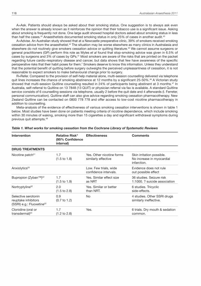
118 Australasian Anaesthesia 2011
A=Ask. Patients should always be asked about their smoking status. One suggestion is to always ask even when the answer is already known as it reinforces the opinion that their tobacco use is a significant issue. Asking about smoking is frequently not done. One large audit showed hospital doctors asked about smoking status in less than half the cases.42 Anaesthetists documented smoking status in only 25% of cases in another audit.43
A=Advise. An Australian study showed that at a Newcastle preoperative clinic, 39% of smokers received smoking cessation advice from the anaesthetist.44 The situation may be worse elsewhere as many clinics in Australasia and elsewhere do not routinely give smokers cessation advice or quitting literature.45 We cannot assume surgeons or general practitioners (GP) perform this role as Myles et al found that stop-smoking advice was given in 6.5% of cases by surgeons and 3% of cases by GPs.31 Most smokers are aware of the risks that are printed on the packet regarding future cardio-respiratory disease and cancer, but data shows that few have awareness of the specific perioperative risks that their habit poses for them.7 Smokers deserve to know this information. Unless they understand that the potential benefit of quitting before surgery outweighs the perceived unpleasantness of cessation, it is not reasonable to expect smokers to make behavioural change prior to surgery.
R=Refer. Compared to the provision of self-help material alone, multi-session counselling delivered via telephone quit lines increases the chance of smoking abstinence at 12 months by a significant 25-50%.46 A Victorian study showed that multi-session Quitline counselling resulted in 24% of participants being abstinent at 3 months.47 In Australia, self-referral to Quitline on 13 7848 (13 QUIT) or physician referral via fax is available. A standard Quitline service consists of 6 counselling sessions via telephone, usually 2 before the quit date and 4 afterwards (I. Ferreter, personal communication). Quitline staff can also give advice regarding smoking cessation pharmacotherapy. New Zealand Quitline can be contacted on 0800 778 778 and offer access to low-cost nicotine pharmacotherapy in addition to counselling.
Meta-analysis of the evidence of effectiveness of various smoking cessation interventions is shown in table 1 below. Most studies have been done on patients meeting criteria of nicotine dependence, which include smoking within 30 minutes of waking, smoking more than 15 cigarettes a day and significant withdrawal symptoms during previous quit attempts.48
Table 1. What works for smoking cessation from the Cochrane Library of Systematic Reviews
Intervention Relative Risk*(95% Confidence interval)
Effectiveness Comments
DRUG TREATMENTS
Nicotine patch51 1.7(1.5 to 1.8)
Yes. Other nicotine forms similarly effective
Skin irritation possible. No increase in myocardial infarction.
Anxiolytics55 Low. Few trials, wide confidence intervals.
Evidence does not rule out possible effect
Bupropion (Zyban™)52 1.7(1.5 to 1.9)
Yes. Similar effect size as NRT
36 studies. Seizure risk 1:1000. ? suicide association
Nortryptyline52 2.0(1.5 to 2.8)
Yes. Similar or better than NRT.
6 studies. Tricyclic side-effects.
Selective serotonin reuptake inhibitors (SSRI) e.g.: Fluoxetine52
0.9(0.7 to 1.2)
No 4 studies. Other SSRI drugs similarly ineffective.
Clonidine (oral or transdermal)54
1.7(1.2 to 2.8)
Yes. 6 trials. Dry mouth & sedation common.

Intervention Relative Risk*(95% Confidence interval)
Effectiveness Comments
Nicotine receptor partial agonists. e.g. Varenicline (Champix™)53
2.3(2.0 to 2.7)
Yes. Lower doses than standard also appear effective.
10 trials. Mild transient nausea? Depression & suicide association.
NON-DRUG TREATMENT
Hypnotherapy56 Low. Possibly not as good as counselling.
11 trials, very different designs.
Individual counselling50 1.4(1.2 to 1.6)
Yes. 30 trials, >7000 patients.
Group behaviour therapy49 2.0(1.6 to 2.5)
Yes. 53 trials. If patients like group attendance works better than individual counselling.
Rapid smoking aversive therapy57
2.0(1.4 to 3.0)
Yes. 12 trials. Problems with methodology in most.
Acupuncture & related techniques58
1.1(0.8 to 1.4)
Low. Little or no different from placebo.
33 studies, most effected by bias.
*Relative risk (RR) refers here to the ‘risk’ of successful cessation, usually measured at 12 months. Thus a RR of 1.7 for nicotine patches means patients receiving patches were 70% more likely to succeed at 12 months than control group patients.
Counselling is highly effective, whether individual or group based.49,50 Effective drug treatments include nicotine replacement therapy (NRT), bupropion and varenicline, while the place of nortryptyline and clonidine in smoking cessation is limited by side-effects.51-54 Varenicline is generally commenced one-week before the patient’s quit date and bupropion commenced two-weeks before the quit date.40 This may cause practical difficulties in cases when the timing of surgery is imminent.
NRT is commenced on the patient’s quit date, making it use easier.40 There is no evidence that NRT has a negative effect on postoperative outcome.59 Given the known cardiovascular side-effects of nicotine, it is somewhat counter-intuitive that the safety of NRT has been firmly established, even in the presence of cardiac disease.60 Guidelines from The Royal Australian College of General Practitioners state there is no evidence of increased risk for patients using NRT with stable cardiovascular disease, but it should be used with caution in recent MI, unstable angina or recent CVA.48 In many cases the benefit that NRT gives cardiac patients in stopping smoking outweighs the harm of continued smoking or the NRT itself.60
It is likely that components of cigarette smoke other than nicotine contribute significantly to the increased cardiovascular risk of smokers.27 Furthermore, peaks and troughs of blood nicotine levels after smoking are far greater with cigarettes than NRT.27 Nicotine patches do not appear to induce vasoconstriction resulting in poor wound healing and infection as wounds in abstinent smokers wearing NRT patches were no more likely to become infected or rupture than abstinent smokers without NRT.59
Studies comparing different forms of NRT (transdermal patches, gum, inhalers etc) did not strongly favour one form over another but studies using NRT combinations showed higher effectiveness (e.g. patch for baseline nicotine requirement and gum for breakthrough cravings).51 The initial dose of a sustained-release nicotine patch usually approximates the current daily nicotine intake so that a 20 cigarettes per day patient would be prescribed patches delivering 21mg/day.40 Nicotine (eg. gums) for breakthrough cravings 12mg/day.40 Tapering of patch doses generally occurs over a 4-week period.40
SMOKING CESSATION: THE PUBLIC HEALTH CHALLENGE FOR ANAESTHETISTS AND SURGEONS.The consistent and routine application of a smoking cessation strategy represents a challenge for anaesthetists, surgeons and health services. The first challenge is overcoming personal attitudes that prevent us from engaging with patients to inform them of the harms of smoking and giving encouragement and support to quit. This includes perceptions that smoking is a social rather than medical problem, assumptions that physician’s advice lacks effect on cessation outcomes, a deficit of knowledge or training and a reluctance to raise the topic because it may upset patients.3,41,44
The second challenge is time. The smoking status of a patient may only be discovered by the anaesthetist on the day of surgery. Systems to have alerting information available earlier should be considered. Despite political and media agendas that focus on long elective surgical waiting lists, the reality is that there is relatively little time to act in most cases as half of elective surgery at Australian public hospitals being currently done within 36 days.22 Whether smokers should have elective surgery delayed in order to participate in a smoking cessation program is a debate long overdue when one considers that since 1944 there have been over 300 papers showing the adverse effects of smoking on surgical outcomes.33
Smoking and surgery: time to clear the air 119

120 Australasian Anaesthesia 2011
The third challenge is to advocate within our health systems for effective and sustainable programs for smoking cessation based on established evidence-based models. Fund providers of acute care in hospitals may balk at funding requests for hospital-based interventions where the savings are not immediately apparent. This is unfortunate as even an intensive preoperative smoking cessation clinic model was shown to be cost-effective in terms of reducing overall hospital costs.61 More long-term community savings could be expected than reported in a hospital-cost analysis as many patients remain smoke-free 12 months or more after discharge from these programs.10
Anecdotally, very few health services in Australasia have systematic programs for patients joining surgical waiting lists that identify smokers and give them information and support on how to quit before surgery. Peninsula Health in Melbourne is currently commencing and evaluating a program where all smokers on the surgical waiting list will receive a brochure explaining the benefits of quitting as well as a reply-paid referral letter to enable Quit Victoria to commence free preoperative telephone counselling sessions*. No other Victorian health service systematically refers elective surgical patients to quit (I. Ferreter, personal communication), yet referral to Quitline is an integral part of smoking cessation guidelines for Australian general practice.48
The scale of the task is a challenge but also a significant opportunity for public health improvement. In the 2009/10 year there were 1.9 million elective operations in Australia, two thirds of which were in the private sector.22 In New Zealand public hospitals, there were 137,279 elective operations over the same period (C. Lewis, personal communication). Based on the current smoking prevalence it is likely that at least 360,000 smokers have elective surgery in our region each year. As cardiothoracic, vascular and certain cancer surgery is over-represented amongst smokers, this figure is probably conservative. Worldwide, an estimated 70 million smokers undergo major surgery each year.26
CONCLUSIONBefore he died of lung cancer at the young age of 45, the great US musician Nat King Cole asked his doctor to “get me well so I can get on television and tell people to stop smoking”62. It was too late for Nat and his cancer denied him that opportunity. Yet by routinely enquiring about smoking habits, advising to stop and referring for further help, anaesthetists can act on their favourable opportunity to end the misery that tobacco inflicts on many lives. Regarding smoking and surgery, it is time to clear the air.
*Further information, brochure template and fax referral form, visit clinician’s area at www.stopbeforetheop.blogspot.com
REFERENCES1. Buerk M: The Oxford Dictionary of Humorous Quotations, 2nd edition. Edited by Sherrin N. Oxford, OUP, 2005,
p. 285.
2. Peto R, Lopez AD, Boreham J, Thun M, Heath C, Doll R: Mortality from smoking worldwide. Br Med Bull 1996; 52: 12-21.
3. Swash M, Mason S: Hutchison’s Clinical Methods, 18th edition. UK, Bailliere Tindall, 1984, p. 10.
4. World Health Organization., Research for International Tobacco Control.: WHO report on the global tobacco epidemic, 2008 : the MPOWER package. Geneva, World Health Organization, 2008.
5. Collins DJ, Lapsley HM, University of New South Wales., National Drug Strategy (Australia), Australia. Dept. of Health and Ageing.: The costs of tobacco, alcohol and illicit drug abuse to Australian society in 2004/05, Dept. of Health and Ageing, 2008.
6. Ponniah S, New Zealand. Ministry of Health.: Tobacco trends: monitoring tobacco in New Zealand. Wellington, N.Z., Ministry of Health, 2006.
7. Shi Y, Warner DO: Surgery as a teachable moment for smoking cessation. Anesthesiology 2010; 112: 102-7
8. Turan A, Mascha EJ, Roberman D, Turner PL, You J, Kurz A, Sessler DI, Saager L: Smoking and perioperative outcomes. Anesthesiology 2011; 114: 837-46.
9. Rigotti NA, Munafo MR, Stead LF: Smoking cessation interventions for hospitalized smokers: a systematic review. Arch Intern Med 2008; 168: 1950-60.
10. Thomsen T, Villebro N, Møller AM: Interventions for preoperative smoking cessation. Cochrane Database Syst Rev 2010: CD002294.
11. Myers K, Hajek P, Hinds C, McRobbie H: Stopping Smoking Shortly Before Surgery and Postoperative Complications: A Systematic Review and Meta-analysis. Arch Intern Med 2011; 171: 983-9.
12. Møller AM, Villebro N, Pedersen T, Tønnesen H: Effect of preoperative smoking intervention on postoperative complications: a randomised clinical trial. Lancet 2002; 359: 114-7.
13. Stead LF, Bergson G, Lancaster T: Physician advice for smoking cessation. Cochrane Database Syst Rev 2008: CD000165.
14. Shi Y, Warner DO: Pediatric surgery and parental smoking behavior. Anesthesiology 2011; 115: 12-7.

15. Lindström D, Sadr Azodi O, Wladis A, Tønnesen H, Linder S, Nåsell H, Ponzer S, Adami J: Effects of a perioperative smoking cessation intervention on postoperative complications: a randomized trial. Ann Surg 2008; 248: 739-45.
16. ANZCA: Statement on smoking as related to the perioperative period. ANZCA, 2007. From: www.anzca.edu.au/resources/professional-documents/ps12.html. Accessed July 2011.
17. Warner MA, Offord KP, Warner ME, Lennon RL, Conover MA, Jansson-Schumacher U: Role of preoperative cessation of smoking and other factors in postoperative pulmonary complications: a blinded prospective study of coronary artery bypass patients. Mayo Clin Proc 1989; 64: 609-16.
18. Mitchell C, Garrahy P, Peake P: Postoperative respiratory morbidity: identification and risk factors. Aust N Z J Surg 1982; 52: 203-9.
19. Bluman LG, Mosca L, Newman N, Simon DG: Preoperative smoking habits and postoperative pulmonary complications. Chest 1998; 113: 883-9.
20. Warner MA, Divertie MB, Tinker JH: Preoperative cessation of smoking and pulmonary complications in coronary artery bypass patients. Anesthesiology 1984; 60: 380-3.
21. Bryson EO, Frost EA: The perioperative implications of tobacco, marijuana, and other inhaled toxins. Int Anesthesiol Clin 2011; 49: 103-18.
22. Australian Institute of Health & Welfare: Australian Hospital statistics 2009-10, Health services series no. 40. Canberra, AIHW, 2011.
23. Wilcox RG, Hughes J, Roland J: Verification of smoking history in patients after infarction using urinary nicotine and cotinine measurements. Br Med J 1979; 2: 1026-8.
24. Scherer G: Smoking behaviour and compensation: a review of the literature. Psychopharmacology (Berl) 1999; 145: 1-20.
25. Yamashita S, Yamaguchi H, Sakaguchi M, Yamamoto S, Aoki K, Shiga Y, Hisajima Y: Effect of smoking on intraoperative sputum and postoperative pulmonary complication in minor surgical patients. Respir Med 2004; 98: 760-6.
26. Chow CK, Devereaux PJ: The optimal timing of smoking cessation before surgery: comment on “smoking cessation shortly before surgery and postoperative complications”. Arch Intern Med 2011; 171: 989-90.
27. Warner DO: Perioperative abstinence from cigarettes: physiologic and clinical consequences. Anesthesiology 2006; 104: 356-67.
28. Hollenberg M, Mangano DT, Browner WS, London MJ, Tubau JF, Tateo IM: Predictors of postoperative myocardial ischemia in patients undergoing noncardiac surgery. The Study of Perioperative Ischemia Research Group. JAMA 1992; 268: 205-9.
29. Al-Sarraf N, Thalib L, Hughes A, Tolan M, Young V, McGovern E: Lack of correlation between smoking status and early postoperative outcome following valve surgery. Thorac Cardiovasc Surg 2008; 56: 449-55.
30. Al-Sarraf N, Thalib L, Hughes A, Tolan M, Young V, McGovern E: Effect of smoking on short-term outcome of patients undergoing coronary artery bypass surgery. Ann Thorac Surg 2008; 86: 517-23.
31. Myles PS, Iacono GA, Hunt JO, Fletcher H, Morris J, McIlroy D, Fritschi L: Risk of respiratory complications and wound infection in patients undergoing ambulatory surgery: smokers versus nonsmokers. Anesthesiology 2002; 97: 842-7.
32. Aköz T, Akan M, Yildirim S: If you continue to smoke, we may have a problem: smoking’s effects on plastic surgery. Aesthetic Plast Surg 2002; 26: 477-82.
33. Peters MJ, Morgan LC, Gluch L: Smoking cessation and elective surgery: the cleanest cut. Med J Aust 2004; 180: 317-8.
34. Sørensen LT, Toft BG, Rygaard J, Ladelund S, Paddon M, James T, Taylor R, Gottrup F: Effect of smoking, smoking cessation, and nicotine patch on wound dimension, vitamin C, and systemic markers of collagen metabolism. Surgery 2010; 148: 982-90.
35. Hampl JS, Taylor CA, Johnston CS: Vitamin C deficiency and depletion in the United States: the Third National Health and Nutrition Examination Survey, 1988 to 1994. Am J Public Health 2004; 94: 870-5.
36. Shi Y, Weingarten TN, Mantilla CB, Hooten WM, Warner DO: Smoking and pain: pathophysiology and clinical implications. Anesthesiology 2010; 113: 977-92.
37. Sweeney BP, Grayling M: Smoking and anaesthesia: the pharmacological implications. Anaesthesia 2009; 64: 179-86.
38. Chimbira W, Sweeney BP: The effect of smoking on postoperative nausea and vomiting. Anaesthesia 2000; 55: 540-4.
39. Twain, M: Brainy Quotes. From: http://www.brainyquote.com/quotes/quotes/m/marktwain128157.html Accessed July 2011.
Smoking and surgery: time to clear the air 121

122 Australasian Anaesthesia 2011
40. Simon JA: Smoking cessation interventions: a primer for physicians: Comment on “Use of varenicline for 4 weeks before quitting smoking”. Arch Intern Med 2011; 171: 777-8.
41. Warner DO, Force ASoASCIT: Feasibility of tobacco interventions in anesthesiology practices: a pilot study. Anesthesiology 2009; 110: 1223-8.
42. Ahluwalia JS, Gibson CA, Kenney RE, Wallace DD, Resnicow K: Smoking status as a vital sign. J Gen Intern Med 1999; 14: 402-8.
43. Simmonds M, Petterson J: Anaesthetists’ records of pre-operative assessment. Clin Perform Qual Health Care 2000; 8: 22-7.
44. Wolfenden L, Wiggers J, Knight J, Campbell E, Spigelman A, Kerridge R, Moore K: Increasing smoking cessation care in a preoperative clinic: a randomized controlled trial. Prev Med 2005; 41: 284-90.
45. Ratner PA, Johnson JL, Richardson CG, Bottorff JL, Moffat B, Mackay M, Fofonoff D, Kingsbury K, Miller C, Budz B: Efficacy of a smoking-cessation intervention for elective-surgical patients. Res Nurs Health 2004; 27: 148-61.
46. Stead LF, Perera R, Lancaster T: Telephone counselling for smoking cessation. Cochrane Database Syst Rev 2006; 3: CD002850.
47. Borland R, Segan CJ, Livingston PM, Owen N: The effectiveness of callback counselling for smoking cessation: a randomized trial. Addiction 2001; 96: 881-9.
48. Zwar N, Royal Australian College of General Practitioners.: Smoking cessation pharmacotherapy : an update for health professionals. Melbourne, Royal Australian College of Practitioners, 2007.
49. Stead LF, Lancaster T: Group behaviour therapy programmes for smoking cessation. Cochrane Database Syst Rev 2005: CD001007.
50. Lancaster T, Stead LF: Individual behavioural counselling for smoking cessation. Cochrane Database Syst Rev 2005: CD001292.
51. Stead LF, Perera R, Bullen C, Mant D, Lancaster T: Nicotine replacement therapy for smoking cessation. Cochrane Database Syst Rev 2008: CD000146.
52. Hughes JR, Stead LF, Lancaster T: Antidepressants for smoking cessation. Cochrane Database Syst Rev 2007: CD000031.
53. Cahill K, Stead LF, Lancaster T: Nicotine receptor partial agonists for smoking cessation. Cochrane Database Syst Rev 2011: CD006103.
54. Gourlay SG, Stead LF, Benowitz NL: Clonidine for smoking cessation. Cochrane Database Syst Rev 2004: CD000058.
55. Hughes JR, Stead LF, Lancaster T: Anxiolytics for smoking cessation. Cochrane Database Syst Rev 2000: CD002849.
56. Barnes J, Dong CY, McRobbie H, Walker N, Mehta M, Stead LF: Hypnotherapy for smoking cessation. Cochrane Database Syst Rev 2010: CD001008.
57. Hajek P, Stead LF: Aversive smoking for smoking cessation. Cochrane Database Syst Rev 2004: CD000546
58. White AR, Rampes H, Liu JP, Stead LF, Campbell J: Acupuncture and related interventions for smoking cessation. Cochrane Database Syst Rev 2011: CD000009.
59. Sorensen LT, Karlsmark T, Gottrup F: Abstinence from smoking reduces incisional wound infection: a randomized controlled trial. Ann Surg 2003; 238: 1-5.
60. Benowitz NL, Gourlay SG: Cardiovascular toxicity of nicotine: implications for nicotine replacement therapy. J Am Coll Cardiol 1997; 29: 1422-31.
61. Hejblum G, Atsou K, Dautzenberg B, Chouaid C: Cost-benefit analysis of a simulated institution-based preoperative smoking cessation intervention in patients undergoing total hip and knee arthroplasties in France. Chest 2009; 135: 477-83.
62. Cole, Nat King: Brainy Quotes. From: http://www.brainyquote.com/quotes/quotes/n/natkingcol315692.html Accessed July 2011.

Smoking and surgery: time to clear the air 123


The Management of Adult Jehovah’s Witnesses in Anaesthesia and Critical Care 125
The Management of Adult Jehovah’s Witnesses in Anaesthesia and Critical CareANNE-MARIE WELSH, BSC, MBCHB (HONS), MRCP, FCICM
Staff Specialist, Department of Critical Care Medicine, Nambour General Hospital, Nambour QLD 4560
Anne-Marie Welsh is a staff specialist in intensive care at Nambour General Hospital. She has a developing interest in follow up from intensive care
INTRODUCTIONJehovah’s Witnesses are widely known for their refusal to accept blood transfusions thus creating a potential obstacle to optimal medical therapy in situations such as trauma, obstetrics and surgery. Such refusal limits the clinician’s clinical freedom and may have both medicolegal and ethical consequences. When the consequence of treatment limitation results in an otherwise unavoidable death, feelings of guilt, frustration, anger and anxiety may occur and create an emotional burden for the care givers.
Working within the restrictions imposed by Jehovah’s Witness patients may also incur additional financial cost to both the hospital and state what with the, not infrequent, requirements for recombinant activated factor VII, prothrombinex concentrate, tranexamic acid and recombinant erythropoietin. In addition there is the cost of cell salvage techniques, additional theatre time to complete bloodless surgery safely and the requirement for consultant anaesthetic and surgical personnel to be present. Transfer of these patients to tertiary centres, or rarely, centres with hyperbaric oxygen facilities are likewise, an otherwise avoidable cost.
In addition, the requirement for limited resources such as intensive care or high dependency beds peri-operatively may deny access to other patients with a medically indicated need for them. This social injustice can create further ethical conflict for the physician.
RELIGIOUS BELIEFS AND THE PHYSICIANIt is estimated that approximately 1000 Jehovah’s Witnesses die annually worldwide and as many as 100,000 may have died by abstaining from blood transfusions since the blood ban was introduced in 1945. Despite this, as physicians we must be aware that every adult competent patient is entitled to refuse to consent to medical treatment for good reason, bad reason or no reason at all. Refusal to accept blood products should not adversely affect the quality of other cares provided.
THE JEHOVAH’S WITNESS FAITHAs the most rapidly growing religion in the western world, Jehovah’s Witnesses number some 7 million members in over 230 countries worldwide, with approximately 64,000 in Australia. In view of the rapidly growing membership all hospital physicians should be prepared to manage these patients well.
Their origins can be traced to a bible study group founded in the USA in 1869 by Charles Taze Russell.1 Some 10 years later, in 1879, the first issue of “The Watchtower”, their illustrated religious magazine, was published. Although originally known as “Zions’s Watchtower Tract Society”, they adopted the name “Jehovah’s Witnesses” in 1931.
Members are of the Christian faith. They are politically neutral, do not salute flags, enlist in the military nor vote in public elections. They celebrate neither Christmas nor birthdays and must satisfy a minimum monthly time requirement to their ministry.
OBJECTION TO MEDICAL TREATMENTSAlthough best known for their refusal to accept blood transfusions, Jehovah’s Witnesses originally had objections to other medical treatment such as vaccination (including the small pox vaccine) and transplantation. Vaccination has been permitted since 1952. Transplantation has been allowed since 1980, with the first solid organ transplant to a Jehovah’s Witness recipient occurring in 1986.
In 1945 the governing body of the Jehovah’s Witnesses “The Watchtower”, introduced the blood ban, based on the strict literal interpretation of several biblical passages, including “you are to abstain... from blood..” (Acts 15) and “none of you may eat blood..” (Leviticus 17).
In 1961 the Watchtower Society issued a statement that Jehovah’s Witnesses who consciously accept a blood transfusion violate the blood ban and are subject to expulsion, so called “disfellowshipping”.2 Other members of the faith are then instructed to shun and ostracise the expelled member, even if they are family members.3
According to a 1994 document, the Watchtower Society disfellowships approximately 40,000 members, or 1% or its membership, each year.4 The consequences include isolation from family and friends who are members and may even include separation from spouses.
In 1995 a policy change occurred such that acute normovolaemic haemodilution and red cell salvage were permitted. Later, in 2000, the Watchtower Society issued a directive stating that the Jehovah’s Witness organisation would no longer excommunicate members who did not comply with the blood ban; rather such an individual “revokes his own membership by his own actions, rather than the congregation initiating the step”.
Despite the media declaring this to be a major policy change, the end result was unchanged, that such an individual would be considered to revoke his or her religious affiliation.

126 Australasian Anaesthesia 2011
In 2000 the Watchtower Society also published the article “Questions from Readers”5 which redefined the guidelines for the use of blood products. It detailed which products are unacceptable and which are for the Christian to decide.
Unacceptable products included:1) Pre-operative autologous blood donation2) Transfusion of the “primary components” of blood, namely whole blood, packed red cells, plasma, platelets
and white cellsHowever, in a policy change, fractions of all the primary components red cells were now permitted as a matter
of personal decision; “beyond that, when it comes to fractions of any of the primary components, each Christian, after careful and prayerful meditation, must conscientiously decide for himself.5” The list of acceptable products includes, but is not limited to, albumin, cryoprecipitate, clotting factors, immunoglobulins, recombinant human erythropoietin, interferon, interleukins and even haemoglobin based blood substitutes or oxygen carriers. The most profound impact from this change will be seen if and when haemoglobin based oxygen carriers are introduced into general use.
As can be seen the blood ban is in a state of evolution and change, with the number of acceptable blood products ever increasing.
Indeed, within the Jehovah’s Witness faith itself there are dissident groups who believe the blood ban is unacceptable, has no biblical basis and is flawed by inconsistency. One such group is “The Associated Jehovah’s Witnesses for Reform on Blood”.6 The founder of this group maintains his status as a Jehovah’s Witness but writes under a pseudonym to avoid being expelled or “disfellowshiped”. The group argues:
“If the scriptures ban blood transfusions why does the Watchtower Society allow transfusion of all minor blood fractions?”
“Why are minor components like platelets (0.17% blood volume) and white cells (1% blood volume) forbidden yet a larger component like albumin (2.2% blood volume) is permitted?”
Why are Jehovah’s Witnesses permitted to accept ever increasing numbers of blood products (and transplants) yet are forbidden from contributing to the donor supply?”
Ultimately it is essential to seek the views of the individual patient. However, many Jehovah’s Witnesses are not aware of the numerous blood products that can now be accepted “as a matter of conscious”. Without such knowledge, decision making regarding blood products can not be considered “informed” or “autonomous” without undue influence from others. A detailed discussion of the evolution of the blood policy may be necessary before any consent can truly be considered “informed”.
The Hospital Liaison Committee for Jehovah’s Witnesses is comprised of a group of educated elders, who may assist a patient in making a decision. However it is imperative that Jehovah’s Witness patients, where possible, are also interviewed alone (including away from family members) so that they may make a decision free from coercion or guilt.
Some may request to receive a blood transfusion secretly after visiting hours or agree to a contingency plan such that they will only accept transfusion in the event of imminent death without transfusion. Confidentiality in these situations needs to be respected to avoid social and religious repercussions.
THE DANGERS OF BLOOD TRANSFUSIONThe lessons learnt from treating Jehovah’s Witness patients may benefit society as a whole.
As the deleterious effects of blood transfusion become more apparent, costs escalate and donor numbers decline, there has been an increase in transfusion avoidance strategies.
Many of the techniques developed for use in these patients will likely become standard practice in time, in an effort to conserve low blood stocks and minimise exposure to transfused blood.
The disadvantages of transfusion are many.
a. InfectionAlthough improvements in screening have reduced the risk of transmission of HIV, HBV and HCV, these infections do still rarely occur. In addition, severe acute respiratory syndrome (SARS), West Nile Virus, protozoa and prion-related disease are the latest to join the list of potentially transmissible diseases.
b. Suppression of Immune System FunctionSo-called Transfusion Related ImmunoModulation or TRIM, may now be one of the greater disadvantages of transfusion. Transfusion predisposes to infection, as seen in a study of 102 patients undergoing spinal fusion procedures. The patients received either autologous transfusion, allogeneic transfusion or neither. The infection rate of 4% in patients receiving no blood products was comparable to those receiving autologous blood transfusion (3.3%). However, patients receiving allogeneic blood transfusions had infection rates in excess of 20%. The number of allogeneic units transfused was the only significant predictor of in-hospital infection (p = 0.016) or days on antibiotics and length of stay.7
The effect of immunomodulation on malignancy remains unclear. In animal studies transfusion increases metastatic formation. Human studies are divided, but some have shown a correlation between transfusion and increased risk of tumour recurrence after potentially curative surgery. In particular, perioperative blood transfusion has been demonstrated to be a significant independent prognostic factor for colorectal cancer recurrence.8

Vincent et al performed a multicentre prospective observational study of 3534 critically ill patients admitted to 146 western European intensive care units during a 2 week period in 1994. Patients receiving transfusion had an increased intensive care length of stay, organ dysfunction score and overall 28-day mortality (29% vs 14.9%) compared to similar non-transfused patients.9
The TRICC trial (Transfusion Requirements In Critical Care) randomly allocated critically ill patients to either a liberal (<10g/dl) or restrictive (<7g/dl) transfusion threshold. There was a non-significant trend towards lower mortality in the restrictive group overall (18.7% vs 23.3%, p= 0.1). In addition these patients had a lower incidence of multiple organ dysfunction, myocardial infarction and acute pulmonary oedema.10
c. OthersImmunological and allergic reactions may complicate transfusion. ABO incompatibility through human error may have disastrous consequences. In addition, the metabolic consequences of transfusion (particularly massive transfusion) are well known and include acidosis, hyperkalaemia, hypocalcaemia and hypothermia.
The storage defect results in red blood cells with increased fragility and reduced ability to transport oxygen. Finally, respiratory failure may complicate transfusion due to either cardiogenic pulmonary oedema, transfusion associated circulatory overload or Transfusion Related Acute Lung Injury (TRALI).
PRINCIPLES OF BLOODLESS SURGERYThe term “bloodless surgery” refers to a series of measures in the pre-, peri- and post- operative care of patients that aims to reduce the need for allogeneic blood transfusion.11 There are over 230 “Bloodless Hospitals” worldwide12, however there is only 1 in Australia, Kaleeya hospital, East Fremantle, West Australia; it is a small hospital without an intensive care unit and provides mainly day surgery.
Bloodless surgery requires a coordinated multidisciplinary approach. Medical, anaesthetic and surgical teams, phlebotomists, pharmacists, physiotherapists and dieticians all need to be involved, where available.
Senior surgical and anaesthetic staff should be made aware of a pre-operative Jehovah’s Witness patient as soon as possible. A thorough discussion between patient, surgeon and anaesthetist should occur, detailing the risks, including intensive care stay and death. Which products will and will not be accepted should be documented in the notes and witnessed. The patient may agree to a contingency plan should death without transfusion become inevitable. The patient should be interviewed both with friends/ family and alone to avoid coercion. If the risk of bleeding and death is high, consider involving the hospital ethics committee, legal department, risk management group and Hospital Liaison Committee for Jehovah’s Witnesses.
Both anaesthetists and surgeons have the right to refuse to anaesthetise or operate on an individual in the elective situation provided they refer the case to a suitably qualified colleague who would be prepared to accept it.13
In the emergency situation however, both the anaesthetist and surgeon are obliged to provide care and legally must respect the patients’ views with respect to blood products.14
To administer a blood product to a competent adult after it has been explicitly refused is both illegal and ethically unacceptable.
PRE-OPERATIVE MANAGEMENTPre-operative patient assessment should include a thorough history and examination to allow estimation of
physiological reserve and ability to withstand hypovolaemia and anaemia. Pre-existing cardiac and respiratory disease should be optimised. Medications that may promote bleeding, such as antiplatelet agents, heparin, warfarin, dabagatrin, NSAIDS and fish oil, should be reviewed and ideally stopped. Coagulopathy should be corrected. Nutritional status should be reviewed and optimised with the use of enteral nutritional supplements and even consideration given to total parenteral nutrition if nutritional status is poor.
Enhanced haematopoiesis requires supplementation of iron, folate, vitamin B12 and ascorbic acid. Even in the absence of anaemia, recombinant human erythropoietin (rhEPO) can be used to improve red cell mass. The use of erythropoietin requires additional iron supplementation, usually intravenous, to be most effective. Erythropoietin has been demonstrated to half the rate of exposure to blood transfusion but requires approximately 4 weeks for maximal erythropoiesis to occur. Erythropoietin however is not devoid of side effects, with hypertension and thrombosis complicating its use.
INTRA-OPERATIVE MANAGEMENT1. SurgeryWith respect to intra-operative surgical technique; only senior personnel should perform procedures that carry a significant risk of bleeding. Where possible a minimally invasive technique should be employed, such as laparoscopic, endoscopic or staged procedures. For example a bilateral procedure should be performed as 2 separate unilateral procedures to minimise acute blood loss at each surgery.
Meticulous haemostasis is essential. The use of diathermy dissection or the harmonic scalpel can minimise bleeding depending on operator expertise.
Local haemostatic agents such as bone wax, fibrin glue, cellulose and collagen may also reduce haemorrhage. Where possible, drains should be placed to facilitate early detection of post operative bleeding.
The Management of Adult Jehovah’s Witnesses in Anaesthesia and Critical Care 127

128 Australasian Anaesthesia 2011
2. Anaesthesia techniquesWith respect to anaesthetic technique, again senior or consultant personnel should be involved. The patient, room and fluids should be warmed to prevent hypothermia and subsequent coagulopathy.
Venous congestion and venous ooze may be minimised by careful positioning and avoidance of high intra-thoracic pressures and hypercapnia.
Where feasible, tourniquets and infiltration of vasoconstrictor agents should be used.Regional techniques, where possible, will minimse blood loss. Serial measurement and correction of coagulation profile and ionized calcium should be considered in long
cases. Invasive monitoring should be considered to optimise tissue oxygen delivery, which is dependent upon haemoglobin
concentration, cardiac output and haemoglobin saturation. These factors may be manipulated using fluids, inotropes and increasing the FiO2.
O2 delivery (DO2) = cardiac output x (1.39 x Hb x SaO2) + 0.02 x PaO2
3. Anaesthesia drugsA number of drugs may be used peri-operatively to minimise bleeding.
a. Systemic antifibrinolytic agents, including tranexamic acid and eicoso-aminocaproic acid, inhibit plasminogen activity and promote coagulation. Tranexamic acid is given as a 1g infusion followed by 1g Q8H. The infusion rate should not exceed 100mg/min.
b. Desmopressin or DDAVP induces the release of Factor VIII, prostacyclin, tissue plasminogen activator and von Willibrand Factor from vascular endothelium. It has been demonstrated to reduce peri-operative blood loss associated with uraemic and aspirin-induced platelet dysfunction.15 The dose given is 0.3mcg/kg as an infusion over 30 minutes.
c. Prothrombin Complex Concentrate or Prothrombinex contains recombinant factors II, VII, IX and X. It may be acceptable to some Jehovah’s Witness patients. The recommended dose is 25-50IU/kg. It is relatively deficient in factor VII so works best when given along with a small amount of FFP and/ or rFVIIa.
d. Recombinant activated Factor 7 or Nova7 (rFVIIa) is reported to be effective in clinical situations associated with severe haemorrhage including cardiac surgery, trauma and obstetrics, but controlled clinical trials are scarce. Most case reports claim its use is associated with a reduction in blood loss and/ or transfusion requirements. Randomised controlled trials using rFVIIa in intra-cerebral haemorrhage demonstrate reduced growth of the hematoma but no improvement in survival or functional outcome.16 Likewise trials in blunt trauma demonstrate benefit on blood loss and transfusion requirements but not mortality.17 Its use is complicated by the occurrence of arterial and venous thromboses, especially in the elderly population and those with risk factors for peripheral vascular disease. As it is an off-license indication, there is no recommended dose, but between 50 and 90mcg/kg is generally given.
e. Individual clotting factors may be acceptable to some Jehovah’s Witnesses.f. Haemoglobin based oxygen carriers (HBOCs) could be employed in the future where available. They have
been in development for over 70 years; however interest has been renewed since the 1980’s, prompted by both the emergence of HIV and the death of trauma victims from exsanguination in the pre-hospital setting; including at accident scenes, in ambulances and on the battlefield. Despite this there are currently no haemoglobin based oxygen carriers approved for human use in Australia, the US or European Union; reflecting both a controversial history and the challenge of creating an ideal blood substitute.
HBOC DEVELOPMENTHaemoglobin based oxygen carriers contain purified haemoglobin derived from either bovine red cells, expired human red cells or from recombinant technology. Free human haemoglobin has a tetrameric structure of 2 alpha and 2 beta polypeptide chains. It rapidly dissociated into alpha/beta dimers which are cleared by glomerular filtration with an intravascular half life of only 30 minutes. Dissociated haemoglobin causes renal failure and scavenges nitric oxide causing hypertension. In addition, due to low concentrations of 2,3 DPG, free haemoglobin is ineffective at oxygenation due to its high affinity for oxygen, with a P50 of 10 to 14mmHg. In order to become therapeutically useful, free haemoglobin requires modification by polymerisation and/ or cross-linkage to prevent dissociation into alpha/beta dimers, right shift the oxyhaemoglobin dissociation curve and to increase its half life in the circulation. The modified haemoglobin is then incorporated into an electrolyte solution.
HBOC ADVANTAGESHaemoglobin solutions have the advantage of being non-immunogenic. They do not contain any intact red blood cells, which express ABO antigens, therefore cross-matching the product or typing the patient is not necessary.
The products undergo an extensive purification process to remove potential contaminants including proteins, red blood cell stroma, bacteria, endotoxins, viruses and prions and are therefore guaranteed to be disease free.
Haemoglobin based oxygen carriers may be stored far longer than the 42 days permitted for packed cells, between 12 months to 3 years, depending on the product. Hemopure is the easiest product to store and transport as it does not require refrigeration and may be stored for up to three years at room temperature.

Haemoglobin solutions are developed to right shift the oxyhaemoglobin dissociation curve compared to native haemoglobin. Hemopure contains bovine haemoglobin; it has a P50 of 40mmHg compared to 27mmHg for human red blood cells. It therefore releases oxygen more readily to the tissues and, on a gram-for-gram basis, restores oxygenation three times more effectively than a transfusion of stored human red blood cells. PolyHeme contains human haemoglobin, it has a P50 of 20-22mmHg which is comparable to packed cells.
HBOC DISADVANTAGESThere are a number of disadvantages of haemoglobin based oxygen carriers compared to blood. Despite modification, they have a short intravascular half life of 16 to 24 hours, compared to 60 to 90 days for red blood cells, making repeat administration necessary. Cost is also higher than transfused red blood cells when compared on a unit-to-unit basis.
Use of haemoglobin based oxygen carriers also interferes with many common laboratory tests, especially those which are measured spectrophotometrically. Albumin, alkaline phosphatase, bilirubin and creatinine may all be inaccurate. Optical methods of measuring coagulation will be misleading. Plasma will have a pink discoloration and routine laboratory tests will not be able to differentiate between haemolysis and the presence of a haemoglobin solution. Plasma free haemoglobin levels are measured to determine the amount of haemoglobin based oxygen carrier present in the specimen but the decision to give additional doses must be determined clinically.
The incidence of adverse effects is not insignificant (approximately ≥ 5%), with complaints of jaundice, nausea, mild to moderate increases in blood pressure, vomiting, oliguria, dysphagia and flatulence.
There are also reports of serious adverse events including myocardial infarction and death, however these risks may be acceptable when allogeneic blood is either not available or effective or not acceptable to the patient.
Furthermore, haemoglobin solutions have a maximum recommended dose, reflecting the maximum dose studied in clinical trials to-date, which may provide temporary oxygen-carrying support, or an “oxygen bridge” but may not be sufficient to completely avoid red cell transfusions in patients experiencing massive or continued blood loss.
HBOC PRODUCT HISTORYThere have been a number of haemoglobin oxygen carriers in production over the last 30 years but PolyHeme is probably the most controversial and Hemopure possibly the most promising.
PolyHeme is a haemoglobin based oxygen carrier derived from human hemoglobin and developed by Northfield Laboratories, Inc. Northfield was predominately a research and development company; PolyHeme was their only product. PolyHeme was the first blood substitute to reach a Phase III clinical trial in the US.
The trial was designed to assess the survival benefit of administering PolyHeme to severely injured trauma patients in hemorrhagic shock, beginning in the pre-hospital setting and continuing for 12-hours post-injury in hospital. It had two primary endpoints of superiority and non-inferiority to standard treatment. It was undertaken between January 2004 and July 2006 at 29 Level I trauma centers across 19 states in the US under a Food and Drug Administration (FDA) special category (21CFR 50.24) that allowed its use without consent. The waived informed consent rule was established by the FDA in 1996 and stipulated that to be used “available treatments …. (must be) unproven or unsatisfactory”. The only way to opt out from the study was by wearing a special bracelet prior to needing emergency care. The study was highly criticised due to the absence of consent. Indeed continuation of the study into the in-hospital period was considered unethical as blood was then both readily available and a proven and satisfactory therapy for haemorrhagic shock.
The results were published in the Journal of the American College of Surgeons in January 2009.18 They concluded there was no significant difference in outcome between the conventionally-resuscitated group and the PolyHeme-treated group. However PolyHeme was associated with an increased risk of myocardial infarction (3% versus 1%).
In May 2009 PolyHeme failed to receive FDA regulatory approval, with the FDA stating the risks of PolyHeme outweighed the benefits. In June 2009 Northfield Laboratories Inc ceased operation and filed for bankruptcy.
Hemopure, or HBOC-201, is developed from highly purified bovine haemoglobin. It is a third generation product developed by Biopure. It has been available for human use in South Africa since 2001 and in Russia since 2011. There is also a “compassionate use” program in the US which makes Hemopure available when a life-threatening situation exists and compatible red blood cell transfusion is either 1) not available 2) not effective or 3) not acceptable to the patient. Approval by the FDA is made on a case-by-case basis.
Following a motor vehicle accident in October 2010, Australian Jehovah’s Witness Tamara Coakley received a life sustaining transfusion of 10 units of Hemopure, flown in from the US and made available via this “compassionate use” scheme. Permission to use the product was granted by The Alfred Hospital Ethics Committee and the Therapeutic Goods Administration’s special access scheme. The manufacturer OPK Biotech paid for the costs involved.
Hemopure has undergone a Phase III clinical trial evaluating its ability to reduce or eliminate perioperative transfusion in orthopaedic patients.19 Hemopure reduced the need for packed cell transfusion in 59% of patients but was associated with a significantly higher incidence of both adverse events (rate of adverse event/ patient 44% higher, p<0.03) and serious adverse events (rate of serious adverse event/ patient 36% higher, p< 0.016).
The Management of Adult Jehovah’s Witnesses in Anaesthesia and Critical Care 129

130 Australasian Anaesthesia 2011
In 2008 a controversial meta-analysis comparing 16 clinical trials involving 5 different HBOC products used on over 3500 patients was published in the Journal of the American Medical Association.20 The study was led by Charles Natanson, a scientist at the US National Institute of Health. It concluded that patients treated with a HBOC had a 30% increased risk of death and 2.7-fold increased risk of myocardial infarction. Biopure responded by claiming there were fundamental errors in the calculations and analysis. Biopure then sued Natanson, claiming he had made “false and defamatory statements” about Hemopure. Following this South Africa’s Medicines Control Council temporarily de-registered Hemopure’s approved use for the treatment of acute surgical anaemia.
A 2009 application to the FDA for clinical approval of Hemopure in the US was declined. In August 2009 Biopure ceased operation and filed for bankruptcy.
Biopure has since been bought by OPK Biotech, a Russian owned company who has recently obtained approval for Hemopure to be used in Russia. OPK Biotech has also bought the intellectual property of Northfield Laboratories Inc, the company that developed PolyHeme. These acquisitions will likely make OPK Biotech a leading company in the field of oxygen therapeutics.
ANAESTHESIA BLOOD SAVING TECHNIQUESIn addition to the use of drugs there are a number of anaesthetic techniques available to minimise blood loss.
a. Controlled hypotension or hypotensive anaesthesia is a technique whereby the mean arterial pressure is maintained at a low level during surgery to minimise bleeding. It may decrease bleeding by as much as 50% but is controversial due to the risk of cerebral, renal and myocardial ischaemia. In addition, haemostasis that is adequate during controlled hypotension may not prove adequate when the patient returns to their normotensive, or worse still hypertensive, state.21 As such, the avoidance of marked haemodynamic shifts intra-operatively is more accepted.
b. Acute normovolaemic haemodilution involves the removal of whole blood from the patient pre-operatively and replacement with crystalloid or colloid to maintain intravascular volume. Blood lost intra-operatively has a reduced haemoglobin concentration resulting in fewer red cells lost overall. Provided the blood is kept in continuity with the patient the removed blood may be re-infused at the end of the case. This technique requires adequate respiratory and cardiac reserve to compensate for acute blood loss.
c. Acute hypervolaemic haemodilution uses the rapid infusion of fluid to achieve haemodilution without venesection. Again blood lost contains fewer red cells. Although acceptable to some Jehovah’s Witness patients it is poorly tolerated by those with cardiac disease, due to risks of fluid overload and heart failure.
d. Red cell salvage is a technique that can be used both intra-operatively and in the post-operative period to replace blood in proportion to the volume lost. Shed blood is collected, washed, mixed with anticoagulant and then re-infused via a filter. Many, but not all, Jehovah’s Witnesses will accept red cell salvage, again provided the circuit is not interrupted and remains in continuity with the patient. Red cell salvage is relatively contraindicated if there is the possibility of contamination with urine, fat, amniotic fluid, bone chips, bowel contents or infected material. Definite contraindications include re-infusion of anything that results in red blood cell lysis. This would include sterile water, hydrogen peroxide, and alcohol.
POST-OPERATIVE MANAGEMENT
a. Early Detection of Blood LossIn the post-operative period, early detection of blood loss is essential and can be facilitated by close monitoring in a critical care area and serial clinical examination of both the patient and their drains.
b. Minimise Iatrogenic Blood LossBlood loss can be minimised by avoidance of hypertension and marked haemodynamic shifts, and by reducing iatrogenic blood loss by infrequent and low volume blood sampling.
Two epidemiological studies of critically ill ICU patients have demonstrated similar figures for the mean volume of blood taken daily; 42.5ml/day in 1 study22 and 41.1ml/day in the other.23 The more unwell the patient, the more blood is likely to be taken.
Unnecessary blood loss can be avoided by abandoning “routine” tests which are not strictly indicated. When available, paediatric or small volume tubes should be used. If these are not available then small volumes should be used for all samples except coagulation profile, which is the only test that requires a full tube. Use of point of care micro-testing should also be employed where available.
c. Promote HaematopoiesisHaematopoiesis can be enhanced in the post operative period with the use of nutritional supplements, iron, folate, vitamin B12, vitamin C and recombinant erythropoietin if necessary.
d. Maximise Oxygen DeliveryOxygen delivery can be maximised in several ways.
Supplemental oxygen, chest physiotherapy and routine breathing exercises, such as the use of incentive spirometry, should be available to all patients. Cardiac output may be optimised with the use of fluids and inotropic agents where necessary.
Restoring intra-vascular volume is a controversial area. It may be prudent to avoid fluid resuscitation to euvolaemia if it results in haemodilution of haemoglobin down to a life threatening levels, however end organ hypoperfusion may also result from too conservative resuscitation.
The use of hyperbaric oxygen has been described in extremely severe anaemia, whereby the dissolved oxygen (PaO2) is sufficient to oxygenate tissues. However, there is very little evidence demonstrating a clear improvement in outcome and it is rarely a practical option but it could be considered for Jehovah’s Witnesses with inadequate oxygen delivery in whom other therapies have failed.
e. Minimise Oxygen ConsumptionSimilar to oxygen delivery, oxygen consumption may also be manipulated. The physiological response to pain, cold, anxiety and infection all result in an increase in basal metabolic rate and consequently higher oxygen consumption. Analgesia should be optimised and where appropriate antibiotic prophylaxis or treatment prescribed. Fevers, seizures and rigors should all be detected early and treated. Sedation, ventilation and maintenance of normothermia will all control basal metabolic rate; whilst in extreme cases paralysis and even cooling can be considered until haematopoiesis is maximised.
CONCLUSIONIn conclusion, Jehovah’s Witnesses refuse blood products not treatment.
Their refusal has led to a greater awareness of blood conservation strategies that are likely to become more common worldwide as the deleterious effects of blood transfusion become more apparent.
Implementation of bloodless surgical programs requires a multidisciplinary approach across all stages of peri-operative care.
REFERENCES1. Hughes DB, Ullery BW, Barie PS. The Contemporary Approach to the Care of Jehovah’s Witnesses. The Journal
of Trauma. 2008; 65: 237-247.
2. Muramoto O. Bioethical aspects of the recent changes in the policy of refusal of blood products by Jehovah’s Witnesses. British Medical Journal. 2001; 322: 37-39.
3. Harrington C. Father shunned by family for defying faith to save child. Canadian Press. Mar 11 2002.
4. Elder L. Why some Jehovah’s Witnesses accept blood and conscientiously reject official Watchtower Society blood policy. Journal of Medical Ethics 2000; 26: 375-380.
5. Watchtower Bible and Tract Society. Questions from readers. Watchtower 2000; June 15; 29-31.
6. Associated Jehovah’s Witnesses for the Reform on Blood. www.ajwrb.org.
7. Triulzi DJ, Vanek K, Ryan DH et al. A clinical and immunologic study of blood transfusion and postoperative bacterial infection in spinal surgery. Transfusion 1992; 32: 517-524.
8. Tartter PI. The association of perioperative blood transfusion with colorectal cancer recurrence. Ann Surg. 1992 December; 216(6): 633–638.
9. Vincent JL, Baron JF, Reinhart K, et al. Anemia and blood transfusion in critically ill patients. JAMA. 2002; 288(12): 1499–1507.
10. Hebert PC, Wells G, Blajchman MA, et al. A multicenter, randomized, controlled clinical trial of transfusion requirements in critical care. Transfusion Requirements in Critical Care Investigators, Canadian Critical Care Trials Group. N Engl J Med. 1999; 340(6): 409–417.
11. Smith WQ. Major Surgery without Blood Transfusion. Current Anaesthesia and Critical Care. 2000; 11: 42-50.
12. myblood. www.mybloodsite.com.
13. Bodnaruk Z, Wong CJ, Thomas MJ. Meeting the Clinical Challenge of Care for Jehovah’s Witnesses. Transfusion Medicine Reviews. 2004; 18: 105-116.
14. Management of Anaesthesia for Jehovah’s Witnesses. 2nd Edition. The Association of Anaesthetists of Great Britain and Ireland. November 2005.
15. Franchini M. The use of desmopressin as a haemostatic agent: a concise review. Am J Haematol 2007; 82: 731-735.
16. Mayer SA, Brun NC, Begtrup K et al; FAST Trial Investigators. Efficacy and Safety of Recombinant Activated Factor VII for Acute Intracerebral Hemorrhage. N Engl J Med 2008; 358: 2127-213.
The Management of Adult Jehovah’s Witnesses in Anaesthesia and Critical Care 131

132 Australasian Anaesthesia 2011
17. Boffard KD, Riou B, Warren B, et al. Recombinant factor VIIa as adjunctive therapy for bleeding control in severely injured trauma patients: two parallel randomized, placebo-controlled, double blind clinical trials. J Trauma 2005; 59: 8-15.
18. Moore EE, Moore FA, Fabian TC et al. Human Polymerized Hemoglobin for the Treatment of Hemorrhagic Shock when Blood Is Unavailable: The USA Multicentre Trial. J Am Coll Surg. 2009; 208: 1-13.
19. Jahr JS, Mackenzie C, Pearce LB et al. HBOC-201 as an Alternative to Blood Transfusion: Efficacy and Safetly Evaluation in a Multicentre Phase III Trial in Elective Orthopedic Surgery. J Trauma. 2008; 64: 1484-1497.
20. Natanson C, Kern SJ, Lurie P et al. Cell-Free Haemoglobin-Based Blood Substitutes and Risk of Myocardial Infarction and Death. A Meta-analysis. JAMA. 2008; 299 (19): 2304-2312.
21. Gohel MS, Bulbulia RA, Slim FJ, et al. How to approach major surgery where patients refuse blood transfusion (including Jehovah’s Witnesses). Ann R Coll Surg Engl. 2005; 87: 3-14.
22. Smoller B, Kruskell M. Phlebotomy for diagnostic laboratory tests in adults: Pattern of use and effect on transfusion requirements. N Engl J Med 1986; 314: 1233-1235.
23. Vincent J, Baron J, Reinhart I et al. Anaemia and blood transfusion in critically ill patients. JAMA 2002; 288: 1499-1507.

133


Intravenous Iron in Surgery and Obstetrics
DR ROGER BROWNING MBCHB (OTAGO), FANZCA
Dr Browning is a staff specialist anaesthetist at both King Edward Memorial Hospital for Women (KEMH) and Fremantle Hospital in Western Australia. He is the medical supervisor of the infusion unit at KEMH, which administers approximately 5 – 10 iron polymaltose infusions a week. He also has an interest in rotational thromboelastometry and it’s role in the management of major obstetric haemorrhage. He is a member of the Western Australia Department of Health Patient Blood Management steering committee.
INTRODUCTIONMany anaesthetists may wonder what relevance intravenous iron has to anaesthesia. However anaemia, blood loss, and blood transfusions are issues we encounter frequently in our surgical patients. The last 10 – 15 years have seen some remarkable advances in the understanding of iron metabolism and related disorders such as anaemia of chronic disease. This has coincided with an increased concern regarding the hazards of blood products and their association with adverse outcomes in many surgical patient groups. New intravenous iron formulations have also been developed, one of which has been recently approved in Australia. They possess advantages such as the ability to infuse iron over short periods of time making this therapy potentially easier to incorporate into everyday clinical practice.
Despite this, intravenous iron is used infrequently in perioperative patients. This may be partially explained by a misperception that intravenous iron has a poor safety profile and a high incidence of anaphylactoid reactions. This association originated with the now historical use of high molecular weight iron dextran,1 a product withdrawn from Australia over 15 years ago. The currently available formulations have greatly improved safety profiles. Intravenous iron is actually very safe, effective, and can more rapidly correct anaemia than oral iron therapy. We should consider it more often as a potentially useful treatment in our anaemic surgical and obstetric patients.
ANAEMIA, IRON DEFICIENCY AND THE SURGICAL PATIENTAnaemia and iron deficiency are common conditions in surgical patients,2 both before and after surgery. Anaemia and iron deficiency are undesirable for a number of reasons, which include:• Impaired aerobic capacity, mental processing and increased fatigue all of which impair recovery after surgery.• Increased risk for perioperative transfusion• An association between preoperative anaemia and increased morbidity and mortality following surgery.2
By identifying and correcting anaemia in patients prior to surgery associated with significant blood loss (e.g. joint replacement surgery) we could potentially improve patient outcomes.
Important causes of anaemia in surgical patients include:• iron deficiency anaemia (IDA), common causes: menorrhagia, colorectal cancer, inflammatory bowel disease
(IBD), previous bariatric surgery, and pregnancy.• surgical blood loss.• venesection for blood tests.• anaemia of chronic disease.
IRON PHYSIOLOGYMany of us may have slept through the lectures at medical school when we were taught about iron physiology! However a basic understanding of iron physiology and the recent advances in this field such as the role of hepcidin and ferroportin are fundamental in order to appreciate the indications and appropriate use of intravenous iron in the management of anaemia.
Where is the Iron?Total body iron ranges from approximately 2500 – 4000mg in healthy individuals,3 the majority of which resides in a patient’s red cell mass. In the hypothetical average person this comprises:
Haemoglobin 2600mg
Iron Stores 500 – 1000mg
Myoglobin / Cytochromes 400mg
Transferrin 3mg
Intravenous Iron in Surgery and Obstetrics 135

136 Australasian Anaesthesia 2011
Iron Absorption and TransportAdults absorb about 1 mg per day of the 15 mg per day of dietary iron present in a normal adult diet. This is the approximate amount that is lost daily by epithelial shedding in the digestive tract. Healthy individuals stay in iron balance by increasing the fraction of iron absorbed from their diet if loss of iron increases (e.g. through blood loss in menstruating women). In iron deficiency, even with increased dietary intake, iron absorption rarely increases to more than about 6 mg/day. Oral iron supplements will facilitate greater absorption rates than this.
If blood loss continually exceeds the body’s ability to increase gastrointestinal iron absorption by a comparable amount, iron deficiency results. Thus, chronic blood loss, intestinal malabsorption, or dietary deficiency may all result in iron deficiency. Both non-haem and haem iron are absorbed from the diet. Haem iron is better absorbed than non-haem iron. Non-haem dietary iron in the form of ferric iron must first be reduced to ferrous iron by ferric reductase enzymes present in duodenal enterocytes, before it can be absorbed. Inhibition of gastric acid secretion, such as with proton pump inhibitors (PPI) or H2 antagonist use, will also impair the conversion of ferric iron to the ferrous form. At this point, all absorbed dietary iron, will be present in the enterocyte as ferrous iron, bound to ferritin in the cell cytoplasm.
The transfer of iron, (stored temporarily as ferritin), out of duodenal enterocytes onto transferrin in the plasma cannot take place without a membrane bound transport protein called ferroportin.
This is the natural control point in intestinal iron absorption. Without ferroportin, iron remains trapped in the enterocytes. The short lifespan of enterocytes (2 days) ensures that iron trapped in this way is shed with the senescent enterocytes into the faecal stream. The journey that dietary iron follows before being incorporated into a new red blood cell involves one more protein: hepcidin. Hepcidin is the primary regulatory protein of iron absorption and transport. It regulates iron absorption and transport by binding to ferroportin. In iron deficiency and in anaemia, hepcidin levels decrease, increasing available ferroportin. Iron absorption into the plasma increases and is ultimately delivered to the bone marrow or reticuloendothelial stores.
Slow onset iron deficiencyIn chronic blood loss iron stores are usually depleted before anaemia develops. Severe chronic iron deficiency is accompanied not only by anaemia, but also by deficiencies in a range of haem and non-haem iron dependent enzymes. Therefore, iron deficiency, if severe and prolonged, can cause dysfunction of iron-containing cellular enzymes and contribute to fatigue and loss of functional status via mechanisms independent of the anaemia itself.
“Acute” iron deficiency (e.g. major surgical blood loss)Following major acute blood loss, because the majority of body iron resides in circulating haemoglobin, a patient may develop an overall body iron deficit but retain normal iron stores at least initially. These iron stores however may be insufficient to allow complete replacement of the lost haemoglobin and the patient should be considered to have an overall body iron deficit. This is best illustrated by an example:
A hypothetical 80 kg male with a preoperative haemoglobin of 140 g/L undergoes a complicated bowel resection for colorectal cancer with an intraoperative blood loss of > 2.5 litres. Postoperatively he has a haemoglobin of 75 g/L. He has lost approximately 1300mg of body iron intraoperatively (contained in the lost red cells). He will only have body iron stores of 500 – 1000mg available to his bone marrow to manufacture new haemoglobin, which is 300 – 800mg short of what he needs. He has just undergone major bowel surgery and his ability to replace these losses through increased enteral absorption will also be limited. If he has a prolonged postoperative hospital stay, regular venesection for blood tests may result in ongoing iron losses, which may further exacerbate his problem.
Anaemia of Chronic DiseaseMany diseases (e.g. chronic kidney disease, inflammatory bowel disease, malignancy) and some physiological states (e.g. postoperatively) result in increased hepcidin levels. High levels of hepcidin bind the available ferroportin and induce its degradation. In the absence of adequate ferroportin, dietary iron is trapped in the enterocytes, preventing its transport into the plasma and delivery to the bone marrow for haemoglobin synthesis. Gastrointestinal iron absorption is inhibited and over time true iron deficiency may result.
High hepcidin levels also inhibit the movement of iron from stores to the bone marrow, leading to a condition known as functional iron deficiency.
Ferritin is also an acute phase reactant protein, which may be elevated in inflammatory states independent of actual iron store status. In a patient with an inflammatory response, a clinician may, inappropriately interpret a raised ferritin, as indicative of adequate iron stores when in fact underlying true iron deficiency may be present.
Menstruation and PregnancyIron deficiency is common in women of reproductive age due to the increased losses from menstruation, and increased requirements during pregnancy. When pregnant the mother must increase her own red cell mass and provide iron to her developing foetus. Iron deficiency and anaemia may be further compounded by any blood loss that occurs during childbirth at the end of a pregnancy, a time when most mothers may have already depleted their iron stores.• Pregnancy results in an overall additional iron requirement of around 1000mg.3
• IDA has been associated with low birth weight, prematurity, and maternal morbidity.4
• Menstruating women on average lose twice as much iron as men, and IDA is very common in women with menorrhagia.

Treatment points• Dietary iron alone (e.g. high red meat intake) is inadequate to treat established iron deficiency anaemia.3
• Common medications such as H2 antagonists or proton pump inhibitors decrease the effectiveness of oral iron therapy.3
• Gastrointestinal iron is poorly absorbed in patients with raised hepcidin levels (inflammatory disease, malignancy, immediate postoperative period)
• Ferritin is an acute phase protein, which is often raised in patients with inflammatory disease processes and so may not accurately reflect body iron stores. Some anaemic patients, with high or normal ferritin, may still have true iron deficiency or functional iron deficiency, which will often respond to intravenous iron therapy.A useful algorithm for assessing anaemia in pre-operative patients has been developed by the Western Australia
Department of Health Patient Blood Management Program a copy of which is provided at the end of this article.
TREATMENT OPTIONS FOR ANAEMIAOral Iron The traditional first line treatment for iron deficiency anaemia (IDA) is oral iron therapy. In appropriate dosage and given enough time, oral iron is an effective treatment for simple iron deficiency anaemia. Some important considerations in the anaemic perioperative surgical patient include:• Oral iron requires sufficient time to produce an adequate response. In IDA an increase of 20g/L every 3 weeks
is the maximum to be expected. In practical terms this implies weeks to months of treatment. • 30 – 50% of patients may experience gastrointestinal adverse effects and non-compliance is high.• Oral iron will be ineffective in patients with raised hepcidin levels, (e.g. immediate postoperative period,
chronic inflammatory disease)• H2-antagonists and proton pump inhibitor medications will reduce the efficacy of oral iron therapy.• It is important that patients take an oral preparation which contains an adequate dose of elemental iron
with100-200mg / day recommended (e.g. 1–2 Ferro grad). There are more than 100 iron containing over the counter preparations available in Australia many of which contain woefully inadequate amounts of elemental iron (often < 5mg). Many would require 20 tablets for a therapeutic dose!3
Red Cell TransfusionThere is evidence that transfusion to correct iron deficiency anaemia is still an overused treatment.5 Targeted use of transfusion is an appropriate strategy when severe anaemia compromises organ function (e.g. cardiac failure, angina pectoris) or there is ongoing serious bleeding. Iron therapy to fully replenish haemoglobin and iron stores will still be required.
Intramuscular IronIntramuscular injection of iron polymaltose is not recommended in most circumstances,6 unless other approaches are impractical (e.g. in remote communities). It is painful, causes skin discolouration, repetitive injections are required, and it is less effective but no safer than IV administration.
Intravenous Iron Pharmacology7
Administration of unbound inorganic ferric (Fe+3) iron in the early 1930s was observed to cause profound toxicity. Therefore all modern intravenous iron preparations are colloids consisting of a protective spheroidal carbohydrate, which encapsulates an inner iron hydroxide core. After IV injection the iron carbohydrates distribute in the plasma, from where the majority then enter the reticuloendothelial system (mainly liver, spleen and bone marrow). From here iron is either stored as ferritin or transferred out via transferrin to the bone marrow to produce haemoglobin. A small amount of “free iron” from the IV iron may be released directly onto transferrin in the plasma; this is more so with the smaller molecules such as iron sucrose, which explains why it cannot be used for total dose infusions.
Variation in the molecular weight of the encapsulating carbohydrate explains most of the differences in the pharmacological properties of the different preparations. The rate of clearance from the plasma and the rate of release of iron from the ferric hydroxide core are inversely related to the total molecular weight. The general rule is the smaller the molecule the more rapid the release of the iron, and the lower the maximum dose you can give.
Adverse Effects and Toxicity8
Reactions During InfusionDuring administration of an intravenous iron polymaltose infusion a number of adverse effects can occur occasionally. The vast majority are not serious, are self-limiting in nature and have been described to varying degrees with all the other intravenous iron preparations. These include nausea, rash, headache, mild hypotension, myalgia, arthralgia, chest and back pain. The mechanism of these adverse reactions is hypothesised to be related to the small amount of “free or labile” iron released during an infusion.
Intravenous Iron in Surgery and Obstetrics 137

138 Australasian Anaesthesia 2011
Tips to Managing ReactionsMost of these adverse reactions can be managed by pausing the infusion for a period of time, decreasing the rate and use of paracetamol or antihistamines. These phenomena can be misinterpreted by inexperienced nursing and medical staff as hypersensitivity/allergic reactions. The usual result in this situation is to abandon the infusion to the detriment of the patient. They may be told they are “allergic to iv iron” and instructed to avoid it in the future. In experienced infusion units where medical and nursing staff are used to observing and managing these phenomena almost all infusions of iron polymaltose can be completed successfully.9 The patients are warned of these possible adverse effects prior to the start of the infusion, so that if they do occur, they are expecting them to be managed and the infusion completed. A good analogy is the use of opioids for someone who has acute pain. Nausea or vomiting is not infrequent, but it is expected, managed with antiemetics and treatment continues.
The “Fishbane” ReactionThis uncommon phenomenon consists of acute chest and back pain, which can occur at any stage during an iron infusion. It is thought to be a form of acute myalgia / muscle cramp occurring in the chest and back muscles. It is potentially distressing to inexperienced staff and patients if misinterpreted as something more serious. It is named after Dr Fishbane, a US physician who describes it occurring during iron dextran infusions.10 It has been seen with other iron preparations including iron polymaltose (personal experience). It is benign, settles with temporary cessation of the infusion and invariably it does not return when the infusion is recommenced.
AnaphylaxisHigh molecular weight iron dextran was associated with a high incidence of true anaphylactoid reactions (wheeze, hypotension, angioedema, urticaria), with some reports of deaths.1 These were due to the high molecular weight dextran molecules (not the iron component). Dextran based intravenous fluids are also used infrequently now because of the high incidence of anaphylactoid reactions observed in these products. This preparation is no longer available in Australia (although it is still available in the US). All of the formulations of parenteral iron currently available in Australia have a low incidence of serious adverse reactions. Iron sucrose, used widely in Europe and the UK is the preparation with the best safety data available. It has a lower documented rate of serious adverse reactions than many antibiotics such as penicillin or cephalosporins.11
Does IV Iron increase infection risk? In vitro studies have shown that iron can encourage growth of some types of bacteria. There is a large body of reassuring data that this is not a problem in clinical practice.12 Large prospective and retrospective studies in chronic renal failure patients receiving peritoneal and haemodialysis (i.e. with long-term invasive catheters at risk of bacterial infection) have failed to show any increased infection risk in those receiving intravenous iron. It is however prudent practice to avoid using intravenous iron in anyone who has a suspected or confirmed bacterial infection.
Delayed ReactionsSelf limiting side effects can occur up to 2 days after an iron infusion and patients should be warned about these.13 These include headache, fever, arthralgias and myalgias, the majority are mild and well tolerated. Methylprednisolone administered with total dose iron dextran infusions has been shown to reduce the frequency and severity of these symptoms.14 However there have been no studies with iron polymaltose and steroids or premedication is not standard practice in Australasia.
IndicationsIntravenous iron therapy is indicated when there is confirmed iron deficiency anaemia (IDA) and one of the following:• Failure of oral iron (e.g. non compliance, adverse effects or lack of response)• A clinical need for a rapid response• Ongoing losses (e.g. bleeding) exceeding oral absorptive capacity.• Known intestinal malabsorption (IBD, celiac disease, postoperatively)• Chronic renal impairment in conjunction with erythropoietin
Intravenous iron may also be useful in patients with anaemia of chronic disease or functional iron deficiency but in most circumstances it should be used in consultation with an expert experienced with iv iron in that patient group.
Contraindications• Iron overload / haemochromatosis• Bacterial infection• Known hypersensitivity• First trimester of pregnancyAlso listed on drug insert for iron polymaltose only:• Chronic polyarthritis• Bronchial asthma• Uncontrolled hyperparathyroidism• Cirrhosis / hepatitis• Renal infections

Intravenous Iron FormulationsIron PolymaltoseThe most common formulation of parenteral iron in Australia and New Zealand is iron polymaltose (iron dextrin), which has been widely used here since the 1960s. This preparation is not used in Europe or North America. Iron polymaltose can be given as a total dose infusion (TDI). Typical doses would be 1000 – 2500mg and a patient only needs one infusion to replace their entire calculated iron deficit. Infusion rates can vary widely but usually require between 1 – 5 hours to administer a TDI. The small number of publications available suggest that it appears to have a very low rate of anaphylaxis or other serious adverse events.9 Less serious adverse effects such as arthralgia, headache, nausea, rashes are seen more frequently.
Iron SucroseIron sucrose has been used for the treatment of general iron deficiency in Europe and North America for decades. When iron sucrose was introduced into Australia it was only licensed for use in chronic kidney disease. Despite this it is used as an alternative to iron polymaltose in other patient groups in many Australian hospitals. Iron sucrose has been used for a long period of time and it has a large body of literature confirming its safety. Published data from the US puts the serious adverse event rate at 0.6 per million doses,11 which put into context is lower than common drugs such as penicillin or cephalosporins.
Iron sucrose cannot be given as a single total dose infusion but instead multiple smaller doses of 100-300mg are used. It is usually given as an infusion over 30 – 60 mins but has been licensed as a slow IV injection in the UK and NZ.
Iron Carboxymaltose Iron Carboxymaltose is a new formulation, which has been available in Europe for a number of years and has recently been approved for use in Australia (2011). It has a low incidence of serious adverse events, and doses of 1000mg can be given over 15min without the need for any test dose. It is however currently unfunded by the Pharmaceutical Benefits Scheme. It will cost more than iron polymaltose but the increased convenience and lower labour costs associated with shorter infusion times may offset this disadvantage to some degree.
Other PreparationsLow molecular weight iron dextran, iron gluconate and ferumoxytol are other preparations available internationally but not here in Australia.
SUMMARY OF THE EVIDENCEObstetricsPostpartum anaemiaA number of studies, of varying quality, have investigated iron sucrose and iron carboxymaltose for the management of postpartum anaemia usually in comparison to oral iron sulphate tablets. In general these studies found a good response to both treatments. The large RCT’s using iron carboxymaltose found a greater proportion of women receiving intravenous iron achieved higher haemoglobins, higher iron stores, over shorter time frames than with oral iron therapy alone.15 Adverse event rates were similar in both oral and intravenous treatment arms in most studies with no serious safety concerns found.
Antepartum AnaemiaThe most relevant study to Australasia is a recently published RCT conducted in Launceston, which recruited 200 women with mild or moderate IDA in pregnancy.16 They compared a single iron polymaltose infusion followed by oral iron to oral iron alone. Those receiving iron polymaltose had higher mean haemoglobin, ferritin and quality of life / fatigue scores.
*Of note the compliance rates with oral iron is generally high in a study population, which has well-motivated participants who are closely followed and encouraged to take their tablets. In real life many patients will not complete a prolonged course of oral iron (usually 6-12 weeks). High discontinuation rates of oral iron therapy have been documented in some audits due to gastrointestinal adverse effects.17 A total dose iron infusion prior to hospital discharge is one way to guarantee high-risk patients receive adequate iron repletion.
SURGICAL PATIENTSOverall there is a disappointing lack of large well conducted prospective studies investigating the role of intravenous iron in surgical patients.18 Lack of high quality evidence doesn’t equate to lack of efficacy. Although most studies were of low quality the available data is suggestive of benefit in anaemic patients with both preoperative and postoperative intravenous iron use. There is however an obvious need to remedy this deficiency and conduct some large prospective RCT’s in this area.
Intravenous Iron in Surgery and Obstetrics 139

140 Australasian Anaesthesia 2011
Oral Iron• Preoperative oral iron: 2 positive studies one in colorectal surgery and the other orthopaedic surgery found
preoperative oral iron reduced transfusions. • Postoperative oral iron: 5 RCT’s (4 orthopaedic and 1 cardiac surgery) found that postoperative oral iron was
not beneficial.18
Intravenous IronOrthopaedics: Intravenous iron use was beneficial in most studies. It was associated with fewer transfusions, less anaemia, decreased length of stay, and decreased postoperative infections. In some studies it was combined with other modalities such as EPO or cell salvage. This is the most well studied surgical group (mainly joint replacement surgery).
Cardiac Surgery: One RCT and one observational study failed to confirm any benefit from postoperative intravenous iron.
Gynaecological Surgery: One RCT of 76 patients found preoperative iron sucrose superior to oral iron in correcting the anaemia of women with menorrhagia and Hb<9g/L prior to surgery.19
Colorectal Surgery: One small preoperative RCT failed to show a benefit to intravenous iron sucrose 600mg prior to surgery. This study has been criticised for having only a small proportion of patients with anaemia (i.e. those patients most likely to benefit from intravenous iron).20
PUBLISHED LITERATURE – OTHER INDICATIONSIntravenous iron has also been shown to be an effective treatment in :• Congestive heart failure (improves NYHA status and fatigue scores)• Restless legs syndrome• Chronic Kidney Disease (adjunct to EPO)• Chemotherapy induced anaemia (+/- EPO)• As an MRI contrast agent (iron ferumoxytol)
GENERAL RECOMMENDATIONS • Patients scheduled for major surgery (or childbirth) with predicted moderate to major blood loss should be
screened for anaemia preoperatively, and if clinically feasible this should be corrected.• Patients without a clear physiological explanation for iron deficiency (especially men and postmenopausal
women) should be investigated to exclude serious underlying disorders especially occult GI malignancy. This should take priority over purely elective surgical procedures.
• With confirmed iron deficiency anaemia, intravenous iron should be considered in preference to oral iron if:• there is fewer 30 days to surgery• when it is reasonable to assume oral iron will be ineffective (e.g. postop)• Intravenous iron may be useful in anaemia of chronic disease, and it will improve the response to
erythropoietin if this is used.• Intravenous iron should be considered in the treatment of antepartum and postpartum iron deficiency
anaemia, especially if a rapid response is desirable or oral iron has failed.• Avoid intravenous iron during acute infections.• If the overall goal is avoiding perioperative blood transfusions this probably best accomplished by combining
intravenous iron with a range of other techniques (e.g. intraoperative cell salvage).
HYPOTHETICAL SCENARIOS TO PONDER:How do you think these patients could be managed? How do you think they would actually be managed currently in the hospital you work in? 1. You see a 64 yr old woman in the anaesthetic preadmission clinic 3 weeks prior to a planned anterior
resection for a recently diagnosed rectal cancer. On reviewing her recent blood tests you note she has a microcytic anaemia with a Haemoglobin (Hb) of 94g/L.
2. A 74 yr old male presents for a left total hip replacement having undergone a right total hip replacement 6 months earlier. He is diabetic, with mild renal impairment (Creatinine 150) and you note he has a normocytic anaemia with a Hb 102 g/L.
3. A 47 yr old woman with longstanding menorrhagia and fibroids presents to preadmission clinic 2 weeks prior to a planned elective total abdominal hysterectomy. She states her gynaecologist advised her to take ferrograd C two months ago but she stopped after 3 days because of nausea and constipation. A FBC from her GP 2 weeks ago shows a microcytic anaemia with a Hb 81g/L.
4. A fit and healthy 27 yr old parturient develops an atonic uterus following emergency caesarean section for twins, and has a 1.5 L blood loss which is successfully managed in theatre. You check her haemoglobin in recovery and it is 76 g/L. You note her full blood count on admission to labour ward yesterday that she had a microcytic anaemia with a Hb 104 g/L. She isn’t that keen on the idea of a blood transfusion but the obstetric registrar wants “to top her up so she’ll have plenty of energy” to look after two newborn twins when she is discharged home in a few days.
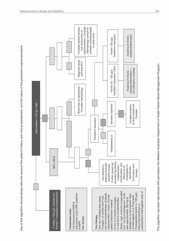
Intravenous Iron in Surgery and Obstetrics 141
Use
of t
his
algo
rithm
sho
uld
alw
ays
take
into
acc
ount
the
pat
ient
’s h
isto
ry a
nd c
linic
al a
sses
smen
t, a
nd t
he n
atur
e of
the
pro
pos
ed s
urgi
cal p
roce
dur
e
Pre
op
erat
ive
test
s:Fu
ll b
lood
pic
ture
(FB
P) &
re
ticul
ocyt
e co
unt
CR
P, c
reat
inin
e
& e
GFR
Iro
n st
udie
s
*Iro
n th
erap
yO
ral i
ron
in d
ivid
ed d
aily
dos
es
if su
rger
y >
2 m
onth
s an
d if
ora
l iro
n no
t co
ntra
ind
icat
ed. E
valu
ate
resp
onse
aft
er 1
mon
th. P
rovi
de
pat
ient
info
rmat
ion
mat
eria
l.IV
iron
if <
2 m
onth
s to
sur
gery
or
ora
l iro
n is
con
trai
ndic
ated
.‡N
ote:
1µg
/L o
f fer
ritin
is e
qui
vale
nt
to 8
–10
mg
of s
tora
ge ir
on. I
t w
ill
take
ap
pro
xim
atel
y 16
5 m
g of
st
orag
e iro
n to
rec
onst
itute
10g
/L
of h
aem
oglo
bin
in a
70
kg a
dul
t.
If p
reop
erat
ive
ferr
itin
is <
100
µg/L
, b
lood
loss
res
ultin
g in
a
pos
top
erat
ive
haem
oglo
bin
dro
p o
f
If fr
ette
n <
100
µg/L
con
sid
er ir
on
ther
apy*
if e
xpec
ted
pos
top
erat
ive
Hae
mog
lob
in <
130
g/L
mal
e
Blo
od lo
ss o
r ha
emol
ysis
. C
onsi
der
hae
mat
olog
y
Ferr
itin
45 –
100
µg/
LTr
ansf
errin
sat
urat
ion
<20
%
Pos
sib
le a
naem
ia
of r
enal
imp
airm
ent.
C
onsi
der
Iron
defi
cien
cy
anae
mia
Eva
luat
e p
ossi
ble
cau
ses.
If ot
her
caus
es a
re
excl
uded
(eg
die
tary
, m
alab
sorp
tion,
m
enst
ruat
ion,
etc
) re
ferr
al fo
r G
I in
vest
igat
ion
is
app
rop
riate
. C
omm
ence
iron
th
erap
y.+
Tran
sfer
rin s
atur
atio
n
Thal
assa
emia
or
Ele
vate
d c
reat
inin
e Fe
rriti
n 10
0 µg
/LTr
ansf
errin
sat
urat
ion
Dia
gnos
e ca
use
and
tre
at B
12/
Con
sid
er a
lcoh
ol e
xces
s,
liver
dys
func
tion
or
hyp
othy
roid
ism
. Con
sid
er
haem
atol
ogy
cons
ult
for
pos
sib
le m
yelo
dys
pla
stic
or
oth
er b
one
Ana
emia
of c
hron
ic
dis
ease
/infla
mm
atio
n +
iro
n d
efici
ency
Con
sid
er
Ana
emia
of c
hron
ic
dis
ease
/infla
mm
atio
n
MC
V <
80 fL
This
alg
orith
m h
as b
een
rep
rod
uced
with
per
mis
sion
the
Wes
tern
Aus
tral
ian
Dep
artm
ent
of H
ealth
Pat
ient
Blo
od M
anag
emen
t P
rogr
am.

142 Australasian Anaesthesia 2011
REFERENCES1. Rodgers GM, Auerbach M, Cella D, Chertow GM, Coyne DW, Glaspy JA et al. High-Molecular Weight Iron
Dextran: A Wolf in Sheep’s Clothing? J Am Soc Nephrol 2008; 19:833-840.
2. Beattie WS, Karkouti K, Wijeysundera DN, Tait G. Risk associated with preoperative anemia in noncardiac surgery: a single centre cohort study. Anesthesiology 2009; 110(3):574-581.
3. Parischa SS, Flecknoe-Brown SC, Aleen KJ, Gibson PR, McMahon LP, Olynyk JK et al. Diagnosis and management of iron deficiency anaemia: a clinical update. Med J Aust 2010; 193(9):525-532.
4. Allen LH. Anemia and iron deficiency: effects on pregnancy outcomes. Am J Clin Nutr 2000; 71(5 Suppl): 1280S-1284S.
5. Grey DE, Finlayson J. Red cell transfusion for iron deficiency anaemia: a retrospective audit at a tertiary hospital. Vox Sang 2008; 94:138-142.
6. Solomons NW, Schumann K. Intramuscular administration of iron dextran is inappropriate for treatment of moderate pregnancy anemia, both in intervention research on underprivileged women and in routine prenatal care provided by public health services. Am J Clin Nutr 2004; 79:1-3.
7. Danielson BG. Structure, Chemistry, and Pharmacokinetics of Intravenous Iron Agents. J Am Soc Nephrol 2004; 15: S93-S98.
8. Van Wyck DB. Labile Iron: Manifestations and Clinical Implications. J Am Soc Nephrol 2004; 15:S107-S111.
9. Newnham E, Ahmad I, Thornton A, et al. Safety of iron polymaltose given as a total dose intravenous iron infusion. Intern Med J 2006; 36:672-674.
10. Auerbach M, Ballard H, Glaspy J. Clinical update: intravenous iron for anaemia. Lancet 2007; 369:1502-1504.
11. Chertow GM, Mason PD, Vaaga-Nilsen O, Ahlmen J: Update on adverse drug events associated with parenteral iron. Nephrol Dial Transplant 2006; 21:378-382.
12. Aronoff GR. Safety of Intravenous Iron in Clinical Practice: Implications for Anemia Management Protocols. J Am Soc Nephrol 2004; 15:S99-S106.
13. Haines ML, Gibson PR. Delayed adverse reactions to total dose intravenous iron polymaltose. Intern Med J 2009; 39:252-255.
14. Auerbach M, Chaudhry M, Goldman H, Ballard H. Value of methylprednisolone in prevention of the arthralgia-myalgia syndrome associated with the total dose infusion of iron dextran: A double blind randomized trial. J Lab Clin Med 1998; 131(3): 257-260.
15. Van Wyck DB, Marten MG, Seid MH, Baker JB, Mangione A. Intravenous Ferric Carboxymaltose Compared With Oral Iron in the Treatment of Postpartum Anemia. A Randomized Controlled Trial. Obstet Gynecol 2007;110:267-278.
16. Khalafallah A, Dennis A, Bates J, Bates G, Robertson IK, Smith L et al. A prospective randomized, controlled trial of intravenous versus oral iron for moderate iron deficiency anaemia of pregnancy. J Intern Med 2010; 268:286-295.
17. Beard JL. Effectiveness and strategies of iron supplementation during pregnancy. Am J Clin Nutr 2000; 71: 1288S-94S.
18. Beris P, Munoz M, Garcia-Erce JA, Thomas D, Maniatis A, Van der Linden P. Perioperative anaemia management: consensus statement on the role of intravenous iron. British Journal of Anaesthesia 2008; 100(5): 599-604.
19. Kim YH, Chung HH, Kang SB, Kim SC, Kim YT. Safety and usefulness of intravenous iron sucrose in the management of preoperative anemia in patients with menorrhagia: a phase IV, open-label, prospective, randomized study. Acta Haematol 2009; 121(1):37-41.
20. Edwards TJ, Noble EJ, Durran A, Mellor N, Hosie KB. Randomized clinical trial of preoperative intravenous iron sucrose to reduce blood transfusion in anaemic patients after colorectal surgery. British Journal of Surgery 2009; 96:1122-1128.

143


Sickle Cell Disease in Australia – a phantom menace? 145
Sickle Cell Disease in Australia – a phantom menace?
CARMEN P OWUSU-ANSAH, BMEDSCI, BMBS, FRCA
Dr Owusu-Ansah has her main interests in anaesthesia for complex plastics, head and neck and reconstructive surgery and medical education. She has recently completed her training in Anaesthesia in London, England and currently works an Anaesthesia Fellow at Royal Perth Hospital.
PETER L MULROONEY, MB CHB, FRCA (1990)
Dr Mulrooney specialises in anaesthesia for bariatric surgery, plastics and major trauma. His other main interests are medical education and administration. Dr Mulrooney is a Consultant Anaesthetist at the Royal Perth Hospital.
INTRODUCTIONSickle cell disease (SCD) is a genetic haemoglobinopathy characterised by the defective synthesis of the β globin chain component of the haemoglobin molecule. The disease manifests in a spectrum of severity, from patients able to live a normal life to debilitating disease and early death.
The gene coding for this disease entity emerged historically in certain geographical locations, meaning the disease would be typically found only in persons of certain ethnic origins. Now, due to population migration and mixing, the disease is becoming more widely distributed around the globe.
This shift in the epidemiology of the disease has implications for healthcare systems that may not have encountered significant numbers of patients with this disease before. Healthcare professionals’ awareness of the disease, systems for screening and ongoing disease management may well be lacking, with huge implications for affected individuals and their families. SCD represents a lifelong chronic illness, the mismanagement of which could have catastrophic consequences.
This article will thus provide an overview of the disease, its management and methods for disease testing, with emphasis on the importance of increased awareness amongst clinicians.
GENETICSNormal adult haemoglobin (HbA) consists of 2α globin and 2β globin chains arranged around central haem moieties.
Figure 1. The structure of adult haemoglobin. Reproduced with permission from the University of Waikato
Haem molecule with iron atom
Haemoglobin is made up of four chains (two α and two β), each one
© 2007 – 2009 The University of Waikato | www.sciencelearn.org.nz
Haem molecule with iron atom
β chains
α chains

146 Australasian Anaesthesia 2011
SCD comprises a collection of autosomal recessive genetic disorders characterised by the presence of an abnormal haemoglobin S (HbS).
The abnormal Hb S molecule arises from a point substitution of one amino acid. The gene coding for synthesis of the β globin chain lies on the short arm of chromosome 11, and in Hb S, this defective gene codes for the amino acid valine to be present in position 6 of the β globin chain instead of glutamine.
Hb S represents one of many abnormal haemoglobins in existence, another clinically important one being haemoglobin C (Hb C). In this mutated haemoglobin, the amino acid lysine replaces glutamine in this position 6.
Currently, over 475 β globin gene variants have been identified1, but most of these genotypes do not result in clinically significant phenotypes.
Table 1. The major sickle genotypes
Condition Genotype Disease expression
HbSS – sickle cell anaemia (SCA) Homozygote for HbS Moderate to severe
HbS/β0 thalassaemia Double heterozygote Moderate to severe
HbSC Double heterozygote Moderate
HbS/β+ thalassaemia Double heterozygote Mild to moderate
HbS/HPFH Double heterozygoteHbS + hereditary persistence of fetal Hb
Asymptomatic
HbS/HbE Double heterozygote Mild
As an autosomal recessive disorder, a carrier state exists whereby an individual inherits one gene for Hb S and the other for the normal Hb A. This state is called sickle trait.
Rarer combinations of Hb S with a number of other abnormal haemoglobins also exist e.g. Hb D Los Angeles, Hb O Arab, Hb G Philadelphia.
Disease expression appears dependent on genotype, β globin haplotype2 and fetal Hb levels3, although the balance of these determinants on disease severity is not fully understood.
HISTORYIn 1904, Melvin Dresbach published a letter in Science, noting “a peculiar anomaly in human red blood corpuscles” that were “elliptical and not circular”.4
These cells had been visualised in the blood film of an apparently healthy medical student, who a year later, was reported to have died suddenly from “cardiac failure subsequent to an attack of acute inflammatory rheumatism…preceded by tonsillitis”.5 Dresbach’s account likely describes typical SCD complications; an infective trigger leading to a pain crisis with subsequent acute chest syndrome and death.
The term sickle cell anaemia was used first by Vernon Mason in 19226 but it was not until 1949 that Linus Pauling linked sickle cell disease with an abnormality of the haemoglobin molecule. His paper, “Sickle Cell Anaemia – A Molecular Disease” was the first to link a disease to a single molecular flaw.7
ORIGINS OF THE SICKLE GENEGenetic studies indicate the sickle gene arose as an independent mutation in at least four separate geographical locations; the Central African Republic, Benin, Senegal and either Saudi Arabia or central India.8
Hb S and to a lesser extent Hb C strongly protect against clinical Plasmodium falciparum malaria in a dose dependent manner. Heterozygotes present a lower risk of infection than non-carriers; homozygotes even less of an infection risk but with the increased risk of fatality if infected.9
This correlation between malarial parasite immunity and the prevalence of the Hb S and Hb C genes is reflected in the geographical distribution of both conditions.

Figure 2. Map detailing the historic distribution of the malarial parasite
From http://en.wikipedia.org/wiki/File:Malaria_distribution.jpg
Figure 3. Map showing the distribution of the sickle cell gene. Darker shading denotes higher gene frequency
From http://en.wikipedia.org/wiki/File:Sickle_cell_distribution.jpg
Sickle Cell Disease in Australia – a phantom menace? 147

148 Australasian Anaesthesia 2011
DEMOGRAPHICSPersons of African descent have the highest frequency of the Hb S gene, but also those of southern Mediterranean, Caribbean, South and Central American, Arab and Indian descent demonstrate high frequencies of this Hb S gene.1
The prevalence of the sickle cell trait ranges from 10-40% across equatorial Africa, decreasing to 1-2% on the north African coast and <1% in South Africa. In west African countries such as Nigeria and Ghana, the carrier rate is 15-30% of the population, and in Nigeria alone, around 150 000 children are born with sickle cell anaemia annually. In parts of east Africa, the disease shows marked tribal variation in prevalence; for example, the Baamba tribe in western Uganda exhibit a carrier rate of 45%.10
In the USA, 70-100 000 people have sickle cell disease, making it one of the most prevalent genetic disorders. The UK has around 10 000 disease sufferers.
In Western Australia, immigration has seen an increase in numbers of these at-risk populations. Government figures from 2006 give the following analysis of ethnicity by birthplace:11
Table 2. Snapshot population data based on Australian census information from 2006
Southern and East Europe
North Africa and Middle East
Americas Sub-Saharan Africa
W. Australia 22, 400 4, 771 10, 791 21, 992
Perth 20, 800 4, 545 8, 868 18, 735
Interestingly, a case series published in the Medical Journal of Australia in 1978 described 11 cases of sickle cell disease in the Sydney area. Patients were from Lebanese, Greek, Cypriot, Spanish, Portuguese and French origins. The article concluded by warning that “sickle cell disease is already a significant health problem within the Sydney area, and likely to increase in incidence with increasing migration from endemic zones.”12
PATHOPHYSIOLOGYThe historic concept of the disease purely as a state of inflexible sickle cells causing microvascular occlusion has been challenged over the last 30 years. SCD may be more accurately described as a chronic inflammatory vascular disease13, with potential novel avenues for treatment.
Haemoglobin S forms polymers under deoxygenated conditions, and also exhibits deranged solubility and molecular instability. The myriad clinical manifestations of SCD stem from this abnormality.
Red blood cells containing polymerized Hb S adopt the characteristic sickle shape due to cell membrane damage. Re-oxygenation will enable cells to resume their normal biconcave shape until recurrent sickling episodes render them irreversibly sickled and rigid. These cells are prone to intravascular haemolysis and phagocytosis by macrophages.
Sickle cells express antigen on their surface which interact with vascular endothelial proteins and cause increased cell adherence to the endothelium.14 Endothelial activation follows, the result of which, is further erythrocyte adhesion, thrombus formation and microvascular occlusion. Ischaemia, microinfarction and pain follow. The process is accelerated by hypoxia and inhibited by nitric oxide. Free Hb molecules released by haemolysed sickle cells bind nitric oxide avidly15 and thus contribute to vasoconstriction.
CLINICAL FEATURESSCD is a multisystem disease characterised by a chronic, haemolytic anaemia, painful crises and progressive organ damage resulting from microinfarction.
Presentation is usually in early childhood for those with Hb SS disease, although this is not universal, and patients with Hb SC have presented in their 2nd and 3rd decades of life.
Fetal Hb comprises 2α and 2γ globin chains and switches to the adult form at around six months of age. In normal adult Hb (Hb A) the 2γ subunits are replaced by 2β subunits. In Hb SS disease, the predominant Hb in the red blood cells is Hb S, characterised by the mutated β globin chains described earlier.
HaematologicalAnaemia is universally present in SCD. In Hb SS disease, the Hb level is around 60-70 g/L and this level of anaemia is normally well tolerated.
It may be complicated by megaloblastic changes from folate deficiency as red cell turnover outstrips folate availability for new cell synthesis.
Severe, acute anaemia can be caused by:Aplastic crises - infection with Parvovirus B-19 causes marrow cessation of erythropoiesis. The already short
RBC lifespan of around 20 days, coupled with reduced erythropoiesis results in a precipitous drop in Hb. The marrow recovers spontaneously within 7-10 days but red cell transfusion may be needed.
Splenic or hepatic sequestration crises – occur when large numbers of sickled cells pool in the liver and/or spleen causing severe pain, rapid enlargement and severe anaemia often requiring red cell transfusion.

Over time, the spleen is subject to repeated infarction, exacerbated by the conditions of low oxygen tension in the sinusoids, leading to eventual fibrosis. During this process, patients are rendered functionally hypo- or asplenic. This places them at high risk of infection from encapsulated microorganisms such as Streptococcus pneumoniae and Haemophilus influenza.
PainVaso-occlusive crises are accompanied by severe, deep pain occurring typically in the long bones, axial skeleton and abdomen. Their frequency can be extremely variable between individuals and even within the same patient.
Attacks can be triggered by infection, abrupt changes in temperature and dehydration; often no trigger is identified. Crises can last hours to days with many individuals requiring opioid analgesia for symptom relief.
PulmonaryThe second commonest cause of admission in sickle cell patients is acute chest syndrome (ACS), defined as the onset of new lobar infiltrates on chest x-ray, excluding atelectasis, with fever >38.5oC, respiratory distress or chest pain.16 The aetiology is multifactorial; infection, both bacterial and viral, fat embolism (from bony infarction) and pulmonary infarction or sequestration have been implicated. It is the most common, serious complication in SCD in the peri-operative period.
Figure 4. Chest x ray showing the changes seen in acute chest syndrome. Image reproduced with permission from Dr Donald J.Innes, Jr.,MD,: University of Virginia School of Medicine
Deoxygenated blood in the pulmonary circulation results in higher levels of Hb S polymer formation and irreversible sickling with resultant areas of microinfarction.
Around 40% of adults with SCD have been found to have pulmonary hypertension.17
CardiovascularChronic haemolysis and repeated red cell transfusion lead to myocardial haemosiderin deposition.
Children and young adults are prone to chronic leg ulcers, resulting from minor trauma with skin breakage, poor peripheral circulation and delayed healing.18
Patients with Hb SC disease are more prone to thromboembolic complications than their Hb SS counterparts.
NeurologicalChildren with SCD have a 10% risk of overt stroke, a 20-25% risk of silent cerebral infarction and a 90% risk of recurrence after a first CVA.19 A history of CVA in childhood is a strong indicator of severe disease and poor prognosis.
Sickle Cell Disease in Australia – a phantom menace? 149

150 Australasian Anaesthesia 2011
MusculoskeletalIn infants with SCD, a common presentation is with hand-foot syndrome; a painful dactylitis with radiological evidence of cortical destruction of the metacarpal and metatarsal bones 3-5 weeks after the swelling begins.
Repeated infarction of joints, bones and growth plates results in aseptic necrosis, particularly of the femoral and humeral heads. Areas of infarcted bone can develop Salmonella osteomyelitis.20
GastrointestinalGallstone disease is common in SCD secondary to chronic haemolysis, with cholecystectomy being the most common surgical procedure carried out in these patients.
RenalRenal disease is not uncommon in SCD21; defective urine concentrating ability causes enuresis and can precipitate dehydration in an already unwell patient. Renal papillary necrosis results in haematuria and renal or ureteric colic. Nephropathy tends to be more common in Hb SC disease.
DIAGNOSISTesting for SCD occurs in several clinical scenarios; pre-natal testing to facilitate genetic counselling, newborn screening and opportunistic testing after, for example, detection of anaemia or abnormalities on a blood film, or in individuals presenting to healthcare services from at risk groups with an unknown sickle status.
Basic laboratory tests include a full blood count to detect anaemia and peripheral blood film examination to search for sickled erythrocytes.
Elective testing can employ several different tests depending on local expertise and availability. Three commonly used tests detect the β globin gene product, haemoglobin. They are performed on blood samples, which can include umbilical cord blood and dried blood spots from neonatal heel prick tests. These methods are listed below alongside their sensitivity and specificity for detecting sickle cell disease.1 Detailed description of each method is beyond the scope of this article.
Table 3 Haemoglobin variant detection methods
Method Sensitivity (%) Specificity (%)
Cellulose acetate / citrate agar electrophoresis 93.1 99.9
Isoelectric focusing 100 100
High performance liquid chromatography 100 100
In an emergency setting, for example prior to anaesthesia and surgery, sickle results need to be obtained rapidly to inform peri-operative management.
The peri-operative period is a well-recognised, predictable time of disease exacerbations. SCD complications range from 0-19% depending on the surgical procedure being carried out, with a peri-operative mortality of 1.1%.22
Identification of SCD and trait patients prior to anaesthesia is important to enable planning of peri-operative care. This would include decisions regarding transfusion requirements, adequate hydration, analgesic options, suitability of cell salvage techniques and tourniquet use, prevention of post-operative sickle complications and the
appropriate level of post-operative care i.e. high dependency unit.Pre-anaesthetic testing for SCD in the emergency setting requires a rapid, easily accessible and accurate means
of determining whether a patient with an unknown sickle status is likely to be a carrier or homozygote for Hb S.This can be readily achieved using a combination of a full blood count, sickle solubility testing +/- blood film
interpretation. A diagnostic flow chart is outlined below.23

Figure 5. A protocol for pre-anaesthetic sickle cell disease testing. FBC, full blood count; HPLC, high performance liquid chromatography. Reproduced with modification with permission from Blackwell Publishing
Sickle Cell Disease in Australia – a phantom menace? 151
Assess if surgery and anaesthesia
are urgent or non-urgent
Surgery is
Assess patient, perform FBC,
blood film, sickle solubility test, Hb electrophoresis or
Proceed to surgery when investigations
Surgery is urgent
Clinical features suggestive of
Clinical features
FBC, sickle solubility test and examine
Clinical features
FBC and sickle solubility test – if pt is anaemic, examine blood
No clinical features, -ve sickle
solubility and
No clinical features, normal or abnormal Hb, blood film not indicative of SCD
but +vesickle
Clinical features +/- blood film suggest SCD
+/- positive sickle
Treat pt as Treat pt as sickle cell trait. Avoid
Treat patient as SCD. Consult
haematologist, avoid hypoxia, hypotension,
hypothermia and
Clinical features +/- blood film suggest SCD +/- positive
sickle solubility
Treat patient as SCD. Consult
haematologist, avoid hypoxia, hypotension,
hypothermia and

152 Australasian Anaesthesia 2011
Sickle cell solubility testing relies on the relative insolubility of Hb S in high concentration phosphate buffers compared with Hb A and other variant Hb. Hb S precipitates in this solution to form a cloudy, turbid solution.
Figure 6. The Itano sickle solubility test. Images reproduced with permission from Dr Donald J.Innes, Jr.,MD,: University of Virginia School of Medicine
Most commercially available kits will reliably detect HbS levels down to 20%. An HbS level below this would be unlikely to cause significant clinical sequelae, so a sickle cell solubility test would be useful for rapid, emergency pre-anaesthetic screening. It does not exclude the presence of HbS concentrations lower than 20% and results should state this. At the earliest opportunity, further Hb screening tests should be utilised e.g. Hb electrophoresis, HPLC to confirm the results.
In terms of which patients should undergo pre-anaesthetic screening, the National Institute for Clinical Excellence (NICE) guidelines of 2003 in the UK recommend:• “It is important to offer to test all patients in these ethnic groups (North African, West African, South / sub
Saharan African, Afro-Caribbean) and people of other ethnic groups considered to be at risk”• “It is important to offer to test patients before they have an anaesthetic if there is any uncertainty about
whether they have the sickle cell gene”The British Committee for Standards in Haematology (BCSH) recommendations24 for pre-operative/ pre-anaesthesia
testing are:• “All patients from groups with a high prevalence of HbS (listed below) should be offered testing as some
cases of milder disease may be unrecognized and the presence of HbS heterozygosity may also influence peri-operative techniques”
• “For routine operations, FBC and Hb analysis using HPLC or a suitable alternative diagnostic method should be performed at the pre-assessment visit”
• “In an emergency, an FBC and sickle solubility test should be performed. Results in this situation should be evaluated clinically and must be followed by definitive testing.”
ETHNIC GROUPS WITH A CLINICALLY SIGNIFICANT PREVALENCE OF HAEMOGLOBIN Ѕ AND β THALASSAEMIA Haemoglobin S African including North Africans, African-Caribbeans, African- Americans, Black British and
any other African ethnicity (e.g. Central and South Americans of partly African ethnicity), Greeks, Southern Italians including Sicilians, Turks, Arabs, Indians
β thalassaemia All ethnic groups other than Northern Europeans
Emergency sickle testing availability in a telephone survey of eight metropolitan hospitals in Australia revealed only half had the laboratory and/or manpower availability to carry this out.
Sickle cell solubility test

MANAGEMENTAs with any chronic illness, a multidisciplinary team approach is ideal to manage these patients, with close liaison between primary care, haematologists, chronic pain services and specialist nurses.
In general, chronic management has seven elements:1. Management of vaso-occlusive crises2. Management of chronic pain syndromes3. Management of haemolytic anaemia4. Prevention and treatment of infections5. Management of end organ damage and complications6. Stroke prevention7. Detection and treatment of pulmonary hypertension
Vaso-occlusive crises are often managed by the patient at home with rest, increased fluid intake and simple analgesics. Severe crises warrant admission, investigation for an infective trigger, intravenous fluid therapy, oxygen and often parenteral opioids.
Chronic, haemolytic anaemia requires folic acid supplementation and exclusion of concurrent iron deficiency anaemia. Red cell transfusion is typically reserved for acute chest syndrome or stroke management, children with abnormal transcranial dopplers who are at risk of stroke, major surgery and pregnancy.25
Penicillin V prophylaxis is administered lifelong from infancy with regular vaccination against Pneumococcus, Haemophilus influenza type B, Meningococcus group C and influenza virus. Suspected infections should be treated promptly, initially with broad spectrum agents until causative organisms are identified and targeted agents used.26
Acute chest syndrome requires treatment with oxygen and appropriate respiratory support, incentive spirometry27, intravenous broad spectrum antibiotics, analgesia, bronchodilators if airway hyperreactivity is present and in refractory cases, exchange transfusion.
All children with SCD require transcranial doppler studies annually. Those with abnormal studies are placed on a blood transfusion regime to prevent stroke.28 29
All adult patients require regular echocardiography to assess their tricuspid regurgitation velocities as a marker of pulmonary hypertension.30
Hydroxyurea is currently the only agent shown to modify disease expression in SCD. It increases fetal Hb levels and decreases the frequency and severity of vaso-occlusive crises.31
Trials of inhaled nitric oxide for pain crises and ACS treatment are ongoing.Bone marrow transplantation is currently the only curative option in SCD, but remains a high risk intervention,
reserved at present for children with severe disease.32 Trials of gene therapy via stem cell transfusion continue.33
PERI-OPERATIVE MANAGEMENTAs discussed earlier, the peri-operative period poses significant risks to SCD patients.
Predictors of higher morbidity include: major surgery, increased patient age, more frequent complications and hospitalisations, abnormalities on chest x-ray, pregnancy, intercurrent infection and the patient’s haplotype (Central African Republic > Benin > Senegal).34
Pre-operative assessment should focus on establishing the frequency and severity of disease exacerbations, existence of end organ damage and whether the patient has predictors of high risk as listed above.
Optimisation of the patient should involve haematology input for guidance on the need for prophylactic red cell transfusion (to achieve a haematocrit of >30%) or exchange transfusion (to achieve an Hb S % of <30) or neither. SCD patients are at risk of alloimmunisation, the incidence of which can be reduced by extended phenotype matching for Rhesus, Kell and Lewis antigen groups.35
Hydroxyurea has also been given pre-operatively to increase fetal Hb levels.Patients should be adequately hydrated.Intra-operative goals are to maintain homeostasis, with avoidance of episodes of hypoxia, hypotension, acidosis
and hypothermia. Arterial tourniquet use is not contra-indicated36 but the duration should be minimised as far as possible. Autologous blood transfusion via cell salvage techniques has been used in these patients, despite evidence of sickling occurring with the reservoir.
Multimodal analgesia should be utilised and regional techniques are useful adjuncts. Post-operative vigilance for vaso-occlusive crises and acute chest syndrome is essential. ACS complicates
10-15% of intra abdominal37 and joint replacement38 surgeries in these patients, and can present up to 72 hours post procedure.
Sickle Cell Disease in Australia – a phantom menace? 153

154 Australasian Anaesthesia 2011
CONCLUSIONSDecisions regarding which individuals should be tested for sickle cell disease should be informed by consensus guidelines based on disease patterns in the general populace. In the US, since the late 1980’s, universal neonatal screening has been advocated by the Agency for Health Care Policy (AHCPR). The agency stated that targeting only high risk racial groups would not identify all affected infants, as health officials could not reliably determine an infant’s race by appearance, name or parental report.
Analysing disease risk by presumed ethnic origin alone presents a potentially dangerous oversimplification. Migration of black Africans to Britain has been documented since Roman times, resulting in genetic mixing that is not obviously apparent.
A group of “indigenous British” men from Yorkshire were found to have genetic markers originating from West Africa.39
In the UK over 10 years ago, a case of unexpected sickle cell trait emerged in a white woman with no discernable African heritage. On donating blood, she was notified by the National Blood Service about her sickle cell trait status. Further investigation revealed she was descended from a Jamaican slave who had lived in Liverpool in the 18th century. The story made the UK national press and was not an isolated case.
With growing evidence of this genetic disease in a population not previously thought to be at risk, it is now UK policy to universally screen all newborns for sickle cell disease, regardless of their presumed ethnic origin. This is aside from the increased numbers of interracial relationships, bringing together heterozygote alleles from different populations, be it sickle or the thalassaemias.
DOES THIS EXPERIENCE EXTRAPOLATE TO THE AUSTRALIAN POPULATION?Certainly, historically at-risk groups are present in the Australian population and their numbers will only increase with further immigration. In addition, ethnic groups with other clinically significant haemoglobinopathies, such as the α and β thalassaemias, are well established in the community, and it is not inconceivable that with population mixing, presentations of double heterozygotes will increase.
Knowledge within the wider medical community needs to increase in line with this change. These patients will present to primary care and to other specialities of secondary care aside from haematology. They require lifelong surveillance, prophylaxis, vaccination and psychosocial support to manage their chronic illness as well as knowledgeable clinicians to manage acute complications and adequately prepared peri-operative teams to safely negotiate surgery and anaesthesia.
REFERENCES1. Ashley-Koch A, Yang Q, Olney RS. Sickle Hemoglobin (HbS) Allel and Sickle Cell Disease: A HuGE Review.
American Journal of Epidemiology 2000; 151 (9): 839-845.
2. Nagel RL, Ranney HM. Genetic epidemiology of structural mutations of the beta-globin gene. Semin Hematol 1990: 27:342-59.
3. Thomas PW, Higgs DR, Serjeant GR. Benign clinical course in homozygous sickle cell disease: a search for predictors. J Clin Epidemiol 1997; 50:121-6.
4. Dresbach M. Elliptical human red corpuscles. Science 1904; 19: 469-70.
5. Dresbach M. Elliptical human red corpuscles (a supplementary statement). Science 1905; 21: 473-5.
6. Mason VR. Sickle cell anemia. J Am Med Assoc 1922; 79:1318-20.
7. Pauling L, Itano HA, Singer SJ, Wells IC. Sickle cell anemia. Science 1949; 110: 543-8.
8. Pagnier J, Mears JG, Dunda-Belkodja O, Schaefer-Rego KE, Beldjord C, Nagel RL, Labie D. Evidence for the multicentric origin of the sickle cell hemoglobin gene in Africa. PNAS. 1984;81 (6):1771-73.
9. Aluoch JR. Higher resistance to Plasmodium falciparum infection in patients with homozygous sickle cell disease in western Kenya. Trap Med Int Health 1997;2:568-71.
10. World Health Organisation. Sickle-cell anaemia – Report by the Secretariat to the 59th World Health Assembly 2006.
11. Government of Western Australia, Department of local government and regional development(http://www.dlgrd.wa.gov.au/Publications/Docs/StatSnapshot_populationDemographics.asp).
12. Harley JD, Concannon AJ. Eleven cases of sickle cell disease in Sydney. Med J Aust 1978; 2(9): 401-4.
13. Firth PG. Anaesthesia for peculiar cells-a century of sickle cell disease. British Journal of Anaesthesia. 2005;95(3):287-99.
14. Hebbel RP, Vercelloti GM. The endothelial biology of sickle cell disease. J Lab Clin Med 1997; 129: 288-93.
15. Lancaster JR jr. Reaping of nitric oxide by sickle cell disease. Proc Natl Acad Sci USA 2002; 99:552-3.
16. Vichinsky EP, Neumayr LD, Earles AN, et al. Causes and outcomes of the acute chest syndrome in sickle cell disease. National Acute Chest Syndrome Study Group. N Eng J Med 2000; 342:1855-65.
17. Ataga K, Sood N, De Gent G, et al. Pulmonary hypertension in sickle cell disease. Am J Med 2004;117(9):665-9.
18. Eckman JR. Leg ulcers in sickle cell disease. Hematol Oncol Clin North Am 1996;10(6):1333-44.

19. Quinn CT et al. Prognostic significance of early vaso-occlusive complications in children with sickle cell anaemia. Blood 2007:109(1):40-45.
20. Smith JA. Bone disorders in sickle cell disease. Hematol Oncol Clin North Am 1996;10(6):1345-56.
21. Saborio P, Scheinman JI. Sickle cell nephropathy. J Am Soc Nephrol 1999;10(1):187-92.
22. Koshy M, Weiner SJ, Miller ST, et al. Surgery and anaesthesia in sickle cell disease. Cooperative Study of Sickle Cell Diseases. Blood 1995; 86: 3676-84.
23. Bain BJ. 2006. Haemoglobinopathy Diagnosis. 2nd ed. London: Blackwell Publishing.
24. Ryan K et al. Significant haemoglobinopathies: guidelines for screening and diagnosis. British Committee for Standards in Haematology writing group 2009. British Journal of Haematology 2010; 149: 35-49.
25. Reed WF, Vichinsky EP. Transfusion practice for patients with sickle cell disease. Curr Opin Hematol 1999;6(6):432-6.
26. Overturf GD. Infections and immunizations of children with sickle cell disease. Adv Pediatr Infect Dis. 1999;14: 191-218.
27. Bellet PS et al. Incentive spirometry to prevent acute pulmonary complications in sickle cell diseases. N Engl J Med 1995;333:699-703.
28. Adams RJ, McKie VC, Hsu L, et al. Prevention of a first stroke by transfusions in children with sickle cell anemia and abnormal results on transcranial Doppler ultrasonography. N Engl J Med 1998;339(1):5-11.
29. Adams RJ. Lessons from the Stroke Prevention Trial in Sickle Cell Anaemia (STOP) study. J Child Neurol 2000; 15(5):344-9.
30. Gladwin MT, Vichinsky EP. Pulmonary complications of sickle cell disease. N Engl J Med 2008;359:2254-2265.
31. Platt OS. Hydroxyurea for the treatment of sickle cell anaemia. N Engl J Med 2008;358:1362-1369.
32. Vermylen C, Cornu G. Bone marrow transplantation for sickle cell anemia. Curr Opin Hematol. Mar 1996;3(2):163-6.
33. Miniero R, Rocha V, Saracco P, et al. Cord blood transplantation (CBT) in hemoglobinopathies. Eurocord. Bone Marrow Transplant 1998;22 Suppl 1:S78-9.
34. Firth PG, Head A. Sickle cell disease and Anaesthesia. Anaesthesiology 2004;101:766-85.
35. Moreira Junior G, Bordin JO, Kuroda A, Kerbauy J. Red blood cell alloimmunization in sickle cell disease:the influence of racial and antigenic pattern differences between donors and recipients in Brazil. Am J Hematol 1996;52:197-200.
36. Stein RE, Urbaniak J. Use of the tourniquet during surgery in patients with sickle cell hemoglobinopathies. Clin Orthop 1980;151:231-3.
37. Haberkern CM, Neumayr LD, Orringer EP et al. Cholecystectomy in sickle cell anemia patients: perioperative outcome of 364 cases from the National Preoperative Transfusion Study. Preoperative Transfusion in Sickle Cell Disease Study Group. Blood 1997;89:1533-42.
38. Vichinsky EP, Neumayr LD, Haberkern C, et al. The perioperative complication rate of orthopaedic surgery in sickle cell disease:report of the National Sickle Cell Surgery Study Group. Am J Haematol 1999;62:129-38.
39. King TE, Parkin EJ et al. Africans in Yorkshire? The deepest-rooting clade of the Y phylogeny within an English geneaology. Eur J Hum Genet 2007;15(3):288-93.
Sickle Cell Disease in Australia – a phantom menace? 155

Oxytocin: A guide for Anaesthetists 157
Oxytocin: A guide for AnaesthetistsDR CELINE ANDREA BABER MBCHB, FANZCA, PGDIPCU
Research Fellow, King Edward Memorial Hospital for Women, Perth, Western AustraliaPO Box 2043, Subiaco, WA 6904, [email protected]
Dr Baber is a part-time consultant at King Edward Memorial Hospital for Women and part-time at Sir Charles Gairdner Hospital. Her interests include teaching and cardiac echocardiography for anaesthesia.
INTRODUCTIONIn the United Kingdom CMACE (Centre for Maternal and Child Enquiries, previously CEMACH) report for the triennium 2006–2008, released in May 2011, haemorrhage accounted for nine direct maternal deaths – making it the sixth leading cause of direct deaths in the UK.1 In Australia, there were four deaths (out of 95 direct deaths) attributable to genital tract haemorrhage, in the 2003-2005 triennium. This was the fourth-ranked cause of direct deaths in mothers.2 The synthetic uterotonic agent, Syntocinon, is widely used to prevent and treat uterine atony and post-partum haemorrhage in labouring women, and at elective and non-elective caesarean delivery. Women delivering by caesarean are at an increased risk of obstetric haemorrhage, most commonly due to uterine atony.3
Syntocinon has remained a topical uterotonic drug since its routine use started in the 1980s, and in particular, after a maternal death reported in the 1997 – 1999 CEMACH report.4 This report identified a rapid 10 IU bolus of Syntocinon in a hypovolaemic patient as a factor contributing to the subsequent death of the woman. The optimal dose at that time was previously unclear but recent data suggests that initial doses less than 5 IU are adequate, for both elective and non-elective caesarean deliveries.
This report reviews the history and background of oxytocin and its synthetic form Syntocinon and discusses the appropriate use of Syntocinon in the theatre environment.
HISTORYSir Henry Dale, a physiologist and zoologist, lays claim to discovering the posterior pituitary extract, oxytocin, and describing its uterine contractile effects on the uterus in 1909. Interestingly, his work on the effects of histamine also led to studies on anaphylaxis and shock. By 1911, this pituitary extract was being used to induce and augment labour. In 1953, Vincent du Vigneaud wrote a letter to the editor of The Journal of American Chemical Society titled “The Synthesis of an Octapeptideamide with the Hormonal Activity of Oxytocin”. This letter described the method of synthesis of a highly purified preparation of oxytocin and identified its polypeptide structure. This new synthetic product had been successfully tested on isolated rats’ uteruses and du Vigneaud claimed it was “fully effective in stimulating labour in the human”.5 This finding led to du Vigneaud being awarded the Nobel Prize in Chemistry in 1955.6
The haemodynamic effects of oxytocin were studied in the 1970s, but in women undergoing termination of pregnancy in the first trimester. In 1980, the Food and Drug Administration (FDA) of the USA approved the use of Syntocinon in pregnancy and a paper in 1998 supported its routine use in the management of the third stage labour7 Shortly after this paper, reports and studies showing adverse cardiovascular effects, such as hypotension and ECG changes, started to appear.
Oxytocin, the natural hormone, has been implicated in much complex social behaviour, such as parental care, bonding and sexual arousal. The prairie vole is a socially monogamous rodent found in the USA and heavily studied because of its high oxytocin receptor density. These animals, along with humans, have been found to have the highest density of oxytocin receptors, and thus have been implicated in the monogamous behavior of both species. Oxytocin is also known as the “cuddle hormone”.8
PHYSIOLOGYOxytocin is a nonapeptide that differs from the hormone vasopressin by two amino acids. It is produced predominantly in the magnocellular neurons of the paraventricular nuclei and to a lesser extent in the supraoptic nucleus. It is synthesized and transported in neurosecretory granules by nerve tracts to the neurohypophysis, where it is stored before it is released.9
Oxytocin-containing axons extend to the dorsal vagal complex and the intermediolateral column in the thoraco-lumbar portion of the spinal cord, and converge on the stellate ganglion. This suggests that they may be involved in modulation of cardiac responses or cardiac sympathetic activity.9
Secretion of oxytocins is stimulated by pain or suckling of the nipple but predominantly by manipulation or distension of the female genital tract.9 In pregnancy, the uterus becomes markedly sensitive to the effects of oxytocin as gestation progresses, with the myometrial receptor population increasing in density to a peak at term (secondary to the effects of oestrogen), along with an increase in messenger RNA within the receptor. In active labour, once the cervix is dilated more than 7cm, the receptor numbers start to decrease8,10, and after partuition, they decline rapidly.8

158 Australasian Anaesthesia 2011
The effects of oxytocin on uterine tone are likely to be due to the density of receptors in the uterus, rather than the plasma concentration.11 Oxytocin also acts on receptors on the mammary ducts to facilitate milk letdown.9 Oxytocin receptors are also found outside of the uterus, in particular in the heart (atrial and ventricular tissue) and the vascular endothelium, and have also been identified in the thymus, adipose tissue and pancreas.8 Oxytocin means “quick birth” in Greek, presumably due to its effects on uterine tone.
Oxytocin receptors are G-protein coupled receptors found on the myometrial and myoepithelial cells. Stimulation causes an increase in the force of contraction. Oxytocin has unknown effects in males but it is stored in similar levels to women. It has a small antidiuretic effect, but only 1/200th that of vasopressin.9
Oxytocin has an elimination half-life in plasma of 3-5 minutes and is removed from the plasma via hydrolysis in the kidneys and the liver (by the action of oxytocinase).9,12
SYNTOCINONSyntocinon, the synthetic form of oxytocin, is a nonapeptide identical in structure to oxytocin. It is wholly synthetic and has minimal vasopressor activity. One mg of Syntocinon is equivalent to 450 IU of hormone.9
The effect of Syntocinon infused at a low dose is a rhythmic uterine contraction, which is indistinguishable from labour. Higher doses cause a sustained tetanic uterine contraction.13
The effect of Syntocinon lasts 30 to 60 minutes after intramuscular injection, and is likely shorter after intravenous injection. Its half-life is approximately 30-60 minutes after intramuscular injection and 4–10 minutes after intravenous injection. Syntocinon distributes into the extracellular fluid, has low plasma protein binding and is eliminated in the liver and kidneys.13
Oxytocinase is a glycoprotein aminopeptidase produced during pregnancy, which degrades Syntocinon. Enzyme activity increases until term, rises steeply at term, and then declines post delivery. There is little or no degradation of Syntocinon in men, non-pregnant women or cord blood.13 The plasma levels of oxytocinase are raised in women receiving Syntocinon infusions, during prolonged labour and in multiple pregnancies. Plasma oxytocinase is derived predominantly from the placenta.14
In Australia and New Zealand, Syntocinon (Novartis) is supplied in 1 mL ampoules with either 5 IU or 10 IU of hormone. The formulation also contains sodium acetate, glacial acetic acid, chlorobutanol, ethanol and water. A study in 1998 investigated the effect of oxytocin and its preservative, chlorobutanol, on (non pregnant) human right atrial tissue samples. Chlorobutanol alone inhibited myocardial contractility, as well as synthetic oxytocin containing chlorobutanol. The magnitude of the negative inotropic effect on the atrial preparations was no different from chlorobutanol alone versus oxytocin and chlorobutanol. Thus, some of the haemodynamic changes witnessed may not be due solely to Syntocinon, as one of its preservatives may be contributing to the negative haemodynamic effects.15
CONSIDERATIONS WHEN ADMINISTERING OXYTOCINDoseIn the triennium 1997 – 1999, the British National Formulary recommended a 5 IU dose of Syntocinon, given slowly.16
Despite this, the CEMACH report suggested that a request from the surgical team for a 10 IU bolus was a common request. This was the dose that was administered to a woman that was hypotensive as a result of a high spinal block and a post partum haemorrhage, and the injection was followed by cardiovascular collapse and death.4 The current United Kingdom Royal College of Obstetrics and Gynaecology and the NICE guidelines are to administer a 5 IU intravenous bolus, regardless of the indication for caesarean delivery.17
Interestingly, a survey of Fellows of the Royal Australian and New Zealand College of Obstetricians and Gynaecologists resident in Australia and New Zealand and published in 2010, noted that the most common dose of IV oxytocin used at caesarean delivery was 10 IU (67% of respondents). Two of the 700 Fellows who responded gave or requested a 20 IU IV bolus dose. This survey did not ask anaesthetists what dose was actually administered.18
There is currently no evidence to support better efficacy from a bolus dose of 5 IU compared with smaller doses, and side effects are common with a dose of 5 IU. There are studies that evaluated smaller doses in elective and non-elective caesarean deliveries. These studies were all performed in patients that received regional anaesthesia, with no studies investigating the dose given during general anaesthesia.
All dose-response studies concluded that a dose of less than 5 IU of Syntocinon was adequate to maintain uterine tone, but none have been adequately powered to show a difference in clinical outcome (such as blood loss or incidence of post-partum haemorrhage). A brief summary of three papers follows:1. Sartain JB, et al. Intravenous oxytocin bolus of 2 units is superior to 5 units during elective caesarean section.
Eighty patients undergoing elective caesarean delivery were recruited and the patients were randomized to either 2 or 5 IU of oxytocin followed by an infusion of 10 IU per hour. All the patients had their surgery performed under spinal anaesthesia with a phenylephrine infusion. There was no difference between the two groups in blood loss, uterine tone or requests for additional uterotonic drugs. In the 5 IU group, there was a greater increase in heart rate, a larger decrease in mean arterial pressure and a higher rate of nausea and vomiting.19
2. Carvalho JAC, et al. Oxytocin Requirements at Elective Cesarean Delivery: A Dose-Finding Study. Forty women having elective caesarean deliveries under spinal anaesthesia were studied and the ED90 of oxytocin was reported to be 0.35IU. These women had no other risk factors for uterine atony.20

3. Butwick AJ, et al. Minimum effective bolus dose of oxytocin during elective caesarean delivery. The study aim was to quantify the lowest effective bolus dose of oxytocin to produce adequate uterine tone during elective caesarean delivery. Seventy-five patients were recruited and randomized to placebo, 0.5, 1, 3 or 5 IU of Syntocinon. All patients received a spinal anaesthetic with intrathecal morphine. If uterine tone was deemed “adequate” by the obstetrician 2 minutes after administration, an oxytocin infusion was started. Seventy-three percent of patients in the placebo group had adequate uterine tone at 2 minutes (these patients also received uterine massage at delivery), although nearly 50% required rescue dose of oxytocin during the study period. The ED50 and ED90 of sytocinon were unable to be calculated due to this high rate of adequate uterine tone after placebo and low dose boluses. There were no significant differences in the prevalence of adequate uterine tone among the study groups, although there was a higher rate of hypotension in the 5 IU group (vs. the placebo).21
TachyphylaxisThe oxytocin G-protein coupled receptor undergoes desensitization in the face of persistent agonist stimulation. There are numerous potential cellular mechanisms responsible for this. Initially, the receptor and the G protein uncouple. This occurs within a matter of seconds to minutes. Once uncoupled, the receptor undergoes internalization or sequestration. Once internalized, the receptor can undergo degradation within lysosomes.8 The oxytocin receptor undergoes rapid internalization in the setting of persistent agonist stimulation, which probably partly explains why women having a non-elective caesarean delivery require more Syntocinon than those having an elective caesarean.9
The internalization of oxytocin receptors with agonist exposure has been postulated as a cause for the reduced effect of Syntocinon after either repeat dosing or among patients who have received a Syntocinon infusion. In a study of haemodynamic changes among women in the first trimester receiving a 10 IU bolus of oxytocin, less pronounced changes in haemodynamics occurred after a second dose.22
InfusionsIt is the author’s experience that it is relatively standard practice to combine a Syntocinon bolus with a postoperative sytocinon infusion at caesarean delivery. The short half-life of Syntocinon suits the administration of an infusion.
A recent study came to the conclusion that a 5 IU bolus prior to an infusion (at 1.3 IU/min for 30 minutes, followed by 0.04 IU/min for 8 hours) did not alter the need for additional uterotonic drugs within the first 24 hours among women with at least one risk factor for uterine atony.23
The recommendation from the CEMACH report for 2003-2005 is that IV oxytocin and/or ergometrine are the treatment of choice for uterine atony and should be followed by an oxytocin infusion for 2-4 hours. The infusion dose rate recommended is 40 IU over 4 hours (0.16 IU/min).24
SIDE EFFECTS OF OXYTOCINCardiacMean Arterial Pressure. Intravenous oxytocin injections cause a biphasic change in mean arterial pressure. Initially, there is a rise in the mean arterial pressure that may be associated with a reflex bradycardia and subsequent decrease in cardiac output. This is followed by a prolonged fall in mean arterial pressure and increased pulse and cardiac output. These changes are dose dependent.22, 25
Studies in the 1970s of women in the first trimester having termination of pregnancy demonstrated oxytocin-induced hypotension, tachycardia and increased cardiac output.22 More recent studies confirm these changes, which were due to a reduction in systemic vascular resistance among women presenting for caesarean delivery.26
ECG changes. A bolus of 10 IU has been shown to produce ST-T depression in both women undergoing caesarean delivery under spinal anaesthesia and women who were neither pregnant nor anaesthetised. The ECG changes were associated with symptoms such as flushing, chest pain and shortness of breath. This is now recognised to be a large dose but it suggests some of the ECG changes are likely related to the oxytocin administration, rather than the regional anaesthesia or pregnancy.27
Oxytocin-induced ECG changes are thought to be due to hypotension, tachycardia and possibly coronary artery vasoconstriction.27,28 The incidence is greater after a 10 IU bolus, but is still possible after a 5 IU dose.28
Pulmonary HaemodynamicsThe effects of oxytocin on the pulmonary vasculature have not been studied well. In a study of nine women in the first trimester who received either a 5 IU or a 10 IU dose of oxytocin, there were significant changes in the pulmonary pressures. The pulmonary artery pressure increased by a maximum of a third, peaking 150 seconds post administration, at a time when the systolic blood pressure had returned to baseline. The elevation in pulmonary artery pressure persisted for 10 minutes. The effects were almost as pronounced after 5 IU as after 10 IU.22
HyponatraemiaDespite the antidiuretic effect of oxytocin being much less pronounced compared with vasopressin, there is a small effect such that high doses, have been associated with water intoxication, due to increased water reabsorption from the glomerular filtrate. Case reports usually also note an association with administration of large volumes of low solute fluids.12,13
Oxytocin: A guide for Anaesthetists 159

160 Australasian Anaesthesia 2011
AnaphylaxisThere are numerous case reports implicating Syntocinon as the cause of an allergic reaction but few where this reaction has been confirmed by skin testing. Oxytocin is a skin irritant if injected subcutaneously. It has been postulated that some of the cases of allergy attributed to syntocinon were more likely to have been a reaction due to latex.29
CARBETOCINCarbetocin is a synthetic analogue of oxytocin, with structural modifications. These modifications protect the carbetocin molecule from degradation by oxytocinase, therefore prolonging its half-life and pharmacological effect. Carbetocin (Duratocin, Ferring) is supplied in a 1 mL ampoule, containing 100 micrograms of the drug, which is the recommended bolus dose when administered intravenously or intramuscularly.30
The half-life of Carbetocin is approximately 40 minutes after an intravenous injection. The tonic uterine activity is 60 minutes after intravenous injection and 120 minutes after intramuscular injection. There have been minimal adverse effects associated with carbetocin’s use and its side effect rate is likely similar to Syntocinon.31
A Cochrane Review from 2007 looked at four studies (three at caesarean delivery and one at vaginal delivery) that compared carbetocin to Syntocinon to prevent postpartum haemorrhage. Carbetocin resulted in a statistically significant reduction in the need for therapeutic uterotonic agents (in the caesarean delivery groups), but there was insufficient evidence that carbetocin was as effective as Syntocinon to prevent postpartum haemorrhage. There was no significant reduction in need for therapeutic uterotonic drugs after vaginal delivery. There was no difference in adverse events.32
The proposed advantages of carbetocin over Syntocinon are its longer half-life and the subsequent need for a single IV or IM injection, rather than an infusion. Further studies looking at its effect in cardiac patients, women with pre-eclampsia and in women already bleeding are required.
SUMMARYSyntocinon remains an important drug for the prevention and treatment of post partum haemorrhage. The evidence supporting injection of a 5 IU dose at delivery is scant and this dose causes transient but profound cardiovascular side effects when given as a rapid bolus. Recent studies suggest that a smaller dose at both elective and non-elective caesarean delivery is sufficient to improve uterine tone, and these doses potentially reduce the severity of haemodynamic side effects.
The international guidelines for the dosage of Syntocinon after caesarean delivery are varied. There are also warnings about this drug, both with a black box warning from the United States FDA33 and inclusion on a list of high alert medications from the Institute for Safe Medication Practices.34
The haemodynamic effects of a 5 IU bolus are unlikely to cause significant problems in healthy women, but it seems appropriate to use a lower dose of 2-3 IU bolused slowly and titrate further doses to effect. An editorial published in 2010 in the International Journal of Obstetric Anaesthesia recommends an initial dose of less than 5IU, as a slow bolus.35 It is encouraging to know that smaller doses result in adequate uterine tone in women with cardiac conditions or hypovolaemia who are unable to produce a compensatory increase in cardiac output.
A recent study by King and colleagues23 also suggests that an initial high dose infusion, rather than a bolus, produces satisfactory uterine tone. Unfortunately, this study was not powered to assess side effects from the high dose initial infusion. An adequate and safe dose rate of Sytnocinon by infusion post delivery is yet to be clarified.
It is important to remember women with prolonged labour prior to presenting for caesarean delivery and those receiving a Syntocinon infusion prior to delivery are likely to require a higher dose of Syntocinon. They may also require other uterotonic drugs that act on the myometrium via other mechanisms, such as Ergomterine or Misoprostol.
The present data supports a dose of 2 IU given as a slow bolus for elective caesarean sections, with an increase to a 3 IU bolus for non-elective caesarean deliveries. These women should receive infusions post delivery.
What is not clear at the present time, is the most suitable dose of Syntocinon in the context of a general anaesthetic, particularly when using volatile agents which relax the myometrium. It has not been established what bolus doses or infusion rates should be used in women who have more than one risk factor for uterine atony, other risk factors for post partum haemorrhage or women with pre-eclampsia.
REFERENCES1. Saving Mothers’ Lives: Reviewing maternal deaths to make motherhood safer: 2006–2008. The Eighth Report
of the Confidential Enquiries into Maternal Deaths in the United Kingdom. British Journal of Obstetrics and Gynaecology, 2011. 118, supplement 1.
2. Sullivan, E., B. Hall, and J. King, Maternal Deaths in Australia 2003-2005, 2008, Australian Government: Australian Institute of Health and Welfare.
3. Al-Zirqi, I., et al., Prevalence and risk factors of severe obstetric haemorrhage. BJOG : an international journal of obstetrics and gynaecology, 2008. 115(10): p. 1265-72.
4. Why Mothers DIe. Report on Confidential Enquiries into Maternal Deaths in the United Kingdom 1997 – 1999, in Royal College of Obstetricians and Gynaecologists2001, London.
5. Du Vigneaud, V., et al., Oxytocin: Synthesis. Journal of the American Chemical Society, 1954. 76(12): p. 3115 –3118.
6. http://www.nobelprize.org.

7. Prendiville, W., The effect of routine oxytocic administration in the management of the third stage of labour: an overview of the evidence from controlled trials. British Journal of Obstetrics and Gynaecology, 1988. 95: p. 3-16.
8. Gimpl, G. and F. Fahrenholz, The Oxytocin Receptor System: Structure, Function and Regulation. Physiological Reviews, 2001. 81(2): p. 629-683.
9. Harrison’s: Principles of Internal Medicine. 17th. ed, ed. B. Fauci, Kasper, Hauser, Longo, Jameson, Loscalzo.2008, New York.: McGraw Hill Medical.
10. Fuchs, A., et al., Oxytocin receptors in the human uterus during pregnancy and partuition. American Journal of Obstetrics and Gynaecology, 1984. 150: p. 734-41.
11. Wray, S., Uterine contraction and physiological mechanism of modulation. American Journal of Physiology, 1993(264): p. C1-18.
12. Sasada, M. and S. Smith, Drugs in Anaesthesia and Intensive Care. Third Edition ed2003, New York: Oxford University Press.
13. Novartis Drug Pamphlet: Syntocinon, in Novartis Pharmaceuticals Australia Pty. Ltd.2009.
14. Mathur, V.S. and J.M. Walker, Oxytocinase in Plasma and Placenta in normal and prolonged labour. British Medical Journal, 1968. 3: p. 96-97.
15. Rosaeg, O.P., N.J. Cicutti, and R.S. Labow, The effect of oxytocin on the contractile force of human atrial trabeculae. Anesthesia and analgesia, 1998. 86(1): p. 40-4.
16. http://www.bnf.org.
17. http://www.rcog.org.uk.
18. Mockler, J.C., D.J. Murphy, and E.M. Wallace, An Australian and New Zealand survey of practice of the use of oxytocin at elective caesarean section. The Australian & New Zealand journal of obstetrics & gynaecology, 2010. 50(1): p. 30-5.
19. Sartain, J.B., et al., Intravenous oxytocin bolus of 2 units is superior to 5 units during elective Caesarean section. British journal of anaesthesia, 2008. 101(6): p. 822-6.
20. Carvalho, J.C., et al., Oxytocin requirements at elective cesarean delivery: a dose-finding study. Obstetrics and gynecology, 2004. 104(5 Pt 1): p. 1005-10.
21. Butwick, A.J., et al., Minimum effective bolus dose of oxytocin during elective Caesarean delivery. British journal of anaesthesia, 2010. 104(3): p. 338-43.
22. Secher, N.J., P. Arnsbo, and L. Wallin, Haemodynamic effects of oxytocin (syntocinon) and methyl ergometrine (methergin) on the systemic and pulmonary circulations of pregnant anaesthetized women. Acta obstetricia et gynecologica Scandinavica, 1978. 57(2): p. 97-103.
23. King, K., et al., Five Unit Bolus Oxytocin at Cesarean Delivery in Women at Risk of Atony: A randomized, Double-Blind, Controlled trial. Anesthesia and analgesia, 2010. 111: p. 1460-1466.
24. Why Mothers Die. Report on Confidential Enquiries into Maternal Deaths in the United Kingdom 2003-2005. Royal College of Obstetricians and Gynaecologists, 2007.
25. Mukaddam-Daher, S., et al., Negative inotropic and chronotropic effects of oxytocin. Hypertension, 2001. 38(2): p. 292-6.
26. Pinder, A., et al., haemodynamic changes caused by oxytoxin during caesarean section under spinal anaesthesia. International Journal of Obstetric Anaesthesia, 2002. 11: p. 156-159.
27. Svanstrom, M., et al., Signs of myocardial ischaemia after injection of oxytocin: a randomised double-blind comparison of oxytocin and methylergometrine during caesarean delivery. British journal of anaesthesia, 2008. 100(5): p. 683-9.
28. Jonsson, M., et al., ST depression at caesarean section and teh relation to oxytocin dose. A randomised controlled trial. British Journal of Obstetrics and Gynaecology, 2009. 117: p. 76-83.
29. Schnider and Levinson’s Anesthesia for Obstetrics. 4th edition ed, ed. L.G. Hughes S, Rosen M.2002, Philadelphie: Lippincott Williams & Wilkins.
30. Duratocin: Carbetocin. Single injection for lasting prevention, F.P.P. Ltd, Editor.
31. Werner, R., Prevention of postpartum haemorrhage with the oxytocin analogue carbetocin. European Journal of Obstetrics & Gynecology and Reproductive Biology, 2009. 147(1): p. 15-20.
32. Su, L.L., Y.S. Chong, and M. Samuel, Oxytocin agonists for preventing postpartum haemorrhage. The Cochrane Database of Systematic Reviews, 2007(3).
33. Formweb. FDA Black Box Warnings. http://www.blackboxrx.com/app/display.php?id=277.
34. Practices, I.f.S.M. High Alert Medications. http://www.ismp.org 2010.
35. Tsen, L. and M. Balki, Oxytocin Protocols during Cesarean Delivery: time to acknowledge the risk/benefit ration? International Journal of Obstetric Anaesthesia, 2010. 19: p. 243-245.
Oxytocin: A guide for Anaesthetists 161

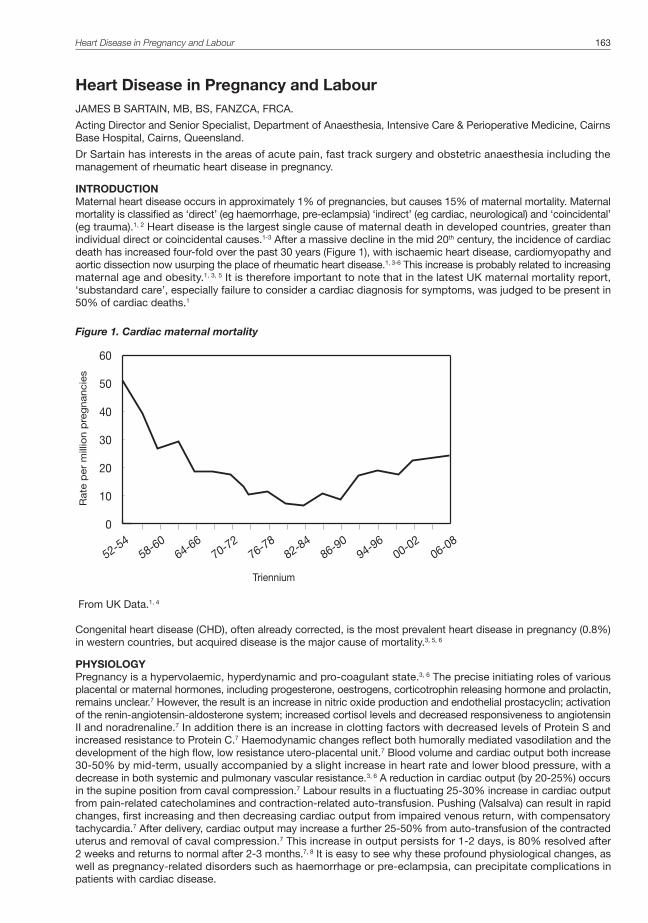
Heart Disease in Pregnancy and Labour 163
Heart Disease in Pregnancy and LabourJAMES B SARTAIN, MB, BS, FANZCA, FRCA.
Acting Director and Senior Specialist, Department of Anaesthesia, Intensive Care & Perioperative Medicine, Cairns Base Hospital, Cairns, Queensland.
Dr Sartain has interests in the areas of acute pain, fast track surgery and obstetric anaesthesia including the management of rheumatic heart disease in pregnancy.
INTRODUCTIONMaternal heart disease occurs in approximately 1% of pregnancies, but causes 15% of maternal mortality. Maternal mortality is classified as ‘direct’ (eg haemorrhage, pre-eclampsia) ‘indirect’ (eg cardiac, neurological) and ‘coincidental’ (eg trauma).1, 2 Heart disease is the largest single cause of maternal death in developed countries, greater than individual direct or coincidental causes.1-3 After a massive decline in the mid 20th century, the incidence of cardiac death has increased four-fold over the past 30 years (Figure 1), with ischaemic heart disease, cardiomyopathy and aortic dissection now usurping the place of rheumatic heart disease.1, 3-6 This increase is probably related to increasing maternal age and obesity.1, 3, 5 It is therefore important to note that in the latest UK maternal mortality report, ‘substandard care’, especially failure to consider a cardiac diagnosis for symptoms, was judged to be present in 50% of cardiac deaths.1
Figure 1. Cardiac maternal mortality
0
10
20
30
40
50
60
52-5458-60
64-6670-72
76-7882-84
86-9094-96
00-0206-08
Triennium
Rate
per
mill
ion p
reg
nancie
s
From UK Data.1, 4
Congenital heart disease (CHD), often already corrected, is the most prevalent heart disease in pregnancy (0.8%) in western countries, but acquired disease is the major cause of mortality.3, 5, 6
PHYSIOLOGYPregnancy is a hypervolaemic, hyperdynamic and pro-coagulant state.3, 6 The precise initiating roles of various placental or maternal hormones, including progesterone, oestrogens, corticotrophin releasing hormone and prolactin, remains unclear.7 However, the result is an increase in nitric oxide production and endothelial prostacyclin; activation of the renin-angiotensin-aldosterone system; increased cortisol levels and decreased responsiveness to angiotensin II and noradrenaline.7 In addition there is an increase in clotting factors with decreased levels of Protein S and increased resistance to Protein C.7 Haemodynamic changes reflect both humorally mediated vasodilation and the development of the high flow, low resistance utero-placental unit.7 Blood volume and cardiac output both increase 30-50% by mid-term, usually accompanied by a slight increase in heart rate and lower blood pressure, with a decrease in both systemic and pulmonary vascular resistance.3, 6 A reduction in cardiac output (by 20-25%) occurs in the supine position from caval compression.7 Labour results in a fluctuating 25-30% increase in cardiac output from pain-related catecholamines and contraction-related auto-transfusion. Pushing (Valsalva) can result in rapid changes, first increasing and then decreasing cardiac output from impaired venous return, with compensatory tachycardia.7 After delivery, cardiac output may increase a further 25-50% from auto-transfusion of the contracted uterus and removal of caval compression.7 This increase in output persists for 1-2 days, is 80% resolved after 2 weeks and returns to normal after 2-3 months.7, 8 It is easy to see why these profound physiological changes, as well as pregnancy-related disorders such as haemorrhage or pre-eclampsia, can precipitate complications in patients with cardiac disease.

164 Australasian Anaesthesia 2011
RISK OF DEATH AND MORBIDITYPrediction of mortality and morbidity relates to specific conditions and to acknowledged risk factors. High mortality rates occur with peripartum cardiomyopathy (10-30%) and in various forms of pulmonary hypertension (25-40%).5,
6, 9 Other conditions have a much lower mortality (<5%) with modern care. History and echocardiography are the keys to determining prognosis. The incidence of cardiac morbidity can then be estimated using the Cardiac Disease in Pregnancy (CARPREG) risk index (Figure 2), which was derived in an observational series of 562 pregnant patients with a wide range of heart conditions.10 Curiously, pulmonary hypertension was not an independent predictor in this study, though it is frequently associated with adverse outcomes.11, 12
Figure 2. CARPREG risk index10
Number of risk factors# Overall Risk Incidence of cardiac complications*
0 Low 5%
1 Moderate 30%
>1 High 70%
# Risk factors:• History of cardiac complications eg pulmonary oedema; symptomatic arrhythmias• Dyspnoea (NYHA 3 or 4) or cyanosis (SpO2 < 90%) • Impaired left ventricular function (EF < 40%)• Left heart obstruction (aortic or mitral valve area < 1.5 cm2)* Complications: pulmonary oedema, symptomatic arrhythmias or stroke
SPECIFIC CONDITIONS1. Congenital heart disease (CHD)Most minor or corrected conditions have normal outcomes for mother and baby.13 Women with uncorrected cyanotic or complex CHD are much more problematic, with the highest mortality (approx 40%) in patients with Eisenmenger’s syndrome (reversal of left-to-right shunt).13, 14 In other complex CHD, the mortality rate is generally less than 5%, though the incidence of miscarriage (40%) premature delivery (30%) and fetal mortality (15%) is high.13 Antibiotic prophylaxis for endocarditis (ampicillin 2g) is currently indicated only for complicated CHD or prosthetic grafts in the presence of prolonged or infected labour.3 The incidence of CHD in the infants of mothers with CHD is ten times the community risk.3, 13
2. Pulmonary hypertension (PHT)PHT is poorly tolerated in pregnancy, because the right ventricle is unable to cope with the additional load imposed by the physiological changes of pregnancy.15 If PHT is secondary to a remediable cause such as mitral stenosis, a good outcome can be expected with appropriate treatment.6 However, in idiopathic pulmonary arterial hypertension (formerly called primary pulmonary hypertension), or when PHT is secondary to pulmonary vasculitides (eg scleroderma, SLE), severe respiratory disease, or uncorrectable heart disease, the mortality is 25-40%. Death is usually from right heart failure or thromboembolism, most commonly in the first week post-partum.3, 6, 15 Treatment is of the specific cause (if secondary), plus general therapy for cardiac failure (eg diuretics) and anticoagulation. Vasodilators such amlodipine, sildenafil and intravenous or inhaled prostacyclin are useful for some sub-categories of PHT.3, 15
3. Rheumatic heart disease (RHD)Rheumatic heart disease (RHD) remains a major problem in indigenous communities of northern Australia (1-3%), and some immigrant groups.16 Outcomes are predictable based on assessment of known CARPREG risk factors (especially mitral stenosis and a history of pulmonary oedema) and the presence of PHT.10, 12, 16 Exacerbation is usually associated with a clear precipitant such as sepsis, pre-eclampsia or IV fluid loads. Medical therapy is usually adequate, though percutaneous balloon mitral valvuloplasty can be performed safely during pregnancy for severe or symptomatic mitral stenosis.12, 16 Previously undiagnosed patients still occur, so RHD should always be considered with new onset cardio-respiratory symptoms in pregnant indigenous patients.16

4. Management of anticoagulationThe care of anticoagulated patients with prosthetic heart valves requires balancing the substantial risks of maternal haemorrhage, maternal thrombosis (including stroke) and fetal loss or embryopathy, all of which are in the 10-20% range.12, 16 Accepted management involves maintaining warfarin until pregnant (low teratogenic risk before 6 weeks), then one of three options: (i) therapeutic dose low molecular weight heparin (LMWH) with anti-Xa monitoring plus aspirin; (ii) warfarin until 36 weeks, then changing to unfractionated heparin (UFH) or LMWH; or (iii) LMWH during 1st trimester (to minimise teratogenic risk) then revert to warfarin regimen. LMWH is stopped 36 hours before labour or surgery; UFH 6 hours beforehand.16, 17 The choice should be individualised based on a range of factors: LMWH is favoured for a well motivated patient, requiring high warfarin dose, with ready access to anti-Xa monitoring; warfarin is favoured with likely poor compliance, high thrombotic risk and a required warfarin dose of <5mg, which has a low risk of embryopathy ( less than 10%).16, 17 Caesarean section is often performed in these patients, either to enable timing of anticoagulation cessation, or if the woman labours within two weeks of stopping warfarin, as it is still active in the fetus after normalisation of maternal coagulation, who is then at risk of intracranial bleeding during labour.17
5. Ischaemic heart disease (IHD) and myocardial infarction (MI)IHD is reported to occur in only 1 in 10,000 pregnancies, but is thirty times more likely in patients over 40 years compared to those less than 20 years.3 Risk factors include age over 35, obesity, smoking, family history, hypertension and diabetes. Appropriate investigations (Troponin levels, serial ECGs) should be performed in patients with typical ischaemic symptoms. Patients with known IHD should be assessed and treated based on standard criteria with delivery planned to minimise haemodynamic stress (see delivery section).18 Pregnancy-associated MI may be due to coronary artery dissection or spasm rather than atherosclerosis.19 During the stress of the peripartum period or post-partum, dissection accounts for 35-50% of MIs, compared to only 10% antepartum.19 Consequently percutaneous coronary intervention (with possible stenting) is the treatment of choice rather than thrombolysis.3, 19 Bare-metal stents, which do not require prolonged treatment with clopidogrel, may be preferable in the antepartum period.3, 19 Standard treatments in addition to antiplatelet agents include nitrates, beta blockade and LMWH.3, 5 Angiotension converting enzyme inhibitors (ACE inhibitors), angiotension II receptor blockers (ARBs), statins and amiodarone are class D drugs causing fetal abnormalities or death, so should not be given while still pregnant.5 Metoprolol is preferred over atenolol, as the latter may cause fetal growth retardation.5 In a recent literature review the mortality of myocardial infarction was 18% in the immediate peripartum period, but 9% when it occurred more than 24 hours before or after labour.19 Historic data also suggests that delivery within 2 weeks of a myocardial infarction is associated with a high mortality – a period of stabilisation may be preferable if both cardiac and obstetric considerations permit.6 Care must also be taken with the use of uterotonic agents (see delivery section).6
6. Aortic dissectionThis is most commonly idiopathic, but may be secondary to Marfan’s syndrome or bicuspid aortic valve disease.1,
3 It should be suspected with severe back or left sided chest pain, diminished left sided pulses or a widened mediastinum on chest X-ray. CT angiogram with contrast or transoesophageal echocardiography will show the diagnosis. It is important to investigate appropriately if suspected rather than having inordinate fear of radiation to the fetus.1 Patients with Marfan’s syndrome require beta blockade and frequent echocardiography during pregnancy. Aortic arch replacement is recommended if aortic diameter is greater than 40mm, at which stage the risk of spontaneous rupture is 10%.3, 11
7. Peripartum cardiomyopathy (PPCM)PPCM occurs in 1 in 4000 pregnancies.3, 6, 9 It is a diagnosis of exclusion, with progressive systolic left ventricular dysfunction developing 1 month pre-partum to 5 months post-partum. Management involves anti-failure treatment plus anticoagulation; heart transplant is a last-ditch option. Patients who recover should be counselled that it recurs in 30% of subsequent pregnancies.3, 6
8. Hypertrophic (obstructive) cardiomyopathy (HCM or HOCM)This is an autosomal dominant condition, with an incidence of 1 in 500.20 There is a wide range of severity from essentially benign to immediately life-threatening. The majority of patients have an uneventful pregnancy, with adverse outcomes predictable based on severity of symptoms (dyspnoea, syncope) and the degree of ventricular outflow obstruction.20, 21 If required, treatment may involve beta blockade, with phenylephrine or metaraminol for first-line treatment of hypotension rather than beta agonists.20
9. Cardiac surgery during pregnancyThe requirement for cardiac surgery and cardio-pulmonary bypass during pregnancy is associated with maternal mortality of 5-10% and a fetal/neonatal death rate of 30%.5, 11 Although these rates probably reflect the maternal condition primarily, other ill-defined factors such as cardiopulmonary bypass flows may also be relevant.5, 11 Therefore, if the fetus is viable, consideration should be given to delivering the baby before the cardiac operation.11
10. Previous heart transplantDespite the expected infection risk from immunotherapy and the lack of intact cardiac autonomic innervation, outcomes are generally positive.6 In a series of 22 patients, hypertension or pre-eclampsia occurred in nearly half
Heart Disease in Pregnancy and Labour 165

166 Australasian Anaesthesia 2011
and infections in 27%, but there were no maternal deaths or instances of heart failure and no fetal abnormalities.22
Overall management planAn overall management plan outline is given in figure 3. It should be individualised based on the severity of disease. In general terms, low risk patients can be treated as standard obstetric patients. At the other extreme, high risk patients may require transfer to a unit with access to immediate interventions including percutaneous valvuloplasty or cardiac surgery.3, 5, 6
Figure 3. Overall management of obstetric patients with cardiac disease
• Determine diagnosis• Assessment of severity
– Specific high risk conditions – CARPREG risk score (history and echocardiography) – Cardiologist assessment
• Medication or other intervention as required• Collaboration between relevant disciplines
– obstetrics, cardiology, anaesthesia, ICU, paediatrics, haematology• Co-ordination of delivery
– Appropriate location (expertise, resources)– Dependent on cardiac and obstetric factors
DELIVERY PLANA plan for delivery and labour is outlined in figure 4. It is clear that vaginal delivery is achievable (and generally preferable) for the vast majority of patients with cardiac disease if obstetric considerations permit.3, 5, 6 In patients at moderate to high risk, the use of well-conducted epidural analgesia (to minimise the haemodynamic perturbations of labour), strict avoidance of caval compression (eg left lateral position), and assisted delivery (to minimise pushing) are rational strategies,3, 5, 6 though there are no data from randomised trials. The anaesthetist should be present for delivery as the person most able to manage haemodynamic changes. Great care must be taken with all uterotonic agents.3, 5, 6 Ergometrine can cause myocardial ischaemia from coronary artery spasm and pulmonary oedema from venoconstriction.6, 23 Oxytocin causes tachycardia and hypotension with decreased systemic and increased pulmonary vascular resistance.6, 24 Prostaglandins have a similar effect.6 However, a balance must be achieved to avoid inadequate uterine contraction and excessive blood loss. An infusion of oxytocin (eg starting at 0.5 -1 units/minute),24 titrated to haemodynamics and uterine tone, is recommended as the first line uterotonic for high risk patients.
Figure 4. Labour and delivery plan for moderate to high cardiac risk patients
Labour:• Consider epidural pain relief • Wedged or lateral position• Avoid excessive IV fluids• Antibiotic prophylaxis indicated with prosthetic valves, complex CHD, history of endocarditis or obstetrics
reasons• Consider invasive monitoring for high risk patients
Delivery:• Consider assisted vaginal delivery• Anaesthetist present for delivery• Avoid ergometrine • Oxytocin infusion eg 0.5-1 U/ minute initially +/- 1 unit increments IV (omit with severe disease)• Consider delivery in monitored situation (eg operating theatre) for high risk patients
Post Delivery:• IV fluids to replace blood loss only• Cease IV fluids once bleeding stopped• Consider frusemide IV or orally if at risk of pulmonary oedema• Observe in high dependency unit or intensive care overnight

CAESAREAN SECTIONA summary of indications for Caesarean Section and anaesthesia recommendations are given in Figure 5. Cardiac indications for Caesarean section are based on logic or practical issues (eg anticoagulation management or planned high risk delivery in daylight hours) rather than convincing data.3, 5, 6 It is far more likely that obstetric considerations will be the determining factor for operative delivery, though many obstetricians have a lower threshold for intervention in patients with co-existing pathology. Should Caesarean Section be indicated, general principles of maintaining haemodynamic stability and oxygenation are more important than specific techniques.3, 6 For instance, worsening of pulmonary hypertension or right-to-left shunt, can be avoided through ‘cardiac’ general anaesthesia and controlled ventilation (despite the effect of increased airway pressure), or by epidural anaesthesia plus the use of peripheral alpha agonists and judicious fluids.6, 25 Similarly, preventing tachycardia and maintaining systemic vascular resistance in patients with left-sided obstructive lesions can also be achieved through opioids and volatile agents or with regional anaesthesia and alpha agonists.20, 25 Consequently, there are many series demonstrating the successful use of either regional (incremental epidural) or general anaesthesia for obstetric patients with severe cardiac disease of virtually any kind.6, 15, 25, 26 The wishes of the patient, her ability to lie flat without dyspnoea, contraindications to regional anaesthesia (eg anticoagulation), and the individual expertise of the anaesthetist may be decisive factors.
Figure 5. Caesarean Section for moderate to high risk cardiac patients
Indications:Obstetric
Cardiac:• Unstable situation (patient likely to be improved after baby delivered)
– Decompensated heart failure despite treatment– Aortic dissection– ?dilated aortic root– ?severe pulmonary hypertension
• Anticoagulation– Prosthetic valves: to fit with timing of stopping anticoagulation– Warfarin within last 2 weeks (fetal protection)– High risk patient with labour epidural relatively contra-indicated
• Logistical issues (timing of high risk delivery when resources available)
Anaesthesia: – Either general or regional (incremental epidural) depending on contraindications, specific patient factors
and anaesthetist expertise– Invasive monitoring indicated for high risk cases– Avoid excessive IV fluids (primarily use to replace fluid losses)– Maintain haemodynamics with vasopressors– Oxytocin via titrated infusion (eg start 0.5-1 unit/ minute)
+/- 1 unit increments IV (omit with severe disease)– Observe in high dependency unit or intensive care overnight
SUMMARYMaternal heart disease occurs in approximately 1% of pregnancies, but causes 15% of maternal mortality. Specific high risk conditions and maternal cardiac risk factors are well established, so the prognosis for patients with known disease is predictable. However, a high index of suspicion and a low threshold for appropriate investigations are required to reduce mortality in patients without previously recognised cardiac disease. Multi-disciplinary collaboration, with tailoring of cardiac therapy and delivery plan to the diagnosis and severity of disease, is associated with a good outcome for the majority of patients. Vaginal delivery is generally preferred if obstetric indications allow. There are very few cardiac indications for Caesarean section and both general and regional anaesthesia are acceptable in most cases.
Heart Disease in Pregnancy and Labour 167

168 Australasian Anaesthesia 2011
REFERENCES1. Centre for Maternal and Child Enquiries (CMACE). Saving mothers’ lives: reviewing maternal deaths to make
motherhood safer: 2006–2008. The eighth report on confidential enquiries into maternal deaths in the United Kingdom. Br J Obstet Gynaecol. 2011;118(suppl.1):1-203.
2. Sullivan EA Hall B & King JF. Maternal deaths in Australia 2003-2005. Sydney: AIHW. National Perinatal statistics unit; 2008.
3. Curry R, Swan L, Steer PJ. Cardiac disease in pregnancy. Curr Opin Obstet Gynecol. 2009 Dec;21(6):508-13.
4. Confidential Enquiry into Maternal and Child Health. Why Mothers Die 2000–2002. London: RCOG Press; 2004.
5. van Mook WN, Peeters L. Severe cardiac disease in pregnancy, part II: impact of congenital and acquired cardiac diseases during pregnancy. Curr Opin Crit Care. 2005 Oct;11(5):435-48.
6. Ray P, Murphy GJ, Shutt LE. Recognition and management of maternal cardiac disease in pregnancy. Br J Anaesth. 2004 Sep;93(3):428-39.
7. Foley MR. Maternal cardiovascular and hemodynamic adaptations to pregnancy. In: UpToDate, Lockwood CJ & Gersh BJ (eds). UpToDate, Waltham MA, 2011.
8. Robson SC, Hunter S, Moore M, Dunlop W. Haemodynamic changes during the puerperium: a Doppler and M-mode echocardiographic study. Br J Obstet Gynaecol. 1987 Nov;94(11):1028-39.
9. Elkayam U, Akhter MW, Singh H, Khan S, Bitar F, Hameed A, et al. Pregnancy-associated cardiomyopathy: clinical characteristics and a comparison between early and late presentation. Circulation. 2005 Apr 26;111(16):2050-5.
10. Siu SC, Sermer M, Colman JM, Alvarez AN, Mercier LA, Morton BC, et al. Prospective multicenter study of pregnancy outcomes in women with heart disease. Circulation. 2001 Jul 31;104(5):515-21.
11. Barth WH, Jr. Cardiac surgery in pregnancy. Clin Obstet Gynecol. 2009 Dec;52(4):630-46.
12. Reimold SC, Rutherford JD. Clinical practice. Valvular heart disease in pregnancy. N Engl J Med. 2003 Jul 3;349(1):52-9.
13. Drenthen W, Pieper PG, Roos-Hesselink JW, van Lottum WA, Voors AA, Mulder BJ, et al. Outcome of pregnancy in women with congenital heart disease: a literature review. J Am Coll Cardiol. 2007 Jun 19;49(24):2303-11.
14. Khairy P, Ouyang DW, Fernandes SM, Lee-Parritz A, Economy KE, Landzberg MJ. Pregnancy outcomes in women with congenital heart disease. Circulation. 2006 Jan 31;113(4):517-24.
15. Bedard E, Dimopoulos K, Gatzoulis MA. Has there been any progress made on pregnancy outcomes among women with pulmonary arterial hypertension? Eur Heart J. 2009 Feb;30(3):256-65.
16. National Heart Foundation of Australia and the Cardiac Society of Australia and New Zealand. Diagnosis and management of acute rheumatic fever and rheumatic heart disease in Australia – an evidence based review. 2006.
17. McLintock C. Anticoagulant therapy in pregnant women with mechanical prosthetic heart valves: no easy option. Thromb Res. 2011 Feb;127 Suppl 3:S56-60.
18. Smith RL, Young SJ, Greer IA. The parturient with coronary heart disease. Int J Obstet Anesth. 2008 Jan;17(1): 46-52.
19. Roth A, Elkayam U. Acute myocardial infarction associated with pregnancy. J Am Coll Cardiol. 2008 Jul 15;52(3):171-80.
20. Poliac LC, Barron ME, Maron BJ. Hypertrophic cardiomyopathy. Anesthesiology. 2006 Jan;104(1):183-92.
21. Autore C, Conte MR, Piccininno M, Bernabo P, Bonfiglio G, Bruzzi P, et al. Risk associated with pregnancy in hypertrophic cardiomyopathy. J Am Coll Cardiol. 2002 Nov 20;40(10):1864-9.
22. Morini A, Spina V, Aleandri V, Cantonetti G, Lambiasi A, Papalia U. Pregnancy after heart transplant: update and case report. Hum Reprod. 1998 Mar;13(3):749-57.
23. Carey M. Adverse cardiovascular sequelae of ergometrine. Br J Obstet Gynaecol. 1993 Sep;100(9):865.
24. Thomas JS, Koh SH, Cooper GM. Haemodynamic effects of oxytocin given as i.v. bolus or infusion on women undergoing Caesarean section. Br J Anaesth. 2007 Jan;98(1):116-9.
25. Findlow D, Doyle E. Congenital heart disease in adults. Br J Anaesth. 1997 Apr;78(4):416-30.
26. Hidano G, Uezono S, Terui K. A retrospective survey of adverse maternal and neonatal outcomes for parturients with congenital heart disease. Int J Obstet Anesth. 2011 Jul;20(3):229-35.

169


Perioperative Transthoracic Echocardiography in Australasia: Current Position and Future Directions 171
Perioperative Transthoracic Echocardiography in Australasia: Current Position and Future DirectionsJOHN FARIS, BSC, DAVMED, FFOM, FANZCA, BA, ASCEXAM, PG DIP CLIN ULTRASOUND.
Sir Charles Gairdner Hospital, Perth; Joondalup Hospital; Perth; Fremantle School of Medicine, University of Notre Dame Australia; Department of Pharmacology, University of Melbourne, and Royal Melbourne Hospital
Professor Faris is a staff specialist at both Sir Charles Gairdner and Joondalup Hospitals in Perth. After twenty years in aviation medicine in New Zealand, he changed specialties and joined the anaesthesia training scheme in Auckland, including a cardiac fellowship where he learned transoesophageal echocardiography. He moved to Perth eight years ago and was introduced to transthoracic echocardiography. He further developed this interest joining the Ultrasound Education Group at the University of Melbourne. He spends a day each week teaching and reporting echocardiography.
COLIN ROYSE, MBBS, MD (MELB), FANZCA
Anaesthesia and Pain Management Unit, Department of Pharmacology, University of Melbourne, and Department of Anaesthesia and Pain Management, Royal Melbourne Hospital
Professor Colin Royse is a specialist cardiothoracic anaesthetist with academic interests in echocardiography, pain management in cardiac surgery, quality of recovery following anaesthesia and surgery, and the evaluation of left ventricular systolic and diastolic function using pressure volume loops. He is Director of the Anaesthesia and Pain Management Unit in the Department of Pharmacology, and Co-Director of the Ultrasound Education Group at the University of Melbourne.
YOUR NEXT CASEIt is 9pm at night and you are the consultant on call at a busy teaching hospital. The next case is an 88 year old man who has fallen and injured his left lower leg requiring evacuation of a large haematoma and split skin graft to close a residual defect. He had a myocardial infarction six months ago complicated by acute pulmonary oedema, but states that he has recovered. The ECG shows atrial fibrillation and inferior Q waves. Your registrar heard a systolic murmur, but was unconcerned as the patient walked his dog each day (JF personal experience).
You have several choices: completely ignore the murmur, delay the case and obtain a transthoracic echocardiography (TTE) examination by the cardiologists, proceed as if this might be significant aortic stenosis, or perform a goal-directed TTE study?
TTE USE IN ANAESTHESIA PRACTICEFor many years since Inge Edler in Sweden in 1953 borrowed a naval reflectoscope to image a heart, cardiac ultrasound has largely remained the province of the cardiologists. General ultrasound was for the radiologists. Surgeons and emergency physicians developed the Focused Abdominal Sonography in Trauma (FAST scan) in the mid 1990s, including the sub-costal window to image the heart. Parasternal and apical windows were added to better diagnose haemodynamic instability in emergency departments and intensive care units. Ultrasound is a very useful imaging modality that has crossed traditional craft group usage and in recent times has entered anaesthesia and critical care practice in a major way.
In anaesthesia there were parallel developments of transoesophageal echocardiography (TOE) in cardiac surgery, and ultrasound-guided procedures such as vascular access and regional anaesthesia. Ultrasound machine manufacturers recognised the potential and produced smaller, cheaper portable models. Phased array probes were added providing a practical path for more widespread use of TTE. The non invasive nature of TTE has increased the scope into both the preoperative and postoperative arenas. Training courses and workshops for anaesthetists appeared in TTE rather than just TOE.
With these developments, TTE is arguably now ready to come of age.
WHY TTE IN ANAESTHESIA?There are many reasons why anaesthetists now seek to perform their own TTE. These include: preoperative assessments, diagnosis of haemodynamic collapse and haemodynamic monitoring. The most obvious and common use of TTE is the preoperative assessment of cardiac disease. Cardiology services may not be available at short notice for either perioperative assessments or in the event of a perioperative emergency. Very few Australasian institutions, if any, have such a service available at any hour of the day or night.
In an ideal world, elective surgery patients would not arrive in the preoperative waiting area with an inadequate assessment of cardiac function. Anaesthetists may resort to increasing invasive monitoring or insist on high dependency or ICU beds after surgery to compensate for potential cardiac disease. Anaesthetist performed TTE may assist in better risk stratification of patients, and facilitate rational use of invasive monitoring or higher dependency postoperative care. In this sense, echo is used as a triage tool to determine the most appropriate management for patients.
A second imperative is to guide the diagnosis and management of perioperative haemodynamic instability. Clinical examination alone cannot reliably diagnose the cause of haemodynamic collapse. Hypotension is the

172 Australasian Anaesthesia 2011
warning sign of haemodynamic instability, but does not identify the cause. Echocardiography can be used to identify patterns of abnormality (such as hypovolaemia), systolic failure or
vasodilatation), and when integrated with the clinical scenario, can help identify the cause of haemodynamic instability. Royse has described a methodology for the evaluation of haemodynamic state using limited TTE.1 Critically ill patients require urgent diagnosis. This is facilitated if the practitioner managing the patient performs the TTE, rather than relying on third party providers such as cardiology, where timely access to their services may be limited or not available.
A third use of cardiac ultrasound is as a perioperative haemodynamic monitor. Echocardiography allows the assessment of both right and left sided preload, of right and left ventricular systolic and diastolic function, of pulmonary artery pressures (in most cases) and of cardiac output. No other haemodynamic monitor is able to measure all of these parameters. TTE, unlike either TOE or a pulmonary artery catheter, is non-invasive and there are no safety issues in image acquisition. It can also be used in an awake patient. Other monitors using pulse pressure variation or transoesophageal doppler devices are arguably easier to use but provide a less complete picture. An example of using TTE for haemodynamic monitoring during a caesarean section is described by Ferguson et al.2
Fourthly, the anaesthetic perspective should be born in mind. It is rare for a cardiac sonographer or cardiologist to have patient in whom there are sudden and significant changes in preload, afterload or contractility, as occurs during anaesthesia. Anaesthetists will view echocardiograms with a different perspective to cardiologists, focusing on ventricular function and haemodynamically important valve lesions to answer specific questions, rather than a comprehensive study aimed at identifying all aspects of the echocardiogram.
Learning to perform and interpret echocardiography requires an additional knowledge base that is not currently part of anaesthesia training. Performing echocardiography also provides the practitioner with a powerful feedback tool, which serves to improve understanding of the pathophysiology and how anaesthesia influences it. Many anaesthetists read only the conclusions of formal report, but the black and white conclusions often hide a greater subtlety in the numbers, findings and the interpretation of the study. The anaesthetic paradigm mentioned earlier can result in a different interpretation for our specialty even though the conclusions are totally appropriate from a cardiology perspective.
An example could be a report of a large pericardial effusion without echocardiographic evidence of tamponade. In another personal example, the patient had undergone a pericardial window 1 month earlier. The cardiologist had reassured the surgeon that tamponade was no longer a risk. The anaesthetist performed a limited TTE and identified a warning sign of a small degree of right atrial collapse, leading the anaesthetist to consider tamponade a significant risk during the forthcoming anaesthetic. A 1500 mL fluid loading and vasopressor with the induction was administered. Despite this, there was a significant, but not life threatening, haemodynamic change on induction. The echo findings were the same for both cardiologist and anaesthetist, but the images were seen from a different perspective.
GOAL DIRECTED CARDIAC ULTRASOUNDEchocardiography examinations performed by cardiology services are (almost always) comprehensive diagnostic studies. Studies are conducted for other practitioners and, irrespective of the indications for a study, the expectation is that all findings will be included in the report. Conversely, in anaesthesia, echocardiography is an extension of the clinical assessment. The echo results are often directly and immediately integrated with other clinical information. Cardiologists need to follow trends overtime so quantification is important, but in anaesthesia, long term trends are unimportant and a qualitative assessment will often suffice, especially in time critical situations.
These differences have led to a paradigm of goal-directed studies to answer only one or more specific clinical questions (and often no more). Terms such as limited or focused, bed-side or point of care are also used because of their location, or hand-held because of the equipment often used.3,4
They may take only five minutes while a comprehensive study may take half an hour. Goal-directed studies may only use one or two echocardiographic windows and fewer than the expected number of views through each window than would be expected in a comprehensive study. Quantification may be minimal or non-existent, pattern recognition being more important than measurement. A goal-directed study may need no more than 2D imaging if only haemodynamic state needs to be assessed, while a global assessment of valvular function requires colour flow imaging as well. The term “cardiac ultrasound” is used by some to cover both types of examination while the term “echocardiography” is reserved by others for comprehensive studies only.
There are some issues with goal-directed studies. First, it must be accepted that some information will be missed. A study by Rugolotto et al5 showed that using clinical examination alone, a cardiologist missed some 40% of significant abnormalities when compared with a comprehensive TTE. Using a hand-held bedside cardiac ultrasound with 2D and colour flow Doppler capabilities only, they still missed 20% of significant abnormalities. However, by using the results of the bedside study they were able to institute earlier treatment and discharge patients a day earlier on average. Importantly, the authors reported no errors in treatment using the bedside studies. Other studies have had similar findings.6-8 Missed information is potentially important but may not be significant in critical care. A minor regional wall motion abnormality might be missed, but will not be the cause of haemodynamic collapse.
There is an increasing body of evidence for the efficacy of point of care, physician and sonographer performed, goal directed studies, using with portable or hand carried machine. These studies are in many fields including cardiology,7,9,10 general medicine,11 emergency medicine (many specialties),12-14 intensive care15-18 and anaesthesia,19,20 However, not all authors agree its utility in all fields or all circumstances.21,22

The potential advantages of goal directed studies should not be understated. In anaesthesia, when faced with a systolic murmur, it is important23 to exclude haemodynamically significant aortic stenosis with reasonable certainty. This cannot be done clinically. A review by Etchells and colleagues,24 showed that while effort syncope with a systolic murmur has the highest positive predictive value, the lack of effort syncope is not helpful in differentiating severe aortic stenosis. The lack of radiation of the murmur to the right carotid is the most useful of the negative clinical sign, but there is a false negative rate of 5 – 10%. They showed cardiologists are better than non cardiologists, but that even cardiologists were no better than about 90% certain. Reichlin et al25 assessed the ability of emergency physicians to diagnose systolic murmurs. The study included 203 consecutive patients admitted to an emergency department and noted to have a systolic murmur. Some 35% of these had significant valvular heart disease shown on TTE within an hour of the clinical examination (equal distribution of mitral regurgitation, aortic stenosis and other lesions). Sensitivity and specificity for the correct diagnosis by clinical examination were only 82% and 69% respectively.
In 1975 using M-mode echocardiography, Weyman and Feigenbaum26 showed that a cusp separation of 15mm or more in the parasternal long axis view excludes moderate or severe aortic stenosis. Since then 2D imaging has dramatically improved, and restricted leaflet movement in the parasternal long and short axis views readily identifies haemodynamically significant moderate or severe aortic stenosis.
There is controversy regarding qualitative versus quantitative grading of ventricular function or valve lesions. If long term trends are important, then quantification is important. For example, if a patient has had a myocardial infarction and an ejection fraction of 40%, they may be put on an angiotensin converting enzyme inhibitor. In three months a cardiologist will be interested if the ejection fraction is now 35, 40 or 45%. But in anaesthesia, all have about the same degree of systolic impairment. An anaesthetist wants to know if the systolic function of a heart is normal, somewhat impaired or severely impaired. These patterns are easy to distinguish by 2D pattern recognition alone. With aortic stenosis, we are accustomed to peak and mean gradients and aortic valve area. But do we really need this data? Anaesthetists need to know whether the stenosis could be haemodynamically significant, so that we can plan the anaesthetic and postoperative care appropriately. In elective surgery, quantification may be important in those few cases in which it would be more appropriate to consider valve surgery prior to their planned operation. But when surgery is urgent and there is little choice but to proceed and anaesthetists will treat moderate and severe stenosis in a similar manner.
Quantification for less experienced operators also has the potential to underestimate severity. The jets are often eccentric and may require the use of non-standard views to obtain the peak velocity and the use of a non-imaging probe. If the peak velocity is not interrogated, the valve area is overestimated and gradients underestimated. An area of active research in preoperative assessment is to determine the learning curves required for the safe quantification of aortic stenosis in comparison to qualitative 2D and colour flow pattern recognition. In a small study by Cowie et al,27 TTE naïve anaesthetic trainees were able to measure peak velocity and potentially gain useful information in addition to that available from 2D and colour flow imaging alone.
TRAINING IN TTE FOR ANAESTHETISTS IN AUSTRALASIAThe penetration of TTE into anaesthesia can be gauged by training numbers. Courses, conferences and workshops in TOE appeared in the late 1990s. Formal training was offered in Australia in 2004 with the establishment of a Postgraduate Diploma in Perioperative and Critical Care Echocardiography by the University of Melbourne (PGDipEcho). The original diploma included a component of TTE, but was largely designed for cardiac anaesthetists performing TOE. The call for training for general anaesthetists and TTE resulted in modifications in 2009. The first six months became a Postgraduate Certificate in Clinical Ultrasound (PGCertCU) with a strong emphasis on TTE with TOE largely consigned to a second six months now a Postgraduate Diploma in Clinical Ultrasound (PGDipCU). The knowledge base for the PGCertCU is aimed at “a good basic sonographer” and the PGDipCU at “diagnostic level”. To date 460 clinicians have completed the PGDipEcho, 297 have completed the PGCertCU with another 135 enrolled. Forty-nine have now completed the PGDipCU with 40 more enrolled.
For skills based (i.e. hands-on) TTE training, the University of Melbourne has offered a large number of workshops. The Point of Care Courses which ran from 2005 to 2009 (353 participants), included two stations (about 40%) on TTE. Since 2005, the H.A.R.T.Scan courses have provided training in goal-directed TTE for 577 doctors. The majority of doctors attending these courses have been anaesthetists.
Alternative qualifications are also available. The National Board of Echocardiography in the USA has examinations and certification (but no specific training courses) in perioperative transoesophageal echocardiography (PTXeXAM) and in adult echocardiography (ASCeXAM). The PTXeXAM is designed for cardiac anaesthetists while the ASCeXAM is for cardiologists. There are 90 physicians in Australia and 25 in New Zealand with the PTXeXAM and 2 anaesthetists with the ASCeXAM.
The Australian Society of Ultrasound Medicine (ASUM) offers a Diploma in Diagnostic Ultrasound with an option of cardiac ultrasound. This course has both knowledge and skills base training. ASUM also provides one of the primary qualifications held by sonographers in Australasia. Other universities are starting to offer training and qualifications that could be of interest to goal-directed TTE.
Comprehensive echocardiography requires extensive training and experience. The joint American College of Cardiology and America Heart Association (ACC/AHA) guidelines on the clinical competence28 require a minimum of 150 personally performed and an additional 300 supervised reports for independent practice. However, many studies have shown that goal-directed TTE can be taught with significantly less training, but with the proviso that any findings may also be limited and integrated with other clinical information as discussed earlier.
Perioperative Transthoracic Echocardiography in Australasia: Current Position and Future Directions 173

174 Australasian Anaesthesia 2011
Royse et al29 studied the learning curve in an echo naïve medical student. They showed 20 studies were required to conduct a basic haemodynamic state assessment. Price et al30 studied physicians attending a one-day training course in peri-resuscitation echocardiography. A test of knowledge base showed an improvement in image interpretation (pre 62%, post 78%, p < 0.01) and 100% of participants were able to obtain a subcostal view of diagnostic quality by the end of the course, most in under 10 sec.
Vignon et al30 reported on the efficacy of training of naïve non-cardiology residents in limited echocardiography in an ICU. Each was given 3 hours of lectures and 5 hours of hands on training. In a study of 61 consecutive patients and comparing residents with an experienced intensivist, they were significantly slower and answered significantly fewer of the 366 clinical questions (3 vs. 27%). However, when addressed, left ventricular systolic function, left and right ventricular dilatation, pericardial effusion and tamponade were all correctly appraised.
Hellman et al31 reported on the training of 31 residents in the use of a hand carried cardiac ultrasound machine for limited echocardiographic studies. They were given a 30 minutes lecture followed by one-on-one instruction and supervision. A linear regression model plotting an overall assessment index against the number of scans showed a learning curve of 20 – 40 scans. However, the authors did note significant differences between residents in their rate of learning.
These studies show that goal-directed limited transthoracic echocardiography is within the reasonable reach of a significant proportion of the anaesthetic community.
TTE IN AUSTRALASIAN ANAESTHESIASome specific examples give an overview of TTE usage in Australasian anaesthesia. The first case series was reported by Canty et al in 200920. They reported on 87 TTE and 14 TOE examinations in 97 patients at Royal Hobart Hospital (75 studies were conducted pre-operatively). Three patients had their surgery changed or cancelled, and in 18 patients there were significant changes in anaesthetic management.
Cowie in 201119, at St Vincent’s Hospital in Melbourne, has reported on three years’ experience in anaesthetists performed goal-directed echocardiography. He reports adequate images obtainable in 167 out of 170 patients (98%). Just over half the studies were conducted because of a systolic murmur. Changes in peri-operative management occurred in 140 out of 170 (82%) patients. Major findings correlated with a formal cardiology transthoracic echocardiogram in 52 out of 57 (92%) patients.
Joondalup Hospital in Perth has established a formal perioperative echocardiography service. Comprehensive echocardiograms are conducted by a cardiac sonographer and reported by an anaesthetist. The most common indications are undiagnosed systolic murmurs or poor exercise tolerance (or an inability to assess it). If an echocardiogram has been conducted elsewhere in the preceding year and a copy of the report can be obtained, it is not repeated unless there is a specific indication to do so. Abnormal findings are referred to cardiologists for follow up with the images and report provided so that the study does not need to be repeated.
In the first 22 months, nearly 700 echocardiograms have been performed. Preliminary results show 26.7% of patients had moderate or severe echocardiographic findings such as significant valvular dysfunction or valvular heart disease. Half of these findings were totally unexpected on clinical grounds. Around 36% of the murmurs were the results of moderate or severe disease. In sixty patients with a murmur, surgery proceeded as planned because haemodynamically significant valvular heart disease was confidently excluded.
Sir Charles Gairdner Hospital, also in Perth, has embraced all forms of ultrasound. One third of the consultant staff have experience and a formal qualification in echocardiography with others in training. The majority are general anaesthetists and most perform only TTE, while three are cardiac anaesthetists practicing only TOE (with two more meeting the “grandfather” requirements of PS4632). The department has seven TTE capable ultrasound machines. Both limited goal-directed and comprehensive echocardiograms have been conducted as required over the past five years. The hospital is considering extending anaesthetist performed goal-directed limited examinations to the preoperative clinic on a more regular basis. With two different models being used in the one city, it is hoped to be able to compare and contrast the efficacy and community costs of each model.
THE NEXT CASESo you performed a goal-directed TTE for our elderly gentleman who fell and injured his lower leg. This showed a heavily calcified aortic valve with an obvious restriction in opening. In addition, there was mild to moderate aortic regurgitation, obvious concentric left ventricular hypertrophy, hypokinesis of the inferior septal wall (consistent with an old right coronary artery territory myocardial infarction) and a huge left atrium typical of significant diastolic impairment. Further questioning of the patient revealed a couple of episodes of possible syncope. Although he walked the dog every day, the dog walked but he rode in a buggy. Although the ejection fraction appeared to be relatively well preserved and the peak and mean gradients across the valve were only 39 and 26mmHg pressure in the moderate range, the aortic valve area was 0.6cm2 and dimensionless index 0.20,33 both suggesting severe if not critical aortic stenosis. However, the 2D appearance was that of severe, not just moderate stenosis, consistent with normal ejection fraction low-gradient severe aortic stenosis. Despite initial patient reluctance, the surgery was successfully conducted under local anaesthesia.

REFERENCES AND FOOTNOTES1. Royse CF: Ultrasound-guided haemodynamic state assessment. Best practice & research. Clinical
anaesthesiology 2009; 23: 273-83.
2. Ferguson EA, Paech MJ, Veltman MG: Hypertrophic cardiomyopathy and caesarean section: intraoperative use of transthoracic echocardiography. International journal of obstetric anesthesia 2006; 15: 311-6.
3. Faris JG, Veltman MG, Royse CF: Limited transthoracic echocardiography assessment in anaesthesia and critical care. Best Pract Res Clin Anaesthesiol 2009; 23: 285-98.
4. Cowie B: Focused cardiovascular ultrasound performed by anesthesiologists in the perioperative period: feasible and alters patient management. J Cardiothorac Vasc Anesth 2009; 23: 450-6.
5. Rugolotto M, Chang CP, Hu B, Schnittger I, Liang DH: Clinical use of cardiac ultrasound performed with a hand-carried device in patients admitted for acute cardiac care. Am J Cardiol 2002; 90: 1040-2.
6. Prinz C, Voigt JU: Diagnostic accuracy of a hand-held ultrasound scanner in routine patients referred for echocardiography. J Am Soc Echocardiogr 2011; 24: 111-6.
7. Giusca S, Jurcut R, Ticulescu R, Dumitru D, Vladaia A, Savu O, Voican A, Popescu BA, Ginghina C: Accuracy of handheld echocardiography for bedside diagnostic evaluation in a tertiary cardiology center: comparison with standard echocardiography. Echocardiography 2011; 28: 136-41.
8. Culp BC, Mock JD, Chiles CD, Culp WC, Jr.: The pocket echocardiograph: validation and feasibility. Echocardiography 2010; 27: 759-64.
9. Kimura BJ, Yogo N, O’Connell CW, Phan JN, Showalter BK, Wolfson T: Cardiopulmonary Limited Ultrasound Examination for “Quick-Look” Bedside Application. Am J Cardiol 2011.
10. Cardim N, Fernandez Golfin C, Ferreira D, Aubele A, Toste J, Cobos MA, Carmelo V, Nunes I, Oliveira AG, Zamorano J: Usefulness of a new miniaturized echocardiographic system in outpatient cardiology consultations as an extension of physical examination. Journal of the American Society of Echocardiography : official publication of the American Society of Echocardiography 2011; 24: 117-24.
11. Potter A: Echocardiography in acute medicine: a clinical review. Br J Hosp Med (Lond) 2010; 71: 626-30.
12. Breitkreutz R, Price S, Steiger HV, Seeger FH, Ilper H, Ackermann H, Rudolph M, Uddin S, Weigand MA, Muller E, Walcher F: Focused echocardiographic evaluation in life support and peri-resuscitation of emergency patients: a prospective trial. Resuscitation 2010; 81: 1527-33.
13. Kirkpatrick A: Clinician-performed focused sonography for the resuscitation of trauma. Critical Care Medicine 2007; 35: S162-S172.
14. Breitkreutz R, Walcher F, Seeger FH: Focused echocardiographic evaluation in resuscitation management: concept of an advanced life support-conformed algorithm. Crit Care Med 2007; 35: S150-61.
15. Stawicki SP, Braslow BM, Panebianco NL, Kirkpatrick JN, Gracias VH, Hayden GE, Dean AJ: Intensivist use of hand-carried ultrasonography to measure IVC collapsibility in estimating intravascular volume status: correlations with CVP. J Am Coll Surg 2009; 209: 55-61.
16. Sloth E, Larsen KM, Schmidt MB, Jensen MB: Focused application of ultrasound in critical care medicine. Crit Care Med 2008; 36: 653-4; author reply 654-5.
17. Guillory RK, Gunter OL: Ultrasound in the surgical intensive care unit. Curr Opin Crit Care 2008; 14: 415-22.
18. Beaulieu Y: Bedside echocardiography in the assessment of the critically ill. Crit Care Med 2007; 35: S235-49.
19. Cowie B: Three years’ experience of focused cardiovascular ultrasound in the peri-operative period. Anaesthesia 2011; 66: 268-73.
20. anty DJ, Royse CF: Audit of anaesthetist-performed echocardiography on perioperative management decisions for non-cardiac surgery. Br J Anaesth 2009; 103: 352-8.
21. Lucas BP, Candotti C, Margeta B, Mba B, Kumapley R, Asmar A, Franco-Sadud R, Baru J, Acob C, Borkowsky S, Evans AT: Hand-Carried Echocardiography by Hospitalists: A Randomized Trial. Am J Med 2011.
22. Kansal M, Kessler C, Frazin L: Hand-held echocardiogram does not aid in triaging chest pain patients from the emergency department. Echocardiography 2009; 26: 625-9.
23. Torsher LC, Shub C, Rettke SR, Brown DL: Risk of patients with severe aortic stenosis undergoing noncardiac surgery. The American journal of cardiology 1998; 81: 448-52.
24. Etchells E, Bell C, Robb K: Does this patient have an abnormal systolic murmur? JAMA : the journal of the American Medical Association 1997; 277: 564-71.
25. Reichlin S, Dieterle T, Camli C, Leimenstoll B, Schoenenberger RA, Martina B: Initial clinical evaluation of cardiac systolic murmurs in the ED by noncardiologists. The American journal of emergency medicine 2004; 22: 71-5.
26. Weyman AE, Feigebaum H, Dillon JC, Chang S: Cross-sectional echocardiography in assessing the severity of valvular aortic stenosis. Circulation 1975; 52: 828-34.
Perioperative Transthoracic Echocardiography in Australasia: Current Position and Future Directions 175

176 Australasian Anaesthesia 2011
27. Cowie B, Kluger R: Evaluation of systolic murmurs using transthoracic echocardiography by anaesthetic trainees. Anaesthesia 2011.
28. Quinones MA, Douglas PS, Foster E, Gorcsan J, 3rd, Lewis JF, Pearlman AS, Rychik J, Salcedo EE, Seward JB, Stevenson JG, Thys DM, Weitz HH, Zoghbi WA, Creager MA, Winters WL, Jr., Elnicki M, Hirshfeld JW, Jr., Lorell BH, Rodgers GP, Tracy CM: ACC/AHA clinical competence statement on echocardiography: a report of the American College of Cardiology/American Heart Association/American College of Physicians-American Society of Internal Medicine Task Force on clinical competence. Journal of the American Society of Echocardiography : official publication of the American Society of Echocardiography 2003; 16: 379-402.
29. Royse CF, Seah JL, Donelan L, Royse AG: Point of care ultrasound for basic haemodynamic assessment: novice compared with an expert operator. Anaesthesia 2006; 61: 849-55.
30. Vignon P, Dugrad A, Abraham J, Belcour D, Gondran G, Pepino F, Marin B, François B, Gastine Hervé Focused training for goal-oriented hand-held echocardiography performed by noncardiologist residents in the intensive care unit. Intensive Care Med (2007) 33:1795-1799.
31. Hellmann DB, Whiting-O’Keefe Q, Shapiro EP, Martin LD, Martire C, Ziegelstein RC: The rate at which residents learn to use hand-held echocardiography at the bedside. Am J Med 2005; 118: 1010-8.
32. Recommendations for Training and Practice of Diagnostic Perioperative Transoesophageal Echocardiography in Adult. ANZCA 2004.
33. Dimensionless index is the ratio between the LVOT peak velocity or velocity time integral and the aortic valve peak velocity or velocity time integral. A dimensionless index of 0.25 or less is consistent with severe aortic stenosis.

177


Cardiac output monitoring in non-cardiac surgery: how and why?A/PROF. PHILIP PEYTON MD PHD FANZCA
Department of Anaesthesia, Austin Hospital, Melbourne, VIC
Philip Peyton is a Staff Anaesthetist with an interest in research, with a focus on haemodynamic monitoring and lung gas exchange physiology. His MD was in the field of pulmonary blood flow measurement, and his PhD in inhalational agent pharmacokinetics.
Conflict of Interest Statement: Philip Peyton and Austin Health are applicants in a US Patent Application. No. 12/743224 “System and method for monitoring cardiac output”.
BACKGROUNDThe number of technologies for monitoring of cardiac output in anaesthesia and critical care has grown substantially over the last 15-20 years. The aim has been to provide less invasive and hazardous alternatives to the pulmonary artery catheter to optimise patient haemodynamics and fluid management. In recent years this has spurred investigation into improvements in patient outcomes. Research continues into the best combinations of fluid volumes, use of crystalloid versus colloid solutions, and inotrope and vasopressor use to maintain physiological homeostasis in the face of the blood and fluid losses, acid-base and thermoregulatory disturbance and cardiovascular instability that can accompany anaesthesia for major surgery.
The older techniques for measurement of cardiac output included methods that were not adapted to real-time use in the perioperative setting. Indicator dilution methods using injection of indicators such as indocyanine green were “single shot” techniques, as were methods based on uptake of inert gas, like nitrous oxide, by the lung. Thermodilution has been useful tool on the perioperative setting, but requires invasive cannulation with a right heart catheter, as does the oxygen Fick method. The routine use of thermodilution and the pulmonary artery catheter is often confined to cardiac surgery and high risk patients undergoing major surgery, such as liver transplantation.
The newer less invasive techniques for cardiac output measurement fall broadly into the categories of pulse contour based methods, Doppler devices, thoracic bioimpedance and partial CO2 rebreathing. In combination with arterial and central venous blood pressure measurement, the data from these devices allow determination of cardiac output as well as systemic vascular resistance (SVR) or left ventricular afterload among other derived variables, which can then guide inotrope, vasopressor and volume therapy (Figure 1). In addition, many devices provide indices of ventricular preload to guide volume replacement therapy. However, while invasive measurement of blood pressure is now common in clinical practice, use of minimally invasive techniques for stroke volume or cardiac output measurement remains far from routine. The limited use of these technologies is largely due to the cost of these devices and their disposable components, and concerns about their accuracy and precision.
The principles behind these techniques, their reliability and their potential to influence patient management will be reviewed.
Figure 1. The components of haemodynamic assessment, and their modifiers
Perfusion pressure
Cardiac output
Fluids/Volume
(Preload) (Afterload)
VascularResistance
Inotropes &Chronotropy
Vasopressors
Cardiac output monitoring in non-cardiac surgery: how and why? 179

180 Australasian Anaesthesia 2011
Principles of measurement:Arterial pressure waveform methods:The different variations of this approach convert a pulse pressure waveform into a measurement of stroke volume, as originally developed from the work of Wesseling.1 The arterial tree behaves like a Windkessel, converting pulsatile ejection to continuous flow in small vessels in the periphery by modifying the rise in pressure that accompanies systolic ejection by virtue of its compliance and resistance characteristics. The mathematics of this approach must incorporate all these factors to estimate stroke volume from arterial pressure (Figure 2). Wesseling pointed out that for most robust accuracy, compliance and resistance should be identified in a given patient using a calibration measurement, such as an indicator dilution measurement. This is an integral part of the technique for some devices, such as PiCCO (Pulsion Medical Systems, Germany) which uses a transpulmonary thermodilution measurement using a central venous line and femoral arterial line for the calibration measurement. The LiDCO system (LiDCO Ltd, UK) uses a bolus intravenous injection of lithium, employing the dye dilution principle, for the calibration. In between calibrations, beat-to-beat stroke volume and cardiac output measurement is provided from the pulse contour algorithm.
Figure 2. Pulse contour calculation of cardiac output (PCCO) used by the PiCCO.
mm
Hg
Time
≈
PCCO = cardiac output, cal = patient specific calibration factor, HR = heart rate, P(t) = arterial pressure at time t, SVR = systemic vascular resistance, C = arterial compliance.
The calibration manoeuvre has been omitted in the Vigeleo/FloTrac pulse contour system (Edwards Lifesciences, USA), which uses a proprietary stochastic (statistical) algorithm incorporating the data from the skewness (tilt) and kurtosis (spread) of the pulse waveform along with patient height, weight and age to estimate circulatory compliance and resistance and thus the baseline stroke volume. The FloTrac transducer is a single use peripheral component which replaces the normal arterial pressure transducer and functions using a standard peripheral arterial catheter. The patient compliance and resistance is recalculated every minute or more frequently on newer software versions.
Pulse pressure variation (PPV) with the respiratory cycle is a useful dynamic indicator of left ventricular preload and volume status in ventilated patients who are in a regular cardiac rhythm. Pulse contour devices readily derive a measurement of stroke volume variation (SVV) from this to estimate left ventricular preload. Along with cardiac output and afterload calculations, this provides further data to guide fluid resuscitation. PPV of greater than 13% is considered to indicate a low preload state, and less than 9% adequate fluid loading.
OESOPHAGEAL DOPPLERThese devices such as the Hemosonic (Arrow International, USA) or CardioQ ODM (Deltex Medical Ltd. UK) are adapted for use in patients under general anaesthesia, and use a disposable ultrasound transducer positioned mid-oesophagus to generate profiles of blood velocity in the descending aorta.2 The area under the velocity curve is integrated with respect to time for each heart beat (velocity-time integral or “stroke distance”) and then multiplication by the aortic cross-sectional area gives a measurement of aortic stroke volume. An arbitrary adjustment for the proportion of blood flow that goes to the head and upper body is made to get an estimate of cardiac output. These measurements are automated but the incident angle of the probe relative to the direction of aortic flow is critical and must be adjusted to optimize the Doppler spectral envelope, and then kept steady.
Indices of preload, afterload and left ventricular function are obtained by these devices from the shape of the velocity profile (Figure 3). The width of the velocity envelope is the flow time (corrected for the heart rate) or FTc. A shorter FTc (less than 350 msec) indicates reduced preload, but in combination with a lower peak velocity indicates increased afterload. A low peak velocity and flatter upstroke on the velocity profile indicates reduced left ventricular contractility. Various other factors such as age affect the “normal” values for these indices. The learning curve with these devices involves some pattern recognition by the user, and changes in waveforms in a patient are more easily
Time

interpreted than the waveform in isolation.Figure 3. Oesophageal Doppler velocity “envelope” or waveform
Velo
city
Time
Stroke distance
FTc
Peak velocity
CO = Stroke distance x aortic cross-sectional area x HR x corr.CO = cardiac output, HR = heart rate, corr = correction factor for proportion of cardiac output to upper body, limbs and head.
TRANSTHORACIC BIOIMPEDANCEChanges in electrical impedance in the chest accompany changes in aortic and thoracic blood volume with each heart beat, and this principle is used to measure stroke volume and cardiac output in these devices, which include the BioZ (Cardiodynamics, USA) systems.3 A series of cutaneous electrodes similar to ECG electrodes is used to collect the signal, and SVV can also be measured as in index of preload similar to that provided by the pulse contour technique, as well as other indices of left ventricular function. Correct electrode placement is important and body size and other physical factors that impact on electrical conductivity through skin, such as temperature and humidity affect accuracy. Signal changes due to respiration are filtered out.
The methodology has been adapted to perioperative use over many years, so that more robust measurements are obtainable in the presence of electrical interference such as diathermy. Bioreactance is an associated approach which uses phase shifts in oscillating current rather than changes in amplitude. Electrical velocimetry (Aesculon, Osypka Medical, Berlin, Germany; ICON, Cardiotronic Inc., La Jolla, CA, USA) incorporates the cyclical change in conductivity that accompanies the change in orientation of erythrocytes that occurs from systole to diastole and interprets the maximum rate of change of bioimpedance to calculate cardiac output, and may improve on the robustness of measurement.
PARTIAL CO2 REBREATHINGThis is based on the Fick principle applied to CO2 elimination by the lungs, or more specifically the differential Fick approach. This measures CO2 elimination (VY CO2) and alveolar (end-tidal) CO2 partial pressure (PE’CO2) at two different levels of alveolar ventilation to calculate non-shunt pulmonary blood flow non-invasively in ventilated patients. The NICO (Philips Respironics, USA) uses an automated disposable rebreathing valve and loop attached near the Y-piece of the breathing circuit to make the change in alveolar ventilation, by altering the serial deadspace in the breathing system for 45 seconds (Figure 4), which causes a change in CO2 elimination () accompanied by a 3-4 mmHg rise in end-tidal CO2 partial pressure (∂PE’CO2).
4 The unit incorporates a pulse oximeter as well as a gas analyzer because adjustment needs to be made for unmeasured pulmonary shunt fraction. A similar measurement of cardiac output can be achieved simply altering the respiratory rate and I:E ratio delivered by the ventilator, so as to prolong the end-expiratory pause instead of partial rebreathing.5,6
Cardiac output monitoring in non-cardiac surgery: how and why? 181

182 Australasian Anaesthesia 2011
Figure 4. The changes in CO2 variables during partial CO2 rebreathing
CO = cardiac output, S = solubility coefficient of CO2 in blood, = change in CO2 elimination, = rise in end-tidal CO2 partial pressure
ACCURACY AND PRECISION: All of these technologies have been examined in numerous studies by comparison with other more invasive standard methods, most commonly thermodilution. The accuracy (overall bias) and precision (scatter of measurement) of these methods is generally measured by making simultaneous paired measurements and calculating the mean difference (bias) between the measurements by the two methods and the standard deviation of the difference (precision), as described by Bland and Altman.7 95% of measurements lay within 2 standard deviations either side of the mean bias, and these are called the upper and lower limits of agreement. When divided by the mean cardiac output in the study, 2 standard deviations are known as the “percentage error”.
The usefulness of this comparison depends on the accuracy and precision of thermodilution itself relative to the true cardiac output. Older studies in the critical care setting which used the oxygen Fick method as the standard suggested that the precision of thermodilution was within ± 20% of the true value.8,9 However newer invasive tools have become available, such as indwelling ultrasonic transit time flow probes which can be applied to the aortic root or pulmonary trunk intraoperatively. Animal and human studies using these devices show that thermodilution has poorer precision than this during haemodynamic instability and cardiac surgery and that it is not significantly better than that of the less invasive methods outlined above10,11
A meta-analysis of published studies over 10 years combined pooled weighted data collected during surgery and critical care and showed that these methods all had similar precision of agreement with bolus thermodilution, with percentage errors of between 40-45%.12 Assuming that thermodilution itself has a similar level of precision, the percentage error of all these methods is likely to be around ± 30% relative to the true cardiac output. This is wider than the acceptable limits recommended by previous authors,9 but likely to be comparable to the real precision of thermodilution during cardiac surgery.10 While much of the direct filling pressure data and enhanced vascular access that the pulmonary artery catheter provides is not given by these less invasive technologies, it is likely that they have comparable performance to thermodilution in measurement of cardiac output.10,11
Baseline period
Partial rebreathing
Time –>
<– 45 sec –>

The percentage errors shown by this meta-analysis represent the combined effects of a number of sources of error of agreement with thermodilution. There are systematic sources of error for some methods, for example, the tendency of thermodilution to overestimate low cardiac output values due to thermal decay during slow right heart passage.13-15 There is inter-patient variability, reflecting a wide variety of sources of error, and there is intra-patient variability.
In general, intra-patient variability is smaller, but most important. Most technologies are able to track changes or trends in cardiac output, at least qualitatively, and arguably this is the most important function of a monitor.16,17
A device which gives misleading data on the direction of change of cardiac output may be of more harm than benefit in clinical practice. Statistical methods have been discussed recently for assessing “concordance” between a method and thermodilution in measuring changes in cardiac output.18,19 The ability of devices to track sudden dramatic changes in cardiac output is hard to study in the clinical setting, and relies on animal studies and occasional case reports of critical events. Some methods, including thermodilution itself, have not performed well in these reports.20-22
USE IN MANAGEMENT OF PATIENTSA growing number of studies have used these devices to influence patient management during surgery. Most of these studies have used the data to guide fluid administration and use of vasopressors. A few have included the use of inotropes to improve cardiac output as well. Where fluid and drug administration is guided by specific measures of preload, cardiac output or afterload, the term “goal directed” management has been coined.
Dynamic indices such as SVV appear to be more reliable than static measurements of preload such as central venous pressure.23 Along with systolic pressure and pulse pressure variation,24 SVV appears to be a useful index of intravascular volume depletion in patients ventilated at tidal volumes of 500 mL or more and who are in a regular cardiac rhythm. Kungys et al showed that SVV increase correlated with a reduction in cardiac index and trans-oesophageal echocardiography-based left ventricular end-diastolic volume in patients during acute normovolaemic haemodilution involving removal of 15% of estimated blood volume and subsequent replacement with colloid.25
The reliability of PPV or SVV in patients with severe ventricular dysfunction is still unclear, however.For both indices of preload and cardiac output measurement, changes in a given patient appear to be more
meaningful than does any isolated value. For this reason, fluid responsiveness has been central to most algorithms using these devices to guide management. Increases in stroke volume (SV) and cardiac output of 10% or more decrease in PPV or SVV in response to fluid challenge are considered significant. If preload is judged to be adequate on the basis of a reduced response to fluid administration, hypotension is then treated with vasopressors, (and low cardiac output is treated with inotropes in more aggressive protocols). A simple approach, targeting blood pressure, is given in Figure 5.
Figure 5. A simple goal directed haemodynamic strategy targeting blood pressure
Cardiac output monitoring in non-cardiac surgery: how and why? 183
Fluid bolus
SVV > 13% ?FTc < 350 msec?SV |̂
> 10%?
MAP < 80 mmHg ?
Yes
Yes No
No
Vasopressor Maintenance fluid & replace losses

184 Australasian Anaesthesia 2011
A more intensive algorithm targeting blood flow, or cardiac index (CI) first, and blood pressure second, is as shown in Figure 6.
Figure 6. An intensive goal directed haemodynamic strategy targeting cardiac index first, and incorporating the possibility of using an inotrope/chronotrope (such as dobutamine for example), where preload appears adequate but cardiac index remains low, implying cardiac dysfunction.
Indices of ventricular dysfunction measured by these devices include stroke volume index or SVI (CI/heart rate). A SVI of less than 35 mL/m2 is considered suboptimal.
In essence these approaches are not very different from traditional management of the inadequate circulation during anaesthesia: first give fluid, and supplement with vasopressor or other supportive drugs if necessary. The new paradigm is that the triggers for each of these interventions are based on dynamic measurements of volume status, and perfusion, which provide information to guide the optimal balance of fluid and vasoactive agents. A better targeted balance of resuscitation strategies is obtained. It is logical to postulate that this may translate into better perioperative outcomes. A recent meta-analysis focusing on studies which target tissue perfusion in a goal directed manner has shown improved outcomes,26 although recent randomised trials using dopexamine have not found this.27,28
FUTURE DIRECTIONS IN HAEMODYNAMIC MONITORING AND MANAGEMENTData from a number of studies, in particular in bowel surgery, suggest that major complications are reduced and outcomes improved by use of a more restricted fluid administration strategy.29 The availability of less invasive devices for haemodynamic monitoring has prompted numerous studies into improved outcome in patients using goal directed therapy. There have been various reviews and meta-analyses published incorporating randomised controlled studies, which have suggested that goal directed fluid management improves outcomes in abdominal surgery.3,26,29-34 A recent editorial has suggested that failure to employ goal directed fluid management may represent substandard care.35 In the United Kingdom, the National Institute for Health and Clinical Excellence (NICE) have recently issued recommendations that the “CardioQ-ODM should be considered for use in patients undergoing major or high-risk surgery or other surgical patients in whom a clinician would consider using invasive cardiovascular monitoring”.36
Fluid bolus
SVI<35 ml/m2
HR low?
SVV > 13% ?FTc < 350 msec?SV |̂
> 10%?
MAP < 80 mmHg ?
CI < 2.5 L/min.m2 ?
Yes
Yes
Yes Yes No
No
No
No
Vasopressor Maintenance fluid & replace losses
Inotrope/ chronotrope

However, there are still reasons to question these conclusions. All the randomised controlled trials (RCTs) using minimally invasive monitors included in these meta-analyses have been small single centre studies, few with greater than 100 – 200 patients. Trials of this size are typically “efficacy studies” which can reflect the intense interest of a centre or research group in an intervention, but may not reflect the results of that intervention in more widespread practice. Research funded by commercial interests or negative studies in a field of research can encounter publication bias and this can affect the results of meta-analyses.37 There are a number of salient examples in the anaesthetic and medical literature of failure of a meta-analysis of small RCTs to be confirmed by a subsequent large RCT conducted across a large number of centres.38,39 There is a need for a large multicentre RCT of goal directed fluid and haemodynamic management in major surgery, to confirm the findings of research to date in this field. The increasing availability of minimally invasive monitors in clinical practice should make this feasible in the near future. A higher standard of evidence for the benefits goal directed management will assist in the wider adoption of these devices.
The slow penetration of these technologies into routine clinical practice up till now has been limited by other factors as well. These include lack of familiarity of the clinician and cost considerations, particularly of single-use disposable components. These devices add to the complexity of the conduct of anaesthesia and may be perceived as a distraction to the anaesthetist. Most of the available devices are marketed as stand-alone systems, and lack of integration with the rest of the patient monitoring system, contribute to this perception.
Better integration into anaesthesia delivery and monitoring platforms will improve this. For example, integration with invasive or non-invasive blood pressure measurement allows automatic calculation of haemodynamic indices such as SVR which gives further data to guide fluid and vasopressor therapy. Indices of oxygen delivery are automatically available when oximetry data is incorporated. Full integration allows optimal data display and alarm settings and prevents overcrowding of the anaesthetist’s work space and better ergonomics.
CONCLUSIONNo one device for cardiac output measurement is ideal in all circumstances. The best option depends on the patient and anaesthetic technique chosen by the clinician.40 Devices adapted to use in patients under general anaesthesia such as partial CO2 rebreathing and oesophageal Doppler can provide data without the necessity for arterial cannulation. Methods compatible with the awake patient such as pulse contour and bio-impedance may be useful under neuraxial blockade and in the postoperative recovery or high dependency unit, but await formal validation in these settings. The widest choice should be available to the anaesthetist to explore and exploit the potential improvements in patient care that routine comprehensive haemodynamic monitoring promises. The need for this is likely to grow in the future as the trend to more prolonged and aggressive surgery in patients with significant co-morbidities increases.
REFERENCES:1. Wesseling KH, Jansen JRC, Settels JJ, Schreuder JJ. Computation of aortic flow from pressure in humans using
a nonlinear, three-element model. Journal of Applied Physiology 1993; 74: 2566-73.
2. Schober P, Loer SA, Schwarte LA: Perioperative Hemodynamic Monitoring with Transesophageal Doppler Technology. Anesth Analg 2009; 109: 340-53.
3. Funk DJ, Moretti EW, Gan TJ: Minimally Invasive Cardiac Output Monitoring in the Perioperative Setting. Anesth Analg 2009; 108: 887-97.
4. Jaffe MB. Partial CO2 rebreathing cardiac output - operating principles of the NICO2 system. J ClinMonit 1999; 15: 387-401.
5. Gedeon A, Forslund L, Hedenstierna G, Romano E. A new method for noninvasive bedside determination of pulmonary blood flow. Med & Biol Eng & Comput 18: 411-8, 1980.
6. Peyton P, Thompson D, Junor P. Non-invasive automated measurement of cardiac output during stable cardiac surgery using a fully integrated differential CO2 Fick method. J Clin Monit Comput. 2008 Aug; 22(4):285-92; oi.org/10.1007/s10877-008-9131-2.
7. Bland JM, Altman DG: Statistical methods for assessing agreement between two methods of clinical measurement. Lancet 1986; i: 307-10.
8. Stetz CW, Miller RG, Kelly GE, Raffin TA: Reliability of the thermodilution method in the determination of cardiac output in clinical practice. Am Rev Resp Dis 1982; 126: 1001-4.
9. Critchley LAH, Critchley JA: A Meta-Analysis of Studies Using Bias and Precision Statistics to Compare Cardiac Output Measurement Techniques. J Clin Monit Comput 1999; 15: 85-91.
10. Botero M, Kirby D, Lobato E, Staples E, Gravenstein N. Measurement of Cardiac Output Before and After Cardiopulmonary Bypass: Comparison Among Aortic Transit-Time Ultrasound, Thermodilution, and Noninvasive Partial CO2 Rebreathing. J Cardiothorac and Vasc Anesth 18(5): 563-72, 2004.
11. Bajorat J, Hofmockel R, Vagts D, Janda M, Pohl B, Beck C, Noeldge-Schomburg G. Comparison of invasive and less-invasive techniques of cardiac output measurement under different haemodynamic conditions in a pig model. Eur J Anaesthesiology, 23: 23-30, 2006.
Cardiac output monitoring in non-cardiac surgery: how and why? 185

186 Australasian Anaesthesia 2011
12. Peyton P, Chong SW. Minimally invasive measurement of cardiac output during surgery and critical care: A meta-analysis of accuracy and precision. Anesthesiology, 2010; 113 (5): 1220-35.
13. Runciman, W. B., A. H. Ilsley, J. G. Roberts. Thermodilution cardiac output – a systematic error. Anaesth Intens Care 9: 135-9, 1981.
14. Tournadre, J.P., D. Chassard and R. Muchada. Overestimation of low cardiac output measured by thermodilution. Br J Anaesth 79: 514-6, 1997.
15. Van Grondelle, A., R. V. Ditchey, B. M. Groves, W. W. Wagner and J. T. Reeves. Thermodilution method overestimates low cardiac output in humans. Am J Physiol (Heart Circ Physiol) 14: H690-2, 1983.
16. Bein B, Renner J, Scholz J, Tonner PH: Comparing different methods of cardiac output determination: A call for consensus. Eur J Anaesthesiol 2006; 23: 710.
17. Linton NWF, Linton RA, Della Rocca G, Costa MG: Is comparison of changes in cardiac output, assessed by different methods, better than only comparing cardiac output to the reference method? Br J Anaesth 2002; 89: 336-7.
18. Feldman JM: Is It a Bird? Is It a Plane? The Role of Patient Monitors in Medical Decision Making. Anesth Analg 2009; 108: 707-10.
19. Critchley LA, Yang XX, Lee A. Assessment of Trending Ability of Cardiac Output Monitors by Polar Plot Methodology. J J Cardiothorac Vasc Anesth 2011; 25(3): 536-546.
20. Bein B, Meybohm P, Carvus E, Renner J, Tonner P, Steinfath M, Scholz J, Doerges V. The Reliability of Pulse Contour-Derived Cardiac Output During Hemorrhage and After Vasopressor Administration. Anesth Analg, 105 (1): 107-13, 2007.
21. Collange O, Xavier L, Kuntzman H, Calon B, Schaeffer R, Pottecher T, Diemunsch P, Pessaux P. FloTrac for monitoring arterial pressure and cardiac output during phaeochromocytoma surgery. Eur J Anaesthesiology. 25: 779-80, 2008.
22. Vannucci A, Krejci V, Kangrga I: Performance of Vigileo and LiDCOplus cardiac output monitors during a prolonged cardiac arrest and resuscitation. Eur J Anaesthesiol 2009; 26: 885-7.
23. Marik PE, Cavallazzi R, Vasu T, Hirani A. Dynamic changes in arterial waveform derived variables and fluid responsiveness in mechanically ventilated patients: a systematic review of the literature. Crit Care Med 2009;37:2642–7.
24. Bendjelid K, Romand JA. Fluid responsiveness in mechanically ventilated patients: a review of indices used in intensive care. Intensive Care Med 2003; 29:352-360.
25. Kungys G, Rose DD, Fleming NW. Stroke Volume Variation During Acute Normovolemic Hemodilution. Anesth Analg 2009;109:1823–30.
26. Gurgel ST, do Nascimento P Jr. Maintaining tissue perfusion in high-risk surgical patients: a systematic review of randomized clinical trials. Anesth Analg 2011; 112:1384-91.
27. Davies SJ, David Yates D, Wilson RJT. Dopexamine Has No Additional Benefit in High-Risk Patients Receiving Goal-Directed Fluid Therapy Undergoing Major Abdominal Surgery. Anesth Analg 2011;112:130-8.
28. Gopal S, Jayakumar D, Nelson PN. Meta-analysis on the effect of dopexamine on in-hospital mortality. Anaesthesia 2009;64: 589-94.
29. Brandstrup B. Fluid therapy for the surgical patient. Best Practice & Research Clinical Anaesthesiology 2006; Vol. 20(2): 265-283.
30. Abbas SM, Hill Ag. Systematic review of the literature for the use of oesophageal Doppler monitor for fluid replacement in major abdominal surgery. Anaesthesia, 2008, 63, pages 44-51.
31. Giglio MT, Marucci M, Testini M, Brienza N. Goal-directed haemodynamic therapy and gastrointestinal complications in major surgery: a meta-analysis of randomized controlled trials. British Journal of Anaesthesia 2009; 103 (5): 637-46.
32. Rahbari NN, Zimmermann JB, Schmidt T, Koch M, Weigand MA, Weitz J. Meta-analysis of standard, restrictive and supplemental fluid administration in colorectal surgery. British Journal of Surgery 2009; 96: 331-341.
33. Roche AM, Miller TE, Gan TJ. Goal-directed fluid management with trans-oesophageal Doppler. Best Practice & Research Clinical Anaesthesiology. 2009; 23: 327-334.
34. Hamilton MA, Cecconi M, Rhodes A. A systematic review and meta-analysis on the use of preemptive hemodynamic interventionto improve postoperative outcomes in moderate- and high-risk surgical patients. Anesth Analg 2011; 112:1392-402.
35. Miller TE, Roche AM, Gan TJ. Poor Adoption of Hemodynamic Optimization During Major Surgery: Are We Practicing Substandard Care? Anesth Analg 2011; 112(6): 1274-6.
36. www.nice.org.uk/guidance/MTG3.

37. Pogue J, Yusuf S. Overcoming the limitations of current meta-analysis of randomised controlled trials. Lancet 1998; 351: 47-52.
38. LeLorier J, Grégoire G, Benhaddad A, Lapierre J, Derderian F. Discrepancies between meta-analyses and subsequent large randomised, controlled trials. N Engl J Med 1997; 337: 536-542.
39. Rigg JRA, Jamrozik K, Myles PS, Silbert BS, Peyton PJ, Parsons RW, Collins KS, for the MASTER Anaesthesia Trial Study Group. Epidural Anaesthesia and Analgesia and Outcome of Major Surgery: a Randomised Trial. Lancet, 2002; 359: 1276-82.
40. Slagt C, Breukers RM, Groeneveld ABJ. Choosing patient-tailored hemodynamic monitoring Critical Care 2010, 14:208-13.
Cardiac output monitoring in non-cardiac surgery: how and why? 187


Teamwork: hard facts, soft skills. 189
Teamwork: hard facts, soft skills.JENNIFER WELLER, MD, MCLINED, MBBS, FANZCA, FRCA
Specialist Anaesthetist and Head of Centre for Medical and Health Sciences Education, Faculty of Medical and Health Sciences, University of Auckland, New Zealand
A/ Prof Weller is an anaesthetist and head of the Centre for Medical and Health Sciences Education at the University of Auckland. Her research interests include simulation-based learning, patient safety and medical error, teamwork and interprofessional collaboration, and performance assessment.
INTRODUCTION“A team refers to two or more individuals each with specific roles, working toward a common goal, and with concrete boundaries. Teams work on complex tasks requiring dynamic exchange of resources (e.g. information), coordination of effort and adaptation to changing situational factors. Teamwork is the vehicle through which such coordination occurs. It is defined in terms of the behaviours (e.g. closed loop communication) cognitions (e.g. shared mental models) and attitudes (e.g. collective efficacy, trust) that combine to make adaptive interdependent performance possible.”1
The days of the heroic, individualist doctor are over. Organisational and structural changes in the way patients are cared for in hospital, and increasingly complex interventions means no one person takes responsibility for the total care of a patient. The patient relies on a team of health professionals – hospital specialists, nurses, allied health professionals – to deliver their care. There is overwhelming evidence that failures of teamwork result in medical errors. Medical error has been called the new epidemic, reportedly the biggest killer next to cancer and heart disease.2 Rather than focussing only on technical proficiency, new knowledge and increasingly sophisticated equipment and interventions, the medical profession may need to take a look at some hard facts about teamwork. This chapter will consider some evidence on teamwork and patient safety, present empirical data on how effective teams operate and describe some practical approaches to improve teamwork and patient safety in the operating theatre.
Over the last decade there have been increasingly pressing calls from patient safety bodies, health commissioners and government agencies for improved collaboration between health professionals. Consumers and providers have the expectation that the health professionals responsible for patient care will collaborate with each other to deliver the most effective and efficient service.3,4 In the United States, the Institute of Medicine published a recommendation for interdisciplinary training of medical teams as one of their key strategies for reducing medical errors.5 Education and training bodies, including the Australian and New Zealand College of Anaesthetists, have responded to the problem by explicitly including communication and collaboration as key domains of medical education and clinical practice.6,7
National level projects have been launched to improve teamwork. The Agency for Healthcare Research and Quality launched TeamSTEPPS,8 a national programme to improve communication and teamwork skills among health care professionals (http://teamstepps.ahrq.gov/abouttoolsmaterials.htm ). The NHS launched “The Productive Operating Room” (TPOR) project to improve quality and deliver care more efficiently to surgical patients (http://www.institute.nhs.uk/quality_and_value/productivity_series/the_productive_operating_theatre.html ).
One module in this programme addresses teamwork. The TPOR programme has been adopted in a number of New Zealand district health boards, funded by the NZ Ministry of Health. The WHO Surgical Safety Checklist (http://www.who.int/patientsafety/safesurgery/tools_resources/SSSL_Checklist_finalJ un08.pdf), now widely adopted internationally by many hospitals, includes elements to promote information sharing and collaboration between the operating room team.
CHALLENGES FOR OPERATING ROOM TEAMSAnaesthetists work in a fast-paced, high pressure environment where errors can have immediate and devastating consequences for patients. Of all medical environments, the operating room requires optimum and finely tuned communication and collaboration between members of the team to avoid error and optimise care. And yet there are a number of factors in the operating theatre that challenge effective teamwork: teams are “unstable”; there is an established culture of parallel teams; and there is a strong hierarchical structure.
The membership of the operating room team is unstable, with constantly changing membership on any day, and over the course of the day. Surgeons, nurses and anaesthetic staff may be uncertain about the roles and capabilities of others. Much of the literature on teams is derived from stable teams, but a fundamental requirement in the operating theatre will be adaptability in the face of changing team membership.
While the staff working together on a surgical list on any one day may be expected to work as a single operating room team, they may not conceive of themselves as a team, but rather a collection of different teams based on their different professional identities. The operating room maintains the appearance of three parallel teams: the surgical team; the nursing team; and the anaesthetic team. These three groups have their own established professional identities, and have biases and stereotypes affecting their perceptions of and interactions with members of other groups. These differences are established in basic medical education. With little evidence of combined educational events, specialist training programmes and ongoing continuing professional development can further entrench professional isolation. Even initiatives in teamwork have, surprisingly, often remained uni-disciplinary. While collaboration between anaesthetists and their anaesthetic assistants may have improved over the last decade

190 Australasian Anaesthesia 2011
with attendance on courses such as the Effective Management of Anaesthetic Crises (EMAC),9 it’s unclear the extent to which this has impacted on the functioning of the operating room team as a whole. No surgeons are involved in EMAC. A crisis management course devised for surgeons from Imperial College, London, has an actor playing the role of the anaesthetist, with a cardboard cut-out of an anaesthetic machine (personal communication). Stereotypical representation of other professional groups may be inaccurate or even derogatory and can potentially be reinforced during single profession team training, for example through consistently simulating negative aspects of other professions, or through language permitted in single profession discussions (e.g. unhelpful, obstructive, idle). We work in multidisciplinary teams and some of the bigger challenges in teamwork are likely to come from interactions across these disciplines. Understanding the capabilities of other professional groups, how they process and prioritise information, and the information they need to know to do their job, may be limited in members of the operating room team. Learning how to negotiate these differences would seem a fundamental requirement for developing teamwork.
In order for teams to share information, team leaders need to be open to suggestion. Where a power gradient inhibits team members for speaking up, mistakes will go unchallenged. Bleakely10 describes the need for democracy in healthcare teams, where all members have a voice. He further describes the “monologic” leader, who does all the talking and doesn’t invite input. This undemocratic culture provides a further challenge for developing effective teams. By way of contrast, the “dialogic” leader, engages in open conversations and encourages suggestions from the team members.
HARD FACTSMedical error is the third biggest killer, after cancer and heart disease, in North America.2 Between 6 and 16% of all hospital admissions are associated with an adverse event, resulting in disability or longer hospital stay.11-13 It’s becoming clear that the majority of these medical errors and adverse events are not due to failures in training, medical knowledge or technical proficiency, but to the so-called non-technical factors. Failures in teamwork and communication make a substantial contribution.14-20 Miscommunication both within teams and across teams is most acute in surgical settings.21
Observational studies in the operating room have identified communication problems. Lingard et al22 classified over 25% of all communications between members of the operating room team as failures, due to poor timing, wrong or incomplete content, or failure to resolve issues. Many of these were observed to result in deleterious effects on the efficiency, use of resources and led to delays, procedural errors or tension between team members.
BETTER TEAMS HAVE BETTER RESULTSIn a meta-analysis of studies of 2650 non-clinical teams, nearly 20% of the differences in team processes and outcomes could be accounted for by prior participation of team members in team training.23 This suggests team training can lead to a change in team behaviour. These improved processes and outcomes also applied to teams who did or did not regularly work together. A review of published studies on leadership and healthcare teams supports the proposal that well functioning teams with good leadership can improve patient safety.24 In a meta-analysis of controlled trials on post-discharge non-pharmacological interventions in patients with heart failure, involving a multidisciplinary team was associated with reduced rates of re-hospitalisation and mortality.25 There is some evidence that medical team training initiatives exhibit similar effects to those observed in aviation and laboratory teams.26 While there are unique factors affecting operating room teams, it seems likely that there will be some similarities in the way teams function across different contexts. Indeed in a survey of participants of a simulation-based course in crisis management, anaesthetists reported very specifically the changes they’d made to their practice, and the way they managed their team in critical clinical events following the training.27
The WHO Surgical Safety Checklist has, as one of its components, an intention to improve the way the operating room staff functions as a team, for example, introduction of team members and sharing of information about significant patient and case issues. Reports of substantial reductions in post-operative morbidity and mortality are compelling.23
SOFT SKILLSHowever, teams do not just happen. As in other contexts, collecting a group of highly skilled health practitioners together in the operating theatre does not ensure optimal outcomes for every patient. The undeniable evidence on suboptimal teamwork, and resultant bad outcomes for patients, obliges us to look at some of the soft, non-technical skills.
Based on an extensive review and analysis of the published studies on teamwork across different contexts, Salas29 identified a number of dimensions that were requirements for a well-functioning team. These are: team leadership; mutual performance monitoring; backup behaviour; adaptability; and team orientation. Salas proposed a number of underlying conditions that were requirements for teams to be achieve these capabilities: mutual trust; shared mental models; and closed loop communication (Table 1).

Table 1. Dimensions required for a well-functioning team [from Salas29]
Teamwork Definition Behavioural markers
Team leadership Ability to direct and coordinate the activities of the other team members, assess team performance, assign tasks, develop team knowledge, skills and abilities, motivate team members, plan and organise, and establish a positive atmosphere.
Facilitate team problem solving. Provide performance expectations and acceptable interaction patterns. Coordinate individual team member contributions. Seek and evaluate information that affects team functioning. Clarify team member roles.Engage in preparatory meetings (briefs) and feedback sessions (debriefs) with the team.
Mutual performance monitoring
Ability to develop common understanding of the team environment and apply appropriate strategies to accurately monitor teammate performance.
Identify mistakes and lapses in other team members’ actions. Provide feedback regarding team members’ actions to facilitate self-correction.
Backup behaviour Ability to anticipate other team members’ needs through accurate knowledge about their responsibilities. This includes the ability to shift workload among members to achieve balance during periods of high workload or pressure.
Recognise a workload distribution problem in their team. Shifting of work responsibilities to underutilised team members. Completion of tasks by other team members.
Adaptability Ability to adjust strategies based on information gathered from the environment including how the team is managing the situation. Altering a course of action or team member task allocation in response to changing conditions (internal or external).
Identify cues that change has occurred, assign meaning to that change and develop a new plan to deal with the change.Identify opportunities for improvement or innovation for habitual or routine practices. Remain vigilant to changes in the internal and external environment of the team.
Team orientation Propensity to take other’s behaviour into account during group interactions and the belief in the importance of the team’s goal over the individual members’ goal.
Taking into account alternative solutions provided by teammates and appraising that input to determine what is most correct. Increased task involvement, information sharing, strategising and participatory goal setting.
Shared mental models An organising knowledge structure of the relationships among the tasks the team is engaged in and how the team members will interact.
Anticipating and predicting each other’s needs. Identifying changes in the team, tasks, or teammates and implicitly adjusting strategies as needed.
Mutual trust The shared belief that team members will perform their roles and protect the interests of their teammates.
Information sharing. Willingness to admit mistakes and accept feedback.
Closed-loop communication
The exchange of information between a sender and a receiver irrespective of the medium.
Following up with team members to ensure message was received. Acknowledge that a message was received. Clarifying with the sender of the message that the message received is the same as the intended message.
Teamwork: hard facts, soft skills. 191

192 Australasian Anaesthesia 2011
TRANSLATION TO THE OPERATING THEATRE Salas suggests that before we can begin to work effectively as an operating room team we need to have developed a degree of trust in each other; we need to have a common understanding of the patient status, the operation and the goals of treatment (shared mental model) and how the tasks of patient care will be managed between the different team members; and we need good basic communication skills.
Mutual trust: Developing trust in a new team member usually takes time. Working with someone over a period of time, you get to know their skills, capabilities and limitations. In the operating room some teams have the luxury of stability, but oftentimes we are working with new staff, and may not even know each others’ names. As anaesthetists, we have developed expertise in rapidly establishing respect and trust with our patients, due to the nature of our brief pre-operative encounters with patients before they place themselves in our care. According to Salas, applying this expertise to establishing similar relations with the staff in our theatre for the day may have equally important results for our patients. Speed dating comes to mind. To some extent the WHO Surgical Safety Checklist aims to introduce the team to each other at the beginning of the day, but, anecdotally, in some theatres this can be a token effort.
Shared mental models: Developing a common understanding of issues for a patient, or indeed for the management of the list, seems an obvious thing to do, but perhaps we don’t do it? Examples include wrong side surgery; list overruns; wrong equipment; wrong anaesthetic; no warning about complicated patients coming in on day of surgery admissions; surgeon not present in theatre until the patient is anaesthetised. The hierarchical, undemocratic nature of the theatre team may discourage input or challenge from junior staff or some professional groups. A democratic team briefing as a standard procedure at the start of the day is being tried as one component of “The Productive Operating Room” initiative in some NZ hospitals.
Closed-loop communication: Communication skills, as championed by Gaba in “Crisis Management in Anesthesiology”30 include clear, directed communication and closing the communication loop. Mnemonics have been proposed to assist communication clarity, including ISBAR31 and SNAPPI32 (Figures 1 and 2).
Figure 1. ISBAR handover communication tool
ISBAR – handover tool
Identify who you are
Situation – describe the problem
Background- clinical context
Assessment – what you think is going on
Recommendation / requirement – what you think needs to be done, what you need the other person to do
Figure 2: SNAPPI Call out
SNAPPI Call out• Stand back and get the attention of the team
• Notify the team of patient status (vital signs etc)
• Assessment of the situation (what you think is going on)
• Proposed plan for treatment
• Priorities (what needs to be done first etc)
• Invite ideas and suggestions
Lingard noted failed communication in her observations of operating room teams.22 Directing communication by stating a person’s name and avoiding making requests to “someone” requires that names are known. This suggests the need for systems to ensure names are known, for example the introduction of person and role in the WHO Surgical Safety Checklist; writing names on a whiteboard; readable name badges. Closing the communication loop (i.e. acknowledging hearing the communication) is clearly demonstrated in the military and aviation industries.
Salas29 proposes that the above three conditions are requirements for effective team leadership; mutual performance monitoring; backup behaviour; adaptability; and team orientation.

Leadership: In anaesthesia, the role of the leader is to centralise communication and co-ordinate tasks. If the team has previously clarified their roles and capabilities, established a common understanding of the pertinent issues, and the goal of treatment, and has the skills to communicate efficiently, the leader’s job is facilitated.
Mutual performance monitoring /Backup behaviour: A common understanding of others roles and clarity about the goals of treatment enables team members to recognise when others are task overloaded, not coping or not on the right track. Mutual trust and respect enables team members to speak up. A shared goal with whole team responsibility facilitates speaking across professional boundaries in the interests of achieving the goal.
Adaptability: In the operating room, team members may not know each other, and the situation can change rapidly. Processes that support understanding of who is in the team and their tasks and responsibilities, and structures to ensure information is shared and updated, enables the whole team to move rapidly to a new understanding of the changed situation and modify their actions accordingly.
Team orientation: When making decisions about patient care, surgeons and/or anaesthetists with a team orientation will take others views into consideration when determining what’s best for the patient or for the running of the day.
A SYSTEMS APPROACH TO TEAMWORK IN THE OPERATING ROOMFollowing the initial publication of the report “To Err is Human” with calls for doctors to improve the way they worked in healthcare teams, progress has been frustratingly slow.33 Most effective public health interventions have taken a systems approach. Roads are made safer not by asking drivers to be careful, but by installing traffic controlling functions such as speed humps, traffic lights, median barriers. Anaesthesia has become safer by, for example, the introduction of pulse oximetry and capnography, and design of gas delivery systems. In the same way, to improve collaboration between members of operating room team we are likely to require systems and organisational structures that support, or indeed require, collaboration by team members. These could include scheduling a brief and debrief into the theatre day, implementing the WHO Surgical Safety Checklist, regular multidisciplinary departmental training days.
There is a problem and we need to fix it, working alongside our surgical and nursing colleagues to firstly better understand the requirements of teamwork in the operating theatre, develop a more democratic operating room environment, and hone up on some of those hard skills of teamwork.
REFERENCES 1. Weaver SJ, Lyons R, DiazGranados D, Rosen MA, Salas E, Oglesby J, et al. The anatomy of health care team
training and the state of practice: a critical review. Academic Medicine. 2010;85(11):1746-60.
2. HealthGrades I. Health Grades Quality Study. Patient Safety in American Hospitals. Health Grades Incorporated; 2004 [cited 2011 December ]. Available from: http://www.healthgrades.com/media/english/pdf/HG_Patient_Safety_Study_Final.pdf.
3. Bosch M, Faber MJ, Cruijsberg J, Voerman GE, Leatherman S, Grol RPTM, et al. Effectiveness of patient care teams and the role of clinical expertise and coordination: A literature review. Medical Care Research & Review. 2009;66(6 Suppl):5S-35S.
4. Health and Disability Commissioner. A Report by the Health and Disability Commissioner. (Case 00/06857). Health and Disability Commission; 2002 [cited 2010 17.4.2010]. Available from: www.hdc.org.nz/media/2695/00HDC06857%20surgeon.pdf.
5. Institute of Medicine. To Err Is Human: Building a Safer Health System. Washington DC: National Academy Press; 2000.
6. Accreditation Council for Medical Graduates. ACGME Core Competencies. ACGME 2001.
7. Jason R Frank, editor. The CanMEDS 2005 Physician Competency Framework: Better standards. Better physicians. Better care. Ottawa: The Royal College of Physicians and Surgeons of Canada; 2005.
8. Agency for Healthcare Research and Quality. TeamSTEPPS: National Implementation. US Department of Health and Human Services; [cited 2011 22.9.2011]. Available from.
9. Weller J, Morris R, Watterson L, Garden A, Flanagan B, Robinson B, et al. Effective Management of Anaesthetic Crises: development and evaluation of a College accredited simulation-based course for anaesthesia education in Australia and New Zealand. Simulation in Healthcare. 2006;1:209-14.
10. Bleakley A. Social comparison, peer learning and democracy in medical education. Medical Teacher. 2010;32(11):878-9.
11. indicative findings. New Zealand Medical Journal. 2001;11(114):203-5.
12. Leape L, Brennan T, Laird N, Lawthers A, Localio A, Barnes B, et al. The nature of adverse events in hospitalised patients: The results of the Harvard Medical Practice Study II. New England Journal of Medicine. 1991;324:377-84.
13. Wilson R, Runciman W, Gibberd R, Harrison B, Newby L, Hamilton J. The Quality in Australian Health Care Study. Medical Journal of Australia. 1995;163:458-71.
14. Alvarez G, Coiera E. Interdisciplinary communication: An uncharted source of medical error? Journal of Critical Care. 2006;21:236 -42.
Teamwork: hard facts, soft skills. 193

194 Australasian Anaesthesia 2011
15. Bognor M. Human Error In Medicine. 1st ed. New Jersey: Lawrence Erlbaum Association Inc; 1994.
16. Helmreich R, editor. Threat and error in aviation and medicine: Similar and different. Special Medical Seminar, Lessons for Health Care: Applied Human Factors Research; 2000. Australian Council of Safety and Quality in Health Care & NSW Ministerial Council for Quality in Health Care.
17. Manser T. Teamwork and patient safety in dynamic domains of healthcare: a review of the literature. Acta Anaesthesiologica Scandinavica. 2009;53:143-51.
18. Reader TW, Flin R, Cuthbertson BH. Communication skills and error in the intensive care unit. Current Opinion in Critical Care. 2007;13(6):732-6.
19. Reason J. Human Error. 1st ed. Cambridge: Cambridge University Press; 1990.
20. Webb RK, Currie M, Morgan CA, Williamson JA, Mackay P, Russell WJ, et al. The Australian Incident Monitoring Study: an analysis of 2000 incident reports. Anaesthesia & Intensive Care. 1993;21(5):520-8.
21. Greenberg CC, Regenbogen SE, Studdert DM, Lipsitz SR, Rogers SO, Zinner MJ, et al. Patterns of Communication Breakdowns Resulting in Injury to Surgical Patients. Journal of the American College of Surgeons. 2007;204(4):533-40.
22. Lingard L, Espin S, Whyte S, Regehr G, Baker G, Reznick R, et al. Communication failures in the operating room: an observational classification of recurrent types and effects. Quality & Safety in Health Care. 2004;13:330–4.
23. Salas E, DiazGranados D, Klein C, Burke CS, Stagl KC, Goodwin GF, et al. Does Team Training Improve Team Performance? A Meta-Analysis Human Factors: The Journal of the Human Factors and Ergonomics Society. 2008;50:903-33.
24. Kunzle B, Kolbe M, Gudela G. Ensuring patient safety through effective leadership behaviour: A literature review. Safety Science. 2010;48:1-17.
25. Raman G, DeVine D, Lau J. Non-Pharmacological Interventions for Post-Discharge Care in Heart Failure. Rockville: Agency for Healthcare Research and Quality; 2008. Contract.
26. Salas E, DiazGranados D, Weaver S, King H. Does team training work? Principles for health care. Academic Emergency Medicine. 2008;11:1002-9.
27. Weller J, Wilson L, Robinson B. Survey of change in practice following simulation-based training in crisis management. Anaesthesia. 2003;58(5):471-3.
28. Haynes A, Weiser T, Berry W, Lipsitz S, Breizat A, Dellinger E, et al. A surgical safety checklist to reduce morbidity and mortality in a global population. New England Journal of Medicine. 2009;360:491-9.
29. Salas E, Sims D, Burke C. Is there a “Big Five” in Teamwork? Small Group Research. 2005;36:555.
30. Gaba D, Fish K, Howard S. Crisis Management in Anesthesiology. 1st ed. Churchill Livingston; 1994.
31. Marshall S, Harrison J, Flanagan B. The teaching of a structured tool improves the clarity and content of interprofessional clinical communication. Quality & Safety in Health Care. 2009;18(2):137-40.
32. Weller J, Torrie J, Hendersdon K, Frengley R. The Anaesthetist as a Team Player: Speed Dating and Other Useful Skills. In: ANZCA, editor. Australian and New Zealand College of Anaesthetists Annual Scientific Meeting; 2010; Christchurch. 2010.
33. Leape L, Berwick D, Clancy C, Conway J, Gluck P, Guest J, et al. Transforming healthcare: a safety imperative. Quality and Safety in Health Care. 2009;18:424-8.

195


Innovations in continuing medical education in the age of Net 2.0 197
Innovations in continuing medical education in the age of Net 2.0DR M. JIM YEN, MD
Professor Yen is a Clinical Assistant Professor at LAC+USC Department of Emergency Medicine and part-time staff at San Gabriel Valley and Lakewood Regional medical centers. His interests include teaching and applications of technology in education.
DR MEL HERBERT, M.B.BS., M.D., B.MED.SCI., F.A.C.E.P., F.A.A.E.M
Professor Herbert is an Associate Professor and Director of continuing medical education for LAC+USC Department of Emergency Medicine. He is the owner and editor of EMRAP, the largest audio series and privately run online CME course in Emergency Medicine. He is keenly interested in the use of technology to distribute medical education.
DR STUART P. SWADRON, M.D., F.A.C.E.P., F.A.A.E.M
Professor Swadron is an Associate Professor and Vice-Chair for Education at LAC+USC Department of Emergency Medicine. His interests include teaching and residency and medical student education and he is co-editor of the monthly audio series EMRAP.
Conflict of Interest: Professor Yen is owner and editor of EMCoreContent.com, a company that provides online Emergency Medicine education. Professor Herbert is owner and editor of EMRAP, a company that provides audio and online Emergency Medicine education material. Professor Swadron is an associate editor for EMRAP.
INTRODUCTIONJust as medicine has evolved with the development of new and powerful technologies, so has continuing medical education (CME). Over the past decade, CME has transformed from traditional live conferences and seminars into new electronic formats, which include streaming and downloading computer and internet-based text, audio, and video education materials. While some physicians still prefer traditional CME formats, the Accreditation Council for Continuing Medical Education (ACCME) in the United States revealed in their 2009 annual report that 43% of all physician participants were using internet-based CME, compared with only 1% in 1998.1 The proportion of physicians using internet-based CME continues to grow with each passing year and now exceeds those participating in live conferences. Internet-based CME is not only here to stay but appears destined to expand and change the way we create and consume medical education.
ADVANTAGES TO ELECTRONIC CMECompared to traditional live lectures and conferences, electronic CME has multiple advantages. Foremost, it allows physicians to plan their education according their own schedule preferences and in an environment of their choosing. As laptops, tablets, and other mobile devices become lighter and more powerful and wireless internet connections become faster and more abundant, physicians will be able to consume educational materials wherever they please; materials that previously were only available by travelling for several days to a live course. Physicians will be able to participate in CME courses via streaming internet video from the comfort of their own home, their favorite café, or even the beach, at any time of the day, night, or year.
Another advantage to electronic CME is that as audio and video recording becomes easier and cheaper to produce and massive amounts of educational material will become available in easily accessible formats. This could be comparable to the YouTube phenomenon in which anyone with a laptop and some simple recording equipment will be able to share their medical educational material. An educator who previously could only teach a few dozen healthcare providers in the settings of a traditional hospital-based conference can now have his or her lecture easily recorded and made available for mass education around the world. As computers, mobile devices, and high-speed internet technology become ubiquitous, high quality educational material can now be easily distributed to and reviewed at will by thousands of healthcare providers across the world. Physicians previously unable to attend live lectures on the other side of the planet will now have access to thousands upon thousands of hours of CME material. This will be particularly useful for healthcare providers in developing countries with limited access to experienced medical educators.

198 Australasian Anaesthesia 2011
LIMITATIONS OF CMEDressing up old CME materials in new technology does not, however, address longstanding criticism that CME in general fails to change physician practice or improve patient-oriented outcomes.2,3 Nonetheless, studies comparing the performance of internet-based CME to traditional formats do demonstrate equal effectiveness in imparting knowledge 4. Indeed, some studies do report that internet-based CME may be more effective at changing physician behavior than its traditional equivalent.5,6
Whether it is delivered in traditional or electronic formats, CME in general faces several debilitating limitations. Using new technologies to improve CME requires an understanding of how practicing physicians learn as well as an appreciation for inherent variation among individual learning styles. No matter how advanced education technology becomes, it will never obviate the prerequisite for quality content and skilled educators. Poorly prepared and delivered content will always fail, no matter how sophisticated the technology used to deliver it becomes.
Another overarching limitation of CME is its lack of relevance to immediate questions and dilemmas that physicians face in their daily practice. It has long been assumed that physicians-in-training learn most effectively from their supervised clinical practice. Practicing physicians, however, rarely have the time or luxury to immediately address gaps in their medical knowledge as they are discovered in real-time. Diligent physicians will take the first opportune moment to redress these deficits but still may not have the resources to do so, for example, when a quick internet search is not enough to answer a complex medical question.
The production of electronic CME faces challenges similar to those inherent in traditional CME. To be effective, CME content must, at a minimum, be reliable, accurate, current, and relevant. If a practitioner identifies a knowledge or skill deficit during their practice, the ideal CME product will have relevant materials immediately available to address and remedy this deficit. Furthermore, content must be continually updated to reflect major changes in accepted practice. It is impossible for a single author or lecturer to produce this breath and depth of content. Many successful CME programs recruit and utilise a team of physician and faculty educators, often in exchange for academic publication credit, a small stipend or royalty. However, ethical considerations should remain unchanged; whether electronic or traditional, we believe that CME should be free of commercial bias, including any funding from pharmaceutical and medical device companies.
CHALLENGES TO ELECTRONIC CMETraditional CME offers the advantage of real-time or synchronous learning, but by design it is focused on the teacher rather than the student. The material covered and discussed is restricted to what the educator has prepared and chosen to present. This may be of limited relevance and usefulness to individual physicians as learners. Electronic CME, on the other hand, is able to offer the user a huge selection of topics and archived material to choose from, thereby focusing more on the needs of the learner.
Nonetheless, electronic CME suffers from delayed or asynchronous interaction in that there is a loss of physical “face-to-face” time between facilitators and participants. This limitation has been reported to negatively affect user perceptions of the effectiveness of internet-based CME.7,8 More importantly, regardless of whether it is synchronous or asynchronous, education is more effective when it features an interactive component that engages the learner.9 Even face-to-face lectures in real-time that do not engage the learner with an interactive component will almost certainly fail to change physician practice and ultimately have little effect on patient outcomes. With advancements in internet technology, electronic CME can now engage learners with a variety of interactive features including quizzes, assignments, chat, and email. To be truly effective, two-way interaction must occur to make learners active participants in their education.
Because it lacks the face-to-face physical interaction of traditional CME, high quality electronic CME should offer multiple formats for online education consumption, including video, audio, text, as well as interactive features. Availability of multiple formats allows electronic CME to effectively disseminate educational information without a real-time physical instructor. Many of these lectures can be recorded digitally at one of the many live CME conferences held annually, then enhanced with audio format conversion, text summaries, and post-test quizzes. Video and audio recording technology has advanced so that this can be accomplished with a simple setup using a laptop, cameras, microphones, and relatively inexpensive software programs.
Making large amounts of digital content accessible in a clear and intuitive online format presents its own challenges. Users must feel confident that the CME product search function returns accurate and relevant results so that they can use it to immediately answer a clinical question or address a knowledge deficit. At the same time, the content should be presented so that users are constantly exposed to and enticed to explore material they have not reviewed recently. For more novice learners, it is helpful for the CME program to offer a step-by-step curriculum of material with proposed timelines for completion. Meeting these challenges by providing features either through a website or mobile device application requires a large investment in software programming time and expertise. Lastly, information or materials that are no longer available or not easily accessible are of limited value. Therefore, any successful CME will need to be enduring, that is available at any time after it is initially accessed.

NEW CME TECHNOLOGIESInnovations in communication technology will change the way we search, gather, process, and learn new medical information. Since repetition is an important cornerstone of assimilating new information into long term memory, “push” technologies that remind participants of available yet uncompleted educational activities appear to enhance engagement with CME.10 These reminders can highlight a knowledge deficit or introduce a novel approach to a difficult problem by engaging participants to think about an interesting clinical scenario or answer a simple question. Electronic mail (email) is the most commonly used format for this purpose. However, newer communication technologies, such as Twitter®, have the advantage of easier accessibility because the recipient can receive short text messages or “tweets” automatically without having to log on to a server.
Another technology used by webcasts and podcasts is Really Simple Syndication (RSS), which allows for the easy and immediate dissemination of text, audio or video updates directly to the end user as soon as the producer creates and uploads the file. RSS has transformed media distribution, allowing niche groups to publish updated content directly onto the preferred devices of the target audience (e.g. handheld, tablet, laptop or desktop devices). Users can then easily organise, search for, label and cross reference these materials on aggregating programs such as iTunes.11
While repetition is important for memorisation, comprehension is aided by reviewing and applying new information using a variety of formats. As previously mentioned, individual learners have differing learning styles. While visual learners may learn best by watching videos, aural learners may benefit more from listening to audio recordings and textual learners may learn most effectively by reading written articles. Still others may be kinesthetic learners and require opportunities to apply and practice new material through testing or simulation. As programming technology continues to develop, more and more options become available for interactive learning activities that can be catered to the individual. SpacedEd (Spaced Education, Burlington, MA, USA) is an example of this technology. This on-line CME program uses “spaced” education to boost long term acquisition and retention of information.12 It takes a question bank with multiple choice answers and sends learners one or two question per day via push technology which can be answered on a mobile device or computer. It then provides real-time feedback of the correct answer. If the user’s response is incorrect it will adapt the sequence of upcoming questions so that items incorrectly answered will appear more frequently. Items repeatedly answered correctly will be retired completely. This is only the beginning of the application of artificial intelligence (AI) to CME; the ability of a computer to identify strengths and weaknesses of learners and incorporate this information into curriculum presentation and comprehension assessment.
Social networking sites and other internet-based tools for online collaboration are being used increasingly for CME. A “wiki” is a collaborative website containing “open” content. This is content that any individual user may add to or modify. A “blog” or weblog is a website containing material posted by a single user or group of users. Both have the potential for abuse and can potentially cause misinformation to be disseminated. However, the open nature of these formats is also their greatest strength, particularly for wikis that involve broad participation from a group of appropriately qualified users sharing a common goal. With programming safeguards and content monitoring, the quality and clarity of material is rapidly and continuously refined and improved.13, 14
THE FUTURE OF CMEEffective healthcare providers can ill afford to invest precious time and resources in ineffective CME. Practicing physicians require continually updated knowledge and skills that span nearly every aspect of medicine. Furthermore, the nature of healthcare delivery frequently requires immediate recall and access to information at a moment’s notice. We must insist upon the highest quality CME, including content designed by skilled and experienced facilitators, and materials that are enduring, regardless of whether these are offered in traditional or electronic formats or a combination of both. While the evidence is limited, electronic CME is more effective when it utilises reminder or “push” techniques to introduce material, allows for real-time interaction, provides post-testing to reinforce learning, and offers easily accessible, searchable archives for review whenever needed. Table 1 summarizes some of these technologies.
Electronic CME has opened up incredible new opportunities for physician learning that only a few years ago would have been impossible. The ultimate CME experience is finally within reach; one that is occurs alongside and in harmony with patient care. Nevertheless, while emerging techniques may be measured as efficacious by instruments typically used in educational research, we must remember focus as much as possible on patient oriented outcomes.
Innovations in continuing medical education in the age of Net 2.0 199

200 Australasian Anaesthesia 2011
Table 1. New Technologies in CME (adapted from reference 14)
Technology Pros Cons Notes
Email Ubiquitous,archived, passive
Overused Short video and audio files can also be sent via email
Twitter Mass transmission to phone and internet
2 way communication more difficult
Excellent choice for reminders and short text based messages
Streaming Video Real time Requires high speed internet access
When linked with chat allows feedback in real time
Archived video Enduring, flexible user-determined curriculum
No live interaction Able to be viewed on multiple occasions from multiple devices
Archived audio Extremely portable – not time dependent
No live interaction Ubiquitous access on compact disc, MP3 players, etc
Social Networking Interaction with colleagues, wide variety of platforms available
Variable quality and accuracy of information
Older physicians may have less exposure to and be less comfortable with technologies
Spaced Education Interactive, adapts to learners level of knowledge and retention
Testing limited to multiple choice-type questions
Requires an initial question bank with answers to be written
Really Simple Syndication (RSS)
Passive receipt of current information
Requires specific procedure to set up
The best implementation is iTunes podcasts, which automatically integrates RSS

REFERENCES1. ACCME Annual Reports 1998-2009. Accreditation Council for Continuing Medical Education.
http://www.accme.org/index.cfm/fa/home.popular/popular_id/127a1c6f-462d-476b-a33a-6b67e131ef1a.cfm (accessed February 9, 2010).
2. Davis D, O’Brien MA, Freemantle N, et al. Impact of formal continuing medical education: do conferences, workshops, rounds, and other traditional continuing education activities change physician behavior or health care outcomes? JAMA 1999; 282:867-874.
3. Davis D, Thomson MA, Oxman AD, et al. Changing physician performance. A systematic review of the effect of continuing medical education strategies. JAMA 1995; 274: 700-705.
4. Wutoh R, Boren SA, Balas EA. eLearning: a review of Internet-based continuing medical education. J Contin Educ Health Prof 2004; 24:20-30.
5. Weston CM, Sciamanna CN, Nash DB. Evaluating online continuing medical education seminars: evidence for improving clinical practices. Am J Med Qual. 2008; 23: 475-483.
6. Fordis M, King JE, Ballantyne CM, et al. Comparison of the instructional efficacy of Internet-based CME with live interactive CME workshops: a randomized controlled trial. JAMA 2005; 294:1043-1051.
7. Sargeant J, Curran V, Jarvis-Selinger S, et al. Interactive on-line continuing medical education: physicians’ perceptions and experiences. J Contin Educ Health Prof. 2004; 24:227-236.
8. Sargeant J, Curran V, Allen M, et al. Facilitating interpersonal interaction and learning online: linking theory and practice. J Contin Educ Health Prof. 2006 Spring; 26: 128-136.
9. Fox RD, Bennett NL. Learning and change: implications for continuing medical education. BMJ. 1998; 316: 466-468.
10. Abdolrasulnia M, Collins BC, Casebeer L, et al. Using email reminders to engage physicians in an Internet-based CME intervention. BMC Med Educ. 2004; 4:17.
11. Boulos MN, Wheeler S. The emerging Web 2.0 social software: an enabling suite of sociable technologies in health and health care education. Health Info Libr J. 2007; 24: 2-23.
12. Cepeda NJ, Vul E, Rohrer D, Wixted JT, Pashler H. Spacing effects in learning: a temporal ridgeline of optimal retention. Psychol Sci 2008; 19:1095-1102.
13. Guan J, Tregonning S, Keenan L. Social interaction and participation: formative evaluation of online CME modules. J Contin Educ Health Prof. 2008; 28 :172-179.
14. Swadron SP, Herbert ME. How do I effectively use electronic continuing medical education? CJEM 2011;13:40-43.
Innovations in continuing medical education in the age of Net 2.0 201


What next for anaesthesia in Australia? 203
What next for anaesthesia in Australia?DR RICHARD HALLIWELL. MB BS, FANZCA. ANAESTHETIST, WESTMEAD HOSPITAL, SYDNEY, NSW.
Dr Halliwell is a Senior Staff Specialist Anaesthetist at Westmead Hospital, and Clinical Senior Lecturer, Discipline of Anaesthesia, Sydney Medical School, University of Sydney. He holds several committee positions in Anaesthesia, Medicine, and Clinical Pharmacology. He has a particular interest in clinical trials.
INTRODUCTIONIt has been said we can only see the future ‘…through a glass darkly’. Predicting the future is fraught with danger. History is littered with predications that now seem ludicrous. Some curious examples include the following; “Everything that can be invented has been invented” (official of the US Patent Office, 1899). More closely related to medicine, from one who would be expected to know better, “We can close the books on infectious diseases” (William H. Stewart, US Surgeon General, 1969). Despite these epic fails, I will venture to make predictions about the future of anaesthesia in Australia.
RECENT HISTORYIn 1986, as a registrar, I thought anaesthesia had reached a state of refinement and maturity, and that further progress seemed unlikely or even unnecessary. Drugs such as thiopentone and isoflurane worked well and fairly safely. Injections of pethidine, if really necessary, were the rule for pain relief; any review of pain relief was delegated to an intern. Patients were admitted a day or two before surgery for leisurely review, and they did not leave hospital until after many days (or weeks) of recuperation. The marvels of technology meant patients had their ECG monitored routinely and new machines could measure the blood pressure automatically. Then it seemed that nothing else more could be imagined to improve the practice of anaesthesia. With hindsight, that view of mature practice was proven completely wrong. I wonder how anaesthetists in 20 years time will look back and consider today’s practice of anaesthesia.
ANAESTHESIA PROVIDERSA substantial issue facing the specialty of anaesthesia is alternative providers, such as nurse anaesthetists. This has been on the public agenda for several decades and does not appear to have been resolved in the public’s mind.1,2,3 Other providers are seen as a realistic alternative to specialist anaesthetists. The public and insurers see this as a legitimate way of containing healthcare expenditure and solving the shortage of anaesthetists. This remains a real threat while anaesthetists stay focused on the procedural and technical aspects of their practice and not the broader medical skills we can bring to the surgical patient. Unfortunately, anaesthesia has become so safe, that the public, politicians and insurers perceive it as unsophisticated, straightforward and routine. Many members of the public do not know that anaesthetists are medical doctors and think we are technicians or paramedics.
PROGRESS IN ANAESTHESIAWhat is needed is the development of anaesthesia to progress to a higher level. This should include the embracing of perioperative medicine. Anaesthetists are uniquely placed, compared with other specialties, to expertly undertake this important area of care. This approach has already been shown to significantly reduce postoperative mortality in an Australian teaching hospital.4 This role will build the profile of anaesthetists in the eye of patients, their relatives and our medical colleagues. It will take us out of the hidden realm of the operating theatre. We will develop a positive, close relationship with our patients. We will not just be seen as the mysterious person that appears moments before the patient fades into unconsciousness.
Why do patients vary so much in their response to drugs? Some variation can be explained by patient’s age, underlying physiology or drug tolerance. However, seemingly similar patients can still be markedly different in their response and dose requirements. New developments in phamacogenetics can explain these puzzling individual differences. Some of our commonly used drugs are subject to this effect. For example, codeine is a drug whose response is strongly influenced by genetics. Codeine itself is just a prodrug. It depends on conversion to morphine to have its analgesic effect. This enzymatic conversion is dependent on CYP2D6 activity which is significantly variable. The activity of CYP2D6 can be tested in patients prospectively. This approach shows the ability to tailor drug selection based on an individual patient’s genetic profile. Other drugs that are known to be influenced by individual pharmacogenetics include ondansetron, warfarin, clopidogrel and tramadol. The future holds the possibility of testing patients pharmacogenetic makeup and using this knowledge to avoid drugs that may be either ineffective or cause significant toxicity.
What makes a profession different from an occupation? What makes us different from the competent builder, the plumber or the mechanic, with whom we have a good and personal relationship? A profession not only delivers a product, it also develops the product through research. This important distinction, that sets anaesthetists apart from alternative providers, is the professional activity of advancing the body of knowledge through research. Research is important to not only improve the standard of care for patients but also to demonstrate to the public, who have an interest in medical research, that we can make important improvements to the care of patients. In Australia, there have been prominent achievements in anaesthesia research through the activities of the ANZCA Clinical Trials Group which has been successful in publishing research in high-impact medical journals (for example ENIGMA, POISE, MASTER, B-AWARE). These have gripped the public’s attention and have featured prominently

204 Australasian Anaesthesia 2011
in the media.5,6
Anaesthesia has been applauded for its progressive improvement in patient safety. Mortality, directly attributed to anaesthesia, is probably 1 in 100,000 patients having anaesthesia. This figure has propelled anaesthesia to be a leader in patient safety. It seems difficult, or even unnecessary, to spend further resources on reducing the very low rate of anaesthesia related mortality. However, this mortality rate is only a short-term measure. Emerging evidence now indicates that anaesthesia has an important influence on long-term morbidity and mortality.7 This can be months or years after surgery. Although our anaesthetic drugs are shorter and shorter acting, they can have important consequences due to interference with organ function in the perioperative period. Examples of anaesthesia related long-term morbidity include cognitive dysfunction8 wound infection, persistent pain9 and cancer recurrence.10 This effect is further exacerbated by undertaking surgery in sicker patients who have diminished ability to withstand these effects. What of the potential effects of the general anaesthetic state itself? Accumulating evidence from several observational trials indicates that deep, prolonged general anaesthesia increases long-term mortality.11 Future improvements in anaesthesia care will need to measure and improve these long-term outcomes. Long-term, disability-free survival is an important goal for future anesthesia care.
COST OF HEALTHCAREThe escalating cost of healthcare is an increasing concern, and it has no clear solution. The rate of medical inflation continues to outpace general inflation. It is fueled by the introduction of new drugs and technologies that are more expensive than the older ones they replace, and the care of elderly patients near the end of life. Unfortunately, while we would all wish for the best possible treatments, there are economical limits to what even the wealthiest societies can provide. All scarce economic resources are rationed and this is accepted in other areas of our lives. If the current growth of medical inflation is to continue unchanged, then the proportion of GDP in Western societies will economically cripple future generations. The Australian Treasury estimates that by 2050 spending on healthcare for those aged over 65 will increase 7 times and for those aged over 85 will increase by 12 times present-day levels.12 Recent US data shows that the elderly undergo high rates of surgery towards the end of their life. 31.9% had surgery within the last year of life and 18.3% within the last 30 days of life. Interestingly this treatment intensity at the end of life varied by a factor of three across regions and was strongly associated with the number of hospital beds and surgeons per capita. This calls for all physicians to identify and recommend appropriate treatment goals for the elderly and their families.13
Each of us has a maximum price we would pay for treatment that will extend our life by an extra year. This has been examined by the eminent bioethicist, Peter Singer, at Princeton.14 A suggested approach is for each speciality, including our own, to decide on which tests or treatments are expensive, yet make no improvement to patient outcome.15 For anaesthesia this could begin with reviewing preoperative testing, choice of anaestheic drugs and techniques, and then extend to preventing futile surgery. The decision to operate is traditionally left to the surgeon and their consultation with the patient. However from time to time these decisons may not benefit the patient for either a cure of disease, or palliation. Surgeons may feel more comfortable to operate than to advise the patient not to have surgery.
The treatment of the very elderly has caused a reassessment of the benefit of medical treatment regarding life expectancy and quality of life. It has been argued that treatment of single-organ diseases in the very elderly does not increase longevity due to the concurrent effect of multiple diseases in the elderly patient. For example, the treatment of hyperlipidaemia with statins in the elderly reduces cardiac deaths, but does not reduce all-cause mortality.16 Consequently, the focus on treating one isolated illness in a very elderly patient may produce no benefit in longevity. This is because one of the other multiple morbidities of the elderly will step in to take its place.
RISK AND OUTCOMEMajor surgery in the elderly typical carries a 5%, 30-day mortality.17 This problem has been referred to as the ‘forgotten group’ in surgery.18 This issue is further exacerbated when the high-risk elderly patient has a prolonged, stormy and expensive recovery in the intensive care unit. Ultimately this produces no benefit for the elderly patient and diverts resources away from other more beneficial areas of care.
How can we predict which patients are at high risk having surgery and anaesthesia, and how can we reduce this risk? The multi-factorial cardiac risk assessment index was first published in 1977 and the most recent update was in 1999.19 However, since then there have been significant changes in the techniques of surgery and anaesthesia along with the characteristics of patients presenting for surgery. The VISION study is an observational study of 40,000 patients that is aiming to answer some of these questions. It will develop a new predictive model of patient risk and particularly look at the value of biomarkers such as brain natriuretic peptide (BNP) and high-sensitivity troponin. When a high cardiac-risk patient is presented for surgery what preventive drug strategies do we have available? Until recently, perioperative beta-blocker therapy was commonly advocated as beneficial, but doubt about this therapy has followed the findings of the POISE I trial. The POISE II trial, which is currently recruiting, will examine the effects of other drugs such as aspirin, clonidine or their combination to reduce cardiac events.

The approach of using a single-therapy intervention to improve patient outcome and recovery after surgery has been criticised.20 This has lead to the growing adoption of multimodal interventions. These are commonly referred to as ‘fast-track surgery’ or ‘enhanced recovery after surgery (ERAS)’. They typically involve a combination of pre, intra, and postoperative interventions (‘bundles of care’) to improve patient outcome. This has seen significant uptake in several European countries with initiation of government-funded programs. This approach is yet to see specific government or non-government financial support in Australia but this would be likely to change in the future. The components include interventions such as regional anaesthesia, minimising systemic opioids, nutrition, goal-directed fluid therapy, pain relief, and early mobilisation. Anaesthetists, by having a central role throughout a patient’s surgical care, are well placed to lead in this reorganisation of practice. In the future there is a need to determine which components are the most important and which are not worthwhile.
ROLE OF THE ANAESTHETISTWhat are the attributes that an anaesthetist should have in the future? Is it enough to be an expert in the knowledge and skills of anesthesia alone? This is being addressed through the development and introduction of the new ANZCA curriculum. Traditionally, the role of the anaesthetist is considered to be a medical expert. This is covered by the attributes of knowledge and skill in the realm of direct patient care. With the changes that have occurred in healthcare, society and patient expectation there is a recognised need to broaden the attributes of a good anaesthetist. These broader roles have been developed and promoted by the Royal College of Physicians and Surgeons of Canada in 1996 and were updated in 2005. This framework has had widespread acceptance in medical education and has been adopted in many universities and medical colleges. Importantly it emphasises the non-traditional attributes which make up the broader role of a medical specialist in contemporary society. These include the attributes of scholar, educator, communicator and manager. These are described further in the CanMEDS documentation.21
INFORMATION TECHNOLOGYHealthcare has lagged behind in the adoption of information technology (IT). We are familiar with and use the benefits of modern IT in our lives every day. Most of us are users of internet banking, social media, email, Wikipedia and Google. We all expect banks, insurance companies and major companies to use IT in their activities. Yet hospitals and healthcare still cling to paper-based records. Patient records are called ‘patient notes’ because they really are on paper. X-rays and pathology results are still mostly paper or film based. Medications are prescribed in barely legible handwriting on medication charts. Medication errors are a common cause of preventable patient harm. Electronic medication management systems can have inbuilt decision support to prevent allergic reactions, drug interactions and wrong doses. This physical record system is made more problematic when patients have attended several hospitals, doctors and pharmacies in the past. This results in fragmentation of patients’ health information and the risk of losing critical information. This has left us stuck in the traditional practice of each undertaking and redoing, a clinical history, a medication history and a physical examination. Tests are repeated just because old results are lost, or not readily accessible. Old patient files are not easily obtainable especially in an emergency. These are the obvious weaknesses and risks of continuing with our current paper-based work-practices. Why have anaesthesia and the rest of medicine been so reluctant to adopt IT systems? Key reasons are the need for mobile access and adequate software design. Unlike workers in the corporate world, we do not stay working in one location. Throughout the day we move from wards, to clinics, to operating rooms and recovery rooms. The conventional work practice of sitting in front of a PC at each location is time consuming and distracting. Typically the software available has a sub-optimal user-interface that further adds to non-acceptance. However these hurdles now seem to be surmountable. We are now seeing the widespread uptake of affordable mobile computing, such as tablets and smart phones, which are easy to use and carry throughout the day. When coupled with wireless data access it seems that we may soon see the acceptance and widespread uptake of this new technology in our clinical practice.
Will technology eventually replace the anaesthetist? Current developments suggest the introduction of computing to replace the decision making of the anaesthetist could happen. This seems likely, with Ethicon seeking FDA approval for its SEDASYS system for propofol sedation during colonoscopy.22 This system has recently been given approval for use in patient sedation for endoscopy in Europe, Canada and Australia. A recent study showed the system produced better and safer sedation for colonoscopy than physician-administered sedation.23 A further development is the fully computerised delivery of general anaesthesia using a system developed at McGill University called “McSleepy.”24 Recently, McSleepy has been teamed up with the Da Vinci surgical robot, to together treat a patient having a prostatectomy.25 Fortunately the aim of the researchers who developed McSleepy is to ensure the delivery of consistently high quality anaesthesia despite any idiosyncratic human vagaries.
CONCLUSIONThe future is approaching us whether we pay attention or not. With the changes in politics and society, we neglect it at our peril. For the advancement of anaesthesia as a specialty, and for better patient care, we must be proactive about our own future. To ignore this call is to leave our profession at risk. Peter Drucker, the preeminent management thinker, said “The best way to predict the future is to create it.”26 We should heed his advice.
What next for anaesthesia in Australia? 205

206 Australasian Anaesthesia 2011
REFERENCES1. Editorial. The New York Times. Who should provide anesthesia care? 6/9/2010. http://www.nytimes.
com/2010/09/07/opinion/07tue3.html (accessed 31/10/2010).
2. Doctor substitutes for a leaner health system. Ruth Pollard and Nick O'Malley. Sydney Morning Herald. December 12, 2005. http://www.smh.com.au/news/national/doctor-substitutes-for-a-leaner-health-system/2005/12/11/1134235951316.html.
3. Turf war as nurses eye bigger role. Ruth Pollard. Sydney Morning Herald. May 9, 2005. http://www.smh.com.au/news/Health/Turf-war-as-nurses-eye-bigger-role/2005/05/08/1115491047100.html.
4. Story, D et al. "Effect of an anaesthesia department led critical care outreach and acute pain service on postoperative serious adverse events. (2006) vol. 61 (1) p. 24-8." Anaesthesia 61, no. 1 (2006): 24-28.
5. Patients wake in fright after anaesthetic gas. Julie Robotham. Sydney Morning Herald. May 5, 2005. p. 1-2.
6. Dunn, A. Aid to keep patients knocked out. The Age (29/05/2004).
7. Sessler, D. Long-term consequences of anesthetic management. Anesthesiology (2009) vol. 111 (1) p. 1-4.
8. Cottrell, JE. We care, therefore we are: anesthesia-related morbidity and mortality: the 46th Rovenstine Lecture. Anesthesiology (2008) vol. 109 (3) p. 377-88.
9. Chan, MT et al. Chronic postsurgical pain after nitrous oxide anesthesia. Pain (Pain 2011 Nov 152 (11), 2514-20.
10. Bovill, J. Surgery for cancer: does anesthesia matter? Anesth Analg (2010) vol. 110 (6) p. 1524-6.
11. Kurata, J. Deep hypnosis as a sign of “imbalance” in balanced anesthesia. Anesthesia and Analgesia (2010) vol. 110 (3) p. 663-5.
12. Australia to 2050: Future Challenges. The Treasury, Australian Government (2010) p. 1-26. Canberra.
13. Kelley, AS. Treatment intensity at end of life – time to act on the evidence. Online. October 6. Lancet (2011) 378, Issue 9800, 1364-1365.
14. Singer, P. Why we must ration health care. 15/07/2009. http://www.nytimes.com/2009/07/19/magazine/19healthcare-t.html (accessed 31/10/2010).
15. Brody, H. Medicine’s ethical responsibility for health care reform – The Top Five list. N Engl J Med (2010) vol. 362 (4) p. 283-5.
16. Heath, I. What do we want to die from? BMJ (2010) vol. 341 pp. 181.
17. Story, DA et al. Complications and mortality in older surgical patients in Australia and New Zealand (the REASON study): a multicentre, prospective, observational study. Anaesthesia (2010) vol. 65 (10) p. 1022-1030.
18. Editorial. Towards risk reduction in non-cardiac surgery. Lancet (2011) online, October 6.
19. Lee, TH et al. Derivation and prospective validation of a simple index for prediction of cardiac risk of major noncardiac surgery. Circulation (1999) vol. 100 (10) p. 1043-1049.
20. Kehlet, H and Mythen, M. Why is the surgical high-risk patient still at risk? British Journal of Anaesthesia (2011) vol. 106 (3) p. 289-91.
21. Royal College of Physicians and Surgeons of Canada. CanMEDS. http://rcpsc.medical.org/canmeds/ 2005. (accessed 31/10/2010).
22. FDA advisory committee approval of SEDASYS sedation system. http://www.fda.gov/AdvisoryCommittees/CommitteesMeetingMaterials/MedicalDevices/MedicalDevicesAdvisoryCommittee/AnesthesiologyandRespiratoryTherapyDevicesPanel/ucm163851.htm. (accessed 31/10/2010).
23. Pambianco, DJ et al. Computer-assisted personalized sedation for upper endoscopy and colonoscopy: A comparative, multicenter randomized study. Gastrointest Endosc (2011) vol. 73 (4) p. 765-772.
24. McGill University news. World first: researchers develop completely automated anesthesia system. http://www.mcgill.ca/newsroom/news/item/?item_id=100263. (accessed 31/10/2010.
25. McSleepy meets Da Vinci - McGill University Health Centre specialists conduct first-ever all-robotic surgery and anesthesia. October 18, 2010. http://muhc.ca/newsroom/news/mcsleepy-meets-davinci (accessed 6/6/2011).
26. Quoted in “The Definitive Drucker”. Elizabeth Edersheim & Peter Drucker. McGraw-Hill. 2007. p. 83.

207


Leadership in Anaesthetic Departments: A Surgeon’s View 209
Leadership in Anaesthetic Departments: A Surgeon’s ViewMOHAMED KHADRA; B MED, GRAD DIP COMP, M ED, PHD, FAICD, FRACS
Head, Department of Urology, Chair, Medical Staff Council, Nepean Hospital; Professor of Surgery, University of Sydney, NSW 2006
Mohamed Khadra is Professor of Surgery at the University of Sydney, a urologist and a published author. His books include, ‘Making the Cut’ (2007), ‘The Patient’ (2009) (Random House) and most recently, ‘Terminal Decline’, which was released in October 2010 (Random House). He has co-authored a play with David Williamson entitled ‘At What Cost?’ which premiered in Sydney in 2011. He has postgraduate degrees in surgery, computing and education and has held senior leadership positions in universities and in hospitals in Australia and internationally.
INTRODUCTIONFrom A Psalm of LifeLives of great men all remind usWe can make our lives sublime,And, departing, leave behind usFootprints on the sand of time;
Footprints, that perhaps another,Sailing o’er life’s solemn main,A forlorn and shipwrecked brother,Seeing, shall take heart again.
Let us then be up and doing, With a heart for any fate; Still achieving, still pursuing, Learn to labor and to wait.
(Henry Wadsworth Longfellow, 1807-1882)
‘Why is a surgeon invited to comment on leadership in Anaesthesia?’ This question has been asked by many perplexed anaesthetists with whom I have the pleasure to work and befriend. ‘Who better?’ is my reply. I have, in my career, had the privilege of seeing anaesthetic departments in a number of different contexts, admittedly, not as a member. During my training at the Royal Prince Alfred Hospital in the 1980s and 1990s I witnessed a department that at once contained Professors Douglas Joseph, Michael Bookalil and Bruce Clifton. How one place could contain all three giants is a question I only afterwards asked. A better question would be ‘How could any one place sustain all three people?’ Yet, there was a distinct sense of leadership and direction in the department and we surgeons were in no doubt of that. In the ensuing years, my career and life path has taken me to a diverse number of settings: rural and remote hospitals in Australia and internationally, senior leadership in university, corporate organisations and a number of tertiary referral hospitals. Throughout my career, anaesthesia and anaesthetic politics has been a companion at work and at home. When the leadership of a department of anaesthesia is functional and worthy, there is a sense of contentment, direction and belonging. The reverse seems to engender discontent, malevolence and Machiavellian politics. The same, of course, can be said of any organisation and even departments of surgery.
LEADERSHIP IN ANAESTHESIAIn the years I have often contemplated what makes good leadership. There are multiple publications in the popular press which purport to turn us into good leaders. Yet, despite the popularity of these best sellers, good leaders seem to be rare in the hospitals and settings in which I have worked.
One problem is the gender inequality among leaders in anaesthesia. In the United States, one study showed that 92% of heads of departments are male and 68% decided early they wanted to be the head early in their career.1
I suspect that these findings could be replicated in our country. Despite the preponderance of females within the senior leadership of the Australia and New Zealand College of Anaesthetists (President, Vice President, CEO, multiple councilors, Chair of Final Examinations Sub-Committee to name but a few), gender equality in leadership has not been achieved in the majority of departments of anaesthesia. This experience is reflected throughout other organisations in Australia. Women make up almost 46% of the workforce but only 2.5% of chairs of companies, 3% of chief executive officers and 8% of executive managers.2
Another issue to be faced is the lack of formal qualifications possessed by many who are thrust into senior leadership roles. Mets suggested that only 9% of chairs of anaesthesia in the US had any leadership qualifications.1 While a Masters of Business Administration (MBA) or a similar qualification may be useful, my suspicion is that there is a difference between the mechanics of administration and leadership. While an MBA may help one become a better administrator I do not believe that it transforms people into leaders. It does however, give the person some of the basic tools that allow them to manage a department’s budget and human resources and importantly, the basic language skills necessary to interface with administration and the bureaucracy.

210 Australasian Anaesthesia 2011
CHANGES IN HEALTHCAREThe growth of the centralised bureaucracy in health, in my view, is a major cause of a disenfranchised medical profession and has been a large deterrent to doctors engaging and volunteering for service roles in their hospitals.
It is worthwhile considering the changes in governance of our public hospitals and the health system in Australia. Prior to 1972 and the advent of the Whitlam government, public teaching hospitals in Australia were the predominant place for surgical intervention and hospitalised medical treatment. Over the ensuing years this preeminent position has shifted. In a report in 2008, the Federal Department notes that of the 2.1million surgical procedures performed in Australia only 908,000 were in public hospitals.3 The public hospitals, where the largest departments of anaesthesia are found, have been eroded over the last two decades in more important ways than can be measured numerically. An important and fundamental shift away from traditional allegiance to public hospitals by specialists occurred as a result of the doctor’s strike of 1985, at the introduction of Medicare during the Hawke government. This era also coincided with the rise of centralised bureaucracy.
The rise of centralised bureaucracy has created a divide between clinical departments and hospital administration; the latter now looks centrally to the state bureaucracy, rather than being accountable to local clinician power. The report into the state of hospitals in NSW by Peter Garling SC perhaps summarised the basic issue most succinctly:
“During the course of this inquiry, I have identified one impediment to good, safe care which infects the whole public hospital system. I liken it to the Great Schism of 1054. It is the breakdown of good working relations between clinicians and management which is very detrimental to patients. It is alienating the most skilled in the medical workforce from service in the public system. If it continues, NSW will risk losing one of the crown jewels of the public hospital system: the engagement of the best and brightest from the professionals who are able to provide world-class care.”4
The Workplace Research Centre at the University of Sydney in its submission to the Garling Report in 2008 found that only 17% of doctors and 33% of nurses in public hospitals trusted their managers. The national workplace average is 70%.5
In his submission to the Garling Report, Professor Michael Cousins, previous President of the Australian and New Zealand College of Anaesthetists said6:
“After 50 years of observing this hospital, I’m sad to say that over the last 10 to 12 years there has been a progressive and, I would have to say, increasing erosion of morale, commitment and loyalty to the institution…. I, like many others, are on a knife edge, of feeling that we’ve just about tolerated as much as we can and we are considering leaving…I would feel an enormous sense of loss – not loss for myself, but a sense of loss for what might happen to the service that I have tried so very hard to build...one of the key issues is a lack of delegation of decision-making…We still bear the responsibility for the clinical services, quality and the safety...The problem from my point of view (is) it’s been very difficult to get a decision.”
Partly, the issues outlined by Cousins and others relate to the nature of bureaucracy. According to German sociologist, Max Weber,7 writing in the late 1800s, bureaucracy has fundamental characteristics:• centrally controlled and hierarchical,• it is policy driven, ie one size fits all,• it is budget driven, even budget obsessed,• rules are implemented by neutral officers, it is highly impersonal,• risk averse.
To these characteristics, Cyril Northcote Parkinson, a British naval historian, added that bureaucracy was self replicative.8 In fact he created a much quoted law of bureaucracy that ‘All work expands to fill the resources and time available for its completion’. A corollary of this law is that bureaucracy is self perpetuating.
If one compares these characteristics to the practice of medicine, one sees an almost opposite set of characteristics. These fundamental characteristics of bureaucracy make it unsuitable as a paradigm for the management and running of health. Consider that medicine runs in an almost opposite milieu:• control is devolved to the point of care. Ultimate control is at the end of an injection of Propofol,• each case is completely different,• spending is emotional,• it is highly personal,• it is risky.
The National Health and Hospitals Reform Commission has tried to address this chasm through the creation of smaller health governance district boards. This may bring about nothing more than further local accountability with delegation and decision making being kept central. Time will tell.

BAD LEADERSHIPRegardless, there is a need to interface with the bureaucracy, and the medical profession can no longer sustain a Ghandi-like non-participation policy. We need to take up the challenge. To steer all clinical departments, including anaesthetic departments, through these changing and stormy times we need good leaders and leadership. What does make good leadership? I think the obverse is probably easier to answer. Bad leadership is clear to any who have struggled under its burden. Each of the bad leaders I have observed in my career thus far tends to exhibit a fatal flaw, a deadly sin that causes them to be less effective. These sins fit into the seven cardinal sins first outlined in 590 AD by Pope Gregory the First. He was the first monk to become a pope and is credited with indirectly converting the pagan English, the Anglo Saxons, to Catholicism. He categorised sins into the venial (everyday forgivable sins) and the cardinal sins (those that relegate the sinner to eternal hell).
Some bad leaders exhibit the sin of pride which is about a feeling or desire that they are more attractive and important than others. These are the leaders whose first order of business after being appointed is to design and print good quality business cards, have the signage in the department changed and order a new computer and, if the budget allows, new office furniture. Often, coupled with the sin of pride, is a lack of insight into their own performance and more importantly, a lack of insight into the bemusement in which they are held by their department.
Other leaders I have met exhibit the sin of envy where they believe that another person has something they perceive themselves as lacking, and wish the other person to be deprived of it. These are the leaders who are constantly whispering in corridors and hatching Machiavellian games to destroy others in order to self promote. They are amusing in the short term and tiresome in the medium to long term. These leaders are so busy with the politics that they fail to achieve tangible advances and benefits in their departments.
Wrath is also a cardinal sin. These leaders display impatience with any of the processes that actually can advance their departments. They advocate withdrawal of services as a first line strategy when dealing with the hospital administration and believe business plans are a waste of time. They see cooperation with hospital administration as weakness and believe that the media is a viable and sustainable forum for their venomous views about their perceived lack of resources. Meanwhile, their departments and their areas suffer. The bureaucracy is not averse to using their own strategies to negate the media attention these leaders generate.
Then there are the leaders with avarice as a cardinal sin. Avarice also includes greedy behaviour, disloyalty, and deliberate betrayal, especially for personal gain. This is the leader who takes the best lists for themselves and uses their position for their own personal gain. In other words, they are disloyal to their colleagues in order to benefit themselves. Sometimes this type of leader will enter pacts with the administration on how to control members of their department. This behaviour engenders distrust among the department which quickly becomes ineffective.
A related sin is that of gluttony. This is about over-indulgence to the point of waste. I have seen this often in departments that have a leader with a strong subspecialty interest. All other departmental pursuits and priorities pale into insignificance when the subspecialty of the departmental leader requires more resources. There are the research nurses, the offices and the laboratories - meanwhile the rest of the department struggles for a meeting place or lounge.
The next cardinal sin is lust which is exhibited by the leader who uses their current post to achieve the next. They take on the leadership of the department as a means of achieving a job in administration, state bureaucracy or even the pharmaceutical industry. Their every action aims to add to their burgeoning curriculum vitae. They are in it for themselves.
Finally, there is sloth. This is the most common of all the cardinal sins. The appointment is made by looking around the department to spot the anaesthetist who has not yet had a turn at being head of department. This reluctance to be appointed is used as an excuse to avoid hospital meetings at which their departmental budget is being decided. The world is run by those who turn up and these slothful leaders do not turn up, much to the disadvantage of their department.
GOOD LEADERSHIPSo let us turn now to trying to answer the more difficult question of what makes a good leader. There is much written about the characteristics of good leaders but all seem to agree on three characteristics. Garry Wills, writing in the Atlantic Weekly in 1994 outlined these as integrity, the ability to listen and finally the ability to create a shared goal or vision.9 Hogan found that leadership is a survival tool for an organisation, is vital for its success and fundamentally is about convincing individuals to give up their selfish goals in order to pursue a common and shared goal.10
When we consider the characteristics of good leadership it becomes clear that anaesthetists are naturally selected leaders. They minimise distractions among the staff, registrars, patients and theatre administration to achieve the shared goal of completing the day’s work with efficiency and safety. More importantly they are called upon to coordinate, counsel, arrange, cajole and organise surgeons! No greater test of leadership exists.
Leadership in Anaesthetic Departments: A Surgeon’s View 211

212 Australasian Anaesthesia 2011
The modern health system needs anaesthetic leaders to apply their skills to the wider medical community to help us all meet the challenges of the National Health and Hospital Reforms, the medicolegal milieu and the complexities of maintaining evidence-based safe practice in an increasingly commoditised operating environment. It is clear that this battle needs to be fought not just at the macroeconomic and macropolitical level, but even more importantly, on the micro, every day level. We are all leaders in our own environment. We can choose to take the mantle or to discard it, but that does not deny nor negate our natural role. Doing the latter simply results in disappointment and defeat.
To paraphrase Professor Malcolm Fisher’s definition of anaesthesia, we now need, more than ever, the half asleep who attempt to keep the half awake from being half killed by the half witted to take a half interest in medical leadership.
REFERENCES1. Mets, B et al. Leadership of United States Academic Anesthesiology Programs 2006: Chairperson Characteristics
and Accomplishments. Vol. 105, No. 5, 1338-1245, November 2007.
2. EOWA Australian Census of Women in Leadership (2008-2010), available at http://www.eowa.gov.au/Information_Centres/Resource_Centre/EOWA_Publications/EOWA_Census/2010_Census/2010_ Census_Report.asp The Census is conducted every two years.
3. http://www.health.gov.au/internet/main/publishing.nsf/Content/E6CAF670D550F646CA25747700074A51/$File/Our%20surgery.pdf.
4. Final Report of the Special Commission of Inquiry into Acute Care Services in NSW Public Hospitals, Commissioner Peter Garling SC, 2008, p11, 1.73.
5. Working Conditions of Doctors and Nurses in NSW Public Hospitals. Survey for submission to the Garling Inquiry, submitted to Australian Medical Association (NSW), Australian Salaried Medical Officers’ Federation (NSW), NSW Nurses’ Association. 20th March 2008. Centre for Workplace Research, University Of Sydney.
6. Final Report of the Special Commission of Inquiry into Acute Care Services in NSW Public Hospitals, Commissioner Peter Garling SC, 2008, p1076, 31.200.
7. Weber, Max. The Theory of Social and Economic Organization. Translated by A.M. Henderson and Talcott Parsons. London: Collier Macmillan Publishers, 1947.
8. Northcote Parkinson, C. Parkinsons Law. The Economist, Nov 19th, 1955.
9. Wills, G. What Makes a Good Leader? The Atlantic Monthly, 1994.
10. Hogan, R and Kaiser, R B. What We Know About Leadership. Review of General Psychology, Vol 9(2), Jun 2005, 169-180.

AUSTRALASIAN ANAESTHESIA 2011