Update on infantile hemangioma - Clinical and Experimental ...
Upload
khangminh22Category
view
3download
0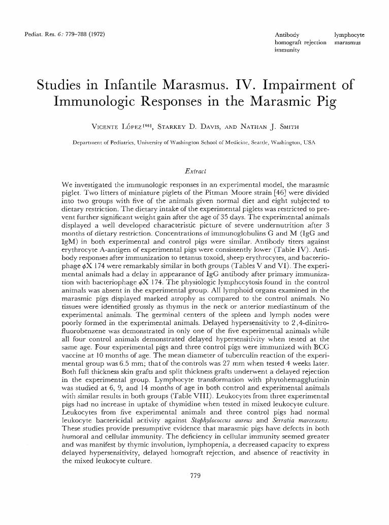
Pediat. Res. 6: 779-788 (1972) Antibodyhomograft rejectionimmunity
lymphocytemarasmus
Studies in Infantile Marasmus. IV. Impairment ofImmunologic Responses in the Marasmic Pig
VICENTE LOPEZ'6 6 1 , STARKEY D. DAVIS, AND NATHAN J. SMITH
Department of Pediatrics, University of Washington School of Medicine, Seattle, Washington, USA
Extract
We investigated the immunologic responses in an experimental model, the marasmicpiglet. Two litters of miniature piglets of the Pitman Moore strain [46J were dividedinto two groups with five of the animals given normal diet and eight subjected todietary restriction. The dietary intake of the experimental piglets was restricted to pre-vent further significant weight gain after the age of 35 days. The experimental animalsdisplayed a well developed characteristic picture of severe undernutrition after 3months of dietary restriction. Concentrations of immunoglobulins G and M (IgG andIgM) in both experimental and control pigs were similar. Antibody titers againsterythrocyte A-antigen of experimental pigs were consistently lower (Table IV). Anti-body responses after immunization to tetanus toxoid, sheep erythrocytes, and bacterio-phage $X 174 were remarkably similar in both groups (Tables V and VI). The experi-mental animals had a delay in appearance of IgG antibody after primary immuniza-tion with bacteriophage </>X 174. The physiologic lymphecytosis found in the controlanimals was absent in the experimental group. All lymphoid organs examined in themarasmic pigs displayed marked atrophy as compared to the control animals. Notissues were identified grossly as thymus in the neck or anterior mediastinum of theexperimental animals. The germinal centers of the spleen and lymph nodes werepoorly formed in the experimental animals. Delayed hypersensitivity to 2,4-dinitro-fluorobenzene was demonstrated in only one of the five experimental animals whileall four control animals demonstrated delayed hypersensitivity when tested at thesame age. Four experimental pigs and three control pigs were immunized with BCGvaccine at 10 months of age. The mean diameter of tuberculin reaction of the experi-mental group was 6.5 mm; that of the controls was 27 mm when tested 4 weeks later.Both full thickness skin grafts and split thickness grafts underwent a delayed rejectionin the experimental group. Lymphocyte transformation with phytohemagglutininwas studied at 6, 9, and 14 months of age in both control and experimental animalswith similar results in both groups (Table VIII). Leukocytes from three experimentalpigs had no increase in uptake of thymidine when tested in mixed leukocyte culture.Leukocytes from five experimental animals and three control pigs had normalleukocyte bactericidal activity against Staphylococcus aureus and Serratia marcescens.These studies provide presumptive evidence that marasmic pigs have defects in bothhumoral and cellular immunity. The deficiency in cellular immunity seemed greaterand was manifest by thymic involution, lymphopenia, a decreased capacity to expressdelayed hypersensitivity, delayed homograft rejection, and absence of reactivity inthe mixed leukocyte culture.
779
780 LOPEZ, DAVIS, AND SMITH
Speculation
Clinical experience and recent experimental studies indicate that malnutrition de-creases resistance to infection. The results presented show that cell-mediated im-munity is impaired in malnutrition. Much of the immunodeficiency in malnutritionmay be mediated by hyperadrenocorticism.
Introduction
"Protein-calorie malnutrition coupled with infection isthe greatest killer of infants and young children andthe major cause of retarded child growth and develop-ment in today's world," stated the Committee on Inter-national Child Health in 1969 [11]. Infantile maras-mus is one of the most common severe nutritionaldisorders affecting children of developing countries.Marasmus is the result of extreme and prolonged die-tary restriction of proteins, calories, and other essentialnutrients in early life. Marasmus is to be differentiatedfrom kwashiorkor, which is the result of dietary pro-tein deficits that are greater than dietary deficits incalories. Patients with kwashiorkor have edema andhypoproteinemia. In contrast, marasmic children havemore nearly normal protein concentrations and noedema.
Clinical experience has indicated that malnourishedchildren resist infections poorly, though the nature ofthe host deficiency is somewhat uncertain, no doubtbecause of the difficulty in conducting well controlledstudies in humans [16, 19, 23, 31]. Children with pro-tein-calorie malnutrition were reported to have atro-phy of lymphoid tissue and a depression of cell-media-ted immunity [38]. The studies reported here weredesigned to investigate the immunologic responses inan experimental model, the marasmic piglet.
Materials and Methods
Animals. Two litters of minature piglets of the Pit-man Moore strain [46] were used. The average weightat birth was 600 g. After weaning at 15 days, they werefed a commercial piglet diet [47]. The young animalsreached an average weight of 4 kg at 35 days of age.The initial litter of nine piglets was divided into twogroups with four of the animals serving as controls andfive subjected to the experimental dietary restriction.Three additional piglets from a second litter were usedas experimental animals, and one of the piglets fromthis litter served as a control animal. The animalswere housed in individual cages. The diet of the exper-
imental piglets was adjusted to prevent further signifi-cant weight gain after the age of 35 days by restrictingtheir dietary intake to 30 g of commercial pig dietoffered three times daily. This limited dietary intakewas continued throughout the entire study period.Water was supplied ad libitum. All animals were given150 mg of iron by intramuscular injection of iron dex-tran on the 3rd day of life. All experimental proce-dures were performed in the temperature-controlledrooms in which the animals were housed.
Weights of four control and five experimental ani-mals are depicted in Figure 1. The experimental ani-mals displayed a well developed characteristic pictureof severe undernutrition after 3 months of dietary re-striction. There was absence of subcutaneous fat,marked muscle atrophy, and an increased amount ofcoarse body hair. The striking similarities of those ani-mals to the clinical picture of infantile marasmus havebeen described in previous studies [30, 40].
Serum proteins. Serum protein concentrations andserum protein electrophoresis were performed bymethods previously described [24, 28]. Serum concen-
200i
1008060
40
20
— Diet restriction begun-
•3D
0 1 3Age
6in month
D Controls* Experimental
9 1
Fig. 1. Weight of control and experimental pigs during the first12 months of life. Diet restriction of the experimental animals wasbegun at 35 days of age.
Infantile marasmus. IV 781
trations of IgG and IgM were measured by singleradial immunodiffusion method [29]. Antiserum toporcine IgM [48] and antiserum to porcine IgG [49]were obtained commercially. Immunologically pureporcine IgG [50] was used as the standard for IgGmeasurements. Purified porcine IgM was not available.The results of IgM determinations were expressed aspercentages of a normal adult pig's serum [51].
Antigens and serum, antibody determinations. Natu-rally occurring antibodies to human type A erythro-cytes were determined by conventional hemagglutina-tion technique. One milliliter of aluminum hydrox-ide-absorbed tetanus toxoid [52] was injected intramus-cularly at the age of 50 days. Secondary immunizationwas carried out by giving a similar dose 6 monthslater. Antibody titers were determined by hemagglu-tination [39]. A saline suspension of 108 sheep erythro-cytes (SRBC) per kg body weight in complete Freund'sadjuvants [53] was given intramuscularly at the age of1 year. Anti-SRBC antibody titers were determined byconventional hemagglutination.
The animals were immunized to bacteriophage ^X174 by intravenous injection of 1.5 X 10° plaque-form-ing units (PFU) of phage per kg body weight. Threeexperimental piglets and three controls were immu-nized at the age of 80 days. A second group was pri-marily injected at 225 days of age. Secondary immuni-zations to phage (f>X 174 were carried out 5 weeks later.Antibody activity was measured by phage neutraliza-tion and expressed as K-values or rates of phage inacti-vation [41]. Antibodies to bacteriophage $X 174 werefurther characterized by reduction with 2-mercaptoeth-anol (2-ME) [18]. After the pigs were immunized withSRBC, selected serum samples were subjected to gelfiltration on Sephadex G-200 [54] on 2.5- X 100-cmcolumns using Tris buffer, pH 7.4. Eluates were col-lected in 6-ml fractions. The protein concentrationswere determined by optical density measurement at280^.
Lymphocyte counts. The number of lymphocytes inthe peripheral blood was calculated from differentialcounts of coverslip smears, and total leukocyte countswere performed using a hemocytometer. In one experi-mental and in one control animal the cisterna chyliwere cannulated and lymphocyte counts were deter-mined on the lymph obtained.
Sensitizalion luilh dinitrofluorobenzene. 2,4-Dinitro-fluorobenzene (DNFB) [55] was diluted 1:10 in 90%acetone and 10% olive oil. The animals were sensi-tized by placing 1-cm squares of filter paper saturatedwith DNFB on the shaved skin of their backs. Delayed
hypersensitivity responses were elicited through intra-dermal injections of 0.1 ml of 1:1000 dilution ofDNFB in the medial aspects of the legs.
Sensitization with BCG. BCG [56] was injected sub-cutaneously with complete Freund's adjuvant. Fourweeks after the initial injection, intradermal injectionsof 0.1 ml mammalian tuberculin [57] were adminis-tered. Reactions to the tuberculin were measured 48 hrlater.
Histopalhology of peripheral lymph nodes, spleen,and thymus. Spleen and lymph nodes were obtainedand thymuses were sought at postmortem examina-tions of three control and four experimental pigs. Aperipheral lymph node was obtained from all otheranimals. The specimens were prepared for histologicexamination.
Skin homograft. Two techniques of skin graftingwere used. In the first, skin was taken from an unre-lated donor pig and three full thickness homograftsmeasuring 2 cm in diameter were placed on preparedbeds in the backs of the experimental and control pigs.In addition, an autograft was placed on each animal.No sutures were taken. In the second technique, skinwas again taken from an unrelated donor using a der-matone set to cut pieces of skin 10 X 5 cm and 0.02mm in thickness. The skin was then cut into smallpieces ( l x l cm). As many as 10 homografts wereplaced on a prepared bed of a single animal. Fiveautografts served as controls. The animals were anes-thetized with intravenous phenobarbital. Second setgrafts were not done.
Lymphocyte transformation with phytohemag-glutinin (PHA). Blood was drawn in plastic syringescontaining 100 units heparin per ml blood. Mononu-clear cells were counted using a counting chamber.Aliquots of whole blood containing 106 mononuclearcells were placed in culture tubes containing 3 ml Ea-gle's TC medium [58] supplemented with 20% autolo-gous pig serum, 500 units penicillin per ml, and 0.1 ml1:10 dilution of PHA-M [59]. Total volumes weremade up to 4 ml with TC medium. Control tubes wereset up without PHA. These cultures were incubatedfor 72 hr at 37°. Eight hours before the end of theincubation period 4 /xCi 3H-thymidine [60] were addedto each tube. The cells were harvested by centrifugingthe cultures at 1000 rpm for 10 min. The supernatantwas then discarded, and the sediment was washedtwice with 5 ml cold 0.2% NaCl. The cultures werecentrifuged again, and, after the supernatant was dis-carded, 2 ml cold 5% trichloroacetic acid (TCA) wereadded to each tube. The tubes were then left over-
782 LOPEZ, DAVIS, AND SMITH
Table I. Total serum proteins and serum albumin concentra-tions of experimental and control pigs at 3, 6, and 12 months ofage1
Anima group (55 days)26 mos
(146 days)212 mos
(330 days)2
Total protein, g/100 mlExperimental 6.3 ± 0.312 6.74 ± 0.23 6.10 ± 0.38
(N = 7)Control 6.4 ± 0.13 7.90 ± 0.47 7.36 ± 0.50
(AT = 5)Albumin, g/100 ml
Experimental 3.0 ± 0.34 3.01 ± 0.36 3.02 ± 0.21(.V = 7)
Control 3.14 db 0.25 4.27 ± 0.73 3.86 ± 0.27(N = 5)
'Values are exprefsed as means ± SD.2 Days on diet for experimental group.
Table II. IgG concentrations, in mg/100 ml, of experimentaand control pigs1
Age Experimental Control
213612
daysmonthmonthsmonthsmonths
2281542107610601257
db±±±±
78812716515591
2000475101214741089
±±±±±
993194125223133
1 Values are expressed as means ± SD for 7 experimental and 5 con-trol animals.
night at 4° and then centrifuged at 2000 rpm. Thesupernatant was discarded. DNA was extracted with0.5 ml 5% TCA and heated at 95° twice for 10 min.Tubes were centrifuged once again, and 0.3 ml super-natant was placed in the vials containing 9 ml scintil-lation counting fluid made up of 4 g PPO [61] per litertoluene and 0.7 ml solubilizer [62]. Specimens werecounted in a Beckman scintillation counter. Countswere expressed as number per 105 cells. All culturetubes were set up in triplicate.
Mixed leukocyte culture. One-way mixed leukocytecultures were carried out as described [1]. Leukocytesof a nonrelated donor were used to stimulate the re-sponding cells. TCI 99 was buffered to a pH of 7.4with "Hepes" (N-2-hydroxyethylpiperazine-iV-2-ethane-sulfuric acid) [63]. Cultures were incubated for 72 hrand then pulsed with 3H-thymidine for 18 hr. Thepreparation of the material for counting was carriedout essentially as described above.
Passive transfer of delayed hypersensitivity. A leuko-cyte suspension of cells from a highly sensitive DNFB-positive control was prepared as described above. Ali-quots containing 5 X 10° mononuclear cells were in-
jected subcutaneously into four experimental animalswhich had been shown to have lost DNFB hypersensi-tivity.
Leukocyte bactericidal activity. The bactericidal ac-tivity of leukocytes in peripheral blood was assayed aspreviously described [26].
Results
Serum proteins. Serum albumin concentrations ofthe experimental group were lower than controls at 6and 12 months (Table I). IgG and IgM concentrationsof both experimental and control pigs were similar(Tables II and III).
Serum antibody. Antibody against erythrocyte A-an-tigen first appeared in control animals at the age of 2months and rose progressively in titer during the next5 months. Anti-A titers of experimental pigs were con-sistently lower (Table IV).
Antibody responses after immunization to tetanustoxoid, SRBC, and bacteriophage <j>X. 174 were remark-ably similar in both groups (Tables V and VI). Thebacteriophage <j>X 174 was cleared from the circulationby 1 week after immunization. All of the animalsshowed an increase in antibody titer after secondaryimmunization.
Table III. IgM concentrations of experimental and control pigs1
Age Experimental (N = 7),% adult serum
Control (N = 5),% adult serum
1 day10 days45 days
3 months6 months
115 ± 4 6
51 ± 7103 ± 22101 ± 16
115'in.. iu
52102102
±
±±±
46
151417
1 Values are expressed as means ± SD.
Table IV. Reciprocals of natural anti-A hemagglutinationtiters of experimental and control pigs
Animal
Cl1
C2C3C4El2
E2E3E4E5
2
4164800208
3
323283224488
Age, mos
5
64321664441688
7
1286432128883288
1 Cl : Control animal 1.2 El : Experimental animal 1.
Infantile marasmus. IV 783
Immunoglobulin class of antibodies. As shown inTable VI, most of the antibody obtained 1 week afterimmunization was sensitive to treatment with 2-ME.Four weeks after primary immunization most of theantibody from experimental animals was sensitive to2-ME whereas antibody from controls was resistant.The experimental animals thus had a delay in appear-ance of IgG antibody after primary immunization.Both experimental and control groups made IgG anti-body after secondary immunization.
Gel filtration of serum samples from two control
'fable V. Reciprocals of antibody titers to tetanus toxoid andsheep red blood cells after primary and secondary immuniza-tions
Animal
Cl 1
C2C3C4C5El 2
E2E3E4E5E6
Tetanus
4 weeksafter
primary
3201280
801280320808080
12805210
320
toxoid
4 weeksafter
secondary
128020480
320
51201280512051205120
Sheep erythrocytes
10 daysafter
primary
5121281286464
102451225612812864
10 daysafter
secondary
204810241024256
204810241024512512256
1 Cl : Control animal 1.2 El : Experimental animal 1.
Table VI. Antibody response to bacteriophage <£X 174
Animal
Cl 2
C2C3C4C5E l 6
E2E3E4E5E6
Dayson
diet
5050
200200
5020050
20050
20050
74012710
2407
l
.93
.8
.47
.2
.9
.1
.23
.3
.8
.17
.3
Primaryimmunization
week
(<0.1(<0.1(<0.1(0.12)(0.9)(0.21)
(0.13)(0.1)
(0.24)
4 weeks
)4 1.4) 0.58) 13.3
1.260.15
16.11.11.35.86.11.3
(1.3)(0.52)(13)(1.12)(0.09)(3.0)(0.2)(0.34)(0.82)(0.78)(0.56)
Secondaryimmunization1
1 week
26.2ND 6
ND22.0NDND77.513.815.226.2
8.8
4 weeks
16.3NDND11.6NDND21.816.0NDND11.5
1 Essentially no IgM antibody was detected after secondaryimmunization.2 Cl : Control animal 1.3 Neutralizing antibody expressed as K-values (see text).4 Neutralizing antibody after treatment with 2-ME.5 ND: not done.6 El : Experimental animal 1.
40 50Tube number
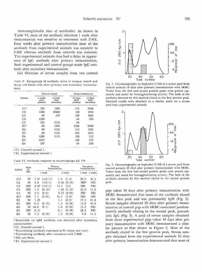
Fig. 2. Chromatography on Sephadex G-200 of a serum pool fromcontrol animals 10 days after primary immunization with SRBC.Tubes from the first and second protein peaks were pooled sep-arately and tested for hemagglutinating activity. The bulk of theantibody detected by this method eluted in the first protein peak.Identical results were obtained in a similar study on a serumpool from experimental animals.
30 50number
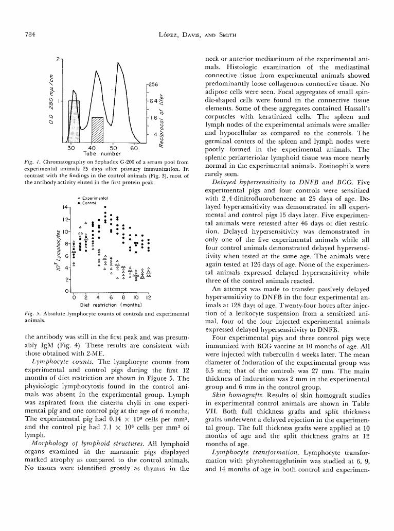
Fig. 3. Chromatography on Sephadex G-200 of a serum pool fromcontrol animals 25 days after primary immunization with SRBC.Tubes from the first and second protein peaks -were pooled sep-arately and tested for hemagglutinating activity. The bulk of theantibody detected by this method eluted in the second proteinpeak.
pigs taken 10 days after primary immunization withSRBC demonstrated that most of the antibody elutedin the first peak and was presumably IgM (Fig. 2).Serum samples obtained 25 days after primary immu-nization of control pigs with SRBC contained predom-inantly antibody eluting in the second peak, presum-ably IgG (Fig. 3). A pool of serum samples obtainedfrom three experimental pigs taken 10 days after pri-mary immunization with SRBC demonstrated a simi-lar pattern to that shown in Figure 2. Most of theantibody eluted in the first protein peak. Serum sam-ples obtained from the experimental animals 25 daysafter primary immunization demonstrated that most of
784 LOPEZ, DAVIS, AND SMITH
r256
50number
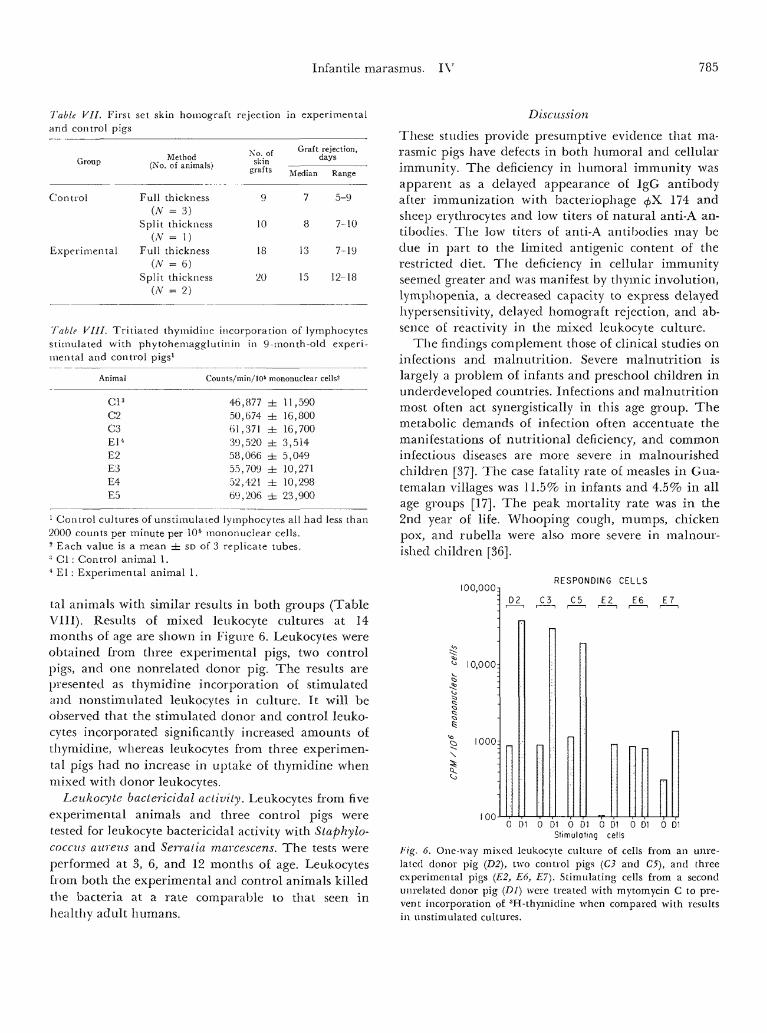
Fig. -I. Chromatography on Sephadex G-200 of a serum pool fromexperimental animals 25 days after primary immunization. Incontrast with the findings in the control animals (Fig. 3), most ofthe antibody activity eluted in the first protein peak.
12-
ExperimentalControl
10-
u2-
0 -
i if i
2 4 6 8 10Diet restriction (months)
12
Fig. 5. Absolute lymphocyte counts of controls and experimentalanimals.
the antibody was still in the first peak and was presum-ably IgM (Fig. 4). These results are consistent withthose obtained with 2-ME.
Lymphocyte counts. The lymphocyte counts fromexperimental and control pigs during the first 12months of diet restriction are shown in Figure 5. Thephysiologic lymphocytosis found in the control ani-mals was absent in the experimental group. Lymphwas aspirated from the cisterna chyli in one experi-mental pig and one control pig at the age of 6 months.The experimental pig had 0.14 X 10⁶ cells per mm3,and the control pig had 7.1 x 10⁶ cells per mm3 oflymph.
Morphology of lymphoid structures. All lymphoidorgans examined in the marasmic pigs displayedmarked atrophy as compared to the control animals.No tissues were identified grossly as thymus in the
neck or anterior mediastinum of the experimental ani-mals. Histologic examination of the mediastinalconnective tissue from experimental animals showedpredominantly loose collagenous connective tissue. Noadipose cells were seen. Focal aggregates of small spin-dle-shaped cells were found in the connective tissueelements. Some of these aggregates contained Hassall'scorpuscles with keratinized cells. The spleen andlymph nodes of the experimental animals were smallerand hypocellular as compared to the controls. Thegerminal centers of the spleen and lymph nodes werepoorly formed in the experimental animals. Thesplenic periarteriolar lymphoid tissue was more nearlynormal in the experimental animals. Eosinophils wererarely seen.
Delayed hypersensitivity to DNFB and BCG. Fiveexperimental pigs and four controls were sensitizedwith 2,4-dinitrofluorobenzene at 25 days of age. De-layed hypersensitivity was demonstrated in all experi-mental and control pigs 15 days later. Five experimen-tal animals were retested after 46 days of diet restric-tion. Delayed hypersensitivity was demonstrated inonly one of the five experimental animals while allfour control animals demonstrated delayed hypersensi-tivity when tested at the same age. The animals wereagain tested at 126 days of age. None of the experimen-tal animals expressed delayed hypersensitivity whilethree of the control animals reacted.
An attempt was made to transfer passively delayedhypersensitivity to DNFB in the four experimental an-imals at 128 days of age. Twenty-four hours after injec-tion of a leukocyte suspension from a sensitized ani-mal, four of the four injected experimental animalsexpressed delayed hypersensitivity to DNFB.
Four experimental pigs and three control pigs wereimmunized with BCG vaccine at 10 months of age. Allwere injected with tuberculin 4 weeks later. The meandiameter of induration of the experimental group was6.5 mm; that of the controls was 27 mm. The mainthickness of induration was 2 mm in the experimentalgroup and 6 mm in the control group.
Skin homografts. Results of skin homograft studiesin experimental control animals are shown in TableVII. Both full thickness grafts and split thicknessgrafts underwent a delayed rejection in the experimen-tal group. The full thickness grafts were applied at 10months of age and the split thickness grafts at 12months of age.
Lymphocyte transformation. Lymphocyte transfor-mation with phytohemagglutinin was studied at 6, 9,and 14 months of age in both control and experimen-
Infantile marasmus. IV 785
Table VII. First set skin homograft rejection in experimentaland control pigs
Group
Control
Experimental
Method(No. of animals)
Full thickness(A' = 3)
Split thickness(N = 1)
Full thickness(N = 6)
Split thickness(/V = 2)
No. ofskin
grafts
9
10
18
20
Graft
Median
7
8
13
15
rejection,days
Range
5-9
7-10
7-19
12-18
Table VIII. Tritiated thymidine incorporation of lymphocytesstimulated with phytohemagglutinin in 9-month-old experi-mental and control pigs1
Animal
Cl 3
C2C3El 4
E2E3E4E5
Counts/min/105 mononuclear cells2
46,87750,67461,37139,52058,06655,70952,42169,206
±±±±±±±±
11,59016,80016,7003,5145,04910,27110,29823,900
1 Control cultures of unstimulated lymphocytes all had less than2000 counts per minute per 105 mononuclear cells.2 Each value is a mean ± SD of 3 replicate tubes.3 Cl : Control animal 1.4 El : Experimental animal 1.
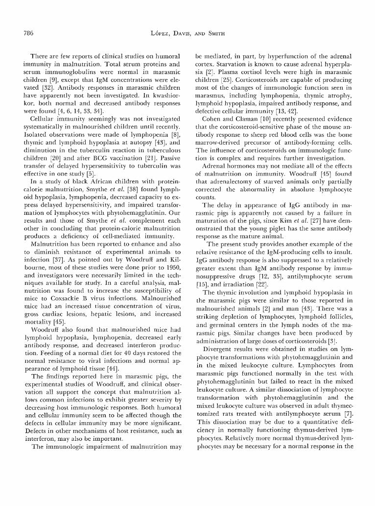
tal animals with similar results in both groups (TableVIII). Results of mixed leukocyte cultures at 14months of age are shown in Figure 6. Leukocytes wereobtained from three experimental pigs, two controlpigs, and one nonrelated donor pig. The results arepresented as thymidine incorporation of stimulatedand nonstimulated leukocytes in culture. It will beobserved that the stimulated donor and control leuko-cytes incorporated significantly increased amounts ofthymidine, whereas leukocytes from three experimen-tal pigs had no increase in uptake of: thymidine whenmixed with donor leukocytes.
Leukocyte bactericidal activity. Leukocytes from fiveexperimental animals and three control pigs weretested for leukocyte bactericidal activity with Staphylo-coccus aureiis and Serratia marcescens. The tests wereperformed at 3, 6, and 12 months of age. Leukocytesfrom both the experimental and control animals killedthe bacteria at a rate comparable to that seen inhealthy adult humans.
Discussion
These studies provide presumptive evidence that ma-rasmic pigs have defects in both humoral and cellularimmunity. The deficiency in humoral immunity wasapparent as a delayed appearance of igG antibodyafter immunization with bacteriophage >̂X 174 andsheep erythrocytes and low titers of natural anti-A an-tibodies. The low titers of anti-A antibodies may bedue in part to the limited antigenic content of therestricted diet. The deficiency in cellular immunityseemed greater and was manifest by thymic involution,lymphopenia, a decreased capacity to express delayedhypersensitivity, delayed homograft rejection, and ab-sence of reactivity in the mixed leukocyte culture.
The findings complement those of clinical studies oninfections and malnutrition. Severe malnutrition islargely a problem of infants and preschool children inunderdeveloped countries. Infections and malnutritionmost often act synergistically in this age group. Themetabolic demands of infection often accentuate themanifestations of nutritional deficiency, and commoninfectious diseases are more severe in malnourishedchildren [37]. The case fatality rate of measles in Gua-temalan villages was 11.5% in infants and 4.5% in allage groups [17]. The peak mortality rate was in the2nd year of life. Whooping cough, mumps, chickenpox, and rubella were also more severe in malnour-ished children [36].
I00,000r: D2
10,000:
1000:
100
RESPONDING CELLS
C3 C5 E2 E6 E7
0 D10 D1 0 01 0 Dl 0 D1 0 D1Stimulating cells
Fig. 6. One-way mixed leukocyte culture of cells from an unre-lated donor pig (D2), two control pigs (C3 and C5), and threeexperimental pigs (E2, E6, E7). Stimulating cells from a secondunrelated donor pig (Dl) were treated with mytomycin C to pre-vent incorporation of 3H-thymidine when compared with resultsin unstimulated cultures.
786 LOPEZ, DAVIS, AND SMITH
There are few reports of clinical studies on humoralimmunity in malnutrition. Total serum proteins andserum immunoglobulins were normal in marasmicchildren [9], except that IgM concentrations were ele-vated [32]. Antibody responses in marasmic childrenhave apparently not been investigated. In kwashior-kor, both normal and decreased antibody responseswere found [4, 6, 14, 33, 34].
Cellular immunity seemingly was not investigatedsystematically in malnourished children until recently.Isolated observations were made of lymphopenia [8],thymic and lymphoid hypoplasia at autopsy [43], anddiminution in the tuberculin reaction in tuberculouschildren [20] and after BCG vaccination [21]. Passivetransfer of delayed hypersensitivity to tuberculin waseffective in one study [5].
In a study of black African children with protein-calorie malnutrition, Smythe et al. [38] found lymph-oid hypoplasia, lymphopenia, decreased capacity to ex-press delayed hypersensitivity, and impaired transfor-mation of lymphocytes with phytohemagglutinin. Ourresults and those of Smythe et al. complement eachother in concluding that protein-calorie malnutritionproduces a deficiency of cell-mediated immunity.
Malnutrition has been reported to enhance and alsoto diminish resistance of experimental animals toinfection [37]. As pointed out by Woodruff and Kil-bourne, most of these studies were done prior to 1950,and investigators were necessarily limited in the tech-niques available for study. In a careful analysis, mal-nutrition was found to increase the susceptibility ofmice to Coxsackie B virus infections. Malnourishedmice had an increased tissue concentration of virus,gross cardiac lesions, hepatic lesions, and increasedmortality [45].
Woodruff also found that malnourished mice hadlymphoid hypoplasia, lymphopenia, decreased earlyantibody response, and decreased interferon produc-tion. Feeding of a normal diet for 40 days restored thenormal resistance to viral infections and normal ap-pearance of lymphoid tissue [44].
The findings reported here in marasmic pigs, theexperimental studies of Woodruff, and clinical obser-vation all support the concept that malnutrition al-lows common infections to exhibit greater severity bydecreasing host immunologic responses. Both humoraland cellular immunity seem to be affected though thedefects in cellular immunity may be more significant.Defects in other mechanisms of host resistance, such asinterferon, may also be important.
The immunologic impairment of malnutrition may
be mediated, in part, by hyperfunction of the adrenalcortex. Starvation is known to cause adrenal hyperpla-sia [2]. Plasma cortisol levels were high in marasmicchildren [25]. Corticosteroids are capable of producingmost of the changes of immunologic function seen inmarasmus, including lymphopenia, thymic atrophy,lymphoid hypoplasia, impaired antibody response, anddefective cellular immunity [13, 42].
Cohen and Claman [10] recently presented evidencethat the corticosteroid-sensitive phase of the mouse an-tibody response to sheep red blood cells was the bonemarrow-derived precursor of antibody-forming cells.The influence of corticosteroids on immunologic func-tion is complex and requires further investigation.
Adrenal hormones may not mediate all of the effectsof malnutrition on immunity. Woodruff [45] foundthat adrenalectomy of starved animals only partiallycorrected the abnormality in absolute lymphocytecounts.
The delay in appearance of IgG antibody in ma-rasmic pigs is apparently not caused by a failure inmaturation of the pigs, since Kim et al. [27] have dem-onstrated that the young piglet has the same antibodyresponse as the mature animal.
The present study provides another example of therelative resistance of the IgM-producing cells to insult.IgG antibody response is also suppressed to a relativelygreater extent than IgM antibody response by immu-nosuppressive drugs [12, 35], antilymphocyte serum[15], and irradiation [22].
The thymic involution and lymphoid hypoplasia inthe marasmic pigs were similar to those reported inmalnourished animals [2] and man [43]. There was astriking depletion of lymphocytes, lymphoid follicles,and germinal centers in the lymph nodes of the ma-rasmic pigs. Similar changes have been produced byadministration of large doses of corticosteroids [3].
Divergent results were obtained in studies on lym-phocyte transformations with phytohemagglutinin andin the mixed leukocyte culture. Lymphocytes frommarasmic pigs functioned normally in the test withphytohemagglutinin but failed to react in the mixedleukocyte culture. A similar dissociation of lymphocytetransformation with phytohemagglutinin and themixed leukocyte culture was observed in adult thymec-tomized rats treated with antilymphocyte serum [7].This dissociation may be due to a quantitative defi-ciency in normally functioning thymus-derived lym-phocytes. Relatively more normal thymus-derived lym-phocytes may be necessary for a normal response in the
Infantile marasmus. IV 787
mixed leukocyte culture than in the transformationreaction with phytohemagglutinin.
Malnutrition is one of the most important healthproblems in the world, and infection constitutes one ofthe major causes of morbidity and mortality. Muchremains to be learned concerning the interactions be-tween nutrition, infections, and host resistance. Beforethe interaction between infection and malnutritioncan be understood, further investigations will be re-quired on immunologic function in malnourishedchildren, immunologic function in malnourished ani-mals, and resistance of malnourished animals to exper-imental infections.
References and Notes
1. AMOS, D. B., AND BACH, F. H.: Phenotypic expressions of themajor histocompatibility locus in man (HL-A): leukocyteantigens and mixed leukocyte culture reactivity. J. Exp. Med.,128: 623 (1968).
2. ANDREASEN, E.: The thymolymphatic system during inanitionand restitution. Acta Pathol. Microbiol. Scand. Suppl. XLIX(1943).
3. BAKER, B. L., INGLE, D. J., AND CHOH HAO LI: The histologyof the lymphoid organs of rats treated with adrenocortico-tropin. Amer. J. Anat., 88: 313 (1951).
4. BROWN, R. E., AND KATZ, M.: Failure of antibody productionto yellow fever vaccine in children with kwashiorkor. Trop.Geogr. Med., 18: 125 (1966).
5. BROWN, R. E., AND KATZ, M.: Passive transfer of delayed hy-persensitivity reaction to tuberculin in children with proteincalorie malnutrition. J. Pediat., 70: 126 (1967).
6. BUDIANSKY, E., AND DA SILVA, N. N.: Formacao de anticorposna distrofia pluricarencial hidropigcncia. Hospital (Rio deJaneiro), 52: 251 (1957).
7. CARLSON, L. S., AND SCHWARZ, M. R.: Thymectomy, antilym-phocyte serum (ALS), and the mixed lymphocyte reaction(MLR). In: O. R. Mclntyre: Proceedings of the 4th LeucocyteCulture Conference. (Dartmouth, Hanover, N. H., 1969).
8. CASTELLANOS, H., AND ARROYAVE, G.: Role of the adrenal corti-cal system in the response of children to severe protein mal-nutrition. Amer. J. Clin. Nutr., 9: 186 (1961).
9. CHATTERJEE, S., BHATTACHARYYA, A. K., AND MANDAL, J. N.:Serum proteins in kwashiorkor and marasmus: 1. Unselectedseries before treatment. Bull. Calcutta Sch. Trop. Med., 16:35 (1968).
10. COHEN, J. J., AND CLAMAN, H. N.: Thymus-marrow immuno-competence. V. Hydrocortisone-resistant cells and processesin the hemolytic antibody response of mice. J. Exp. Med.,133: 1026 (1971).
11. Committee on International Child Health: Malnutrition inthe World's Children. Pediatrics,*?: 131 (1969).
12. DUKOR, P., AND DIETRICH, F. M.: The immune response toheterologous red cells in mice. V. The effect of cyclophos-phamide and cortisone on antigenic competition. J. Im-munol., 105: 118 (1970).
13. DURKIN, H. G., AND THORBEKE, G. J.: Relationship of germi-nal centers in lymphoid tissue to immunologic memory. V.
The effect of prednisolone administered after the peak of theprimary response. J. Immunol., 106: 1079 (1971).
14. FERNANDEZ, N. A.: Serum antibody response of malnourishedchildren as compared with well nourished children. Bol. Asoc.Med. P. R., 52: 222 (1960).
15. FIELD, E. J., HUGHES, D., AND CASPARY, E. A.: Mode of actionof antilymphocyte serum. Lancet, ii: 964 (1969).
16. GORDON, J. F., GUZMAN, M. A., ASCOLI, W., AND SCRIMSHAW,N. S.: Acute diarrhoea! disease in less developed countries. 2.Patterns of epidemiological behaviour in rural Guatemalanvillages. Bull. World Health Organ., 31: 9 (1964).
17. GORDON, J. E., JANSEN, A. A. J., AND ASCOLI, W.: Measles inrural Guatemala. J. Pediat., 66: 779 (1965).
18. GRUBB, R., AND SWAHN, B.: Destruction of some agglutininsbut not of others by two sulfhydryl compounds. Acta Pathol.Microbiol. Scand., 43: 305 (1958).
19. HALL, S. A.: Nutrition and infection. East Afr. Med. J., 46: 58(1969).
20. HARLAND, P. S.: Tuberculin reactions in malnourished chil-dren. Lancet, ii: 791 (1965).
21. HARLAND, P. S., AND BROWN, R. E.: Tuberculin sensitivity test-ing following BCG vaccination in undernourished children.East Afr. Med. J., 42: 233 (1965).
22. HASEK, M., AND LENGEROVA, A.: Immunology. In: Mechanismsof Radiobiology, Vol. II, p. 207 (Academic Press, New York,1960).
23. HENDRICK.SE, R. G.: Interactions of nutrition and infection:experience in Nigeria. In: G. E. W. Wolstenholme and M.O'Connor: Nutrition and Infection, p. 98. (Ciba FoundationGroup No. 31, Churchill, London, 1967).
24. HUFRGA, J. DE LA, SMETTERS, G. W., AND SHERRICK, J. C:Chlorometric determination of serum protein: the Biuretreaction. In: F. W. Sunderman and F. W. Sunderman: SerumProteins and the Dysproteinemias, p. 54. (Pitman Medical,London, 1964).
25. KAMALA, S. J. R., SRIKANTIA, S. G., AND GOPALAN, C: Plasmacortisol levels in protein-calorie malnutrition. Arch. Dis.Childhood,*?: 365 (1968).
26. KAPLAN, E. L., LAXDAL, T., AND QUIE, P. G.: Studies on poly-morphonuclear leukocytes from patients with chronic gran-ulomatous disease of childhood: bactericidal capacity forstreptococci. Pediatrics, 41: 591 (1968).
27. KIM, Y. B., BRADLEY, S. G., AND WATSON, D. W.: Ontogeny ofthe immune response. I. Development of immunoglobulinsin germ-free and conventional colostrum-deprived piglets. J.Immunol., 97: 52 (1966).
28. KOHN, J.: Cellulose acetate membrane (CAM) clectrophoresis.In: C. A. Williams and M. W. Chase: Methods in Immunologyand Immunochemistry, Vol. II, p. 20. (Academic Press, NewYork, 1968).
29. LOPEZ, V., AND GOLDER, S.: A new immunodiffusion methodfor quantitative determination of proteins in biologicalfluids based on a separated application of antibody and anti-gen to small agar discs. Clin. Chim. Acta, 21: 517 (1968).
30. MCCANCE, L. A., AND MOUNT, L. E.: Severe undernutrition ingrowing and adult animals. 5. Metabolic rate and body tem-perature in the pig. Brit. J. Nutr., 14: 509 (1960).
31. MORLEY, D.: The public health problems created by measlesin the developing countries. Arch. Gesamte Virusforsch., 16:19 (1965).
32. NAJJAR, S. S., STEPHAN, M., AND ASFOUR, R. Y.: Serum levels
788 LOPEZ, DAVIS, AND SMITH
of immunoglobulins in marasmic infants. Arch. Dis. Child-hood, 44: 120(1969).
33. PRETORIUS, P. J., AND DE VILLIERS, L. S.: Antibody responsein children with protein malnutrition. Amer. J. Clin. Nutr.,10: 379 (1962).
34. REDDY, V., AND SRIKANTIA, S. G.: Antibody response inkwashiorkor. Indian J. Med. Res., 52: 1154 (1964).
35. SAHIAR, K., AND SCHWARTZ, R. S.: The immunoglobulin se-quence. I. Arrest by 6-mercaptopurine and restitution by anti-body, antigen or spleenotomy. J. Immunol., 95: 345 (1965).
36. SALOMON, J. B., MATA, L. J., AND GORDON, J. E.: Malnutritionand the common communicable diseases of childhood inGuatemala. Amer. J. Public Health, 58: 505 (1968).
37. SCRIMSHAW, N. S., TAYLOR, C. E., AND GORDON, J. E.: Interac-tions of nutrition and infection. World Health Organ.Monogr. Ser. 57 (1968).
38. SMYTHE, P. M., BRERETON-STILES, G. G., GRACE, H. J., MA-FOYANE, A., SCHONLAND, M., COOVADIA, H . M., LOENING, W . E.
K., PARENT, M. A., AND VOS, G. H.: Thymolymphatic deficiencyand depression of cell-mediated immunity in protein-caloriemalnutrition. Lancet, ii: 7731 (1971).
39. STAVITSKY, A. B.: Micromethods for the study of proteins andantibodies. I. Procedure and general applications of hemag-glutination and hemagglutination-inhibition reactions withtannic acid and protein-treated red blood cells. J. Immunol.,72: 360 (1954).
40. STECKEL, A., AND SMITH, N. J.: Hematologic studies of severeundernutrition of infancy. I. The anemia of prolonged caloricdeprivation in the pig. Pediat. Res., 3: 338 (1969).
41. UHR, J. W., FINKELSTEIN, M. S., AND BAUMANN, J. B.: Antibody
formation. III. The primary and secondary antibody responseto bacteriophage ^X 174 in guinea pigs. J. Exp. Med., 115:655 (1962).
42. WARD, P., AND JOHNSON, A. G.: Studies on the adjuvant ac-tion of bacterial endotoxins on antibody formation. II. Anti-body formation in cortisone-treated rabbits. J. Immunol., 82:427 (1958).
43. WATTS, T.: Thymus weights in malnourished children. J.Trop. Pediat., 15: 155 (1969).
44. WOODRUFF, J. F.: The influence of quantitated post-weaningundernutrition on Coxsackievirus B3 infection of adultmice. II. Alteration of host defense mechanisms. J. Infec. Dis.,121: 164 (1970).
45. WOODRUFF, J. F., AND KILBOURNE, E. D.: The influence ofquantitated post-weaning undernutrition on CoxsackievirusB3 infection of adult mice. I. Viral persistence and increasedseverity of lesions. J. Infec. Dis., 121: 137 (1970).
46. Vita Vet Laboratories, Marion, Ind.47. Startena, Purina-Ralston Company, St. Louis, Mo.48. Sevac Laboratories, Prague, Czechoslovakia.49. Hyland Laboratories, Los Angeles, Calif.50. Miles Laboratories, Elkhart, Ind.51. Pel-Freez Biological Laboratories, Rogers, Ark.52. Haver-Lockhart, Kansas, Mo.53. Hyland Laboratories, Costa Mesa, Calif.54. Pharmacia, Uppsala, Sweden.55. Nutritional Biochemical Corporation, Cleveland, Ohio.56. Research Foundation, Chicago, 111.57. U.S. Department of Agriculture.58. Bacteriological Associates, Bethesda, Md.59. Difco, Detroit, Mich.60. New England Nuclear, Boston, Mass.61. Packard Instrument Company, Downers Grove, 111.62. Baltimore Biological, Baltimore, Md.63. Calbiochem, Los Angeles, Calif.64. The authors wish to thank A. Iannetta, A. Millar, L. Moore,
and T. Lopez for their excellent technical assistance and H.Cowen for his invaluable help and advice in handling theanimals.
65. This work was supported in part by the University of Wash-ington Graduate School Research Fund, Pediatric Microbiol-ogy Training Grant no. 5 T01 AI00227 from the PublicHealth Service, and Fellowship Award no. 1-F03-HD42316-01from the National Institute of Child Health and HumanDevelopment.
66. Requests for reprints should be addressed to: VICENTE LOPEZ,M.D., HH627 Harborview Hospital, 325 9th Avenue, Seattle.Wash. 98104 (USA).
67. Accepted for publication May 23, 1972.
Copyright © 1972 International Pediatric Research Foundation, Inc. Printed in U.S.A.
Copyright © 2022 FDOKUMEN