Reactive oxygen intermediates in autoimmune islet cell destruction of the NOD mouse induced by...
10
Diabetologia (1994) 37:22-31 Diabetologia Springer-Verlag1994 Reactive oxygen intermediates in autoimmune islet cell destruction of the NOD mouse induced by peritoneal exudate cells (rich in macrophages) but not T cells E Horio 1 , M. Fukuda I , H. Katoh 2 , M. Petruzzelli I, N. Yano 1 , C. Rittershaus 3, S. Bonnet-Weir 4 , M. Hattori 1 1Section on Immunology and Immunogenetics, Joslin Diabetes Center, Department of Medicine, Brigham and Women's Hospital, Harvard Medical School, Boston, Massachusetts, USA 2 Central Institute for Experimental Animals, Kawasaki, Japan 3T-Cell Sciences, Cambridge, Massachusetts, USA 4 Section on Islet Cell Physiology and Transplantation, Joslin Diabetes Center, Department of Medicine, Brigham and Women's Hospital, Harvard Medical School, Boston, Massachusetts, USA Summary The non-obese diabetic (NOD) mouse spontaneously develops autoimmune Type i (insulin- dependent) diabetes mellitus. NOD mice exhibit massive infiltrates of T cells and macrophages into pan- creatic islets (insulitis) prior to diabetes. The contribu- tion of oxygen free radicals to the development of in- sulitis in NOD mice was examined by administration of its scavengers, such as superoxide dismutase and cata- lase. Bovine superoxide dismutase and catalase were each coupled to polyethylene glycol. The treatment with superoxide dismutase-polyethylene glycol re- duced the number of islets with insulitis and increased the undamaged islet tissue, as compared with the con- trol group. The treatment with catalase-polyethylene glycol showed a similar tendency which did not reach significance. Using a flow cytometric assay of the oxida- tion of 2', 7'-dichlorofluorescein, the content of reac- tive oxygen intermediates in islet cells in the culture system was measured and the effect of peritoneal exu- date cells and T cells on their production examined. Peritoneal exudate cells, but not T cells, from NOD mice increased the content of reactive oxygen inter- mediates in islet cells of either the NOD mouse or the ILI mouse (MHC-identical to NOD); the addition of snperoxide dismutase to the culture medium sup- pressed this increase in NOD or ILI islet cells. The present data support the concept that production of oxygen free radicals mediated by macrophages can damage islet beta cells, directly resulting in autoim- mune Type i diabetes in NOD mice. [Diabetologia (1994) 37: 22-31] Key words NOD mice, insulitis, reactive oxygen inter- mediates, superoxide dismutase, peritoneal macro- phages. Lymphoid cell infiltration into pancreatic islets (in- sulitis) is a well-recognized feature of Type i (insulin- dependent) diabetes mellitus both in humans [1] and in rodents with spontaneous diabetes [2, 3]. The non- obese diabetic (NOD) mouse spontaneously develops an insulin-dependent diabetes. NOD mice exhibit massive infiltrates of lymphoid cells and macrophages into pancreatic islets (insulitis) prior to complete beta- cell destruction, and insulitis appears as early as 5 weeks of age. Makino and co-workers [4] have shown Received: 26 April 1993 and in revised form: 15 July 1993 Corresponding author: Dr. M. Hattori, Section on Immunology and Immunogenetics, Joslin Diabetes Center, Harvard Medical School, One Joslin Place, Boston, MA 02215, USA that T cells play an important role in the development of insulitis and diabetes in nude NOD mice. The major infiltrating cell populations in the insulitis lesion are CD4 § (L3T4, helper) and CD8 § (Ly2, cytotoxic/sup- pressor) T cells, and macrophages [5]. Splenocytes from diabetic NOD mice can transfer diabetes to ir- radiated non-diabetic young NOD mice [6]. Both CD4 ~ and CD8 + splenic T cells are required to transfer diabetes from diabetic NOD mice to irradiated non- diabetic NOD mice. In T-cell mediated immune pro- cesses, the immune responses to self-antigens are de- pendent on certain combinations of MHC and the T- cell receptor (TcR) Vfl family. Restricted TcR usage has been found in some T-cell dependent autoimmune disease, for example TcR Vfl 8 is preferentially used in experimental autoimmune encephalomyelitis [7]. Re- striction of TcR usage in autoimmune diabetes has not
-
Upload
independent -
Category
Documents
-
view
2 -
download
0
Transcript of Reactive oxygen intermediates in autoimmune islet cell destruction of the NOD mouse induced by...

Diabetologia (1994) 37:22-31
Diabetologia �9 Springer-Verlag 1994
Reactive oxygen intermediates in autoimmune islet cell destruction of the NOD mouse induced by peritoneal exudate cells (rich in macrophages) but not T cells E H o r i o 1 , M . F u k u d a I , H . K a t o h 2 , M . P e t r u z z e l l i I , N . Y a n o 1 , C . R i t t e r s h a u s 3 , S. B o n n e t - W e i r 4 , M . H a t t o r i 1
1 Section on Immunology and Immunogenetics, Joslin Diabetes Center, Department of Medicine, Brigham and Women's Hospital, Harvard Medical School, Boston, Massachusetts, USA
2 Central Institute for Experimental Animals, Kawasaki, Japan 3 T-Cell Sciences, Cambridge, Massachusetts, USA 4 Section on Islet Cell Physiology and Transplantation, Joslin Diabetes Center, Department of Medicine, Brigham and Women's Hospital, Harvard Medical School, Boston, Massachusetts, USA
S u m m a r y The non-obese diabetic (NOD) mouse spontaneously develops autoimmune Type i (insulin- dependent) diabetes mellitus. NOD mice exhibit massive infiltrates of T cells and macrophages into pan- creatic islets (insulitis) prior to diabetes. The contribu- tion of oxygen free radicals to the development of in- sulitis in NOD mice was examined by administration of its scavengers, such as superoxide dismutase and cata- lase. Bovine superoxide dismutase and catalase were each coupled to polyethylene glycol. The treatment with superoxide dismutase-polyethylene glycol re- duced the number of islets with insulitis and increased the undamaged islet tissue, as compared with the con- trol group. The treatment with catalase-polyethylene glycol showed a similar tendency which did not reach significance. Using a flow cytometric assay of the oxida- tion of 2', 7'-dichlorofluorescein, the content of reac- tive oxygen intermediates in islet cells in the culture
system was measured and the effect of peritoneal exu- date cells and T cells on their production examined. Peritoneal exudate cells, but not T cells, from NOD mice increased the content of reactive oxygen inter- mediates in islet cells of either the NOD mouse or the ILI mouse (MHC-identical to NOD); the addition of snperoxide dismutase to the culture medium sup- pressed this increase in NOD or ILI islet cells. The present data support the concept that production of oxygen free radicals mediated by macrophages can damage islet beta cells, directly resulting in autoim- mune Type i diabetes in NOD mice. [Diabetologia (1994) 37: 22-31]
Key w o r d s NOD mice, insulitis, reactive oxygen inter- mediates, superoxide dismutase, peritoneal macro- phages.
Lymphoid cell infiltration into pancreatic islets (in- sulitis) is a well-recognized feature of Type i (insulin- dependent) diabetes mellitus both in humans [1] and in rodents with spontaneous diabetes [2, 3]. The non- obese diabetic (NOD) mouse spontaneously develops an insulin-dependent diabetes. NOD mice exhibit massive infiltrates of lymphoid cells and macrophages into pancreatic islets (insulitis) prior to complete beta- cell destruction, and insulitis appears as early as 5 weeks of age. Makino and co-workers [4] have shown
Received: 26 April 1993 and in revised form: 15 July 1993
Corresponding author: Dr. M. Hattori, Section on Immunology and Immunogenetics, Joslin Diabetes Center, Harvard Medical School, One Joslin Place, Boston, MA 02215, USA
that T cells play an important role in the development of insulitis and diabetes in nude NOD mice. The major infiltrating cell populations in the insulitis lesion are CD4 § (L3T4, helper) and CD8 § (Ly2, cytotoxic/sup- pressor) T cells, and macrophages [5]. Splenocytes from diabetic NOD mice can transfer diabetes to ir- radiated non-diabetic young NOD mice [6]. Both CD4 ~ and CD8 + splenic T cells are required to transfer diabetes from diabetic NOD mice to irradiated non- diabetic NOD mice. In T-cell mediated immune pro- cesses, the immune responses to self-antigens are de- pendent on certain combinations of MHC and the T- cell receptor (TcR) Vfl family. Restricted TcR usage has been found in some T-cell dependent autoimmune disease, for example TcR Vfl 8 is preferentially used in experimental autoimmune encephalomyelitis [7]. Re- striction of TcR usage in autoimmune diabetes has not
E Horio et al.: Reactive oxygen intermediates in autoimmune islet cell destruction 23
been conf i rmed in the N O D mouse [8, 9]. However , T cells bear ing TcR Vf l8 are the m a j o r popu la t ion in the islet-infiltrating cells of d iabet ic N O D mice [5]. T h e precise mechan i sms by which lymphocy tes and macro - phages d a m a g e islet be ta cells r em a i n unclear.
In chemical ly- induced diabetes , severa l mechan i sms of islet beta-cel l d a m a g e have b e e n p roposed . S t repto- zotocin and al loxan b r e a k nuclear D N A strands of islet be ta cells by genera t ing oxygen f ree radicals [10 , 11]. S t rep tozo toc in and its n i t rosoamide m o i e t y methylni- t r ou rea alkylate the D N A of be ta cells and cause com- parab le amoun t s of D N A s t rand b r e a k a g e [12, 13]. S t rep tozo toc in alkylates not only D N A but also o ther key cellular c o m p o n e n t s [14]. Poly ( A D P r i b o s e ) syn- the tase inhibitors such as n ico t inamide and 3-amino- benzamide can scavenge oxygen f ree radicals in pan- creatic be ta cells p roduced by s t rep tozo toc in and allox- an in m o n o l a y e r cul ture sys tem [15]. Since n icot inamide and 3 - aminobenzamide were also r epo r t ed to p reven t the d e v e l o p m e n t of spon taneous d iabetes in the N O D mouse [16], the same m e c h a n i s m m a y be involved in islet beta-cel l des t ruct ion of the N O D mouse as that in s t reptozotocin- and a l loxan- induced diabetes. Macro- phages [17] and p ro bab l y T cells p roduce oxygen f ree radicals in i n f l ammato ry reactions. I t is r easonab le to hypothes ize that oxygen f ree radicals p roduced by mac- rophages and T cells in the insulitis lesion m a y des t roy islet be t a cells leading to the d e v e l o p m e n t of d iabetes in N O D mice [18, 19] and tha t oxygen free radical scaven- gers, such as superoxide d ismutase ( S O D ) and catalase, m a y p reven t the des t ruct ion of be ta cells and ensuing diabetes. In this study, we have examined the effects of S O D and catalase on the d e v e l o p m e n t of insulitis, and splenic T-cell popula t ions in N O D mice. In addit ion, the p roduc t ion of react ive oxygen in te rmedia tes in pancre- atic islet cells induced by pe r i tonea l exuda te cells (rich in mac rophages ) and T cells f r o m celiac lymph nodes of the N O D mouse in vi t ro was investigated.
Materials and methods
Animals
Non-obese diabetic (NOD) mice were originally obtained from the colony of Dr. S. Makino at the Aburahi Laboratories, Shio- nogi and Company, Ltd., (Shiga, Japan). NOD/Shi//Jos females develop diabetes at 92 % incidence and NOD males at less than 60 % incidence by 30 weeks of age. The Institute of Cancer Re- search-L-Ishibe (ILI) mouse, which is a sister strain of the NOD mouse and is MHC-identical to the NOD mouse, does not de- velop insulitis or diabetes [20]. Male and female NOD/Shi//Jos and ILI/Jic//Jos mice at 5 months of age were used as a source of pancreatic islet cells in this study. Diabetic NOD mice within 2 weeks after the onset of diabetes were used as a source of peri- toneal exudate cells. For the assay of oxygen free radical produc- tion in islet cells by NOD peritoneal exudate cells, sex-matched animals were used. The NOD mice were given ad libitum com- mercial chow (Agway Prolab 3000 Formula; Agway, Syracuse, NY, USA), and water.
Treatment with superoxide dismutase-polyethylene glycol and catalase-p olyethylene glycol
Fifteen NOD female litter mates from three litters were divided into three groups at 10 weeks of age. Five female NOD mice in each group were injected (intraperitoneally, daily for 14 days) with either superoxide dismutase-polyethylene glycol (SOD- PEG, 225 U per day per mouse, 55 gg protein, source: bovine erythrocytes; Sigma, St. Louis, Mo, USA), catalase-PEG (2,310 U per day per mouse, 57 ~tg protein, source: bovine liver; Sigma), or the vehicle alone (PEG, 45 gg per day per mouse; Sigma). SOD-PEG, catalase-PEG and PEG were dissolved in phosphate-buffered saline (PBS; 137 mmol NaC1, 15 mmol Na2HPO4, 1.47 mmol KH2PO4, 2.68 mmol KCL), pH 7.3. For PEG alone 45 gg was an equivalent dose compared to the com- pound of SOD-PEG (225 U). The animals were killed at 12 weeks of age after collecting blood samples under ether an- aesthesia. The pancreas and spleen were removed and subjected to histological and immunological examinations, respectively.
Assay of the activities of SOD and catalase in plasma
Blood samples (0.4-0.5 ml) were collected with a heparinized pasteur pipette from the retro-orbital venous plexus of the ani- mals under ether anaesthesia. Each animal was assayed for SOD and catalase before and after the treatment. The activity of SOD in heparinized plasma was measured according to the method of McCord and Fridovich [21]. Plasma was diluted ten-fold with 0.9% NaC1 (in catalase-PEG and control group) or 100-fold (SOD-PEG group) and used for the SOD assay. The activity of catalase in plasma was assayed according to the method of Beers and Sizer [22]. One unit of catalase decomposed 1.0 gmol hy- drogen peroxide per min at 25 ~
Point-counting morphometrics of the islets
The animals were killed under ether anaesthesia, and pan- creases removed and weighed. For morphometric analysis it has been shown that there is usually more biological variation be- tween animals than between sections if the tissue is either ho- mogenous or consistently oriented in the same plane. The pan- creases were oriented in the same plane and spread prior to fixa- tion in Bouin's solution. With these procedures, a large cross-sec- tional area of pancreas was obtained and only one "level" or sec- tion per animal was required to have adequate number of inter- cepts (5000 points per animal). The tissues were embedded in paraffin, cut at 6 gm and stained with haematoxylin and eosin. Using one section per animal, all islets on each section were scored for insulitis; the number of islets per section ranged from 17 to 41. Two separate quantifications were performed to evalu- ate the protective effect of SOD and catalase on islet cell destruc- tion. First, the number of islets with insulitis and the total num- ber of islets were counted at 100 x magnification. The ratio of these values multiplied by 100 gave the percent islets with in- sulitis. Secondly, the islet portion without infiltrating immune cells was defined as the undamaged islet portion. The relative volume of undamaged islet portion was measured by point- counting morphometrics [23] at 170 x magnification, covering the entire section with non-overlapping fields of a 50-point grid. The number of points counted per animal was approximately 5,000. The absolute mass of undamaged islet portion was then calculated by multiplying the relative volume by the pancreatic weight for each animal.

24
Antibodies
Monoclonal antibodies (mAb) used to characterize splenic lym- phoid cells and peritoneal exudate cells were fluorescein iso- thionate (FITC)-conjugated or biotinylated mAb anti-mouse Thyl.2 (Becton Dickinson, Mountain View, Calif., USA) for T cells, phycoerythrin (PE)-conjugated mAb GK1.5 anti-L3T4 (Becton Dickinson) for CD4 T cells, FITC-conjugated mAb 53-6 anti-mouse Ly2 (Becton Dickinson) for CD8 T cells, FITC-con- jugated goat anti-mouse IgM (Organon Teknika, Malvern, Pa., USA) for B cells, mAb M3/84.6.34 (rat IgG1, American Type Culture Collection, Rockville, Md., USA) and mAb M1/70.15 (rat IgG2b, Caltag Laboratories, South San Francisco, Calif., USA) for macrophages, mAb PK 136 anti-mouse natural killer cells (IgG2a) and rabbit anti-asialo GM1 (Wako Fine Chemical, Dallas, Tx., USA) for natural killer cells. Antisera used as con- trols were: monoclonal antibody P3X63 for mAb anti-mouse Thyl.2, normal mouse serum for anti-mouse IgM, mAb anti- L3T4 and Ly2, normal rat IgG for mAb M3/84.6.34 and mAb M1/70.15, and normal rabbit serum for rabbit anti-asialo GM1.
Monoclonal antibodies used in studying TcRc~fl, TcR Vfl 8 and CD3 (6) were mAb H57-597 (hamster IgG) pan anti-mouse TcRc~fl [24], mAb F23.1 (mouse IgG 2a) anti TcR Vfl 8.1, 2, 3 [25], and mAb 145-2Cll hamster anti-mouse CD3 (6) [26], re- spectively. Antisera used as controls were: normal hamster serum for mAb 57-597.2 and 145-2Cll, and purified mouse myeloma RPC 5.4 (IgG 2a, Organon Teknika) for mAb F23.1.
Splenic T cells
Spleens were dissected from the 12-week-old NOD mice after the 2-week administration of SOD-PEG, catalase-PEG and PEG, and dissociated by gentle passage through a wire screen. Lymphoid cells were separated from erythrocytes with Fico11- 400 (Lympholyte-M; Cederlane, Province, Canada), and washed twice with RPMI-1640 medium containing 5 % fetal calf serum (FCS). B cells were stained directly with FITC-conjugated goat anti-mouse IgM. The lymphoid cell suspensions were applied to a nylon wool column (Wako Fine Chemical), and incubated at 37 ~ for 45 rain. T cells were then eluted with pre-warmed RPMI-1640 medium containing 5 % FCS. T cells were stained with FITC-conjugated mAb anti-mouse Thyl.2 before and after the purification on nylon wool column to determine the recovery and the purity of T cells (95 %).
The purified T cells were incubated with FITC-conjugated mAb anti-mouse Ly2 or phycoerythrin-conjugated mAb anti- mouse L3T4 on ice for 45 min, washed three times with medium RPMI-1640. Furthermore, the purified T cells were incubated with mAb 145-2Cll (TcRcq3), mAb H57-597 (TCR Vfl 8) or heat- inactivated normal hamster serum on ice for 45 min. T cells were incubated with mAb F23.1 or mouse myeloma protein RPC 5.4 at 37 ~ for 30 min. After the incubation, T cells were washed three times with RPMI-1640 medium, and then stained with FITC-con- jugated goat anti-hamster IgG or goat anti-mouse IgG (Fc spe- cific). The cells were fixed in 2 % formaldehyde-PBS, and stored at 4 ~ until flow cytometric analysis. The percentage of fluores- cein-positive cells were measured by flow cytometer. The abso- lute number of B cells and T cells in each subset was estimated from the total number of cells and the percentage of positive cells.
Islet cell preparations
Islets were isolated from non-diabetic 16-week-old NOD or ILI males. NOD males were chosen because of the lesser degree of islet damage than in NOD females. Islets were also isolated from
E Horio et al.: Reactive oxygen intermediates in autoimmune islet cell destruction
ILI males. ILI mice were chosen because this strain is MHC- identical to the NOD mouse and develops neither insulitis nor diabetes. The animals were anaesthetized intraperitoneally with pentobarbital (Na Salt; 0.5-1.0 mg/mouse; Sigma). Collagenase Type IV (Organon Teknika) solution (4 ml of 2 mg/ml in Hank's balanced salt solution) was infused into the pancreatic duct after ligating the common bile duct at its entrance to the duodenum. The expanded pancreas was digested at 38~ for 30 min and is- lets were purified on Ficoll gradients (25, 23, 20.5 and 11%) and centrifuged at room temperature ( x 800 g, 10 min). The islets were hand picked to avoid contamination by exocrine cells and connective tissues. To prepare single islet cells, isolated islets were washed with Ca- or Mg-free PBS and digested with trypsin solution for 2 min. After complete dissociation, the islet cells were washed twice with medium RPMI-1640 containing 5% FCS.
Peritoneal exudate cells
Peritoneal exudate cells were collected from non-diabetic 16- week-old NOD males in Experiment 1 and diabetic NOD fe- males in Experiment 2 by lavage of the peritoneal cavity with RPMI-1640 medium containing 2 % FCS. The peritoneal exu- date cells consisted of macrophages (60 %), T cells (7 %), B cells (6 %) and anti-asialo GM1 + cells (27 %) in non-diabetic NOD males and diabetic NOD females shortly after the onset of diabetes. Peritoneal T cells (PTc) were prepared by passing the peritoneal exudate cells through nylon wool columns (T-cell purity: 97%). The macrophage-enriched fraction (peritoneal macrophages, PM0) was recovered from the nylon wool (PMr purity: 75 %) and contained a very small number of T cells (less than 3 % ).
Celiac lymph node T cells
Lymphoid cells from the pancreas are mainly drained into the celiac lymph nodes and through the efferent lymphatic vessels leading to the cisterna chyli. Diabetic and non-diabetic NOD mice but not C57BL/6J, BALB/cJ and NON/Jos mice showed enlargement of the celiac lymph nodes with an increased number of T cells (unpublished observation). Injection of lymphocytes (2 • 107) from the enlarged celiac lymph nodes from diabetic NOD mice transferred diabetes into irradiated young NOD mice. As a source of T cells, celiac lymph nodes were dissected from non-diabetic 16-week-old NOD males or diabetic NOD fe- males, and dissociated to yield single-cell suspensions. The cell suspensions contained T cells (70 %), B cells (26 %) and macro- phages (6%) and T cells were purified by passing through a nylon wool column.
Measurement of reactive oxygen intermediates (hydrogen peroxide) in islet cells
Four experiments were designed to investigate the production of reactive oxygen intermediates in islet cells. Experiment 1 was designed to test the production of reactive oxygen intermediates in islet cells from male NOD or ILI mice with peritoneal exudate cells and T cells from non-diabetic NOD males. Islet cells iso- lated from either NOD males (culture A-E) or ILI males (cul- ture F-J) were used for the measurement of reactive oxygen in- termediates. Peritoneal exudate cells were prepared from the non-diabetic NOD male mice. Isolated islet cells (2 x 10 s cells),
5 celiac T cells (nylon wool purified T cells, 4 x 10 cells), and peri- 5 toneal exudate cells (4 x 10 cells) were cultured overnight in the

E Horio et al.: Reactive oxygen intermediates in autoimmune islet cell destruction
l o o A 100 B
" . . . . . : . ' [ ' . i ' : " . . " . ' . : . " ." �9 ~ ' . . ' . :
80 - 2::. :... :i-::.., :,.? "!; 80 ' " ' :::::::~ :7; i:', '!,..: ".'~;,~" ,~'
e 4C O ~ i7 , . -~ - ' ! : ' ; a "~%~"~:~'*':"~"~"" '" ' " 4 0 , ,
.'~ . . . . Nt~.,...y-;~" 2 " " "
2c 2oJ . ' i ' " �9 . ~.:. . .
' ' -2'() 4 0 6 0 : 8 0 1 0 0 i -20 40 6 0 80 1 O0
F L S F L S
C 100
40
2O
1"
E c :-.-.:..!<?.,:; .... <"" ,~ ..,'. :? .-:'"~; ".:,~:T~," "L
\ , s , e ; ce,,s _ t , ,_ , i , . t , _ , _
20 4 0 60 80 1 O0
' F L S
25
Fig. 1 A-C. Cytograms of bit map gating for distinguishing (A) peritoneal exudate cells (PEC), (B) islet cells and (C) both PEC + islet cells from NOD mice. Five thousand PEC and 5000 islet cells were examined by light scatter characteristic. FLS, forward light scatter; 90 o LS, 90 ~ light scatter
medium RPMI-1640 (total volume, 200 H1) containing 5 % FCS, 15 % concanavalin-A-stimulated NOD splenocyte conditioned media, and 1% fresh L-glutamine in a humidified atmosphere (5 % CO2 and 95 % air, at 37 ~ Several combinations of islet cells, T cells and peritoneal exudate cells were tested in the cul- ture system. After overnight culture, the content of reactive oxygen intermediates (hydrogen peroxide) in islet cells was measured using the flow cytometric method of Bass and co-wor- kers [27]. The 2', 7'-dichlorofluorescein diacetate (DCFH-DA; Eastman Kodak, Rochester, NY, USA) was added to the me- dium (final concentration, 50 gmol/1), and cells were then cul- tured for 20 min at 37 ~ DCFH incorporated into islet cells was oxidized rapidly to fluorescent form by intracellular hydrogen peroxide. SOD was added to culture E or J (final concentration, 13.4 U/ml) in combination with DCFH-DA. Then, the fluores- cence of islet cells was determined on a flow cytometer (Cytoflu- orograph 50H; Ortho Diagnostic System Inc., Westwood, Mass., USA). Islet cells were separated from the co-culturing perito- neal exudate cells (PEC) by electric gating on a forward light scatter vs 90 ~ bit map (Fig. 1).
Experiment 2 was designed to test the production of reactive oxygen intermediates in islet cells from female ILI mice with peritoneal exudate cells and T cells from diabetic female NOD mice. Islets were isolated from non-diabetic 16-week-old ILI fe- males. Peritoneal exudate cells and celiac lymph node cells were prepared from diabetic NOD females within 2 weeks after the onset of diabetes. The ILI islet cells were cultured with perito- neal exudate cells, peritoneal macrophages, peritoneal T cells or T cells of the celiac lymph nodes from the diabetic NOD females. The culture conditions were the same as in Experiment 1 except that the culture time of the islet cells with peritoneal exudate cells was reduced from overnight to 3 h. Reactive oxygen inter- mediate production in the islet ceils was measured in the same manner as in Experiment 1.
Experiment 3 was designed to test direct oxidation of 2', 7'- dichlorofluorescein (DCFH) in islet cells by hydrogen peroxide
5 (H202). Islet cells (2 x 10 ) from C3H/HeJ mice (MHC-haplo- type: k) were incubated with 2', 7'-dichlorofluorescein diacetate (final concentration, 50 I.tmol/1) for 30 min and followed by incu- bation with hydrogen peroxide (10 to 100 gmol/1; Sigma) for 15 rain. The fluorescence of islet cells was then determined on a
flow cytometer. The specificity of peritoneal exudate cells from NOD, ILI and C3H/He mice was also examined.
Experiment 4 was designed to test the production of reactive oxygen intermediates in islet cells by cytokines produced by macrophages. Islet cells (2 x 10⁵) from ILI mice were incubated with recombinant mouse interleukin-l-c~ (40 U; Genzyme Bos- ton, Mass., USA) for 3 h and followed by incubation with DCFH-DA for 20 min and flow cytometric analysis.
Statistical analysis
Statistical analysis was performed by the Mann-Whitney U test.
Results
Treatment with S O D - P E G or catalase-PEG
The 2-week t r e a t m e n t with S O D - P E G or catalase- P E G did not affect the growth nor cause signs of visible allergic react ions such as skin rash and anaphylaxis in the animals. The p lasma activity of S O D in the S O D - P E G group increased m o r e than 100-fold c o m p a r e d with that in the control g roup (Table 1). The p l a sma ac- tivity of catalase in the ca t a l a se -PEG g r o u p increased four-fold c o m p a r e d to tha t in the contro l group.
The pancreases f rom the above three groups were subjected to m o r p h o m e t r i c analysis. The n u m b e r of is- lets examined on each sect ion is listed in Table 2. T h e r e were no significant changes in the pancrea t ic weight a m o n g the S O D - P E G , ca t a l a se -PEG and P E G groups (213.9 + 8.0 mg, 204.5 + 8.8 mg, and 196.2 + 14.2 mg, re- spectively). T h e pe rcen tage of islets with insulitis was reduced in the S O D - P E G group to 50 % of the P E G group and in the c a t a l a s e - P E G group to 60 % of the P E G group (Table 2). With ei ther S O D - P E G or cata- l a s e - P E G t r ea tmen t , the absolute mass of u n d a m a g e d islets was two t imes g rea te r than that of the P E G group. Wi thout adminis t ra t ion of S O D - P E G , c a t a l a s e - P E G or P E G , in 3 -month-o ld female N O D mice (n = 6) the m e a n pe rcen t age of islets with insulitis was 51.2 + 9 .1%, the relat ive vo lume of u n d a m a g e d islets 0.45 + 0.10 %, and the absolute mass of u n d a m a g e d is-

26 F. Horio et al.: Reactive oxygen intermediates in autoimmune islet cell destruction
Table 1. The plasma activity of superoxide dismutase (SOD) and catalase in NOD mice before and after the 2-week administration of SOD-PEG and catalase-PEG
Body weight (g)
10 weeks old 12 weeks old SOD activity (U/ml) Catalase activity (U/ml)
before after before after before after
SOD-PEG 21.6 + 0.6 22.6 + 0.9 3.3 + 0.1 532 + 17" 16.6 +_ 1.2 27.2 + 1.3 Catalase-PEG 22.2 + 0.2 23.0 + 0.1 3.0 + 0.4 2.2 + 0.1 17.8 _+ 1.0 83.0 + 13.5 ~ PEG alone 21.5 + 0.7 22.1 + 0.7 3.6 + 0.3 3.5 + 1.2 15.0 _+ 2.5 22.2 + 1.9
Values are expressed as mean + SEM, n = 5 mice in each group. "p < 0.05, significant difference from the value of control (PEG
alone) in each experiment by Mann-Whitney U test. PEG, Poly- ethylene glycol
Table 2. Effects of superoxide dismutase-polyethylene glycol (SOD-PEG) and catalase-PEG on the degree of islet damage of the female NOD mouse at 3 months of age
Group No. of No. of islets/section % of islets with Relative volume of un- Absolute mass of un- animals observed insulitis damaged islet portion (%) damaged islet portion (rag)
SOD-PEG 5 34 + 3 18.7 + 4.8 a 0.91 + 0.04 a 1.93 + 0.04 ~ (26-41) (6.9-30.8) (0.79-1.03) (1.83-2.06)
Catalase-PEG 5 25 + 4 25.7 + 10.5 0.83 + 0.16 1.68 _+ 0.30 a (17-34) (4.0-54.2) (0.36-1.27) (0.76-2.60)
PEG alone 5 24 + 4 45.2 -+ 5.2 0.45 + 0.05 0.86 +_ 0.09 (17-38) (31.6-57.1) (0.36-0.57) (0;66-1.14)
Values are expressed as mean + SEM, Number in parentheses is the range. a p < 0.05, significant difference from the value of control (PEG alone) in each experiment by Mann-Whitney U test
Table 3. Quantitative analysis of B cells and T cells in the spleen of the NOD mouse after administration of superoxide dismutase- polyethylene glycol (SOD-PEG) and catalase-PEG
B cells T cells
CD3 + CD4 + CD8 + TcRc~fl + TcR Vfl8 + (145-2Cll) (GK 1.5) (53-6) (H57-597) (F23.1)
SOD-PEG 19.4 + 3.6 22.1 + 0.8 17.9 + 1.1 8.8 + 1.2 21.9 + 1.5 3.9 + 0.5 Catalase-PEG 15.1 + 1.1 23.8 + 1.8 19.1 + 1.7 7.0 + 0.9 24.8 + 1.8 2.9 + 0.3 PEG alone 15.7 + 1.3 24.2 + 1.0 18.9 + 2.1 6.8 + 0.8 24.7 + 1.1 2.9 + 0.9
Values are expressed as mean _+ SEM ( x 10 _6 cells), n = 5 mice in each group. For T cells, monoclonal antibodies in parentheses
lets 0.82 + 0.18 mg. The degree of islet dam ages in the un t r ea t ed N O D females was similar to tha t in the P E G - t r ea t ed N O D females at 3 m on t hs of age.
The n u m b e r of splenic B ceils and T-cell subsets was examined in the S O D - P E G , c a t a l a s e - P E G and P E G animals (Table 3). T h e r e were no changes in the num- ber of B cells, T cells, C D 4 ÷ T cells and CD8 ÷ T cells af ter the t r e a t m e n t with S O D - P E G or ca t a l a se -PEG, nor in the T-cell subsets bear ing TcR ~ f l chains, and TcR Vfl8.1, 2, 3, as c o m p a r e d with those of the P E G a lone control group.
Reactive oxygen intermediate production in islet cells
Experiment l: peritoneal exudate cells from non- diabetic N O D males. His tog rams ( expanded log scale- X axis) in Figure 2 show f luorescence dis t r ibut ion of D C F H oxidat ion in islet cells of the N O D mouse and
the I L I mouse . I so la ted islet ceils o f the N O D mouse did not p roduce react ive oxygen in te rmedia tes (Fig. 2 A ) . T h e T cells f r o m celiac l ymph nodes of the N O D mouse did not induce p roduc t ion of react ive oxygen in te rmed ia tes in the N O D islet cells (Fig. 2B) . Per i tonea l exuda te cells of the N O D mouse resul ted in the p roduc t ion of react ive oxygen in te rmedia tes in the N O D islet cells (Fig.2 C). C o m b i n a t i o n of pe r i tonea l exuda te cells (4 x 10⁵ cells) and T cells (4 x 10⁵ cells) did not enhance and ra the r slightly suppressed the p roduc- t ion of react ive oxygen in te rmedia tes in the N O D islet cells (Fig. 2 D). Addi t ion of S O D (13.4 U/ml) to per i to- neal exuda te cells + T cells part ial ly inhibi ted the pro- duct ion of react ive oxygen in te rmedia tes in the N O D islet cells (Fig. 2E) . Nylon wool (and Sephadex G-10) pur i f ied T cells (purity: > 9 5 % ) isolated f r o m the N O D per i tonea l exuda te cells fai led to induce the pro- duct ion of react ive oxygen in te rmedia tes in the islet cells.

E H o r i o e t al.: R e a c t i v e o x y g e n i n t e r m e d i a t e s in a u t o i m m u n e is let cell d e s t r u c t i o n
NOD islet cells ILl islet cells
A F
B G
0 0 Lt~
II
0 o t~
o
$
E z
in " ~ . I I t I I I
C H
D
1 E
i
i
J
g w ! i
Expanded log fluorescence intensity
27
Fig .2A-J . Histograms of fluor- escence distribution of 2', 7'-Di- chlorofluorescein (DCFH) oxida- tion in islet cells from NOD or ILI male mice induced by peritoneal exudate cells from non-diabetic NOD male mice. T ceils of the celiac lymph nodes from non- diabetic male NOD mice were also tested to induce reactive oxygen intermediates in the islet cells. The islet cells and the effector cells were co-cultured overnight prior to the addition of DCFH-DA. Note the fluorescence intensity (on the X-axis) is expressed as "expanded log scale". Culture con- ditions were described in Materi- als and methods. The histograms represent numbers of ceils (on the Y-axis) as a function of fluorescen- ce intensity (on the X-axis). Per- cent of fluorescence-positive islet cells of the NOD mouse is shown in parentheses: A: NOD islet cells alone (1.2 % ); B: NOD islet cells + T cells (0.4%); C: NOD islet cells + peritoneal exudate cells (35.1%); D: NOD islet cells + peritoneal exudate ceils + T-cells (28.0 %); E: NOD islet cells + peritoneal ex- udate cells + T cells + superoxide dismutase (19.1%); F: ILI islet cells alone (7.1% ); G: ILI islet cells + T cells (10.2 % ); H: ILI islet cells + peritoneal exu- date cells (52.6 % ); I: ILI islet cells + peritoneal exu- date cells + T cells (44.7 %); J: ILI islet cells + peritoneal exu- date cells + T cells + superoxide dismutase, (21.0 %). The histograms represent one of five experiments
The ILI mouse is MHC-identical to the NOD mouse but does not develop insulitis or diabetes. Islet cells of the ILI mouse were tested for reactive oxygen inter- mediate production by peritoneal exudate cells of the NOD mouse�9 The results from the flow cytometric ana- lysis of the ILI islet cells were similar to those of the NOD islet cells (Fig. 2F-J). These results indicate that the peritoneal exudate cells (rich in macrophages) but not T cells may induce the production of reactive oxygen intermediates in islet cells of the N O D and ILI mice. This production of reactive oxygen intermediates was partially blocked by SOD.
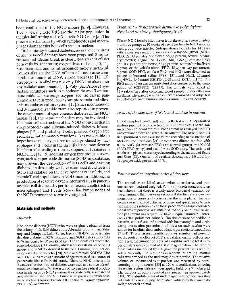
Experiment2: peritoneal exudate cells from diabetic NOD females. Reactive oxygenintermediateswere pro- duced in islet cells from ILI females by peritoneal exu- date cells from diabetic NOD females and the sup- pressive effect of SOD on the production of reactive oxygen intermediates is shown in Figure 3. The islet cells
(2 x 10⁵) were incubated with peritoneal exudate cells (5 x 10²). The islet cells showed only 0.3 % of fluorescein positive cells by themselves, 51.3 % in the presence of peritoneal exudate cells, and 14.0 % in the presence of peritoneal exudate cells and SOD (final concentration 13.4 U/ml). The suppressive effect of SOD on the pro- duction of reactive oxygen intermediates in ILI islet cells by diabetic NOD females from assays in triplicate is shown in Figure 4. A doubled number of peritoneal exu- date cells (10 x 105) slightly increased reactive oxygen intermediates in the islet cells. However, the production of reactive oxygen intermediates was suppressed by pe- ritoneal exudate cells from SOD in both sets of samples.
Reactive oxygen intermediates were produced in islet cells from ILI females by macrophages but not T cells from diabetic NOD females (Fig.5). Macro- phages but not T cells from the diabetic NOD mice in- duced the production of reactive oxygen intermediates in the ILI islet cells.
28
A ~;
iiii!~ ~iiiii~ I !!!!I!!!
~ul=:=:::
l l l l ] l l l l ] l
{!Nh~}!~, =.
I : ~ : : ] l l I I I l l : ~ .
,i!g~ii~iiiiiiii.~ : : : : : : : : :m::: l : :
I l I ~ l ~ : l l l l l ~ l l l l I l ',!=!!~}q!!!!!!~!2~!
~ l ] ~ l * ; ] l l l l l l l l l l l l ~ ¢ ========================,
I " 1 I ' I "I
O O
II
¢-
O
if)
O
"6
E z
B
I I
, , , , , , i . . . . . . . . i . . . . . . . . i " " 't
C !
i!ii!
i m : : : : m
~iiiiiiiii
~iiiiiiiiiii IllI{;~]lllllll
"1 ! I ! ! ~ ! ! . . . . i " ' - ' ~
Log fluorescence intensity Fig.3A-C. Suppressive effect of superoxide dismutase (SOD) on reactive oxygen intermediate production by peritoneal exu- date cells from diabetic NOD mice. Islet cells were prepared from ILI females and peritoneal exudate cells from diabetic NOD females. The islet cells and peritoneal exudate cells were co-cultured for 3 h prior to the addition of 2', 7'-dichlorofluores- cein diacetate. Peritoneal exudate ceils from diabetic NOD fe- male mice induced the production of reactive oxygen intermedi- ates even with the ILI islet cells. (A) Islet cells only. Reactive oxygen intermediates were produced in islet cells from ILI fe- males by peritoneal exudate ceils from diabetic NOD females (B) and suppressive effect of superoxide dismutase on the reac- tive oxygen intermediate production (C)
E Horio et al.: Reactive oxygen intermediates in autoimmune islet cell destruction
80 m
.~ 40 u
20
Islet cells Islet cells Islet c e l l s
+ +
P E C PEC (5x10 5 cells'J (10x1~ cells)
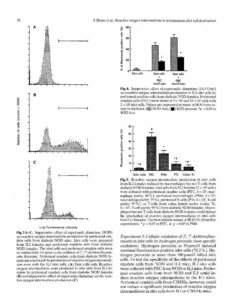
Fig.4. Suppressive effect of superoxide dismutase (14.3 U/ml) on reactive oxygen intermediate production in ILI islet cells by peritoneal exudate cells from diabetic NOD females. Peritoneal exudate cells (PEC) were tested at 5 x 10 s and 10 x 105 cells with 2 x 10⁵ islet cells. Values are expressed as mean + SEM from as- says in triplicate. ( [ ] ) SOD-free, (m) SOD-present. *p < 0.05 vs SOD-free
o
60 [ 50
40
30
20
10
0 I s l e t c e l l s PEC PMo PTc Celiac Tc
Fig.5. Reactive oxygen intermediate production in islet cells from ILI females induced by macrophages but not T cells from diabetic NOD females. Islet cells from ILI females (2 x 10⁵ cells) were cultured with peritoneal exudate cells (PEC, 4 x 10⁵; mac- rophage purity: 60 %), peritoneal macrophages (PM¢, 4 x 10⁵; macrophage purity: 75 % ), peritoneal T cells (PTc, 4 x 10 ~, T-cell purity: 97 %), or T cells from celiac lymph nodes (celiac Tc, 4 x 10⁵, T-cell purity: 95 % ) from diabetic NOD females. Macro- phages but not T cells from diabetic NOD females could induce the production of reactive oxygen intermediates in islet cells from ILI females. The bars indicate means + SEM (%) from five experiments. * p < 0.05 vs PEC, # p < 0.05 vs PM~
Experiment 3: Cellular oxidation of 2', 7'-dichlorofluo- rescein in islet cells by hydrogen peroxide (non-specific oxidation). H y d r o g e n p e r o x i d e at 50 gmol/1 i n d u c e d m a x i m a l f luo rescence -pos i t ive islet cells (70.2 %) . H y - d r o g e n p e r o x i d e at m o r e t h a n 100 gmol/1 ki l led islet cells. To test the specif ici ty of the effects o f pe r i t onea l e x u d a t e cells f r o m N O D and I L I mice, I L I islet cells w e r e cu l tu red wi th P E C f r o m N O D or I L I mice. Per i to - nea l e x u d a t e cells f r o m b o t h N O D and I L I cou ld in- duce reac t ive o x y g e n i n t e r m e d i a t e s in the I L I islets. P e r i t o n e a l e x u d a t e cells f r o m C 3 H / H e , however , could no t i nduce a s ignif icant p r o d u c t i o n o f reac t ive o x y g e n i n t e r m e d i a t e s in islet cells f r o m I L I o r C 3 H / H e mice.

F. Horio et al.: Reactive oxygen intermediates in autoimmune islet cell destruction
A ,~i~ D i s c u s s i o n
H P ~
~:~!! I I ~4~ I i .:1::::::
!giigggii
: : : :m: : : : : i : : : : : : : : : : : :
. :m: : : : : : : : : : :
t : : : : : : : : : : : : : : : : : : : ..:::1:::::::::::::: :::: :::::::::::::r
B
i
C
Q Q L ~
JJ
E
0 Q
= (~
"8
E z
, r
.':+~, [
, N ~ I I
..::::::1:1:
*.:::::::::::
!" F:" L::".:..'L:
:::::::::::::, �9 ". : :. p: ! :.:..::.:- -:. ~
i:::::::::::::1::
,r:::::::::::::::::. .::::::::::::::::::
~iiiii~iii!iiiii!ii~ , ! h ~'::iiii!iii!!iii!i.~i.-",~ ,, "#.,q.~.,, ..'
i ! i i I ! i
F i i
ii~i~i!!!!i!~ . . . . . . . . . . . . . . . , :
i . . . . . . . . . . . . . . . . . ~ - ~ ' ~ ~ ' : : : - - - - : * " - ' : : : . : l J * : " - : : : : - " : ~
I i . I 1 * | J I |
Log f luorescence intensi ty
Fig .6 . Reactive oxygen intermediate production in islet cells from ILI females induced by recombinant mouse interleukin-1- c~. The islet cells were incubated with recombinant mouse inter- leukin-l-c~ (40 U/2 x 10 ~ islet cells) for 3 h prior to the addi- tion of 2', 7'-dichlorofluorescein diacetate. Percent of fluor- escence-positive islet cells of the ILI mouse is shown in pa- rentheses: A: islet cells alone (4.2%); B: islet cells + interleu- kin-l-c~ (25.4 %); C: islet cells + PEC from diabetic NOD mice (45.9%)
Experiment 4: Reactive oxygen intermediate production by interleukin-l-(z. Recombinant mouse interleukin-1- (z could produce reactive oxygen intermediates in ILI islet cells (Fig. 6).
29
Pre-treatment with a single intravenous injection of SOD-PEG protects mice against alloxan-induced hyperglycaemia [28]. The treatment with desferriox- amine (inhibitor of hydroxyl radical formation from superoxide anion), nicotinamide, or a combination of desferrioxamine and nicotinamide prevents disease re- currence in diabetic NOD mice after the allograft of BALB/c islets [18]. Nomikos and co-workers [29] have recently reported that SOD and catalase are effective in preventing destruction of islet allografts of the BALB/c mouse in diabetic NOD mice, as measured by continuing ability of the graft to regulate blood glucose.
In the present study, both SOD-PEG and catalase- PEG were effective in preventing islet destruction of the NOD mice. The markedly increased absolute mass of undamaged islet portion observed in the treated groups may be due to the prevention of further beta- cell destruction by the action of SOD and catalase. Meanwhile, SOD and catalase from different species did not cause any allergic reactions, islet beta-cell tu- mours, or change in body weight in NOD mice. PEG at- tachment to purified enzymes has been shown to pro- long the biological half-life of the enzymes [30]. There- fore, we used SOD-PEG and catalase-PEG instead of SOD or catalase alone. Although PEG itself can sup- press immune reactions [31], a comparison of the data from the PEG-treated NOD mice with those from un- treated NOD mice indicates that PEG alone had no protective effect on the islet in this study. The degree of islet damage in the untreated NOD females was similar to that in the PEG-treated NOD females at 3 months of age [32]. Therefore, the protection from islet-cell de- struction observed in this study seems to be due to SOD and catalase themselves. During the present experi- ments with SOD-PEG, we had to evaluate its short- term efficacy due to the high cost of SOD-PEG. A long- term (8 month) administration of SOD-PEG and cata- lase-PEG may be required to see whether the oxygen free radical scavengers coupled to PEG can prevent the development of diabetes in NOD mice.
Injection of splenocytes from diabetic NOD mice can transfer diabetes into irradiated young NOD mice [33]. Both CD4 + and CD8 § splenic T cells are required for the adoptive transfer of diabetes in NOD mice [6]. Restricted usage of TcR in autoimmune diabetes has not been found in the NOD mouse [8, 9]. However, T cells bearing TcR Vfl8 are the major population in the islet-infiltrating cells of diabetic NOD mice, and that removing a major TcR Vflfamily (Vfl 8) appears to influence disease progression [5]. In this study, the treatment with either SOD-PEG or catalase-PEG did not change the proportion of B cells, CD3 +, CD4 + and CD8 § T cells, T cells bearing TcR(xflchains, nor T cells bearing TcR Vfl 8.1,2, 3 in the spleen, as compared with those of the control group. Our data suggest that SOD- PEG and catalase-PEG do not exert the preventive el-

30 F. Horio et al.: Reactive oxygen intermediates in autoimmune islet cell destruction
fect on islet beta-cell destruction by changing the num- ber of T cells. In our in vitro studies [34], peritoneal exudate cells (rich in macrophages) or T-cell depleted peritoneal exudate cells (macrophage-enriched frac- tion) could induce production of reactive-oxygen inter- mediates in the islet cells. However, nylon wool puri- fied peritoneal T cells or T cells from celiac lymph nodes failed to induce production of reactive oxygen intermediates in the islet cells. The effect of peritoneal exudate cells (PEC) appears to be greater than the ad- ditive effects of peritoneal macrophages (PMr and T cells (PTc) despite the increased number of macro- phages in PMr fraction. This observation indicates that macrophages may require interaction with T cells for maximal production of reactive oxygen intermediates in islet cells. Furthermore, the production of reactive oxygen intermediates in the islet cells by peritoneal exudate cells was inhibited by SOD-PEG (oxygen free radical scavenger). These observations suggest that SOD-PEG suppressed the production of reactive oxygen intermediates in the islet cells induced by mac- rophages, and thereby prevented the islet beta-cell destruction in the NOD mice.
Recent studies [35-37] suggest that macrophages play an important role in the initiation of insulitis in N O D mice. We speculate that the role of T cells and macrophages in the development of insulitis is as fol- lows: Initially, an antigen(s) on islet beta cells may be processed by macrophages and presented to CD4 § hel- per T cells in an M H C class II restricted manner [38]. Indeed, the production of reactive oxygen intermedi- ates in the islet cells by NOD peritoneal exudate cells appears to be M H C class II I-A-restricted [39]. Produc- tion of reactive oxygen intermediates in the islet cells was completely blocked by mAb 40A reacting with NOD's M H C class II I-A [40], but not by mAb 14-4-4S reacting with MHC class II I-E, mAb 31-3-4S reacting with M H C class I K d, or mAb 28-14-8S reacting with class I D b. After the antigen presentation by the macro- phages, the CD4 § helper T cells may be activated by the antigen-presenting cells and secrete lymphokines such as interferon-7(INF-7) resulting in activation of macrophages [41]. Activated macrophages may pro- duce and secrete cytokines, such as interleukin-1 (IL-1) [42] and tumour necrosis factor [43], and simultaneous- ly produce oxygen free radicals [11]. It was reported that IL-1 impaired the islet beta-cell activity in vitro [44, 45], and that IL-1, tumour necrosis factor and INF- ),have synergistically cytotoxic effects on islet cells in vitro [46]. In our flow cytometric analysis, recombinant murine IL-1 per se could induce production of reactive oxygen intermediates in the islet cells in vitro. Reactive oxygen intermediates produced either in the islet beta cells by interaction with the cytokines or directly by the activated macrophages could lead to beta-cell destruc- tion.
The data in the present study suggest that in the prevention of islet beta-cell destruction of the NOD
mouse SOD-PE G exerts its action/effect on peritoneal macrophages as effector cells rather than on T cells as antigen recognizing and helper cells. It remains unclear whether the peritoneal macrophages must be stimu- lated by T-cell derived cytokines to produce reactive oxygen intermediates, or whether the peritoneal mac- rophages stimulate islet beta cells to produce reactive oxygen intermediates. The present study supports the concept that oxygen free radicals, such as superoxide anion and hydrogen peroxide, damage islet beta cells resulting in the development of diabetes.
Acknowledgements. We thank Dr. C. R. Kahn for his encourage- ment, Dr. J. W. Moorhead and Dr. R. T. Kubo for their construc- tive criticisms, and Mr. EBettinelli, Mr. D.DeLutis, Mr. K.J. Hirokawa and Dr. E. Yamato for technical assistance. This study was supported by grants from the Adler Foundation (M. H.), the Juvenile Diabetes Foundation International (M. H.), Procept, Inc., Shionogi & Co., Ltd., NIH RO1 DK43613 (M. H.) and NIH 1P30 AM36836 (Diabetes and Endocrinology Research Center, Pilot and Feasibility Grant to M.H., Animal Core Facility and Tissue Culture Core). M. F. is the recipient of a Juvenile Diabetes Foundation Fellowship.
References
1. Gepts W (1965) Pathologic anatomy of the pancreas in juvenile diabetes mellitus. Diabetes 14:619-633
2. Like AA, Rossini AA (1984) Spontaneous autoimmune diabetes mellitus in the BioBreeding/Worcester rat. Surv Synth Pathol Res 3:131-138
3. Makino S, Kunimoto K, Muraoka Y, Katagiri K, Tochino Y (1980) Breeding of a non-obese, diabetic strain of mice. Exp Anita 29:1-13
4. Makino S, Harada M, Kishimoto Y, Hayashi Y (1986) Ab- sence of insulitis and overt diabetes in athymic nude mice with NOD genetic background. Exp Anim 35:495-498
5. Fukuda M, Horio F, Kubo R, Hattori M (1989) Monoclonal antibody F23.1 against T-cell bearing Vfl 8 TcR elements can prevent the development of insulitis in NOD mice. Diabetes 38 [Suppl 2]: 12A (Abstract)
6. Miller B J, Appel MC, O'Neil JJ, Wicker LS (1988) Both the Lyt-2 + and L3T4 § subsets are required for the transfer of diabetes in non-obese diabetic mice. J Immuno1140:52-58
7. Acha-Orbea H, Mitchell D J, Timmermann L, Steinmann L (1988) Limited heterogeneity of T-cell receptors from lym- phocytes mediating autoimmune encephalomyelitis allow specific immune intervention. Cell 54:263-273
8. Shizuru JA, Taylor-Edwards C, Livingstone A, Fathmann CG (1991) Genetic dissection of T cell receptor Vflgene re- quirements for spontaneous murine diabetes. J Exp Med 174: 633-638
9. McDuffie M (1991) Diabetes in NOD mice does not require T-lymphocytes expressing of Vfl8 or Vfl5. Diabetes 40: 1555-1559
10. Uchigata Y, Yamamoto H, Kawamura A, Okamoto H (1982) Protection by superoxide dismutase, catalase, and poly (ADP-ribose) synthetase inhibitors against alloxan- and streptozotocin-induced islet DNA standard breaks and against the inhibition of proinsulin synthesis. J Biol Chem 257:6084-6088
11. Uchigata Y, Yamamoto H, Nagai H, Okamoto H (1983) Ef- fect of poly (ADP-ribose) synthetase inhibitor administra-

E Horio et al.: Reactive oxygen intermediates in autoimmune islet cell destruction
tion to rats before and after injection of alloxan and strepto- zotocin on islet proinsulin synthesis. Diabetes 32:316-318
12. Wilson GL, Leiter HE (1990) Streptozotocin interactions with pancreatic fl cells and the induction of insulin-depen- dent diabetes. Curr Top Microbiol Immuno1156: 27-54
13. Wilson GL, LeDoux SP (1993) Interactions of chemicals with pancreatic t-cells. In: Shafrir E (ed); Lessons from animal diabetes; Vol 4. Smith-Gordon, London, pp 17-26
14. Wilson GL, Harfig PC, Patton N J, LeDoux SP (1988) Mech- anisms of nitrosourea-induced t-cell damage: activation of poly (ADP-ribose) synthetase and cellular distribution. Diabetes 37:213-216
15. Wilson GL, Patton NJ, McCord JM, Mullins DW, Mossman BT (1984) Mechanisms of streptozotocin- and alloxan-in- duced damage in rat B cells. Diabetologia 27:587-591
16. Yamada K, Nonaka K, Hanafusa T, Miyazaki A, Toyoshima H, Tarui S (1982) Preventive and therapeutic effects of large- dose nicotinamide injections on diabetes associated with in- sulitis: an observation in non-obese diabetic (NOD) mice. Diabetes 31:749-753
17. Murray HW, Spitalny GL, Nathan CF (1985) Activation of mouse peritoneal macrophages in vitro and in vivo by inter- feron. J Immuno1134:1619-1622
18. Nomikos IN, Prowse S J, Carotenute R Lafferty KJ (1986) Combined treatment with nicotinamide and desferrioxamine prevents islet allograft destruction in NOD mice. Diabetes 35:1302-1304
19. Hattori M, Makino S (1988) Type I (insufin-dependent) diabetes. Exp Med 6:78-86
20. Hattori M, Fukuda M, Ichikawa T, Baumgartl H-J, Kato H, Makino S (1990) A single recessive non-MHC diabetogenic gene determines the development of insulitis in the presence of an MHC-linked diabetogenic gene in NOD mice. J Auto- immunity 3:1-10
21. McCord JM, Fridovich I (1969) Superoxide dismutase. J Biol Chem 244:604%6055
22. Beers RF, Sizer IW (1952) A spectrophotometric method for measuring the breakdown of hydrogen peroxide by catalase. J Biol Chem 195:133-140
23. Bonnet-Weir S, Trent DE Honey RN, Weir GC (1981) Re- sponses of neonatal rat islets to streptozotocin: limited t-cell regeneration and hyperglycemia. Diabetes 30:64-69
24. Kubo TR, Born W, Kappler JW, Marrack E Pigeon M (1989) Characterization of a monoclonal anitbody which detects all murine c~flT-cell receptors. J Immuno1142: 2736-2742
25. Staertz UD, Rammensee H, Benedetto JD, Bevan MJ (1985) Characterization of a murine monoclonal antibody specific for an allotypic determinant on T cell antigen receptor. J Im- muno1134:3994-4000
26. Leo O, Foo M, Sach DH, Samelson LE, Bluestone JA (1987) Identification of a monoclonal antibody specific for a murine T3 polypeptide. Proc Natl Acad Sci USA 84:1374-1378
27. Bass DA, Parce JW, Dechatelet LR, Szejda R Seeds MC, Thomas M (1983) Flow cytometric studies of oxidative pro- duct formation by neutrophils: a graded response to mem- brane stimulation. J Immuno1130:1910-1917
28. Grankvist K, Markland S, Taljedal I (1981) Superoxide dis- mutase is a prophylactic against alloxan diabetes. Nature 294: 158-160
29. Nomikos IN, Wang Y, Lafferty KJ (1989) Involvement of ra- dicals in autoimmune diabetes. Immunol Cell Bio167:85-87
31
30. Abuchowski A, McCoy JR, Paiczuk NC, Van Es T, Davis FF (1977) Effect of covalent attachment of polyethylene glycol on immunogenecity and circulating life of bovine liver cata- lase. J Biol Chem 252:3582-3586
31. Hershfield MS, Buckley RH, Greenberg ML et al. (1987) Treatment of adenosine deaminase deficiency with polyethylene glycol-modified adenosine deaminase. N Engl J Med 316:589-596
32. Yano N, Ikegami H, Sato T et al. (1988) Orchiectomy in- creases L3T4 (helper) and Lyt2 (cytotoxic/suppressor) T lymphocytes in the thymus and accelerates B-cell destruc- tion of the non-obese diabetic mouse. Diabetologia 31: 560A (Abstract)
33. Wicker LS, Miller B J, Mullen Y (1986) Transfer of autoim- mune diabetes mellitus with splenocytes from non-obese diabetes (NOD) mice. Diabetes 35:855-860
34. Fukuda M, Horio F, Rittershaus C, Kato H, Hattori M (1990) Macrophages but not T-cells can increase the content of free- radical oxygen in islet t-cells of the NOD mouse. Clin Res 38: 308A (Abstract)
35. Charlton B, Bacelj A, Mandel TE (1988) Administration of silica particles or anti-Lyt2 antibody prevents t-cell destruc- tion in NOD mice given cyclophosphamide. Diabetes 37: 930-935
36. Lee K, Amano K, Yoon J (1988) Evidence for initialinvolve- ment of macrophage in the development of insulitis in NOD mice. Diabetes 37:98%991
37. Hutchings R Rosen H, O'Reilly L, Simpson E, Gordon S, Cooke A (1990) Transfer of diabetes in mice prevented by blockade of adhesion-promoting receptor on macrophages. Nature 348:639-642
38. Ziegler K, Unanue ER (1981) Identification of a macro- phage antigen-processing event required for I-region-re- stricted antigen presentation to lymphocytes. J Immuno1127: 1869-1875
39. Fukuda M, Horio F, Rittershaus C, Kato H, Hattori M (1991) MHC class-II I-A restriction of free-radical oxygen produc- tion in islet beta-cells by peritoneal exudate cells of the diabetic NOD mouse. Clin Res 39: 246A (Abstract)
40. Ikegami H, Makino S, Harada M, Eisenbarth GS, Hattori M (1988) The cataract Shionogi mouse, a sister strain of the non-obese diabetic mouse: similar class II but different class I gene products. Diabetologia 31:254-258
41. Adams D (1982) Molecules, membranes and macrophage activation. Immunol Today 3:285-287
42. Durum SK, Schmidt JA, Oppenheim JJ (1985) Interleukin-l: an immunological perspective. Ann Rev Immunol 3:263-287
43. Beutler B, Greenwald D, Hulmes JD et al. (1985) Identity of tumor necrosis factor and macrophage-secreted factor ca- chectin. Nature 316:552-554
44. Bendtzen K, Mandrup-Poulsen T, Nerup J, Nielsen JH, Di- narello CA, Svenson M (1986) Cytotoxicity of human pI 7 in- terleukin-1 for pancreatic islets of Langerhans. Science 232: 1545-1547
45. Palmer JR Helqvist S, Spinas GA et al. (1989) Interaction of t-cell activity and. IL-1 concentration and exposure time in isolated rat islets of Langerhans. Diabetes 28:1211-1216
46. Pukel C, Baquerizo H, Rabinovitch A (1988) Destruction of rat islet cell monolayers by cytokines: synergistic interactions of interferon-),, tumor necrosis factor, lymphotoxin, and in- terleukin 1. Diabetes 37:133-136