Multipotent stem cells from trabecular meshwork become phagocytic TM cells
10
Multipotent Stem Cells from Trabecular Meshwork Become Phagocytic TM Cells Yiqin Du, 1,2 Danny S. Roh, 1 Mary M. Mann, 1 Martha L. Funderburgh, 1 James L. Funderburgh, 1,2 and Joel S. Schuman 1,2 PURPOSE. To isolate and characterize stem cells from human trabecular meshwork (TM) and to investigate the potential of these stem cells to differentiate into TM cells. METHODS. Human trabecular meshwork stem cells (TMSCs) were isolated as side population cells by fluorescence-activated cell sorting or isolated by clonal cultures. Passaged TMSCs were compared with primary TM cells by immunostaining and quantitative RT-PCR. TMSC purity was assessed by flow cytom- etry and TMSC multipotency was examined by induction of neural cells, adipocytes, keratocytes, or TM cells. Differential gene expression was detected by quantitative RT-PCR, immu- nostaining, and immunoblotting. TM cell function was evalu- ated by phagocytic assay using inactivated Staphylococcus au- reus bioparticles. RESULTS. Side population and clonal isolated cells expressed stem cell markers ABCG2, Notch1, OCT-3/4, AnkG, and MUC1 but not TM markers AQP1, MGP, CHI3L1, or TIMP3. Passaged TMSCs are a homogeneous population with 95% cells posi- tive to CD73, CD90, CD166, or Bmi1. TMSCs exhibited multi- potent ability of differentiation into a variety of cell types with expression of neural markers neurofilament, -tubulin III, GFAP; or keratocyte-specific markers keratan sulfate and ker- atocan; or adipocyte markers ap2 and leptin. TMSC readily differentiated into TM cells with phagocytic function and ex- pression of TM markers AQP1, CHI3L1, and TIMP3. CONCLUSIONS. TMSCs, isolated as side population or as clones, express specific stem cell markers, are homogeneous and mul- tipotent, with the ability to differentiate into phagocytic TM cells. These cells offer a potential for development of a novel stem cell– based therapy for glaucoma. (Invest Ophthalmol Vis Sci. 2012;53:1566 –1575) DOI:10.1167/iovs.11-9134 G laucoma is a leading cause of irreversible blindness world- wide and the second leading cause of blindness overall in the United States. 1 Glaucoma damages the optic nerve leading to progressive visual field loss and eventual blindness. For most forms of glaucoma, elevated intraocular pressure (IOP) and aging remain the most important risk factors. 2 Although the pathogenesis is multifactorial, optic nerve damage is strongly associated with increased IOP. Experimental animal models demonstrate that elevated IOP is sufficient to produce glauco- ma-like optic nerve damage. 3 The main outflow pathway for aqueous humor in the eye consists of a series of endothelial cell–lined channels in the angle of the anterior chamber comprising the trabecular mesh- work (TM), Schlemm’s canal, the collector channels, and the episcleral venous system. The TM, especially the juxtacanalicu- lar region, and the inner wall of Schlemm’s canal are thought to be the source of the most resistance to aqueous outflow. The cells lining the lamellae of the TM play two primary roles: secretion of specific enzymes and extracellular matrix and phagocytosis of debris in the aqueous humor. 4 Both functions help maintain aqueous outflow over the trabecular lamellae. 5 Reduced cellularity within the TM is observed with age and correlates with increased outflow resistance and elevated IOP. 6–9 Current therapies for IOP control involve pharmaco- logic reduction of aqueous humor production and surgical enhancement of outflow. These therapies are effective but still have significant limitations; toxicity, side effects, complica- tions, failure, and patient noncompliance are common. Resident pools of somatic stem cells in many organs are responsible for tissue maintenance and repair. 10 Many of these stem cells expanded in vitro exhibit effective tissue regenera- tion when introduced to pathologic tissues in vivo. 11 Cell- based restoration of TM function to glaucomatous eyes is a potential therapy not yet explored due to lack of cells that maintain TM phenotype in vitro and home to the TM in vivo. Several studies have reported cells in TM which express prop- erties of adult stem cells (Challa P, et al. IOVS 2003;44:ARVO E-Abstract 3164) 12–14 ; however, characterization of the pre- sumptive TM stem cells (TMSCs) is incomplete. Our present study demonstrates isolation of a multipotent population of adult stem cells from human TM, which can be greatly ex- panded in vitro while maintaining the novel ability to differen- tiate to TM cells. MATERIALS AND METHODS Primary Cell Culture Deidentified human corneas were obtained from the Center for Organ Recovery and Education (Pittsburgh, PA). Donor human corneas in- cluding scleral rim and TM not usable for transplantation were used for experiments. Cells from three donors at 23, 41, and 55 years of age were used in the experiments shown. For each cell population, every experiment was repeated at least once. After careful removal of the iris, a cut was made through the inner edge of Schwalbe’s line and the TM tissue was peeled off. We processed TMSCs as either explant culture or dissociated cell culture. For explant culture, the tissue was From the 1 University of Pittsburgh Medical Center (UPMC) Eye Center, Ophthalmology and Visual Science Research Center, Depart- ment of Ophthalmology, University of Pittsburgh School of Medicine, Pittsburgh, Pennsylvania; and 2 The Louis J. Fox Center for Vision Restoration of UPMC and the University of Pittsburgh, Pittsburgh, Pennsylvania. Supported in part by National Institutes of Health/National Eye Institute Grants EY016415 (JLF) and P30-EY008098; Eye and Ear Foun- dation (Pittsburgh, PA); Research to Prevent Blindness; and an anony- mous philanthropic donation (YD). Submitted for publication November 22, 2011; revised December 30, 2011; accepted January 24, 2012. Disclosure: Y. Du, None; D.S. Roh, None; M.M. Mann, None; M.L. Funderburgh, None; J.L. Funderburgh, None; J.S. Schuman, None Corresponding author: Yiqin Du, Eye and Ear Institute, Room 930, 203 Lothrop Street, Pittsburgh, PA 15213; [email protected]. Glaucoma Investigative Ophthalmology & Visual Science, March 2012, Vol. 53, No. 3 1566 Copyright 2012 The Association for Research in Vision and Ophthalmology, Inc.
Transcript of Multipotent stem cells from trabecular meshwork become phagocytic TM cells

Multipotent Stem Cells from Trabecular MeshworkBecome Phagocytic TM Cells
Yiqin Du,1,2 Danny S. Roh,1 Mary M. Mann,1 Martha L. Funderburgh,1
James L. Funderburgh,1,2 and Joel S. Schuman1,2
PURPOSE. To isolate and characterize stem cells from humantrabecular meshwork (TM) and to investigate the potential ofthese stem cells to differentiate into TM cells.
METHODS. Human trabecular meshwork stem cells (TMSCs)were isolated as side population cells by fluorescence-activatedcell sorting or isolated by clonal cultures. Passaged TMSCswere compared with primary TM cells by immunostaining andquantitative RT-PCR. TMSC purity was assessed by flow cytom-etry and TMSC multipotency was examined by induction ofneural cells, adipocytes, keratocytes, or TM cells. Differentialgene expression was detected by quantitative RT-PCR, immu-nostaining, and immunoblotting. TM cell function was evalu-ated by phagocytic assay using inactivated Staphylococcus au-reus bioparticles.
RESULTS. Side population and clonal isolated cells expressedstem cell markers ABCG2, Notch1, OCT-3/4, AnkG, and MUC1but not TM markers AQP1, MGP, CHI3L1, or TIMP3. PassagedTMSCs are a homogeneous population with �95% cells posi-tive to CD73, CD90, CD166, or Bmi1. TMSCs exhibited multi-potent ability of differentiation into a variety of cell types withexpression of neural markers neurofilament, �-tubulin III,GFAP; or keratocyte-specific markers keratan sulfate and ker-atocan; or adipocyte markers ap2 and leptin. TMSC readilydifferentiated into TM cells with phagocytic function and ex-pression of TM markers AQP1, CHI3L1, and TIMP3.
CONCLUSIONS. TMSCs, isolated as side population or as clones,express specific stem cell markers, are homogeneous and mul-tipotent, with the ability to differentiate into phagocytic TMcells. These cells offer a potential for development of a novelstem cell–based therapy for glaucoma. (Invest Ophthalmol VisSci. 2012;53:1566–1575) DOI:10.1167/iovs.11-9134
Glaucoma is a leading cause of irreversible blindness world-wide and the second leading cause of blindness overall in
the United States.1 Glaucoma damages the optic nerve leading
to progressive visual field loss and eventual blindness. For mostforms of glaucoma, elevated intraocular pressure (IOP) andaging remain the most important risk factors.2 Although thepathogenesis is multifactorial, optic nerve damage is stronglyassociated with increased IOP. Experimental animal modelsdemonstrate that elevated IOP is sufficient to produce glauco-ma-like optic nerve damage.3
The main outflow pathway for aqueous humor in the eyeconsists of a series of endothelial cell–lined channels in theangle of the anterior chamber comprising the trabecular mesh-work (TM), Schlemm’s canal, the collector channels, and theepiscleral venous system. The TM, especially the juxtacanalicu-lar region, and the inner wall of Schlemm’s canal are thought tobe the source of the most resistance to aqueous outflow. Thecells lining the lamellae of the TM play two primary roles:secretion of specific enzymes and extracellular matrix andphagocytosis of debris in the aqueous humor.4 Both functionshelp maintain aqueous outflow over the trabecular lamellae.5
Reduced cellularity within the TM is observed with age andcorrelates with increased outflow resistance and elevatedIOP.6–9 Current therapies for IOP control involve pharmaco-logic reduction of aqueous humor production and surgicalenhancement of outflow. These therapies are effective but stillhave significant limitations; toxicity, side effects, complica-tions, failure, and patient noncompliance are common.
Resident pools of somatic stem cells in many organs areresponsible for tissue maintenance and repair.10 Many of thesestem cells expanded in vitro exhibit effective tissue regenera-tion when introduced to pathologic tissues in vivo.11 Cell-based restoration of TM function to glaucomatous eyes is apotential therapy not yet explored due to lack of cells thatmaintain TM phenotype in vitro and home to the TM in vivo.Several studies have reported cells in TM which express prop-erties of adult stem cells (Challa P, et al. IOVS 2003;44:ARVOE-Abstract 3164)12–14; however, characterization of the pre-sumptive TM stem cells (TMSCs) is incomplete. Our presentstudy demonstrates isolation of a multipotent population ofadult stem cells from human TM, which can be greatly ex-panded in vitro while maintaining the novel ability to differen-tiate to TM cells.
MATERIALS AND METHODS
Primary Cell Culture
Deidentified human corneas were obtained from the Center for OrganRecovery and Education (Pittsburgh, PA). Donor human corneas in-cluding scleral rim and TM not usable for transplantation were used forexperiments. Cells from three donors at 23, 41, and 55 years of agewere used in the experiments shown. For each cell population, everyexperiment was repeated at least once. After careful removal of theiris, a cut was made through the inner edge of Schwalbe’s line and theTM tissue was peeled off. We processed TMSCs as either explantculture or dissociated cell culture. For explant culture, the tissue was
From the 1University of Pittsburgh Medical Center (UPMC) EyeCenter, Ophthalmology and Visual Science Research Center, Depart-ment of Ophthalmology, University of Pittsburgh School of Medicine,Pittsburgh, Pennsylvania; and 2The Louis J. Fox Center for VisionRestoration of UPMC and the University of Pittsburgh, Pittsburgh,Pennsylvania.
Supported in part by National Institutes of Health/National EyeInstitute Grants EY016415 (JLF) and P30-EY008098; Eye and Ear Foun-dation (Pittsburgh, PA); Research to Prevent Blindness; and an anony-mous philanthropic donation (YD).
Submitted for publication November 22, 2011; revised December30, 2011; accepted January 24, 2012.
Disclosure: Y. Du, None; D.S. Roh, None; M.M. Mann, None;M.L. Funderburgh, None; J.L. Funderburgh, None; J.S. Schuman,None
Corresponding author: Yiqin Du, Eye and Ear Institute, Room 930,203 Lothrop Street, Pittsburgh, PA 15213; [email protected].
Glaucoma
Investigative Ophthalmology & Visual Science, March 2012, Vol. 53, No. 31566 Copyright 2012 The Association for Research in Vision and Ophthalmology, Inc.

cut into pieces and put in a 25-cm2 culture flask. Stem cell growthmedium (SCGM) was added and the culture was left undisturbed for 10to 14 days. For dissociated cell culture, the dissected TM tissue wasdigested in 0.3 mg/mL collagenase type L (Sigma-Aldrich, St. Louis,MO) in Dulbecco’s modified Eagle’s medium (DMEM) at 37°C for 20 to22 hours. After digestion, the cells were filtered through a 70-�m meshand washed twice with DMEM. Cells were seeded at 2 � 104 cells/cm2
in SCGM. For both cultures, cells were passaged at 80–90% confluencyby trypsinization and seeded at 2 to 5 � 103 cells/cm2 in SCGM orseeded for clonal expansion by limiting dilution at 30 cells in a 96-wellplate (0.3 cells/well). On average, approximately 1% to 10% of the 96wells had clones with small cells, which were picked up for subculti-vation at 2 to 3 weeks after seeding. Among them, approximately athird to a half could be continuously passaged up to 30 to 50 popula-tion doublings. At least six clones from three donors were used andrepeated in this study. SCGM was modified from a corneal endothelialcell culture medium15 containing multipurpose reduced-serum media(OptiMEM-1; Invitrogen, Carlsbad, CA) supplemented with 5% fetalbovine serum (FBS; Hyclone, Logan, UT), 10 ng/mL EGF (UpstateBiotechnologies, Billerica, MA), 100 �g/mL bovine pituitary extract(Biomedical Technologies, Stoughton, MA), 20 �g/mL ascorbic acid,200 �g/mL calcium chloride, 0.08% chondroitin sulfate (Sigma-Al-drich), 100 IU/mL penicillin, 100 �g/mL streptomycin, and 50 �g/mLgentamicin (Sigma-Aldrich). Primary TM cells were cultured in DMEMwithout FBS or any growth factors.
Isolation of Stem Cells by Side PopulationCell Sorting
Side population (SP) cell sorting was carried out as previously de-scribed16–18 (DyeCycle Violet [DCV] dye; Invitrogen) with minor mod-ifications. After 2 to 3 passages, 5 � 105 to 2 � 106 trypsinized cellswere incubated at 1 � 106 cells/mL in prewarmed DMEM with 2% FBSand 10 �M DCV for 100 minutes at 37°C. To inhibit DCV efflux, 1 �105 to 5 � 105 cells were preincubated for 20 minutes with 25 �g/mLfumitremorgin C (a gift from Susan Bates, National Institutes of Health)before DCV incubation. After staining, the cells were washed twice inHank’s balanced salt solution with 2% FBS and stored on ice. Immedi-ately before sorting, 2 �g/mL propidium iodide (PI) was added toidentify nonviable cells. Cells were analyzed on a flow cytometerhigh-speed cell sorter (FACSAria; BD Biosciences, San Jose, CA), using405-nm excitation. Cells showing reduced fluorescence at both blue(450 nm) and red (670 nm), designated SP cells, were collected. Asmall proportion of non-SP cells were collected separately as control.Dead cells stained with PI were omitted from the population. The
sorted SP and non-SP cells were cultured and passaged without cloningfor further studies.
Flow Cytometry
Clonal passaged TMSCs were trypsinized and cell suspensions werepassed through a 40-�m filter-cap tube to remove debris. Antibodiesused are listed in Table 1. For cell surface marker staining, fluorescent-conjugated antibodies (CD73-PE, CD90-Alexa 647, CD166-FITC) orappropriate isotype controls were incubated with cells on ice for 30minutes followed by washing in 1% bovine serum albumin (BSA) in PBSonce and resuspended in the same buffer for flow analysis using a flowcytometer (FACSAria; BD Biosciences). PI (2 �g/mL) was added toidentify nonviable cells. For unconjugated antibody staining (Bmi1,AQP1, CHI3L1), cells were fixed in 1% paraformaldehyde on ice for 30minutes and permeabilized with 0.1% Triton X-100 for 10 minutes.Cells were blocked in 1% BSA and stained with primary antibodiesfollowed by secondary antibodies, 30 minutes on ice each. After beingwashed, cells were resuspended in 1% BSA for flow analysis.
Multipotential Differentiation of ClonalIsolated TMSCs
Neural Differentiation. Clonal TMSCs were seeded ontocoated 35-mm dishes (FNC Coating Mix; AthenaES, Baltimore, MD) at1 � 104 cells/cm2 in neural differentiation medium (NDM) containingcommercial medium (Advanced DMEM; Gibco, Carlsbad, CA) with 10ng/mL EGF, 10 ng/mL FGF2, and 1 �M all-trans-retinoic acid.16 Themedium was changed every 3 days and fresh 1 �M all-trans-retinoicacid was added each time. The cells were cultured for 1 to 2 weeks forneural induction.
Adipogenic Differentiation. Adipocytes were induced as pre-viously described,19 with minor modifications. TMSCs were seededonto 1% gelatin-coated (Sigma-Aldrich) 35-mm dishes at 2 � 104 cells/cm2 and cultured in adipogenic differentiation medium (ADM) for 7days, switched to adipogenic maintenance medium (AMM) for 4 days,then cycled again through ADM (7 days) and AMM (4 days) beforefixation for histology or lysis for RNA. ADM consists of DMEM with 10%FBS, 1 �M dexamethasone, 0.5 mM methyl-isobutylxanthine, 10 �g/mLrecombinant human insulin, and 200 �M indomethacin. AMM containsDMEM with 15% FBS and 10 �g/mL insulin.
Corneal Keratocyte Differentiation. Keratocyte differenti-ation was carried out as previously described.18 In brief, 3 � 105
TMSCs were collected in a conical-bottom 15-mL tube and centrifugedat 1500 rpm (400g) for 5 minutes to form a pellet. The pellets were
TABLE 1. Primary Antibodies Used for Immunostaining, Western Blotting, and Flow Cytometry
Antibody Type Source Catalog Number
ABCG2 Mouse monoclonal Chemicon (Billerica, MA) MAB4146Mucin 1 Mouse monoclonal Santa Cruz (Santa Cruz, CA) Sc-7313Ankyrin G Mouse monoclonal Santa Cruz Sc-12719AQP1 Rabbit polyclonal Santa Cruz Sc-20810MGP Mouse monoclonal Santa Cruz Sc-81546CHI3L1 Goat polyclonal R&D Systems (Minneapolis, MN) AF2599TIMP3 Mouse monoclonal Santa Cruz Sc-101578MYOC Rabbit polyclonal Santa Cruz Sc-20976OCT-3/4 Rabbit polyclonal Santa Cruz Sc-9081Notch1 Mouse monoclonal BD Pharmingen (San Diego, CA) 552466CD73 PE-conjugated eBioscience (San Diego, CA) 12–0731-81CD90 Alexa Fluor 647-conjugated Biolegend (San Diego, CA) 328115CD166 FITC-conjugated MBL International (Woburn, MA) K0044–4Bmi1 Mouse monoclonal Millipore (Billericia, MA) 05–637Neurofilament Mouse monoclonal Sigma (St. Louis, MO) N0142�-Tubulin III Mouse monoclonal Chemicon MAB1637GFAP Mouse monoclonal Chemicon MAB360Keratan sulfate Mouse monoclonal Kind gift from Bruce KatersonKeratocan Goat polyclonal Kind gift from Winston Kao�-Tubulin Mouse monoclonal Sigma T5168
IOVS, March 2012, Vol. 53, No. 3 Trabecular Meshwork Stem Cells for Glaucoma 1567

cultured in SCGM for 3 days and then changed into keratocyte differ-entiation medium (KDM) (Advanced DMEM with 10 ng/mL fibroblastgrowth factor 2 and 0.5 mM ascorbic acid; Invitrogen), which waschanged every 3 days for up to 3 weeks. Pellets cultured in SCGMserved as control.
TM Cell Differentiation. Bovine aqueous humor (AH) wascollected from enucleated bovine eyes by inserting a 27-gauge needlethrough the corneal limbus. AH was pooled and centrifuged at 10,000gfor 1 hour at 4°C followed by filtering through 0.22-�m filter units (50mL Steriflip Filter Units; Millipore, Billerica, MA). The AH was aliquotedand stored at �80°C for later use. TM cell differentiation was inducedby culturing TMSC 35-mm dishes in three different conditions: 50% AHin SCGM, 100% AH, or DMEM/F12 plus 10% FBS. The media waschanged every 3 days for up to 10 days.
Phagocytosis Assay. Assessment of phagocytosis was per-formed following the procedures described by Zhang et al.20 withminor modifications. In brief, Alexa 488-conjugated Staphylococcusaureus bioparticles (from heat- or chemically killed S. aureus) wereincubated with opsonizing reagent (purified rabbit IgG antibody; In-vitrogen) at 37°C for 1 hour to enhance particle uptake. The cells wereincubated with opsonized Alexa 488-conjugated S. aureus bioparticlesat a ratio of 20 bioparticles per cell at 37°C for 1 hour. After incubation,the cells were fixed with 4% paraformaldehyde solution for 15 minutesat room temperature (RT) and incubated with Alexa 546 goat anti-rabbit IgG secondary antibody for 1 hour. The secondary antibodybinds to the extracellular bioparticles opsonized with rabbit IgG, so theunphagocytosed bioparticles can be excluded when counting. Cellnuclei were labeled with 4�,6-diamidino-2-phenylindole (DAPI; Invitro-gen) at 1 �g/mL for 10 minutes. Cellular phagocytosis of bioparticleswas visualized and imaged with a confocal microscope (OlympusFluoView FV1000). The number of phagocytosed bioparticles wasquantified by counting the cells and total bioparticles ingested by thesecells. At least 10 individual views per condition were counted andaveraged. The data were analyzed statistically by one-way ANOVAfollowed by the Tukey posttest to assess the significance of differencesbetween all groups.
Quantitative Reverse Transcription–PolymeraseChain Reaction
Cells were lysed with RLT buffer (RNeasy mini kit; Qiagen, Valencia,CA) and RNAs were isolated following the manufacturer’s instructions,including treatment with DNase I (Invitrogen) and concentration byethanol precipitation. cDNAs were transcribed from the RNAs usingreverse-transcriptase (SuperScript II; Invitrogen). Quantitative reversetranscription–polymerase chain reaction (qRT-PCR) of cDNAs was per-formed by direct dye binding (SYBR Green; Fermentas, Thermo Fisher,Pittsburgh, PA) as previously described.16 Primers (for SYBR assays)were designed using online software (Primer3; http://fokker.wi.mit.edu/primer3),21 with the sequences shown in Table 2. Amplification of18S rRNA was performed for each cDNA (in triplicate) for normaliza-tion of RNA content. A negative control lacking cDNA was also in-cluded in each assay. Relative mRNA abundance was calculated as theCt for amplification of a gene-specific cDNA minus the average Ct for18S expressed as a power of 2 (2���Ct). Three individual gene-specificvalues thus calculated were averaged to obtain mean � SD.
Histology
Cells cultured directly on 35-mm tissue-culture dishes were rinsedbriefly in PBS, fixed in 4% paraformaldehyde at RT for 15 minutes,rinsed in PBS, and stored at 4°C in 50% glycerol and 50% PBS (v/v) untilstaining. Cells cultured as pellets were rinsed and fixed in 4% parafor-maldehyde and embedded in optimal cutting temperature (OCT) em-bedding compound (Tissue-Tek OCT; Electron Microscopy Sciences,Hatfield, PA), cut into 8-�m sections, and stored at �20°C until stain-ing. Nonspecific binding was blocked with 10% heat-inactivated goatserum. Sections were incubated overnight at 4°C with primary anti-bodies (shown in Table 1). After three washes, anti-mouse Alexa 488 or
546, anti-rabbit Alexa 546 or 647 secondary antibodies, and nucleardye DAPI were added and incubated for 2 hours at RT. Samples wereimaged using a confocal microscope (Olympus) with a 40� oil objec-tive.
Oil red O stain for adipogenic differentiation (Sigma-Aldrich) wasprepared at 0.5% in isopropanol, diluted to 0.3% in water, and filteredbefore use. Cells were stained with oil red O for 20 minutes and rinsedwith 60% isopropanol followed by hematoxylin stain for nuclei. Bright-field micrography was performed with a 40x objective.
Immunoblotting
Cells were lysed directly in 1 � SDS sample buffer, heated at 95°C for5 minutes, and sonicated until solubilized. Protein concentration wasdetermined by a commercial protein assay (Bio-Rad DC Protein Assay;Bio-Rad, Hercules, CA) and then �-mercaptoethanol was added to afinal concentration of 1% to the lysates and heated at 70°C for 20minutes. An equal amount of protein was loaded to precast 4–20%gradient (Bio-Rad) and electrophoresis was performed for 1 hour at200 V. Protein was transferred to a polyvinylidene difluoride mem-brane (Millipore) and blocked for 1 hour at RT in blocking buffer (1%gelatin in PBS). Membranes were incubated with primary antibodiesdiluted in blocking buffer with 0.1% Tween-20 followed by incubationwith goat anti-mouse, goat anti-rabbit, or donkey anti-goat secondaryantibodies (IRDye 680LT, IRDye 800CW, and IRDye 800CW, respec-tively; Odyssey Infrared Imaging System; LI-COR Biosciences, Lincoln,NE). The fluorescent signal was visualized with a commercial substratefollowed by detection and capture of 16-bit images with an infraredimager (Odyssey Infrared Imager; LI-COR Biosciences).
TABLE 2. Primer Sequences Used in Quantitative RT-PCR
Gene Name DNA Sequence
18S Ribosomal RNA Forward: CCCTGTAATTGGAATGAGTCCACReverse: GCTGGAATTACCGCGGCT
ABCG2 Forward: TGCAACATGTACTGGCGAAGAReverse: TCTTCCACAAGCCCCAGG
Pax6 Forward: CAATCAAAACGTGTCCAACGReverse: TAGCCAGGTTGCGAAGAACT
Notch1 Forward: AGTCTCTGCAGTGCTGGAAGTAReverse: CTTGCAGTACTGGTCGTACAGG
Muc1 (Mucin 1) Forward: CCATTCCACTCCACTCAGGTReverse: CCACATGAGCTTCCACACAC
AnkG (Ankyrin G) Forward: CATTCCTCCACGCAAGTGTAReverse: GTGGGTTGGCCAGTTTATGT
AQP1 (Aquaporin 1) Forward: CTGCACAGGCTTGCTGTATGReverse: TGTTCCTTGGGCTGCAACTA
MGP Forward: GCCGCCTTAGCGGTAGTAACReverse: TCTCTGCTGAGGGGATATGA
CHI3L1 Forward: CCTTGACCGCTTCCTCTGTAReverse: GTGTTGAGCATGCCGTAGAG
MYOC (Myocilin) Forward: AAGCCCACCTACCCCTACACReverse: TCCAGTGGCCTAGGCAGTAT
ELAM1 Forward: ACACCTCCACGGAAGCTATGReverse: AATTGCAACCAGGTGTGTGTA
MMP1 Forward: TGGACCTGGAGGAAATCTTGReverse: AGAATGGCCGAGTTCATGAG
Neurofilament Forward: GAGGAACACCAAGTGGGAGAReverse: CTCCTCCTCTTTGGCCTCTT
GFAP Forward: ACTACATCGCCCTCCACATCReverse: CAAAGGCACAGTTCCCAGAT
ap2 Forward: CATGGCCAAACCTAACATGAReverse: AATTCCTGGCCCAGTATGAA
Leptin Forward: TCCTGGATTCCTTTCCTTCAReverse: CAATCGAGGAGGGCAGAATA
KERA (keratocan) Forward: ATCTGCAGCACCTTCACCTTReverse: CATTGGAATTGGTGGTTTGA
ALDH Forward: CATTGGCACCTGGAACTACCReverse: GGCTTGAGGACCACTGAGTT
1568 Du et al. IOVS, March 2012, Vol. 53, No. 3

RESULTS
SP Cells Express Stem Cell Markers
We previously isolated SP cells from human corneal stromausing a DNA-binding dye (Hoechst 33342; Molecular Probes,Carlsbad, CA),16,22 which is excited by ultraviolet light. Herewe used a commercial dye (DyeCycle Violet Dye), which isexcited by violet light rather than ultraviolet laser to isolate SPcells from TM tissue. Both dyes can be actively effluxed byATP-binding cassette transporter proteins and will generate SPcell population.17,23 To confirm the dissected TM tissue wasnot contaminated with adjacent corneal stromal tissue, theexpression of TM-specific markers AnkG (Challa P, et al. IOVS2003;44:ARVO E-Abstract 3164) and CHI3L1 (Liton PB, et al.IOVS 2009;50:ARVO E-Abstract 4859); corneal stromal markerkeratocan (KERA)24; and AQP1, expressed in both kerato-cytes25 and TMs,26 was compared on the TM tissue and theadjacent stromal tissue by qRT-PCR. Figure 1A shows thatunfractionated cells from TM contain a small but significantside population that was collected by fluorescence-activatedcell sorting (FACS). The percentage of SP cells ranged from 0.1to 1.5% of the total population across multiple sorts. Theisolated SP and non-SP cells were further expanded in SCGMand at passage 8, gene expression was analyzed by qRT-PCR(Fig. 1B). The expression of stem cell genes ABCG2, Notch1,MUC1, and AnkG of SP cells was approximately 1.5- to 2-foldhigher than that of non-SP cells. The expression of TM genesAQP1, MGP, and CHI3L1 was approximately 4-fold lower in SPcells compared with non-SP cells. SP cells had almost no MYOCor ELAM-1 expression. The differences between SP and non-SPcells are statistically significant (P � 0.05). Figure 1C confirmsthat the isolated TM tissue, which generated the SP cells, wasTM and not cornea. The isolated TM expressed AnkG, AQP1,and CHI3L1 but not the corneal gene KERA. In contrast,corneal stroma expressed AQP1 and KERA, but not AnkG norCHI3L1.
Clonal Cultured TMSCs Are Distinct from PrimaryTM Cells
TMSCs isolated by clonal growth and expanded in SCGMwere initially compared with unfractionated primary TMcells cultured in medium without FBS or any growth factors.Immunofluorescent staining shows clonal TMSCs were pos-
itive for stem cell markers ABCG2, Notch1, OCT-3/4, AnkG,MUC1; but not TM cell markers TIMP3, CHI3L1, AQP1, MGP,or MYOC (Figs. 2A–E). In contrast, primary TM cells ex-pressed these TM cell markers but not stem cell markers(Figs. 2F–J). The qRT-PCR result (Fig. 2K) shows clonalTMSCs, compared with primary TM cells, have significantlyincreased expression of stem cell genes (ABCG2, Notch1,MUC1, AnkG), decreased expression of TM genes (AQP1,MGP, CHI3L1), and decreased expression of MYOC. Thedifferences of each transcript between the two groups arestatistically significant (P � 0.05).
Flow cytometry was used to assess the purity of clonalpassaged TMSC. Figure 3 revealed passage 4 TMSCs enrichedwith stem cell markers CD73, CD90, CD166, and Bmi1 (�95%)and lowered in TM markers AQP1 and CHI3L1 (�10%). Thisexperiment was repeated on passage 2 and passage 6 TMSCswith similar results.
Passaged TMSCs Are Multipotent
One of the characteristics of adult stem cells is multipo-tency, the ability to differentiate into a number of differentcell types. We examined this property of the cloned TMSCsby culturing them under different conditions. When TMSCswere cultured in neural differentiation medium containingEGF, FGF, and retinoic acid, expressions of neurofilamentprotein, �-tubulin III, and GFAP were observed (Figs. 4A–C),whereas MUC1 expression disappeared (Fig. 4D). In con-trast, TMSCs cultured in SCGM were MUC1 positive (Fig.4H) without neural marker expression (Figs. 4E–G). Analysisof mRNA levels (Fig. 4I) confirmed significantly upregulatedtranscript of both neurofilament and GFAP after induction(P � 0.05).
Under conditions inducing adipocytes, oil red O–staineddistinct intracellular lipid deposits in the induced TMSCs(Fig. 5B), which were not present in TMSCs cultured inSCGM (Fig. 5A). mRNA levels for adipocyte markers ap2 andleptin were significantly increased (P � 0.05) after induc-tion in ADM/AMM compared with TMSCs in SCGM (Fig. 5C).
When TMSCs were cultured as pellets in a medium shown toinduce keratocyte phenotype,18 TMSCs secreted corneal stromal-specific extracellular matrix components keratan sulfate (Fig. 6A)and keratocan (Fig. 6B), similar to the pellets from human cornealstromal stem cells (Figs. 6E, 6F). When TMSCs were cultured as
FIGURE 1. Isolation of TMSC as SP cells. (A) SP cells were isolated by FACS from passage 3 human TM cells (using DyeCycle Violet Dye;Invitrogen). Cells showing reduction of both blue and red fluorescence (SP cells) were collected as defined by the box outlined on the left (arrow).(B) The sorted SP and non-SP cells were passaged and assessed for differential gene expression by qRT-PCR. Error bars show SD of triplicateanalyses. *P � 0.05 (n � 3, Student’s t-test). (C) RNAs extracted from dissected TM tissue and the adjacent corneal stroma tissue were comparedon the expression of TM markers AnkG, CHI3L1; stromal marker KERA; and the common marker for both TM and stroma AQP1 to assess if thedissected TM tissue contains corneal tissue.
IOVS, March 2012, Vol. 53, No. 3 Trabecular Meshwork Stem Cells for Glaucoma 1569

pellets in SCGM, however, they formed fragile aggregates, a littleextracellular matrix, and did not stain for keratan sulfate andkeratocan (Figs. 6C, 6D). Figure 6G shows increased mRNA ex-pression of keratocan (KERA) and ALDH after keratocyte induc-tion. The upregulation is significant (P � 0.05).
TMSCs Differentiate into TM Cells withPhagocytic Function
TM cells function to maintain aqueous humor outflow andproper IOP with phagocytic activity and secretion of specific
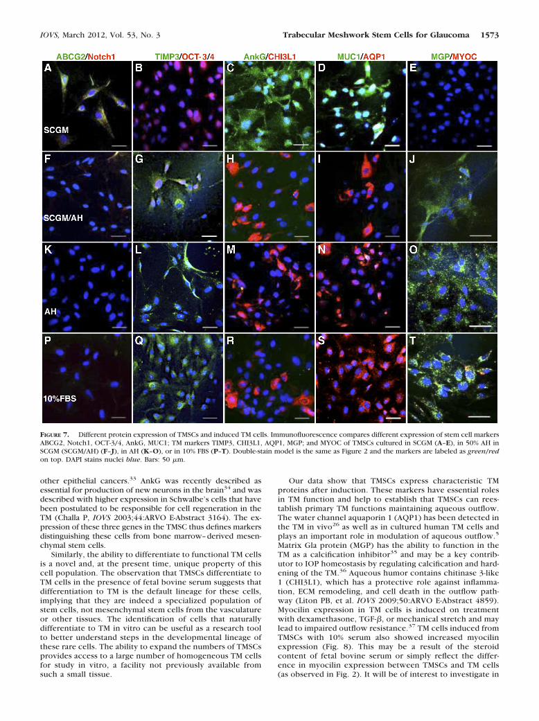
enzymes and extracellular matrix.4 TM cell number decreaseswith age and affects IOP.6–9 We examined if TMSCs can dif-ferentiate into TM cells. Clonal TMSCs were incubated in sev-eral media to induce TM cell differentiation: 50% bovine aque-ous humor in SCGM (SCGM/AH), 100% bovine aqueous humor(AH), or DMEM/F12 containing 10% FBS. The expression ofstem cell– and TM-cell markers was compared by immunoflu-orescence, qRT-PCR, and Western blotting. Figures 7A–E showTMSCs cultured in SCGM retained expression of stem cellmarkers ABCG2, Notch1, OCT-3/4, AnkG, MUC1, but not TM
FIGURE 2. Differential expression of stem cell and TM markers between TMSCs and primary TM cells. Clonal passaged TMSCs (A–E) and primaryTM cells (F–J) were double-stained with stem cell markers ABCG2 (green), Notch1 (red), OCT-3/4 (red), AnkG (green), MUC1 (green); TM markersTIMP3 (green), CHI3L1 (red), AQP1 (red), MGP (green); and MYOC (red). Arrows in (A) point to the ABCG2 and Notch1 double-positive cells.Arrow in (J) points to the MGP and MYOC double-positive cell. DAPI stains nuclei blue. Bars: 50 �m. (K) Expression of stem cell and TM markersfrom primary TM cells and clonal TMSCs at passage 4 was quantified by qRT-PCR. Error bars show SD of triplicate analyses. *P � 0.05 (n � 3,Student’s t-test).
FIGURE 3. Flow analysis of passage 4 TMSCs to assess the cell purity. Stem cell markers CD73, CD90, CD166, and Bmi1, and TM cell markersCHI3L1 and AQP1 were analyzed by flow cytometry. The peak on the left in each histogram represents the isotype control for comparison withthe cells labeled for each marker (on the right).
1570 Du et al. IOVS, March 2012, Vol. 53, No. 3

differentiation markers TIMP3, CHI3L1, AQP1, MGP, and notMYOC. When cultured in SCGM/AH (Figs. 7F–J), in AH (Figs.7K–O), or in 10% FBS (Figs. 7P–T), TMSC lost expression ofstem cell markers but expressed the TM markers. Some in-duced TM cells were found to become MYOC positive (Fig. 7T,red).
qRT-PCR (Fig. 8A) revealed expression of ABCG2, Pax6,Notch1, MUC1, and AnkG to be decreased, whereas expres-sion of AQP1 and MGP increased on TMSC differentiation inSCGM/AH, AH, or 10% FBS. The expression of MYOC andELAM-1 also increased after induction. Expression differencesof all genes between TMSC in SCGM and induced cells in AH orin 10% FBS were statistically significant (P � 0.05, one-wayANOVA followed by Dunnett’s posttest). The differences be-tween cells in SCGM and SCGM/AH were not consistentlystatistically significant. There were no statistical differencesbetween cells in AH and 10% FBS.
Western blotting (Fig. 8B) identified ABCG2 and OCT-3/4expression in clonal TMSCs but not in primary TM cells. A lowlevel of ABCG2 was detected in cells cultured in SCGM/AH but
not in cells in AH or 10% FBS. AQP1 and MGP were expressedin primary TM cells and induced cells but not in TMSCs.CHI3L1 appeared as a doublet migrating at approximately 39kDa in primary and induced TM cells and weakly in TMSCs.The lower molecular weight band may represent a glycosyla-tion variant of the protein.27
TM cells have phagocytic activity that eliminates debris,pigment, and other materials from the aqueous to maintainthe outflow pathway.20,28,29 The phagocytic ability of TM-SCs was assessed using fluorescently tagged S. aureus bio-particles comparing clonal TMSCs, induced and primary TMcells (Figs. 9A–E). Ingested S. aureus bioparticles appeargreen. On average, 0.18 green particles per cell were ingestedby TMSCs in SCGM (Fig. 9F). In contrast, TMSCs induced inSCGM/AH ingested 1.12 particles per cell; AH, 1.61; 10% FBS,1.83; and primary TM cells had 2.11 particles per cell. ANOVAfollowed by Dunnett’s test shows the increase in phagocyticactivity by TSMCs to be significant (P � 0.0001) under allconditions, although none of the induced TSMCs differed sig-nificantly from the activity of primary TM cells.
FIGURE 4. Induction of neural cell differentiation from TMSCs. Immunofluorescent staining on neural markers neurofilament, �-tubulin III, GFAP,and TMSC marker MUC1 shows different expression between TMSCs in NDM (A–D) for neural induction and in SCGM (E–H) to stem cellmaintenance. DAPI stains nuclei blue. Bars: 50 �m. (I) Neural markers NF and GFAP from TMSCs in SCGM and in NDM for neural induction werequantified by qRT-PCR. Error bars show SD of triplicate analyses. *P � 0.05 (n � 3, Student’s t-test). GFAP, glial fibrillary acidic protein; NF,neurofilament.
IOVS, March 2012, Vol. 53, No. 3 Trabecular Meshwork Stem Cells for Glaucoma 1571

DISCUSSION
In this report we describe the isolation and characterization ofa population of stem cells from human TM. These cells can beisolated as a side population by FACS or by clonal growth. Inculture they present a homogeneous population displayingantigenic markers previously characterized for mesenchymalstem cells (ABCG2, CD73, CD90, CD166, and Bmi1) as well asexpressing gene products associated with pluripotent stemcells (Notch1, OCT-3/4). Their stem cell character was con-firmed by the ability of these cells to display phenotypic prop-erties of cells from several different developmental lineages(neural, adipose, cornea) under culture conditions known toinduce differentiation of multipotent stem cells. These cells arecapable of differentiating into TM cells with phagocytic func-tion and expressing TM markers AQP1, MGP, CHI3L1, andTIMP3 in the presence of aqueous humor or 10% serum. Allthese support the hypotheses that these cells represent a res-ident population of adult stem cells in the human TM.
Our results confirm and extend conclusions of previousstudies suggesting stem cells in human TM. Challa and col-leagues (IOVS 2003;44:ARVO E-Abstract 3164) described“novel ” cells in primary TM cultures expressing MUC-1 andAnkG. Kelley et al.14 confirmed the expression, suggesting itmight be associated with a stem cell population. Gonzalez etal.12 found cultured TM cells capable of forming free-floatingneurospheres, a function associated with neural stem cells.More recently, McGowan et al.13 observed cells expressingOct-3/4, nestin, telomerase, PAX6, and Sox2 in the peripheralendothelium and TM of human corneas. Our data confirm the
presence of a stem cell population in TM, which expressedMUC1, AnkG, PAX6, and Oct-3/4. Expression of these markersclearly distinguishes TMSCs from typical mesenchymal stemcells. PAX6 is a homeobox gene essential to ocular develop-ment and is present in some adult ocular tissues but notgenerally present in TM.30 PAX6 is present in corneal stromalstem cells16,31 but is not expressed by mesenchymal stemcells.32 MUC1 is a cell surface mucin associated with breast and
FIGURE 5. Induction of adipocyte differentiation from TMSCs. TMSCswere cultured in ADM and AMM alternately for adipogenic induction.Oil red O (red) staining was compared on TMSCs in SCGM (A) and inADM/AMM (B). Hematoxylin stains nuclei blue. Bars: 50 �m. (C)Adipocytic markers ap2 and leptin from TMSCs in SCGM and inducedcells in ADM/AMM were quantified by qRT-PCR. Error bars show SD oftriplicate analyses. *P � 0.05 (n � 3, Student’s t-test). ap2, adipocyteprotein 2.
FIGURE 6. Induction of corneal keratocyte differentiation from TM-SCs. Cryosections of TMSCs cultured as pellets in KDM (A, B) or inSCGM (C, D) were stained with keratan sulfate (A, C) and keratocan(B, D). Human corneal stromal stem cells cultured as pellets in KDM,serving as positive control, were also stained with keratan sulfate (E)and keratocan (F). DAPI stains nuclei blue. Bars: 50 �m. (G) Kerato-cytic markers KERA and ALDH from TMSCs in SCGM and TMSCscultured as pellets in KDM were quantified by qRT-PCR. Error barsshow SD of triplicate analyses. *P � 0.05 (n � 3, Student’s t-test).KERA, keratocan; ALDH, aldehyde dehydrogenase.
1572 Du et al. IOVS, March 2012, Vol. 53, No. 3
other epithelial cancers.33 AnkG was recently described asessential for production of new neurons in the brain34 and wasdescribed with higher expression in Schwalbe’s cells that havebeen postulated to be responsible for cell regeneration in theTM (Challa P, IOVS 2003;44:ARVO E-Abstract 3164). The ex-pression of these three genes in the TMSC thus defines markersdistinguishing these cells from bone marrow–derived mesen-chymal stem cells.
Similarly, the ability to differentiate to functional TM cellsis a novel and, at the present time, unique property of thiscell population. The observation that TMSCs differentiate toTM cells in the presence of fetal bovine serum suggests thatdifferentiation to TM is the default lineage for these cells,implying that they are indeed a specialized population ofstem cells, not mesenchymal stem cells from the vasculatureor other tissues. The identification of cells that naturallydifferentiate to TM in vitro can be useful as a research toolto better understand steps in the developmental lineage ofthese rare cells. The ability to expand the numbers of TMSCsprovides access to a large number of homogeneous TM cellsfor study in vitro, a facility not previously available fromsuch a small tissue.
Our data show that TMSCs express characteristic TMproteins after induction. These markers have essential rolesin TM function and help to establish that TMSCs can rees-tablish primary TM functions maintaining aqueous outflow.The water channel aquaporin 1 (AQP1) has been detected inthe TM in vivo26 as well as in cultured human TM cells andplays an important role in modulation of aqueous outflow.5
Matrix Gla protein (MGP) has the ability to function in theTM as a calcification inhibitor35 and may be a key contrib-utor to IOP homeostasis by regulating calcification and hard-ening of the TM.36 Aqueous humor contains chitinase 3-like1 (CHI3L1), which has a protective role against inflamma-tion, ECM remodeling, and cell death in the outflow path-way (Liton PB, et al. IOVS 2009;50:ARVO E-Abstract 4859).Myocilin expression in TM cells is induced on treatmentwith dexamethasone, TGF-�, or mechanical stretch and maylead to impaired outflow resistance.37 TM cells induced fromTMSCs with 10% serum also showed increased myocilinexpression (Fig. 8). This may be a result of the steroidcontent of fetal bovine serum or simply reflect the differ-ence in myocilin expression between TMSCs and TM cells(as observed in Fig. 2). It will be of interest to investigate in
FIGURE 7. Different protein expression of TMSCs and induced TM cells. Immunofluorescence compares different expression of stem cell markersABCG2, Notch1, OCT-3/4, AnkG, MUC1; TM markers TIMP3, CHI3L1, AQP1, MGP; and MYOC of TMSCs cultured in SCGM (A–E), in 50% AH inSCGM (SCGM/AH) (F–J), in AH (K–O), or in 10% FBS (P–T). Double-stain model is the same as Figure 2 and the markers are labeled as green/redon top. DAPI stains nuclei blue. Bars: 50 �m.
IOVS, March 2012, Vol. 53, No. 3 Trabecular Meshwork Stem Cells for Glaucoma 1573

future studies whether TMSCs respond to stressors in amanner similar to that of TM cells.
TM cells have phagocytic activity that is essential inmaintaining normal aqueous outflow. We report here thatTMSCs after induction demonstrated phagocytic functionalmost as strong as that of primary TM cells. These areimportant findings that provide a biological source of differ-entiated TM cells for stem cell– based therapy on glaucoma.
Our present study provides new tools to design experi-ments to test the role of the TMSCs in cellular dynamics ofnormal and pathologic TM. Pharmacologic stimulation ofdifferentiation of existing TMSCs to new functional TM cellsis one avenue in which these cells might be useful in
restoration of normal outflow. We confirmed that the TMSCsare exclusively from TM and not corneal stroma, althoughthe potential of the stem cell population from cornealstroma to differentiate to phagocytic TM cells is worthy ofexploration. It may open a wider pharmacologic and clinicalapplication on TM restoration.
Another potential use of TMSCs is developing a cell-basedtherapy for glaucoma. We are exploring the feasibility ofTMSCs to be introduced into the anterior chamber and theirability to locate in the TM and adopt a TM phenotype. Thesedata will support the idea that TM damaged by high IOP orinflammation may be restored via a cell-based–therapy ap-proach.
FIGURE 8. Different gene expression of TMSCs and induced TM cells by qRT-PCR and Western blotting.(A) mRNA pools for stem cell makers ABCG2, Pax6, Notch1, MUC1, AnkG; TM markers AQP1, MGP; andMYOC and ELAM1 were quantified by qRT-PCR to compare the differences between TMSCs and TM cellsinduced in different media. Error bars show SD of triplicate analyses. (B) Western blotting comparesexpression of stem cell markers ABCG2, OCT-3/4, and TM cell markers AQP1, MGP, and CHI3L1 onpassage 4 TMSCs in SCGM, 1°TM cells, induced TM cells in SCGM/AH, AH or 10% FBS. �-Tubulin servesas loading control. 1°TM, primary trabecular meshwork cells.
FIGURE 9. Phagocytosis. (A–E) Phagocytosis assay to compare TMSCs and both induced and primary TM cells. Green dots are phagocytosedfluorescently conjugated S. aureus bioparticles. Yellow dots are unphagocytosed free bioparticles labeled with red secondary antibody. DAPI stainsnuclei blue. Bars: 50 �m. (F) The number of phagocytosed bioparticles per cell shows on the y-axis. The green particles and the nuclei in eachview were counted and at least 10 different views were counted in each condition. *P � 0.0001 between TMSCs in SCGM and induced TM cellsin different media and 1°TM (n � 10, one-way ANOVA followed by the Tukey posttest).
1574 Du et al. IOVS, March 2012, Vol. 53, No. 3

Acknowledgments
The authors thank Nancy Zurowski for help in cell sorting and flowcytometry and Kira Lathrop for help in imaging.
References
1. Gupta D. Glaucoma Diagnosis and Management. Philadelphia,PA: Lippincott Williams & Wilkins; 2004.
2. Le A, Mukesh BN, McCarty CA, Taylor HR. Risk factors associatedwith the incidence of open-angle glaucoma: the visual impairmentproject. Invest Ophthalmol Vis Sci. 2003;44:3783–3789.
3. Levkovitch-Verbin H. Animal models of optic nerve diseases. Eye.2004;18:1066–1074.
4. Buller C, Johnson DH, Tschumper RC. Human trabecular mesh-work phagocytosis. Observations in an organ culture system. In-vest Ophthalmol Vis Sci. 1990;31:2156–2163.
5. Stamer WD, Seftor RE, Snyder RW, Regan JW. Cultured humantrabecular meshwork cells express aquaporin-1 water channels.Curr Eye Res. 1995;14:1095–1100.
6. Alvarado J, Murphy C, Polansky J, Juster R. Age-related changes intrabecular meshwork cellularity. Invest Ophthalmol Vis Sci. 1981;21:714–727.
7. He Y, Leung KW, Zhang YH, et al. Mitochondrial complex I defectinduces ROS release and degeneration in trabecular meshworkcells of POAG patients: protection by antioxidants. Invest Oph-thalmol Vis Sci. 2008;49:1447–1458.
8. Lutjen-Drecoll E. Morphological changes in glaucomatous eyes andthe role of TGFbeta2 for the pathogenesis of the disease. Exp EyeRes. 2005;81:1–4.
9. Alvarado J, Murphy C, Juster R. Trabecular meshwork cellularity inprimary open-angle glaucoma and nonglaucomatous normals. Oph-thalmology. 1984;91:564–579.
10. Daley GQ, Scadden DT. Prospects for stem cell-based therapy. Cell.2008;132:544–548.
11. Verfaillie CM. Adult stem cells: assessing the case for pluripotency.Trends Cell Biol. 2002;12:502–508.
12. Gonzalez P, Epstein DL, Luna C, Liton PB. Characterization offree-floating spheres from human trabecular meshwork (HTM) cellculture in vitro. Exp Eye Res. 2006;82:959–967.
13. McGowan SL, Edelhauser HF, Pfister RR, Whikehart DR. Stem cellmarkers in the human posterior limbus and corneal endotheliumof unwounded and wounded corneas. Mol Vis. 2007;13:1984–2000.
14. Kelley MJ, Rose AY, Keller KE, Hessle H, Samples JR, Acott TS.Stem cells in the trabecular meshwork: present and future prom-ises. Exp Eye Res. 2009;88:747–751.
15. Mimura T, Joyce NC. Replication competence and senescence incentral and peripheral human corneal endothelium. Invest Oph-thalmol Vis Sci. 2006;47:1387–1396.
16. Du Y, Funderburgh ML, Mann MM, SundarRaj N, Funderburgh JL.Multipotent stem cells in human corneal stroma. Stem Cells. 2005;23:1266–1275.
17. Telford WG, Bradford J, Godfrey W, Robey RW, Bates SE. Sidepopulation analysis using a violet-excited cell-permeable DNAbinding dye. Stem Cells. 2007;25:1029–1036.
18. Du Y, Sundarraj N, Funderburgh ML, Harvey SA, Birk DE, Funder-burgh JL. Secretion and organization of a cornea-like tissue in vitroby stem cells from human corneal stroma. Invest Ophthalmol VisSci. 2007;48:5038–5045.
19. Patel AN, Park E, Kuzman M, Benetti F, Silva FJ, Allickson JG.Multipotent menstrual blood stromal stem cells: isolation, charac-terization, and differentiation. Cell Transplant. 2008;17:303–311.
20. Zhang X, Ognibene CM, Clark AF, Yorio T. Dexamethasone inhi-bition of trabecular meshwork cell phagocytosis and its modula-tion by glucocorticoid receptor beta. Exp Eye Res. 2007;84:275–284.
21. Rozen S, Skaletsky HJ. Primer3 on the WWW for general users andfor biologist programmers. In: Krawetz S, Misener S, eds. Bioin-formatics Methods and Protocols: Methods in Molecular Biology.Totowa, NJ: Humana Press; 2000:365–386.
22. Du Y, Carlson EC, Funderburgh ML, et al. Stem cell therapyrestores transparency to defective murine corneas. Stem Cells.2009;27:1635–1642.
23. Goodell MA, Brose K, Paradis G, Conner AS, Mulligan RC. Isolationand functional properties of murine hematopoietic stem cells thatare replicating in vivo. J Exp Med. 1996;183:1797–1806.
24. Liu CY, Birk DE, Hassell JR, Kane B, Kao WW. Keratocan-deficientmice display alterations in corneal structure. J Biol Chem. 2003;278:21672–21677.
25. Ruiz-Ederra J, Verkman AS. Aquaporin-1-facilitated keratocyte mi-gration in cell culture and in vivo corneal wound healing models.Exp Eye Res. 2009;89:159–165.
26. Stamer WD, Snyder RW, Smith BL, Agre P, Regan JW. Localizationof aquaporin CHIP in the human eye: implications in the patho-genesis of glaucoma and other disorders of ocular fluid balance.Invest Ophthalmol Vis Sci. 1994;35:3867–3872.
27. Recklies AD, Ling H, White C, Bernier SM. Inflammatory cytokinesinduce production of CHI3L1 by articular chondrocytes. J BiolChem. 2005;280:41213–41221.
28. Bill A. The drainage of aqueous humor (Editorial). Invest Ophthal-mol. 1975;14:1–3.
29. Johnson DH, Richardson TM, Epstein DL. Trabecular meshworkrecovery after phagocytic challenge. Curr Eye Res. 1989;8:1121–1130.
30. Collinson JM, Quinn JC, Hill RE, West JD. The roles of Pax6 in thecornea, retina, and olfactory epithelium of the developing mouseembryo. Dev Biol. 2003;255:303–312.
31. Funderburgh ML, Du Y, Mann MM, SundarRaj N, Funderburgh JL.PAX6 expression identifies progenitor cells for corneal kerato-cytes. FASEB J. 2005;19:1371–1373.
32. Nagai A, Kim WK, Lee HJ, et al. Multilineage potential of stablehuman mesenchymal stem cell line derived from fetal marrow.PLoS One. 2007;2:e1272.
33. Mukhopadhyay P, Chakraborty S, Ponnusamy MP, Lakshmanan I,Jain M, Batra SK. Mucins in the pathogenesis of breast cancer:implications in diagnosis, prognosis and therapy. Biochim BiophysActa. 2011;1815:224–240.
34. Paez-Gonzalez P, Abdi K, Luciano D, et al. Ank3-dependent SVZniche assembly is required for the continued production of newneurons. Neuron. 2011;71:61–75.
35. Xue W, Comes N, Borras T. Presence of an established calcificationmarker in trabecular meshwork tissue of glaucoma donors. InvestOphthalmol Vis Sci. 2007;48:3184–3194.
36. Vittitow J, Borras T. Genes expressed in the human trabecularmeshwork during pressure-induced homeostatic response. J CellPhysiol. 2004;201:126–137.
37. Tamm ER. Myocilin and glaucoma: facts and ideas. Prog Retin EyeRes. 2002;21:395–428.
IOVS, March 2012, Vol. 53, No. 3 Trabecular Meshwork Stem Cells for Glaucoma 1575