A Novel Beta-Defensin Antimicrobial Peptide in Atlantic Cod with Stimulatory Effect on Phagocytic...
13
A Novel Beta-Defensin Antimicrobial Peptide in Atlantic Cod with Stimulatory Effect on Phagocytic Activity Jareeporn Ruangsri, Yoichiro Kitani, Viswanath Kiron, Jep Lokesh, Monica F. Brinchmann, Ba ˚rd Ove Karlsen, Jorge M. O. Fernandes* Faculty of Biosciences and Aquaculture, University of Nordland, Bodø, Norway Abstract A novel defensin antimicrobial peptide gene was identified in Atlantic cod, Gadus morhua. This three exon/two intron defensin gene codes for a peptide precursor consisting of two domains: a signal peptide of 26 amino acids and a mature peptide of 40 residues. The mature cod defensin has six conserved cysteine residues that form 1–5, 2–4 and 3–6 disulphide bridges. This pattern is typical of beta-defensins and this gene was therefore named cod beta-defensin (defb). The tertiary structure of Defb exhibits an a/b fold with one a helix and b 1 b 2 b 3 sheets . RT-PCR analysis indicated that defb transcripts were present mainly in the swim bladder and peritoneum wall but could also be detected at moderate to low levels in skin, head- and excretory kidneys. In situ hybridisation revealed that defb was specifically expressed by cells located in the swim bladder submucosa and the oocytes. During embryonic development, defb gene transcripts were detectable from the golden eye stage onwards and their expression was restricted to the swim bladder and retina. Defb was differentially expressed in several tissues following antigenic challenge with Vibrio anguillarum, being up-regulated up to 25-fold in head kidney. Recombinant Defb displayed antibacterial activity, with a minimal inhibitory concentration of 0.4–0.8 mM and 25– 50 mM against the Gram-(+) bacteria Planococcus citreus and Micrococcus luteus, respectively. In addition, Defb stimulated phagocytic activity of cod head kidney leucocytes in vitro. These findings imply that beta-defensins may play an important role in the innate immune response of Atlantic cod. Citation: Ruangsri J, Kitani Y, Kiron V, Lokesh J, Brinchmann MF, et al. (2013) A Novel Beta-Defensin Antimicrobial Peptide in Atlantic Cod with Stimulatory Effect on Phagocytic Activity. PLoS ONE 8(4): e62302. doi:10.1371/journal.pone.0062302 Editor: Nikolas Nikolaidis, California State University Fullerton, United States of America Received January 26, 2012; Accepted March 22, 2013; Published April 25, 2013 Copyright: ß 2013 Ruangsri et al. This is an open-access article distributed under the terms of the Creative Commons Attribution License, which permits unrestricted use, distribution, and reproduction in any medium, provided the original author and source are credited. Funding: This study was undertaken as part of the project ‘Mucosal Immune System of Atlantic Cod - Creating a Knowledge Base’ (184703), funded by the Research Council of Norway (NFR, www.forskningsradet.no). The pathogen challenge study was a component of another NFR project ‘Preventive Health Care of Farmed Fish (ref. 176528). YK was supported by an Yggdrasil grant from the NFR (ref. 211233/F11). JR would like to express her gratitude to the Prince of Songkla University, (Thailand) for granting the PhD scholarship that enabled her to do research at the University of Nordland, Norway. The funders had no role in study design, data collection and analysis, decision to publish, or preparation of the manuscript. Competing Interests: The authors have declared that no competing interests exist. * E-mail: [email protected] Introduction Defensins are important components of the innate arsenal of antimicrobial peptides (AMPs) and proteins (AMPPs) that provide protection against potential pathogens. These cationic AMPs with a b-sheet structure stabilised by disulphide bridges are widely distributed across both plant and animal kingdoms [1,2]. In addition to their antimicrobial activity, animal defensins have other known biological roles. They are involved in host-microbiota interaction, immunomodulation through chemotaxis of immune cells and serve as potential links between innate and adaptive immunity [2,3]. Bony fish are the most diverse group of vertebrates and they live in a complex aquatic environment, where they are exposed to water-borne pathogens and deeply affected by abiotic factors. In response, these animals secrete a wide range of AMPs and AMPPs as part of their defence mechanism [4,5,6,7]. Even though defensins are important components of the immune system of multicellular organisms, their presence in teleosts have received scant attention. In fact, defensins have only been characterized from 8 teleost species to date. Fish defensins were first discovered through genome mining in model species, namely zebrafish Danio rerio defb1 to 3 (zfDB-1 to -3), the green-spotted pufferfish, Tetraodon nigroviridis defb1 and 2 (tnDB-1 and -2) and the tiger pufferfish, Takifugu rubripes defb (fuDB) [8]. Later, the presence of beta- defensin genes has been reported in other fishes: defb1 to 4 isoforms (omDB-1 to -4) in rainbow trout, Oncorhynchus mykiss [9,10], defb (mkDB) in medaka, Oryzias latipes [11], defb (ogDB) in orange-spotted grouper, Epinephelus coioides [12], defb1 to 5 (ofDBI-1 to -5) in olive flounder, Paralichthys olivaceus [13], and most recently defb (saDB) in gilthead seabream, Sparus aurata [14]. Additionally, EST sequences that correspond to 3 beta-defensin isoforms in Atlantic salmon, Salmo salar defb1,-3 and -4 (ssDB-1, -3 and -4) and one beta- defensin in Nile tilapia, Oreochromis niloticus defb (onDB) were deposited in the National Center for Biotechnology Information (NCBI) database by Adzhubei et al. [15] and Lee et al. [16], respectively. Constitutive beta-defensin expression varies between species and even among gene isoforms in the same species [8,9,13,14,17]. Furthermore, depending on the type of the tissue and or stimulatory factors, the genes are found to be either constitutive or inducible and they even have overlapping roles in protecting and maintaining fish health. Biological functions of beta-defensin have been investigated only in a few fish species and their multifunctional roles are still obscure. In most of the cases they were found to inhibit the growth of Gram - (+) and Gram - (2) PLOS ONE | www.plosone.org 1 April 2013 | Volume 8 | Issue 4 | e62302
Transcript of A Novel Beta-Defensin Antimicrobial Peptide in Atlantic Cod with Stimulatory Effect on Phagocytic...

A Novel Beta-Defensin Antimicrobial Peptide in AtlanticCod with Stimulatory Effect on Phagocytic ActivityJareeporn Ruangsri, Yoichiro Kitani, Viswanath Kiron, Jep Lokesh, Monica F. Brinchmann, Bard
Ove Karlsen, Jorge M. O. Fernandes*
Faculty of Biosciences and Aquaculture, University of Nordland, Bodø, Norway
Abstract
A novel defensin antimicrobial peptide gene was identified in Atlantic cod, Gadus morhua. This three exon/two introndefensin gene codes for a peptide precursor consisting of two domains: a signal peptide of 26 amino acids and a maturepeptide of 40 residues. The mature cod defensin has six conserved cysteine residues that form 1–5, 2–4 and 3–6 disulphidebridges. This pattern is typical of beta-defensins and this gene was therefore named cod beta-defensin (defb). The tertiarystructure of Defb exhibits an a/b fold with one a helix and b1b2b3 sheets. RT-PCR analysis indicated that defb transcriptswere present mainly in the swim bladder and peritoneum wall but could also be detected at moderate to low levels in skin,head- and excretory kidneys. In situ hybridisation revealed that defb was specifically expressed by cells located in the swimbladder submucosa and the oocytes. During embryonic development, defb gene transcripts were detectable from thegolden eye stage onwards and their expression was restricted to the swim bladder and retina. Defb was differentiallyexpressed in several tissues following antigenic challenge with Vibrio anguillarum, being up-regulated up to 25-fold in headkidney. Recombinant Defb displayed antibacterial activity, with a minimal inhibitory concentration of 0.4–0.8 mM and 25–50 mM against the Gram-(+) bacteria Planococcus citreus and Micrococcus luteus, respectively. In addition, Defb stimulatedphagocytic activity of cod head kidney leucocytes in vitro. These findings imply that beta-defensins may play an importantrole in the innate immune response of Atlantic cod.
Citation: Ruangsri J, Kitani Y, Kiron V, Lokesh J, Brinchmann MF, et al. (2013) A Novel Beta-Defensin Antimicrobial Peptide in Atlantic Cod with Stimulatory Effecton Phagocytic Activity. PLoS ONE 8(4): e62302. doi:10.1371/journal.pone.0062302
Editor: Nikolas Nikolaidis, California State University Fullerton, United States of America
Received January 26, 2012; Accepted March 22, 2013; Published April 25, 2013
Copyright: � 2013 Ruangsri et al. This is an open-access article distributed under the terms of the Creative Commons Attribution License, which permitsunrestricted use, distribution, and reproduction in any medium, provided the original author and source are credited.
Funding: This study was undertaken as part of the project ‘Mucosal Immune System of Atlantic Cod - Creating a Knowledge Base’ (184703), funded by theResearch Council of Norway (NFR, www.forskningsradet.no). The pathogen challenge study was a component of another NFR project ‘Preventive Health Care ofFarmed Fish (ref. 176528). YK was supported by an Yggdrasil grant from the NFR (ref. 211233/F11). JR would like to express her gratitude to the Prince of SongklaUniversity, (Thailand) for granting the PhD scholarship that enabled her to do research at the University of Nordland, Norway. The funders had no role in studydesign, data collection and analysis, decision to publish, or preparation of the manuscript.
Competing Interests: The authors have declared that no competing interests exist.
* E-mail: [email protected]
Introduction
Defensins are important components of the innate arsenal of
antimicrobial peptides (AMPs) and proteins (AMPPs) that provide
protection against potential pathogens. These cationic AMPs with
a b-sheet structure stabilised by disulphide bridges are widely
distributed across both plant and animal kingdoms [1,2]. In
addition to their antimicrobial activity, animal defensins have
other known biological roles. They are involved in host-microbiota
interaction, immunomodulation through chemotaxis of immune
cells and serve as potential links between innate and adaptive
immunity [2,3].
Bony fish are the most diverse group of vertebrates and they live
in a complex aquatic environment, where they are exposed to
water-borne pathogens and deeply affected by abiotic factors. In
response, these animals secrete a wide range of AMPs and AMPPs
as part of their defence mechanism [4,5,6,7]. Even though
defensins are important components of the immune system of
multicellular organisms, their presence in teleosts have received
scant attention. In fact, defensins have only been characterized
from 8 teleost species to date. Fish defensins were first discovered
through genome mining in model species, namely zebrafish Danio
rerio defb1 to 3 (zfDB-1 to -3), the green-spotted pufferfish, Tetraodon
nigroviridis defb1 and 2 (tnDB-1 and -2) and the tiger pufferfish,
Takifugu rubripes defb (fuDB) [8]. Later, the presence of beta-
defensin genes has been reported in other fishes: defb1 to 4 isoforms
(omDB-1 to -4) in rainbow trout, Oncorhynchus mykiss [9,10], defb
(mkDB) in medaka, Oryzias latipes [11], defb (ogDB) in orange-spotted
grouper, Epinephelus coioides [12], defb1 to 5 (ofDBI-1 to -5) in olive
flounder, Paralichthys olivaceus [13], and most recently defb (saDB) in
gilthead seabream, Sparus aurata [14]. Additionally, EST sequences
that correspond to 3 beta-defensin isoforms in Atlantic salmon,
Salmo salar defb1, -3 and -4 (ssDB-1, -3 and -4) and one beta-
defensin in Nile tilapia, Oreochromis niloticus defb (onDB) were
deposited in the National Center for Biotechnology Information
(NCBI) database by Adzhubei et al. [15] and Lee et al. [16],
respectively.
Constitutive beta-defensin expression varies between species
and even among gene isoforms in the same species [8,9,13,14,17].
Furthermore, depending on the type of the tissue and or
stimulatory factors, the genes are found to be either constitutive
or inducible and they even have overlapping roles in protecting
and maintaining fish health. Biological functions of beta-defensin
have been investigated only in a few fish species and their
multifunctional roles are still obscure. In most of the cases they
were found to inhibit the growth of Gram - (+) and Gram - (2)
PLOS ONE | www.plosone.org 1 April 2013 | Volume 8 | Issue 4 | e62302

bacteria [11,13,14,17]. Others, like Defb of orange-spotted
grouper and Defb1 of rainbow trout exhibited potential antiviral
activity [10,17]. In addition to its broad-spectrum antimicrobial
activity, chemotactic ability has been reported for Defb from
gilthead seabream [14]. Interestingly, defensins are thought to
have some function related to the reproductive system in olive
flounder and grouper [13,17].
The Atlantic cod (Gadus morhua) is a marine cold-water fish
species that is commercially and ecologically important. Its
economic value as a farmed fish has stimulated interest in
understanding its immune system due to the need to tackle
diseases. The recent completion of its draft genome revealed that
Atlantic cod has a peculiar immune system, inasmuch as it lacks
MHC class II [18]. Moreover, antibody production against
pathogens and or in response to vaccination is relatively poor
[19,20]. Hence, it is likely that Atlantic cod relies heavily on its
innate immune system for host defence. In the present study we
report the characterization of the first beta-defensin gene in
Atlantic cod, which was termed defb. Our results demonstrated
that this gene may play an important role in the cod innate
immune response.
Materials and Methods
Naıve Fish – Husbandry and Tissue SamplingJuvenile Atlantic cod were maintained indoors at the University
of Nordland (UiN) Research Station, Bodø, Norway. The fish
were reared at 7–8uC in 2,800 L fibre glass tanks that were part of
a flow-through system and fed ad libitum with a commercial diet
(Amber Neptune, Skretting AS, Norway). All animal handling
protocols were in accordance with the guidelines adopted by the
National Animal Research Authority (FDU) in Norway.
The transcription of defb gene was investigated in different
tissues and organs of naıve specimens. Six randomly selected and
apparently healthy fish weighing 200 to 300 g were anesthetised
with 100 mg?L21 of tricaine methanesulphonate (MS-222,
Chemical Laboratories, Washington, USA) and immediately killed
with a sharp blow to the head followed by transection of the spinal
cord. Mucus samples were collected from the dorsal side of the
body using a glass slide and then blood was collected from the
caudal vein without anticoagulant, using a 1 mL syringe fitted with
a 23 gauge needle. Thereafter, skin and fast muscle samples were
taken from the left dorsal side of the fish. Next, the operculum was
removed to excise the gill filament. Finally, following aseptic
procedures internal organs and tissues - head kidney, excretory
kidney, spleen, swim bladder, peritoneum wall, liver, pyloric
caeca, proximal and distal intestines, rectum, heart and brain -
were sampled, snap frozen in liquid nitrogen and stored at 280uCuntil use. Fractions of the same tissues and organs were fixed
overnight in 4% paraformaldehyde prepared in phosphate
buffered saline (0.1 M PBS, pH 7.4) treated with 0.1% diethylpyr-
ocarbonate. Standard histological procedures were adopted to
process these samples and embed them in paraffin.
Immune Challenged Fish- Experimental Design,Husbandry and Sampling
The challenge experiment was conducted at the facilities of the
Fish Health Unit of the Institute of Marine Research (HI), Bergen,
Norway. Forty healthy unvaccinated Atlantic cod weighing
around 60 g were randomly introduced into each of the three
500 L experimental tanks, which were part of a flow-through
system. After the acclimation period, two fish from each tank were
randomly sampled to collect the pre-challenge control samples.
Skin samples from the left dorsal side of the fish, gill filament, head
kidney and proximal intestine were collected adopting aseptic
procedures, snap-frozen in liquid nitrogen and stored at 280uCuntil use. The challenge was performed using a V. anguillarum
(strain H610 from the collection of the Fish Health Group at HI)
cell suspension, prepared as detailed in Ruangsri et al [21].
Immediately prior to challenge, the water flow was stopped and
the V. anguillarum suspension was added to each tank to attain
a final concentration of 2.66107 cfu mL21. Fish were exposed to
bacteria for a period of one hour, after which the water flow was
returned to normal. Following the challenge, two fish from each
tank were sampled (6 fish per treatment) at 4 and 48 h to collect
different tissues (post-challenge samples) as detailed above.
Embryo CollectionCod eggs were kindly supplied by CodFarmers ASA (Bodø,
Norway). Unfertilised eggs were immediately frozen in liquid
nitrogen and stored at 280uC until use. Artificially fertilised eggs
from individual cod spawning pairs were incubated at 7uC without
aeration in 5 L sterile glass bowls filled with 4 L drum-filtered
(30 mm) UV-sterilised seawater. They were stocked at a density of
approximately 10 mL eggs?L21 and the bowls were covered with
aluminium foil. Oxygen concentration in the incubating bowl was
maintained above 6.5 mg?L21 by replacing at least one third of
the water daily. Embryos at different developmental stages (1-cell,
2-cells, 16-cells, oblong, germ ring, 50% epiboly, 10-somite and
golden eye) and larvae (hindgut stage, first feeding and 20 days
post-hatch) were observed under the optical microscope and
approximately 50 specimens from each stage were collected, snap-
frozen in liquid nitrogen and stored at 280uC for the defb gene
expression study. In addition, embryos at the above developmental
stages were fixed overnight in 4% paraformaldehyde, later
dehydrated through graded levels of methanol and stored in
100% methanol at 280uC until used for whole mount in situ
hybridisation.
Database Mining of Cod Beta-defensinThe translated nucleotide sequences of defensins from different
fish species were obtained from NCBI (http://www.ncbi.nlm.nih.
gov/). These sequences were then used as queries to search the
publicly available cod ESTs using TBLASTN. Possible defensin
sequences retrieved were then used to design suitable primers for
experimental confirmation.
cDNA and gDNA Cloning of Cod Beta-defensincDNA sequences of beta-defensin were obtained from 20 days
post-hatch whole larvae cDNA using the Def1 primer set shown
on Table 1. Total RNA was extracted from approximately 50 to
100 mg of embryos using the Trizol method and cDNA
synthesised using the QuantiTect RT kit (Qiagen GmbH, Hilden,
Germany), as detailed in Fernandes et al. [22]. The amplification
conditions were set as follows: initial denaturation at 95uC for
2 min, followed by 34 cycles of denaturation at 95uC for 30 sec,
annealing step at 56uC for 30 sec, extension step at 72uC for 1 min
and a final elongation at 72uC for 10 min. Amplicons were first
separated by 1.2% (w/v) agarose gel electrophoresis, extracted
from the gel, cloned and as detailed elsewhere [23].
Genomic DNA (gDNA) was extracted from the spleen of V.
anguillarum challenged fish using the Nexttec DNA isolation kit
(Nexttec Biotechnologies GmbH, Leverkusen, Germany), follow-
ing the manufacturer’s instructions. Partial gDNA sequence was
obtained using the Def2 primer pair listed in Table 1 using a 6 min
initial denaturation at 95uC and annealing at 58uC. PCR products
were subsequently extracted, cloned and sequenced as above.
A Novel Beta-Defensin in Atlantic Cod
PLOS ONE | www.plosone.org 2 April 2013 | Volume 8 | Issue 4 | e62302

Sequence AnalysisFollowing base calling, experimental sequencing data were
assembled into contigs using the CodonCode aligner package
(www.codoncode.com). Nucleotide sequences were translated with
the ExPASy translation tool and basic properties of the mature
peptides were predicted by the ProtParam software at ExPASy
(www.expasy.ch). The presence of conserved domains and signal
peptides was investigated with SignalP 3.0 (www.cbs.dtu.dk/
services/SignalP), SMART (smart.embl-heidelberg.de) and Scan-
Prosite (prosite.expasy.org) tools. Homology modelling was
performed with the LOMETS server (zhanglab.ccmb.med.umi-
ch.edu/LOMETS) using the three-dimensional structure of the
crotamine, a myotoxin from rattlesnake (Crotalus durissus terrificus)
(PDB ID: 1H5O), as template. The results were then visualised
with the DeepView, which also enabled the placement of the
disulphide linkages (www.spdbv.vital-it.ch). Homologous se-
quences of fish beta-defensins were retrieved from the NCBI
database using BLASTx or tBLASTx and the translated cod defb
sequence as query. Putative beta-defensin peptide sequences were
aligned with MUSCLE (www.ebi.ac.uk/Tools/msa/muscle/) and
pair-wise sequence comparisons were performed with BioEdit
[24].
Phylogenetic trees were constructed based on deduced full-
length amino acid sequences using two different methods: i) the
maximum likelihood (ML) algorithm implemented in PhyML
(www.atgc-montpellier.fr/phyml/) with an LG model, estimated
gamma shape parameter and an approximate likelihood-ratio test;
and ii) Bayesian inference using a mixed model of amino acid
substitution in MrBayes 3 [25] for 56105 generations and burning
the first 500 trees. Accession numbers of the sequences used for
homology comparisons and phylogenetic analysis are listed on
Table 2.
Expression of Cod Beta-defensin1. Semi-quantitative RT-PCR. Total RNA was extracted
from approximately 100 mg of sample using the Trizol method
and cDNA synthesised using the QuantiTect RT kit (Qiagen), as
detailed in Fernandes et al. [22]. RNA quality and quantity were
evaluated by 1.2% agarose gel electrophoresis and spectropho-
tometry using the Quant-iT RNA assay kit (Qubit Fluorometer,
Invitrogen, California, USA). Cod beta-defensin was amplified
with the Def3 primer pair (Table 1) and beta-actin (actb) was used
as an endogenous control. Amplification conditions were set as
follows: initial denaturation at 95uC for 2 min, 30 cycles of
denaturation at 95uC for 30 sec, annealing at 58uC for 30 sec and
extension at 72uC for 1 min, followed by final elongation at 72uCfor 10 min. Amplicons were separated by electrophoresis on
a 1.2% (w/v) agarose gel, photographed and analysed using the
ChemiDoc gel documentation system from BioRad (Laboratories
Inc, California, USA).
2. Whole-mount and section in situ hybridisation. Cod
beta-defensin was subcloned using the Def1 primer pair and
a DNA template for probe synthesis was obtained from the
corresponding pCR4-TOPO plasmids by PCR using standard
M13 primers (Invitrogen) and the thermocycling conditions
described in section 2.5. T7 and T3 RNA polymerases (Roche,
East Sussex, UK) were used to synthesise digoxigenin (DIG)-
labelled RNA probes by in vitro transcription, according to the
manufacturer’s protocol.
For each selected developmental stage (i.e. bladder, hindgut and
first feeding stage), five cod embryos were used. Paraffin-
embedded tissue samples of naıve fish were used to prepare 4–
5 mm microtome sections (Shandon Finesse, Thermo Scientific,
Barrington, USA) on poly-L-lysine coated slides (VWR Interna-
tional, Leuven, Belgium). The slides were then incubated over-
night at 50uC, dewaxed by two subsequent treatments with xylene
and rehydrated through decreasing graded levels of methanol.
Table 1. List of primers employed and their application in this study.
Primer Sequence (59–39) T (uC) E (%) Application
Def1F AGGATGTCTTGCCACCGAGTCT 56 2 Partial cDNA
Def1R AGATGGTGTCATTGCAGAGTCC Probe synthesis
Def2F CAGTGCTGTTTAAGGATGTCTTG 58 2 Partial genomic
Def2R AGATGGTGTCATTGCAGAGTCC DNA
Def3F TTTTGTTGAGAATGAGGCAGC 58 100 RT-PCR, qPCR
Def3R ATGAGACACACAGCACTGGAAT
Def4F TTCCCCTGGTCCTGCCCCAC 60 2 Recombinant
Def4R TTAAAAGAAATGAGACACACAGCAC 2 Defb expression
Def5F GTTCCCCTGGTCCTGCCCCAC 60 2 Recombinant
Def5R GGGATCCTTAAAAGAAATGAGACACACAGCAC 2 Defb expression
eef1aF CACTGAGGTGAAGTCCGTTG 58 91 q PCR
eef1aR GGGGTCGTTCTTGCTGTCT
actbF TGACCCTGAAGTACCCCATC 58 94 q PCR
ActbR TCTTCTCCCTGTTGGCTTTG
rps9F TCTTTGAAGGTAATGCTCTGTTGAGA 58 97 q PCR
rps9R CGAGGATGTAATCCAACTTCATCTT
UbiF GGCCGCAAAGATGCAGAT 58 97 q PCR
UbiR CTGGGCTCGACCTCAAGAGT
T = annealing temperature; E = qPCR efficiency.doi:10.1371/journal.pone.0062302.t001
A Novel Beta-Defensin in Atlantic Cod
PLOS ONE | www.plosone.org 3 April 2013 | Volume 8 | Issue 4 | e62302
Embryos and tissue sections were permeabilised with 20?g?mL–1
proteinase K (Roche) for 20 min and 30 min, respectively. In situ
hybridisation (ISH) was performed essentially as described pre-
viously in Fernandes et al. [23]. Sense probes were used as
negative controls. Tissues sections were counterstained with
neutral fast red (Sigma, Missouri, USA). Whole embryos and
tissues were then observed under a binocular microscope (Leica
DM4000B, Leica Microsystem, Wetzlar, Germany) and a Lieca
DMIL microscope (Leica), respectively, and images were acquired
with a Leica DFC420 camera (Leica).
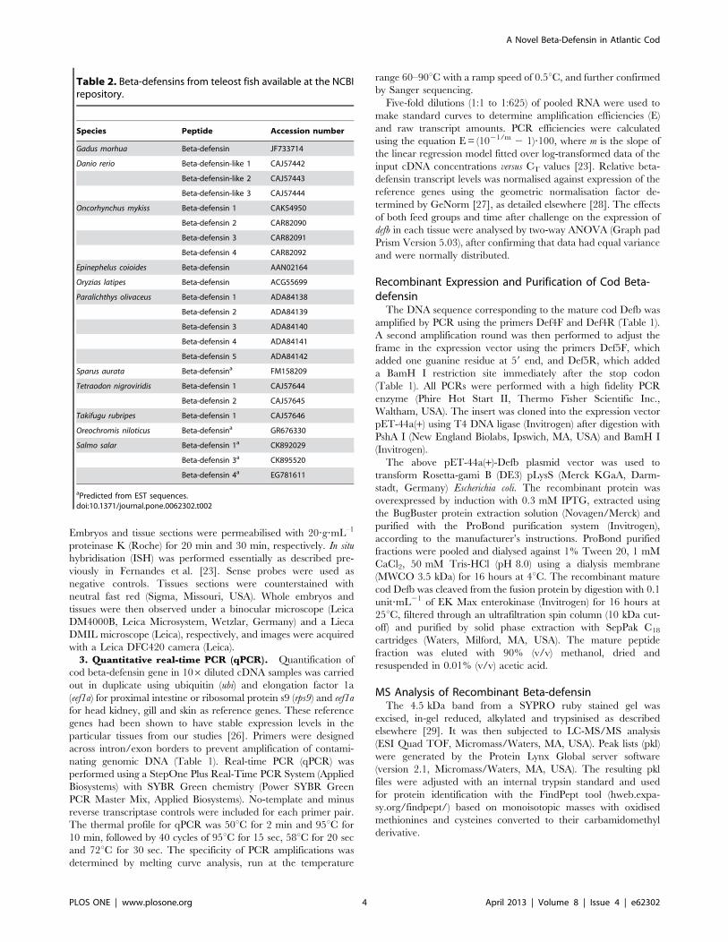
3. Quantitative real-time PCR (qPCR). Quantification of
cod beta-defensin gene in 106diluted cDNA samples was carried
out in duplicate using ubiquitin (ubi) and elongation factor 1a
(eef1a) for proximal intestine or ribosomal protein s9 (rps9) and eef1a
for head kidney, gill and skin as reference genes. These reference
genes had been shown to have stable expression levels in the
particular tissues from our studies [26]. Primers were designed
across intron/exon borders to prevent amplification of contami-
nating genomic DNA (Table 1). Real-time PCR (qPCR) was
performed using a StepOne Plus Real-Time PCR System (Applied
Biosystems) with SYBR Green chemistry (Power SYBR Green
PCR Master Mix, Applied Biosystems). No-template and minus
reverse transcriptase controls were included for each primer pair.
The thermal profile for qPCR was 50uC for 2 min and 95uC for
10 min, followed by 40 cycles of 95uC for 15 sec, 58uC for 20 sec
and 72uC for 30 sec. The specificity of PCR amplifications was
determined by melting curve analysis, run at the temperature
range 60–90uC with a ramp speed of 0.5uC, and further confirmed
by Sanger sequencing.
Five-fold dilutions (1:1 to 1:625) of pooled RNA were used to
make standard curves to determine amplification efficiencies (E)
and raw transcript amounts. PCR efficiencies were calculated
using the equation E = (1021/m 2 1)?100, where m is the slope of
the linear regression model fitted over log-transformed data of the
input cDNA concentrations versus CT values [23]. Relative beta-
defensin transcript levels was normalised against expression of the
reference genes using the geometric normalisation factor de-
termined by GeNorm [27], as detailed elsewhere [28]. The effects
of both feed groups and time after challenge on the expression of
defb in each tissue were analysed by two-way ANOVA (Graph pad
Prism Version 5.03), after confirming that data had equal variance
and were normally distributed.
Recombinant Expression and Purification of Cod Beta-defensin
The DNA sequence corresponding to the mature cod Defb was
amplified by PCR using the primers Def4F and Def4R (Table 1).
A second amplification round was then performed to adjust the
frame in the expression vector using the primers Def5F, which
added one guanine residue at 59 end, and Def5R, which added
a BamH I restriction site immediately after the stop codon
(Table 1). All PCRs were performed with a high fidelity PCR
enzyme (Phire Hot Start II, Thermo Fisher Scientific Inc.,
Waltham, USA). The insert was cloned into the expression vector
pET-44a(+) using T4 DNA ligase (Invitrogen) after digestion with
PshA I (New England Biolabs, Ipswich, MA, USA) and BamH I
(Invitrogen).
The above pET-44a(+)-Defb plasmid vector was used to
transform Rosetta-gami B (DE3) pLysS (Merck KGaA, Darm-
stadt, Germany) Escherichia coli. The recombinant protein was
overexpressed by induction with 0.3 mM IPTG, extracted using
the BugBuster protein extraction solution (Novagen/Merck) and
purified with the ProBond purification system (Invitrogen),
according to the manufacturer’s instructions. ProBond purified
fractions were pooled and dialysed against 1% Tween 20, 1 mM
CaCl2, 50 mM Tris-HCl (pH 8.0) using a dialysis membrane
(MWCO 3.5 kDa) for 16 hours at 4uC. The recombinant mature
cod Defb was cleaved from the fusion protein by digestion with 0.1
unit?mL21 of EK Max enterokinase (Invitrogen) for 16 hours at
25uC, filtered through an ultrafiltration spin column (10 kDa cut-
off) and purified by solid phase extraction with SepPak C18
cartridges (Waters, Milford, MA, USA). The mature peptide
fraction was eluted with 90% (v/v) methanol, dried and
resuspended in 0.01% (v/v) acetic acid.
MS Analysis of Recombinant Beta-defensinThe 4.5 kDa band from a SYPRO ruby stained gel was
excised, in-gel reduced, alkylated and trypsinised as described
elsewhere [29]. It was then subjected to LC-MS/MS analysis
(ESI Quad TOF, Micromass/Waters, MA, USA). Peak lists (pkl)
were generated by the Protein Lynx Global server software
(version 2.1, Micromass/Waters, MA, USA). The resulting pkl
files were adjusted with an internal trypsin standard and used
for protein identification with the FindPept tool (hweb.expa-
sy.org/findpept/) based on monoisotopic masses with oxidised
methionines and cysteines converted to their carbamidomethyl
derivative.
Table 2. Beta-defensins from teleost fish available at the NCBIrepository.
Species Peptide Accession number
Gadus morhua Beta-defensin JF733714
Danio rerio Beta-defensin-like 1 CAJ57442
Beta-defensin-like 2 CAJ57443
Beta-defensin-like 3 CAJ57444
Oncorhynchus mykiss Beta-defensin 1 CAK54950
Beta-defensin 2 CAR82090
Beta-defensin 3 CAR82091
Beta-defensin 4 CAR82092
Epinephelus coioides Beta-defensin AAN02164
Oryzias latipes Beta-defensin ACG55699
Paralichthys olivaceus Beta-defensin 1 ADA84138
Beta-defensin 2 ADA84139
Beta-defensin 3 ADA84140
Beta-defensin 4 ADA84141
Beta-defensin 5 ADA84142
Sparus aurata Beta-defensina FM158209
Tetraodon nigroviridis Beta-defensin 1 CAJ57644
Beta-defensin 2 CAJ57645
Takifugu rubripes Beta-defensin 1 CAJ57646
Oreochromis niloticus Beta-defensina GR676330
Salmo salar Beta-defensin 1a CK892029
Beta-defensin 3a CK895520
Beta-defensin 4a EG781611
aPredicted from EST sequences.doi:10.1371/journal.pone.0062302.t002
A Novel Beta-Defensin in Atlantic Cod
PLOS ONE | www.plosone.org 4 April 2013 | Volume 8 | Issue 4 | e62302

Bioactivity of Cod Beta-defensin1. Antibacterial activity. Antibacterial activity was mea-
sured by a liquid growth inhibition assay in a 96 well-microtitre
plate, as reported [30]. The following 4 bacterial strains were
tested: Planococcus citreus, Micrococcus luteus, Aeromonas salmonicida
and Vibrio anguillarum. A mixture of 50 mL of defensin (0.1 to
50 mM dilution series) and 50 mL of bacterial suspension (16105
colony forming unit/mL) in Muller–Hinton broth (Difco
Laboratories, Detroit, MI, USA), was incubated at a suitable
temperature until log phase was reached for each bacterium
(Table 3). After incubation, bacterial growth was quantified by
absorbance measurement at 595 nm using a microtitre plate
reader (FLUOstar Optima, BMG Labtech GmbH, Ortenberg,
Germany). The minimal inhibitory concentration was defined as
concentration of defensin that inhibited growth by 50%
compared to the control without peptide.
2. Phagocytosis assay. Head kidney leucocytes from five
Atlantic cod were prepared according to Meng et al. [31] and
suspended in L-15 medium. All solutions were adjusted to an
osmolality of 380 mOsm with NaCl. Phagocytic activity was
measured using pHrodo BioParticles (E. coli bioparticles,
Invitrogen). The cell suspension (106 cells in 100 mL) was
placed in wells of a flat bottom 96-well plate and incubated for
2 hours at 15uC for cell attachment. The media was replaced
with L-15 with 2% fetal calf serum and the cells were kept
overnight (14 h) at 15uC. The cell culture medium was then
aspirated to remove cell debris and unattached cells, washed
with Hanks buffer supplemented with NaCl and replaced with
working solution of the same buffer but with pHrodo
bioparticles and recombinant cod Defb (0.2 to 20 mM). In
addition, an aliquot of the particles was opsonized with 5% cod
serum at 4uC for 1 hour and added to separate wells with cells.
Cell viability, assessed in separate wells, was more than 90%, as
determined by the trypan blue exclusion method. Duplicate
samples for each fish were treated and measured in parallel.
After incubation at 15uC for 4 hours, fluorescence was
measured in a microplate reader (FLUOstar Optima, BMG
Labtech) with excitation at 560 nm and emission at 590 nm.
The effect of cod Defb on phagocytic activity was determined
according to the following equation: % effect = (Itest 2 Inc)/(Iop
2 Inc)?100, where Itest, Inc and Iop are the mean fluorescent
intensities of the test group, non-cell control and opsonized
positive control, respectively. Data from the phagocytosis assay
were analysed by paired Student’s t-test or Mann-Whitney U
test when the data were not normally distributed.
Results
Sequence Analysis of Cod Beta-defensinAfter identifying two cod ESTs with homology to fish defensins,
specific primers were design to amplify the complete defensin
coding sequence (CDS) in Atlantic cod. The cDNA sequence
obtained was 248 nucleotides long and comprised a partial 3 base
pair (bp) 59-untranslated region (UTR), a 201 bp open reading
frame (ORF) that encoded the 66-residue peptide precursor, and
a partial 44 bp 39UTR. Sequence homology searches against the
non-redundant protein database at NCBI revealed that cod
defensin is most similar to fuDB-1, a defensin-like protein 1 in tiger
pufferfish (89%; E-value = 10220), and omDB-1, a beta-defensin 1
from rainbow trout (85%; E-value = 10218). Hence, the cod
defensin sequence was named as beta- defensin (Defb) and
deposited in GenBank under the accession JF733714. The cod defb
was also partially sequenced and revealed a CDS of three exons
and two introns, similarly to other fish beta-defensin members
(Fig. 1).
The 66-amino acid cod Defb precursor contained an N-
terminal signal peptide with a likely cleavage site between amino
acid Ala 26 and Phe 27 (AAA_FP), as well as the C-terminal beta-
defensin signature. The signal peptide is encoded by the first exon
and part of the second exon, whilst the mature peptide
corresponds to part of the second and third exons. The latter
has a calculated MW of 4.49 KDa, a predicted pI of 7.79 and an
overall net charge of +1. Defb has six conserved cysteine residues
at positions 31, 38, 42, 54, 60 and 61 in the mature peptide, and
an additional 3 conserved glycines at positions 36, 50 and 53.
Cysteines C1, C2, C3 and C4 are located in the second exon,
while C5 and C6 are found in the last exon (Fig. 1). The putative
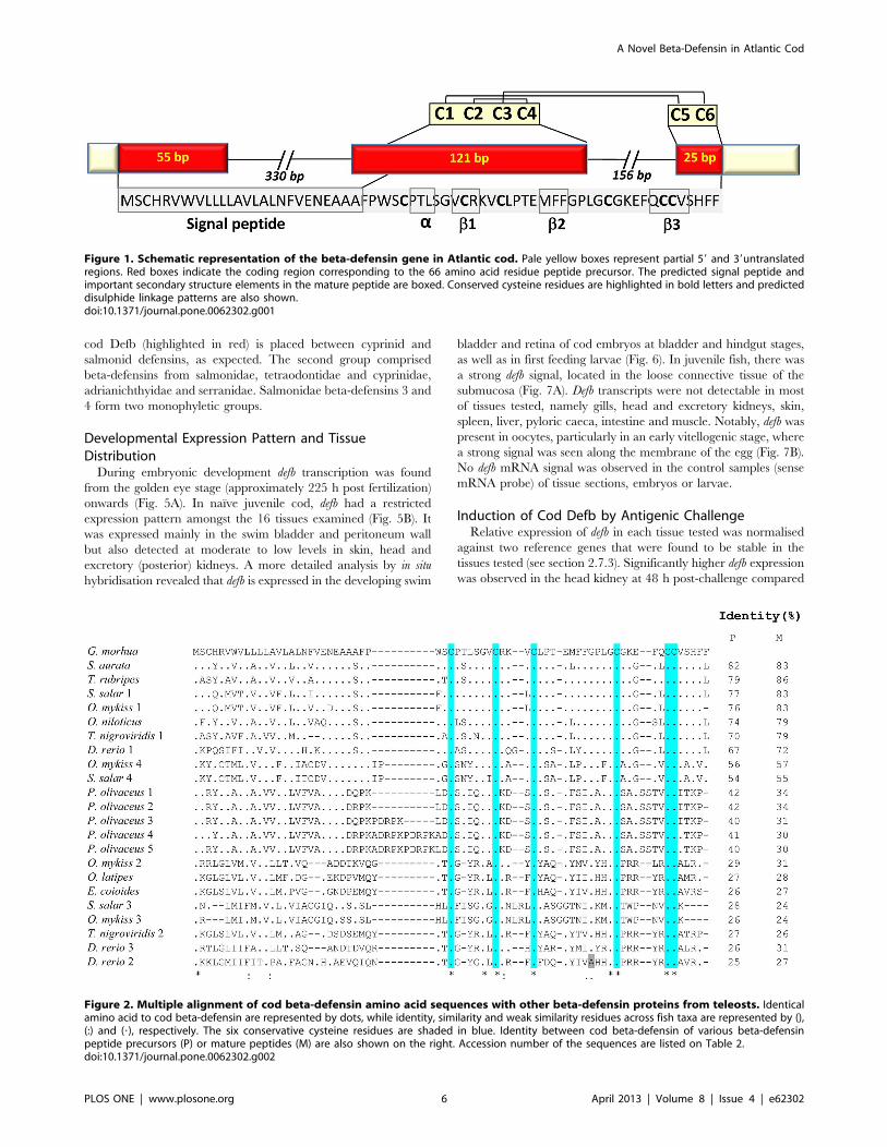
amino acid sequence of Defb was compared to 22 beta-defensins
from 10 fish species (Table 2). Sequence alignments showed that
cod Defb has a conservative motif of common beta-defensins,
sharing six conserved cysteines and additional 3 glycine residues
that exist close to the positions of C2 and C4 in most species, the
only exception being zebrafish Defb2, where the glycine at
position 50 is replaced by alanine (Fig. 2). The identity at the
protein level between cod Defb peptide precursor and their
homologues from fish species varied widely, ranging from 25
(Defb2 of zebrafish) to 82% (Defb of gilthead seabream).
The Structure of Cod DefbThe predicted three dimensional structure of cod Defb is shown
in Fig. 3. Comparison of the cod Defb peptide signature with the
structure of crotamine revealed that Defb contains a short N-
terminal a-helix and a small antiparallel triple-stranded b-sheet
arranged in an ab1b2b3 topology (Fig. 3A, B). Compared to other
beta-defensins, cod Defb has the highest structure similarity with
that of mouse mBD8 (Fig. 3C). Amongst fish beta-defensins, cod
Defb structure resembles zebrafish Defb1, with 72.1% identity
between both sequences (Fig. 3D). Disulphide linkages of each
beta-defensin were examined using Swiss-PdbViewer and all
bridges were predicted to exist between cysteine residues 1–5, 2–4
and 3–6, in common to all other vertebrate beta-defensins (Figs. 1,
3).
Phylogeny of Cod Beta-defensinBayesian and likelihood phylogenetic reconstructions produced
trees with same topologies that consisted for two main clades
(Fig. 4). The largest cluster included cichlidae, sparidae, all group 1
beta-defensins from salmonidae, tetraodontidae, cyprinidae and
beta-defensin-1 to -5 from paralichthyidae. This clade followed the
accepted taxonomic relationship between these teleost species and
Table 3. Antibacterial activity of recombinant cod beta-defensin.
Gram T MICa
(uC) (mM) (mg?ml21)
Micrococcus luteus ATCC 4698 + 37 25–50 125–250
Planococcus citreus NCIMB 1493 + 25 0.4–0.8 2–4
Aeromonas salmonicida NCIMB1102
2 20 .50 .250
Vibrio anguillarum NCIMB 2133 2 20 .50 .250
aThe minimal inhibitory concentration (MIC) is defined as the concentration ofpeptide that inhibits bacterial growth by 50% compared to the negative controlwithout peptide.doi:10.1371/journal.pone.0062302.t003
A Novel Beta-Defensin in Atlantic Cod
PLOS ONE | www.plosone.org 5 April 2013 | Volume 8 | Issue 4 | e62302
cod Defb (highlighted in red) is placed between cyprinid and
salmonid defensins, as expected. The second group comprised
beta-defensins from salmonidae, tetraodontidae and cyprinidae,
adrianichthyidae and serranidae. Salmonidae beta-defensins 3 and
4 form two monophyletic groups.
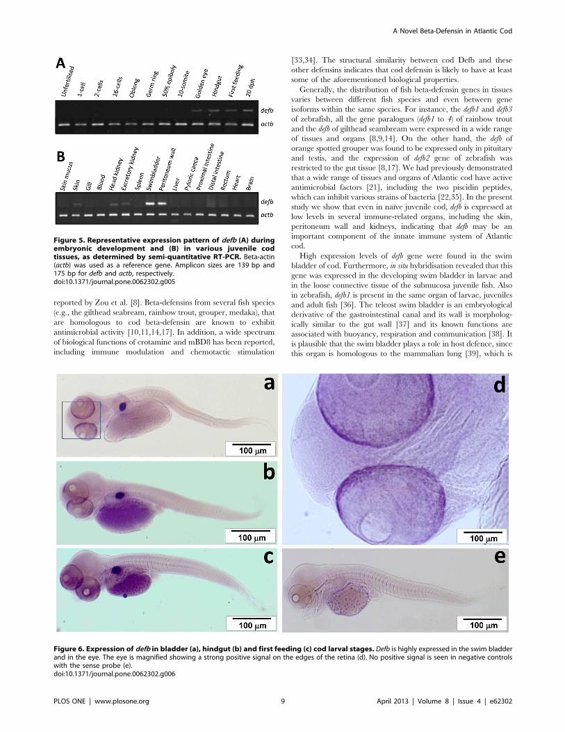
Developmental Expression Pattern and TissueDistribution
During embryonic development defb transcription was found
from the golden eye stage (approximately 225 h post fertilization)
onwards (Fig. 5A). In naıve juvenile cod, defb had a restricted
expression pattern amongst the 16 tissues examined (Fig. 5B). It
was expressed mainly in the swim bladder and peritoneum wall
but also detected at moderate to low levels in skin, head and
excretory (posterior) kidneys. A more detailed analysis by in situ
hybridisation revealed that defb is expressed in the developing swim
bladder and retina of cod embryos at bladder and hindgut stages,
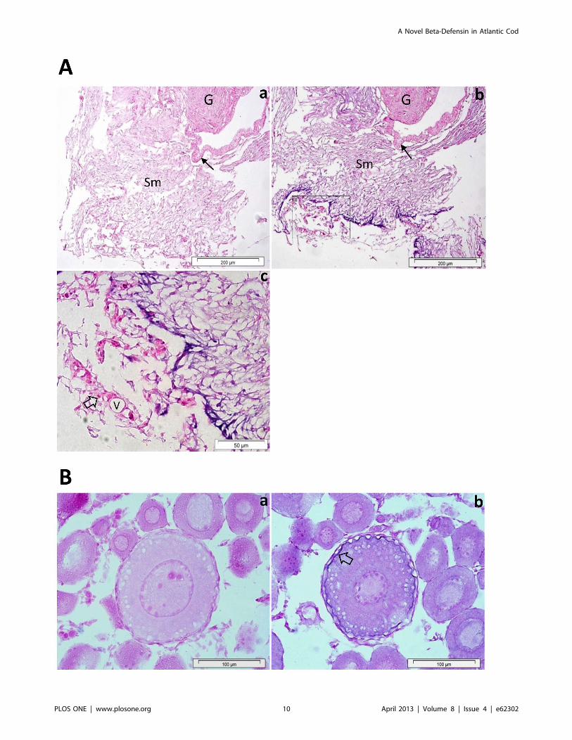
as well as in first feeding larvae (Fig. 6). In juvenile fish, there was
a strong defb signal, located in the loose connective tissue of the
submucosa (Fig. 7A). Defb transcripts were not detectable in most
of tissues tested, namely gills, head and excretory kidneys, skin,
spleen, liver, pyloric caeca, intestine and muscle. Notably, defb was
present in oocytes, particularly in an early vitellogenic stage, where
a strong signal was seen along the membrane of the egg (Fig. 7B).
No defb mRNA signal was observed in the control samples (sense
mRNA probe) of tissue sections, embryos or larvae.
Induction of Cod Defb by Antigenic ChallengeRelative expression of defb in each tissue tested was normalised
against two reference genes that were found to be stable in the
tissues tested (see section 2.7.3). Significantly higher defb expression
was observed in the head kidney at 48 h post-challenge compared
Figure 1. Schematic representation of the beta-defensin gene in Atlantic cod. Pale yellow boxes represent partial 59 and 39untranslatedregions. Red boxes indicate the coding region corresponding to the 66 amino acid residue peptide precursor. The predicted signal peptide andimportant secondary structure elements in the mature peptide are boxed. Conserved cysteine residues are highlighted in bold letters and predicteddisulphide linkage patterns are also shown.doi:10.1371/journal.pone.0062302.g001
Figure 2. Multiple alignment of cod beta-defensin amino acid sequences with other beta-defensin proteins from teleosts. Identicalamino acid to cod beta-defensin are represented by dots, while identity, similarity and weak similarity residues across fish taxa are represented by (),(:) and (?), respectively. The six conservative cysteine residues are shaded in blue. Identity between cod beta-defensin of various beta-defensinpeptide precursors (P) or mature peptides (M) are also shown on the right. Accession number of the sequences are listed on Table 2.doi:10.1371/journal.pone.0062302.g002
A Novel Beta-Defensin in Atlantic Cod
PLOS ONE | www.plosone.org 6 April 2013 | Volume 8 | Issue 4 | e62302

to 4 h post-challenge (30.6-fold, p,0.01) and the pre-challenge
group (24.8-fold, p,0.01) (Fig. 8A). In skin and proximal intestine,
defb transcript levels remained unchanged by challenge with V.
anguillarum (Fig. 8B, D). An apparent down-regulation of defb was
observed in the gills at 4 and 48 hours post-challenge, albeit this
difference was not significant (Fig. 8C, p.0.05). Overall, there was
a significant interaction between time and relative defb expression
levels in all tissues tested (p = 0.006).
Bioactivity of Recombinant Cod Beta-defensinThe recombinant cod Defb was highly expressed in E. Coli
RosettaGami (DE3) cells as a 66 kDa fusion protein with a Hisx6-
Nus Tag at the N terminus. After enterokinase digestion, the
mature 4.5 kDa Defb was released from the fusion protein
(Supplementary Fig. S1). Identity of the recombinant mature cod
defensin was confirmed by mass spectrometry, since the identified
peptide fragments covered the entire sequence (Supplementary
Table S1).
Cod Defb was active against Gram-(+) bacteria, with minimal
inhibitory concentrations against P. citreus and M. luteus in the
range 0.4–0.8 mM and 25–50 mM, respectively. In contrast, no
antibacterial activity was detected against the Gram-(2) bacteria
tested, i.e. A. salmonicida and V. anguillarum (Table 3).
The phagocytic activity of cod head kidney leucocytes was
stimulated by cod Defb in a concentration-dependent manner
(Fig. 9). At a concentration of 20 mM cod Defb the phagocytic
index was 6865.63% (mean 6 SEM, n = 5), which corresponded
to an increase of 62% compared to the control without defensin
(p,0.05, Fig. 9).
Discussion
In the present study, we report for the first time the
identification and characterization of a defensin gene in a gadoid
fish. The putative cod defensin precursor comprises a carboxy-
terminal signal peptide and an amino-terminal defensin signature
with six conserved cysteine residues that form 1–5, 2–4 and 3–6
disulphide linkages. This bridge pattern along with the short
spacing between C1 and C2 indicate that the cod defensin is
a member of the vertebrate beta-defensin family rather than an
alpha-defensin [8,32] and, therefore, it was named cod beta-
defensin or Defb for short.
Figure 3. Three dimensional structures of cod Defb (A), crotamine (B), mBD8 (C) and zebrafish Defb1 (D). The presence of an a-helix(red), three antiparallel b-strands (blue), and the disulphide linkages of 1–5, 2–4 and 3–6 pattern (yellow) were found in all species. Homologymodelling of cod Defb was performed using the PBD structure of crotamine, a neurotoxin from rattlesnake, Crotalus durissus trrificus (PDB ID: 1H5O),and mouse beta-defensin 8 (mBD8, PDB ID: 1E4R) as templates.doi:10.1371/journal.pone.0062302.g003
A Novel Beta-Defensin in Atlantic Cod
PLOS ONE | www.plosone.org 7 April 2013 | Volume 8 | Issue 4 | e62302

Cod defb and beta-defensin genes of zebrafish, tiger pufferfish,
medaka, olive flounder, rainbow trout and orange-spotted grouper
share a similar structure, consisting of three exons and two introns.
Moreover, cysteine residues C1 to C4 are located in exon 2,
whereas C5 and C6 are in exon 3 [8,9,11,13,17]. The putative
mature cod Defb has 4.49 kDa, a net charge of +1 and a predicted
isoelectric point of 7.79, features that are typical of fish defensins.
Our phylogenetic analysis placed cod Defb between zebrafish and
salmonid type 1 defensins, in accordance to currently accepted
taxonomic relationships between these taxa. A high degree of
conservation between known structural elements was observed
between cod and other fish defensins. The six cysteine residues
also present in cod defensin are conserved across human defensins
(both alpha and beta), chicken beta-defensins and snake crotamine
[8], which indicates that they are essential for the structure and
function of these molecules. In human, the hBD2 and hBD3
exhibits an a/b fold, with an a helix and b1b2b3 sheets with a 1–5,
2–4 and 3–6 disulphide linkage pattern [2]. A similar structure is
seen for cod Defb, even if it is more akin to those of crotamine of
rattlesnake and mBD8 of mouse. This could be explained by the
presence of a conserved motif Gly-X-Cys in the second b-strand in
humans, in contrast to the first b-strand in all fish taxa, snake and
mouse. Amongst fish species, the cod Defb tertiary structure has
greater similarity to the predicted structure of zebrafish Defb1
Figure 4. Unrooted radiation tree illustrating the phylogenetic relationship between fish beta-defensins. Cod beta-defensin(highlighted by red) falls into the same clade with a large group of other beta-defensins from different teleost taxa. Bayesian posterior probabilitiesand maximum likelihood values are indicated as percentages on the tree nodes, respectively.doi:10.1371/journal.pone.0062302.g004
A Novel Beta-Defensin in Atlantic Cod
PLOS ONE | www.plosone.org 8 April 2013 | Volume 8 | Issue 4 | e62302
reported by Zou et al. [8]. Beta-defensins from several fish species
(e.g., the gilthead seabream, rainbow trout, grouper, medaka), that
are homologous to cod beta-defensin are known to exhibit
antimicrobial activity [10,11,14,17]. In addition, a wide spectrum
of biological functions of crotamine and mBD8 has been reported,
including immune modulation and chemotactic stimulation
[33,34]. The structural similarity between cod Defb and these
other defensins indicates that cod defensin is likely to have at least
some of the aforementioned biological properties.
Generally, the distribution of fish beta-defensin genes in tissues
varies between different fish species and even between gene
isoforms within the same species. For instance, the defb1 and defb3
of zebrafish, all the gene paralogues (defb1 to 4) of rainbow trout
and the defb of gilthead seambream were expressed in a wide range
of tissues and organs [8,9,14]. On the other hand, the defb of
orange spotted grouper was found to be expressed only in pituitary
and testis, and the expression of defb2 gene of zebrafish was
restricted to the gut tissue [8,17]. We had previously demonstrated
that a wide range of tissues and organs of Atlantic cod have active
antimicrobial factors [21], including the two piscidin peptides,
which can inhibit various strains of bacteria [22,35]. In the present
study we show that even in naıve juvenile cod, defb is expressed at
low levels in several immune-related organs, including the skin,
peritoneum wall and kidneys, indicating that defb may be an
important component of the innate immune system of Atlantic
cod.
High expression levels of defb gene were found in the swim
bladder of cod. Furthermore, in situ hybridisation revealed that this
gene was expressed in the developing swim bladder in larvae and
in the loose connective tissue of the submucosa juvenile fish. Also
in zebrafish, defb1 is present in the same organ of larvae, juveniles
and adult fish [36]. The teleost swim bladder is an embryological
derivative of the gastrointestinal canal and its wall is morpholog-
ically similar to the gut wall [37] and its known functions are
associated with buoyancy, respiration and communication [38]. It
is plausible that the swim bladder plays a role in host defence, since
this organ is homologous to the mammalian lung [39], which is
Figure 5. Representative expression pattern of defb (A) duringembryonic development and (B) in various juvenile codtissues, as determined by semi-quantitative RT-PCR. Beta-actin(actb) was used as a reference gene. Amplicon sizes are 139 bp and175 bp for defb and actb, respectively.doi:10.1371/journal.pone.0062302.g005
Figure 6. Expression of defb in bladder (a), hindgut (b) and first feeding (c) cod larval stages. Defb is highly expressed in the swim bladderand in the eye. The eye is magnified showing a strong positive signal on the edges of the retina (d). No positive signal is seen in negative controlswith the sense probe (e).doi:10.1371/journal.pone.0062302.g006
A Novel Beta-Defensin in Atlantic Cod
PLOS ONE | www.plosone.org 9 April 2013 | Volume 8 | Issue 4 | e62302
A Novel Beta-Defensin in Atlantic Cod
PLOS ONE | www.plosone.org 10 April 2013 | Volume 8 | Issue 4 | e62302

known to express several beta-defensins [40]. It is also worth to
investigate whether cod beta-defensin or piscidins have significant
roles in hydrostatic regulation. In mammals, beta-defensins are
known to be involved in protection and tissue repair [41,42]. A
previous study on medaka reported that beta-defensin was
abundantly present in several parts of eyes and the authors
suggested that these molecules may hold therapeutic potential
[11]. Moreover, levels of beta-defensin mRNA in the eye increased
by more than 10-fold following stimulation with lipopolysaccha-
ride for 12 h [11]. The presence of defb transcripts in the retina of
cod larvae might be explained by their bioactivity against
pathogens present in the complex aquatic environment or by
a developmental role for defensins.
Beta-defensins that are expressed in the reproductive tract of
several higher vertebrate species are thought to protect the sperm
and the egg from microbial infection, enabling them to survive in
the genital tracts [2,43]. Defb of orange spotted grouper and the
defb gene isoforms of olive flounder fish are highly expressed in the
testis and during embryonic development, respectively [13,17].
Therefore, fish defensins are thought to play an important part in
the integrity of the reproductive system, perhaps linked to immune
defence. The presence of cod defb transcripts along the egg
membrane of early vitellogenic stage oocytes indicate that Defb
may be involved in oocyte maturation.
Exposure of Atlantic cod to live pathogenic bacteria (V.
anguillarum) was found to induce a 30.6-fold up-regulation of defb
expression in the head kidney of challenged fish at 48 h post-
stimulation. Such an induction was not observed in the mucosal
tissues even though the fish were directly exposed to the bacteria.
This implies that the head kidney of cod is involved in the
recruitment of beta-defensin producing cells in response to
bacterial infection. In rainbow trout, upon Yersinia ruckeri infection,
the defb isoforms were differentially expressed by immune tissues
and organs [9]. A similar trend was seen in the head kidney of
olive flounder after infecting the fish with bacteria, Edwardsiella
tarda [13], and inducible expression of the rainbow trout beta-
defensin gene was observed in defb1-transfected EPC cells exposed
to viral haemorrhagic septicaemia virus [10]. Furthermore, in the
rainbow trout head kidney all four defb genes transcripts were seen
very clearly after the cell was stimulated with PolyI:C [9]. The
prompt up-regulation of cod defb upon antigenic challenge strongly
suggests that it is involved in the innate immune response. On the
other hand, the constitutive expression of defb gene found in
several tissues of naıve cod may be signalling its role in
homeostasis.
The antibacterial spectrum of fish defensins varies significantly
amongst different species [11,13,14,17,44]. Recombinant cod
Defb displayed potent antibacterial activity in the micromolar
Figure 7. Localization of defb in the swim bladder (A) and oocytes (B) of Atlantic cod. In the swim bladder of juvenile fish, defb is expressedin the loose connective tissue of the submucosa (Sm) (Ab) but not in the secretory epithelial cells of the gas gland (G) and the epithelium of swimbladder wall (arrow). The sense mRNA probe (a) shows no defb positive signal (Aa). A portion of the swim bladder’s submucosa is magnified, (Ac)showing a strong defb positive signal in the loose connective tissue but not in red blood cells (open arrow) or blood vessels (V). In oocytes (B), defbtranscripts are present along the egg membrane of early vitellogenic stage (Bb, open arrow). No signal was observed with the negative control probe(Ba).doi:10.1371/journal.pone.0062302.g007
Figure 8. Relative expression of defb in immune-related tissues of Atlantic cod upon challenge with pathogenic bacteria (Vibrioanguillarum, strain H610). Each bar represents the mean (n = 6) with error bars indicating the SEM. Different letters above the bar indicatestatistically significant expression differences between the challenged group of a particular treatment. Data are normalised against expression of ubiand eef1a for proximal intestine or rps9 and eef1a for head kidney, skin and gill.doi:10.1371/journal.pone.0062302.g008
A Novel Beta-Defensin in Atlantic Cod
PLOS ONE | www.plosone.org 11 April 2013 | Volume 8 | Issue 4 | e62302

range against Gram-(+) bacteria, namely P. citreus and M. luteus. A
similar selective activity has been reported for gilthead seabream
Defb, which is antibacterial against Bacillus subtilis but shows little
or no activity against the Gram-(2) strains Photobacterium damselae
subsp.piscicida, Vibrio harvey and V. anguillarum [14]. In contrast,
medaka Defb is more active against Gram-(2) bacteria even if it is
able to inhibit growth of the Gram-(+) bacterium M. luteus at
similar concentrations to those required by cod Defb [11].
Recombinant Defb from mandarin fish (Siniperca chuatsi) seems to
display a broad spectrum against Gram-(+) and Gram-(2) bacteria
but its antibacterial activity has not been quantified [44].
It is noteworthy that cod Defb did not display antibacterial
activity against V. anguillarum, in spite of marked up-regulation of
this gene in head kidney in vivo following antigenic challenge with
these bacteria. Nevertheless, cod Defb increased phagocytic
activity of head kidney leucocytes by over 60% at a concentration
of 20 mM. To the best of our knowledge, this is the first report
describing phagocytosis stimulation by a beta-defensin but alpha-
defensins in human neutrophils are known stimulants of phago-
cytosis by neutrophils [3]. Also, seabream head-kidney leucocytes
showed chemotactic activity towards supernatants containing
recombinant Defb [14]. It is plausible that cod defb is induced by
exposure to V. anguillarum because it stimulates phagocytic activity
of head kidney leucocytes as an important component of the
immune response.
In conclusion, cod defb is similar to other known fish beta-
defensins in terms of genomic organisation, primary and putative
tertiary structures. Expression analysis indicated that cod defb gene
transcript levels were relatively high in the swim bladder, eyes and
oocytes and moderate in skin and head kidney. Recombinant Defb
was active only against Gram-(+) bacteria and showed potent
stimulatory activity of phagocytosis in head kidney leucocytes. Defb
was up-regulated in vivo 48 h after challenge with pathogenic
Gram-(2) bacteria. Taken together, our data indicate that cod defb
may play an important role in the innate immune response.
Supporting Information
Figure S1 Purification of recombinant cod beta-defensin. Lane
1; non-induced host cell lysate, lane 2; induced host cell lysate, lane
3; BugBuster soluble fraction, lane 4; ProBond purified fraction,
lane 5; enterokinase digest and lane 6; purified recombinant cod
beta-defensin. Lane M; molecular weight markers.
(TIF)
Table S1 Matches for unspecific trypsin cleavage of cod
defensin. Positions of peptide fragments and their monoisotopic
masses are indicated. The identified fragments covered the entire
cod defensin sequence (FPWSCPTLSG VCRKVCLPTE
MFFGPLGCGK EFQCCVSHFF).
(DOCX)
Acknowledgments
We are grateful to Ms Ingvild Berg and Ms Hilde Ribe (University of
Nordland) for their technical support.
Author Contributions
Conceived and designed the experiments: JMOF JR YK VK. Performed
the experiments: JR YK JL MB. Analyzed the data: JR YK JL MB JMOF.
Contributed reagents/materials/analysis tools: VK JMOF BOK. Wrote
the paper: JMOF JR YK VK.
References
1. Stotz HU, Thomson JG, Wang Y (2009) Plant defensins: defense, development
and application. Plant Signal Behav 4: 1010–1012.
2. Taylor K, Barran PE, Dorin JR (2008) Review: Structure-activity relationships
in beta-defensin peptides. Biopolymers 90: 1–7.
3. Ganz T (2003) Defensins: antimicrobial peptides of innate immunity. Nat Rev
Immunol 3: 710–720.
4. Fernandes JM, Saint N, Kemp GD, Smith VJ (2003) Oncorhyncin III: a potent
antimicrobial peptide derived from the non-histone chromosomal protein H6 of
rainbow trout, Oncorhynchus mykiss. Biochem J 373: 621–628.
5. Fernandes JM, Smith VJ (2002) A novel antimicrobial function for a ribosomal
peptide from rainbow trout skin. Biochem Biophys Res Commun 296: 167–171.
6. Noga EJ, Ullal AJ, Corrales J, Fernandes JM (2011) Application of antimicrobial
polypeptide host defenses to aquaculture: Exploitation of downregulation and
upregulation responses. Comp Biochem Physiol D 6: 44–54.
7. Rajanbabu V, Chen J-Y (2011) Applications of antimicrobial peptides from fish
and perspectives for the future. Peptides 32: 415–420.
8. Zou J, Mercier C, Koussounadis A, Secombes C (2007) Discovery of multiple
beta-defensin like homologues in teleost fish. Mol Immunol 44: 638–647.
9. Casadei E, Wang T, Zou J, Gonzalez Vecino JL, Wadsworth S, et al. (2009)
Characterization of three novel [beta]-defensin antimicrobial peptides in
rainbow trout (Oncorhynchus mykiss). Mol Immunol 46: 3358–3366.
10. Falco A, Chico V, Marroquı L, Perez L, Coll JM, et al. (2008) Expression and
antiviral activity of a [beta]-defensin-like peptide identified in the rainbow trout
(Oncorhynchus mykiss) EST sequences. Mol Immunol 45: 757–765.
11. Zhao J-G, Zhou L, Jin J-Y, Zhao Z, Lan J, et al. (2009) Antimicrobial activity-
specific to Gram-negative bacteria and immune modulation-mediated NF-
[kappa] B and Sp1 of a medaka [beta]-defensin. Dev Comp Immunol 33: 624–
637.
12. Jin Y, Hammer J, Pate M, Zhang Y, Zhu F, et al. (2005) Antimicrobial activities
and structures of two linear cationic peptide families with various amphipathic
beta-sheet and alpha-helical potentials. Antimicrob Agents Chemother 49:
4957–4964.
13. Nam B-H, Moon J-Y, Kim Y-O, Kong HJ, Kim W-J, et al. (2010) Multiple
[beta]-defensin isoforms identified in early developmental stages of the teleost
Paralichthys olivaceus. Fish Shellfish Immunol 28: 267–274.
14. Cuesta A, Meseguer J, Esteban MA (2011) Molecular and functional
characterization of the gilthead seabream [beta]-defensin demonstrate its
chemotactic and antimicrobial activity. Mol Immunol 48: 1432–1438.
15. Adzhubei A, Vlasova A, Hagen-Larsen H, Ruden T, Laerdahl J, et al. (2007)
Annotated expressed sequence tags (ESTs) from pre-smolt Atlantic salmon (Salmo
salar) in a searchable data resource. BMC Genomics 8: 209.
16. Lee BY, Howe A, Conte M, D’Cotta H, Pepey E, et al. (2010) An EST resource
for tilapia based on 17 normalized libraries and assembly of 116,899 sequence
tags. BMC Genomics 11: 278.
17. Jin JY, Zhou L, Wang Y, Li Z, Zhao JG, et al. (2010) Antibacterial and antiviral
roles of a fish beta-defensin expressed both in pituitary and testis. PLoS ONE 5:
e12883.
Figure 9. Effect of cod beta-defensin on phagocytic activity ofcod head kidney leucocytes. Data are represented as mean 6 SEM(n = 5) and different letters indicates significant between two groups(p,0.05).doi:10.1371/journal.pone.0062302.g009
A Novel Beta-Defensin in Atlantic Cod
PLOS ONE | www.plosone.org 12 April 2013 | Volume 8 | Issue 4 | e62302

18. Star B, Nederbragt AJ, Jentoft S, Grimholt U, Malmstrøm M, et al. (2011) The
genome sequence of Atlantic cod reveals a unique immune system. Nature 477:207–210.
19. Lund V, Børdal S, Kjellsen O, Mikkelsen H, Schrøder MB (2006) Comparison
of antibody responses in Atlantic cod (Gadus morhua L.) to Aeromonas salmonicida
and Vibrio anguillarum. Dev Comp Immunol 30: 1145–1155.
20. Mikkelsen H, Lund V, Larsen R, Seppola M (2011) Vibriosis vaccines based onvarious sero-subgroups of Vibrio anguillarum O2 induce specific protection in
Atlantic cod (Gadus morhua L.) juveniles. Fish Shellfish Immunol 30: 330–339.
21. Ruangsri J, Fernandes JM, Brinchmann M, Kiron V (2010) Antimicrobialactivity in the tissues of Atlantic cod (Gadus morhua L.). Fish Shellfish Immunol 28:
879–886.22. Fernandes JM, Ruangsri J, Kiron V (2010) Atlantic cod piscidin and its
diversification through positive selection. PLoS ONE 5: e9501.23. Fernandes JM, Mackenzie MG, Wright PA, Steele SL, Suzuki Y, et al. (2006)
Myogenin in model pufferfish species: Comparative genomic analysis and
thermal plasticity of expression during early development. Comp BiochemPhysiol D 1: 35–45.
24. Hall TA (1999) BioEdit: a user-friendly biological sequence alignment editor andanalysis program for Windows 95/98/NT. Nucl Acids Symp: 95–98.
25. Ronquist F, Huelsenbeck J (2003) MrBayes 3: Bayesian phylogenetic inference
under mixed models. Bioinformatics 19: 1572–1574.26. Lokesh J, Fernandes JM, Korsnes K, Bergh O, Brinchmann MF, et al. (2012)
Transcriptional regulation of cytokines in the intestine of Atlantic cod fed yeastderived mannan oligosaccharide or beta-glucan and challenged with Vibrio
anguillarum. Fish Shellfish Immunol 33: 626–631.27. Vandesompele J, De Preter K, Pattyn F, Poppe B, Van Roy N, et al. (2002)
Accurate normalization of real-time quantitative RT-PCR data by geometric
averaging of multiple internal control genes. Genome Biol 3: Re-search0034.0031-0011.
28. Fernandes JM, Mommens M, Hagen O, Babiak I, Solberg C (2008) Selection ofsuitable reference genes for real-time PCR studies of Atlantic halibut
development. Comp Biochem Physiol B 150: 23–32.
29. Øverbye A, Fengsrud M, Seglen PO (2007) Proteomic analysis of membrane-associated proteins from rat liver autophagosomes. Autophagy 3: 300–322.
30. Fernandes JM, Smith VJ (2004) Partial purification of antibacterial pro-teinaceous factors from erythrocytes of Oncorhynchus mykiss. Fish Shellfish
Immunol 16: 1–9.
31. Meng Z, Shao J, Xiang L (2003) CpG oligodeoxynucleotides activate grass carp
(Ctenopharyngodon idellus) macrophages. Dev Comp Immunol 27: 313–321.32. Lehrer RI, Ganz T (2002) Defensins of vertebrate animals. Curr Opin Immunol
14: 96–102.
33. Nicastro G, Franzoni L, de Chiara C, Mancin A, Giglio J, et al. (2003) Solutionstructure of crotamine, a Na+ channel affecting toxin from Crotalus durissus
terrificus venom. Eur J Biochem 270: 1969–1979.34. Taylor K, Rolfe M, Reynolds N, Kilanowski F, Pathania U, et al. (2009)
Defensin-related peptide 1 (Defr1) is allelic to Defb8 and chemoattracts
immature DC and CD4+ T cells independently of CCR6. Eur J Immunol 39:1353–1360.
35. Ruangsri J, Salger SA, Caipang CM, Kiron V, Fernandes JM (2012) Differentialexpression and biological activity of two piscidin paralogues and a novel splice
variant in Atlantic cod (Gadus morhua L.). Fish Shellfish Immunol 32: 396–406.36. Oehlers SH, Flores MV, Chen T, Hall CJ, Crosier KE, et al. (2011)
Topographical distribution of antimicrobial genes in the zebrafish intestine.
Dev Comp Immunol 35: 385–391.37. Molnar K, Baska K, Csaba G, Glavits R, Szekely C (1993) Pathological and
histopathological studies of the swimbladder of eels Anguilla anguilla infected byAnguillicola crassus (Nematoda: Dracunculoidea). Dis Aqua Org 15: 41–50.
38. Nilsson S (2009) Nervous control of fish swimbladders. Acta Histochemica 111:
176–184.39. Thisse C, Zon LI (2002) Organogenesis-heart and blood formation from the
zebrafish point of view. Science 295: 457–462.40. Shestakova T, Zhuravel E, Bolgova L, Alekseenko O, Soldatkina M, et al. (2008)
Expression of human beta-defensins-1, 2 and 4 mRNA in human lung tumortissue: a pilot study. Exp Oncol 30: 153–156.
41. Aarbiou J, Rabe KF, Hiemstra PS (2002) Role of defensins in inflammatory lung
disease. Ann Med 34: 96–101.42. McDermott AM (2009) The role of antimicrobial peptides at the ocular surface.
Ophthalmic Res 41: 60–75.43. Shimizu M, Watanabe Y, Isobe N, Yoshimura Y (2008) Expression of avian
beta-defensin 3, an antimicrobial peptide, by sperm in the male reproductive
organs and oviduct in chickens: an immunohistochemical study. Poult Sci 87:2653–2659.
44. Wang G, Li J, Zou P, Xie H, Huang B, et al. (2012) Expression pattern,promoter activity and bactericidal property of beta-defensin from the mandarin
fish Siniperca chuatsi. Fish Shellfish Immunol 33: 522–531.
A Novel Beta-Defensin in Atlantic Cod
PLOS ONE | www.plosone.org 13 April 2013 | Volume 8 | Issue 4 | e62302