Human Defensin 2 Induces a Vigorous Cytokine Response in Peripheral Blood Mononuclear Cells
Upload
independentCategory
view
1download
0
Ple
ase
note
that
this
is a
n au
thor
-pro
duce
d P
DF
of a
n ar
ticle
acc
epte
d fo
r pub
licat
ion
follo
win
g pe
er re
view
. The
def
initi
ve p
ublis
her-a
uthe
ntic
ated
ver
sion
is a
vaila
ble
on th
e pu
blis
her W
eb s
ite
1
The Journal of biological chemistry JAN 6 2006; 281 (1) 313-323 http://dx.doi.org/10.1074/jbc.M510850200 © 2006 American Society for Biochemistry and Molecular Biology
Archimer, archive institutionnelle de l’Ifremerhttp://www.ifremer.fr/docelec/
Characterization of a defensin from the oyster Crassostrea gigas - Recombinant production, folding, solution structure, antimicrobial
activities, and gene expression Yannick Gueguen1, Amaury Herpin 2,5, André Aumelas3, Julien Garnier1, Julie Fievet1, Jean-Michel Escoubas1,&, Philippe Bulet4, Marcelo Gonzalez1, Christophe Lelong2, Pascal Favrel2,
Evelyne Bachère1
1 Ifremer-CNRS-Université de Montpellier II ; UMR 5171 – Génome Populations Interactions Adaptation, 2 Place E. Bataillon, CC80, F-34095 Montpellier cedex 5, France ; 2 Laboratoire de Biologie et Biotechnologies Marines, IBFA, UMR 100 IFREMER-Université de Caen, Physiologie et Ecophysiologie des Mollusques Marins, 14032 Caen cedex, France ; 3 Centre de Biochimie Structurale, CNRS UMR 5048, INSERM U554, CNRS UMR5048, Université Montpellier I, 29 rue de Navacelles, F-34090 Montpellier Cedex, France ; 4 Atheris Laboratories, Case Postale 314, CH-1233 Bernex-Geneva, Switzerland ; 5 Sars International Centre for Marine Molecular Biology, High Technology Centre, 5008 Bergen, Norway. *: Corresponding author : [email protected]
Abstract: In invertebrates, defensins were found in arthropods and in the mussels. Here, we report for the first time the identification and characterization of a defensin (Cg-Def) from an oyster. Cg-def mRNA was isolated from Crassostrea gigas mantle using an expressed sequence tag approach. To gain insight into potential roles of Cg-Def in oyster immunity, we produced the recombinant peptide in Escherichia coli, characterized its antimicrobial activities, determined its solution structure by NMR spectroscopy, and quantified its gene expression in vivo following bacterial challenge of oysters. Recombinant Cg-Def was active in vitro against Gram-positive bacteria but showed no or limited activities against Gram-negative bacteria and fungi. The activity of Cg-Def was retained in vitro at a salt concentration similar to that of seawater. The Cg-Def structure shares the so-called cystine-stabilized - motif (CS- ) with arthropod defensins but is characterized by the presence of an additional disulfide bond, as previously
observed in the mussel defensin (MGD-1). Nevertheless, despite a similar global fold, the Cg-Def and MGD-1 structures mainly differ by the size of their loops and by the presence of two aspartic residues in Cg-Def. Distribution of Cg-def mRNA in various oyster tissues revealed that Cg-def is mainly expressed in mantle edge where it was detected by mass spectrometry analyses. Furthermore, we observed that the Cg-def messenger concentration was unchanged after bacterial challenge. Our results suggest that Cg-def gene is continuously expressed in the mantle and would play a key role in oyster by providing a first line of defense against pathogen colonization.

1
CHARACTERIZATION OF A DEFENSIN FROM THE OYSTER CRASSOSTREA GIGAS: RECOMBINANT PRODUCTION, FOLDING, SOLUTION STRUCTURE, ANTIMICROBIAL ACTIVITIES AND GENE EXPRESSION *
Yannick Gueguen1#, Amaury Herpin 2,5#, André Aumelas3, Julien Garnier1, Julie Fievet1, Jean-Michel Escoubas1,&, Philippe Bulet4, Marcelo Gonzalez1, Christophe Lelong2, Pascal Favrel2, Evelyne Bachère1
From the 1 Ifremer-CNRS-Université de Montpellier II ; UMR 5171 – Génome Populations Interactions
Adaptation, 2 Place E. Bataillon, CC80, F-34095 Montpellier cedex 5, France ; the 2 Laboratoire de Biologie et Biotechnologies Marines, IBFA, UMR 100 IFREMER-Université de Caen, Physiologie et Ecophysiologie des Mollusques Marins, 14032 Caen cedex, France ; the 3 Centre de Biochimie Structurale, CNRS UMR 5048,
INSERM U554, CNRS UMR5048, Université Montpellier I, 29 rue de Navacelles, F-34090 Montpellier Cedex, France ; the 4 Atheris Laboratories, Case Postale 314, CH-1233 Bernex-Geneva, Switzerland ; the 5 Sars
International Centre for Marine Molecular Biology, High Technology Centre, 5008 Bergen, Norway.
Running title: Crassostrea gigas oyster defensin
Address correspondence to Yannick Gueguen, Ifremer-CNRS-Université de Montpellier II ; UMR 5171 – Génome Populations Interactions Adaptation, 2 Place E. Bataillon, CC80, F-34095 Montpellier cedex 5, France. E-Mail : [email protected]
In invertebrates, defensins were found in arthropods and in the mussels. Here, we report for the first time the identification and characterization of a defensin (Cg-Def) from an oyster. Cg-def mRNA was isolated from Crassostrea gigas mantle using EST approach. To gain insight into potential roles of Cg-Def in oyster immunity, we produced the recombinant peptide in Escherichia coli, characterized its antimicrobial activities, determined its solution structure by NMR spectroscopy and quantified its gene expression in vivo following bacterial challenge of oysters. Recombinant Cg-Def was active in vitro against Gram-positive bacteria but showed no or limited activities against Gram-negative bacteria and fungi. The activity of Cg-Def was retained in vitro at a salt concentration similar to that of sea water. The Cg-Def structure shares the so-called Cysteine-Stabilized alpha-beta motif (CS-αβ) with arthropod defensins, but is characterized by the presence of an additional disulfide bond, as previously observed in the mussel defensin (MGD-1). Nevertheless, despite a similar global fold, the Cg-Def and MGD-1 structures mainly differ by the size of their loops and by the presence of two aspartic residues in Cg-Def. Distribution of Cg-def mRNA in various oyster tissues revealed that Cg-def is mainly expressed in mantle edge where it was detected by mass spectrometry analyses. Furthermore, we observed that the Cg-def
messenger concentration was unchanged after bacterial challenge. Our results suggest that Cg-def gene is continuously expressed in the mantle and would play a key role in oyster by providing a first line of defence against pathogen colonization.
INTRODUCTION
Antimicrobial peptides (AMPs) are important
components of the innate immune system that have been conserved during evolution (1). They constitute a first line of host defence against pathogens in plants and animals (2, 3). We estimate that more than 1,000 antimicrobial peptides have been described at the level of their primary structure (2). They are gathered in the Antimicrobial Sequence Database http://www.bbcm.units.it/~tossi/amsdb.html). AMPs can be classified into three major groups: (i) linear peptides that form amphipathic α–helices, (ii) cyclic peptides containing cysteine-residue engaged in disulfide bonds and (iii) peptides with an overrepresentation in certain amino acids (proline, arginine, glycine or histidine). Despite their great diversity in terms of size, primary structure, amino acid composition and mode of action, most AMPs are characterized by the preponderance in cationic and hydrophobic amino acids (2). In most of the cases, this amphipathic character is considered as crucial for the interaction of the effective peptide with the membrane of sensitive micro-organisms. This first interaction
2
seems to be essential whatever the exact mode of action, (i) through disruption of their negatively charged cytoplasmic membranes or (ii) through killing following translocation into the bacteria without membrane lyses and binding to a specific target protein (4). Depending on their tissue distribution, AMPs ensure either a systemic or a local protection of the organism against pathogens.
Among the AMPs, defensins represent an important peptide family. They are abundant and widely distributed in human and animal tissues that are involved in host defence against microbial infection. Defensins are compact cationic peptides, approximately 3-5 kDa in size, containing three or four disulfide bridges, and are active against a wide range of bacteria and fungi (5). The vertebrate defensins can be grouped into three subfamilies, the α-defensins and β-defensins, which are distinguished on the basis of the connectivity of their six cysteine residues, and more recently the cyclic θ–defensins, (6). The α-defensins are produced constitutively and stored in neutrophils of many animals and in human Paneth cells. β-defensins, that have been identified in many cell types, including epithelial cells and neutrophils, were reported to be inducible or constitutively expressed (3, 7, 8). In mammals, apart from their antimicrobial activities, defensins play an important role in inflammation, wound healing and regulation of specific immunity reactions (9). In contrast with the classification of the vertebrate defensins, based on their secondary structure, the grouping in clear distinct subfamilies of the invertebrate defensins is based on their biological properties, antibacterial versus antifungal (2). The invertebrate defensins differ from the vertebrate defensins by their disulfide bridging, (10). Defensins are the most widespread family of invertebrate AMPs and more than 70 different defensins have been isolated in arthropods (insects, ticks, spiders, scorpions) and mollusks (2). Most of the insect defensins were isolated from the hemolymph of experimentally infected animals, whereas in scorpions, termites and mollusks, defensins are present in granular hemocytes of non-infected animals (2). In mollusks, AMPs have only been reported in bivalves, such as in the mussels Mytilus edulis (11) and M. galloprovincialis (12). Interestingly, defensins from the Mediterranean mussels have been found to display sequence homology to defensins from arthropods, even though they belong to distant phylogenetic groups. Until now, in oyster, antimicrobial activities have been detected in the hemolymph of some species,
however, AMPs have never been fully characterized despite many attempts to purify from hemolymph and other tissues such molecules by RP-HPLC approaches (13).
In the present study, we report the characterization of the first AMP in oyster. The Crassostrea gigas defensin (Cg-Def) mRNA has been isolated from mantle edge using the Expressed Sequence Tag approach. Cg-Def displays sequence homology with members of the arthropod defensin family and shares with the defensins (MGD-1 and -2) from the mussel M. galloprovincialis, a fourth pair of cysteine residues. To shed light into the evolutionary conservation of defensins in invertebrates, and the function of this peptide in the defence reactions of the oyster, we expressed Cg-Def in Escherichia coli, and determined using the refolded peptide, its spectrum of activity against a panel of bacteria and fungi. To draw some structure-function features, the 3D structure of the Cg-Def was determined in aqueous solution by 1H NMR spectroscopy and molecular modelling, and then compared to MGD-1, its counterpart from mussels. Finally, the Cg-Def gene expression was analyzed in response to experimental bacterial challenge of oysters.
EXPERIMENTAL PROCEDURES
Animals and RT-PCR Amplification of Cg-
def–Adult oysters, Crassostrea gigas, were purchased from a local oyster farm in Normandie (France) or Palavas (Gulf of Lion, France) and kept in sea water at 15°C. A cDNA encoding part of a putative defensin was identified by randomly sequencing clones from a C. gigas mantle edge cDNA library constructed in λ-ZAP II (University of Caen). Specific primers were then used to isolate the full-length Cg-def cDNA from the same library. Briefly, the 5’ missing end of the transcript was identified using the orientated C. gigas mantle edge cDNA library as template and using gene specific oligonucleotide (Cg-DefR1: 5’-ACCAGAGCGTGGCTGCATCACAG-3’) and vector specific oligonucleotide (T3: 5’-AATTAACCCTCACTAAAGG-3’) as primers. PCR products were analyzed by electrophoresis on 1.5% (w/v) agarose gel. Fragment of the expected size was extracted from the gel using Qiagen Kit, cloned into the pGEM-T easy using TA cloning kit (Promega, Madison, USA) and sequenced.

3
Screening of C. gigas Genomic Library–A genomic library of C. gigas was constructed in λ-DASHII (Stratagene, La Jolla, USA). A total of 1.8 x 10⁶ independent clones were recovered. After amplification to 4.5 x 10¹⁰ pfu/mL, 2.5 x 10⁵ recombinant λ-DASH phages were plated at 5 x 10⁴ per dish, adsorbed to nitrocellulose membranes and screened at high stringency with digoxigenin-11-dUTP labelled 322 bp encoding the full length Cg-def cDNA. Positive clones were purified (Qiagen λ Kit, Courtaboeuf, France) subjected to restriction analysis and Southern blot hybridization using the original probe to confirm that the λ-DASH clones contained Cg-def specific sequence. Genomic organization of positive clones was subsequently determined by direct sequencing. Exon/intron boundaries were determined by comparing the genomic sequence to the corresponding cDNA one.
Determination of Cg-def Expression in
Different Tissues–Total RNA was isolated from adult tissues using Tri-Reagent (Sigma-Aldrich, Lyon, France) according to the manufacturer’s instructions. After treatment during 20 min at 37°C with 1U of DNase I (Sigma-Aldrich) to prevent genomic DNA contamination, 1µg of total RNA was reverse transcribed using 1µg of random hexanucleotidic primers (Promega, Madison, USA), 0.5 mM dNTPs and 200U M-MLV Reverse Transcriptase (Promega) at 37°C for 1 h in the appropriate buffer. The reaction was stopped by incubation at 70°C for 10 min. Then, quantitative RT-PCR analysis was performed using the iCycler apparatus (Bio-Rad, Herculas, USA). iQ™ SYBR Green supermix PCR kit (Biorad) was used for real time monitoring of amplification (5 ng of template cDNA, 40 cycles: 95°C/15s, 60°C/15s) with the following primers: QsDef 5’-CCACAATCACTGCAAGTCCATTA-3’ and QaDef 5’-CTTTCCATTACAATCGGTACATG-3’ as sense and antisense primers, respectively. Accurate amplification of the target amplicon was checked by performing a melting curve. Using QsGAPDH (5’-TTCTCTTGCCCCTCTTGC-3’) and QaGAPDH (5’-CGCCCAATCCTTGTTGCTT-3’) primers, a parallel amplification of oyster GAPDH transcript (EMBL CGI548886) was carried out to normalize the expression data of the Cg-def transcript. The relative level of Cg-def expression is calculated for 100 copies of the GAPDH housekeeping gene following the formula: N=100 x 2(Ct GAPDH – Ct Cg-Def).
Recombinant Expression of Cg-Def–Recombinant Cg-Def was expressed in E. coli as N-terminal His6-tagged fusion protein using the pET-28a system (Novagen, Madison, USA). By PCR amplification, a Met-coding trideoxynucleotide was incorporated 5’ of the Cg-def cDNA and cloned in-frame with the N-terminal His6 in the EcoRI/SalI sites of pET-28a. The Cg-def coding cDNA sequence was amplified by PCR using forward primer DefFw1 (5’-GCGCGAATTCATGGGATTTGGGTGTCCGGG) paired with reverse primer DefRv1 (5’-ATATATGTCGACTTACTTCTTTCCATTACAATCGG). The reaction was performed by incubating the reaction mixtures at 94°C for 4 min, followed by successive cycles at 94°C for 1 min, 55°C for 1 min and 72°C for 1 min for 26 cycles using IsisTM DNA polymerase (Qbiogene). The underlined codon in the forward primer denotes a Met codon to incorporate a CNBr cleavage site immediately upstream of the N-terminus of the designed peptide according to the method described for recombinant mouse α-defensin production (14).
Recombinant Cg-Def was expressed in E. coli Rosetta (DE3) pLysS cells (Novagen) transformed with the pET-28a/Cg-Def construct. The cells were grown at 37°C to OD600 0.9 in Luria-Bertani (LB) medium (10 g of bacto tryptone, 5 g of bacto yeast extract 10 g of NaCl) supplemented with 50 µg/ml of kanamycin. Expression of fusion proteins was induced with 0.5mM isopropyl-β-D-1-thiogalactopyranoside. After growth at 37°C for 3 h, bacterial cells were harvested by centrifugation and stored at -20°C. The cells were lysed by resuspending bacteria pellets in 6 M guanidine HCl in 100 mM Tris-HCl, pH 8.1, followed by sonication at 40% amplitude for 2 min using a Vibra cell Sonifier 450 (Branson Ultrasons, Annemasse, France). The lysate was clarified by centrifugation in a Sorvall SA-600 rotor at 10,000g for 30 min at 4°C prior to protein purification.
Purification and Folding of Recombinant Cg-Def–His-tagged Cg-Def fusion protein was purified by affinity chromatography by incubating cell lysates with nickel-nitrilotriacetic acid resin (Novagen) at a ratio of 25:1 (v/v) in 6 M guanidine HCl, 20 mM Tris-HCl (pH 8.1) for 4 h at 4 °C. Fusion proteins were eluted with two column volumes of 6 M guanidine HCl, 1 M imidazole, 20 mM Tris-HCl (pH 6.4) dialyzed against 5% acetic acid (HOAc) in SpectraPor dialysis membranes (Spectrum Laboratories Inc., Rancho Dominguez, USA) and

4
lyophilized. The methionine residue introduced at the Cg-Def N-terminus was subjected to CNBr cleavage by dissolving the lyophilized His6 fusion proteins in 50% formic acid, addition of solid CNBr to 10 mg/ml (final concentration), and incubation of the mixtures for 8 h in the darkness at 25°C. The cleavage reaction was terminated by adding 10 volumes of water, followed by freezing and lyophilization. Then, the cleaved fusion peptide mixture was reduced in 100 mM Tris-HCl buffer at pH 8 in the presence of 100 mM DTT. The reaction mixture was purified to homogeneity using a C8 reverse-phase high performance liquid chromatography (RP-HPLC) and the fractions of interest lyophilized. The peptide was resolved with a 0-55% acetonitrile gradient developed over 30 min at a flow rate of 5 ml/min on a UP10C8 column (Interchrom modulocart uptisphere 10 C8, 250 x 10 mm). The refolding of the Cg-Def was performed at room temperature in 100 mM Tris-HCl buffer at pH 8 during 48 h. Then, the refolded Cg-Def was purified to homogeneity using an additional RP-HPLC step using the same column and eluting conditions as mentioned above. Alternatively, the cleaved fusion peptide mixture was directly folded at pH 8.1 in a buffer solution containing 0.1 M NaHCO3 and 3 mM reduced and 0.3 mM oxidized glutathione in the presence of 2 M urea and 25% N,N-dimethylformamide (DMF) (15). Then, the folded Cg-Def was purified to homogeneity by RP-HPLC using a C8 column, as described above. Peptide purity was controlled by acid-urea PAGE and peptide concentration estimated by amino acid analysis and UV absorption at 280 nm based on the extinction coefficients of the molecule. Molecular masses of the purified peptides were determined using matrix-assisted laser desorption ionization mode mass spectrometry (MALDI-TOF-MS) and electrospray ionization mass spectrometry (ESI-MS).
Antimicrobial Assays–Antimicrobial activity
of recombinant Cg-Def was assayed against several bacteria including the Gram-positive Micrococcus lysodeikticus, Staphylococcus aureus, S. haemolyticus, Bacillus megaterium, Brevibacterium stationi, Microbacterium maritypicum, the Gram-negative E. coli 363, Vibrio alginolyticus and Salmonella typhimurium. The activity of the peptide was also investigated against the following filamentous fungi Fusarium oxysporum, Botrytis cinerea and, Penicillium crustosum. Minimum inhibitory concentrations (MICs) were determined in duplicate
by the liquid growth inhibition assay based on the procedure described by Hetru and Bulet (1997) (16). Poor broth (PB: 1% bactotryptone, 0.5% NaCl w/v, pH 7.5) nutrient medium was used for standard bacteria, and saline peptone water (1.5% peptone, 1.5% NaCl, pH 7.2) was used for marine bacteria. Antifungal assay was performed in potato dextrose broth (Difco, Sparks, USA) at half strength supplemented with tetracycline (10 µg/ml final concentration). Growth was monitored spectrophotometrically at 620 nm on a Multiscan microplate reader colorimeter (Dynatech).
Bactericidal or bacteriostatic effect was measured by Colony Forming Unit (CFU) counting following a 15 h incubation at 30°C. A bactericidal kinetic was assayed with M. lysodeikticus. Ten microliters of purified peptide, at a concentration 10 times over the MIC value, was mixed with 90µl of an exponential phase culture of M. lysodeikticus at a starting OD600=0.01 in PB nutrient medium and incubated at 30°C. Aliquots of 10 µl were plated after 0 min, 2 min, 10 min, 90 min, and 15 h of incubation on nutrient agar plates. The number of CFUs was
established after a 15 h incubation at 30°C. As a control, the bacterial culture was incubated with 10 µl of sterile water.
NMR Spectroscopy–NMR samples of Cg-Def
were prepared either in a 95:5 H2O:D2O mixture (v/v) or in 99.98% D2O to yield a 0.6-0.8 mM solutions. The pH was adjusted to the desired values by addition of DCl or NaOD and measured at room temperature with a 3-mm electrode. They are given uncorrected for the deuterium isotopic effect. Proton chemical shifts were referenced with respect to sodium 4,4-dimethyl-4-silapentane-1-sulfonate according to the IUPAC recommendations. 1H NMR experiments were performed on a Bruker Avance 600 spectrometer equipped with a triple resonance cryoprobe and spectra were recorded in the temperature range of 10 to 30°C. In all experiments, the carrier frequency was set at the center of the spectrum at the water frequency. Double-quantum filtered-correlated spectroscopy (DQF-COSY) (17, 18), z-filtered total-correlated spectroscopy (z-TOCSY) (19, 20) and nuclear Overhauser effect spectroscopy (NOESY) (21) spectra were acquired in the phase-sensitive mode using the States-TPPI method (22). For spectra recorded in H2O, the water resonance was suppressed by the WATERGATE method (23), except for the DQF-COSY spectra where a low-power irradiation was used. The z-TOCSY spectra were obtained with a
5
mixing time of 90 ms and NOESY spectra with mixing times of 100, 150 and 300 ms. Data were processed by using the XWINNMR software. The full sequential assignment was achieved using the general strategy described by Wüthrich (24). To identify the slow exchanging amide protons, the sample was dissolved in D2O at 25 °C and first a TOCSY (80 mn) and then a NOESY (180 mn) experiments were recorded for their identification.
Structure Calculation and Analysis–The NOESY cross-peaks were measured from two NOESY spectra (pH 3.25, 20 and 25°C) with a mixing time of 150 ms, and subsequently divided into five classes, according to their intensities. Very strong, strong, medium, weak, and very weak NOEs were then converted into 2.5, 3.0, 3.5, 4.0, and 5.0 Å distance constraints, respectively. The φ angle restraints were derived from the 3JNH-CαH coupling constants, and the χ1 angle restraints were derived from the combined analysis of the 3JHα-Hβ,β’ coupling constants and intra-residues NOEs, respectively. To calculate the 3D structures, distance and dihedral angle restraints were used as input in the DYANA program that uses simulated annealing combined with molecular dynamics in torsion angle space (25). In the first stage of the calculation, an initial ensemble of 20 structures was generated from a template structure with randomized φ, ψ dihedral angles and extended side chains. In preliminary calculations, neither hydrogen bond nor disulfide bond was used as restraint. Hydrogen bonds were considered as present if the distance between heavy atoms was less than 3.5 Å and the donor-hydrogen-acceptor angle was greater than 120°. Finally, to calculate the Cg-Def 3D structure, 456 NOE-derived distances and 13 dihedral constraints including the cis conformation of the Cys4-Pro5 amide bond (ω = 0°) and the disulfide bonds were used as input. A calculation of 60 conformers was carried out, and the resulting 10 structures with a minimum of restrained violations (no violation > 0.3 Å) were analyzed with INSIGHT 97 (Molecular Simulation Inc., San Diego). The Ramachandran analysis was performed with PROCHECK (26) and the limits of the secondary structure elements and the van der Waals surfaces were determined with STRIDE (27). The chemical shifts and coordinates of Cg-Def are deposited in the BioMagResBank (BMRB entry: 6849) and the Protein Data Bank (PDB entry: 2B68), respectively.
Analyses of C. gigas Mantle by RP-HPLC and MALDI-TOF MS–Mantle tissue from 200 bacteria challenged C. gigas oysters was collected, rapidly frozen with liquid nitrogen and ground to fine powder. Then, the mantle sample was diluted in 10% acetic acid, homogenized using an ultra-turax and left at 4°C under gentle stirring for 15 h. Extracts were centrifuged at 8000 g for 20 min at 4°C and the supernatant prepurified by solid-phase extraction on Sep-Pak C18 cartridges (Waters Associates)
equilibrated with acidified water (0.05% trifluoroacetic acid). After washing with acidified water, peptides were eluted with 100% acetonitrile containing 0.05% trifluoroacetic acid. The Sep-Pak fraction was then lyophilized, reconstituted in
ultrapure water and loaded on a C8-reversed-phase high performance liquid chromatography (RP-HPLC) on an UP10C8 column (Interchrom modulocart uptisphere 10 C8 250 X 10 mm) and elution was performed using a 0-55% acetonitrile gradient developed over 30 min at a flow rate of 5 ml/min. Fractions were hand collected, lyophilized, resuspended in 100 µl of ultrapure water and then assayed for antimicrobial activity. Finally, the active fractions were purified in a third step by C18 RP-HPLC, on a Symetry Shield RP18 column (Waters, 5µm, 4.6x250) using a 0-45% acetonitrile gradient developed over 45 min at a flow rate of 1 ml/min. The corresponding fractions were then analyzed by MALDI-TOF-MS.
Bacterial Challenge and Quantification of Cg-def Gene Expression–Adult C. gigas oysters were stimulated by bath with heat-killed bacteria (5.108 bacteria/liter). The stimulation was performed with three bacterial species, M. lysodeikticus, Vibrio splendidus and V. anguillarum. The bacterial strains were grown separately overnight, in saline peptone water (SPW) at 25°C for Vibrio strains and in Luria-Bertani (LB) medium for M. lysodeikticus (30°C). Bacteria were collected by centrifugation (10000 g, 5 min) and suspended in fresh growth medium. Bacteria concentration was calculated from the optical density at 550 nm (1 unit OD550 corresponds to 5x10⁸
bacteria/ml). Mantle tissue samples (100 to 300 mg) were then collected at two times post-stimulation (24 and 48 h) and washed in sterile sea water, cut into small pieces and incubated overnight at 4°C in Trizol reagent (1 ml/100 mg of tissue). Total RNAs were extracted following manufacturer instructions (InvitrogenTM) and treated with DNAse Turbo

6
(Ambion). The experiments were done in triplicate and, to minimize individual variability, at least ten oysters were used in each experimental condition.
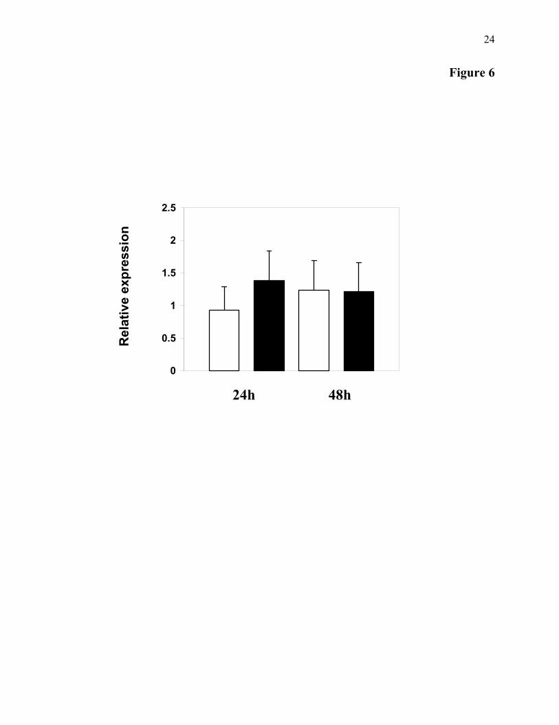
Following heat denaturation (70°C for 5 min), reverse transcription was performed using 1 µg of total RNA prepared with 50 ng/µl oligo-(dT)12-18 in a 50 µl reaction volume containing 1mM dNTPs, 1unit/µl of RnaseOUTTM (Invitrogen) and 200 units/µl M-MLV reverse transcriptase in reverse transcriptase buffer. PCR amplifications were performed with the LightCycler (Roche) in the presence of SYBR-Green (Master SYBR Green) with the following primers: Defm1 5’- GATGGTTTCTGCAGACATGG -3’ and Defm2 5’- CACAGTAGCCCGCTCTACAAC -3’ as sense and antisense primers, respectively. The gene encoding the elongation factor (EF, GenBank AB122066) was used as internal control. For EF the forward and reverse primers were EF (5’-ATGCACCAAGGCTGCACAGAAAG-3’) and EFR (5’-TCCGACGTATTTCTTTGCGATGT-3’), respectively. Samples were run under the following conditions: 95°C (10 min); and 39 cycles of 95°C (10 s), 62°C (5 s) and 72°C (15 s). For further expression level analysis, the crossing points (CP) were determined for each transcript using the LightCycler software. Specificity of RT-PCR product was analyzed on agarose gel and melting curve analysis. The copy ratio of each analyzed cDNA was determined as the mean of three replicates. The relative expression ratio of Cg-def was calculated based on the CP deviation of each RT-PCR product of RNA extracted from stimulated oyster versus the appropriate control sample, and expressed in comparison to the reference gene EF. The relative expression ratio of Cg-Def was calculated based on the delta-delta method for comparing relative expression results (28)
RESULTS
Cg-def cDNA Cloning and Primary Structure
Analyses–The complete cDNA of C. gigas defensin (Cg-Def) was obtained by 5’ amplification of cDNA ends-PCR on a C. gigas mantle edge cDNA library, as described in Materials and Methods (GenBank AJ565499). The oyster Cg-def cDNA contained 323 bp, comprising a coding region of 195-bp, a 110–bp 3’ untranslated region containing a polyadenylation consensus sequence (AATAAA) at position 66 to 72 of the 3’UTR and poly(A) tail. The 195-bp coding region encoded a 65-amino-acid propeptide (GenBank CAJ19280). The deduced amino acid sequence starts
with a 22-residue signal peptide and the cleavage site for signal peptidase is most likely located after the alanine residue preceding the glycine in position 23 as predicted by SignalP 3.0 software (data not shown). Cg-Def is not synthesized as a precursor with a C-terminal extension or a N-terminal pro-region as observed with the Mediterranean mussel or the Drosophila defensins, respectively. The amino acid sequence of the mature peptide was aligned with other defensins from the “arthropod defensin family” available in GenBank and Expasy, that contains defensins from arthropods, mollusks and fungi (Fig. 1A and 1B). As already observed for the mussel defensins MGD-1 and MGD-2, Cg-Def is an original member of this family due to the presence of two extra–cysteines residues. Cg-Def shares 50% of identity with MGD-1 from Mytilus galloprovincialis, but has a unique doublet of lysine residue at the C-terminal part of the molecule (Fig. 1A). Less identity was shared with defensin from the scorpion Androctonus australis (46%) and the tick Ixodes scapularis (44%). The selected members of the “arthropod defensin family” belonging to the so-called primitive defensin family show a high degree of conservation with both Cg-Def and the different mussel defensins (Fig. 1A) (29). Interestingly, very recently, a 40-amino acid AMP named plectasin (GenBank number CAI83768) with marked homologies to the arthropod and mollusk defensins has been isolated from the saprophytic fungus Pseudoplectania nigrella.
Genomic Organization and Analysis of the
Promoter Region–Cg-def gene (EMBL AM050547) harbours a unique intron whose splice junction follows the AG-GT rule (Fig. 1C). The genomic organization of Cg-def is similar to that of the mussel and scorpion defensin genes (29). It also displays the features of the other arthropod and mollusk defensin genes studied so far that demonstrate a genetic relatedness: (i) the exon encoding the mature defensin is never split by any intron, (ii) the intron flanking the exon encoding the mature defensin shows for all genes a strict level of phase conservation (phase 1) (29). Analysis of the 3.6 kb genomic DNA sequence upstream from the putative ATG start codon identified several consensus sequences for transcription factors commonly observed in promoters of cellular house keeping genes (Sp1), genes expressed during early embryogenesis (AP2, Gata 1, Gata 2), development (Zeste), cell cycle regulation (H1TF2, H4TF1 and 2), cell differentiation

7
(Gata 2, Ik1, Ik2, Ik3, IRF2, Oct1, 2, 4, 6 and 9) and genes expressed in a tissue specific manner (Pit-1, SRF).
Production, Folding and Purification of
Recombinant Cg-Def–In order to produce sufficient amounts of Cg-Def for detailed studies (5 mg scale) of its biological activity and its 3D structure, Cg-Def was expressed in E. coli Rosetta (DE3) pLysS cells transformed with the pET-28a/Cg-Def construct. Recombinant His6-tagged fusion protein was purified by affinity chromatography from the bacteria cell lysates. After chemical cleavage with CNBr and refolding, Cg-Def was purified by RP-HPLC, and its purety was judged by analytical RP-HPLC, MALDI-TOF and ESI-MS mass spectrometry analyses (Fig. 2, panels A and B). The oxidative folding of Cg-Def was assessed under two different conditions. First, after complete reduction with DTT, purified denatured Cg-Def was refolded during 48 h at room temperature in the presence of a Tris-HCl 100 mM pH 8 buffer. The refolding process was followed by RP-HPLC revealing at 48 h an additional more hydrophobic peak eluting around 35 min (Fig. 2, panel A, asterisk). By ESI-MS analyses, this hydrophobic fraction was found to contain a molecule with a molecular mass of 4,634.10 Da (see Fig. 2 panel B1). The value measured by ESI-MS is in perfect agreement with the calculated average molecular mass of 4,634.34 Da. This suggests that the recombinant Cg-Def has its eight cysteine residues paired. Alternatively, as described by Wu et al. (15), the use of DMF and urea was shown to enhance 5-times the folding efficiency of Cg-Def (data not shown). Using this protocol, the purified recombinant Cg-Def was also found to have a molecular mass measured by ESI-MS that is in perfect agreement to the calculated average molecular mass (se Fig.2, panel B1). The purified refolded peptide was then submitted to antimicrobial assays and NMR spectroscopy studies.
Antimicrobial Activity of Cg-Def–The
antimicrobial activity of the recombinant Cg-Def was determined against a panel of micro-organisms including Gram-positive, Gram-negative bacteria and filamentous fungi. The MIC values obtained are reported in Table 1. The peptide was active at very low concentration against most of the Gram-positive bacteria tested including Micrococcus lysodeikticus, Staphylococcus aureus and the marine bacteria Brevibacterium stationis and Microbacterium
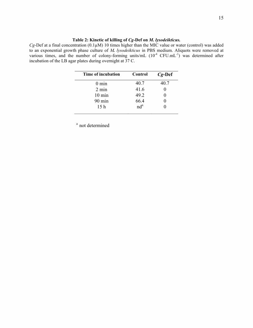
maritypicum. However, Cg-Def was not active at 20 µM against Bacillus megaterium. Cg-Def showed no activity at 20 µM against the Gram-negative bacteria Vibrio alginolyticus and Salmonella typhimurium. At higher concentration (35 µM), the peptide was active against E. coli. Cg-Def displayed anti-fungal activity against Fusarium oxysporum at relatively high concentrations (9 µM; Table 1). Experiments were conducted to examine the bactericidal effects of Cg-Def. The bactericidal activity of the peptide was assessed by plating cultures and the number of CFUs counted after overnight incubation at 30°C. Cg-Def exerted bactericidal effects against all the Gram-positive bacteria tested, excepted M. maritypicum (Table 1). When M. lysodeikticus was incubated with Cg-Def at concentration 10 times higher than the MIC value, all bacteria were killed in few minutes (Table 2).
In order to determine if the high salinity of the sea water might modify the efficacy of Cg-Def in vivo, the effect of the peptide on bacterial growth was tested in vitro at NaCl concentrations ranging from 85 mM to 1 M. Cg-Def is highly active against M. lysodeikticus and B. stationis even at 600 mM NaCl, a concentration closed to the value measured in sea water. Interestingly, the MIC value is unchanged throughout the NaCl concentration range tested [85 mM - 600 mM] (0.01 µM for M. lysodeikticus and 0.2 µM for B. stationis; Table 3). At 1 M NaCl, M. lysodeikticus did not grow in the control and a low bacterial growth can be observed for the marine bacteria B. stationis. In contrast to most AMPs, Cg-Def seems to conserve its antibacterial activity at such a high salt concentration (30, 31).
NMR Structural Study–The 2D NMR spectra
(TOCSY, DQF-COSY and NOESY) of Cg-Def were recorded at different temperatures, ranging from 17 to 32°C. The identification of all the spin systems of Cg-Def was obtained by analysis and comparison of DQF-COSY, TOCSY, and NOESY spectra according to the strategy described by Wüthrich (24). Nevertheless, we noticed that the 2D spectra showed, for each residue of the D38CNGK42 sequence, two spin systems approximately equally populated. This suggests a heterogeneity for this C-terminal part inferred to result from the partial deamidation to yield the Asp40 and isoAsp40 mixture. The AsnGly sequence (Asn40Gly41) has been shown to experience the highest propensity for the deamidation process through the succinimide intermediate (32). Indeed, such a

8
deamination process is known to occur and depend upon primary sequence, 3D structure, and solution properties such as pH, temperature, ionic strength, and buffer ions (33). The two spin systems were assigned to the initial molecule (Asn40) and to the Asp40 analogue. These two similar spin systems were identified by recording TOCSY spectra at several pH values in the 2-5 pH range to monitor the ionization state change of the carboxyl group. The spin system sensitive to the ionization state change was assigned to Asp40. The other one, unaffected in this pH range, was assigned to Asn40. Only a small amount of the third expected compound (isoAsp40) was formed. It was detected in the TOCSY experiment but not fully characterized in the NOESY.
Chemical shifts of the recombinant Cg-Def are reported in the Table S1 provided as “Supplementary Materials”. Both amide and alpha protons resonances were spread in a large range of chemical shifts and some atypical values were measured. When compared to statistical values, among the alpha proton resonances, those of Ala27 (1.87 ppm) and of Trp31 (3.17 ppm) were upfield shifted by around 2.5 and 1.5 ppm, respectively. The large spreading of amide signals is illustrated by the two extreme values measured for Ala27 (9.84 ppm) and for Ile18 (6.91 ppm). Altogether, the large spreadings of chemical shifts suggest a highly constrained structure, in agreement with the presence of the four disulfide bonds. This is also supported by several large non-equivalencies such as for the Gly3 and Gly23 alpha protons resonances for which non-equivalencies of 1.24 ppm and 1.29 ppm were measured, respectively. Similarly, non-equivalencies of 1.21 ppm and 1.02 ppm were also observed for ββ' signals of Cys4 and Cys11, respectively. The large difference of chemical shifts measured between the alpha (at 4.14 ppm) and the beta (at 3.08 ppm) protons of Thr35 residue and in a lesser extent for Thr37 (at 4.61 and 4.05 ppm) also suggests that they belong to highly constrained parts of the molecule. By comparison, chemical shifts of Thr29 (at 4.45 and 4.52 ppm) were very close to that usually observed for a less constrained sequence. The NOESY data showed several dNN and dαα NOEs suggesting that the molecule both contains a helical and a β-stranded parts. Moreover, the strong dαCys4-αPro5 NOE and the absence of the dαC4-δδ’P5 NOE unambiguously indicated a cis conformation for the Cys4-Pro5 amide bond. Among the amide protons found in slow exchange, eight of them were successive and span the 13-21 sequence. The amide protons of Ala22, Thr35,
Cys36 and Thr37 residues were also found in slow exchange.
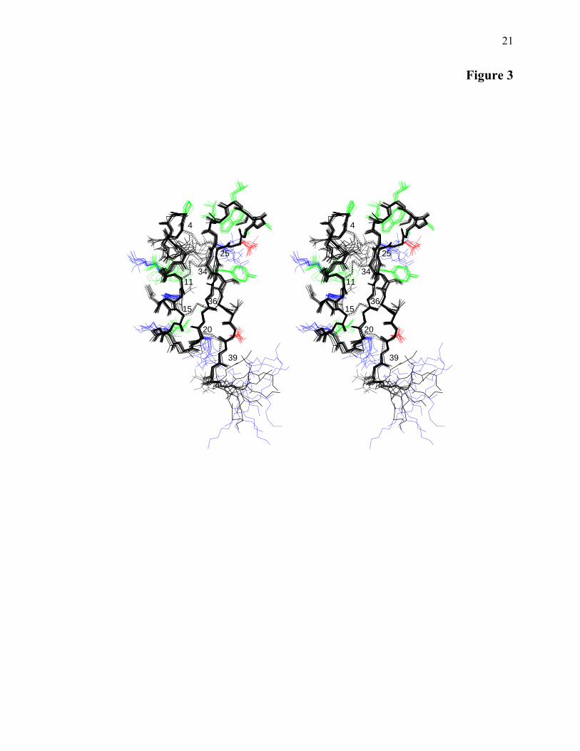
Solution Structure of Cg-Def–A stereoview of
a family of 10 Cg-Def conformers is reported in Fig. 3. The global structure of Cg-Def was well defined and displays the Cystine Stabilized-αβ motif (CS-αβ) that consists of an helical structure and two β strands cross linked by three disulfide bonds (34) and sometimes by a fourth disulfide bond (35-37). The backbone atoms of the ten conformers were overlaid for the well-defined regions spanning residues 4-39 resulting in a pairwise average rmsd of 0.43 ± 0.09 and 0.98 ± 0.16 Å for the backbone and heavy atoms, respectively. The Ramachandran plot of all residues (except for the glycine and proline residues) of the ten best conformers indicated that 89.7% were located in the most favoured and the additional allowed regions, and 5.8% in the generously allowed regions. A total of 4.4% of the residues were found in the disallowed regions. Limits of secondary structure elements indicated that the Cg-Def structure mainly consisted of an helical part (8-18 residues), two β-strands (residues 22-26 and 33-37), and three loops (residues 1-7, 19-21 and 27-32 for loops L1 L2 and L3, respectively) (Fig. 1B and 3). Three successive type IV turns were identified in L1 (1-4, 3-6, and 4-7) and L3 (27-30, 28-31 and, 29-32) loops, and a type I turn was found for the 26-29 sequence. It is interesting to note that the Cys4-Pro5 amide bond adopted the unusual cis conformation. Finally, the global fold is highly constrained by the 4-25, 11-34, 15-36, and 20-39 disulfide bonds. The Cg-Def helix exhibits an amphiphilic character with a hydrophobic side contributing to the hydrophobic core of the molecule and a hydrophilic side accessible to the solvent including mainly, Lys10, Asn13, His14, Lys16 and, Ser17 residues. Nevertheless, the Leu9 and Ile18 hydrophobic residues are solvent exposed. The hydrophobic core mainly includes the four disulfide bonds, the Ala22 side chain and is extended to the L1 (Pro5) and L3 hydrophobic loops (Ala27, Ala28, Leu30, Trp31, Leu32). As expected, the positively (Lys10, Lys16, Arg21, Arg33, Lys42, Lys43) and negatively charged (Asp26, Asp38) residues are solvent exposed.
The Cg-Def structure was compared to that of MGD-1 (PDB entry: 1FJN) whose 3D structure was previously determined (37). Their superimposition showed that the helical and β-strands elements were very conserved (Fig. 4).
9
Cg-def mRNA and Peptide Tissue
Localization–To investigate the tissue distribution of Cg-def expression, we analyzed by real-time RT-PCR mRNA content in various oyster tissues. Low or no Cg-def mRNA amount was measured in most of the organs analyzed including hemocytes, heart, digestive gland and gills whereas a high mRNA level, around 20 to 60 fold the level measured in the remaining animal tissues, was detected in mantle (Fig. 5). In non-stimulated C. gigas oysters, the mantle appears as the main tissue expressing Cg-def transcripts.
In order to demonstrate the presence of native Cg-Def in mantle, an acidic extract of this tissue was prepared, subjected first to RP-HPLC and then to antibacterial activity screening. Following solid-phase extraction of the acidified extracts from oyster mantle, RP-HPLC revealed the presence of two fractions with antimicrobial activity. These two fractions were subjected to molecular mass fingerprint analysis by MALDI-TOF-MS. In order to ascertain the molecular mass measured, the well-folded recombinant Cg-Def was used as external calibrant (see Fig. 2 panel B2). A difference of 3 Da was observed between the molecular mass measured by ESI-MS (4,634.10 Da, panel B1) and the one measured by MALDI-TOF-MS (4,637.96 Da, arrow in panel B2). Following solid-phase extraction of the acidified extracts from oyster mantle, RP-HPLC revealed the presence of two fractions with antimicrobial activity. The less hydrophobic fraction (see Fig. 2, panel C) contains a component with a molecular mass at 4637.96 corresponding to that measured by MALDI-TOF-MS for the recombinant folded Cg-Def. This peptide with antimicrobial activity against M. lysodeyticus might likely correspond to the native form of Cg-Def. Interestingly, the second active fraction (more hydrophobic), that has not yet been identified, showed a close but different molecular mass of 4631.45 Da (see Fig. 2, arrow in panel D).
Real-Time PCR Analyses of Cg-def transcript
levels after oyster bacterial challenges–In order to determine the expression pattern of Cg-def during bacterial challenge, two batches of oysters were selected. In the first one, oysters were stimulated by bath with killed bacteria (see Material and Methods) and in the second one, non-stimulated oysters were used as control. Then, qRT-PCR analyses were performed with total RNA extract from mantle collected at two times post challenge (24 and 48 h). No
striking discrepancies were observed for Cg-def expression between naive and bacteria challenged oysters at the different times analyzed (Fig. 6). These preliminary analyses revealed that Cg-def mRNA are present in naive C. gigas oyster and that, the level of transcript was unaffected by the bacterial-challenge performed.
DISCUSSION
Efficient host defence mechanisms are needed
to neutralize microbial invasions to which living organisms are exposed. In higher vertebrates, innate and adaptive immunity are present. In contrast, invertebrate and plant defence against pathogens takes place exclusively through mechanisms that are part of the innate immunity. The involvement of AMPs in natural resistance to infection is sustained by their strategic location in phagocytes, in body fluids and at the epithelial level, ie at interfaces between the organisms and its environment. In oyster, even if antimicrobial activities have been detected in the hemolymph of some species (38), no AMP has been fully characterized despite many attempts to purify such molecules by biochemical approaches from hemolymph and other tissues (13). In this report, and for the first time in an oyster, we describe the characterization of an AMP isolated from mantle tissue. Using molecular approaches, a new member of the widespread defensin family, which is present in animal and plant kingdoms, was identified. Usually, three disulfide bonds characterize this defensin family (Fig. 1A). The amino acid sequence of the mature C. gigas defensin displays interesting homologies with defensins from the “arthropod family” including the mussel’s defensins. Moreover, as observed with the defensins MGD-1, MGD-2 and MGD-2b from M. galloprovincialis, Cg-Def has four disulfide bonds. This additional bridge was proposed to render the peptide more stable in high osmolarity media such as in sea water (37). However, this fact is not common to all marine mollusk defensins because the mussel M. edulis defensins do not bear this additional disulfide bond (Fig. 1A) (11). Interestingly, Cg-Def possesses a N-terminal pre-segment presumed to be a signal sequence for translocation to the lumen of the rough endoplasmic reticulum, but has no C-terminal extension nor a N-terminal pro-region as observed in the mussel or the Drosophila defensins, respectively (39). This property would suggest a different way of processing for the oyster defensin.

10
To investigate its biological properties, Cg-Def was produced in E. coli and refolded in vitro. This recombinant approach was efficient, yielding enough material (approx. 1 mg of well-folded pure peptide per liter of culture) for functional assays and structural characterization. A challenging aspect for the production of Cg-Def in E. coli was the presence of eight cysteine residues in the mature peptide. Indeed, the correct refolding of proteins with several disulfide bonds is a general problem. As discussed by Harder et al., only few reports describe the production of AMP in bacteria, a fact that reflects the difficulty of expressing and folding such cysteine rich molecules in a bacterial host system (6). Previous reports described the production of human α- and β-defensins in E. coli (6, 40). We report here for the first time the efficient production in a bacterial-host of an AMP with four cysteine bridges that can be properly refolded in vitro. The activity spectrum of the recombinant Cg-Def was evaluated against a panel of bacteria and fungi. Consistent with studies on invertebrate defensins (2), Cg-Def was active in vitro against Gram-positive bacteria but showed no or limited activities against Gram-negative bacteria and fungi. Most interestingly, Cg-Def kept its antibacterial properties in the presence of high NaCl concentrations (up to 1M). This favours the hypothesis that Cg-Def retains its activity in in vivo conditions (sea water) and would play a role in the antimicrobial defence of the oyster C. gigas. For these reasons, Cg-Def represents a model of choice for structure-activity relationship studies to design a lead antibacterial drug to treat infections of bacteria in a salt-rich environment (41). This is particularly interesting for cystic fibrosis, for which it has been suggested that the primary genetic defect increases the salt content of fluid lining the airway, which reversibly inactivates antimicrobial molecules (42, 43).
As expected from the disulfide bridges array, the global fold of Cg-Def includes the CS-αβ motif observed in the defensins isolated from mussels (37), insects (34) and plants (44). The Cg-Def and MGD-1 sequences were aligned and their structures compared (Fig. 1B and 4). This alignment revealed 50 % of identity (23 residues with three gaps) including eight cysteines (45). Cg-Def and MGD-1 share the fourth disulfide bond that contributes to the stability and to the rigidity of their 3D structure. The identical residues were mainly gathered in four segments (residues 1-5, 14-18, 23-25 and 32-36). Beyond their sequence comparison the Cg-Def structure was compared to that of MGD-1 (PDB entry: 1FJN) (37). Their
superimposition showed that the helical and β-strand elements were rather conserved and the best fit was obtained for the backbone superimposition of the residues 1-4, 7-17, 21-25, 34-36 of Cg-Def with residues 1-4, 6-16, 21-25, 33-35 of MGD-1 (23 residues). A rmsd of 1.04 Å was measured for the backbone. Moreover, the two structures share the Cys4-Pro5 cis-amide bond and a similar disulfide bond pattern. Nevertheless, some differences were observed for the Cys20-Cys39 disulfide bond and for the length of the three loops. While the three equivalent disulfide bonds (4-25, 11-34, 15-36 for Cg-Def and 4-25, 10-33, 14-35 for MGD-1) well overlaid, the fourth one (Cys20-Cys39 for Cg-Def and Cys21-Cys38 for MGD-1) is significantly different mainly due to the translation of Cys20 by one residue towards the center of the molecule. This is certainly due to the shorter L2 loop (one residue less) joining the helix and the first β-strand. The consequence is a shift of this disulfide bridge that locates on the opposite side of the β-sheet ("above" in Cg-Def and "below" in MGD-1 as displayed in Fig. 4). Conversely, the L1 and L3 loops of Cg-Def display one additional residue. Whereas the sequences of the L1 loops including the Cys4-Pro5 cis amide bond are well conserved (Fig. 4), the sequences of the L3 loops are totally different. This latter is much more hydrophobic (A27ATLWL32 for Cg-Def) than that of MGD-1 (G27WHRL31) that contains two positively charged residues. Nevertheless, due to the positions in the sequence (at the end for Cg-Def and at the beginning for MGD-1), it appeared that, they have opposite location with regard to the loop. In contrast, the Leu32(Cg-Def)/Leu31(MGD-1) side chains were similarly located (Fig. 4B). Since it has been shown that the L3 loop of MGD-1 is responsible for a large part of the antibacterial activity (46), this significant structural difference is worth being noted.
It is commonly admitted that the surface distribution of hydrophobic and hydrophilic side chains in AMPs is essential for their antimicrobial activity. The alignment showed that several of them (Phe2, Pro5, Ile18, Tyr24, Leu32 and His14, Lys16, Arg33) were conserved (Fig. 1 and see below). However, the two antimicrobial sequences mainly differ by the presence of two aspartic residues (Asp26 and Asp38) and by the addition of a dibasic peptide (Lys42 and Lys43) at the C-terminus. Although, the Cg-Def and MGD-1 surfaces display significant differences mainly due to the two aspartic acids, the hydrophobic L3 loop and, by the "above"/"below" locations of the Cys20-Cys39/Cys21-Cys38 disulfide

11
bonds, several hydrophobic (Phe2/2, Pro5/5, Leu9/Tyr8, Ile18/17, Tyr24/24, Leu32/31 and charged residues (Lys10/Arg12, His14/13, Arg21/Lys15, Arg33/32, Lys42/Arg20, Lys43/Arg37) are similarly located at the surface giving rise to a comparable location of hydrophobic and hydrophilic clusters (Fig. 4B).
Our results showed that in non-stimulated animals, the mantle appears as the main tissue expressing Cg-Def. Although, we were not able to isolate by HPLC significant amount of pure defensin from the oyster, we clearly detected its presence by mass spectrometry analyses of mantle tissue (Fig. 2, panel C). The results presented here are in contrast with those reported for the mollusk, M. galloprovencialis. In the mussel, AMPs are only produced in hemocytes where they are stored and released following bacterial challenges (45). In the well-studied insect model, Drosophila melanogaster, AMPs are predominantly produced in the fat body (the functional equivalent of mammalian liver) and secreted into the blood in response to a microbial challenge, which characterizes the systemic response in insects. Otherwise, the expression of these peptides is also locally regulated in the surface epithelia of several Drosophila tissues (47), as also observed for the expression of peptides in mammalian epithelia. For example, Drosophila defensin is expressed in the
labellar glands and in the seminal receptacle and spermatheca (for a review see Imler and Bulet 2005 (48)). Additionally, in mammals, AMPs are constitutively produced by blood cells (49). Apart from insects, most of the AMPs reported in the different groups of invertebrates have been isolated from hemocytes (13). Our preliminary results would suggest that Cg-Def is continuously expressed in the oyster mantle, ie at epithelial surface, indicating that this AMP has an important function in host protection against environmental microbes. Indeed, in oyster, the mantle is a site of intense exposure to external environment that represents a dynamic ecosystem for numerous bacterial species, including commensals and potential pathogens. A more detailed study of Cg-def gene expression in response to various microbial challenges will be performed to gain insight into the role of Cg-Def in oyster immunity. In addition, AMPs isolation and characterization must be investigated from other oyster tissues. These data on AMPs are of great interest to understand how the oyster immune system interacts with the commensal flora and how it responds to environmental pathogens.
ACKNOWLEDGEMENTS
We are very grateful to Dr. A. J. Ouellette for excellent advices during defensin recombinant
expression. We also thank B. Romestand, J. de Lorgeril, G. Desserre and M. Leroy for helpful discussion and assistance.

12
REFERENCES
1. Yang, D., Biragyn, A., Kwak, L. W., and Oppenheim, J. J. (2002) Trends Immunol 23, 291-296 2. Bulet, P., Stocklin, R., and Menin, L. (2004) Immunol Rev 198, 169-184 3. Boman, H. G. (2003) J Intern Med 254, 197-215 4. Brogden, K. A. (2005) Nat Rev Microbiol 3, 238-250 5. Boulanger, N., Lowenberger, C., Volf, P., Ursic, R., Sigutova, L., Sabatier, L., Svobodova, M.,
Beverley, S. M., Spath, G., Brun, R., Pesson, B., and Bulet, P. (2004) Infect Immun 72, 7140-7146
6. Harder, J., Bartels, J., Christophers, E., and Schroder, J. M. (2001) J Biol Chem 276, 5707-5713 7. Izadpanah, A., and Gallo, R. L. (2005) J Am Acad Dermatol 52, 381-390; quiz 391-382 8. Raj, P. A., and Dentino, A. R. (2002) FEMS Microbiol Lett 206, 9-18 9. Sima, P., Trebichavsky, I., and Sigler, K. (2003) Folia Microbiol (Praha) 48, 123-137 10. Dimarcq, J.-L., Bulet, P., Hétru, C., and Fofmann, J. A. (1998) Biopolymers 47, 465-477 11. Charlet, M., Chernysh, S., Philippe, H., Hétru, C., Hoffmann, J. A., and Bulet, P. (1996) J. Biol.
Chem. 271, 21808-21813 12. Mitta, G., Vandenbulcke, F., and Roch, P. (2000) FEBS Letters 486, 185-190 13. Bachère, E., Gueguen, Y., Gonzalez, M., de Lorgeril, J., Garnier, J., and Romestand, B. (2004)
Immunol. Rev. 198, 149-168 14. Satchell, D. P., Sheynis, T., Kolusheva, S., Cummings, J., Vanderlick, T. K., Jelinek, R., Selsted,
M. E., and Ouellette, A. J. (2003) Peptides 24, 1795-1805 15. Wu, Z., Powell, R., and Lu, W. (2003) J Am Chem Soc 125, 2402-2403 16. Hetru, C., and Bulet, P. (1997) in Methods in Molecular Biology (Shafer, W. M., ed) Vol. 78, pp.
35-49, Humana Press Inc., Totowa, NJ 17. Rance, M., Sorensen, O. W., Bodenhausen, G., Wagner, G., Ernst, R. R., and Wuthrich, K. (1983)
Biochem. Biophys. Res. Commun. 117, 479-485. 18. Derome, A. E., and Williamson, M. P. (1990) Journal of Magnetic Resonance 88, 177-185 19. Bax, A., and Davis, G. D. (1985) Journal of Magnetic Resonance 65, 355-360 20. Rance, M. (1987) J. Magn. Reson. 74, 557-564 21. Macura, S., Huang, Y., Sutter, D., and Ernst, R. R. (1981) J. Magn. Reson. 43, 259-281 22. Marion, D., Ikura, M., Tschudin, R., and Bax, A. (1989) J. Magn. Reson. 85, 393-399 23. Piotto, M., Saudek, V., and Sklenar, V. (1992) J. Biomol. NMR 2, 661-665. 24. Wüthrich, K. (1986) NMR of Proteins and Nucleic Acids, John Wiley & sons, New York 25. Guntert, P., Mumenthaler, C., and Wuthrich, K. (1997) J Mol Biol 273, 283-298 26. Laskowski, R. A., Rullmannn, J. A., MacArthur, M. W., Kaptein, R., and Thornton, J. M. (1996)
J. Biomol. NMR 8, 477-486. 27. Frishman, D., and Argos, P. (1995) Proteins 23, 566-579 28. Livak, K. J., and Schmittgen, T. D. (2001) Methods 25, 402-408 29. Froy, O., and Gurevitz, M. (2003) Trends Genet 19, 684-687 30. Landon, C., Thouzeau, C., Labbe, H., Bulet, P., and Vovelle, F. (2004) J Biol Chem 279, 30433-
30439 31. Zaballos, A., Villares, R., Albar, J. P., Martinez, A. C., and Marquez, G. (2004) J Biol Chem 279,
12421-12426 32. Li, B., Gorman, E. M., Moore, K. D., Williams, T., Schowen, R. L., Topp, E. M., and Borchardt,
R. T. (2005) J Pharm Sci 94, 666-675 33. Robinson, N. E. (2002) Proc Natl Acad Sci U S A 99, 5283-5288 34. Cornet, B., Bonmatin, J. M., Hetru, C., Hoffmann, J. A., Ptak, M., and Vovelle, F. (1995)
Structure 3, 435-448 35. Savarin, P., Romi-Lebrun, R., Zinn-Justin, S., Lebrun, B., Nakajima, T., Gilquin, B., and Menez,
A. (1999) Protein Sci 8, 2672-2685

13
36. Guijarro, J. I., M'Barek, S., Gomez-Lagunas, F., Garnier, D., Rochat, H., Sabatier, J. M., Possani, L., and Delepierre, M. (2003) Protein Sci 12, 1844-1854
37. Yang, Y. S., Mitta, G., Chavanieu, A., Calas, B., Sanchez, J. F., Roch, P., and Aumelas, A. (2000) Biochemistry 39, 14436 - 14447
38. Anderson, R. S., and Beaven, A. E. (2001) Aquat Living Resour 14, 343-349 39. Mitta, G., Hubert, F., Dyrynda, E. A., Boudry, P., and Roch, P. (2000) Develop. Comp. Immunol.
24, 381-393 40. Ouellette, A. J., Satchell, D. P., Hsieh, M. M., Hagen, S. J., and Selsted, M. E. (2000) J Biol
Chem 275, 33969-33973 41. Friedrich, C., Scott, M. G., Karunaratne, N., Yan, H., and Hancock, R. E. (1999) Antimicrob
Agents Chemother 43, 1542-1548 42. Goldman, M. J., Anderson, G. M., Stolzenberg, E. D., Kari, U. P., Zasloff, M., and Wilson, J. M.
(1997) Cell 88, 553-560 43. Aarbiou, J., Rabe, K. F., and Hiemstra, P. S. (2002) Ann Med 34, 96-101 44. Da Silva, P., Jouvensal, L., Lamberty, M., Bulet, P., Caille, A., and Vovelle, F. (2003) Protein Sci
12, 438-446 45. Mitta, G., Vandenbulcke, F., Hubert, F., and Roch, P. (1999) J. Cell Sc. 112, 4233-4242 46. Romestand, B., Molina, F., Richard, V., Roch, P., and Granier, C. (2003) Eur J Biochem 270,
2805-2813 47. Tzou, P., Ohresser, S., Ferrandon, D., Capovilla, M., Reichhart, J. M., Lemaitre, B., Hoffmann, J.
A., and Imler, J. L. (2000) Immunity 13, 737 - 748 48. Imler, J. L., and Bulet, P. (2005) Chem Immunol Allergy 86, 1-21 49. Lehrer, R. I., and Ganz, T. (1999) Curr. Opin. Immunol. 11, 23-27 50. Casteels, P., Ampe, C., Jacobs, F., and Tempst, P. (1993) J. Biol. Chem. 268, 7044-7054 51. Mitta, G., Hubert, F., Nöel, T., and Roch, P. (1999) Eur. J. Biochem. 264, 1-9
FOOTNOTES # These authors contributed equally to this work. & Present address: Ecologie microbienne des insectes et interactions hôte-pathogène, UMR INRA Université de Montpellier II, 2 Place E. Bataillon, CC80, F-34095 Montpellier cedex 5, France. * Works in the author’ laboratories are supported by the Ifremer, the CNRS, The INSERM and the University of Montpellier II and Caen. This study was part of a collaborative project supported by the European Commission, DG XII, in the program International Co-operation with Developing Countries, INCO-DC, Contract n° ICA4-CT-2001-10023 (IMMUNAQUA). This study was also part of a French program MOREST funded by Ifremer, the Regions “Basse-Normandie, Bretagne, Pays de la Loire and Poitou-Charentes and by the “Conseil Général du Calvados”. Atheris Laboratories was supported by the Swiss Federal Office for Education and Science contract n° 02.0360. A. Herpin, C. Lelong and P. Favrel were financially supported by the Conseil Régional de Basse-Normandie, Agence de l’Eau “Seine Normandie” and FEDER N°4474 (program PROMESSE). The abbreviations used are: Cg-Def, Crassostrea gigas defensin; DG, distance geometry; DQF-COSY, 2D double-quantum filter correlation spectroscopy; MALDI-TOF-MS; matrix-assisted laser desorption/ionization time of flight mass spectrometry; ESI-MS, electrospray ionization mass spectrometry; NMR, nuclear magnetic resonance; NOE, nuclear Overhauser effect; NOESY, 2D nuclear Overhauser effect spectroscopy; RP-HPLC, reverse-phase high performance liquid chromatography; rmsd, root mean square deviation; TOCSY, total correlation spectroscopy; TPPI, time proportional phase incrementation; PAGE, polyacrylamide gel electrophoresis; qRT-PCR, quantitative reverse transcriptase-polymerase chain reaction, CFU, colony forming unit.

14
Table 1. Antimicrobial activity of recombinant Cg-Def. MIC values are expressed as the interval of concentration [a]-[b], where [a] is the highest concentration tested at which microbial growth can be observed and [b] is the lowest concentration that causes 100% growth inhibition (50).
Microorganisms MIC (µM) Bactericidal
effect of Cg-Def
Bacteria Gram-positive
Micrococcus lysodeikticus 0.005-0.01 Yes Staphylococcus aureus 1.25-2.5 Yes Brevibacterium stationis 0.1-0.2 Yes Microbacterium maritypicum 0.5-1 No Bacillus megaterium >20
Gram-negative Escherichia coli 363 35-20 nd a Vibrio alginolyticus >20 Salmonella typhimurium >20
Filamentous fungi
Fusarium oxysporum 9-4.5 Botrytis cinerea >20 Penicillium crustosum >20
a not determined
15
Table 2: Kinetic of killing of Cg-Def on M. lysodeikticus. Cg-Def at a final concentration (0.1µM) 10 times higher than the MIC value or water (control) was added to an exponential growth phase culture of M. lysodeikticus in PBS medium. Aliquots were removed at various times, and the number of colony-forming units/mL (10-4 CFU.mL-1) was determined after incubation of the LB agar plates during overnight at 37 C.
Time of incubation Control Cg-Def
0 min 40.7 40.7 2 min 41.6 0 10 min 49.2 0 90 min 66.4 0
15 h nda 0
a not determined

16
Table 3. Effect of sodium chloride on Cg-Def antimicrobial activity. MIC values were determined on the bacterial strains Micrococcus lysodeikticus and Brevibacterium stationis using liquid growth assay. In this table, only the lowest concentration of Cg-Def that causes 100% growth inhibition is indicated. Final OD600 of control M. lysodeikticus and B. stationis cultures without Cg-Def in the same corresponding medium is indicated in parentheses.
Final NaCl concentration
in poor broth
MIC in µM (OD600 after 24 h in the control
experiment without peptide) M. lysodeikticus B. stationis
85 mM a 0,01 (0,6) 0,2 (0,55)
300 mM 0,01 (0,4) 0,2 (0,5) 600 mM b 0,01 (0,15) 0,2 (0,35) 1000 mM - c 0,06 (0,1)
a Control poor broth. b NaCl concentration close to the value measured in sea water. c No bacterial growth in the control experiment

17
Figure legends Figure 1: Sequence alignment and genomic organization of representative of the defensin family AMP. (A) Amino acid sequence alignment of defensins from arthropods, mollusks and a fungus. Oyster Crassostrea gigas (Cg-Def); mussels Mytilus edulis (DEFA-MYTED, [P81610], DEFB-MYTED, [P8161]) and M. galloprovincialis (MGD-1, [P80571]; MGD-2, [AAD52660]); tick Dermacentor variabilis (varisin, VSNA1, [AAO24323]), Ornithodoros moubata (Om-DEF-C, [AB041814]), Haemaphysalis longicornis (HlDfs, [BAD93183]), Boophilus microplus (BOMICR, [AAO48943]) and Ixodes scapularis (IXSCA, scapularisin, [AAV74387]); dragonfly Aeschna cyanea (DEFI-AESCY, [P80154]); scorpion Androctonus australis (DEF4-ANDAU, [P56686]) and Leiurus quinquestriatus (DEF4-LEIQH, [P41965]) and the saprophytic fungus Pseudoplectania nigrella (plectasin, [CAI83768]). The nomenclature derived from the SwissProt database and the accession numbers are in brackets. Identical residues are shaded. The two additional cysteine residues found in the Cg-Def, MGD-1 and MGD-2 are in bold. (B) Cg-Def and MGD-1 sequence alignment manually modified according to the structure comparison (this work). The similar elements of structure are grey shaded and reported above. Conserved residues are in bold. Notice the shift for one cysteine (C20/C21) out of the eight. (C) Organization of Cg-def gene and the related genes coding MGD-2 and myticin A from the mussel Mytilus galloprovincialis. Boxes represent the coding region; the signal sequence appears in black and the mature peptide in white. Grey lines represent the untranslated regions of the exons. Potential sites of cleavage (mono [R] or dibasic [RR] and [KK]) involved in the splitting of the mature peptide (white) from the C-terminal peptide spotted) are indicated. Accession numbers: Cg-Def: CAJ19280, Cg-Def genomic sequence AJ582630. Gene organization of MGD-2b is from Mitta et al. (39) and incomplete structure of Myticin A (dotted box) is deduced from Mitta et al. (51) Figure 2: Quality control of recombinant Crassostrea gigas defensin (Cg-Def) and detection of the natural form in acidic extracts of mantle tissue. (A) Following reduction, refolding of the recombinant peptide was performed at room temperature in the presence of Tris-HCl buffer. The refolding was monitored by RP-HPLC using an acetonitrile gradient in acidified water (for details see Materials and Methods). The grey and black lines correspond to the linear and to the refolded (see *) recombinant peptides, respectively. The molecular mass of the refolded peptide was measured by ESI-MS (B1) and MALDI-TOF-MS (B2). The molecular mass (average value) measured at 4,634.10 Da is in perfect agreement with the calculated average molecular mass at 4,634.34 Da. A difference of 3 Da was observed between the molecular masses measured using MALDI-TOF-MS (m/z 4638.46, see B2) versus the one detected in ESI-MS at 4,634.10 Da (m/z 4,635.10). (C, D) Following purification by RP-HPLC of an acidic extract of mantle tissue, the two fractions active against the tested microorganisms were analyzed by MALDI-TOF-MS. The first fraction (see the arrow head in panel C) eluting at the same retention time as the recombinant-refolded Cg-Def contains ions of m/z 4,638.96 corresponding to the one measured by MALDI-MS for the recombinant peptide. In the second bioactive fraction (see the arrow head in panel D), ions of m/z 4,631.45 were detected. Figure 3: Structure of Cg-Def. Stereo-view of the ten best conformers of Cg-Def. The 4-39 heavy atoms of the backbone were used for the superimposition. The mean pairwise rmsd is of 0.43 ± 0.09 Å. The four disulfide bridges are labeled and displayed as dashed lines. Hydrophobic, positively and negatively charged side chains are colored in green, blue and red, respectively. Figure 4: Structure comparison of Cg-Def and MGD-1. (A) Global fold comparison of the Cg-Def (left) and MGD-1 (right) structures. The structures prepared using MOLSCRIPT show the arrangement of the four disulfide bonds (in yellow). (B) - With a similar orientation, comparison of their Van der Waals surface showing the location of the hydrophobic (green) and positively charged (blue) residues. For this orientation, the W31 side chain of Cg-Def is hidden. Most of the labeled residues are conserved in the two peptides. Aspartic and cysteine residues are in red and yellow, respectively.

18
Figure 5: Tissue expression of Cg-def mRNA analyzed by real time quantitative RT-PCR. Each value is the mean of four pools of four animals. Hc : Hemocytes ; M : Mantle ; PAM : Posterior Adductor Muscle ; DG : Digestive Gland ; G : Gills ; H : Heart ; LP : Labial Palps. ND: Not detected. Bars represent the relative Cg-def transcript levels normalized to GAPDH transcript levels. Figure 6: Cg-def mRNA expression in oyster mantle following a bacterial challenge. Results are means ± SE of three independent experiments realized on a pool of ten oysters from non-stimulated (white) and stimulated (black) oysters, at 24 and 48 h post-infection. Bars represent the relative Cg-def transcript levels normalized to elongation factor transcript levels.

19
Figure 1 A Cg-DEF GFGC--PGNQLK--CNNHCKSIS--CRAGYCDAATLWLRCTCTDCNGKK MGD-1 GFGC--PNN-YQ--CHRHCKSIPGRC-GGYCGGWHR-LRCTCYRCG MGD-2 GFGC--PNN-YA--CHQHCKSIRGYC-GGYCAGWFR-LRCTCYRCG DEFA-MYTED GFGC--PND-YP--CHRHCKSIPGRX-GGYCGGXHR-LRCTCYR DEFB-MYTED GFGC--PND-YP--CHRHCKSIPGRY-GGYCGGXHR-LRCTC DEFI-AESCY GFGC--PLDQMQ--CHRHCQTITGRS-GGYCSGPLK-LTCTCYR VSNA1 GFGC--PLNQGA--CHNHCRSIRRR-—GGYCSGIIK-QTCTCYRN DEF4-ANDAU GFGC--PFNQGA--CHRHCRSIRRR--GGYCAGLFK-QTCTCYR DEF4-LEIQH GFGC--PLNQGA--CHRHCRSIRRR--GGYCAGFFK-QTCTCYRN Om-DEF-C GYGC--PFNQYQ--CHSHCSGIRGYK-GGYCKGLFK-QTCNCY HlDfs GFGC--PLNQGA--CHNHCRSIGRR--GGYCAGIIK-QTCTCYRK BOMICR GFGC--PFNQGA--CHRHCRSIRRR--GGYCAGLIK-QTCTCYRN Scapularisin GFGC--PFDQGA--CHRHCQSIGRR--GGYCAGFIK-QTCTCYHN Plectasin GFGCNGPWDEDDMQCHNHCKSIKGYK-GGYCAKGGFV--CKCY B Helix Beta I Beta II 1 10 20 30 40
Cg-Def GFGCPGNQLKCNNHCKSIS-CRAGYCDAATLWLRCTCTDCNGKK MGD-1 GFGCPNN-YQCHRHCKSIPGRCGGYCGGWHR-LRCTCYRCG--- 1 10 20 30
C 5’ UTR Exon 1 Intron Exon 2 3’UTR
RR
Myticin A
MGD-2
Cg-def

20
800 1000 1200 1400 1600m/z0
100
%
A: 4634.10±0.31B: 4672.06±0.53C: 4431.10±1.33
A5927.84
A6773.31
C6739.35
C5886.91
782.30
A41159.54
B5935.32 A3
1545.71B4
1168.97
B1A
Figu
re 2
(
re)
blac
k an
d w
hite
figu
Abs
orbe
ncy
at 2
25 n
m
B2 *
r.i.
Retention time (min) m/z
C D
r.i.
r.i.
m/z m/z
21
Figure 3
4
11
15
20
25
34
36
39
4
11
15
20
25
34
36
39

22
Figure 4 A
C
N
39
20
36
25
15
11
34
4
38
21
3514
10
25
33
4
C
N
L1L1
L2
L2
L3
L3
Cg-Def MGD-1
B

23
Figure 5
70.00
ND
Rel
ativ
e Cg-
Def
tran
scrip
t lev
el
60.00
50.00
40.00
30.00
20.00
10.00
0.00 M H Hc DG GPAM LP
24
Figure 6
0
0.5
1
1.5
2
2.5
Rel
ativ
e ex
pres
sion
24h 48h
Copyright © 2022 FDOKUMEN