Bacterial depuration by the American oyster (Crassostrea ...
Upload
independentCategory
view
1download
0
Cod
HJU
a
ARR2A
KMDGGEC
1
iplwctMmw
olf
0h
Aquatic Toxicology 147 (2014) 48– 56
Contents lists available at ScienceDirect
Aquatic Toxicology
jou rn al hom epage: www.elsev ier .com/ locate /aquatox
omparative responses of sperm cells and embryos of Pacificyster (Crassostrea gigas) to exposure to metolachlor and itsegradation products
uong Mai, Patrice Gonzalez, Patrick Pardon, Nathalie Tapie, Hélène Budzinski,érôme Cachot, Bénédicte Morin ∗
niv. Bordeaux, EPOC, UMR 5805, F-33400 Talence, France
r t i c l e i n f o
rticle history:eceived 30 July 2013eceived in revised form6 November 2013ccepted 30 November 2013
eywords:etolachloregradation productsene transcriptionenotoxicitymbryotoxicity
a b s t r a c t
Metolachlor is one of the most intensively used chloroacetanilide herbicides in agriculture. Consequently,it has been frequently detected in coastal waters as well as its major degradation products, metolachlorethane sulfonic acid (MESA) and metolachlor oxanilic acid (MOA) which are encountered at higher con-centrations than metolachlor. Although a few studies of metolachlor toxicity have been conducted onmarine organisms, little is known about the environmental toxicity of metolachlor degradation products.In this study, the deleterious effects of metolachlor and its degradation products on spermatozoa andembryos of Crassostrea gigas have been compared using biomarkers of developmental defects, DNA dam-age and gene transcription levels. After 24 h exposure, significant increases in the percentage of abnormalD-larvae and DNA damage were observed from 0.01 �g L−1 for S-metolachlor and 0.1 �g L−1 for MESAand MOA. Results showed that S-metolachlor was more embryotoxic and genotoxic than its degrada-tion products. Oyster sperm was also very sensitive to metolachlor exposure and followed the pattern:
−1 −1 −1
rassostrea gigas metolachlor (0.01 �g L ) > MOA (0.1 �g L ) > MESA (1 �g L ). Metolachlor and MESA mainly triggeredvariations in the transcription level of genes encoding proteins involved in oxidative stress responses(mitochondrial superoxide dismutase and catalase). Overall, no significant variation in transcription lev-els could be detected in C. gigas embryos exposed to MOA. This study demonstrates that metolachlorand its main degradation products have the potential to impact several steps of oyster development andtherefore recruitment in coastal areas exposed to chronic inputs of pesticides.. Introduction
Most herbicides applied in agriculture are transformed by phys-cal, chemical and biological processes into one or more metaboliteroducts. The application of herbicides by distribution over the
and crop introduces them into the environment including groundater, surface water and sediment. Among chloroacetanilide herbi-
ides, metolachlor is one of the most important pesticides appliedo corn and other crops for controlling broadleaf and grass weeds.
etolachlor is ranked as intermediate in terms of environmentalobility and persistency with a half life degradation in soil of 6–10eeks (Hostetler and Thurman, 2000).
Metolachlor ethane sulfonic acid (MESA) and metolachlor
xanilic acid (MOA) are the major degradation products of meto-achlor. A study of degradation products in tile drain dischargerom agricultural fields in central New York indicated that MESA∗ Corresponding author. Tel.: +33 0540 002 256; fax: +33 0540 008 719.E-mail address: [email protected] (B. Morin).
166-445X/$ – see front matter © 2013 Elsevier B.V. All rights reserved.ttp://dx.doi.org/10.1016/j.aquatox.2013.11.024
© 2013 Elsevier B.V. All rights reserved.
and MOA can persist in agricultural soils for 3 or more years afterapplication (Phillips et al., 1999). Previous studies showed thatmetolachlor degradation products were generally present 3–45times more frequently than the parent compound (Boyd, 2000;Kalkhoff et al., 1998; Phillips et al., 1999; Rebich et al., 2004). Forexample, study of 355 water samples from 12 stream sites in East-ern Iowa found MESA in 99.7% of samples, MOA in 94.3% of samplesand the parent compound in 54.1% of samples (Kalkhoff et al., 1998).Recently, Rebich et al. (2004) also reported that MESA was detectedin all sampling sites of the Mississippi River Basin and more fre-quently than its parent compound, whereas MOA was found in 89%of the samples. Concentrations of metolachlor from ppt (ng L−1) tosub-ppm (mg L−1) were frequently found in surface and ground-water surveys throughout North America (Kolpin et al., 2002). Forinstance, in surface water in the Mid-Western US in 2003, aver-age concentrations of MESA and MOA were 1.55 and 0.73 �g L−1,
respectively (Battaglin et al., 2003).The widespread occurrence of metolachlor and its degrada-tion products in aquatic systems could represent a threat foraquatic species. Indeed, small number of studies showed acute
Toxico
t((ietPoaoaeslpao
bobaabdtptat2rtbGtatt(mw1clp1cg(
2
2
(Clac
Fotfi
H. Mai et al. / Aquatic
oxicity in the crustacean Daphnia magna and fish Oncorhynchus sp.Wan et al., 2006) or reduced activities of detoxification enzymescytochrome P450 O-deethylation and glutathione S-transferases)n metolachlor treated midge larvae, Chironomus tentans (Jin-Clarkt al., 2008). We previously demonstrated adverse effects on fer-ilization success, offspring development, and DNA integrity in theacific oyster (Mai et al., 2012, 2013). However, the toxic effectsf the major degradation products of metolachlor, on non-targetquatic organisms, remain to be characterized. Therefore, the aimf the present study is to compare the adverse effects of metolachlornd its main degradation products MESA and MOA following a 24 h-xposure of Pacific oysters at their embryonic stage. The early lifetages of Pacific oyster Crassostrea gigas have been selected as a bio-ogical model because of their high sensitivity to a large range ofollutants (Geffard et al., 2002; His et al., 1999a; Wessel et al., 2007)nd year-around availability of fertilized eggs from adult breedingysters.
Over time, it has become increasingly clear that a singleiomarker is not able to determine the health status of a livingrganism. A multi-biomarker approach has been reported as theest tool for identifying the effects and the mechanisms of pollut-nt toxicity to different biological levels of organization (Adamsnd Greeley, 2000; Faria et al., 2010; Viarengo et al., 2007). A com-ination of bioassays (embryo-larval test) and biomarkers (DNAamage and gene transcription analyses) were used in this studyo investigate the toxic effects of metolachlor and its degradationroducts on the early life stages of Pacific oyster C. gigas. Embryo-oxicity and genotoxicity by means of the comet assay can be useds screening methods due to their simplicity and wide applica-ion to any eukaryotic organisms (His et al., 1999b; Orieux et al.,011). In recent years, real-time quantitative polymerase chaineaction (RT-qPCR) has been considered one of the most sensi-ive techniques in detecting changes in gene transcription inducedy environmental contaminants in aquatic systems (Neumann andalvez, 2002). Therefore, RT-qPCR was used to analyze transcrip-
ion levels of a panel of 11 genes involved in xenobiotic metabolism,ntioxidant defense, mitochondrial metabolism, cell cycle regula-ion and apoptosis. The response to oxidative stress was studiedhrough mitochondrial superoxide dismutase (sodmt), catalasecat), glutathion peroxidase (gpx) and metallothionein isoformst1 and mt2 gene transcription. The mitochondrial metabolismas investigated by analysis the cytochrome C oxidase subunit
(cox1) transcript levels. The quantity of mitochondria in theells was estimated using mitochondrial 12S ribosomal transcriptevels. Activation of metolachlor and its degradation productshase I and II metabolism was studied using cytochrome P450A (cyp1A) transcripts and glutathion S-transferase (gst) trans-ripts. Metolachlor-induced apoptosis was studied through p53ene transcription levels. Finally, multixenobiotic resistance genemxr) involved in cell detoxication was also investigated.
. Materials and methods
.1. Chemicals and seawater
Reference toxicants, including S-metolachlor, MESA and MOAFig. 1), and 37% formalin were purchased from Sigma–Aldrichhemical (St. Quentin Fallavier, France). Dispase II, Triton X-100,
ow melting point (LMP) agarose, normal melting point (NMP)garose, and MEM-alpha (Minimum Essential Medium) were pur-hased from Gibco (Invitrogen, Cergy Pontoise, France).
Seawater was collected from Arguin in Arcachon Bay (SW
rance), an area which has a naturally reproducing population ofysters and has been frequently used in the laboratory for eco-oxicological tests. Immediately following sampling, seawater wasltered using a 0.2 �m-pore membrane filter to eliminate debrislogy 147 (2014) 48– 56 49
and microorganisms. Filtered seawater (FSW) was stocked at 4 ◦Cin the dark and was used within 3 days. A few hours before theexperiment, FSW was filtered again at 0.2 �m.
2.2. Animals
Mature oysters (C. gigas, Thunberg, 1793) came from a commer-cial hatchery specialized in the year-round production of matureoysters (Guernsey Sea Farms, UK). Oysters were kept at around10 ◦C for transportation and kept in FSW for 1 h before the startof the experiment. All oysters were used within 3 days.
2.3. Pesticide solutions
Depending on the assays, three to four replicates were per-formed for each condition. Experimental concentrations werechosen on the basis of previous results (Mai et al., 2012).
Due to their high solubility in water (e.g. 530 mg L−1 formetolachlor), stock solutions (100 mg L−1) of metolachlor and itsdegradation products (metolachlor ESA and metolachlor OA) wereprepared in Milli-Q water. Working solutions of each pesticide wereprepared by diluting the stock solution in FSW. The negative controlfor all experiments was FSW.
2.4. Pesticide analysis
All contamination solutions were controlled in concentration aswell as reference seawater used to prepare the test solutions. Meto-lachlor and its degradation products were extracted via solid-phaseextraction (SPE), using Oasis HLB cartridges (3 cc, 60 mg) and ana-lyzed by LC/MS/MS. The analytical procedure was adapted fromAlder et al. (2006). The cartridges were conditioned successivelywith 3 mL of MeOH and 3 mL of acidified water (pH 2). Water sam-ples were acidified (HCl, pH 2), and spiked with internal standards(carbofuran d3) before percolation under vacuum. The cartridgeswere dried for 30 min by application of a gentle vacuum. Finally, theanalytes were eluted with 3 mL of MeOH and concentrated using anitrogen stream evaporator. 200 �L of each individual solution wasmixed with 200 �L of mixture of internal standards in methanol(nominal concentrations 1 �g g−1) and were directly analyzed byLC/MS/MS to determine response factors.
Analyses were performed using a HPLC/MS/MS systemfrom Agilent Technologies (HPLC 1290 system coupled to6460 mass spectrometer) (mobile phase: water (5 mM ammo-nium acetate + 0.1% acetic acid)/methanol (100%/0%–0%/100%within 17 min at 0.5 mL/min); column: Kinetex C18100 mm × 2.1 mm × 1.7 mm; ionization mode: ESI+). Proceduralblanks were performed to ensure the absence of laboratory-contamination. Recoveries and reproducibility were determinedusing spiked water samples (at a nominal concentration of100 ng L−1) processed at the same time as the samples to becharacterized and were the following for respectively metolachlor,MESA and MOA: 105 ± 10% (n = 10), 90 ± 15% (n = 10), 102 ± 12%(n = 10). Detection limits were 0.1 ng L−1 for metolachlor and1 ng L−1 for MOA and MESA in water sample.
2.5. Embryotoxicity assay
Embryotoxicity tests were performed in this study with meto-lachlor degradation products. Data on metalochlor embryotoxicityhas been previously published (Mai et al., 2012) but a new exper-iment with metolachlor was repeated in this study to compare
toxicity with metolachlor degradation products within the sameexperiment. The embryotoxicity bioassay has been described indetail by His et al. (1997), Quiniou et al. (2005) and recently by Maiet al. (2012, 2013). Briefly, after fertilization, oocytes (500 oocytes)
50 H. Mai et al. / Aquatic Toxicology 147 (2014) 48– 56
etolac
wwciaAitlmQ
2
debtidwrta(
l(oeaa
MsalE
(oaeDn
2
dd
Fig. 1. The structure of m
ere exposed in 24-well microplate (Greiner Bio-One, polystyreneith physical treatment surface) containing 1.8 mL of the toxi-
ant solution. These microplates were incubated at 24 ◦C for 24 hn the dark. After incubation, 50 �L of 1% buffered formalin weredded and the percentage of abnormal oyster larvae was recorded.
hundred individuals per well were directly observed under annverted microscope (Olympus, magnification ×200) to determinehe number of abnormal D shaped larvae. The abnormalities (Darvae presenting mantle and/or shell abnormalities) were deter-
ined according to the criteria described in His et al. (1999b) anduiniou et al. (2005).
.6. Comet assay
Comet assay was performed in this study with metolachloregradation products. Data on the DNA damage of oyster embryosxposed to metalochlor was previously published (Mai et al., 2012),ut a comet assay was repeated here with metolachlor to ensurehe same exposure conditions for all contaminants. Embryos werencubated in 250 mL beakers (PyrexTM glass) for 16 h at 24 ◦C in theark. This exposure period allows the recovery of unshelled larvaehich were enzymatically dissociated for the comet assay. Three
eplicates were performed per condition and each replicate con-ained a total of 1,000,000 oyster larvae. Cell isolation and the cometssay on oyster embryos were performed as previously describedMai et al., 2012).
Sperm cells (150 �L ≈ 1.5 × 10⁶ sperm cells per mL) were col-ected from male oysters induced to spawn by thermal stimulationalternating immersion in seawater at 18 ◦C and 28 ◦C for 30 min)r by stripping the gonad (Mai et al., 2012) and were immediatelyxposed to 5 mL contaminant solutions in 15 mL tubes for 30 mint 24 ◦C in the dark. Three replicates were performed per conditionnd each replicate contains a total of 525,000 sperm cells.
The comet assay was performed on sperm cells as described byorin et al. (2011) with slight modifications. 50 �L of cell suspen-
ion (about 120 × 103 cells) was added to 100 �L of 1% LMP agarosend two agarose gels of 50 �L were laid on a pre-coated slide. Alka-ine treatment was performed for 20 min to allow DNA unwinding.lectrophoresis was carried out at 25 V, 300 mA for 20 min.
The slides were stained with 20 �L of ethidium bromide20 �g/mL) and were analyzed at 400× magnification using anptical fluorescence microscope (Olympus BX 51) and an imagenalysis system (Komet 5.5, Kinetic Imaging Ltd.). DNA damage wasxpressed as percentage of “Tail DNA”, i.e. the percentage of totalNA that has migrated from the head. A hundred randomly selecteducleoids were analyzed on two replicate gels.
.7. Gene expression analysis
After oyster embryos exposure to metolachlor and its degra-ation products for 24 h at 24 ◦C, the density of larvae wasetermined using a Malassez’s counting cell. Three replicates for
hlor and its metabolites.
each contamination condition were performed and each replicatecontained a total of 35,000 oyster larvae. Larvae solutions werethen concentrated for RNA extraction, by centrifugation at 4000 × gfor 10 min at 4 ◦C. The pelleted larvae were resuspended in 500 �Lof “RNA later” buffer (Quiagen). Those samples were then storedat −80 ◦C until required.
2.7.1. Extraction of RNATotal RNAs were extracted using the “Absolutely RNA®
Miniprep” Kit (Strategene, Agilent) according to manufacturer’sinstructions. The quality of all RNA extraction was evaluated byelectrophoresis on a 1% agarose-formaldehyde gel, and their con-centration determined by spectrophotometry.
2.7.2. cDNA synthesisFirst-strand cDNA was synthesized from total RNAs (3 �g)
using the “AffinityScriptTM Multiple Temperature DNAc synthe-sis” kit (Agilent, Stratagene). In each of the above three tests,the techniques were carried out according to the manufacturer’sinstructions. The cDNA mixture was stored at −20 ◦C, until required.
2.7.3. Real-time PCRAfter extraction and reverse transcription, real-time PCR reac-
tions were performed with an Mx3000P (Stratagene) followingthe manufacturer’s instructions. Primer sequences for all of theeleven studied genes and the housekeeping gene are reported inTable 2. Real-time PCR was performed in a total volume of 20 �Lwith 1 �L cDNA, 1 �L of reverse and forward primers (200 �M,each), 7 �L of distilled water and 10 �L of GoTaq® qPCR MasterMix (Promega). The amplification program consisted of one cycleat 95 ◦C for 10 min followed by 40 amplification cycles at 95 ◦C for30 s, 55 ◦C for 30 s and 72 ◦C for 30 s. PCR specificity was determinedfor each reaction from the dissociation curve of each PCR prod-uct. The cycle threshold (CT) value corresponded to the number ofcycles at which the fluorescence emission monitored in real-timeexceeded the threshold limit. Transcription levels of the selectedgenes were normalized according to the expression of the house-keeping gene ˇ-actin which was observed to be expressed at thesame level in our experimental conditions. Relative expression ofa gene was calculated using the 2−�CT method as described byLivak and Schmittgen (2001) where �CT represents the differencebetween the cycle threshold of a specific gene and the cycle thresh-old of the ˇ-actin gene. Therefore, the Induction factor (IF) of eachgene compared with control corresponds to the following equation:IF = 2−�CT (Treatment)/2−�CT (Control).
2.8. Statistical analysis
All data are expressed as means ± standard error (S.E). Statis-tical software SPSS (16.0) was used for data analysis. Normalityof the data distribution was tested on data residues using theShapiro–Wilk test (p < 0.01). Homogeneity of variance was checked

H. Mai et al. / Aquatic Toxicology 147 (2014) 48– 56 51
Table 1Measured concentrations of metolachlor and its metabolites in working solutions (ng L−1).
Nominal RSWa 1 10 100 1000 10,000
Metolachlor 8 11 24 134 1110 8056Metolachlor ESA 26 22 30 139 998 7479Metolachlor OA 60 56 86 190 1290 12,653
a Reference seawater (RSW) was collected from Arguin site of Arcachon Bay in May 2012.
Table 2Nucleotide sequences of primers used in real-time PCR analysis of C. gigas.
Gene Sequence 5′–3′ Function Accession number(EMBL or GenBank)
ˇ-actin AGTACCCCATTGAACACGGa
TGGCGGGAGCGTTGAAbCytoskeletal gene (housekeeping gene) AF026063
p53 CCCTCAAAGCAGTCCCCAa
TGTAGCGATCCACCTGATTbCell cycle arrest/apoptosis AM236465.2
12S CTCAGTCTTGCGGGAGGa
GGTTATGCGGAACCGCCbMitochondrial metabolism EF484875
coxI GTGCCAACTGGTATTAAGGTGTa
ACACCGCACCCATTGATbMitochondrial metabolism AB033687
sodmt ACAAAGTCAATCAGTGCCCTa
CCATTGCCTCTGCCAGTbMitochondrial oxidative stress EU420128
cat GTCGTGCCCCTTTACAACCa
CGCCCGTCCGAAGTTTbOxidative stress EF687775.1
gpx ATCGAACGCTGCACCAa
AGCTCCGTCGCATTGTbOxidative stress EF692639
mt1 TGTCTGCTCTGATTCGTGTCCAGCa
GGTCCTTTGTTACACGCACTCATTTbDetoxification AJ242657
mt2 TCCGGATGTGGCTGCAAAGTCAAGa
GGTCCTTTGTTACACGCACTCATTTbDetoxification AJ297818
mxr AGGAAGGGCAGTTGAGTGa
CGTTGGCCTCCTTAGCGbDetoxification AJ422120
gst AGGCTACCGAAATGGCTGa
CTCTGACTTGTAATAGGCCGCbBiotransformation AJ557140
cyp1A AGGCATAGGGCT. . .ACAa
CTGGTTTCGCGGGTTTCATbBiotransformation EF645271
bepapeutTot
3
3
dtl1dwtctctar
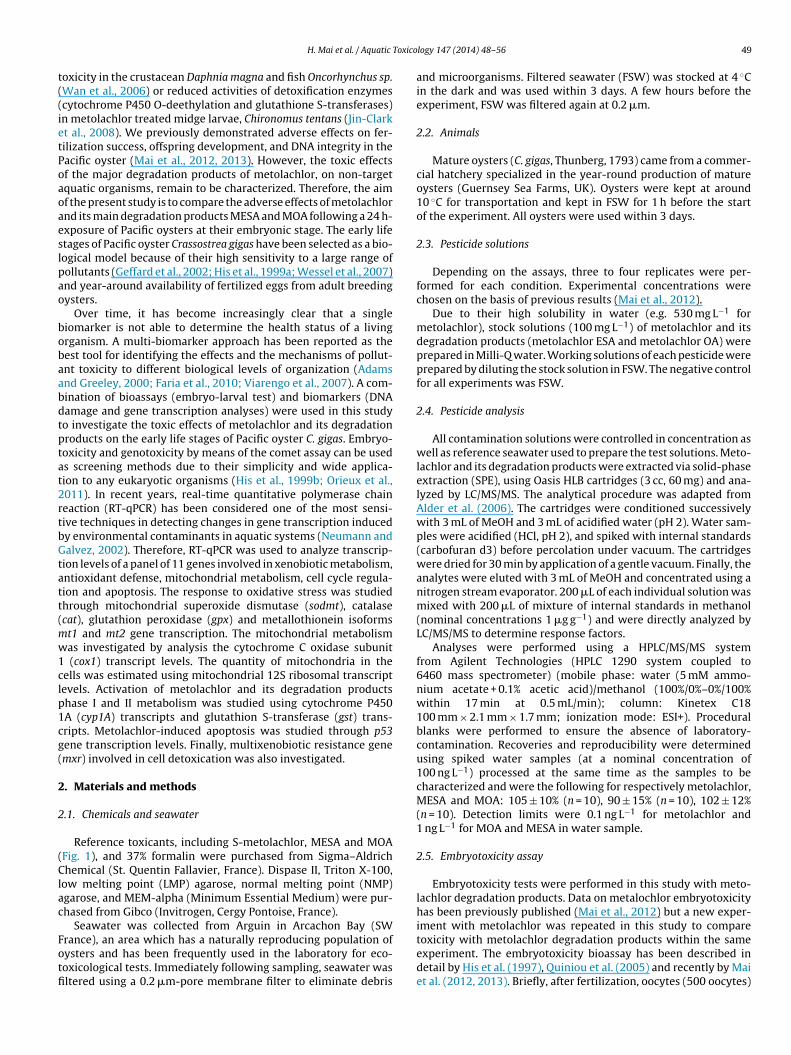
lachlor degradation products (MESA and MOA). Compared with thecontrol group, the percentage of abnormal D-larvae significantlyincreased from 0.1 �g L−1 of MESA or MOA. Larval abnormalities
a a
b b
bb
a aa,b
b,c
c c
aa
a,bb,c
c c
0
10
20
30
40
50
Ctrl 0.001 0.01 0.1 1 10
Abno
rmal
larv
ae (%
)
Metolachlor MESA MOA
a Forward primer.b Reverse primer.
y the Levene test. As both conditions were always verified formbryotoxicity and genotoxicity, statistical data comparisons wereerformed by One-way Analysis of Variance (ANOVA). Differencesmong tested concentration means were performed using Tukeyost hoc test. Significant differences in gene expression betweenxposed and control treatments were also statistically analyzedsing Tukey’s test. Significance was accepted at p < 0.05. The statis-ical tests were conducted separately for each studied compounds.he Lowest Observed Effective Concentration (LOEC) and the nobserved effective concentration (NOEC) were then deduced fromhe statistical analysis.
. Results
.1. Pesticides analysis
Nominal and measured concentrations of S-metolachlor and itsegradation products (MESA and MOA) for the different appliedreatments were determined (Table 1). Background contaminantevels measured in reference filtrated seawater were less than0 ng L−1 for S-metolachlor and less than 100 ng L−1 for its degra-ation products (Table 1), confirming that the tests were done withater sampled in a clean area. Pesticide concentrations in seawa-
er were in accordance with those expected for the three highestoncentrations tested (0.1, 1, 10 �g L−1) and were within 1–25% ofhe nominal concentrations. However, at the two lowest nominal
oncentrations (1 ng L−1 and 10 ng L−1), the measured concentra-ions were higher than the nominal concentrations because theylso included the pesticide concentration already present in theeference FSW.3.2. Embryotoxicity
Embryotoxicity for C. gigas exposed to metolachlor has been pre-viously investigated (Mai et al., 2012). Similar results were obtainedin the present study, with a significant increase in abnormal D-larvae over background levels at the lowest tested concentrationof 0.01 �g L−1 metolachlor (p < 0.05). Fig. 2 shows the levels ofabnormal D-larvae after exposure of oyster embryos to meto-
Fig. 2. Percentage of abnormal larvae of C. gigas following a 24-h exposure to meto-lachlor, MESA, and MOA. Values are mean ± S.E of four replicates. Different lettersindicate significant differences between exposed and control treatments for eachstudied compounds (p < 0.05; Tukey’s test, N = 4).

52 H. Mai et al. / Aquatic Toxico
Table 3LOEC values for embryotoxic and genotoxic assays (�g L−1).
Pollutants Embryotoxicity Genotoxicity-embryos Genotoxicity-sperm
Metolachlor 0.01 0.01 0.01MESA 0.1 0.1 1.0MOA 0.1 0.01 0.1
LOEC = lowest observed effective concentration.
Fig. 3. Levels of DNA damage in oyster sperm cells following a 30 min exposure tomle
r1rf
3
ulFdaddhaoM
u
Flis
etolachlor, MESA, and MOA. Values are mean ± S.E of three replicates. Differentetters indicate significant differences between exposed and control treatments forach studied compounds (p < 0.05; Tukey’s test, N = 4).
eached a 2–3-fold increase at the highest tested concentrations of and 10 �g L−1. The estimated LOEC values for oyster embryos areeported in Table 3. LOEC was 10 times less for metolachlor thanor its two degradation products.
.3. Comet assay for oyster spermatozoa and embryos
The genotoxic effects of metolachlor and its degradation prod-cts on oyster spermatozoa are depicted in Fig. 3. The background
evel of DNA damage in non-exposed sperm cells was low (10–12%).ollowing 30 min exposure to metolachlor, a significant and doseependant increase in DNA damage was observed by the cometssay from the lowest tested concentration 0.01 �g L−1. For theegradation products MESA and MOA, a significant and dose depen-ant increase of DNA strand breaks could also be observed but atigher concentrations than for metolachlor e.g. 1 �g L−1 for MESAnd 0.1 �g L−1 for MOA (Fig. 3). At the highest tested concentration
−1
f 10 �g L , DNA damage reached 19.7% for metolachlor, 22.9% forESA, and 19.8% for MOA.The genotoxic effects of metolachlor and its degradation prod-cts on oyster embryos are depicted in Fig. 4. The DNA strand breaks
ig. 4. Levels of DNA damage in oyster embryos following a 16 h exposure to meto-achlor, MESA, and MOA. Values are mean ± S.E of three replicates. Different lettersndicate significant differences between exposed and control treatments for eachtudied compounds (p < 0.05; Tukey’s test, N = 4).
logy 147 (2014) 48– 56
significantly increased following exposure to metolachlor as pre-viously reported (Mai et al., 2012). A dose dependant increase ofDNA damage was observed from 0.01 �g L−1 of MOA. For MESA,a significant increase of DNA strand breaks was noticed but athigher concentrations than for metolachlor and MOA e.g. 0.1 �g L−1
(Fig. 4).LOEC values for DNA integrity of C. gigas spermatozoa and
embryos are summarized in Table 3. LOEC values for metolachlorare equal or much lower (10–100 times) than for its two degrada-tion products. MESA had the highest LOEC values regardless of theconsidered endpoints. Genotoxicity thresholds are overall higherin sperm than in embryos.
3.4. Gene expression
During the subsequent qPCR amplifications, the output cyclecorresponding to the ˇ-actin was examined. This output was alwaysobtained around the same value; i.e. 20.8 ± 0.43 (mean ± SE, n = 3)for control, 20.6 ± 0.51 (mean ± SE, n = 12) for metolachlor-exposedembryos and 20.7 ± 0.29 (mean ± SE, n = 18) for metolachlormetabolites (MOA and MESA)-exposed embryos, demonstratingthe relevance of the ˇ-actin as reference gene in our conditions.Therefore, the transcriptions levels of the target genes were nor-malized using the ˇ-actin gene. Induction factors of each gene inoyster embryos exposed to metolachlor, MESA and MOA in com-parison to the control are represented in Table 4.
Following the metolachlor exposure, a significant difference ingene transcription levels was observed for targeted genes, sodmt(mitochondrial superoxide dismutase), 12S and p53 (tumor sup-pressor gene P53). Strong induction of the sodmt gene involvedin oxidative stress defense was observed for low metolachlorconcentrations of 0.001 and 0.01 �g L−1. At higher metolachlor con-centrations of 0.1 and 1 �g L−1, slight but significant transcriptrepression was observed. Exposure to 0.1 �g L−1 metolachlor sig-nificantly induced p53 transcript expression. Finally, expressionof 12S gene was down regulated by exposure to 0.1 �g L−1 meto-lachlor.
Cat (catalase) transcript expression was significantly down reg-ulated by metolachlor ESA at all tested concentrations. Downregulation was also observed for mitochondrial transcript coxI(cytochrome C oxidase subunit I) at 0.01 �g L−1 and for mt2 (metal-lothionein) gene at 1 �g L−1. In contrast, a strong induction of gsttranscription (IF = 4.1) was noticed at 1 �g L−1.
Following exposure of oyster embryos to MOA, only the coxIgene was repressed at 0.1 and 1 �g L−1.
4. Discussion
4.1. Metolachlor, MOA and MESA contamination in the spikedseawater
In the present study, metolachlor was examined as it is one ofthe most intensively used herbicides in agriculture. Its degradationproducts are more persistent and found in higher concentrationsand more frequently in coastal water than metolachlor itself. Forinstance, pesticide contamination in the Arcachon bay (SouthWest France), although present at low levels among coastal areas,is mainly dominated by metolachlor and its degradation products(Auby et al., 2007; REPAR, 2011). These molecules were respec-tively detected at 0.01 and 0.1 �g L−1 in this lagoon depending onsampling sites and seasons as reported in a 2011 study (REPAR,2011). Among those sites, Arguin, located at the entrance of the
lagoon, is subjected to oceanic influence and is characterized by anaturally reproducing population of C. gigas. It is the least pollutedsite in the Arcachon Bay exhibiting a low pesticide and metal con-centrations compared to other bay locations (REPAR, 2010, 2011).
H. Mai et al. / Aquatic Toxicology 147 (2014) 48– 56 53
Table 4Induction factors (IF) of the 11 studied genes in oyster embryos exposed to metolachlor, MESA and MOA (N = 3 in each treatment condition).
Functions Genes Metolachlor (�g L−1) MESA (�g L−1) MOA (�g L−1)
0.001 0.01 0.1 1 0.01 0.1 1 0.01 0.1 1
Cells cycle arrest/apoptosis p53 / / 3.2* / / / / / / /Mitochondrial metabolism 12s / / 0.1* / / / / / / /
coxI / / / / 0.3* / / 0.3* / 0.2*
Oxidative stress sodmt 10.7* 14.7* 0.9* 0.8* / / / / / /cat / 2.2 2.2 2.9 0.1* 0.01* 0.1* / / /gpx / / / / / / / / / /
Detoxification mt1 / / / / / / / / / /mt2 / / / / / / 0.2* / / /mxr / / / / / / / / / /
Biotransformation gst / / / / / / 4.1* / / /cyp1A / / / / / / / / / /
The results are given in the form of induction (>2) or repression (<0.5) compared to control. Asterisk indicates significant difference in expression difference between exposeda
SttoOelua(wrtaptoeoace1wtderaamc
4
dobmtswdmd
nd control treatments by Tukey’s test (p < 0.05). The sign / means equal to control.
everal authors used artificial sea water to perform embryolarvaeests in order to ensure good water quality and no variability overime (His et al., 1997; Anselmo et al., 2011). However, the usef artificial seawater prepared with commercial sea salt (Instantcean® and Red Sea Salt®) was inadequate for conducting ourmbryo larval assay, producing a very high level of abnormalarvae in the controls (above 50%; unpublished data). Moreover,sing synthetic seawater is a disadvantage because conditioningnd monitoring of the water is required when using dry saltAnselmo et al., 2011). Furthermore, synergistic effects of dry saltsith toxicants are not well understood and the effects may not be
eflective of the receiving environment (Jonczyk et al., 2001). Inhis study, natural sea water taken in Arguin was used as a controlnd contained a low amount of metolachlor and its degradationroducts (Table 1). Given the low larval abnormality frequencies inhe controls (16.8 ± 3.8%) (Fig. 2) and the significant adverse effectsf tested pesticides, the embryolarvae test could be validated (Hist al., 1997; Quiniou et al., 2005). Moreover, the chemical valuesbtained were consistent with those expected from this samplingrea. As shown in Table 1, chemical analyses confirmed the con-entrations of S-metolachlor and its degradation products in thexposure solutions for the three highest concentrations tested (0.1,, 10 �g L−1). Regarding the two lowest concentrations used, whichere in the same order of magnitude as the reference seawater,
he measured concentrations were higher than the nominal onesue to the additive effect of the reference seawater. Preliminaryxperiments were performed to compare the concentration valuesecorded at the beginning of the experiment with those measuredfter 24 h of exposure in 24-multiwell plates (polystyrene) in thebsence of larvae (data not shown). An average loss of 33% foretolachlor, 18% for MOA and 13% for ESA was found. Such results
ould be explained by herbicide adsorption by the container walls.
.2. Embryotoxic effects of metolachlor, MOA and MESA
In terms of embryotoxicity, abnormal D-shell larvae have beenescribed as one of the most useful toxicity endpoints in Pacificysters (His et al., 1999a). Different types of abnormalities coulde distinguished: shell abnormality (convex hinge, indented shellargin, incomplete shell), mantle abnormality (protruding man-
le) and retarded/arrested development (His et al., 1999a). In thistudy, no distinction between the different types of abnormality
as made. However, retarded/arrested development and mantleeformities appeared to be more severe compared to shell abnor-alities, leading to larvae mortality. It would be interesting toetermine whether shell deformities could lead to increasing dead
larvae or could reduce larvae seetlement at a later developmentstage.
Recently, the embryotoxic effects of several pesticides, includ-ing diuron, irgarol and glyphosate have been investigated in Pacificoysters (Akcha et al., 2012; Mai et al., 2012; Mottier et al., 2013).These authors reported toxic effects in C. gigas embryos at environ-mental concentrations of diuron (40 ng L−1) and irgarol (7 ng L−1).The results from our previous work (Mai et al., 2012) combined withthis study have demonstrated the embryotoxicity of metolachlorto oyster embryos at environmentally realistic concentrations. Thesame toxicity threshold (LOEC) at 0.01 �g L−1 was obtained fromthis study and our previous work (Mai et al., 2012) showing the reli-ability of the experiment. It should be emphasized that the effectsof metolachlor on oyster embryos were detected at concentrationsnotably lower than on other invertebrate species such as crayfishand aquatic midge in the studies of Cook and Moore (2008) andJin-Clark et al. (2008). Being an herbicide, metolachlor is muchmore toxic to plants, it is acting as a growth rate inhibitor. Stud-ies of chloroacetanilide herbicides on higher plants and microalgaeshow that metolachlor primarily inhibits the synthesis of very longchain fatty acids by inactivating one of the enzymes involved in thispathway. The resulting imbalance of the very long chain fatty acidsinterferes with the normal cell division (Bach et al., 2011; Thakkaret al., 2013). Other recent studies with HepG2 cells, an immor-talized human cell line, exposed to 50 ppb metolachlor showeda decrease in cell number. Proteins that are involved in cell cycleprogression are altered in the presence of metolachlor leading tocell cycle arrest/slowdown (Lowry et al., 2013; Hartnett et al., 2013).p53 transcripts were significantly overexpressed in oyster embryosexposed to 0.1 �g L−1 of metolachlor. p53 induction could resultin cell cycle arrest before S phase allowing repair of DNA dam-age prior to DNA replication or the induction of apoptosis whenthe DNA damage are too severe to be properly repaired. We spec-ulate that metolachlor affect protein synthesis of oyster embryosand decrease cell proliferation, growth and hence cause retardedor arrested development.
Relatively few investigations have addressed the impact ofpesticide degradation products on bivalve larvae. The toxicity ofdegradation products of irgarol and diuron has been studied in sev-eral aquatic species such as crustaceans, algae and higher plants(Mestankova et al., 2011; Okamura et al., 2000). According toOkamura et al. (2000), irgarol’s degradation products appear to
have lower toxicity to crustacean species, with 24 h-LC50 valuesfrom 17 to 40 mg L−1, compared to irgarol. To the best of ourknowledge, the present study is the first relating to the embry-otoxic effects of metolachlor’s degradation products on oyster early
5 Toxico
lttppp(tirbiisrGtcicbbaoo
4tm
lrtib(timem2eiopfIaahRg2dganwfMtw
i
4 H. Mai et al. / Aquatic
ife stages. An interesting finding of the present experiments ishe fact metolachlor breakdown products appeared less toxic thanhe parent compound. Ecotoxicological tests carried out on rivereriphytic diatoms exposed to metolachlor and its degradationroducts, demonstrated the low toxic impact of the degradationroducts on diatom growth in spite of the high doses applied100 �g L−1 to 10 mg L−1) (Roubeix et al., 2012). Metabolite produc-ion involves the dechlorination of the chlororoacetanilides. Thisndicates that the chlorine element probably plays an importantole in the toxicity of the parent herbicide. Indeed, glutathione haseen suggested to play an important detoxification role through
ts reaction with electrophilic xenobiotics compounds to form typ-cally less toxic, water-soluble, and excretable products. The initialteps in metolachlor metabolism proceed via removal of chlo-ine through a glutation-S-transferase reaction with glutathione.ene transcription analysis performed on oyster embryos exposed
o MESA at the highest tested concentration revealed a signifi-ant induction of gst gene transcription. In accordance with this,t was shown that degradation products of the acetanilide herbi-ides (metolachlor, acetochlor and propachlor) could be detoxifiedy the formation of glutathione-acetanilide conjugates mediatedy glutathione-S-transferase (Stamper and Tuovinen, 1998; Fieldnd Thurman, 1996). No significant variation of transcription ofther gene involved in xenobiotics detoxification (mt1, mt2, mxr)r biotransformation (cyp1a) was observed.
.3. Impact of metolachlor, MOA and MESA exposure onranscription of oxidative stress defense and mitochondrialetabolism genes
For metolachlor and MESA exposures, transcription deregu-ation was mainly observed for genes involved in anti-oxidantesponses. But a striking difference can be observed between thesewo compounds. Metolachlor at low or moderate concentrationsnduced significant increase of sodmt transcript levels which cane interpreted as a defense response to reactive oxygen speciesROS) production. In contrast, MESA exposure even at low concen-rations led to strong repression of cat gene transcription probablyndicating toxic effects. Catalase and mitochondrial superoxide dis-
utase proteins are known to play a key role as ROS scavengers andnhanced activity of those proteins represent an important defenseechanism to pollutant-induced oxidative stress (Richardson et al.,
008). Significant decrease of cox1 transcript levels in embryosxposed to MESA and MOA has been observed in our study, suggest-ng an impact on mitochondrial metabolism. COXI is the subunitne of complex IV of the mitochondrial respiratory chain whichlays a critical function during respiration by transferring electronsrom Cytochrome c to oxygen and contributing to ATP generation.ndeed, it has been demonstrated that complex IV inhibitors cause
rapid and severe depletion of cellular ATP content resulting incute cell death in rat hepatocytes (Zhang et al., 2001). Moreover, itas been shown that inhibition of Cytochrome c oxydase enhancedOS production (Wang et al., 2004), triggering overexpression ofenes involved in anti-oxidative stress response (Al Kaddissi et al.,011). The decrease of cox1 gene transcripts could reflect either aecrease of mitochondria cell number or a repression of the coxIene. The gene encoding for RNA 12s (12S) is expressed equally inll mitochondria of the same tissue and provided a baseline for theumber of mitochondria in cells. In the present work, cox1/12S ratioas constant for all treatments and did not show any significant dif-
erence compared with the control group, except for metolachlorOA at 1 �g L−1 (Table 5), suggesting that the decreased of coxI
ranscripts is probably due to a decrease of the mitochondrial net-ork in cell.
Overall results seemed to indicate that MOA and MESA inducempairment of the cellular energetic metabolism in exposed
logy 147 (2014) 48– 56
embryos which could lead to developmental defects and cell death.Metolachlor exposure could lead to ROS overproduction, as sup-ported by the transcription induction of antioxidant gene followingmetolachlor exposure.
4.4. DNA damage induction in embryos by metolachlor, MOA andMESA
Single cells are needed for the comet assay, thus an isolationtechnique that does not injure DNA is required. DNA damage incontrol cell did not exceeded 10–12% DNA in tail, indicating thatthe enzymatic and mechanical procedure for cell isolation usedherein has a minimal effect on cell survival and DNA integrity. Sim-ilar background level for oyster embryos was previously observed(Mai et al., 2012; Akcha et al., 2012). DNA damage was seen aftermetolachlor exposure in several in vitro studies. Erythrocytes ofRana catesbeiana tadpoles (270 ppb; Clements et al., 1997) andmouse embryos incubated in metolachlor (100 ppb; Greenlae et al.,2004) exhibited a significant increase in the percentage of apo-ptosis. Metolachlor was shown to be genotoxic at concentrationof 0.01 �g L−1 and upwards in oyster embryos (Mai et al., 2012,this study), but no data was available for its degradation prod-ucts. DNA damage could be the result of ROS overproduction assuggested by cox1 and sodmt transcripts modulation. As discussedpreviously in this study, metolachlor exposure led to ROS over-production, as supported by the transcription induction of sodmtgene following metolachlor exposure, which would be responsiblefor primary DNA damage induction such as strand breaks. More-over, a significant increase of p53 transcriptional level was shown inembryos exposed to 0.1 �g L−1 metolachlor. This also suggests thepresence of DNA damage requiring cell cycle arrest for the activa-tion of DNA repair mechanisms. MOA and MESA induce impairmentof the electron transport chain in exposed embryos, as suggestedby cox1 transcripts down regulation, possibly enhancing ROS pro-duction (Wang et al., 2004) and altering DNA integrity (Barjhoux,2011).
4.5. DNA damage induction in oyster sperm by metolachlor, MOAand MESA
Gametes are released directly into seawater and thus areexposed to all environmental pollutants present during spawning,so the potential for contaminants to disrupt fertilization processesexists. The genotoxic effects of metolachlor and its degradationproducts on the sperm cells of bivalves, particularly of Pacific oys-ter, are presently unknown. However, several recent laboratoryexperiments have demonstrated the potential for environmentalstressors to disrupt sperm function in marine invertebrates. Allof these experiments identified mechanisms that impaired spermswimming or induced DNA damage in sperm (Au et al., 2000,2003; Yurchenko et al., 2009). In the present study, DNA dam-age in oyster spermatozoa was observed at low concentrations ofmetolachlor (0.01 �g L−1), MESA (1.0 �g L−1) and MOA (0.1 �g L−1).Akcha et al. (2012) reported that diuron was able to alter oystersperm DNA integrity at concentrations of 0.05 �g L−1. The mecha-nisms by which metolachlor and its degradation products inducedDNA damage are not known. Sperm is generally considered to havelimited capacity for DNA repair and antioxidant defenses, thus thiscell type is potentially more susceptible to genotoxicant effectsthan oocytes and somatic cells (Lacaze et al., 2011; Lewis andGalloway, 2009). Damage to sperm DNA is a very important issueas it can affect reproductive success and offspring quality such as
defective embryonic development and high morbidity (Aitken andDe Iuliis, 2007). However it is difficult to accurately assess whetherthe damaged DNA can be transmitted to oocytes. As previouslydescribed, the fertilizing capacity of spermatozoa was significantly
H. Mai et al. / Aquatic Toxicology 147 (2014) 48– 56 55
Table 5Expression ratio coxI/12S and mt2/mt1 in oyster embryo after exposure to S-metolachlor, MESA and MOA.
Expression ratio Metolachlor (�g L−1) MESA (�g L−1) MOA (�g L−1)
Ctrl 0.001 0.01 0.1 1 0.01 0.1 1 0.01 0.1 1
coxI/12S 0.59 0.11 0.11 0.29 0.18 0.08 0.07 0.43 0.3 0.45 0.04*.86
A ukey’
atibhcmn
5
peeomtwpiaiotsttfsacimbittso
A
nNAirta
R
A
mt2/mt1 0.94 1.14 1.07 0.98 0
sterisk indicates significant difference between exposed and control treatments (T
ffected after 30 min gamete exposure to metolachlor concentra-ions as low as 0.1 �g L−1 and offspring displayed a dose-dependentncrease in developmental abnormalities (Mai et al., 2013). It cane hypothesized that the tested pesticides affected sperm motility,ence decreasing fertilization success. They can also impair spermell and DNA integrity which can be transmitted to oocytes. Bothechanisms of sperm toxicity could result in a reduction in the
umber of offspring or their quality.
. Conclusion
This study highlighted that metolachlor and its degradationroducts can induce developmental defects, DNA damage and alterxpression of several genes in the early life stages of C. gigas atnvironmentally relevant concentrations. A different pattern wasbserved in the transcriptional response of studied genes betweenetolachlor and its degradation products. Metolachlor appeared
o enhance antioxidant responses by inducing sodmt expression,hile MOA and MESA did not. It suggests that these degradationroducts act through different mechanisms to produce embryotox-
city or genotoxicity in C. gigas larvae. However, due to the limitedvailability of C. gigas genome resources at this time, we have stud-ed only a reduced number of genes, which are not representativef all potential molecular targets of metolachlor and its degrada-ion products. It was shown that gene deregulation was a moreensitive endpoint than embryotoxicity and genotoxicity to detecthe effects of metolachlor and its degradation products. Therefore,he study of gene expression by RT-PCR could be a promising toolor evaluating exposure or effects of pollutants in oyster early lifetages. The study also showed that embryos of the Pacific oysterre highly sensitive to metolachlor and its degradation productsonfirming that it is a good bioindicator species for pollution mon-toring and ecological risk assessments. Our results showed that
etolachlor and its degradation products can induce DNA strandreaks in sperm which could lead to poor fertilization rates and
nduction of developmental abnormalities. Future work is plannedo investigate the reproductive consequences of parent exposureo genotoxicants on both fertilization capacities and developmentuccess, notably the role of paternal DNA damage in the disruptionf early developmental processes.
cknowledgements
The authors would like to acknowledge the French and Viet-amese Governments. This study was supported by the Frenchational Research Agency, under reference “ANR-09-CESA-005”,quitaine Region (OSQUAR project) and CPER A2E. This project
s co-financed by the European Union with the European fund ofegional development. This work was part of the LABEX COTE clus-er of excellence “Continental To coastal Ecosystems: evolution,daptability and gouvernance”.
eferences
dams, S.M., Greeley, M.S., 2000. Ecotoxicological indicators of water quality: usingmulti-response indicators to assess the health of aquatic ecosystems. Water AirSoil Pollut. 123, 103–115.
0.74 0.73 0.13* 0.81 1.63 1.35
s test, p < 0.05).
Aitken, R.J., De Iuliis, G.N., 2007. Origins and consequences of DNA damage in malegerm cells. Reprod. BioMed. Online 14, 727–733.
Akcha, F., Spagnol, C., Rouxel, J., 2012. Genotoxicity of diuron and glyphosate inoyster spermatozoa and embryos. Aquat. Toxicol. 106–107, 104–113.
Alder, L., Greulich, K., Kempe, G., Vieth, B., 2006. Residue analysis of 500 high prioritypesticides: better by GC–MS or LC–MS/MS? Mass Spectrom. Rev. 25, 838–865.
Al Kaddissi, S., Legeay, A., Gonzalez, P., Floriani, M., Camilleri, V., Gilbin, R., Simon, O.,2011. Effects of uranium uptake on transcriptional responses, histological struc-tures and survival rate of the crayfish Procambarus clarkii. Ecotoxicol. Environ.Saf. 74, 1800–1807.
Anselmo, H., Koerting, L., Devito, S., van den Berg, J., Dubbeldam, M., Kwadijk,C., Murk, A., 2011. Early life stage developmental effects of marine persistentorganic pollutants on the sea urchin Psammechinus miliaris. Ecotoxicol. Environ.Saf. 74, 2182–2192.
Au, D.W.T., Chiang, M.W.L., Wu, R.S.S., 2000. Effects of cadmium and phenol on motil-ity and ultrastructure of sea urchin and mussel spermatozoa. Arch. Environ.Contam. Toxicol. 38, 455–463.
Au, D.W.T., Yurchenko, O.V., Reunov, A.A., 2003. Sublethal effects of phenol on sper-matogenesis in sea urchins (Anthocidaris crassispina). Environ. Res. 93, 92–98.
Auby, I., Bocquene, G., Quiniou, F., Dreno, J.P., 2007. Etat de la contamination duBassin d’Arcachon par les insecticides et les herbicides sur la période 2005–2006,pp. 35.
Bach, L., Gissot, L., Marion, J., Tellier, F., Moreau, P., Satiat-Jeunemaître, B., Palauqui,J.C., Napier, J.A., Faure, J.D., 2011. Very-long-chain fatty acids are required forcell plate formation during cytokinesis in Arabidopsis thaliana. J. Cell Sci. 124,3223–3234.
Barjhoux, I., 2011. Étude de la biodisponibilité et de la toxicité de polluants chim-iques à risque dans les sédiments aquatiques vis-à-vis des premiers stades dedéveloppement d’un poisson modèle, Oryzias latipes. École Doctorale Scienceset Environnements, Université Bordeaux 1, Bordeaux, p. 354.
Battaglin, W.A., Thurman, E.M., Kalkhoff, S.J., Porter, S.D., 2003. Herbicides and trans-formation products in surface waters of the Mid-Western United States. J. Am.Water Resour. Assoc. 39, 743–756.
Boyd, R.A., 2000. Herbicides and herbicide degradates in shallow groundwater andthe Cedar River near a municipal well field, Cedar Rapids, Iowa. Sci. Total Environ.248, 241–253.
Clements, C., Ralph, S., Petras, M., 1997. Genotoxicity of select herbicides in Ranacatesbeiana tadpoles using the alkaline single-cell gel DNA electrophoresis(Comet) assay. Environ. Mol. Mutagen 29, 277–288.
Cook, M., Moore, P., 2008. The effects of the herbicide metolachlor on agonisticbehavior in the Crayfish, Orconectes rusticus. Arch. Environ. Contam. Toxicol.55, 94–102.
Faria, M., Huertas, D., Soto, D.X., Grimalt, J.O., Catalan, J., Riva, M.C., Barata, C., 2010.Contaminant accumulation and multi-biomarker responses in field collectedzebra mussels (Dreissena polymorpha) and crayfish (Procambarus clarkii), to eval-uate toxicological effects of industrial hazardous dumps in the Ebro river (NESpain). Chemosphere 78, 232–240.
Field, J., Thurman, E.M., 1996. Glutathione conjugation and contaminant transfor-mation. Environ. Sci. Technol. 30, 1413–1418.
Geffard, O., Budzinski, H., His, E., Seaman, M.N.L., Garrigues, P., 2002. Relationshipsbetween contaminant levels in marine sediments and their biological effects onembryos of oysters, Crassostrea gigas. Environ. Toxicol. Chem. 21, 2310–2318.
Greenlae, A.R., Ellis, T.M., Berg, R.L., 2004. Low dose agrochemicals and lawn-carepesticides induce developmental toxicity in murine preimplantation embryos.Environ. Health Pers. 12, 703–709.
Hartnett, S., Musah, S., Dhanwada, K.R., 2013. Cellular effects of metolachlor expo-sure to human liver (HepG2) cells. Chemosphere 90, 1258–1266.
His, E., Beiras, R., Seaman, M.N.L., Southward, A.J., Tyler, P.A., Young, C.M., 1999a. TheAssessment of Marine Pollution-Bioassays with Bivalve Embryos and Larvae.Adv. Mar. Biol. Academic Press, pp. 1–178.
His, E., Heyvang, I., Geffard, O., de Montaudouin, X., 1999b. A comparison betweenoyster (Crassostrea gigas) and sea urchin (Paracentrotus lividus) larval bioassaysfor toxicological studies. Water Res. 33, 1706–1718.
His, E., Seaman, M.N.L., Beiras, R., 1997. A simplification the bivalve embryogenesisand larval development bioassay method for water quality assessment. WaterRes. 31, 351–355.
Hostetler, K.A., Thurman, E.M., 2000. Determination of chloroacetanilide herbicidedegradation products in water using high-performance liquid chromatography-diode array detection and high-performance liquid chromatography/mass
spectrometry. Sci. Total Environ. 248, 147–155.Jin-Clark, Y., Anderson, T., Zhu, K., 2008. Effect of alachlor and metolachlor on tox-icity of chlorpyrifos and major detoxification enzymes in the aquatic midge,Chironomus tentans (Diptera: Chironomidae). Arch. Environ. Contam. Toxicol.54, 645–652.

5 Toxico
J
K
K
L
L
L
L
M
M
M
M
M
N
O
O
P
6 H. Mai et al. / Aquatic
onczyk, E., Gilron, G., Zajdlik, B., 2001. Sea urchin fertilization assay: an evaluationof assumptions related to sample salinity adjustment and use of natural andsynthetic marine waters for testing. Environ. Toxicol. Chem. 4, 804–809.
alkhoff, S.J., Kolpin, D.W., Thurman, E.M., Ferrer, I., Barcelo, D., 1998. Degradation ofchloroacetanilide herbicides: the prevalence of sulfonic and oxanilic acid degra-dation products in Iowa groundwaters and surface waters. Environ. Sci. Technol.32, 1738–1740.
olpin, D.W., Barbash, J.E., Gilliom, R.J., 2002. Atrazine and metolachlor occurrence inshallow ground water of the United States, 1993–1995: relations to explanatoryfactors 1. J. Am. Water Resour. Assoc. 38, 301–311.
acaze, E., Geffard, O., Goyet, D., Bony, S., Devaux, A., 2011. Linking genotoxicresponses in Gammarus fossarum germ cells with reproduction impairment,using the Comet assay. Environ. Res. 111, 626–634.
ewis, C., Galloway, T., 2009. Reproductive consequences of paternal genotoxinexposure in marine invertebrates. Environ. Sci. Technol. 43, 928–933.
ivak, K.J., Schmittgen, T.D., 2001. Analysis of relative gene expression data usingreal-time quantitative PCR and the 2−�CT method. Methods 25, 402–408.
owry, D.M., Greiner, D., Fretheim, M., Ubben, M., Dhanwada, K.R., 2013. Mecha-nism of metolachlor action due to alterations in cell cycle progression. Cell Biol.Toxicol. 29, 283–291.
ai, H., Cachot, J., Brune, J., Geffard, O., Belles, A., Budzinski, H., Morin, B., 2012.Embryotoxic and genotoxic effects of heavy metals and pesticides on early lifestages of Pacific oyster (Crassostrea gigas). Mar. Pollut. Bull. 64, 2663–2670.
ai, H., Morin, B., Budzinski, H., Cachot, J., 2013. Environmental concentrations ofirgarol, diuron and S-metolachlor induce deleterious effects on gametes andembryos of the Pacific oyster, Crassostrea gigas. Mar. Environ. Res. 89, 1–8.
estankova, H., Escher, B., Schirmer, K., von Gunten, U., Canonica, S., 2011. Evolutionof algal toxicity during (photo)oxidative degradation of diuron. Aquat. Toxicol.101, 466–473.
orin, B., Filatreau, J., Vicquelin, L., Barjhoux, I., Guinel, S., Leray-Forget, J., Cachot,J., 2011. Detection of DNA damage in yolk-sac larvae of the Japanese Medaka,Oryzias latipes, by the comet assay. Anal. Bioanal. Chem. 399, 2235–2242.
ottier, A., Kientz-Bouchart, V., Serpentini, A., Lebel, J.M., Jha, A.N., Costil, K.,2013. Effects of glyphosate-based herbicides on embryo-larval development andmetamorphosis in the Pacific oyster, Crassostrea gigas. Aquat. Toxicol. 128–129,67–78.
eumann, N.F., Galvez, F., 2002. DNA microarrays and toxicogenomics: applicationsfor ecotoxicology? Biotechnol. Adv. 20, 391–419.
kamura, H., Aoyama, I., Liu, D., Maguire, R.J., Pacepavicius, G.J., Lau, Y.L., 2000. Fateand ecotoxicity of the new antifouling compound Irgarol 1051 in the aquaticenvironment. Water Res. 34, 3523–3530.
rieux, N., Cambier, S., Gonzalez, P., Morin, B., Adam, C., Garnier-Laplace, J., Bour-dineaud, J.-P., 2011. Genotoxic damages in zebrafish submitted to a polymetallic
gradient displayed by the Lot River (France). Ecotoxicol. Environ. Saf. 74,974–983.hillips, P.J., Wall, G.R., Thurman, E.M., Eckhardt, D.A., 1999. Metolachlor and itsdegradation products in tile drain and stream runoff in the canajoharie creekwatershed. Environ. Sci. Technol. 33, 3531–3537.
logy 147 (2014) 48– 56
Quiniou, F., His, E., Delesmont, R., Caisey, X., 2005. Bio-indicateur de la toxic-ité aqueux: developpement embryo-larvaire de bivalve. Ifremer, Méthodesd’analyse en milieu marin.
Rebich, R.A., Coupe, R.H., Thurman, E.M., 2004. Herbicide concentrations in the Mis-sissippi River Basin – the importance of chloroacetanilide herbicide degradates.Sci. Total Environ. 321, 189–199.
REPAR, 2010. REseau Pesticides Bassin d’Arcachon Action 2: Quantification de laprésence. Résultats des analyses chimiques sur prélèvements ponctuels 2010.
REPAR, 2011. REseau Pesticides Bassin d’Arcachon Action 2: Quantification de laprésence. Résultats des analyses chimiques sur prélèvements ponctuels 2011.
Richardson, B.J., Mak, E., De Luca-Abbott, S.B., Martin, M., McClellan, K.,Lam, P.K.S., 2008. Antioxidant responses to polycyclic aromatic hydrocar-bons and organochlorine pesticides in green-lipped mussels (Perna viridis):do mussels integrate biomarker responses? Mar. Pollut. Bull. 57, 503–514.
Roubeix, V., Fauvelle, V., Tison-Rosebery, J., Mazzella, N., Coste, M., Delmas, F.,2012. Assessing the impact of chloroacetanilide herbicides and their degra-dation products on periphyton in the Leyre River (SW France) via shortterm growth inhibition tests on autochthonous diatoms. J. Environ. Monit. 14,1655–1663.
Stamper, D.M., Tuovinen, O.H., 1998. Biodegradation of the acetanilide herbicidesalachlor, metolachlor, and propachlor. Crit. Rev. Microbiol. 24, 1–22.
Thakkar, M., Randhawa, V., Wei, L., 2013. Comparative responses of two speciesof marine phytoplankton to metolachlor exposure. Aquat. Toxicol. 126,198–206.
Viarengo, A., Lowe, D., Bolognesi, C., Fabbri, E., Koehler, A., 2007. The use of biomark-ers in biomonitoring: a 2-tier approach assessing the level of pollutant-inducedstress syndrome in sentinel organisms. Comp. Biochem. Physiol. C: Toxicol.Pharmacol. 146, 281–300.
Wan, M.T., Buday, C., Schroeder, G., Kuo, J., Pasternak, J., 2006. Toxicity toDaphnia magna, Hyalella azteca, Oncorhynchus kisutch, Oncorhynchus mykiss,Oncorhynchus tshawytscha, and Rana catesbeiana of Atrazine, Metolachlor,Simazine, and their formulated products. Bull. Environ. Contam. Toxicol. 76,52–58.
Wang, Y., Fang, J., Leonard, S.S., Krishna Rao, K.M., 2004. Cadmium inhibits the elec-tron transfer chain and induces reactive oxygen species. Free Radic. Biol. Med.36, 1434–1443.
Wessel, N., Rousseau, S., Caisey, X., Quiniou, F., Akcha, F., 2007. Investigating therelationship between embryotoxic and genotoxic effects of benzo[a]pyrene,17[alpha]-ethinylestradiol and endosulfan on Crassostrea gigas embryos. Aquat.Toxicol. 85, 133–142.
Yurchenko, O., Radashevsky, V., Hsieh, H.-L., Reunov, A., 2009. Ultrastructural com-parison of the spermatozoa of the Pacific oyster Crassostrea gigas inhabiting
polluted and relatively clean areas in Taiwan. Aquat. Ecol. 43, 513–519.Zhang, Z.-G., Tirmenstein, M.A., Nicholls-Grzemski, F.A., Fariss, M.W., 2001. Mito-chondrial electron transport inhibitors cause lipid peroxidation-dependent and-independent cell death: protective role of antioxidants. Arch. Biochem. Biophys.393, 87–96.
Copyright © 2022 FDOKUMEN