Subcutaneous photophores in the jumbo squid Dosidicus gigas (d'Orbigny, 1835) (Cephalopoda:...
10
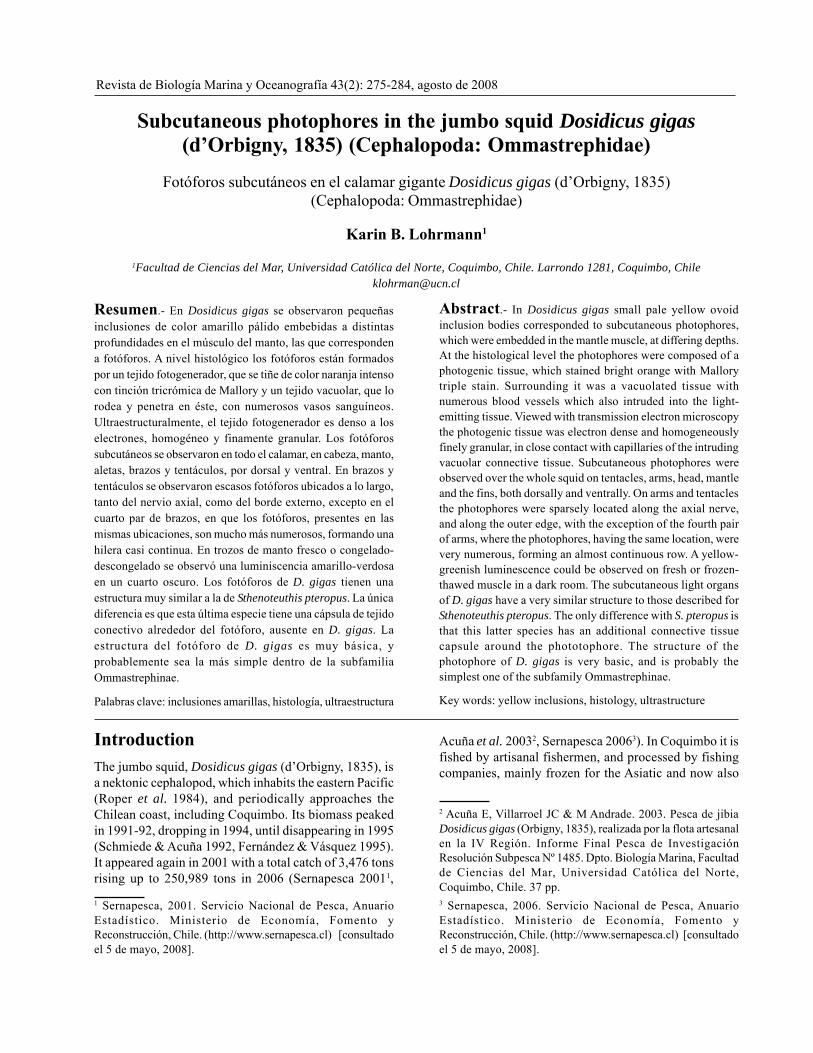
Revista de Biología Marina y Oceanografía 43(2): 275-284, agosto de 2008 Subcutaneous photophores in the jumbo squid Dosidicus gigas (d’Orbigny, 1835) (Cephalopoda: Ommastrephidae) Fotóforos subcutáneos en el calamar gigante Dosidicus gigas (d’Orbigny, 1835) (Cephalopoda: Ommastrephidae) Karin B. Lohrmann 1 1 Facultad de Ciencias del Mar, Universidad Católica del Norte, Coquimbo, Chile. Larrondo 1281, Coquimbo, Chile [email protected] Resumen.- En Dosidicus gigas se observaron pequeñas inclusiones de color amarillo pálido embebidas a distintas profundidades en el músculo del manto, las que corresponden a fotóforos. A nivel histológico los fotóforos están formados por un tejido fotogenerador, que se tiñe de color naranja intenso con tinción tricrómica de Mallory y un tejido vacuolar, que lo rodea y penetra en éste, con numerosos vasos sanguíneos. Ultraestructuralmente, el tejido fotogenerador es denso a los electrones, homogéneo y finamente granular. Los fotóforos subcutáneos se observaron en todo el calamar, en cabeza, manto, aletas, brazos y tentáculos, por dorsal y ventral. En brazos y tentáculos se observaron escasos fotóforos ubicados a lo largo, tanto del nervio axial, como del borde externo, excepto en el cuarto par de brazos, en que los fotóforos, presentes en las mismas ubicaciones, son mucho más numerosos, formando una hilera casi continua. En trozos de manto fresco o congelado- descongelado se observó una luminiscencia amarillo-verdosa en un cuarto oscuro. Los fotóforos de D. gigas tienen una estructura muy similar a la de Sthenoteuthis pteropus. La única diferencia es que esta última especie tiene una cápsula de tejido conectivo alrededor del fotóforo, ausente en D. gigas. La estructura del fotóforo de D. gigas es muy básica, y probablemente sea la más simple dentro de la subfamilia Ommastrephinae. Palabras clave: inclusiones amarillas, histología, ultraestructura Abstract.- In Dosidicus gigas small pale yellow ovoid inclusion bodies corresponded to subcutaneous photophores, which were embedded in the mantle muscle, at differing depths. At the histological level the photophores were composed of a photogenic tissue, which stained bright orange with Mallory triple stain. Surrounding it was a vacuolated tissue with numerous blood vessels which also intruded into the light- emitting tissue. Viewed with transmission electron microscopy the photogenic tissue was electron dense and homogeneously finely granular, in close contact with capillaries of the intruding vacuolar connective tissue. Subcutaneous photophores were observed over the whole squid on tentacles, arms, head, mantle and the fins, both dorsally and ventrally. On arms and tentacles the photophores were sparsely located along the axial nerve, and along the outer edge, with the exception of the fourth pair of arms, where the photophores, having the same location, were very numerous, forming an almost continuous row. A yellow- greenish luminescence could be observed on fresh or frozen- thawed muscle in a dark room. The subcutaneous light organs of D. gigas have a very similar structure to those described for Sthenoteuthis pteropus. The only difference with S. pteropus is that this latter species has an additional connective tissue capsule around the phototophore. The structure of the photophore of D. gigas is very basic, and is probably the simplest one of the subfamily Ommastrephinae. Key words: yellow inclusions, histology, ultrastructure Introduction The jumbo squid, Dosidicus gigas (d’Orbigny, 1835), is a nektonic cephalopod, which inhabits the eastern Pacific (Roper et al. 1984), and periodically approaches the Chilean coast, including Coquimbo. Its biomass peaked in 1991-92, dropping in 1994, until disappearing in 1995 (Schmiede & Acuña 1992, Fernández & Vásquez 1995). It appeared again in 2001 with a total catch of 3,476 tons rising up to 250,989 tons in 2006 (Sernapesca 2001 1 , 1 Sernapesca, 2001. Servicio Nacional de Pesca, Anuario Estadístico. Ministerio de Economía, Fomento y Reconstrucción, Chile. (http://www.sernapesca.cl) [consultado el 5 de mayo, 2008]. Acuña et al. 2003 2 , Sernapesca 2006 3 ). In Coquimbo it is fished by artisanal fishermen, and processed by fishing companies, mainly frozen for the Asiatic and now also 2 Acuña E, Villarroel JC & M Andrade. 2003. Pesca de jibia Dosidicus gigas (Orbigny, 1835), realizada por la flota artesanal en la IV Región. Informe Final Pesca de Investigación Resolución Subpesca Nº 1485. Dpto. Biología Marina, Facultad de Ciencias del Mar, Universidad Católica del Norte, Coquimbo, Chile. 37 pp. 3 Sernapesca, 2006. Servicio Nacional de Pesca, Anuario Estadístico. Ministerio de Economía, Fomento y Reconstrucción, Chile. (http://www.sernapesca.cl) [consultado el 5 de mayo, 2008].
Transcript of Subcutaneous photophores in the jumbo squid Dosidicus gigas (d'Orbigny, 1835) (Cephalopoda:...
Revista de Biología Marina y Oceanografía 43(2): 275-284, agosto de 2008
Subcutaneous photophores in the jumbo squid Dosidicus gigas(d’Orbigny, 1835) (Cephalopoda: Ommastrephidae)
Fotóforos subcutáneos en el calamar gigante Dosidicus gigas (d’Orbigny, 1835)(Cephalopoda: Ommastrephidae)
Karin B. Lohrmann1
1Facultad de Ciencias del Mar, Universidad Católica del Norte, Coquimbo, Chile. Larrondo 1281, Coquimbo, [email protected]
Resumen.- En Dosidicus gigas se observaron pequeñasinclusiones de color amarillo pálido embebidas a distintasprofundidades en el músculo del manto, las que correspondena fotóforos. A nivel histológico los fotóforos están formadospor un tejido fotogenerador, que se tiñe de color naranja intensocon tinción tricrómica de Mallory y un tejido vacuolar, que lorodea y penetra en éste, con numerosos vasos sanguíneos.Ultraestructuralmente, el tejido fotogenerador es denso a loselectrones, homogéneo y finamente granular. Los fotóforossubcutáneos se observaron en todo el calamar, en cabeza, manto,aletas, brazos y tentáculos, por dorsal y ventral. En brazos ytentáculos se observaron escasos fotóforos ubicados a lo largo,tanto del nervio axial, como del borde externo, excepto en elcuarto par de brazos, en que los fotóforos, presentes en lasmismas ubicaciones, son mucho más numerosos, formando unahilera casi continua. En trozos de manto fresco o congelado-descongelado se observó una luminiscencia amarillo-verdosaen un cuarto oscuro. Los fotóforos de D. gigas tienen unaestructura muy similar a la de Sthenoteuthis pteropus. La únicadiferencia es que esta última especie tiene una cápsula de tejidoconectivo alrededor del fotóforo, ausente en D. gigas. Laestructura del fotóforo de D. gigas es muy básica, yprobablemente sea la más simple dentro de la subfamiliaOmmastrephinae.
Palabras clave: inclusiones amarillas, histología, ultraestructura
Abstract.- In Dosidicus gigas small pale yellow ovoidinclusion bodies corresponded to subcutaneous photophores,which were embedded in the mantle muscle, at differing depths.At the histological level the photophores were composed of aphotogenic tissue, which stained bright orange with Mallorytriple stain. Surrounding it was a vacuolated tissue withnumerous blood vessels which also intruded into the light-emitting tissue. Viewed with transmission electron microscopythe photogenic tissue was electron dense and homogeneouslyfinely granular, in close contact with capillaries of the intrudingvacuolar connective tissue. Subcutaneous photophores wereobserved over the whole squid on tentacles, arms, head, mantleand the fins, both dorsally and ventrally. On arms and tentaclesthe photophores were sparsely located along the axial nerve,and along the outer edge, with the exception of the fourth pairof arms, where the photophores, having the same location, werevery numerous, forming an almost continuous row. A yellow-greenish luminescence could be observed on fresh or frozen-thawed muscle in a dark room. The subcutaneous light organsof D. gigas have a very similar structure to those described forSthenoteuthis pteropus. The only difference with S. pteropus isthat this latter species has an additional connective tissuecapsule around the phototophore. The structure of thephotophore of D. gigas is very basic, and is probably thesimplest one of the subfamily Ommastrephinae.
Key words: yellow inclusions, histology, ultrastructure
IntroductionThe jumbo squid, Dosidicus gigas (d’Orbigny, 1835), isa nektonic cephalopod, which inhabits the eastern Pacific(Roper et al. 1984), and periodically approaches theChilean coast, including Coquimbo. Its biomass peakedin 1991-92, dropping in 1994, until disappearing in 1995(Schmiede & Acuña 1992, Fernández & Vásquez 1995).It appeared again in 2001 with a total catch of 3,476 tonsrising up to 250,989 tons in 2006 (Sernapesca 20011,
1 Sernapesca, 2001. Servicio Nacional de Pesca, AnuarioEstadístico. Ministerio de Economía, Fomento yReconstrucción, Chile. (http://www.sernapesca.cl) [consultadoel 5 de mayo, 2008].
Acuña et al. 20032, Sernapesca 20063). In Coquimbo it isfished by artisanal fishermen, and processed by fishingcompanies, mainly frozen for the Asiatic and now also
2 Acuña E, Villarroel JC & M Andrade. 2003. Pesca de jibiaDosidicus gigas (Orbigny, 1835), realizada por la flota artesanalen la IV Región. Informe Final Pesca de InvestigaciónResolución Subpesca Nº 1485. Dpto. Biología Marina, Facultadde Ciencias del Mar, Universidad Católica del Norte,Coquimbo, Chile. 37 pp.3 Sernapesca, 2006. Servicio Nacional de Pesca, AnuarioEstadístico. Ministerio de Economía, Fomento yReconstrucción, Chile. (http://www.sernapesca.cl) [consultadoel 5 de mayo, 2008].

276 Revista de Biología Marina y Oceanografía Vol. 43, Nº2, 2008
for European markets. When it has been exported to Spain,one questioned the presence of small yellow rice-likeinclusions in the mantle muscle.
D. gigas is the largest member of the familyOmmastrephidae, and belongs to the most advancedsubfamily, the Ommastrephinae (Nigmatullin et al. 2001).The five genera that belong to this subfamily arecharacterized by the presence of subcutaneous lightorgans, or photophores, which are often encountered inthe tissue of the mantle, head and arms (Roper et al. 1984,Nigmatullin 2007).
Photophores are present in many cephalopods, atdifferent locations, within the mantle, in or by internal organs,in the skin of the mantle, head, tentacles or eyes (Herring1988). The luminescence emitted by these photophores iseither intrinsic, or produced by symbiotic bacteria (Arnold& Young 1974, Leisman et al. 1980, Herring et al. 1981,Ruby & McFall-Ngai 1992). The photophores of the squidsof the family Ommastrephidae belong to the type of intrinsiclight organs (Girsch et al. 1976, Herring et al. 1981). In twomembers of the subfamily Ommastrephinae, both belongingto the genus Sthenoteuthis, a large dorsal organ, formed byaggregation of many individual light organs which is visibleexternally, is present. For S. pteropus the individual lightorgans have been observed with histology and transmissionelectron microscopy (Clarke 1965, Girsch et al. 1976) andwith Alcian Blue in S. oualaniensis (Kishimoto & Kohno1992). However, this kind of dorsal photophore has not beenobserved in D. gigas, which, along with the absence of eyeand intestinal photophores in paralarvae and adults (presentthough in juveniles and subadults) make this species to beconsidered the least advanced member of the subfamily(Clarke 1965, Clarke & Paliza 2000, Nigmatullin et al. 2001).
Bioluminescence was reported from D. gigas, as anumber of spots of brilliant blue light, located on the head,along the third arm, and as three strong luminous spotson the tips of the arms. This observation was made bychance, when a squid was brought on deck and the lighthad gone off on board, the luminescence lasting less than2 minutes (García-Tello 1964). Nesis (1970) describedthe photophores of D. gigas as inconspicuous elongateor oval granules scattered in the muscle of the mantle,the ventral side of the head, and on the outside edge ofthe 4th pair of arms. The present study analyses thedistribution, structure, ultrastructure, and luminescenceof the subcutaneous body photophores of D. gigas.
Material and methodsThe squids were obtained from artisanal fishermen, whofished for them near the shore off Coquimbo (30°S) atnight and delivered them to the fish processing company,
where the squids were headed, skinned, washed, andfurther processed as required. The samples for this studywere taken from the clean mantle on site, at the followingdates: 2003/07/07, 2003/07/14, 2003/08/15, 2003/10/07& 10, 2003/11/20, 2003/12/13, 2004/02/26, 2004/04/ 6& 24, 2005/08/11, 2005/10/28, 2005/11/02. Subcutaneousphotophores were cut out including part of the surroundingmuscle tissue and processed for histology. Whole, isolatedventral mantle photophores were sampled on 2003/07/26 and 2004/04/24, and processed for transmissionelectron microscopy (TEM). No arm photophores werefixed for TEM. Whole squid, arms and tentacles wererefrigerated, and taken next morning to the HistologyLaboratory of the Universidad Católica del Norte. Herethey were examined for the locations of photophores, andsampled for histology. For histology, no difference in thequality of preservation could be detected between samplestaken at night, shortly after the capture of the squids, andthose taken next day from refrigerated material.
Measurements of the photophores were taken with adigital vernier caliper with a precision of 0.01 mm fromfrozen-thawed ventral and dorsal mantle, fins, arms andtentacles.
Bioluminescence was looked at in a dark room fromrefrigerated squid caught the night before, and also infrozen-thawed squid. The observations were made ondorsal and ventral sides of the mantle, on arms andtentacles, and on the fin, with the skin on. Additionally,the photophores were exposed by peeling off theepidermis and dermis of dorsal and ventral mantle pieces.For stimulating the luminescence, a gentle flow of 2%H2O2 (by volume with distilled water or with seawater)was pipetted over the tissue pieces.
For histology the tissue pieces were fixed inDavidson’s fixative (Shaw & Battle 1957) for 24 h,dehydrated in ethanol, cleared in xylene, and embeddedin Paraplast. Five micrometer sections were cut andstained with haematoxylin and eosin (H&E) and Mallorytriple stain.
For transmission electron microscopy (TEM) thephotophores were fixed in 3% glutaraldehyde in 0.2Mcacodylate buffer with 1.75% NaCl, for two hours at roomtemperature. Tissues were washed three times in 0.2Mcacodylate buffer with 1.75% NaCl, and post-fixed forone hour either in 1% OsO4 in the same buffer or inreduced OsO4 (1:1 mixture of 3% aqueous potassiumferrocyanide and 2% osmium tetroxide). After washingthree times with buffer, they were rinsed twice in distilledwater, stained for 15 min with 2% aqueous uranyl acetate,dehydrated in ethanol, washed in acetone, and embeddedin Epon 812. Semi-thin sections, 1 μm thick, were cut on

a Reichert Ultracut S microtome, and stained withtoluidine blue. Ninety nm thin sections were cut with adiamond knife, collected on copper grids, and stained withaqueous uranyl acetate and lead citrate. The sections wereviewed with a Zeiss EM-9 electron microscope at 50 kV,and photographs were taken.
For scanning electron microscopy (SEM), sectionsfrom selected wax blocks were cut at 12 to 15 μm, andmounted on coverslips. Sections were de-waxed in threechanges of xylene, passed through three changes of 100%ethanol and critical point dried using CO2. Samples weremounted on stubs, and ion sputtered with gold. Thesections were viewed and photographed using a JEOLTS 300 microscope (Lohrmann et al. 2002).
ResultsMorphology and histology
Subcutaneous photophores are small, elongate bodies,located under the dermis, embedded in the mantle muscle.
They are a very light yellow colour when fresh becomingyellow when refrigerated or frozen. Histologically theywere composed of the photogenic tissue, which showeda lobular, irregular outline, and stained orange withMallory triple stain, surrounded by a highly vacuolatedand irrigated connective tissue which also intruded intoit. The collagen fibrils of the connective tissue stainedblue and the hemocyanin in the interior of the bloodvessels stained orange or red with Mallory triple stain(Fig. 1). Cellular and vascular elements of this tissue werehighly concentrated around the photogenic tissue, withoutforming a true capsule around it. The connective tissuewhich lies between the photophore and the mantle muscleresembled damaged tissue when observed in 5μmhistological sections (Figs. 1 and 2a). However, on 12μm thick sections viewed with SEM it was realized thatit corresponded to connective tissue with many bigvacuoles (Fig. 2b). On arms and tentacles photophoreswere either on the outer edge, especially on arm IV, orburied deeply, in close association with the axial nerve
Figure 1
Histological section of a D. gigas mantle photophore stained with Mallory triple stain. The photogenic tissue (Ph) stainsorange, the connective tissue (Ct) surrounds it, and intrudes into it. The hemocyanin inside the numerous blood vessels
(arrows) stains orange or red, the collagen stains blue. On the left top corner some mantle muscle tissue can be seen (M)
Corte histológico de un fotóforo del manto de D. gigas teñido con tinción tricrómica de Mallory. El tejido fotogenerador (Ph)se tiñe color naranja, el tejido conectivo (Ct) lo rodea y penetra en él. La hemocianina dentro de los numerosos vasos
sanguíneos (flechas) se tiñe color naranja o rojo, el colágeno se tiñe color azul. En la esquina superiorizquierda se observa una pequeña porción de tejido muscular del manto (M)
Lohrmann Subcutaneous photophores in the jumbo squid 277
278 Revista de Biología Marina y Oceanografía Vol. 43, Nº2, 2008
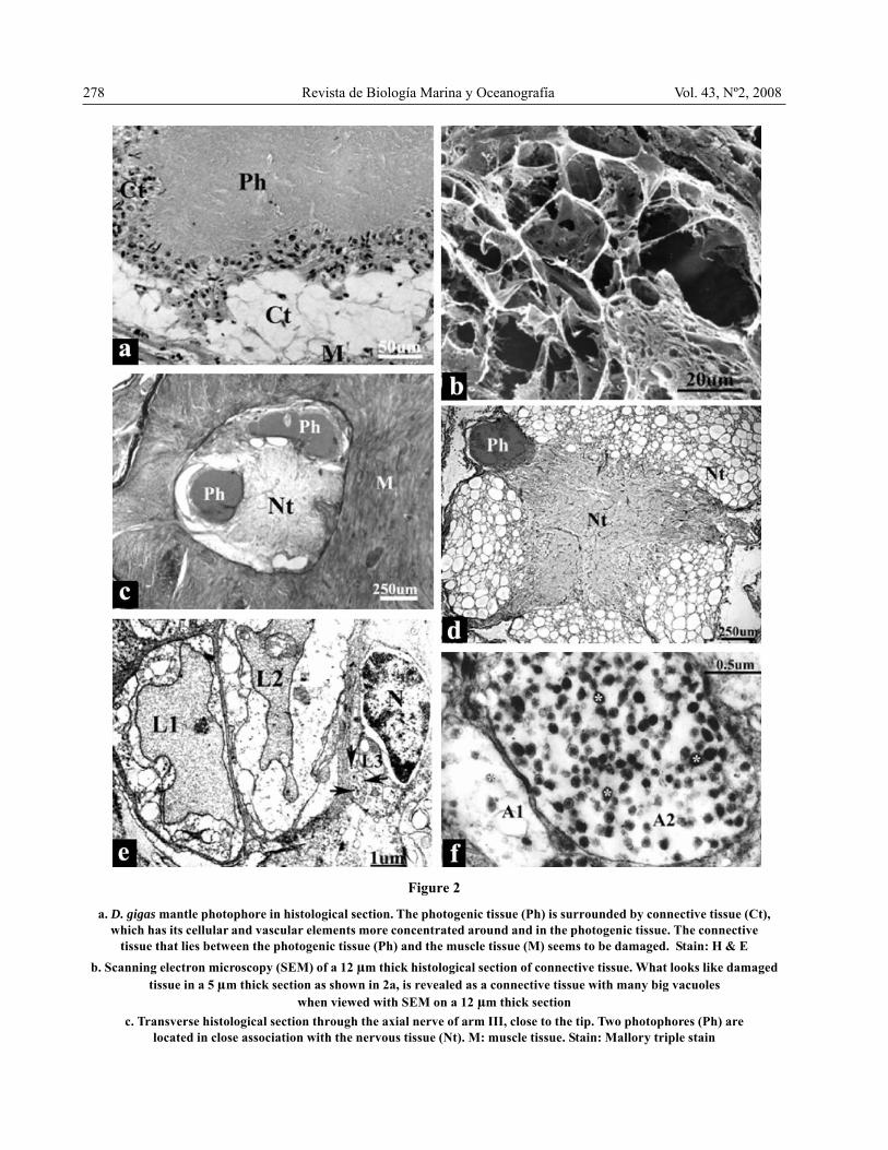
a. D. gigas mantle photophore in histological section. The photogenic tissue (Ph) is surrounded by connective tissue (Ct),which has its cellular and vascular elements more concentrated around and in the photogenic tissue. The connective
tissue that lies between the photogenic tissue (Ph) and the muscle tissue (M) seems to be damaged. Stain: H & Eb. Scanning electron microscopy (SEM) of a 12 μm thick histological section of connective tissue. What looks like damaged
tissue in a 5 μm thick section as shown in 2a, is revealed as a connective tissue with many big vacuoleswhen viewed with SEM on a 12 μm thick section
c. Transverse histological section through the axial nerve of arm III, close to the tip. Two photophores (Ph) arelocated in close association with the nervous tissue (Nt). M: muscle tissue. Stain: Mallory triple stain
Figure 2

cord (Figs. 2c and 2d). Figure 2d shows a photophorefrom a section in the middle region of arm IV; figure 2cshows two photophores from a section very close to thetip of arm III.
Ultrastructure
Only mantle photophores were processed for transmissionelectron microscope (TEM). In the vacuolated tissue thatsurrounds and intrudes into the photogenic tissuenumerous small blood vessels could be observed, (Figs.2e and 3b), some of them in association with nerveelements (Figs. 2e and 2f). The latter were never seenclose to the photogenic tissue. In addition to the smallblood vessels, numerous capillaries were seen. They couldbe recognized by their basement membrane, and thehemocyanin, which shows a paracrystalline pattern (Figs.2e, 3a and 3d). Near to the capillaries bundles ofmyofilaments were observed, some in longitudinal andsome in transverse section (Fig. 3a). The photogenic tissueitself was electron dense, homogeneously finely granular(Figs. 3a, 3b and 3c), close to capillaries (Fig. 3a) andadjacent to cells with numerous mitochondria (Figs. 3cand 3d). The tissues shown in figures 3b, 3c and 3d wereprocessed with reduced OsO4, which enhances themembranes. The plasma membrane of a vacuolated tissuecell filled with many mitochondria could be observed (Fig.3c); figure 3d shows a high magnification image ofmitochondria, where the internal membranes can clearlybe seen. The photogenic tissue did not show anymembranes, it rather seemed to be filling the spaces
between the cells of the vacuolated tissue.
Photophore distribution and size structure
Photophores were observed all over the squid on tentacles,arms, head, mantle and the fin. However, the abundance,size and orientation of the photophores varied dependingon their location. On the ventral side of the mantle, theywere oriented perpendicular to the long axis of the squid,increasing in number, and also in size, from cephalic tocaudal. Table 1 summarizes the sizes of photophores fromdorsal and ventral sides of the mantle, fins, and from eacharm and tentacle. On the ventral and dorsal surfaces ofthe head a few photophores were seen, similar in size tothe ventral mantle photophores. The size of photophoresthat run along the axial nerve is given separately fromthose that run on the outer arms’ edge. They are scarceand very small (mean length from 0.1 to 0.25 mm), andtheir long axis is parallel to the long axis of arms andtentacles. However, arm IV has more and longerphotophores running along the axial nerve than the otherarms; the length of these photophores was highly variable,hence the high standard deviation. Here the longestphotophores of the whole squid were observed measuringup to 80 mm in length (mean length of 3.6 mm). Theywere located half way from the base of the arm.Additionally, an almost continuous row of photophoreswas lined along the outer edge of arm IV, though smallerwith a mean length of 1.3 mm. The width, however, wasquite stable, ranging from 0.11 to 0.17 mm in arm andtentacle photophores (Table 1). All arms had 3 to 6
cont. Fig. 2
d. Transverse histological section through the axial nerve of arm IV. A photophore (Ph) is located in close associationwith the nervous tissue (Nt). Stain: Mallory triple stain
e. Transmission electron microscopy (TEM) of three small blood vessels. L1 & L2: lumen of blood vessels Nº 1 & Nº 2respectively, containing hemocyanin showing a paracrystalline pattern. L3: endotelial cell of blood vessel Nº 3¸
N: nucleus of endothelial cell; arrows point to two sectioned axonsf. High power TEM image of transverse sections of axons from Fig. 2e. A1: axon 1, A2: axon 2. (*): neurosecretory vesicles
a. Fotóforo del manto de D. gigas en corte histológico. El tejido fotogenerador (Ph) se encuentra rodeado por tejido conectivo (Ct),cuyos elementos celulares y vasculares están más concentrados en la periferia y en el interior del tejido fotogenerador. El tejido
conectivo que se encuentra entre el tejido fotogenerador y el tejido muscular (M) parece estar dañado. Tinción: H & Eb. Corte histológico de 12 μm de grosor preparado para su observación en el microscopio electrónico de barrido (MEB). Lo que en
cortes delgados (5 μm), como el que se muestra en 2a, parece ser un tejido dañado, se revela como un tejido conectivocon muchas vacuolas de gran tamaño, al observar un corte grueso (12 μm) con el MEB
c. Corte histológico transversal a través del nervio axial del brazo III, cerca del extremo distal del brazo. Dos fotóforos (Ph) seencuentran en íntima asociación con el tejido nervioso (Nt). M: tejido muscular. Tinción: Tricrómico de Mallory
d. Corte histológico transversal a través de nervio axial del brazo IV. Se observa un fotóforo en íntima asociación con el tejidonervioso (Nt). Tinción: Tricrómico de Mallory
e. Tres pequeños vasos sanguíneos vistos con microscopía electrónica de transmisión (MET). L1 y L2 corresponden al lumende los vasos L1 y L2 respectivamente, conteniendo hemocianina, que muestra un arreglo paracristalino. L3: célula
endotelial de vaso Nº 3; N: núcleo de célula endotelial; flechas indican dos axones en corte transversalf. Imagen aumentada de los axones cortados transversalmente de la Fig. 2e. A1: axon 1; A2: axon 2. (*): Vesículas neurosecretoras
Lohrmann Subcutaneous photophores in the jumbo squid 279

280 Revista de Biología Marina y Oceanografía Vol. 43, Nº2, 2008
Figure 3
Transmission electron microscopy (TEM) of D. gigas photophores. The tissues observed in Fig. 3a were processed usingbuffered OsO4. Reduced OsO4, which enhances the membranes, was used in the processing of the
tissues observed in Figs. 3b, 3c and 3da. A capillary (C) in longitudinal section, containing hemocyanin. Myofilament bundles (Mf) in longitudinal and transverse
section. M: Mitochondria; Ph: photogenic tissue; arrowheads: basement membrane of capillaryb. Photogenic tissue (Ph) close to blood vessel (Bv); arrow: basement membrane of the blood vessel; C: capillary
c. Photogenic tissue (Ph) and cell with numerous mitochondria (M). Arrows point to the plasma membraneof the cell with mitochondria
d. Higher power TEM image of some mitochondria (M), arrows point to mitochondrial cristae. C: capillary;arrowheads: basement membrane of capillary. The contents of the capillary corresponds to hemocyanin
Microscopía electrónica de transmisión (MET) de fotóforos de D. gigas. Los tejidos observados en la Fig. 3a fueronprocesados usando OsO4 tamponado. Para el procesamiento de los tejidos de las Figs. 3b, 3c y 3d,
se utilizó OsO4 reducido, que realza las membranasa. Capilar (C) en corte longitudinal, conteniendo hemocianina. Haces de miofilamentos (Mf) cortados longitudinal y
transversalmente. M: Mitocondria; Ph: tejido fotogenerador; puntas de flecha indican membrana basal del capilarb. Tejido fotogenerador (Ph) cerca de vaso sanguíneo (Bv); flecha: membrana basal del vaso sanguíneo; C: capilar sanguíneo
c. Tejido fotogenerador (Ph) asociado a célula con muchas mitocondrias (M). Las flechas apuntan a la membranaplasmática de la célula con mitocondrias
d. Algunas mitocondrias (M) observadas con mayor aumento, las flechas indican las crestas mitocondriales. C: capilar;puntas de flecha: membrana basal de capilar. El contenido del capilar corresponde a hemocianina

photophores concentrated at 5 to 6 cm from the tip. Onthe tentacles, photophores of small size (0.2 mm meanlength) were observed along the axial nerve, and alongthe outer edge. However, there was a long photophore(1.9 mm long) located at the outer edge 1-2 cm from thetip. On the fin, small photophores were buried in themiddle region under or over a median sheath of collagen,where the muscle bundles are inserted. On the fin rimvery small, superficial photophores were observed.
Bioluminescence
Bioluminescence was observed in a dark room fromrefrigerated squid caught the night before, and also infrozen-thawed squid. Once the observer’s eyes adaptedto the darkness, a weak, yellow-greenish luminescencewas visible from exposed photophores. It was stronglyenhanced pouring 2% H2O2 over the tissue, no matterwhether it had been diluted with seawater or distilledwater, the latter having a faster effect (almostinstantaneous).The luminescence from fresh photophoreswas stronger than from frozen-thawed photophores.Nevertheless, mantle pieces that had been frozen for 3months would still emit luminescence. The photophoresfrom dorsal mantle gave intermittent flashes of light of1or 2 s duration. The ventral photophores also gave flashes
of light, but these were longer lasting, and did not changeso quickly. The isolated arm IV string of photophoresfrom the outer edge gave a strong, steady luminescence.
Whole arms, tentacles, dorsal and ventral mantle andfin with skin were also stimulated with 2% H2O2 in thedark room. The luminescence took 2 or 3 min longer tobecome visible than in exposed photophores, however, itwas the same colour as in them. It was also useful forfinding photophores without skinning the squid. Thewhole dorsal fin rim was illuminated, as well as arm andtentacle tips, which were then processed for histology.Histologically, the photophores from mantle, arms,tentacles and fins showed a common structure.
DiscussionThe results of this study show that the subcutaneousphotophore of D. gigas is very basic, formed byphotogenic tissue surrounded by a vacuolar connectivetissue, with numerous blood vessels. This vacuolar tissueintrudes into the photogenic tissue, providing it with manycapillaries. The photophore from S. pteropus shows a verysimilar structure, the only difference being that in S.pteropus an additional layer of connective tissue linesthe photogenic tissue (Girsch et al. 1976). Clarke (1965),also for S. pteropus, described a ‘white tissue’, modified
Table 1Size of photophores in Dosidicus gigas
Tamaño de los fotóforos de Dosidicus gigas
Lohrmann Subcutaneous photophores in the jumbo squid 281

282 Revista de Biología Marina y Oceanografía Vol. 43, Nº2, 2008
as a reflector, located immediately underneath thephotophores of the dorsal organ. This was observed in atransverse section of the mantle with the dorsal organ.This ‘white tissue’ stained blue with Mallory, probablycorresponding to collagen, and was oriented parallel tothe skin surface. This layer was not mentioned by Girschet al. (1976) who studied photophores that had beenisolated from the dorsal organ, so they could not haveobserved it. In this study nothing but muscle fibres wasobserved around the photophores from all locations,nothing like a reflector could be discerned. Okada (1968)described the structure of the subcutaneous photophoresof Eucleoteuthis luminosa and Hyaloteuthis pelagica.Histologically the photogenic tissue is similar to what isencountered in D. gigas and in S. pteropus. However,there are two additional layers, a reflective muscle layer(similar to the one described by Clarke (1965) for S.pteropus) underneath the photogenic body, and a fanshaped light dispersing layer composed of connectivetissue located under the dermis. The other speciesbelonging to the subfamily Ommastrephinae isOmmastrephes bartramii. It only has photophores as anadult, and in this species they are restricted to small,irregular light organs embedded in the ventral mantle andventral head (Young 1972, Roper et al. 1984, Dunning1998). No information on the structure of the photophorescould be found, and to our knowledge no histology hasbeen undertaken.
The ultrastructure of the photophore of D. gigas, asevidenced by transmission electron microscopy (TEM)is comparable to the photophore of the other speciesstudied with TEM, S. pteropus, which is also very similarat the histological level (Girsch et al. 1976). Capillariesare very numerous in the vacuolar connective tissue, andthey are in direct contact with the photogenic tissue.Barber & Graziadei (1965) described the fine structureof four types of cephalopod small blood vessels. Theseconsist of pericytes, a basement membrane, and one orfew endothelial cells. Those seen in D. gigas are verysimilar to two of them, type 2 and type 4 vessels. Someblood vessels seem to consist of the basement membraneonly; as endothelial cells are sparse, and sometimes onlyone endothelial cell is present, there are vast extensionsof apparently naked basement membrane, especially whenthe capillary is filled with haemolymph. The same intimateassociation between photogenic tissue and capillaries seenwith TEM in D. gigas was observed in S. pteropus (Girschet al. 1976), and in the enoploteuthid squid Pterygioteuthismicrolampas (Arnold & Young 1974).
Can the ultrastructure of the photophore answer howthe light production is controlled? In D. gigas, some axonswere observed in one of the sections, closely associated
with blood vessels. They were probably not verynumerous, since they were not observed in other sections.It is also possible that in such a thin section as is requiredfor TEM they could have been missed, as it discussed byGirsch et al. (1976), who did not see any nerve elementsin their TEM sections. However, these axons were in closeproximity to the blood vessels, not to the photogenictissue. So it is possible, as suggested by Girsch et al.(1976) that the function of the light organs could beindirectly controlled through the blood supply. Thephotogenic tissue requires oxygen for light emission,which is provided by the hemocyanin in the haemolymph.On the other hand, the presence of cells with numerousmitochondria also indicates the production of largeamounts of ATP. Similar ‘mitochondrial cells’, have beendescribed by Arnold & Young (1974) as forming part ofthe photogenic organ of the squid Pterygiotheuthismicrolampas. The nerve elements observed in closeassociation with blood vessels in D. gigas might controlthe perivascular myofibrils, modifying the lumina of theblood vessels, and hence the haemolymph supply.Unfortunately no TEM samples were prepared from thosearm or tentacle photophores that run close to the axialnerve. At light microscopy level, they looked exactly likethe mantle light organs, with a high blood supply fromthe vacuolated connective tissue. However, it would havebeen interesting to see if there is a direct connectionbetween these axons and the photophores.
Subcutaneous photophores are difficult to see in freshspecimens, partly because of their small size, but mainlydue to their colour, which is very similar to the colour ofmuscle tissue. Kishimoto & Kohno (1992) used atechnique that gave good results for counting andmeasuring photophores in S. oualaniensis. It consistedof staining with alcian blue and clearing with KOH,making the muscle tissue translucent, and the bluephotophores visible. However, this technique would bevery difficult to be used in D. gigas due to their big size,at an average mantle length (ML) of around 65 to 72 cm.The biggest specimen of S. oualaniensis studied byKishimoto & Kohno (1992) had a ML of 15.4 cm. Roper(1963) found that squid muscle that had been fixed informalin for several years, and sometimes kept in 70%ethanol, became translucent, while the photophores kepttheir yellow colour. Again, this is also difficult to do withvery large specimens. In this study it was found thatfreezing the squid muscle darkens the colour of thephotophores, from the fresh light cream-yellow, to yellow,which made them easier to see. The other means of findingphotophores was pouring H2O2 on whole parts of the squid,and afterwards dissecting the portions which glowed inthe dark, either for measuring them, or for processing them
for histology. Each of these methods has contributed tothe knowledge of the distribution of the subcutaneousphotophores of squids of the subfamily Ommastrephinae.Roper (1963) detected them in formalin fixed tissues ofS. pteropus in the ventral mantle muscle, on the dorso-lateral part of the head, along the outer edge of the ventral(4th) arm pair, as well as on the other arms and thetentacles. However, he did not find any dorsalphotophores, except for a few scattered ones on the regionnear to the fins. He stated that the location and distributionof the subcutaneous photophores of S. pteropus is similarin S. oualaniensis and in D. gigas. However, in this studydorsal mantle photophores were found, smaller thoughthan the ventral ones and at first they had not beendetected. Kishimoto & Kohno (1992) also observed dorsalsubcutaneous photophores on the mantle of S.oualaniensis and on both ventral and dorsal mantle ofone specimen of O. bartramii stained with alcian blue.On this latter species, Young (1972) did not see any dorsalphotophores in one small 21 cm ML specimen. However,from the same specimen he described a long band ofphotogenic tissue from the base of the ventral arm almostto the tip. Roper (1963) also described a row of closelypacked photophores along the outer edge of the arm IVin S. pteropus, the same was observed in this study for D.gigas. Photophores on the outer edge of arm IV had alsobeen reported for this species by Nesis (1970), being thesame for males and females. García-Tello (1964)described the luminescence, when a specimen of D. gigasfell on board in complete darkness, along the third arm,on the ventral side, around the suckers, and also threelight emitting points on the tips of arms and tentacles. Asthis only lasted a few minutes, and it was dark, he mighthave confused the arms. The arms other than the fourtharm and the tentacles have very few photophores, exceptfor the tips, where they are more numerous. With histologysmall photophores were detected, sparsely distributedalong the axial nerve in arms and tentacles in D. gigas;the same was described in E. luminosa by Okada (1968).On other members of the subfamily no histology has beenperformed on arms or tentacles; they might also have thesephotophores running along the axial nerve.
The length of the photophores was highly variable,longer photophores might be formed by fusion or a verytight, lengthwise, apposition of them. The width (ordiameter), however, was quite stable, and may be a morereliable measure of the size. Comparing the size of thephotophores of D. gigas with the size of them in othermembers of this subfamily, it can be said that the dorsalmantle ones are similar in size to those from S.oualaniensis measured on a 15.4 cm ML specimen(Kishimoto & Kohno 1992). In the same species the
largest specimen had ventral mantle photophoresmeasuring 1.8 x 0.8 mm. The authors state that the numberas well as the size of the photophores increase withincreasing mantle length. Roper (1963) measured thediameter of photophores of S. pteropus of three sizes, 8cm, 11.6, and 27.5 cm ML. The photophores of the latterwere 0.8 to 2.7 mm in diameter. This is similar to thesize of the mantle photophores of D. gigas. He also foundthat smaller specimens had smaller photophores. In thisstudy it was not possible to have access to any small D.gigas although it may be possible to make measurementsof small specimens in the future.
Light emission of S. pteropus has been described asbright blue light flashes (Girsch et al. 1976), or pale bluelight (Roper 1963), and for D. gigas as brilliant blue(García-Tello 1964). It was not possible to observe anylive D. gigas, the isolated photophores viewed in a darkroom, enhanced or not with H2O2 showed a yellow-greenish colour, no blue colour could be observed.
ConclusionDosidicus gigas has the simplest possible subcutaneousphotophore structure, formed by photogenic tissue andvacuolated, highly vascular connective tissue surroundingand penetrating it. So it may have the simplest light organof the subfamily Ommastrephinae, which would be inaccordance with this species being the least advancedmember of the subfamily. However, the structure of thephotophores of O. bartramii is still to be studied, beforea conclusive statement on subcutaneous photophorestructure in this subfamily can be made.
AcknowledgmentsMy gratitude to Héctor Pujado from Pesquera San Joséfor his help in providing and sampling the squids. Manythanks to Ana Luisa Valdivia for superb histology and toFidel Vargas for his skillful assistance in TEM. I wish tothank Dr. Richard Young for his orientation when I startedlooking at what I thought was an infection, and veryspecial thanks to Dr. Chingis Nigmatullin for his expert,kind and patient help with the manuscript, improving itnotoriously. And last but not least, I am very grateful totwo anonymous reviewers whose most valuable commentsshaped the final version of this manuscript.
Literature citedArnold JM & RE Young. 1974. Ultrastructure of a cephalopod
photophore. I. Structure of the photogenic tissue. TheBiological Bulletin 147(3): 507-521.
Barber VC & P Graziadei. 1965. The fine structure ofcephalopod blood vessels. I: some smaller peripheral
Lohrmann Subcutaneous photophores in the jumbo squid 283

284 Revista de Biología Marina y Oceanografía Vol. 43, Nº2, 2008
vessels. Zeitschrift für Zellforschung und MikroskopischeAnatomie 66: 765-781.
Clarke MR. 1965. Large light organs on the dorsal surfaces ofthe squid Ommastrephes pteropus, Symplectoteuthisoualaniensis and Dosidicus gigas. Proceedings of theMalacological Society of London 36: 319-321.
Clarke R & O Paliza. 2000. The Humboldt Current squidDosidicus gigas (Orbigny, 1835). Revista de BiologíaMarina y Oceanografía 35(1): 1-39.
Dunning MC. 1998. A review of the systematics, distribution,and biology of the arrow squid genera OmmastrephesOrbigny, 1835, Sthenoteuthis Verril, 1880, and OrnithoteuthisOkada, 1927 (Cephalopoda: Ommastrephidae). In: Voss NA,M Vecchione, RB Toll & MJ Sweeney (eds), Systematicsand Biogeography of Cephalopods. SmithsonianContributions to Zoology 586(2): 425-433.
Fernández F & JA Vásquez. 1995. The jumbo flying squidDosidicus gigas (Orbigny, 1835) in Chile: Analysis of anephemeral fishery. Estudios Oceanológicos 14: 17-21.
García-Tello P. 1964. Nota preliminar sobre una observaciónde bioluminiscencia en Dosidicus gigas (d’Orb)Cephalopoda. Boletín de la Universidad de Chile 46: 2 7-28.
Girsch SJ, PJ Herring & F McCapra. 1976. Structure andpreliminary biochemical characterization of thebioluminescent system of Ommastrephes pteropus(Steenstrup) (Mollusca: Cephalopoda). Journal of theMarine Biological Association of the United Kingdom 56:707-722.
Herring PJ. 1988. Luminescent organs. In: Trueman ER &MR Clarke (eds), The Mollusca 11: Form and function,pp. 449-489. Academic Press, San Diego.
Herring PJ, MR Clarke, S. v. Boletzky & KP Ryan. 1981.The light organs of Sepiola atlantica and Spirula spirula(Mollusca:Cephalopoda): bacterial and intrinsic systems inthe order Sepiodea. Journal of the Marine BiologicalAssociation of the United Kingdom 61: 901-916.
Kishimoto H & H Kohno. 1992. Development of the luminousorgan in the Purpleback Flying squid, Stenoteuthisoualaniensis, as shown by alcian blue stain techniques.Bulletin of the Institute of Oceanic Research &Development 13: 71-83.
Leisman G, DH Cohn & KH Nealson. 1980. Bacterial originof luminescence in marine animals. Science 208: 1271-1273.
Lohrmann KB, AR Brand & SW Feist. 2002. Comparisonof the parasites and pathogens present in a cultivated andin a wild population of scallops (Argopecten purpuratusLamarck, 1819) in Tongoy Bay, Chile. Journal of ShellfishResearch 21(2): 557-561.
Nesis KN. 1970. The biology of the giant squid Dosidicusgigas. Oceanology 10: 108-118. (English translation).
Nigmatullin ChM. 2007. Brief review on evolutionary andecological aspects of biology of squids familyOmmastrephidae (Cephalopoda: Teuthida). ScientificProceedings of Kazan State University, Series: NaturalSciences 149(3): 182-193. (In Russian with Englishabstract).
Nigmatullin ChM, KN Nesis & AI Arkhipkin. 2001. A reviewof the biology of the jumbo squid Dosidicus gigas(Cephalopoda: Ommastrephidae). Fisheries Research 54:9-19.
Okada YK. 1968. Study of luminous Cephalopoda I. Luminoussquids belonging to the Ommastrephidae. Science Reportof the Yokosuka City Museum 14: 88-94.
Roper CFE. 1963. Observations on bioluminescence inOmmastrephes pteropus (Steenstrup, 1855), with notes onits occurrence in the family Ommastrephidae (Mollusca:Cephalopoda). Bulletin of Marine Science 13: 343-353.
Roper CFE, MJ Sweeney & CE Nauen. 1984. FAO SpeciesCatalogue. Vol. 3. Cephalopods of the World. An annotatedand illustrated catalogue of species of interest to fisheries.FAO Fisheries Synopsis 125 (3): 1-277.
Ruby EG & MJ McFall-Ngai. 1992. A squid that glows inthe night: development of an animal-bacterial mutualism.Journal of Bacteriology 174(15): 4865-4870.
Schmiede P & E Acuña. 1992. Regreso de las jibias (Dosidicusgigas) a Coquimbo. Revista Chilena de Historia Natural65: 389-390.
Shaw BL & HI Battle. 1957. The gross microscopic anatomyof the digestive tract of the oyster Crassostrea virginica(Gmelin). Canadian Journal of Zoology 35: 325-347.
Recibido el 3 de marzo de 2008 y aceptado el 25 de abril de 2008