
Studies on the Redox Centers of the Terminal Oxidase from Desulfovibrio gigas and Evidence for Its...
7
Studies on the Redox Centers of the Terminal Oxidase from Desulfovibrio gigas and Evidence for Its Interaction with Rubredoxin* (Received for publication, April 22, 1997, and in revised form, June 30, 1997) Cla ´ udio M. Gomes‡, Gabriela Silva‡§, Solange Oliveira§, Jean LeGall‡¶, Ming-Yih Liu¶, Anto ´ nio V. Xavier‡, Claudina Rodrigues-Pousada§, and Miguel Teixeira‡i From the ‡Instituto de Tecnologia Quı ´mica e Biolo ´gica, Universidade Nova de Lisboa, Oeiras, Portugal, the §Instituto Gulbenkian de Cie ˆncia, Laborato ´rio de Gene ´tica Molecular, Oeiras, Portugal, and the ¶Department of Biochemistry and Molecular Biology, University of Georgia, Athens, Georgia 30602 Rubredoxin-oxygen oxidoreductase (ROO) is the final component of a soluble electron transfer chain that cou- ples NADH oxidation to oxygen consumption in the an- aerobic sulfate reducer Desulfovibrio gigas. It is an 86- kDa homodimeric flavohemeprotein containing two FAD molecules, one mesoheme IX, and one Fe-uropor- phyrin I per monomer, capable of fully reducing oxygen to water. EPR studies on the native enzyme reveal two components with g values at ;2.46, 2.29, and 1.89, which are assigned to low spin hemes and are similar to the EPR features of P-450 hemes, suggesting that ROO hemes have a cysteinyl axial ligation. At pH 7.6, the flavin redox transitions occur at 0 6 15 mV for the qui- none/semiquinone couple and at 2130 6 15 mV for the semiquinone/hydroquinone couple; the hemes reduc- tion potential is 2350 6 15 mV. Spectroscopic studies provided unequivocal evidence that the flavins are the electron acceptor centers from rubredoxin, and that their reduction proceed through an anionic semiqui- none radical. The reaction with oxygen occurs in the flavin moiety. These data are strongly corroborated by the finding that rubredoxin and ROO are located in the same polycistronic unit of D. gigas genome. For the first time, a clear role for a rubredoxin in a sulfate-reducing bacterium is presented. Despite the fact that they are still being considered as strict anaerobes, sulfate reducing bacteria when exposed to oxygen are capable of surviving (1, 2) as well as of taking advantage of its presence in terms of energy conservation (3, 4). In the presence of oxygen, the sulfate reducer Desulfovibrio gigas, uses internal reserves of polyglucose, which is metabolized by the Embden-Meyerhof-Parnas pathway thus generating NADH and ATP (3). In this bacterium a three-component sol- uble electron transfer chain couples NADH oxidation with ox- ygen reduction to water, allowing simultaneously for NAD 1 regeneration and oxygen utilization. The proteins involved are NADH:rubredoxin oxidoreductase (NRO) 1 (5), rubredoxin (Rd) (6), and rubredoxin:oxygen oxidoreductase (ROO) (7), as shown in Scheme I. NRO, the first component of this pathway, contains two subunits of 27 and 32 kDa and is a flavoprotein containing four flavin groups (FMN and FAD) per molecule with reduction potentials (Fl ox /Fl red ) of 2295 and 2325 mV, at pH 7.6. This NADH oxidase reduces D. gigas Rd, a 6-kDa iron protein, with a reduction potential of 0 6 5 mV. NRO is highly specific toward this redox protein (5). D. gigas desulforedoxin (8), De- sulfovibrio desulfuricans Rd (9), or even Desulfovibrio vulgaris Rd (10) are hardly reduced by NRO (5), despite the similarity between the reduction potentials of these proteins. D. gigas Rd was also found to be necessary for the coupling of NRO to the final component of the chain, ROO (7). This enzyme can be seen as a true terminal oxidase since it is capable of catalyzing the four-electron reduction of oxygen to water without the forma- tion of partially reduced oxygen species (7). It is an 86-kDa homodimeric flavohemeprotein, containing two FAD and two distinct hemes per monomer. These hemes are of unusual types: uroporphyrin I and a noncovalently bound derivative of mesoheme IX (7, 11). Finding the type I Fe-uroporphyrin iso- mer, so far considered as a non-metabolite that accumulates in some genetically linked porphyrias, in a physiologically active enzyme is unprecedented and raises the possibility that uro- porphyrin I may be of physiological importance in other organisms. Preliminary studies of this system suggested the involve- ment of rubredoxin in a physiological reaction in a strict anaer- obe (7). However, its direct reaction with the terminal oxidase remained to be demonstrated. In the present study, the redox centers of this so far unique enzyme are characterized spectro- scopically as well as in terms of their reduction potentials. The electron transfer between the components of D. gigas oxygen- utilizing pathway is probed by EPR and visible spectroscopy. Unequivocal spectroscopic evidence for the direct involvement of Rd in the electron transfer to ROO is provided. Also, using genetic approaches, we show that the Rd is located in the same polycistronic unit of the ROO coding region. EXPERIMENTAL PROCEDURES Bacterial Strains and Plasmids—D. gigas (ATCC 19364) was grown as described previously (12). Escherichia coli strain LE 392, and E. coli P2 392 were used to screen the genomic library and in the purification of the recombinant positive phages. Competent cells of E. coli XL2-Blue * This work was supported by Praxis XXI (Praxis/2/2.1/Bio/20/94 and 1075/95) and European Commission Grants Bio4-CT96-0413 and ER- BCHRXCT940626 (to M. T.), National Institutes of Health Grant GM56001-01 (to J. L. G. and M. Y. L.), Fundac ¸ao ˜ Calouste Gulbentian grants (to C. R. P.), and Programa Gulbentian Doutoramento em Bio- logia e Medicina and Praxis XXI (BD9793/96) (to C. G.) and Praxis XXI (BD9016/96) (to G. S.). The costs of publication of this article were defrayed in part by the payment of page charges. This article must therefore be hereby marked “advertisement” in accordance with 18 U.S.C. Section 1734 solely to indicate this fact. i To whom correspondence should be addressed. Tel.: 351-1-442-62- 46; Fax: 351-1-442-87-66; E-mail: [email protected]. 1 The abbreviations used are: NRO, NADH:rubredoxin oxidoreduc- tase; ROO, rubredoxin-oxygen oxidoreductase; Rd, rubredoxin; Fl ox , flavin quinone; Fl sq , flavin semiquinone; Fl red , flavin hydroquinone; ORF, open reading frame; mT, millitesla. THE JOURNAL OF BIOLOGICAL CHEMISTRY Vol. 272, No. 36, Issue of September 5, pp. 22502–22508, 1997 © 1997 by The American Society for Biochemistry and Molecular Biology, Inc. Printed in U.S.A. This paper is available on line at http://www.jbc.org 22502 at Instituto Tecnologia Química e Biológica on July 14, 2008 www.jbc.org Downloaded from
-
Upload
independent -
Category
Documents
-
view
0 -
download
0
Transcript of Studies on the Redox Centers of the Terminal Oxidase from Desulfovibrio gigas and Evidence for Its...
Studies on the Redox Centers of the Terminal Oxidase fromDesulfovibrio gigas and Evidence for Its Interaction withRubredoxin*
(Received for publication, April 22, 1997, and in revised form, June 30, 1997)
Claudio M. Gomes‡, Gabriela Silva‡§, Solange Oliveira§, Jean LeGall‡¶, Ming-Yih Liu¶,Antonio V. Xavier‡, Claudina Rodrigues-Pousada§, and Miguel Teixeira‡i
From the ‡Instituto de Tecnologia Quımica e Biologica, Universidade Nova de Lisboa, Oeiras, Portugal, the §InstitutoGulbenkian de Ciencia, Laboratorio de Genetica Molecular, Oeiras, Portugal, and the ¶Department of Biochemistry andMolecular Biology, University of Georgia, Athens, Georgia 30602
Rubredoxin-oxygen oxidoreductase (ROO) is the finalcomponent of a soluble electron transfer chain that cou-ples NADH oxidation to oxygen consumption in the an-aerobic sulfate reducer Desulfovibrio gigas. It is an 86-kDa homodimeric flavohemeprotein containing twoFAD molecules, one mesoheme IX, and one Fe-uropor-phyrin I per monomer, capable of fully reducing oxygento water. EPR studies on the native enzyme reveal twocomponents with g values at ;2.46, 2.29, and 1.89, whichare assigned to low spin hemes and are similar to theEPR features of P-450 hemes, suggesting that ROOhemes have a cysteinyl axial ligation. At pH 7.6, theflavin redox transitions occur at 0 6 15 mV for the qui-none/semiquinone couple and at 2130 6 15 mV for thesemiquinone/hydroquinone couple; the hemes reduc-tion potential is 2350 6 15 mV. Spectroscopic studiesprovided unequivocal evidence that the flavins are theelectron acceptor centers from rubredoxin, and thattheir reduction proceed through an anionic semiqui-none radical. The reaction with oxygen occurs in theflavin moiety. These data are strongly corroborated bythe finding that rubredoxin and ROO are located in thesame polycistronic unit of D. gigas genome. For the firsttime, a clear role for a rubredoxin in a sulfate-reducingbacterium is presented.
Despite the fact that they are still being considered as strictanaerobes, sulfate reducing bacteria when exposed to oxygenare capable of surviving (1, 2) as well as of taking advantage ofits presence in terms of energy conservation (3, 4). In thepresence of oxygen, the sulfate reducer Desulfovibrio gigas,uses internal reserves of polyglucose, which is metabolized bythe Embden-Meyerhof-Parnas pathway thus generatingNADH and ATP (3). In this bacterium a three-component sol-uble electron transfer chain couples NADH oxidation with ox-ygen reduction to water, allowing simultaneously for NAD1
regeneration and oxygen utilization. The proteins involved are
NADH:rubredoxin oxidoreductase (NRO)1 (5), rubredoxin (Rd)(6), and rubredoxin:oxygen oxidoreductase (ROO) (7), as shownin Scheme I.
NRO, the first component of this pathway, contains twosubunits of 27 and 32 kDa and is a flavoprotein containing fourflavin groups (FMN and FAD) per molecule with reductionpotentials (Flox/Flred) of 2295 and 2325 mV, at pH 7.6. ThisNADH oxidase reduces D. gigas Rd, a 6-kDa iron protein, witha reduction potential of 0 6 5 mV. NRO is highly specifictoward this redox protein (5). D. gigas desulforedoxin (8), De-sulfovibrio desulfuricans Rd (9), or even Desulfovibrio vulgarisRd (10) are hardly reduced by NRO (5), despite the similaritybetween the reduction potentials of these proteins. D. gigas Rdwas also found to be necessary for the coupling of NRO to thefinal component of the chain, ROO (7). This enzyme can be seenas a true terminal oxidase since it is capable of catalyzing thefour-electron reduction of oxygen to water without the forma-tion of partially reduced oxygen species (7). It is an 86-kDahomodimeric flavohemeprotein, containing two FAD and twodistinct hemes per monomer. These hemes are of unusualtypes: uroporphyrin I and a noncovalently bound derivative ofmesoheme IX (7, 11). Finding the type I Fe-uroporphyrin iso-mer, so far considered as a non-metabolite that accumulates insome genetically linked porphyrias, in a physiologically activeenzyme is unprecedented and raises the possibility that uro-porphyrin I may be of physiological importance in otherorganisms.
Preliminary studies of this system suggested the involve-ment of rubredoxin in a physiological reaction in a strict anaer-obe (7). However, its direct reaction with the terminal oxidaseremained to be demonstrated. In the present study, the redoxcenters of this so far unique enzyme are characterized spectro-scopically as well as in terms of their reduction potentials. Theelectron transfer between the components of D. gigas oxygen-utilizing pathway is probed by EPR and visible spectroscopy.Unequivocal spectroscopic evidence for the direct involvementof Rd in the electron transfer to ROO is provided. Also, usinggenetic approaches, we show that the Rd is located in the samepolycistronic unit of the ROO coding region.
EXPERIMENTAL PROCEDURES
Bacterial Strains and Plasmids—D. gigas (ATCC 19364) was grownas described previously (12). Escherichia coli strain LE 392, and E. coliP2 392 were used to screen the genomic library and in the purificationof the recombinant positive phages. Competent cells of E. coli XL2-Blue
* This work was supported by Praxis XXI (Praxis/2/2.1/Bio/20/94 and1075/95) and European Commission Grants Bio4-CT96-0413 and ER-BCHRXCT940626 (to M. T.), National Institutes of Health GrantGM56001-01 (to J. L. G. and M. Y. L.), Fundacao Calouste Gulbentiangrants (to C. R. P.), and Programa Gulbentian Doutoramento em Bio-logia e Medicina and Praxis XXI (BD9793/96) (to C. G.) and Praxis XXI(BD9016/96) (to G. S.). The costs of publication of this article weredefrayed in part by the payment of page charges. This article musttherefore be hereby marked “advertisement” in accordance with 18U.S.C. Section 1734 solely to indicate this fact.
i To whom correspondence should be addressed. Tel.: 351-1-442-62-46; Fax: 351-1-442-87-66; E-mail: [email protected].
1 The abbreviations used are: NRO, NADH:rubredoxin oxidoreduc-tase; ROO, rubredoxin-oxygen oxidoreductase; Rd, rubredoxin; Flox,flavin quinone; Flsq, flavin semiquinone; Flred, flavin hydroquinone;ORF, open reading frame; mT, millitesla.
THE JOURNAL OF BIOLOGICAL CHEMISTRY Vol. 272, No. 36, Issue of September 5, pp. 22502–22508, 1997© 1997 by The American Society for Biochemistry and Molecular Biology, Inc. Printed in U.S.A.
This paper is available on line at http://www.jbc.org22502
at Instituto Tecnologia Q
uímica e B
iológica on July 14, 2008 w
ww
.jbc.orgD
ownloaded from

Cells (Stratagene) and/or E. coli TOP 10F9 (Invitrogen), prepared ac-cording to standard protocols (13), were used to transform the DNAfragments subcloned into the polylinker of the plasmid pZErOTM-1(Invitrogen).
Preparation of Genomic DNA—Genomic DNA isolated from D. gigaswas extracted as described (14). Plasmid DNA was prepared using theplasmid purification kit Qiagen (Diagen).
Cloning of Rd and ROO—Degenerate primers (59-ATGCARGC-RACRAARATHAT-39 and 59-TTRTAYGTYGTYCCCATYGG-39) of bothextremities of the amino acid sequence corresponding to the N terminusof ROO were used to amplify genomic DNA. The polymerase chainreaction cycling conditions were: 5 cycles at 94 °C for 30 s, 40 °C for 60 s,and 72 °C for 30 s followed by 30 cycles at 92 °C for 30 s, 48 °C for 30 s,72 °C for 30 s, and finally 72 °C for 10 min. The reaction product with106 base pairs was then subcloned in pZErOTM-1 and sequenced (seebelow). This procedure allowed us to obtain a homologous probe whichwas labeled with [a-32P]dATP using the megaprime DNA labeling sys-tem (Amersham). This probe was used to screen a Sau3A1 genomiclibrary from D. gigas constructed in l Dash II as vector (Stratagene).Filters were prehybridized and hybridized at 55 °C with 6 3 SSC (SSC,0.15 M NaCl, 15 mM trisodium citrate, pH 7.0), 5 3 Denhardt’s solution,0.5% SDS (mass/volume), 200 mg/ml sonicated salmon sperm DNA. Thefilters were washed using 2 3 SSC, or 2 3 SSC with 0.2% SDS at thehybridization temperature. The washings were performed at a temper-ature higher than 55 °C, depending on the intensity of the signal.
Positive phages were purified and their DNA isolated according tothe technique described as in Ref. 15. The DNA was then digested withBamHI, and the fragments corresponding to a size of 3.6 kilobases weresubcloned into pZErOTM-1.
DNA Sequencing—Suitable restriction fragments were subcloned inpZErOTM-1 and sequenced using the Termo Sequenase cycle sequenc-ing kit according to the supplier’s instructions (Amersham). This pro-cedure was used to solve the compressions, as the D. gigas genome hasa high G 1 C content (16). Sequential deletions were also made usingthe double-stranded Nested Deletion kit (Pharmacia).
Protein Purification—D. gigas soluble extract was prepared as de-scribed (12). ROO was purified essentially according to Ref. 7 but anextra purification step was performed after the last gel filtration col-umn. The ROO-containing fraction from this column was dialyzed over-night against 10 mM Tris-HCl, pH 7.6, buffer and applied on a Phar-macia Mono-Q FPLC column. A 10–500 mM gradient of the same bufferat 0.5 ml min21 was applied; pure ROO eluted at ;75 mM ionicstrength. Typical yields are 4–7 mg of ROO/kg of cell mass (wet weight).NRO and Rd were prepared by the procedures of Refs. 5 and 17,respectively. The protein was judged pure by SDS-polyacrylamide gelelectrophoresis which showed a single band at 43 kDa.
Protein Quantitation and Analysis—The protein N-terminal se-quence was determined using an Applied Biosystem Model 470A se-quenator. Search of protein sequences showing homology with the Nterminus was performed at the NCBI using the BLAST network ser-vice. The protein concentration was determined by the method of Brad-ford (18).
Spectroscopic Methods—Room temperature ultraviolet/visible spec-tra were recorded in a Beckman DU-70 spectrometer. Visible redoxtitrations were performed in a Shimadzu spectrometer (UV 265)equipped with a cell stirring system. Low temperature (77 K) visiblespectra were recorded on a DW-2 OLIS spectrophotometer. EPR spectrawere recorded as in Ref. 19. The EPR spectra obtained under nonsat-urating conditions were integrated using the Aasa and Vanngård (20)correction factor. Myoglobin azide (1.2 mM) was used as a standard (21).Fluorescence spectra were taken in a SPEX Fluorolog 212.
Redox Titrations—ROO (1.5 mM) was titrated anaerobically in 50 mM
Tris-HCl, pH 7.6, by stepwise addition of buffered sodium dithionite.The following compounds were used as redox mediators (0.25 mM each):methylene blue (E90 5 11 mV), indigo tetrasulfonate (E90 5 230 mV),indigo trisulfonate (E90 5 270 mV), indigo disulfate (E90 5 2182 mV),antraquinone 2,7-disulfonate (E90 5 2182 mV), safranine (E90 5 2280mV), neutral red (E90 5 2325 mV), benzyl viologen (E90 5 2359 mV),and methyl viologen (E90 5 2446 mV). The reduction potentials are
quoted versus the standard hydrogen electrode.The experimental data were manipulated and analyzed using MAT-
LAB (Mathworks, South Natick, MA) for Windows. Spectral smoothingand optical deconvolution was performed using singular value decom-position in combination with a curve fitting algorithm (22).
RESULTS
Spectroscopic Characterization—The UV-visible spectrum ofnative ROO is dominated by a Soret band at 416 nm due to thehemes, a flavin contribution in the 460-nm region and a dis-tinctive feature at 587 nm whose origin is unclear (Fig. 1, tracea). The spectra are similar both when recorded at 77 K and atroom temperature. A red flavin semiquinone species accumu-lates transiently during reduction, as detected at 480 and 380nm (Fig. 1, trace b). In the fully reduced enzyme, a and b bandsare present at 551 and 522 nm, respectively (cf. Fig. 5A).Moreover, the Soret nor the 587-nm bands shift (7). The inter-action of native ROO with exogenous ligands, such as cyanide,cysteine, dithiothreitol, and mercaptoethanol was studied.However, no spectral changes were observed, even upon ratherextensive incubation periods (data not shown).
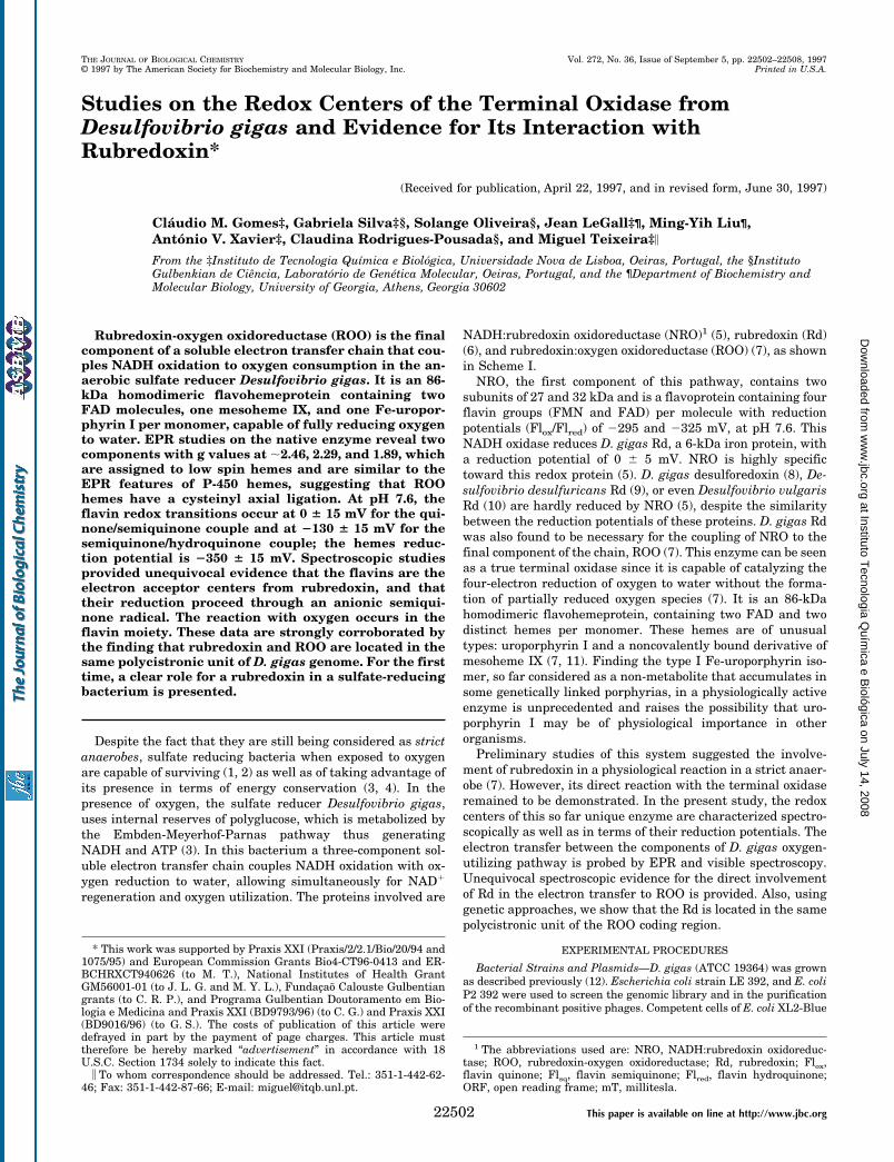
Fluorescence excitation and emission spectra of native ROOonly revealed the characteristic fingerprint of the flavin moiety(Fig. 2) (23). The emission spectrum, obtained with the excita-tion at 416 nm gives a single band at 527 nm. The excitationspectrum, with the emission wavelength at 520 nm, showsbands at 373 and 464 nm with a shoulder at 342 nm. Noemission was obtained with excitation at 587 nm. The emissionspectra of ROO is similar to those of free FAD and FMN, D.gigas flavodoxin,2 and ferredoxin-NADP1 oxidoreductase (24).Since there is a different degree of exposure to the solvent ofthe isoalloxazine rings in flavodoxins (25) and in FNR (26),nothing can be concluded about the flavin environment in ROOon the basis of its fluorescence characteristics.
The EPR spectra of ROO present some rather unique char-acteristics (Fig. 3). The spectra are dominated by resonances atg ;2.46, 2.29, and 1.89, which appear to result from two rhom-
2 C. M. Gomes, J. LeGall, A. V. Xavier, and M. Teixeira, unpublishedobservations.
FIG. 1. UV-visible spectra of ROO in several redox states. Tracea, as prepared ROO (1.5 mM) in 50 mM Tris-HCl buffer, pH 7.6; traces band c, partially reduced samples with dithionite. Only 3 out of 46spectra from a titration in the absence of redox mediators are shown.
SCHEME 1.
The Terminal Oxidase from D. gigas 22503
at Instituto Tecnologia Q
uímica e B
iológica on July 14, 2008 w
ww
.jbc.orgD
ownloaded from
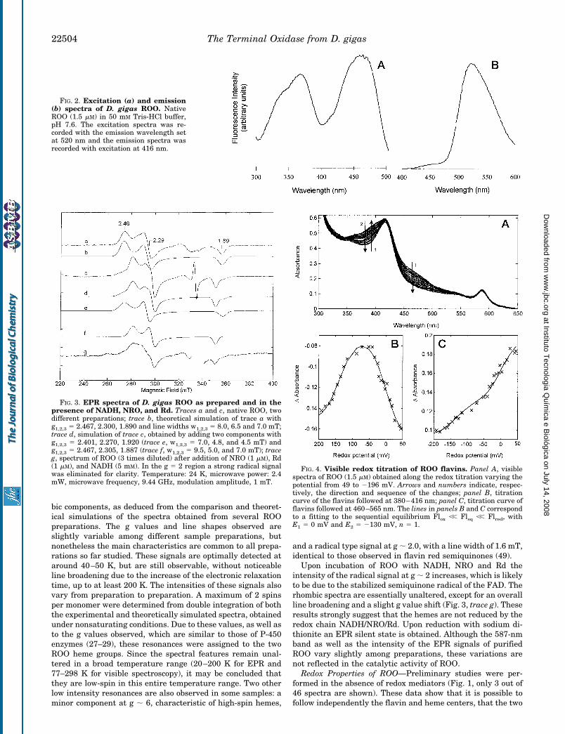
bic components, as deduced from the comparison and theoret-ical simulations of the spectra obtained from several ROOpreparations. The g values and line shapes observed areslightly variable among different sample preparations, butnonetheless the main characteristics are common to all prepa-rations so far studied. These signals are optimally detected ataround 40–50 K, but are still observable, without noticeableline broadening due to the increase of the electronic relaxationtime, up to at least 200 K. The intensities of these signals alsovary from preparation to preparation. A maximum of 2 spinsper monomer were determined from double integration of boththe experimental and theoretically simulated spectra, obtainedunder nonsaturating conditions. Due to these values, as well asto the g values observed, which are similar to those of P-450enzymes (27–29), these resonances were assigned to the twoROO heme groups. Since the spectral features remain unal-tered in a broad temperature range (20–200 K for EPR and77–298 K for visible spectroscopy), it may be concluded thatthey are low-spin in this entire temperature range. Two otherlow intensity resonances are also observed in some samples: aminor component at g ; 6, characteristic of high-spin hemes,
and a radical type signal at g ; 2.0, with a line width of 1.6 mT,identical to those observed in flavin red semiquinones (49).
Upon incubation of ROO with NADH, NRO and Rd theintensity of the radical signal at g ; 2 increases, which is likelyto be due to the stabilized semiquinone radical of the FAD. Therhombic spectra are essentially unaltered, except for an overallline broadening and a slight g value shift (Fig. 3, trace g). Theseresults strongly suggest that the hemes are not reduced by theredox chain NADH/NRO/Rd. Upon reduction with sodium di-thionite an EPR silent state is obtained. Although the 587-nmband as well as the intensity of the EPR signals of purifiedROO vary slightly among preparations, these variations arenot reflected in the catalytic activity of ROO.
Redox Properties of ROO—Preliminary studies were per-formed in the absence of redox mediators (Fig. 1, only 3 out of46 spectra are shown). These data show that it is possible tofollow independently the flavin and heme centers, that the two
FIG. 2. Excitation (a) and emission(b) spectra of D. gigas ROO. NativeROO (1.5 mM) in 50 mM Tris-HCl buffer,pH 7.6. The excitation spectra was re-corded with the emission wavelength setat 520 nm and the emission spectra wasrecorded with excitation at 416 nm.
FIG. 3. EPR spectra of D. gigas ROO as prepared and in thepresence of NADH, NRO, and Rd. Traces a and c, native ROO, twodifferent preparations; trace b, theoretical simulation of trace a withg1,2,3 5 2.467, 2.300, 1.890 and line widths w1,2,3 5 8.0, 6.5 and 7.0 mT;trace d, simulation of trace c, obtained by adding two components withg1,2,3 5 2.401, 2.270, 1.920 (trace e, w1,2,3 5 7.0, 4.8, and 4.5 mT) andg1,2,3 5 2.467, 2.305, 1.887 (trace f, w1,2,3 5 9.5, 5.0, and 7.0 mT); traceg, spectrum of ROO (3 times diluted) after addition of NRO (1 mM), Rd(1 mM), and NADH (5 mM). In the g 5 2 region a strong radical signalwas eliminated for clarity. Temperature: 24 K, microwave power: 2.4mW, microwave frequency, 9.44 GHz, modulation amplitude, 1 mT.
FIG. 4. Visible redox titration of ROO flavins. Panel A, visiblespectra of ROO (1.5 mM) obtained along the redox titration varying thepotential from 49 to 2196 mV. Arrows and numbers indicate, respec-tively, the direction and sequence of the changes; panel B, titrationcurve of the flavins followed at 380–416 nm; panel C, titration curve offlavins followed at 460–565 nm. The lines in panels B and C correspondto a fitting to the sequential equilibrium Flox ,, Flsq ,, Flred, withE1 5 0 mV and E2 5 2130 mV, n 5 1.
The Terminal Oxidase from D. gigas22504
at Instituto Tecnologia Q
uímica e B
iológica on July 14, 2008 w
ww
.jbc.orgD
ownloaded from
flavins are equivalent and have a reduction potential higherthan the hemes, and that flavin reduction occurs in two sequen-tial processes through a red anionic type semiquinoneintermediate.
The reduction potentials of ROO flavins were determinedfrom two independent visible titrations (Fig. 4, panel A). Largespectral changes occur at 460 nm (oxidized flavin decay) and at380 and 480 nm (appearance and disappearance of the flavinsemiquinone). Since absorbance does not change at 416 and565 nm, the variations of absorbance at 380–416 nm (Fig. 4,panel B) and 460–565 nm (Fig. 4, panel C) were plotted againstthe redox potential. Fitting the data to two sequential Nernstprocesses yielded the following reduction potentials: 0 6 15 mV(Flox/Flsq) and 2130 6 15 mV (Flsq/Flred). Singular value de-composition analysis of the data confirmed these results. Thedeconvolution of the optical species yielded a spectral compo-nent which was assigned to the red flavin semiquinone. Thefitted reduction potentials were similar to those obtained byconventional analysis (data not shown). From the reductionpotentials determined, the expected intensity of the semiqui-none species for a sequential redox process would be 0.88, ingood agreement with the theoretical value of 0.73 calculatedbased upon the extinction coefficients of quinone and redsemiquinone at 385 nm (23).
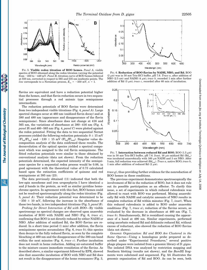
The data previously obtained (11) indicated that both thelow-spin mesoheme and iron uroporphyrin I have identical aand b bands in the protein, as well as similar pyridine hemo-chrome spectra. In agreement with this fact, ROO hemes couldnot be resolved spectroscopically along the redox titration (Fig.5, panel A). Their reduction potential was determined to be2350 6 10 mV, following the increase in the absorbance ofthese two bands, in two independent titrations (Fig. 5, panel B).
Probing for Direct Interaction of Rd with ROO—By visiblespectroscopy no spectral changes are observed upon anaerobicincubation of ROO with NADH and NRO (Fig. 6, trace a),confirming that ROO is not directly reduced by either NADH orNRO. After addition of oxidized Rd, the ROO flavins are re-duced. In a short time period (1 min) after addition, the flavinsemiquinone species accumulates (Fig. 6, trace b); this speciesthen decays to the fully reduced flavin, as seen by the completebleaching at 460 nm and the disappearance of the 380-nm bandwithin the next minutes (Fig. 6, trace c). Further incubationdoes not result in heme reduction. Adding air-saturated bufferto the mixture causes immediate reoxidation of the flavins. Asdescribed above, a similar experiment followed by EPR, showedalso that anaerobic incubation of ROO with NRO and Rd doesnot result in the disappearance of the heme resonances (Fig. 3,
trace g), thus providing further evidence for the nonreduction ofROO hemes in these conditions.
The previous experiment demonstrates spectroscopically theinvolvement of Rd in the reduction of ROO, but it does not ruleout its possible participation as an effector. To clarify thisissue, a set of experiments in which reduced rubredoxin wasallowed to react with ROO was performed. Mixing anaerobi-cally Rd with NADH and catalytic amounts of NRO results incomplete reduction of Rd within minutes (Fig. 7, inset). Whenthis reduced rubredoxin is added to ROO under anaerobicconditions (Fig. 7, trace a), reduction of the flavins occurs, asevaluated by the decrease in absorbance at 460 nm (Fig. 7,trace b). Simultaneously, Rd is reoxidized causing the appear-ance of a band at 490 nm. Similar experiments, performedusing ascorbate reduced Rd and dithionite reduced Rd, withoutexcess of reductant, also showed the reduction of ROO flavins(data not shown).
Genomic Organization: Rd and ROO Are Clustered in theSame Operon—Using a homologous probe prepared as de-scribed under “Experimental Procedures,” several positivephage plaques were isolated from a genomic library of D. gigas.The isolated DNA was analyzed by restriction mapping andSouthern blotting (results not shown), and the BamHI frag-ments were subcloned and sequenced. Fig. 8A illustrates thegenomic organization of Rd and ROO. As can be seen, both
FIG. 5. Visible redox titration of ROO hemes. Panel A, visiblespectra of ROO obtained along the redox titration varying the potentialfrom 2250 to 2449 mV. Panel B, titration curve of ROO hemes followedat 550 nm, corrected in respect to 565 and 535 nm isosbestic points. Theline corresponds to a Nernstian process, Em 5 2350 mV, n 5 1.
FIG. 6. Reduction of ROO flavins by NADH, NRO, and Rd. ROO(2 mM) was in 50 mM Tris-HCl buffer, pH 7.6. Trace a, after addition ofNRO (2.5 nM) and NADH (4 mM); trace b, recorded 1 min after furtheraddition of Rd (3 mM); trace c, recorded after 60 min of incubation.
FIG. 7. Interaction between reduced Rd and ROO. ROO (1.5 mM)was in 50 mM Tris-HCl buffer, pH 7.6. Inset, 16 mM oxidized Rd (Rdox)was incubated anaerobically with 160 mM NADH and 3 nM NRO. After5 min, full reduction was achieved (Rdred). Trace a, native ROO; trace b,1 min after addition of reduced Rd (2 mM).
The Terminal Oxidase from D. gigas 22505
at Instituto Tecnologia Q
uímica e B
iológica on July 14, 2008 w
ww
.jbc.orgD
ownloaded from
coding units are located in the same polycistronic unit. Thesequences of Rd and the N-terminal end of ROO are identical tothose obtained by chemical procedures (Fig. 8B).
Screening of protein sequence data bases revealed that theROO N terminus shows high homologies toward a few proteins:two putative flavoproteins from Methanococcus jannaschii (31);a potential FMN-binding protein from Rhodobacter capsulatus,the product of the ORFU1 coding region (32); a flavoproteinfrom Methanobacterium thermoautotrophicum (33) and E. coliORF o479 product, a not yet identified protein from E. coli (Fig.9). In the case of the protein from M. thermoautotrophicum,toward which the ROO N-terminal end exhibits 56% identity, itis interesting to note that there is evidence for a rubredoxinbeing encoded in the same operon (33). Most striking is thefinding of a block of seven residues which is conserved amongthese proteins: GTTYNAY. The sequence was comparedagainst the Prosite motifs data bank; this block of residues wasidentified as a potential N-myristoylation site. However, sincethis block is internal to the sequence and ROO is most likely afully processed protein, this possibility remains uncertain.Since most of the mentioned proteins contain flavin cofactors,
the GTTYNAY sequence could represent a novel consensusmotif for the binding of flavins.
DISCUSSION
D. gigas rubredoxin-oxygen oxidoreductase, in terms of itscofactors, may be considered as a member of the large super-family of flavoheme proteins. From that large and heterogene-ous family, two broad groups can be derived, based on theirreactivity toward oxygen (Table I). Group I includes proteinsthat are directly involved in reacting or binding dioxygen. Itcomprises mainly the so-called flavohemoglobins, like the he-moglobin-like protein from E. coli (34). These soluble proteinshave protoheme IX and FAD as cofactors and share domainsfrom the globin and ferredoxin-NADP1 reductase families (37).Their function is still uncertain, but they seem to play a role asoxygen sensors. Another member of this group is the cyto-chrome P-450 from Bacillus megaterium (P-450 BM3), whichpresents the unique feature of having in a single polypeptidechain both the heme and the flavin moieties (37). Group IIincludes the flavohemeproteins that are not reported to bereactive with oxygen and is somehow more heterogeneous in
FIG. 8. Partial restriction map andpredicted amino acid sequences ofRd and ROO. Panel A, partial restrictionmap of the subcloned 3.6-kilobase pairBamHI-BamHI DNA fragment. Panel B,amino acid sequence of Rd and the N ter-minus of ROO.
FIG. 9. Sequence alignment of ROO N terminus with homologous proteins. D. gigas rubredoxin:oxygen oxidoreductase (Dg ROO),Methanococcus jannaschii MJ0748 and MJ0732 products (Mj MJ0748 and Mj MJ0732), M. thermoautotrophicum flavoprotein (Mt fpaA), E. coliORF 0479 product (Ec ORFo479) and R. capsulatus ORFU1 product (Rc ORFU1). Between parentheses is the range of residues compared.
TABLE IComparison of properties from flavoheme proteins
Protein Organism Molecular mass Cofactors Function Reference
kDa
Group I: Oxygen reactive flavoheme proteinsRubredoxin-oxygen
oxidoreductaseD. gigas 86 FAD, mesoheme IX
Fe-Uroporphyrin ITerminal O2
reductaseThiswork
Hemoglobin-likeprotein (Hmp)
E. coli 44 FAD, protoheme IX O2 sensor 34
Yeast hemoglobin Candida mycoderma 50 FAD, protoheme IX O2 transport 35Flavoheme protein Alcaligenes eutrophus 43 FAD, protoheme IX 36P-450 BM3 Bacillus megaterium 119 FAD, FMN, protoheme IX Monooxygenase
activity38
Group II: Non-oxygen reactive flavoheme proteinsNitric oxide synthase Mammalian 150 FAD, FMN, protoheme IX NO synthase 39Flavocytochrome b2 E. coli, Saccharomyces
cerevisiae230 FMN, protoheme IX L-Lactate
dehydrogenase40
Flavocytochrome c Chlorobiumthiosulfatophilum
57 FAD, mesoheme IX Sulfidedehydrogenase
41
Flavocytochrome c Shewanella putrefaciens 63.8 FAD, mesoheme IX Fumaratereductase
42
Secondary aminemonooxygenase
Pseudomonasaminovorans
210 Heme, Flavin and non-heme iron
Amine oxygenase 43
The Terminal Oxidase from D. gigas22506
at Instituto Tecnologia Q
uímica e B
iológica on July 14, 2008 w
ww
.jbc.orgD
ownloaded from
terms of size and cofactors of its elements. Very interestingly, amember of this group, the rat nitric oxide synthase, was shownto contain a FMN-binding domain which has a strong analogywith the flavodoxins from sulfate reducing bacteria (43). How-ever, homologies between the N termini of the flavohemepro-teins from both groups and the N terminus from ROO were notfound. Nevertheless, the homology of this ROO partial se-quence with some flavoproteins in such diverse organisms asmethanogens and photosynthetic bacteria is surprising (Fig. 9).Unfortunately, the functions of these last proteins are not yetknown.
The EPR features of ROO, namely the g values observed andthe relaxation properties of the heme resonances, are strikinglysimilar to those of P-450 hemes (Table II), being so far observedonly in cysteinyl coordinated hemes. It may then be proposedthat one or both hemes in ROO are also coordinated to acysteine residue. Also, the g value shift detected upon incuba-tion with the ROO reductase system (NRO and Rd) is observedwhen P-450 is mixed with P-450 reductase (28). Interestingly,similar EPR signals were detected, with variable intensitiesdepending on the preparation, for some of the flavohemepro-teins presented in Table I, but these were not clearly assigned.
The reduction potentials of the flavin transitions (0 6 15 and2130 6 15 mV) are well in the range of what is found inflavoproteins (from 2495 to 1 80 mV) (46). The fact that theelectron donor is the one-electron carrier Rd leads necessarilyto the formation of a stable semiquinone radical species.
Contrary to what is observed for the other flavohemeproteinsfrom Group I, the reaction with oxygen in ROO appears tooccur exclusively at the level of the flavins. However, in con-trast to most of the oxidases containing flavins, ROO is notreduced by NADH. The low redox potential of ROO hemes(2350 6 15 mV) when compared with those of the flavins (0 615 mV and 2130 6 15 mV), together with the fact that they areneither reduced by NADH, NRO, and Rd, nor by reduced Rd,and the fact that this does not prevent catalytic activity, sug-gests that they are absent from reactivity with oxygen.
The coupling properties of some redox proteins from D. gigaswith ROO were tested. However, as evaluated by EPR, mixingROO under hydrogen with either hydrogenase and cytochromec3 or hydrogenase, cytochrome c3, and the high molecularweight cytochrome does not result in heme reduction. Possibly,the hemes provide a second electron entry point to the enzymethrough an unknown electron donor and/or are involved in anas yet undetermined catalytic reaction, such as enzymaticregulation.
Very interestingly, both coding units for Rd and ROO areclustered in the same operon (Fig. 8), indicating that they areunder the same transcriptional control. In fact, it has beenshown that proteins involved in the same metabolic pathwayare organized as an operon (47, 48). This finding stronglyreinforces the evidence obtained by spectroscopic analysis that
reduced rubredoxin is directly involved in electron donation toROO.
The determined redox potentials of ROO flavins are appar-ently in thermodynamic contradiction with the observationsreported here according to which flavins are fully reduced byRd, which has a reduction potential of ;0 mV. In fact, in thepresence of the complete redox chain (NADH, NRO, Rd, andROO), there is enough thermodynamic power to reduce theoxidase, but since reduced Rd can also reduce ROO flavins itmay be suggested that the interaction of Rd with ROO causesa slight shift on the reduction potentials of either Rd or ROOflavins.
This is the first example for a rubredoxin function in sulfatereducing bacteria, despite the fact that they have been knownand well characterized for a long time. Examples of physiolog-ical pathways in which a clear role for rubredoxin was foundare scarce (51, 52). The best documented example so far is themultienzyme system in Pseudomonads, responsible for the me-tabolism of n-alkanes which is composed by NADH-rubredoxinoxidoreductase, rubredoxin, and a hydroxylase acting as a mo-nooxygenase, found in the aerobe Pseudomonas oleovorans(52), a catalytic redox chain reminiscent of that operative in D.gigas for the reduction of dioxygen.
Acknowledgments—We are indebted to A. Mariano, P. Fareleira (In-stituto de Tecnologia Quımica e Biologica (ITQB)), and L. Chen (Uni-versity of Georgia) for their collaboration in the early stages of thiswork; M. Regalla (ITQB) for performing the N-terminal sequence; Prof.E. Melo (ITQB) for the collaboration in the fluorescence spectroscopy,and the staff of University of Georgia fermentation plant for growingthe bacteria.
REFERENCES
1. Diling, W., and Cypionka, H. (1990) FEMS Microbiol. Lett. 71, 123–1282. Marschall, C., Frenzel, P., and Cypionka, H. (1993) Arch. Microbiol. 159,
168–1733. Santos, H., Fareleira, P., Xavier, A. V., Chen, L., Liu, M.-Y., and LeGall, J.
(1995) Biochem. Biophys. Res. Commun. 195, 551–5574. LeGall, J., and Xavier, A. V. (1996) Anaerobe 2, 1–95. Chen, L., Lui, M.-Y., LeGall, J., Liu, Fareleira, P., Santos, H., and Xavier, A. V.
(1993) Eur. J. Biochem. 216, 443–4486. Moura, I., Moura J. J. G., Santos, M. H., Xavier, A. V., and LeGall, J. (1979)
FEBS Lett. 107, 419–4217. Chen, L., Liu M.-Y., LeGall, J., Fareleira P., Santos, H., and Xavier, A. V.
(1993) Biochem. Biophys. Res. Commun. 193, 100–1058. Moura, I., Bruschi, M., LeGall, J. Moura, J. J. G., and Xavier, A. V. (1977)
Biochem. Biophys. Res. Commun. 75, 1037–10449. Bruschi, M., Hatchikian, C. E., Golovleva, L. A., and Le Gall, J. (1977) J.
Bacteriol. 129, 30–3810. Bruschi, M., and LeGall, J. (1972) Biochim. Biophys. Acta 263, 279–28211. Timkovich, R., Burkhalter, R. S., Xavier, A. V., Chen, L., and LeGall, J. (1994)
Bioorg. Chem. 22, 284–29312. LeGall, J., Mazza, G., and Dragoni, N. (1965) Biochim. Biophys. Acta 99,
385–38713. Thoenes, U., Flores, O. L., Neves, A., Devreese, B., Van Beeumen, J. J., Huber,
R., Romao, M. J., LeGall, J., and Rodrigues-Pousada, R. (1994) Eur. J. Bio-chem. 220, 901–910
14. Ausubel, F. M., Brent, R., Kingston, R. E., Moore, D. D., Seidman, J. G., Smith,J. A., and Struhl, K. (1995) Current Protocols in Molecular Biology, GreenePublishing Associates and Wiley-Interscience, New York
15. Sambrook, J. Fritsch, E. F., and Maniatis, T. (1989) Molecular Cloning: ALaboratory Manual, 2nd Ed., Cold Spring Harbor Laboratory, Cold SpringHarbor, NY
16. Sigal, N., Senez, J. C., Le Gall, J., and Sebald, M. (1963) J. Bacteriol. 85,1315–1318
17. LeGall, J., and Dragoni, N. (1966) Biochem. Biophys. Res. Commun. 23,145–149
18. Bradford M. M. (1976) Anal. Biochem. 72, 248–25419. Teixeira, M., Batista, R., Gomes, C., Mendes, J., Pacheco, I., Anemuller, S., and
Hagen, W. R. (1995) Eur. J. Biochem. 227, 322–32720. Aasa, R., and Vanngård, T. (1975) J. Magn. Resonance 19, 308–31521. Bolard, J., and Garnier, A. (1972) Biochim. Biophys. Acta 263, 535–54922. Henry, E. R., and Hofrichter, J. (1992) Methods Enzymol. 210, 129–19223. Massey, V., and Hemmerich, P. (1980) Biochem. Soc. Trans. 8, 246–25724. Calcaterra, N. B., Pico, G. A., Orellano, E. G., Ottado, J., Carrillo, N., and
Ceccarelli, E. A. (1995) Biochemistry 34, 12842–1284825. Romero, A., Caldeira, J., LeGall, J., Moura, I., Moura, J. J. G., and Romao,
M. J. (1996) Eur. J. Biochem. 239, 190–19626. Karplus, P. A., Daniels, M. J., and Herriot, J. R. (1991) Science 251, 60–6627. Lipscomb, J. D. (1980) Biochemistry 19, 3590–359928. Dawson, J. H., Andersson, L. A., and Sono, M. (1982) J. Biol. Chem. 257,
3606–361729. McKnight, J., Cheesman, M. R., Thomson, A. J., Miles, J. S., and Munro, A. W.
TABLE IIComparison of EPR properties of D. gigas ROO, cytochromes P-450,
and other heme containing proteins
Protein g1 g2 g3 Reference
ROO (range ofpreparations)
2.39–2.47 2.26–2.32 1.88–1.92 Thiswork
P-450 CAM 2.45 2.26 1.91 28P-450 BM3 2.42 2.26 1.92 29E. coli Hmp (cysteine
derivative)2.48 2.28 1.87 34
Mammalian NOsynthase
2.44 2.29 1.89 39
Vitreoscilla bacterialhemoglobin
2.43 2.24 1.91 45
The Terminal Oxidase from D. gigas 22507
at Instituto Tecnologia Q
uímica e B
iológica on July 14, 2008 w
ww
.jbc.orgD
ownloaded from

(1993) Eur. J. Biochem. 213, 683–68730. LeGall, J. (1968) Ann. Inst. Pasteur (Paris) 114, 109–11531. Bult, C. J., White, O., Olsen, G. J., Zhou, L., Fleischmann, R. D., Sutton, G. G.,
Blake, J. A., FitzGerald, L. M., Clayton, R. A., Gocayne, J. D., Kerlavage,A. R., Dougherty, B. A., Tomb, J. F., Adams, M. D., Reich, C. I., Overbeek,R., Kirkness, E. F., Weinstock, K. G., Merrick, J. M., Glodek, A., Scott, J. L.,Geoghagen, N. S. M., Weidman, J. F., Fuhrmann, J. L., Nguven, D., Utter-back, T. R., Kelly, J. M., Peterson, J. D., Sadow, P. W., Hanna, M. C.,Cotton, M. D., Roberts, K. M., Hurst, M. A., Kaine, B. P., Borodovsky, M.,Klenk, H. P., Fraser, C. M., Smith, H. O., Woese, C. R., and Venter, J. C.(1996) Science 273, 1058–1073
32. Saeki, K., Tokuda, K., Fujiwara, T., and Matsubara, H. (1993) Plant CellPhysiol. 34, 185–199
33. Nolling, J., Ishii, M., Kock, J., Pihl, T., Reeve, J., Thauer, R., and Hedderich,R. (1995) Eur. J. Biochem. 231, 628–638
34. Ioannidis, N., Cooper, C. E., and Poole, R. K. (1992) Biochem. J. 288, 649–65535. Oshino, R., Asakura, T., Takio, K., Oshino, N., and Chance, B. (1973) Eur.
J. Biochem. 39, 581–59036. Probst, I., Wolf, G., and Schlegel, H. G. (1979) Biochim. Biophys. Acta 576,
471–47837. Narhi, L. O., and Fulco, A. J. (1986) J. Biol. Chem. 261, 7160–716938. Porter, T. D. (1991) Trends Biochem. Sci. 16, 154–15839. Stuehr, D. J., and Ikeda-Saito, M. (1992) J. Biol. Chem. 267, 20547–2055040. Black, M. T., White, S. A., Reid, G. A., and Chapman, S. K. (1989) Biochem. J.
258, 255–25941. Van Beeumen, J. J., Demol, H., Samyn, B., Bartsch, R. G., Meyer, T. E.,
Dolata, M. M., and Cusanovitch, M. A. (1990) J. Biol. Chem. 266,12921–12931
42. Morris, C. J., Black, A. C., Pealing, S. L., Manson, F. D. C., Chapman, S. K.,Reid, G. A., Gibson, D. M., and Ward, F. B. (1994) Biochem. J. 302, 587–593
43. Alberta, J. A., Andersson, L. A., and Dawson, J. H. (1989) J. Biol. Chem. 264,20467–20473
44. Deleted in proof45. Kroneck, P. M., Jakob, W., Webster, D. A., and DeMaio, R. (1991) Biol. Metals
4, 119–12546. Cooper, C. E., Ioannidis, N., D’Mello, R., and Poole, R. K. (1994) Bioch. Soc.
Trans. 22, 709–71347. Nielsen, A. K., Gerdes, K., Degn, H., and Murrel, J. C. (1996) Microbioloby 142,
1289–129648. Yamazaki, M., Thorne, L., Mikolajczak, M., Armentrout, R. W., and Pollock,
T. J. (1996) J. Bacteriol. 178, 2676–268749. Tegoni, M., Janot, J. M., and Labeyrie, F. (1986) Eur. J. Biochem. 155, 491–50350. Deleted in proof51. Ragsdale, S. W., Ljungdahl, L. G., and DerVartanian, D. V. (1983) J. Bacteriol.
155, 1224–123752. Ruettinger, R. T., Griffith, G. R., and Coon, M. J. (1977) Arch. Biochem.
Biophys. 183, 528–537
The Terminal Oxidase from D. gigas22508
at Instituto Tecnologia Q
uímica e B
iológica on July 14, 2008 w
ww
.jbc.orgD
ownloaded from


![Nickel[iron-sulfur]-selenium-containing hydrogenases from Desulfovibrio baculatus (DSM 1743). Redox centers and catalytic properties](https://static.fdokumen.com/doc/165x107/6316e17bc32ab5e46f0dfeb7/nickeliron-sulfur-selenium-containing-hydrogenases-from-desulfovibrio-baculatus.jpg)

