Nitrite dynamics in the open ocean—clues from seasonal and diurnal variations
Upload
independentCategory
view
4download
0
Cytochrome c Nitrite Reductase from Desulfovibrio desulfuricansATCC 27774THE RELEVANCE OF THE TWO CALCIUM SITES IN THE STRUCTURE OF THE CATALYTIC SUBUNIT (NrfA)*
Received for publication, November 19, 2002, and in revised form, January 16, 2003Published, JBC Papers in Press, March 4, 2003, DOI 10.1074/jbc.M211777200
Carlos A. Cunha‡, Sofia Macieira§, Joao M. Dias‡, Gabriela Almeida‡¶, Luisa L. Goncalves‡¶,Cristina Costa‡, Jorge Lampreia‡, Robert Huber§, Jose J. G. Moura‡, Isabel Moura‡,and Maria Joao Romao‡�
From the ‡Rede de Quımica e Tecnologia (REQUIMTE) Centre de Quımica Fina e Biotecnologia (CQFB),Departamento de Quımica, Faculdade de Ciencias e Tecnologia, Universidade Nova de Lisboa, 2829-516 Caparica,Portugal, the ¶Instituto Superior de Ciencias da Saude-Sul, Campus Universitario Quinta da Granja, 2825-511 Caparica,Portugal, and the §Max-Planck-Institut fur Biochemie, Abteilung Strukturforschung, Am Klopferspitz 18a,82152 Martinsried, Germany
The gene encoding cytochrome c nitrite reductase(NrfA) from Desulfovibrio desulfuricans ATCC 27774was sequenced and the crystal structure of the enzymewas determined to 2.3-Å resolution. In comparison withhomologous structures, it presents structural differ-ences mainly located at the regions surrounding theputative substrate inlet and product outlet, and in-cludes a well defined second calcium site with octahe-dral geometry, coordinated to propionates of hemes 3and 4, and caged by a loop non-existent in the previousstructures. The highly negative electrostatic potentialin the environment around hemes 3 and 4 suggests thatthe main role of this calcium ion may not be electrostaticbut structural, namely in the stabilization of the confor-mation of the additional loop that cages it and influ-ences the solvent accessibility of heme 4. The NrfA ac-tive site is similar to that of peroxidases with a nearbycalcium site at the heme distal side nearly in the samelocation as occurs in the class II and class III peroxi-dases. This fact suggests that the calcium ion at thedistal side of the active site in the NrfA enzymes mayhave a similar physiological role to that reported forthe peroxidases.
In Desulfovibrio desulfuricans, as happens in other pro-teobacteria, cytochrome c nitrite reductase was shown to be theterminal enzyme in the anaerobic respiratory pathway usingnitrate or nitrite as terminal electron acceptors (Refs. 1–3, seeRefs. 4–6 for reviews). This is a process making part of thebiogeochemical nitrogen cycle that may start with the reduc-
tion of nitrate to nitrite catalyzed by nitrate reductase (NapA)(7), followed by the six-electron reduction of nitrite to ammoniacatalyzed by the five-heme cytochrome c nitrite reductase. Thiselectron transport chain is located at the periplasmic mem-brane. Mutation studies in the operon encoding cytochrome cnitrite reductase from Wolinella succinogenes suggest thatthese enzymes are not integral membrane proteins but they areanchored to the membrane by a second subunit (NrfH) encodedin the same operon (8), a NapC/NirT-type cytochrome c thathas been suggested also to mediate the electron transfer be-tween the membranous menaquinone pool and the catalyticunit (9).
Crystal structures have been determined for the catalyticsubunit of cytochrome c nitrite reductase (NrfA) from the �-pro-teobacteria Sulfurospirillum deleyianum (10) and Wolinellasuccinogenes (11) and, more recently, from the �-proteobacte-rium Escherichia coli (12). These enzymes are homologous andshare a highly conserved three-dimensional structure. The se-quence of W. succinogenes NrfA is 75% identical to the one fromS. deleyianum, whereas E. coli NrfA is 48 and 46% identicalwith the ones from W. succinogenes and S. deleyianum, respec-tively. The crystal structures of these enzymes show the samehomodimeric structure, the same packing for the five c-typeheme groups within each monomer, and the same environmentat the active site, localized at heme 1, an unusual lysine-coordinated heme with the distal coordination position free toaccommodate the substrate molecule. Biochemical studies sug-gest that the homodimer is the functional form of the catalyticunit (13). In all these structures, a conserved calcium ion (cal-cium I) with octahedral coordination is present near theactive site.
Here we report the crystal structure of the catalytic subunitof cytochrome c nitrite reductase from D. desulfuricans ATCC27774, a 61-kDa protein encoded by the nrfA gene (14–16).This is the first structure of this family of enzymes from a�-proteobacterium, and it reveals considerable structural dif-ferences relative to the previously reported structures. A sec-ond calcium site (calcium II) with nearly perfect octahedralcoordination, caged by a loop not existent in the previous struc-tures, was identified coordinating the propionates A of hemes 3and 4. This calcium ion is located at nearly the same position asa yttrium and calcium ions bound at the protein surface in thecrystal structures of NrfA from W. succinogenes and E. coli,respectively. However, in these cases, the ions presented in-complete coordination shells and no physiological relevance
* This work was supported by Fundo Social Europeu (FSE) and FCT(Fundacao para a Ciencia e Tecnologia) through Ph.D. grants PRAXISXXI/BD/15752/98 (to C. A. C.), PRAXIS XXI/BD/13530/97 (to J. M. D.),PRAXIS XXI/BD/16009/98 (to S. M.), and PRAXIS XXI/BD/11349/97 (toG. A.), the European Co-operation in the Field of Science and Technol-ogy (COST) working group, and support for measurements at theEuropean Synchrotron Radiation Facility under the European UnionTMR/LSF Program. The costs of publication of this article were de-frayed in part by the payment of page charges. This article musttherefore be hereby marked “advertisement” in accordance with 18U.S.C. Section 1734 solely to indicate this fact.
The atomic coordinates and structure factors (code 1oah) have beendeposited in the Protein Data Bank, Research Collaboratory for Struc-tural Bioinformatics, Rutgers University, New Brunswick, NJ(http://www.rcsb.org/).
� To whom correspondence should be addressed. Tel.: 351-21-2948310; Fax: 351-21-2948385; E-mail: [email protected].
THE JOURNAL OF BIOLOGICAL CHEMISTRY Vol. 278, No. 19, Issue of May 9, pp. 17455–17465, 2003© 2003 by The American Society for Biochemistry and Molecular Biology, Inc. Printed in U.S.A.
This paper is available on line at http://www.jbc.org 17455
by on Septem
ber 4, 2008 w
ww
.jbc.orgD
ownloaded from
had been assigned to them. In the present work, the physiolog-ical role of the two calcium sites present in D. desulfuricansNrfA is discussed.
EXPERIMENTAL PROCEDURES
Gene Sequence Determination—The NrfA internal peptidesAETETKM and KAEQWEGQDR obtained by automated Edman deg-radation1 were used to design the degenerate primers: Nir-AETETKM,5�-GCIGARACIGARACIAARATG-3�, and Nir-Cterm, 5�-TCYTGIC-CYTCCCASACYTGYTC-3�. Using these oligonucleotides a DNA frag-ment of about 1431 base pairs was amplified by PCR. The resultingproduct was cloned in the vector pPCR-ScriptTM Amp SK(�) (Strat-agene) and sequenced in both strands with primers T3 and T7 (NewEngland Biolabs) and with internal primers, using an automated DNAsequencer (model 373, Applied Biosystems, Foster City, CA) and thePRISM ready reaction dye deoxyterminator cycle sequencing kit (Ap-plied Biosystems). More information on the N terminus was gainedafter identification and sequencing of the gene encoding the smallsubunit.1 The presence of a signal peptide was checked with the pro-gram Signal P V1.12 (18).
Protein Purification and Crystallization—D. desulfuricans NrfA wasextracted by mild treatment of the membrane fraction with sodiumcholeate (4–6 mg/liter) in 0.1 M potassium phosphate buffer at pH 7.6,as previously described (15). The enzyme was purified by sequentialammonium sulfate fractionation (30–60%), resuspension in potassiumphosphate buffer, and high performance liquid chromatography/gel fil-tration on a Superdex 200 (Amersham Biosciences) column equilibratedand eluted with 0.1 M potassium phosphate buffer at pH 7.6. The puritywas checked by UV-visible spectroscopy and by SDS-polyacrylamidemini-gel (12.5%) electrophoresis according to the Laemmli method (19).Despite the fact that NrfA enzymes are not integral membrane proteinsbut anchored to the membrane by the second subunit, crystals of D. des-ulfuricans NrfA could only be obtained using detergents in the crystal-lization conditions (20). Single crystals of dimensions 0.3 � 0.15 � 0.15mm suitable for x-ray diffraction studies were grown for 1 month using3-(decylmethylammonium)propane-1-sulfonate (Zwittergent 3-10 fromCalbiochem) added to the crystallization conditions as a solution with aconcentration of about 10 times the critical micellar concentration (40mM). The crystallization conditions contained 15% (w/v) PEG 3350, 0.2M CaCl2, and 0.1 M HEPES buffer at pH 7.5. The protein was used at aconcentration of 10 mg/ml. The crystals were obtained by the vapordiffusion method with hanging drop. The drops were prepared by add-ing 4 �l of protein solution, 1 �l of detergent at 10 times the criticalmicellar concentration, and 5 �l of reservoir solution.
Multiple Wavelength Anomalous Dispersion (MAD)3 Phasing—Acryo-cooled single crystal of dimensions 0.3 � 0.15 � 0.15 mm3 was usedto collect MAD data at the iron absorption edge, on beamline BM-14 atthe European Synchrotron Radiation Facility, Grenoble, France. Eth-ylene glycol added at a concentration of 25% to the crystallizationsolution was used as cryoprotectant and the crystal was cryo-cooled to100 K in a flux of nitrogen gas. The wavelengths at the point ofinflection and at the peak of the K-shell absorption edge of iron weredetermined to be �1 � 1.7403 Å and �2 � 1.7390 Å, respectively, from anx-ray fluorescence spectrum. Data sets were collected at these wave-lengths with a resolution up to 2.7 Å, using a MAR 345 image platedetector. The remote wavelength data set was collected at �3 � 0.9919Å, up to a resolution of 2.5 Å. Data were processed using version 1.96.1
of programs DENZO and SCALEPACK (21). The crystals belong to thespace group P212121 with unit cell constants a � 78.94 Å, b � 104.59 Å,and c � 143.18 Å. The statistics of data processing are presented inTable I. Using the program SOLVE (22), 10 iron sites were found in theasymmetric unit and phases were obtained with the program SHARP(23), resulting in a phasing power of 2.24 and an overall figure of meritof 0.49 (Table I). Density modification was performed with programsSOLOMON (24) (solvent flattening) and DM (25, 26) (solvent flatteningand averaging). At this stage, the protein boundaries, the helical re-gions at the dimer interface, and the heme cofactors could be distin-guished in the electron density maps. However, large regions of theprotein showed highly discontinuous electron density that did not im-prove much with the density modification procedures, making difficultthe task of model building in those regions. Because at this stage theprimary sequence was not yet known and the related S. deleyianumNrfA structure was available, attempts to solve the structure by mo-lecular replacement were done in parallel.
Structure Solution by Molecular Replacement—A solution of thestructure was obtained by molecular replacement using the programAMoRe (27, 26). The data set used in this case, with a resolution up to2.3 Å, was collected from a crystal similar to the one used in the MADexperiment, using radiation at �4 � 0.932 Å, on beamline ID14-EH4 atthe European Synchrotron Radiation Facility and using the local 2 � 2array of ADSC CCD detectors. The structure of S. deleyianum NrfA (10)was used as a search model. The solution was obtained using the modelcomprising the residue ranges 60 to 140, 150 to 217, 239 to 304, 319 to490, and hemes 1, 3, and 4. Heme 2, located in an exposed and flexibleregion of the protein, and heme 5, located at the dimer interface, werenot included in the search model because there could be significantshifts in their position and orientation between the model and theD. desulfuricans NrfA structure as was found to be true for heme 2 aftersolving the structure (see Fig. 3A). A dimer was found in the asymmet-ric unit that corresponds to a Matthews volume (28) of 2.3 Å3 Da�1 andin agreement with the 10 iron sites found in the asymmetric unit by theMAD experiment. The MR solution has a R-factor of 51.9% and acorrelation of 36.9%, in the resolution range between 10.0 and 3.0 Å,which is a high R-factor accompanied by a low correlation. However, theMR solution could superimpose well on the electron density mapsobtained by MAD if its coordinates were shifted half a unit cell along thea axis that corresponds to an alternative allowed origin in spacegroup P212121.
Model Building and Refinement—Model building was done withprogram O (29) and refinement was carried out with program REFMAC(26, 30). The initial model obtained by MR was corrected accordingly tothe electron density maps calculated after rigid body refinement andalso by comparison to the ones calculated using the experimentalphases determined by MAD. Hemes 2 and 5 as well as the calcium ion
1 M. G. Almeida, S. Macieira, L. L. Goncalves, R. Huber, C. A. Cunha,M. J. Romao, C. Costa, J. Lampreia, J. J. G. Moura, and I. Moura,submitted for publication.
2 www.cbs.dtu.dk.3 The abbreviations used are: MAD, multiple wavelength anomalous
dispersion; H-bond, hydrogen-bond; MR, molecular replacement; ncs,non-crystallographic symmetry.
TABLE IIRefinement statistics
Wavelength (Å) 0.932Resolution range (Å) 20.0–2.3Number of independent reflections 51633I/�(I) (last shell) 11.7 (2.2)Completeness (last shell) 97.3 (89.8)Rmerge (last shell) 0.08 (0.39)Number of amino acid residues (waters) 964 (498)Number of hemes 10Number of calcium ions 4Number of chloride ions 3Number of zinc ions 1Average B-factor for protein atoms (water) (Å2) 27.6 (29.1)Root mean square deviation in bond lengths (Å) 0.008Root mean square deviation in bond angles (degrees) 1.378Crystallographic Rfactor (Rfree) 0.189 (0.224)
TABLE IMAD data processing and phasing statistics
Wavelength Resolutionlimit Completeness Rmerge I/�(I) f � f � Phasing
powerIso
Phasingpowerano
RCullis
Å %
1.7403 2.7 95.1 (91.5)a 0.09 (0.20) 6.9 (3.4) �10.8 3.0 2.14 1.08 72.11.7390 2.7 94.9 (84.5) 0.09 (0.21) 7.3 (3.2) �11.1 2.0 2.24 0.92 68.30.9919 2.5 96.2 (85.2) 0.04 (0.12) 14.6 (5.6) 0.24b 1.53b 1.20
a Values in parentheses refer to the last resolution shell.b Indicates that the values were not refined.
Three-dimensional Structure of the Cytochrome c Nitrite Reductase17456
by on Septem
ber 4, 2008 w
ww
.jbc.orgD
ownloaded from

near the active site were incorporated in the model obtained by MR, andthe residue ranges of the model were corrected to the following regions:56 to 151, 153 to 222, 234 to 244, 252 to 263, 266 to 308, 315 to 377, and385 to 441. At this point it could already be observed that the last 73residues in the C terminus region of the S. deleyianum NrfA structuredid not follow the electron density and, for this reason, they wereremoved from the initial model. The side chains that did not fit theelectron density were mutated to alanines. Refinement of the modelthus obtained, with inclusion of the experimental phases from MAD,lowered the R-factor to 44.1% in the resolution range between 14.5 and2.5 Å. During the early stages of model building, until the R-factordecreased to below 40%, refinements were carried out including theexperimental phases from MAD. Following the refinement with pro-gram REFMAC, the program ArpWarp (31) was used in mode molrep,not including the experimental phases and using data in the resolutionrange between 14.5 and 2.3 Å. Model building was performed using theimproved electron density maps obtained from ArpWarp until the R-factor of the protein model decreased to below 30% when refined withprogram REFMAC after model building. During the last stages of modelbuilding, refinement was carried out with the program REFMAC5. TheR-free calculation was done using 1% of the reflections. The stereochem-ical quality of the final refined model was analyzed with WHATCHECK(32). The final model of the dimer was refined to a R-factor of 18.9% andR-free of 22.4% and refinement statistics are summarized in Table II.Each monomer is composed of 482 residues (sequence residues 38 to519), five c-type heme groups, two calcium ions, and one chloride ion.One of the monomers in the asymmetric unit binds one zinc ion with oneadditional chloride ion as one of its ligands, which corresponds to atetrahedral metal center that was identified at an intermolecular con-tact between crystallographic symmetry related copies of that mono-mer. Electron density corresponding to at least one more amino acid
after residue 518, in the C terminus, could be seen in the maps. How-ever, more amino acids after residue 518 could not be detected duringthe sequence determination. For this reason, residue 519 is present asan alanine in the crystal structure. There was no electron densitycorresponding to the residues from 325 to 331 in monomer A, suggestingthat this region was disordered in the crystal. The final model has 498water molecules, 174 of which have a non-crystallographic symmetry-related mate. The model coordinates have been deposited in the ProteinData Bank with the accession code 1oah.
RESULTS
Primary Sequence—A sequence alignment performed withthe amino acid sequences of the known cytochrome c nitritereductases and with the NrfA internal peptides AETETKM andKAEQWEGQDR allowed the identification of these two se-quences as portions of NrfA N and C terminus, respectively.The designed oligonucleotides were used to amplify by PCR aDNA fragment of about 1431 base pairs containing part of thenrfA gene sequence. The identification of the gene encoding thesmall subunit (nrfH) located upstream of nrfA allowed theidentification of the NrfA N-terminal sequence. The encodedNrfA is a 518-residue polypeptide chain harboring a signalpeptide targeting protein export to the periplasm. According tothe program Signal P (18) this peptide is predicted to be 28residues long, but N-terminal sequencing of the protein1 indi-cates that the cleavage site should be located between positions24 and 25 (Fig. 1). Further attempts to identify the sequencelocated downstream of nrfA were not successful, so the stop
FIG. 1. Amino acid sequence alignment. NrfA_Ddes, NrfA from D. desulfuricans ATCC 27774 (EMBL accession number AJ316232);NrfA_Ecoli, NrfA from E. coli (SWISS-PROT accession number P32050); NrfA_Sdel, NrfA from S. deleyianum (SWISS-PROT accession numberQ9Z4P4); NrfA_Wsuc, NrfA from W. succinogenes (TREMBL accession number Q9S1E5). Conserved residues are colored in pink, cysteines are ingreen, and calcium II ligands are in violet. The meanings of the symbols are: black inverted triangle, probable signal peptide cleavage site; whitetriangles, calcium I ligands; red triangles, residues forming the loop L1 that cages calcium II; green triangles, residues forming the loop L2 thathinders the product outlet; blue cylinders, �-helices; yellow arrows, �-strands. Numbering and positioning of the secondary structural elementsrefer to the NrfA_Ddes sequence. This figure was prepared with the programs PILEUP, in the Wisconsin Package version 10.0 (Genetics ComputerGroup (GCG), Madison, WI) and ALSCRIPT (51).
Three-dimensional Structure of the Cytochrome c Nitrite Reductase 17457
by on Septem
ber 4, 2008 w
ww
.jbc.orgD
ownloaded from
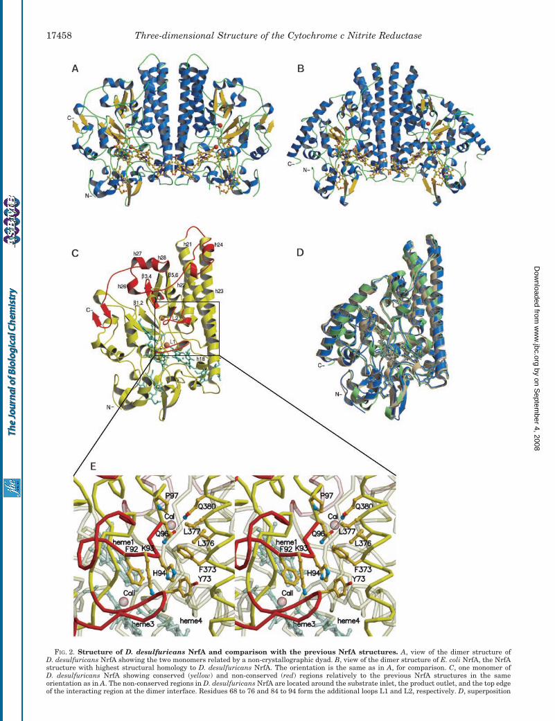
FIG. 2. Structure of D. desulfuricans NrfA and comparison with the previous NrfA structures. A, view of the dimer structure ofD. desulfuricans NrfA showing the two monomers related by a non-crystallographic dyad. B, view of the dimer structure of E. coli NrfA, the NrfAstructure with highest structural homology to D. desulfuricans NrfA. The orientation is the same as in A, for comparison. C, one monomer ofD. desulfuricans NrfA showing conserved (yellow) and non-conserved (red) regions relatively to the previous NrfA structures in the sameorientation as in A. The non-conserved regions in D. desulfuricans NrfA are located around the substrate inlet, the product outlet, and the top edgeof the interacting region at the dimer interface. Residues 68 to 76 and 84 to 94 form the additional loops L1 and L2, respectively. D, superposition
Three-dimensional Structure of the Cytochrome c Nitrite Reductase17458
by on Septem
ber 4, 2008 w
ww
.jbc.orgD
ownloaded from
codon was not identified and the gene was considered to be onlypartially sequenced. The nrfA sequence has been submitted tothe EMBL data base under accession number AJ316232. Themature NrfA polypeptide chain is then composed of 494 resi-dues according to the nrfA sequence, but it can be one residuelonger, as shown by the electron density maps from x-raycrystallography. The polypeptide chain in the crystal structurebegins at residue Thr-38 suggesting that the initial amino acidsin the N-terminal region of the mature enzyme are disordered.D. desulfuricans NrfA is homologous to its counterparts withknown structure (Fig. 1) but its shared identity at the primarystructure level is relatively low, with a degree of sequenceidentity of 35.9, 34.3, and 32.9% relatively to the cytochrome cnitrite reductases from S. deleyianum, E. coli, and W. succino-genes, respectively.
Overall Structure—The structure of D. desulfuricans NrfA(Fig. 2A) follows the typical dimer structure of the NrfA familyof enzymes (Fig. 2B), which is believed to be the active form ofthe enzyme (13). The monomers are related in the dimer by a2-fold ncs axis and they are structurally homologous to theprevious NrfA structures but to a lesser degree in this case(Fig. 2, C and D). The conserved regions correspond to 77% ofthe total number of amino acid residues with a root meansquare deviation of 1.7 Å in relation to the NrfA from E. coli (forthe main chain atoms of 370 residues) and a root mean squaredeviation of 2.0 Å in relation to the NrfA from W. succinogenes(for the main chain atoms of 372 residues).
The structure is dominated by the conserved characteristicthree-helix bundle in the region at the dimer interface (h21,h22, and h23 in Fig. 2C). Helix h21 shows a kink at residueThr-368 where its main chain nitrogen atom is H-bonded to themain chain carbonyl group of Val-365 changing the �-helicalstructure to a 310 helical geometry in this region. On the otherhand, helices h22 and h23 are straight along their extension. Inthe case of h22 this is made possible by the fact that the loopconnecting helices h21 and h22 is longer than in the other NrfAstructures where it is found that the N terminus of helix h22bends toward the C terminus of helix h21.
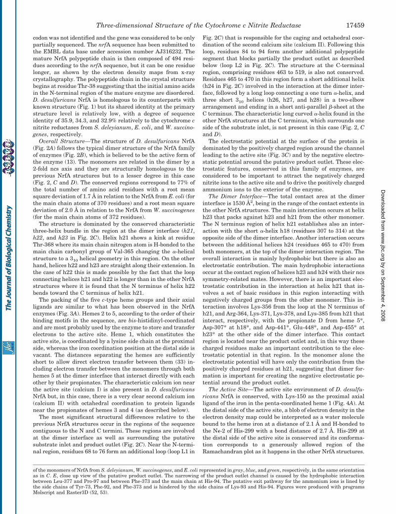
The packing of the five c-type heme groups and their axialligands are similar to what has been observed in the NrfAenzymes (Fig. 3A). Hemes 2 to 5, according to the order of theirbinding motifs in the sequence, are bis-histidinyl-coordinatedand are most probably used by the enzyme to store and transferelectrons to the active site. Heme 1, which constitutes theactive site, is coordinated by a lysine side chain at the proximalside, whereas the iron coordination position at the distal side isvacant. The distances separating the hemes are sufficientlyshort to allow direct electron transfer between them (33) in-cluding electron transfer between the monomers through bothhemes 5 at the dimer interface that interact directly with eachother by their propionates. The characteristic calcium ion nearthe active site (calcium I) is also present in D. desulfuricansNrfA but, in this case, there is a very clear second calcium ion(calcium II) with octahedral coordination to protein ligandsnear the propionates of hemes 3 and 4 (as described below).
The most significant structural differences relative to theprevious NrfA structures occur in the regions of the sequencecontiguous to the N and C termini. These regions are involvedat the dimer interface as well as surrounding the putativesubstrate inlet and product outlet (Fig. 2C). Near the N-termi-nal region, residues 68 to 76 form an additional loop (loop L1 in
Fig. 2C) that is responsible for the caging and octahedral coor-dination of the second calcium site (calcium II). Following thisloop, residues 84 to 94 form another additional polypeptidesegment that blocks partially the product outlet as describedbelow (loop L2 in Fig. 2C). The structure at the C-terminalregion, comprising residues 463 to 519, is also not conserved.Residues 465 to 470 in this region form a short additional helix(h24 in Fig. 2C) involved in the interaction at the dimer inter-face, followed by a long loop connecting a one turn �-helix, andthree short 310 helices (h26, h27, and h28) in a two-elbowarrangement and ending in a short anti-parallel �-sheet at theC terminus. The characteristic long curved �-helix found in theother NrfA structures at the C terminus, which surrounds oneside of the substrate inlet, is not present in this case (Fig. 2, Cand D).
The electrostatic potential at the surface of the protein isdominated by the positively charged region around the channelleading to the active site (Fig. 3C) and by the negative electro-static potential around the putative product outlet. These elec-trostatic features, conserved in this family of enzymes, areconsidered to be important to attract the negatively chargednitrite ions to the active site and to drive the positively chargedammonium ions to the exterior of the enzyme.
The Dimer Interface—The total contact area at the dimerinterface is 1530 Å2, being in the range of the contact extents inthe other NrfA structures. The main interaction occurs at helixh23 that packs against h23 and h21 from the other monomer.The N terminus region of helix h21 establishes also an inter-action with the short �-helix h18 (residues 307 to 314) at theopposite side of the dimer interface. Another interaction occursbetween the additional helices h24 (residues 465 to 470) fromboth monomers, at the top of the dimer interaction region. Theoverall interaction is mainly hydrophobic but there is also anelectrostatic contribution. The main hydrophobic interactionsoccur at the contact region of helices h23 and h24 with their ncssymmetry-related mates. However, there is an important elec-trostatic contribution in the interaction at helix h21 that in-volves a set of basic residues in this region interacting withnegatively charged groups from the other monomer. This in-teraction involves Lys-356 from the loop at the N terminus ofh21, and Arg-364, Lys-371, Lys-378, and Lys-385 from h21 thatinteract, respectively, with the propionate D from heme 5*,Asp-307* at h18*, and Asp-441*, Glu-448*, and Asp-455* ath23* at the other side of the dimer interface. This contactregion is located near the product outlet and, in this way thesecharged residues make an important contribution to the elec-trostatic potential in that region. In the monomer alone theelectrostatic potential will have only the contribution from thepositively charged residues at h21, suggesting that dimer for-mation is important for creating the negative electrostatic po-tential around the product outlet.
The Active Site—The active site environment of D. desulfu-ricans NrfA is conserved, with Lys-150 as the proximal axialligand of the iron in the penta-coordinated heme 1 (Fig. 4A). Atthe distal side of the active site, a blob of electron density in theelectron density map could be interpreted as a water moleculebound to the heme iron at a distance of 2.1 Å and H-bonded tothe N�-2 of His-299 with a bond distance of 2.7 Å. His-299 atthe distal side of the active site is conserved and its conforma-tion corresponds to a generously allowed region of theRamachandran plot as it happens in the other NrfA structures.
of the monomers of NrfA from S. deleyianum, W. succinogenes, and E. coli represented in gray, blue, and green, respectively, in the same orientationas in C. E, close up view of the putative product outlet. The narrowing of the product outlet channel is caused by the hydrophobic interactionbetween Leu-377 and Pro-97 and between Phe-373 and the main chain at His-94. The putative exit pathway for the ammonium ions is lined bythe side chains of Tyr-73, Phe-92, and Phe-373 and is hindered by the side chains of Lys-93 and His-94. Figures were produced with programsMolscript and Raster3D (52, 53).
Three-dimensional Structure of the Cytochrome c Nitrite Reductase 17459
by on Septem
ber 4, 2008 w
ww
.jbc.orgD
ownloaded from
The other residues present at the distal side of the active site,Arg-130 and Tyr-237, are also conserved. In addition to Tyr-237, the cavity of the active site is lined by a set of aromatic sidechains that are also conserved within the NrfA structures:these include Tyr-106, Phe-108, Tyr-112, and Phe-239. Amongthese residues, Tyr-237 and Phe-239 in close proximity to theheme may have an important influence in its redox potential,because this is largely affected by the dielectric constant of theheme crevice (34). This in turn is affected by the solvent acces-sibility of the heme and the polarity of the surrounding envi-ronment. Aromatic amino acids in the immediate surroundingsof hemes were shown to be important in the modulation of theredox potential in cases such as in the yeast iso-1-cytochrome cand the heme 4 of cytochrome c3 (Mr 26,000) (35, 36). NearTyr-237, a set of conserved tyrosine residues, Tyr-238, Tyr-266,and Tyr-267, have been suggested to play a role in dealing withpossible radical intermediates during the reduction of nitrite toammonia, a hypothesis supported by the observation of partialortho-hydroxylation of Tyr-219 (corresponding to Tyr-238 inD. desulfuricans NrfA) in the structure of W. succinogenesNrfA (11).
Based in the B-factors and in the difference electron densitymaps, a spherical blob of electron density near the side chain ofArg-130 was assigned to a chloride ion, acting as a counterionat 3.6 Å from the N� atom of the arginine. The presence of thision probably arises from the use of CaCl2 in the crystallizationconditions. The side chain of Arg-130 is H-bonded to the pro-pionate A of the active site heme at its distal side. The presenceof the chloride ion near Arg-130 suggests that the shared pro-ton in the H-bond between the side chain of the arginine andthe propionate A of the active site heme is mainly kept at thearginine maintaining its positive charge. The crystal structureof W. succinogenes NrfA complexed with the substrate showsthat nitrite bound at the active site is stabilized by H-bondswith the distal arginine and histidine (37), which suggests thatthe positive charge of the arginine may play an important rolein the binding of nitrite to the active site.
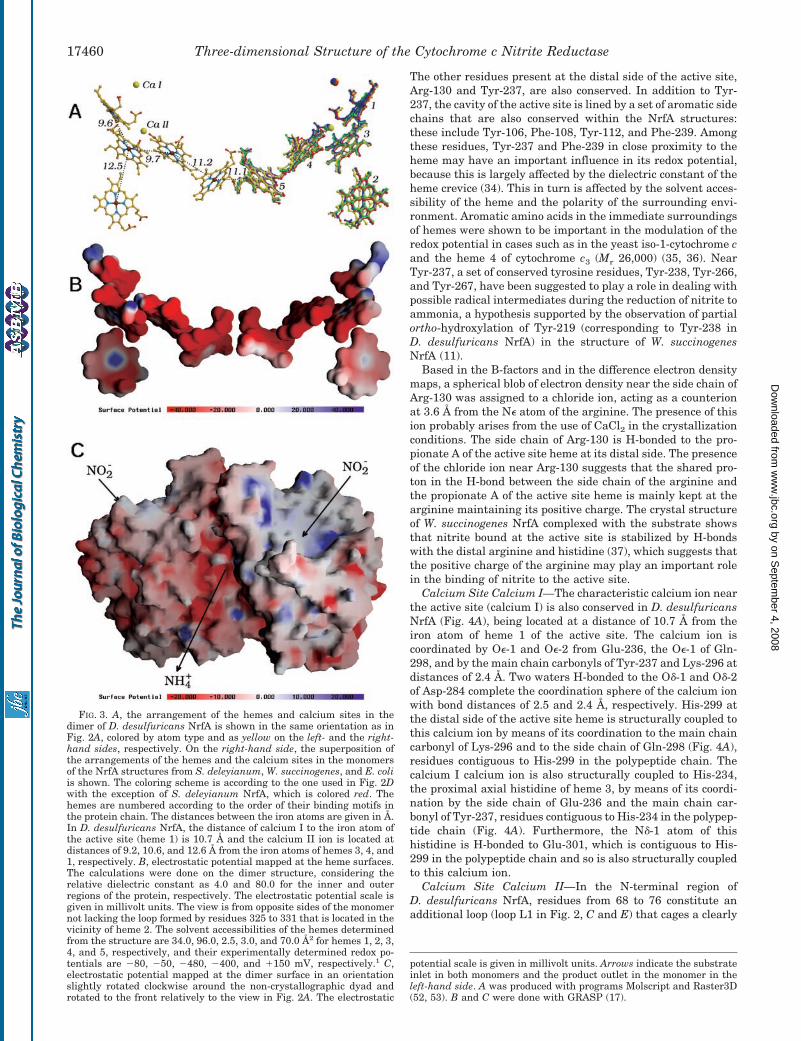
Calcium Site Calcium I—The characteristic calcium ion nearthe active site (calcium I) is also conserved in D. desulfuricansNrfA (Fig. 4A), being located at a distance of 10.7 Å from theiron atom of heme 1 of the active site. The calcium ion iscoordinated by O�-1 and O�-2 from Glu-236, the O�-1 of Gln-298, and by the main chain carbonyls of Tyr-237 and Lys-296 atdistances of 2.4 Å. Two waters H-bonded to the O�-1 and O�-2of Asp-284 complete the coordination sphere of the calcium ionwith bond distances of 2.5 and 2.4 Å, respectively. His-299 atthe distal side of the active site heme is structurally coupled tothis calcium ion by means of its coordination to the main chaincarbonyl of Lys-296 and to the side chain of Gln-298 (Fig. 4A),residues contiguous to His-299 in the polypeptide chain. Thecalcium I calcium ion is also structurally coupled to His-234,the proximal axial histidine of heme 3, by means of its coordi-nation by the side chain of Glu-236 and the main chain car-bonyl of Tyr-237, residues contiguous to His-234 in the polypep-tide chain (Fig. 4A). Furthermore, the N�-1 atom of thishistidine is H-bonded to Glu-301, which is contiguous to His-299 in the polypeptide chain and so is also structurally coupledto this calcium ion.
Calcium Site Calcium II—In the N-terminal region ofD. desulfuricans NrfA, residues from 68 to 76 constitute anadditional loop (loop L1 in Fig. 2, C and E) that cages a clearly
potential scale is given in millivolt units. Arrows indicate the substrateinlet in both monomers and the product outlet in the monomer in theleft-hand side. A was produced with programs Molscript and Raster3D(52, 53). B and C were done with GRASP (17).
FIG. 3. A, the arrangement of the hemes and calcium sites in thedimer of D. desulfuricans NrfA is shown in the same orientation as inFig. 2A, colored by atom type and as yellow on the left- and the right-hand sides, respectively. On the right-hand side, the superposition ofthe arrangements of the hemes and the calcium sites in the monomersof the NrfA structures from S. deleyianum, W. succinogenes, and E. coliis shown. The coloring scheme is according to the one used in Fig. 2Dwith the exception of S. deleyianum NrfA, which is colored red. Thehemes are numbered according to the order of their binding motifs inthe protein chain. The distances between the iron atoms are given in Å.In D. desulfuricans NrfA, the distance of calcium I to the iron atom ofthe active site (heme 1) is 10.7 Å and the calcium II ion is located atdistances of 9.2, 10.6, and 12.6 Å from the iron atoms of hemes 3, 4, and1, respectively. B, electrostatic potential mapped at the heme surfaces.The calculations were done on the dimer structure, considering therelative dielectric constant as 4.0 and 80.0 for the inner and outerregions of the protein, respectively. The electrostatic potential scale isgiven in millivolt units. The view is from opposite sides of the monomernot lacking the loop formed by residues 325 to 331 that is located in thevicinity of heme 2. The solvent accessibilities of the hemes determinedfrom the structure are 34.0, 96.0, 2.5, 3.0, and 70.0 Å2 for hemes 1, 2, 3,4, and 5, respectively, and their experimentally determined redox po-tentials are �80, �50, �480, �400, and �150 mV, respectively.1 C,electrostatic potential mapped at the dimer surface in an orientationslightly rotated clockwise around the non-crystallographic dyad androtated to the front relatively to the view in Fig. 2A. The electrostatic
Three-dimensional Structure of the Cytochrome c Nitrite Reductase17460
by on Septem
ber 4, 2008 w
ww
.jbc.orgD
ownloaded from
defined calcium ion (calcium II) with octahedral coordination.The presence of this loop constitutes a remarkable structuraldifference relative to the previous NrfA structures, whereas thepresence of this calcium site was detected only in the case of theE. coli enzyme but without attributing any role to it. Two of thecalcium ligands are the propionates A of both hemes 3 and 4(Figs. 4A and 5A). The coordination sphere is completed by themain chain carbonyls of Gly-75, from the caging loop L1, and ofThr-115 as well as the side chains of Glu-114 and Thr-115. Allthe ligands are at distances of 2.4 Å from the calcium ion andthis site is located at distances of 9.2, 10.6, and 12.6 Å from theiron atoms of hemes 3, 4, and 1, respectively. Whereas theproximal histidine of heme 3 is structurally coupled to thecalcium I ion (as described above), the distal histidine of thatheme (His-118) is structurally coupled to the calcium II ion bymeans of its coordination to the side chains of Glu-114 andThr-115 as well as to the main chain carbonyl group of Thr-115.
In E. coli NrfA this calcium site is exposed because of theabsence of the loop L1 that is absent in all the previous NrfAstructures, and was reported to be coordinated to the propi-onates of hemes 3 and 4 as well as to the carbonyl group ofPro-91. It presented an incomplete coordination sphere with awater molecule as a fourth ligand (Fig. 5C) and no biologicalrelevance was assigned to it at that time. There is some evi-dence for the existence of this calcium site also in the structureof W. succinogenes NrfA, but present as a Y3� ion arising fromthe use of YCl3 as an additive in the crystallization conditions.The Y3� was also coordinated to the A propionates of hemes 3
and 4 and to the main chain carbonyl group of Pro-99. Theother coordinating positions are occupied by three water mol-ecules and by an acetate molecule (Fig. 5B). The only structurewhere this site was not observed was the one from S. deleyia-num where the propionate A of heme 3 is in a different confor-mation making a H-bond with the main chain carbonyl group ofVal-320 (Fig. 5D), but that may be because of the fact thatcalcium was not present in the crystallization conditions inthis case.
Putative Substrate Inlet—The substrate inlet is surroundedon one side by the non-conserved C-terminal region composedof the short helices h26, h27, and h28 and the loop leading tothe short �-sheet at the C terminus (Fig. 2C). On the oppositeside, the substrate inlet is surrounded by three anti-parallel�-sheets: �1,2, �3,4, and �5,6. The polypeptide segment thatforms �3,4 is non-existent in the previous NrfA structures (Fig.2, C and D). Additionally, in NrfA from S. deleyianum and W.succinogenes, �1,2 is longer extending to the exterior of theenzyme. These structural differences in the �-sheet structureas well as in the non-conserved C-terminal region make theprotein surface around the substrate inlet steeper in the case ofD. desulfuricans NrfA. However, the electrostatic characteris-tics of the protein surface in this region are very similar tothose of the other NrfA structures, namely the positivelycharged patch around the substrate channel (Fig. 3C).
Putative Product Outlet—The exit of the channel for therelease of the product identified in the previous structures ispartially blocked in D. desulfuricans NrfA. In this case, on one
FIG. 4. A, the active site environment and the two calcium ions. At the active site, the electron density at the distal side of heme 1 could beinterpreted as a water molecule coordinated to the iron atom. The structural coupling of the calcium sites to both axial histidines of heme 3 isshown. B, the active site of P. chrysosporium lignin peroxidase isoenzyme H2 and the distal calcium site characteristic of the class II and class IIImono-heme peroxidases. The calcium ion is structurally coupled to the distal histidine at the active site in a similar manner as happens in the NrfAenzymes: it is coordinated by a main chain carbonyl group and a side chain belonging to residues in the polypeptide chain that are contiguous tothe histidine at the distal side of the heme. Surprisingly, the deviation between the locations of the calcium sites in the superposition of the activesites (by superposing the hemes) of P. chrysosporium lignin peroxidase isoenzyme H2 and D. desulfuricans NrfA is only 5.5 Å (picture not shown).Also, the environment at the distal side of the active site heme is similar in these types of enzymes: a histidine, an arginine, and an aromaticresidue are present in both cases. Figures were produced with programs Molscript and Raster3D (52, 53).
Three-dimensional Structure of the Cytochrome c Nitrite Reductase 17461
by on Septem
ber 4, 2008 w
ww
.jbc.orgD
ownloaded from
side of the product outlet, there is a loop constituted by residues84 to 94 that corresponds to an insertion in the protein se-quence relative to the previous NrfA structures (loop L2 in Fig.2C). This polypeptide segment has hydrophobic character andforms a one turn 310 helix that interacts with Phe-373 fromhelix h21 at the opposite side of the product outlet. This inter-action is hydrophobic involving the side chain of Phe-373 thatinteracts with both the C� atom and the main chain carbonylgroup of His-94, and the main chain carbonyl group of Ala-95,blocking this region of the channel (Fig. 2E). Phe-373 as well asthe nearby Leu-376 are substituted by hydrophilic residues inthe other NrfA structures. In one of the monomers, His-94 atloop L2 is coordinating a tetrahedral metal center by its N�-2atom at a distance of 2.1 Å. This metal center is located at anintermolecular contact with another copy of the same monomerrelated by crystallographic symmetry. The symmetry mate pro-vides as additional ligands the N�-1 atom of His-399* and theO�-2 atom of Asp-462* at 2.1 and 2.0 Å, respectively. A fourth
ligand was assigned to a chloride ion at a distance of 2.3 Å fromthe metal center and in contact with the N atom of Lys-93 ata distance of 3.4 Å. Electron density maps calculated using theanomalous signal at the iron absorption edge (� � 1.7390 Å)indicate that the metal cannot be iron. Considering the type ofligands coordinating it, one possibility is a zinc ion probablypresent as a residual contaminant of the reagents used forcrystallization. The intermolecular contact originating thismetal center does not exist in the crystal packing of the othermonomer that, however, shows the same conformation for loopL2 that blocks the product channel. This fact suggests that thiscrystal contact most probably is a crystallization artifact and itis not responsible for the conformation of loop L2. Thereforethis conformation will probably correspond to the one in solu-tion. This is also supported by the fact that the interactionbetween this loop and Phe-373 from helix h21, responsible forhindering the putative product channel, is a hydrophobic in-teraction that will be favored in aqueous solution. However, the
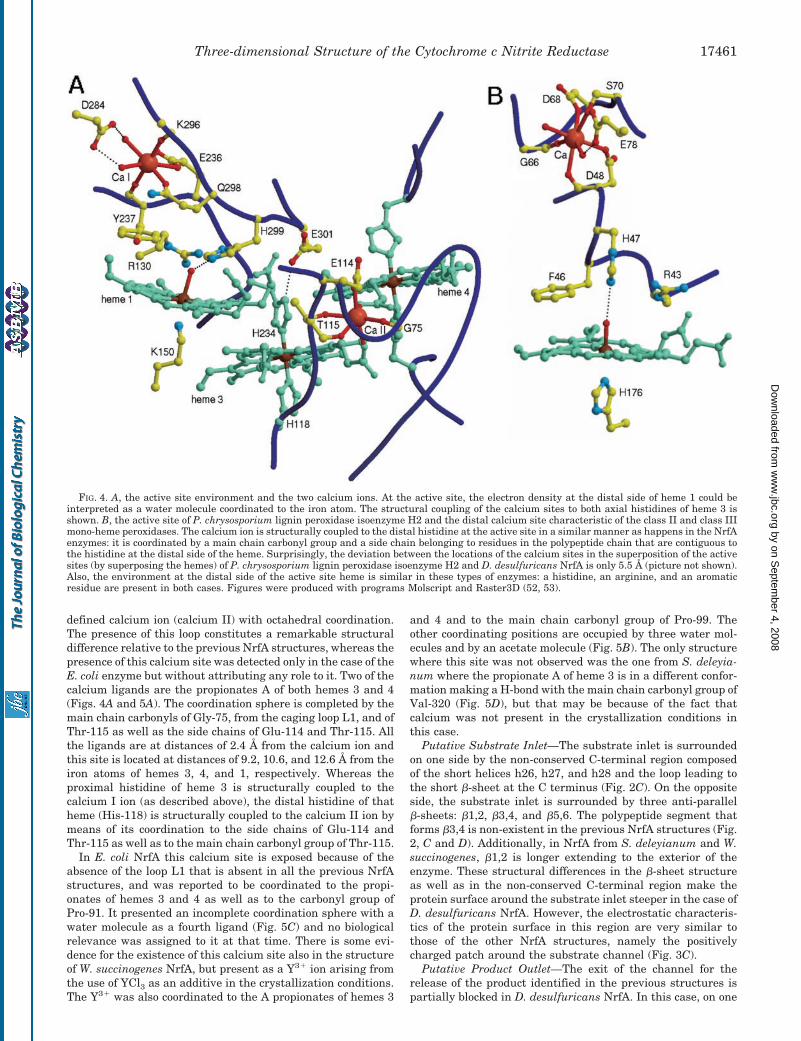
FIG. 5. The calcium II site and the environments of hemes 3 and 4 in the NrfA structures of D. desulfuricans (A), W. succinogenes(B), E. coli (C), and S. deleyianum (D). The calcium ion is present in the structures from D. desulfuricans and E. coli but poorly defined in thelatter. In W. succinogenes NrfA a yttrium ion is present at this site with an acetate (act) ligand in its octahedral coordination sphere. The proximalhistidine of heme 3 is H-bonded to a carboxylic side chain in NrfA from D. desulfuricans, E. coli, and W. succinogenes but not in S. deleyianum NrfA.The distal histidines of hemes 3 and 4 are H-bonded to a methionine and a carboxylic side chain, respectively, only in D. desulfuricans NrfA.Figures were produced with programs Molscript and Raster3D (52, 53).
Three-dimensional Structure of the Cytochrome c Nitrite Reductase17462
by on Septem
ber 4, 2008 w
ww
.jbc.orgD
ownloaded from
absence of an obvious alternative channel for the release of theproduct as well as the conserved negative electrostatic poten-tial in this region suggests that it functions as the outlet for therelease of the ammonium ions also in D. desulfuricans NrfA.The loop partially blocking the product outlet in D. desulfuri-cans NrfA suggests that some dynamic behavior of the struc-ture in this region is necessary for the release of the product.One feasible path for this is through the narrow channel linedby the aromatic side chains of Tyr-73, Phe-92, and Phe-373(Fig. 2E), hindered by the side chain of His-94 that must moveaway to allow the release of the ammonium ions. Some dy-namic behavior of the side chain of the nearby Lys-93 as well asof the main chain of loop L2 at residues Lys-93 and His-94 andof loop L1 at residue Tyr-73 may also be necessary. The inter-action between the -orbitals of the aromatic side chains sur-rounding this channel and the ammonium protons may play arole in driving the ammonium ions to travel through the chan-nel. H-bond formation between the ammonium ions and theside chain of His-94 may also occur as a last step to assist therelease of the product.
DISCUSSION
The Calcium I Site and Comparison to Mono-Heme Peroxi-dases—Despite the lack of sequence and structural homology,the environment of the vacant distal side of the active siteheme of cytochrome c nitrite reductases is very similar to theenvironment of the distal side of the active site in the mono-heme peroxidases such as cytochrome c peroxidase and fungal,plant, and horseradish peroxidases (38–43). In both types ofenzymes, a histidine and an arginine are conserved near thefree axial coordinating position at the distal side of the activesite heme. In both cases, a reduction reaction of an oxygen-containing substrate occurs at the heme: nitrite is reduced toammonia in cytochrome c nitrite reductase while hydrogenperoxide is reduced to water in the peroxidases. The active sitehas evolved independently in these two families of enzymes toa similar solution for similar biological processes.
Mutational and kinetic studies established a key role for thedistal arginine in the active site of peroxidases in the rapidbinding of the substrate to the active site and in the cleavage ofthe O–O bond (44, 45). In the structure of the horseradishperoxidase-cyanide complex the distal arginine H-bonds thebound cyanide, thereby contributing to the stabilization of thecomplex (46). A similar interaction was proposed to be involvedin the stabilization of the bound peroxy transition state duringO–O bond cleavage. Recently, in the structures of the com-plexes of cytochrome c nitrite reductase with nitrite and hy-droxylamine, it was observed that the distal arginine also es-tablishes H-bonds with the nitrite and hydroxylamine bound inthe active site (37). This fact suggests a similar role of the distalarginine in the NrfA enzymes in the binding of the substrateand in the stabilization of the transition states of the variousintermediate products in the reduction of nitrite to ammonium.
Class II peroxidases such as the fungal peroxidases as wellas class III peroxidases such as the peanut and horseradishperoxidases contain two structural calcium ions. One of thesecalcium ions is located near the active site at its distal side andis surprisingly similar to the calcium I site in the NrfA enzymes(Fig. 4, A and B). This calcium ion in the peroxidases is struc-turally coupled to the distal histidine at the active site in asimilar manner as happens in the NrfA enzymes as describedabove. In both cases, the calcium ion is coordinated by a mainchain carbonyl group and a side chain belonging to residues inthe polypeptide chain that are contiguous to the histidine at thedistal side of the heme (Fig. 4, A and B). Despite the fact thatthe structure is not conserved, the location of these calciumsites relatively to the active site heme is similar in both types
of enzymes as is illustrated by the deviation of only 5.5 Åbetween the locations of these calcium sites in the superposi-tion of the active sites of Phanerochaete chrysosporium ligninperoxidase isoenzyme H2 and D. desulfuricans NrfA by super-posing the hemes (picture not shown). For the lignin peroxidaseas well as for the manganese peroxidase from P. chrysosporium(47, 48) it was shown that the release of this calcium ion fromthe structure resulted in the inactivation of the enzyme andthis was related with the fact that upon release of the calciumion the heme becomes coordinated by the histidine at the distalside. The coordination spheres of the calcium sites in theseperoxidases are similar to the ones of the calcium I site in theNrfA enzymes. In both cases, the calcium sites are partiallyexposed to the solvent, and are coordinated by two water mol-ecules and two main chain carbonyl groups. The coordinationsphere is completed by the side chains of two aspartates andone serine in the case of the peroxidases, while in the NrfAenzymes the side chains of one glutamate and one glutamineare the additional ligands. These facts suggest that the bindingconstants for these calcium sites may be of similar magnitudes,and therefore that the calcium ion at the calcium I site in theNrfA will be strongly bound to the protein as happens in thecase of the peroxidases.
The high similarity between the distal calcium ion near theactive site in the peroxidases and the calcium I site in the NrfAenzymes suggests that the release of this ion in the NrfAenzymes will have the same structural consequences that areobserved for the peroxidases. Apart from the contribution tothe positive electrostatic potential at the active site cavity, thecalcium I ion in the NrfA enzymes may play an important rolein keeping the distal histidine away from the iron atom at theactive site heme for the enzyme to be active. Previously re-ported studies regarding the influence of calcium in the activityof the NrfA enzymes from S. deleyianum and E. coli do not referto the complete loss of activity of the enzyme in the absence ofcalcium ions in solution (13, 49) contradicting the above hy-pothesis. However, the way these studies were carried outsuggests a labile character of the putative calcium site in-volved. For this reason, the calcium II site may be the oneinvolved in the effects observed in the NrfA activity in thesestudies. Because of its incomplete coordination sphere in thecase of NrfA from S. deleyianum and E. coli as well as in W.succinogenes (Fig. 5, B–D), the calcium II calcium site may beexpected to be labile in these cases. On the other hand, thecoordination sphere of the calcium I site (Fig. 4A) suggests thatit will be strongly bound to the protein and very difficult to bereleased as happens in the case of the peroxidases. The effectsreported in the above studies were not interpreted according tothe existence of the calcium II site maybe because this site wasnot clearly revealed at that time. A calcium ion was not presentat this site in the S. deleyianum NrfA structure, the first NrfAstructure reported. However, as already mentioned above, thecrystallization conditions in this case did not contain calciumions and, for this reason, the existence of this calcium site alsoin S. deleyianum NrfA cannot be excluded.
The Calcium II Site—There is some evidence for the exist-ence of the calcium II site also in the crystal structures of NrfAfrom E. coli and W. succinogenes. However, in these cases theions are located at the protein surface and only three proteinligands are present in their coordination spheres: both thepropionates A of hemes 3 and 4 and a main chain carbonylgroup, being the other coordination positions exposed to thesolvent. In D. desulfuricans NrfA this calcium site is identifiedfor the first time with complete octahedral coordination byprotein ligands (Fig. 5A). In this case, the additional ligandsare the main chain carbonyl group of Gly-75 and both side
Three-dimensional Structure of the Cytochrome c Nitrite Reductase 17463
by on Septem
ber 4, 2008 w
ww
.jbc.orgD
ownloaded from
chains of Glu-114 and Thr-115. In NrfA from E. coli and W.succinogenes, Glu-114 and Thr-115 are mutated to residueswhose side chains are unable to coordinate the calcium. Addi-tionally Gly-75 in D. desulfuricans NrfA belongs to a loop thatis non-existent in the other NrfA structures (Fig. 5, B–D). Thefact that the calcium II ion is firmly bound by protein ligands innearly perfect octahedral coordination in D. desulfuricans NrfAsuggests that it may play an important physiological role aris-ing either from an electrostatic effect, because of its positivecharge, or simply from structural effects. From these, perhapsthe most obvious effect will be the conformation of the addi-tional loop that coordinates the calcium ion and that exertsinfluence in the solvent accessibility to the interior of the pro-tein, namely to the cavity of heme 4. Both effects can influencethe redox potential of the hemes involved.
To evaluate the electrostatic effect of the charge of the cal-cium ion at the calcium II site in the nearby hemes, the elec-trostatic potential arising from the charged groups in the pro-tein structure was mapped at the surface of the hemes (Fig.3B). The electrostatic potential was calculated without consid-ering the presence of the membrane anchoring subunit (NrfH)whose structure is not known. The presence of this subunit willinfluence the electrostatic potential in the vicinity of hemes 2and 5, which are located near the putative binding region ofthat subunit. For this reason, the electrostatic potentialsmapped at the surface of these hemes may be different from theones at the physiological conditions, in the NrfHA complex.However, despite the absence of the NrfH subunit, the electro-static potentials mapped at the surface of hemes 1, 3, and 4 areexpected to be a good approximation of the electrostatic poten-tial in the physiological conditions, because these hemes aremore distantly located from the binding region of the NrfHsubunit. The results show that the active site heme 1 is par-tially subjected to positive electrostatic potential that arisesfrom the positively charged residues that line the substratechannel. On the contrary, hemes 3 and 4 near the productoutlet are subjected to strongly negative electrostatic potential.This is in accordance with the change from positive to negativeelectrostatic potential along the channel that traverses themonomer from the substrate inlet to the product outlet as hasbeen reported in previous structures (11). In the left-hand sideof Fig. 3B, it can be observed that the positive electrostaticpotential arising from the calcium ion at the calcium II site isrestricted to the regions of the propionates involved in itscoordination, whereas the porphirin rings of hemes 3 and 4 aresubjected to strong negative electrostatic potential. This factsuggests that the negative electrostatic potential in the hemecavities overwhelms the positive electrostatic contribution ofthe calcium ion being in accordance with the highly negativeoxidation-reduction potentials assigned to these hemes. Com-bining information from potentiometric titrations and EPR andMossbauer spectroscopic studies, the oxidation-reduction po-tentials of hemes 3 and 4 were assigned the redox potentials of�480 and �400 mV,1 respectively. These redox potentials re-flect the destabilization of the reduced state of the hemebecause of the influence of the strong negative electrostaticpotentials at the heme cavities, despite the presence of thecalcium ion near the propionates of the hemes. These factssuggest that the main influence of this calcium site in the redoxpotential of the nearby hemes will not be the electrostaticeffect.
The calcium ion at the calcium II site may also have animportant structural role, namely in stabilizing the conforma-tion of the loop formed by residues 68 to 76 (loop L1 in Fig. 2C)by means of its coordination to the calcium ion by the mainchain carbonyl group of Gly-75. The very low solvent accessi-
bility of heme 4 in D. desulfuricans NrfA (3.0 Å2) comparedwith the high solvent accessibilities reported for this heme inthe previous NrfA structures lacking the loop L1 (61.7 to 83.6Å2) (12), suggests that the presence of this loop in D. desulfu-ricans NrfA exerts a strong influence in the solvent accessibil-ity of this heme. Heme 3 also presents a very low solventaccessibility (2.5 Å2) but this also happens in the previousstructures (2.5 to 17.5 Å2). The low solvent accessibility of thecavities of hemes 3 and 4 suggests that the dielectric constantof the environment around these hemes will be low. This factwill tend to increase the effect of the negative electrostaticinfluence in the hemes increasing the destabilization of thereduced state and this may explain the highly negative redoxpotentials determined for these hemes. In this way, the lowsolvent accessibility of heme 4, caused by the loop L1, and theconsequent low dielectric constant of the environment aroundthat heme may exert a more effective influence in the redoxpotential of the heme than the electrostatic charge of the cal-cium ion caged by the loop. Furthermore, the heme propionatesthat coordinate the calcium ion may become protonated in itsabsence reducing the electrostatic effect caused by the absenceof the positive charge of the calcium ion. These facts suggestthat the role of the calcium II ion may be of structural nature,by stabilizing the conformation of the loop L1, rather than anelectrostatic effect caused by its positive charge.
Another factor that may influence the redox potential ofhemes 3 and 4 is the type of the acceptor group of the H-bondsformed by the axial histidines of those hemes. In D. desulfuri-cans NrfA, the N�-1 atoms of the proximal histidine of heme 3(His-234) and the distal histidine of heme 4 (His-434) areH-bonded by the carboxylic side chains of Glu-301 and Glu-428,respectively (Fig. 5A). The H-bond interaction between axialheme histidines and carboxylic side chains has been shown toinfluence the redox potential of hemes (50). The carboxylate issupposed to deprotonate partially or completely the histidine toform the imidazolate thus increasing the strength of the histi-dine-iron bond and shifting downwards the redox potential ofthe heme. The negative shift of the redox potential of the hemewas shown to depend on the geometry of the H-bond interac-tion, namely the C � O–H angle defined by the hydrogen ridingthe N�-1 atom of the histidine and the carbonyl group of theH-bonded carboxylic function. The maximum effect reportedwas achieved for an ideal geometry corresponding to a C � O–Hangle of 120° causing a shift of about �100 mV in the redoxpotential of the heme. This effect contributes also for the highlynegative redox potentials assigned to hemes 3 and 4, beinganother factor counteracting the influence of the positivecharge of the calcium ion at the calcium II site on those redoxcenters. Additionally, changes in the geometry of the H-bondsbetween the N�-1 atoms of the histidines and the carboxylicgroups, because of dynamical behavior of the protein structure,may provide a means for a dynamic modulation of the potentialof the hemes involved.
On the contrary to what happens with the other heme axialhistidines, the acceptor group for the H-bonds formed by theN�-1 atom of both axial histidines of heme 3 and the distalhistidine of heme 4 are not conserved in the NrfA enzymes. Theproximal histidine of heme 3 is H-bonded to a carboxylic sidechain in the NrfA structures from D. desulfuricans, W. succi-nogenes, and E. coli but not in the one from S. deleyianum (Fig.5, A–D). On the other hand, the distal histidine of heme 3 isH-bonded to water molecules in all the currently known struc-tures with the exception of the one from D. desulfuricans whereit is unusually H-bonded to the sulfur atom of a methionineside chain (Met-337). The distal histidine of heme 4 is alsoH-bonded to water molecules in all the previous structures but
Three-dimensional Structure of the Cytochrome c Nitrite Reductase17464
by on Septem
ber 4, 2008 w
ww
.jbc.orgD
ownloaded from

it is observed to make a H-bond to a carboxylic side chain(Glu-428) in the case of D. desulfuricans NrfA. In contrast tothis, the H-bonding to the N�-1 atom of the axial histidines ofhemes 2 and 5 has been conserved in the NrfA structures. Withthe exception of the proximal histidine of heme 5, at the dimerinterface, which is H-bonded to the propionate D of the ncs-related heme 5* at the opposite side of the dimer interface, theother heme axial histidines are H-bonded to main chain car-bonyls or water molecules. The non-conserved H-bonds be-tween the axial histidines of hemes 3 and 4 and carboxylic sidechains that are observed in some of the NrfA structures may bea factor involved in the tuning of the redox potential of thosehemes, as referred above. The variability of this structuralmotif within this family of enzymes reflects the variability ofthe environment around those hemes, mainly in the regioncloser to the putative product outlet, a region with a low degreeof conserved residues, in contrast with the highly conservedregion of the active site cavity. To test these hypotheses addi-tional theoretical and experimental work must be done and isplanned in the near future.
Acknowledgments—We thank Claudio Soares (Instituto de Tecnolo-gia Quımica e Biologica, Oeiras, Portugal) for fruitful discussions andOliver Einsle (MPI Biochemie, Martinsried, Germany) for providing thecoordinates of NrfA from S. deleyianum when they were not available atthe Protein Data Bank and the supporting staff at beamlines BM-14and ID14-EH4 are also acknowledged.
REFERENCES
1. Liu, M. C., and Peck, H. D., Jr. (1981) J. Biol. Chem. 256, 13159–131642. Steenkamp, D. J., and Peck, H. D., Jr. (1981) J. Biol. Chem. 256, 5450–54583. Schroder, I., Roberton, A. M., Bokranz, M., Unden, G., Bocher, R., and Kroger,
K. (1985) Arch. Microbiol. 140, 380–3864. Moura, I., Bursakov, S., Costa, C., and Moura, J. J. G. (1997) Anaerobe 3,
279–2905. Ferguson, S. J. (1998) Curr. Opin. Chem. Biol. 2, 182–1936. Simon, J. (2002) FEMS Microbiol. Rev. 26, 2857. Dias, J. M., Than, M. E., Humm, A., Huber, R., Bourenkov, G. P., Bartunik,
H. D., Bursakov, S., Calvete, J., Caldeira, J., Carneiro, C., Moura, J. J.,Moura, I., and Romao, M. J. (1999) Struct. Fold. Des. 7, 65–79
8. Simon, J., Pisa, R., Stein, T., Eichler, R., Klimmek, O., and Gross, R. (2001)Eur. J. Biochem. 268, 5776–5782
9. Simon, J., Gross, R., Einsle, O., Kroneck, P. M. H., Kroger, A., and Klimmek,O. (2000) Mol. Microbiol. 35, 686–696
10. Einsle, O., Messerschmidt, A., Stach, P., Bourenkov, G. P., Bartunik, H. D.,Huber, R., and Kroneck, P. M. H. (1999) Nature 400, 476–480
11. Einsle, O., Stach, P., Messerschmidt, A., Simon, J., Kroger, A., Huber, R., andKroneck, P. M. H. (2000) J. Biol. Chem. 275, 39608–39616
12. Bamford, V. A., Angove, H. C., Seward, H. E., Thomson, A. J., Cole, J. A., Butt,J. N., Hemmings, A. M., and Richardson, D. J. (2002) Biochemistry 41,2921–2931
13. Stach, P., Einsle, O., Schumacher, W., Kurun, E., and Kroneck, P. M. H. (2000)J. Inorg. Biochem. 79, 381–385
14. Costa, C., Macedo, A., Moura, I., Moura, J. J. G., LeGall, J., Berlier, Y., Liu,M. Y., and Payne, W. J. (1990) FEBS Lett. 276, 67–70
15. Liu, M. C., Costa, C., and Moura, I. (1994) in Inorganic Microbial SulphurMetabolism (Peck, H. D., Jr., and LeGall, J., eds) Vol. 243, pp. 303–319,Academic Press, Inc., London, UK
16. Costa, C., Moura, J. J. G., Moura, I., Wang, Y., and Huynh, B. H. (1996) J. Biol.Chem. 271, 23191–23196
17. Nicholls, A., Sharp, K. A., and Honig, B. (1991) Proteins 11, 281–296
18. Nielsen, H., Engelbrecht, J., Brunak, S., and von Heijne, G. (1997) ProteinEng. 10, 1–6
19. Laemmli, U. K. (1970) Nature 227, 680–68520. Dias, J. M., Cunha, C. A., Teixeira, S., Almeida, G., Costa, C., Lampreia, J.,
Moura, J. J. G., Moura, I., and Romao, J. M. (2000) Acta Crystallogr. Sect.D 56, 215–217
21. Otwinowski, Z., and Minor, W. (1997) in Macromolecular Crystallography,Part A (Carter, C. W., Jr., and Sweet, R. M., eds) Vol. 276, pp. 307–326,Academic Press, New York
22. Terwilliger, T. C., and Berendzen, J. (1999) Acta Crystallogr. Sect. D 55,849–861
23. La Fortelle, E. D., and Bricogne, G. (1997) in Macromolecular Crystallography,Part A (Carter, C. W., Jr., and Sweet, R. M., eds) Vol. 276, pp. 472–494,Academic Press, New York
24. Abrahams, J. P., and Leslie, A. G. W. (1996) Acta Crystallogr. Sect. D 52,30–42
25. Cowtan, K. (1994) Joint CCP4 and ESF-EACBM Newsletter on Protein Crys-tallography 31, 34–38
26. Collaborative Computational Project, N. (1994) Acta Crystallogr. Sect. D 50,760–763
27. Navaza, J. (1994) Acta Crystallogr. Sect. D 50, 157–16328. Matthews, B. W. (1968) J. Mol. Biol. 33, 491–49729. Jones, T. A., Zou, J.-Y., Cowan, S. W., and Kjelgaard, M. (1991) Acta Crystal-
logr. Sect. A 47, 110–11930. Murshudov, G. N., Vagin, A. A., and Dodson, E. J. (1997) Acta Crystallogr.
Sect. D 53, 240–25531. Perrakis, A., Morris, R. J., and Lamzin, V. S. (1999) Nat. Struct. Biol. 6,
458–46332. Hooft, R. W. W., Vriend, G., Sander, C., and Abola, E. E. (1996) Nature 381,
272–27233. Page, C. C., Moser, C. C., Chen, X., and Dutton, P. L. (1999) Nature 402, 47–5234. Kassner, R. J. (1973) J. Am. Chem. Soc. 95, 2674–267735. Rafferty, S. P., Pearce, L. L., Barker, P. D., Guillemette, J. G., Kay, C. M.,
Smith, M., and Mauk, A. G. (1990) Biochemistry 29, 9365–936936. Aubert, C., Leroy, G., Bruschi, M., Wall, J. D., and Dolla, A. (1997) J. Biol.
Chem. 272, 15128–1513437. Einsle, O., Messerschmidt, A., Huber, R., Kroneck, P. M. H., and Neese, F.
(2002) J. Am. Chem. Soc. 124, 11737–1174538. Poulos, T. L., Freer, S. T., Alden, R. A., Edwards, S. L., Skogland, U., Takio, K.,
Eriksson, B., Xuong, N., Yonetani, T., and Kraut, J. (1980) J. Biol. Chem.255, 575–580
39. Poulos, T. L., Edwards, S. L., Wariishi, H., and Gold, M. H. (1993) J. Biol.Chem. 268, 4429–4440
40. Sundaramoorthy, M., Kishi, K., Gold, M. H., and Poulos, T. L. (1994) J. Biol.Chem. 269, 32759–32767
41. Kunishima, N., Fukuyama, K., Matsubara, H., Hatanaka, H., Shibano, Y., andAmachi, T. (1994) J. Mol. Biol. 235, 331–344
42. Schuller, D. J., Ban, N., Huystee, R. B., McPherson, A., and Poulos, T. L. (1996)Structure 4, 311–321
43. Gajhede, M., Schuller, D. J., Henriksen, A., Smith, A. T., and Poulos, T. L.(1997) Nat. Struct. Biol. 4, 1032–1038
44. Vitello, L. B., Erman, J. E., Miller, M. A., Wang, J., and Kraut, J. (1993)Biochemistry 32, 9807–9818
45. Rodriguez-Lopez, J. N., Smith, A. T., and Thornley, R. N. (1996) J. Biol. Chem.271, 4023–4030
46. Henriksen, A., Smith, A. T., and Gajhede, M. (1999) J. Biol. Chem. 274,35005–35011
47. George, S. J., Kvaratskhelia, M., Dilworth, M. J., and Thorneley, R. N. F.(1999) Biochem. J. 344, 237–244
48. Sutherland, G. R. J., Zapanta, L. S., Tien, M., and Aust, S. D. (1997) Biochem-istry 36, 3654–3662
49. Angove, H. C., Cole, J. A., Richardson, D. J., and Butt, J. N. (2002) J. Biol.Chem. 277, 23374–23381
50. Goodin, D. B., and McRee, D. E. (1993) Biochemistry 32, 3313–332451. Barton, G. J. (1993) Protein Eng. 6, 37–4052. Kraulis, P. J. (1991) J. Appl. Crystallogr. 24, 946–95053. Merritt, E. A., and Bacon, D. J. (1997) in Macromolecular Crystallography,
Part B (Carter, C. W., Jr., and Sweet, R. M., eds) Vol. 277, pp. 505–524,Academic Press, New York
Three-dimensional Structure of the Cytochrome c Nitrite Reductase 17465
by on Septem
ber 4, 2008 w
ww
.jbc.orgD
ownloaded from
Copyright © 2022 FDOKUMEN


![Nickel[iron-sulfur]-selenium-containing hydrogenases from Desulfovibrio baculatus (DSM 1743). Redox centers and catalytic properties](https://static.fdokumen.com/doc/165x107/6316e17bc32ab5e46f0dfeb7/nickeliron-sulfur-selenium-containing-hydrogenases-from-desulfovibrio-baculatus.jpg)