A partial proteome reference map of the wine lactic acid bacterium Oenococcus oeni ATCC BAA-1163
10
rsob.royalsocietypublishing.org Research Cite this article: Mohedano ML, Russo P, de los Rı ´os V, Capozzi V, Ferna ´ndez de Palencia P, Spano G, Lo ´pez P. 2014 A partial proteome reference map of the wine lactic acid bacterium Oenococcus oeni ATCC BAA-1163. Open Biol. 4: 130154. http://dx.doi.org/10.1098/rsob.130154 Received: 11 September 2013 Accepted: 3 February 2014 Subject Area: microbiology Keywords: Oenococcus oeni, proteome, two-dimensional electrophoresis Author for correspondence: Paloma Lo ´pez e-mail: [email protected] † These authors contributed equally to this study. Electronic supplementary material is available at http://dx.doi.org/10.1098/rsob.130154. A partial proteome reference map of the wine lactic acid bacterium Oenococcus oeni ATCC BAA-1163 Marı ´a de la Luz Mohedano 1,† , Pasquale Russo 1,2,† , Vivian de los Rı ´os 1,† , Vittorio Capozzi 2 , Pilar Ferna ´ndez de Palencia 1 , Giuseppe Spano 2 and Paloma Lo ´pez 1 1 Departamento de Microbiologı ´a Molecular y Biologı ´a de las Infecciones, Centro de Investigaciones Biolo ´gicas, Consejo Superior de Investigaciones Cientı ´ficas, Calle Ramiro de Maeztu 9, Madrid 28040, Spain 2 Dipartimento di Scienze Agrarie, degli Alimenti e dell’Ambiente, University of Foggia, Via Napoli 25, Foggia 71122, Italy 1. Summary Oenococcus oeni is the main lactic acid bacterium that carries out the malolactic fermentation in virtually all red wines and in some white and sparkling wines. Oenococcus oeni possesses an array of metabolic activities that can modify the taste and aromatic properties of wine. There is, therefore, industrial interest in the proteins involved in these metabolic pathways and related transport sys- tems of this bacterium. In this work, we report the characterization of the O. oeni ATCC BAA-1163 proteome. Total and membrane protein preparations from O. oeni were standardized and analysed by two-dimensional gel electro- phoresis. Using tandem mass spectrometry, we identified 224 different spots corresponding to 152 unique proteins, which have been classified by their putative function and subjected to bioinformatics analysis. 2. Introduction Oenococcus oeni is the most important lactic acid bacterium (LAB) in the wine industry, because it de-acidifies the wine following the alcoholic fermentation, a process termed malolactic fermentation. Nevertheless, the harsh wine environment represents a challenge to the survival of O. oeni and can strongly affect the successful outcome of the vinification [1]. Therefore, a better under- standing of the molecular mechanisms related to the stress adaptation and technical performance of O. oeni is crucial for the characterization and selection of strains for industrial purposes [2,3]. Currently, a fully complete genome sequence is available only for the O. oeni PSU-1 strain [4]. However, the genome sequences of ATCC BAA-1163 and AWRIB129, AWRIB202, AWRIB304, AWRIB318, AWRIB418, AWRIB419, AWRIB422, AWRIB429, AWRIB548, AWRIB553, AWRIB568, AWRIB576 are in assembly [5]. Unluckily, a recombinant approach in O. oeni usually does not give satisfac- tory results. Despite many attempts over the years, only in rare cases has it been possible to insert foreign genetic material in O. oeni [6–9]. Therefore, the main strategy for molecular analysis of O. oeni has been based on the heterologous expression of genes or clusters of interest. In particular, the malic acid metab- olism has been extensively investigated as well as the production of & 2014 The Authors. Published by the Royal Society under the terms of the Creative Commons Attribution License http://creativecommons.org/licenses/by/3.0/, which permits unrestricted use, provided the original author and source are credited.
Transcript of A partial proteome reference map of the wine lactic acid bacterium Oenococcus oeni ATCC BAA-1163

rsob.royalsocietypublishing.org
ResearchCite this article: Mohedano ML, Russo P, de
los Rıos V, Capozzi V, Fernandez de Palencia P,
Spano G, Lopez P. 2014 A partial proteome
reference map of the wine lactic acid
bacterium Oenococcus oeni ATCC BAA-1163.
Open Biol. 4: 130154.
http://dx.doi.org/10.1098/rsob.130154
Received: 11 September 2013
Accepted: 3 February 2014
Subject Area:microbiology
Keywords:Oenococcus oeni, proteome, two-dimensional
electrophoresis
Author for correspondence:Paloma Lopez
e-mail: [email protected]
†These authors contributed equally to this
study.
Electronic supplementary material is available
at http://dx.doi.org/10.1098/rsob.130154.
& 2014 The Authors. Published by the Royal Society under the terms of the Creative Commons AttributionLicense http://creativecommons.org/licenses/by/3.0/, which permits unrestricted use, provided the originalauthor and source are credited.
A partial proteome reference mapof the wine lactic acid bacteriumOenococcus oeni ATCC BAA-1163Marıa de la Luz Mohedano1,†, Pasquale Russo1,2,†,
Vivian de los Rıos1,†, Vittorio Capozzi2, Pilar Fernandez de
Palencia1, Giuseppe Spano2 and Paloma Lopez1
1Departamento de Microbiologıa Molecular y Biologıa de las Infecciones, Centro deInvestigaciones Biologicas, Consejo Superior de Investigaciones Cientıficas, Calle Ramiro deMaeztu 9, Madrid 28040, Spain2Dipartimento di Scienze Agrarie, degli Alimenti e dell’Ambiente, University of Foggia,Via Napoli 25, Foggia 71122, Italy
1. SummaryOenococcus oeni is the main lactic acid bacterium that carries out the malolactic
fermentation in virtually all red wines and in some white and sparkling wines.
Oenococcus oeni possesses an array of metabolic activities that can modify the
taste and aromatic properties of wine. There is, therefore, industrial interest
in the proteins involved in these metabolic pathways and related transport sys-
tems of this bacterium. In this work, we report the characterization of the
O. oeni ATCC BAA-1163 proteome. Total and membrane protein preparations
from O. oeni were standardized and analysed by two-dimensional gel electro-
phoresis. Using tandem mass spectrometry, we identified 224 different spots
corresponding to 152 unique proteins, which have been classified by their
putative function and subjected to bioinformatics analysis.
2. IntroductionOenococcus oeni is the most important lactic acid bacterium (LAB) in the wine
industry, because it de-acidifies the wine following the alcoholic fermentation,
a process termed malolactic fermentation. Nevertheless, the harsh wine
environment represents a challenge to the survival of O. oeni and can strongly
affect the successful outcome of the vinification [1]. Therefore, a better under-
standing of the molecular mechanisms related to the stress adaptation and
technical performance of O. oeni is crucial for the characterization and selection
of strains for industrial purposes [2,3].
Currently, a fully complete genome sequence is available only for the O. oeniPSU-1 strain [4]. However, the genome sequences of ATCC BAA-1163 and
AWRIB129, AWRIB202, AWRIB304, AWRIB318, AWRIB418, AWRIB419,
AWRIB422, AWRIB429, AWRIB548, AWRIB553, AWRIB568, AWRIB576 are in
assembly [5].
Unluckily, a recombinant approach in O. oeni usually does not give satisfac-
tory results. Despite many attempts over the years, only in rare cases has it been
possible to insert foreign genetic material in O. oeni [6–9]. Therefore, the main
strategy for molecular analysis of O. oeni has been based on the heterologous
expression of genes or clusters of interest. In particular, the malic acid metab-
olism has been extensively investigated as well as the production of

rsob.royalsocietypublishing.orgOpen
Biol.4:130154
2
compounds affecting wine quality or safety such as flavoursor biogenic amine formation (for a comprehensive review,
see Bartowsky [10]). The adaptive stress response of O. oeniin wine uses principally three mechanisms: (i) the establish-
ment of a proton-motive force generated by the malolactic
fermentation [11], (ii) the synthesis of heat-shock proteins
[12] and (iii) physico-chemical changes in the membrane
composition [13,14].
Although the significance of these mechanisms is clear,
few authors have addressed the study of O. oeni from a pro-
teomic perspective. Two-dimensional gel analysis of total
cellular proteins provides a global overview on the real bio-
logical response under specific conditions. Currently, only
few comparative analyses of O. oeni proteomes have been
reported [15–17]. Detection of 81 out of 186 differently
expressed peptides was observed during starvation con-
ditions, although none of the differently expressed spots
were identified [16]. Cecconi et al. [17] were able to obtain
high-resolution two-dimensional gel maps of O. oeni, when
investigating the acclimation of freeze-dried cultures. The
same authors attributed the changes in two-dimensional gel
profiles mainly to a set of 20 cytosolic proteins involved in
stress response and metabolic processes. None of these
studies provided information on the membrane proteome.
In this work, we report the characterization of the mem-
brane and cytosolic proteomes of O. oeni ATCC BAA-1163
[18]. In addition, we describe a standardized and optimized
method to obtain membrane protein extracts from O. oeni.In the course of this study, 224 different spots corresponding
to 152 different proteins of the O. oeni ATCC BAA-1163
proteome have been identified, classified by their putative
function and subjected to bioinformatics analysis. This partial
proteomic approach has allowed us to draw a proteome
reference map of O. oeni, which could help subsequent com-
parative analysis as well as representing a valuable source of
information for the validation of annotated genes.
3. Material and methods3.1. Bacterial strain and growth conditionsOenococcus oeni ATCC BAA-1163 (formerly O. oeni IOB 8413,
[18]) was grown at 308C in FT80 broth [19] at pH 5.3, under
anaerobic conditions (AnaeroGen 3.5 l, Oxoid, Basingstoke,
Hampshire, UK). A draft version of O. oeni ATCC BAA-
1163 genome is available under the GenBank accession
number AAUV00000000.1. This draft has a GC content of
37.9% and it contains a total of 1398 predicted genes and
280 pseudogenes.
3.2. Preparation of protein extractsStock culture of O. oeni ATCC BAA-1163 (stored at 2808C)
was diluted 1 : 1000 in 1800 ml of fresh medium. When
the culture reached the end of the exponential phase
(OD620 ¼ 1.2), cells were used to prepare both total and
membrane protein extracts. Bacteria were sedimented by cen-
trifugation (11 000�g for 20 min at 48C) and washed with
200 ml of cold 0.1 M potassium phosphate buffer (0.1 M
KH2PO4, 0.1 M K2HPO4, Merck, Darmstadt, Germany) pH
6.0. The cell pellet then was frozen at 2808C. Experiments
were performed in triplicate.
3.3. Preparation of total extractsThe frozen pellet was defrosted and resuspended in 60 ml of
0.1 M potassium phosphate buffer supplemented with
30 mg ml21 of protease-free DNase I (Roche Diagnostics
GmbH, Mannheim, Germany), 10 mM MgSO4 (Merck) and
1� concentrate Complete Protease Inhibitor cocktail (Roche
Diagnostics GmbH). Total extracts were obtained by passing
the cells four times through a French Press at 12 000 lb in22.
Cell debris was removed by centrifugation (1252�g, 15 min,
48C). The supernatant was designated total protein extracts
and frozen at 2808C.
3.4. Preparation of membrane extractsThirty millilitres of total protein extracts were diluted with
120 ml of 0.1 M sodium carbonate (Merck) pH 11.0, in
order to optimize the linearization of membrane vesicles.
This solution was gently shaken at 08C for 1 h then centri-
fuged at 154 980�g, 40 min at 48C in a Beckman L8-60M
Ultracentrifuge (Type 90 Ti rotor). The pellet was resus-
pended in 10 ml of 50 mM Tris/HCl (Merck) pH 7.3, 1 mM
MgCl2 (Merck). After another sedimentation by ultracentrifu-
gation (154 980�g, 40 min at 48C), the membranes were
resuspended in 2 ml of 50 mM ammonium bicarbonate
(Merck). To increase the proportion of membrane proteins,
the membrane extracts were supplemented with 13.3 ml of
2 : 1 v/v trifluoroethanol (Sigma-Aldrich, St. Louis, MO,
USA)/chloroform (Merck) and stored on ice for 1 h as reported
by Pessione et al. [20]. After centrifugation (10 000�g, 5 min
at 48C), the upper phase was recovered and dried using a
vacuum centrifuge. To increase the solubilization of membrane
proteins, the samples were treated with the zwitterionic deter-
gent amidosulfobetaine-14 (ASB-14) (Calbiochem, Darmstadt,
Germany) [21] and tributyl phosphine (TBP) (Bio-Rad Labora-
tories, Hercules, CA, USA) by resuspension in the rehydration
buffer A (7 M urea (Merck), 2 M thiourea (GE Healthcare,
Piscataway, NJ, USA), 1% ASB-14, 40 mM Tris, 2 mM TBP).
The samples then were submitted to three cycles of sonication
for 30 s and cooling on ice for 30 s. Finally, they were aliquoted
and frozen at 2808C.
3.5. Protein extraction and determinationThe protein preparations were solubilized for 1 h at room
temperature with agitation. The total protein concentration
present in the extracts was determined by two methods: RC
DC Protein Assay (Bio-Rad Laboratories) Kit and Quant-iT
technology by the use of the Qubit quantification platform
(Invitrogen, Paisley, UK).
3.6. Two-dimensional electrophoresis analysisTriplicate two-dimensional gels for each sample were carried
out as described below: 50 ml aliquots of extracts containing
150 mg of proteins were mixed with a buffer containing 7 M
urea, 2 M thiourea, 4% CHAPS, 20 mM dithiothreitol, 1%
carrier ampholytes pH 3–11 (GE Healthcare), up to a final
volume of 100 ml, and applied by Cup Loading to 18 cm
IPG strips pH 3–11 NL (GE Healthcare) previously rehy-
drated with 340 ml of the isoelectrofocusing (IEF) buffer
(7 M urea, 2 M thiourea, 4% CHAPS, 0.5% carrier ampholites

rsob.royalsocietypublishing.orgOpen
Biol.4:130154
3
pH 3–11, 1.2% DeStreak (GE Healthcare)). The first dimen-sion was run at 0.05 mA/IPG strip in the IPGphor IEF
System (GE Healthcare) with voltage increase in five steps:
300 V h21 for 3 h, linear gradient up to 1000 V in 6 h, linear gra-
dient up to 8000 V in 3 h and 8000 V h21 until 42000 V h21 was
reached. After IEF separation, the strips were equilibrated
twice for 10 min in 50 mM Tris–HCl (pH 8.8), 6 M urea, 30%
glycerol (Merck), 2% sodium dodecyl sulfate (SDS; Merck)
and trace amounts of bromophenol blue (Sigma-Aldrich).
The first equilibration solution contained 1% dithiothreitol
(Bio-Rad Laboratories), whereas the second contained 4%
iodoacetamide (Sigma-Aldrich). The second dimension (SDS-
PAGE) was performed on 12.5% polyacrylamide gels (1 mm,
16 � 15 cm). Gels were run at 7 mA per gel overnight maintain-
ing buffer temperature at 48C. Staining was carried out with
SYPRO Ruby Protein Gel Stain from Invitrogen as follows:
gel was fixed in 10% methanol, 7% acetic acid (Merck) for
30 min, incubated overnight in SYPRO Ruby staining solution,
washed twice with 10% methanol, 7% acetic acid for 30 min,
and finally washed twice with water for 10 min. Gels were
then scanned in a Typhoon 9400 Variable Mode Imager
(GE Healthcare) equipped with a 532 nm excitation laser
with the emission filter adjusted to 610 nm and 100 mm resol-
ution. The photomultiplier tube settings were modified to
optimize sensitivity to background ratios.
The protein spots present in the two-dimensional gels
were matched and quantified with the DECYDER v. 7.0 soft-
ware (GE Healthcare). For this quantification, stained spots
were matched in all gels and used for normalization of the
average intensity. The selected spots were automatically
excised with the Spot Picker (GE Healthcare).
3.7. In-gel protein digestion and sample preparationTwo hundred and twenty-four spots of interest from Sypro
Ruby-stained two-dimensional gels were excised manually,
deposited in 96-well plates and processed automatically in
a Proteineer DP (Bruker Daltonics, Bremen, Germany). The
digestion protocol used was based on that of Shevchenko
et al. [22] with minor variations: gel plugs were washed
first with 50 mM ammonium bicarbonate (Sigma-Aldrich)
and then with acetronile (ACN) (Scharlau, Barcelona, Spain)
prior to reduction with 10 mM dithiothreitol in 25 mM
ammonium bicarbonate solution, and alkylation was carried
out with 55 mM iodoacetamine in 50 mM ammonium bicar-
bonate solution. Gel slices were then rinsed first with
50 mM ammonium bicarbonate and then with ACN, and
finally were dried under a stream of nitrogen. Modified por-
cine trypsin (sequencing grade; Promega, Madison, WI, USA)
at a final concentration of 16 ng ml21 in 25% ACN, 50 mM
ammonium bicarbonate solution was added and the diges-
tion took place at 37 8C for 6 h. The reaction was stopped
by adding 0.5% trifluoroacetic acid (TFA) (Sigma-Aldrich) for
peptide extraction. The tryptic-eluted peptides were dried
by speed-vacuum centrifugation and were resuspended in
4 ml of 33% ACN, 16% isopropanol, 0.5% TFA (MALDI sol-
ution). A 0.8 ml aliquot of each peptide mixture was
deposited onto a 389-well OptiTOF Plate (Applied Biosystems,
Framingham, MA, USA) and allowed to dry at room tempera-
ture. A 0.8 ml aliquot of matrix solution (3 mg ml21 of a-cyano-
4-hydroxycinnamic acid (Bruker Daltonik) in MALDI solution
was then deposited onto dried digest and allowed to dry at
room temperature.
3.8. MALDI peptide mass fingerprinting, MS/MSanalysis and database searching
For MALDI-TOF/TOF analysis, samples were automatically
acquired in an ABi 4800 MALDI-TOF/TOF mass spec-
trometer (Applied Biosystems) in positive ion reflector
mode (the ion acceleration voltage was 25 kV to MS acqui-
sition and 1 kV to MSMS) and the obtained spectra were
stored into the ABi 4000 SERIES EXPLORER Spot Set Manager.
PMF and MSMS fragment ion spectra were smoothed and
corrected to zero baseline using routines embedded in ABi
4000 SERIES EXPLORER software v. 3.6. Each PMF spectrum
was internally calibrated with the mass signals of trypsin
autolysis ions to reach a typical mass measurement accuracy
of less than 25 ppm. Known trypsin and keratin mass signals
as well as potential sodium and potassium adducts (þ21 and
þ39 Da) were removed from the peak list. To submit the com-
bined PMF and MS/MS data to MASCOT software v. 2.1
(Matrix Science, London, UK), GPS EXPLORER v. 4.9 was
used, searching in the non-redundant NCBI protein database.
The mass tolerance for precursors was set to +50 ppm and to
+0.3 Da for MS/MS fragment ions. Peptide identifications
were accepted when scored at greater than 95.0% probability
by the MASCOT algorithm [23].
4. Results and discussion4.1. Protein extraction and protein identificationIn order to obtain a reference map representative of both
cytoplasmic and membrane proteins, two protein fractions
were prepared from three independent O. oeni ATCC BAA-
1163 cultures for further fractionation by two-dimensional
electrophoresis. Thus, after disruption of the cells, we gener-
ated a total fraction containing both cytoplasmic and
membrane proteins. Membrane proteins are generally
poorly represented on two-dimensional gels owing to their
low abundance and poor solubility, and to self-aggregation
during extraction or fractionation [24]. Recently, Choi et al.[25] reported that a sodium carbonate precipitation coupled
with ultracentrifugation is an effective method to increase
the proportion of cytoplasmic membrane proteins in extracts
of Streptococcus pneumoniae. We therefore optimized a similar
protocol to generate the membrane preparation. In addition,
to improve the solubility of proteins, we subjected the
sodium carbonate precipitated samples to a delipidation pro-
cess by treatment with trifluoroethanol/chloroform, as
previously described [20,26], and to membrane solubilization
by treatment with the zwitterionic detergent ASB-14 [21].
Finally, the membrane extracts were sonicated to increase
protein solubility prior to analysis. The two protein prep-
arations were analysed by two-dimensional electrophoresis,
as described above, which detected focused polypeptides in
the range of pI 4.5–10.3. The second dimension separated
in the range from 150 to 10 kDa. Image analysis of repre-
sentative two-dimensional gels from total and membrane
preparations prepared from the same extract of O. oeni ATCC
BAA-1163 revealed a high degree of overlap of the spots,
although the relative proportions of them were different
(figure 1). Therefore, we chose the total protein fractions analy-
sis for the identification of 203 spots, which are depicted in
figure 1a (gel A). An additional 21 spots, mainly characterized
(a)
97.4
66.2
45.5
31.0
21.0
14.4
4.5 pH 10.3 4.5 pH 10.3
Mr
(kD
a)
(b)
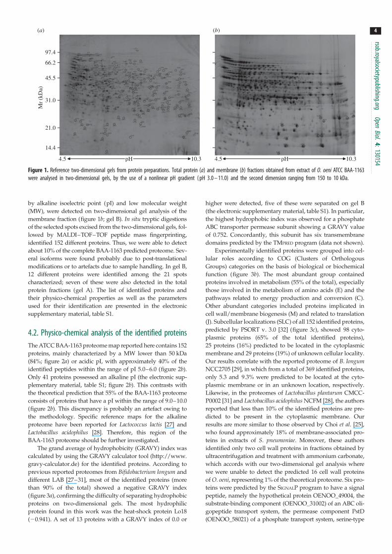
Figure 1. Reference two-dimensional gels from protein preparations. Total protein (a) and membrane (b) fractions obtained from extract of O. oeni ATCC BAA-1163were analysed in two-dimensional gels, by the use of a nonlinear pH gradient ( pH 3.0 – 11.0) and the second dimension ranging from 150 to 10 kDa.
rsob.royalsocietypublishing.orgOpen
Biol.4:130154
4
by alkaline isoelectric point (pI) and low molecular weight
(MW), were detected on two-dimensional gel analysis of the
membrane fraction (figure 1b; gel B). In situ tryptic digestions
of the selected spots excised from the two-dimensional gels, fol-
lowed by MALDI–TOF–TOF peptide mass fingerprinting,
identified 152 different proteins. Thus, we were able to detect
about 10% of the complete BAA-1163 predicted proteome. Sev-
eral isoforms were found probably due to post-translational
modifications or to artefacts due to sample handling. In gel B,
12 different proteins were identified among the 21 spots
characterized; seven of these were also detected in the total
protein fractions (gel A). The list of identified proteins and
their physico-chemical properties as well as the parameters
used for their identification are presented in the electronic
supplementary material, table S1.
4.2. Physico-chemical analysis of the identified proteinsThe ATCC BAA-1163 proteome map reported here contains 152
proteins, mainly characterized by a MW lower than 50 kDa
(84%; figure 2a) or acidic pI, with approximately 40% of the
identified peptides within the range of pI 5.0–6.0 (figure 2b).
Only 41 proteins possessed an alkaline pI (the electronic sup-
plementary material, table S1; figure 2b). This contrasts with
the theoretical prediction that 55% of the BAA-1163 proteome
consists of proteins that have a pI within the range of 9.0–10.0
(figure 2b). This discrepancy is probably an artefact owing to
the methodology. Specific reference maps for the alkaline
proteome have been reported for Lactococcus lactis [27] and
Lactobacillus acidophilus [28]. Therefore, this region of the
BAA-1163 proteome should be further investigated.
The grand average of hydrophobicity (GRAVY) index was
calculated by using the GRAVY calculator tool (http://www.
gravy-calculator.de) for the identified proteins. According to
previous reported proteomes from Bifidobacterium longum and
different LAB [27–31], most of the identified proteins (more
than 90% of the total) showed a negative GRAVY index
(figure 3a), confirming the difficulty of separating hydrophobic
proteins on two-dimensional gels. The most hydrophilic
protein found in this work was the heat-shock protein Lo18
(20.941). A set of 13 proteins with a GRAVY index of 0.0 or
higher were detected, five of these were separated on gel B
(the electronic supplementary material, table S1). In particular,
the highest hydrophobic index was observed for a phosphate
ABC transporter permease subunit showing a GRAVY value
of 0.752. Concordantly, this subunit has six transmembrane
domains predicted by the TMPRED program (data not shown).
Experimentally identified proteins were grouped into cel-
lular roles according to COG (Clusters of Orthologous
Groups) categories on the basis of biological or biochemical
function (figure 3b). The most abundant group contained
proteins involved in metabolism (55% of the total), especially
those involved in the metabolism of amino acids (E) and the
pathways related to energy production and conversion (C).
Other abundant categories included proteins implicated in
cell wall/membrane biogenesis (M) and related to translation
(J). Subcellular localizations (SLC) of all 152 identified proteins,
predicted by PSORT v. 3.0 [32] (figure 3c), showed 98 cyto-
plasmic proteins (65% of the total identified proteins),
25 proteins (16%) predicted to be located in the cytoplasmic
membrane and 29 proteins (19%) of unknown cellular locality.
Our results correlate with the reported proteome of B. longumNCC2705 [29], in which from a total of 369 identified proteins,
only 5.3 and 9.3% were predicted to be located at the cyto-
plasmic membrane or in an unknown location, respectively.
Likewise, in the proteomes of Lactobacillus plantarum CMCC-
P0002 [31] and Lactobacillus acidophilus NCFM [28], the authors
reported that less than 10% of the identified proteins are pre-
dicted to be present in the cytoplasmic membrane. Our
results are more similar to those observed by Choi et al. [25],
who found approximately 18% of membrane-associated pro-
teins in extracts of S. pneumoniae. Moreover, these authors
identified only two cell wall proteins in fractions obtained by
ultracentrifugation and treatment with ammonium carbonate,
which accords with our two-dimensional gel analysis where
we were unable to detect the predicted 16 cell wall proteins
of O. oeni, representing 1% of the theoretical proteome. Six pro-
teins were predicted by the SIGNALP program to have a signal
peptide, namely the hypothetical protein OENOO_49004, the
substrate-binding component (OENOO_31002) of an ABC oli-
gopeptide transport system, the permease component PstD
(OENOO_58021) of a phosphate transport system, serine-type

(a)35
30
25
20
15
10
5
0
45
40
35
30
25
20
15
10
5
0
0–10 10–20 20–30 30–40 40–50MW50–60 60–70 70–80 80–90 90–100 >100
3–4 4–5 5–6 6–7 7–8pI
8–9 9–10 10–11 11–12 12–13
perc
enta
ge o
f se
ries
(%
)pe
rcen
tage
of
seri
es (
%)
(b)
Figure 2. Comparative analyses of the predicted and the actual proteome ofO. oeni ATCC BAA-1163. Frequency distribution of the MW (a) and pI (b) ofthe whole set of putative proteins encoded by the genome (white bars) andof the 152 proteins identified in this work (black bars).
40
35
30
25
20
15
10
5
0
GRAVY
COG
C
SLC
C CM U E CW
G E F H I P Q D V T M N U O J K L R S _
<–0.8 –0.8 to –0.6 –0.6 to –0.4 –0.4 to –0.2 –0.2 to 0 0 to 0.2 0.2 to 0.4 >0.4
perc
enta
ge o
f se
ries
(%
)
40
35
30
25
20
15
10
5
0
perc
enta
ge o
f se
ries
(%
)
40
50
60
70
30
20
10
0
perc
enta
ge o
f se
ries
(%
)
(a)
(b)
(c)
Figure 3. Comparative analyses of the predicted and the actual proteome ofO. oeni ATCC BAA-1163. Frequency distribution of the GRAVY index (a), COGcategories (b) and subcellular location (c) of the theoretical proteome (whitebars) and of the identified proteins (black bars).
rsob.royalsocietypublishing.orgOpen
Biol.4:130154
5
D-Ala-D-Ala carboxypeptidase OENOO_60073, located at
the cytoplasmic membrane level as well as the UDP-glucose
6-dehydrogenase OENOO_64076 (with unknown subcellular
location) and the UTP-glucose-1-phosphate uridylyltransferase
OENOO_58029 (cytoplasmic location).
4.3. Hypothetical proteinsThe DNA sequence of O. oeni ATCC BAA-1163 genome is still
in a provisional state of annotation. Nevertheless, all the pro-
teins described in this study were identified by reference to
this genome.
Proteomic analysis is a powerful tool to prove the existence
of hypothetical gene products. In this work, we confirmed the
occurrence of 14 proteins previously designated as hypothetical
that should now be considered as real proteins (the electronic
supplementary material, table S2). A BLAST search of these
proteins against the Conserved Domain Database (http://
www.ncbi.nlm.nih.gov/Structure/cdd/cdd.shtml) and simi-
larity to homologues predicted a possible function for
10 hypothetical proteins based on the conserved domains (the
electronic supplementary material, table S2). SMART biotools
(http://smart.embl-heidelberg.de/) was used for the motif
search. In particular, we found that the hypothetical protein
OENOO_53026 could be a secreted small protein. This pre-
diction is supported by its unknown SLC and the occurrence
of a transmembrane domain. Extra-cytoplasmic proteins often
have roles in establishing and maintaining interactions between
a bacterium and its environment. In O. oeni, secreted proteins
have been reported for their proteolytic activity, crucial for sur-
vival in the wine environment [33]. One hypothetical protein,
OENOO_37002, possesses the domain characteristics of the uni-
versal stress protein UspA, suggesting its involvement in stress
response. UspA is highly expressed in response to heat, star-
vation, exposure to antimicrobial agents and oxidative stress
[34,35]. Recently, Gury et al. [36] demonstrated that in L. plan-tarum NC8 the putative universal stress protein Usp1 is
involved in phenolic acid stress response. The GRAVY index
of the hypothetical stress protein (20.018) indicated the pres-
ence of hydrophobic regions suggesting their interaction with
the membrane. The cytoplasmic membrane is an important
barrier with the external environment and stress proteins can
be associated with cellular membrane fractions [15]. The role
of the small heat-shock protein Lo18 from O. oeni in the modu-
lation of membrane fluidity has been reported recently [37] and
the protein from O. oeni ATCC BAA-1163 (OENOO_66120) has
been identified in this work (the electronic supplementary
material, table S1).
A set of three proteins characterized by the domains
Metallophos (OENOO_06001 and OENOO_47010) and
PRTases_type I (OENOO_66092) with phosphoesterase func-
tions are possibly involved in nucleic acid biosynthesis
and repair. Hypothetical protein OENOO_54035 contains a
Glo_EDI_BRP-like domain, which is a characteristic region
in enzymes that degrade aromatic compounds. Hypothetical
protein OENOO_45020 carries the ACT domain charac-
teristic of metabolic enzymes regulated by the amino acid

rsob.royalsocietypublishing.or
6
concentration. Also, the conserved DUF4230 domain ofunknown function was detected in OENOO_49004. Finally,
the hypothetical protein OENOO_63029 seems to be impli-
cated in the pathway for peptidoglycan biosynthesis. No
domains were identified for the conserved hypothetical
protein (OENOO_38016), the site-specific DNA-methyl-
transferase (OENOO_53044) and the hypothetical proteins
OENOO_63062, OENOO_6403 and OENOO_6606. The latter
possesses three transmembrane domains and presumably is
an integral membrane protein.
gOpen
Biol.4:130154
4.4. Oenococcus oeni ATTC BAA-1163 strain-specificproteins
Recently, a comparative analysis has been carried out on all
of the 14 available O. oeni genomes [5]. The in silico analysis
revealed 2846 non-degenerate ORFs that were shown to
comprise the chromosomal pan genome of O. oeni, with
1165 of these being core ORFs conserved across all the strains
[5,38]. Accordingly, we investigated the occurrence of the
polypeptides identified in ATCC BAA-1163 within the gen-
omes of PSU-1 and the 12 sequenced AWRIB strains. Based
on the inferred protein sequence homology, we found that
genes encoding 14 proteins were present in the genome of
at least one of the considered strains, but not reported as
encoding proteins probably owing to an incorrect annotation
(the electronic supplementary material, table S3).
The genes encoding two proteins (oxidoreductase
OENOO_40005 and hypothetical protein OENOO_63029)
were absent in the genomes of all of the examined strains.
Furthermore, oxidoreductase OENOO_40005, a functionally
predicted nucleoside-diphosphate-sugar epimerase (the elec-
tronic supplementary material, table S1), had a 99% of
identity with its predicted homologues in Lactobacillus viniand Lactobacillus casei. Strain-specific genes of O. oeni with an
increased probability of being horizontally acquired from Lac-tobacillus spp. have been investigated in silico [5]. The analysis
of the surrounding region of OENOO_40005 revealed a down-
stream gene encoding for a phosphotransferase system (PTS)
cellobiose-specific component IIC, supporting the existence of
strain-dependent genomic insertion events correlated with
differences in carbohydrate utilization [5]. In this work, we
found that the hypothetical protein OENOO_63029 belonging
to the pyruvyl-transferases family (the electronic supplemen-
tary material, table S2) involved in peptidoglycan-associated
polymer biosynthesis was expressed in FT80 (figure 1), a
model semi-defined medium for growth of O. oeni containing
glucose and fructose as carbon sources. The hypothetical
protein OENOO_63029 coding gene is located between the
loci (EAV38986 and EAV39008) encoding the aminotransferase
OENOO_63016 and the PTS system, sucrose-specific IIBC com-
ponent OENOO_63040 in ATCC BAA-1163. Neither this gene
nor the corresponding locus is present in the genome of the
other 13 sequenced strains. This region contains genes encod-
ing for exopolysaccharides (EPS) biosynthesis and different
PTS systems, including a PTS for fructose-specific utilization.
However, in O. oeni differences in EPS production across the
strains have been associated with variation in the EPS loci
[5,38]. The strain-variable ability of O. oeni to synthesize EPS
has been investigated for its technological and biological inter-
est [39–41]. ATCC BAA-1163 was found to be a non-ropy
strain able to produce low amounts of EPS in an MRS-derived
media [40]. The same strain produces significantly more EPS
in a semi-defined medium supplemented with a mixture of
glucose and sucrose [41].
With regard to esterase C (OENOO_51026), the protein-
coding gene is absent in the genomes of the AWRIB422 and
AWRIB548 strains and it is truncated prior to the active site-
coding region in all the other strains, apart from AWRIB419
(the electronic supplementary material, table S3). This fre-
quency of non-functional genes correlates with a previous
study performed by Sumby et al. [42]. The authors reported
that sequencing of the EstC of 20 O. oeni strains, whose gen-
omes have not been characterized, revealed that in only four
strains the gene was not carrying an early stop codon. More-
over, the analysis of ATCC BAA-1163, PSU-1 and AWRIB429
revealed that only the former carries an entire EstC gene
[42]. The significance of the presence of truncated genes in
most of the strains is unclear but suggests that the gene
tends to be lost during evolution. This could be owing to the
existence of other esterases in O. oeni like EstA, which we
have not identified in this work, that could eliminate the
requirement for EstC. Sumby et al. [42] proposed that it
should be investigated whether EstC has an effect on strain-
specific differences in ester hydrolysis and synthesis in wine.
4.5. Overview of metabolic pathwaysA scheme for the most significant reconstructed metabolic
pathways based in the proteomic data (figure 1 and the elec-
tronic supplementary material, table S1) and supported by
the analysis of O. oeni ATCC BAA-1163 genome is illustrated
in figure 4. Oenococcus oeni is a heterofermentative microor-
ganism able to use hexoses and pentoses via the
phosphoketolase pathway [43]. Expression of enzymes of
central metabolism, such as the glycolytic (four out of 10 pro-
teins), pentose phosphate and pyruvate biosynthetic, and
metabolic (including ethanol production) pathways are
reported in the present map (figure 4). Also enzymes
involved in citrate and malic acid utilization were identified
(figure 4). In addition, we detected UTP-glucose-1-phosphate
uridylyltransferase (OENOO_58029), UDP-glucose 6-dehy-
drogenase (OENOO_64076) and UDP-glucose 4-epimerase
(OENOO_50027), which are involved in several pathways of
carbohydrate metabolism, such as pentose and glucuronate
interconversions, galactose, ascorbate and aldarate, starch
and sucrose, amino sugar and nucleotide sugar metabolism.
ATCC BAA-1163 has been described as being able to use L-ara-
binose, arabinan, sucrose and D-sorbitol through their
conversion to D-fructose [5]. However, in this work we were
unable to detect the corresponding enzymes.
Previous analyses of O. oeni strains have revealed a large
number of auxotrophies [44,45]. In particular, the require-
ment for amino acids seems to be related to a genomic
intra-species diversity [4,5]. We found enzymes involved in
several amino acid metabolic pathways, including methion-
ine biosynthesis from aspartate, glutamine utilization and
conversion in glutamic acid and arginine as well as aromatic
amino acids catabolism (figure 5). However, we were unable
to detect proteins for phenylalanine, tyrosine and tryptophan
biosynthesis, nor could we detect the coding genes in the
ATCC BAA-1163 genome. In agreement with these findings,
Remize et al. [45] showed that phenylalanine and tyrosine
were essential for growth of the strain in FT80 media and
they detected only a residual growth (20% of the control) in

malic acid
citrate
glucose
glucose-6-P glucose-1-P
dTDP-glucose
phosphoribosylpyrophosphate
purine andpyrimidine
metabolisms
dTDP-4-dehydro-6-deoxy-D-glucose
dTDP-4-dehydro-b-L-rhamnose
dTDP-L-rhamnose
UDP-glucose
UDP-galactose
EPS
fructose
fructose-6-Poxaloacetate
pyruvate
a-acetolactate
acetaldehyde
acetyl-CoA
acetoin
butanediol ethanol
diacetyl
O2
lactate
MLEOAD
LDHALS
ALD
DRAR
ALDH
PO
PK, ENO, PGM, PGK
CL
FK
G6Pl
UTPG1PU
dTDPG4,6D
dTDPG4DR
UDPG4E
G6P1DH,
RPPK
Figure 4. Proteomic and genomic prediction of metabolic pathways in O. oeni ATCC BAA-1163. Enzymatic reactions: continuous lines indicate proteomic detection ofthe catalysing enzyme; dashed arrow lines indicate detection of the enzyme-coding gene in the bacterial genome. MLE, malolactic enzyme; LDH, lactate dehy-drogenase; CL, citrate lyase; OAD, oxaloacetate decarboxylase; ALS, a-acetolactate synthase; ALD, a-acetolactate decarboxylase; AR, acetoin reductase; DR,diacetyl reductase; PGK, phosphoglycerate kinase; PGM, phosphoglycerate mutase; ENO, enolase; PK, pyruvate kinase; PO, pyruvate oxidase; ALDH, alcohol dehy-drogenase; FK, fructokinase; G6PI, glucose-6-phosphate isomerase; G6P1DH, glucose-6-phosphate 1-dehydrogenase; RPPK, ribose-phosphate pyrophosphokinase;UTPG1PU, UTP-glucose-1-phosphate uridylyltransferase; dTDPG4,6D, dTDP glucose 4,6-dehydratase; dTDPG4DR, dTDP-4-dehydrorhamnose reductase; UDPG4E,UDP-glucose 4-epimerase.
out
glutamine
glutamineNH3 + glutamate
tryptophantyrosine
phenylalanine
glutamate + carbamoyl-P
methionine
homoserine
aspartate-4-semialdehyde
4-phosphate-aspartate aspartate a-keto acids aroma compounds
arginine
citrulline
ornithine
putrescine
putrescine
in
GLNS
HSDH
ASADH
AAT
AAT
ADI
CPS
ABC Gln PotABCD
Figure 5. Proteomic and genomic prediction of amino acid metabolism in O. oeni ATCC BAA-1163. Enzymatic reactions: continuous arrow lines indicate proteomicdetection of the catalysing enzyme; dashed lines indicate detection of the enzyme-coding gene in the bacterial genome. ABC Gln, glutamine ABC transporter;PotABCD, putrescine ABC transporter; GLNS, glutamine synthetase; CPS, carbamoyl-phosphate synthase; ADI, arginine deiminase; AAT, arginine/aromatic aminoacids aminotransferase; ASADH, aspartate-semialdehyde dehydrogenase; HSDH, homoserine dehydrogenase.
rsob.royalsocietypublishing.orgOpen
Biol.4:130154
7
the absence of tryptophan supporting the predicted amino
acids auxotrophies.
After the alcoholic fermentation, the free amino acid concen-
tration in the must increases owing to the autolysis of the yeasts,
with arginine usually being the most abundant. In our proteomic
map (figure 1 and the electronic supplementary material, table
S1), we found the OENOO_57007 arginine deiminase (ADI), a
key enzyme in arginine catabolism ([46] and figure 5). Analysis
of the bacterial genome confirmed that ATCC BAA-1163 pos-
sesses the arcABC operon encoding the three enzymes that
constitute the ADI pathway involved in counteracting stress
and associated with the potential production of the toxic com-
pound putrescine (figure 5). Putrescine is one of the most
commonly occurring biogenic amines in wine [47] and can be
taken up into the cell via the ABC transporter complex PotABCD,
responsible for energy coupling to the transport system. As we

rsob.royalsocietypublishing.orgOpen
Biol.4:130154
8
detected PotA, the ATP-binding subunit (OENOO_19003 in theelectronic supplementary material, table S1) of the spermidine/
putrescine transporter during the growth of ATCC BAA-1163 in
a synthetic medium, it could be argued that the intake of polya-
mine is not purely a mechanism to cope with the harsh wine
environment. The importance of exchanging a wide variety of
substrates is supported by the transporters identified in this
work, namely two ABC-type oligopeptide transport systems,
three subunits of one (OENOO_31002, OENOO_30004
and OENOO_30005) and the ATP-binding cassette (OENOO_
31006) of the other and also the ATP-binding proteins of a
glutamine ABC transporter (OENOO_41008), a multiple
sugar ABC transporter (OENOO_37007) and an uncharacterized
ABC transporter (OENOO_65021). In addition, we identified
subunits of the ABC transporters associated with iron
(OENOO_57011 and OENOO_57012), cobalt (OENOO_
44010) and phosphate (OENOO_58021, OENOO_58023 and
OENOO_58024) metabolism.
We detected three proteins, namely UTP-glucose-1-
phosphate uridylyltransferase (OENOO_59028), dTDP glucose
4,6-dehydratase (OENOO_59030) and dTDP-4-dehydro-
rhamnose reductase (OENOO_59032), whose coding genes
are probably organized into an operon, and being part of
the nucleotide sugars biosynthetic pathway (figure 4) seem
to provide the substrates for the synthesis of heteropoly-
saccharides catalysed by glycosyltransferases, whose coding
genes are located in the specific region OENOO_63027 and
OENOO_63040 in the ATCC BAA-1163 genome.
In recent years, several works have investigated the contri-
bution of the malolactic fermentation and of the strain-specific
variability of O. oeni on the modulation of the wine’s flavour
profile [10,48,49]. For example, it is well known that compounds
such as diacetyl, acetoin and 2,3-butanediol from citric acid
metabolism via pyruvate can affect the aromatic complexity
of wine [50]. These molecules responsible for buttery and
nutty fragrances proceed from pyruvate by the activity of
the enzymes a-acetolactate synthase (OENOO_54033), a-aceto-
lactate decarboxylase (OENOO_54034), acetoin reductase
(OENOO_48023) and diacetyl reductase (OENOO_43013).
All these proteins were identified in our two-dimensional
electrophoresis analysis (figures 1 and 4; the electronic
supplementary material, table S1). Moreover, the enzymes
involved in conversion of citrate to pyruvate—two subunits
(a and b, OENOO_66031 and OENOO_66032) of the citrate
lyase, CitXG protein, which includes Apo-citrate lyase phos-
phoribosyl-dephospho-CoA transferase (OENOO_66029) and
the oxaloacetate decarboxylase (OENOO_66036)—were also
identified and their coding genes are located in a cluster that
also includes the determinant of the putative citrate transporter
MaeP [51]. Furthermore, we identified two putative aspartate/
aromatic amino acid aminotransferases (OENOO_47017 and
OENOO_60020). Transamination reactions have recently
attracted attention, because they are the first step for the syn-
thesis of important flavour or aroma compounds in amino
acid catabolism pathways. Finally, esters from microbial metab-
olism often underlie the fruit aroma of the wine [38,52]. Some
authors have suggested the use of purified esterases from
LAB, including O. oeni, as additives in winemaking [53,54].
For this reason, the identified protein (the electronic supplemen-
tary material, table S1) tributyrin esterase (OENOO_60072), and
in particular, the esterase C (OENOO_51026), which does not
belong to the core genome of O. oeni, deserves to be the focus
of further investigation. Thus, recently the esterase C from
O. oeni has been overproduced in Escherichia coli and biochemi-
cal characterization of the purified enzyme under conditions
relevant to winemaking indicate that indeed esterase C is a
potential candidate to alter the ester profile of wine [42].
5. Concluding remarksUsing standardized extraction techniques and two-dimensional
gel electrophoresis coupled with MS/MS analysis, we ident-
ified 224 different spot polypeptides, corresponding to 152
unique proteins, from O. oeni ATCC BAA-1163. This represents
approximately 10% of the BAA-1163 predicted proteome.
A total of 21 spots were associated with the membrane prep-
aration. The methodology was found to cause some bias in
the types of proteins identified, e.g. proteins in the MW range
of 30–50 kDa were over-represented compared with the pre-
dicted proteome, as were proteins in the pI range 5–6
(contrasting with the prediction that 55% of BAA-1163 proteins
have a pI within the range of 9–10). Hydrophobic and very
hydrophilic proteins were under-represented.
The analysis allowed the detection of a wide variety of
metabolic enzymes, including many involved in the synthesis
and catabolism of various carbohydrates, amino acids and
amines. This study should, therefore, be helpful to those
researching the biochemistry of O. oeni ATCC BAA-1163.
Acknowledgements. We thank Dr Stephen Elson for the critical reading ofthe manuscript. We thank S. Ciordia and I Zapico, National Centerfor Biotechnology a member of ProteoRed network for the helpwith mass spectrometry.
Data accessibility. The mass spectrometry proteomics data have beendeposited in the ProteomeXchange Consortium (http://proteome-central.proteomexchange.org) via the PRIDE partner repository [55]with the dataset identifier PXD000579 and doi: 10.6019/PXD000579.
Funding statement. This work was supported by European Union grantFP7-2008-FOOD-211441 BIAMFOOD and by grant AGL2012-40084-C03-01 from the Spanish Ministry of Economics and Competitiveness.
References
1. Lonvaud-Funel A. 1999 Lactic acid bacteria in thequality improvement and depreciation of wine. Ant.van Leeuwenhoek 76, 317 – 331. (doi:10.1023/A:1002088931106)
2. Coucheney F, Desroches N, Bou M, Tourdot-MarechalR, Dulau L, Guzzo J. 2005 A new approach forselection of Oenococcus oeni strains in order toproduce malolactic starters. Int. J. Food Microbiol.
105, 463 – 470. (doi:10.1016/j.ijfoodmicro.2005.04.023)
3. Capozzi V, Russo P, Beneduce L, Weidmann S,Grieco F, Guzzo J, Spano G. 2010 Technologicalproperties of Oenococcus oeni strains isolated fromtypical southern Italian wines. Lett. Appl. Microbiol.50, 327 – 334. (doi:10.1111/j.1472-765X.2010.02795.x)
4. Mills DA, Rawsthorne H, Parker C, Tamir D,Makarova K. 2005 Genomic analysis of Oenococcusoeni PSU-1 and its relevance to winemaking. FEMSMicrobiol. Rev. 29, 465 – 475. (doi:10.1016/j.fmrre.2005.04.011)
5. Borneman AR, McCarthy JM, Chambers PJ,Bartowsky EJ. 2012 Comparative analysisof the Oenococcus oeni pan genome reveals

rsob.royalsocietypublishing.orgOpen
Biol.4:130154
9
genetic diversity in industrially-relevant pathways.BMC Genomics 13, 373. (doi:10.1186/1471-2164-13-373)6. Dicks LMT. 1994 Transformation of Leuconostocoenos by electroporation. Biotechnol. Tech. 8,901 – 904. (doi:10.1007/BF02447736)
7. Zuniga M, Pardo I, Ferrer S. 2003 Conjugativeplasmid pIP501 undergoes specific deletions aftertransfer from Lactococcus lactis to Oenococcus oeni.Arch. Microbiol. 180, 367 – 373. (doi:10.1007/s00203-003-0599-3)
8. Beltramo C, Oraby M, Bourel G, Garmyn D, Guzzo J.2004 A new vector, pGID052, for genetic transfer inOenococcus oeni. FEMS Microbiol. Lett. 236, 53 – 60.(doi:10.1111/j.1574-6968.2004.tb09626.x)
9. Assad-Garcıa JS, Bonnin-Jusserand M, Garmyn D,Guzzo J, Alexandre H, Grandvalet C. 2008 Animproved protocol for electroporation of Oenococcusoeni ATCC BAA-1163 using ethanol as immediatemembrane fluidizing agent. Lett. Appl. Microbiol.47, 333 – 338. (doi:10.1111/j.1472-765X.2008.02435.x)
10. Bartowsky E. 2005 Oenococcus oeni and malolacticfermentation: moving into the molecular arena.Aust. J. Grape Wine Res. 11, 174 – 187. (doi:10.1111/j.1755-0238.2005.tb00286.x)
11. Salema M, Lolkema J, SanRamao MV, Dias MCL.1996 The proton motive force generated inLeuconostoc oenos by L-malate fermentation.J. Bacteriol. 178, 3127 – 3132.
12. Delmas F, Pierre F, Coucheney F, Divies C, Guzzo J.2001 Biochemical and physiological studies of thesmall heat shock protein Lo18 from the lactic acidbacterium Oenococcus oeni. J. Mol. Microbiol.Biotechnol. 3, 601 – 610.
13. Tourdot-Marechal R, Gaboriau D, Beney L, Divies C.2000 Membrane fluidity of stressed cells ofOenococcus oeni. Int. J. Food Microbiol. 55,269 – 273. (doi:10.1016/S0168-1605(00)00202-6)
14. Chu-Ky S, Tourdot-Marechal R, Marechal PA, GuzzoJ. 2005 Combined cold, acid, ethanol shocks inOenococcus oeni: effects on membrane fluidityand cell viability. Biochim. Biophys. Acta 1717,118 – 124. (doi:10.1016/j.bbamem.2005.09.015)
15. da Silveira MG, Baumgartner M, Rombouts FM, AbeeT. 2004 Effect of adaptation to ethanol on cytoplasmicand membrane protein profiles of Oenococcus oeni.Appl. Environ. Microbiol. 70, 2748 – 2755. (doi:10.1128/AEM.70.5.2748-2755.2004)
16. Zapparoli G. 2004 Colony dimorphism associatedwith stress resistance in Oenococcus oeni VP01 cellsduring stationary growth phase. FEMS Microbiol.Lett. 239, 261 – 265. (doi:10.1016/j.femsle.2004.08.047)
17. Cecconi D, Milli A, Rinalducci S, Zolla L, Zapparoli G.2009 Proteomic analysis of Oenococcus oeni freeze-dried culture to assess the importance of cellacclimatation to conduct malolactic fermentation inwine. Electrophoresis 30, 2988 – 2995. (doi:10.1002/elps.200900228)
18. Klaenhammer T et al. 2002 Discovering lactic acidbacteria by genomics. Ant. Van Leeuwenhoek 82,29 – 58. (doi:10.1023/A:1020638309912)
19. Cavin J, Prevost H, Lin J, Schmitt P, Divies C. 1989Medium for screening Leuconostoc oenos strainsdefective in malolactic fermentation. Appl. Environ.Microbiol. 55, 751 – 753.
20. Pessione E, Pessione A, Lamberti C, Coısson DJ,Riedel K, Mazzoli R, Bonetta S, Eberl L, Giunta C.2009 First evidence of a membrane-bound,tyramine and b-phenylethylamine producing,tyrosine decarboxylase in Enterococcus faecalis:a two-dimensional electrophoresis proteomic study.Proteomics 9, 2695 – 2710. (doi:10.1002/pmic.200800780)
21. Chevallet M et al. 1998 New zwitterionic detergentsimprove the analysis of membrane proteins by two-dimensional electrophoresis. Electrophoresis 19,1901 – 1909. (doi:10.1002/elps.1150191108)
22. Shevchenko A, Tomas H, Havlis J, Olsen JV, Mann M.2006 In-gel digestion for mass spectrometriccharacterization of proteins and proteomes. Nat.Protoc. 1, 2856 – 2860. (doi:10.1038/nprot.2006.468)
23. Perkins DN, Pappin DJ, Creasy DM, Cottrell JS. 1999Probability-based protein identification by searchingsequence databases using mass spectrometry data.Electrophoresis 20, 3551 – 3567. (doi:10.1002/(SICI)1522-2683(19991201)20:18,3551::AID-ELPS3551.3.0.CO;2-2)
24. Helbig AO, Heck AJ, Slijper M. 2010 Exploring themembrane proteome: challenges and analyticalstrategies. J. Proteomics 73, 868 – 878. (doi:10.1016/j.jprot.2010.01.005)
25. Choi CW, Yun SH, Kwon SO, Leem SH, Choi JS, YunCY, Kim SI. 2010 Analysis of cytoplasmic membraneproteome of Streptococcus pneumoniae by shotgunproteomic approach. J. Microbiol. 48, 872 – 876.(doi:10.1007/s12275-010-0220-9)
26. Deshusses JM et al. 2003 Exploitation of specificproperties of trifluoroethanol for extraction andseparation of membrane proteins. Proteomics 3,1418 – 1424. (doi:10.1002/pmic.200300492)
27. Drews O, Reil G, Parlar H, Gorg A. 2004 Setting upstandards and a reference map for the alkalineproteome of the Gram-positive bacteriumLactococcus lactis. Proteomics 4, 1293 – 1304.(doi:10.1002/pmic.200300720)
28. Majumder A, Cai L, Ejby M, Schmidt BG, LahtinenSJ, Jacobsen S, Svensson B. 2012 Two-dimensionalgel-based alkaline proteome of the probioticbacterium Lactobacillus acidophilus NCFM.Proteomics 12, 1006 – 1014. (doi:10.1002/pmic.201100455)
29. Yuan J et al. 2006 A proteome reference map andproteomic analysis of Bifidobacterium longumNCC2705. Mol. Cell. Proteomics 5, 1105 – 1118.(doi:10.1074/mcp.M500410-MCP200)
30. Wu R, Wang W, Yu D, Zhang W, Li Y, Sun Z, Wu J,Meng H, Zhang H. 2009 Proteomics analysis ofLactobacillus casei Zhang, a new probiotic bacteriumisolated from traditional home-made koumiss inInner Mongolia of China. Mol. Cell. Proteomics 8,2321 – 2338. (doi:10.1074/mcp.M800483-MCP200)
31. Zhu L et al. 2011 A reference proteomic database ofLactobacillus plantarum CMCC-P0002. PLoS ONE 6,e25596. (doi:10.1371/journal.pone.0025596)
32. Yu NY et al. 2010 PSORTb 3.0: improved proteinsubcellular localization prediction with refinedlocalization subcategories and predictive capabilitiesfor all prokaryotes. Bioinformatics 26, 1608 – 1615.(doi:10.1093/bioinformatics/btq249)
33. Folio P, Ritt JF, Alexandre H, Remize F. 2008Characterization of EprA, a major extracellularprotein of Oenococcus oeni with protease activity.Int. J. Food Microbiol. 127, 26 – 31. (doi:10.1016/j.ijfoodmicro.2008.05.039)
34. Kvint K, Nachin L, Diez A, Nystrom T. 2003 Thebacterial universal stress protein: function andregulation. Curr. Opin. Microbiol. 6, 140 – 145.(doi:10.1016/S1369-5274(03)00025-0)
35. Seifart Gomes C et al. 2011 Universal stress proteinsare important for oxidative and acid stress resistanceand growth of Listeria monocytogenes EGD-e in vitroand in vivo. PLoS ONE 6, e24965. (doi:10.1371/journal.pone.0024965)
36. Gury J, Seraut H, Tran NP, Barthelmebs L, WeidmannS, Gervais P, Cavin JF. 2009 Inactivation of PadR, therepressor of the phenolic acid stress response, bymolecular interaction with Usp1, a universal stressprotein from Lactobacillus plantarum, in Escherichiacoli. Appl. Environ. Microbiol. 75, 5273 – 5283. (doi:10.1128/AEM.00774-09)
37. Coucheney F, Gal L, Beney L, Lherminier J, Gervais P,Guzzo J. 2005 A small HSP, Lo18, interacts with thecell membrane and modulates lipid physical stateunder heat shock conditions in a lactic acidbacterium. Biochim. Biophys. Acta 1720, 92 – 98.(doi:10.1016/j.bbamem.2005.11.017)
38. Borneman AR, Bartowsky EJ, McCarthy J, ChambersPJ. 2010 Genotypic diversity in Oenococcus oeni byhigh-density microarray comparative genomehybridization and whole genome sequencing. Appl.Microbiol. Biotechnol. 86, 681 – 691. (doi:10.1007/s00253-009-2425-6)
39. Ibarburu I, Soria-Dıaz ME, Rodrıguez-Carvajal MA,Velasco SE, Tejero-Mateo P, Gil-Serrano AM,Irastorza A, Duenas MT. 2007 Growth andexopolysaccharide (EPS) production by Oenococcusoeni I4 and structural characterization of their EPSs.J. Appl. Microbiol. 103, 477 – 486. (doi:10.1111/j.1365-2672.2006.03266.x)
40. Ciezack G, Hazo L, Chambat G, Heyraud A, Lonvaud-Funel A, Dols-Lafargue M. 2010 Evidence forexopolysaccharides production by Oenococcus oeni strainsisolated from non-ropy wines. J. Appl. Microbiol. 108,499 – 509. (doi:10.1111/j.1365-2672.2009.04449.x)
41. Dimopoulou M, Hazo L, Dols-Lafargue M. 2012Exploration of phenomena contributing to thediversity of Oenococcus oeni exopolysaccharides.Int. J. Food Microbiol. 153, 114 – 122. (doi:10.1016/j.ijfoodmicro.2011.10.024)
42. Sumby KM, Grbin PR, Jiranek V. 2012 Characterizationof EstCOo8 and EstC34, intracellular esterases, from thewine-associated lactic acid bacteria Oenococcus oeniand Lactobacillus hilgardii. J. Appl. Microbiol. 114,413 – 422. (doi:10.1111/jam.12060)
43. Garvie EI. 1967 Leuconostoc oenos sp. nov. J. Gen.Microbiol. 48, 431 – 438. (doi:10.1099/00221287-48-3-431)

rsob.royalsocietypublishing.orgOpen
Biol.4:130154
10
44. Terrade N, Mira de Orduna R. 2009 Determination ofthe essential nutrient requirements of wine-relatedbacteria from the genera Oenococcus andLactobacillus. Int. J. Food Microbiol. 133, 8 – 13.(doi:10.1016/j.ijfoodmicro.2009.03.020)45. Remize F, Gaudin A, Kong Y, Guzzo J, Alexandre H,Krieger S, Guilloux-Benatier M. 2006 Oenococcusoeni preference for peptides: qualitative andquantitative analysis of nitrogen assimilation. Arch.Microbiol. 185, 459 – 469. (doi:10.1007/s00203-006-0116-6)
46. Tonon T, Bourdineaud JP, Lonvaud-Funel A. 2001The arcABC gene cluster encoding the argininedeiminase pathway of Oenococcus oeni, andarginine induction of a CRP-like gene. Res.Microbiol. 152, 653 – 661. (doi:10.1016/S0923-2508(01)01244-X)
47. Spano G et al. 2010 Biogenic amines in fermentedfood. Eur. J. Clin. Nutr. 64, 95 – 100. (doi:10.1038/ejcn.2010.218)
48. Bartowsky EJ, Borneman AR. 2011 Genomicvariations of Oenococcus oeni strains and thepotential to impact on malolactic fermentation andaroma compounds in wine. Appl. Microbiol.Biotechnol. 92, 441 – 447. (doi:10.1007/s00253-011-3546-2)
49. Ruız P, Izquierdo PM, Sesena S, Garcıa E, LlanosPalop M. 2012 Malolactic fermentation andsecondary metabolite production by Oenoccocusoeni strains in low pH wines. J. Food Sci. 77,579 – 585. (doi:10.1111/j.1750-3841.2012.02895.x)
50. Bartowsky EJ, Henschke PA. 2004 The ‘buttery’attribute of wine – diacetyl – desirability, spoilageand beyond. Int. J. Food Microbiol. 96, 235 – 252.(doi:10.1016/j.ijfoodmicro.2004.05.013)
51. Garcıa-Quintans N, Blancato V, Repizo G, Magni C,Lopez P. 2008 Citrate metabolism and aromacompound production in lactic acid bacteria. InMolecular aspects of lactic acid bacteria fortraditional and new applications (eds B Mayo,
P LopezG Perez-Martın), pp. 65 – 88. Kerala, India:Research Signpost.
52. Sumby KM, Grbin PR, Jiranek V. 2010 Microbialmodulation of aromatic esters in wine: currentknowledge and future prospects. Food Chem. 121,1 – 16. (doi:10.1016/j.foodchem.2009.12.004)
53. Matthews A, Grimaldi A, Walker M, Bartowsky E,Grbin P, Jiranek V. 2004 Lactic acid bacteria as apotential source of enzymes for use in vinification.Appl. Environ. Microbiol. 70, 5715 – 5731. (doi:10.1128/AEM.70.10.5715-5731.2004)
54. Sumby KM, Matthews AH, Grbin PR, Jiranek V. 2009Cloning and characterization of an intracellularesterase from the wine-associated lactic acidbacterium Oenococcus oeni. Appl. Environ. Microbiol.75, 6729 – 6735. (doi:10.1128/AEM.01563-09)
55. Vizcaino JA et al. 2013 The ProteomicsIdentifications (PRIDE) database and associatedtools: status in 2013. Nucleic Acids Res. 41,D1063 – D1069. (doi:10.1093/nar/gks1262)