Paneth cell trypsin is the processing enzyme for human defensin-5
Upload
independentCategory
view
0download
0
Protein and Peptide Letters, Accepted May 2009
1
Characterization of a new defensin from cowpea (Vigna
unguiculata (L.) Walp.)
Lara Padovan*1,2
, Ludovica Segat2, Alessandro Tossi
1, Tercilio Calsa Jr.
4, Ederson Akio Kido
4, Lucas
Brandao3, Rafael L. Guimarães
3, Valesca Pandolfi
4, Maria Clara Pestana-Calsa
4, Luís Carlos
Belarmino4, Ana Maria Benko-Iseppon
4, Sergio Crovella
2,4.
Affiliations
1. Department of Life Sciences, University of Trieste -
Via Giorgieri, 1 - 34127 TRIESTE, Italy
2. Genetic Service, IRCCS Burlo Garofolo and
Department of Developmental and Reproductive
Sciences, University of Trieste -Via dell'Istria, 65/1 -
34137 TRIESTE, Italy
3. Laboratório de Imunopatologia Keizo Azami (LIKA)
Federal University of Pernambuco Campus
Universitário, Cidade Universitária CEP: 50670-901
Recife - PE – Brazil
4. Universidade Federal de Pernambuco, Centro de
Ciências Biológicas, Departamento de Genética,
Recife, PE – Brazil
Corresponding author:
Lara Padovan
Genetic Service, IRCCS Burlo Garofolo and Department of Developmental and
Reproductive Sciences, University of Trieste -Via dell'Istria, 65/1 - 34137
TRIESTE, Italy
Tel. +39.040.3785422
Fax. +39.040.3785540
e-mail: [email protected]
Abstract
Using available Phaseoleae defensins in databases a putative defensin gene was isolated in
cowpea (Vigna unguiculata (L.) Walp.) and cloned from genomic cowpea DNA. The putative
mature defensin sequence displays the characteristic defensins residues arrangement, secondary
and tertiary structures were predicted and splicing analysis was performed. Using RT-PCR,
defensin expression and differences in response to biotic stimuli between infected and non
infected plants were tested.
Key words: antimicrobial peptides, cowpea mosaic virus, evolution, Fabaceae, plant
defensin, protein function.
GeneBank accession number for Vigna unguiculata defensin gene sequence: FJ94789.
Abbreviations:
EST = Expressed Sequences Tags
PDEF_VIGUN = Vigna unguiculata defensin
PCR = Polymerase Chain Reaction
RT-PCR = Reverse Transcription Polymerase
Chain Reaction
CPSMV = Cowpea Severe Mosaic Virus
Protein and Peptide Letters, Accepted May 2009
2
Introduction Antimicrobial peptides are important components of
the innate immune systems of all living organisms and
are widely distributed throughout the plant kingdom
[1, 2, 3]. In particular plant defensins play an
important role in host defense against pathogens,
having a direct antimicrobial effect against both fungi
and bacteria [1]. Defensins are cysteine-rich peptides
consisting of 45-54 amino acids and characterised by
the presence of conserved intramolecular disulfide
bonds [4, 5]. The three-dimensional structure of plant
defensins presents a triple-stranded, antiparallel β-
sheet platform overlaid by a single α-helix and is
stabilised by four disulfide bonds. It closely resembles
defensin structures observed in vertebrate and
invertebrate animals as well as moulds [1, 6, 7].
The first plant defensin was isolated from wheat in
1990 [8] and since then the number of identified plant
defensins number has rapidly increased [6]. In
particular, defensins from several leguminous species
were reported, such as Phaseolus vulgaris [9], Pisum sativum [5], Vicia faba [10] and Clitoria ternatea [11].
We report herein the characterisation of a putative
defensin in Vigna unguiculata (L.) Walp. (cowpea), a
leguminous plant grown extensively as a food and
fodder crop in West Africa, north-eastern Brazil, part
of the Middle East and southern regions of North
America [12, 13]. In V. unguiculata, the structures and
activities of two other plant defensins (formerly also
known as thionins) named Cp thionins I II have been
already described [14, 15]. We have identified another
region in the cowpea genome codifying for a novel
defensin, with a different primary structure to those
previously reported, but sharing with them the
distinctive features of plant defensins. This study
confirms that multiple defensins act within the host
defence pathways of important legume food crops, in
this as well as in other cultivated plants species (such
as sugarcane, maize and rice).
Materials and Methods Computational search for new defensins: sequences
from cowpea (V. unguiculata Taxonomy ID 3917
GenBank) and other Phaseoleae were obtained from
the National Centre for Biotechnology Information
(http://www.ncbi.nlm.nih.gov/sites/entrez). BLAST
searches were performed at the Gene Index Databases
(http://compbio.dfci.harvard.edu/tgi/cgi-
bin/tgi/Blast/index.cgi). Cowpea sequence tags (EST)
that could refer to a defensin were not found in Gene
Index databases, so common bean (Phaseolus vulgaris
that belongs to the same tribe as V. unguiculata) was
chosen as reference and its ESTs were blasted against
Phaseoleae sequences present in databases.
Computational searches showed interesting
similarities between P. vulgaris TC147 EST (a cluster
from Gene Index Databases) and sequences codifying
for defensins that belong to other plants families, in
particular sunflower (Helianthus annuus, AF141131
GenBank) and peach (Prunus persica, AY078426
GenBank) defensins. Thus, TC147 was chosen for
primers design to perform a heterologous PCR on V. unguiculata.
Primer design: primers were selected by using “Primer
3” software (http://frodo.wi.mit.edu) according to
default parameters and were: primer forward: 5’-
TCCATGGCTCGCTCTGTGTCTT-3’ and reverse 5’-
TGAAGTTTTAACAGTGTTTGGTGCACAAG-3'.
Genomic DNA and RNA extraction: Genomic DNA was
extracted from young leaves of the Brazilian cultivar
BR14-Mulato (kindly provided by EMBRAPA-CPAMN,
Teresina, Piauì State), using a modified CTAB (cetyl-
trimethyl-ammoniumbromide)-based protocol [16].
DNA concentration was determined
electrophoretically using known amounts of phage
Lambda/Hind III DNA marker as reference.
Total RNA was extracted from 30-days old plants of a
unique CPSMV (Cowpea Severe Mosaic Virus) resistant
genotype, named BRIT63, derived from a breeding
cross (BR14-Mulato x IT85F-2687) grown in the
greenhouse and either native or inoculated using a
standard protocol [17]. Leaves were harvested 30, 60,
90 min and 16 h after mechanical wounding with
Carborundum™ and virus inoculation . Negative
controls consisted in BRIT63 and IT85F (the CPSMV-
susceptible parental cultivar) samples, both neither
infected nor mechanically injured. Harvested plant
material was immediately frozen in liquid nitrogen and
stored at -80ºC until RNA extraction [18].
PCR: Genomic DNA of V. unguiculata was amplified in
a 25 μL volume containing 50 ng of DNA template; 6
mmol MgCl2, 200 mmol dNTPs, 10 pmol of each
primer, 1.0 U Taq DNA polymerase (AmpliTaq Gold –
PE Biosystem) and 2.5 L buffer 10x (500 mM KCl, 100
mM Tris-HCl pH 8.3, 1.4 mM MgCl2). Amplification was
carried out in an automated thermal cycler (Applied
Biosystem) according to the following program: an
initial denaturation at 95°C for 10 min, after which 40
cycles of: denaturation (20 s at 95°C), primer
annealing (30 s at 58°C) and primer extension (30 s at
72°C) were performed, followed by a final extension at
72°C for 7 min. PCR products were visualized on 2%
agarose gel.
Sequences analysis: Sequences were analysed with
SeqMan Genome Assembler (DNASTAR, Inc., Madison,
WI, USA), multiple alignments of the nucleotide and
amino acid sequences were performed using the
Protein and Peptide Letters, Accepted May 2009
3
CLUSTALW software (http://www.ebi.ac.uk/Tools/clustalw).
The candidate defensin nucleotide sequence (FJ94789
GenBank) was then analysed and translated into the
corresponding amino acid sequence using the
GENSCAN package (http://genes.mit.edu/GENSCAN.html) and
Expasy software (http://www.expasy.org/cgi-
bin/dna_aa). The obtained sequence was blasted
against available plant defensins in databases using
both the NCBI BLAST service
(http://www.ncbi.nlm.nih.gov/blast/Blast.cgi) and FASTA
Nucleotide Similarity Search at EMBL-EBI
(http://www.ebi.ac.uk/fasta33/nucleotide.html).
Disulfide-bonds formation was predicted using the
DISULFIND program at the PredictProtein server
http://www.predictprotein.org.
Structure prediction was carried out by identifying
equivalent sequence positions in the closest plant
defensin structure present in the PDB databank: 1GTP
belonging to a barley defensins
(http://www.rcsb.org/pdb/home/home.do).
Gene structure comparison: The predicted nucleotide
sequence encoding for PDEF_VIGUN protein was
aligned against the genome of Arabidopsis thaliana
(TAIR; http://www.arabidopsis.org) and Glycine max
(PHYTOZOME http://www.phytozome.net). Additionally, the
predicted protein was blasted against BAC sequences
from Medicago truncatula and Lotus japonicus
(http://www.kazusa.or.jp). The genome location with best
match was excised and compared to the genomic
sequence of V. unguiculata. For evaluation of the
splicing patterns, the exon/intron boundaries of A. thaliana and G. max were used, since these are well
defined in the databanks. Nevertheless it was
performed a Blastx search against the nr protein
databank and megaBlast against EST database (dbEST
and NordEST, a Brazilian cowpea EST database
initiative) to confirm these boundaries and to identify
the putative boundaries from V. unguiculata, M. truncatula and L. japonicus. Subsequently, the
nucleotide sequences were aligned with ClustalW and
manually adjusted to fit the ATG initiation codon of
the ORFs, as well as the remaining coding positions in
the first and second exons, as observed in the Blastx
step. Signal peptide and the probable cleavage site
was identified with SignalP [19].
RT-QPCR: Retro-transcription and quantitative
amplification were performed by using the RT-PCR Core
kit (Applied Biosystems, Foster City, CA) following the
manufacturer’s protocol. Real-time PCR was performed
on the obtained cDNA by using the SYBR Green I
chemistry and Corbett Rotor Gene 3000 real time PCR
platform (Corbett Research). After an initial
denaturation step at 95°C for 10 min, forty cycles were
performed by two steps at 95°C for 16 s, and 60°C for 1
min. We used PCR primers designed in the exons
flanking regions: primer forward 5’-
CTGGTGGCCACTGAGATG-3’ and reverse 5’-
TCACACATGGTCCCTTGAAA-3’ in order to avoid
genomic DNA amplification. Plant defensin primers
concentrations were optimized in order to determine
the minimum concentration required to give the
maximal signal (ΔRn), the lowest threshold cycle (Ct)
and minimizing non-specific amplification.
The specificity of defensin amplification was verified by
melting temperature assay (MTA) protocol including a
slow cooling from 95°C to 60°C in 20 min at the end of
PCR. The Corbett Rotor Gene 3000 MTA software
allowed the identification of the point at which the re-
association occurred (flexus point) as well as the
melting temperature and the melting curve.
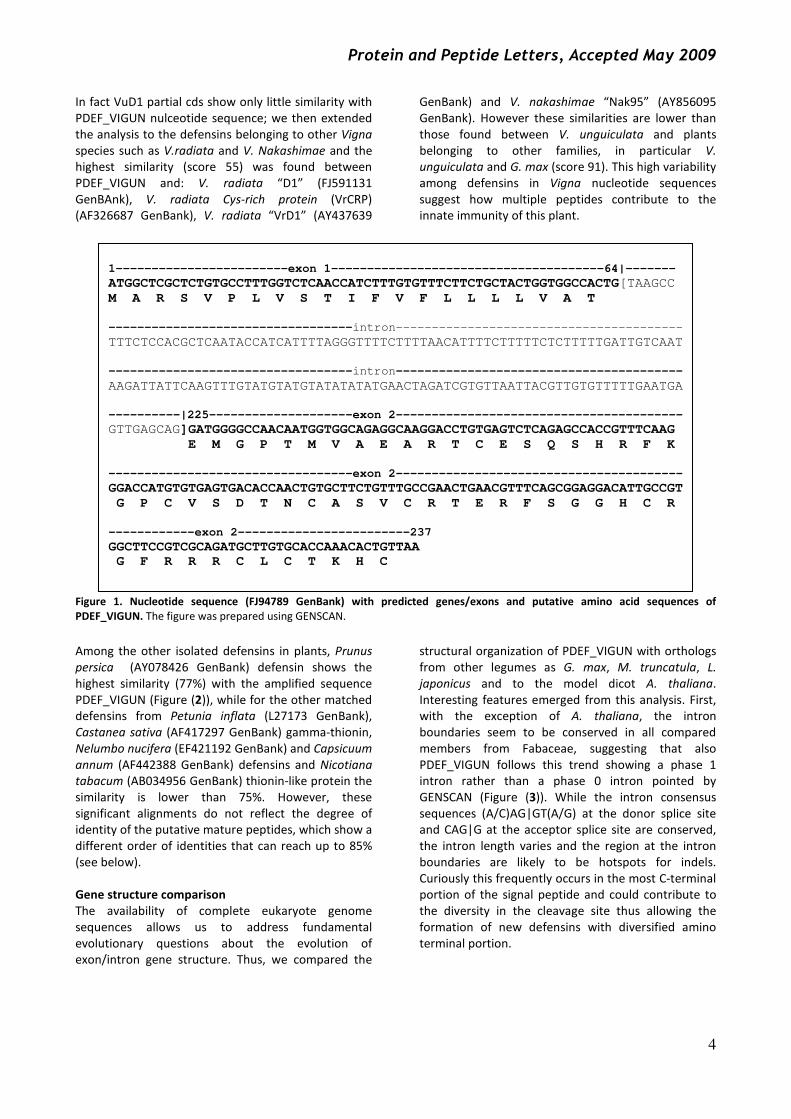
Results and Discussion Nucleotide Sequence Analysis
By performing a heterologous PCR on V. unguiculata,
using primers designed from P. vulgaris TC147 EST, we
have amplified a region (so on named PDEF_VIGUN)
that shows a 150 bp insertion in comparison with the
reference sequence TC147. By using the GENSCAN
program we described the putative organisation of the
genomic region we amplified (Figure (1)): the
sequence is expected to account for two exons
(namely exon 1 and exon 2), separated by an intron
(the additional 150 bp). The position of the intron,
localized among the signal peptide, is consistent with
other genomic defensins that have already been
isolates both in V. unguiculata (VuD1) (Pelegrini 2008)
and, among other plants, such as P. inflata (Karunanandaa 1994), C. annuum (Houlne, 1998) and
S. officinarum (Padovan 2008).
PDEF_VIGUN nucleotide sequence was then blasted
against EMBL and NCBI databases and a similarity
higher than 98% emerged with some EST sequences
from V. unguiculata and P. vulgaris common bean;
moreover PDEF_VIGUN shows significant similarity
with sequences that codify for defensins, thionins and
protease inhibitors belonging to different plants
families; this suggests to us that the amplified
sequence codifies for a defensin and/or might act as a
protease inhibitor. In fact the highest similarity among
deposited nucleotide sequences emerged with a
protease inhibitor from G. max (U12150 GenBank)
(91% similarity); this plant shares with V. unguiculata
not only the same family (Fabaceae) but also the same
tribe “Phaseoleae”. It is noteworthy that G. max is the
only plant sequence belonging to Phaseoleae that
shows high identity with V. unguiculata nucleotide
sequence (Figure (2)). PDEF_VIGUN was compared
with the other Vigna defensins available in the
genomic databases and few homologies were found.
Protein and Peptide Letters, Accepted May 2009
4
In fact VuD1 partial cds show only little similarity with
PDEF_VIGUN nulceotide sequence; we then extended
the analysis to the defensins belonging to other Vigna
species such as V.radiata and V. Nakashimae and the
highest similarity (score 55) was found between
PDEF_VIGUN and: V. radiata “D1” (FJ591131
GenBAnk), V. radiata Cys-rich protein (VrCRP)
(AF326687 GenBank), V. radiata “VrD1” (AY437639
GenBank) and V. nakashimae “Nak95” (AY856095
GenBank). However these similarities are lower than
those found between V. unguiculata and plants
belonging to other families, in particular V. unguiculata and G. max (score 91). This high variability
among defensins in Vigna nucleotide sequences suggest how multiple peptides contribute to the
innate immunity of this plant.
Figure 1. Nucleotide sequence (FJ94789 GenBank) with predicted genes/exons and putative amino acid sequences of
PDEF_VIGUN. The figure was prepared using GENSCAN.
Among the other isolated defensins in plants, Prunus
persica (AY078426 GenBank) defensin shows the
highest similarity (77%) with the amplified sequence
PDEF_VIGUN (Figure (2)), while for the other matched
defensins from Petunia inflata (L27173 GenBank),
Castanea sativa (AF417297 GenBank) gamma-thionin, Nelumbo nucifera (EF421192 GenBank) and Capsicuum annum (AF442388 GenBank) defensins and Nicotiana tabacum (AB034956 GenBank) thionin-like protein the
similarity is lower than 75%. However, these
significant alignments do not reflect the degree of
identity of the putative mature peptides, which show a
different order of identities that can reach up to 85%
(see below).
Gene structure comparison
The availability of complete eukaryote genome
sequences allows us to address fundamental
evolutionary questions about the evolution of
exon/intron gene structure. Thus, we compared the
structural organization of PDEF_VIGUN with orthologs
from other legumes as G. max, M. truncatula, L. japonicus and to the model dicot A. thaliana.
Interesting features emerged from this analysis. First,
with the exception of A. thaliana, the intron
boundaries seem to be conserved in all compared
members from Fabaceae, suggesting that also
PDEF_VIGUN follows this trend showing a phase 1
intron rather than a phase 0 intron pointed by
GENSCAN (Figure (3)). While the intron consensus
sequences (A/C)AG|GT(A/G) at the donor splice site
and CAG|G at the acceptor splice site are conserved,
the intron length varies and the region at the intron
boundaries are likely to be hotspots for indels.
Curiously this frequently occurs in the most C-terminal
portion of the signal peptide and could contribute to
the diversity in the cleavage site thus allowing the
formation of new defensins with diversified amino
terminal portion.
1------------------------exon 1--------------------------------------64|-------
ATGGCTCGCTCTGTGCCTTTGGTCTCAACCATCTTTGTGTTTCTTCTGCTACTGGTGGCCACTG[TAAGCC
M A R S V P L V S T I F V F L L L L V A T
----------------------------------intron----------------------------------------
TTTCTCCACGCTCAATACCATCATTTTAGGGTTTTCTTTTAACATTTTCTTTTTCTCTTTTTGATTGTCAAT
----------------------------------intron----------------------------------------
AAGATTATTCAAGTTTGTATGTATGTATATATATGAACTAGATCGTGTTAATTACGTTGTGTTTTTGAATGA
----------|225--------------------exon 2----------------------------------------
GTTGAGCAG]GATGGGGCCAACAATGGTGGCAGAGGCAAGGACCTGTGAGTCTCAGAGCCACCGTTTCAAG
E M G P T M V A E A R T C E S Q S H R F K
----------------------------------exon 2----------------------------------------
GGACCATGTGTGAGTGACACCAACTGTGCTTCTGTTTGCCGAACTGAACGTTTCAGCGGAGGACATTGCCGT
G P C V S D T N C A S V C R T E R F S G G H C R
------------exon 2------------------------237
GGCTTCCGTCGCAGATGCTTGTGCACCAAACACTGTTAA
G F R R R C L C T K H C

Protein and Peptide Letters, Accepted May 2009
5
Figure 2 Vigna unguiculata defensin DNA sequence compared with that of the other plant EST obtained from
databank (for a more detailed comparison with more plant species see attached materials figure X) Aligned
nucleotide sequences include Vigna unguiculata PDEF_VIGUN, Gycine max protease inhibitor (U12150 GenBank) and
Prunus persica defensin (AY078426 GenBank). Vigna/Glycine: score 91; Vigna/Prunus score 77.
* identifies conserved residues.
Amino acid sequence analysis The deduced amino acid sequence of PDEF_VIGUN
consists on 78 residues with a putative signal sequence
of 31 amino acids at the N-terminus (Figure (3)); the
putative mature peptide was identified by homology
with the conserved cleavage site present in known
defensin sequences and using SignalP software [19].
We then compared PDEF_VIGUN with other V. unguiculata defensins already described in the
literature. Cp_thionin I and II [14, 15] show 68% and
37% similarity with the deduced PDEF_VIGUN, while
for VuD1 (Pelegrini 2008) only 27% similarity was
found. The presence of a new defensin in V. unguiculata confirms how multiple peptides
contribute to the innate immunity of this plant, as it
occurs in Oryza sativa and Zea mays.
Although in databases Fabaceae defensins are
present, blasting the PDEF_VIGUN amino acid
sequence against plant sequences returned
homologies higher than 75% emerged only with
defensins (formerly known as γ-thionins) isolated from
plant species belonging to families other than
Fabaceae (see Figure (4A)), for example H. annuus
(sunflower, Asteraceae) γ-thionin precursor (P82659
GenBank), P. persica (peach, Rosaceae) (Q84UH1
UniProt), Solanum pimpinellifolium and S. lycopersicum (tomato, Solanaceae) defensin (e.g.
Q9XG53 UniProt) and Nicotiana tabacum (tobacco,
Solanaceae) thionin-like protein (Q9MB66 UniProt). It
seems strange that except for the G. max (Q39807
UniProt) protease inhibitor with 91% similarity
PDEF_VIGUN, other plants that share with V.
unguiculata the same family and tribe, Fabaceae and
Phaseoleae respectively, only show an identity lower
than 75% with peptides identified as a defensin in
Phaseolus vulgaris γ-thionin (kidney bean, A0JJX6
UniProt) and as kunits trypsin inhibitor protein in P. coccineus (scarlet runner bean, Q9FUP3 UniProt) and
not in other bean species.
The similarity to peptides that belong to classes
alternatively defined as defensins or protease
inhibitors suggests that these plant peptides are likely
characterized by a multifunctional activity and could
act either in defence from microbes or from insects,
and/or be involved in activation of other stress-
responsive mechanisms.
Likewise, PDEF_VIGUN presents amino acids
characteristic in plant defensins (see Figure (3)),
namely a serine residue at position 7, an aromatic
residue at position 10, two glycines at position 12 and
32, a glutamic acid at position 27 as well as the 8
cysteines residues that give the typical disulphuric
bonds pattern [20]. Curiously, all the abovementioned
defensins/protease inhibitor sequences show a
conserved phenylalanine residue at position 42,
whereas in PDEF_VIGUN this is replaced by leucine. It
is notewothy that a large variation in plant defensins
primary sequences is evident (apart from the cysteine
residues) when comparing PDEF_VIGUN with other
plant defensin sequences (Fig. 3). Despite this high
variability they all share the three dimensional
structures as it is described below.
Vigna ATGGCTCGCTCTGTGCCTTTGGTCTCAACCATCTTTGTGTTTCTTCTGCTACTGGTGGCC 60
Glycine ATGTCTCGCTCCGTGCCTTTGGTTTCAACCATTTGTGTCTTGCTTCTGCTTCTGGTGGCC 60
Prunus ATGGAGCGCTCCATGCGTTTATTTTCAACTGCCTTCGTCTTCTTTCTGCTTCTGGCAGCT 60
*** ***** *** *** * ***** * ** ** ******* **** **
Vigna ACTG-GATG---GGGCCAACAATGGTGGCAGAGGCAAGGACCTGTGAGTCTCAGAGCCAC 117
Glycine ACTGAGATGATGGGGCCAACAATGGTGGCAGAAGCAAGAACTTGTGAGTCTCAGAGCCAC 120
Prunus GCTGGGATGATGATGGGGCCAATGGTTGCTGAGGCTAGGACCTGTGAGTCTCAGAGTAAT 120
*** **** * ******* ** ** ** ** ** ************** *
Vigna CGTTTCAAGGGACCATGTGTGAGTGACACCAACTGTGCTTCTGTTTGCCGAACTGAACGT 177
Glycine CGTTTCAAGGGGCCATGTTTGAGTGACACCAACTGTGGCTCTGTTTGCCGAACCGAACGT 180
Prunus CGGTTCAAGGGAACTTGCGTGAGTACAAGCAACTGTGCATCTGTTTGCCAAACTGAGGGC 180
** ******** * ** ***** * ******** ********** *** ** *
Vigna TTCAGCGGAGGACATTGCCGTGGCTTCCGTCGCAGATGCTTGTGCACCAAACACTGTTAA 237
Glycine TTCACTGGAGGACACTGCCGTGGCTTCCGTCGCAGATGCTTCTGCACCAAACATTGTTAA 240
Prunus TTCCCTGGTGGCCATTGTCGTGGCTTTCGCCGCAGATGCTTTTGCACTAAACATTGTTAA 240
*** ** ** ** ** ******** ** *********** ***** ***** ******

Protein and Peptide Letters, Accepted May 2009
6
Figure 3. Plant defensin gene structure after heterologous comparison. The intron boundaries are conserved in all
compared members from Fabaceae, with the exception of A. thaliana. Black boxes indicate exon while grey boxes the
introns. The nucleotide length of the DNA stretch is reported below the boxes. CDS: translated coding sequence;
Arath: Arabidopsis thaliana; Glyma: Glycine max; Medtr: Medicago truncatula; Lotja: Lotus japonicus; Vigun: Vigna unguiculata. Phavu: Phaseolus vulgaris (TC147). PTR: Protein added to the alignment; Vigra: Vigna radiata
(CAA34760), Vigun2: Vigna unguiculata (P18646); Vigra2: Vigna radiata (BAB82453). Disulphuric bonds are
highlighted: Cys3-Cys47; Cys14-Cys43; Cys20-Cys41; Cys24-Cys34).
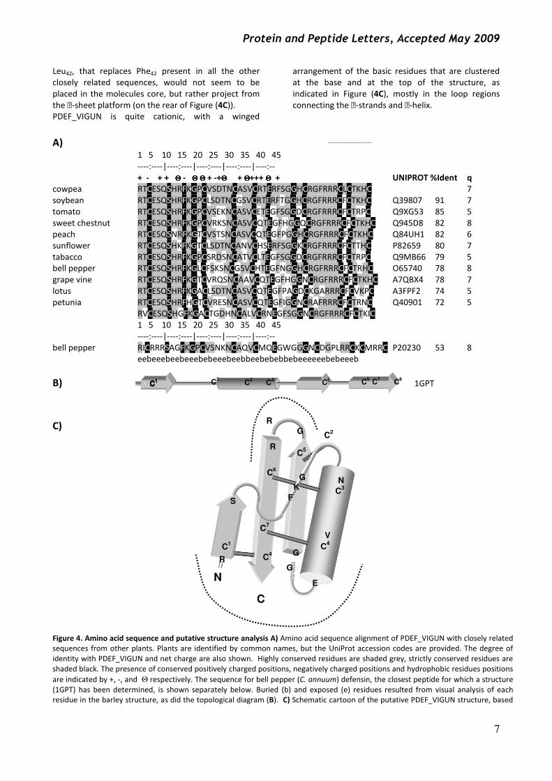
Protein structure prediction Blasting the PDEF_VIGUN sequence against the PDB
database reveals the sequence of Hordeum vulgare
defensins (gamma-hordothionin) as having the closest
identity (53%, see Figure (4A)). The sequence identity,
as well as other primary structural features, suggests
that its structure (1GTP) is representative of the
PDEF_VIGUN tertiary structure. The peptide’s
topology, as well as a schematic representation of the
structure are shown in Figure (4B) and (4C). An
analysis of this structure and the predicted
PDEF_VIGUN structure (derived by a direct residue
substitution), is revealing. Conservation of residues
Ser7 and Glu27 are explained by the fact that although
polar, they are both buried and form a network of H-
bonds that likely has an essential role in stabilising the
defensin scaffold. The conserved aromatic residues at
positions 10 and 29 and the conserved valine at
position 23 contribute to the core of the molecule.
Gly12 seems at a key position in a loop connecting the
first strand with the helix (see Figure (4C)) and its main
chain amide and carboxyl form a network of H-bonds
with other conserved residues, Phe10 and Arg40. Gly32 is
in a highly hindered position where strand 2 passes
directly over the helix. Apart from this, other
conserved residues in PDEF-VIGUN may have
important structural roles. Asn19, for example, is
involved in a network of H-bonds that may indicate it
acts as a helix-stabilising cap, also involving Ser16.
Protein and Peptide Letters, Accepted May 2009
7
Leu42, that replaces Phe42 present in all the other
closely related sequences, would not seem to be
placed in the molecules core, but rather project from
the -sheet platform (on the rear of Figure (4C)).
PDEF_VIGUN is quite cationic, with a winged
arrangement of the basic residues that are clustered
at the base and at the top of the structure, as
indicated in Figure (4C), mostly in the loop regions
connecting the -strands and -helix.
A) 1 5 10 15 20 25 30 35 40 45
----:----|----:----|----:----|----:----|----:--
+ - + + ΘΘΘΘ - ΘΘΘΘ ΘΘΘΘ + -+ΘΘΘΘ + ΘΘΘΘ+++ ΘΘΘΘ + UNIPROT %Ident q
cowpea RTCESQSHRFKGPCVSDTNCASVCRTERFSGGHCRGFRRRCLCTKHC 7
soybean RTCESQSHRFKGPCLSDTNCGSVCRTERFTGGHCRGFRRRCFCTKHC Q39807 91 7
tomato RTCESQSHRFKGPCVSEKNCASVCETEGFSGGDCRGFRRRCFCTRPC Q9XG53 85 5
sweet chestnut RTCESQSHRFKGPCVRKSNCASVCQTEGFHGGQCRGFRRRCFCTKHC Q945D8 82 8
peach RTCESQSNRFKGTCVSTSNCASVCQTEGFPGGHCRGFRRRCFCTKHC Q84UH1 82 6
sunflower RTCESQSHKFKGTCLSDTNCANVCHSERFSGGKCRGFRRRCFCTTHC P82659 80 7
tabacco RTCESQSHRFKGPCSRDSNCATVCLTEGFSGGDCRGFRRRCFCTRPC Q9MB66 79 5
bell pepper RTCESQSHRFKGLCFSKSNCGSVCHTEGFNGGHCRGFRRRCFCTRHC O65740 78 8
grape vine RTCESQSHRFKGTCVRQSNCAAVCQTEGFHGGNCRGFRRRCFCTKHC A7QBX4 78 7
lotus RTCESQSHRFKGACLSDTNCASVCQTEGFPAGDCKGARRRCFCVKPC A3FPF2 74 5
petunia RTCESQSHRFHGTCVRESNCASVCQTEGFIGGNCRAFRRRCFCTRNC Q40901 72 5
RVCESQSHGFKGACTGDHNCALVCRNEGFSGGNCRGFRRRCFCTKIC
1 5 10 15 20 25 30 35 40 45
----:----|----:----|----:----|----:----|----:--
bell pepper RICRRRSAGFKGPCVSNKNCAQVCMQEGWGGGNCDGPLRRCKCMRRC P20230 53 8
eebeeebeebeeebebeeebeebbeebebebbebeeeeeebebeeeb
B) 1GPT
C)
Figure 4. Amino acid sequence and putative structure analysis A) Amino acid sequence alignment of PDEF_VIGUN with closely related
sequences from other plants. Plants are identified by common names, but the UniProt accession codes are provided. The degree of
identity with PDEF_VIGUN and net charge are also shown. Highly conserved residues are shaded grey, strictly conserved residues are
shaded black. The presence of conserved positively charged positions, negatively charged positions and hydrophobic residues positions
are indicated by +, -, and Θ respectively. The sequence for bell pepper (C. annuum) defensin, the closest peptide for which a structure
(1GPT) has been determined, is shown separately below. Buried (b) and exposed (e) residues resulted from visual analysis of each
residue in the barley structure, as did the topological diagram (B). C) Schematic cartoon of the putative PDEF_VIGUN structure, based
N
C
R
C1
S F
K
G
C2
N
C3
V
C4
E
G
G
C5
G
R
R
C6
C7
C4
C1 C
2 C
3 C
4 C
5 C
5 C
7 C
8 C
1 C
1
Protein and Peptide Letters, Accepted May 2009
8
on that of bell pepper defensin (1GPT). The putative position of highly conserved residues in this schematic representation is
approximately indicated. The dotted lines indicate areas where cationic residues would be concentrated.
Protein and Peptide Letters, Accepted May 2009
9
RT-QPCR
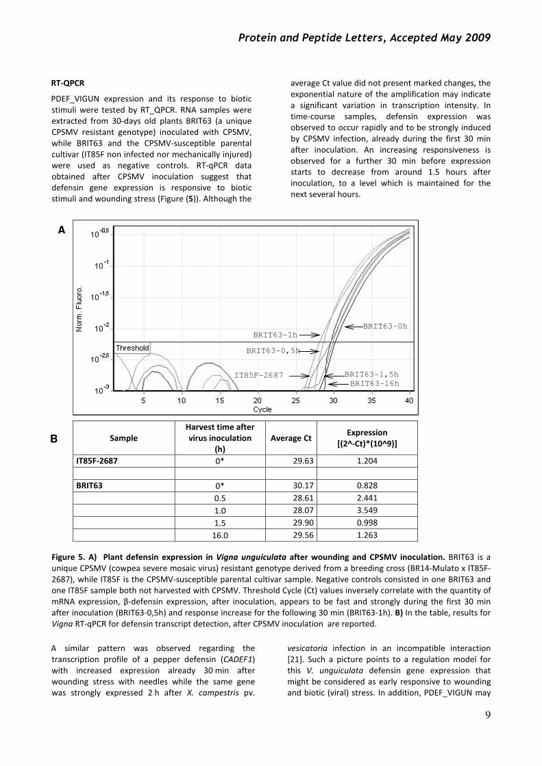
PDEF_VIGUN expression and its response to biotic
stimuli were tested by RT_QPCR. RNA samples were
extracted from 30-days old plants BRIT63 (a unique
CPSMV resistant genotype) inoculated with CPSMV,
while BRIT63 and the CPSMV-susceptible parental
cultivar (IT85F non infected nor mechanically injured)
were used as negative controls. RT-qPCR data
obtained after CPSMV inoculation suggest that
defensin gene expression is responsive to biotic
stimuli and wounding stress (Figure (5)). Although the
average Ct value did not present marked changes, the
exponential nature of the amplification may indicate
a significant variation in transcription intensity. In
time-course samples, defensin expression was
observed to occur rapidly and to be strongly induced
by CPSMV infection, already during the first 30 min
after inoculation. An increasing responsiveness is
observed for a further 30 min before expression
starts to decrease from around 1.5 hours after
inoculation, to a level which is maintained for the
next several hours.
Figure 5. A) Plant defensin expression in Vigna unguiculata after wounding and CPSMV inoculation. BRIT63 is a
unique CPSMV (cowpea severe mosaic virus) resistant genotype derived from a breeding cross (BR14-Mulato x IT85F-
2687), while IT85F is the CPSMV-susceptible parental cultivar sample. Negative controls consisted in one BRIT63 and
one IT85F sample both not harvested with CPSMV. Threshold Cycle (Ct) values inversely correlate with the quantity of
mRNA expression, β-defensin expression, after inoculation, appears to be fast and strongly during the first 30 min
after inoculation (BRIT63-0,5h) and response increase for the following 30 min (BRIT63-1h). B) In the table, results for
Vigna RT-qPCR for defensin transcript detection, after CPSMV inoculation are reported.
A similar pattern was observed regarding the
transcription profile of a pepper defensin (CADEF1)
with increased expression already 30 min after
wounding stress with needles while the same gene
was strongly expressed 2 h after X. campestris pv.
vesicatoria infection in an incompatible interaction
[21]. Such a picture points to a regulation model for this V. unguiculata defensin gene expression that
might be considered as early responsive to wounding
and biotic (viral) stress. In addition, PDEF_VIGUN may
Sample
Harvest time after
virus inoculation
(h)
Average Ct Expression
[(2^-Ct)*(10^9)]
IT85F-2687 0* 29.63 1.204
BRIT63 0* 30.17 0.828
0.5 28.61 2.441
1.0 28.07 3.549
1.5 29.90 0.998
16.0 29.56 1.263
BRIT63-0h
BRIT63-1h
BRIT63-0,5h
IT85F-2687
BRIT63-16h
BRIT63-1,5h
A
B

Protein and Peptide Letters, Accepted May 2009
10
perform direct roles as virus antagonist molecules,
since some plant defensins (γ-thionins) have
presented a very large spectrum of action against
diverse plant parasitic bacterial genera through
impairing cell growth [15]. Noteworthy, legume
defensins have already been described as harbouring
effective antiproliferative and anti-HIV-1 reverse
transcriptase activities in mammal pathosystems [22].
Indeed, PDEF_VIGUN may be part of a plant defense
mechanisms acting as one of the initial responses,
necessary to activate, or allow time for activation of
secondary and/or late defense-related metabolic and
physiological responses more directly associated to
resistant phenotype.
In conclusion we have isolated from V. unguiculata a
sequence with characteristics that suggest it
corresponds to a defensin-like gene. Amino acid
sequences analyses indicate that the deduced
PDEF_VIGUN sequence is homologous to already
known defensin proteins and thus might have similar
function to these other defensins. Moreover,
preliminary functional results demonstrate the
presence of mRNA for this cowpea defensin in the
plant tissue and also an increase in expression in
response to biotic stimuli such as CPSMV infection.
This suggests that a lack of defensins expression could
make the plant more susceptible to the invasion by
pathogens.
ACKNOWLEDGEMENTS This work was partially supported by RC03/04 from
IRCCS Burlo Garofolo. L.P. is recipient of a fellowship
from FVG2008. None of the authors has any potential
financial conflict of interest related to this manuscript.
References [1] Broekaert, W.F.; Terras, F.R.; Cammue, B.P.; Osborn, R.W.
Plant defensins: novel antimicrobial peptides as components
of the host defense system. Plant Physiol., 1995, 108(4),1353-
1358.
[2] Thomma, B.P.; Cammue, B.P.; Thevissen, K. Plant defensins.
Planta, 2002, 216(2), 193-202.
[3] Castro, M.S.; Fontes, W. Plant defense and antimicrobial
peptides. Protein Pept Lett., 2005, 12(1), 13-18.
[4] Bloch, C. Jr; Richardson, M. A new family of small (5 kDa)
protein inhibitors of insect alpha-amylases from seeds or
sorghum (Sorghum bicolor (L) Moench) have sequence
homologies with wheat γ-purothionins. FEBS Lett., 1991,
279(1), 101-104.
[5] Almeida, M.S.; Cabral, K.M.; Zingali, R.B.; Kurtenbach, E.
Characterization of two novel defense peptides from pea
(Pisum sativum) seeds. Arch Biochem Biophys, 2000, 378(2),
278-86.
[6] Antcheva, N.; Zelezetsky, I.; Tossi, A. Cationic Antimicrobial
Peptides—The Defensins. In The Handbook of Biologically Active Peptides; A.J. Kastin, Ed.; Elsevier Science B. V:
Amsterdam, 2006; pp. 55-66.
[7] Liu, Y.J.; Cheng, C.S.; Lai, S.M.; Hsu, M.P.; Chen, C.S.; Lyu, P.C.
Solution structure of the plant defensin VrD1 from mung bean
and its possible role in insecticidal activity against bruchids.
Proteins, 2006, 63(4), 777-786.
[8] Mendez, E.; Moreno, A.; Colilla, F.; Pelaez, F.; Limas, G.G.;
Mendez, R. Soriano F, Salinas M, de Haro C. Primary structure
and inhibition of protein synthesis in eukaryotic cell-free
system of a novel thionin, γ -hordothionin, from barley
endosperm. Eur J Biochem, 1990, 94, 533-539.
[9] Games, P.D.; Dos Santos, I.S.; Mello, E.O.; Diz, M.S.; Carvalho,
A.O.; de Souza-Filho, G.A.; Da Cunha, M.; Vasconcelos, I.M.;
Ferreira, B. dos S.; Gomes, V.M. Isolation, characterization and
cloning of a cDNA encoding a new antifungal defensin from
Phaseolus vulgaris L. seeds. Peptides, 2008, 29(12), 2090-2100.
[10] Zhang, Y.; Lewis, K. Fabatins: new antimicrobial plant peptides.
FEMS Microbiol Lett, 1997, 149, 59-64.
[11] Dimarcq, J.L.; Zachary, D.; Hoffmann, J.A.; Hoffmann, D.;
Reichhart, J.M. Insect immunity: expression of the two major
inducible antibacterial peptides, defensin and diptericin, in
Phormia terranovae. EMBO J, 1990, 9, 2507-2515.
[12] Ehlers, J.D.; Hall, A.E. Cowpea (Vigna unguiculata L. Walp.).
Field Crops Research, 1997, 53(1–3),187-204.
[13] Azevedo, H.; Houllou-Kido, L.; Benko-Iseppon, A.M. Análise do
Potencial Regenerativo in vitro de Diferentes Cultivares de
Feijão-Caupi. Revista Brasileira de Biociências, 2007 , 5(2), 528-
530.
[14] Melo, F.R.; Rigden, D.J.; Franco, O.L.; Mello, L.V.; Ary, M.B.;
Grossi de Sá, M.F.; Bloch Jr, C. Inhibition of trypsin by cowpea
thionin: characterization, molecular modeling, and docking.
Proteins, 2002, 48(2), 311-319.
[15] Franco, O.L.; Murad, A.M.; Leite, J.R.; Mendes, P.A.; Prates,
M.V.; Bloch Jr, C. Identification of a cowpea gamma-thionin
with bactericidal activity. FEBS J., 2006, 273(15), 3489-3497.
[16] Weising, K. DNA fingerprinting in plants: principles, methods, and applications, 2nd ed.; CRC Press: Boca Raton, FL, 2005.
[17] Pio-Ribeiro, G.; Paz, C.D.; Andrade, G.P.; Assis Filho, F.M. Efeito
de quatro isolados do vírus do mosaico severo, CPSMV, na
produção de quatro cultivares de caupi, em condições de casa
de vegetação. Caderno Ômega, 2001, 12, 44-45.
[18] Chang, S.; Puryear, J.; Cairney, J. A simple and efficient
method for isolating RNA from pine trees. PMB Reporter,
1993, 11, 113-116.
[19] Bendtsen, J.D.; Nielsen, H.; von Heijne, G.; Brunak, S. Improved
prediction of signal peptides: SignalP 3.0. J Mol Biol., 2004,
340(4), 783-795.
[20] Janssen, B.J.; Schirra, H.J.; Lay, F.T.; Anderson, M.A.; Craik, D.J.
Structure of Petunia hybrida defensin 1, a novel plant defensin
with five disulfide bonds. Biochemistry, 2003, 42(27), 8214-
8222.
[21] Do, H.M; Lee, S.C.; Jung, H.W.; Sohn, K.H.; Hwang, B.K.
Differential expression and in situ localization of a pepper
defensin (CADEF1) gene in response to pathogen infection,
abiotic elicitors and environmental stresses in Capsicum annuum. Plant Science, 2004, 166(5), 1297-1305.
[22] Ngai, P.H.; Ng, T.B. Phaseococcin, an antifungal protein with
antiproliferative and anti-HIV-1 reverse transcriptase activities
from small scarlet runner beans. Biochem Cell Biol 2005 83(2),
212-220.
Copyright © 2022 FDOKUMEN