Calcium dysregulation in heart diseases: Targeting calcium ...
Upload
independentCategory
view
2download
0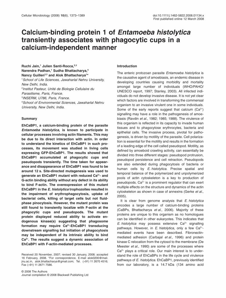
Calcium-binding protein 1 of Entamoeba histolyticatransiently associates with phagocytic cups in acalcium-independent manner
Ruchi Jain,1 Julien Santi-Rocca,2,3
Narendra Padhan,1 Sudha Bhattacharya,4
Nancy Guillen2,3 and Alok Bhattacharya1*1School of Life Sciences, Jawaharlal Nehru University,New Delhi, India.2Institut Pasteur, Unité de Biologie Cellulaire duParasitisme, Paris, France.3INSERM, U786, Paris, France.4School of Environmental Sciences, Jawaharlal NehruUniversity, New Delhi, India.
Summary
EhCaBP1, a calcium-binding protein of the parasiteEntamoeba histolytica, is known to participate incellular processes involving actin filaments. This maybe due to its direct interaction with actin. In orderto understand the kinetics of EhCaBP1 in such pro-cesses, its movement was studied in living cellsexpressing GFP-EhCaBP1. The results showed thatEhCaBP1 accumulated at phagocytic cups andpseudopods transiently. The time taken for appear-ance and disappearance of EhCaBP1 was found to bearound 12 s. Site-directed mutagenesis was used togenerate an EhCaBP1 mutant with reduced Ca2+- andG-actin binding ability without any defect in its abilityto bind F-actin. The overexpression of this mutantEhCaBP1 in the E. histolytica trophozoites resulted inthe impairment of erythrophagocytosis, uptake ofbacterial cells, killing of target cells but not fluid-phase pinocytosis. However, the mutant protein wasstill found to transiently localize with F-actin at thephagocytic cups and pseudopods. The mutantprotein displayed reduced ability to activate en-dogenous kinase(s) suggesting that phagosomeformation may require Ca2+-EhCaBP1 transducingdownstream signalling but initiation of phagocytosismay be independent of its intrinsic ability to bindCa2+. The results suggest a dynamic association ofEhCaBP1 with F-actin-mediated processes.
Introduction
The enteric protozoan parasite Entamoeba histolytica isthe causative agent of amoebiasis, an endemic disease indeveloping countries causing morbidity and mortalityamongst large number of individuals (WHO/PAHO/UNESCO report, 1997; Stanley, 2003). All infected indi-viduals do not develop invasive disease. It is not yet clearwhich factors are involved in transforming the commensalorganism to an invasive virulent one in some individuals.Some of the early reports suggest that calcium (Ca2+)signalling may have a role in the pathogenesis of amoe-biasis (Ravdin et al., 1982; 1985; 1988). The virulence ofthis organism is reflected in its capacity to invade humantissues and to phagocytose erythrocytes, bacteria andepithelial cells. The invasive process, pivotal for patho-genesis, is driven by motility of the parasite. Cell polariza-tion is essential for the motility and results in the formationof a leading edge of the cell called pseudopod. Motility, asdefined by amoeboid crawling activity, can essentially bedivided into three different stages: pseudopod protrusion,pseudopod persistence and cell retraction. Pseudopodsare also extended during phagocytosis of bacteria orhuman cells by E. histolytica. Precise spatial andtemporal balance of the polymerized and unpolymerizedpools of actin cytoskeleton is a key to production ofpseudopods. Ca2+ is a prominent regulator that can exertmultiple effects on the structure and dynamics of the actincytoskeleton as shown in case of annexins (Gerke et al.,2005).
It is clear from genome analysis that E. histolyticaencodes a large number of calcium-binding proteins(CaBPs, Bhattacharya et al., 2006). Majority of theseproteins are unique to this organism as no homologuescan be identified in other eukaryotes. This indicates thatE. histolytica may possess extensive Ca2+ signallingpathways. However, in E. histolytica, only a few Ca2+-mediated events have been described. Fibronectin-mediated adhesion (Carbajal et al., 1996) and proteinkinase C relocation from the cytosol to the membrane (DeMeester et al., 1990) are some of the processes whereCa2+ plays a critical role. Our main interest is to under-stand the role of EhCaBPs in the life cycle and virulencepathways of E. histolytica. EhCaBP1, previously identifiedfrom our laboratory, is a 14.7 kDa (134 amino acid
Received 20 November, 2007; revised 30 January, 2008; accepted16 February, 2008. *For correspondence. E-mail [email protected], [email protected]; Tel. (+91) 11 2670 4516;Fax (+91) 11 2671 7586.
Cellular Microbiology (2008) 10(6), 1373–1389 doi:10.1111/j.1462-5822.2008.01134.xFirst published online 12 March 2008
© 2008 The AuthorsJournal compilation © 2008 Blackwell Publishing Ltd

residues) protein, which shares low (29%) sequence iden-tity with the well-studied eukaryotic EF hand-containingprotein, calmodulin (CaM) (Prasad et al., 1992). Thethree-dimensional solution structure of holo-EhCaBP1was determined by multidimensional nuclear magneticresonance (NMR) spectroscopy (Sahu et al., 1999;Atreya et al., 2001) and X-ray crystallography (Gopalet al., 1998). Interestingly, NMR studies showed thatEhCaBP1 has some structural similarity to CaM andTroponinC (TnC), with two globular domains connectedby a flexible linker region spanning eight amino acidresidues. Each domain consists of a pair of canonicalCa2+-binding EF hand motifs. EhCaBP1 binds four Ca2+
ions with unequal affinity for Ca2+ (Gopal et al., 1997).However, differences exist which make EhCaBP1 distinctfrom CaM. Biochemically, the inability of EhCaBP1 toactivate c-AMP phosphodiesterase differentiate it from allknown CaMs (Yadava et al., 1997). Structural studies indi-cated a more open C-terminal domain for EhCaBP1 withlarger water exposed total hydrophobic surface area ascompared with CaM and TnC. Moreover, the central linkerof EhCaBP1 contains two glycine residues (G63 and G67)making it more flexible compared with CaM. High-resolution X-ray crystallographic structure has recentlybeen reported for EhCaBP1 (Kumar et al., 2007). Thestructure revealed an unusual arrangement of differentdomains not observed in previous studies. Unlike CaM,the first two EF hand motifs in EhCaBP1 are connected bya long helix and form a dumbbell-shaped structure. Threemolecules of EhCaBP1 N-terminal domains participate indomain swapping to form trimers. The functional differ-ences between EhCaBP1 and CaM could be attributed tothe difference in the flexibility of the helices connecting theEF hand domains.
A decrease in the expression of EhCaBP1 gene inE. histolytica trophozoites by regulatable antisense RNAtechnology resulted in the inhibition of cellular prolifera-tion, implicating the essentiality of the EhCaBP1 encod-ing gene in this parasite (Sahoo et al., 2003). Detailedanalysis revealed that the defect in proliferation may bedue to an inhibition of pinocytosis and phagocytosisand there was a direct interaction of EhCaBP1 withF-actin (Sahoo et al., 2004). Therefore it appeared thatEhCaBP1 participated in processes involving actin skel-etal dynamics and reduction in its cytosolic concentra-tion could lead to a defect in actin-mediated processes,such as phagocytosis. Involvement of EhCaBP1 inphagocytosis was confirmed using phagocytosis-defective L6 mutant (Orozco et al., 1983). In these cells,EhCaBP1 expression was found to be highly reduced(Hirata et al., 2007). Phagocytosis plays an essentialrole in growth and constitutes one of the key virulencedeterminants of E. histolytica (Bracha and Mirelman,1984).
Although EhCaBP1 localization to the pseudopods andinvolvement in phagocytosis has been reported, its kinet-ics in the process remains totally uncharacterized. Asactin filament assembly is a dynamic process it is likelythat EhCaBP1 may be involved in pseudopod formationduring movement and phagocytosis in a transient manner.In the present work, multidimensional time-lapse imagingwas used to study kinetics of EhCaBP1 in the live amoebaexpressing EhCaBP1 tagged to GFP. The role of Ca2+ inthe dynamics of EhCaBP1 was also investigated using amutant form of EhCaBP1 having reduced ability to bindCa2+. The data highlight a very rapid kinetics of EhCaBP1in the dynamic processes of pseudopod formation duringparasite motility and phagocytosis. Reduction in the abilityof EhCaBP1 to bind Ca2+ did not affect pseudopod forma-tion at its early stages but altered a number of virulenceproperties of E. histolytica including red blood cell (RBC)phagocytosis and cell cytotoxicity. Analysis of phagocyticdynamic process revealed a dual behaviour of EhCaBP1.At early stages the interaction with filamentous actin, astep that does not require the Ca2+-bound form ofEhCaBP1, is important. In contrast, cytoskeleton dynam-ics during phagosome maturation requires EhCaBP1 tobe in Ca2+-bound state.
Results
Ca2+ requirement for phagocytosis in E. histolytica
It is known from many systems that Ca2+ is involvedin phagocytosis (Lew et al., 1985; Stendahl et al., 1994;Vieira et al., 2002). To analyse the involvement of Ca2+ inphagocytosis of E. histolytica, human erythrocytes wereused. The level of erythrophagocytosis was determinedby a spectrophotometric assay after treating the tropho-zoites with different concentrations of the Ca2+ chelator,BAPTA-AM. The cells showed 60% reduction in theuptake of RBC at 50 mM of the chelator (Fig. 1). No sig-nificant effect was seen with DMSO, the solvent used todissolve BAPTA-AM. Internal Ca2+ concentrations [Ca2+]iwas also determined. There was a 25% drop when thetrophozoites were treated with 50 mM BAPTA-AM. Thisdata suggest that [Ca2+]i plays an important role inerythrophagocytosis.
Expression of GFP-EhCaBP1 in trophozoites
The cellular dynamics of any protein molecule can bestudied in living cells by expressing the protein of interesttagged with GFP. In order to study the dynamics ofEhCaBP1 in live trophozoites, GFP-EhCaBP1 wasexpressed in E. histolytica cells. The coding region ofEhCaBP1 was inserted in frame into the BamHI site ofE. histolytica shuttle plasmid pEh-NEO-GFP (Fig. 2A).
1374 R. Jain et al.
© 2008 The AuthorsJournal compilation © 2008 Blackwell Publishing Ltd, Cellular Microbiology, 10, 1373–1389

E. histolytica cells were then transformed and stable trans-formants were selected as described in Experimentalprocedures. The cells carrying pEh-NEO-GFP-EhCaBP1and the vector pEh-NEO-GFP plasmids will henceforthbe referred to as GC1 and as GFP respectively.
Expression of GFP-EhCaBP1 from GC1 cells wasstudied by immunoblot analysis using anti-EhCaBP1 anti-body and the results are shown in Fig. 2B. As the numberof plasmids in transformed cells increases with increasingconcentration of the selectable drug G418, the level ofexpression was determined at different concentration ofthe drug. The immunostained band for GFP-EhCaBP1was seen at the expected size of 37 kDa and the intensityof the band increased with increasing concentration ofG418 (Fig. 2B). The 14 kDa band represents the endog-enous EhCaBP1 (Fig. 2B). Densitometric analysis of theblots revealed a threefold increase in the level of GFP-EhCaBP1 at 30 mg ml-1 G418 as compared with 1.5-foldat 10 mg ml-1 G418. The endogenous EhCaBP1 and actinshowed no change in their levels at different G418concentrations. In order to test if GFP-EhCaBP1 retainsthe properties of the endogenous EhCaBP1 in vivo, theproperty of the latter to localize at the phagocytic cupduring erythrophagocytosis (Sahoo et al., 2004) wastested with the GFP-tagged protein. The localization of thefusion protein during erythrophagocytosis was studied byimmunostaining with either anti-EhCaBP1 or anti-GFPantibodies. The cells showed an accumulation of GFP-EhCaBP1 in the phagocytic cups (Fig. 2D). The patternwas identical to that observed for endogenous wild-type(WT) EhCaBP1 (Fig. 2C and E) suggesting that GFP-EhCaBP1 behaves in a similar way as EhCaBP1 alone.The control GFP cells were also stained and the results
showed that there is no accumulation of GFP at thephagocytic cup (Fig. 2F).
Dynamics of GFP-EhCaBP1 movement in liveE. histolytica cells
Phagocytosis of particles is a rapid event in E. histolyticaand closed phagosomes appear three minutes afterthe engulfment (Marion et al., 2005). The presence ofEhCaBP1 in phagocytic cups suggested that it might beinvolved in the early stages of phagocytosis. In an attemptto examine the occurrence of EhCaBP1 on the maturephagosomes, the cells were stained for actin andEhCaBP1 after 10 min of erythrophagocytosis. As phago-cytosis is not a synchronized event, it was possible tovisualize cells undergoing phagocytic cup formation andalso observe mature phagosomes. It was observed thatF-actin was detached from the mature phagosomes (Voigtand Guillen, 1999). Focal planes in the middle of cellswere analysed and engulfed RBC were seen inside thecells; however, neither actin nor EhCaBP1 was visiblearound the internalized RBC unlike EhCaBP1 colocalizedwith F-actin at the phagocytic cups (Fig. 3). These obser-vations suggest that EhCaBP1 may be involved in phago-cytosis only transiently at the initiation of the process andleaves the site during phagosome maturation. In order tofurther analyse this hypothesized dynamics of EhCaBP1within the cell, GFP-EhCaBP1 trafficking was studiedusing multidimensional time-lapse fluorescence micros-copy in GC1 cells. Video microscopy was carried out withspinning disk confocal scanning microscope in order tominimize damage to the cells due to laser energy andatmospheric oxygen and providing rapid acquisition ofimages. EhCaBP1 was found to move rapidly to and fromthe phagocytic cups. Live cell imaging of the GC1 cells inthe presence of RBC showed that EhCaBP1 started toaccumulate in the phagocytic cup as soon as the RBCbound to the amoeba surface. The intensity of EhCaBP1increased rapidly during the uptake of RBC. There was acomplete loss of fluorescence as soon as the RBC wascompletely inside the cell (Figs 3 and 4A). It took about30 s from the appearance of EhCaBP1 to its eventualdisappearance. Images from GFP cells were also cap-tured but these showed a diffuse distribution of GFP alone(data not shown).
To gain insight in the dynamics of EhCaBP1, we tookadvantage of larger surface of pseudopods comparedwith those of phagocytic cups that have small areas. Theacquired images are presented as fluorescent micro-graphs of a moving trophozoite taken every 3 s (Fig. 4B,see also Movie S1). A progressively marked enrichmentof GFP-EhCaBP1 at the leading edge of the protrudingpseudopods in comparison with cytoplasm and its subse-quent disappearance was observed (Fig. 4B and C). The
0
0.05
0.1
0.15
0.2
0.25
0 5 50 500 DMSO[BAPTA-AM] (mM)
O.D
at
400 n
m
Fig. 1. Effect of calcium chelator BAPTA-AM onerythrophagocytosis in E. histolytica. The cells were grown for 48 hbefore treatment with different concentrations of BAPTA-AM (0 mM,5 mM, 50 mM, 500 mM) or DMSO for 10 min at room temperature.Hundred thousand trophozoites were incubated with 107 RBC for10 min followed by washing with chilled water to remove adheringcells. The amoebae with engulfed RBC were then lysed with formicacid. The data are presented as the amount of haem, determinedby optical density at 400 nm � SD for three separate experimentsin triplicate.
EhCaBP1 and phagocytosis 1375
© 2008 The AuthorsJournal compilation © 2008 Blackwell Publishing Ltd, Cellular Microbiology, 10, 1373–1389
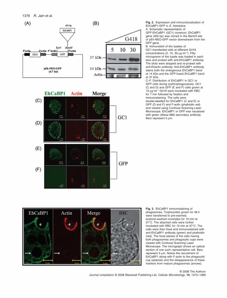
Fig. 2. Expression and immunolocalization ofEhCaBP1-GFP in E. histolytica.A. Schematic representation ofGFP-EhCaBP1 (GC1) construct. EhCaBP1gene (405 bp) was cloned in the BamHI siteof pEh-NEO-GFP vector downstream from theGFP gene.B. Immunoblot of the lysates ofGC1-transfected cells at different G418concentrations (5, 10, 30 mg ml-1). Fiftymicrograms of the lysate was loaded in eachlane and probed with anti-EhCaBP1 antibody.The blots were stripped and re-probed withanti-Ehactin antibody. Anti-EhCaBP1 antibodystains both the endogenous EhCaBP1 bandat 14 kDa and the GFP-fused EhCaBP1 bandat 37 kDa.C–F. Distribution of EhCaBP1 in GC1 orGFP cells during erythrophagocytosis. GC1(C and D) and GFP (E and F) cells grown at10 mg ml-1 G418 were incubated with RBCfor 7 min followed by fixation andimmunostaining. The cells weredouble-labelled for EhCaBP1 (C and E) orGFP (D and F) and F-actin (phalloidin red)and viewed using Confocal Scanning LaserMicroscope. EhCaBP1 or GFP was visualizedwith green (Alexa-488) secondary antibody.Bars represent 5 mm.
Fig. 3. EhCaBP1 immunostaining ofphagosomes. Trophozoites grown for 48 hwere transferred to pre-warmed,acetone-washed coverslips for 10 min at37°C. The attached cells were furtherincubated with RBC for 10 min at 37°C. Thecells were then fixed and immunostained withanti-EhCaBP1 antibody (green) and phalloidin(red). The focal planes of the cells havingboth phagosomes and phagocytic cups wereviewed with Confocal Scanning LaserMicroscope. The micrograph shows an opticalsection of one such representative cell. Barsrepresent 5 mm. Notice the recruitment ofEhCaBP1 along with F-actin to the phagocyticcup (asterisk) and the disappearance of thesemarkers from mature phagosomes (arrows).
EhCaBP1 Actin Merge DIC
1376 R. Jain et al.
© 2008 The AuthorsJournal compilation © 2008 Blackwell Publishing Ltd, Cellular Microbiology, 10, 1373–1389

image panel (Fig. 4C, left) clearly indicated that GFP-EhCaBP1 exhibited the highest degree of accumulation infully formed pseudopods (Fig. 4C, ii) and rapid dissipationduring retraction (Fig. 4C, iv). The result is also presentedas a line scan (Fig. 4C, right), which exhibits changes influorescent intensity across a cell along a line. The linescan has been plotted for a pseudopod showing a cycle ofprojection and retraction of a representative cell.
Quantitative analysis of such pseudopods (n = 30)was carried out to determine the average time takenfor EhCaBP1 molecule to be enriched within thepseudopods. Here, the amount of fluorescence inpseudopods was determined along a time kinetics. Thefluorescence intensity increased as the pseudopod wasformed and peaked at the time of protrusion (~6 s)(Fig. 4D) followed by a gradual decline of about 12 s.A decrease in the fluorescence was observed duringretraction. The rate of loss of fluorescence was muchslower than the rate of accumulation paralleling the for-mation and retraction of pseudopods.
It was also of interest to determine the fraction of totalEhCaBP1 that may be present in a given pseudopod. Inorder to compute this, fluorescent intensity of protrudedpseudopods was determined. Analysis of the data gener-ated from 30 different pseudopods and cytoplasmicsegments suggested that 12 � 4% of the total cellularEhCaBP1 was present in a given pseudopod. Low level offluorescent molecules in cytoplasm may reflect dilution ofthe label due to larger volume. The results suggest thatEhCaBP1 is a highly dynamic molecule with a high rate ofaccumulation and disappearance from preferred sites.
Generation of mutant EhCaBP1
It is difficult to conclude from the previous results thatCa2+ binding is important for participation of EhCaBP1 inpseudopod extension during motility or phagocytosis asCa2+ chelator would affect whole repertoire of EhCaBPs.EhCaBP1 possess four canonical EF hand Ca2+-bindingmotifs, of which two display high affinities (Gopal et al.,1997). The role of Ca2+ in some of the activities of EhCaBP1was investigated by generating point mutations. Pointmutations were introduced by site-directed mutagenesisinto each of the EF hands to disrupt the Ca2+ binding ability,while maintaining the overall structure of the domain. Thefirst aspartate (D) residue of EF hand domains is one of theimportant residues involved in coordinating Ca2+ andmutating it affects Ca2+ binding (Geiser et al., 1991). There-fore, all the four D residues (10, 46, 85, 117) of EhCaBP1EF hand domains were converted to alanine (A) singly orall at once. The mutants were named EFI, EFII, EFIII andEFIV, after the EF hand domain which was mutated. Themutant having the first D’s of all four EF hands mutated toA was termed CaBP1DEF (Fig. 5A). The mutant proteins
were expressed and purified from bacterial cells asdescribed for the WT protein (Prasad et al., 1993). Thesemutants were tested for their Ca2+-binding properties. As afirst indication of the ability of the proteins to bind Ca2+ andundergo a conformational change, electrophoretic mobilityin the presence of Ca2+ was checked by SDS-PAGE(Fig. 5B). The Ca2+-bound form of EhCaBP1 undergoes amobility shift in SDS-PAGE, reflecting Ca2+ binding andsubsequent conformational change (Prasad et al., 1993).The single-point mutants showed a similar change in rela-tive mobility of Ca2+-free (EGTA) and Ca2+-bound forms asthe WT protein. In contrast, there was no difference in themobility of the two forms in the case of CaBP1DEF, themutant with all the four EF hands mutated.
It was shown before by CD spectroscopy that there is amajor increase in helicity of EhCaBP1 on binding Ca2+.This increase in helicity is reflected in a large change inconformation (Gopal et al., 1997). The variation in sec-ondary structure of Ca2+-bound form of mutant EhCaBP1swas also studied using CD spectroscopy (Fig. 5C). Whiletwo of the single mutants (EFI and EFIII) did not showmuch difference from the WT protein, some differenceswere observed for the other two (EFII and EFIV) mutants.On the other hand CD spectrum of CaBP1DEF proteinsuggested a molecule with low level of secondary struc-ture, very different from the WT molecule. Moreover, nochange in helicity was observed for CaBP1DEF in thepresence or absence of Ca2+ (data not shown) in contrastto the WT EhCaBP1. These results suggest that eitherCaBP1DEF is unable to bind Ca2+ or it is defective in itsability to undergo Ca2+-induced conformation change.
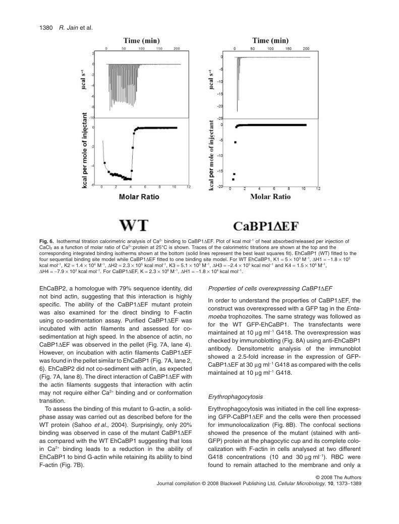
Isothermal calorimetry (ITC) has been frequently usedto study Ca2+ and other metal ions interaction with pro-teins (Leavitt and Freire, 2001). It has also been used tostudy the thermodynamics of Ca2+ binding to EhCaBP1(Gopal et al., 1997). Thus, in order to check if CaBP1DEFcan bind Ca2+, ITC was used (Fig. 6). The figure showsthe integrated binding isotherms obtained after fitting tothe best least square fit. WT EhCaBP1 was best fitted tothe four site binding model while CaBP1DEF fitted to thesingle site binding model, indicating a major loss in Ca2+
binding sites. Loss of Ca2+ binding by the mutant proteinwas also confirmed by [45Ca] binding assay in blots (datanot shown here). The structural analysis suggests thatCaBP1DEF is defective in Ca2+ binding and it has resultedin its inability to form secondary structure in the presenceof Ca2+. Therefore CaBP1DEF mutant was used for furtherfunctional study.
Characterization of the CaBP1DEF binding to G-actinand F-actin
The direct binding of EhCaBP1 to F-actin was previouslydemonstrated (Sahoo et al., 2004). It was also shown that
EhCaBP1 and phagocytosis 1377
© 2008 The AuthorsJournal compilation © 2008 Blackwell Publishing Ltd, Cellular Microbiology, 10, 1373–1389

1378 R. Jain et al.
© 2008 The AuthorsJournal compilation © 2008 Blackwell Publishing Ltd, Cellular Microbiology, 10, 1373–1389

Fig. 4. Live cell time-lapse imaging of EhCaBP1.A. Time-lapse micrographs during phagocytosis of RBC by an amoeba expressing GFP-EhCaBP1, showing de novo formation of a phagocyticcup. GC1 cells were mixed with RBC and then images of a stack of 15 sections along the z-axis (every 1.5 mm) were recorded every 4 s.Each stack of focal planes was projected after three-dimensional reconstruction as described in Experimental procedures. The montage showsa time series of a representative cell showing the formation of a phagocytic cup (star) and finally the closure of the phagocytic cup. Time isshown in seconds. Bar represents 5 mm.B. The micrograph represents a time series of movement of a trophozoite expressing GFP-EhCaBP1. GC1 cells were allowed to attach to theculture dish and the images of the moving amoeba were captured using video microscopy. The images were recorded every 3 s, capturing astack of 12 focal planes. The analysis and the three-dimensional reconstruction was performed as described in Experimental procedures.Arrowheads represent the images showing the protrusion of the pseudopod and the marked enrichment of GFP-EhCaBP1. Bar represents10 mm.C. Left: A representative cell, going through a cycle beginning with pseudopod protrusion (ii) and ending with retraction (iv), is shown. Thefluorescent images were taken every 6 s. Right: The line intensity scan of the fluorescence intensity measured on the line overlaid on thecorresponding images in left panel. Bars represent 10 mm.D. Quantitative analysis of GFP-EhCaBP1 localization in the pseudopods. The graph presents the kinetics of movement of GFP-EhCaBP1 inthe pseudopods of moving amoebae. The fluorescence intensity associated with the pseudopods was plotted against time in seconds. Thedata shown are the mean intensity � SD from 30 different pseudopods.
Fig. 5. Generation and characterization ofEhCaBP1 mutants.A. Schematic representation of EhCaBP1indicating the four EF hand domains. Thesequence of all the EF hands is shown andthe first aspartate (D) of each EF hand wasmutated to alanine (A) individually orsimultaneously to generate EFI, II, III, IV orCaBP1DEF mutants respectively.B. Gel mobility shift of Ca2+-bound andCa2+-free forms of EhCaBP1 wild type (WT)and mutants. Recombinant proteins werepurified from E. coli lysates expressingEhCaBP1 WT or mutants. Five micrograms ofpurified proteins (as indicated) were subjectedto electrophoresis on a 15% SDS-PAGE inthe presence of 5 mM Ca2+ (C) or 2 mMEGTA (E). The proteins were stained withCoomassie brilliant blue R-250.C. CD spectrum of EhCaBP1 WT andmutants. The CD profiles of WT (�), EFI (�),EFII (D), EFIII (x), EFIV (�) and CaBP1DEF(+) in the presence of 50 mM Tris·Cl (pH 7.5),100 mM NaCl and 5 mM CaCl2 are shown.
(A)
(B)
(C)
-20
-15
-10
-5
0
5
10
200 210 220 230 240 250 260
wavelength (nm)
mo
lar
ellip
tic
ity
in
de
g c
m2 d
mo
l-1
(x 1
05)
EhCaBP1 and phagocytosis 1379
© 2008 The AuthorsJournal compilation © 2008 Blackwell Publishing Ltd, Cellular Microbiology, 10, 1373–1389
EhCaBP2, a homologue with 79% sequence identity, didnot bind actin, suggesting that this interaction is highlyspecific. The ability of the CaBP1DEF mutant proteinwas also examined for the direct binding to F-actinusing co-sedimentation assay. Purified CaBP1DEF wasincubated with actin filaments and assessed for co-sedimentation at high speed. In the absence of actin, noCaBP1DEF was observed in the pellet (Fig. 7A, lane 4).However, on incubation with actin filaments CaBP1DEFwas found in the pellet similar to EhCaBP1 (Fig. 7A, lane 2,6). EhCaBP2 did not co-sediment with actin, as expected(Fig. 7A, lane 8). The direct interaction of CaBP1DEF withthe actin filaments suggests that interaction with actinmay not require either Ca2+ binding and or conformationtransition.
To assess the binding of this mutant to G-actin, a solid-phase assay was carried out as described before for theWT protein (Sahoo et al., 2004). Surprisingly, only 20%binding was observed in case of the mutant CaBP1DEFas compared with the WT EhCaBP1 suggesting that lossin Ca2+ binding leads to a reduction in the ability ofEhCaBP1 to bind G-actin while retaining its ability to bindF-actin (Fig. 7B).
Properties of cells overexpressing CaBP1DEF
In order to understand the properties of CaBP1DEF, theconstruct was overexpressed with a GFP tag in the Enta-moeba trophozoites. The same strategy was followed asfor the WT GFP-EhCaBP1. The transfectants weremaintained at 10 mg ml-1 G418. The overexpression waschecked by immunoblotting (Fig. 8A) using anti-EhCaBP1antibody. Densitometric analysis of the immunoblotshowed a 2.5-fold increase in the expression of GFP-CaBP1DEF at 30 mg ml-1 G418 as compared with the cellsmaintained at 10 mg ml-1 G418.
Erythrophagocytosis
Erythrophagocytosis was initiated in the cell line express-ing GFP-CaBP1DEF and the cells were then processedfor immunolocalization (Fig. 8B). The confocal sectionsshowed the presence of the mutant (stained with anti-GFP) protein at the phagocytic cup and its complete colo-calization with F-actin in cells analysed at two differentG418 concentrations (10 and 30 mg ml-1). RBC werefound to remain attached to the membrane and only a
Fig. 6. Isothermal titration calorimetric analysis of Ca2+ binding to CaBP1DEF. Plot of kcal mol-1 of heat absorbed/released per injection ofCaCl2 as a function of molar ratio of Ca2+:protein at 25°C is shown. Traces of the calorimetric titrations are shown at the top and thecorresponding integrated binding isotherms shown at the bottom (solid lines represent the best least squares fit). EhCaBP1 (WT) fitted to thefour sequential binding site model while CaBP1DEF fitted to one binding site model. For WT EhCaBP1, K1 = 5 ¥ 103 M-1, DH1 = -1.8 ¥ 103
kcal mol-1, K2 = 1.4 ¥ 104 M-1, DH2 = 2.3 ¥ 105 kcal mol-1, K3 = 5.1 ¥ 105 M-1, DH3 = -2.4 ¥ 105 kcal mol-1 and K4 = 1.5 ¥ 106 M-1,DH4 = -7.9 ¥ 103 kcal mol-1. For CaBP1DEF, K = 2.3 ¥ 106 M-1, DH1 = -1.8 ¥ 104 kcal mol-1.
1380 R. Jain et al.
© 2008 The AuthorsJournal compilation © 2008 Blackwell Publishing Ltd, Cellular Microbiology, 10, 1373–1389

very few internalized RBC were observed at 30 mg ml-1
G418 suggesting a defect in the uptake of RBC. The levelof erythrophagocytosis was also determined spectro-photometrically (Fig. 8D). RBC uptake was comparablein all the cell lines expressing GFP vector alone,GFP-EhCaBP1 or GFP-CaBP1DEF expressing cells at10 mg ml-1 G418. However, at higher G418 concentration,a 60% reduction was observed in cells carrying GFP-CaBP1DEF (2.5-fold increased) confirming results
obtained by confocal imaging. The data suggest that over-expression of a Ca2+-insensitive form of EhCaBP1 leadsto a dominant negative phenotype affecting late stages ofphagosome maturation rather than early stages ofphagocytosis.
Time-lapse microscopy was also used to determine therate of appearance and disappearance of the mutantEhCaBP1 from the pseudopods. The results are shownas a micrograph of a representative cell (Fig. 8E). Asdescribed above for the WT protein, the mutant proteinwas also found to disappear from the pseudopods rapidlyduring retraction. The results suggest that the dynamicassociation of EhCaBP1 with actin-mediated processes,such as pseudopod formation, does not require Ca2+
binding.
Bacterial uptake and cytotoxicity
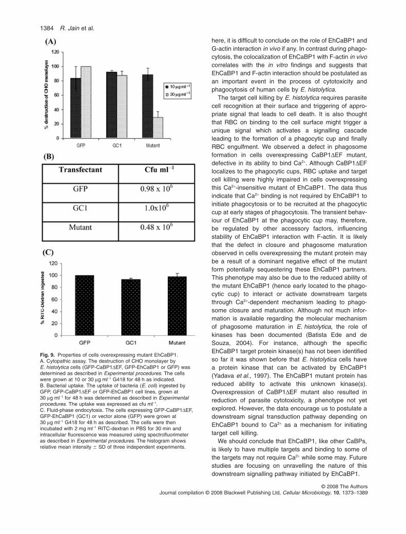
Both the properties, bacterial uptake and the ability to killtarget cells, were found to be defective in cells overex-pressing the mutant protein. Cytotoxicity was measuredby the ability of E. histolytica cells to kill Chinese hamsterovary cells (CHO). The results are shown in Fig. 9A and B.As observed for the RBC uptake, the cells expressingGFP-CaBP1DEF at high levels showed a 70% reductionin their ability to destroy CHO monolayer (Fig. 9A) andalso a 50% defect in bacterial uptake (Fig. 9B). No signifi-cant effect was observed for the parental cells or cellscarrying vector alone.
Pinocytosis
Fluid-phase endocytosis was measured by determiningthe uptake of the fluorescent marker (RITC-dextran) byGFP, GC1 and GFP-CaBP1DEF cells. There was no sig-nificant difference observed for any of the transfectantsunder the assay conditions (Fig. 9C).
Reduced activation of kinase by mutant EhCaBP1
The decrease in the RBC uptake by the cells overex-pressing the Ca2+-insensitive EhCaBP1 suggested thatCa2+-bound form of EhCaBP1 may be responsible forinitiating downstream signalling leading to phagosomeformation. As it is known that EhCaBP1 can activate spe-cific kinase(s), the ability of CaBP1DEF to activate endog-enous kinase(s) was investigated (Yadava et al., 1997).The reaction was carried out in the presence of 10 mMCaCl2 and 2 nM protein as these have been shown earlierto be the optimum concentration for activation ofEhCaBP1-dependent protein kinase(s). The result isshown in Fig. 10. There was a marked reduction (60%) inthe ability of the mutant to activate kinase(s) as comparedwith the WT EhCaBP1 suggesting that activation of thekinase may require the ability of EhCaBP1 to bind Ca2+.
Fig. 7. Binding of CaBP1DEF to actin.A. Co-sedimentation of CaBP1DEF with F-actin. Purifiedrecombinant CaBP1DEF (5 mM, lanes 5, 6), EhCaBP1 (5 mM, lanes1, 2) and EhCaBP2 (5 mM, lanes 7, 8) were incubated withpolymerized actin (5 mM) in polymerization (G) buffer containingsalts. CaBP1DEF was also incubated alone without actin (lane 3,4). This was followed by ultra centrifugation to separatesupernatant (S) and pellet (P) fractions. S (one-fourth of the total)and P (total) fractions were separated on a 15% SDS-PAGEfollowed by Coomassie blue staining.B. Binding of CaBP1DEF to G-actin. Actin (5 mM) was coated onthe microtitre plates overnight at 4°C. After blocking with BSA,CaBP1DEF and EhCaBP1 were added at the indicatedconcentrations in the presence of 5 mM CaCl2 or 2 mM EGTA. Thiswas followed by incubation with anti-EhCaBP1 antibody. Theamount of bound EhCaBP1 was detected by using anti-rabbitIgG-HRPO. The amount of bound second antibody is determinedby absorbance at 405 nm. The histogram shows the relative meanintensity � SD of three independent experiments.
EhCaBP1 and phagocytosis 1381
© 2008 The AuthorsJournal compilation © 2008 Blackwell Publishing Ltd, Cellular Microbiology, 10, 1373–1389

(A)
(B)
(i) (ii)
(C)
0
0.05
0.1
0.15
0.2
0.25
0.3
0.35
GFP GC1 Mutant
O.D
at
400 n
m
10 μg ml–1
30 μg ml–1
(D)
(E)
37
14
1382 R. Jain et al.
© 2008 The AuthorsJournal compilation © 2008 Blackwell Publishing Ltd, Cellular Microbiology, 10, 1373–1389

Discussion
Entamoeba histolytica has an extensive signalling systemunlike many other protozoan parasites. The extent of thesignalling network can be gauged by the fact that thismicroorganism has a large repertoire of novel CaBPs andtrans-membrane kinases (Loftus et al., 2005; Bhatta-charya et al., 2006). The function of most of these mol-ecules in amoebic biology has not been studied except forEhCaBP1. It has been implicated in cellular processesassociated with dynamic membrane-cytoskeletal struc-tures involving actin remodelling, such as pseudopod for-mation and phagocytosis (Sahoo et al., 2004). NormallyCa2+ signalling is mediated through CaBPs, many ofwhich are Ca2+ sensors and participate in downstreamsignal transduction processes and are known to carry outdiversity of functions (Berridge et al., 2000).
The results presented here clearly showed thatEhCaBP1 is a highly dynamic molecule and it associatestransiently with subcellular assemblies, such as actin fila-ments, mostly in the initial stages when the phagocyticcups or pseudopods are being formed. Interestingly, itwas also observed using a mutant protein defective inCa2+ binding (CaBP1DEF) that this association ofEhCaBP1 with actin filaments in the formation of phago-cytic cups may not have a strict requirement for its abilityto bind Ca2+. It is unlikely that full functional property ofEhCaBP1 is due to little residual Ca2+-binding activity ofCaBP1DEF, as the presence of low-affinity binding sites inEhCaBP1 and the residual binding does not change con-formation in the presence of Ca2+ (Moorthy et al., 2001).There are very few examples where the functions ofCaBPs are independent of their ability to bind Ca2+. There-fore it is significant that the recruitment of EhCaBP1 to thephagocytic cup does not require Ca2+ binding. A few otherexamples are functional complementation of yeast calm-odulin by a mutated form of calmodulin with no Ca2+
binding ability (Geiser et al., 1991), formation of autoph-agic tubes in a Ca2+-independent manner (Uttenweileret al., 2005) and localization of a flagellar CaBP of Trypa-
nosoma cruzi to the flagella in a Ca2+-independentmanner (Buchanan et al., 2005).
Phagocytosis is a crucial activity for the survival andvirulence of E. histolytica (Orozco et al., 1983). Although anumber of studies have been carried out, there is stillno clear understanding of the molecular mechanismsinvolved in the process which start with the binding on theamoebic surface of the target cell. The signalling uponbinding triggers the formation of a phagocytic cup leadingto actin filaments re-organization. Closure of this cup endswith the formation of an early phagosome that maturatesto fuse with lysosomes. Proteomic strategy has beenprimarily used to identify the molecules present in earlyphagosomes (Marion et al., 2005; Okada et al., 2005).However, EhCaBP1 was not found in the phagosomeproteome, in spite of its clear involvement in the process.The absence of EhCaBP1 in the proteome of thesephagosomes was in accord with the observation usingGFP-tagged EhCaBP1 showing that EhCaBP1 is a highlydynamic molecule and is transiently associated withdynamic ruffling structures (this study). Thus, in vitroassociation of EhCaBP1 and actin filaments demon-strated earlier (Sahoo et al., 2004) appears to be transientin vivo. The rate of GFP-EhCaBP1 movement duringpseudopod formation suggested that the re-organizationof EhCaBP1 is a highly rapid process occurring in theinitial 3–4 s. The dynamics of EhCaBP1 redistributionduring phagocytosis was similar to other actin-associatedmolecules, such as myosin I (Ostap et al., 2003; Marionet al., 2004), coronin (Lu and Clarke, 2005), or ER-linkedproteins calreticulin and calnexin (Muller-Taubenbergeret al., 2001). However, for the first time we have shownthat a Ca2+-sensing protein, EhCaBP1, undergoesdynamic translocation that correlates with actin filamentre-organization during pseudopods extension.
EhCaBP1 was shown to bind both F- and G-actin invitro although with low Kd values (Sahoo et al., 2004). Thismeans that EhCaBP1 and actin should normally remainas a complex, with transient composition and stability,according to the need of the cell. From the data presented
Fig. 8. Expression and localization of CaBP1DEF in E. histolytica during erythrophagocytosis.A. GFP-CaBP1DEF-expressing cells were maintained at different G418 concentrations (5, 10, 30 mg ml-1). Fifty micrograms of the lysate wasused for Western blotting and immunolocalization using anti-EhCaBP1 antibody. The blots were stripped and re-probed with anti-Ehactinantibody. Anti-EhCaBP1 antibody stains both the endogenous EhCaBP1 band at 14 kDa and the GFP-fused EhCaBP1DEF band at 37 kDa.B–C. GFP-CaBP1DEF during erythrophagocytosis was visualized by immunolocalization in cells expressing GFP-CaBP1DEF at 10 mg ml-1
G418 (B) or 30 mg ml-1 G418 (C, i, ii). The slides were prepared as described in Experimental procedures. GFP-CaBP1DEF was stained withanti-GFP antibody (green) and phalloidin (red) and viewed using CSLM. (C, i) Confocal section of a representative cell; (C, ii) an enlargedview of the membrane of a cell. RBC binding to the surface and the colocalization of GFP-CaBP1DEF and F-actin are clearly visible.D. Erythrophagocytosis in cells expressing high level of mutant protein. The cells expressing GFP-CaBP1DEF, GFP-EhCaBP1 (GC1) or vectoralone (GFP) were grown at 10 mg ml-1 and 30 mg ml-1 G418. Erythrophagocytosis was measured after incubating 105 amoebae with 107 RBCfor 10 min at 37°C. The level of haem was determined by measuring absorbance at 400 nm which was a direct estimate of the ingested RBC.The histogram shows relative mean optical density � SD of three independent experiments.E. The micrograph represents a time sequence of a motile trophozoite expressing GFP-CaBP1DEF. Acquisitions were performed as forthe GC1 cells expressing WT EhCaBP1. The analysis and the three-dimensional reconstruction was performed as described above.Bar represents 10 mm. Note the accumulation of GFP-CaBP1DEF in the protrusions (star) and the fast disappearance (arrow) as the cellmoves.
EhCaBP1 and phagocytosis 1383
© 2008 The AuthorsJournal compilation © 2008 Blackwell Publishing Ltd, Cellular Microbiology, 10, 1373–1389
here, it is difficult to conclude on the role of EhCaBP1 andG-actin interaction in vivo if any. In contrast during phago-cytosis, the colocalization of EhCaBP1 with F-actin in vivocorrelates with the in vitro findings and suggests thatEhCaBP1 and F-actin interaction should be postulated asan important event in the process of cytotoxicity andphagocytosis of human cells by E. histolytica.
The target cell killing by E. histolytica requires parasitecell recognition at their surface and triggering of appro-priate signal that leads to cell death. It is also thoughtthat RBC on binding to the cell surface might trigger aunique signal which activates a signalling cascadeleading to the formation of a phagocytic cup and finallyRBC engulfment. We observed a defect in phagosomeformation in cells overexpressing CaBP1DEF mutant,defective in its ability to bind Ca2+. Although CaBP1DEFlocalizes to the phagocytic cups, RBC uptake and targetcell killing were highly impaired in cells overexpressingthis Ca2+-insensitive mutant of EhCaBP1. The data thusindicate that Ca2+ binding is not required by EhCaBP1 toinitiate phagocytosis or to be recruited at the phagocyticcup at early stages of phagocytosis. The transient behav-iour of EhCaBP1 at the phagocytic cup may, therefore,be regulated by other accessory factors, influencingstability of EhCaBP1 interaction with F-actin. It is likelythat the defect in closure and phagosome maturationobserved in cells overexpressing the mutant protein maybe a result of a dominant negative effect of the mutantform potentially sequestering these EhCaBP1 partners.This phenotype may also be due to the reduced ability ofthe mutant EhCaBP1 (hence early located to the phago-cytic cup) to interact or activate downstream targetsthrough Ca2+-dependent mechanism leading to phago-some closure and maturation. Although not much infor-mation is available regarding the molecular mechanismof phagosome maturation in E. histolytica, the role ofkinases has been documented (Batista Ede and deSouza, 2004). For instance, although the specificEhCaBP1 target protein kinase(s) has not been identifiedso far it was shown before that E. histolytica cells havea protein kinase that can be activated by EhCaBP1(Yadava et al., 1997). The EhCaBP1 mutant protein hasreduced ability to activate this unknown kinase(s).Overexpression of CaBP1DEF mutant also resulted inreduction of parasite cytotoxicity, a phenotype not yetexplored. However, the data encourage us to postulate adownstream signal transduction pathway depending onEhCaBP1 bound to Ca2+ as a mechanism for initiatingtarget cell killing.
We should conclude that EhCaBP1, like other CaBPs,is likely to have multiple targets and binding to some ofthe targets may not require Ca2+ while some may. Futurestudies are focusing on unravelling the nature of thisdownstream signalling pathway initiated by EhCaBP1.
Fig. 9. Properties of cells overexpressing mutant EhCaBP1.A. Cytopathic assay. The destruction of CHO monolayer byE. histolytica cells (GFP-CaBP1DEF, GFP-EhCaBP1 or GFP) wasdetermined as described in Experimental procedures. The cellswere grown at 10 or 30 mg ml-1 G418 for 48 h as indicated.B. Bacterial uptake. The uptake of bacteria (E. coli) ingested byGFP, GFP-CaBP1DEF or GFP-EhCaBP1 cell lines, grown at30 mg ml-1 for 48 h was determined as described in Experimentalprocedures. The uptake was expressed as cfu ml-1.C. Fluid-phase endocytosis. The cells expressing GFP-CaBP1DEF,GFP-EhCaBP1 (GC1) or vector alone (GFP) were grown at30 mg ml-1 G418 for 48 h as described. The cells were thenincubated with 2 mg ml-1 RITC-dextran in PBS for 30 min andintracellular fluorescence was measured using spectrofluorimeteras described in Experimental procedures. The histogram showsrelative mean intensity � SD of three independent experiments.
1384 R. Jain et al.
© 2008 The AuthorsJournal compilation © 2008 Blackwell Publishing Ltd, Cellular Microbiology, 10, 1373–1389

Experimental procedures
Strains and cell culture
Entamoeba histolytica strain HM1:IMSS clone 6 was maintainedand grown in TYI-S-33 medium containing 125 ml of 250 U ml-1
Benzyl Penicillin and 0.25 mg ml-1 Streptomycin per 100 ml ofmedium. Neomycin (Sigma) was added at 10 mg ml-1 for main-taining transgenic cell lines.
Intracellular Ca2+ measurements
Trophozoites (106) were washed twice with PBS and loaded with20 mM FURA 2-AM (Sigma Chemicals) at room temperature inthe dark for 1 h in buffer A (20 mM Hepes, pH 7.2, 140 mM NaCl)supplemented with 0.1% BSA. Loaded cells were washed twicewith PBS and re-suspended in buffer A. Cells were transferred toa thermostated fluorometric cuvette containing a magnetic stirbar and maintained at 37°C with gentle agitation. BAPTA-AM wasadded directly to the cuvette. Fluorescence values were regis-tered in a Cary Fluorescence Spectrophotometer, programmedto obtain the emission at 510 nm on excitation at 340 and 380 nmsimultaneously. [Ca2+]i is represented as the 340–380 nm ratio asthese are considered proportional to each other (Grynkiewiczet al., 1985).
Cloning of EhCaBP1 gene in pEh-NEO-GFP vector
Primers to amplify the EhCaBP1 were designed based on theEntamoeba genome sequence database. BamHI restriction sitewas incorporated in both the primers. The primers usedwere GC1-F, 5′-GGGGGATCCCATATGGCTGAAGCACT-3′ andGC1-R, 5′-GGGGGATCCAGTTTAGAGTGAAAACT-3′. An ampli-con of 405 bp was obtained by PCR amplification. It was clonedin the BamHI site of the pEh-NEO-GFP vector downstream toGFP gene. The vector has been previously constructed (N.Guillen, unpublished) by cloning the GFP mut3 allele of GFP
(Cormack et al., 1996) in the unique BamHI site of the pExEhNeoplasmid (Hamann et al., 1995). The different constructs (GC1 orvector) were used to transform Escherichia coli strain DH5a cellsfollowing standard methods.
Transfection and selection of E. histolytica trophozoites
Transfection of E. histolytica cells by pEhNEO/GC1 DNA wasperformed by electroporation as described previously (Sahooet al., 2003). Briefly, trophozoites in log phase were harvestedand washed with PBS followed by incomplete cytomix buffer[10 mM K2HPO4/KH2PO4 (pH 7.6), 120 mM KCl, 0.15 mM CaCl2,25 mM Hepes, 2 mM EGTA, 5 mM MgCl2]. The washed cellswere then re-suspended in 0.8 ml of complete cytomix buffer(incomplete cytomix containing 4 mM Adenosine Triphosphate,10 mM Glutathione) containing 200 mg of plasmid DNA and sub-jected to two consecutive pulses of 3000 V cm-1 (1.2 kV) at 25 mF(Bio-Rad, electroporator). The transfectants were initially allowedto grow without any selection. Drug selection was initiated after2 days of transfection in the presence of 10 mg ml-1 G418(Sigma).
Immunofluorescence staining
Immunofluorescence staining was carried out as describedbefore (Sahoo et al., 2004). Briefly, E. histolytica cells wereallowed to adhere on to a glass coverslip for 5 min at 37°C inTYI-33 medium. RBC was added to the cells for indicated time atthe ratio of 10 RBC per cell. Phagocytosis was stopped byfixation in 3.7% PFA for 30 min. The cells were then incubated in50 mM NH4Cl for 30 min before permeabilization with 0.1% TritonX-100 for 1 min. Non-specific sites were blocked by incubationwith 1% BSA/PBS for 30 min at 37°C. While actin was labelled byPhalloidin-TRITC (1/500) both GFP and EhCaBP1 were identi-fied by respective antibodies (GFP at 1:200, Molecular probes;EhCaBP1 at 1:30). The bound antibodies were visualized by
Fig. 10. Reduced activation of kinase(s) byCaBP1DEF. E. histolytica cell-free lysate(25 mg) was used as the source of kinase andhistone type III (15 mg) as the substrate.EhCaBP1 and CaBP1DEF were used for theassay at 2 nM. The concentration of Ca2+
used was 10 mM. The assay was performedas described in Experimental proceduresusing [g-32P]-ATP as the phosphate donor.The reaction products were separated on a12% SDS-PAGE and the amount ofradioactivity incorporated into histone wasdetermined by densitometry. The graphrepresents the histogram of the variousreactions, as indicated in the table �SD ofthree independent experiments. The lowestpanel depicts a representative gel picture.The first lane in all cases is histone alone.
EhCaBP1 and phagocytosis 1385
© 2008 The AuthorsJournal compilation © 2008 Blackwell Publishing Ltd, Cellular Microbiology, 10, 1373–1389

anti-rabbit secondary antibodies coupled with Alexa 488 (1/200)or Cy3 (1/300) (Molecular Probes). The preparations were furtherwashed with PBS and mounted on a glass-slide using DABCO[1,4-diazbicyclo (2,2,2) octane, Sigma, 10 mg ml-1 in 80%glycerol]. Sealing of the coverslip edges was performed withnail-paint to avoid drying.
Confocal laser scanning microscopy
Fluorescent samples were examined on an LSM 510 confocallaser scanning microscope (Zeiss, Germany) equipped with a63¥ objective. Alexa-red-labelled samples were visualized afterexcitation at 543 nm using He/Ne Laser and Alexa-green-labelledsamples after excitation at 488 nm using Argon Laser. Focalplanes of 0.8 mm sections with a shift of objective by 1 mm werecaptured for about 20 planes from the bottom to the top of eachcell. Images were processed using LSM 510 software, Zeiss,Germany.
Time-lapse imaging
The cells expressing GFP-CaBP1, GFP-CaBP1DEF or GFP alonewere plated onto a 35 mm Mat Tek glass bottom culture dish (MatTek Corporation) at 37°C. After the cells got settled at the bottom,the medium was removed and the glass chamber was filled withpre-warmed PBS. The dish was kept on a platform with a tempera-ture controller (Tempcontrol 37-2 digital, Zeiss) to maintain tem-perature at 37°C. High-resolution fluorescent time-lapse imagingof a moving and phagocytosing amoeba was performed using ahigh-speed spinning disk confocal system (UltraView RS, PerkinElmer) equipped for axial z-stack sampling throughout the cellvolume. This system was attached to an inverted 200 M micro-scope (Zeiss). The images were captured with an Orca D-ERdetector (Hamamatsu) and a 40¥ objective and 2 ¥ 2 binning.Images of 1.5 mm (along z-axis) at 3 or 4 s interval (as indicated)were captured. This z-spacing was optimized to: (i) monitor theentire depth of amoeba from top to bottom and (ii) accomplish fastcapturing of a moving amoeba. The raw images were processedusing ImageJ software available freely on the web (http://rsb.info.nih.gov/ij/). For each time point, raw images were three-dimensionally reconstituted before further analysis.
Image analysis
Raw images obtained from the microscope were processed andanalysed using Image J software (Plugins/PE_raw/Convert filesto tiff series) and the three-dimensional images were recon-structed (Images/Stacks/Z project) by taking standard deviationof the selected slices and projected as a time series. For quan-titative measurements of the pseudopods, the time point justbefore the pseudopod protrusion was referred to as t = 0. Themeasurement of grey scale intensity of the images was per-formed as described elsewhere (Aizawa et al., 1997).
Generation of CaBP1DEF mutant
EhCaBP1 mutants were generated using a site-directedmutagenesis kit (Stratagene, La Jolla, CA). The protocol followedwas essentially as described by the manufacturer. The first
aspartate (D) residue of each EF hand was mutated to alanine(A) residue. The mutations were performed in a sequential orderone residue at a time. The modified amplicon was used as atemplate for the next round of mutation to get a mutant having allthe four D mutated to A (CaBP1DEF). The mutations were con-firmed by nucleotide sequencing. The primers used were
EFI F: 5′-CTTTTTAAAGAAATTGCAGTTAATGGAGATGGAG-3′EFI R: 5′-CTCCATCTCCATTAACTGCAATTTCTTTAAAAAG-3′EFII F: 5′-CAAATCTATTGCAGCTGATGGAAA-3′EFII R: 5′-TTTCCATCAGCTGCAATAGATTTG-3′EFIII F: 5′-CTATAAACTTATGGCAGTTGATGGAGATGG-3′EFIII R: 5′-CCATCTCCATCAACTGCCATAAGTTTATAG-3′EFIV F: 5′-GTTATGAAAGCTGCAGCTAATGGTGATG-3′EFIV R: 5′-CATCACCATTAGCTGCAGCTTTCATAAC-3′
Expression and purification of recombinantproteins in E. coli
The purification of all the recombinant proteins (WT and mutants)was carried out essentially as for WT EhCaBP1 (Prasad et al.,1993).
Circular dichroism spectroscopy
CD measurements were performed using a Jasco-815spectropolarimeter. Each spectrum was measured in the far-UVregion (200–260 nm) and was an average of five scans. Scanswere performed at a protein concentration of 33 mM (50 mMTris·Cl, pH 7.0 and 100 mM NaCl) using a cuvette of path length1.0 cm in the presence of 5 mM CaCl2. Percentage helicalcontent was calculated using the method described by Barrowet al. (1992).
Isothermal calorimetry
Isothermal calorimetry measurements were performed with aMicrocal Omega titration calorimeter at 25°C. Samples werecentrifuged and degassed prior to the titration. A typical titrationconsisted of injecting 2 ml aliquots of 20 mM Ca2+ solution (dilutedfrom 1 M standard CaCl2 solution supplied from Sigma-Aldrichchemicals) into 200 mM protein solution after every 3 min toensure that the titration peak returned to the baseline prior to thenext injection. A total of 70 injections were carried out. Aliquots ofconcentrated ligand solution were injected into the buffer solution(without the protein) in a separate ITC run, to subtract the heat ofdilution. Two sets of titrations were carried out: (i) apo-EhCaBP1in 50 mM Tris·Cl, pH 7.0 and 100 mM NaCl and (ii) apo-CaBP1DEF in 50 mM Tris·Cl, pH 7.0 and 100 mM NaCl. The ITCdata were analysed using the software ORIGIN (supplied withOmega Microcalorimeter). The amount of heat released per addi-tion of the titrant was fitted to best least squares fit model asgiven by Wiseman et al. (1989).
Actin and CaBP1DEF co-sedimentation assay
Co-sedimentation assay was carried out following published con-ditions (Sahoo et al., 2004). Briefly, 5 mM of rabbit muscle G-actin(Sigma) was polymerized for 60 min in polymerization buffer
1386 R. Jain et al.
© 2008 The AuthorsJournal compilation © 2008 Blackwell Publishing Ltd, Cellular Microbiology, 10, 1373–1389
containing 100 mM KCl and 2 mM MgCl2 at room temperature.After polymerization, actin was mixed with 1 mM ATP and appro-priate target protein (5 mM) in a total volume of 150 ml of G-buffer(10 mM Tris-Cl, pH 7.5, 2 mM CaCl2, 2.5 mM b-Mercaptoethanol,0.5 M KCl, 10 mM MgCl2) and incubated for 2 h at roomtemperature. The samples were centrifuged at 100 000 g for45 min at 4°C. The supernatant (one-fourth of total) and pelletfractions (total) were analysed by 15% SDS-PAGE followed byCoomassie blue staining. In addition to CaBP1DEF, WTEhCaBP1 and EhCaBP2 were also used as positive and nega-tive controls respectively.
Solid-phase assay
Different wells of a 96-well plate were coated with 5 mM G-actin inPBS overnight at 4°C and were blocked with 3% BSA in PBS foran additional 24 h. After washing with PBS-T (PBS containing0.05% Tween-20), EhCaBP1 and CaBP1DEF were added to thewells in duplicates at concentrations ranging from 1.4 mM to5 mM. Bound protein was detected with anti-EhCaBP1 antibodyfollowed by HRPO-linked anti-rabbit IgG using the colorimetricsubstrate TMB (Sigma). The absorbance was monitored at405 nm with a microplate reader (Bio-Rad, USA) after stoppingthe reaction with 2 N H2SO4. The reaction was carried out in thepresence of 5 mM CaCl2 or 2 mM EGTA as indicated.
Phagocytosis of RBC by trophozoites
To quantify the RBC ingested by amoebae, the colorimetricmethod of estimation was followed as described earlier (Sahooet al., 2004). Briefly, RBC washed with PBS and TYI-33 mediumwere incubated with amoebae for indicated time at 37°C in 0.2 mlof culture medium at a ratio of 100:1 (RBC:Amoeba). To test theeffect of BAPTA-AM on erythrophagocytosis, amoeba were ini-tially treated with the specified concentrations of BAPTA-AM atroom temperature for 10 min and then incubated with RBC. Theamoebae and erythrocytes were pelleted down and non-engulfedRBC were lysed with cold distilled water and centrifuged at1000 g for 2 min. This step was repeated twice, followed byre-suspension in 1 ml formic acid to burst amoebae containingengulfed RBC. The optical density of the samples was deter-mined by a spectrophotometer at 400 nm using formic acid asblank.
Cytopathic assay
The destruction of monolayer of CHO cells was assayed asdescribed by Bracha and Mirelman (1984). Briefly, trophozoites(105 ml-1 suspended in DMEM without FCS) were added in trip-licate to wells containing a confluent monolayer of CHO cells(105 ml-1) pre-washed with DMEM to remove traces of fetal calfserum and incubated for 60 min at 37°C in an atmosphere of 95%air and 5% CO2. The reaction was stopped by chilling for 10 minand the wells were then washed thrice with cold PBS. The mono-layer was fixed with 4% paraformaldehyde for 10 min and stainedwith methylene blue (0.1% in borate buffer, 0.1 M, pH 8.7). Theexcess stain was washed with 0.01 M borate buffer and theincorporated dye was extracted by adding 1.0 ml of 0.1 M HCl at37°C for 30 min. The colour was read in a spectrophotometer at660 nm after appropriate dilutions with 0.1 M HCl. Destruction of
cells was expressed in relation to the amount of dye extractedfrom the control monolayer of CHO cells.
Bacterial uptake assay
Bacterial uptake by E. histolytica transfectants was determinedby measuring the number of bacteria ingested [colony-formingunits (cfu) ml-1]. Briefly, 105 amoebae were incubated with 107
bacteria (E. coli) at 37°C for 15 min in a volume of 200 ml. Themixture was centrifuged at 300 g for 5 min to pellet only theamoeba and not the non-engulfed bacteria. Amoebae were thenwashed with PBS three times and then re-suspended in PBScontaining 0.1% Triton X-100. The ingested bacteria were pel-leted by centrifugation and washed with PBS (thrice) to removetraces of the detergent. The number of bacteria was then deter-mined by plating on LB-Agar plates at different dilutions.
RITC-dextran uptake analysis
The pinocytosis of E. histolytica was studied by observing theuptake of RITC-dextran as described before (Sahoo et al., 2004).The mid-log-phase cells were harvested, washed andre-suspended in fresh medium. The washed cells were thenincubated with RITC-dextran (2 mg ml-1, Sigma) for 30 min at36°C followed by harvesting and washing with PBS. The cellswere then re-suspended in PBS containing 0.1% Triton X-100.The amount of pinocytosed particles was determined by mea-surement of total fluorescence using a Cary FluorescenceSpectrophotometer.
In vitro kinase assay
Total Entamoeba cell extract was prepared and the activity ofEhCaBP1-dependent kinases was estimated according toChandok and Sopory (1992). The reaction mixture contained30 mM Hepes, pH 7.5, 5 mM MgCl2, 15 mg of Histone (Type IIIS,Sigma), 25 mg of E. histolytica cell extract, protease inhibitorcocktail, CaCl2 or EGTA. Either EhCaBP1 (2 nM) or CaBP1DEF(2 nM) was also added. The reaction volume was adjusted to40 ml. The reaction was initiated by adding 100 mM [g-32P]-rATP(specific activity 5000 Ci mmol-1, BRIT) and allowed to proceedat 30°C for 20 min. The reaction was terminated by adding 1¥SDS-PAGE buffer and resolved on a 12% SDS-PAGE. The gelswere dried and exposed to an X-ray film or an imaging plate.
Western analysis
For immunodetection, samples were separated on a 12% or a15% SDS-PAGE as indicated. The gel was then transferred to anitrocellulose membrane by semidry method and processedusing standard methods. The antigens were detected with poly-clonal anti-GFP (1:2000, Molecular probes) or anti-EhCaBP1(1:1000, Sahoo et al., 2004) and with anti-Rabbit HRPO(1:10 000, Amersham). ECL reagents were used for visualization(Amersham). Actin was detected using anti-Ehactin (raised in ourlaboratory) at 1:1000 dilution.
Acknowledgements
We gratefully acknowledge Spencer Shorte and Pascal Rouxfrom the Imagopole at Pasteur Institute for assistance and
EhCaBP1 and phagocytosis 1387
© 2008 The AuthorsJournal compilation © 2008 Blackwell Publishing Ltd, Cellular Microbiology, 10, 1373–1389

support in the imaging experiments. This work was partially sup-ported by grants from the Department of Biotechnology andCSIR, Government of India. This work was also supported byGrant INCO-DEV in the Fifth Framework Program of the Euro-pean Union to Nancy Guillén. R.J. thanks UGC, India for Juniorand Senior Research Fellowships and French Embassy, NewDelhi for the travel grants and fellowship.
References
Aizawa, H., Fukui, Y., and Yahara, I. (1997) Live dynamics ofDictyostelium cofilin suggests a role in remodeling actinlatticework into bundles. J Cell Sci 110 (Part 19): 2333–2344.
Atreya, H.S., Sahu, S.C., Bhattacharya, A., Chary, K.V.,and Govil, G. (2001) NMR derived solution structure ofan EF-hand calcium-binding protein from Entamoebahistolytica. Biochemistry 40: 14392–14403.
Barrow, C.J., Yasuda, A., Kenny, P.T., and Zagorski, M.G.(1992) Solution conformations and aggregational proper-ties of synthetic amyloid beta-peptides of Alzheimer’sdisease. Analysis of circular dichroism spectra. J Mol Biol225: 1075–1093.
Batista Ede, J., and de Souza, W. (2004) Involvement ofprotein kinases on the process of erythrophagocytis byEntamoeba histolytica. Cell Biol Int 28: 243–248.
Berridge, M.J., Lipp, P., and Bootman, M.D. (2000) The ver-satility and universality of calcium signalling. Nat Rev MolCell Biol 1: 11–21.
Bhattacharya, A., Padhan, N., Jain, R., and Bhattacharya, S.(2006) Calcium-binding proteins of Entamoeba histolytica.Arch Med Res 37: 221–225.
Bracha, R., and Mirelman, D. (1984) Virulence of Entamoebahistolytica trophozoites. Effects of bacteria, microaerobicconditions, and metronidazole. J Exp Med 160: 353–368.
Buchanan, K.T., Ames, J.B., Asfaw, S.H., Wingard, J.N.,Olson, C.L., Campana, P.T., et al. (2005) A flagellum-specific calcium sensor. J Biol Chem 280: 40104–40111.
Carbajal, M.E., Manning-Cela, R., Pina, A., Franco, E., andMeza, I. (1996) Fibronectin-induced intracellular calciumrise in Entamoeba histolytica trophozoites: effect on adhe-sion and the actin cytoskeleton. Exp Parasitol 82: 11–20.
Chandok, M.R., and Sopory, S.K. (1992) Phorbol myristateacetate replaces phytochrome-mediated stimulation ofnitrate reductase in maize. Phytochemistry 31: 2255–2258.
Cormack, B.P., Valdivia, R.H., and Falkow, S. (1996) FACS-optimized mutants of the green fluorescent protein (GFP).Gene 173: 33–38.
De Meester, F., Mirelman, D., Stolarsky, T., and Lester, D.S.(1990) Identification of protein kinase C and its potentialsubstrate in Entamoeba histolytica. Comp Biochem PhysiolB 97: 707–711.
Geiser, J.R., van Tuinen, D., Brockerhoff, S.E., Neff, M.M.,and Davis, T.N. (1991) Can calmodulin function withoutbinding calcium? Cell 65: 949–959.
Gerke, V., Creutz, C.E., and Moss, S.E. (2005) Annexins:linking Ca2+ signalling to membrane dynamics. Nat Rev MolCell Biol 6: 449–461.
Gopal, B., Swaminathan, C.P., Bhattacharya, S., Bhatta-charya, A., Murthy, M.R., and Surolia, A. (1997) Thermo-dynamics of metal ion binding and denaturation of a
calcium binding protein from Entamoeba histolytica. Bio-chemistry 36: 10910–10916.
Gopal, B., Suma, R., Murthy, M.R., Bhattacharya, A., andBhattacharya, S. (1998) Crystallization and preliminaryX-ray studies of a recombinant calcium-binding proteinfrom Entamoeba histolytica. Acta Crystallogr D Biol Crys-tallogr 54: 1442–1445.
Grynkiewicz, G., Poenie, M., and Tsien, R.Y. (1985) A newgeneration of Ca2+ indicators with greatly improved fluores-cence properties. J Biol Chem 260: 3440–3450.
Hamann, L., Nickel, R., and Tannich, E. (1995) Transfectionand continuous expression of heterologous genes in theprotozoan parasite Entamoeba histolytica. Proc Natl AcadSci USA 92: 8975–8979.
Hirata, K.K., Que, X., Melendez-Lopez, S.G., Debnath, A.,Myers, S., Herdman, D.S., et al. (2007) A phagocytosismutant of Entamoeba histolytica is less virulent due todeficient proteinase expression and release. Exp Parasitol115: 192–199.
Kumar, S., Padhan, N., Alam, N., and Gourinath, S. (2007)Crystal structure of calcium binding protein-1 from Entam-oeba histolytica: a novel arrangement of EF hand motifs.Proteins 68: 990–998.
Leavitt, S., and Freire, E. (2001) Direct measurementof protein binding energetics by isothermal titrationcalorimetry. Curr Opin Struct Biol 11: 560–566.
Lew, D.P., Andersson, T., Hed, J., Di Virgilio, F., Pozzan, T.,and Stendahl, O. (1985) Ca2+-dependent and Ca2+-independent phagocytosis in human neutrophils. Nature315: 509–511.
Loftus, B., Anderson, I., Davies, R., Alsmark, U.C., Samuel-son, J., Amedeo, P., et al. (2005) The genome of the protistparasite Entamoeba histolytica. Nature 433: 865–868.
Lu, H., and Clarke, M. (2005) Dynamic properties ofLegionella-containing phagosomes in Dictyosteliumamoebae. Cell Microbiol 7: 995–1007.
Marion, S., Wilhelm, C., Voigt, H., Bacri, J.C., and Guillen, N.(2004) Overexpression of myosin IB in living Entamoebahistolytica enhances cytoplasm viscosity and reducesphagocytosis. J Cell Sci 117: 3271–3279.
Marion, S., Laurent, C., and Guillen, N. (2005) Signalizationand cytoskeleton activity through myosin IB during theearly steps of phagocytosis in Entamoeba histolytica: aproteomic approach. Cell Microbiol 7: 1504–1518.
Moorthy, A.K., Singh, S.K., Gopal, B., Surolia, A., andMurthy, M.R. (2001) Variability of calcium binding toEF-hand motifs probed by electrospray ionization massspectrometry. J Am Soc Mass Spectrom 12: 1296–1301.
Muller-Taubenberger, A., Lupas, A.N., Li, H., Ecke, M.,Simmeth, E., and Gerisch, G. (2001) Calreticulin andcalnexin in the endoplasmic reticulum are important forphagocytosis. EMBO J 20: 6772–6782.
Okada, M., Huston, C.D., Mann, B.J., Petri, W.A., Jr, Kita, K.,and Nozaki, T. (2005) Proteomic analysis of phagocytosisin the enteric protozoan parasite Entamoeba histolytica.Eukaryot Cell 4: 827–831.
Orozco, E., Guarneros, G., Martinez-Palomo, A., andSanchez, T. (1983) Entamoeba histolytica. Phagocytosisas a virulence factor. J Exp Med 158: 1511–1521.
Ostap, E.M., Maupin, P., Doberstein, S.K., Baines, I.C., Korn,E.D., and Pollard, T.D. (2003) Dynamic localization of
1388 R. Jain et al.
© 2008 The AuthorsJournal compilation © 2008 Blackwell Publishing Ltd, Cellular Microbiology, 10, 1373–1389

myosin-I to endocytic structures in Acanthamoeba. CellMotil Cytoskeleton 54: 29–40.
Prasad, J., Bhattaharya, S., and Bhattacharya, A. (1992)Cloning and sequence analysis of a calcium-bindingprotein gene from a pathogenic strain of Entamoebahistolytica. Mol Biochem Parasitol 52: 137–140.
Prasad, J., Bhattacharya, S., and Bhattacharya, A. (1993)The calcium binding protein of Entamoeba histolytica:expression in Escherichia coli and immunochemicalcharacterization. Cell Mol Biol Res 39: 167–175.
Ravdin, J.I., Sperelakis, N., and Guerrant, R.L. (1982) Effectof ion channel inhibitors on the cytopathogenicity of Enta-moeba histolytica. J Infect Dis 146: 335–340.
Ravdin, J.I., Murphy, C.F., Guerrant, R.L., and Long-Krug,S.A. (1985) Effect of antagonists of calcium and phospho-lipase A on the cytopathogenicity of Entamoeba histolytica.J Infect Dis 152: 542–549.
Ravdin, J.I., Moreau, F., Sullivan, J.A., Petri, W.A., Jr, andMandell, G.L. (1988) Relationship of free intracellularcalcium to the cytolytic activity of Entamoeba histolytica.Infect Immun 56: 1505–1512.
Sahoo, N., Bhattacharya, S., and Bhattacharya, A. (2003)Blocking the expression of a calcium binding protein of theprotozoan parasite Entamoeba histolytica by tetracyclineregulatable antisense-RNA. Mol Biochem Parasitol 126:281–284.
Sahoo, N., Labruyere, E., Bhattacharya, S., Sen, P., Guillen,N., and Bhattacharya, A. (2004) Calcium binding protein 1of the protozoan parasite Entamoeba histolytica interactswith actin and is involved in cytoskeleton dynamics. J CellSci 117: 3625–3634.
Sahu, S.C., Bhattacharya, A., Chary, K.V., and Govil, G.(1999) Secondary structure of a calcium binding protein(CaBP) from Entamoeba histolytica. FEBS Lett 459:51–56.
Stanley, S.L., Jr. (2003) Amoebiasis. Lancet 361: 1025–1034.
Stendahl, O., Krause, K.H., Krischer, J., Jerstrom, P., Theler,J.M., Clark, R.A., et al. (1994) Redistribution of intracellularCa2+ stores during phagocytosis in human neutrophils.Science 265: 1439–1441.
Uttenweiler, A., Schwarz, H., and Mayer, A. (2005) Microau-tophagic vacuole invagination requires calmodulin in aCa2+-independent function. J Biol Chem 280: 33289–33297.
Vieira, O.V., Botelho, R.J., and Grinstein, S. (2002) Phago-some maturation: aging gracefully. Biochem J 366: 689–704.
Voigt, H., and Guillen, N. (1999) New insights into the role ofthe cytoskeleton in phagocytosis of Entamoeba histolytica.Cell Microbiol 1: 195–203.
WHO/PAHO/UNESCO report (1997) A consultation withexperts on amoebiasis. Mexico City, Mexico 28–29January, 1997. Epidemiol Bull 18: 13–14.
Wiseman, T., Williston, S., Brandts, J.F., and Lin, L.N. (1989)Rapid measurement of binding constants and heats ofbinding using a new titration calorimeter. Anal Biochem179: 131–137.
Yadava, N., Chandok, M.R., Prasad, J., Bhattacharya, S.,Sopory, S.K., and Bhattacharya, A. (1997) Characteriza-tion of EhCaBP, a calcium-binding protein of Entamoebahistolytica and its binding proteins. Mol Biochem Parasitol84: 69–82.
Supplementary material
The following supplementary material is available for this articleonline:Movie S1. The movie represents the time sequence of amoving trophozoite expressing GFP-EhCaBP1. Note thedynamic appearance and disappearance of EhCaBP1 duringpseudopod formation and retraction respectively. Bar represents5 mm.
This material is available as part of the online article from:http://www.blackwell-synergy.com/doi/abs/10.1111/j.1462-5822.2008.01134.x
Please note: Blackwell Publishing is not responsible for thecontent or functionality of any supplementary materials suppliedby the authors. Any queries (other than missing material) shouldbe directed to the corresponding author for the article.
EhCaBP1 and phagocytosis 1389
© 2008 The AuthorsJournal compilation © 2008 Blackwell Publishing Ltd, Cellular Microbiology, 10, 1373–1389
Copyright © 2022 FDOKUMEN