Cord-Blood Engraftment with Ex Vivo Mesenchymal-Cell Coculture
Upload
independentCategory
view
4download
0
Experimental Hematology 35 (2007) 1366–1375
Establishment of BCWM.1cell line for Waldenstrom’s macroglobulinemia
with productive in vivo engraftment in SCID-hu mice
Daniel Ditzel Santosa,b, Allen W. Hoa,c, Olivier Tournilhaca,b,Evdoxia Hatjiharissia,b, Xavier Leleua,b, Lian Xua, Pierfrancesco Tassonea,c,d,
Paola Neria,c,d, Zachary R. Huntera, Mariana A.Z. Chemalya, Andrew R. Branagana,Robert J. Manninga, Christopher J. Pattersona, Anne Sophie Moreaua,b, Bryan Ciccarellia,Sophia Adamiaa,e, Jitra Kriangkume, Jeffery L. Kutokb, Yu-Tzu Taia,b, Jiangwen Zhangf,
Linda M. Pilarskie, Kenneth C. Andersonb, Nikhil Munshib,c, and Steven P. Treona,b
aBing Center for Waldenstrom’s Macroglobulinemia, Dana-Farber
Cancer Institute. Boston, Mass., USA; bDepartment of Medicine, Harvard Medical
School, Boston, Mass., USA; cVA Boston Healthcare System, Boston, Mass., USA; dUniversity
of ‘‘Magna Græcia’’ and Cancer Center, Catanzaro, Italy; eDepartment of Oncology, University of Alberta,Cross Cancer Institute, Edmonton, Alberta, Canada; fFAS Center for Systems Biology, Harvard University, Cambridge Mass., USA
(Received 31 March 2007; revised 23 May 2007; accepted 31 May 2007)
A significant impairment in understanding the biology and advancing therapeutics forWaldenstrom’s macroglobulinemia (WM) has been the lack of a representative cell line andanimal model. We, therefore, report on the establishment of the BCWM.1 cell line, whichwas derived from the long-term culture of CD19+ selected bone marrow lymphoplasmacyticcells isolated from an untreated patient with WM. BCWM.1 cells morphologically resemblelymphoplasmacytic cells (LPC) and propagate in RPMI-1640 medium supplemented with10% fetal bovine serum. Phenotypic characterization by flow cytometric analysis demon-strated typical WM LPC characteristics: CD5L, CD10L, CD19+, CD20+, CD23+, CD27L,CD38+, CD138+, CD40+, CD52+, CD70+, CD117+, cIgM+, cIgGL, cIgAL, ckL, cl+, as wellas the survival proteins APRIL and BLYS, and their receptors TACI, BCMA and BAFF-R.Enzyme-linked immunosorbent assay studies demonstrated secretion of IgMl and solubleCD27. Karyotypic and multicolor fluorescence in situ hybridization studies did not demon-strate cytogenetic abnormalities. Molecular analysis of BCWM.1 cells confirmed clonalityby determination of IgH rearrangements. Inoculation of BCWM.1 cells in human bone mar-row chips implanted in severe combined immunodeficient-hu mice led to rapid engraftment oftumor cells and serum detection of human IgM, l, and soluble CD27. These studies supportthe use of BCWM.1 cells as an appropriate model for the study of WM, which in conjunctionwith the severe combined immunodeficient-hu mouse model may be used as a convenientmodel for studies focused on both WM pathogenesis and development of targeted therapiesfor WM. � 2007 ISEH - Society for Hematology and Stem Cells. Published by Elsevier Inc.
Waldenstrom’s macroglobulinemia (WM) is an incurableB-cell malignancy primarily characterized by bone marrowinfiltration with lymphoplasmacytic cells (LPC), along withdemonstration of an IgM monoclonal gammopathy [1].
Offprint requests to: Steven P. Treon, M.D., M.A., Ph.D., Bing Center
for Waldenstrom’s Macroglobulinemia, Dana Farber Cancer Institute,
M547/8, 44 Binney Street, Boston, MA 02115 USA; E-mail: steven_treon
@dfci.harvard.edu
0301-472X/07 $–see front matter. Copyright � 2007 ISEH - Society for Hem
doi: 10.1016/j.exphem.2007.05.022
The underlying pathological disorder for WM is consideredto be lymphoplasmacytic lymphoma as defined by the Re-vised European-American Lymphoma and World HealthOrganization classification systems [2,3]. A strong familialpredisposition has been reported in WM, with up to 20% ofpatients demonstrating a first-degree relative with WM ora closely related B-cell malignancy [4].
Several studies have been published on cytogenetic find-ings in WM, with deletions of chromosome 6q21-22
atology and Stem Cells. Published by Elsevier Inc.

1367D.D. Santos et al./ Experimental Hematology 35 (2007) 1366–1375
constituting the most widely reported cytogenetic abnormal-ity. Deletions in 6q21-22 have been observed in half of WMpatients, irrespective of familial history, and may help dis-criminate WM from IgM monoclonal gammopathy of unde-termined significance [4–6]. Several candidate tumorsuppressor genes in this region are under study, includingBLIMP-1, a master regulatory gene implicated in lympho-plasmacytic differentiation and genes downstream ofBLIMP-1, including PAX-5, XBP-1, and the XBP-1 splicingprotein IRE-1 [7,8]. A consistent finding in the genetic stud-ies of WM has been the absence of IgH region switch rear-rangements, a finding that may be used to discern cases ofIgM myeloma where IgH region switch rearrangements area predominant feature [9]. A strong preferential usage ofVH3/JH4 gene families, without intraclonal variation andisotype-switched transcripts has also been reported in WM[10,11], suggesting that WM may have originated froma IgMþ and/or IgMþ IgDþ memory B cell.
The phenotype of LPC in WM cell further suggests thatthe malignant clone is likely to represent a postgerminalcenter B cell. LPC in WM express cell surface CD19,CD20, CD22, CD52, IgM, IgD, as well as the activationmarkers CD25, CD38, CD40, and CD70 [12–15]. Variably,WM LPC may also express CD5, CD10, CD23 [16]. Nor-mally CD27 is expressed on the cell surface of memoryB cells from which the WM clone is thought to derive.However, in WM, CD27 is heterogeneously expressed,and more often is absent on the cell surface of WM LPC[11,12,14]. In addition, WM LPC widely express the tumornecrosis factor family members B-lymphocyte stimulatorprotein and a proliferation-inducing ligand (APRIL) aswell as their receptors, which may provide growth and sur-vival signals [17–20]. Lastly, WM cells secrete both IgMand soluble CD27 (sCD27), both of which serve as markersof disease burden in WM [19,20]. Importantly, sCD27 mayalso have an important biological role in WM pathogenesisby inducing the growth and survival factors APRIL andCD40L on mast cells, which are found in increased numberin the bone marrow (BM) of patients and support the expan-sion of WM cells [19–24].
A significant impairment in understanding the biologyand advancing therapeutics for WM has been the lack ofrepresentative cell lines and an animal model. In this study,we report on the establishment of a cell line that demon-strates typical cytogenetic, morphological, and phenotypicfeatures of WM, and that readily engrafts and providesa representative model of WM disease in severe combinedimmunodeficient (SCID)-hu mice.
Materials and methods
Case descriptionIn April 2004, a previously untreated female patient presentedwith complaints of headaches, blurry vision, lethargy, persistent
sweating, bilateral hand tingling, nose bleeds, easy bruising, legcramps, diffuse joint pain, and frequent sinus infections with noconcomitant loss of weight. Her social history was unremarkablefor cigarette smoking, alcohol use, or occupational exposures.Likewise, her family history was unremarkable for any malig-nancy or other B-cell disorder. On physical examination, she dem-onstrated no adenopathy or hepatosplenomegaly. Laboratory testsdemonstrated normocytic anemia, along with a normal whiteblood and platelet count. Electrolytes and liver function testswere within normal limits. Serum IgM level at time of evaluationwas 4430 (normal range, 40–230 mg/dL), and serum viscosity was2.5 (normal range, 1.4–1.8 CP). Moreover, as is typical of WM,IgG and IgA levels were subnormal at 676 (range, 700–1600mg/dL) and 26 (range, 70–400 mg/dL), respectively [25]. b2-microglobulin, a prognostic factor in WM, was normal at 1.8mg/dL [15]. Both antimyelin-associated glycoprotein and gangli-oside M1 antibodies obtained as part of a workup for an IgM-re-lated neuropathy were not present. A bone marrow biopsydemonstrated 5% intertrabecular space involvement with lympho-plasmacytic cells consistent with the Revised European-AmericanLymphoma/World Health Organization pathological diagnosis oflymphoplasmacytic lymphoma [2,3]. By flow cytometric analysis,LPC coexpressed CD19, CD20, and CD52, and were negative forCD5, CD10, and CD11c. Serological studies were unremarkablefor hepatitis B, C, and HIV, but demonstrated remote infectionwith Epstein-Barr virus.
Isolation of BCWM.1 WM cellsA BM aspirate was obtained for these studies following informedconsent, and with the approval of our Institutional Review Board.The patient was newly diagnosed and untreated at time the BM as-pirate was obtained. Mononuclear cells from the BM aspirate wereisolated by density-gradient centrifugation using Ficoll-PaquePlus-1070 (Pharmacia Biotech, Piscataway, NJ, USA). LPCimmunoselection was performed using a CD19þ cell isolationkit (Miltenyi Biotec, Auburn, CA, USA) according to the manu-facturer’s instructions. Following CD19þ isolation, O90% of cellscoexpressed CD20. LPCs were then cultured in Stem Pro 34serum-free media (Life Technologies, Grand Island, NY, USA),supplemented with 2 mM L-glutamine (Mediatech, Cellgro, AK,USA), 100 U/mL penicillin, 10 mg streptomycin (Mediatech),and stem cell factor at 100 ng/mL (Amgen, Thousand Oaks,CA, USA), given that 96% of cells expressed CD117. Expandedcells were doubled every 3 days for the first 4 months of culture.At 4 months, cells were subcultured in RPMI-1640 plus 10% heat-inactivated fetal bovine serum, with 2 mM L-glutamine (Medi-atech), 100 U/mL penicillin and 10 ug streptomycin (Mediatech).In this new culture environment, the cell line maintained the sameproliferation rate as well as morphological characteristics. A sub-culture was then established from a single cell clone isolated fromcell sorting with a FC500 Analyzer (Beckman Coulter, Miami, FL,USA).
Morphological characterization of BCWM.1 cellsMorphological assessment was performed by a cytopathologistfollowing Giemsa staining of cytospins. For electron microscopicanalysis, cells were fixed in 2% parcformaldehyde/2.5% glutaral-dehyde in 0.1 M sodium cacodylate buffer, at a pH of 7.4 for 1hour at room temperature; postfixed in 1% osmium tetroxide/1.5% potassium ferrocyanide in water for 30 minutes; and then

1368 D.D. Santos et al./ Experimental Hematology 35 (2007) 1366–1375
stained in 1% uranyl acetate in maleate buffer pH 5.2 for 30 min-utes at room temperature. After dehydration in graduated ethanols(70%, 95%, and 2� 100%), cells were removed from the dish,placed in propylene oxide, and centrifuged at 3000 rpm for 3 min-utes. Pellets were suspended in a 1:1 mixture of propylene oxideand Epon (TAAB Epon, Marivac Ltd, Nova Scotia, Canada) for 2hours at room temperature, transferred to embedding molds filledwith pure TAAB Epon, and polymerized for 48 hours at 60�C. Ultra-thin sections (80–90 nm) were mounted on copper grids, stainedwith 2% uranyl acetate in acetone followed by 0.2% lead citrate,and then examined in a JEOL 1200EX transmission electron micro-scope, as described previously [12].
Phenotypic characterization of BCWM.1 cellsMulticolor flow cytometric analysis was performed using FC500Analyzer. Staining patterns for all antibodies used were comparedto their respective isotype control. The following antibodies wereused: CD5, CD9, CD10, CD11a, CD11c, CD19, CD20, CD22,CD25, CD23, CD30, CD34, CD38, CD66b, CD80, IgM, IgG,IgD, CD138, FMC7, and CD40, all conjugated to PC5 (BD Bio-sciences, San Jose, CA, USA); CD4, CD8, CD14, CD16, CD27,CD56 (Beckman Coulter, Miami, FL, USA), CD70 (RDI, Flan-ders, NJ, USA); CD52 (Serotec Inc., Raleigh, NC, USA), anti-FceRIa (Upstate, Lake Placid, NY, USA), all conjugated tofluorescein isothiocyanate (Pharmacia); BLyS (eBioscience, SanDiego, CA, USA); TACI (R & D Systems, Minneapolis, MN,USA), BCMA (Axxora, San Diego, CA, USA), and BR3 (Genen-tech BioOncology, Inc., San Francisco, CA, USA), all conjugatedto phycoerythrin (Pharmacia); and CD117, conjugated to PerCP-Cy5.5. Antigen expression was deemed positive if $20% of cellsdemonstrated specific binding. Intracellular staining was also an-alyzed for immunoglobulins and light chain presence followingmembrane permeabilization.
Reverse transcriptase polymerase chainreaction analysis for APRIL, BLYS, and their receptorsTotal RNA was extracted from 1–5 � 106 LPCs and 0.2–1 � 106
MCs using RNeasy Mini Kit (Qiagen, Valencia, CA, USA). The0.3 mg RNA was reverse transcripted in a 20 mL reaction byoligo-p-(dT)15 priming using Superscript III reverse transcriptaseaccording to the protocol provided by Invitrogen. First-strandcDNA was synthesized using Superscript III reverse transcriptaseaccording to the protocol also provided by Invitrogen. Two micro-liters first-strand cDNA was used as template for PCR amplifica-tion. PCR was performed using the PTC-200 DNAEngineThermal Cycler (MJ Research Inc., Waltham, MA, USA).The pool of primers used in these experiments was as follows:BCMA (sense): 50-TAA CTT GTC CTT CCA GGC TGT TCT-30; BCMA (antisense): 50-CAT AGA AAC CAA GGA AGTTTC TAC C-30; TACI (sense): 50 CAC CCT AAG CAA TGTGC-30; TACI (antisense): 50- TGG GAC TCA GAG TGC C -30;BAFF_R (sense): 50- AAT CTC TGA TGC CAC AGC TCCTGC -30; BAFF_R (antisense): 50- TCA AAG ATG GTG AGGTCT GAA GCC -30; BlyS (sense): 50 TCA GGG TCC AGAAGA AAC AGT C 30; BlyS (antisense): 50 GCT ACA GACATG GTG TAA GTA GG 3; APRIL (sense): 50-CCT TGC TACCCC ACT CTT G-30; APRIL (antisense): 50-ACA CTC AGAATA TCC CCT TGG-30. After an initial denaturation, glyceralde-hyde phosphate dehydrogenase amplification was performed. Theamplified fragments were stained with 0.3 mg/mL ethidium bro-mide (Sigma, St Louis, MO, USA) and detected by electrophoresisin 2% (w/v) agarose gel.
Cytogenetic analysis of BCWM.1 cellsCytogenetic studies of BCWM.1 cells were performed using con-ventional GTG and both metaphase and interphase fluorescence insitu hybridization (FISH). FISH analyses were performed usingthe bacterial artificial chromosome probes RP11-79L7, RP11-91C23, RP11-171J20, which hybridize to 6q21, 6q21-22, and
Figure 1. Morphology of BCWM.1 cells discerned at 400� magnification following Giemsa staining under light microscopy (A) and under electron
microscopy at 5000� (B) demonstrating typical features of lymphoplasmacytic cells.

1369D.D. Santos et al./ Experimental Hematology 35 (2007) 1366–1375
6q22.1, respectively, and CEP6, which hybridizes to the centro-mere of chromosome 6 (Children’s Hospital Oakland ResearchInstitute) as before [4]. One-hundred cells were counted, and de-tection of the 6q21-22 deletion was deemed to be positive when$5% and $6% of the cells showed loss of hybridization toRP11-91C23 and RP11171J20, and RP11-79L7, respectively [4].
Assessment of clonality by DNAfragment analysis of IgH V/D/J rearrangementsGenomic DNA from BCWM.1 cells was prepared by the Trizolmethod (Invitrogen) according to manufacturer’s instructions. Re-arranged IgH V/D/J segments were amplified from 20 to 80 mggenomic DNA template using 50hexachloro-fluorescein phosphorami-dite labeled FR1c (50GGTGCAGCTG(G/C)(A/T)G(G/C)AGTC(G/A/T)GG30) and JHc (50ACCTGAGGAGACGGTGACC(A/G)(G/T)(G/T)GT30) primers as before [11]. The PCR product was mixedwith formamide and size standard (GeneScan-500, Applied Bio-systems, Foster City, CA, USA), and then analyzed on an ABI
Figure 2. Immunophenotypic characterization of BCWM.1 cells by flow
cytometric analysis. First peak represents isotype staining, whereas second
peak demonstrates antigen-specific staining for select B-cell surface differ-
entiation and activation antigens.
Prism 3100 Genetic Analyzer (Applied Biosystems) accordingto manufacturer’s instructions. Data analysis was performed usingGeneMapper software version 3.5.
Gene array analysis of BCWM.1 cellsTotal RNA was extracted from BCWM.1 cells and CD19þ se-lected mononuclear cells obtained from healthy donors using theRNeasy Mini Kit (Qiagen). Two to five micrograms extractedRNA were used to generate a cRNA probe by T7 transcription.Fragmented cRNA was then hybridized on human HG-U133Plus 2 oligonucleotide probe arrays (Affymetrix, Santa Clara,CA, USA) for analysis of mRNA expression levels correspondingto 22,284 transcripts. Arrays were prepared and processed accord-ing to manufacturer’s directions. The arrays were scanned usingthe Gene Array scanner (Affymetrix), and the raw intensity.CEL files were normalized with R/Bioconductor software affypackage. Bioconductor limma package was used to identify differ-entially expressed genes. Genes with fold change above 1.5 and pvalue (multitest adjusted) !5% were chosen for further analysis,such as pathway enrichment and Gene Ontology classificationanalysis. All arrays were performed twice in independentexperiments.
SNP array analysisThe extraction of genomic DNA from the founding patient’ssorted primary lymphoplasmacytic cells and BCWM.1 cells wasperformed using QIAamp DNA Extraction Kit (Qiagen) accordingto manufacturer’s instructions. The Affymetrix gene chip mapping500K array (Sty I and Nsp I arrays) was used according to manu-facturer’s instructions at the Microarray Core Facility, Dana-Farber Cancer Institute, Harvard Medical School (Boston, MA,USA). SNP expression was analyzed with dChipSNP Software.Inferred chromosome copy number changed to 3 or 1 was consid-ered a significant amplification or deletion.
In vivo engraftment of BCWM.1 cells in SCID-hu miceSix- to eight-week-old male CB-17 SCID mice (Taconic, NY,USA) were housed and monitored in our Animal Research Facil-ity. All experimental procedures and protocols were approved bythe Institutional Animal Care and Use Committee (VA BostonHealthcare System). Human fetal long bone grafts (SCID-hu)were implanted into SCID mouse as described previously [26].Briefly, SCID mice were surgically implanted with human bonechips from fetal femurs or tibia obtained at 19 to 23 weeks gesta-tion. Approximately 4 weeks after implantation, 2.75 � 106
BCWM.1 cells suspended in 50 mL phosphate-buffered salinewere injected directly into human fetal bone implants withinSCID-hu mice. Increasing levels of circulating human paraproteinin mice sera were used to monitor tumor engraftment and growthof BCWM.1 cells in SCID-hu mice. Peripheral blood from micewas serially collected from tail veins and serum tested for circulat-ing human IgM, IgG, k and l light chain by enzyme-linked immu-nosorbent assay (ELISA; Bethyl Inc., Montgomery, TX, USA), asdescribed previously [26]. ELISA kits used reacted specificallywith human immunoglobulins, and did not cross-react with murineimmunoglobulins. In some experiments, soluble CD27 was alsoassessed by ELISA (Bender Medsystems, Burlingame, CA, USA).Fetal bone chips and murine femurs were assessed for BCWM.1engraftment by histological examination, and assessment by
1370 D.D. Santos et al./ Experimental Hematology 35 (2007) 1366–1375
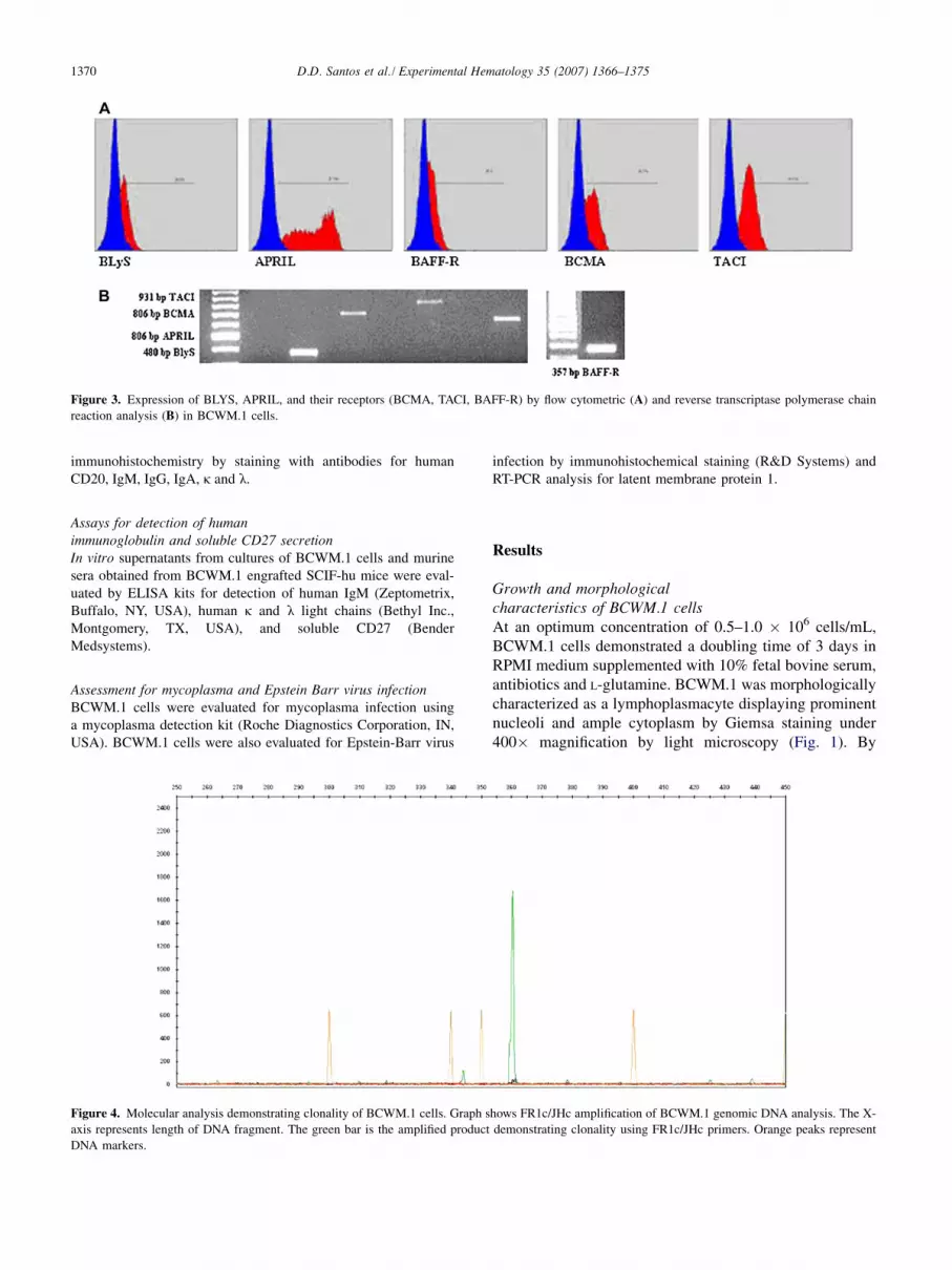
Figure 3. Expression of BLYS, APRIL, and their receptors (BCMA, TACI, BAFF-R) by flow cytometric (A) and reverse transcriptase polymerase chain
reaction analysis (B) in BCWM.1 cells.
immunohistochemistry by staining with antibodies for humanCD20, IgM, IgG, IgA, k and l.
Assays for detection of humanimmunoglobulin and soluble CD27 secretionIn vitro supernatants from cultures of BCWM.1 cells and murinesera obtained from BCWM.1 engrafted SCIF-hu mice were eval-uated by ELISA kits for detection of human IgM (Zeptometrix,Buffalo, NY, USA), human k and l light chains (Bethyl Inc.,Montgomery, TX, USA), and soluble CD27 (BenderMedsystems).
Assessment for mycoplasma and Epstein Barr virus infectionBCWM.1 cells were evaluated for mycoplasma infection usinga mycoplasma detection kit (Roche Diagnostics Corporation, IN,USA). BCWM.1 cells were also evaluated for Epstein-Barr virus
infection by immunohistochemical staining (R&D Systems) andRT-PCR analysis for latent membrane protein 1.
Results
Growth and morphologicalcharacteristics of BCWM.1 cellsAt an optimum concentration of 0.5–1.0 � 106 cells/mL,BCWM.1 cells demonstrated a doubling time of 3 days inRPMI medium supplemented with 10% fetal bovine serum,antibiotics and L-glutamine. BCWM.1 was morphologicallycharacterized as a lymphoplasmacyte displaying prominentnucleoli and ample cytoplasm by Giemsa staining under400� magnification by light microscopy (Fig. 1). By
Figure 4. Molecular analysis demonstrating clonality of BCWM.1 cells. Graph shows FR1c/JHc amplification of BCWM.1 genomic DNA analysis. The X-
axis represents length of DNA fragment. The green bar is the amplified product demonstrating clonality using FR1c/JHc primers. Orange peaks represent
DNA markers.

1371D.D. Santos et al./ Experimental Hematology 35 (2007) 1366–1375
electron microscopy, the nucleus of BCWM.1 cells showedevenly dispersed chromatin with prominent nucleoli, abun-dant cytoplasm, organelles, and mitochondria and displayedboth short and long profiles of rough endoplasmic reticulumat 5000� magnification (Fig. 1). BCWM.1 cells were pos-itive for Epstein-Barr virus latent membrane protein 1 ex-pression by immunohistochemistry and RT-PCR analysis,and negative for mycoplasma infection.
Immunophenotypic characterization of BCWM.1 cellsBy flow cytometric analysis, BCWM.1 cells demonstratedthe following immunophenotypic characteristics: CD3�,CD4�, CD5�, CD8�, CD9�, CD10�, CD11aþ, CD11c�,CD14-, CD16�, CD19þ, CD20þ, CD22�, CD23þ,CD25�, CD27�, CD30þ, CD30L�, CD34�, CD38þ,CD40þ, CD154 (CD40L)�, CD45RAþ, CD45RO�,CD52þ, CD56�, CD66b�, CD70þ, CD80þ, CD103�,CD117þ, CD138�, FMC7�; positive intracellular stainingfor cIgMþ, cIgG�, cIgA�, ck�, clþ (Fig. 2). BCWM.1cells also expressed APRIL and B-Lymphocyte Stimulator,as well as the APRIL and BLYS receptors TACI, BCMA,and BAFF-R by flow cytometric and RT-PCR analysis(Fig. 3).
Cytogenetic analysisKaryotype analysis demonstrated 46 chromosomes, XX, inBCWM.1 cells without cytogenetic anomalies (data notshown). By FISH analysis for the 6q21 deletion, 94 of100 cells (94%) showed a hybridization pattern consistentwith the presence of two copies of this chromosomal locus(data not shown). Greater than 10% of cells with the dele-tion is considered positive, as previously reported [4].
Molecular analysis forFR1c/JHc amplification of genomic DNAMolecular analysis by FR1c/JHc amplification of genomicDNA using single-cell sorting demonstrated a consistentmonoclonal peak, thereby establishing clonality forBCWM.1 cells (Fig. 4).
Gene and SNP array analysisTo understand the tumorgenesis mechanism of BCWM.1cell line, Affymetrix GeneChip Hgu133Plus2 was em-ployed to compare the gene expression profile betweenBCWM.1 cells (performed in independent duplicate exper-iments), the founding primary lymphoplasmacytic cells andB cells isolated from the bone marrow of four healthy do-nors. Genes with fold change O1.5 and p value (multitestadjusted) #0.05 were considered significant and chosenfor further analysis. As shown in Figure 5, gene expressionprofiling demonstrated considerable overlap betweenBCWM.1 and the founding patient’s primary lymphoplas-macytic cells. However, when BCWM.1 cells were com-pared by gene expression profiling to normal donor BMB cells for genes relevant in B-cell growth and signaling,17 genes were significantly up- or downregulated as shown
in Table 1. To detect any potential chromosome changes be-tween BCWM.1 cell line and the founding primary lym-phoplasmacytic cells, we also performed 500K SNP arrayhybridization and analysis. Inferred chromosome copynumber change from dChipSNP software revealed a de-crease in copy number (to 1) at chromosome 3p14.2(chr3:60,097,950-60,762,791) when BCWM.1 cells werecompared against the founder patient’s primary lympho-plasmacytic cells.
In vivo engraftment in SCID-hu miceWe next evaluated the engraftment ability of BCWM.1 cellsin five SCID-hu mice by direct subcutaneous injection at2.75 � 106 cells/mouse. After 30 weeks of observation,
Figure 5. Gene array expression profile for BCWM.1 cells (run in two in-
dependent experiments), sorted lymphoplasmacytic cells (CD19þ) from
founder patient and bone marrow B cells (CD19þ) from four healthy
donors.

1372 D.D. Santos et al./ Experimental Hematology 35 (2007) 1366–1375
Table 1. Dysregulated genes involved in B-cell homeostasis by comparative gene expression profiling of BCWM.1 vs normal donor bone marrow B cells
Gene Chromosomal locus Function p Value
Downregulated
Interleukin-6 7p21 B-cell differentiation 0.0003
syk 9q22 B-cell receptor–independent calcium-induced apoptosis 0.0018
Burkitt lymphoma receptor 1, GTP binding protein 11q23 B-cell migration 0.0054
fyn 6q21 Src family tyrosine kinase 0.0124
z-chain (TCR) associated protein kinase 2q12 IgM signaling 0.0134
Phospholipase C, b2 15q15 PKC-catalyzed phosphorylation 0.0162
Rho guanine nucleoside exchange factor (GEF) 1 19q13 Erk and PI-3 kinase pathway 0.0267
lef-1 4q23 Wnt signaling, B-cell development 0.0361
Upregulated
fas (CD95) 10q24 Programmed cell death 0.0018
CASP8 and FADD-like apoptosis regulator 2q33 Programmed cell death 0.0018
Calnexin 5q35 Protein folding and assembly 0.0134
Mitogen-activated protein kinase 7 17p11 TNFR1 signaling pathway 0.0188
p21 (CDKN1A)- activated kinase 2 3q29 Regulation of B-cell proliferation 0.0188
Eukaryotic translation initiation factor 2, subunit 1 a 14q23 Protein synthesis initiation 0.0205
Caspase-1, apoptosis-related cysteine protease 11q23 Programmed cell death 0.0258
no tumor growth occurred, and no human paraprotein wasdetectable in serum. We subsequently inoculatedBCWM.1 at the same concentration in a fetal bone chip,which was implanted in five other SCID-hu mice. Humanparaproteins were undetectable at baseline, but were readilydetectable in four mice at 2 weeks, and in one mouse at 4weeks. Importantly, follow-up evaluation at 4 weeksshowed both increasing serum IgM and l-light chain levelsin all five mice (Table 2). In a separate pilot study, and un-der the same conditions, we implanted BCWM.1 inoculatedfetal bone chips into five other SCID-hu mice in which bothserum IgM and sCD27 levels were followed. Both serumIgM and sCD27 levels were undetectable at baseline, butbecame readily detectable at 2 weeks and continued torise to 12 weeks (data not shown).
To ascertain the distribution of engraftment of BCWM.1cells, as well as the necessity for a human bone marrow en-vironment to support BCWM.1 engraftment, we performeda detailed histological examination of the human andmouse bone marrows by examining decalcified sectionsof the human fetal bone implanted bone chips and mousefemurs, as well as murine spleens, liver, lung, kidney, andlymph nodes from four SCID-hu mice following 4 weeksof BCWM.1 engraftment. Histological and immunohisto-chemical evaluation demonstrated the presence of diffuse
lymphoplasmacytic cells in the human bone marrow(Fig. 6), but not murine bone marrow, spleen, liver, lung,or kidney (data not shown) which stained for humanCD20, IgM, and l-light chains but not human IgA, IgG,or k light chains (Fig. 7).
DiscussionA significant impairment in understanding the biology andadvancing novel therapeutics for WM has been the lack ofa representative cell line and animal model. We, therefore,report on work accomplished in our laboratories in estab-lishing the BCWM.1 cell line and a SCID-hu mouse modelutilizing BCWM.1 cells to recapitulate the biology of WM.
As demonstrated in these studies, BCWM.1 bore themorphological, phenotypic, and genotypic properties ofLPC typically found in WM, as well as important biologicalfeatures, such as the secretion of IgM and sCD27 that serveas surrogate markers of disease in WM [20]. The produc-tion of sCD27 may particularly be significant for the path-ogenesis of WM, since this ligand induces the growth andsurvival factors APRIL and CD40L on mast cells, whichexpress the receptor for CD27 (CD70); are increased innumber in the BM of patients with WM; and are usuallyfound in close association with LPC [24]. Soluble CD27
Table 2. Serial paraprotein measurements in SCID-hu mice engrafted with BCWM.1 WM cells
Mouse Code Isotype
Cells (�106)
Inoculated
Weeks to IgM
detection
Level of IgM (mg/mL)
Week 2
Level of IgM (mg/mL)
Week 4
Level of l- light
chain (mg/mL)
Week 2
Level of l-light
chain (mg/mL)
Week 4
680D IgM l 2.75 2 9.1 20.1 9.4 23.6
323B IgM l 2.75 4 0 12.4 4.6 10.3
NA IgM l 2.75 2 54.4 152.6 57.5 114.2
072D IgM l 2.75 2 153.7 381.3 115.4 227.2
2D36 IgM l 2.75 2 20.16 50.4 16.8 35.2

1373D.D. Santos et al./ Experimental Hematology 35 (2007) 1366–1375
Figure 6. Histological analysis for human bone marrow engraftment in severe combined immunodeficient (SCID)-hu mice with BCWM.1 lymphoplasma-
cytic cells. Histological analysis was performed on decalcified fetal bone chips implanted in SCID-hu mice following engraftment with BCWM.1 lympho-
plasmacytic cells. Bone chips were retrieved from SCID-hu mice at 12 weeks following serial detection of rising human paraproteins in murine serum. Fetal
bone chips were then stained with hematoxylin and eosin, and viewed under light microscopy at 100� (1), 400� (2), and 200� (3).
may also have other important roles in WM pathogenesis,including immune suppression [27].
BCWM.1 cells also expressed APRIL and BLYS, as wellas their receptors TACI and BCMA (for both APRIL andBLYS) and BAFF-R (for BLYS). Similar findings havebeen reported by us and others in primary WM LPC[17,18], as well as on malignant cells in other B-cell disor-ders [28,29]. The expression of both APRIL and BLYS aswell as their receptors on WM cells may have importantroles in WM pathogenesis, particularly because theseligand-receptor pathways have well-defined roles in LPCdifferentiation and IgH class switching. The BCWM.1cell line may therefore provide a convenient model for theregulatory study of APRIL and BLYS in WM.
As part of these studies, we also performed extensivecytogenetic, molecular, and gene expression studies onBCWM.1 cells. These studies confirmed the clonality ofBCWM.1 cells by determination of IgH rearrangementsthrough FR1c/JHc amplification of genomic DNA, whileSNP-1 analysis demonstrated close propinquity of
BCWM.1 cells to the founder patient’s primary LPC,with the sole change being an decrease (to one) in copynumber at 3p14.2. Two potential genes of interest at3p14.2 are the fragile histidine triad gene (FHIT) and theprotein tyrosine phosphatase receptor type G gene, bothof which are candidate tumor suppressor genes [30]. Aber-rant expression of FHIT has been reported in several B-cellmalignancies [31,32], and further investigation of both theFHIT and phosphatase receptor type G genes in the patho-genesis and progression of WM would appear warranted.Interestingly, gene expression profiling revealed significantdifferences in 17 genes with a role in B-cell homeostasisthat are worthy of further study in WM, including fyn,which is located at 6q21, a site shown by us and others tobe frequently deleted in patients with WM, and whichmay have a role in the progression of IgM MGUS toWM [4–6].
As is typical of primary LPC from most WM patients, nogross structural abnormalities were found in BCWM.1 cellsby karyotype and multicolor fluorescence in situ

1374 D.D. Santos et al./ Experimental Hematology 35 (2007) 1366–1375
Figure 7. Immunohistochemical analysis for human bone marrow engraftment in severe combined immunodeficient (SCID)-hu mice with BCWM.1 lym-
phoplasmacytic cells. Immunohistochemical analysis was performed on decalcified fetal bone chips implanted in SCID-hu mice following engraftment with
BCWM.1 lymphoplasmacytic cells. Bone chips were retrieved from SCID-hu mice at 12 weeks, following serial detection of rising human paraproteins, and
stained with antibodies directed against human CD20 (1), l light chain (2), IgM (3), k light chain (4), IgG (5), and IgA (6). Slides viewed at 200�magnification.
hybridization studies. However, a vast number of upregu-lated (n 5 5,409) and downregulated (n 5 3,155) genes be-tween BCWM.1 cells and normal donor bone marrow Bcells were observed. These differences might reflect epige-netic changes resulting from transforming events, or the dif-ferentiation (i.e., lymphoplasmacytic) state of WM cellsrelative to normal donor BM B cells. Further studies, partic-ularly of genes detected in aberrant B-cell signaling path-ways (as described in Table 1) may provide informativeclues behind the transforming events in WM.
An important characteristic of BCWM.1 cells is theirrapid engraftment in the SCID-hu mice. As with primaryWM LPC [26], BCWM.1 cells also required the humanBM milieu for their growth and expansion since subcutane-ous injection of BCWM.1 cells in SCID mice failed to leadto engraftment. Human serum IgM and l light chain, aswell as soluble CD27, a novel tumor marker for WM[19,20], were readily detectable following engraftment ofBCWM.1 cells. The potential use of BCWM.1 engraftedSCID-hu mice as a relevant model for the study of novelWM therapeutics was recently demonstrated by us usingthe CD70-directed monoclonal antibody SGN-70.BCWM.1 engrafted SCID-hu mice responded to SGN-70therapy, whereas untreated mice demonstrated continueddisease progression [20].
In summary, the BCWM.1 cell line provides a diseaseappropriate cell system for the study of WM, which in con-junction with the SCID-hu mouse model may be used as
a convenient model for studies focused on both WM path-ogenesis and development of targeted therapies for WM.
AcknowledgmentsThe authors wish to acknowledge and thank John Daley and SusanLazo at the Dana-Farber Cancer Institute for their help with flowcytometric analysis and cell sorting; Courtney Hyland, CharlesLee and Cynthia Morton from the Cytogenetics Core of Dana-Farber /Harvard Cancer Center; Dr. Maria Ericsson from the Elec-tron Microscopy Core Facility at Harvard Medical School; Drs.Edward Fox, Sigitas Jonas Verselis, Changzhong Chen and MauraA. Berkeley from the Microarray Core Facility at the Dana-Farber/Harvard Cancer Center.
Supported by the Bing Fund for Waldenstrom’s macroglobuli-nemia, a Laurie Strauss Leukemia Foundation Fellowship Award(to D.D.S.), and a National Institutes of Health Career Develop-ment Award (K-23CA087977-03) to S.P.T. D.D.S., A.W.H.,O.T., X.L., E.H., L.X., J.K., J.L.K., Y.T., B.C. performed cellularand molecular studies. P.T., A.W.H., M.C., and P.N. performed an-imal studies. A.R.B., Z.R.H., and R.J.M. oversaw specimen col-lection and clinical data analysis. J.Z. performed bioinformaticsanalysis. L.M.P., K.C., N.M., S.P.T. designed and oversaw studies.
References1. Owen RG, Treon SP, Al-Katib A, et al. Clinicopathological definition
of Waldenstrom’s macroglobulinemia: Consensus Panel Recommen-
dations from the Second International Workshop on Waldenstrom’s
macroglobulinemia. Semin Oncol. 2003;30:110–115.

1375D.D. Santos et al./ Experimental Hematology 35 (2007) 1366–1375
2. Harris NL, Jaffe ES, Stein H. A revised European-American classifi-
cation of lymphoid neoplasms: a proposal from the International Lym-
phoma Study Group. Blood. 1994;84:1361–1392.
3. Harris NL, Jaffe ES, Diebold J, et al. World Health Organization clas-
sification of neoplastic diseases of the hematopoietic and lymphoid
tissues: report of the Clinical Advisory Committee meeting- Airlie
House, Virginia, November 1997. J Clin Oncol. 1999;17:3835–3849.
4. Treon SP, Hunter ZR, Aggarwal A, et al. Characterization of familial
Waldenstrom’s macroglobulinemia. Ann Oncol. 2006;17:488–494.
5. Schop RFJ, Kuehl WM, Van Wier SA, et al. Waldenstrom’s macro-
globulinemia neoplastic cells lack immunoglobulin heavy chain locus
translocations but have frequent 6q deletions. Blood. 2002;100:2996–
3001.
6. Schop RF, Van Wier SA, Xu R, et al. 6q deletion discriminates Wal-
denstrom macroglobulinemia from IgM monoclonal gammopathy
of undetermined significance. Cancer Genet Cytogenet. 2006;169:
150–153.
7. Xu L, Leleu X, Hunter ZR, et al. Abnormal expression of the plasma
cell differentiation factor X-box protein 1 (Xbp-1) in Waldenstrom’s
macroglobulinemia. Blood. 2005;106:1003.
8. Leleu X, Xu L, Ho A, et al. Multiple defects in expression of genes
promoting plasmacytic differentiation define Waldenstrom’s macro-
globulinemia. Ann Oncol. 2005;16:v86.
9. Avet-Loiseau H, Garand R, Lode L, Robillard N, Bataille R. 14q32
translocations discriminate IgM multiple myeloma from Walden-
strom’s macroglobulinemia. Semin Oncol. 2003;30:153–155.
10. Sahota SS, Forconi F, Ottensmeier CH, et al. Typical Waldenstrom
macroglobulinemia is derived from a B-cell arrested after cessation
of somatic mutation but prior to isotype switch events. Blood. 2002;
100:1505–1507.
11. Kriangkum J, Taylor BJ, Treon SP, Mant MJ, Belch AR, Pilarski LM.
Clonotypic IgM V/D/J sequence analysis in Waldenstrom macroglob-
ulinemia suggests an unusual B-cell origin and an expansion of poly-
clonal B cells in peripheral blood. Blood. 2004;104:2134–2142.
12. San Miguel JF, Vidriales MB, Ocio E, et al. Immunophenotypic anal-
ysis of Waldenstrom’s macroglobulinemia. Semin Oncol. 2003;30:
187–195.
13. Treon SP, Kelliher A, Keele B, et al. Expression of serotherapy target
antigens in Waldenstrom’s macroglobulinemia: therapeutic applica-
tions and considerations. Semin Oncol. 2003;30:243–247.
14. Hunter Z, Branagan AR, Ditzel Santos D, et al. High levels of soluble
immunoregulatory receptors in patients with Waldenstrom’s macro-
globulinemia. Blood. 2004;104:303b.
15. Dimopoulos MA, Kyle RA, Anagnostopoulos A, Treon SP. Diagnosis
and management of Waldenstrom’s macroglobulinemia. J Clin Oncol.
2005;23:1564–1577.
16. Hunter ZR, Branagan AR, Manning R, et al. CD5, CD10, CD23 ex-
pression in Waldenstrom’s macroglobulinemia. Clin Lymph. 2005;5:
246–249.
17. Ditzel Santos D, Tournilhac O, Xu L, et al. B-Lymphocyte stimulator
protein (BLYS) is expressed by bone marrow mast and lymphoplasma-
cytic cells in Waldenstrom’s macroglobulinemia, and provides signal-
ing for growth, survival and IgM secretion. Blood. 2004;104:917a.
18. Elsawa SF, Novak AJ, Grote DM, et al. B-lymphocyte stimulator
(BlyS) stimulates immunoglobulin production and malignant B-cell
growth in Waldenstrom macroglobulinemia. Blood. 2006;107:2882–
2888.
19. Ho AW, Leleu X, Hatjiharissi E, et al. A novel functional role for sol-
uble CD27 in the pathogenesis of Waldenstrom’s macroglobulinemia.
Blood. 2005;106:4701.
20. Ho AW, Hatjiharissi E, Branagan AR, et al. Therapeutic targeting of
CD70 and CD27-CD70 interactions with the monoclonal antibody
SGN-70 in Waldenstrom’s Macroglobulinemia (WM). Proc Am Soc
Clin Oncol. 2006;24:102s.
21. Tischendorf W, Hartmann F. Waldenstrom’s macroglobulinemia asso-
ciated with hyperplasia of the tissue mast cells. Acta Haematol. 1950;
4:374–383.
22. Wilkins BS, Buchan SL, Webster J, Jones DB. Tryptase-positive mast
cells accompany lymphocytic as well as lymphoplasmacytic lym-
phoma infiltrates in bone marrow trephine biopsies. Histopathology.
2001;2:150–155.
23. Santos DD, Chemaly M, Tournilhac O, et al. Bone marrow mast cells
are significantly increased in patients with Waldenstrom’s macroglob-
ulinemia, and their number following therapeutic intervention is
dependent on extent of response. Blood. 2005;106:980.
24. Tournilhac O, Santos DD, Xu L, et al. Mast cells in Waldenstrom’s
macroglobulinemia support lymphoplasmacytic cell growth through
CD154/CD40 signaling. Ann Oncol. 2006;17:1275–1282.
25. Treon SP, Branagan AR, Hunter Z, et al. IgA and IgG hypogammaglo-
bulinemia persists in most patients with Waldenstrom’s macroglobuli-
nemia despite therapeutic responses, including complete remissions.
Blood. 2004;104:306b.
26. Tassone P, Neri P, Kutok JL, et al. A SCID-hu in vivo model of human
Waldenstrom macroglobulinemia. Blood. 2005;4:1341–1345.
27. Arens R, Tesselaar K, Baars PA, et al. Constitutive CD27/CD70 inter-
action induces expansion of effector-type T-cells and results in IFN
gamma-mediated B-cell depletion. Immunity. 2001;15:801–812.
28. Haiat S, Billard C, Quiney C, Ajchenbaum-Cymbalista F, Kolb JP.
Role of BAFF and APRIL in human B-cell chronic lymphocytic
leukemia. Immunology. 2006;118:281–292.
29. Moreaux J, Legouffe E, Jourdan E, et al. BAFF and APRIL protect
myeloma cells from apoptosis induced by interleukin 6 deprivation
and dexamethasone. Blood. 2004;103:3148–3157.
30. Matsuyama A, Shiraishi T, Trapasso F, et al. Fragile site orthologs
FHIT/FRA3B and FHIT/FRA14A2: Evolutionary conserved but
highly recombinogenic. PNAS. 2003;100:14988–14993.
31. Zhao P, Lam AK, Lu YL, Zhong M, Chen LH, Pu XL. Aberrant FHIT
protein expression in classical Hodgkin’s lymphoma. Pathology. 2006;
38:399–402.
32. Kameoka Y, Tagawa H, Tsuzuki S, et al. Contiguous array CHG at
13p14.2 to the FRA3B/FHIT common fragile region as the target
gene in diffuse large cell lymphoma. Oncogene. 2004;23:9148–9154.
Copyright © 2022 FDOKUMEN