Pre-conditioned mesenchymal stem cells ameliorate renal ischemic injury in rats by augmented...
11
RESEARCH Open Access Pre-conditioned mesenchymal stem cells ameliorate renal ischemic injury in rats by augmented survival and engraftment Muhammad Shareef Masoud 1,2* , Sanam Saiqa Anwar 1 , Muhammad Zeeshan Afzal 1,3 , Azra Mehmood 1 , Shaheen N Khan 1 and Sheikh Riazuddin 1,4,5 Abstract Background: Ischemia is the major cause of acute kidney injury (AKI), associated with high mortality and morbidity. Mesenchymal stem cells (MSCs) have multilineage differentiation potential and can be a potent therapeutic option for the cure of AKI. Methods: MSCs were cultured in four groups SNAP (S-nitroso N-acetyl penicillamine), SNAP + Methylene Blue (MB), MB and a control for in vitro analysis. Cultured MSCs were pre-conditioned with either SNAP (100 μM) or MB (1 μM) or both for 6 hours. Renal ischemia was induced in four groups (as in in vitro study) of rats by clamping the left renal padicle for 45 minutes and then different pre-conditioned stem cells were transplanted. Results: We report that pre-conditioning of MSCs with SNAP enhances their proliferation, survival and engraftment in ischemic kidney. Rat MSCs pre-conditioned with SNAP decreased cell apoptosis and increased proliferation and cytoprotective genes’ expression in vitro. Our in vivo data showed enhanced survival and engraftment, proliferation, reduction in fibrosis, significant improvement in renal function and higher expression of pro-survival and pro-angiogenic factors in ischemic renal tissue in SNAP pre-conditioned group of animals. Cytoprotective effects of SNAP pre-conditioning were abrogated by MB, an inhibitor of nitric oxide synthase (NOS) and guanylate cyclase. Conclusion: The results of these studies demonstrate that SNAP pre-conditioning might be useful to enhance therapeutic potential of MSCs in attenuating renal ischemia reperfusion injury. Keywords: MSCs, SNAP, Pre-conditioning, Renal ischemia, Cytoprotective factors Background The mammalian kidney has multifarious functions, includ- ing maintenance of blood pressure, regulation of pH and other metabolic processes, such as elimination of nitrogen- ous wastes and synthesis and release of endocrine factors. Loss of regional or total blood flow to the kidney often happens in shock, sepsis and during renal transplantation, can lead to acute kidney injury (AKI). Inflammatory process and primary ischemic injury in vascular cells results in prolonged localized ischemia that substantially increases severity of injury in affected areas. During ischemia, the endothelium produces free radicals and secretes chemo-attractants which upon reperfusion seques- ter and activate neutrophils and intensification of the injury [1]. AKI associated mortality is estimated as 50% in hospi- talized patients and 70-80% in patients under intensive care [2,3]. Currently, the therapeutic choices are confined to supportive measures and preventive strategies. None of these have shown a decrease in mortality rate caused by AKI. Kidney transplant is an alternate option but scarcity of donor; high cost and immune rejection limits its utility. It is therefore imperative to find other therapeutic options that could regenerate the tubular epithelium, restore kid- ney function and reverse the effects of AKI. MSCs are multi-potent cell population residing in bone marrow. These are capable of differentiating into cell types other than their tissue of origin. In culture, * Correspondence: [email protected] 1 National Centre of Excellence in Molecular Biology, University of the Punjab, Lahore, Pakistan 2 Current Affiliation: Department of Bioinformatics and Biotechnology, Government College University, Faisalabad 38000, Pakistan Full list of author information is available at the end of the article © 2012 Masoud et al.; licensee BioMed Central Ltd. This is an Open Access article distributed under the terms of the Creative Commons Attribution License (http://creativecommons.org/licenses/by/2.0), which permits unrestricted use, distribution, and reproduction in any medium, provided the original work is properly cited. Masoud et al. Journal of Translational Medicine 2012, 10:243 http://www.translational-medicine.com/content/10/1/243
Transcript of Pre-conditioned mesenchymal stem cells ameliorate renal ischemic injury in rats by augmented...

Masoud et al. Journal of Translational Medicine 2012, 10:243http://www.translational-medicine.com/content/10/1/243
RESEARCH Open Access
Pre-conditioned mesenchymal stem cellsameliorate renal ischemic injury in rats byaugmented survival and engraftmentMuhammad Shareef Masoud1,2*, Sanam Saiqa Anwar1, Muhammad Zeeshan Afzal1,3, Azra Mehmood1,Shaheen N Khan1 and Sheikh Riazuddin1,4,5
Abstract
Background: Ischemia is the major cause of acute kidney injury (AKI), associated with high mortality and morbidity.Mesenchymal stem cells (MSCs) have multilineage differentiation potential and can be a potent therapeutic optionfor the cure of AKI.
Methods: MSCs were cultured in four groups SNAP (S-nitroso N-acetyl penicillamine), SNAP + Methylene Blue (MB),MB and a control for in vitro analysis. Cultured MSCs were pre-conditioned with either SNAP (100 μM) or MB (1 μM)or both for 6 hours. Renal ischemia was induced in four groups (as in in vitro study) of rats by clamping the leftrenal padicle for 45 minutes and then different pre-conditioned stem cells were transplanted.
Results: We report that pre-conditioning of MSCs with SNAP enhances their proliferation, survival and engraftmentin ischemic kidney. Rat MSCs pre-conditioned with SNAP decreased cell apoptosis and increased proliferation andcytoprotective genes’ expression in vitro. Our in vivo data showed enhanced survival and engraftment, proliferation,reduction in fibrosis, significant improvement in renal function and higher expression of pro-survival andpro-angiogenic factors in ischemic renal tissue in SNAP pre-conditioned group of animals. Cytoprotective effects ofSNAP pre-conditioning were abrogated by MB, an inhibitor of nitric oxide synthase (NOS) and guanylate cyclase.
Conclusion: The results of these studies demonstrate that SNAP pre-conditioning might be useful to enhancetherapeutic potential of MSCs in attenuating renal ischemia reperfusion injury.
Keywords: MSCs, SNAP, Pre-conditioning, Renal ischemia, Cytoprotective factors
BackgroundThe mammalian kidney has multifarious functions, includ-ing maintenance of blood pressure, regulation of pH andother metabolic processes, such as elimination of nitrogen-ous wastes and synthesis and release of endocrine factors.Loss of regional or total blood flow to the kidney oftenhappens in shock, sepsis and during renal transplantation,can lead to acute kidney injury (AKI). Inflammatoryprocess and primary ischemic injury in vascular cellsresults in prolonged localized ischemia that substantiallyincreases severity of injury in affected areas. During
* Correspondence: [email protected] Centre of Excellence in Molecular Biology, University of the Punjab,Lahore, Pakistan2Current Affiliation: Department of Bioinformatics and Biotechnology,Government College University, Faisalabad 38000, PakistanFull list of author information is available at the end of the article
© 2012 Masoud et al.; licensee BioMed CentraCommons Attribution License (http://creativecreproduction in any medium, provided the or
ischemia, the endothelium produces free radicals andsecretes chemo-attractants which upon reperfusion seques-ter and activate neutrophils and intensification of the injury[1]. AKI associated mortality is estimated as 50% in hospi-talized patients and 70-80% in patients under intensivecare [2,3]. Currently, the therapeutic choices are confinedto supportive measures and preventive strategies. None ofthese have shown a decrease in mortality rate caused byAKI. Kidney transplant is an alternate option but scarcityof donor; high cost and immune rejection limits its utility.It is therefore imperative to find other therapeutic optionsthat could regenerate the tubular epithelium, restore kid-ney function and reverse the effects of AKI.MSCs are multi-potent cell population residing in
bone marrow. These are capable of differentiating intocell types other than their tissue of origin. In culture,
l Ltd. This is an Open Access article distributed under the terms of the Creativeommons.org/licenses/by/2.0), which permits unrestricted use, distribution, andiginal work is properly cited.

Masoud et al. Journal of Translational Medicine 2012, 10:243 Page 2 of 11http://www.translational-medicine.com/content/10/1/243
these cells are characterized by their unique ability toadhere to the plastic surface and exhibit fibroblast likemorphology [4]. Moreover MSCs secrete an array ofgrowth factors as vascular endothelial growth factor(VEGF), insulin like growth factor-1 (IGF-1), hepatocytegrowth factor (HGF) and anti-apoptotic cytokines [5].The therapeutic effects of MSCs after transplantationare dependent on their survival in the recipient tissue.Taking into account that prolonged cell survival mayaugment the usefulness of stem cell therapy. It has beenreported that pre-conditioning of different tissues by aprior stimulus provides protection against injury [6].The most extensively studied pre-conditioning stimulusis brief sub-injurious ischemia, which induces protectionagainst subsequent ischemic injury [7]. Similar protect-ive effects can also be triggered by growth factors, cyto-kines or with some pharmacological agents [5,8].Nitric oxide (NO) is a free radical generated as a result
of oxidation of L-arginine catalyzed by all three isoforms(neuronal, inducible and endothelial) of nitric oxide syn-thase (NOS) and performs different patho-physiologicalfunctions in different cellular environments [9-13]. Itprotects macrophages, hepatocytes, cardiomyocytesagainst different injuries by modifying expression of dif-ferent genes [14]. Various donors of NO have beenimplicated as cyto-protective and tissue protective fac-tors in vivo [1,15].This study was designed to evaluate the effects of pre-
conditioning of bone marrow derived MSCs with apharmacological agent S-nitroso N acetyl penicillamine(SNAP, a NO donor) on the regenerative potential ofMSCs against oxidative and ischemic stress in kidney. Wereport that SNAP pre-conditioning of MSCs improvedtheir potential against oxidative stress, enhanced their pro-liferation and reduced apoptosis in vitro. Further it aug-mented the homing of MSCs in ischemic renalparenchyma, induced different cytoprotective and para-crine factors and improved renal function in “rat model ofrenal ischemic insult”.
Materials and methodsAnimal careThe investigation conforms to the Guide for the Careand Use of Laboratory Animals published by the USNational Institutes of Health (NIH Publication No. 85–23,revised 1985). All animals were treated according to theprocedures approved by the Institutional Review Board(IRB) at the National Center of Excellence in MolecularBiology, Lahore, Pakistan.
In vitro studyIsolation of MSCs from bone marrowMSCs were isolated on the basis of their preferential at-tachment to plastic surface of culture flask as previously
described [16]. Bone marrow MSCs were isolated byflushing femurs and tibiae of male Sprague–Dawley rats.The suspension was then centrifuged at 1200 rpm for10 min at room temperature. The cells were plated intissue culture flask containing Iscove’s Modified Delbeco’sMedium (IMDM, MP Biomedicals, USA) supplementedwith 20% fetal bovine serum (Sigma-Aldrich, Germany),100ug/ml streptomycin and 100U/ml penicillin and placedin incubator having 95% humidity and 5% CO2. Themedium was changed after three days of plating andwashed with phosphate buffered saline (PBS). The mediumwas then changed after every three days. When confluencyis reached, the cells were sub-cultured in a ratio of 1:3 tillthey reached 70%-80% confluency. The cells of passages2–3 were used in the study.
Pre-conditioning of stem cellsThe cells were plated in tissue culture plates in equalnumber (2x10⁵) and divided in four groups. Group 1:control; cells incubated in normal serum free medium,group 2: cells treated with 100uM S-nitroso N acetylpe-nicillamine (SNAP) a NO donor, group 3: cells treatedwith both 100uM SNAP and 1uM methylene blue (aninhibitor of NOS and gyanalate cyclase) and group 4: thecells treated with 1uM methylene blue alone (group 4).
Measurement of lactate dehydrogenase, cell viability andapoptosis assayCell viability was assessed after exposing above men-tioned groups of cells to 200μl H2O2 for 1 hour (hr).The medium was removed and trypan blue solution(Sigma-Aldrich, Germany) was added to the culture.The cells were then analyzed under phase contrastmicroscope [17]. The number of trypan blue positivecells was then counted per field for each treatment.LDH release was analyzed using cell supernatants by acommercially available kit (Sigma-Aldrich, Germany).Apoptosis was assessed by annexin V binding assay(Abcam, UK). The slides were then analyzed under themicroscope (BX61 Olympus). The cells were countedmanually and percentage of annexin V positive cells wascalculated in each treatment group.
Measurement of release of nitric oxideNO release in the culture medium was assessed fromthe amount of nitrite in the medium which is a stable re-action product of NO with molecular oxygen. 100μl ofculture supernatant was treated with equal volume ofGriess reagent (0.5% sulfanilamide, 0.05% N-1-naphthylethylenediamine dihydrochloride in 2.5% H3PO4) in 96well plate and placed on shaker for 10 min at roomtemperature. The resulting color product was spectro-photometrically quantified at 538nm.

Masoud et al. Journal of Translational Medicine 2012, 10:243 Page 3 of 11http://www.translational-medicine.com/content/10/1/243
Measurement of gene activityThe effects of pre-conditioning on gene expression wereassessed by performing quantitative real-time polymer-ase chain reaction using ABI Real-Time system 7500. Allfour groups of cells were exposed to H2O2 for 1 hr andthen medium was aspirated and RNA from all fourgroups of cells was extracted using TRIZOL (Invitrogen,USA) according to the manufacturer’s instructions.cDNA was synthesized using 1μg of total RNA fromeach group by Revert Aid H Minus first strand cDNAsynthesis kit (Fermentas).The expression of genes shown in Table 1 was ana-
lyzed through real-time quantitative PCR, stromalderived factor-1 (SDF1), IGF1, AKT-1, VEGF, Bcl-2,PCNA and β-actin was used as an internal control usingMaxima syber green qPCR mix (Fermentas) accordingto manufacturer’s protocol on 7500 thermal cycler (Ap-plied Biosystem, USA). The relative gene expression nor-malized with β-actin and analysis was done by usingSDS 3.1 software provided by Applied Biosystems, USA.
In vivo studyRenal ischemic insult modelAll animal procedures were according to instructions ofinstitutional committee for animal care. Renal ischemicmodel was developed in male Sprague Dawley rats (200-300g) by blocking the left renal pedicle. Briefly, the ratswere anesthetized with pentobarbital (40mg/kg bodyweight). A left lateral abdominal incision was made ex-posing kidney. The renal pedicle was occluded withmicro vascular clamp maintaining body temperature at37°C. The clamp was then removed after 45 minutesand kidney was reperfused. The peritoneal cavity and
Table 1 Primer sequences of various genes
Genes Primer sequence
BCL-2 Forward, 50-CGACTTTGCAGAGATGTCCA-30
Reverse, 50-ATGCCGGTTCAGGTACTCAG-30
VEGF Forward, 50-GCCCTGAGTCAAGAGGACAG-30
Reverse, 50-GAGGAGGAGGAGCCATTACC-30
IGF1 Forward, 50-GCTGAAGCCGTTCATTTAGC-30
Reverse, 50-CCACCCAGTTGCTATTGCTT-30
PCNA Forward, 50- GACCTCGCTCCCCTTACAGT -30
Reverse, 50- TCCAGCACCTTCTTCAGGAT -30
SDF-1α Forward, 50- AGCCAGTCAGCCTGAGCTAC-30
Reverse, 50- GGCACAGTTTGGAGTGTTGA-30
HIF-1α Forward, 50-CTAGGGATGCAGCACGATCT–3
Reverse, 50-AGATGGGAGCTCACGTTGTG–30
AKT 1 Forward, 50-CCTCAAGAACGATGGCACCT-30
Reverse, 50-CAGGCAGCGGATGATAAAGG-30
β-Actin Forward, 50- GCTGTGTTGTCCCTGTATGC-30
Reverse, 50- GAGCGCGTAACCCTCATAGA-30
skin were closed with 4/0 suture. The animals wereallowed to recover and returned to their cages.
Labeling of MSCs and transplantation in ischemic kidneyTo track the transplanted cells and to evaluate the yieldof transplantation in vivo, the cell suspension was la-beled with CM-Dil (invitrogen, USA), a lipophilic dyethat binds to cell membrane irreversibly.Male rats were divided into five groups (n=6) after in-
duction of renal ischemia to receive intra-renal paren-chymal injection of either normal MSCs without pre-conditioning (MCs group), or SNAP pre-conditionedMSCs (SNAP group): methylene blue pre-conditionedMSCs (MB group) and SNAP plus methylene blue pre-conditioned MSCs (SNAP+MB group) and serum freemedium injected control group (control). After 45 minischemia to the kidney, 1.5 × 10⁶ labeled cells in serumfree medium, were transplanted into renal parenchymaat three different sites just after reflow to each groupdescribed above while the control animals receivedonly serum free medium. The incision was closed withsuture and animals were revived and transferred to ani-mal facility.
Collection of urine and bloodThe rats were kept in metabolic cages and 24hrs urinewas collected and measured. Blood was drawn throughtail vein and serum was isolated for each rat.
Immunoflourescence stainingAfter 3 weeks of transplantation, animals were sacrificedand kidneys were harvested and processed. Frozen tissuesections (5 μm thickness) were air dried and washedwith PBS for 5 min. Sections were incubated with spe-cific primary antibody of actin (abcam, UK) with speci-fied dilution after non-specific blocking with 2% donkeyserum and then FITC (flourocinisothiocynate) conjua-gated donkey anti mouse secondary antibody. The sec-tions were counter stained with DAPI and mounted(prolong gold anti-fade, Invitrogen, USA). Fluorescenceimages were taken by Olympus BX 61 microscope withDP 70 camera.
Immunohistochemical analysis of ischemic renal tissueParaffin sections of kidney from all experimental groupswere analyzed for the detection of different factorsthrough application of enzyme labeled polymer (VectorImmPRESS kit, Vector Laboratories, USA) according tomanufacturer’s instructions. Briefly, the sections weresubjected to citrate based antigen retrieval followed byendogenous peroxidase and avidin-biotin blocking usinga Streptavidin-Biotin blocking reagent (Abcam, UK). Fol-lowing serum blocking, sections were incubated eitherfor one hour at room temperature or overnight at 4°C in

Masoud et al. Journal of Translational Medicine 2012, 10:243 Page 4 of 11http://www.translational-medicine.com/content/10/1/243
humid conditions with the following primary antibodiesof specified dilutions: a Rabbit polyclonal anti iNOSantibody, mouse monoclonal to VEGF, rabbit polyclonalto Ki-67, rabbit polyclonal anti eNOS antibody (Abcam,UK), rabbit polyclonal to IGF1 and rabbit polyclonalBcl-2 (Santa Cruz, USA) followed by HRP conjugatedsecondary antibodies. The chromogen substrate 3, 3’-Diaminobenzidine Tetra hydrochloride (DAB) was incu-bated until the characteristic color was developed fol-lowed by hematoxylin counterstaining.
Measurement of collagenParaffin sections were stained with Sirius red to analyzefibrosis in the tissue. The slides were analyzed underBX61 microscope. Total collagen content was measuredas the percent of area with the help of Image J (NIH)software.
Measurement of creatinine clearance and Blood UreaNitrogen (BUN)Creatinine was measured in 24 hr urine and serum ofthe rats using Crea Plus kit (Roche, Germany) accordingto the manufacturer’s procedure. Briefly the urine sam-ples were diluted 1:50 and undiluted serum sampleswere used. The samples were analyzed @ 546 nm testwave length and @ 700 nm reference wave length andcreatinine values were calculated as follow:
Creatinine Clearance ¼ creatinine of urine � 24hr urine
volume � 1440� creatinine of
serum:
The blood urea nitrogen level in serum was measuredusing a urea estimation kit (Diasys, Germany) accordingto the manufacturer’s procedure. Briefly serum was iso-lated from blood of each rat. The reagent was mixedwith samples and analyzed at 340 nm wavelength byspectrophotometer. BUN was calculated as follow:
BUN ¼ OD of sample � concentration of standard� 0:47� OD of standard
Statistical analysisAll experiments were performed in triplicates andrepeated at least three times. Statistical significance wasanalyzed using one way ANOVA followed by Bonferronitesting through Graphpad Prism 5 software. A p-value ≤0.05 was considered statistically significant.
ResultsIn vitro studyCytoprotective effects of pre-conditioning on MSCsMSCs showed significant loss of viability when exposed toH2O2 (200 μM) for 1 hr. After SNAP pre-conditioning,cytotoxic effects of H2O2 were markedly decreased com-pared to non pre-conditioned control. This effect wasdiminished in SNAP/MB and MB alone groups(Figure 1A). Likewise SNAP pre-conditioned MSCs weredistinctly protected against H2O2 as shown by decreased re-lease of LDH as compared to control group (Figure 1B).Apoptosis in MSCs after pre-conditioning was assessed byannexin V staining. The percentage of annexin V positivecells decreased from 27% in control group to 9% in SNAPpre-conditioned group (Figure 1C). This decrease in celldeath was diminished with methylene blue treatment (22%and 23% in SNAP/MB and MB groups respectively).
Release of nitric oxide in mediumThe amount of nitrite produced as a result of reaction ofNO with molecular oxygen was estimated. Griess re-agent was added to the medium and OD at 538 nmwavelength was taken. Level of nitrite was significantlyhigh (2.59 ± 0.3 μM) in the medium of SNAP groupcompared to the control (0.27 ± 0.2 μM).
Enhanced Expression of Cytoprotective, Proangiogenic andproliferating genes after SNAP pre-conditioningThe quantitative analysis of cyto-protective and pro-angiogenic growth factor genes by real time PCRrevealed many folds increase (2.11 ± 0.178, 2.94 ± 0.026,2.01 ± 0.052 and 1.76 ± 0.21) in the expression of SDF1,IGF1, Akt and VEGF respectively in SNAP pre-conditioned group (Figure 2A and B) compared to nor-mal MSCs, SNAP+MB and MB gourps. Similarly, B celllymphoma 2 (Bcl-2) a cytoprotective gene expressedunder the influence of Akt [18] was up-regulated afterSNAP pre-conditioning (Figure 2A and B).Effect of SNAP pre-conditioning on proliferation of
MSCs was assessed by analyzing the expression of prolif-erating cell nuclear antigen (PCNA) by real-time RT-PCR. Significant increase in the expression of PCNA(Figure 2A and B) was observed in SNAP treated group(2.012 ± 0.242) when judged against control (1 ± 0.241).
In vivo studiesPre-conditioning enhanced homing of transplanted MSCs inischemic renal tissueA significant increase in the homing of CM-Dil labeledMSCs was detected at the site of injured tubular cells inSNAP pre-conditioned group compared to control(22.0 ± 1.0 vs. 7.0 ± 1.0; Figure 3). The homing of MSCsreduced to 10.0 ± 1.0 and 8.0 ± 1.0 CM-Dil positive cells

Figure 1 Effect of SNAP pre-conditioning on viability of MSCs against hypoxic injury. A: H2O2 induced cell death was decreased in SNAPpre-condition compared to the control and other groups. B: LDH release was decreased in SNAP pre-conditioned group compared to othertreatment groups C: Annexin V staining of different pre-conditioned groups (magnification 200X). All values were expressed as mean ± SEM.
Masoud et al. Journal of Translational Medicine 2012, 10:243 Page 5 of 11http://www.translational-medicine.com/content/10/1/243
per field in SNAP/MB and MB groups (Figure 3). Allvalues were expressed as mean ±SEM.
Effect of Pre-conditioning on renal function and fibrosisAdministration of SNAP pre-conditioned MSCs amelio-rated the kidney function as assessed by sustained im-provement in creatinine clearance (Table 2) anddecreased blood urea nitrogen levels over a period ofthree weeks compared to other treatment groups.Fibrosis in ischemic kidney tissue was analyzed by
sirius red staining which stains the fibrillar collagencontent in a tissue. Three slides per animal and sixfields per slide were analyzed for fibrosis measurement.Figure 4 (A-F) exhibits decreased fibrillary collagen inSNAP group compared to the other treatment groups.Quantitative analysis performed by Image J software(Figure 4G) indicates that pre-conditioning reduced thefibrosis in SNAP group while no significant reduction in
fibrotic area was seen in other groups when comparedto the ischemic control.
Effect of Pre-conditioning on proliferation of tubular cellsThe transplantation of SNAP pre-conditioned MSCs sig-nificantly enhanced the proliferation of tubular cells asdepicted by Ki 67 expression in kidney. It was revealedthat SNAP group had numerous Ki 67 positive nucleicompared to control group (114.0 ± 2.05 vs. 33.0 ± 2.01)while normal MSCs group, SNAP/MB and MB groups(Figure 5A and B) did not show such proliferation inkidney tissue (40.0 ± 2.36, 46.0 ± 2.7 and 43.0 ± 2.1).
Pro-survival and Pro-angiogenic factors induced byPre-conditioned MSCs in ischemic kidney tissueAmong three isoforms of NOS inducible Nitric OxideSynthase (iNOS) is induced in response to differentpatho-physiolocial conditions and acts independent to

Figure 2 Gene Expression Analysis of pre-conditioned MSCs. Real time PCR was performed to relatively quantify the expressions ofcytoprotective and proangiogenic genes in different groups after pre-conditioning. The expression of cytoprotective and proangiogenic geneswas more pronounced in SNAP pre-conditioned group compared to other treatment groups. A: graphical presentation of fold induction inexpression of various genes and B: agarose gel electorphoresis pictures of different genes.
Masoud et al. Journal of Translational Medicine 2012, 10:243 Page 6 of 11http://www.translational-medicine.com/content/10/1/243
the release of calcium. iNOS aggravates the ischemiareperfusion injury in the kidney. Transplantation ofSNAP pre-conditioned MSCs in the ischemic kidney tis-sue resulted in decreased iNOS levels compared to theischemic control and treatment control (Figure 5). Thisdecrease in the expression of iNOS was not seen inSNAP/MB and MB groups.Bcl-2, IGF-1 and VEGF are cyto-protective growth fac-
tors and impart a vital role in cell survival, angiogenesisand vasculogenesis in the tissue. Expression of these fac-tors in transplanted groups was analyzed by immunohis-tochemical staining. SNAP pre-conditioned groupshowed enhanced Bcl-2, IGF-1 and VEGF levels in is-chemic kidney tissue compared with ischemic control,
SNAP/MB and MB treated MSC transplanted groups.Concurrently the expression of eNOS, a constitutiveform of nitric oxide synthase, expressed in endothelialcells was also found to be increased in the ischemic kid-ney tissue after SNAP group (Figure 5).
DiscussionBone marrow derived MSCs have the ability to amelior-ate renal ischemia [19]. During renal ischemia, kidneybecomes rich in reactive oxygen species, pro inflamma-tory cytokines and factors facilitating apoptosis [20].Transplanted MSCs encounter such harsh environmentresulting in poor survival which limits their utility in re-generative cellular therapy for renal ischemia [21]. In an

Figure 3 Effect of pre-conditioning on homing of MSCs in ischemic renal tissue. Homing of pre-conditioned MSCs in ischemic kidney tissuewas assessed by tracking the CM-Dil positive cells under fluorescence microscope. The tissues were stained with actin (green), CM-Dil in red andnuclei were stained with DAPI (blue). Extensive homing of the CM-Dil positive MSCs was observed in SNAP pre-conditioned group (A) while alimited number of CM-Dil positive cells were observed in non pre-conditioned MSCs group (B). Whereas fewer MSCs were observed inSNAP/methylne blue and methylene blue only treated groups relative to the SNAP pre-conditioned group (C & D), magnification = 200x. E isgraphical presentation of cell homing.
Table 2 Renal function (creatinine clearance and blood urea nitrogen) analysis of rats
Treatments/Parameters Creatinine clearance Blood urea nitrogen
1st Week 2nd Week 3rd Week 1st Week 2nd Week 3rd Week
NORMAL 1.148 ± 0.07 13.76 ± 1.4
CONTROL 0.358 ± 0.09 0.429 ± 0.09 0.553 ± 0.07 41.14 ± 3.04 37.77 ± 1.45 36.81 ± 1.30
N-MSC 0.480 ± 0.04 0.584 ± 0.04 0.791 ± 0.04 33.79 ± 1.40 28.88 ± 1.16 24.95 ± 1.35
SNAP 0.581 ± 0.03* 0.796 ± 0.05* 1.008 ± 0.05* 28.10 ± 1.78* 21.80 ± 1.69* 14.58 ± 1.10*
SNAP+MB 0.432 ± 0.09 0.581 ± 0.08 0.792 ± 0.05 32.93 ± 0.61 28.13 ± 1.53 23.03 ± 0.92
MB 0.425 ± 0.08 0.578 ± 0.05 0.790 ± 0.05 34.16 ± 1.73 27.86 ± 1.03 23.14 ± 1.03
*= p is equal or less than 0.05.N-MSC= Normal MSCs; SNAP= SNAP pre-conditioned MSCs; SNAP + MB=SNAP + methylene blue treated MSCs and.MB= Methylene blue treated MSCs.The values are mean ± SEM and analyzed by one way ANOVA.
Masoud et al. Journal of Translational Medicine 2012, 10:243 Page 7 of 11http://www.translational-medicine.com/content/10/1/243
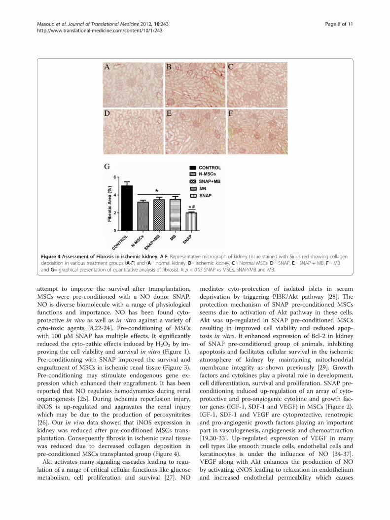
Figure 4 Assessment of Fibrosis in ischemic kidney. A-F: Representative micrograph of kidney tissue stained with Sirius red showing collagendeposition in various treatment groups (A-F) and (A= normal kidney, B= ischemic kidney, C= Normal MSCs, D= SNAP, E= SNAP + MB, F= MBand G= graphical presentation of quantitative analysis of fibrosis). #: p < 0.05 SNAP vs MSCs, SNAP/MB and MB.
Masoud et al. Journal of Translational Medicine 2012, 10:243 Page 8 of 11http://www.translational-medicine.com/content/10/1/243
attempt to improve the survival after transplantation,MSCs were pre-conditioned with a NO donor SNAP.NO is diverse biomolecule with a range of physiologicalfunctions and importance. NO has been found cyto-protective in vivo as well as in vitro against a variety ofcyto-toxic agents [8,22-24]. Pre-conditioning of MSCswith 100 μM SNAP has multiple effects. It significantlyreduced the cyto-pathic effects induced by H2O2 by im-proving the cell viability and survival in vitro (Figure 1).Pre-conditioning with SNAP improved the survival andengraftment of MSCs in ischemic renal tissue (Figure 3).Pre-conditioning may stimulate endogenous gene ex-pression which enhanced their engraftment. It has beenreported that NO regulates hemodynamics during renalorganogenesis [25]. During ischemia reperfusion injury,iNOS is up-regulated and aggravates the renal injurywhich may be due to the production of peroxynitrites[26]. Our in vivo data showed that iNOS expression inkidney was reduced after pre-conditioned MSCs trans-plantation. Consequently fibrosis in ischemic renal tissuewas reduced due to decreased collagen deposition inpre-conditioned MSCs transplanted group (Figure 4).Akt activates many signaling cascades leading to regu-
lation of a range of critical cellular functions like glucosemetabolism, cell proliferation and survival [27]. NO
mediates cyto-protection of isolated islets in serumdeprivation by triggering PI3K/Akt pathway [28]. Theprotection mechanism of SNAP pre-conditioned MSCsseems due to activation of Akt pathway in these cells.Akt was up-regulated in SNAP pre-conditioned MSCsresulting in improved cell viability and reduced apop-tosis in vitro. It enhanced expression of Bcl-2 in kidneyof SNAP pre-conditioned group of animals, inhibitingapoptosis and facilitates cellular survival in the ischemicatmosphere of kidney by maintaining mitochondrialmembrane integrity as shown previously [29]. Growthfactors and cytokines play a pivotal role in development,cell differentiation, survival and proliferation. SNAP pre-conditioning induced up-regulation of an array of cyto-protective and pro-angiogenic cytokine and growth fac-tor genes (IGF-1, SDF-1 and VEGF) in MSCs (Figure 2).IGF-1, SDF-1 and VEGF are cytoprotective, renotropicand pro-angiogenic growth factors playing an importantpart in vasculogenesis, angiogenesis and chemoattraction[19,30-33]. Up-regulated expression of VEGF in manycell types like smooth muscle cells, endothelial cells andkeratinocytes is under the influence of NO [34-37].VEGF along with Akt enhances the production of NOby activating eNOS leading to relaxation in endotheliumand increased endothelial permeability which causes
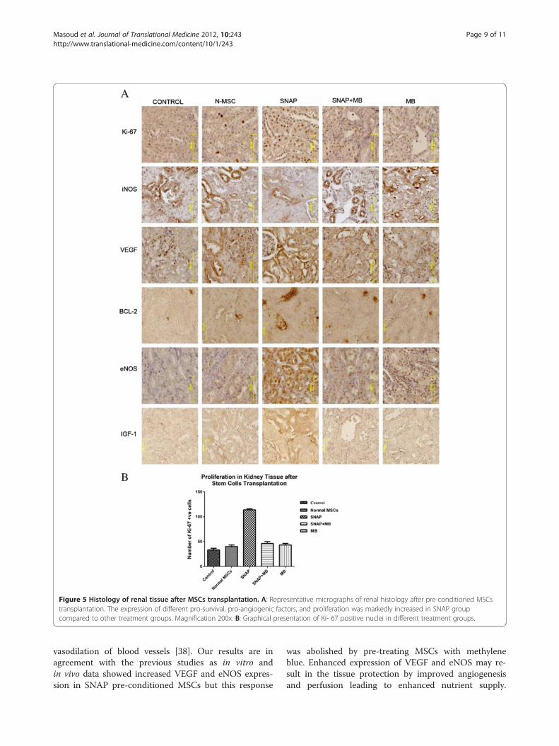
Figure 5 Histology of renal tissue after MSCs transplantation. A: Representative micrographs of renal histology after pre-conditioned MSCstransplantation. The expression of different pro-survival, pro-angiogenic factors, and proliferation was markedly increased in SNAP groupcompared to other treatment groups. Magnification 200x. B: Graphical presentation of Ki- 67 positive nuclei in different treatment groups.
Masoud et al. Journal of Translational Medicine 2012, 10:243 Page 9 of 11http://www.translational-medicine.com/content/10/1/243
vasodilation of blood vessels [38]. Our results are inagreement with the previous studies as in vitro andin vivo data showed increased VEGF and eNOS expres-sion in SNAP pre-conditioned MSCs but this response
was abolished by pre-treating MSCs with methyleneblue. Enhanced expression of VEGF and eNOS may re-sult in the tissue protection by improved angiogenesisand perfusion leading to enhanced nutrient supply.

Masoud et al. Journal of Translational Medicine 2012, 10:243 Page 10 of 11http://www.translational-medicine.com/content/10/1/243
Further, VEGF and SDF-1 could guide transendothelialmigration of MSCs [39,40] into the ischemic renal tissueand their proliferation, but methylene blue treatmentinhibited these factors. This whole milieu of up-regulated growth factors, chemokine, Bcl-2 and Akt mayfavor the maintenance of mitochondrial integrity andcell survival ensuing to renal protection againstischemia.Proliferation is one of the important aspects in cellular
therapies in order to repopulate the damaged tissue. Inour studies, PCNA expression was higher which showsthat SNAP Pre-conditioning elicited proliferation inMSCs in vitro (Figure 2) [41]. Concurrently, there wasmarked increase in Ki-67 expression in tissue sectionsfrom pre-conditioned MSCs transplanted kidneys(Figure 5A and B) compared to all other treatmentgroups showing early make up of cellular loss in kidneyparenchyma resulting in improvement of renal function.This increase in proliferation activity was diminished bymethylene blue treatment in vitro and in vivo.Tubular epithelium is lost dramatically in ischemia
reperfusion injury, resulting in impaired kidney function[42]. Our results demonstrated that mutilation of kidneyfunction caused by ischemia was overcome by the trans-plantation of SNAP pre-conditioned MSCs in a betterway by increased creatinine clearance and reduced BUN(Table 2) which could be due to improved perfusion andtubulogenesis.
ConclusionThis study demonstrates that pharmacological pre-conditioning of MSCs plays a major part in augmentingresistance of MSCs to the oxidative stress in vitro andin vivo injury. Up-regulation of many cytoprotective fac-tors help MSCs survive better in renal ischemic environ-ment by activating Akt cell signaling. MSCs’ homing wasimproved in ischemic renal tissue leading to enhancedperfusion and tubulogenesis and improved renal func-tion. Hence, pre-conditioning can be a potential strategyto enhance the outcomes of cell based therapies in at-tenuating renal ischemia injury.
Competing interestsThe authors declare that they have no competing interests.
Authors’ contributionsMSM participated in the design of study, perform culturing and in vitrotreatments, participated in in vivo experiments, executed statistical analysisand drafted the paper. SSA carried out polymerase chain reaction. MZAperformed in vitro immunoassays and helped to draft the manuscript. AManalyzed the in vivo data and helped to draft the manuscript. SNKparticipated in study design and proofreading the manuscript and SRcontributed in study design, funding and final approval of the manuscript.All authors read and approved the final manuscript.
AcknowledgementsThis work was supported by Higher Education Commission Islamabad,Pakistan. The authors thank to Dr. Muhammad Ashraf for his technicalassistance.
Author details1National Centre of Excellence in Molecular Biology, University of the Punjab,Lahore, Pakistan. 2Current Affiliation: Department of Bioinformatics andBiotechnology, Government College University, Faisalabad 38000, Pakistan.3College of Medicine- Pathology and Laboratory Medicine, University ofCicinnati Ohio, Albert Sabin way, Cicinnati, USA. 4Allama Iqbal MedicalCollege/Jinnah Hospital Complex, University of Health Sciences, Lahore,Pakistan. 5The University of Lahore, 1Km Raiwind Road Lahore, Lahore,Pakistan.
Received: 14 September 2012 Accepted: 28 November 2012Published: 5 December 2012
References1. Mahmoud IM, Hussein Ael A, Sarhan ME, Awad AA, El Desoky I: Role of
combined L-arginine and prostaglandin E1 in renal ischemia-reperfusioninjury. Nephron Physiol 2007, 105:p57–p65.
2. Bonventre JV, Weinberg JM: Recent advances in the pathophysiology ofischemic acute renal failure. J Am Soc Nephrol 2003, 14:2199–2210.
3. Marschall SR, Patterson C: The Pathophysiology of Acute Renal Failure.Principles of molecular medicine. (book) 2nd edition 2006.
4. Humphreys BD, Bonventre JV: Mesenchymal stem cells in acute kidneyinjury. Annu Rev Med 2008, 59:311–325.
5. Khan M, Akhtar S, Mohsin S, S NK, Riazuddin S: Growth factorpreconditioning increases the function of diabetes-impairedmesenchymal stem cells. Stem Cells Dev 2011, 20:67–75.
6. Bernhardt WM, Campean V, Kany S, Jurgensen JS, Weidemann A, WarneckeC, Arend M, Klaus S, Gunzler V, Amann K, et al: Preconditional activation ofhypoxia-inducible factors ameliorates ischemic acute renal failure.J Am Soc Nephrol 2006, 17:1970–1978.
7. Pasha Z, Wang Y, Sheikh R, Zhang D, Zhao T, Ashraf M: Preconditioningenhances cell survival and differentiation of stem cells duringtransplantation in infarcted myocardium. Cardiovasc Res 2008, 77:134–142.
8. Ma D, Lim T, Xu J, Tang H, Wan Y, Zhao H, Hossain M, Maxwell PH, Maze M:Xenon preconditioning protects against renal ischemic-reperfusioninjury via HIF-1alpha activation. J Am Soc Nephrol 2009, 20:713–720.
9. Nathan C: Nitric oxide as a secretory product of mammalian cells. FASEB J1992, 6:3051–3064.
10. Lane P, Gross SS: Cell signalling by nitric oxide. Semin Nephrol 1999,19:215–229.
11. Ignarro LJ: Nitric oxide: a unique endogenous signaling molecule invascular biology. Biosci Rep 1999, 19:51–71.
12. Droge W: Free radicals in the physiological control of cell function.Physiol Rev 2002, 82:47–95.
13. Foster MW, McMahon TJ, Stamler JS: S-nitrosylation in health and disease.Trends Mol Med 2003, 9:160–168.
14. Chae H, J1, Ki-Chan H, Moon-Suk J, Dong-Hyeon Y, Hee-Young C, Soo-WanCH-RK: Nitric Oxide Protects Reactive Oxygen Species-induced Apoptosisin H9C2 Cardiac Muscle Cells. Korean journal of Gerontol 2003, 13:32–38.
15. Sato S, Suzuki A, Nakajima Y, Iwamoto T, Bito H, Miyabe M: S-Nitroso-N-acetylpenicillamine (SNAP) during hemorrhagic shock improvesmortality as a result of recovery from vascular hyporeactivity.Anesth Analg 2000, 90:362–368.
16. Kudo M, Wang Y, Wani MA, Xu M, Ayub A, Ashraf M: Implantation of bonemarrow stem cells reduces the infarction and fibrosis in ischemic mouseheart. J Mol Cell Cardiol 2003, 35:1113–1119.
17. Motterlini R, Foresti R, Intaglietta M, Winslow RM: NO-mediated activationof heme oxygenase: endogenous cytoprotection against oxidative stressto endothelium. Am J Physiol 1996, 270:H107–H114.
18. Gustafsson AB, Gottlieb RA: Heart mitochondria: gates of life and death.Cardiovasc Res 2008, 77:334–343.
19. Imberti B, Morigi M, Tomasoni S, Rota C, Corna D, Longaretti L, Rottoli D,Valsecchi F, Benigni A, Wang J, et al: Insulin-like growth factor-1 sustainsstem cell mediated renal repair. J Am Soc Nephrol 2007, 18:2921–2928.
20. Kaushal GP, Basnakian AG, Shah SV: Apoptotic pathways in ischemic acuterenal failure. Kidney Int 2004, 66:500–506.

Masoud et al. Journal of Translational Medicine 2012, 10:243 Page 11 of 11http://www.translational-medicine.com/content/10/1/243
21. Burst VR, Gillis M, Putsch F, Herzog R, Fischer JH, Heid P, Muller-Ehmsen J,Schenk K, Fries JW, Baldamus CA, Benzing T: Poor cell survival limits thebeneficial impact of mesenchymal stem cell transplantation on acutekidney injury. Nephron Exp Nephrol 2010, 114:e107–e116.
22. Kucera T, Canova NK, Farghali H, Martinek J: The morphological andimmunocytochemical evaluation of primary rat hepatocytes undergoingspontaneous cell death: modulation by the nitric oxide donor S-nitroso-N-acetylpenicillamine. Biomed Pap Med Fac Univ Palacky Olomouc CzechRepub 2006, 150:75–82.
23. Rauhala P, Andoh T, Yeh K, Chiueh CC: Contradictory effects of sodiumnitroprusside and S-nitroso-N-acetylpenicillamine on oxidative stress inbrain dopamine neurons in vivo. Ann N Y Acad Sci 2002, 962:60–72.
24. Kim YM, de Vera ME, Watkins SC, Billiar TR: Nitric oxide protects culturedrat hepatocytes from tumor necrosis factor-alpha-induced apoptosis byinducing heat shock protein 70 expression. J Biol Chem 1997,272:1402–1411.
25. Han KH, Lim JM, Kim WY, Kim H, Madsen KM, Kim J: Expression ofendothelial nitric oxide synthase in developing rat kidney. Am J PhysiolRenal Physiol 2005, 288:F694–F702.
26. Nakajima A, Ueda K, Takaoka M, Yoshimi Y, Matsumura Y: Opposite effectsof pre- and postischemic treatments with nitric oxide donor onischemia/reperfusion-induced renal injury. J Pharmacol Exp Ther 2006,316:1038–1046.
27. Testa JR, Tsichlis PN: AKT signaling in normal and malignant cells.Oncogene 2005, 24:7391–7393.
28. Tejedo JR, Cahuana GM, Ramirez R, Esbert M, Jimenez J, Sobrino F, BedoyaFJ: nitric oxide triggers the phosphatidylinositol 3-kinase/Akt survivalpathway in insulin-producing RINm5F cells by arousing Src to activateinsulin receptor substrate-1. Endocrinology 2004, 145:2319–2327.
29. Chipuk JE, Moldoveanu T, Llambi F, Parsons MJ, Green DR: The BCL-2family reunion. Mol Cell 2010, 37:299–310.
30. Takabatake Y, Sugiyama T, Kohara H, Matsusaka T, Kurihara H, Koni PA,Nagasawa Y, Hamano T, Matsui I, Kawada N, et al: The CXCL12 (SDF-1)/CXCR4 axis is essential for the development of renal vasculature.J Am Soc Nephrol 2009, 20:1714–1723.
31. Little MH, Bertram JF: Is there such a thing as a renal stem cell? J Am SocNephrol 2009, 20:2112–2117.
32. Togel F, Weiss K, Yang Y, Hu Z, Zhang P, Westenfelder C: Vasculotropic,paracrine actions of infused mesenchymal stem cells are important tothe recovery from acute kidney injury. Am J Physiol Renal Physiol 2007,292:F1626–F1635.
33. Khan M, Mohsin S, Khan SN, Riazuddin S: Repair of senescent myocardiumby mesenchymal stem cells is dependent on the age of donor mice.J Cell Mol Med 2011, 15:1515–1527.
34. Kuwabara M, Kakinuma Y, Ando M, Katare RG, Yamasaki F, Doi Y, Sato T:Nitric oxide stimulates vascular endothelial growth factor production incardiomyocytes involved in angiogenesis. J Physiol Sci 2006, 56:95–101.
35. Kimura H, Weisz A, Kurashima Y, Hashimoto K, Ogura T, D’Acquisto F, AddeoR, Makuuchi M, Esumi H: Hypoxia response element of the humanvascular endothelial growth factor gene mediates transcriptionalregulation by nitric oxide: control of hypoxia-inducible factor-1 activityby nitric oxide. Blood 2000, 95:189–197.
36. Dulak J, Jozkowicz A, Dembinska-Kiec A, Guevara I, Zdzienicka A,Zmudzinska-Grochot D, Florek I, Wojtowicz A, Szuba A, Cooke JP: Nitricoxide induces the synthesis of vascular endothelial growth factor by ratvascular smooth muscle cells. Arterioscler Thromb Vasc Biol 2000,20:659–666.
37. Frank S, Stallmeyer B, Kampfer H, Kolb N, Pfeilschifter J: Nitric oxide triggersenhanced induction of vascular endothelial growth factor expression incultured keratinocytes (HaCaT) and during cutaneous wound repair.FASEB J 1999, 13:2002–2014.
38. Sanchez FA, Rana R, Kim DD, Iwahashi T, Zheng R, Lal BK, Gordon DM,Meininger CJ, Duran WN: Internalization of eNOS and NO delivery tosubcellular targets determine agonist-induced hyperpermeability.Proc Natl Acad Sci U S A 2009, 106:6849–6853.
39. Prager GW, Lackner EM, Krauth MT, Unseld M, Poettler M, Laffer S, Cerny-Reiterer S, Lamm W, Kornek GV, Binder BR, et al: Targeting ofVEGF-dependent transendothelial migration of cancer cells bybevacizumab. Mol Oncol 2010, 4:150–160.
40. Uchida D, Onoue T, Tomizuka Y, Begum NM, Miwa Y, Yoshida H, Sato M:Involvement of an autocrine stromal cell derived factor-1/CXCR4 system
on the distant metastasis of human oral squamous cell carcinoma.Mol Cancer Res 2007, 5:685–694.
41. Togel F, Hu Z, Weiss K, Isaac J, Lange C, Westenfelder C: Administeredmesenchymal stem cells protect against ischemic acute renal failurethrough differentiation-independent mechanisms. Am J Physiol RenalPhysiol 2005, 289:F31–F42.
42. da Silva LB, Palma PV, Cury PM, Bueno V: Evaluation of stem celladministration in a model of kidney ischemia-reperfusion injury.Int Immunopharmacol 2007, 7:1609–1616.
doi:10.1186/1479-5876-10-243Cite this article as: Masoud et al.: Pre-conditioned mesenchymal stemcells ameliorate renal ischemic injury in rats by augmented survival andengraftment. Journal of Translational Medicine 2012 10:243.
Submit your next manuscript to BioMed Centraland take full advantage of:
• Convenient online submission
• Thorough peer review
• No space constraints or color figure charges
• Immediate publication on acceptance
• Inclusion in PubMed, CAS, Scopus and Google Scholar
• Research which is freely available for redistribution
Submit your manuscript at www.biomedcentral.com/submit