B-cell development in the thymus is limited by inhibitory signals from the thymic microenvironment
31
doi:10.1182/blood-2002-03-0733 Prepublished online July 5, 2002; Kenneth Dorshkind Yoshiko Hashimoto, Encarnacion Montecino-Rodriguez, Hyosuk Leathers, Robert P Stephan and thymic microenvironment B cell development in the thymus is limited by inhibitory signals from the (3131 articles) Hematopoiesis and Stem Cells Articles on similar topics can be found in the following Blood collections http://bloodjournal.hematologylibrary.org/site/misc/rights.xhtml#repub_requests Information about reproducing this article in parts or in its entirety may be found online at: http://bloodjournal.hematologylibrary.org/site/misc/rights.xhtml#reprints Information about ordering reprints may be found online at: http://bloodjournal.hematologylibrary.org/site/subscriptions/index.xhtml Information about subscriptions and ASH membership may be found online at: digital object identifier (DOIs) and date of initial publication. the indexed by PubMed from initial publication. Citations to Advance online articles must include final publication). Advance online articles are citable and establish publication priority; they are appeared in the paper journal (edited, typeset versions may be posted when available prior to Advance online articles have been peer reviewed and accepted for publication but have not yet Copyright 2011 by The American Society of Hematology; all rights reserved. 20036. the American Society of Hematology, 2021 L St, NW, Suite 900, Washington DC Blood (print ISSN 0006-4971, online ISSN 1528-0020), is published weekly by For personal use only. by guest on June 7, 2013. bloodjournal.hematologylibrary.org From
-
Upload
independent -
Category
Documents
-
view
2 -
download
0
Transcript of B-cell development in the thymus is limited by inhibitory signals from the thymic microenvironment

doi:10.1182/blood-2002-03-0733Prepublished online July 5, 2002;
Kenneth DorshkindYoshiko Hashimoto, Encarnacion Montecino-Rodriguez, Hyosuk Leathers, Robert P Stephan and thymic microenvironmentB cell development in the thymus is limited by inhibitory signals from the
(3131 articles)Hematopoiesis and Stem Cells �Articles on similar topics can be found in the following Blood collections
http://bloodjournal.hematologylibrary.org/site/misc/rights.xhtml#repub_requestsInformation about reproducing this article in parts or in its entirety may be found online at:
http://bloodjournal.hematologylibrary.org/site/misc/rights.xhtml#reprintsInformation about ordering reprints may be found online at:
http://bloodjournal.hematologylibrary.org/site/subscriptions/index.xhtmlInformation about subscriptions and ASH membership may be found online at:
digital object identifier (DOIs) and date of initial publication. theindexed by PubMed from initial publication. Citations to Advance online articles must include
final publication). Advance online articles are citable and establish publication priority; they areappeared in the paper journal (edited, typeset versions may be posted when available prior to Advance online articles have been peer reviewed and accepted for publication but have not yet
Copyright 2011 by The American Society of Hematology; all rights reserved.20036.the American Society of Hematology, 2021 L St, NW, Suite 900, Washington DC Blood (print ISSN 0006-4971, online ISSN 1528-0020), is published weekly by
For personal use only. by guest on June 7, 2013. bloodjournal.hematologylibrary.orgFrom

1
B Cell Development in the Thymus is Limited By Inhibitory Signals from the Thymic
Microenvironment
Running Head: Inhibition of Thymic B Lymphopoiesis
Scientific Section: Hematopoiesis
Yoshiko Hashimoto*, Encarnacion Montecino-Rodriguez*, Hyosuk Leathers, Robert P.
Stephan+, and Kenneth Dorshkind
*These authors contributed equally to this work
Department of Pathology and Laboratory Medicine and the Jonsson Comprehensive Cancer
Center, UCLA School of Medicine, Los Angeles, CA 90095, +Department of Developmental and
Clinical Immunology, University of Alabama, Birmingham, AL 35294
Supported by National Institutes of Health Grant HL60658
Correspondence: Kenneth Dorshkind
Department of Pathology and Laboratory Medicine and the Jonsson Comprehensive Cancer
Center 173216
10833 Le Conte Avenue
Los Angeles, CA 90095
310 206-9535/206-9391 FAX
Total Text: 4757 words/Abstract: 151 words
Copyright 2002 American Society of Hematology
Blood First Edition Paper, prepublished online July 5, 2002; DOI 10.1182/blood-2002-03-0733 For personal use only. by guest on June 7, 2013. bloodjournal.hematologylibrary.orgFrom

2
SUMMARY
B cell precursors are present in the thymus, and the thymic microenvironment is the
source of lymphopoietic factors that include IL-7. Despite the fact that they are bone marrow
derived cells, the data in this report demonstrate that intrathymic B cell progenitors accumulate
at an early pro-B cell stage of development, cycle less than their bone marrow counterparts, and
fail to differentiate efficiently. Additional studies presented herein indicate that these effects are
mediated, at least in part, by soluble factors produced by the thymic microenvironment and
suggest that they affect the ability of pro-B cells to respond optimally to interleukin 7. Taken
together, these observations demonstrate a specific inhibition of intrathymic B lymphopoiesis,
which in turn may explain why lymphoid cell production in the thymus is largely restricted to
production of T lineage cells despite the fact that B cell precursors and B lymphopoietic stimuli
are present in that organ.
Corresponding Author e-mail: [email protected]
For personal use only. by guest on June 7, 2013. bloodjournal.hematologylibrary.orgFrom
3
INTRODUCTION
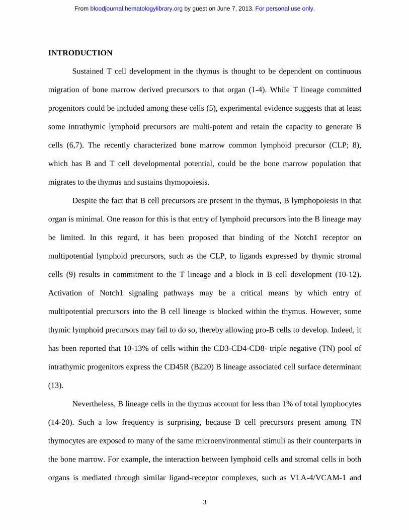
Sustained T cell development in the thymus is thought to be dependent on continuous
migration of bone marrow derived precursors to that organ (1-4). While T lineage committed
progenitors could be included among these cells (5), experimental evidence suggests that at least
some intrathymic lymphoid precursors are multi-potent and retain the capacity to generate B
cells (6,7). The recently characterized bone marrow common lymphoid precursor (CLP; 8),
which has B and T cell developmental potential, could be the bone marrow population that
migrates to the thymus and sustains thymopoiesis.
Despite the fact that B cell precursors are present in the thymus, B lymphopoiesis in that
organ is minimal. One reason for this is that entry of lymphoid precursors into the B lineage may
be limited. In this regard, it has been proposed that binding of the Notch1 receptor on
multipotential lymphoid precursors, such as the CLP, to ligands expressed by thymic stromal
cells (9) results in commitment to the T lineage and a block in B cell development (10-12).
Activation of Notch1 signaling pathways may be a critical means by which entry of
multipotential precursors into the B cell lineage is blocked within the thymus. However, some
thymic lymphoid precursors may fail to do so, thereby allowing pro-B cells to develop. Indeed, it
has been reported that 10-13% of cells within the CD3-CD4-CD8- triple negative (TN) pool of
intrathymic progenitors express the CD45R (B220) B lineage associated cell surface determinant
(13).
Nevertheless, B lineage cells in the thymus account for less than 1% of total lymphocytes
(14-20). Such a low frequency is surprising, because B cell precursors present among TN
thymocytes are exposed to many of the same microenvironmental stimuli as their counterparts in
the bone marrow. For example, the interaction between lymphoid cells and stromal cells in both
organs is mediated through similar ligand-receptor complexes, such as VLA-4/VCAM-1 and
For personal use only. by guest on June 7, 2013. bloodjournal.hematologylibrary.orgFrom

4
stromal cell derived cytokines that include IL-7 (21). IL-7 stimulates pro-B cell proliferation and
potentiates immunoglobulin heavy chain gene rearrangements in those precursors (22-24).
Because it has been established that they are bone marrow derived cells (25), intrathymic B cell
progenitors would be expected to respond to IL-7 and other lymphopoietic factors.
These observations suggest that mechanism(s) which limit the maturation of intrathymic
B cell progenitors exist. Such regulatory pathway(s) would ensure that expansion of B lineage
cells able to compete for microenvironmental niches required for T lymphopoiesis does not
occur. There may be additional reasons for limiting intrathymic B cell production. A case has
been made that thymic B cells are involved in selection of the T cell repertoire (26). Thus, an
inordinate increase in their numbers might be detrimental to the thymic education process.
Therefore, it is likely that signals from the thymic microenvironment minimize the development
of B lineage cells that develop in that organ.
The data in this report support this hypothesis and demonstrate that B cell progenitors in
the thymus accumulate at an early pro-B cell stage of development and fail to differentiate
efficiently. These effects are consistent with findings demonstrating that intrathymic pro-B cells
respond inefficiently to IL-7. Taken together, these results document an unappreciated inhibitory
effect on B lymphopoiesis by the thymic microenvironment that helps to explain why lymphoid
cell production in the thymus is largely restricted to production of T lineage cells.
MATERIALS AND METHODS
Mice. BALB/cJ mice were purchased from the Jackson Laboratory (Bar Harbor, ME) and
housed in the vivarium of the UCLA Division of Laboratory Animal Medicine. Timed pregnant
Swiss/Webster mice were purchased from Taconic Farms (Germantown, NY). Ifnar1-/- mice
were bred and maintained in the vivarium of the University of Alabama, Birmingham, AL.
For personal use only. by guest on June 7, 2013. bloodjournal.hematologylibrary.orgFrom

5
Preparation of cell suspensions. Single cell suspensions of thymus were prepared by gently
pushing tissues through a fine mesh screen into α-Minimal Essential Medium (MEM; GIBCO,
Grand Island, NY) supplemented with 5% fetal calf serum (Atlanta Biologicals, Atlanta, GA).
Bone marrow cells were flushed from femurs using a syringe fitted with a 23-gauge needle. Cells
were counted with a hemacytometer and viability was determined by eosin dye exclusion.
Fetal Thymic Organ Cultures. Fetal thymic organ cultures (FTOC) were established according
to the protocol described by Jenkinson et al. (27). Briefly, fetal thymic lobes from 15 day old
Thy-1.1 Swiss/Webster embryos were harvested aseptically, dissected free from extraneous
tissue, and cultured for 5 days in the presence of 1.35mM deoxyguanosine to deplete endogenous
thymocytes. The lobes were then rinsed and incubated with donor cells in hanging drop cultures
in Terasaki plates for 48 hr. Subsequently, lobes were transferred to FTOC on filter/gelfoam
rafts in RPMI-1640 supplemented with 10% FCS, 5 x 10⁻⁵M 2-β mercaptoethanol, 100 U/ml
streptomycin, and 100 U/ml penicillin and placed in a humidifed, 5%CO2/air incubator at 37oC.
Cell proliferation and differentiation in FTOC was assessed at regular intervals following culture
initiation as described below.
Long-term bone marrow cultures. Long-term myeloid bone marrow cultures (LTBMC) were
initiated as described by Dexter et al. (28). Briefly, the contents of a femur were flushed into a 25
cm2 tissue culture flask in α-MEM supplemented with 20% horse serum and 10-6 M sodium
hydrocortisone succinate. Cultures were incubated at 33oC in a 5% CO2/air incubator. After two
weeks, cultures were recharged with 106 bone marrow cells per flask. B cell development was
induced by switching cultures to the B lymphoid permissive conditions (RPMI-1460
For personal use only. by guest on June 7, 2013. bloodjournal.hematologylibrary.orgFrom

6
supplemented with 5% FCS and 5x10⁻⁵ M β-mercaptoethanol) described by Whitlock and Witte
(29,30).
Preparation of bone marrow and thymus stromal cells. Confluent, heterogeneous bone
marrow stromal cell cultures were established by treating LTBMC with 5µg/ml mycophenolic
acid for 2 to 4 weeks to eliminate hematopoietic cells as described (31). The adherent stromal
cells were then maintained in α-MEM supplemented with 10% FCS in a 37oC, 5% CO2 /air
incubator and were passaged minimally to retain the characteristics of primary stroma.
Hetergenous thymic stromal cell cultures were established as described (32). Thymuses were
digested with trypsin/EDTA and collagenase dispase in Ca++, Mg++ free medium for 35 min at
37oC. The confluent thymic stromal cells were the fed weekly with MEM D-valine supplemented
with 10% FCS and maintained in a 37oC, 5% CO2 /air incubator. These cells were passaged
minimally to retain the characteristics of primary stroma. The generation of the S17 stromal cell
line has been described (33). The S17 bone marrow stromal cell line was fed weekly with α-
MEM supplemented with 10% FCS.
Diffusion chamber cultures. In some experiments, heterogeneous bone marrow or thymic
stromal cells were grown to confluency in transwells fitted with membranes containing 0.4 µm
pores (Becton-Dickinson). Inserts were inserted into wells of six-well plates in which myeloid
LTBMC had been established. Cultures were then switched to B lymphoid permissive
conditions, fed twice weekly, and cells assayed for B cell production by immunophenotyping.
Immunofluorescence and cell sorting. Bone marrow cell suspensions were treated with Tris-
ammonium chloride (pH 7.2) to lyse red blood cells. Cells were then incubated with an anti-
For personal use only. by guest on June 7, 2013. bloodjournal.hematologylibrary.orgFrom
7
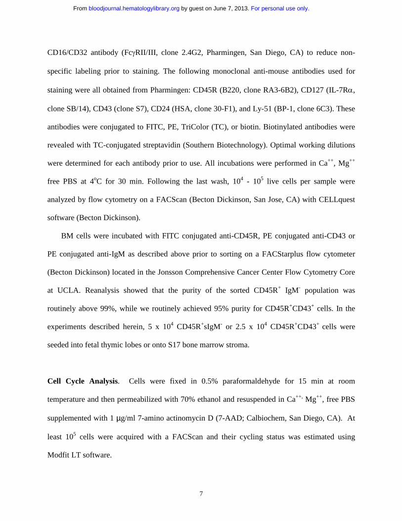
CD16/CD32 antibody (FcγRII/III, clone 2.4G2, Pharmingen, San Diego, CA) to reduce non-
specific labeling prior to staining. The following monoclonal anti-mouse antibodies used for
staining were all obtained from Pharmingen: CD45R (B220, clone RA3-6B2), CD127 (IL-7Rα,
clone SB/14), CD43 (clone S7), CD24 (HSA, clone 30-F1), and Ly-51 (BP-1, clone 6C3). These
antibodies were conjugated to FITC, PE, TriColor (TC), or biotin. Biotinylated antibodies were
revealed with TC-conjugated streptavidin (Southern Biotechnology). Optimal working dilutions
were determined for each antibody prior to use. All incubations were performed in Ca++, Mg++
free PBS at 4oC for 30 min. Following the last wash, 104 - 105 live cells per sample were
analyzed by flow cytometry on a FACScan (Becton Dickinson, San Jose, CA) with CELLquest
software (Becton Dickinson).
BM cells were incubated with FITC conjugated anti-CD45R, PE conjugated anti-CD43 or
PE conjugated anti-IgM as described above prior to sorting on a FACStarplus flow cytometer
(Becton Dickinson) located in the Jonsson Comprehensive Cancer Center Flow Cytometry Core
at UCLA. Reanalysis showed that the purity of the sorted CD45R+ IgM- population was
routinely above 99%, while we routinely achieved 95% purity for CD45R+CD43+ cells. In the
experiments described herein, 5 x 10⁴ CD45R+sIgM- or 2.5 x 10⁴ CD45R+CD43+ cells were
seeded into fetal thymic lobes or onto S17 bone marrow stroma.
Cell Cycle Analysis. Cells were fixed in 0.5% paraformaldehyde for 15 min at room
temperature and then permeabilized with 70% ethanol and resuspended in Ca++, Mg++, free PBS
supplemented with 1 µg/ml 7-amino actinomycin D (7-AAD; Calbiochem, San Diego, CA). At
least 105 cells were acquired with a FACScan and their cycling status was estimated using
Modfit LT software.
For personal use only. by guest on June 7, 2013. bloodjournal.hematologylibrary.orgFrom

8
RESULTS
Thymic B Cell Differentiation is Blocked at an Early Stage In Vivo
Multiple stages of B cell differentiation in the bone marrow can be defined based on the
differential expression of various cell surface determinants. Since thymic B lineage cells are
bone marrow derived (25), their expression of the CD45R, CD43, Ly51(BP-1), and CD24 cell
surface determinants was assessed by flow cytometry. This combination of reagents allows the
pro-B cell pool to be subdivided into fractions A, B, C, and C’ as defined by Hardy et al. (34).
Fraction A includes the most immature pro-B cells while those in fraction C’ are completing
immunoglobulin (Ig) heavy chain gene rearrangements before their transition into pre-B cells.
As expected, cells in the bone marrow pro-B cell compartment were present at the
frequency described previously (35) and included populations that had matured to the C’ stage of
development (Figure 1A). In contrast, very few thymic pro-B cells had matured to the fraction C’
stage (Figure 1A). Because the most actively proliferating B lineage cells are those in the pro-B
cell compartment, the cell cycle status of intrathymic CD45R+CD43+ cells was also determined.
As shown in Figure 1B, the frequency of cycling thymic pro-B cells was approximately three-
fold lower than observed with comparable populations from the bone marrow.
For personal use only. by guest on June 7, 2013. bloodjournal.hematologylibrary.orgFrom

9
Figure 1. B cell development in the
thymus is blocked at the pro-B cell stage.
(A) Phenotype of CD45R+CD43+ pro-B
cells harvested directly from the bone
marrow (BM) or thymus (THY) of BALB/c
mice. The frequency of cells in Fractions A,
B, C and C’ stages of development was
assessed by analysis of Ly51 (BP-1) and
CD24 expression on the gated
CD45R+CD43+ cells. (B) Cell cycle status
of pro-B cells in the bone marrow and
thymus. Data in the figures are
representative of three experiments.
B Cell Development is Blocked in Fetal
Thymic Organ Culture
The above results indicate that B cell production in the thymus is blocked at a relatively
early stage of development. In order to assess the fate of isolated B cell progenitors in the thymic
environment in more detail, a modification of the fetal thymic organ culture (FTOC) system was
used. Fetal thymic lobes from day 15 embryos were treated with deoxyguanosine to remove
endogenous thymocytes prior to seeding with FACS purified CD45R+ sIgM- bone marrow cells.
Thymic lobes prepared in this manner are fully functional and support T cell development (36).
Other aliquots of CD45R+sIgM- cells were seeded on the bone marrow stromal cell line S17
which has been shown to support B cell differentiation in vitro (33). Seven days following
initiation of these cultures, cells were recovered and analyzed phenotypically as described above.
B. A-C THY
5.1
CD
45R
A-C25.1 6.4
BM THY0.4
CD43
A B C C’15.5 50 .0 14.3 12.8
Ly-
51 /
BP
-1
A B
C C’
CD24
A B C C’39.6 21.2 27.0 1.7
For personal use only. by guest on June 7, 2013. bloodjournal.hematologylibrary.orgFrom

10
Figure 2 demonstrates that while B lineage cells were recovered from thymic lobes, they
did not efficiently mature past the Fraction C stage. These data are consistent with observations
made on freshly harvested thymic pro-B cells (Figure 1A) and indicate the validity of the FTOC
system for analyzing thymic B lymphopoiesis. The fate of CD45R+sIgM- cells seeded in FTOC
contrasts with that of other aliquots of CD45R+sIgM- cells seeded onto the S17 stromal cells. In
this case, cells that were recovered seven days later had efficiently matured to the C’ stage of
development (Figure 2A). Similar results were obtained when fetal thymic lobes were seeded
with FACS purified CD45R+CD43+ (fractions A-C) pro-B cells. Again, when lobes were
processed seven days after seeding, few pro-B cells had matured past the fraction C stage of
development (Figure 2B).
Figure 2. Phenotype
of CD45R+CD43+
pro-B cells following
culture on S17
stromal cells or in
FTOC. (A) 5 x 10⁴
CD45R+sIgM- cells
were seeded into
FTOC or onto S17
stroma. Cells were
phenotyped as
described in Figure 1
seven days later. (B) 2.5 x 10⁴ FACS purified CD45R+CD43+ pro-B cells were seeded into
FTOC. Phenotypic analysis was performed on cells harvested from lobes seven days after
initiation of cultures. Data in the figures are representative of three experiments.
84.19.2L
y-51
(BP
-1)
CD
45R
A. CD45R+ IgM- B. CD45R+ CD43+
77.513.5
d7 FTOC
A B C C’ 11.9 5.5 67.2 8.2
CD43
CD24
CD
45R
Ly-
51(B
P-1
)
47.5 24.9
S17
A B C C’ 1.4 53.8 8.3 38.3
CD
45R
CD43
Ly-
51(B
P-1
)
CD24
A B
C C’
For personal use only. by guest on June 7, 2013. bloodjournal.hematologylibrary.orgFrom

11
Exposure to the Thymic Environment Renders B Cell Progenitors Unresponsive to
Lymphopoietic Signals
Since cytokines such as IL-7 are produced in the thymus (37,38), it seemed unlikely that
failure of pro-B cells to mature was due to the absence of B lymphopoietic factors. Instead, the
possibility existed that the thymic microenvironment rendered pro-B cells unresponsive to B
lymphopoietic signals. In order to test this hypothesis, cells harvested from FTOC at various
times after seeding lobes with bone marrow derived CD45R+sIgM- cells were re-seeded onto the
S17 bone marrow stromal cell line under B cell permissive conditions.
As shown in Table 1 and Figure 3, cells harvested two days after culture in FTOC
established vigorous long-term B cell cultures when re-seeded on S17 stroma. However by 7
days of culture in FTOC, they were no longer able to do so. These results are consistent with data
from two experiments demonstrating that CD45R+sIgM- cells directly isolated from the adult
thymus did not establish long-term B cell cultures following seeding on S17 stromal cells (data
not shown).
Table I. B lineage cells harvested from FTOC do not respond to bone marrow stromal cell signals.a
Week after seeding frequency of CD45R+ cells on S17 stroma S17 stroma FTOC� S17 stroma
(x 104) (x 104)d2 d7 d11 d16
1 11.8 3.9 1.9 0.3 ND
2 6.6 4.7 <0.2 5.7 ND
3 11.1 28.1 <0.1 0.6 <0.1
a CD45R+ sIgM- cells were isolated from the bone marrow. Aliquots of 5 x 10⁴ cells from this pool were used to establish long-term cultures on S17 stroma (S17 stroma) or to seed FTOC. Following 2, 7, 11 or 16 days of incubation in these FTOC, cells were harvested and reseeded on S17 stroma (FTOC� S17 stroma). At weekly intervals for 3 weeks thereafter,cells were harvested from the cultures and examined for CD45R+ expression by flow cytometry. Data representative of 5 independent experiments. ND: no cells were not harvested as too few cells were present in the cultures.
For personal use only. by guest on June 7, 2013. bloodjournal.hematologylibrary.orgFrom

12
Figure 3. Exposure to the thymic microenvironment renders pro-B cells unresponsive to B
lymphopoietic stimuli. (A) Cells harvested from FTOC two days after seeding lobes with 5 x
104 CD45R+sIgM- bone marrow cells can still establish long-term cultures on S17 stroma. (B)
CD45R+sIgM- cells can no longer establish long-term bone marrow cultures following 7 days of
culture in FTOC.
Pro-B Cells in the Thymus Are Hyporesponsive to IL-7
IL-7 is required for murine pro-B cells to proliferate and complete Ig heavy chain gene
rearrangements and mature into pre-B cells (39). Because thymic pro-B cells accumulate in
Fraction C and cycle at a lower rate than their bone marrow counterparts, their capacity to
respond to IL-7 was examined. Following seven days of culture in FTOC, cells were harvested
from the lobes and their proliferative response to IL-7 was compared to that of CD45R+sIgM-
cells from fresh bone marrow. The frequency of CD45R+CD43+ cells, which includes the most
IL-7 responsive cells, in each population was determined by FACS. Based on this information,
cultures were initiated using the same total number of CD45R+CD43+ cells from each source.
As shown in Figure 4A, cells harvested from thymic lobes responded to IL-7, but the magnitude
of their proliferative response was lower than that of the freshly harvested bone marrow
population.
This observation prompted an analysis of IL-7 receptor α chain (IL-7Rα (CD127))
expression on thymic B lineage cells. Cells were harvested from thymic lobes seven days after
A. 2 days in FTOC 7 days in FTOC
For personal use only. by guest on June 7, 2013. bloodjournal.hematologylibrary.orgFrom

13
seeding with CD45R+sIgM- cells, and the level of CD127 expression on CD45R+CD43+ pro-B
cells was compared to that on freshly isolated CD45R+CD43+ bone marrow cells. The results
indicated that both the frequency of pro-B cells that express the IL-7Rα chain and those which
express it at relatively high levels are reduced following 7 days of culture in FTOC (Figure 4B,
top).
Figure 4. IL-7 responsiveness and IL-7R�receptor levels are decreased on B lineage cells
exposed to the thymic microenvironment.. (A)
CD45R+sIgM- cells were cultured in FTOC and
seven days later their proliferative response to
increasing concentrations of IL-7 was compared
to that of CD45R+sIgM- cells harvested from the
bone marrow. The frequency of CD45R+CD43+
cells in each population was determined by FACS
and based on this information, cultures were
initiated using the same total number of
CD45R+CD43+ cells from each source. (B)
Seven days after seeding thymic lobes with 5 x
104 CD45R+sIgM- bone marrow cells, CD127
expression on CD45R+CD43+ pro-B cells was
examined by FACS. Freshly isolated
CD45R+CD43+ pro-B cells from the bone
marrow were examined in parallel. Studies were
performed using cells from BALB/c (top), Ifnar1-
/- (middle), and 129 strain mice. The frequency of
high and low CD127 expressing cells is indicated
in each plot. (C) Frequency of B lineage cells in
the thymus of BALB/c and Ifnar1-/- mice.
C. Ifnar1-/-
CD
45R
0.43 0.370.02
CD
45R
CD43
A.
0
500
1000
1500
2000
2500
CD45R+IgM- BMCD45R+IgM- BM FTOC
B.30.6 6.1
CD127
BALB/c
Ifnar1-/-
BM17.3 0.4
CD127
24.3 1.1
d7 FTOC
35.4 11.5
129
33.0 4.0 12.8 1.0
For personal use only. by guest on June 7, 2013. bloodjournal.hematologylibrary.orgFrom

14
CD
45R
+ ce
ll nu
mbe
r x
104
0
50
100
150
0 1 2 3
week of Culture
B . CD45R+ cells no transwellheterogenousbone marrow stroma
heterogenousthymic stroma
Dexter cultures
Transwell (with stromal cells)
- harvest cells - FACS analysis
1-3wks
A.
B cell culture conditions
Thymic Stromal Cell Derived Factors Inhibit B Lymphopoiesis
The above observations indicate that the thymic microenvironment is a source of
mediators that render pro-B cells hypo-responsive to IL-7. To determine whether such inhibitors
were soluble molecules, initial experiments used a modification of the myeloid to lymphoid
long-term bone marrow switch culture
system (29) to test whether thymic
stomal cells could inhibit B cell
development in the absence of direct
cell contact.
Figure 5. Inhibitory signals from
thymic stromal cells can counteract
B lymphopoietic stimuli. (A) shows
the experimental protocol used to
introduce transwells containing
heterogeneous bone marrow or thymic
stromal cells into myeloid (Dexter)
long-term bone marrow cultures at the
time of their transfer to B lymphoid
permissive conditions. (B) Non-
adherent cells were harvested weekly
from the cultures and analyzed for
expression of CD45R. Data are
representative of three experiments.
For personal use only. by guest on June 7, 2013. bloodjournal.hematologylibrary.orgFrom

15
Within a week following transfer of long-term bone marrow cultures established under
the myeloid conditions described by Dexter and colleagues (28) to B lymphoid permissive
conditions (30), pro-B cells emerge, and by three weeks B lineage cells predominate in the
cultures. As shown in Figure 5A, heterogeneous populations of bone marrow or thymic stroma
growing in transwells were introduced into these cultures. By three weeks following transfer of
long-term myeloid bone marrow cultures to B lymphoid permissive conditions, vigorous cultures
containing CD45R+ cells established (Figure 5B). This same pattern of growth occurred when
empty transwells (data not shown) or transwells containing heterogeneous bone marrow stroma
(Figure 5B) were placed in the cultures at the time of their transfer to the lymphoid conditions.
However, when transwells containing heterogeneous preparations of thymic stromal cells were
introduced into the cultures, CD45R+ cell production was dramatically reduced (Figure 5B),
indicating that soluble mediators produced by the thymic microenvironment can inhibit
intrathymic B lymphopoiesis.
Downregulation of IL-7Rαααα Occurs Through a Type 1 IFN Receptor Independent
Mechanism
Type 1 interferons (IFN) have been shown to inhibit the response of B lineage cells to IL-
7 (40-43). In view of the above data indicating the involvement of soluble factors in limiting
intrathymic B lymphopoiesis, it was of interest to assess their potential involvement. In order to
do so, CD45R+sIgM- cells from Type 1 IFN receptor deficient (Ifnar1-/-) mice were used to seed
fetal thymic lobes. CD45R+sIgM- cells from 129 mice, the background strain of the Ifnar1-/-
mice, were assayed in parallel. Seven days later CD127 expression on CD45R+CD43+ cells
harvested from these FTOCs was compared to that on bone marrow pro-B cells. As shown in
figure 4B, exposure to the thymic microenvironment resulted in a greater than 90% reduction of
For personal use only. by guest on June 7, 2013. bloodjournal.hematologylibrary.orgFrom

16
CD127 high expressing cells in Ifnar1-/- mice. This level of inhibition was comparable to that in
BALB/c and greater than that in 129 strain mice. A prediction based on this observation is that
the frequency of B lineage cells in the thymus of Ifnar1-/- mice should not be elevated. This is in
fact the case, as the frequency of B lineage cells in the thymus of Ifnar1-/- mice was comparable
to that in control animals (Figure 4C).
Resumption of B Cell Maturation in the Presence of Exogenous IL-7
Taken together, the above data suggest that the growth and development of pro-B cells in
the thymus is limited through IL-7Rα downregulation. This event may in turn limit their capacity
to compete with T cell progenitors for the IL-7 produced by the thymic microenvironment. A
prediction based on this hypothesis is that increasing intrathymic IL-7 levels may in turn allow
their growth. In order to test this premise, FTOC were initiated with CD45R+sIgM- bone
marrow cells in the presence of 50 U/ml of exogenous IL-7. As shown in Figure 6A,
approximately half of the CD45R+ cells no longer expressed CD43 after seven days of culture,
suggesting that they had matured to the pre-B cell stage. This result contrasts with the finding
that the majority of B lineage cells in non-IL-7 supplemented FTOC were CD45R+CD43+. In
addition, although B lineage cells harvested from day 7 FTOC were not able to establish long-
term bone marrow cultures on S17 bone marrow stromal cells (Figure 3), they were able to do so
if the medium was supplemented with IL-7 (Figure 6B). These cultures could be maintained for
at least three weeks and phenotypic analysis confirmed that the cultures contained B lineage cells
(data not shown).
Since IL-7 is required for T cell development (44), it was important to determine how levels
of IL7Rα expression on thymic B and T cell progenitors compared. As shown in Figure 6C, the
CD3-CD4-CD8- triple negative (TN) fraction of cells was isolated from the fresh thymus and
For personal use only. by guest on June 7, 2013. bloodjournal.hematologylibrary.orgFrom

17
analyzed for expression of CD127 and CD45R. This allowed levels of CD127 on CD45R+ B
lineage cells to be compared to that on CD45R- cells, which primarily include T cell progenitors.
The data demonstrate that the frequency of cells that express CD127 is higher in the CD45R- TN
thymocyte population and that they express it
at higher levels than is found among CD45R+
cells.
Figure 6. Responsiveness of thymic B cells
to exogenous IL-7. (A) Phenotype of cells
harvested seven days after seeding FTOC
with 5 x 10⁴ CD45R+sIgM- bone marrow
cells in the presence or absence of 50 U/ml of
IL-7. (B) Cells harvested 7 days after seeding
FTOC with CD45R+sIgM- bone marrow cells
could establish long-term cultures when
seeded on S17 bone marrow stromal cells in
the presence of IL-7. (C) Expression of
CD127 and CD45R on CD3-CD4-CD8- triple
negative (TN) thymocytes harvested from the
fresh thymus.
DISCUSSION
Both bone marrow and thymic stromal cells express ligands required for direct
interactions with developing B lineage cells and secrete cytokines required for B cell
development. Despite these similarities, B cell development in the thymus is limited. Studies
aimed at investigating this phenomenon revealed that B cell development is blocked at a
C.
30.7
6.0
0.8
0.1
CD
127
CD45R
TN thymocytes
B. no IL-7 + IL-7
A.
0
20
40
60
80
% c
ells
CD45R+ CD43+ CD45R+ CD43-
50U/ml IL-7
no IL-7
For personal use only. by guest on June 7, 2013. bloodjournal.hematologylibrary.orgFrom
18
relatively early stage of development in the thymus because signals produced the thymic
microenvironment inhibit that process.
Initial analyses focused on characterizing B lineage cells harvested from the thymus.
These studies demonstrated that the frequency of pro-B cells in cycle was lower in the thymus
than in the bone marrow and that B lineage cells did not efficiently mature past the Fraction C
stage of development. When similar analyses were performed on bone marrow B cell progenitors
seven days after being seeded in fetal thymic lobes in vitro, the same results were obtained. In
addition to corroborating the results obtained with primary cells, this result established the fetal
thymic organ culture system as a model for analyzing thymic B cell production.
It is paradoxical that culture in the thymic microenvironment in the FTOC system
inhibited B cell development while other studies have reported the isolation of thymic stromal
cell lines that can support B cell differentiation. Indeed, our own laboratory characterized a
thymic stromal cell line that efficiently supported the pre-B to B cell transition (45). However, it
is important to emphasize that the thymic stromal cell line in question only potentiated this latter
phase of development but not the short or long-term growth of pro-B cells. In fact, our
characterization of numerous thymic stromal cell lines has so far failed to identify any capable of
supporting long-term B cell development. Furthermore, even if thymic stromal cells with such
potential were isolated, they do not represent the thymic microenvironment as a whole. That is
why, in order to assess the effects of the thymic microenvironment on B cell development,
primary cultures of heterogeneous thymic stromal cells rather than thymic stromal cell lines were
used in the diffusion chamber studies.
That a mechanism to inhibit intrathymic B cell development exists at all might seem
puzzling, since the overwhelming majority of lymphoid cells in the thymus are T lineage cells.
Instead, the inefficient expansion and maturation of thymic pro-B cells could result from their
For personal use only. by guest on June 7, 2013. bloodjournal.hematologylibrary.orgFrom

19
failure to compete effectively for microenvironmental niches. However, it is important to focus
discussion on the most immature thymocytes contained within the TN compartment. While the
majority of cells within this population are likely committed to the T lineage, it has been reported
that 10-13% are CD45R+ (13). If even a fraction of these cells did come into contact with thymic
stromal cells that supported their growth and development, this in turn could compromise overall
levels of T cell production, particularly if enough mature B cells that affected the process of
thymic education subsequently developed. Thus, it is logical to propose the existence of a
mechanism(s) to specifically inhibit(s) thymic pro-B cell development.
That such signals exist is supported by recent studies in which selective inactivation of
Notch1 by gene targeting was performed. Radtke et al. (11) and Wilson et al. (12) demonstrated
that T cell development was effectively blocked in mice in which Notch1 was conditionally
inactivated and that there was an expansion in the number of B lineage cells in the thymus.
However, while their incidence was higher than in control mice, a critical point is that the total
number of B lineage cells in the thymus of these mice increased to only about four million cells.
This number is considerably lower than the 100 million thymocytes that are present in the
thymus of young animals. More recently, Han et al. (46) demonstrated that mice in which the
RBP-J transcription factor, which associates with the intracellular domain of Notch to allow
DNA binding, has been inactivated, also exhibit impaired T cell development and increased
intrathymic B lymphopoiesis. However, even though Notch signaling in these mice was
effectively blocked, the total number of thymic B lineage cells was again only about 4 x 10⁶
cells. Taken together, these results indicate that even when not competing for environmental
niches with developing T cells, B cell precursors do not undergo extensive expansion in the
thymic microenvironment. Importantly, no differences between bone marrow B lineage cells in
For personal use only. by guest on June 7, 2013. bloodjournal.hematologylibrary.orgFrom

20
mice in which Notch1 expression was conditionally inactivated and their control littermates were
reported.
A recent study of mice that expressed a lunatic fringe transgene, which results in the
inhibition of Notch1 activation, showed that their thymus contained up to 50 million B lineage
cells (13). That report would also seem to contradict the conclusion that the thymus actively
inhibits B lymphopoiesis. However, while the results of that study could be interpreted to infer
that inhibitors of thymic B lymphopoiesis do not exist, that conclusion may be too simplistic in
view of the minimal level of B lymphopoiesis in the Notch1-/- and RBP-J-/- mice. Instead, it is
important to consider that lunatic fringe normally functions as an intracellular mediator, but its
transgenic expression resulted in secretion of the protein. This fact, combined with findings that
the lunatic fringe protein can have effects beyond the inhibition of Notch1 activity (47), raises
the distinct possibility that normal thymus physiology was altered in lunatic fringe transgenic
mice. That this in turn impacted upon one or more regulatory pathways, such as those which
inhibit B cell development, must be considered.
In any case, the studies presented herein clearly demonstrate that exposure to the thymic
microenvironment renders B cell precursors unresponsive to B lymphopoietic stimuli. After
seven days in thymic lobes, B lineage cells could no longer proliferate and differentiate on bone
marrow stromal cells. Further analysis showed that this occurs because pro-B cells that have
been exposed to the thymic microenvironment are hyporesponsive to IL-7. This seems to occur
through IL-7Rα chain down-regulation. Proposing that interference with IL-7 signaling
pathways is ultimately responsible for the inhibition of intrathymic B cell development is
entirely consistent with the observation that thymic pro-B cells or pro-B cells exposed to the
thymic environment do not efficiently mature from the fraction C to the C’ stage of development.
As described by Hardy et al. (34), Fraction C cells have undergone Ig heavy chain gene
For personal use only. by guest on June 7, 2013. bloodjournal.hematologylibrary.orgFrom

21
rearrangements at the D-J loci but V-DJ rearrangements have not yet occurred. IL-7 is required
for cells to complete the latter stage of Ig gene rearrangement and to transition from fraction C to
the pre-B (fraction D) stage of development.
The long-term bone marrow culture experiments described in this study suggest that
soluble factors produced by thymic stromal cell(s) are at least partially responsible for the
inhibition of thymic B cell production. In fact, thymic stromal cells present in diffusion chambers
were able to inhibit the emergence of B lineage cells, even when precursors were in contact with
a supporting layer of bone marrow stromal cells. While aspecific effects of thymic stromal cells
on B lymphopoiesis in this system through excessive consumption of nutrients can not be
excluded, it does not seem likely. First, there is no a priori reason to assume that confluent
thymic and bone marrow stroma differ significantly in the nutrients they consume. Second,
cultures were fed twice weekly and neither pH fluctuations nor excessive cell death were
observed in cultures in which thymic stroma was present. Therefore, it is logical to propose that
soluble factors from the thymic stroma are able to inhibit B cell development and that their
effects are potent enough to counteract positively acting B lymphopoietic signals.
It has been reported that type 1 interferons (IFN) can inhibit the response of B cell
progenitors to IL-7 (40-43). Since IFNs are produced in the thymus, it was of interest to
determine if they were responsible for the observed effects by seeding pro-B cells from type 1
IFN receptor deficient mice into fetal thymic lobes. This analysis revealed that the frequency of
CD45R+CD43+ cells that expressed CD127 at high levels had decreased by over 90% after
seven days in FTOC. This finding, combined with the fact that the frequency of B lineage cells
in the thymus of these knockout mice was not elevated, strongly suggests that signaling through
the type 1 IFN receptor is not responsible for the inhibition of intrathymic B lymphopoiesis.
For personal use only. by guest on June 7, 2013. bloodjournal.hematologylibrary.orgFrom

22
These findings would also seem to exclude the involvement of a new type 1 IFN family member,
limitin, which has been proposed as a selective inhibitor of B cell production (48).
Taken together, the data in this and other studies make it possible to formulate a model in
which checkpoints operative at multiple levels act in concert to limit B cell development in the
thymus. Initially, intrathymic lymphoid precursors, such as the CLP, might receive signals that
preferentially potentiate their development along the T rather than the B cell lineage, and Notch1
activated signaling pathways may be critical at this juncture (10,11,49). Nevertheless, some pro-
B cells develop and their growth and differentiation is severely limited by thymic
microenvironmental signals that render them hyporesponsive to IL-7. While IL-7 is not critical
during human B lymphopoiesis (50), similar or alternative thymic stromal cell factors can be
postulated to limit pro-B cell expansion in the human thymus. The inhibition of intrathymic B
lymphopoiesis may not be absolute, however, and some pro-B cells may mature into pre-B cells
from which B lymphocytes may be generated. A few of these may be retained in the thymus as
antigen presenting cells (26) while others are exported to the periphery (19).
It is becoming increasingly appreciated that negative regulators play an important role in
the regulation of hematopoiesis. In this regard, numerous mediators have been described which
specifically inhibit B cell production (51). However, these factors have generally been
considered in terms of their effects on bone marrow B lymphopoiesis. The findings herein
describe an unexpected role of negative regulatory factors in the homeostasis of thymic
lymphocyte production. These data contribute to the understanding of how an organ which has
the potential to support B cell production limits that process and suggest that further comparisons
of bone marrow and thymic lymphopoiesis will provide additional insights into the regulation of
primary lymphocyte production.
For personal use only. by guest on June 7, 2013. bloodjournal.hematologylibrary.orgFrom

23
ACKNOWLEDGEMENTS
The authors appreciate the helpful discussions with Drs. Ellen Rothenberg, Max Cooper, David
Rawlings, and Andrew Farr. This work was supported by grant HL60658 from the National
Institutes of Health.
REFERENCES
1. Ford CE, Micklem HS, Evans EP, Gray JS, Ogden DA. The inflow of bone marrow cells to
the thymus. Ann NY Acad Sci 1996;129:283-296.
2. Adkins B, Mueller C, Okada CY, Reichert RA, Weissman IL, Spangrude GJ. Early events in
T-cell maturation. Annu Rev Immunol 1987;5:325-365.
3. Donskoy E, Goldschneider I. Thymocytopoiesis is maintained by blood-borne precursors
throughout postnatal life. A study in parabiotic mice. J Immunol 1992;148:1604-1612.
4. Foss DL, Donskoy E, Goldschneider I. The importation of hematogenous precursors by the
thymus is a gated phenomenon in normal adult mice. J Exp Med 2001;193:365-374.
5. Rodewald H-R, Kretzschmar K, Takeda S, Hoho C, Dessing M. Identification of pro-
thymocytes in murine fetal blood: T lineage commitment can precede thymus colonization.
EMBO J 1994;13:4229-4240.
For personal use only. by guest on June 7, 2013. bloodjournal.hematologylibrary.orgFrom

24
6. Matsuzaki Y, Gyotoku J, Ogawa M, Nishikawa S, Katsura Y, Gachelin G, Nakuchi H.
Characterization of c-kit positive intrathymic stem cells that are restricted to lymphoid
differentiation. J Exp Med 1993;178:1283-1292.
7. Wu L, Antica M, Johnson GR, Scollay R, Shortman K. Developmental potential of the
earliest precursor cells from the adult mouse thymus. J Exp Med 1991;174:1617-1627.
8. Kondo M, Weissman IL, Akashi, K Identification of clonogenic common lymphoid
progenitors in mouse bone marrow. Cell 1997;91:661-672.
9. Feil M P, Maroder M, Mitsiadis TA, Campese AF, Bellavia D, Vacca A, Mann, RS, Frati L,
Lendahl U, Gulino A, Screpanti I. Expression pattern of Notch1, 2, and 3 and Jagged1 and 2 in
lymphoid and stromal thymus components: distinct ligand-receptor interactions in intrathymic T
cell development. Int Immunol 1999;11:1017-1025.
10. Pui JC, Allman D, Xu L, DeRocco S, Karnell FG, Bakkour S, Lee JY, Kadesch T, Hardy RR,
Aster JC, Pear WS Notch1 expression in early lymphopoiesis influences B versus T lineage
determination. Immunity 1999;11:299-308.
11. Radtke F, Wilson A., Stark G, Bauer M, van Meerwijk J, MacDonald HR, Aguet M.
Deficient T cell fate specification in mice with an induced inactivation of Notch 1. Immunity
1999;10:547-558.
For personal use only. by guest on June 7, 2013. bloodjournal.hematologylibrary.orgFrom

25
12. Wilson A, MacDonald HR, Radtke F. Notch 1-deficient common lymphoid precursors adopt
a B cell fate in the thymus. J Exp Med 2002;194:1003-1012.
13. Koch U, Lacombe TA, Holland D, Bowman JL, Cohen BL, Egan SE, Guidos CJ.
Subversion of the T/B lineage decision in the thymus by lunatic fringe-mediated inhibition of
notch-1. Immunity 2001;15;225-236.
14. Miyama-Inaba M, Kuma S-I, Inaba K., Ogata H., Iwai H, Yasumizu R, Muramatsu S,
Steinman R, Ikehara S. Unusual phenotype of B cells in the thymus of normal mice. J Exp Med
1988; 168:811-816.
15. Kimoto H, Shirasawa T, Taniguchi M, Takemori T. B cell precursors are present in the
thymus during early development. Eur J Immunol 1989;19:97-104.
16. Andreu-Sanchez JL, Faro J, Alonso JM, Paige CJ, Martinez-A C, Marcos MAR. Ontogenic
characterization of thymic B lymphocytes. Analysis in different mouse strains. Eur J Immunol
1990;20:1767-1773.
17. Peault B, Khazaal I, Weissman IL. In vitro development of B cells and macrophages from
early mouse fetal thymocytes. Eur J Immunol 1994;24:781-784.
18. Mori S-I, Inaba M, Sugihara A, Taketani S, Doi H, Fukuba Y, Yamamoto Y, Adachi Y,
Inaba K, Fukuhara S, Ikehara S. Presence of B cell progenitors in the thymus. J Immunol
1997;158:4193-4199.
For personal use only. by guest on June 7, 2013. bloodjournal.hematologylibrary.orgFrom

26
19. Akashi K, Richie LI, Miyamoto T, Carr WH, Weissman IL. B lymphopoiesis in the thymus.
J Immunol 2000;164:5221-5226.
20. Sugihara A, Inaba M, Mori S-I, Taketani S, Adachi Y, Hisha H, Inaba K, Toki J, Horio T,
Gershwin ME, Ikehara S. Differentiation from thymic B cell progenitors to mature B cells in
vitro. Immunobiol 2000;201:515-526.
21. Montecino-Rodriguez E, Dorshkind K. Regulation of lymphocyte development by
microenvironmental and systemic factors. In: Monroe JG, Rothenberg EV, eds. Molecular
Biology of B-Cell and T-Cell Development. Towtowa, N.J: Humana Press; 1998:197-211.
22. Namen AE, Lupton S, Hjerrild K, Wagnall J, Mochizuki DY, Schmierer A, Mosley B, March
C, Urdal D, Gillis S, Copsman D, Goodwin RG. Stimulation of B cell progenitors by cloned
murine interleukin-7. Nature 1988;333:571-573.
23. Corcoran AE, Smart FM, Cowling RJ, Crompton T, Owen MJ, Venkitaraman AR. The
interleukin-7 receptor α chain transmits distinct signals for proliferation and differentiation
during B lymphopoiesis. EMBO J 1996;15:1924-1932.
24. Corcoran AE, Riddell A, Krooshoop D, Venkitaraman AR. Impaired immunoglobulin gene
rearrangement in mice lacking the IL-7 receptor. Nature 1998;391:904-907.
For personal use only. by guest on June 7, 2013. bloodjournal.hematologylibrary.orgFrom

27
25. Than S, Inaba M, Inaba K, Fukuba Y, Adachi Y, Ikehara S. Origin of thymic and peritoneal
Ly-1 B cells. Eur J Immunol 1992;22:1299-1303.
26. Inaba M, Inaba K, Hosono M, Kumamoto T, Ishida T, Muramatsu S, Masuda T, Ikehara S.
Distinct mechanisms of neonatal tolerance induced by dendritic cells and thymic B cells. J Exp
Med 1991;173:549-559.
27. Jenkinson EJ, Franchi LL, Kingston,R, Owen JJ. Effect of deoxyguanosine on lymphopoiesis
in the developing thymus rudiment in vitro: application in the production of chimeric thymus
rudiments. Eur J Immunol 1982;12:583-587.
28. Dexter T, Allan T, Lajtha L. Conditions controlling the proliferation of haematopoietic stem
cells in vitro. J Cell Physiol 1977;91:334-344.
29. Dorshkind K. In vitro differentiation of B lymphocytes from primitive hemopoietic
precursors present in long-term bone marrow cultures. J Immunol 1986; 136:422-429.
30. Whitlock C, Witte O. Long-term culture of B lymphocytes and their precursors from murine
bone marrow. Proc Natl Acad Sci USA 1982;79:308-312.
31. Johnson A, Dorshkind K. Stromal cells in myeloid and lymphoid long-term bone marrow
cultures can support multiple hemopoietic lineages and modulate their production of hemopoietic
growth factors. Blood 1986;68:1348-1354.
For personal use only. by guest on June 7, 2013. bloodjournal.hematologylibrary.orgFrom
28
32. Montecino-Rodriguez E, Dorshkind K. Long-term culture of triple-negative thymocytes. J
Immunol 1996;156:957-962.
33. Collins LS, Dorshkind, K. A stromal cell line from myeloid long-term bone marrow cultures
can support myelopoiesis and B lymphopoiesis. J Immunol 1987; 138:1082-1087.
34. Hardy RR, Carmack CE, Shinton SA, Kemp JD, Hayakawa K. Resolution and
characterization of pro-B and pre-pro-B cell stages in normal mouse bone marrow. J Exp Med
1991;173:1213-1225.
35. Allman D, Li J, Hardy RR. Commitment to the B lymphoid lineage occurs before DH-JH
recombination. J Exp Med 1999;18:735-740.
36. Hashimoto Y, Montecino-Rodriguez E, Gershwin ME, Dorshkind K. Impaired development
of T lymphoid precursors from pluripotent hematopoietic stem cells in New Zealand Black mice.
J Immunol 2002;168:81-86.
37. Wiles MV, Ruiz P, Imhof BA. Interleukin-7 expression during mouse thymic development.
Eur J Immunol 1992;22:1037-1042.
38. Moore NC, Anderson G., Smith CA, Owen JTT, Jenkinson EJ. Analysis of cytokine gene
expression in subpopulations of freshly isolated thymocytes and thymic stromal cells using
semiquantitative polymerase chain reaction. Eur J Immunol 1993;23:992-997.
For personal use only. by guest on June 7, 2013. bloodjournal.hematologylibrary.orgFrom
29
39. Von Freeden-Jeffry U, Vieira P, Lucian LA, McNeil T, Burdach SEG, Murray R.
Lymphopenia in interleukin (IL)-7 gene-deleted mice identifies IL-7 as a nonredundant cytokine.
J Exp Med 1995;181:1519-1526.
40. Lin Q, Dong C, Cooper MD. Impairment of T and B cell development by treatment with a
type I interferon. J Exp Med 1998;187:79-87.
41. Wang J, Lin,Q, Langston H, Cooper MD. Resident bone marrow macrophages produce type
1 interferons that can selectively inhibit interleukin-7-driven growth of B lineage cells. Immunity
1995;3:475-484.
42. Gongora R, Stephan RP, Zhang Z, Cooper MD. An essential role for Daxx in the inhibition
of B lymphopoiesis by type 1 interferons. Immunity. 2001;14:727-737.
43. Gongora R, Stephan RP, Schreiber RD, Cooper MD. Stat-1 is not essential for inhibiton of B
lymphopoiesis by Type 1 IFNs. J Immunol 2000;165:2362-2366.
44. Peschon JJ, Morrissey PJ, Grabstein KH, et al. Early lymphocyte expansion is severely
impaired in interleukin 7 receptor-deficient mice. J Exp Med 1994;180:1955-1960.
45. Montecino-Rodriguez E, Johnson A, Dorshkind K. Thymic stromal cells can support B cell
differentiation from intrathymic precursors. J Immunol 1996;156:963-967.
46. Han H, Tanigaki K, Yamamoto N, Kuroda K, Yoshimoto M, Nakahata T, Ikuta K, Honjo T.
For personal use only. by guest on June 7, 2013. bloodjournal.hematologylibrary.orgFrom

30
Inducible gene knockout of transcription factor recombination signal binding protein-J reveals its
essential role in T versus B lineage decision. Int Inmmunol 2002; 14:637-645.
47. Wu JY, Rao Y. Fringe: defining borders by regulating the notch pathway. Curr Opin
Neurobiol 111; 9:537-543.
48. Oritani K, Medina KL, Tomiyama Y, et al. Limitin: an interferon-like cytokine that
preferentially influences B-lymphocyte precursors. Nature Med 2000;6:659-666.
49. MacDonald HR, Wilson A, Radtke F. Notch1 and T-cell develoment: insights from
conditional knockout mice. Trends Immunol 2001;22:155-160
50. LeBien TW. Fates of human B-cell precursors. Blood 2000;96:9- 23.
51. Kouro T, Medina KL, Oritani K, Kincade PW. Characteristics of early murine B-lymphocyte
precursors and their direct sensitivity to negative regulators. Blood 2001;97:2708-2715.
For personal use only. by guest on June 7, 2013. bloodjournal.hematologylibrary.orgFrom