Microenvironment driven invasion: a multiscale multimodel investigation
Upload
independentCategory
view
0download
0
Review ArticleMicroenvironment, Oncoantigens, and Antitumor Vaccination:Lessons Learned from BALB-neuT Mice
Laura Conti, Roberto Ruiu, Giuseppina Barutello, Marco Macagno, Silvio Bandini,Federica Cavallo, and Stefania Lanzardo
Department of Molecular Biotechnology and Health Sciences, Molecular Biotechnology Center, University of Torino,Via Nizza 52, 10126 Torino, Italy
Correspondence should be addressed toFederica Cavallo; [email protected]
Received 10 April 2014; Accepted 12 May 2014; Published 3 June 2014
Academic Editor: Zhiqiang Meng
Copyright © 2014 Laura Conti et al. This is an open access article distributed under the Creative Commons Attribution License,which permits unrestricted use, distribution, and reproduction in any medium, provided the original work is properly cited.
The tyrosine kinase human epidermal growth factor receptor 2 (HER2) gene is amplified in approximately 20% of human breastcancers and is associated with an aggressive clinical course and the early development of metastasis. Its crucial role in tumor growthand progression makes HER2 a prototypic oncoantigen, the targeting of which may be critical for the development of effectiveanticancer therapies. The setup of anti-HER2 targeting strategies has revolutionized the clinical outcome of HER2+ breast cancer.However, their initial success has been overshadowed by the onset of pharmacological resistance that renders them ineffective.Since the tumor microenvironment (TME) plays a crucial role in drug resistance, the design of more effective anticancer therapiesshould depend on the targeting of both cancer cells and their TME as a whole. In this review, starting from the successful know-howobtained with a HER2+ mouse model of mammary carcinogenesis, the BALB-neuTmice, we discuss the role of TME in mammarytumor development. Indeed, a deeper knowledge of antigens critical for cancer outbreak and progression and of the mechanismsthat regulate the interplay between cancer and stromal cell populations could advise promising ways for the development of thebest anticancer strategy.
1. Introduction
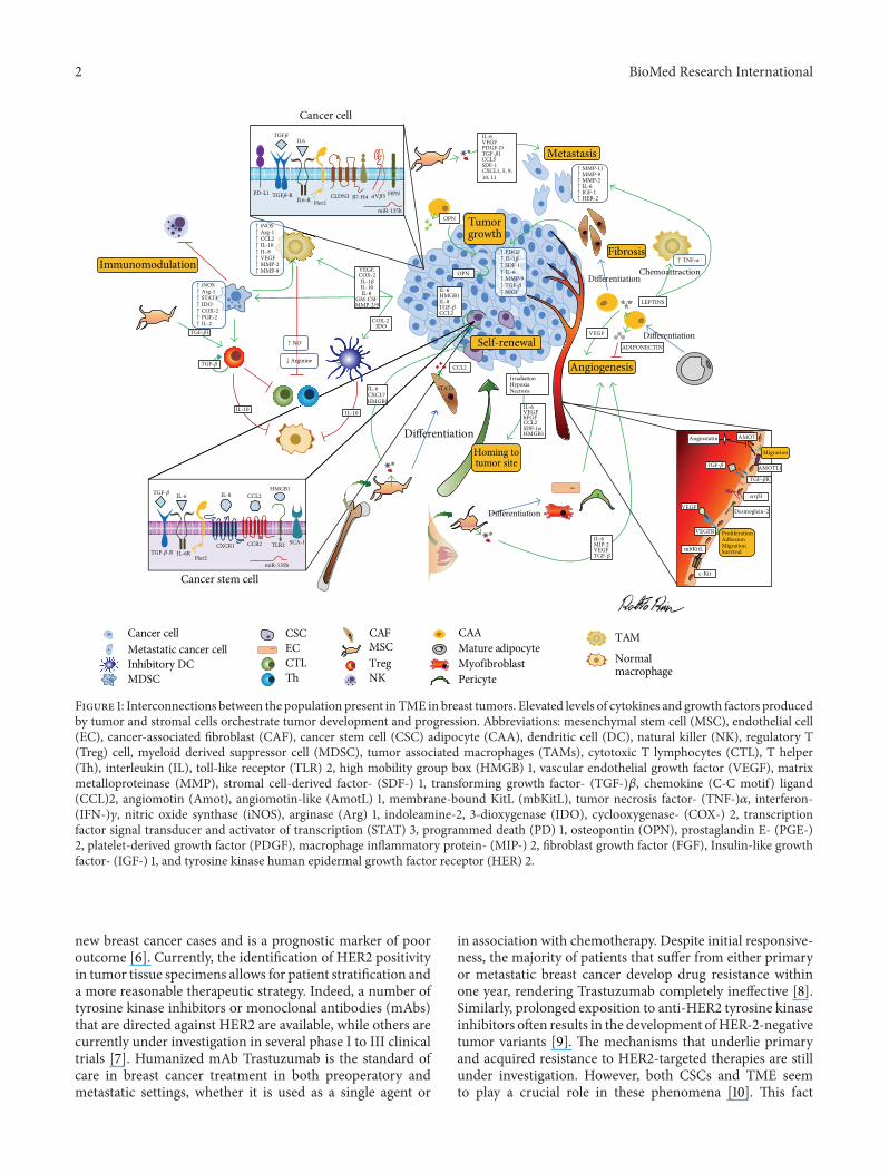
Solid tumors are currently considered to be organ-likestructures, composed of cancer cells and other cells thatsupport tumor development. While deep understanding ofcancer cells has been reached, less light has been shed on thecell populations that make up the tumor microenvironment(TME), as they have been ostracized for several decades andare only now being reappraised as a driving force for tumorpathogenesis. TME is composed of cells—such as inflamma-tory cells, mesenchymal stem cells (MSCs), endothelial cells(ECs), cancer-associated fibroblasts (CAFs), and adipocytes(CAAs)—and soluble factors, cytokines, and the extracellu-lar matrix (Figure 1) that bidirectionally communicate withcancer cells. This continuous and finely tuned interplaycan promote cancer outbreak, sustain tumor developmentand invasion, defend a tumor from host immunity, fostertherapeutic resistance, and provide niches for cancer stem
cells (CSCs) and dormantmetastases [1]. In this respect, TMEis now considered to be a good target for anticancer therapies,as it provides the opportunity to perturb the delicate balancethat promotes tumor progression. In fact, similarly to tumorcells [2], TME is now thought of as the source of a broadrange of targets, of which the most promising are tumor-associated antigens that play a key role in cancer developmentand progression, called oncoantigens (OAs) [3]. We haverecently classified OAs according to cellular localization[4]: Class I (cancer cell surface antigens), Class II (solubleantigens and antigens expressed in the TME), and ClassIII (intracellular proteins expressed by cancer cells). Theyare currently emerging as ideal targets for a very specificanticancer treatment, as demonstrated by several studies inpreclinical models [3].
HER2 represents the prototypic Class I OA and is foundto be overexpressed in a variety of human cancers [5]. HER2amplification or overexpression is found in 15–20% of all
Hindawi Publishing CorporationBioMed Research InternationalVolume 2014, Article ID 534969, 16 pageshttp://dx.doi.org/10.1155/2014/534969
2 BioMed Research International
Immunomodulation
Angiogenesis
Metastasis
Fibrosis
Differentiation
Differentiation
Tumorgrowth
LEPTINS
ADIPONECTIN
Chemoattraction
OPN
OPN
Self-renewal
HMGB1
CCL2
IL6-R Her2CLDN3 B7-H4
miR-135b
Cancer cell
IL6
ProliferationAdhesionMigrationSurvival
Migration
Angiostatin
Desmoglein-2VEGF
VEGFR
AMOT
c-Kit
mbKitL
Differentiation
DifferentiationHoming totumor site
IL-6VEGFbFGFCCL2
HMGB1
IrradiationHypoxiaNecrosis
IL-6MIP-2VEGF
IL-6CXCL7HMGB1
Her2
CXCR1 CCR2 TLR2 SCA-1
miR-135b
Cancer stem cell
IL-8 CCL2HMGB1
CCL2
STAT3
IDO
IL-10 IL-10
Cancer cellMetastatic cancer cell
CSC CAFMSC
CAAMature adipocyte
TAM
NormalmacrophageInhibitory DC
MDSCCTLTh
TregNK
MyofibroblastPericyte
HPN
VEGF
EC
AMOTL1
PD-L1 TGF𝛽-R
IL-6
IL-6RTGF-𝛽-R
TGF-𝛽R
𝛼V𝛽3
TGF𝛽
↑ iNOS↑ Arg-1↑ CCL2↑ IL-10↑ IL-8↑ VEGF↑ MMP-2↑ MMP-9
↑ iNOS↑ Arg-1↑ STAT3↑ IDO↑ COX-2↑ PGE-2↑ IL-2
TGF-𝛽1
TGF-𝛽
TGF-𝛽
TGF-𝛽
TGF-𝛽
↑ NO
↓ Arginine
VEGF,
COX-2
COX-2IL-1𝛽IL-10IL-6 IL-6
IL-8GM-CSFMMP-2/9
↑ TNF-𝛼
TGF-𝛽
IL-6VEGFPDGF-DTGF-𝛽1CCL5SDF-1CXCL1, 5, 9,10, 11
↑ MMP-11↑ MMP-9↑ MMP-2↑ IL-6↑ IGF-1↑ HER-2
↑ PDGF↑ IL-1𝛽↑ SDF-1↑ IL-6↑ MMP-9↑ TGF-𝛽↑ bFGF
SDF-1𝛼
𝛼v𝛽3
Figure 1: Interconnections between the population present in TME in breast tumors. Elevated levels of cytokines and growth factors producedby tumor and stromal cells orchestrate tumor development and progression. Abbreviations: mesenchymal stem cell (MSC), endothelial cell(EC), cancer-associated fibroblast (CAF), cancer stem cell (CSC) adipocyte (CAA), dendritic cell (DC), natural killer (NK), regulatory T(Treg) cell, myeloid derived suppressor cell (MDSC), tumor associated macrophages (TAMs), cytotoxic T lymphocytes (CTL), T helper(Th), interleukin (IL), toll-like receptor (TLR) 2, high mobility group box (HMGB) 1, vascular endothelial growth factor (VEGF), matrixmetalloproteinase (MMP), stromal cell-derived factor- (SDF-) 1, transforming growth factor- (TGF-)𝛽, chemokine (C-C motif) ligand(CCL)2, angiomotin (Amot), angiomotin-like (AmotL) 1, membrane-bound KitL (mbKitL), tumor necrosis factor- (TNF-)𝛼, interferon-(IFN-)𝛾, nitric oxide synthase (iNOS), arginase (Arg) 1, indoleamine-2, 3-dioxygenase (IDO), cyclooxygenase- (COX-) 2, transcriptionfactor signal transducer and activator of transcription (STAT) 3, programmed death (PD) 1, osteopontin (OPN), prostaglandin E- (PGE-)2, platelet-derived growth factor (PDGF), macrophage inflammatory protein- (MIP-) 2, fibroblast growth factor (FGF), Insulin-like growthfactor- (IGF-) 1, and tyrosine kinase human epidermal growth factor receptor (HER) 2.
new breast cancer cases and is a prognostic marker of pooroutcome [6]. Currently, the identification of HER2 positivityin tumor tissue specimens allows for patient stratification anda more reasonable therapeutic strategy. Indeed, a number oftyrosine kinase inhibitors or monoclonal antibodies (mAbs)that are directed against HER2 are available, while others arecurrently under investigation in several phase I to III clinicaltrials [7]. Humanized mAb Trastuzumab is the standard ofcare in breast cancer treatment in both preoperatory andmetastatic settings, whether it is used as a single agent or
in association with chemotherapy. Despite initial responsive-ness, the majority of patients that suffer from either primaryor metastatic breast cancer develop drug resistance withinone year, rendering Trastuzumab completely ineffective [8].Similarly, prolonged exposition to anti-HER2 tyrosine kinaseinhibitors often results in the development ofHER-2-negativetumor variants [9]. The mechanisms that underlie primaryand acquired resistance to HER2-targeted therapies are stillunder investigation. However, both CSCs and TME seemto play a crucial role in these phenomena [10]. This fact
BioMed Research International 3
emphasizes the need to consider cancer cells and their TMEas a whole when designing effective anticancer therapies andtells us that targeting a single OA is not sufficient to freezetumor progression, a possibility that can only be exploredthanks to the availability of appropriate in vivo cancermodels.
The identification of appropriate murine models thatare able to mimic most of the features of a human canceroffers considerable potential to give advantages in the racetowards the clinic. In particular, the availability of tumor-transplantable models and genetically engineered mammarycancer-prone mice has allowed laboratories to decipher themost important mechanisms involved in mammary tumordevelopment and progression, thus permitting current ther-apies to be refined. A great deal of data has been obtainedby our group from transgenic mice, called BALB-neuT,that overexpress the rat HER2 (neu) oncogene under themouse mammary tumor virus (MMTV) promoter [11], withthis very fact in mind. These mice spontaneously developmammary carcinomas with 100% penetrance [12] and displaya histopathologically [13] and transcriptionally [14] well char-acterized course that closely recapitulates many features ofhuman breast carcinogenesis. In virtue of the high homologyof BALB-neuT tumors to humanHER2positive breast cancer,this is an ideal model to use when setting up new anticancertherapies. Actually, BALB-neuTmice and the cell line derivedfrom a BALB-neuT adenocarcinoma (TUBO cells) haveprovided us with a fascinating tool and one that is used inmany laboratories worldwide to deepen current knowledgeof the pathogenic mechanisms that promote HER2 positivetumor growth and consequently elaborate more efficaciousantitumor strategies. We herein discuss the lessons learnedabout TME, HER2, and other OAs from BALB-neuT miceand how this knowledge can help develop a winning strategyagainst cancer.
2. The Urgency of Defining the MostPromising TME-Associated OAs
Neoplastic transformation is a multistep process whichinvolves specific proteins and regulatory pathways at eachstage. The identification of the genes that constitute thedriving force of cancer progression is an extraordinaryopportunity to gain an advantage over cancer. HER2 rep-resents a paradigm of this conception; its expression at theneoplastic stage, its overexpression in established tumors,and its causal role in cancer progression [14] make it theideal immunological target. This observation has paved theway for the development of new immunologically basedtherapies against neoplastic cells that overexpress HER2,which have made some important clinical achievements[15]; the U.S. Food and Drug Administration (FDA) hasapproved mAbs that target HER2, such as Trastuzumaband Pertuzumab, and several drugs (i.e., TDM1 and ARRY-380) [16], which have prolonged the disease-free survivalrates in patients with metastatic HER2 positive breast cancer[17] and are currently under investigation in clinical trials.However, the majority of patients treated with these agentsdevelop resistance within one year of treatment, resulting in
disease progression, recurrence, and reduced overall survival[18]. Similar results have also been obtained using activeimmunotherapy against HER2 in preclinical models [19].Theefficacy ofDNAvaccines targetingHER2 inBALB-neuTmice[20] relies mostly on the direct activity of vaccine-elicitedAbs [21–23] and is strictly dependent on the tumor stageat the time of vaccination; the sooner the vaccination isperformed, the better the outcome [24]. When the vaccine isadministered to a still healthy BALB-neuT mouse, repeatedboosts keep it tumor free for a period of time that maywell equate to its natural life span. However, when the samevaccine is administered to a mouse in a more advanced stageof microscopic lesions, the appearance of palpable tumorsis only slightly delayed. This suggests that targeting a singleoncoantigen is not sufficient to freeze tumor progression,especially when it is applied to patients that suffer fromadvanced cancer, as commonly happens in the clinical setting[12].
This partial failure of anti-HER2 treatment suggests thatsome key elements that drive mammary carcinogenesis muststill be sought out and not only on the tumor cells themselves;the best chance of defeating cancer that we have is offered bytargeting both cancer cells and TME. TME can dynamicallycontrol cancer progression thanks to its continuous interplaywith cancer cells [25]. Therefore, the identification of addi-tional OAs that are expressed by either tumor or stromalcells surrounding HER2 positive lesions is urgently neededif we are to develop a combined andmore efficient anticancerapproach which may prevent the development of the veryresistance to anti-HER2 therapy that is responsible for tumorrelapse [26].
To address this point, we performed a transcriptionprofile analysis of BALB-neuT preneoplastic and invasivelesions, integrated with ameta-analysis of data obtained fromhealthy human and neoplastic specimens. Of the 46 putativeOAs identified [27], B7-H4 [28], Claudin 3 [29], Hepsin[30], CD52 [31], and Desmoglein 2 [32] are Class I OAs,expressed on the plasma membrane of cancer and TME cellsand therefore constitute promising targets for vaccination.Class II OAs are another group of identified OAs andincludes cytokines and chemokines copiously released in theTME. These molecules play important roles in establishingthe strictly tuned relationship between tumor and stromalcells whose balance is critical for tumor development andprogression, as will be discussed in the following sections ofthis review.Moreover, this analysis led us to identifyingmanyClass III OAs that belong to signal transduction pathwaysreported to be deregulated in breast and other cancers, suchas mitogen activated protein kinase (MAPK) [33], Survivin[34], Aurora kinase [35], and src pathway molecules [36].It is worth noting that some of these networks seem to beregulatory keys of therapeutic resistance, such as Survivin[37], Topoisomerase II 𝛼 [38], Desmoglein 2 [39], BCL2-interacting killer [40], and ribonucleotide reductase M2polypeptide [41]. In addition, several identified proteins havea role in CSC self-renewal, which has been demonstrated inthe cases of maternal embryonic leucine zipper kinase [42],transcription factor AP-2 𝛾 [43], the microtubule associatedTPX2 protein [44], and Aurora kinase A [44]. At present, our

4 BioMed Research International
efforts are focused on the characterization of some of thesetargets and our final goal is the setup of new DNA vaccinesthat will be tested in BALB-neuT mice in association withanti-HER2 vaccination, in order to improve the vaccination’sefficacy against advanced tumor and metastases. A moredetailed analysis of OAs that are selectively expressed bythe various populations that constitute TME may end upproviding us with a sort of tumor Rosetta Stone which couldhelp unveil the reciprocal connection between tumor, CSCs,and stroma.
As reported in several clinical studies, the expression ofnoncoding genes, such as microRNAs (miRNAs), correlateswith cancer relapse and metastasis formation [45]. SeveralmiRNAs contribute to tumor progression in virtue of theirability to posttranscriptionally modulate the expression ofoncogenes or oncosuppressors.They can act directly onTME,regulating both the survival of more differentiated cancercells and the maintenance of a CSC phenotype [46] andcontrolling neoangiogenesis during tumor progression [47].Results from experimental studies, which have been strength-ened by the human cancer miRNA expression profile, haveled researchers to the identification of miRNAs as potentregulators of the crosstalk between cancer and stromal cells[48]. Even if miRNAs cannot be considered oncoantigensbecause of their lack of immunogenicity, the identificationof miRNAs, which are differentially expressed in the tumor,can lead to the identification of their target genes as potentialoncoantigens or oncosuppressors, nevertheless [19].
Of note among the miRNAs that have recently beenidentified is the strong upregulation of miR-135b which hasbeen found in invasive mammary BALB-neuT carcinomas;acting on its targets, midline 1 (MID1) and mitochondrialcarrier homolog 2 (MTCH2), it regulates CSC stemness invitro and cancer cell metastatization in vivo [49]. This newlyunveiled role for miR-135b in mammary carcinogenesis,as observed in other tumors such as colon cancer [50],osteosarcoma [51], ependymoma [52], and hepatocellularcarcinoma [53], can provide the basis for the exploration ofmiR-135b, MID1, and MTCH2’s potential as new therapeutictargets in mammary carcinogenesis.
3. CSCs on Stage
The scientific spotlight has very recently been pointed onCSCs, the subpopulation of cells endowed with self-renewalpotential and refractoriness to chemo- and radiotherapy thatare capable of sustaining tumor growth and progression bygiving rise to the heterogeneous population of tumor cellsfound within a tumor [54]. Even though the initial idea ofCSCs as static entities [55] has been overtaken [56], it is wellaccepted that they control cancer development and progres-sion in amanner that is guided by environmental factors [57].CSCs are thought to reside in a highly specialized niche thatis made up of stromal, endothelial, and more differentiatedtumor cells that stimulate CSC survival and stemness via cellto cell contact, paracrine, and other signals [58]. A centralrole is played here by interleukin- (IL-) 6, which is producedby CSCs and noncancerous cells, MSCs, and immune cells.
IL-6 promotes CSC self-renewal, the recruitment of MSCsand immune cells, and the preservation of an inflammatorystate that favors tumor growth. Moreover, IL-6 promotesthe conversion of more differentiated tumor cells into CSCs,inducing the epithelial-to-mesenchymal transition (EMT).Recently, it has been shown that HER2 overexpression inbreast CSCs increases IL-6 secretion [59] which is involvedin Trastuzumab resistance [60].
We have recently demonstrated that an autocrine loopinvolving toll-like receptor 2/high mobility group box-1/NF𝜅B (TLR2/HMGB1/NF𝜅B) induces the enhanced secre-tion of vascular endothelial growth factor (VEGF) and IL-6 in Sca1+ [61] CSCs, derived from BALB-neuT TUBOcells, that in turn activates the transcription factor signaltransducer and activator of transcription 3 (STAT3), thuspromoting CSC self-renewal [62]. This pathway also inducesthe secretion of transforming growth factor- (TGF-)𝛽, acytokine that induces EMT and the secretion of matrixcomponents that favor metastatization [63]. Moreover, TGF-𝛽 recruits endothelial cells and promotes their proliferation,enhancing angiogenesis [64].Therefore, HER2 positive CSCspromote their own self-renewal, by upregulating TLR2 andsecreting its endogenous ligand HMGB1, and generate afavorable microenvironment for tumor progression. This is avery important observation sinceHMGB1 is not only secretedby CSCs but also secreted by activated dendritic cells (DCs)[65] and necrotic cells [66] and thus is one of the mostimportant molecules driving tumor escape from cytotoxictreatment.
IL-6 stimulates CSCs, MSCs, and fibroblasts and causesthem to secrete IL-8, another key cytokine that promotesCSC self-renewal. It is worth noting that HER2 positiveCSCs overexpress IL-8 receptors CXCR1/2 [67], which inturn induce HER2 phosphorylation and the activation ofits downstream signaling pathway, generating a positivefeedbackmechanism that promotes CSC expansion [68].Theinhibition of CXCR1, either by mAbs or specific inhibitors,reduces CSC self-renewal, induces cell apoptosis, and inhibitsmetastatization in breast cancer, indicating that this receptormay be a promising target for combined anticancer therapies[69]. Similar IL-6-dependent upregulation is observed inthe chemokine (C-C motif) ligand (CCL) 2 (also known asmonocyte chemotactic protein-1, MCP-1), whose productionis induced by IL-6 in both tumor cells and stromal cells andthat supports the expansion of the CSC compartment by acti-vating the Notch1 signaling pathway [70]. We demonstrated,by microarray analysis, that CCL2 expression increases inBALB-neuT mice as carcinogenesis progresses [71], and itscausal role in cancer development was further supported bythe observation that BALB-neuTmice, which were knocked-out (KO) for CCL2, displayed prolonged survival over BALB-neuT mice wild-type (WT) for this chemokine [72].
The characterization of all the cytokine networks thatconnect CSCs, tumor cells, and stromal cells may pave theway for new therapeutic strategies and provide diagnosticand prognostic markers for patients. In this regard, manyclinical studies have shown that high serum levels of IL-8 and IL-6 correlate with poor prognosis in breast cancer
BioMed Research International 5
patients [73, 74]. Therefore, the design of specific cytokinereceptor inhibitors and the assessment of their efficacy inclinical settings may be a source of great potential for futureresearch.
4. Fighting against Proangiogenic OAs
Vascular ECs thoroughly govern angiogenesis, a process thatsupports the growth of many kinds of solid tumors includingbreast cancer, providing nutrients and oxygen to proliferatingcells, thereby allowing cancer cells to invade tissues anddevelop metastases. Tumor cells have been observed topreferentially align towards and associate with ECs, evenprior to the angiogenic switch [75]. Thanks to this strategictidiness, ECs and tumor cells can bidirectionally communi-cate through a complex network of both soluble and insolublesignaling molecules that drive cellular differentiation andfind ways to foster the tumor. Moreover, ECs are the mostimportant interface between circulating blood cells, tumorcells, and extracellular matrix and play a pivotal role incontrolling leukocyte recruitment and tumor cell behaviorduring angiogenesis.
A great deal of effort has been poured into attemptsto block tumor angiogenesis. In this respect, VEGF-A isnowadays the most renowned therapeutic target. The inter-action between VEGF ligands and their EC expressed recep-tors stimulates angiogenesis and promotes EC permeability,survival, migration, and the invasive potential of cancercells [76]. Bevacizumab is a recombinant humanized mAbdeveloped against VEGF-A [77], which has been broadlystudied in phase III clinical trials and is now FDA-approvedfor the treatment of metastatic colorectal cancer, nonsmallcell lung cancer, and breast cancer [78]. Other drugs thatinhibit the tyrosine kinase activity of VEGFRs, like sunitinib[79], sorafenib [80], axitinib [81], pazopanib [82], vande-tanib [83], cabozantinib [84], tivozatinib [85], and linifanib[86], have been developed. Sorafenib has been approved forthe treatment of unresectable hepatocellular carcinoma andadvanced renal cell carcinoma (RCC), whereas sunitinib hasbeen approved for the treatment of gastrointestinal stromaltumors andmetastatic RCC, but onlymodest benefit has beenobserved in other types of cancer [87].
Despitemany steps forward in the setup of antiangiogenicprotocols being made, the development of tumor resistanceand the occurrence of relapse in a high percentage of patientshave prompted clinicians and researchers to join forces andfind new targets for the development of more efficacioustherapies. For these reasons, the immune-targeting of OAsexpressed on ECs seems to be a successful direction to movetowards. As described below, we have tested various DNAvaccination strategies that target tumor angiogenesis; all thesevaccines have demonstrated high efficacy without any toxiceffect, further stressing the therapeutic potential of targetingtumor ECs in HER2 positive tumors.
Of the class II OAs found to be overexpressed in tumorECs during BALB-neuT cancer progression [88], the mostpromising is angiomotin (Amot), a member of the Motinprotein family. Using a construct that encodes the kringle
domains 1–4 of angiostatin to screen a yeast two-hybridplacenta cDNA library for angiostatin-binding peptides [89],Amot was originally identified as one of the angiostatinreceptors. Amot is normally expressed on ECs, where itexerts its proangiogenic activity and stimulates ECmigrationduring angiogenesis [90]. Amot is overexpressed comparedto normal tissues in human breast tumors and its presencecorrelates with poor prognosis and metastatic disease [90].These findings suggest that Amot has an important roleto play during breast tumor progression and may be anoptimal target for anticancer therapy [91]. In virtue of thesefeatures, we decided to elicit an immunological responseagainst Amot, by means of DNA vaccination, in mice thatbear microscopic invasive mammary cancers. This strategywas successfully applied in BALB-neuT mice as well as in thePyMTmousemodel of breast cancer, in which carcinogenesisis driven by the polyoma middle T oncoantigen [92]. Thetherapeutic effect of anti-Amot vaccination was mediated bythe induction of specific antibodies that induced increasedtumor vessel permeability, which, in turn, resulted in bothan increase in chemotherapy efficacy and major epitopespreading, which was accompanied by the induction of aspecific anti-HER2 antibody response that further contrastedtumor growth [93].
Another member of the Motin family, angiomotin-like 1(AmotL1), is an attractive target for antitumor interventions.AmotL1 is endowed with proangiogenic properties that affectEC polarization, directional migration, and the stability oftight junctions during angiogenic sprouting; it may com-pensate for the absence of Amot and vice versa [94]. Eventhough our preliminary data indicate that DNA vaccinationagainst AmotL1 is not effective in the prevention ofmammarytumor appearance in BALB-neuT mice, encouraging datahave come from a combined DNA vaccine against HER2 andAmotL1. Even more promising results have been obtainedusing a combined DNA vaccine against HER2, Amot, andAmotL1 (Barutello G et al., unpublished data). This kind ofvaccination exploits the synergistic effect which stems fromthe combined action of antibodies which target both the ECsof neoformed tumor vessels and the tumor cells themselves.
Membrane-bound KitL (mbKitL), which is involved inthe c-Kit/KitL system required for tumor angiogenesis [95],is an additional promising target for antiangiogenic cancerimmunotherapy. mbKitL is expressed on tumor ECs and isessential for providing themwith survival signals, as is clearlyexploited in the role that c-Kit signaling network plays inmaintaining breast cancer cells [96]. A DNA vaccine thattargets mbKitL is able to inhibit the growth of a mouse HER2positive transplantable tumor; vaccination impairs tumorvessel formation and stabilization and thus interferes withtumor cell-derived VEGF bioavailability [97].
Besides representing good targets for anticancer thera-pies, antigens expressed on tumor ECs may also be exploitedfor tumor diagnosis. In this context, we have recentlydemonstrated that both ECs and cancer cells in mammarytumors arising in BALB-neuT mice express 𝛼V𝛽3 integrin,a receptor for several extracellular matrix proteins whichharbor an arginine-glycine-aspartic acid (RGD) sequence[98]. 𝛼V𝛽3 integrin is widely considered to be a marker of
6 BioMed Research International
the angiogenesis, tumor progression, and invasion of differenttypes of cancer. Since its level of expression correlates withcancer progression [99], we have developed a probe for theoptical imaging detection of 𝛼V𝛽3 integrin and have shownthat it can successfully detect microscopic in situ carcinomasinBALB-neuTmice, therefore proving itself to be a promisingtool for the early diagnosis of breast cancer [98].
5. The Controversial Role of Inflammation andImmune Cells in the TME
Despite the fact that natural immune surveillance mecha-nisms are activated during the early stages of BALB-neuTcarcinogenesis [100–103], tumors finally acquire the threeimmune hallmarks required to progress: the ability to thrivein a chronically inflamed TME, to suppress immune reactiv-ity, and to evade immune recognition [104].Thefight betweennatural immune surveillancemechanisms and these acquiredcapabilities is mirrored by the important, yet controversial,role that immune cell infiltrates play in the TME. Thetumor stroma of BALB-neuT mice is infiltrated by CD4and CD8 T lymphocytes and a few B, natural killer (NK),and 𝛾𝛿 T lymphocytes, but mostly by regulatory T (Tregs)cells, myeloid derived suppressor cells (MDSCs), and tumorassociated macrophages (TAMs) that are recruited into TMEin response to inflammatory molecules and cytokines beingreleased in the tumor milieu [105, 106].
The acquired ability of BALB-neuT tumors to thrive ina chronically inflamed microenvironment has been high-lighted by microarray analyses that have shown the occur-rence of an upregulation in four transcriptional networks,in advanced as compared to preneoplastic lesions, whosehub genes code for proinflammatory cytokines IL-1𝛽, tumornecrosis factor- (TNF-)𝛼, interferon- (IFN-)𝛾, and CCL2[71]. The final outcome of the activation of these fournetworks is tumor promotion; however, how each individualnetwork influences tumor progression is neither simple norunequivocal. For instance, increased IFN-𝛾 release in TMEduring tumor progression appears to play a major tumorinhibitory role and is a marker of the M1 TAMs that expressimmunostimulatory, antiangiogenic, and tumoricidal func-tions [107]. Accordingly, IFN-𝛾 KO BALB-neuTmice displayfaster tumor progression, associated with a more intensetumor angiogenesis [71, 108, 109].Moreover, chronic systemicadministration of recombinant IL-12 in BALB-neuT miceinduced high and sustained IFN-𝛾 production, as detected inthe sera of treated mice that in turn caused a delay in tumoronset and a reduction in the number of mammary glandsaffected by the tumor [109, 110]. The role that the other threenetworks play in tumor progression is the opposite. They caninitially show antitumor activity, but the incipient tumor soonuses them to provide itself with a shortcut for progression.In reality, the activation of CCL2 is directly associated withenhanced progression [72], as discussed above. Similarly,increases in IL-1𝛽 and TNF-𝛼 in TME may favor cancerprogression either directly [71, 111] or by recruiting suppressorcells [112, 113].
A tumor’s ability to exploit inflammation to its ownbenefit is strictly related to the second immune hallmarkof cancer, the capability to suppress the immune responsedirectly or via the recruitment of suppressor cells [104].IL-1𝛽 released by stromal cells together with other tumor-derived factors, including granulocyte macrophage colony-stimulating factor (GM-CSF), cyclooxygenase 2 (COX-2), IL-6, and VEGF, induce the accumulation and expansion ofMDSCs [112, 113] by triggering Janus kinase (JAK)/STAT3pathways [114]. MDSCs are a phenotypically heterogeneouspopulation with an immunosuppressive capacity that are,in normal conditions, generated from the bone marrowand rapidly differentiates into mature DCs, macrophages,or granulocytes, while, in cancer bearing patients, presenta partial block of maturation [115]. In BALB-neuT tumors,VEGF was detected in the supernatant from primary tumorcultures and from tumor cell lines as well as in the seraof BALB-neuT tumor-bearing mice. A possible explanationmay lie in the increase of matrix metalloproteinase-(MMP-)9 within the tumor mass, as previously shown [116], thatmediates the release of growth factors, such as VEGF, stromalcell-derived factor- (SDF-) 1, and mbKitL [117]. Accordingly,any interference with VEGF ormbKitL activity, besides ham-pering the angiogenic process [97, 118], has been reported toinduce MDSC shrinkage [97, 119].
MDSCs exhibit immunosuppressive functions that occurvia multiple mechanisms, such as inducible nitric oxidesynthase (iNOS) and arginase-1 (Arg-1) production, whichsuppresses the T-cell immune response in TME via therelease of nitric oxide and reactive oxygen species thatcause T cell receptor (TCR) nitration and T cell apoptosisand the depletion of L-arginine required for T cell func-tions [120]. As indoleamine-2, 3-dioxygenase (IDO) appearsto be involved in MDSC-mediated T cell inhibition [121]and cyclooxygenase- (COX-) 2 is required to induce, viaprostaglandin E- (PGE-) 2, Arg-1 expression by MDSCs[122, 123], a considerable amount of effort is going intoinhibiting these molecules [124]. In this respect, we aretesting a therapeutic protocol that consists of the concomitantadministration of anti-HER2 DNA vaccines and plasmidsthat code for IDO [125] or COX-2 or short hairpin (sh)RNAsin BALB-neuT mice [24].
In order to curb the significant MDSC contributionto suppressing the immune system, we have looked foradditional targets that these cells express both in tumorbearing mice and in cancer patients. As discussed above,B7-H4, a member of the B7 family, has been identified asbeing overexpressed in BALB-neuT mouse invasive lesionsand appears to be an excellent target candidate, thanks toits critical role in the regulation of antigen specific immuneresponses [3]. Indeed, within TME, the expression of B7-H4 by tumor cells and MDSCs seems to be involved inthe inhibition of the T cell response to tumor associatedantigens [126]. In the light of these considerations, we aredeveloping DNA plasmids that code for both HER2 and B7-H4 shRNAs, and we propose an evaluation of their efficacyin the inhibition of mammary carcinogenesis (Macagno M,unpublished data). Another important pathway that con-tributes to tumor mediated immune suppression is found in
BioMed Research International 7
the CD28 family member, programmed death 1 (PD-1) andits ligand PD-L1 [127]. PD-L1 is expressed by both MDSCsand tumor cells [128] and its interaction with activatedT cell expressed PD-1 promotes T cell tolerance by suppress-ing their cytotoxic capacity and cytokine secretion [127]. Wewere among the first to show that the PD-1 blockade resultsin an increased response to antitumor vaccination. In theseexperiments BALB-neuTmice were vaccinated against HER2and concomitantly treated with the administration of anti-PD-1 mAb BAT [129].
In response to IL-1𝛽 stimulation, MDSCs also producethe suppressive cytokine IL-10 [130] which acts on TAMsinducing their reprogramming and polarization towardsan M2 phenotype. M2 TAMs support tumor progressionthrough the release of immunosuppressive (i.e., CCL2 and IL-10), proangiogenic (i.e., IL-8 and VEGF), and tissue remod-eling (i.e., MMP-2 and MMP-9) factors. Their expansion inbreast cancer tissues has been correlated with poor prognosis[131]. In BALB-neuTmiceM2TAMs are themain tumor infil-trating population [105]. The administration of zoledronicacid to BALB-neuT mice can revert M2 polarization byinterferingwith themevalonate pathway and thus hamper IL-10 and VEGF production, recovering the release of IFN-𝛾 inthe mammary glands of treated mice [105].
HER2 and the other OAs expressed by mammary tumorsin BALB-neuT mice are self-molecules toward which theimmune system is tolerant [132]. As a consequence, thepredominant effector T-cells in the TME are presumablyconstituted of low avidity OA-specific T cells whose activityis inhibited by Tregs that first expand in the spleen and tumordraining lymph nodes during cancer progression and in TMEin later phases [100, 132, 133]. This situation reproduces whatnormally happens in tumor bearing patients [134] and ispart of the ability to suppress immune reactivity that thetumor acquires during progression [104]. Indeed, naturalimmune surveillance somehow counteracts Treg expansionin the early phases of carcinogenesis in BALB-neuT mice.In complement C3 KO BALB-neuT mice, tumor progressionoccurs earlier and this is associated with the increasedexpansion of Treg cells over complement competent BALB-neuT mice [102]. This increased Treg expansion is promptedby a lack of C3a and C5a, whose receptor signaling is requiredduring the early events of effector T cell activation [135]and negatively modulates Treg function by inducing FoxP3downregulation [136]. Its absence in BALB-neuTC3KOmicedeflects naıve T cells into Treg [137] and potentiates theirfunction [136].
The down modulation of MHC class I (MHC I) [138]is the mechanism most frequently exploited by tumor cellsto escape from immune recognition [139]. It is intriguingthat an inverse correlation exists between HER2 overex-pression and the expression of MHC I and of the com-ponents of the antigen-processing machinery [140]. MHCI down modulation, albeit incomplete, means that cancercells are more susceptible to NK cell-mediated lysis, if NKreceptor activating ligands are present. This may have animpact on cancer progression at least in the initial stages ofcarcinogenesis. The fundamental role that NK cells play inhampering the expansion of incipient BALB-neuT tumors
has been investigated in perforin (PFP) KO BALB-neuTmice, as the majority of NK mediated protection relies onthe release of PFP on target cells. In fact, both female [103]and male [141] BALB-neuT PFP KO mice show fourfoldincreases in mammary carcinoma incidence. Nevertheless,preliminary results also indicate that advanced BALB-neuTtumors downregulate the expression of ligands that activateNK receptors (Lanzardo S, unpublished data), suggesting thatadvanced tumors reach a balance between a loss of sensitivityto CD8+ T cell killing and the maintenance of NK-cell-inhibitory specificities. We are now evaluating the expressionof MHC I and of some NK ligands in TUBO-derived CSCsto assess whether NK cells recognize and more efficiently killCSCs than their differentiated counterparts, as has alreadybeen shown for colon cancer-derived CSCs [142].
6. Role of Adipocytes and Fibroblasts inBreast Cancer Progression
While immune cells are well recognized as major players inthe orchestration of a permissive TME, other cell populationshave only recently been recognized as active parts of thetumor promoting ability of TME. These include CAAs andFACs.
Besides its classical definition as a fat reservoir, adiposetissue is now considered to be a fully functioning endocrineorgan [143] that secretes growth factors and cytokines, knownas adipokines, which are involved in angiogenesis, immunity,and endocrine signaling [144]. Adipocytes enshroud themammary gland, regulating epithelial cell growth during thehormonally controlled courses of mammary gland develop-ment, from pubertal maturation to involution after lactation[145].
The understanding of the important, but still underes-timated, role of adipocytes in cancer stems from severalstudies which highlight the anatomical proximity of manytumors to adipose tissues and point to the positive correlationbetween obesity and higher cancer risk [146–149]. Adipocytescan, under the pressure of cancer cell stimuli, abdicate theirphysiological role in favor of tumor promoting activities inbreast cancers that grow in an adipose tissue dominatedcontext. In this way they becomeCAAs that exhibit decreasedlipid content, reduced adipocytes marker expression, and anoverexpression of proinflammatory cytokines and MMPs,such as MMP-11 and MMP-9 [150, 151]. It is worth notingthat MMP-9 has been identified as being overexpressed inBALB-neuT mammary cancer which would seem to point toits important role during tumor progression.
A number of studies have shown that CAAs support andexpedite breast cancer progression [152–154] by providingproinflammatory cytokines, such as IL-6, TNF-𝛼, and reac-tive oxygen species [155]. On the other hand, IL-6 in breastTME seems to stimulate the proinvasive effects of CAAs,besides promotingCSC self-renewal as discussed above [150].Moreover, CAAs in TME can differentiate in fibroblast-like cells that, together with other stromal cell populations,participate in the generation of dense collagenous stroma, the
8 BioMed Research International
so called desmoplastic response, typically observed in breastcancer [156].
CAAs functions are mainly mediated by leptin andadiponectin, two functionally opposite members of theadipokine family, that seem to play a pivotal role in cancerprogression [157]. Leptin promotes tumor growth, elicitingthe activity of several signaling pathways such as insulin-like growth factor-1 (IGF-1) and HER2 and inducing theexpression of MMP-2, MMP-9, and VEGF, which finallypromote cell migration and metastatic spreading [158, 159].Furthermore, leptin exerts a chemoattractant effect onmacrophages and monocytes [160] and stimulates them toproduce the inflammatory cytokine TNF-𝛼 that in turnmanifests proangiogenic activity [161]. On the other hand,adiponectin acts as an antiangiogenic and anti-inflammatoryfactor that is able to repress proliferation and induce apop-tosis in breast cancer cells [147, 162]. Interestingly, somestudies have found that caloric restriction can exert ananticancer effect via alterations in systemic IGF-1 and NF-𝜅Blevels [163].
Altogether these data suggest that the recently discov-ered therapeutic potential of adipocytes could open newand promising perspectives in breast cancer treatment. Oneexample of this comes from the preclinical experience gainedwith adipokine osteopontin (OPN), also called “early Tcell-activation gene 1,” a multifunctional component of theextracellular matrix that has been linked to a plethora ofautoimmune diseases [164]. OPN has very recently beenrediscovered as a diagnostic and prognostic marker inHER2 positive breast cancer [165] and one whose abnormalexpression in patients is linked to poor prognosis [166].It has also been proposed that the autocrine productionof OPN by tumor cells may be an important factor thatallows invasion and survival to occur [167]. In fact, theinteraction between extracellular matrix deposited OPN andcell adhesion molecules, such as 𝛼V𝛽3 integrins which areoverexpressed in BALB-neuT tumors [98], increases boththe expression of VEGF in ECs, allowing neovascularization,and the activation of connective tissue growth factor andcysteine-rich angiogenic inducer 61(CYR61), which enhancesneovascularization and mammary tumor growth in vivo[168].
As previously mentioned, CAAs can differentiate intofibroblast-like cells that share many properties with CAFs[169]. CAFs promote tumor growth and invasion secretingproangiogenic factors (i.e., VEGF-A and MMP-9) [170],proinflammatory molecules (i.e., SDF-1, IL-6, and IL-1𝛽)[171], and several growth factors (i.e., TGF-𝛽, platelet-derivedgrowth factor, PDGF, and basic fibroblast growth factor,bFGF) [172, 173]. In particular, the aberrant production of IL-6 and CCL2 in mammary cancer activates STAT3 in CAFs,which finally sustains tumor-associated inflammation and isrequired for breast cancer cell migration [174]. Certainly, inBALB-neuTmice this network seems to be particularly inter-esting, as in a BALB-neuT mice knock-in for a constitutivelyactive Stat3 allele, we observed an earlier and more invasiveonset of mammary tumors [175].
7. MSCs Are Key Players in the TME Orchestra
Adult multipotent MSCs make for a fascinating TME popu-lation which is able to control the interplay between cancercells and tumor stroma. Physiologically, MSCs are locatedpredominantly in the bone marrow and contribute to themaintenance and regeneration of a variety of connectivetissues [176]. During injury and inflammation, they arerecruited to damaged sites via the release of solublemoleculesand operate in tissue remodeling [177].
MSCs also localize into different types of solid tumorswhich they first migrate towards then integrate into thetumor-associated stroma [178]. Recent studies have provideddirect evidence that MSCs are recruited in TME by a broadrange of soluble factors which are secreted by cancer cellsand CSCs, including IL-6 [179], VEGF and bFGF [180], CCL2[181], SDF-1𝛼 [182], and HMGB1 [183]. Moreover, stressfulconditions, such as irradiation [184], hypoxia [185] and,cellular damage [183], can enhance the recruitment of MSCsto the site of growing tumors. Once there,MSCs contribute tothe development of an active TME, in which bone marrow-derived MSCs generate CAFs, while local adipose tissue-derivedMSCs contributemainly to the vascular and fibrovas-cular stroma (pericytes, myofibroblasts, and ECs) [186]. Inaddition, MSCs interact with tumor cells and with all otherstromal cells through a broad range of signaling molecules,generating complex crosstalk whose net effect is to stimulatetumor progression. For example, MSCs can promote breastcancer neoangiogenesis, possibly thorough the secretion ofmacrophage inflammatory protein 2 (MIP-2), VEGF, TGF-𝛽, and IL-6 [187] and display potent immunomodulatoryproperties [188] that enable them to inhibit CTLs and NKcells by stimulatingTregs through the release of TGF-𝛽1 [189].
Conflicting data have led to the hypothesis that twoopposing immunological MSC phenotypes exist, one proin-flammatory and one immunosuppressive, which are depen-dent on the engagement of specific TLRs [190]. The role ofTLR2 is still debated, with some studies claiming that TLR2activation on MSCs inhibits their immunosuppressive prop-erties [191], while others argue that TLR2 stimulation doesnot affect this capability [192]. Notably, these considerationsare mostly based on in vitro experiments. Therefore, BALB-neuT mice may well be a suitable tool for the difficult taskof definitely clarifying the role of TLR2 in MSCs. Startingfrom our observation that TLR2 drives mammary CSC self-renewal [62], we are developing BALB-neuTmice that areKOfor TLR2, in which we would like to characterize the role ofTLR2 not only in CSCs but also in MSCs and other stromalpopulations.
MSCs are thought to contribute to CSC niche generation,thus regulating cancer cell stemness through multiple path-ways and secreted factors (i.e., IL-6 and CXCL7 [193], PGE-2[194], EGF, bFGF, bone morphogenic protein (BMP) 4, TGF-𝛽1, SDF-1𝛼, andCCL5 [195], among others) that increase CSCself-renewal and expand the CSC population.
Furthermore, MSCs promote various malignant features;they control the metastatic ability of breast cancer cells byinducing EMT through the secretion of PDGF-D [196], TGF-𝛽1 [197], IL-6, and VEGF [198] and promote cancer cell

BioMed Research International 9
migration through the release of a plethora of chemokinessuch as CCL5 [199], CXCL1 and CXCL5 [200], CXCL9,CXCL10, and CXCL11 [201] or SDF-1 [202]. For all thesereasons, MSCs represent an attractive target when consider-ing the design of new and promising anticancer treatments.However, the lack of specificmarkers that discriminateMSCsfrom other cell types makes the direct targeting of the MSCpopulation an unrealistic approach. An attempt to disruptsignaling pathways betweenMSCs and CSCs is more feasible.In fact, the experience we have gained with the BALB-neuTmodel suggests that some of the molecules released byMSCs,such as IL-6, TGF-𝛽, and HMGB1, are key molecules inCSC self-renewal and cancer progression [62]. The targetingof these molecules or their receptors, which are somehowredundant in different malignant processes, may be a meansby which to interfere with tumor pathogenesis on multiplelevels.
In recent years, there has been growing interest in theuse of MSCs as a tool for the target-specific delivery oftherapeutic agents, because their avid tumor tropism meansthat they can act as a sort of Trojan horse. MSCs can begenetically engineered to express antitumor cytokines, suchas IFN-𝛽 [203], IL-12 [204], and TRAIL [205], or prodrugssuch as cytosine deaminase [206], which are then releaseddirectly into the tumor milieu, thus greatly reducing theirsystemic toxicity. These approaches have been shown to beeffective in the management of various preclinical tumormodels. However, these killer MSCs may still maintain allthe protumoral features here described and some concernsstill exist about the potential conversion of MSCs into cancercells themselves [207]. Therefore, the actual exploitation ofMSCs as a tool for anticancer therapy still needs more study,and BALB-neuTmice represent a goodmodel throughwhichto evaluate the feasibility of this approach, in the context ofHER2 positive breast cancers.
8. Conclusions
The growth and progression of breast cancer cells dependnot only on their intrinsic malignant potential but alsoon a mutual and continuous dialogue between cancer cellsand stromal, immune, and endothelial cells within TME.Multidirectional interactions between several substances,such as cytokines, MMPs, and growth factors, secreted byall these populations closely cooperate for the generationof a permissive TME that is crucial for successful cancerprogression.This complex and finely tuned interplay betweencancer and stromal cells during breast cancer development issummarized in Figure 1.
Experimental studies, conducted on preclinical models,have provided significant hints as to how TME affects tumorprogression and response to therapy. BALB-neuT mice arean emblematic example in this regard. Over the years, theexploitation of this model has allowed the identification ofnovel molecular targets to be carried out and has promptedus to develop new, promising therapeutic approaches. On theother hand, it has provided evidence that the direct targetingof cancer cells is not enough to obtain complete disease
remission. This highlights the need to extend antitumorintervention beyond the tumor bulk, as targeting both cancercells and other TME cell populationsmay be amore completeand effective strategy.
Given the significant role that CSCs play in the varioussteps of tumor development and TME modulation, we haverecently focused on the identification of pathways that regu-late CSC self-renewal and influence, on TME as well as on theinvestigation of CSC-specific antigens. Another promisingfield of study can be found in action on tumor angiogenesis;in particular, strategies thatmodulate vessel permeabilitymayalso stabilize tumor vessels and favor both the distributionof traditional drugs into the tumor milieu and immunecell accessibility. As in the BALB-neuT model, the tumorinfiltrate is mainly composed of immunosuppressive cells.The addition of immunomodulatory strategies to standardanticancer approaches could be essential for a therapeuticsuccess.
Other TME cell populations, which are still almostunexplored in this model and whose involvement in tumorpathogenesis is still in its infancy, are found in CAAs andCAFs. Given the tissue organization of mammary glands andof the tumor within, which is rich in adipose cells and fibroustissue, the identification of markers that are overexpressedby CAFs and CAAs may lead to the eradication of thesecells which favor cancer progression through the productionof various cytokines and extracellular matrix proteins. Theblockade of these soluble molecules or their receptors maybe an interesting option, as the disruption of the TMEsignaling network may make cancer cells themselves moreamenable to traditional approaches. The drugs used in thesecombined treatments may be successfully delivered to TMEby exploiting the avid tropism of MSCs, which may beengineered in order to produce molecules that inhibit thedifferent populations present into the TME.
In conclusion, the targeting of multiple TME populationsmay represent the best strategy for setting up innovative anti-cancer treatments that significantly improve patient survivaland shrink the development of drug resistance; in this regard,BALB-neuT mice provide a suitable experimental setting,thanks to the high translational value of this model.
Conflict of Interests
The authors declare that there is no conflict of interestsregarding the publication of this paper.
Authors’ Contribution
Federica Cavallo and Stefania Lanzardo equally contributedto this paper.
Acknowledgments
This work was supported by Grants from the Italian Asso-ciation for Cancer Research (IG 11675), Fondazione RicercaMolinette Onlus, the University of Turin, and the Compagniadi San Paolo (Progetti di Ricerca Ateneo/CSP).This work has
10 BioMed Research International
been supported by Fondazione Veronesi that granted L.C. inthe 2014 Pink is Good Program. The authors thank Dr. DaleLawson for critically reading the paper.
References
[1] H. Korkaya, S. Liu, and M. S. Wicha, “Breast cancer stem cells,cytokine networks, and the tumor microenvironment,” Journalof Clinical Investigation, vol. 121, no. 10, pp. 3804–3809, 2011.
[2] P. Zhou, D. R. Shaffer, D. A. Alvarez Arias et al., “In vivodiscovery of immunotherapy targets in the tumour microenvi-ronment,” Nature, vol. 506, no. 7486, pp. 52–57, 2014.
[3] F. Cavallo, R. A. Calogero, andG. Forni, “Are oncoantigens suit-able targets for anti-tumour therapy?” Nature Reviews Cancer,vol. 7, no. 9, pp. 707–713, 2007.
[4] M. Iezzi, E. Quaglino, A. Amici, P. L. Lollini, G. Forni, and F.Cavallo, “DNA vaccination against oncoantigens: a promise,”Oncoimmunology, vol. 1, no. 3, pp. 316–325, 2012.
[5] S. Menard, P. Casalini, M. Campiglio, S. Pupa, R. Agresti, andE. Tagliabue, “HER2 overexpression in various tumor types,focussing on its relationship to the development of invasivebreast cancer,” Annals of Oncology, vol. 12, supplement 1, pp.S15–S19, 2001.
[6] D. J. Slamon, G. M. Clark, and S. G. Wong, “Human breastcancer: correlation of relapse and survival with amplification ofthe HER-2/neu oncogene,” Science, vol. 235, no. 4785, pp. 177–182, 1987.
[7] D. L. Nielsen, M. Andersson, and C. Kamby, “HER2-targetedtherapy in breast cancer. Monoclonal antibodies and tyrosinekinase inhibitors,” Cancer Treatment Reviews, vol. 35, no. 2, pp.121–136, 2009.
[8] J. C. Thery, J. P. Spano, D. Azria, E. Raymond, and F. PenaultLlorca, “Resistance to human epidermal growth factor receptortype 2-targeted therapies,” European Journal of Cancer, vol. 50,no. 5, pp. 892–901, 2014.
[9] X.-R. Ren, J. Wei, G. Lei et al., “Polyclonal HER2-specific anti-bodies induced by vaccination mediate receptor internalizationand degradation in tumor cells,” Breast Cancer Research, vol. 14,no. 3, p. R89, 2012.
[10] F. M. Frame and N. J. Maitland, “Cancer stem cells, modelsof study and implications of therapy resistance mechanisms,”Advances in Experimental Medicine and Biology, vol. 720, pp.105–118, 2011.
[11] S. Rovero, A. Amici, E. Di Carlo et al., “DNAvaccination againstrat Her-2/Neu p185 more effectively inhibits carcinogenesisthan transplantable carcinomas in transgenic BALB/c mice,”Journal of Immunology, vol. 165, no. 9, pp. 5133–5142, 2000.
[12] F. Cavallo, R. Offringa, S. H. van der Burg, G. Forni, and C. J. M.Melief, “Vaccination for treatment and prevention of cancer inanimal models,” Advances in Immunology, vol. 90, pp. 175–213,2006.
[13] E. Quaglino, S. Rolla, M. Iezzi et al., “Concordant morphologicand gene expression data show that a vaccine halts HER-2/neupreneoplastic lesions,” Journal of Clinical Investigation, vol. 113,no. 5, pp. 709–717, 2004.
[14] F. Cavallo, A. Astolfi, M. Iezzi et al., “An integrated approachof immunogenomics and bioinformatics to identify new TumorAssociated Antigens (TAA) for mammary cancer immunolog-ical prevention,” BMC Bioinformatics, vol. 6, no. 4, article S7,2005.
[15] A. M. Scott, J. P. Allison, and J. D. Wolchok, “Monoclonalantibodies in cancer therapy,” Cancer Immunity, vol. 12, p. 14,2012.
[16] G. D. Phillips, C. T. Fields, G. Li et al., “Dual targeting of HER2-positive cancer with trastuzumab emtansine and pertuzumab:critical role for neuregulin blockade in antitumor response tocombination therapy,” Clinical Cancer Research, vol. 20, no. 2,pp. 456–468, 2014.
[17] J. Baselga, J. Cortes, S.-B. Kim et al., “Pertuzumab plustrastuzumab plus docetaxel for metastatic breast cancer,” TheNew England Journal of Medicine, vol. 366, no. 2, pp. 109–119,2012.
[18] V. Guarneri, E. Barbieri, M. V. Dieci, F. Piacentini, and P.Conte, “Anti-HER2 neoadjuvant and adjuvant therapies inHER2positive breast cancer,”Cancer Treatment Reviews, vol. 36,supplement 3, pp. S62–S66, 2010.
[19] P. L. Lollini, F. Cavallo, C. De Giovanni, and P. Nanni, “Preclin-ical vaccines against mammary carcinoma,” Expert Review ofVaccines, vol. 12, no. 12, pp. 1449–1463, 2013.
[20] E. Quaglino, M. Iezzi, C. Mastini et al., “Electroporated DNAvaccine clears away multifocal mammary carcinomas in her-2/neu transgenic mice,” Cancer Research, vol. 64, no. 8, pp.2858–2864, 2004.
[21] A. Porzia, S. Lanzardo, A. Citti et al., “Attenuation ofPI3K/Akt-mediated tumorigenic signals through PTEN activa-tion by DNA vaccine-induced anti-ErbB2 antibodies,” Journalof Immunology, vol. 184, no. 8, pp. 4170–4177, 2010.
[22] E. Quaglino, C. Mastini, A. Amici et al., “A better immunereaction to Erbb-2 tumors is elicited in mice by DNA vaccinesencoding rat/human chimeric proteins,” Cancer Research, vol.70, no. 7, pp. 2604–2612, 2010.
[23] E. Quaglino, F. Riccardo, M. Macagno et al., “Chimeric DNAvaccines against ErbB2+ carcinomas: from mice to humans,”Cancers, vol. 3, no. 3, pp. 3225–3241, 2011.
[24] E. Bolli, E. Quaglino, M. Arigoni et al., “Oncoantigens for animmune prevention of cancer,”The American Journal of CancerResearch, vol. 1, no. 2, pp. 255–264, 2011.
[25] L. Vera-Ramirez, P. Sanchez-Rovira, C. L. Ramirez-Tortosa etal., “Gene-expression profiles, tumor microenvironment, andcancer stem cells in breast cancer: latest advances towards anintegrated approach,” Cancer Treatment Reviews, vol. 36, no. 6,pp. 477–484, 2010.
[26] A. Goltsov, D. Faratian, S. P. Langdon, P. Mullen, D. J. Harrison,and J. Bown, “Features of the reversible sensitivity-resistancetransition in PI3K/PTEN/AKT signalling network after HER2inhibition,” Cellular Signalling, vol. 24, no. 2, pp. 493–504, 2012.
[27] R.A.Calogero, E.Quaglino, S. Saviozzi, G. Forni, and F. Cavallo,“Oncoantigens as anti-tumor vaccination targets: the chance ofa lucky strike?”Cancer Immunology, Immunotherapy, vol. 57, no.11, pp. 1685–1694, 2008.
[28] Y. Qian, L. Shen, L. Cheng, Z. Wu, and H. Yao, “B7-H4 expres-sion in various tumors determined using a novel developedmonoclonal antibody,” Clinical and Experimental Medicine, vol.11, no. 3, pp. 163–170, 2011.
[29] S. Lu, K. Singh, S. Mangray et al., “Claudin expression in high-grade invasive ductal carcinoma of the breast: correlation withthe molecular subtype,” Modern Pathology, vol. 26, no. 4, pp.485–495, 2013.
[30] P. Xing, J.-G. Li, F. Jin et al., “Clinical and biological significanceof hepsin overexpression in breast cancer,” Journal of Investiga-tive Medicine, vol. 59, no. 5, pp. 803–810, 2011.
BioMed Research International 11
[31] B. Bisig, P. Gaulard, and L. De Leval, “New biomarkers in T-celllymphomas,” Best Practice and Research: Clinical Haematology,vol. 25, no. 1, pp. 13–28, 2012.
[32] W. K. Fang, W. Gu, L. D. Liao et al., “Prognostic significanceof desmoglein 2 and desmoglein 3 in esophageal squamous cellcarcinoma,” Asian Pacific Journal of Cancer Prevention, vol. 15,no. 2, pp. 871–876, 2014.
[33] K. K. Haagenson and G. S. Wu, “Mitogen activated proteinkinase phosphatases and cancer,” Cancer Biology and Therapy,vol. 9, no. 5, pp. 337–340, 2010.
[34] L. Klampfer, “The role of signal transducers and activators oftranscription in colon cancer,” Frontiers in Bioscience, vol. 13, no.8, pp. 2888–2899, 2008.
[35] G. Vader and S. M. A. Lens, “The Aurora kinase family in celldivision and cancer,” Biochimica et Biophysica Acta, vol. 1786,no. 1, pp. 60–72, 2008.
[36] E. L. Mayer and I. E. Krop, “Advances in targeting Src inthe treatment of breast cancer and other solid malignancies,”Clinical Cancer Research, vol. 16, no. 14, pp. 3526–3532, 2010.
[37] B.N.Rexer andC. L.Arteaga, “Optimal targeting ofHER2-PI3Ksignaling in breast cancer: mechanistic insights and clinicalimplications,” Cancer Research, vol. 73, no. 13, pp. 3817–3820,2013.
[38] N. Shafee, C. R. Smith, S. Wei et al., “Cancer stem cellscontribute to cisplatin resistance in Brca1/p53-mediated mousemammary tumors,” Cancer Research, vol. 68, no. 9, pp. 3243–3250, 2008.
[39] I. Beyer, H. Cao, J. Persson et al., “Coadministration of epithelialjunction opener JO-1 improves the efficacy and safety ofchemotherapeutic drugs,” Clinical Cancer Research, vol. 18, no.12, pp. 3340–3351, 2012.
[40] H. Zhou, Y. Zhang, Y. Fu, L. Chan, and A. S. Lee, “Novelmechanism of anti-apoptotic function of 78-kDa glucose-regulated protein (GRP78): endocrine resistance factor in breastcancer, through release of B-cell lymphoma 2 (BCL-2) fromBCL-2-interacting killer (BIK),” Journal of Biological Chemistry,vol. 286, no. 29, pp. 25687–25696, 2011.
[41] K. N. Shah, K. R. Mehta, D. Peterson, M. Evangelista, J. C.Livesey, and J. S. Faridi, “AKT-induced tamoxifen resistance isoverturned by RRM2 inhibition,” Molecular Cancer Research,vol. 12, no. 3, pp. 394–407, 2014.
[42] L. W. Hebbard, J. Maurer, A. Miller et al., “Maternal embryonicleucine zipper kinase is upregulated and required in mammarytumor-initiating cells in vivo,” Cancer Research, vol. 70, no. 21,pp. 8863–8873, 2010.
[43] T.-H. Hsieh, C.-F. Tsai, C.-Y. Hsu et al., “Phthalates stimulatethe epithelial to mesenchymal transitionthrough an HDAC6-dependent mechanism in human breastepithelial stem cells,”Toxicological Sciences, vol. 128, no. 2, pp. 365–376, 2012.
[44] J. Regan, T. Sourisseau, K. Soady et al., “Aurora A kinaseregulates mammary epithelial cell fate by determining mitoticspindle orientation in aNotch-dependentmanner,”Cell Reports,vol. 4, no. 1, pp. 110–123, 2013.
[45] V. S. Nair, L. S. Maeda, and J. P. A. Ioannidis, “Clinical outcomeprediction by MicroRNAs in human cancer: a systematicreview,” Journal of the National Cancer Institute, vol. 104, no. 7,pp. 528–540, 2012.
[46] S. F. Tavazoie, C. Alarcon, T. Oskarsson et al., “Endogenoushuman microRNAs that suppress breast cancer metastasis,”Nature, vol. 451, no. 7175, pp. 147–152, 2008.
[47] C. Urbich, A. Kuehbacher, and S. Dimmeler, “Role of microR-NAs in vascular diseases, inflammation, and angiogenesis,”Cardiovascular Research, vol. 79, no. 4, pp. 581–588, 2008.
[48] J. A. Wright, J. K. Richer, and G. J. Goodall, “MicroRNAs andEMT inmammary cells and breast cancer,” Journal ofMammaryGland Biology and Neoplasia, vol. 15, no. 2, pp. 213–223, 2010.
[49] M. Arigoni, G. Barutello, F. Riccardo et al., “MiR-135b coor-dinates progression of ErbB2-driven mammary carcinomasthrough suppression of MID1 and MTCH2,” The AmericanJournal of Pathology, vol. 182, no. 6, pp. 2058–2070, 2013.
[50] Y. X. Wang, X. Y. Zhang, B. F. Zhang, C. Q. Yang, X. M. Chen,and H. J. Gao, “Initial study of microRNA expression profilesof colonic cancer without lymph node metastasis,” Journal ofDigestive Diseases, vol. 11, no. 1, pp. 50–54, 2010.
[51] R. R. Lulla, F. F. Costa, J. M. Bischof et al., “Identification ofdifferentially expressedmicroRNAs in osteosarcoma,” Sarcoma,vol. 2011, Article ID 732690, 6 pages, 2011.
[52] F. F. Costa, J. M. Bischof, E. F. Vanin et al., “Identification ofmicrornas as potential prognostic markers in ependymoma,”PLoS ONE, vol. 6, no. 10, Article ID e25114, 2011.
[53] S. Liu, W. Guo, J. Shi et al., “MicroRNA-135a contributes tothe development of portal vein tumor thrombus by promotingmetastasis in hepatocellular carcinoma,” Journal of Hepatology,vol. 56, no. 2, pp. 389–396, 2012.
[54] N. Y. Frank, T. Schatton, and M. H. Frank, “The therapeuticpromise of the cancer stem cell concept,” Journal of ClinicalInvestigation, vol. 120, no. 1, pp. 41–50, 2010.
[55] L. V. Nguyen, R. Vanner, P. Dirks, and C. J. Eaves, “Cancer stemcells: an evolving concept,” Nature Reviews Cancer, vol. 12, no.2, pp. 133–143, 2012.
[56] F. Cordero, M. Beccuti, C. Fornari et al., “Multi-level modelfor the investigation of oncoantigen-driven vaccination effect,”BMC Bioinformatics, vol. 14, supplement 6, p. S11, 2013.
[57] E. Fessler, F. E. Dijkgraaf, F. De Sousa E Melo, and J. P.Medema, “Cancer stem cell dynamics in tumor progression andmetastasis: is the microenvironment to blame?” Cancer Letters,vol. 341, no. 1, pp. 97–104, 2013.
[58] A. R. Chin and S. E. Wang, “Cytokines driving breast cancerstemness,”Molecular and Cellular Endocrinology, vol. 382, no. 1,pp. 598–602, 2014.
[59] Z. C. Hartman, X.-Y. Yang, O. Glass et al., “HER2 overexpres-sion elicits a proinflammatory IL-6 autocrine signaling loop thatis critical for tumorigenesis,”Cancer Research, vol. 71, no. 13, pp.4380–4391, 2011.
[60] H. Korkaya, G.-I. Kim, A. Davis et al., “Activation of an IL6inflammatory loop mediates trastuzumab resistance in HER2+breast cancer by expanding the cancer stem cell population,”Molecular Cell, vol. 47, no. 4, pp. 570–584, 2012.
[61] C. Grange, S. Lanzardo, F. Cavallo, G. Camussi, and B. Bussolati,“SCA-1 identifies the tumor-initiating cells inmammary tumorsof BALB-neuT trangenic mice,” Neoplasia, vol. 10, no. 12, pp.1433–1443, 2008.
[62] L. Conti, S. Lanzardo, M. Arigoni et al., “The noninflammatoryrole of high mobility group box 1/Toll-like receptor 2 axis inthe self-renewal of mammary cancer stem cells,” The FASEBJournal, vol. 27, no. 12, pp. 4731–4744, 2013.
[63] M. Pickup, S. Novitskiy, andH. L.Moses, “The roles of TGFbetain the tumour microenvironment,” Nature Reviews Cancer, vol.13, no. 11, pp. 788–799, 2013.
[64] A. Chiechi, D. L.Waning, K. R. Stayrook, J. T. Buijs, T. A. Guise,and K. S. Mohammad, “Role of TGF- in breast cancer bone
12 BioMed Research International
metastases,”Advances in Bioscience andBiotechnology, vol. 4, no.10C, pp. 15–30, 2013.
[65] M. E. Bianchi and A. A. Manfredi, “High-mobility group box1 (HMGB1) protein at the crossroads between innate andadaptive immunity,” Immunological Reviews, vol. 220, no. 1, pp.35–46, 2007.
[66] P. Scaffidi, T. Misteli, and M. E. Bianchi, “Release of chro-matin protein HMGB1 by necrotic cells triggers inflammation,”Nature, vol. 418, no. 6894, pp. 191–195, 2002.
[67] J. Singh, B. M. Simoes, S. J. Howell, G. Farnie, and R. B. Clarke,“Recent advances reveal IL-8 signalling as a potential key totargeting breast cancer stem cells,” Breast Cancer Research, vol.15, no. 4, p. 210, 2013.
[68] J. K. Singh, G. Farnie, N. J. Bundred et al., “Targeting CXCR1/2significantly reduces breast cancer stem cell activity andincreases the efficacy of inhibiting HER2 via HER2-dependentand -independent mechanisms,” Clinical Cancer Research, vol.19, no. 3, pp. 643–656, 2013.
[69] C. Ginestier, S. Liu, M. E. Diebel et al., “CXCR1 blockadeselectively targets human breast cancer stem cells in vitro andin xenografts,” Journal of Clinical Investigation, vol. 120, no. 2,pp. 485–497, 2010.
[70] A. Tsuyada, A. Chow, J. Wu et al., “CCL2 mediates cross-talk between cancer cells and stromal fibroblasts that regulatesbreast cancer stem cells,” Cancer Research, vol. 72, no. 11, pp.2768–2779, 2012.
[71] R. A. Calogero, F. Cordero, G. Forni, and F. Cavallo, “Inflam-mation and breast cancer. Inflammatory component of mam-mary carcinogenesis in ErbB2 transgenic mice,” Breast CancerResearch, vol. 9, no. 4, article 211, 2007.
[72] I. Conti and B. J. Rollins, “CCL2 (monocyte chemoattractantprotein-1) and cancer,” Seminars in Cancer Biology, vol. 14, no.3, pp. 149–154, 2004.
[73] I. H. Benoy, R. Salgado, P. Van Dam et al., “Increased seruminterleukin-8 in patients with early andmetastatic breast cancercorrelates with early dissemination and survival,” Clinical Can-cer Research, vol. 10, no. 21, pp. 7157–7162, 2004.
[74] O. I. Ahmed, A. M. Adel, D. R. Diab, and N. S. Gobran, “Prog-nostic value of serum level of interleukin-6 and interleukin-8in metastatic breast cancer patients,” The Egyptian Journal ofImmunology, vol. 13, no. 2, pp. 61–68, 2006.
[75] C.-Y. Li, S. Shan, Q. Huang et al., “Initial stages of tumor cell-induced angiogenesis: evaluation via skin window chambers inrodent models,” Journal of the National Cancer Institute, vol. 92,no. 2, pp. 143–147, 2000.
[76] D. J. Hicklin and L. M. Ellis, “Role of the vascular endothelialgrowth factor pathway in tumor growth and angiogenesis,”Journal of Clinical Oncology, vol. 23, no. 5, pp. 1011–1027, 2005.
[77] K. J. Kim, B. Li, J.Winer et al., “Inhibition of vascular endothelialgrowth factor-induced angiogenesis suppresses tumour growthin vivo,” Nature, vol. 362, no. 6423, pp. 841–844, 1993.
[78] A. Grothey and L. M. Ellis, “Targeting angiogenesis drivenby vascular endothelial growth factors using antibody-basedtherapies,” Cancer Journal, vol. 14, no. 3, pp. 170–177, 2008.
[79] D. B. Mendel, A. Douglas Laird, X. Xin et al., “In vivoantitumor activity of SU11248, a novel tyrosine kinase inhibitortargeting vascular endothelial growth factor and platelet-derived growth factor receptors: determination of a phar-macokinetic/pharmacodynamic relationship,” Clinical CancerResearch, vol. 9, no. 1 I, pp. 327–337, 2003.
[80] L. Adnane, P. A. Trail, I. Taylor, and S. M. Wilhelm, “Sorafenib(BAY 43-9006, Nexavar), a dual-action inhibitor that targetsRAF/MEK/ERK pathway in tumor cells and tyrosine kinasesVEGFR/PDGFR in tumor vasculature,” Methods in Enzymol-ogy, vol. 407, pp. 597–612, 2005.
[81] L. J.Wilmes, M. G. Pallavicini, L. M. Fleming et al., “AG-013736,a novel inhibitor of VEGF receptor tyrosine kinases, inhibitsbreast cancer growth and decreases vascular permeability asdetected by dynamic contrast-enhanced magnetic resonanceimaging,” Magnetic Resonance Imaging, vol. 25, no. 3, pp. 319–327, 2007.
[82] G. Sonpavde and T. E. Hutson, “Pazopanib: a novel multitar-geted tyrosine kinase inhibitor,” Current Oncology Reports, vol.9, no. 2, pp. 115–119, 2007.
[83] A. J. Ryan and S. R. Wedge, “ZD6474—a novel inhibitor ofVEGFR and EGFR tyrosine kinase activity,” British Journal ofCancer, vol. 92, supplement 1, pp. S6–S13, 2005.
[84] U. Vaishampayan, “Cabozantinib as a novel therapy for renalcell carcinoma,”Current Oncology Reports, vol. 15, no. 2, pp. 76–82, 2013.
[85] D. A. Nosov, B. Esteves, O. N. Lipatov et al., “Antitumor activityand safety of tivozanib (AV-951) in a phase II randomizeddiscontinuation trial in patients with renal cell carcinoma,”Journal of Clinical Oncology, vol. 30, no. 14, pp. 1678–1685, 2012.
[86] D. H. Albert, P. Tapang, T. J. Magoc et al., “Preclinical activityof ABT-869, a multitargeted receptor tyrosine kinase inhibitor,”Molecular CancerTherapeutics, vol. 5, no. 4, pp. 995–1006, 2006.
[87] S. Giuliano and G. Pages, “Mechanisms of resistance to anti-angiogenesis therapies,” Biochimie, vol. 95, no. 6, pp. 1110–1119,2013.
[88] M. Ernkvist, O. Birot, I. Sinha et al., “Differential roles of p80-and p130-angiomotin in the switch between migration andstabilization of endothelial cells,” Biochimica et Biophysica Acta,vol. 1783, no. 3, pp. 429–437, 2008.
[89] B. Troyanovsky, T. Levchenko, G. Mansson, O. Matvijenko,and L. Holmgren, “Angiomotin: an angiostatin binding proteinthat regulates endothelial cell migration and tube formation,”Journal of Cell Biology, vol. 152, no. 6, pp. 1247–1254, 2001.
[90] A. Bratt, W. J. Wilson, B. Troyanovsky et al., “Angiomotinbelongs to a novel protein family with conserved coiled-coil andPDZ binding domains,” Gene, vol. 298, no. 1, pp. 69–77, 2002.
[91] W. G. Jiang, G. Watkins, A. Douglas-Jones, L. Holmgren,and R. E. Mansel, “Angiomotin and angiomotin like proteins,their expression and correlation with angiogenesis and clinicaloutcome in human breast cancer,” BMC cancer, vol. 6, article 16,2006.
[92] E. Y. Lin, J. G. Jones, P. Li et al., “Progression to malignancy inthe polyoma middle T oncoprotein mouse breast cancer modelprovides a reliable model for human diseases,” The AmericanJournal of Pathology, vol. 163, no. 5, pp. 2113–2126, 2003.
[93] M. Arigoni, G. Barutello, S. Lanzardo et al., “A vaccine targetingangiomotin induces an antibody response which alters tumorvessel permeability and hampers the growth of establishedtumors,” Angiogenesis, vol. 15, no. 2, pp. 305–316, 2012.
[94] Y. Zheng, S. Vertuani, S. Nystrom et al., “Angiomotin-likeprotein 1 controls endothelial polarity and junction stabilityduring sprouting angiogenesis,” Circulation Research, vol. 105,no. 3, pp. 260–270, 2009.
[95] Q. Li, G. Kondoh, S. Inafuku, Y. Nishimune, and A. Hakura,“Abrogation of c-kit/Steel factor-dependent tumorigenesis bykinase defective mutants of the c-kit receptor: c-kit Kinase
BioMed Research International 13
defective mutants as candidate tools for cancer gene therapy,”Cancer Research, vol. 56, no. 19, pp. 4343–4346, 1996.
[96] J. L. Regan,H.Kendrick, F.-A.Magnay, V.Vafaizadeh, B.Groner,and M. J. Smalley, “C-Kit is required for growth and survival ofthe cells of origin of Brca1-mutation-associated breast cancer,”Oncogene, vol. 31, no. 7, pp. 869–883, 2012.
[97] C. Olgasi, P. Dentelli, A. Rosso et al., “DNA vaccination againstmembrane-bound Kit ligand: a new approach to inhibitingtumour growth and angiogenesis,” European Journal of Cancer,vol. 50, no. 1, pp. 234–246, 2014.
[98] L. Conti, S. Lanzardo, M. Iezzi et al., “Optical imaging detectionof microscopic mammary cancer in ErbB-2 transgenic micethrough the DA364 probe binding 𝛼v𝛽3 integrins,” ContrastMedia and Molecular Imaging, vol. 8, no. 4, pp. 350–360, 2013.
[99] P. C. Brooks, R. A. F. Clark, and D. A. Cheresh, “Requirementof vascular integrin𝛼v𝛽3 for angiogenesis,” Science, vol. 264, no.5158, pp. 569–571, 1994.
[100] E. Ambrosino,M. Spadaro,M. Iezzi et al., “Immunosurveillanceof Erbb2 carcinogenesis in transgenic mice is concealed by adominant regulatory T-cell self-tolerance,”Cancer Research, vol.66, no. 15, pp. 7734–7740, 2006.
[101] J. M. Park, M. Terabe, D. D. Donaldson, G. Forni, and J. A.Berzofsky, “Natural immunosurveillance against spontaneous,autochthonous breast cancers revealed and enhanced by block-ade of IL-13-mediated negative regulation,”Cancer Immunology,Immunotherapy, vol. 57, no. 6, pp. 907–912, 2008.
[102] S. Bandini, C. Curcio, M. Macagno et al., “Early onset andenhanced growth of autochthonous mammary carcinomas inC3-deficient Her2/neu transgenic mice,”Oncoimmunology, vol.2, no. 9, Article ID e26137, 2013.
[103] S. E. A. Street, N. Zerafa, M. Iezzi et al., “Host perforin reducestumor number but does not increase survival in oncogene-driven mammary adenocarcinoma,” Cancer Research, vol. 67,no. 11, pp. 5454–5460, 2007.
[104] F. Cavallo, C. De Giovanni, P. Nanni, G. Forni, and P.-L. Lollini,“2011: the immune hallmarks of cancer,” Cancer Immunology,Immunotherapy, vol. 60, no. 3, pp. 319–326, 2011.
[105] M. Coscia, E. Quaglino, M. Iezzi et al., “Zoledronic acid repo-larizes tumour-associated macrophages and inhibits mammarycarcinogenesis by targeting the mevalonate pathway,” Journal ofCellular and Molecular Medicine, vol. 14, no. 12, pp. 2803–2815,2010.
[106] C. Mastini, P. D. Becker, M. Iezzi et al., “Intramammary appli-cation of non-methylated-CpG oligodeoxynucleotides (CpG)inhibits both local and systemic mammary carcinogenesis infemale BALB/c Her-2/neu transgenic mice,” Current CancerDrug Targets, vol. 8, no. 3, pp. 230–242, 2008.
[107] A. Sica, T. Schioppa, A. Mantovani, and P. Allavena, “Tumour-associated macrophages are a distinct M2 polarised populationpromoting tumour progression: potential targets of anti-cancertherapy,” European Journal of Cancer, vol. 42, no. 6, pp. 717–727,2006.
[108] M. Spadaro, E. Ambrosino, M. Iezzi et al., “Cure of mam-mary carcinomas in Her-2 transgenic mice through sequentialstimulation of innate (neoadjuvant interleukin-12) and adaptive(DNA vaccine electroporation) immunity,” Clinical CancerResearch, vol. 11, no. 5, pp. 1941–1952, 2005.
[109] L. Cifaldi, E. Quaglino, E. Di Carlo et al., “A light, non-toxic interleukin 12 protocol inhibits HER-2/neu mammarycarcinogenesis in BALB/c transgenic mice with establishedhyperplasia,”Cancer Research, vol. 61, no. 7, pp. 2809–2812, 2001.
[110] M. Vagliani, M. Rodolfo, F. Cavallo et al., “Interleukin 12potentiates the curative effect of a vaccine based on interleukin2-transduced tumor cells,” Cancer Research, vol. 56, no. 3, pp.467–470, 1996.
[111] R. J. Moore, D. M. Owens, G. Stamp et al., “Mice deficient intumor necrosis factor-alpha are resistant to skin carcinogene-sis,” Nature Medicine, vol. 5, no. 7, pp. 828–831, 1999.
[112] X. Song, Y. Krelin, T. Dvorkin et al., “CD11b+/Gr-1+ immaturemyeloid cells mediate suppression of T cells in mice bearingtumors of IL-1𝛽-secreting cells,” Journal of Immunology, vol. 175,no. 12, pp. 8200–8208, 2005.
[113] C. Melani, C. Chiodoni, G. Forni, andM. P. Colombo, “Myeloidcell expansion elicited by the progression of spontaneousmammary carcinomas in c-erbB-2 transgenic BALB/c micesuppresses immune reactivity,” Blood, vol. 102, no. 6, pp. 2138–2145, 2003.
[114] J. Bromberg, “Stat proteins and oncogenesis,” Journal of ClinicalInvestigation, vol. 109, no. 9, pp. 1139–1142, 2002.
[115] B. Almand, J. I. Clark, E. Nikitina et al., “Increased productionof immature myeloid cells in cancer patients: a mechanism ofimmunosuppression in cancer,” Journal of Immunology, vol. 166,no. 1, pp. 678–689, 2001.
[116] C. Melani, S. Sangaletti, F. M. Barazzetta, Z. Werb, and M. P.Colombo, “Amino-biphosphonate-mediatedMMP-9 inhibitionbreaks the tumor-bone marrow axis responsible for myeloid-derived suppressor cell expansion and macrophage infiltrationin tumor stroma,” Cancer Research, vol. 67, no. 23, pp. 11438–11446, 2007.
[117] B. Heissig, K. Hattori, S. Dias et al., “Recruitment of stem andprogenitor cells from the bone marrow niche requires MMP-9mediated release of Kit-ligand,” Cell, vol. 109, no. 5, pp. 625–637,2002.
[118] I. Dimova, G. Popivanov, and V. Djonov, “Angiogenesis incancer—general pathways and their therapeutic implications,”Journal of BUON, vol. 19, no. 1, pp. 15–21, 2014.
[119] S. Kusmartsev, E. Eruslanov, H. Kubler et al., “Oxidative stressregulates expression of VEGFR1 in myeloid cells: link to tumor-induced immune suppression in renal cell carcinoma,” Journalof Immunology, vol. 181, no. 1, pp. 346–353, 2008.
[120] B.Hoechst, T. Voigtlaender, L.Ormandy et al., “Myeloid derivedsuppressor cells inhibit natural killer cells in patients withhepatocellular carcinoma via the NKp30 receptor,” Hepatology,vol. 50, no. 3, pp. 799–807, 2009.
[121] J. Yu, W. Du, F. Yan et al., “Myeloid-derived suppressor cellssuppress antitumor immune responses through IDOexpressionand correlatewith lymphnodemetastasis in patientswith breastcancer,” Journal of Immunology, vol. 190, no. 7, pp. 3783–3797,2013.
[122] N.Obermajer, J. L.Wong, R. P. Edwards, K.Odunsi, K.Moysich,and P. Kalinski, “PGE2-driven induction and maintenance ofcancer-associated myeloid-derived suppressor cells,” Immuno-logical Investigations, vol. 41, no. 6-7, pp. 635–657, 2012.
[123] Y.-R. Na, Y.-N. Yoon, D.-I. Son, and S.-H. Seok,“Cyclooxygenase-2 inhibition blocks M2 macrophagedifferentiation and suppresses metastasis in murine breastcancer model,” PLoS ONE, vol. 8, no. 5, Article ID e63451, 2013.
[124] B. N. P. Kumar, S. Rajput, K. K. Dey et al., “Celecoxib allevi-ates tamoxifen-instigated angiogenic effects by ROS-dependentVEGF/VEGFR2 autocrine signaling,” BMC Cancer, vol. 13,article 273, 2013.

14 BioMed Research International
[125] M.-C. Yen, C.-C. Lin, Y.-L. Chen et al., “A novel cancer therapyby skin delivery of indoleamine 2,3-dioxygenase siRNA,” Clini-cal Cancer Research, vol. 15, no. 2, pp. 641–649, 2009.
[126] C. He, H. Qiao, H. Jiang, and X. Sun, “The inhibitory role of b7-h4 in antitumor immunity: association with cancer progressionand survival,” Clinical and Developmental Immunology, vol.2011, Article ID 695834, 8 pages, 2011.
[127] G. J. Freeman, A. J. Long, Y. Iwai et al., “Engagement of thePD-1 immunoinhibitory receptor by a novel B7 family memberleads to negative regulation of lymphocyte activation,” Journalof Experimental Medicine, vol. 192, no. 7, pp. 1027–1034, 2000.
[128] J.-I. Youn, S.Nagaraj,M.Collazo, andD. I. Gabrilovich, “Subsetsof myeloid-derived suppressor cells in tumor-bearing mice,”Journal of Immunology, vol. 181, no. 8, pp. 5791–5802, 2008.
[129] E. Quaglino, C. Mastini, M. Iezzi et al., “The adjuvant activityof BAT antibody enables DNA vaccination to inhibit the pro-gression of established autochthonousHer-2/neu carcinomas inBALB/c mice,” Vaccine, vol. 23, no. 25, pp. 3280–3287, 2005.
[130] S. K. Bunt, V. K. Clements, E. M. Hanson, P. Sinha, and S.Ostrand-Rosenberg, “Inflammation enhances myeloid-derivedsuppressor cell cross-talk by signaling through Toll-like recep-tor 4,” Journal of Leukocyte Biology, vol. 85, no. 6, pp. 996–1004,2009.
[131] X. Tang, “Tumor-associated macrophages as potential diagnos-tic and prognostic biomarkers in breast cancer,” Cancer Letters,vol. 332, no. 1, pp. 3–10, 2013.
[132] S. Rolla, C. Nicolo, S. Malinarich et al., “Distinct and non-overlapping T cell receptor repertoires expanded by DNAvaccination in wild-type and HER-2 transgenic BALB/c mice,”Journal of Immunology, vol. 177, no. 11, pp. 7626–7633, 2006.
[133] H. Von Boehmer, “Mechanisms of suppression by suppressor Tcells,” Nature Immunology, vol. 6, no. 4, pp. 338–344, 2005.
[134] S. Occhipinti, L. Sponton, S. Rolla et al., “Chimeric Rat/HumanHER2 efficiently circumvents HER2 tolerance in cancerpatients,” Clinical Cancer Research, 2014.
[135] M.G. Strainic, J. Liu,D.Huang et al., “Locally produced comple-ment fragments C5a and C3a provide both costimulatory andsurvival signals to naive CD4+ T cells,” Immunity, vol. 28, no. 3,pp. 425–435, 2008.
[136] M. G. Strainic, E. M. Shevach, F. An, F. Lin, and M. E.Medof, “Absence of signaling into CD4+ cells via C3aR andC5aR enables autoinductive TGF-𝛽1 signaling and induction ofFoxp3+ regulatory T cells,” Nature Immunology, vol. 14, no. 2,pp. 162–171, 2013.
[137] W.-H. Kwan, W. van der Touw, E. Paz-Artal, M. O. Li, and P.S. Heeger, “Signaling through C5a receptor and C3a receptordiminishes function of murine natural regulatory T cells,”Journal of Experimental Medicine, vol. 210, no. 2, pp. 257–268,2013.
[138] P. L. Lollini, G. Nicoletti, L. Landuzzi et al., “Down regulationof major histocompatibility complex class I expression inmam-mary carcinoma of HER-2/neu transgenic mice,” InternationalJournal of Cancer, vol. 77, no. 6, pp. 937–941, 1998.
[139] F. Garrido and I. Algarra, “MHC antigens and tumor escapefrom immune surveillance,” Advances in Cancer Research, vol.83, pp. 117–158, 2001.
[140] M. Inoue, K. Mimura, S. Izawa et al., “Expression of MHCClass I on breast cancer cells correlates inversely with HER2expression,” Oncoimmunology, vol. 1, no. 7, pp. 1104–1110, 2012.
[141] M. Macagno, S. Bandini, L. Stramucci et al., “Multiple roles ofperforin in hampering ERBB-2 (Her-2/neu) carcinogenesis in
transgenic male mice,” Journal of Immunology, vol. 192, no. 11,pp. 5434–5441, 2014.
[142] R. Tallerico, M. Todaro, S. Di Franco et al., “Human NK cellsselective targeting of colon cancer-initiating cells: a role fornatural cytotoxicity receptors and MHC class i molecules,”Journal of Immunology, vol. 190, no. 5, pp. 2381–2390, 2013.
[143] P. E. Scherer, “Adipose tissue: from lipid storage compartmentto endocrine organ,”Diabetes, vol. 55, no. 6, pp. 1537–1545, 2006.
[144] A. R. Johnson, J. Justin Milner, and L. Makowski, “The inflam-mation highway: metabolism accelerates inflammatory trafficin obesity,” Immunological Reviews, vol. 249, no. 1, pp. 218–238,2012.
[145] R. C. Hovey and L. Aimo, “Diverse and active roles foradipocytes during mammary gland growth and function,”Journal of Mammary Gland Biology and Neoplasia, vol. 15, no.3, pp. 279–290, 2010.
[146] Y. Mao, E. T. Keller, D. H. Garfield, K. Shen, and J. Wang,“Stromal cells in tumor microenvironment and breast cancer,”Cancer andMetastasis Reviews, vol. 32, no. 1-2, pp. 303–315, 2013.
[147] J. Park, D.M. Euhus, and P. E. Scherer, “Paracrine and endocrineeffects of adipose tissue on cancer development and progres-sion,” Endocrine Reviews, vol. 32, no. 4, pp. 550–570, 2011.
[148] B. Majed, T. Moreau, K. Senouci, R. J. Salmon, A. Fourquet,and B. Asselain, “Is obesity an independent prognosis factor inwoman breast cancer?” Breast Cancer Research and Treatment,vol. 111, no. 2, pp. 329–342, 2008.
[149] J. Ligibel, “Obesity and breast cancer,” Oncology, vol. 25, no. 11,pp. 994–1000, 2011.
[150] B. Dirat, L. Bochet, M. Dabek et al., “Cancer-associatedadipocytes exhibit an activated phenotype and contribute tobreast cancer invasion,”Cancer Research, vol. 71, no. 7, pp. 2455–2465, 2011.
[151] K. Sun, C. M. Kusminski, and P. E. Scherer, “Adipose tissueremodeling and obesity,” Journal of Clinical Investigation, vol.121, no. 6, pp. 2094–2101, 2011.
[152] B. E. Elliott, S.-P. Tam, D. Dexter, and Z. Q. Chen, “Capacity ofadipose tissue to promote growth and metastasis of a murinemammary carcinoma: effect of estrogen and progesterone,”International Journal of Cancer, vol. 51, no. 3, pp. 416–424, 1992.
[153] J. M. Fleming, T. C. Miller, M. J. Meyer, E. Ginsburg, and B.K. Vonderhaar, “Local regulation of human breast xenograftmodels,” Journal of Cellular Physiology, vol. 224, no. 3, pp. 795–806, 2010.
[154] J. Yamaguchi, H. Ohtani, K. Nakamura, I. Shimokawa, andT. Kanematsu, “Prognostic impact of marginal adipose tissueinvasion in ductal carcinoma of the breast,” The AmericanJournal of Clinical Pathology, vol. 130, no. 3, pp. 382–388, 2008.
[155] L. M. Berstein, A. Y. Kovalevskij, T. E. Poroshina et al., “Signsof proinflammatory/genotoxic switch (adipogenotoxicosis) inmammary fat of breast cancer patients: role of menopausalstatus, estrogens and hyperglycemia,” International Journal ofCancer, vol. 121, no. 3, pp. 514–519, 2007.
[156] L. Bochet, C. Lehuede, S. Dauvillier et al., “Adipocyte-derivedfibroblasts promote tumor progression and contribute to thedesmoplastic reaction in breast cancer,”Cancer Research, vol. 73,no. 18, pp. 5657–5668, 2013.
[157] M. N. VanSaun, “Molecular pathways: adiponectin and leptinsignaling in cancer,” Clinical Cancer Research, vol. 19, no. 8, pp.1926–1932, 2013.
[158] N. K. Saxena, L. Taliaferro-Smith, B. B. Knight et al., “Bidirec-tional crosstalk between leptin and insulin-like growth factor-I

BioMed Research International 15
signaling promotes invasion andmigration of breast cancer cellsvia transactivation of epidermal growth factor receptor,”CancerResearch, vol. 68, no. 23, pp. 9712–9722, 2008.
[159] A. Eisenberg, E. Biener, M. Charlier et al., “Transactivation oferbB2 by short and long isoforms of leptin receptors,” FEBSLetters, vol. 565, no. 1–3, pp. 139–142, 2004.
[160] M. L. Gruen, M. Hao, D. W. Piston, and A. H. Hasty, “Leptinrequires canonical migratory signaling pathways for inductionof monocyte and macrophage chemotaxis,”The American Jour-nal of Physiology—Cell Physiology, vol. 293, no. 5, pp. C1481–C1488, 2007.
[161] J.-P. Bastard, M. Maachi, C. Lagathu et al., “Recent advancesin the relationship between obesity, inflammation, and insulinresistance,” European Cytokine Network, vol. 17, no. 1, pp. 4–12,2006.
[162] T. Jarde, S. Perrier, M.-P. Vasson, and F. Caldefie-Chezet,“Molecular mechanisms of leptin and adiponectin in breastcancer,” European Journal of Cancer, vol. 47, no. 1, pp. 33–43,2011.
[163] A. E. Harvey, L. M. Lashinger, G. Otto, N. P. Nunez, andS. D. Hursting, “Decreased systemic IGF-1 in response tocalorie restrictionmodulatesmurine tumor cell growth, nuclearfactor-𝜅B activation, and inflammation-related gene expres-sion,” Molecular Carcinogenesis, vol. 52, no. 12, pp. 997–1006,2013.
[164] D. Chabas, S. E. Baranzini, D. Mitchell et al., “The influenceof the proinflammatory cytokine, osteopontin, on autoimmuedemyelinating desease,” Science, vol. 294, no. 5547, pp. 1731–1735,2001.
[165] M. Mirza, E. Shaughnessy, J. K. Hurley et al., “Osteopontin-cis a selective marker of breast cancer,” International Journal ofCancer, vol. 122, no. 4, pp. 889–897, 2008.
[166] D.Thorat, A. Sahu, R. Behera et al., “Association of osteopontinand cyclooxygenase-2 expression with breast cancer subtypesand their use as potential biomarkers,” Oncology Letters, vol. 6,no. 6, pp. 1559–1564, 2013.
[167] S. Meierjohann, M. Schartl, and J.-N. Volff, “Genetic, bio-chemical and evolutionary facets of Xmrk-induced melanomaformation in the fish Xiphophorus,” Comparative Biochemistryand Physiology C: Toxicology and Pharmacology, vol. 138, no. 3,pp. 281–289, 2004.
[168] A. C. Cook, A. B. Tuck, S. McCarthy et al., “Osteopontininduces multiple changes in gene expression that reflect the six“hallmarks of cancer” in a model of breast cancer progression,”Molecular Carcinogenesis, vol. 43, no. 4, pp. 225–236, 2005.
[169] Y.-Y. Wang, C. Lehuede, V. Laurent et al., “Adipose tissueand breast epithelial cells: a dangerous dynamic duo in breastcancer,” Cancer Letters, vol. 324, no. 2, pp. 142–151, 2012.
[170] C. H. Stuelten, S. DaCosta Byfield, P. R. Arany, T. S. Karpova,W. G. Stetler-Stevenson, and A. B. Roberts, “Breast cancer cellsinduce stromal fibroblasts to express MMP-9 via secretion ofTNF-𝛼 and TGF-𝛽,” Journal of Cell Science, vol. 118, no. 10, pp.2143–2153, 2005.
[171] N. Erez, M. Truitt, P. Olson, and D. Hanahan, “ CancerAssociated Fibroblasts are activated in incipient neoplasiato orchestrate tumor-promoting inflammation in an NF-𝜅B-dependent manner,” Cancer Cell, vol. 17, no. 2, pp. 135–147, 2010.
[172] K. Pietras and A. Ostman, “Hallmarks of cancer: interactionswith the tumor stroma,” Experimental Cell Research, vol. 316, no.8, pp. 1324–1331, 2010.
[173] R. Kalluri and M. Zeisberg, “Fibroblasts in cancer,” NatureReviews Cancer, vol. 6, no. 5, pp. 392–401, 2006.
[174] M. Snyder, X.-Y. Huang, and J. J. Zhang, “Signal Transducersand Activators of Transcription 3 (STAT3) directly regulatescytokine-induced fascin expression and is required for breastcancer cell migration,” Journal of Biological Chemistry, vol. 286,no. 45, pp. 38886–38893, 2011.
[175] I. Barbieri, S. Pensa, T. Pannellini et al., “Constitutively activeStat3 enhances neu-mediated migration and metastasis inmammary tumors via upregulation of Cten,” Cancer Research,vol. 70, no. 6, pp. 2558–2567, 2010.
[176] D. J. Prockop, “Marrow stromal cells as stem cells for non-hematopoietic tissues,” Science, vol. 276, no. 5309, pp. 71–74,1997.
[177] S. Maxson, E. A. Lopez, D. Yoo, A. Danilkovitch-Miagkova, andM. A. LeRoux, “Concise review: role of mesenchymal stem cellsin wound repair,” Stem Cells Translational Medicine, vol. 1, no.2, pp. 142–149, 2012.
[178] E. Spaeth, A. Klopp, J. Dembinski, M. Andreeff, and F. Marini,“Inflammation and tumor microenvironments: defining themigratory itinerary of mesenchymal stem cells,” Gene Therapy,vol. 15, no. 10, pp. 730–738, 2008.
[179] Y. Rattigan, J.-M. Hsu, P. J. Mishra, J. Glod, and D. Banerjee,“Interleukin 6mediated recruitment of mesenchymal stem cellsto the hypoxic tumor milieu,” Experimental Cell Research, vol.316, no. 20, pp. 3417–3424, 2010.
[180] E. Ritter, A. Perry, J. Yu, T. Wang, L. Tang, and E. Bieberich,“Breast cancer cell-derived fibroblast growth factor 2 andvascular endothelial growth factor are chemoattractants forbonemarrow stromal stem cells,”Annals of Surgery, vol. 247, no.2, pp. 310–314, 2008.
[181] R. M. Dwyer, S. M. Potter-Beirne, K. A. Harrington et al.,“Monocyte chemotactic protein-1 secreted by primary breasttumors stimulates migration of mesenchymal stem cells,” Clin-ical Cancer Research, vol. 13, no. 17, pp. 5020–5027, 2007.
[182] Y. Zhang, P. Yang, T. Sun et al., “MiR-126 and miR-126∗ repressrecruitment of mesenchymal stem cells and inflammatorymonocytes to inhibit breast cancer metastasis,” Nature CellBiology, vol. 15, no. 3, pp. 284–294, 2013.
[183] R. Lotfi, J. Eisenbacher, G. Solgi et al., “Human mesenchymalstem cells respond to native but not oxidized damage associatedmolecular pattern molecules from necrotic (tumor) material,”European Journal of Immunology, vol. 41, no. 7, pp. 2021–2028,2011.
[184] A. H. Klopp, E. L. Spaeth, J. L. Dembinski et al., “Tumor irra-diation increases the recruitment of circulating mesenchymalstem cells into the tumor microenvironment,” Cancer Research,vol. 67, no. 24, pp. 11687–11695, 2007.
[185] P. Chaturvedi, D. M. Gilkes, C. C. Wong et al., “Hypoxia-inducible factor-dependent breast cancer-mesenchymal stemcell bidirectional signaling promotes metastasis,” Journal ofClinical Investigation, vol. 123, no. 1, pp. 189–205, 2013.
[186] S. Kidd, E. Spaeth, K. Watson et al., “Origins of the tumormicroenvironment: quantitative assessment of adipose-derivedand bone marrow-derived stroma,” PLoS ONE, vol. 7, no. 2,Article ID e30563, 2012.
[187] T. Zhang, Y. W. Lee, Y. F. Rui, T. Y. Cheng, X. H. Jiang, andG. Li, “Bone marrow-derived mesenchymal stem cells promotegrowth and angiogenesis of breast and prostate tumors,” StemCell Research andTherapy, vol. 4, no. 3, article 70, 2013.
[188] J. M. Ryan, F. P. Barry, J. M. Murphy, and B. P. Mahon,“Mesenchymal stem cells avoid allogeneic rejection,” Journal ofInflammation, vol. 2, article 8, 2005.
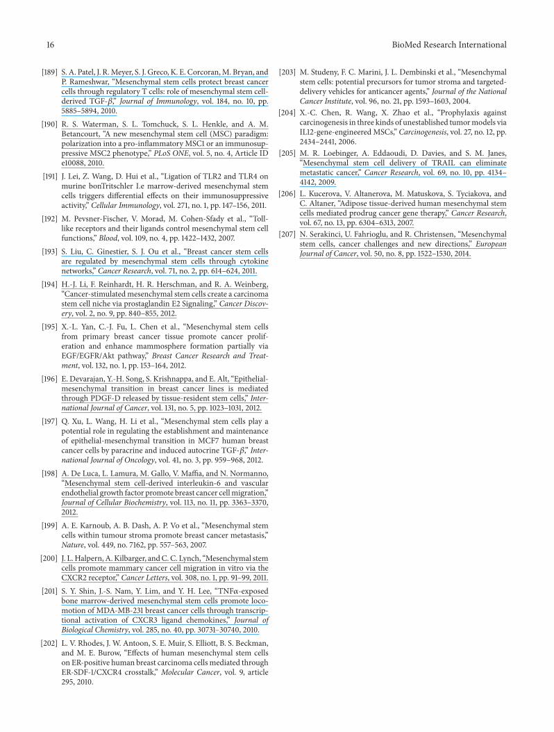
16 BioMed Research International
[189] S. A. Patel, J. R.Meyer, S. J. Greco, K. E. Corcoran,M. Bryan, andP. Rameshwar, “Mesenchymal stem cells protect breast cancercells through regulatory T cells: role of mesenchymal stem cell-derived TGF-𝛽,” Journal of Immunology, vol. 184, no. 10, pp.5885–5894, 2010.
[190] R. S. Waterman, S. L. Tomchuck, S. L. Henkle, and A. M.Betancourt, “A new mesenchymal stem cell (MSC) paradigm:polarization into a pro-inflammatory MSC1 or an immunosup-pressive MSC2 phenotype,” PLoS ONE, vol. 5, no. 4, Article IDe10088, 2010.
[191] J. Lei, Z. Wang, D. Hui et al., “Ligation of TLR2 and TLR4 onmurine bonTritschler I.e marrow-derived mesenchymal stemcells triggers differential effects on their immunosuppressiveactivity,” Cellular Immunology, vol. 271, no. 1, pp. 147–156, 2011.
[192] M. Pevsner-Fischer, V. Morad, M. Cohen-Sfady et al., “Toll-like receptors and their ligands control mesenchymal stem cellfunctions,” Blood, vol. 109, no. 4, pp. 1422–1432, 2007.
[193] S. Liu, C. Ginestier, S. J. Ou et al., “Breast cancer stem cellsare regulated by mesenchymal stem cells through cytokinenetworks,” Cancer Research, vol. 71, no. 2, pp. 614–624, 2011.
[194] H.-J. Li, F. Reinhardt, H. R. Herschman, and R. A. Weinberg,“Cancer-stimulatedmesenchymal stem cells create a carcinomastem cell niche via prostaglandin E2 Signaling,” Cancer Discov-ery, vol. 2, no. 9, pp. 840–855, 2012.
[195] X.-L. Yan, C.-J. Fu, L. Chen et al., “Mesenchymal stem cellsfrom primary breast cancer tissue promote cancer prolif-eration and enhance mammosphere formation partially viaEGF/EGFR/Akt pathway,” Breast Cancer Research and Treat-ment, vol. 132, no. 1, pp. 153–164, 2012.
[196] E. Devarajan, Y.-H. Song, S. Krishnappa, and E. Alt, “Epithelial-mesenchymal transition in breast cancer lines is mediatedthrough PDGF-D released by tissue-resident stem cells,” Inter-national Journal of Cancer, vol. 131, no. 5, pp. 1023–1031, 2012.
[197] Q. Xu, L. Wang, H. Li et al., “Mesenchymal stem cells play apotential role in regulating the establishment and maintenanceof epithelial-mesenchymal transition in MCF7 human breastcancer cells by paracrine and induced autocrine TGF-𝛽,” Inter-national Journal of Oncology, vol. 41, no. 3, pp. 959–968, 2012.
[198] A. De Luca, L. Lamura, M. Gallo, V. Maffia, and N. Normanno,“Mesenchymal stem cell-derived interleukin-6 and vascularendothelial growth factor promote breast cancer cellmigration,”Journal of Cellular Biochemistry, vol. 113, no. 11, pp. 3363–3370,2012.
[199] A. E. Karnoub, A. B. Dash, A. P. Vo et al., “Mesenchymal stemcells within tumour stroma promote breast cancer metastasis,”Nature, vol. 449, no. 7162, pp. 557–563, 2007.
[200] J. L.Halpern,A.Kilbarger, andC.C. Lynch, “Mesenchymal stemcells promote mammary cancer cell migration in vitro via theCXCR2 receptor,” Cancer Letters, vol. 308, no. 1, pp. 91–99, 2011.
[201] S. Y. Shin, J.-S. Nam, Y. Lim, and Y. H. Lee, “TNF𝛼-exposedbone marrow-derived mesenchymal stem cells promote loco-motion of MDA-MB-231 breast cancer cells through transcrip-tional activation of CXCR3 ligand chemokines,” Journal ofBiological Chemistry, vol. 285, no. 40, pp. 30731–30740, 2010.
[202] L. V. Rhodes, J. W. Antoon, S. E. Muir, S. Elliott, B. S. Beckman,and M. E. Burow, “Effects of human mesenchymal stem cellson ER-positive human breast carcinoma cells mediated throughER-SDF-1/CXCR4 crosstalk,” Molecular Cancer, vol. 9, article295, 2010.
[203] M. Studeny, F. C. Marini, J. L. Dembinski et al., “Mesenchymalstem cells: potential precursors for tumor stroma and targeted-delivery vehicles for anticancer agents,” Journal of the NationalCancer Institute, vol. 96, no. 21, pp. 1593–1603, 2004.
[204] X.-C. Chen, R. Wang, X. Zhao et al., “Prophylaxis againstcarcinogenesis in three kinds of unestablished tumormodels viaIL12-gene-engineered MSCs,” Carcinogenesis, vol. 27, no. 12, pp.2434–2441, 2006.
[205] M. R. Loebinger, A. Eddaoudi, D. Davies, and S. M. Janes,“Mesenchymal stem cell delivery of TRAIL can eliminatemetastatic cancer,” Cancer Research, vol. 69, no. 10, pp. 4134–4142, 2009.
[206] L. Kucerova, V. Altanerova, M. Matuskova, S. Tyciakova, andC. Altaner, “Adipose tissue-derived human mesenchymal stemcells mediated prodrug cancer gene therapy,” Cancer Research,vol. 67, no. 13, pp. 6304–6313, 2007.
[207] N. Serakinci, U. Fahrioglu, and R. Christensen, “Mesenchymalstem cells, cancer challenges and new directions,” EuropeanJournal of Cancer, vol. 50, no. 8, pp. 1522–1530, 2014.
Copyright © 2022 FDOKUMEN