Zinc protoporphyrin IX, a heme oxygenase-1 inhibitor, demonstrates potent antitumor effects but is...
12
BioMed Central Page 1 of 12 (page number not for citation purposes) BMC Cancer Open Access Research article Zinc protoporphyrin IX, a heme oxygenase-1 inhibitor, demonstrates potent antitumor effects but is unable to potentiate antitumor effects of chemotherapeutics in mice Dominika Nowis 1 , Marek Bugajski 1 , Magdalena Winiarska 1,2 , Jacek Bil 1 , Angelika Szokalska 1 , Pawel Salwa 1 , Tadeusz Issat 1 , Halina Was 3 , Alicja Jozkowicz 3 , Jozef Dulak 3 , Tomasz Stoklosa 1 and Jakub Golab* 1 Address: 1 Department of Immunology, Center of Biostructure Research, the Medical University of Warsaw, Banacha 1A, 02-097 Warsaw, Poland, 2 Department of Laboratory Diagnostics and Clinical Immunology, the Medical University of Warsaw, Marszalkowska 24, 00-576 Warsaw, Poland and 3 Department of Medical Biotechnology, Faculty of Biochemistry, Biophysics and Biotechnology, Jagiellonian University, Gronostajowa 7, 30- 387 Krakow, Poland Email: Dominika Nowis - [email protected]; Marek Bugajski - [email protected]; Magdalena Winiarska - [email protected]; Jacek Bil - [email protected]; Angelika Szokalska - [email protected]; Pawel Salwa - [email protected]; Tadeusz Issat - [email protected]; Halina Was - [email protected]; Alicja Jozkowicz - [email protected]; Jozef Dulak - [email protected]; Tomasz Stoklosa - [email protected]; Jakub Golab* - [email protected] * Corresponding author Abstract Background: HO-1 participates in the degradation of heme. Its products can exert unique cytoprotective effects. Numerous tumors express high levels of HO-1 indicating that this enzyme might be a potential therapeutic target. In this study we decided to evaluate potential cytostatic/ cytotoxic effects of zinc protoporphyrin IX (Zn(II)PPIX), a selective HO-1 inhibitor and to evaluate its antitumor activity in combination with chemotherapeutics. Methods: Cytostatic/cytotoxic effects of Zn(II)PPIX were evaluated with crystal violet staining and clonogenic assay. Western blotting was used for the evaluation of protein expression. Flow cytometry was used to evaluate the influence of Zn(II)PPIX on the induction of apoptosis and generation of reactive oxygen species. Knock-down of HO-1 expression was achieved with siRNA. Antitumor effects of Zn(II)PPIX alone or in combination with chemotherapeutics were measured in transplantation tumor models. Results: Zn(II)PPIX induced significant accumulation of reactive oxygen species in tumor cells. This effect was partly reversed by administration of exogenous bilirubin. Moreover, Zn(II)PPIX exerted potent cytostatic/cytotoxic effects against human and murine tumor cell lines. Despite a significant time and dose-dependent decrease in cyclin D expression in Zn(II)PPIX-treated cells no accumulation of tumor cells in G1 phase of the cell cycle was observed. However, incubation of C- 26 cells with Zn(II)PPIX increased the percentage of cells in sub-G1 phase of the cells cycle. Flow cytometry studies with propidium iodide and annexin V staining as well as detection of cleaved caspase 3 by Western blotting revealed that Zn(II)PPIX can induce apoptosis of tumor cells. B16F10 melanoma cells overexpressing HO-1 and transplanted into syngeneic mice were resistant to either Zn(II)PPIX or antitumor effects of cisplatin. Zn(II)PPIX was unable to potentiate antitumor effects Published: 11 July 2008 BMC Cancer 2008, 8:197 doi:10.1186/1471-2407-8-197 Received: 9 February 2008 Accepted: 11 July 2008 This article is available from: http://www.biomedcentral.com/1471-2407/8/197 © 2008 Nowis et al; licensee BioMed Central Ltd. This is an Open Access article distributed under the terms of the Creative Commons Attribution License (http://creativecommons.org/licenses/by/2.0 ), which permits unrestricted use, distribution, and reproduction in any medium, provided the original work is properly cited.
Transcript of Zinc protoporphyrin IX, a heme oxygenase-1 inhibitor, demonstrates potent antitumor effects but is...
BioMed CentralBMC Cancer
ss
Open AcceResearch articleZinc protoporphyrin IX, a heme oxygenase-1 inhibitor, demonstrates potent antitumor effects but is unable to potentiate antitumor effects of chemotherapeutics in miceDominika Nowis1, Marek Bugajski1, Magdalena Winiarska1,2, Jacek Bil1, Angelika Szokalska1, Pawel Salwa1, Tadeusz Issat1, Halina Was3, Alicja Jozkowicz3, Jozef Dulak3, Tomasz Stoklosa1 and Jakub Golab*1Address: 1Department of Immunology, Center of Biostructure Research, the Medical University of Warsaw, Banacha 1A, 02-097 Warsaw, Poland, 2Department of Laboratory Diagnostics and Clinical Immunology, the Medical University of Warsaw, Marszalkowska 24, 00-576 Warsaw, Poland and 3Department of Medical Biotechnology, Faculty of Biochemistry, Biophysics and Biotechnology, Jagiellonian University, Gronostajowa 7, 30-387 Krakow, Poland
Email: Dominika Nowis - [email protected]; Marek Bugajski - [email protected]; Magdalena Winiarska - [email protected]; Jacek Bil - [email protected]; Angelika Szokalska - [email protected]; Pawel Salwa - [email protected]; Tadeusz Issat - [email protected]; Halina Was - [email protected]; Alicja Jozkowicz - [email protected]; Jozef Dulak - [email protected]; Tomasz Stoklosa - [email protected]; Jakub Golab* - [email protected]
* Corresponding author
AbstractBackground: HO-1 participates in the degradation of heme. Its products can exert uniquecytoprotective effects. Numerous tumors express high levels of HO-1 indicating that this enzymemight be a potential therapeutic target. In this study we decided to evaluate potential cytostatic/cytotoxic effects of zinc protoporphyrin IX (Zn(II)PPIX), a selective HO-1 inhibitor and to evaluateits antitumor activity in combination with chemotherapeutics.
Methods: Cytostatic/cytotoxic effects of Zn(II)PPIX were evaluated with crystal violet staining andclonogenic assay. Western blotting was used for the evaluation of protein expression. Flowcytometry was used to evaluate the influence of Zn(II)PPIX on the induction of apoptosis andgeneration of reactive oxygen species. Knock-down of HO-1 expression was achieved with siRNA.Antitumor effects of Zn(II)PPIX alone or in combination with chemotherapeutics were measuredin transplantation tumor models.
Results: Zn(II)PPIX induced significant accumulation of reactive oxygen species in tumor cells. Thiseffect was partly reversed by administration of exogenous bilirubin. Moreover, Zn(II)PPIX exertedpotent cytostatic/cytotoxic effects against human and murine tumor cell lines. Despite a significanttime and dose-dependent decrease in cyclin D expression in Zn(II)PPIX-treated cells noaccumulation of tumor cells in G1 phase of the cell cycle was observed. However, incubation of C-26 cells with Zn(II)PPIX increased the percentage of cells in sub-G1 phase of the cells cycle. Flowcytometry studies with propidium iodide and annexin V staining as well as detection of cleavedcaspase 3 by Western blotting revealed that Zn(II)PPIX can induce apoptosis of tumor cells. B16F10melanoma cells overexpressing HO-1 and transplanted into syngeneic mice were resistant to eitherZn(II)PPIX or antitumor effects of cisplatin. Zn(II)PPIX was unable to potentiate antitumor effects
Published: 11 July 2008
BMC Cancer 2008, 8:197 doi:10.1186/1471-2407-8-197
Received: 9 February 2008Accepted: 11 July 2008
This article is available from: http://www.biomedcentral.com/1471-2407/8/197
© 2008 Nowis et al; licensee BioMed Central Ltd. This is an Open Access article distributed under the terms of the Creative Commons Attribution License (http://creativecommons.org/licenses/by/2.0), which permits unrestricted use, distribution, and reproduction in any medium, provided the original work is properly cited.
Page 1 of 12(page number not for citation purposes)

BMC Cancer 2008, 8:197 http://www.biomedcentral.com/1471-2407/8/197
of 5-fluorouracil, cisplatin or doxorubicin in three different tumor models, but significantlypotentiated toxicity of 5-FU and cisplatin.
Conclusion: Inhibition of HO-1 exerts antitumor effects but should not be used to potentiateantitumor effects of cancer chemotherapeutics unless procedures of selective tumor targeting ofHO-1 inhibitors are developed.
BackgroundHeme oxygenase (HO) is a microsomal enzyme that cata-lyzes oxidative cleavage of the porphyrin ring in hememolecule leading to the formation of biliverdin, carbonmonoxide (CO) and free iron [1,2]. Biliverdin is furtherconverted into bilirubin by biliverdin reductase. All HOproducts exert pleiotropic effects including numerouscytoprotective responses [3]. Bilirubin and biliverdin areamong the most potent endogenous scavengers of reactiveoxygen species (ROS) [4]. CO exerts strong antiapoptoticand anti-inflammatory effects through induction of solu-ble guanylyl cyclase. It suppresses production of tumornecrosis factor (TNF), interleukin-1β (IL-1β) and CCL4chemokine (macrophage inflammatory protein-1β), butup-regulates synthesis of anti-inflammatory IL-10 [5].Finally, free iron (Fe2+) despite participation in Fentonreaction that leads to formation of highly reactivehydroxyl radicals, also activates Fe-ATPase, a transporterthat removes intracellular iron, as well as induces expres-sion of ferritin heavy chains which sequester free iron andexert specific cytoprotective roles [6].
Two isoforms of heme oxygenase exist. HO-1 is an induc-ible enzyme that belongs to the heat shock protein(HSP32) family. Its expression is induced by a vast arrayof stress-inducing stimuli that include: oxidative stress,heat shock, UV irradiation, exposure to heavy metals andnumerous other toxins, including chemotherapeutics [7].Some observations indicate that HO-1 and its productsalso exert anti-inflammatory effects and participate in thecontrol of growth and proliferation of tumor cells. Ele-vated constitutive levels of HO-1 have been observed in anumber of human tumors including glioma, melanoma,prostate, pancreatic and renal cell carcinoma, lymphosar-comas, Kaposi sarcoma and hepatoma [7]. Enhancedexpression of HO-1 can also contribute to tumor progres-sion through promotion of angiogenesis and metastasesformation [8,9]. Furthermore, the increased basal level ofHO-1 expression in tumor cells can be further elevated bychemotherapeutics, radiotherapy or photodynamic ther-apy [10,11].
Altogether HO products participate in attenuation of oxi-dative stress, suppression of inflammatory responses,inhibition of apoptosis and promotion of angiogenesis[12,13]. Therefore, accumulating evidence indicates thatHO-1 can be a therapeutic target for antitumor treatment.
Indeed, it was shown that zinc protoporphyrin IX(Zn(II)PPIX) or its pegylated derivative, a potent HOinhibitor, can exert significant antitumor effects againstseveral tumors in mice [14-16]. Moreover, inhibition ofHO-1 expression or activity was shown to increase respon-siveness of tumor cells to other anticancer treatments invitro [10,16,17]. The aim of these studies was to explorethe in vivo role of HO-1 in tumor growth and in protectingtumor cells against chemotherapeutics.
MethodsTumor cellsHuman ovarian carcinoma (MDAH2774), human pan-creatic adenocarcinoma (Mia PaCa2), human breast carci-noma (MDA-MB231), murine breast carcinoma (EMT6)cell lines were purchased from ATCC (Manassas, VA,USA). Murine colon-26 (C-26), a poorly differentiatedcolon adenocarcinoma cell line was obtained from prof.Danuta Dus (Institute of Immunology and ExperimentalMedicine, Wroclaw, Poland). B16F10 murine melanomacells were provided by Dr. M. Kubin (Wistar Institute,Philadelphia, PA). Cells were cultured in RPMI 1640medium (C-26 and B16F10) (Invitrogen, Carlsbad, CA,USA) or DMEM (MDAH2774, Mia PaCa2, MDA-MB231and EMT6) supplemented with 10% heat-inactivated fetalcalf serum, antibiotics, 2-mercaptoethanol (50 μM) andL-glutamine (2 mM) (all from Invitrogen), hereafterreferred to as culture medium.
ReagentsZinc (II) propoporphyrin IX (Zn(II)PPIX), a HO-1 inhib-itor, was purchased from Frontier Scientific Europe Ltd.(Carnforth, Lancashire, United Kingdom) and was dis-solved in dimethylsulfoxide (DMSO) (Sigma, St. Louis,USA) to the final stock concentration of 5 mM. Bilirubin(Sigma, St Louis, MO, USA) was dissolved in 0.1 N NaOHto the final stock concentration of 10 mM. Hemin (fromSigma) was dissolved in 0.1 N KOH to the final stock con-centration of 10 mM. All the solutions were prepared inthe dark right before adding to the cell cultures.
MiceFemale BALB/c and C57Bl/6 mice, 8–12 weeks of age wereused for in vivo experiments. Breeding pairs were obtainedfrom the Institute of Oncology (Warsaw, Poland). Micewere kept in conventional conditions with full access tofood and water during experiments. All of the animal
Page 2 of 12(page number not for citation purposes)

BMC Cancer 2008, 8:197 http://www.biomedcentral.com/1471-2407/8/197
studies were performed in accordance with the guidelinesapproved by the Ethical Committee of the Medical Uni-versity of Warsaw.
Western blottingFor Western blotting C-26 were treated with Zn(II)PPIXfor 24, 48 or 72 h. After indicated times of culture cellswere washed with PBS and lysed with radioimmunopre-cipitation assay buffer (RIPA) containing Tris base 50mM, NaCl 150 mM, NP-40 1%, sodium deoxycholate0.25% and EDTA 1 mM supplemented with Complete®
protease inhibitor cocktail tablets (Roche Diagnostics,Mannheim, Germany). Protein concentration was meas-ured using Bio-Rad Protein Assay (BioRad, Hercules, CA,USA). Equal amounts of whole cell proteins were sepa-rated on 12% SDS-polyacrylamide gel, transferred ontoProtran® nitrocellulose membranes (Schleicher andSchuell BioScience Inc., Keene, NH, USA), blocked withTBST [Tris buffered saline (pH 7.4) and 0.05% Tween 20]supplemented with 5% nonfat milk and 5% FBS. The fol-lowing antibodies at 1:1000 dilution were used for the 24h incubation: mouse monoclonal anti-α-tubulin(Promega Corporation, Madison, WI, USA), rabbit poly-clonal anti-cleaved caspase 3 (Cell Signaling Technology,Beverly, MA, USA) and mouse monoclonal anti-cyclin D1(Santa Cruz). After extensive washing with TBST, themembranes were incubated for 45 min with correspond-ing alkaline phosphatase-coupled secondary antibodies(from Jackson Immuno Research). The color reaction foralkaline phosphatase was developed using nitroblue tetra-zolium and 5-bromo-4-chloro-3-indolyl phosphate(Sigma).
Cytostatic/cytotoxic assayThe cytostatic and/or cytotoxic effects were measuredusing crystal violet staining as described [18]. Briefly,tumor cells were dispensed into 96-well plates (Sarstedt,Numbrecht, Germany) at a concentration of 5 × 10³ cellsper well and allowed to attach overnight. The followingday investigated agents were added at indicated concen-trations. Cells were kept in dark for 48 or 72 h. After theincubation time the cells were rinsed with PBS and stainedwith 0.5% crystal violet in 2% ethanol for 10 min at roomtemperature. Plates were washed four times with tap waterand the cells were lysed with 1% SDS solution. Absorb-ance was measured at 595 nm using an enzyme-linkedimmunosorbent assay reader (SLT Labinstrument GmbH,Salzburg, Austria), equipped with a 595 nm filter. Cyto-toxicity was expressed as relative viability of tumor cells(% of control cultures incubated with medium only) andwas calculated as follows: relative viability = (Ae - Ab) ×100/(Ac - Ab), where Ab is the background absorbance, Aeis experimental absorbance, and Ac is the absorbance ofuntreated controls.
Clonogenic assayMDAH2774 or Mia PaCa2 cells were plated at 2.5 × 10⁵
cells per 35-mm dish (Sarstedt). Four hours after seeding,Zn(II)PPIX was added at indicated concentrations. After24 hours of incubation in dark, the cells were washed withPBS, trypsinized and seeded into 35-well plates in tripletsat the concentration of 1 × 10³ cells per a dish. Freshmedium containing Zn(II)PPIX was added. The mediumwas removed daily for the 6 following days. After 14 dayincubation in the dark, the cells were rinsed with PBS,fixed for 10 min in pure methanol and stained with 0.5%crystal violet in 2% ethanol for 10 min at room tempera-ture. Then the plates were washed four times with tapwater and air-dried. The images of the plates were madeusing the Olympus Camedia C750 Ultra Zoom digitalcamera.
FACS analysis of cell cycle and apoptosisFor the cell cycle analysis, C-26 cells were seeded into 6-well plates (Sarstedt) at the concentrations 1 to 3 × 10⁵
cells per well and incubated in dark with 2.5 μM or 5 μMZn(II)PPIX. The incubation time varied from 24 to 72 h.After the incubation, the cells were washed with PBS,trypsinized and centrifuged at 1000 rpm for 10 min in4°C. The pellet was resuspended in 0.5 ml of PBS andinjected under the surface of ice cold 70% ethanol for 24-h fixation in -20°C. At the day of analysis, the cells werewashed from ethanol, stained with 5 μg/ml propidiumiodide (PI, Becton Dickinson, Mountain View, CA, USA)at the presence of RNase A (Becton Dickinson) for 30 minat 37°C. For the analysis of apoptosis induction C-26 orMia PaCa2 cells were seeded into 6-well plates (Sarstedt)at the concentrations 1 to 3 × 10⁵ cells per well and incu-bated in dark with 2.5 μM or 5 μM Zn(II)PPIX. The incu-bation time varied from 6 to 72 h. After the incubation,the cells were washed with PBS, trypsinized and centri-fuged at 1000 rpm for 10 min in 4°C. The pellet was resus-pended in 0.5 ml of Annexin-binding buffer (BioSourceInternational, Camarillo, CA, USA) and then incubatedfor 15 min with 5 μl of Annexin V-FITC (BioSource).Finally, the cells were stained with 5 μg/ml PI. Thecytofluorometric analysis was performed using FACSCali-bur (Becton Dickinson). For single analysis 1 × 10⁴ cellswere used. Data were collected at the wavelength of 580nm (for PI) and 520 nm for FITC, and analysed with CEL-LQuest 1.2 software (Becton Dickinson).
HO-1 RNAiSmall interfering RNA against murine HO-1 was obtainedby chemical synthesis from Dharmacon (Lafayette, CO,USA). The following sequence was used for targeting ofmurine HO-1: 5'-GCA-GAA-CCC-AGU-CUA-UGC-C-3'.For RNAi experiments 3,5 × 10⁵ C-26 cells were seededinto 6-well plates (Sarstedt). After 24 h cells were washedwith Optimem™ medium (Invitrogen) and transfected
Page 3 of 12(page number not for citation purposes)
BMC Cancer 2008, 8:197 http://www.biomedcentral.com/1471-2407/8/197
with 100 nM siRNA using OligofectAMINE™ according tothe manufacturer's protocol. 10 μM hemin was added tothe appropriate groups 2 h befor RNAi procedure. After 8h trasfection medium was replaced with full DMEM cul-ture medium. 24 h after transfection cells were harvestedand ROS generation analysis was performed (see below).
ROS generation assayFor determination of ROS generation, C-26 cells (3,5 ×10⁵ cells per well) were seeded into 6-well paltes(Sarstedt) and treated for 24 h with 2.5 μM Zn(II)PPIXand/or 50 μM biliubin or subjected to HO-1 RNAi (asdescribed above). On the day of the analysis, cells weretrypsynized, washed 3 times in ice-cold PBS and resus-pended in 1 ml of PBS. One μl of CM-H2DCFDA (Invitro-gen, 5 mM DMSO solution) was added to each sample for20 min incubation at 37°C. Then, cells were washed twicewith ice-cold PBS and subjected to flow cytometry usingFACSCalibur (Becton Dickinson). For single analysis 1 ×10⁴ cells were used. Data were collected at the wavelengthof 520 nm and analysed with CELLQuest 1.2 software(Becton Dickinson).
Tumor treatment and monitoringFor assessment of antitumor activity of Zn(II)PPIX in vivo,exponentially growing C-26 were harvested, re-suspendedin PBS medium to the appropriate concentration, andinjected at the dose of 1 × 10⁵ cells per mouse into thefootpad of the right hind limb of experimental mice.Tumor cell viability measured by trypan blue exclusionwas always above 95%. For in vivo treatment Zn(II)PPIXwas dissolved in DMSO and further diluted in 0.9% NaClto required concentrations. Final DMSO concentrationwas always less then 0.1%. Zn(II)PPIX was distributedintraperitoneally at doses from 12.5 to 50 mg per kg ofbody weight or orally at doses from 11 to 22 mg per kg ofbody weight. Control animals received 0.1% DMSO solu-tion in 0.9% NaCl i.p. or orally.
For in vivo experiments evaluating the effectiveness ofcombine treatment using Zn(II)PPIX and chemothera-peutics, exponentially growing C-26, EMT6 and B16F10cells were injected at the dose of 1 × 10⁵, 1 × 10⁵ and 1 ×10⁶ cells per mouse, respectively into the footpad of theright hind limb of experimental mice. Zn(II)PPIX treat-ment (50 mg/kg i.p.) was started on the day 7 after inocu-lation of tumor cells and continued for 7 consecutivedays. First dose of HO-1 inhibitor was administered 1 hbefore each of the chemotherapeutics to eliminate anypossible interactions (such as neutralization) betweendrugs. Cisplatin (Platidam, Pliva-Lachema, Cech Repub-lic) at the dose of 7.5 mg/kg i.p., 5-FU (Fluorouracil 1000Medac, Medac, Hamburg, Germany) – 50 mg/kg i.p., ordoxorubicin (Adriblastina RD, Pharmacia Italia, Milan,
Italy) – 7.5 mg/kg i.p. were administered at a single doseon the day 7th after inoculation of tumor cells.
For in vivo experiments determining influence of HO-1gene transfer on sensitivity of B16F10 melanoma cells tocisplatin treatment, exponentially growing B1 (HO-1transfected) and B5E (empty plasmid transfected) cellswere injected at the dose of 1 × 10⁶ cells per mouse intothe footpad of the right hind limb of experimental mice.Cisplatin (Platidam, Pliva-Lachema) was administeredi.p. at a single dose (2.5, 5.0 or 7.5 mg/kg) on the day 7th
after inoculation of tumor cells.
For in vivo experiments exponentially growing B1 and B5Ecells were injected at a dose of 1 × 10⁶ cells per mouse intothe footpad of the right hind limb of experimental mice.Cisplatin was administered i.p. at a single dose of 7.5 mg/kg on the 7th day after inoculation of tumor cells.Zn(II)PPIX treatment (50 mg/kg i.p.) was started on theday 7 after inoculation of tumor cells and continued for 7consecutive days. First dose of HO-1 inhibitor was admin-istered 1 h before the chemotherapeutic.
In all the experiments mentioned above, local tumorgrowth was determined every second day as described pre-viously [19] by the formula: tumor volume (mm3) =(longer diameter) × (shorter diameter)2.
Statistical analysisData were calculated using Microsoft™ Excel 2003. Differ-ences in in vitro cytotoxicity assays and tumor volumewere analyzed for significance by Student's t test. Signifi-cance was defined as a two-sided P < 0.03.
ResultsZn(II)PPIX induces potent cytostatic/cytotoxic effects against murine and human tumor cellsFour different cell lines of murine (C-26, colon adenocar-cinoma) and human (Mia PaCa2, a pancreatic cancer,MDAH2774, ovarian carcinoma, and MDA-MB231,breast carcinoma) origin were incubated with increasingconcentrations of Zn(II)PPIX for 48 and/or 72 hours. HO-1 inhibitor exerted dose- and time-dependent cytostatic/cytotoxic effects as measured with crystal violet staining(Fig. 1). Similar effects were observed in MTT assay (notshown) and in a clonogenic assay performed with MiaPaCa2 and MDAH2774 cells (Fig. 2).
To get insight into the mechanism of cytostatic/cytotoxiceffects induced by Zn(II)PPIX cell cycle analysis andinduction of apoptosis were performed. Incubation of C-26 cells with Zn(II)PPIX for 48 or 72 h resulted in dose-and time-dependent reduction of cells in G1 phase of thecell cycle (Fig. 3A). This effect correlated with decreasedcyclin D1 expression (Fig. 3B) and increased percentage of
Page 4 of 12(page number not for citation purposes)

BMC Cancer 2008, 8:197 http://www.biomedcentral.com/1471-2407/8/197
cells in sub-G1 phase (Fig. 3A). Propidium iodide andannexin V staining of C-26 cells incubated with 5 μMZn(II)PPIX for 24–72 h indicated that the sub-G1 fractionof cells might represent apoptotic cells (Fig. 3C). The frac-tion of late apoptotic and necrotic cells increased from10.9% in controls to 30.4% after 72 h incubation withZn(II)PPIX. Western blotting analysis indicated that a 48-h incubation of C-26 cells leads to accumulation ofcleaved (active) caspase-3 (Fig. 3D).
An influence of Zn(II)PPIX as well as knock-down of HO-1 expression with siRNA were used to get insight into thespecificity of the Zn(II)PPIX-mediated effects. As shownin Fig. 3E siRNA against a murine HO-1 (moHO-1 siRNA)was effective in reducing the expression level of HO-1 inmurine C-26 cells. A siRNA against a human gene (huHO-1 siRNA) was ineffective (Fig. 3E) as well as an irrelevantsiRNA against enhanced green fluorescent protein (eGFP,
not shown). Inhibition of HO-1 activity with Zn(II)PPIXresulted in increased ROS generation in C-26 cells (Fig.3F). Similar effects were observed when HO-1 expressionwas knocked-down with a specific siRNA against HO-1(Fig. 3G). The influence of Zn(II)PPIX on ROS formationwas completely abrogated when C-26 were co-incubatedwith 50 μM bilirubin (Fig. 3F). Similarly, the influence ofsiRNA was significantly decreased by forced expression ofHO-1 by pre-incubation of C-26 with 10 μM hemin (Fig.3G).
Zn(II)PPIX induces antitumor effects in a murine C-26 modelBALB/c mice inoculated with C-26 cells were treated withZn(II)PPIX for 7 consecutive days. HO-1 inhibitor wasadministered either intraperitoneally (i.p.) or per os andthe tumor volume was monitored every second day, start-ing from day 7 after inoculation of tumor cells.
Zn(II)PPIX exerts cystostatic/cytotoxic effects against tumor cellsFigure 1Zn(II)PPIX exerts cystostatic/cytotoxic effects against tumor cells. Tumor cells were incubated with serial dilutions of Zn(II)PPIX for 48 or 72 hours. The cytostatic and/or cytotoxic effects of treatment were measured with a standard crystal violet staining assay. Bars represent means ± SD. *P < 0.05 (two way Student's t-test) in comparison with controls.
Page 5 of 12(page number not for citation purposes)

BMC Cancer 2008, 8:197 http://www.biomedcentral.com/1471-2407/8/197
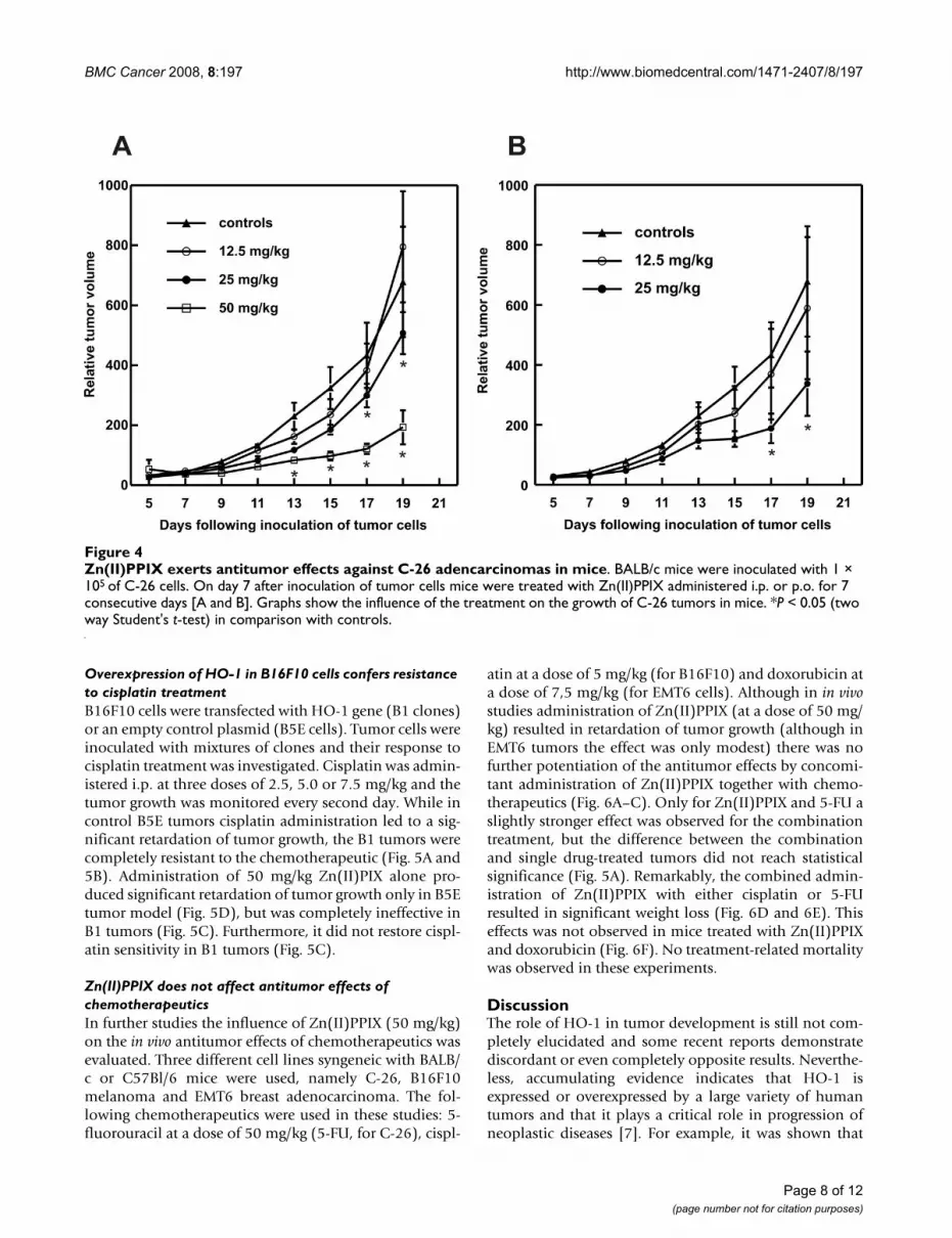
Zn(II)PPIX exerted dose-dependent antitumor effectsmanifested by the retardation of tumor growth. A statisti-cal significance was reached on days 17 and 19 forZn(II)PPIX administered at a dose of 25 mg/kg either i.p.or orally (Fig. 4A and 4B). A stronger effect was observed
when Zn(II)PPIX was administered i.p. at a dose of 50mg/kg, where a statistically significant retardation oftumor growth was observed on days 13–19, as comparedwith controls (Fig. 4A).
The influence of Zn(II)PPIX on formation of tumor cell coloniesFigure 2The influence of Zn(II)PPIX on formation of tumor cell colonies. For the clonogenic assay, MDAH2774 or Mia PaCa2 cells were plated at a concentration of 1 × 10³ cells/dish. Medium containing Zn(II)PPIX was replaced daily for 1–6 consecutive days. On day 14 after PDT, the plates were fixed with methanol and stained with crystal violet.
Page 6 of 12(page number not for citation purposes)

BMC Cancer 2008, 8:197 http://www.biomedcentral.com/1471-2407/8/197
Page 7 of 12(page number not for citation purposes)
Zn(II)PPIX induces apoptosis and increases formation of reactive oxygen speciesFigure 3Zn(II)PPIX induces apoptosis and increases formation of reactive oxygen species. C-26 cells were grown in Petri dishes for 24 hours before addition of Zn(II)PPIX for the indicated time. [A] Cell cycle analysis was performed by flow cytom-etry analysis using ethanol-fixed, propidium iodide-stained cells. [B and D] Western blotting for expression of cyclin D1, cleaved caspase 3 or tubulin as a loading control was performed. [C] Induction of apoptosis was evaluated by staining of cells with propidium iodide and annexin V. [E] HO-1 silencing was evaluated with Western blotting. Controls represent expression of HO-1 in hemin-treated C-26 cells. HuHO-1 siRNA is a negative control targeting xenogeneic (human) HO-1 gene, muHO-1 siRNA is a murine sequence that knocks-down HO-1 expression in C-26 cells. [F and G] production of reactive oxygen spe-cies was evaluated with flow cytometry by measuring CM-H2DCFDA fluorescence in comparison with controls.
BMC Cancer 2008, 8:197 http://www.biomedcentral.com/1471-2407/8/197
Overexpression of HO-1 in B16F10 cells confers resistance to cisplatin treatmentB16F10 cells were transfected with HO-1 gene (B1 clones)or an empty control plasmid (B5E cells). Tumor cells wereinoculated with mixtures of clones and their response tocisplatin treatment was investigated. Cisplatin was admin-istered i.p. at three doses of 2.5, 5.0 or 7.5 mg/kg and thetumor growth was monitored every second day. While incontrol B5E tumors cisplatin administration led to a sig-nificant retardation of tumor growth, the B1 tumors werecompletely resistant to the chemotherapeutic (Fig. 5A and5B). Administration of 50 mg/kg Zn(II)PIX alone pro-duced significant retardation of tumor growth only in B5Etumor model (Fig. 5D), but was completely ineffective inB1 tumors (Fig. 5C). Furthermore, it did not restore cispl-atin sensitivity in B1 tumors (Fig. 5C).
Zn(II)PPIX does not affect antitumor effects of chemotherapeuticsIn further studies the influence of Zn(II)PPIX (50 mg/kg)on the in vivo antitumor effects of chemotherapeutics wasevaluated. Three different cell lines syngeneic with BALB/c or C57Bl/6 mice were used, namely C-26, B16F10melanoma and EMT6 breast adenocarcinoma. The fol-lowing chemotherapeutics were used in these studies: 5-fluorouracil at a dose of 50 mg/kg (5-FU, for C-26), cispl-
atin at a dose of 5 mg/kg (for B16F10) and doxorubicin ata dose of 7,5 mg/kg (for EMT6 cells). Although in in vivostudies administration of Zn(II)PPIX (at a dose of 50 mg/kg) resulted in retardation of tumor growth (although inEMT6 tumors the effect was only modest) there was nofurther potentiation of the antitumor effects by concomi-tant administration of Zn(II)PPIX together with chemo-therapeutics (Fig. 6A–C). Only for Zn(II)PPIX and 5-FU aslightly stronger effect was observed for the combinationtreatment, but the difference between the combinationand single drug-treated tumors did not reach statisticalsignificance (Fig. 5A). Remarkably, the combined admin-istration of Zn(II)PPIX with either cisplatin or 5-FUresulted in significant weight loss (Fig. 6D and 6E). Thiseffects was not observed in mice treated with Zn(II)PPIXand doxorubicin (Fig. 6F). No treatment-related mortalitywas observed in these experiments.
DiscussionThe role of HO-1 in tumor development is still not com-pletely elucidated and some recent reports demonstratediscordant or even completely opposite results. Neverthe-less, accumulating evidence indicates that HO-1 isexpressed or overexpressed by a large variety of humantumors and that it plays a critical role in progression ofneoplastic diseases [7]. For example, it was shown that
Zn(II)PPIX exerts antitumor effects against C-26 adencarcinomas in miceFigure 4Zn(II)PPIX exerts antitumor effects against C-26 adencarcinomas in mice. BALB/c mice were inoculated with 1 × 10⁵ of C-26 cells. On day 7 after inoculation of tumor cells mice were treated with Zn(II)PPIX administered i.p. or p.o. for 7 consecutive days [A and B]. Graphs show the influence of the treatment on the growth of C-26 tumors in mice. *P < 0.05 (two way Student's t-test) in comparison with controls.
Page 8 of 12(page number not for citation purposes)

BMC Cancer 2008, 8:197 http://www.biomedcentral.com/1471-2407/8/197
increased expression of HO-1 is associated with higherproliferation rate of various tumor cells [7,10,15,20],although opposite effects are observed in breast cancercells [21]. Specific siRNA-mediated down-regulation ofHO-1 resulted in suppression of proliferation of pancre-
atic cancer cells [10] or in induction of apoptosis of lungcancer cells [17]. Similarly, cytostatic/cytotoxic effectswere observed with HO-1 inhibitors, such as Zn(II)PPIX[15,22] or its pegylated derivative (PEG-ZnPP) [16].Moreover, overexpression of HO-1 increased viability,
Antitumor effect of cisplatin or the combined treatment with Zn(II)PPIX and/or cisplatin in mice transplanted with control or HO-1 overexpressing B16F10 tumorsFigure 5Antitumor effect of cisplatin or the combined treatment with Zn(II)PPIX and/or cisplatin in mice transplanted with control or HO-1 overexpressing B16F10 tumors. C57Bl/6 mice were inoculated with 1 × 10⁶ of HO-1 overex-pressing (B1) or mock-transfected (B5E) cells. On day 7 after inoculation of tumor cells mice were treated with cisplatin at a dose of 2.5, 5.0 or 7.5 mg/kg administered i.p. [A and B] or with cisplatin (5 mg/kg in a single i.p. injection) and 50 mg/kg of Zn(II)PPIX administered i.p. for 7 consecutive days [C and D]. Graphs show the influence of the treatment on the growth of melanoma in mice. *P < 0.05 (two way Student's t-test) in comparison with controls.
Page 9 of 12(page number not for citation purposes)

BMC Cancer 2008, 8:197 http://www.biomedcentral.com/1471-2407/8/197
proliferation and angiogenic potential of melanomas [9].Mice inoculated with HO-1 overexpressing melanomasfared worse than controls, had a higher number of metas-tases and a significantly shortened survival [9].
The results of present experiments performed with addi-tional cell lines further establish that Zn(II)PPIX is anactive agent capable of inducing cytostatic and cytotoxiceffects against a number of human and mouse tumor celllines. Moreover, in two different tumor modelsZn(II)PPIX was demonstrated to exert potent antitumoreffects manifested by the retardation of tumor growth.
Development of effective antitumor regimens requiresadministration of drugs in combination. The role of thecombination treatment is to target different pathways intumor cells in order to elicit more robust antitumorresponse. Synergistic antitumor effects might lead to
decreased dosing and elimination or a significant attenu-ation of toxic side effects of drugs used in monotherapy.Several previous reports demonstrated that HO-1, whichis frequently over-expressed in tumor cells, might partici-pate in tumor-protective effects against a number ofchemotherapeutics, radiotherapy and photodynamictherapy [10,11]. By targeting HO-1 it might be possible tosensitize tumor cells to more effective cytotoxic effects ofother therapeutic regimens. Indeed, in vitro data seem tosupport this idea. HO-1 knock-down sensitized pancreaticcancer cells to gemcitabine [10] and lung cancer cells tocisplatin [17]. Moreover, PEG-ZnPP sensitized colon can-cer cells to cytostatic/cytotoxic effects of camptothecin ordoxorubicin [16]. However, the efficacy of combiningchemotherapeutics with HO-1 inhibitor in vivo has not yetbeen addressed.
Antitumor effect of the combined treatment with Zn(II)PPIX and/or chemotherapeuticsFigure 6Antitumor effect of the combined treatment with Zn(II)PPIX and/or chemotherapeutics. Mice were inoculated with 1 × 10⁵ of C-26 or EMT6 cells or with 1 × 10⁶ of B16F10 cells. On day 7 after inoculation of tumor cells mice received a single dose of chemotherapeutics: 50 mg/kg of 5-FU [A, D], 5 mg/kg of cisplatin [B, E] or 7.5 mg/kg of doxorubicin [C, F]. Zn(II)PPIX was administered i.p. for 7 consecutive days at a dose of 50 mg/kg [A-F]. Graphs show the influence of the treat-ment on the growth of tumors in mice [A-C] or on the change in mice weight as compared with initial weight. *P < 0.05 (two way Student's t-test) in comparison all other groups.
Page 10 of 12(page number not for citation purposes)

BMC Cancer 2008, 8:197 http://www.biomedcentral.com/1471-2407/8/197
Considering the use of HO-1 inhibitors in combinationwith other antitumor agents it must be kept in mind thatHO-1 and its products affect multiple signaling and met-abolic pathways and play an important role in protectionof normal cells against multiple environmental insults.For example, HO-1 protects retinal cells against light-induced damage [23], ameliorates ischemia-reperfusioninjury elicited by a number of different conditions [24],inhibits endothelial cell apoptosis induced by endoplas-mic reticulum stress [25], protects mice from apoptoticliver damage [26], prevents development of atherosclero-sis in LDL-receptor knock-out mice [27], improves sur-vival of transplanted organs [28] or prevents developmentof gastric ulcers in rats [29]. HO-1 also protects normalcells and tissues against toxic effects of chemotherapeu-tics. Heme-induced HO-1 expression protects againstcyclophosphamide-mediated hemorrhagic cystitis [30],HO-1 attenuates cisplatin-induced toxicity in renal tubu-lar cells [31] or in auditory cells [32], it also decreases dox-orubicin-mediated cardiotoxicity [33]. Moreover,transgenic mice deficient in HO-1 (-/-), develop moresevere renal failure and have significantly greater renalinjury compared with wild-type (+/+) mice treated withcisplatin [34].
At doses used in the studies presented here Zn(II)PPIXwas unable to restore cisplatin sensitivity in HO-1 overex-pressing melanoma cells nor was it capable of potentiat-ing antitumor effects of cisplatin, doxorubicin or 5-FU inthree different models. Nonetheless, Zn(II)PPIX signifi-cantly potentiated toxicity of cisplatin and 5-FU as deter-mined by decreased weight loss. Tozer et al, have shownthat administration of Zn(II)PPIX at a dose of 45 μmoles/kg is ineffective in inhibiting HO-1 activity in tumors [35].The dose used in these studies (50 mg/kg) corresponds toalmost 80 μmoles/kg/mouse. Although it was not meas-ured whether Zn(II)PPIX used at this dose and at thisadministration schedule was capable of inhibiting HO-1activity it can be concluded that it is ineffective in neitherinducing antitumor effects in HO-1-overexpressingB16F10 melanomas nor in restoring sensitivity to cispla-tin. Future studies should address an important, and notaddressed in these studies, question of drug administra-tion schedule. Specifically, what should be the timing ofZn(II)PPIX administration in combination with chemo-therapeutics to effectively inhibit HO-1 activity in tumorcells? Is it possible to design combination strategies thatwould target HO-1 in tumors and not in normal tissues?It seems that the influence of Zn(II)PPIX administrationon the activity of HO-1 in tumor versus normal tissueswill be of utmost importance in designing combinationtreatments.
ConclusionAltogether, these studies show that despite promisingcytostatic/cytotoxic and even antitumor effects elicited byZn(II)PPIX, this HO-1 inhibitor should not be used incombination with chemotherapeutics. It can be hypothe-sized however, that selective and efficient delivery of HO-1 inhibitors to the tumor might prove to be a morerational approach for combination therapies with chemo-therapeutics. Indeed, as demonstrated by Fang et al. [16]and Iyer at al. [36] it is possible to prepare Zn(II)PIX deriv-atives with a higher tumor selectivity. It remains to be elu-cidated whether these strategies are more effective and lesstoxic.
Competing interestsThe authors declare that they have no competing interests.
Authors' contributionsDN participated in experimental design, performed mostof in vitro and in vivo experiments and participated in thewriting of the manuscript, MB participated in in vivoexperiments, MW and JB did most of the flow cytometrystudies, AS measured ROS formation in tumor cells, PSparticipated in cytostatic/cytotoxic asssays in vitro, TI par-ticipated in in vivo experiments, HW prepared control andHO-1 overexpressing B16F10 melanoma cells, AJ partici-pated in experimental design, JD participated in experi-mental design and participated in the writing of themanuscript, TS participated in experimental design and inin vitro experiments, and participated in manuscript cor-rection, JG conceived the study, participated in its designand coordination and wrote the first draft of the manu-script. All authors read and approved the manuscript.
AcknowledgementsSupported by grant PBZ KBN 107/P04/2004 from Ministry of Science and Higher Education. Alicja Jozkowicz is the International Senior Research Fel-low of Wellcome Trust. We would like to thank Elzbieta Gutowska and Anna Czerepinska for excellent technical assistance. Tadeusz Issat and Magdalena Winiarska are recipients of the START stipend from the Foun-dation for Polish Science.
References1. Ryter SW, Alam J, Choi AM: Heme oxygenase-1/carbon monox-
ide: from basic science to therapeutic applications. Physiol Rev2006, 86:583-650.
2. Dulak J, Deshane J, Jozkowicz A, Agarwal A: Heme oxygenase-1and carbon monoxide in vascular pathobiology: focus on ang-iogenesis. Circulation 2008, 117:231-241.
3. Takahashi T, Shimizu H, Morimatsu H, Inoue K, Akagi R, Morita K,Sassa S: Heme oxygenase-1: a fundamental guardian againstoxidative tissue injuries in acute inflammation. Mini Rev MedChem 2007, 7:745-753.
4. Stocker R, Yamamoto Y, McDonagh AF, Glazer AN, Ames BN:Bilirubin is an antioxidant of possible physiological impor-tance. Science 1987, 235:1043-1046.
5. Otterbein LE, Bach FH, Alam J, Soares M, Tao Lu H, Wysk M, DavisRJ, Flavell RA, Choi AM: Carbon monoxide has anti-inflamma-tory effects involving the mitogen-activated protein kinasepathway. Nature Med 2000, 6:422-428.
Page 11 of 12(page number not for citation purposes)
http://www.ncbi.nlm.nih.gov/entrez/query.fcgi?cmd=Retrieve&db=PubMed&dopt=Abstract&list_uids=3029864
http://www.ncbi.nlm.nih.gov/entrez/query.fcgi?cmd=Retrieve&db=PubMed&dopt=Abstract&list_uids=3029864
BMC Cancer 2008, 8:197 http://www.biomedcentral.com/1471-2407/8/197
6. Crichton RR, Wilmet S, Legssyer R, Ward RJ: Molecular and cellu-lar mechanisms of iron homeostasis and toxicity in mamma-lian cells. J Inorg Biochem 2002, 91:9-18.
7. Jozkowicz A, Was H, Dulak J: Heme oxygenase-1 in tumors: is ita false friend? Antioxid Redox Signal 2007, 9(12):2099-2117.
8. Sunamura M, Duda DG, Ghattas MH, Lozonschi L, Motoi F, YamauchiJ, Matsuno S, Shibahara S, Abraham NG: Heme oxygenase-1 accel-erates tumor angiogenesis of human pancreatic cancer. Ang-iogenesis 2003, 6:15-24.
9. Was H, Cichon T, Smolarczyk R, Rudnicka D, Stopa M, Chevalier C,Leger JJ, Lackowska B, Grochot A, Bojkowska K, et al.: Overexpres-sion of heme oxygenase-1 in murine melanoma: increasedproliferation and viability of tumor cells, decreased survivalof mice. Am J Pathol 2006, 169:2181-2198.
10. Berberat PO, Dambrauskas Z, Gulbinas A, Giese T, Giese N, KunzliB, Autschbach F, Meuer S, Buchler MW, Friess H: Inhibition ofheme oxygenase-1 increases responsiveness of pancreaticcancer cells to anticancer treatment. Clin Cancer Res 2005,11:3790-3798.
11. Nowis D, Legat M, Grzela T, Niderla J, Wilczek E, Wilczynski GM,Glodkowska E, Mrowka P, Issat T, Dulak J, et al.: Heme oxygenase-1 protects tumor cells against photodynamic therapy-medi-ated cytotoxicity. Oncogene 2006, 25:3365-3374.
12. Loboda A, Jazwa A, Wegiel B, Jozkowicz A, Dulak J: Heme oxygen-ase-1-dependent and -independent regulation of angiogenicgenes expression: effect of cobalt protoporphyrin and cobaltchloride on VEGF and IL-8 synthesis in human microvascularendothelial cells. Cell Mol Biol (Noisy-le-grand) 2005, 51(4):347-355.
13. Deshane J, Chen S, Caballero S, Grochot-Przeczek A, Was H, Li CalziS, Lach R, Hock TD, Chen B, Hill-Kapturczak N, et al.: Stromal cell-derived factor 1 promotes angiogenesis via a heme oxygen-ase 1-dependent mechanism. J Exp Med 2007, 204:605-618.
14. Fang J, Sawa T, Akaike T, Akuta T, Sahoo SK, Khaled G, Hamada A,Maeda H: In vivo antitumor activity of pegylated zinc pro-toporphyrin: targeted inhibition of heme oxygenase in solidtumor. Cancer Res 2003, 63:3567-3574.
15. Hirai K, Sasahira T, Ohmori H, Fujii K, Kuniyasu H: Inhibition ofheme oxygenase-1 by zinc protoporphyrin IX reduces tumorgrowth of LL/2 lung cancer in C57BL mice. Int J Cancer 2007,120:500-505.
16. Fang J, Sawa T, Akaike T, Greish K, Maeda H: Enhancement ofchemotherapeutic response of tumor cells by a heme oxyge-nase inhibitor, pegylated zinc protoporphyrin. Int J Cancer2004, 109:1-8.
17. Kim HR, Kim S, Kim EJ, Park JH, Yang SH, Jeong ET, Park C, Youn MJ,So HS, Park R: Suppression of Nrf2-driven heme oxygenase-1enhances the chemosensitivity of lung cancer A549 cellstoward cisplatin. Lung Cancer 2007, 60:47-56.
18. Issat T, Nowis D, Legat M, Makowski M, Klejman MP, Urbanski J, Ski-erski J, Koronkiewicz M, Stoklosa T, Brzezinska A, et al.: Potentiatedantitumor effects of the combination treatment with statinsand pamidronate in vitro and in vivo. Int J Oncol 2007,30:1413-1425.
19. Golab J, Nowis D, Skrzycki M, Czeczot H, Baranczyk-Kuzma A, Wilc-zynski GM, Makowski M, Mroz P, Kozar K, Kaminski R, et al.: Anti-tumor effects of photodynamic therapy are potentiated by 2-methoxyestradiol. A superoxide dismutase inhibitor. J BiolChem 2003, 278:407-414.
20. Doi K, Akaike T, Fujii S, Tanaka S, Ikebe N, Beppu T, Shibahara S,Ogawa M, Maeda H: Induction of haem oxygenase-1 nitricoxide and ischaemia in experimental solid tumours andimplications for tumour growth. Brit J Cancer 1999,80:1945-1954.
21. Hill M, Pereira V, Chauveau C, Zagani R, Remy S, Tesson L, Mazal D,Ubillos L, Brion R, Asghar K, et al.: Heme oxygenase-1 inhibits ratand human breast cancer cell proliferation: mutual crossinhibition with indoleamine 2,3-dioxygenase. Faseb J 2005,19:1957-1968.
22. Yang G, Nguyen X, Ou J, Rekulapelli P, Stevenson DK, Dennery PA:Unique effects of zinc protoporphyrin on HO-1 inductionand apoptosis. Blood 2001, 97:1306-1313.
23. Sun MH, Pang JH, Chen SL, Kuo PC, Chen KJ, Kao LY, Wu JY, Lin KK,Tsao YP: Photoreceptor Protection against Light Damage byAAV-Mediated Overexpression of Heme Oxygenase-1. InvestOphthalmol Vis Sci 2007, 48:5699-5707.
24. Vitek L, Schwertner HA: The heme catabolic pathway and itsprotective effects on oxidative stress-mediated diseases. AdvClin Chem 2007, 43:1-57.
25. Kim KM, Pae HO, Zheng M, Park R, Kim YM, Chung HT: Carbonmonoxide induces heme oxygenase-1 via activation of pro-tein kinase R-like endoplasmic reticulum kinase and inhibitsendothelial cell apoptosis triggered by endoplasmic reticu-lum stress. Circ Res 2007, 101:919-927.
26. Sass G, Seyfried S, Parreira Soares M, Yamashita K, Kaczmarek E,Neuhuber WL, Tiegs G: Cooperative effect of biliverdin andcarbon monoxide on survival of mice in immune-mediatedliver injury. Hepatology 2004, 40:1128-1135.
27. Araujo JA, Meng L, Tward AD, Hancock WW, Zhai Y, Lee A, IshikawaK, Iyer S, Buelow R, Busuttil RW, et al.: Systemic rather than localheme oxygenase-1 overexpression improves cardiac allo-graft outcomes in a new transgenic mouse. J Immunol 2003,171:1572-1580.
28. Soares MP, Lin Y, Anrather J, Csizmadia E, Takigami K, Sato K, GreyST, Colvin RB, Choi AM, Poss KD, Bach FH: Expression of hemeoxygenase-1 can determine cardiac xenograft survival.Nature Med 1998, 4:1073-1077.
29. Guo JS, Cho CH, Wang WP, Shen XZ, Cheng CL, Koo MW: Expres-sion and activities of three inducible enzymes in the healingof gastric ulcers in rats. World J Gastroenterol 2003, 9:1767-1771.
30. Matsuoka Y, Masuda H, Yokoyama M, Kihara K: Protective effectsof heme oxygenase-1 against cyclophosphamide-inducedhaemorrhagic cystitis in rats. BJU Int 2007, 100:1402-1408.
31. Schaaf GJ, Maas RF, de Groene EM, Fink-Gremmels J: Managementof oxidative stress by heme oxygenase-1 in cisplatin-inducedtoxicity in renal tubular cells. Free Radic Res 2002,36(8):835-843.
32. So HS, Kim HJ, Lee JH, Lee JH, Park SY, Park C, Kim YH, Kim JK, LeeKM, Kim KS, et al.: Flunarizine induces Nrf2-mediated tran-scriptional activation of heme oxygenase-1 in protection ofauditory cells from cisplatin. Cell Death Differ 2006,13:1763-1775.
33. Suliman HB, Carraway MS, Ali AS, Reynolds CM, Welty-Wolf KE,Piantadosi CA: The CO/HO system reverses inhibition ofmitochondrial biogenesis and prevents murine doxorubicincardiomyopathy. J Clin Invest 2007, 117:3730-3741.
34. Shiraishi F, Curtis LM, Truong L, Poss K, Visner GA, Madsen K, NickHS, Agarwal A: Heme oxygenase-1 gene ablation or expres-sion modulates cisplatin-induced renal tubular apoptosis. AmJ Physiol Renal Physiol 2000, 278(5):F726-736.
35. Tozer GM, Prise VE, Motterlini R, Poole BA, Wilson J, Chaplin DJ:The comparative effects of the NOS inhibitor, Nomega-nitro-L-arginine, and the haemoxygenase inhibitor, zinc pro-toporphyrin IX, on tumour blood flow. Int J Radiat Oncol Biol Phys1998, 42:849-853.
36. Iyer AK, Greish K, Seki T, Okazaki S, Fang J, Takeshita K, Maeda H:Polymeric micelles of zinc protoporphyrin for tumor tar-geted delivery based on EPR effect and singlet oxygen gener-ation. J Drug Target 2007, 15:496-506.
Pre-publication historyThe pre-publication history for this paper can be accessedhere:
http://www.biomedcentral.com/1471-2407/8/197/prepub
Page 12 of 12(page number not for citation purposes)
http://www.ncbi.nlm.nih.gov/entrez/query.fcgi?cmd=Retrieve&db=PubMed&dopt=Abstract&list_uids=9734404
http://www.ncbi.nlm.nih.gov/entrez/query.fcgi?cmd=Retrieve&db=PubMed&dopt=Abstract&list_uids=9734404
http://www.ncbi.nlm.nih.gov/entrez/query.fcgi?cmd=Retrieve&db=PubMed&dopt=Abstract&list_uids=9845109
http://www.ncbi.nlm.nih.gov/entrez/query.fcgi?cmd=Retrieve&db=PubMed&dopt=Abstract&list_uids=9845109