Iterative methods for variational and complementarity problems
Upload
independentCategory
view
0download
0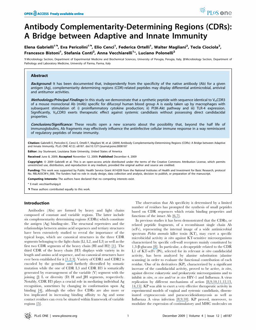
Antibody Complementarity-Determining Regions (CDRs):A Bridge between Adaptive and Innate ImmunityElena Gabrielli1., Eva Pericolini1., Elio Cenci1, Federica Ortelli1, Walter Magliani2, Tecla Ciociola2,
Francesco Bistoni1, Stefania Conti2, Anna Vecchiarelli1*, Luciano Polonelli2
1 Microbiology Section, Department of Experimental Medicine and Biochemical Sciences, University of Perugia, Perugia, Italy, 2 Microbiology Section, Department of
Pathology and Laboratory Medicine, University of Parma, Parma, Italy
Abstract
Background: It has been documented that, independently from the specificity of the native antibody (Ab) for a givenantigen (Ag), complementarity determining regions (CDR)-related peptides may display differential antimicrobial, antiviraland antitumor activities.
Methodology/Principal Findings: In this study we demonstrate that a synthetic peptide with sequence identical to VHCDR3of a mouse monoclonal Ab (mAb) specific for difucosyl human blood group A is easily taken up by macrophages withsubsequent stimulation of: i) proinflammatory cytokine production; ii) PI3K-Akt pathway and iii) TLR-4 expression.Significantly, VHCDR3 exerts therapeutic effect against systemic candidiasis without possessing direct candidacidalproperties.
Conclusions/Significance: These results open a new scenario about the possibility that, beyond the half life ofimmunoglobulins, Ab fragments may effectively influence the antiinfective cellular immune response in a way reminiscentof regulatory peptides of innate immunity.
Citation: Gabrielli E, Pericolini E, Cenci E, Ortelli F, Magliani W, et al. (2009) Antibody Complementarity-Determining Regions (CDRs): A Bridge between Adaptiveand Innate Immunity. PLoS ONE 4(12): e8187. doi:10.1371/journal.pone.0008187
Editor: Joy Sturtevant, Louisiana State University, United States of America
Received June 8, 2009; Accepted November 12, 2009; Published December 4, 2009
Copyright: � 2009 Gabrielli et al. This is an open-access article distributed under the terms of the Creative Commons Attribution License, which permitsunrestricted use, distribution, and reproduction in any medium, provided the original author and source are credited.
Funding: This work was supported by Public Health Service Grant AI14209 from the National Institutes of Health and Investment for Basic Research, protocolNo. RBLA03C9F4_006. The funders had no role in study design, data collection and analysis, decision to publish, or preparation of the manuscript.
Competing Interests: The authors have declared that no competing interests exist.
* E-mail: [email protected]
. These authors contributed equally to this work.
Introduction
Antibodies (Abs) are formed by heavy and light chains
composed of constant and variable regions. The latter include
six complementarity determining regions (CDRs) which constitute
the antigen (Ag) binding-site. The structural repertoires and the
relationships between amino acid sequences and tertiary structures
have been extensively studied to reveal the importance of the
typical loops, which are canonical structures in the three CDR
segments belonging to the light chain (Ll, L2, and L3) as well as the
first two CDR segments of the heavy chain (Hl and H2) [1]. The
third CDR of the heavy chain (H3) displays wide variety in its
length and amino acid sequence, and no canonical structures have
ever been established for it [1,2,3]. Variety of CDR1 and CDR2 is
encoded by the germline and furtherly diversified by somatic
mutation while the one of CDR L3 and CDR H3 is somatically
generated by rearrangement of the variable (V) segment with the
joining (J) L or diversity (D) H and JH segments, respectively.
Notably, CDR H3 plays a crucial role in mediating individual Ag
recognition, sometimes by changing its conformation upon Ag
binding [4], although the other five CDRs are also more or
less implicated in increasing binding affinity to Ag and some
contact residues can even be situated within framework of variable
regions [5].
The observation that Ab specificity is determined by a limited
number of residues has prompted the synthesis of small peptides
based on CDR sequences which retain binding properties and
functions of the intact Ab [6,7].
In previous studies it has been demonstrated that the CDRs, or
related peptidic fragments, of a recombinant single chain Ab
(scFv), representing the internal image of a wide antimicrobial
spectrum Pichia anomala killer toxin (KT), may exert a specific
microbicidal activity in vitro against KT-sensitive microorganisms
characterized by specific cell-wall receptors mainly constituted by
1,3-b-glucans [8]. In particular, a decapeptide related to the CDR
L1 of KT-scFv (P6), selected for its relevant in vitro candidacidal
activity, has been analyzed by alanine substitution (alanine
scanning) in order to evaluate the functional contribution of each
residue. One of its derivatives (KP), characterized by a significant
increase of the candidacidal activity, proved to be active, in vitro,
against diverse eukaryotic and prokaryotic microorganisms and to
inhibit in vitro, ex vivo and/or in vivo HIV-1 and Influenza A virus
replication by different mechanisms of action [8,9,10,11,12,13,
14,15]. KP was able to exert a very effective therapeutic activity in
experimental models of vaginal and systemic candidiasis, dissem-
inated cryptococcosis and paracoccidioidomycosis as well as
Influenza A virus infection [8,9,10]. KP proved, moreover, to
modulate the expression of costimulatory and MHC molecules on
PLoS ONE | www.plosone.org 1 December 2009 | Volume 4 | Issue 12 | e8187
murine dendritic cells (DC), after selective binding, and to improve
their capacity to induce lymphocyte proliferation [16]. Recent
studies on the structure-function relationship of KP showed its
reversible self-assembly in an hydrogel-like state. Significantly, this
process is catalyzed by 1,3-b-glucans. KP self-assembled state may
provide protection against proteases and regulate the release of the
active form over time, while the b-glucans affinity is responsible for
targeted delivery [17].
Polonelli et al. studied synthetic peptides with sequences
identical to CDRs of the light and heavy chain of three
monoclonal Abs (mAbs) characterized by different specificity:
mAb C7, directed to a protein epitope of a Candida albicans (C.
albicans) cell wall stress mannoprotein, mAb pc42, directed to a
synthetic peptide containing well-characterized B-cell and T-
cell epitopes, and a human mAb HuA, directed to blood group
A substance [18,19,20,21]. The study showed that, irrespective
of the specificity of the native Ab, the synthetic CDRs may
exert in vitro, ex vivo and/or in vivo differential inhibitory
activities against C. albicans, HIV-1 and B16F10-Nex2 mela-
noma cells, conceivably mediated by different mechanisms of
action. Alanine substituted synthetic CDRs, used as surrogates
of natural point mutations, showed further differential
increased/unaltered/decreased antimicrobial, antiviral and/
or antitumor activities [22].
As bioactive molecules, CDR-related peptides may present
some advantages over whole Abs of adaptive immunity owing to
their small size, e.g. lack of immunogenicity and better tissue
penetration, as well as over natural peptides of innate immunity
(e.g. defensins, cathelicidins, hystatins) owing to higher specificity
and affinity for targets and low systemic toxicity [23,24].
In the aim to establish whether CDRs may drive a protective
cellular immune response exclusively due to their immunomod-
ulatory activity, we studied the synthetic CDRs of mAb HuA and a
mouse mAb (MoA) which binds to the same carbohydrate epitope
[21,25]. MAbs MoA and HuA are nearly identical immunochem-
ically, even though present unrelated primary sequences, and are
representative of different ways by which the same epitope can be
recognized by the immune system [21].
Results
Candidacidal and Immunomodulatory Effects of theCDRs Peptides
In recent studies the therapeutic effect of Ab-derived peptides
characterized by immunomodulatory and/or direct candidacidal
activity has been demonstrated in the experimental model of
systemic candidiasis [8,16,22].
Here we investigated the immunomodulatory effects of the
CDRs of a human IgM mAb (HuA) specific for difucosyl human
blood group A substance, previously evaluated for their candida-
cidal properties [22], as well as the candidacidal and immuno-
modulatory effects of the CDRs of a mouse IgM mAb (MoA)
which bind to the same carbohydrate epitope [21].
First, we tested the capacity of synthetic peptides representing
mAb MoA CDRs to kill in vitro C. albicans cells. None of them
proved to display any candidacidal effect in vitro in the adopted
experimental conditions.
In parallel experiments we analyzed the capacity of all murine
and human synthetic CDRs to stimulate cytokine production by a
non homogeneous cell population such as murine splenocytes. An
irrelevant synthetic peptide previously recognized unable to
stimulate immune cells was used as negative control (NC) in this
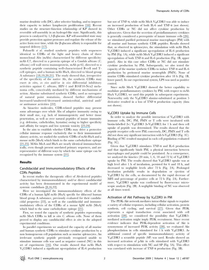
set of experiments [16]. Our results showed that mAb HuA
VLCDR3 induced a significant up-regulation of IL-6 production
but not of TNF-a, while mAb MoA VHCDR3 was able to induce
an increased production of both IL-6 and TNF-a (not shown).
Other CDRs or NC did not affect cytokine production by
splenocytes. Given that the secretion of proinflammatory cytokines
is generally considered a prerequisite of innate immune cells [26],
we stimulated purified peritoneal murine macrophages (PM) with
all murine and human synthetic CDR peptides. Results showed
that, as observed in splenocytes, the stimulation with mAb HuA
VLCDR3 induced a significant up-regulation of IL-6 production
by PM (Fig. 1A), while mAb MoA VHCDR3 induced a significant
up-regulation of both TNF-a and IL-6 production (Fig. 1B, upper
panel). Also in this case other CDRs or NC did not stimulate
cytokine production by PM. Subsequently, we also tested the
capacity of the murine synthetic CDRs to induce TNF-a and IL-6
production by peritoneal murine neutrophils (PMN). None of
murine CDRs stimulated cytokine production after 18 h (Fig. 1B,
lower panel). In our experimental system, LPS was used as positive
control.
Since mAb MoA VHCDR3 showed the better capability to
modulate proinflammatory cytokines by PM, with respect to mAb
HuA VLCDR3, we used this peptide in subsequent experiments.
Noteworthy only the VHCDR3 alanine-substituted at position 5
derivative resulted in a loss of TNF-a production capacity (data
not shown).
VHCDR3 Uptake by Immune CellsIn order to analyze the possible interaction of VHCDR3 with
immune cells, DC, PM, PMN or T cells were incubated with
biotin-labelled (b-) VHCDR3 (b-VHCDR3) for 1 h and then cell
uptake of peptide was determined. The results showed that the
peptide receptive cells were PM; conversely, DC, PMN and T cells
did not show any significant interaction with b-VHCDR3 (Fig. 1C).
Binding of NC resulted marginal in each cell population evaluated
(Fig. 1C).
Given that VHCDR3 stimulates TNF-a and IL-6 production
and that significantly binds PM, a physical interaction between
macrophages and peptide could be postulated. As a consequence,
we analyzed the kinetics (20 min, 1, 6, 18 and 72 h) of VHCDR3
uptake by PM. The results showed that VHCDR3 uptake was at
high level after 1 h of incubation, persisted for 6 and 18 h, and
decreased to negligible levels after 72 h (Fig. 2A). Prolonged
incubation probably results in degradation or ejection of
VHCDR3 by the cells, as documented by the rapid decrease of
MFI and percentage of positive cells at 72 h (Fig. 2A). Further-
more, VHCDR3 uptake was confirmed by fluorescence micro-
scopic analysis (Fig. 2B). A negligible binding of NC was observed
at all times tested.
Activation of Akt Pathway by VHCDR3 in PMThe PI3K-Akt network mediates intracellular signals to regulate
a variety of cellular responses, including cellular activation, protein
synthesis, cell cycling, and survival [27]. Given that PI3K
represents a signal transduction event associated with cell
activation [28], we considered the possibility that VHCDR3-
mediated activation might imply PI3K recruitment. Since recent
evidence indicates that PI3K-dependent activation of Akt is
synonymous of increased PI3K activity [28], we evaluated Akt
phosphorylation in cells stimulated for 1 h with VHCDR3. As
additional control of specific immunomodulatory activity of
VHCDR3 a scramble peptide (SP) was used. Results showed an
increased activation of pAkt in cells stimulated with VHCDR3
with respect to stimulation with NC and SP (Fig. 3A). This effect
was correlated with increase of TNF-a production (Fig. 3B).
Therapeutic Activity of CDRs
PLoS ONE | www.plosone.org 2 December 2009 | Volume 4 | Issue 12 | e8187
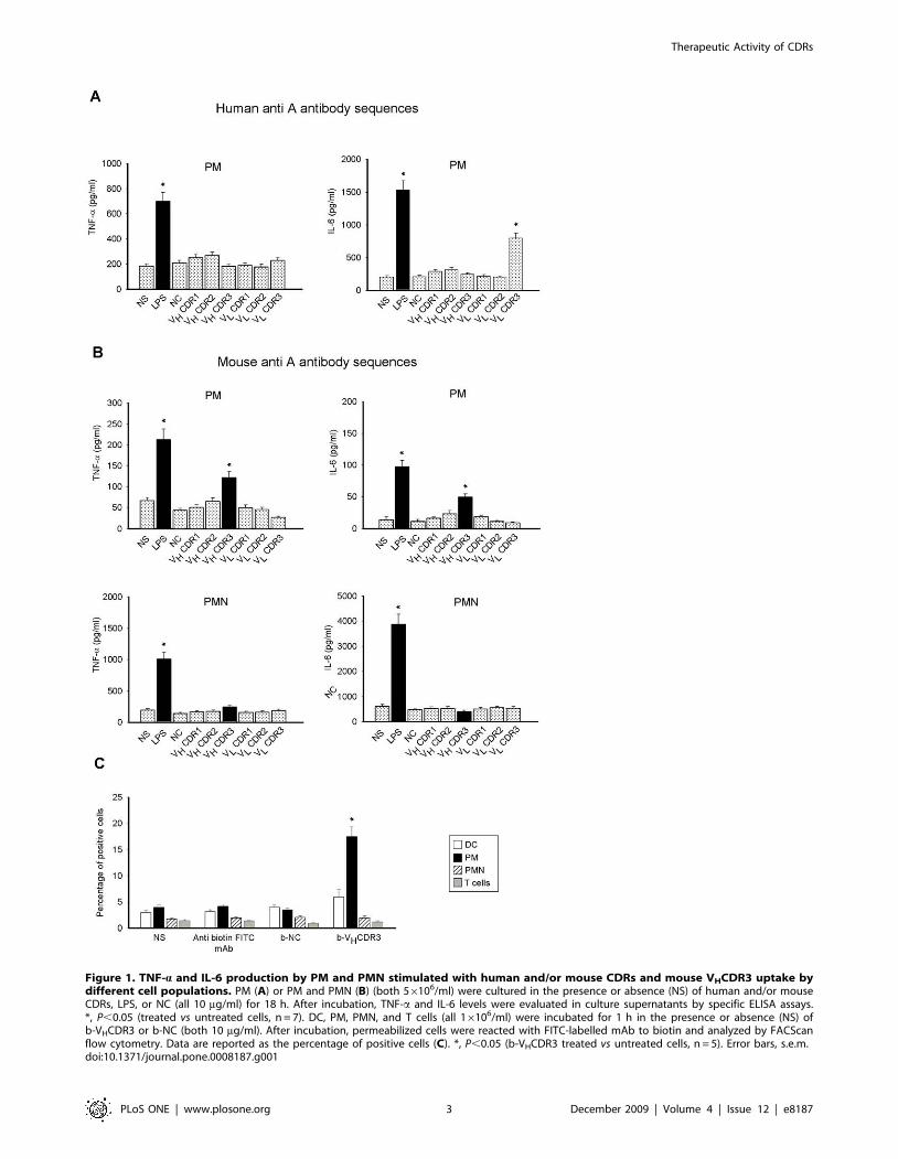
Figure 1. TNF-a and IL-6 production by PM and PMN stimulated with human and/or mouse CDRs and mouse VHCDR3 uptake bydifferent cell populations. PM (A) or PM and PMN (B) (both 56106/ml) were cultured in the presence or absence (NS) of human and/or mouseCDRs, LPS, or NC (all 10 mg/ml) for 18 h. After incubation, TNF-a and IL-6 levels were evaluated in culture supernatants by specific ELISA assays.*, P,0.05 (treated vs untreated cells, n = 7). DC, PM, PMN, and T cells (all 16106/ml) were incubated for 1 h in the presence or absence (NS) ofb-VHCDR3 or b-NC (both 10 mg/ml). After incubation, permeabilized cells were reacted with FITC-labelled mAb to biotin and analyzed by FACScanflow cytometry. Data are reported as the percentage of positive cells (C). *, P,0.05 (b-VHCDR3 treated vs untreated cells, n = 5). Error bars, s.e.m.doi:10.1371/journal.pone.0008187.g001
Therapeutic Activity of CDRs
PLoS ONE | www.plosone.org 3 December 2009 | Volume 4 | Issue 12 | e8187
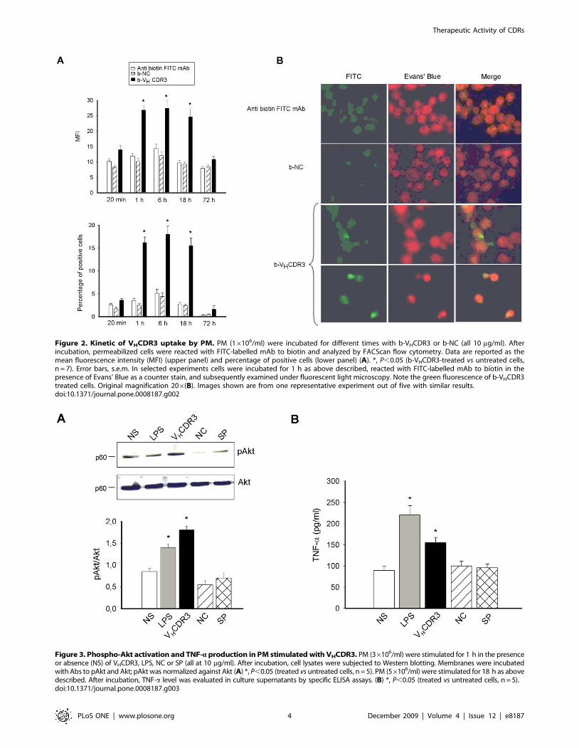
Figure 2. Kinetic of VHCDR3 uptake by PM. PM (16106/ml) were incubated for different times with b-VHCDR3 or b-NC (all 10 mg/ml). Afterincubation, permeabilized cells were reacted with FITC-labelled mAb to biotin and analyzed by FACScan flow cytometry. Data are reported as themean fluorescence intensity (MFI) (upper panel) and percentage of positive cells (lower panel) (A). *, P,0.05 (b-VHCDR3-treated vs untreated cells,n = 7). Error bars, s.e.m. In selected experiments cells were incubated for 1 h as above described, reacted with FITC-labelled mAb to biotin in thepresence of Evans’ Blue as a counter stain, and subsequently examined under fluorescent light microscopy. Note the green fluorescence of b-VHCDR3treated cells. Original magnification 206(B). Images shown are from one representative experiment out of five with similar results.doi:10.1371/journal.pone.0008187.g002
Figure 3. Phospho-Akt activation and TNF-a production in PM stimulated with VHCDR3. PM (36106/ml) were stimulated for 1 h in the presenceor absence (NS) of VHCDR3, LPS, NC or SP (all at 10 mg/ml). After incubation, cell lysates were subjected to Western blotting. Membranes were incubatedwith Abs to pAkt and Akt; pAkt was normalized against Akt (A) *, P,0.05 (treated vs untreated cells, n = 5). PM (56106/ml) were stimulated for 18 h as abovedescribed. After incubation, TNF-a level was evaluated in culture supernatants by specific ELISA assays. (B) *, P,0.05 (treated vs untreated cells, n = 5).doi:10.1371/journal.pone.0008187.g003
Therapeutic Activity of CDRs
PLoS ONE | www.plosone.org 4 December 2009 | Volume 4 | Issue 12 | e8187
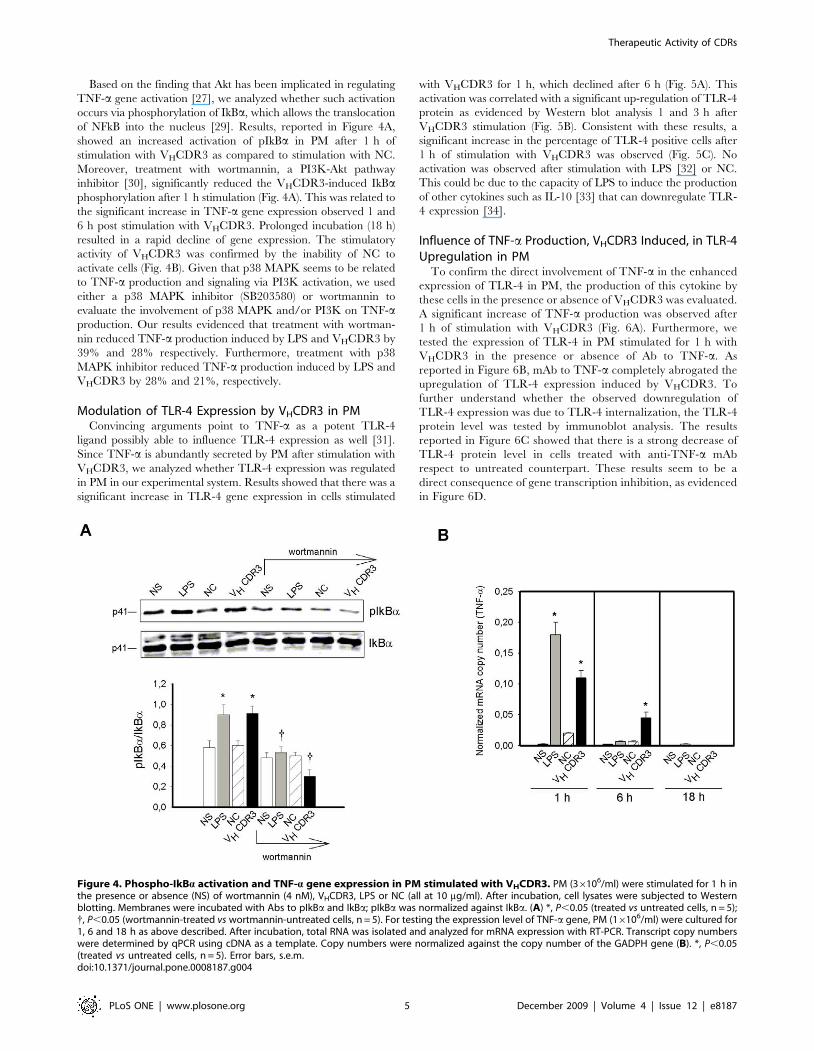
Based on the finding that Akt has been implicated in regulating
TNF-a gene activation [27], we analyzed whether such activation
occurs via phosphorylation of IkBa, which allows the translocation
of NFkB into the nucleus [29]. Results, reported in Figure 4A,
showed an increased activation of pIkBa in PM after 1 h of
stimulation with VHCDR3 as compared to stimulation with NC.
Moreover, treatment with wortmannin, a PI3K-Akt pathway
inhibitor [30], significantly reduced the VHCDR3-induced IkBaphosphorylation after 1 h stimulation (Fig. 4A). This was related to
the significant increase in TNF-a gene expression observed 1 and
6 h post stimulation with VHCDR3. Prolonged incubation (18 h)
resulted in a rapid decline of gene expression. The stimulatory
activity of VHCDR3 was confirmed by the inability of NC to
activate cells (Fig. 4B). Given that p38 MAPK seems to be related
to TNF-a production and signaling via PI3K activation, we used
either a p38 MAPK inhibitor (SB203580) or wortmannin to
evaluate the involvement of p38 MAPK and/or PI3K on TNF-aproduction. Our results evidenced that treatment with wortman-
nin reduced TNF-a production induced by LPS and VHCDR3 by
39% and 28% respectively. Furthermore, treatment with p38
MAPK inhibitor reduced TNF-a production induced by LPS and
VHCDR3 by 28% and 21%, respectively.
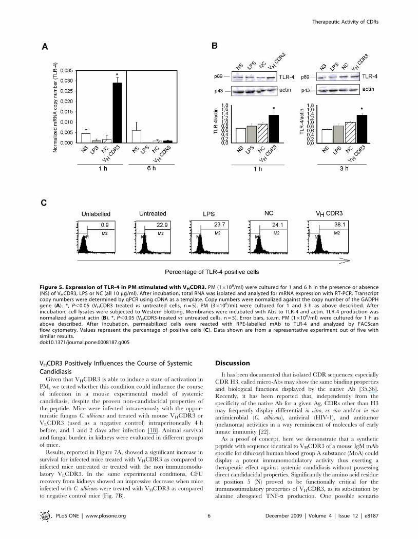
Modulation of TLR-4 Expression by VHCDR3 in PMConvincing arguments point to TNF-a as a potent TLR-4
ligand possibly able to influence TLR-4 expression as well [31].
Since TNF-a is abundantly secreted by PM after stimulation with
VHCDR3, we analyzed whether TLR-4 expression was regulated
in PM in our experimental system. Results showed that there was a
significant increase in TLR-4 gene expression in cells stimulated
with VHCDR3 for 1 h, which declined after 6 h (Fig. 5A). This
activation was correlated with a significant up-regulation of TLR-4
protein as evidenced by Western blot analysis 1 and 3 h after
VHCDR3 stimulation (Fig. 5B). Consistent with these results, a
significant increase in the percentage of TLR-4 positive cells after
1 h of stimulation with VHCDR3 was observed (Fig. 5C). No
activation was observed after stimulation with LPS [32] or NC.
This could be due to the capacity of LPS to induce the production
of other cytokines such as IL-10 [33] that can downregulate TLR-
4 expression [34].
Influence of TNF-a Production, VHCDR3 Induced, in TLR-4Upregulation in PM
To confirm the direct involvement of TNF-a in the enhanced
expression of TLR-4 in PM, the production of this cytokine by
these cells in the presence or absence of VHCDR3 was evaluated.
A significant increase of TNF-a production was observed after
1 h of stimulation with VHCDR3 (Fig. 6A). Furthermore, we
tested the expression of TLR-4 in PM stimulated for 1 h with
VHCDR3 in the presence or absence of Ab to TNF-a. As
reported in Figure 6B, mAb to TNF-a completely abrogated the
upregulation of TLR-4 expression induced by VHCDR3. To
further understand whether the observed downregulation of
TLR-4 expression was due to TLR-4 internalization, the TLR-4
protein level was tested by immunoblot analysis. The results
reported in Figure 6C showed that there is a strong decrease of
TLR-4 protein level in cells treated with anti-TNF-a mAb
respect to untreated counterpart. These results seem to be a
direct consequence of gene transcription inhibition, as evidenced
in Figure 6D.
Figure 4. Phospho-IkBa activation and TNF-a gene expression in PM stimulated with VHCDR3. PM (36106/ml) were stimulated for 1 h inthe presence or absence (NS) of wortmannin (4 nM), VHCDR3, LPS or NC (all at 10 mg/ml). After incubation, cell lysates were subjected to Westernblotting. Membranes were incubated with Abs to pIkBa and IkBa; pIkBa was normalized against IkBa. (A) *, P,0.05 (treated vs untreated cells, n = 5);{, P,0.05 (wortmannin-treated vs wortmannin-untreated cells, n = 5). For testing the expression level of TNF-a gene, PM (16106/ml) were cultured for1, 6 and 18 h as above described. After incubation, total RNA was isolated and analyzed for mRNA expression with RT-PCR. Transcript copy numberswere determined by qPCR using cDNA as a template. Copy numbers were normalized against the copy number of the GADPH gene (B). *, P,0.05(treated vs untreated cells, n = 5). Error bars, s.e.m.doi:10.1371/journal.pone.0008187.g004
Therapeutic Activity of CDRs
PLoS ONE | www.plosone.org 5 December 2009 | Volume 4 | Issue 12 | e8187
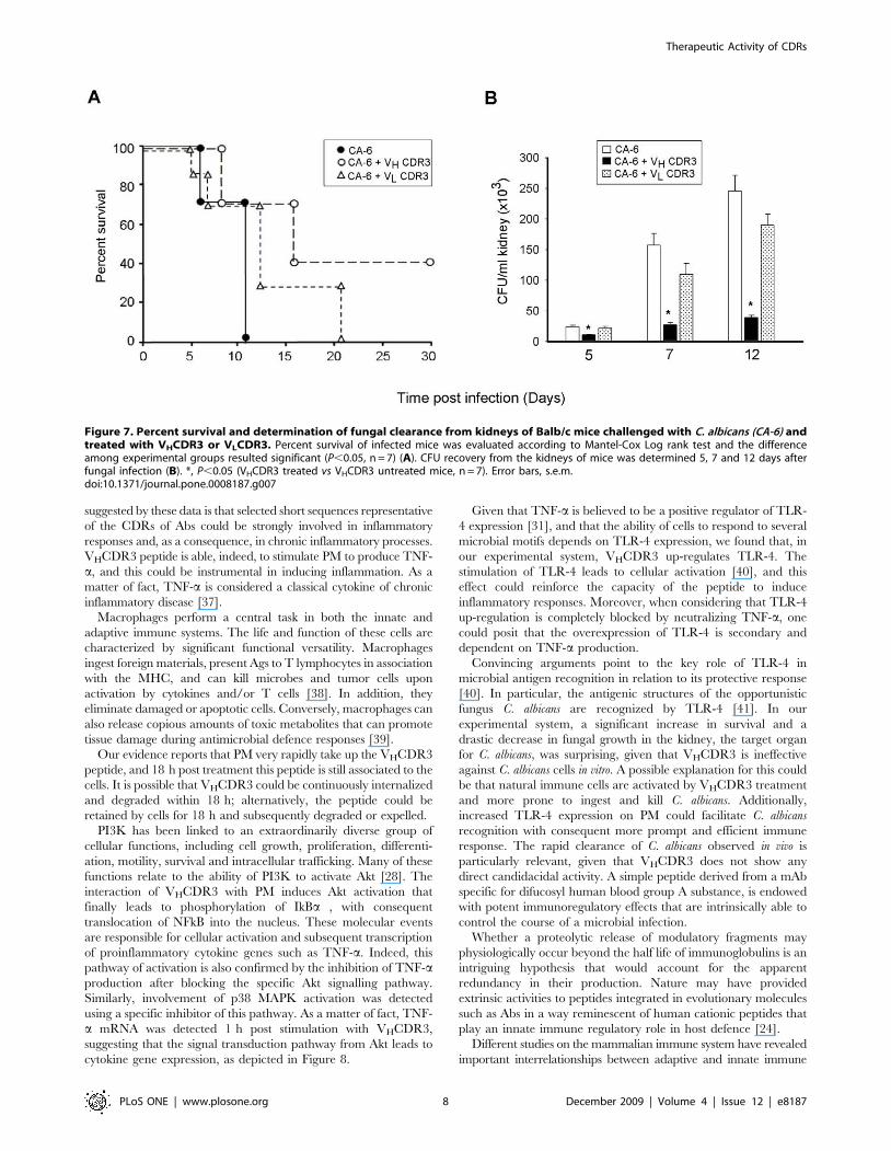
VHCDR3 Positively Influences the Course of SystemicCandidiasis
Given that VHCDR3 is able to induce a state of activation in
PM, we tested whether this condition could influence the course
of infection in a mouse experimental model of systemic
candidiasis, despite the proven non-candidacidal properties of
the peptide. Mice were infected intravenously with the oppor-
tunistic fungus C. albicans and treated with mouse VHCDR3 or
VLCDR3 (used as a negative control) intraperitoneally 4 h
before, and 1 and 2 days after infection [18]. Animal survival
and fungal burden in kidneys were evaluated in different groups
of mice.
Results, reported in Figure 7A, showed a significant increase in
survival for infected mice treated with VHCDR3 as compared to
infected mice untreated or treated with the non immunomodu-
latory VLCDR3. In the same experimental conditions, CFU
recovery from kidneys showed an impressive decrease when mice
infected with C. albicans were treated with VHCDR3 as compared
to negative control mice (Fig. 7B).
Discussion
It has been documented that isolated CDR sequences, especially
CDR H3, called micro-Abs may show the same binding properties
and biological functions displayed by the native Ab [35,36].
Recently, it has been reported that, independently from the
specificity of the native Ab for a given Ag, CDRs other than H3
may frequently display differential in vitro, ex vivo and/or in vivo
antimicrobial (C. albicans), antiviral (HIV-1), and antitumor
(melanoma) activities in a way reminiscent of molecules of early
innate immunity [22].
As a proof of concept, here we demonstrate that a synthetic
peptide with sequence identical to VHCDR3 of a mouse IgM mAb
specific for difucosyl human blood group A substance (MoA) could
display a potent immunomodulatory activity thus exerting a
therapeutic effect against systemic candidiasis without possessing
direct candidacidal properties. Significantly the amino acid residue
at position 5 (N) proved to be functionally critical for the
immunostimulatory properties of VHCDR3, as its substitution by
alanine abrogated TNF-a production. One possible scenario
Figure 5. Expression of TLR-4 in PM stimulated with VHCDR3. PM (16106/ml) were cultured for 1 and 6 h in the presence or absence(NS) of VHCDR3, LPS or NC (all 10 mg/ml). After incubation, total RNA was isolated and analyzed for mRNA expression with RT-PCR. Transcriptcopy numbers were determined by qPCR using cDNA as a template. Copy numbers were normalized against the copy number of the GADPHgene (A). *, P,0.05 (VHCDR3 treated vs untreated cells, n = 5). PM (36106/ml) were cultured for 1 and 3 h as above described. Afterincubation, cell lysates were subjected to Western blotting. Membranes were incubated with Abs to TLR-4 and actin. TLR-4 production wasnormalized against actin (B). *, P,0.05 (VHCDR3-treated vs untreated cells, n = 5). Error bars, s.e.m. PM (16106/ml) were cultured for 1 h asabove described. After incubation, permeabilized cells were reacted with RPE-labelled mAb to TLR-4 and analyzed by FACScanflow cytometry. Values represent the percentage of positive cells (C). Data shown are from a representative experiment out of five withsimilar results.doi:10.1371/journal.pone.0008187.g005
Therapeutic Activity of CDRs
PLoS ONE | www.plosone.org 6 December 2009 | Volume 4 | Issue 12 | e8187

Figure 6. TNF-a induced TLR-4 expression in PM stimulated with VHCDR3. PM (56106/ml) were cultured for 1 h in the presence or absence(NS) of VHCDR3, LPS or NC (all 10 mg/ml). After incubation, TNF-a level was evaluated in culture supernatants by specific ELISA assay (A). *, P,0.05(treated vs untreated cells, n = 7). PM (16106/ml) were cultured for 1 h with VHCDR3, LPS or NC (all 10 mg/ml), in the presence or absence (NS) of mAbto TNF-a (0.5 mg/ml). After incubation, permeabilized cells were reacted with RPE-labelled mAb to TLR-4 and analyzed by FACScan flow cytometry.Values represent the percentage of positive cells (B) *, P,0.05 (VHCDR3 plus mAb to TNF-a treated vs VHCDR3 treated cells, n = 7). PM (36106/ml)were cultured for 1 h as above described. After incubation, cell lysates were subjected to Western blotting. Membranes were incubated with Abs toTLR-4 and actin. TLR-4 production was normalized against actin (C) *, P,0.05 (VHCDR3 plus mAb to TNF-a treated vs VHCDR3 treated cells, n = 5). Fortesting the expression level of TLR-4 gene, PM (16106/ml) were cultured for 1 h as above described. After incubation, total RNA was isolated andanalyzed for mRNA expression with RT-PCR. Transcript copy numbers were determined by qPCR using cDNA as a template. Copy numbers werenormalized against the copy number of the GADPH gene (D). *, P,0.05 (VHCDR3 plus mAb to TNF-a treated vs VHCDR3 treated cells, n = 5). Error bars,s.e.m.doi:10.1371/journal.pone.0008187.g006
Therapeutic Activity of CDRs
PLoS ONE | www.plosone.org 7 December 2009 | Volume 4 | Issue 12 | e8187
suggested by these data is that selected short sequences representative
of the CDRs of Abs could be strongly involved in inflammatory
responses and, as a consequence, in chronic inflammatory processes.
VHCDR3 peptide is able, indeed, to stimulate PM to produce TNF-
a, and this could be instrumental in inducing inflammation. As a
matter of fact, TNF-a is considered a classical cytokine of chronic
inflammatory disease [37].
Macrophages perform a central task in both the innate and
adaptive immune systems. The life and function of these cells are
characterized by significant functional versatility. Macrophages
ingest foreign materials, present Ags to T lymphocytes in association
with the MHC, and can kill microbes and tumor cells upon
activation by cytokines and/or T cells [38]. In addition, they
eliminate damaged or apoptotic cells. Conversely, macrophages can
also release copious amounts of toxic metabolites that can promote
tissue damage during antimicrobial defence responses [39].
Our evidence reports that PM very rapidly take up the VHCDR3
peptide, and 18 h post treatment this peptide is still associated to the
cells. It is possible that VHCDR3 could be continuously internalized
and degraded within 18 h; alternatively, the peptide could be
retained by cells for 18 h and subsequently degraded or expelled.
PI3K has been linked to an extraordinarily diverse group of
cellular functions, including cell growth, proliferation, differenti-
ation, motility, survival and intracellular trafficking. Many of these
functions relate to the ability of PI3K to activate Akt [28]. The
interaction of VHCDR3 with PM induces Akt activation that
finally leads to phosphorylation of IkBa , with consequent
translocation of NFkB into the nucleus. These molecular events
are responsible for cellular activation and subsequent transcription
of proinflammatory cytokine genes such as TNF-a. Indeed, this
pathway of activation is also confirmed by the inhibition of TNF-aproduction after blocking the specific Akt signalling pathway.
Similarly, involvement of p38 MAPK activation was detected
using a specific inhibitor of this pathway. As a matter of fact, TNF-
a mRNA was detected 1 h post stimulation with VHCDR3,
suggesting that the signal transduction pathway from Akt leads to
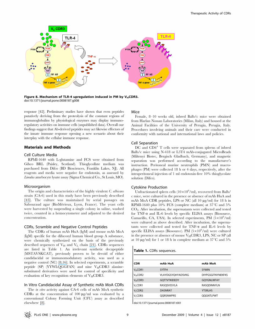
cytokine gene expression, as depicted in Figure 8.
Given that TNF-a is believed to be a positive regulator of TLR-
4 expression [31], and that the ability of cells to respond to several
microbial motifs depends on TLR-4 expression, we found that, in
our experimental system, VHCDR3 up-regulates TLR-4. The
stimulation of TLR-4 leads to cellular activation [40], and this
effect could reinforce the capacity of the peptide to induce
inflammatory responses. Moreover, when considering that TLR-4
up-regulation is completely blocked by neutralizing TNF-a, one
could posit that the overexpression of TLR-4 is secondary and
dependent on TNF-a production.
Convincing arguments point to the key role of TLR-4 in
microbial antigen recognition in relation to its protective response
[40]. In particular, the antigenic structures of the opportunistic
fungus C. albicans are recognized by TLR-4 [41]. In our
experimental system, a significant increase in survival and a
drastic decrease in fungal growth in the kidney, the target organ
for C. albicans, was surprising, given that VHCDR3 is ineffective
against C. albicans cells in vitro. A possible explanation for this could
be that natural immune cells are activated by VHCDR3 treatment
and more prone to ingest and kill C. albicans. Additionally,
increased TLR-4 expression on PM could facilitate C. albicans
recognition with consequent more prompt and efficient immune
response. The rapid clearance of C. albicans observed in vivo is
particularly relevant, given that VHCDR3 does not show any
direct candidacidal activity. A simple peptide derived from a mAb
specific for difucosyl human blood group A substance, is endowed
with potent immunoregulatory effects that are intrinsically able to
control the course of a microbial infection.
Whether a proteolytic release of modulatory fragments may
physiologically occur beyond the half life of immunoglobulins is an
intriguing hypothesis that would account for the apparent
redundancy in their production. Nature may have provided
extrinsic activities to peptides integrated in evolutionary molecules
such as Abs in a way reminescent of human cationic peptides that
play an innate immune regulatory role in host defence [24].
Different studies on the mammalian immune system have revealed
important interrelationships between adaptive and innate immune
Figure 7. Percent survival and determination of fungal clearance from kidneys of Balb/c mice challenged with C. albicans (CA-6) andtreated with VHCDR3 or VLCDR3. Percent survival of infected mice was evaluated according to Mantel-Cox Log rank test and the differenceamong experimental groups resulted significant (P,0.05, n = 7) (A). CFU recovery from the kidneys of mice was determined 5, 7 and 12 days afterfungal infection (B). *, P,0.05 (VHCDR3 treated vs VHCDR3 untreated mice, n = 7). Error bars, s.e.m.doi:10.1371/journal.pone.0008187.g007
Therapeutic Activity of CDRs
PLoS ONE | www.plosone.org 8 December 2009 | Volume 4 | Issue 12 | e8187
response [42]. Preliminary studies have shown that even peptides
putatively deriving from the proteolysis of the constant regions of
immunoglobulins by physiological enzymes may display immuno-
regulatory activities on immune cells (unpublished data). Overall our
findings suggest that Ab-derived peptides may act likewise effectors of
the innate immune response opening a new scenario about their
interplay with the cellular immune response.
Materials and Methods
Cell Culture MediaRPMI-1640 with L-glutamine and FCS were obtained from
Gibco BRL (Paisley, Scotland). Thioglycollate medium was
purchased from Difco (BD Biosciences, Franklin Lakes, NJ). All
reagents and media were negative for endotoxin, as assessed by
Limulus amebocyte lysate assay (Sigma Chemical Co., St Louis, MO).
MicroorganismThe origin and characteristics of the highly virulent C. albicans
strain (CA-6) used in this study have been previously described
[43]. The culture was maintained by serial passages on
Sabouraud agar (BioMerieux, Lyon, France). The yeast cells
were harvested by suspending a single colony in saline, washed
twice, counted in a hemocytometer and adjusted to the desired
concentration.
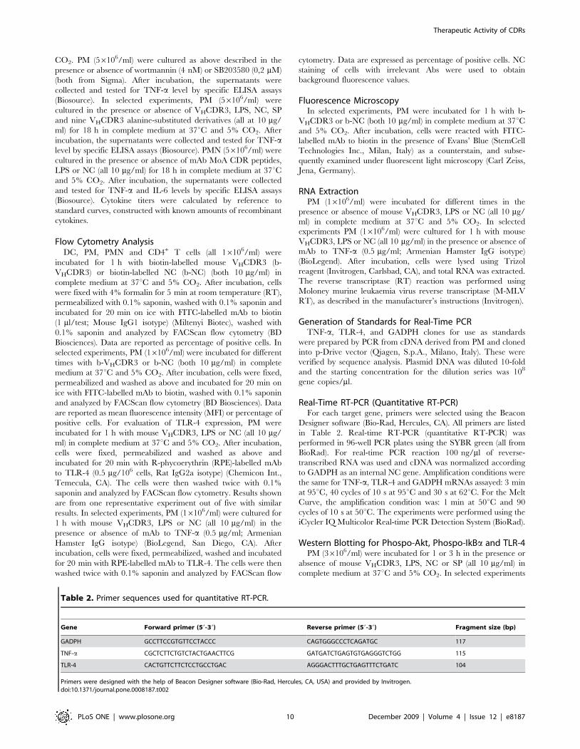
CDRs, Scramble and Negative Control PeptidesThe CDRs of human mAb HuA (IgM) and mouse mAb MoA
(IgM) specific for the difucosyl human blood group A substance,
were chemically synthesized on the basis of the previously
described sequences of VH and VL chain [21]. CDRs sequences
are listed in Table 1. An irrelevant synthetic decapeptide
(MSTAVSKCAT), previously proven to be devoid of either
candidacidal or immunomodulatory activity, was used as a
negative control (NC) [8,16]. In selected experiments, a scramble
peptide (SP) (YYWLQGGFAN) and nine VHCDR3 alanine-
substituted derivatives were used for control of specificity and
evaluation of key recognition elements of VHCDR3.
In Vitro Candidacidal Assay of Synthetic mAb MoA CDRsThe in vitro activity against CA-6 cells of mAb MoA synthetic
CDRs at the concentration of 100 mg/ml was evaluated by a
conventional Colony Forming Unit (CFU) assay as described
elsewhere [8].
MiceFemale, 8–10 weeks old, inbred Balb/c mice were obtained
from Harlan Nossan Laboratories (Milan, Italy) and housed at the
Animal Facilities of the University of Perugia, Perugia, Italy.
Procedures involving animals and their care were conducted in
conformity with national and international laws and policies.
Cell SeparationDC and CD4+ T cells were separated from spleens of inbred
Balb/c mice using N-418 or L3T4 mAb-conjugated MicroBeads
(Miltenyi Biotec, Bergisch Gladbach, Germany), and magnetic
separation was performed according to the manufacturer’s
instruction. Peritoneal murine neutrophils (PMN) and macro-
phages (PM) were collected 18 h or 4 days, respectively, after the
intraperitoneal injection of 1 ml endotoxin-free 10% thioglycolate
solution (Difco).
Cytokine ProductionUnfractionated spleen cells (106106/ml), recovered from Balb/
c mice, were cultured in the presence or absence of mAb HuA and
mAb MoA CDR peptides, LPS or NC (all 10 mg/ml) for 18 h in
RPMI-1640 plus 10% FCS (complete medium) at 37uC and 5%
CO2. After incubation, the supernatants were collected and tested
for TNF-a and IL-6 levels by specific ELISA assays (Biosource,
Camarillo, CA, USA). In selected experiments, PM (56106/ml)
were cultured as above described. After incubation, the superna-
tants were collected and tested for TNF-a and IL-6 levels by
specific ELISA assays (Biosource). PM (56106/ml) were cultured
in the presence or absence of mouse VHCDR3, LPS, NC or SP (all
at 10 mg/ml) for 1 or 18 h in complete medium at 37uC and 5%
Table 1. CDRs sequences.
CDR mAb HuA mAb MoA
VHCDR1 SYTFH SYWIN
VHCDR2 VLAYDGSYQHYADSVKG DIYPGSGITNYNEKFKS
VHCDR3 GQTTVTKIDEDY GQYGNLWFAY
VLCDR1 RASQSVSSYLA RASQDINNYLN
VLCDR2 DASNRAT YTSRLHS
VLCDR3 QQRSNWPRS QQGNTLPWT
doi:10.1371/journal.pone.0008187.t001
Figure 8. Mechanism of TLR-4 upregulation induced in PM by VHCDR3.doi:10.1371/journal.pone.0008187.g008
Therapeutic Activity of CDRs
PLoS ONE | www.plosone.org 9 December 2009 | Volume 4 | Issue 12 | e8187
CO2. PM (56106/ml) were cultured as above described in the
presence or absence of wortmannin (4 nM) or SB203580 (0,2 mM)
(both from Sigma). After incubation, the supernatants were
collected and tested for TNF-a level by specific ELISA assays
(Biosource). In selected experiments, PM (56106/ml) were
cultured in the presence or absence of VHCDR3, LPS, NC, SP
and nine VHCDR3 alanine-substituted derivatives (all at 10 mg/
ml) for 18 h in complete medium at 37uC and 5% CO2. After
incubation, the supernatants were collected and tested for TNF-alevel by specific ELISA assays (Biosource). PMN (56106/ml) were
cultured in the presence or absence of mAb MoA CDR peptides,
LPS or NC (all 10 mg/ml) for 18 h in complete medium at 37uCand 5% CO2. After incubation, the supernatants were collected
and tested for TNF-a and IL-6 levels by specific ELISA assays
(Biosource). Cytokine titers were calculated by reference to
standard curves, constructed with known amounts of recombinant
cytokines.
Flow Cytometry AnalysisDC, PM, PMN and CD4+ T cells (all 16106/ml) were
incubated for 1 h with biotin-labelled mouse VHCDR3 (b-
VHCDR3) or biotin-labelled NC (b-NC) (both 10 mg/ml) in
complete medium at 37uC and 5% CO2. After incubation, cells
were fixed with 4% formalin for 5 min at room temperature (RT),
permeabilized with 0.1% saponin, washed with 0.1% saponin and
incubated for 20 min on ice with FITC-labelled mAb to biotin
(1 ml/test; Mouse IgG1 isotype) (Miltenyi Biotec), washed with
0.1% saponin and analyzed by FACScan flow cytometry (BD
Biosciences). Data are reported as percentage of positive cells. In
selected experiments, PM (16106/ml) were incubated for different
times with b-VHCDR3 or b-NC (both 10 mg/ml) in complete
medium at 37uC and 5% CO2. After incubation, cells were fixed,
permeabilized and washed as above and incubated for 20 min on
ice with FITC-labelled mAb to biotin, washed with 0.1% saponin
and analyzed by FACScan flow cytometry (BD Biosciences). Data
are reported as mean fluorescence intensity (MFI) or percentage of
positive cells. For evaluation of TLR-4 expression, PM were
incubated for 1 h with mouse VHCDR3, LPS or NC (all 10 mg/
ml) in complete medium at 37uC and 5% CO2. After incubation,
cells were fixed, permeabilized and washed as above and
incubated for 20 min with R-phycoerythrin (RPE)-labelled mAb
to TLR-4 (0.5 mg/106 cells, Rat IgG2a isotype) (Chemicon Int.,
Temecula, CA). The cells were then washed twice with 0.1%
saponin and analyzed by FACScan flow cytometry. Results shown
are from one representative experiment out of five with similar
results. In selected experiments, PM (16106/ml) were cultured for
1 h with mouse VHCDR3, LPS or NC (all 10 mg/ml) in the
presence or absence of mAb to TNF-a (0.5 mg/ml; Armenian
Hamster IgG isotype) (BioLegend, San Diego, CA). After
incubation, cells were fixed, permeabilized, washed and incubated
for 20 min with RPE-labelled mAb to TLR-4. The cells were then
washed twice with 0.1% saponin and analyzed by FACScan flow
cytometry. Data are expressed as percentage of positive cells. NC
staining of cells with irrelevant Abs were used to obtain
background fluorescence values.
Fluorescence MicroscopyIn selected experiments, PM were incubated for 1 h with b-
VHCDR3 or b-NC (both 10 mg/ml) in complete medium at 37uCand 5% CO2. After incubation, cells were reacted with FITC-
labelled mAb to biotin in the presence of Evans’ Blue (StemCell
Technologies Inc., Milan, Italy) as a counterstain, and subse-
quently examined under fluorescent light microscopy (Carl Zeiss,
Jena, Germany).
RNA ExtractionPM (16106/ml) were incubated for different times in the
presence or absence of mouse VHCDR3, LPS or NC (all 10 mg/
ml) in complete medium at 37uC and 5% CO2. In selected
experiments PM (16106/ml) were cultured for 1 h with mouse
VHCDR3, LPS or NC (all 10 mg/ml) in the presence or absence of
mAb to TNF-a (0.5 mg/ml; Armenian Hamster IgG isotype)
(BioLegend). After incubation, cells were lysed using Trizol
reagent (Invitrogen, Carlsbad, CA), and total RNA was extracted.
The reverse transcriptase (RT) reaction was performed using
Moloney murine leukaemia virus reverse transcriptase (M-MLV
RT), as described in the manufacturer’s instructions (Invitrogen).
Generation of Standards for Real-Time PCRTNF-a, TLR-4, and GADPH clones for use as standards
were prepared by PCR from cDNA derived from PM and cloned
into p-Drive vector (Qiagen, S.p.A., Milano, Italy). These were
verified by sequence analysis. Plasmid DNA was diluted 10-fold
and the starting concentration for the dilution series was 108
gene copies/ml.
Real-Time RT-PCR (Quantitative RT-PCR)For each target gene, primers were selected using the Beacon
Designer software (Bio-Rad, Hercules, CA). All primers are listed
in Table 2. Real-time RT-PCR (quantitative RT-PCR) was
performed in 96-well PCR plates using the SYBR green (all from
BioRad). For real-time PCR reaction 100 ng/ml of reverse-
transcribed RNA was used and cDNA was normalized according
to GADPH as an internal NC gene. Amplification conditions were
the same for TNF-a, TLR-4 and GADPH mRNAs assayed: 3 min
at 95uC, 40 cycles of 10 s at 95uC and 30 s at 62uC. For the Melt
Curve, the amplification condition was: 1 min at 50uC and 90
cycles of 10 s at 50uC. The experiments were performed using the
iCycler IQ Multicolor Real-time PCR Detection System (BioRad).
Western Blotting for Phospo-Akt, Phospo-IkBa and TLR-4PM (36106/ml) were incubated for 1 or 3 h in the presence or
absence of mouse VHCDR3, LPS, NC or SP (all 10 mg/ml) in
complete medium at 37uC and 5% CO2. In selected experiments
Table 2. Primer sequences used for quantitative RT-PCR.
Gene Forward primer (59-39) Reverse primer (59-39) Fragment size (bp)
GADPH GCCTTCCGTGTTCCTACCC CAGTGGGCCCTCAGATGC 117
TNF-a CGCTCTTCTGTCTACTGAACTTCG GATGATCTGAGTGTGAGGGTCTGG 115
TLR-4 CACTGTTCTTCTCCTGCCTGAC AGGGACTTTGCTGAGTTTCTGATC 104
Primers were designed with the help of Beacon Designer software (Bio-Rad, Hercules, CA, USA) and provided by Invitrogen.doi:10.1371/journal.pone.0008187.t002
Therapeutic Activity of CDRs
PLoS ONE | www.plosone.org 10 December 2009 | Volume 4 | Issue 12 | e8187
PM (36106/ml) were incubated for 1 h in the presence or absence
of wortmannin (4 nM) (Sigma) and mouse VHCDR3, LPS or NC
(all at 10 mg/ml) in complete medium at 37uC and 5% CO2. After
incubation, cells were washed and lysated with Mammalian
protein extraction reagent (M-PER) in the presence of protease
and phosphatase inhibitors (all from Pierce, Rockford, IL). Protein
concentration was determined with a BCA protein Assay Reagent
kit (Pierce). The lysates (30 mg of each sample) were separated by
sodium dodecyl-sulfate-10% PAGE, transferred to a nitrocellulose
membrane (Pierce) for 1 h at 100 V in a blotting system (Bio-Rad)
for Western Blot analysis, and the membranes were incubated
over-night with rabbit polyclonal Abs to phospho-Akt (pAkt;
dilution 1/1000), phospho-IkBa (pIkBa; dilution 1/1000) (all from
Cell Signalling Technology, Beverly, MA) and rabbit polyclonal
Ab to TLR-4 (dilution 1/200) (Santa Cruz Biotechnology Inc.) in
blocking buffer. Detection was achieved with the appropriate
secondary Abs coupled to HRP, followed by ChemiLucent Trial
Kit (Chemicon Int.). Immunoblotting with rabbit polyclonal Abs
to Akt, IkBa and actin (all dilution 1/1000) (all from Cell
Signalling Technology) were used as internal loading controls to
ensure equivalent amounts of protein in each lane. Immunoreac-
tive bands were visualized and quantified by Chemidoc Instru-
ment (Bio-Rad). In particular, a quantitative analysis of the region
of interest drawn around the outer edge of the band, was
performed based on the guidelines for the use of the Chemidoc
Instrument (Bio-Rad). This analysis is defined as ‘‘volumetric’’
because the software of Chemidoc Instrument transforms each
pixel that constitute a band in a parallelogram as higher as bigger
his intensity. All the parallelograms that form a band create one
unique three-dimensional solid structure on which the volume is
calculated. In this way each pixel has given the right contribution
to the final volumetric analysis. Moreover, for the correct analysis
of the bands the background from all the bands with Global
methods based on the guidelines of the Chemidoc Instrument
software was excluded.
Evaluation of the Therapeutic Activity of Selected mAbMoA CDRs against Systemic Candidiasis
Groups of 7 Balb/c mice were infected intravenously with
56105 or 7.56105 CA-6 cells suspended in 0.5 ml saline for
evaluation of fungal clearance or mice survival, respectively.
Infected animals were given 200 mg of mouse VLCDR3 or
VHCDR3 peptides intraperitoneally 4 h before infection, and
100 mg 1 and 2 days after infection for a total of 400 mg/mouse
[18]. Untreated animals served as a NC. Quantification of fungal
growth at 5, 7, and 12 days after infection was assessed by plating
serial dilutions of kidney homogenates onto Sabouraud agar.
Animal survival was monitored for 30 days after infection.
Statistical AnalysisData are reported as the mean 6 s.e.m. from 5–7 separate
experiments and the Student’s t test was used to determine the
statistical significance of differences between experimental groups.
The Kaplan-Meier and Log rank tests were applied to survival
data. A value of P,0.05 was considered significant.
Acknowledgments
The authors thank Francesca Selmo for editorial and secretarial assistance.
Author Contributions
Conceived and designed the experiments: EG EP EC WM SC AV LP.
Performed the experiments: EG EP FO TC. Analyzed the data: EG EP EC
FO WM TC FB SC AV LP. Contributed reagents/materials/analysis
tools: FO WM TC SC. Wrote the paper: EG EP EC AV LP.
References
1. Alzari PM, Lascombe MB, Poljak RJ (1988) Three-dimensional structure of
antibodies. Annu Rev Immunol 6: 555–580.
2. Davies DR, Padlan EA, Sheriff S (1990) Antibody-antigen complexes. Annu Rev
Biochem 59: 439–473.
3. Padlan EA (1994) Anatomy of the antibody molecule. Mol Immunol 31:
169–217.
4. Shirai H, Kidera A, Nakamura H (1996) Structural classification of CDR-H3 in
antibodies. FEBS Lett 399: 1–8.
5. Davies DR, Cohen GH (1996) Interactions of protein antigens with antibodies.
Proc Natl Acad Sci U S A 93: 7–12.
6. Padlan EA, Abergel C, Tipper JP (1995) Identification of specificity-determining
residues in antibodies. Faseb J 9: 133–139.
7. Eisenhardt SU, Schwarz M, Schallner N, Soosairajah J, Bassler N, et al. (2007)
Generation of activation-specific human anti-alphaMbeta2 single-chain anti-
bodies as potential diagnostic tools and therapeutic agents. Blood 109:
3521–3528.
8. Polonelli L, Magliani W, Conti S, Bracci L, Lozzi L, et al. (2003) Therapeutic
activity of an engineered synthetic killer antiidiotypic antibody fragment against
experimental mucosal and systemic candidiasis. Infect Immun 71: 6205–6212.
9. Cenci E, Bistoni F, Mencacci A, Perito S, Magliani W, et al. (2004) A synthetic
peptide as a novel anticryptococcal agent. Cell Microbiol 6: 953–961.
10. Travassos LR, Silva LS, Rodrigues EG, Conti S, Salati A, et al. (2004)
Therapeutic activity of a killer peptide against experimental paracoccidioido-
mycosis. J Antimicrob Chemother 54: 956–958.
11. Manfredi M, McCullough MJ, Conti S, Polonelli L, Vescovi P, et al. (2005) In
vitro activity of a monoclonal killer anti-idiotypic antibody and a synthetic killer
peptide against oral isolates of Candida spp. differently susceptible to
conventional antifungals. Oral Microbiol Immunol 20: 226–232.
12. Fiori PL, Mattana A, Dessi D, Conti S, Magliani W, et al. (2006) In vitro
acanthamoebicidal activity of a killer monoclonal antibody and a synthetic
peptide. J Antimicrob Chemother 57: 891–898.
13. Savoia D, Scutera S, Raimondo S, Conti S, Magliani W, et al. (2006) Activity of
an engineered synthetic killer peptide on Leishmania major and Leishmania
infantum promastigotes. Exp Parasitol 113: 186–192.
14. Casoli C, Pilotti E, Perno CF, Balestra E, Polverini E, et al. (2006) A killer
mimotope with therapeutic activity against AIDS-related opportunistic micro-
organisms inhibits ex-vivo HIV-1 replication. Aids 20: 975–980.
15. Conti G, Magliani W, Conti S, Nencioni L, Sgarbanti R, et al. (2008)
Therapeutic activity of an anti-idiotypic antibody-derived killer peptide against
influenza A virus experimental infection. Antimicrob Agents Chemother 52:
4331–4337.
16. Cenci E, Pericolini E, Mencacci A, Conti S, Magliani W, et al. (2006)
Modulation of phenotype and function of dendritic cells by a therapeutic
synthetic killer peptide. J Leukoc Biol 79: 40–45.
17. Pertinhez TA, Conti S, Ferrari E, Magliani W, Spisni A, et al. (2009) Reversible
self-assembly: a key feature for a new class of autodelivering therapeutic
peptides. Mol Pharm 6: 1036–1039.
18. Sevilla MJ, Robledo B, Rementeria A, Moragues MD, Ponton J (2006) A
fungicidal monoclonal antibody protects against murine invasive candidiasis.
Infect Immun 74: 3042–3045.
19. Moragues MD, Omaetxebarria MJ, Elguezabal N, Sevilla MJ, Conti S, et al.
(2003) A monoclonal antibody directed against a Candida albicans cell wall
mannoprotein exerts three anti-C. albicans activities. Infect Immun 71:
5273–5279.
20. Tuteja R (1999) B-cell responses to a peptide epitope: mutations in heavy chain
alone lead to maturation of antibody responses. Immunology 97: 1–8.
21. Nickerson KG, Tao MH, Chen HT, Larrick J, Kabat EA (1995) Human and
mouse monoclonal antibodies to blood group A substance, which are nearly
identical immunochemically, use radically different primary sequences. J Biol
Chem 270: 12457–12465.
22. Polonelli L, Ponton J, Elguezabal N, Moragues MD, Casoli C, et al. (2008)
Antibody complementarity-determining regions (CDRs) can display differential
antimicrobial, antiviral and antitumor activities. PLoS ONE 3: e2371.
23. De Smet K, Contreras R (2005) Human antimicrobial peptides: defensins,
cathelicidins and histatins. Biotechnol Lett 27: 1337–1347.
24. Mookherjee N, Hancock RE (2007) Cationic host defence peptides: innate
immune regulatory peptides as a novel approach for treating infections. Cell Mol
Life Sci 64: 922–933.
25. Chen HT, Kabat EA, Lundblad A, Ratcliffe RM (1987) Nucleotide and
translated amino acid sequences of cDNA coding for the variable regions of the
light and heavy chains of mouse hybridoma antibodies to blood group A and B
substances. J Biol Chem 262: 13579–13583.
26. Azuma M (2006) Fundamental mechanisms of host immune responses to
infection. J Periodontal Res 41: 361–373.
Therapeutic Activity of CDRs
PLoS ONE | www.plosone.org 11 December 2009 | Volume 4 | Issue 12 | e8187
27. Dillon RL, White DE, Muller WJ (2007) The phosphatidyl inositol 3-kinase
signaling network: implications for human breast cancer. Oncogene 26:1338–1345.
28. Hawkins PT, Anderson KE, Davidson K, Stephens LR (2006) Signalling
through Class I PI3Ks in mammalian cells. Biochem Soc Trans 34: 647–662.29. Kawai T, Akira S (2006) TLR signaling. Cell Death Differ 13: 816–825.
30. Allen MP, Zeng C, Schneider K, Xiong X, Meintzer MK, et al. (1999) Growtharrest-specific gene 6 (Gas6)/adhesion related kinase (Ark) signaling promotes
gonadotropin-releasing hormone neuronal survival via extracellular signal-
regulated kinase (ERK) and Akt. Mol Endocrinol 13: 191–201.31. Liew FY, Xu D, Brint EK, O’Neill LA (2005) Negative regulation of toll-like
receptor-mediated immune responses. Nat Rev Immunol 5: 446–458.32. Nhu QM, Cuesta N, Vogel SN (2006) Transcriptional regulation of
lipopolysaccharide (LPS)-induced Toll-like receptor (TLR) expression in murinemacrophages: role of interferon regulatory factors 1 (IRF-1) and 2 (IRF-2).
J Endotoxin Res 12: 285–295.
33. Fan H, Williams DL, Zingarelli B, Breuel KF, Teti G, et al. (2007) Differentialregulation of lipopolysaccharide and Gram-positive bacteria induced cytokine
and chemokine production in macrophages by Galpha(i) proteins. Immunology122: 116–123.
34. Armstrong L, Medford AR, Hunter KJ, Uppington KM, Millar AB (2004)
Differential expression of Toll-like receptor (TLR)-2 and TLR-4 on monocytesin human sepsis. Clin Exp Immunol 136: 312–319.
35. Levi M, Sallberg M, Ruden U, Herlyn D, Maruyama H, et al. (1993) Acomplementarity-determining region synthetic peptide acts as a miniantibody
and neutralizes human immunodeficiency virus type 1 in vitro. Proc Natl Acad
Sci U S A 90: 4374–4378.
36. Bourgeois C, Bour JB, Aho LS, Pothier P (1998) Prophylactic administration of a
complementarity-determining region derived from a neutralizing monoclonal
antibody is effective against respiratory syncytial virus infection in BALB/c mice.
J Virol 72: 807–810.
37. Shealy DJ, Visvanathan S (2008) Anti-TNF antibodies: lessons from the past,
roadmap for the future. Handb Exp Pharmacol. pp 101–129.
38. Naito M (2008) Macrophage differentiation and function in health and disease.
Pathol Int 58: 143–155.
39. Monari C, Pericolini E, Bistoni G, Casadevall A, Kozel TR, et al. (2005)
Cryptococcus neoformans capsular glucuronoxylomannan induces expression of
fas ligand in macrophages. J Immunol 174: 3461–3468.
40. Kawai T, Akira S (2005) Pathogen recognition with Toll-like receptors. Curr
Opin Immunol 17: 338–344.
41. Netea MG, Van Der Graaf CA, Vonk AG, Verschueren I, Van Der Meer JW, et
al. (2002) The role of toll-like receptor (TLR) 2 and TLR4 in the host defense
against disseminated candidiasis. J Infect Dis 185: 1483–1489.
42. Litman GW, Cannon JP, Dishaw LJ (2005) Reconstructing immune phylogeny:
new perspectives. Nat Rev Immunol 5: 866–879.
43. Romani L, Mencacci A, Cenci E, Spaccapelo R, Mosci P, et al. (1993) CD4+subset expression in murine candidiasis. Th responses correlate directly with
genetically determined susceptibility or vaccine-induced resistance. J Immunol
150: 925–931.
Therapeutic Activity of CDRs
PLoS ONE | www.plosone.org 12 December 2009 | Volume 4 | Issue 12 | e8187
Copyright © 2022 FDOKUMEN